WO2022085719A1 - ビタミンa定量用イムノクロマトストリップおよび測定キット - Google Patents
ビタミンa定量用イムノクロマトストリップおよび測定キット Download PDFInfo
- Publication number
- WO2022085719A1 WO2022085719A1 PCT/JP2021/038752 JP2021038752W WO2022085719A1 WO 2022085719 A1 WO2022085719 A1 WO 2022085719A1 JP 2021038752 W JP2021038752 W JP 2021038752W WO 2022085719 A1 WO2022085719 A1 WO 2022085719A1
- Authority
- WO
- WIPO (PCT)
- Prior art keywords
- vitamin
- antibody
- immunochromatographic strip
- immunochromatographic
- sample
- Prior art date
Links
Images


Classifications
-
- G—PHYSICS
- G01—MEASURING; TESTING
- G01N—INVESTIGATING OR ANALYSING MATERIALS BY DETERMINING THEIR CHEMICAL OR PHYSICAL PROPERTIES
- G01N33/00—Investigating or analysing materials by specific methods not covered by groups G01N1/00 - G01N31/00
- G01N33/48—Biological material, e.g. blood, urine; Haemocytometers
- G01N33/50—Chemical analysis of biological material, e.g. blood, urine; Testing involving biospecific ligand binding methods; Immunological testing
- G01N33/53—Immunoassay; Biospecific binding assay; Materials therefor
- G01N33/543—Immunoassay; Biospecific binding assay; Materials therefor with an insoluble carrier for immobilising immunochemicals
Definitions
- the present invention relates to an immunochromatographic strip and an immunochromatographic measurement kit for quantifying vitamin A in a biological sample by a competitive method.
- vitamin control which induces crossbreeding by extremely reducing the feeding amount of vitamin A having a function of promoting fat decomposition, has become mainstream in recent years.
- an extreme deficiency of vitamin A causes health problems due to deterioration of liver function and the like, and may cause deterioration of meat quality due to the occurrence of myositis and myositis, which is a big problem. Therefore, in order to know the nutritional status of cattle and prevent the above-mentioned problems from occurring, the concentration of vitamin A in blood is measured.
- High performance liquid chromatography is commonly used to measure vitamin A concentration.
- veterinarians collect blood at the site, take it home, and then ask a laboratory to measure it. The test results will be available a few days later, and even if vitamin A is deficient, it will be immediate. There is a drawback that it cannot be dealt with properly. Therefore, it is desired to develop a quick and simple measuring method capable of immediately obtaining the measurement result of the blood vitamin A concentration.
- Patent Document 1 and Patent Document 2 describe a method for quantifying vitamin A by irradiating vitamin A extracted from serum with an organic solvent with light having a specific wavelength, and a measuring device.
- this method requires the operation of removing serum protein with ethanol and the operation of extracting vitamin A component with heptane after centrifuging the collected blood to separate the serum, and although a large-scale device such as HPLC is not required. Since the operation is complicated and time-consuming and time-consuming to carry out at the cattle breeding site, it is desired to measure more easily.
- An object of the present invention is to provide an immunochromatographic strip and a measurement kit capable of quickly and easily measuring the vitamin A concentration in blood at a breeding site such as beef cattle.
- the present invention has the following configuration.
- An immunochromatographic strip for quantifying vitamin A in a biological sample has a sample dropping portion at the most upstream portion, and a test line and a control line on the downstream side of the sample dropping portion in this order.
- An anti-vitamin A antibody is immobilized on the test line.
- an impregnating member is provided on the downstream side of the sample dropping portion, and the impregnating member is impregnated with a conjugate of vitamin A and a compound and a labeled substance in which a labeling substance is bound to an antibody against the compound.
- the immunochromatographic strip according to (1) is provided on the downstream side of the sample dropping portion, and the impregnating member is impregnated with a conjugate of vitamin A and a compound and a labeled substance in which a labeling substance is bound to an antibody against the compound.
- the immunochromatographic strip according to (2) wherein the compound forming a conjugate with vitamin A is biotin.
- the immunochromatographic strip according to (3), wherein the antibody against the compound is an anti-biotin antibody.
- the immunochromatographic strip according to any one of (2) to (4), wherein the labeling substance is colloidal gold or fine particles of cellulose.
- the immunochromatographic strip according to any one of (2) to (4), wherein the labeling substance is blue cellulose fine particles.
- the immunochromatographic strip according to any one of (1) to (6), wherein the anti-vitamin A antibody is a monoclonal antibody.
- An immunochromatographic measurement kit comprising the immunochromatographic strip according to any one of (1) to (10) and a sample diluent.
- An immunochromatographic measurement kit comprising the immunochromatographic strip according to any one of (1) to (10), a sample diluent, and a competing reagent.
- an immunochromatographic kit for measuring vitamin A capable of quickly and easily measuring the concentration of vitamin A in blood at a beef cattle production site is provided.
- FIG. 1 It is a schematic diagram (top view) which shows an example of the immunochromatographic strip of this invention. It is a schematic diagram (side view) which shows an example of an immunochromatographic strip of this invention. It is a schematic diagram which shows an example of an immunochromatographic strip housed in a housing case. It is a figure which shows an example of the measurement result of vitamin A obtained by using the immunochromatographic strip of this invention. It is a figure which shows another example of the measurement result of vitamin A obtained by using the immunochromatographic strip of this invention. It is a figure which shows an example of the relationship of the measured value by the immunochromatographic strip of this invention and the HPLC method. It is a figure which shows another example of the relationship of the measurement value by the immunochromatographic strip of this invention and the HPLC method.
- the present invention is an immunochromatographic strip for quantifying vitamin A in a biological sample.
- the immunochromatographic strip has a sample dropping portion on the most upstream portion, and a test line and a control line on the downstream side of the sample dropping portion in this order.
- the target sample is not particularly limited, but blood (whole blood, serum, plasma, etc.) or the like is suitable, but is not particularly limited.
- animal species in addition to cattle, blood of humans, horses, dogs, cats and the like can be measured.
- vitamin A refers to retinoids such as retinol, retinal, retinoic acid, and retinyl ester.
- vitamin A may be a complex formed with retinol-binding protein (RBP) and prealbumin.
- the conjugate of vitamin A and the compound is capable of competing with vitamin A in a free or protein-bound state in a biological sample, and if it can be detected by a labeled substance, if it can be detected.
- a conjugate because vitamin A is stabilized and a high-performance antibody is easily available.
- the compound include bovine serum albumin, ovalbumin and biotin, and among these, biotin is preferably used.
- vitamin A in the biological sample forms a complex with retinol-binding protein (RBP)
- RBP retinol-binding protein
- the competing reagent may be impregnated in advance in the impregnated member of the immunochromatographic strip, or if the impregnated member is not used, a separately prepared reagent may be prepared as a competing reagent, or a sample diluent may be prepared. May be included in advance.
- vitamin A which forms a bond with biotin
- biotin is an unstable compound, and is easily decomposed because it is prone to isomerization of double bonds by light and heat and also easily reacts with acids, air, and metal ions.
- the storage temperature is preferably 4 ° C. or lower, more preferably ⁇ 20 ° C. or lower, and even more preferably ⁇ 80 ° C. or lower.
- the labeled substance can be obtained by binding the labeled substance to an antibody against a compound bound to vitamin A.
- the antibody may be any antibody against a compound bound to Vitamin A, and may be a polyclonal antibody or a monoclonal antibody, but is preferably a monoclonal antibody from the viewpoint of reaction specificity.
- the labeling substance is not particularly limited, and examples thereof include a color labeling substance and an enzyme labeling substance, but a color labeling substance is preferable because test results can be obtained quickly.
- the color-developing labeling substance include colloidal metals, colored latex particles, and colored cellulose fine particles.
- colloidal metals include platinum colloid, gold colloid, silver colloid, platinum colloid, palladium colloid, gold nanorods, gold nanoplates, and silver nanoplates.
- the size of the particles of the colloidal metal is usually about 3 to 100 nm in diameter.
- Typical examples of colored latex include polystyrene latex colored with each pigment such as red and blue, polymethyl methacrylate, and acrylic acid polymer.
- the particle size of the latex particles is not particularly limited, but those having a particle size of 25 to 500 nm are preferable. In addition to this, commercially available colored cellulose fine particles and the like can also be used. The particle size of the colored cellulose fine particles is not particularly limited, but those having a particle size of 100 to 500 nm are preferable.
- the color of the colored cellulose fine particles is not particularly limited, and examples thereof include red, blue, yellow, green, black, white, and fluorescent color. Among these, blue and black, which are not easily affected by the red color derived from the background hemoglobin, are preferable, and blue is more preferable.
- Examples of such colored cellulose fine particles include colored cellulose nanobeads (NanoAct (registered trademark)) manufactured by Asahi Kasei Corporation. Among them, Navy (BL1), Dark Navy (BL2), and Black (KR1) are preferable, and Navy (BL1) is preferable. BL1) and Dark Navy (BL2) are more preferable.
- a blocking agent in order to suppress non-specific binding to the surface of the labeling substance.
- a blocking agent polyethylene glycol or protein is preferably used.
- the protein Blocking Peptide Fragment, bovine serum albumin (BSA), casein and the like are preferable. If any of these blocking agents is commercially available, they may be used, or they may be manufactured by a separately known method.
- the molecular size is not particularly limited, but the average molecular weight is preferably 100 kDa or less. Generally, the smaller the molecular size of the blocking agent, the greater the amount of protein bound to one detection particle and the higher the performance such as sensitivity.
- the antibody immobilized on the test line may be an anti-vitamin A antibody capable of specifically binding to vitamin A, and may be a polyclonal antibody or a monoclonal antibody, but the reaction may occur. From the viewpoint of specificity, a monoclonal antibody is preferable.
- Vitamin A is a small molecule compound and is not complex enough to normally elicit an immune response. Therefore, in order to produce an antibody in an immunized animal, it is necessary to use a carrier protein such as ovalbumin chemically bound to vitamin A as an immunogen. Injecting an immunogen mixed with an adjuvant also increases the strength of the immune response and increases the likelihood of obtaining a good antibody.
- the polyclonal antibody can be obtained by purification from antiserum obtained by immunizing rabbits, mice and the like.
- Monoclonal antibodies can, for example, immunize an animal such as a mouse with a conjugate of Vitamin A and ovoalbumin with an appropriate adjuvant, then fuse the immunized animal's splenocytes with myeloma cells, allowing only the fused cells to proliferate. It can be obtained by culturing in a selective medium and selecting the proliferated cells by, for example, an enzyme-labeled immunization method using a conjugate with the vitamin A.
- an antibody that specifically binds the compound in the labeled substance is immobilized on the control line.
- it can be formed by immobilizing an anti-rabbit IgG antibody, an anti-mouse IgG antibody, or the like on a membrane carrier.
- the control line it can be confirmed that the labeled substance has moved to the most downstream part of the membrane carrier, that is, the immunochromatographic reaction has been (normally) performed.
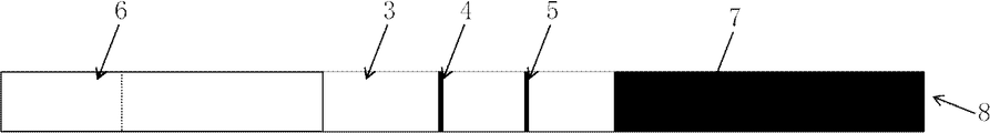
- immunochromatographic strip Specific examples of the immunochromatographic strip include an immunochromatographic strip 8 as shown in FIGS. 1 and 2.
- 1 is an adhesive sheet
- 2 is an impregnated member
- 3 is a membrane carrier
- 4 is a detection site
- 5 is an absorption member
- 6 is a sample addition member.
- the membrane carrier 3 is made of an elongated strip-shaped nitrocellulose membrane filter having a width of 5 mm and a length of 25 mm, and is attached to the middle of the pressure-sensitive adhesive sheet 1 also having a width of 5 mm.
- the membrane carrier 3 is located at a position 3 to 15 mm downstream from the end of the starting point side of chromatographic development, that is, the left side of FIG.
- upstream side the opposite right side is referred to as "downstream side”
- An anti-vitamin A antibody is immobilized on the cell, and a first capture site (test line) 4 for competitively capturing a conjugate of vitamin A and a compound and vitamin A in a test sample is formed.
- a second capture site (control line) 5 is provided at a position 8 to 25 mm downstream from the upstream end of the membrane carrier 3.
- This control line 5 is for confirming that the immunochromatographic development was performed regardless of the presence or absence of vitamin A, which is the substance to be analyzed.
- it can be formed by immobilizing an antibody against an antibody (IgG) bound to a labeled substance on the control line 5.
- the test line is arranged on the upstream side of the control line, and the distance between the test line and the control line is preferably 3 mm or more and less than 10 mm.
- sample addition member 6 for example, a sheet or film of a porous synthetic resin such as porous polyethylene and porous polypropylene, or a cellulose paper or non-woven fabric such as filter paper and cotton cloth may be used. can.
- a porous synthetic resin such as porous polyethylene and porous polypropylene
- a cellulose paper or non-woven fabric such as filter paper and cotton cloth
- the impregnating member 2 uses a strip-shaped glass fiber of 5 mm ⁇ 15 mm, but is not limited to this, and for example, filter paper, a nitrocellulose film, a porous plastic non-woven fabric such as polyethylene or polypropylene can also be used.
- the impregnated member 2 can be produced by impregnating a member such as glass fiber with a suspension containing the labeled body and drying the impregnated member 2. When the impregnated member is not used, the labeled substance may be contained in the sample diluent.
- the membrane carrier 3 uses a membrane filter made of nitrocellulose, a substance such as an antibody that can chromatographically develop the substance to be analyzed contained in the test sample and forms the first capture site (test line) 4 can be used. Any material may be used as long as it can be fixed, and other cellulose membranes, nylon membranes, glass fiber membranes and the like can also be used.
- the absorbing member 7 may be made of a material capable of quickly absorbing and holding a liquid, and examples thereof include cotton cloth, filter paper, and a porous plastic non-woven fabric made of polyethylene, polypropylene, etc., but filter paper is particularly suitable. be.
- the membrane carrier 3 is attached to the immunochromatographic strip 8 in the middle of the pressure-sensitive adhesive sheet 1, and is placed on the upstream end of the membrane carrier 3 and, if necessary, downstream of the impregnating member 2. It can be created by overlapping and connecting the end ends on the side and attaching the upstream portion of the impregnating member 2 to the adhesive sheet 1. Further, the downstream portion of the sample dropping portion (sample addition member) 6 is placed on the upper surface of the impregnation member 2, and the upstream portion of the sample addition member 6 is attached to the pressure-sensitive adhesive sheet 1 and also a film.
- the immunochromatographic strip 8 is formed by placing the upstream portion of the absorbing member 7 on the upper surface of the downstream portion of the carrier 3 and attaching the downstream portion of the absorbing member 7 to the adhesive sheet 1.
- the immunochromatographic strip is housed in a plastic housing case 9 or the like in order to protect it and to make it easy to handle (FIG. 2).
- the test sample dropping part 10 and the determination part 11 are above the sample addition member 6 and the first trapping site (test line) 4 and the second trapping site (control line) 5 of the immunochromatographic strip. It is preferably provided with an opening.
- the labeled body is arranged so as to be mixed with the mixed solution and chromatographically developed on the membrane carrier 3.
- the labeled substance is mixed with the test sample and the diluted solution in a suitable container separate from the immunochromatographic strip 8 to form a mixed solution, and then this mixed solution is injected into the sample addition member 6 of the immunochromatographic strip 8. It may be chromatographically developed on the membrane carrier 3.
- vitamin A in a biological sample is preferably quantified by a competitive method. Since it is difficult to sandwich a small molecule compound such as vitamin A with two kinds of antibodies, it is preferable to use a competitive method. That is, a biological sample diluted solution obtained by mixing a biological sample, a competing reagent (complex of vitamin A and a compound), and a sample diluted solution is dropped onto an immunochromatographic strip and developed to obtain vitamin A in the sample. And competing reagents are competitively captured by the anti-vitamin A antibody immobilized on the test line. The captured competing reagents can be quantified by coloring with a label (substance) and measuring the signal on the test line.
- the immunochromatographic measurement kit of the present invention contains at least a diluent for diluting a sample, and if necessary, a competing reagent (combination of vitamin A and a compound) and a calibration curve. Includes a vitamin A standard solution for preparation, a container for dilution, and the like. It may also include a measuring device (chromatography reader) for measuring the results of the immunochromatography.
- the sample diluent can be used as a developing solution for developing a biological sample.
- the sample diluent preferably contains a nonionic surfactant that improves the expandability of the biological sample and does not affect the immune response.
- the nonionic surfactant include polyoxyethylene alkyl phenyl ether (Triton (registered trademark) -based surfactant, etc.), polyoxyethylene alkyl ether (Brij (registered trademark) -based surfactant, etc.), and polyoxyethylene sorbitan fatty acid.
- esters Teween (registered trademark) -based surfactants and the like
- polyoxyethylene fatty acid esters examples thereof include polyoxyethylene fatty acid esters, sorbitan fatty acid esters, alkyl glucosides, sucrose fatty acid esters and the like.
- the surfactant may be used alone or in combination of two or more.
- the concentration of the nonionic surfactant is preferably 0.01 wt% to 5.0 wt%.
- Inorganic salts and a buffer used for pH adjustment may be further added to the sample diluted solution.
- the buffer any kind of buffer may be used as long as it has a sufficient buffering capacity in the target pH range, and for example, tris, phosphoric acid, phthalic acid, citric acid, maleic acid, etc. may be used. Examples thereof include succinic acid, oxalic acid, boric acid, tartaric acid, acetic acid, carbonic acid, good buffer (MES, ADA, PIPES, ACES, coramine hydrochloride, BES, TES, HEPES, acetamide glycine, tricin, glycine amide, bicin) and the like.
- TritonX-100 (Sigma-Aldrich, 10789704001) was dissolved in phosphate buffered saline (pH 7.4, Nacalai Tesque, 27576-21), and the TritonX-100 concentration was 0.1% by mass. pH 7.4) was adjusted.
- Vitamin A-biotin conjugates were prepared by biotinlating vitamin A (retinal, MyBiosource. Inc., MBS602324) using Biotin-hydrazide (Dojin Kagaku, B303). After preparation, it was stored at -30 ° C until use.
- test line and control line After preparing an anti-vitamin A antibody (Cloud-Clone Corp., PAD051Ge01) at a concentration of 1 mg / mL, 1.0 ⁇ L / of this was applied to a 25 mm ⁇ 300 mm nitrocellulose membrane filter. A test line was prepared by applying linearly in an amount of cm. Next, an anti-rabbit IgG antibody (MyBiosource. Inc., MBS5397780) was prepared at a concentration of 1 mg / mL, and then linearly applied to the above nitrocellulose membrane filter at an amount of 1.0 ⁇ L / cm to form a control line. Created. After preparing the test line and the control line, they were dried at 50 ° C. for 30 minutes, cut into a size of 25 mm ⁇ 5 mm, and used as a membrane carrier for developing an immunochromatography.
- an anti-vitamin A antibody Cloud-Clone Corp., PAD051Ge01
- a test line was prepared by applying linearly in an
- Vitamin A standard solution The vitamin A concentration in the serum obtained by collecting blood from a 20-month-old cow was measured by an HPLC method, and the price was used as the vitamin A standard solution. The measurement by HPLC was performed by requesting the Clinical Laboratory Center (Kinki Preventive Medicine Research Institute Co., Ltd.).
- Vitamin A-biotin conjugates were prepared by biotinlating vitamin A (retinal, MyBiosource. Inc., MBS602324) using Biotin-hydrazide (Dojin Kagaku, B303). After preparation, it was stored at -30 ° C until use.
- TritonX-100 (Sigma Aldrich, 10789704001) and the prepared vitamin A-biotin conjugate were dissolved in phosphate buffered saline (pH 7.4, Nakaraitesk, 27576-21) to pH 7.4, TritonX-.
- the sample diluent was adjusted so that the concentrations of 100 and the vitamin A biotin conjugate were 0.1 chamber% and 0.1 ⁇ M, respectively, and stored at ⁇ 30 ° C. until use.
- test line and control line After preparing an anti-vitamin A antibody (Cloud-Clone Corp., PAD051Ge01) at a concentration of 1 mg / mL, 1.0 ⁇ L / of this was applied to a 25 mm ⁇ 300 mm nitrocellulose membrane filter. A test line was prepared by applying linearly in an amount of cm. Next, an anti-rabbit IgG antibody (MyBiosource. Inc., MBS5397780) was prepared at a concentration of 1 mg / mL, and then linearly applied to the above nitrocellulose membrane filter at an amount of 1.0 ⁇ L / cm to form a control line. Created. After preparing the test line and the control line, they were dried at 50 ° C. for 30 minutes, cut into a size of 25 mm ⁇ 5 mm, and used as a membrane carrier for developing an immunochromatography.
- an anti-vitamin A antibody Cloud-Clone Corp., PAD051Ge01
- a test line was prepared by applying linearly in an
- Vitamin A standard solution The vitamin A concentration in the serum obtained by collecting blood from a 20-month-old cow was measured by an HPLC method, and the price was used as the vitamin A standard solution. The measurement by HPLC was performed by requesting the Clinical Laboratory Center (Kinki Preventive Medicine Research Institute Co., Ltd.).
- the concentration of vitamin A in serum obtained by collecting blood from 20 cows was measured by the HPLC method and the immunochromatography measurement kit of the present invention, respectively.
- the measurement by HPLC was performed by requesting the Clinical Laboratory Center (Kinki Preventive Medicine Research Institute Co., Ltd.).
- the immunochromatography method the vitamin A concentration of each serum was calculated using the standard curve obtained by simultaneously measuring the standard solution.
- the measurement results are shown in Table 7 and FIG. 10 (gold colloid), and Table 8 and FIG. 11 (colored cellulose fine particles).
- the correlation coefficients of the measured value by the immunochromatography method and the measured value by the HPLC method were 0.98 and 0.98, respectively, showing a good correlation.
- Vitamin A-biotin conjugates were prepared by biotinlating vitamin A (retinal, MyBiosource. Inc., MBS602324) using Biotin-hydrazide (Dojin Kagaku, B303). After preparation, it was stored at ⁇ 30 ° C. until use.
- TritonX-100 (Sigma-Aldrich, 10789704001) was dissolved in phosphate buffered saline (pH 7.4, Nacalai Tesque, 27576-21), and the TritonX-100 concentration was 0.1% by mass. pH 7.4) was adjusted.
- test line and control line After preparing an anti-vitamin A antibody (Cloud-Clone Corp., PAD051Ge01) at a concentration of 1 mg / mL, 1.0 ⁇ L / of this was applied to a 25 mm ⁇ 300 mm nitrocellulose membrane filter. A test line was prepared by applying linearly in an amount of cm. Next, an anti-rabbit IgG antibody (MyBiosource. Inc., MBS5397780) was prepared at a concentration of 1 mg / mL, and then linearly applied to the above nitrocellulose membrane filter at an amount of 1.0 ⁇ L / cm to form a control line. Created. After preparing the test line and the control line, they were dried at 50 ° C. for 30 minutes, cut into a size of 25 mm ⁇ 5 mm, and used as a membrane carrier for developing an immunochromatography.
- an anti-vitamin A antibody Cloud-Clone Corp., PAD051Ge01
- a test line was prepared by applying linearly in an
- Immunochromatographic Strip An immunochromatographic strip was prepared by arranging the membrane carrier, the impregnating member, and the absorbing member obtained above on the pressure-sensitive adhesive sheet.
- Vitamin A standard solution The vitamin A concentration in the serum obtained by collecting blood from a 20-month-old cow was measured by an HPLC method, and the price was used as the vitamin A standard solution. The measurement by HPLC was performed by requesting the Clinical Laboratory Center (Kinki Preventive Medicine Research Institute Co., Ltd.).
- the concentration of vitamin A in serum obtained by collecting blood from 20 cows was measured by the HPLC method and the immunochromatography measurement kit of the present invention, respectively.
- the measurement by HPLC was performed by requesting the Clinical Laboratory Center (Kinki Preventive Medicine Research Institute Co., Ltd.).
- the immunochromatography method the vitamin A concentration of each serum was calculated using the standard curve obtained by simultaneously measuring the standard solution.
- the measurement results are shown in Table 11 and FIG. 14 (gold colloid), and Table 12 and FIG. 15 (colored cellulose fine particles).
- the correlation coefficients of the measured value by the immunochromatography method and the measured value by the HPLC method were 0.98 and 0.97, respectively, showing a good correlation.
Abstract
【課題】 本発明は、肉牛の飼育現場において血液中のビタミンA濃度を迅速、簡便に測定可能なイムノクロマトストリップおよび測定キットを提供する。
【解決手段】 本発明は、生体試料中のビタミンAを定量するためのイムノクロマトストリップであって、前記イムノクロマトストリップは、最上流部に試料滴下部、および前記試料滴下部の下流側に順にテストライン、コントロールラインを有し、前記テストラインには、抗ビタミンA抗体が固定化されている、イムノクロマトストリップおよびイムノクロマト測定キットである。
Description
本発明は、生体試料中のビタミンAを競合法により定量するためのイムノクロマトストリップおよびイムノクロマト測定キットに関する。
肉牛、とくに高品質牛肉である和牛生産においては、筋肉中の脂肪交雑を高めることにより肉の評価が上がる。このため、脂肪の分解を促進する機能を持つビタミンAの給餌量を極端に減少させて脂肪交雑を誘導するビタミンコントロールと呼ばれる飼育管理方法が近年では主流となっている。
しかしながら、ビタミンAの極端な不足は、肝機能低下などによる健康障害を起こし、また筋肉水腫や筋炎の発生による肉質低下をきたすことがあり大きな問題となる。
このため、牛の栄養状態を知り、前述のような問題発生を防ぐため、血液中のビタミンA濃度を測定することが行われている。
しかしながら、ビタミンAの極端な不足は、肝機能低下などによる健康障害を起こし、また筋肉水腫や筋炎の発生による肉質低下をきたすことがあり大きな問題となる。
このため、牛の栄養状態を知り、前述のような問題発生を防ぐため、血液中のビタミンA濃度を測定することが行われている。
ビタミンA濃度の測定には、高速液体クロマトグラフィー(HPLC)が一般に用いられている。しかし獣医師等が現場で血液を採取し、持ち帰った後、検査機関に測定を依頼するケースがほとんどで、検査結果が出るのは数日後であり、もしビタミンAが不足していても即時的な対処が出来ないという欠点がある。
このため、血中ビタミンA濃度の測定結果を即時に得ることが出来る迅速、簡便な測定方法の開発が望まれている。
このため、血中ビタミンA濃度の測定結果を即時に得ることが出来る迅速、簡便な測定方法の開発が望まれている。
例えば、特許文献1および特許文献2には、血清から有機溶剤で抽出したビタミンAに特定波長の光を照射することにより、ビタミンAを定量する方法、および測定装置が記載されている。
しかしながら、この方法は、採取した血液を遠心して血清を分離した後、エタノールによる血清タンパク質の除去操作、およびヘプタンによるビタミンA成分の抽出操作が必要であり、HPLCのような大型装置は要らないものの、牛の飼育現場で実施するには操作が煩雑であり、時間や手間が掛かるといった問題があるため、より簡便に測定することが望まれている。
しかしながら、この方法は、採取した血液を遠心して血清を分離した後、エタノールによる血清タンパク質の除去操作、およびヘプタンによるビタミンA成分の抽出操作が必要であり、HPLCのような大型装置は要らないものの、牛の飼育現場で実施するには操作が煩雑であり、時間や手間が掛かるといった問題があるため、より簡便に測定することが望まれている。
本発明は、肉牛等の飼育現場において血液中のビタミンA濃度を迅速、簡便に測定することができるイムノクロマトストリップおよび測定キットを提供することを目的とする。
本発明者は、前記課題を解決するために鋭意検討した結果、以下に示す手段により、上記課題を解決できることを見出し、本発明に到達した。
すなわち、本発明は、以下の構成からなる。
(1) 生体試料中のビタミンAを定量するためのイムノクロマトストリップであって、
前記イムノクロマトストリップは、最上流部に試料滴下部、および前記試料滴下部の下流側に順にテストライン、コントロールラインを有し、
前記テストラインには、抗ビタミンA抗体が固定化されている、
ことを特徴とするイムノクロマトストリップ。
(2)さらに、前記試料滴下部の下流側に含浸部材を有し、前記含浸部材にはビタミンAと化合物との結合体、および前記化合物に対する抗体に標識物質を結合した標識体が含浸されていることを特徴とする(1)に記載のイムノクロマトストリップ。
(3) 前記ビタミンAと結合体をなす化合物は、ビオチンであることを特徴とする(2)に記載のイムノクロマトストリップ。
(4) 前記化合物に対する抗体は、抗ビオチン抗体であることを特徴とする(3)に記載のイムノクロマトストリップ。
(5) 前記標識物質は、金コロイドまたはセルロース微粒子であることを特徴とする(2)~(4)のいずれかに記載のイムノクロマトストリップ。
(6) 前記標識物質は、青色セルロース微粒子であることを特徴とする(2)~(4)のいずれかに記載のイムノクロマトストリップ。
(7) 前記抗ビタミンA抗体は、モノクローナル抗体であることを特徴とする(1)~(6)のいずれかに記載のイムノクロマトストリップ。
(8) 前記標識体を特異的に結合する抗体は、抗IgG抗体であることを特徴とする(1)~(7)のいずれかに記載のイムノクロマトストリップ。
(9) 前記コントロールラインには、前記標識体を特異的に結合する抗体が固定化されていることを特徴とする(1)~(8)のいずれかに記載のイムノクロマトストリップ。
(10) 全血または血清または血漿中のビタミンAを定量することを特徴とする(1)~(9)のいずれかに記載のイムノクロマトストリップ。
(11) (1)~(10)のいずれかに記載のイムノクロマトストリップ、および検体希釈液を含むことを特徴とするイムノクロマト測定キット。
(12) (1)~(10)のいずれかに記載のイムノクロマトストリップ、検体希釈液、および競合試薬を含むことを特徴とするイムノクロマト測定キット。
本発明により、肉牛の生産現場において血液中のビタミンA濃度を迅速、簡便に測定することが可能なビタミンA測定用イムノクロマトキットが提供される。
以下、本発明を詳細に説明する。
本発明は、生体試料中のビタミンAを定量するためのイムノクロマトストリップであって、前記イムノクロマトストリップは、最上流部に試料滴下部、および前記試料滴下部の下流側に順にテストライン、コントロールラインを有し、前記テストラインには、抗ビタミンA抗体が固定化されている、イムノクロマトストリップである。
(対象となる検体)
本発明において、係る対象となる試料としては、特に限定されるものではないが、血液(全血でも血清でも血漿でもよい)等が適しているが特に制限はない。動物種も、ウシの他、ヒト、ウマ、イヌ、ネコなどの血液を測定対象とすることが出来る。
本発明において、係る対象となる試料としては、特に限定されるものではないが、血液(全血でも血清でも血漿でもよい)等が適しているが特に制限はない。動物種も、ウシの他、ヒト、ウマ、イヌ、ネコなどの血液を測定対象とすることが出来る。
(ビタミンA)
本発明において、ビタミンAは、レチノール、レチナール、レチノイン酸、レチニルエステルなどのレチノイド類を指す。また、ビタミンAは、レチノール結合タンパク(RBP)、プレアルブミンと複合体を形成したものであってもよい。
本発明において、ビタミンAは、レチノール、レチナール、レチノイン酸、レチニルエステルなどのレチノイド類を指す。また、ビタミンAは、レチノール結合タンパク(RBP)、プレアルブミンと複合体を形成したものであってもよい。
(ビタミンAと化合物との結合体)
本発明において、ビタミンAと化合物との結合体(競合試薬)は、生体試料中の遊離またはタンパク質に結合した状態のビタミンAと競合することが出来、かつ標識体により検出が可能であれば、特に制限はない。結合体とすることでビタミンAが安定化し、また性能の良い抗体が入手し易いことから好ましい。化合物としては、牛血清アルブミン、卵白アルブミンやビオチンなどが挙げられ、これらの中でもビオチンが好適に用いられる。詳細な理由は不明だが、生体試料中のビタミンAはレチノール結合タンパク(RBP)と複合体を形成しているため、低分子量のビオチンとの結合体を用いることにより、競合法による定量に好ましいと推測している。なお、競合試薬は、イムノクロマトストリップの含浸部材に予め含浸させておいてもよいし、含浸部材を用いない場合には競合試薬として別調製したものを準備しておいてもよいし、検体希釈液に予め含ませておいてもよい。いずれいしも、ビオチンと結合体を形成したビタミンAは不安定な化合物であり、光や熱によって二重結合の異性化が起こりやすく、また酸や空気、金属イオンとも反応しやすいため容易に分解してしまう畏れがある。そのため、競合試薬、競合試薬を含むイムノクロマトストリップおよび検体希釈液は低温、暗所にて使用時まで保存するのが好ましい。保存温度としては4℃以下が好ましく、-20℃以下がより好ましく、-80℃以下がさらに好ましい。
本発明において、ビタミンAと化合物との結合体(競合試薬)は、生体試料中の遊離またはタンパク質に結合した状態のビタミンAと競合することが出来、かつ標識体により検出が可能であれば、特に制限はない。結合体とすることでビタミンAが安定化し、また性能の良い抗体が入手し易いことから好ましい。化合物としては、牛血清アルブミン、卵白アルブミンやビオチンなどが挙げられ、これらの中でもビオチンが好適に用いられる。詳細な理由は不明だが、生体試料中のビタミンAはレチノール結合タンパク(RBP)と複合体を形成しているため、低分子量のビオチンとの結合体を用いることにより、競合法による定量に好ましいと推測している。なお、競合試薬は、イムノクロマトストリップの含浸部材に予め含浸させておいてもよいし、含浸部材を用いない場合には競合試薬として別調製したものを準備しておいてもよいし、検体希釈液に予め含ませておいてもよい。いずれいしも、ビオチンと結合体を形成したビタミンAは不安定な化合物であり、光や熱によって二重結合の異性化が起こりやすく、また酸や空気、金属イオンとも反応しやすいため容易に分解してしまう畏れがある。そのため、競合試薬、競合試薬を含むイムノクロマトストリップおよび検体希釈液は低温、暗所にて使用時まで保存するのが好ましい。保存温度としては4℃以下が好ましく、-20℃以下がより好ましく、-80℃以下がさらに好ましい。
(標識体)
本発明において、標識体は、ビタミンAに結合した化合物に対する抗体に標識物質を結合させて得ることが出来る。抗体は、ビタミンAに結合した化合物に対する抗体であればよく、ポリクローナル抗体であってもモノクローナル抗体であってもよいが、反応特異性の観点からモノクローナル抗体であることが好ましい。
本発明において、標識体は、ビタミンAに結合した化合物に対する抗体に標識物質を結合させて得ることが出来る。抗体は、ビタミンAに結合した化合物に対する抗体であればよく、ポリクローナル抗体であってもモノクローナル抗体であってもよいが、反応特異性の観点からモノクローナル抗体であることが好ましい。
標識物質は特に制限はなく、例えば、呈色標識物質、酵素標識物質などが挙げられるが、迅速に検査結果が得られることから呈色標識物質であることが好ましい。呈色標識物質としては、コロイド金属および着色ラテックス粒子、着色セルロース微粒子などが挙げられる。コロイド金属の代表例としては、白金コロイド、金コロイド、銀コロイド、白金コロイド、パラジウムコロイド、金ナノロッド、金ナノプレート、銀ナノプレートなどが挙げられる。コロイド金属の粒子の大きさは通常、直径3~100nm程度とされる。着色ラテックスの代表例としては、赤色および青色などのそれぞれの顔料で着色されたポリスチレンラテックス、ポリメタクリル酸メチル、アクリル酸重合体などが挙げられる。ラテックス粒子の粒径としては特に制限されないが、粒径25~500nmのものが好ましい。この他に、市販されている着色セルロース微粒子なども使用出来る。着色セルロース微粒子の粒径としては特に制限されないが、粒径100~500nmのものが好ましい。
前記着色セルロース微粒子の色は、特に限定されないが、例えば赤色、青色、黄色、緑色、黒色、白色、蛍光色が挙げられる。これらの中でも、バックグラウンドのヘモグロビン由来の赤色の影響を受けにくい青色、黒色が好ましく、青色がより好ましい。このような着色セルロース微粒子としては、旭化成社製の着色セルロースナノビーズ(NanoAct(登録商標))が挙げられるが、この中でもNavy(BL1)、Dark Navy(BL2)、Black(KR1)が好ましく、Navy(BL1)、Dark Navy(BL2)がより好ましい。
本発明において、標識物質表面への非特異結合を抑えるためにブロッキング剤を用いて処理するのが好ましい。ブロッキング剤は、ポリエチレングリコールやタンパク質を用いるのが好ましい。タンパク質としてはBlocking Peptide Fragment、ウシ血清アルブミン(BSA)、カゼインなどが好ましい。これらのブロッキング剤は市販されているものがあればそれを用いても良いし、別途公知の方法で製造しても良い。分子サイズも特に制限されないが、平均分子量で100kDa以下が好ましい。一般的にブロッキング剤の分子サイズが小さいほど検出粒子1粒子に対するタンパク質の結合量が増加し感度などの性能が高くなる。
(テストライン)
本発明において、テストラインに固定化する抗体は、ビタミンAに特異的に結合することが出来る抗ビタミンA抗体であればよく、ポリクローナル抗体であっても、モノクローナル抗体であってもよいが、反応特異性の観点から、モノクローナル抗体であることが好ましい。ビタミンAは低分子化合物であり十分な複雑性を備えていないため、通常では免疫応答を誘発できない。このため、免疫した動物に抗体を産出させるには、オボアルブミンなどのキャリアタンパク質にビタミンAを化学結合したものを免疫原として用いる必要がある。また、アジュバントを混合して免疫原を注入すると、免疫応答強度が上がり、よい抗体を得る可能性が高まる。ポリクローナル抗体は、ウサギやマウスなどに免疫して得られた抗血清から精製して得ることが出来る。モノクローナル抗体は、例えば、ビタミンAとオボアルブミンの結合物を適当なアジュバントとともにマウスのような動物に免疫したのち、免疫された動物の脾細胞とミエローマ細胞とを融合し、融合細胞のみが増殖出来る選択培地で培養し、増殖した細胞を前記ビタミンAとの結合物などを使用して、たとえば酵素標識免疫法などにより選別することにより取得することができる。
本発明において、テストラインに固定化する抗体は、ビタミンAに特異的に結合することが出来る抗ビタミンA抗体であればよく、ポリクローナル抗体であっても、モノクローナル抗体であってもよいが、反応特異性の観点から、モノクローナル抗体であることが好ましい。ビタミンAは低分子化合物であり十分な複雑性を備えていないため、通常では免疫応答を誘発できない。このため、免疫した動物に抗体を産出させるには、オボアルブミンなどのキャリアタンパク質にビタミンAを化学結合したものを免疫原として用いる必要がある。また、アジュバントを混合して免疫原を注入すると、免疫応答強度が上がり、よい抗体を得る可能性が高まる。ポリクローナル抗体は、ウサギやマウスなどに免疫して得られた抗血清から精製して得ることが出来る。モノクローナル抗体は、例えば、ビタミンAとオボアルブミンの結合物を適当なアジュバントとともにマウスのような動物に免疫したのち、免疫された動物の脾細胞とミエローマ細胞とを融合し、融合細胞のみが増殖出来る選択培地で培養し、増殖した細胞を前記ビタミンAとの結合物などを使用して、たとえば酵素標識免疫法などにより選別することにより取得することができる。
本発明において、コントロールラインには、標識体中の化合物を特異的に結合する抗体が固定化されているのが好ましい。例えば、抗ウサギIgG抗体や抗マウスIgG抗体などを膜担体に固定化することによって形成することができる。コントロールラインを用いることにより、標識体が膜担体の最下流部まで移動したこと、即ち、イムノクロマト反応が(正常に)行われたことを確認することができる。
(イムノクロマトストリップ)
イムノクロマトストリップの具体例としては、図1、2に示すようなイムノクロマトストリップ8が挙げられる。図1、2において、1は粘着シート、2は含浸部材、3は膜担体、4は検出部位、5は吸収用部材、6は試料添加用部材を示している。膜担体3は、幅5mm、長さ25mmの細長い帯状のニトロセルロース製メンブレンフィルターからなり、同じく幅5mmの粘着シート1の中ほどに貼り付けられている。膜担体3には、クロマト展開の始点側、すなわち図1の左側(以下「上流側」とする。また、反対の右側を「下流側」とする。)の末端から下流側3~15mmの位置に抗ビタミンA抗体が固定され、ビタミンAと化合物との結合体と被験試料中のビタミンAを競合的に捕捉するための第一の捕捉部位(テストライン)4が形成されている。さらに、膜担体3の上流側の末端から下流側8~25mmの位置に第二の捕捉部位(コントロールライン)5が設けられている。このコントロールライン5は、分析対象物質であるビタミンAの存否に係わらずイムノクロマト展開が行われたことを確認するためのものである。例えば、標識体に結合している抗体(IgG)に対する抗体をコントロールライン5に固定化することによって形成することができる。なお、テストラインはコントロールラインよりも上流側に配置され、テストラインとコントロールラインとの距離は3mm以上10mm未満とするのが好ましい。
イムノクロマトストリップの具体例としては、図1、2に示すようなイムノクロマトストリップ8が挙げられる。図1、2において、1は粘着シート、2は含浸部材、3は膜担体、4は検出部位、5は吸収用部材、6は試料添加用部材を示している。膜担体3は、幅5mm、長さ25mmの細長い帯状のニトロセルロース製メンブレンフィルターからなり、同じく幅5mmの粘着シート1の中ほどに貼り付けられている。膜担体3には、クロマト展開の始点側、すなわち図1の左側(以下「上流側」とする。また、反対の右側を「下流側」とする。)の末端から下流側3~15mmの位置に抗ビタミンA抗体が固定され、ビタミンAと化合物との結合体と被験試料中のビタミンAを競合的に捕捉するための第一の捕捉部位(テストライン)4が形成されている。さらに、膜担体3の上流側の末端から下流側8~25mmの位置に第二の捕捉部位(コントロールライン)5が設けられている。このコントロールライン5は、分析対象物質であるビタミンAの存否に係わらずイムノクロマト展開が行われたことを確認するためのものである。例えば、標識体に結合している抗体(IgG)に対する抗体をコントロールライン5に固定化することによって形成することができる。なお、テストラインはコントロールラインよりも上流側に配置され、テストラインとコントロールラインとの距離は3mm以上10mm未満とするのが好ましい。
試料添加用部材6としては、例えば、多孔質ポリエチレンおよび多孔質ポリプロピレンなどのような多孔質合成樹脂のシートまたはフィルム、あるいは、濾紙および綿布などのようなセルロース製の紙または不織布などを用いることができる。
含浸部材2は、5mm×15mmの帯状のガラス繊維を用いるが、これに限定されるものではなく、例えば、濾紙、ニトロセルロース膜、ポリエチレン、ポリプロピレン等の多孔質プラスチック不織布なども使用できる。含浸部材2は、前記標識体を含む懸濁液を前記ガラス繊維等の部材に含浸せしめ、これを乾燥させることなどによって作製できる。なお、含浸部材を用いない場合には、前記標識体は検体希釈液に含ませておけばよい。
膜担体3は、ニトロセルロース製メンブレンフィルターを用いているが、被験試料に含まれる分析対象物質をクロマト展開可能で、かつ、第一の捕捉部位(テストライン)4を形成する抗体等の物質を固定可能なものであれば、いかなるものであってもよく、他のセルロース類膜、ナイロン膜、ガラス繊維膜なども使用できる。
吸収用部材7は、液体をすみやかに吸収、保持できる材質のものであればよく、綿布、濾紙、およびポリエチレン、ポリプロピレン等からなる多孔質プラスチック不織布等を挙げることができるが、特に濾紙が最適である。
イムノクロマトストリップ8は、図1、2に示されるように、膜担体3を粘着シート1の中ほどに貼着し、該膜担体3の上流側の末端の上に、必要により含浸部材2の下流側の末端を重ね合わせて連接するとともに、この含浸部材2の上流側部分を粘着シート1に貼着して作成できる。さらに、含浸部材2の上面に試料滴下部(試料添加用部材)6の下流側部分を載置するとともに、該試料添加用部材6の上流側部分を粘着シート1に貼着し、また、膜担体3の下流側部分の上面に吸収用部材7の上流側部分を載置するとともに、該吸収用部材7の下流側部分を粘着シート1に貼着せしめてイムノクロマトストリップ8を構成している。
イムノクロマトストリップは、これを保護するため、また、取り扱いがし易いように、プラスチック製のハウジングケース9などに収容されるのが好ましい(図2)。このケースは、例えば、イムノクロマトストリップの試料添加用部材6および第一の捕捉部位(テストライン)4および第二の捕捉部位(コントロールライン)5の上方に、被験試料滴下部10と判定部11が開口されて提供されることが好ましい。
(イムノクロマト展開)
被験試料と希釈液とを混合して調製した混合液を試料添加用部材6の試料滴下部10に注入した時、膜担体3の上流側の端部に連接した含浸部材2に予め含浸させた標識体が、該混合液と混合して膜担体3へとクロマト展開されるように、配置しておくことが好ましい。あるいは、標識体を、イムノクロマトストリップ8とは別の適当な容器内で、被験試料及び希釈液と混合して混合液とした後、この混合液をイムノクロマトストリップ8の試料添加用部材6に注入して膜担体3にクロマト展開させても構わない。
被験試料と希釈液とを混合して調製した混合液を試料添加用部材6の試料滴下部10に注入した時、膜担体3の上流側の端部に連接した含浸部材2に予め含浸させた標識体が、該混合液と混合して膜担体3へとクロマト展開されるように、配置しておくことが好ましい。あるいは、標識体を、イムノクロマトストリップ8とは別の適当な容器内で、被験試料及び希釈液と混合して混合液とした後、この混合液をイムノクロマトストリップ8の試料添加用部材6に注入して膜担体3にクロマト展開させても構わない。
(競合法)
本発明において、生体試料中のビタミンAは競合法により定量するのが好ましい。ビタミンAのような低分子化合物は、2種類の抗体でサンドイッチすることが難しいため、競合法をとることが好ましい。即ち、生体試料、競合試薬(ビタミンAと化合物との複合体)、および検体希釈液を混合して得られた生体試料希釈液をイムノクロマトストリップ上に滴下、展開することにより、試料中のビタミンA及び競合試薬は、テストラインに固定化された抗ビタミンA抗体に競合的に捕捉される。捕捉された競合試薬を、標識体(物質)により呈色させ、テストライン上のシグナルを測定することにより定量することができる。
本発明において、生体試料中のビタミンAは競合法により定量するのが好ましい。ビタミンAのような低分子化合物は、2種類の抗体でサンドイッチすることが難しいため、競合法をとることが好ましい。即ち、生体試料、競合試薬(ビタミンAと化合物との複合体)、および検体希釈液を混合して得られた生体試料希釈液をイムノクロマトストリップ上に滴下、展開することにより、試料中のビタミンA及び競合試薬は、テストラインに固定化された抗ビタミンA抗体に競合的に捕捉される。捕捉された競合試薬を、標識体(物質)により呈色させ、テストライン上のシグナルを測定することにより定量することができる。
(イムノクロマト測定キット)
本発明のイムノクロマト測定キットは、上記のイムノクロマトストリップに加えて、検体を希釈するための希釈液を少なくとも含み、更に必要に応じて、競合試薬(ビタミンAと化合物との結合体)、検量線を作成するためのビタミンA標準液や、希釈するための容器などを含む。また、イムノクロマト結果を測定するための測定装置(クロマトリーダー)も含む場合がある。
本発明のイムノクロマト測定キットは、上記のイムノクロマトストリップに加えて、検体を希釈するための希釈液を少なくとも含み、更に必要に応じて、競合試薬(ビタミンAと化合物との結合体)、検量線を作成するためのビタミンA標準液や、希釈するための容器などを含む。また、イムノクロマト結果を測定するための測定装置(クロマトリーダー)も含む場合がある。
本発明において、検体希釈液は、生体試料を展開させるための展開液として使用することができる。検体希釈液は、生体試料の展開性を向上させかつ免疫反応に影響しないノニオン性界面活性剤を含むことが好ましい。ノニオン性界面活性剤としては、ポリオキシエチレンアルキルフェニルエーテル(Triton(登録商標)系界面活性剤等)、ポリオキシエチレンアルキルエーテル(Brij(登録商標)系界面活性剤等)、ポリオキシエチレンソルビタン脂肪酸エステル(Tween(登録商標)系界面活性剤等)、ポリオキシエチレン脂肪酸エステル、ソルビタン脂肪酸エステル、アルキルグルコシド、ショ糖脂肪酸エステル等が挙げられる。また、前記界面活性剤は単独で用いても、二種以上を組み合わせて用いてもよい。また、ノニオン性界面活性剤の濃度としては、好ましくは0.01wt%~5.0wt%である。
前記検体希釈液にはさらに、無機塩類やpH調整に用いる緩衝剤を添加しても良い。
前記緩衝剤としては、目的とするpH範囲において充分な緩衝能力を有していれば、いかなる種類の緩衝剤を用いてもよく、例えば、トリス、リン酸、フタル酸、クエン酸、マレイン酸、コハク酸、シュウ酸、ホウ酸、酒石酸、酢酸、炭酸、グッドバッファー(MES、ADA、PIPES、ACES、コラミン塩酸、BES、TES、HEPES、アセトアミドグリシン、トリシン、グリシンアミド、ビシン)等が挙げられる。
前記緩衝剤としては、目的とするpH範囲において充分な緩衝能力を有していれば、いかなる種類の緩衝剤を用いてもよく、例えば、トリス、リン酸、フタル酸、クエン酸、マレイン酸、コハク酸、シュウ酸、ホウ酸、酒石酸、酢酸、炭酸、グッドバッファー(MES、ADA、PIPES、ACES、コラミン塩酸、BES、TES、HEPES、アセトアミドグリシン、トリシン、グリシンアミド、ビシン)等が挙げられる。
(実施例1)
(検体希釈液の調製)
リン酸緩衝生理食塩水(pH7.4、ナカライテスク社、27576-21)にTritonX-100(シグマアルドリッチ社、10789704001)を溶解させ、TritonX-100の濃度が0.1質量%の検体希釈液(pH7.4)を調整した。
(検体希釈液の調製)
リン酸緩衝生理食塩水(pH7.4、ナカライテスク社、27576-21)にTritonX-100(シグマアルドリッチ社、10789704001)を溶解させ、TritonX-100の濃度が0.1質量%の検体希釈液(pH7.4)を調整した。
(ビタミンA-ビオチン結合体の調製)
ビタミンA(レチナール、MyBiosource.Inc.、MBS6023224)を、Biotin-hydrazide(同仁化学、B303)を用いて、ビオチン化することにより、ビタミンA-ビオチン結合体を調製した。調製後、使用時まで-30℃に保存した。
ビタミンA(レチナール、MyBiosource.Inc.、MBS6023224)を、Biotin-hydrazide(同仁化学、B303)を用いて、ビオチン化することにより、ビタミンA-ビオチン結合体を調製した。調製後、使用時まで-30℃に保存した。
(抗ビオチン抗体結合金コロイドの調製)
金コロイド液(BBI Solutions、EMGC40、OD=1)をpH8.0の50mM KH2PO4に懸濁させ、これに抗ビオチン抗体(SIGMA、B3640)を加えて混合し、室温で10分間静置して、抗体を金コロイド表面に結合させた。更に、金コロイド表面への非特異結合を抑えるために、1質量%PEG200、10質量%BSAを添加しブロッキング処理を行った。この後、洗浄操作を繰り返し、20mMTris-HCl(pH8.2)、0.05質量%PEG2000、150mMNaCl、1質量%BSA溶液に懸濁して、抗ビオチン抗体結合金コロイド液を調製した。
金コロイド液(BBI Solutions、EMGC40、OD=1)をpH8.0の50mM KH2PO4に懸濁させ、これに抗ビオチン抗体(SIGMA、B3640)を加えて混合し、室温で10分間静置して、抗体を金コロイド表面に結合させた。更に、金コロイド表面への非特異結合を抑えるために、1質量%PEG200、10質量%BSAを添加しブロッキング処理を行った。この後、洗浄操作を繰り返し、20mMTris-HCl(pH8.2)、0.05質量%PEG2000、150mMNaCl、1質量%BSA溶液に懸濁して、抗ビオチン抗体結合金コロイド液を調製した。
(抗ビオチン抗体結合セルロース微粒子の調製)
セルロース微粒子液(旭化成、BL1、1質量%)をpH7.0の10mM Tris Buffer(PBS)に懸濁させ、これに抗ビオチン抗体(SIGMA、B3640)を加えて混合し、37℃で120分間静置して、抗体をセルロース微粒子表面に結合させた。更に、セルロース微粒子表面への非特異結合を抑えるために、1質量%カゼインを添加し、37℃で60分間静置してブロッキング処理を行った。この後、洗浄操作を行った後、1質量%スクロース含有pH7.4のPBSに懸濁して、抗ビオチン抗体結合セルロース微粒子液を調製した。
セルロース微粒子液(旭化成、BL1、1質量%)をpH7.0の10mM Tris Buffer(PBS)に懸濁させ、これに抗ビオチン抗体(SIGMA、B3640)を加えて混合し、37℃で120分間静置して、抗体をセルロース微粒子表面に結合させた。更に、セルロース微粒子表面への非特異結合を抑えるために、1質量%カゼインを添加し、37℃で60分間静置してブロッキング処理を行った。この後、洗浄操作を行った後、1質量%スクロース含有pH7.4のPBSに懸濁して、抗ビオチン抗体結合セルロース微粒子液を調製した。
(イムノクロマトストリップの作製)
(1-1)抗ビオチン抗体結合金コロイド含浸部材の作製
8mm×150mmの帯状のガラス繊維不織布に、上記で得られた抗ビオチン抗体結合金コロイドを20mMTris-HCl、0.05質量%PEG2000、37.5mMNaCl、0.25質量%BSA、3質量%スクロース溶液に懸濁し、これを0.5mL含浸させた。室温で乾燥させた後に、8mm×5mmの大きさに切断し、抗ビオチン抗体結合金コロイド含浸部材とした。
(1-1)抗ビオチン抗体結合金コロイド含浸部材の作製
8mm×150mmの帯状のガラス繊維不織布に、上記で得られた抗ビオチン抗体結合金コロイドを20mMTris-HCl、0.05質量%PEG2000、37.5mMNaCl、0.25質量%BSA、3質量%スクロース溶液に懸濁し、これを0.5mL含浸させた。室温で乾燥させた後に、8mm×5mmの大きさに切断し、抗ビオチン抗体結合金コロイド含浸部材とした。
(1-2)抗ビオチン抗体結合セルロース微粒子含浸部材の作製
8mm×150mmの帯状のガラス繊維不織布に、上記で得られた抗ビオチン抗体結合セルロース微粒子液を0.5mL含浸させた。室温で乾燥させた後に、8mm×5mmの大きさに切断し、抗ビオチン抗体結合セルロース含浸部材とした。
8mm×150mmの帯状のガラス繊維不織布に、上記で得られた抗ビオチン抗体結合セルロース微粒子液を0.5mL含浸させた。室温で乾燥させた後に、8mm×5mmの大きさに切断し、抗ビオチン抗体結合セルロース含浸部材とした。
(2)テストラインおよびコントロールラインの作製
抗ビタミンA抗体(Cloud-Clone Corp.、PAD051Ge01)を1mg/mLの濃度に調製した後、これを25mm×300mmのニトロセルロース製メンブレンフィルターに1.0μL/cmの量で線状に塗布してテストラインを作製した。
次に、抗ウサギIgG抗体(MyBiosource.Inc.、MBS539780)を1mg/mLの濃度に調製した後、上記ニトロセルロース製メンブレンフィルターに1.0μL/cmの量で線状に塗布してコントロールラインを作成した。
テストラインおよびコントロールラインを作成後、50℃で30分間乾燥させ、25mm×5mmの大きさに切断し、イムノクロマト展開用膜担体とした。
(3)イムノクロマトストリップの作製
図1に示すように、粘着シート1の上に、上記(2)で得られた膜担体3、上記(1-1)および(1-2)で得られた含浸部材2、吸収用部材7を配置し、イムノクロマトストリップを作製した。
図1に示すように、粘着シート1の上に、上記(2)で得られた膜担体3、上記(1-1)および(1-2)で得られた含浸部材2、吸収用部材7を配置し、イムノクロマトストリップを作製した。
(4)ビタミンA標準液
20か月齢の牛から採血して得られた血清中のビタミンA濃度をHPLC法にて測定し、値付けしたものをビタミンA標準液とした。HPLCによる測定は、臨床検査センター(株式会社近畿予防医学研究所)に依頼して測定した。
20か月齢の牛から採血して得られた血清中のビタミンA濃度をHPLC法にて測定し、値付けしたものをビタミンA標準液とした。HPLCによる測定は、臨床検査センター(株式会社近畿予防医学研究所)に依頼して測定した。
(5)測定キットを用いた定量
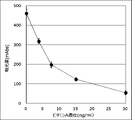
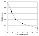
上記標準液を検体希釈液(pH7.4 PBS、0.1質量%TritonX-100)を用いて希釈し、各濃度のビタミンA液を調製した。そして、この被験試料を前記(3)で得られたイムノクロマトストリップの試料添加用部材6の試料滴下部10にマイクロピペットで100μL滴下し、10分後、テストラインにおける吸光度をイムノクロマトリーダ(浜松ホトニクス、C10066-10)で測定した。その結果を表1(金コロイド)および表2(着色セルロース微粒子)に示した。また、測定の結果得られたグラフを図4および図5に示した。
上記標準液を検体希釈液(pH7.4 PBS、0.1質量%TritonX-100)を用いて希釈し、各濃度のビタミンA液を調製した。そして、この被験試料を前記(3)で得られたイムノクロマトストリップの試料添加用部材6の試料滴下部10にマイクロピペットで100μL滴下し、10分後、テストラインにおける吸光度をイムノクロマトリーダ(浜松ホトニクス、C10066-10)で測定した。その結果を表1(金コロイド)および表2(着色セルロース微粒子)に示した。また、測定の結果得られたグラフを図4および図5に示した。


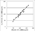
(6)ウシ血清のビタミンA測定
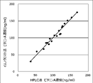
20頭の牛から採血して得られた血清中のビタミンA濃度を、それぞれHPLC法および本発明のイムノクロマトストリップを用いて測定した。HPLCによる測定は、臨床検査センター(株式会社近畿予防医学研究所)に依頼して測定した。イムノクロマト法では、標準液を同時に測定して得られた標準曲線を用いて、各血清のビタミンA濃度を算出した。測定結果を表3および図6(金コロイド)、表4および図7(着色セルロース微粒子)に示した。イムノクロマト法の測定値とHPLC法による測定値の相関係数はそれぞれ0.98、0.98であり、良好な相関関係を示した。
20頭の牛から採血して得られた血清中のビタミンA濃度を、それぞれHPLC法および本発明のイムノクロマトストリップを用いて測定した。HPLCによる測定は、臨床検査センター(株式会社近畿予防医学研究所)に依頼して測定した。イムノクロマト法では、標準液を同時に測定して得られた標準曲線を用いて、各血清のビタミンA濃度を算出した。測定結果を表3および図6(金コロイド)、表4および図7(着色セルロース微粒子)に示した。イムノクロマト法の測定値とHPLC法による測定値の相関係数はそれぞれ0.98、0.98であり、良好な相関関係を示した。


(実施例2)
(ビタミンAビオチン結合体の調製)
ビタミンA(レチナール、MyBiosource.Inc.、MBS6023224)を、Biotin-hydrazide(同仁化学、B303)を用いて、ビオチン化することにより、ビタミンA-ビオチン結合体を調製した。調製後、使用時まで-30℃に保存した。
(ビタミンAビオチン結合体の調製)
ビタミンA(レチナール、MyBiosource.Inc.、MBS6023224)を、Biotin-hydrazide(同仁化学、B303)を用いて、ビオチン化することにより、ビタミンA-ビオチン結合体を調製した。調製後、使用時まで-30℃に保存した。
(検体希釈液の調製)
リン酸緩衝生理食塩水(pH7.4、ナカライテスク社、27576-21)にTritonX-100(シグマアルドリッチ社、10789704001)、および調製したビタミンA-ビオチン結合体を溶解させ、pH7.4、TritonX-100およびビタミンAビオチン結合体の濃度がそれぞれ、0.1室用%、0.1μMになるように検体希釈液を調整し、使用時まで-30℃で保管した。
リン酸緩衝生理食塩水(pH7.4、ナカライテスク社、27576-21)にTritonX-100(シグマアルドリッチ社、10789704001)、および調製したビタミンA-ビオチン結合体を溶解させ、pH7.4、TritonX-100およびビタミンAビオチン結合体の濃度がそれぞれ、0.1室用%、0.1μMになるように検体希釈液を調整し、使用時まで-30℃で保管した。
(抗ビオチン抗体結合金コロイドの調製)
金コロイド液(BBI Solutions、EMGC40、OD=1)をpH8.0の50mM KH2PO4に懸濁させ、これに抗ビオチン抗体(SIGMA、B3640)を加えて混合し、室温で10分間静置して抗体を金コロイド表面に結合させた。更に、金コロイド表面への非特異結合を抑えるために、1質量%PEG200、10質量%BSAを添加しブロッキング処理を行った。この後、洗浄操作を繰り返し、20mMTris-HCl(pH8.2)、0.05質量%PEG2000、150mM NaCl、1質量%BSA溶液に懸濁して、抗ビオチン抗体結合金コロイド液を調製した。
金コロイド液(BBI Solutions、EMGC40、OD=1)をpH8.0の50mM KH2PO4に懸濁させ、これに抗ビオチン抗体(SIGMA、B3640)を加えて混合し、室温で10分間静置して抗体を金コロイド表面に結合させた。更に、金コロイド表面への非特異結合を抑えるために、1質量%PEG200、10質量%BSAを添加しブロッキング処理を行った。この後、洗浄操作を繰り返し、20mMTris-HCl(pH8.2)、0.05質量%PEG2000、150mM NaCl、1質量%BSA溶液に懸濁して、抗ビオチン抗体結合金コロイド液を調製した。
(抗ビオチン抗体結合セルロース微粒子の調製)
標識物質としてセルロース微粒子液(旭化成、BL1、1質量%)をpH7.0の10mM Tris Buffer(PBS)に懸濁させ、これに抗ビオチン抗体(SIGMA、B3640)を加えて混合し、37℃で120分間静置して、抗体をセルロース微粒子表面に結合させた。更に、セルロース微粒子表面への非特異結合を抑えるために、1質量%カゼインを添加し、37℃で60分間静置してブロッキング処理を行った。この後、洗浄操作を行った後、1質量%スクロース含有PBS(pH7.4)に懸濁して、抗ビオチン抗体結合セルロース微粒子液を調製した。
標識物質としてセルロース微粒子液(旭化成、BL1、1質量%)をpH7.0の10mM Tris Buffer(PBS)に懸濁させ、これに抗ビオチン抗体(SIGMA、B3640)を加えて混合し、37℃で120分間静置して、抗体をセルロース微粒子表面に結合させた。更に、セルロース微粒子表面への非特異結合を抑えるために、1質量%カゼインを添加し、37℃で60分間静置してブロッキング処理を行った。この後、洗浄操作を行った後、1質量%スクロース含有PBS(pH7.4)に懸濁して、抗ビオチン抗体結合セルロース微粒子液を調製した。
(イムノクロマトストリップの作製)
(1-1)抗ビオチン抗体結合金コロイド含浸部材の作製
8mm×150mmの帯状のガラス繊維不織布に、上記で得られた抗ビオチン抗体結合金コロイドを20mMTris-HCl、0.05質量%PEG2000、37.5mMNaCl、0.25質量%BSA、3質量%スクロース溶液に懸濁し、これを0.5mL含浸させた。室温で乾燥させた後に、8mm×5mmの大きさに切断し、抗ビオチン抗体結合金コロイド含浸部材とした。
(1-1)抗ビオチン抗体結合金コロイド含浸部材の作製
8mm×150mmの帯状のガラス繊維不織布に、上記で得られた抗ビオチン抗体結合金コロイドを20mMTris-HCl、0.05質量%PEG2000、37.5mMNaCl、0.25質量%BSA、3質量%スクロース溶液に懸濁し、これを0.5mL含浸させた。室温で乾燥させた後に、8mm×5mmの大きさに切断し、抗ビオチン抗体結合金コロイド含浸部材とした。
(1-2)抗ビオチン抗体結合セルロース微粒子含浸部材の作製
8mm×150mmの帯状のガラス繊維不織布に、上記で得られた抗ビオチン抗体結合セルロース微粒子液を0.5mL含浸させた。室温で乾燥させた後に、8mm×5mmの大きさに切断し、抗ビオチン抗体結合セルロース含浸部材とした。
8mm×150mmの帯状のガラス繊維不織布に、上記で得られた抗ビオチン抗体結合セルロース微粒子液を0.5mL含浸させた。室温で乾燥させた後に、8mm×5mmの大きさに切断し、抗ビオチン抗体結合セルロース含浸部材とした。
(2)テストラインおよびコントロールラインの作製
抗ビタミンA抗体(Cloud-Clone Corp.、PAD051Ge01)を1mg/mLの濃度に調製した後、これを25mm×300mmのニトロセルロース製メンブレンフィルターに1.0μL/cmの量で線状に塗布してテストラインを作製した。
次に、抗ウサギIgG抗体(MyBiosource.Inc.、MBS539780)を1mg/mLの濃度に調製した後、上記ニトロセルロース製メンブレンフィルターに1.0μL/cmの量で線状に塗布してコントロールラインを作成した。
テストラインおよびコントロールラインを作成後、50℃で30分間乾燥させ、25mm×5mmの大きさに切断し、イムノクロマト展開用膜担体とした。
抗ビタミンA抗体(Cloud-Clone Corp.、PAD051Ge01)を1mg/mLの濃度に調製した後、これを25mm×300mmのニトロセルロース製メンブレンフィルターに1.0μL/cmの量で線状に塗布してテストラインを作製した。
次に、抗ウサギIgG抗体(MyBiosource.Inc.、MBS539780)を1mg/mLの濃度に調製した後、上記ニトロセルロース製メンブレンフィルターに1.0μL/cmの量で線状に塗布してコントロールラインを作成した。
テストラインおよびコントロールラインを作成後、50℃で30分間乾燥させ、25mm×5mmの大きさに切断し、イムノクロマト展開用膜担体とした。
(3)イムノクロマトストリップの作製
粘着シートの上に、上記得られた膜担体、含浸部材、および吸収用部材を配置し、イムノクロマトストリップを作製した。
粘着シートの上に、上記得られた膜担体、含浸部材、および吸収用部材を配置し、イムノクロマトストリップを作製した。
(4)ビタミンA標準液
20か月齢の牛から採血して得られた血清中のビタミンA濃度をHPLC法にて測定し、値付けしたものをビタミンA標準液とした。HPLCによる測定は、臨床検査センター(株式会社近畿予防医学研究所)に依頼して測定した。
20か月齢の牛から採血して得られた血清中のビタミンA濃度をHPLC法にて測定し、値付けしたものをビタミンA標準液とした。HPLCによる測定は、臨床検査センター(株式会社近畿予防医学研究所)に依頼して測定した。
(5)測定キットを用いた定量
上記標準液を検体希釈液(pH7.4 PBS、0.1質量%TritonX-100、0.1μM ビタミンA-ビオチン結合体)を用いて希釈し、各濃度のビタミンA液を調製した。そして、この被験試料に前記(3)で得られたイムノクロマトストリップの試料添加用部材の試料滴下部にマイクロピペットで100μL滴下し、10分後、テストラインにおける吸光度をイムノクロマトリーダ(浜松ホトニクス、C10066-10)にて測定した。その結果を表5(金コロイド)および表6(着色セルロース微粒子)に示した。また、測定の結果得られたグラフを図8(金コロイド)および図9(着色セルロース微粒子)に示した。
上記標準液を検体希釈液(pH7.4 PBS、0.1質量%TritonX-100、0.1μM ビタミンA-ビオチン結合体)を用いて希釈し、各濃度のビタミンA液を調製した。そして、この被験試料に前記(3)で得られたイムノクロマトストリップの試料添加用部材の試料滴下部にマイクロピペットで100μL滴下し、10分後、テストラインにおける吸光度をイムノクロマトリーダ(浜松ホトニクス、C10066-10)にて測定した。その結果を表5(金コロイド)および表6(着色セルロース微粒子)に示した。また、測定の結果得られたグラフを図8(金コロイド)および図9(着色セルロース微粒子)に示した。


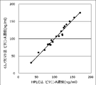
(6)ウシ血清のビタミンA測定
20頭の牛から採血して得られた血清中のビタミンA濃度を、それぞれHPLC法および本発明のイムノクロマト測定キットを用いて測定した。HPLCによる測定は、臨床検査センター(株式会社近畿予防医学研究所)に依頼して測定した。イムノクロマト法では、標準液を同時に測定して得られた標準曲線を用いて、各血清のビタミンA濃度を算出した。測定結果を表7および図10(金コロイド)、表8および図11(着色セルロース微粒子)に示した。イムノクロマト法の測定値とHPLC法による測定値の相関係数はそれぞれ0.98、0.98であり、良好な相関関係を示した。
20頭の牛から採血して得られた血清中のビタミンA濃度を、それぞれHPLC法および本発明のイムノクロマト測定キットを用いて測定した。HPLCによる測定は、臨床検査センター(株式会社近畿予防医学研究所)に依頼して測定した。イムノクロマト法では、標準液を同時に測定して得られた標準曲線を用いて、各血清のビタミンA濃度を算出した。測定結果を表7および図10(金コロイド)、表8および図11(着色セルロース微粒子)に示した。イムノクロマト法の測定値とHPLC法による測定値の相関係数はそれぞれ0.98、0.98であり、良好な相関関係を示した。


(比較例1)
保冷庫(-30℃)から取出し、室温(25℃)に1日放置した検体希釈液を用いて、ウシ血清中のビタミンA濃度を測定したところ、測定値のバラつきが大きく、またHPLC法による測定値との相関係数も0.81であり、測定精度の著しい低下が認められた。
保冷庫(-30℃)から取出し、室温(25℃)に1日放置した検体希釈液を用いて、ウシ血清中のビタミンA濃度を測定したところ、測定値のバラつきが大きく、またHPLC法による測定値との相関係数も0.81であり、測定精度の著しい低下が認められた。
(実施例3)
(競合試薬の調製)
ビタミンA(レチナール、MyBiosource.Inc.、MBS6023224)を、Biotin-hydrazide(同仁化学、B303)を用いて、ビオチン化することにより、ビタミンA-ビオチン結合物を調製した。調製後、使用時まで-30℃にて保存した。
(競合試薬の調製)
ビタミンA(レチナール、MyBiosource.Inc.、MBS6023224)を、Biotin-hydrazide(同仁化学、B303)を用いて、ビオチン化することにより、ビタミンA-ビオチン結合物を調製した。調製後、使用時まで-30℃にて保存した。
(検体希釈液の調製)
リン酸緩衝生理食塩水(pH7.4、ナカライテスク社、27576-21)にTritonX-100(シグマアルドリッチ社、10789704001)を溶解させ、TritonX-100の濃度が0.1質量%の検体希釈液(pH7.4)を調整した。
リン酸緩衝生理食塩水(pH7.4、ナカライテスク社、27576-21)にTritonX-100(シグマアルドリッチ社、10789704001)を溶解させ、TritonX-100の濃度が0.1質量%の検体希釈液(pH7.4)を調整した。
(抗ビオチン抗体結合金コロイドの調製)
金コロイド液(BBI Solutions、EMGC40、OD=1)をpH8.0の50mM KH2PO4に懸濁させ、これに抗ビオチン抗体(SIGMA、B3640)を加えて混合し、室温で10分間静置して抗体を金コロイド表面に結合させた。更に、金コロイド表面への非特異結合を抑えるために、1質量%PEG200、10質量%BSAを添加しブロッキング処理を行った。この後、洗浄操作を繰り返し、20mMTris-HCl(pH8.2)、0.05質量%PEG2000、150mM NaCl、1質量%BSA溶液に懸濁して、抗ビオチン抗体結合金コロイド液を調製した。
金コロイド液(BBI Solutions、EMGC40、OD=1)をpH8.0の50mM KH2PO4に懸濁させ、これに抗ビオチン抗体(SIGMA、B3640)を加えて混合し、室温で10分間静置して抗体を金コロイド表面に結合させた。更に、金コロイド表面への非特異結合を抑えるために、1質量%PEG200、10質量%BSAを添加しブロッキング処理を行った。この後、洗浄操作を繰り返し、20mMTris-HCl(pH8.2)、0.05質量%PEG2000、150mM NaCl、1質量%BSA溶液に懸濁して、抗ビオチン抗体結合金コロイド液を調製した。
(抗ビオチン抗体結合セルロース微粒子の調製)
標識物質としてセルロース微粒子液(旭化成、BL1、1質量%)をpH7.0の10mM Tris Buffer(PBS)に懸濁させ、これに抗ビオチン抗体(SIGMA、B3640)を加えて混合し、37℃で120分間静置して、抗体をセルロース微粒子表面に結合させた。更に、セルロース微粒子表面への非特異結合を抑えるために、1質量%カゼインを添加し、37℃で60分間静置してブロッキング処理を行った。この後、洗浄操作を行った後、1質量%スクロース含有PBS(pH7.4)に懸濁して、抗ビオチン抗体結合セルロース微粒子液を調製した。
標識物質としてセルロース微粒子液(旭化成、BL1、1質量%)をpH7.0の10mM Tris Buffer(PBS)に懸濁させ、これに抗ビオチン抗体(SIGMA、B3640)を加えて混合し、37℃で120分間静置して、抗体をセルロース微粒子表面に結合させた。更に、セルロース微粒子表面への非特異結合を抑えるために、1質量%カゼインを添加し、37℃で60分間静置してブロッキング処理を行った。この後、洗浄操作を行った後、1質量%スクロース含有PBS(pH7.4)に懸濁して、抗ビオチン抗体結合セルロース微粒子液を調製した。
(イムノクロマトストリップの作製)
(1-1)抗ビオチン抗体結合金コロイド含浸部材の作製
8mm×150mmの帯状のガラス繊維不織布に、上記で得られた抗ビオチン抗体結合金コロイドを20mMTris-HCl、0.05質量%PEG2000、37.5mMNaCl、0.25質量%BSA、3質量%スクロース溶液に懸濁し、これを0.5mL含浸させた。室温で乾燥させた後に、8mm×5mmの大きさに切断し、抗ビオチン抗体結合金コロイド含浸部材とした。
(1-1)抗ビオチン抗体結合金コロイド含浸部材の作製
8mm×150mmの帯状のガラス繊維不織布に、上記で得られた抗ビオチン抗体結合金コロイドを20mMTris-HCl、0.05質量%PEG2000、37.5mMNaCl、0.25質量%BSA、3質量%スクロース溶液に懸濁し、これを0.5mL含浸させた。室温で乾燥させた後に、8mm×5mmの大きさに切断し、抗ビオチン抗体結合金コロイド含浸部材とした。
(1-2)抗ビオチン抗体結合セルロース粒子含浸部材の作製
8mm×150mmの帯状のガラス繊維不織布に、上記で得られた抗ビオチン抗体結合セルロース微粒子液を0.5mL含浸させた。室温で乾燥させた後に、8mm×5mmの大きさに切断し、抗ビオチン抗体結合セルロース含浸部材とした。
8mm×150mmの帯状のガラス繊維不織布に、上記で得られた抗ビオチン抗体結合セルロース微粒子液を0.5mL含浸させた。室温で乾燥させた後に、8mm×5mmの大きさに切断し、抗ビオチン抗体結合セルロース含浸部材とした。
(2)テストラインおよびコントロールラインの作製
抗ビタミンA抗体(Cloud-Clone Corp.、PAD051Ge01)を1mg/mLの濃度に調製した後、これを25mm×300mmのニトロセルロース製メンブレンフィルターに1.0μL/cmの量で線状に塗布してテストラインを作製した。
次に、抗ウサギIgG抗体(MyBiosource.Inc.、MBS539780)を1mg/mLの濃度に調製した後、上記ニトロセルロース製メンブレンフィルターに1.0μL/cmの量で線状に塗布してコントロールラインを作成した。
テストラインおよびコントロールラインを作成後、50℃で30分間乾燥させ、25mm×5mmの大きさに切断し、イムノクロマト展開用膜担体とした。
抗ビタミンA抗体(Cloud-Clone Corp.、PAD051Ge01)を1mg/mLの濃度に調製した後、これを25mm×300mmのニトロセルロース製メンブレンフィルターに1.0μL/cmの量で線状に塗布してテストラインを作製した。
次に、抗ウサギIgG抗体(MyBiosource.Inc.、MBS539780)を1mg/mLの濃度に調製した後、上記ニトロセルロース製メンブレンフィルターに1.0μL/cmの量で線状に塗布してコントロールラインを作成した。
テストラインおよびコントロールラインを作成後、50℃で30分間乾燥させ、25mm×5mmの大きさに切断し、イムノクロマト展開用膜担体とした。
(3)イムノクロマトストリップの作製
粘着シートの上に、上記で得られた膜担体、含浸部材、および吸収用部材を配置し、イムノクロマトストリップを作製した。
粘着シートの上に、上記で得られた膜担体、含浸部材、および吸収用部材を配置し、イムノクロマトストリップを作製した。
(4)ビタミンA標準液
20か月齢の牛から採血して得られた血清中のビタミンA濃度をHPLC法にて測定し、値付けしたものをビタミンA標準液とした。HPLCによる測定は、臨床検査センター(株式会社近畿予防医学研究所)に依頼して測定した。
20か月齢の牛から採血して得られた血清中のビタミンA濃度をHPLC法にて測定し、値付けしたものをビタミンA標準液とした。HPLCによる測定は、臨床検査センター(株式会社近畿予防医学研究所)に依頼して測定した。
(5)測定キットを用いた定量
上記標準液を検体希釈液(pH7.4 PBS、0.1質量%TritonX-100)を用いて希釈し、各濃度のビタミンA液を調製した。そして、前記ビタミンA液にビタミンA-ビオチン結合物体を最終濃度が0.1μMになるように加え、前記(3)で得られたイムノクロマトストリップの試料添加用部材の試料滴下部にマイクロピペットで100μL滴下し、10分後、テストラインにおける吸光度をイムノクロマトリーダ(浜松ホトニクス、C10066-10)にて測定した。その結果を表9(金コロイド)および表10(着色セルロース微粒子)に示した。また、測定の結果得られたグラフを図12(金コロイド)および図13(着色セルロース微粒子)に示した。
上記標準液を検体希釈液(pH7.4 PBS、0.1質量%TritonX-100)を用いて希釈し、各濃度のビタミンA液を調製した。そして、前記ビタミンA液にビタミンA-ビオチン結合物体を最終濃度が0.1μMになるように加え、前記(3)で得られたイムノクロマトストリップの試料添加用部材の試料滴下部にマイクロピペットで100μL滴下し、10分後、テストラインにおける吸光度をイムノクロマトリーダ(浜松ホトニクス、C10066-10)にて測定した。その結果を表9(金コロイド)および表10(着色セルロース微粒子)に示した。また、測定の結果得られたグラフを図12(金コロイド)および図13(着色セルロース微粒子)に示した。


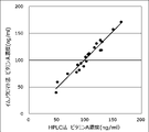
(6)ウシ血清のビタミンA測定
20頭の牛から採血して得られた血清中のビタミンA濃度を、それぞれHPLC法および本発明のイムノクロマト測定キットを用いて測定した。HPLCによる測定は、臨床検査センター(株式会社近畿予防医学研究所)に依頼して測定した。イムノクロマト法では、標準液を同時に測定して得られた標準曲線を用いて、各血清のビタミンA濃度を算出した。測定結果を表11および図14(金コロイド)、表12および図15(着色セルロース微粒子)に示した。イムノクロマト法の測定値とHPLC法による測定値の相関係数はそれぞれ0.98、0.97であり、良好な相関関係を示した。
20頭の牛から採血して得られた血清中のビタミンA濃度を、それぞれHPLC法および本発明のイムノクロマト測定キットを用いて測定した。HPLCによる測定は、臨床検査センター(株式会社近畿予防医学研究所)に依頼して測定した。イムノクロマト法では、標準液を同時に測定して得られた標準曲線を用いて、各血清のビタミンA濃度を算出した。測定結果を表11および図14(金コロイド)、表12および図15(着色セルロース微粒子)に示した。イムノクロマト法の測定値とHPLC法による測定値の相関係数はそれぞれ0.98、0.97であり、良好な相関関係を示した。


(比較例2)
保冷庫(-30℃)から取出し、室温(25℃)に1日放置した競合試薬を用いて、ウシ血清中のビタミンA濃度を測定したところ、測定値のバラつきが大きく、またHPLC法による測定値との相関係数も0.81であり、測定精度の著しい低下が認められた。
保冷庫(-30℃)から取出し、室温(25℃)に1日放置した競合試薬を用いて、ウシ血清中のビタミンA濃度を測定したところ、測定値のバラつきが大きく、またHPLC法による測定値との相関係数も0.81であり、測定精度の著しい低下が認められた。
本発明により、肉牛の生産現場において牛血液中のビタミンA濃度を迅速、簡便に測定するビタミンAイムノクロマトキットを提供することが可能となる。
1 粘着シート
2 含浸部材
3 膜担体
4 第一の捕捉部位(テストライン)
5 第二の捕捉部位(コントロールライン)
6 試料添加用部材
7 吸収用部材
8 イムノクロマトストリップ
9 ハウジングケース
10 試料滴下部
11 判定部
2 含浸部材
3 膜担体
4 第一の捕捉部位(テストライン)
5 第二の捕捉部位(コントロールライン)
6 試料添加用部材
7 吸収用部材
8 イムノクロマトストリップ
9 ハウジングケース
10 試料滴下部
11 判定部
Claims (12)
- 生体試料中のビタミンAを定量するためのイムノクロマトストリップであって、
前記イムノクロマトストリップは、最上流部に試料滴下部、および前記試料滴下部の下流側に順にテストライン、コントロールラインを有し、
前記テストラインには、抗ビタミンA抗体が固定化されている、
ことを特徴とするイムノクロマトストリップ。 - さらに、前記試料滴下部の下流側に含浸部材を有し、前記含浸部材にはビタミンAと化合物との結合体、および前記化合物に対する抗体に標識物質を結合した標識体が含浸されていることを特徴とする請求項1に記載のイムノクロマトストリップ。
- 前記ビタミンAと結合体をなす化合物は、ビオチンであることを特徴とする請求項2に記載のイムノクロマトストリップ。
- 前記化合物に対する抗体は、抗ビオチン抗体であることを特徴とする請求項3に記載のイムノクロマト試験片。
- 前記標識物質は、金コロイドまたはセルロース微粒子であることを特徴とする請求項2~4のいずれかに記載のイムノクロマトストリップ。
- 前記標識物質は、青色セルロース微粒子であることを特徴とする請求項2~4のいずれかに記載のイムノクロマトストリップ。
- 前記抗ビタミンA抗体はモノクローナル抗体であることを特徴とする請求項1~6のいずれかに記載のイムノクロマトストリップ。
- 前記標識体を特異的に結合する抗体は抗IgG抗体であることを特徴とする請求項1~7のいずれかに記載のイムノクロマトストリップ。
- 前記コントロールラインには、前記標識体を特異的に結合する抗体が固定化されていることを特徴とする請求項1~8のいずれかに記載のイムノクロマトストリップ。
- 全血または血清または血漿中のビタミンAを定量することを特徴とする請求項1~9のいずれかに記載のイムノクロマトストリップ。
- 請求項1~10のいずれかに記載のイムノクロマトストリップ、および検体希釈液を含むことを特徴とするイムノクロマト測定キット。
- 請求項1~10のいずれかに記載のイムノクロマトストリップ、検体希釈液、および競合試薬を含むことを特徴とするイムノクロマト測定キット。
Priority Applications (1)
| Application Number | Priority Date | Filing Date | Title |
|---|---|---|---|
| JP2022557582A JPWO2022085719A1 (ja) | 2020-10-21 | 2021-10-20 |
Applications Claiming Priority (6)
| Application Number | Priority Date | Filing Date | Title |
|---|---|---|---|
| JP2020-176635 | 2020-10-21 | ||
| JP2020176635 | 2020-10-21 | ||
| JP2020-193654 | 2020-11-20 | ||
| JP2020193654 | 2020-11-20 | ||
| JP2020193655 | 2020-11-20 | ||
| JP2020-193655 | 2020-11-20 |
Publications (1)
| Publication Number | Publication Date |
|---|---|
| WO2022085719A1 true WO2022085719A1 (ja) | 2022-04-28 |
Family
ID=81290625
Family Applications (1)
| Application Number | Title | Priority Date | Filing Date |
|---|---|---|---|
| PCT/JP2021/038752 WO2022085719A1 (ja) | 2020-10-21 | 2021-10-20 | ビタミンa定量用イムノクロマトストリップおよび測定キット |
Country Status (2)
| Country | Link |
|---|---|
| JP (1) | JPWO2022085719A1 (ja) |
| WO (1) | WO2022085719A1 (ja) |
Citations (3)
| Publication number | Priority date | Publication date | Assignee | Title |
|---|---|---|---|---|
| JP2018513983A (ja) * | 2015-04-06 | 2018-05-31 | ブルーダイアグノスティックス・インコーポレイテッドBludiagnostics, Inc. | 唾液試料中の分析物を検出するための試験装置および使用方法 |
| JP2019507890A (ja) * | 2016-01-22 | 2019-03-22 | アフィメディックス, インコーポレイテッド | ビタミンd代謝産物の検出のためのデバイス |
| WO2019215199A1 (en) * | 2018-05-07 | 2019-11-14 | Immundiagnostik Ag | System for analysing quantitative lateral flow chromatography |
-
2021
- 2021-10-20 WO PCT/JP2021/038752 patent/WO2022085719A1/ja active Application Filing
- 2021-10-20 JP JP2022557582A patent/JPWO2022085719A1/ja active Pending
Patent Citations (3)
| Publication number | Priority date | Publication date | Assignee | Title |
|---|---|---|---|---|
| JP2018513983A (ja) * | 2015-04-06 | 2018-05-31 | ブルーダイアグノスティックス・インコーポレイテッドBludiagnostics, Inc. | 唾液試料中の分析物を検出するための試験装置および使用方法 |
| JP2019507890A (ja) * | 2016-01-22 | 2019-03-22 | アフィメディックス, インコーポレイテッド | ビタミンd代謝産物の検出のためのデバイス |
| WO2019215199A1 (en) * | 2018-05-07 | 2019-11-14 | Immundiagnostik Ag | System for analysing quantitative lateral flow chromatography |
Non-Patent Citations (2)
| Title |
|---|
| GUPTA SEEMA, XIHUA SUI, SIKORA ROBERT, BANASURE KAILASH: "Validation study of the VitaKit A Test kit for the Determination of Vitamin A Fluid Milk(2% Fat) for Routine Quality Control", JOURNAL OF AOAC INTERNATIONAL, vol. 94, no. 1, 1 January 2011 (2011-01-01), US , pages 191 - 200, XP009535911, ISSN: 1060-3271 * |
| WESTFALL S S, WIRTZ G H: "VITAMIN A ANTIBODIES: APPLICATION TO RADIOIMMUNOASSAY", EXPERIENTIA, SPRINGER BASEL AG, CH, vol. 36, no. 12, 15 December 1980 (1980-12-15), CH , pages 1351 - 1353, XP009026953, ISSN: 0014-4754, DOI: 10.1007/BF01960092 * |
Also Published As
| Publication number | Publication date |
|---|---|
| JPWO2022085719A1 (ja) | 2022-04-28 |
Similar Documents
| Publication | Publication Date | Title |
|---|---|---|
| AU2005259012B2 (en) | Analyte detection system | |
| WO2022085719A1 (ja) | ビタミンa定量用イムノクロマトストリップおよび測定キット | |
| WO2018181741A1 (ja) | イムノクロマト試験片およびキットおよび測定方法 | |
| JP2019015583A (ja) | イムノクロマト試験片 | |
| JP2022175456A (ja) | イムノクロマトストリップおよび測定キット | |
| JP2022165855A (ja) | βカロテン定量用のイムノクロマト測定キット | |
| JP2022165858A (ja) | βカロテン定量用のイムノクロマト測定キット | |
| JP2022165857A (ja) | βカロテン定量用のイムノクロマト測定キット | |
| JP2022154825A (ja) | ビタミンaの定量方法 | |
| JP2022165856A (ja) | βカロテン定量用のイムノクロマト測定キット | |
| JP2022154827A (ja) | 全血中のビタミンaの定量方法 | |
| JP2023157760A (ja) | コルチゾール定量用のイムノクロマト測定キット | |
| JP2022154826A (ja) | ビタミンaの定量方法 | |
| JP2023173347A (ja) | オキシトシン定量用のイムノクロマト測定キット | |
| JP2023157758A (ja) | コルチゾール定量用のイムノクロマト測定キット | |
| JP6595215B2 (ja) | 免疫クロマト分析装置およびその製造方法並びに免疫クロマト分析方法 | |
| JP2023173345A (ja) | オキシトシン定量用のイムノクロマト測定キット | |
| JP2023069667A (ja) | エストラジオール定量用のイムノクロマト測定キット | |
| JP2023067045A (ja) | エストラジオール定量用のイムノクロマト測定キット | |
| JP2023182292A (ja) | 牛妊娠関連糖タンパク質測定キット | |
| JP2023067049A (ja) | エストロン定量用のイムノクロマト測定キット | |
| JP2023067052A (ja) | エストロン定量用のイムノクロマト測定キット | |
| JP2023067047A (ja) | エストラジオール定量用のイムノクロマト測定キット | |
| JP2023067048A (ja) | エストラジオール定量用のイムノクロマト測定キット | |
| JP2024031479A (ja) | チロキシン定量用のイムノクロマト測定キット |
Legal Events
| Date | Code | Title | Description |
|---|---|---|---|
| 121 | Ep: the epo has been informed by wipo that ep was designated in this application |
Ref document number: 21882860 Country of ref document: EP Kind code of ref document: A1 |
|
| ENP | Entry into the national phase |
Ref document number: 2022557582 Country of ref document: JP Kind code of ref document: A |
|
| NENP | Non-entry into the national phase |
Ref country code: DE |
|
| 122 | Ep: pct application non-entry in european phase |
Ref document number: 21882860 Country of ref document: EP Kind code of ref document: A1 |