WO2021172477A1 - Document creation assistance device, method, and program - Google Patents
Document creation assistance device, method, and program Download PDFInfo
- Publication number
- WO2021172477A1 WO2021172477A1 PCT/JP2021/007207 JP2021007207W WO2021172477A1 WO 2021172477 A1 WO2021172477 A1 WO 2021172477A1 JP 2021007207 W JP2021007207 W JP 2021007207W WO 2021172477 A1 WO2021172477 A1 WO 2021172477A1
- Authority
- WO
- WIPO (PCT)
- Prior art keywords
- interpretation report
- image
- diagnostic
- diagnostic guideline
- guideline
- Prior art date
Links
Images


Classifications
-
- A—HUMAN NECESSITIES
- A61—MEDICAL OR VETERINARY SCIENCE; HYGIENE
- A61B—DIAGNOSIS; SURGERY; IDENTIFICATION
- A61B5/00—Measuring for diagnostic purposes; Identification of persons
-
- G—PHYSICS
- G16—INFORMATION AND COMMUNICATION TECHNOLOGY [ICT] SPECIALLY ADAPTED FOR SPECIFIC APPLICATION FIELDS
- G16H—HEALTHCARE INFORMATICS, i.e. INFORMATION AND COMMUNICATION TECHNOLOGY [ICT] SPECIALLY ADAPTED FOR THE HANDLING OR PROCESSING OF MEDICAL OR HEALTHCARE DATA
- G16H15/00—ICT specially adapted for medical reports, e.g. generation or transmission thereof
-
- G—PHYSICS
- G16—INFORMATION AND COMMUNICATION TECHNOLOGY [ICT] SPECIALLY ADAPTED FOR SPECIFIC APPLICATION FIELDS
- G16H—HEALTHCARE INFORMATICS, i.e. INFORMATION AND COMMUNICATION TECHNOLOGY [ICT] SPECIALLY ADAPTED FOR THE HANDLING OR PROCESSING OF MEDICAL OR HEALTHCARE DATA
- G16H30/00—ICT specially adapted for the handling or processing of medical images
Definitions
- This disclosure relates to a document creation support device, method, and program that support the creation of documents such as interpretation reports.
- CT Computer Tomography
- MRI Magnetic Resonance Imaging
- medical images are analyzed by CAD (Computer-Aided Diagnosis) using a learning model that has been machine-learned by deep learning, etc., and the shape, density, and position of structures of interest such as abnormal shadow candidates included in the medical images. It is also practiced to discriminate properties such as size and size, and obtain these as analysis results.
- the analysis result acquired by CAD is associated with the test information such as the patient name, gender, age, and the modality from which the medical image was acquired, and is stored in the database.
- the medical image and the analysis result are transmitted to the terminal of the image interpreting doctor who interprets the medical image.
- the image interpreting doctor interprets the medical image by referring to the transmitted medical image and the analysis result on his / her terminal, and creates an image interpretation report.
- JP-A-2019-153250 a learning model in which machine learning such as a recurrent neural network is trained so as to generate a sentence from characters representing input property information is used.
- Medical texts (hereinafter referred to as medical texts) are generated.
- Medical practice guidelines are easy-to-understand, easy-to-understand information on the basis and procedure of medical care such as disease prevention, diagnosis, treatment, and prognosis, with the aim of assisting appropriate diagnosis and treatment in the medical field. It is a summary guideline.
- a diagnostic guideline database that associates the information expected to be obtained in the medical examination with the information used for diagnosing the disease, and accepts the input of the information actually obtained in the medical examination and obtained it.
- a method has been proposed in which information used for diagnosing a disease is extracted from a diagnostic guideline database based on information related to medical examination and displayed (see Japanese Patent Application Laid-Open No. 2005-110944).
- Diagnostic guidelines are diverse and constantly revised for each disease. For this reason, it is a burden for the interpreting doctor to remember all the diagnostic guidelines, and the interpretation report may be described with reference to the latest version of the diagnostic guidelines. However, the work of creating an interpretation report while comparing the diagnostic guideline and the interpretation report places a heavy burden on the interpretation doctor.
- This disclosure was made in view of the above circumstances, and aims to reduce the burden on the operator who creates an interpretation report by referring to the diagnostic guidelines.
- the document creation support device includes at least one processor.
- the processor identifies the corresponding part corresponding to the item included in the diagnostic guideline for the disease described in the interpretation report.
- the interpretation report and the diagnostic guideline are configured to be displayed on the display by associating the corresponding part in the interpretation report with the items in the diagnostic guideline.
- the processor may be configured to store the associated result.
- the processor analyzes the medical image to derive the analysis result corresponding to the item of the diagnostic guideline, and generates an interpretation report based on the analysis result.
- the analysis result may be used to identify the part corresponding to the item of the diagnostic guideline in the interpretation report.
- the processor may be configured to specify and display an item that is not included in the interpretation report in the diagnostic guideline.
- the diagnostic guideline includes information on the stage of the disease.
- the processor may be configured to identify stage information for diagnostic guidelines in interpretation reports.
- the stage information may be the stage information based on the TNM classification.
- the document creation support method identifies the corresponding part corresponding to the item included in the diagnostic guideline for the disease described in the image interpretation report in the image interpretation report showing the image interpretation result.
- the interpretation report and the diagnostic guideline are displayed on the display by associating the corresponding part in the interpretation report with the items in the diagnostic guideline.
- Functional configuration diagram of the document creation support device according to this embodiment Diagram for explaining an example of property information The figure which shows the schematic structure of the recurrent neural network Diagram showing the input medical image and the interpretation report generated from the medical image Diagram showing diagnostic guidelines for lung cancer Diagram showing diagnostic guidelines for lung cancer Diagram showing the display screen of the interpretation report and diagnostic guidelines Diagram showing the display screen of the interpretation report and diagnostic guidelines Diagram showing the display screen of the interpretation report and diagnostic guidelines Diagram showing the display screen of the interpretation report and diagnostic guidelines Flowchart showing processing performed in this embodiment Diagram showing other examples of interpretation report and diagnostic guideline display screens Diagram showing other examples of interpretation report and diagnostic guideline display screens
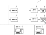
- FIG. 1 is a diagram showing a schematic configuration of the medical information system 1.
- the medical information system 1 shown in FIG. 1 is based on an inspection order from a doctor in a clinical department using a known ordering system, photographs of a part to be inspected of a subject, storage of a medical image acquired by the imaging, and an interpretation doctor. It is a system for interpreting medical images and creating an interpretation report, and for viewing the interpretation report by the doctor of the requesting clinical department and observing the details of the medical image to be interpreted.
- the medical information system 1 includes a plurality of imaging devices 2, a plurality of image interpretation WS (WorkStation) 3 which are image interpretation terminals, a medical care WS 4, an image server 5, and an image database (hereinafter, image DB (DataBase)).
- the report server 7 and the report database (hereinafter referred to as the report DB) 8 are connected to each other via a wired or wireless network 10 so as to be able to communicate with each other.
- Each device is a computer on which an application program for functioning as a component of the medical information system 1 is installed.
- the application program is recorded and distributed on a recording medium such as a DVD (Digital Versatile Disc) and a CD-ROM (Compact Disc Read Only Memory), and is installed on a computer from the recording medium.
- a recording medium such as a DVD (Digital Versatile Disc) and a CD-ROM (Compact Disc Read Only Memory)
- it is stored in the storage device of the server computer connected to the network 10 or in the network storage in a state of being accessible from the outside, and is downloaded and installed in the computer upon request.
- the photographing device 2 is a device (modality) that generates a medical image representing the diagnosis target part by photographing the part to be diagnosed of the subject. Specifically, it is a simple X-ray imaging apparatus, a CT apparatus, an MRI apparatus, a PET (Positron Emission Tomography) apparatus, and the like.
- the medical image generated by the imaging device 2 is transmitted to the image server 5 and stored in the image DB 6.
- the image interpretation WS3 is a computer used by, for example, an image interpretation doctor in a radiology department to interpret a medical image and create an image interpretation report, and includes a document creation support device 20 according to the present embodiment.
- a request for viewing a medical image to the image server 5 various image processing on the medical image received from the image server 5, a display of the medical image, an input acceptance of a finding sentence related to the medical image, and the like are performed.
- analysis processing for medical images and input findings support for creating an interpretation report based on the analysis results, a request for registration and viewing of an interpretation report for the report server 7, and an interpretation received from the report server 7 are performed.
- the report is displayed.
- the clinical WS4 is a computer used by doctors in clinical departments for detailed observation of images, viewing of interpretation reports, creation of electronic medical records, etc., and is a processing device, a display device such as a display, and an input device such as a keyboard and a mouse. Consists of.
- an image viewing request is made to the image server 5
- an image received from the image server 5 is displayed
- an image interpretation report viewing request is made to the report server 7
- an image interpretation report received from the report server 7 is displayed.
- the image server 5 is a general-purpose computer in which a software program that provides a database management system (DataBase Management System: DBMS) function is installed. Further, the image server 5 includes a storage in which the image DB 6 is configured. This storage may be a hard disk device connected by the image server 5 and the data bus, or a disk device connected to NAS (Network Attached Storage) and SAN (Storage Area Network) connected to the network 10. It may be.
- NAS Network Attached Storage
- SAN Storage Area Network
- the image data and incidental information of the medical image acquired by the imaging device 2 are registered in the image DB 6.
- the incidental information includes, for example, an image ID (identification) for identifying an individual medical image, a patient ID for identifying a subject, an examination ID for identifying an examination, and a unique ID assigned to each medical image ( UID: unique identification), examination date when the medical image was generated, examination time, type of imaging device used in the examination to acquire the medical image, patient information such as patient name, age, gender, examination site (imaging) Includes information such as site), imaging information (imaging protocol, imaging sequence, imaging method, imaging conditions, use of contrast medium, etc.), series number or collection number when multiple medical images are acquired in one examination. ..
- the image server 5 when the image server 5 receives the viewing request from the image interpretation WS3 and the medical examination WS4 via the network 10, the image server 5 searches for the medical image registered in the image DB 6, and uses the searched medical image as the requesting image interpretation WS3 and the medical examination. Send to WS4.
- the report server 7 incorporates a software program that provides the functions of a database management system to a general-purpose computer.
- the report server 7 receives the image interpretation report registration request from the image interpretation WS3, the report server 7 prepares the image interpretation report in a database format and registers the image interpretation report in the report DB 8.
- the image interpretation report includes, for example, a medical image to be interpreted, an image ID for identifying the medical image, an image interpretation doctor ID for identifying the image interpretation doctor who performed the image interpretation, a lesion name, a lesion position information, and a medical image including a specific area. It may include information for access and information such as property information.
- the report server 7 when the report server 7 receives a viewing request for the interpretation report from the interpretation WS3 and the medical treatment WS4 via the network 10, the report server 7 searches for the interpretation report registered in the report DB 8 and uses the searched interpretation report as the requester's interpretation. It is transmitted to WS3 and medical treatment WS4.
- the medical image is a three-dimensional CT image composed of a plurality of tomographic images with the diagnosis target as the lung, and the CT image is interpreted by the interpretation WS3 to obtain an abnormal shadow contained in the lung.
- An interpretation report shall be created.
- the medical image is not limited to the CT image, and any medical image such as an MRI image and a simple two-dimensional image acquired by a simple X-ray imaging device can be used.
- Network 10 is a wired or wireless local area network that connects various devices in the hospital.
- the network 10 may be configured such that the local area networks of each hospital are connected to each other by the Internet or a dedicated line.
- FIG. 2 illustrates the hardware configuration of the document creation support device according to the present embodiment.
- the document creation support device 20 includes a CPU (Central Processing Unit) 11, a non-volatile storage 13, and a memory 16 as a temporary storage area.
- the document creation support device 20 includes a display 14 such as a liquid crystal display, an input device 15 such as a keyboard and a mouse, and a network I / F (InterFace) 17 connected to the network 10.
- the CPU 11, the storage 13, the display 14, the input device 15, the memory 16, and the network I / F 17 are connected to the bus 18.
- the CPU 11 is an example of the processor in the present disclosure.
- the storage 13 is realized by an HDD (Hard Disk Drive), an SSD (Solid State Drive), a flash memory, or the like.
- the document creation support program 12 is stored in the storage 13 as a storage medium.
- the CPU 11 reads the document creation support program from the storage 13, expands it into the memory 16, and executes the expanded document creation support program 12.
- FIG. 3 is a diagram showing a functional configuration of the document creation support device according to the present embodiment.
- the document creation support device 20 includes an acquisition unit 21, a sentence generation unit 22, a specific unit 23, a display control unit 24, a storage control unit 25, and a communication unit 26.
- the CPU 11 executes the document creation support program, the CPU 11 functions as an acquisition unit 21, a sentence generation unit 22, a specific unit 23, a display control unit 24, a storage control unit 25, and a communication unit 26.
- the acquisition unit 21 acquires a medical image for creating an image interpretation report from the image server 5 in response to an instruction from the input device 15 by the image interpretation doctor who is the operator.
- diagnostic guidelines for lung cancer which is a target disease, are also obtained from the image server 5.
- the acquired medical image and diagnostic guideline are stored in the storage 13.
- the sentence generation unit 22 derives the analysis result by analyzing the medical image, and generates an interpretation report based on the analysis result. For this purpose, the sentence generation unit 22 discriminates the abnormal shadow candidate in the medical image, and discriminates the properties of the discriminated abnormal shadow candidate for each of the plurality of predetermined property items. Examples of property items identified for abnormal shadows are the location of the abnormal shadow, the size of the abnormal shadow, the shape of the border (clear and irregular), the type of absorption value (full and pleural), the presence or absence of spicula, and the mass. These include nodules, pleural contact, pleural infiltration, pleural infiltration, cavities, and calcification.
- the sentence generation unit 22 has a learning model in which machine learning is performed so as to discriminate the properties of abnormal shadow candidates from the medical image.
- the learning model uses teacher data to determine, for example, whether or not each pixel (voxel) in a medical image represents an abnormal shadow candidate, and if it is an abnormal shadow candidate, to determine its properties. It consists of a convolutional neural network (CNN (Convolutional Neural Network)) that has undergone deep learning.
- CNN Convolutional Neural Network
- the learning model is learned by machine learning using, for example, a plurality of combinations of a medical image including an abnormal shadow and a property label representing the property of the abnormal shadow as teacher data.
- the learning model takes a medical image as an input, and outputs a property score derived for each property item in the abnormal shadow included in the input medical image.
- the property score is a score indicating the prominence of the property for each property item.
- the property score takes, for example, a value of 0 or more and 1 or less, and the larger the value of the property score, the more remarkable the property.
- the property score for "presence or absence of spicula”, which is one of the property items of abnormal shadow is 0.5 or more
- the property for "presence or absence of spicula" of abnormal shadow is "with spicula (positive)”.
- the property score for "presence or absence of spicula” is, for example, less than 0.5, it is specified that the property for the presence or absence of spicula in abnormal shadow is "no spicula (negative)”.
- the threshold value 0.5 used for the property determination is merely an example, and is set to an appropriate value for each property item.
- an arbitrary learning model such as a support vector machine (SVM (Support Vector Machine)) can be used in addition to a convolutional neural network. Further, the learning model for detecting the abnormal shadow candidate from the medical image and the learning model for detecting the property information of the abnormal shadow candidate may be constructed separately.
- SVM Support Vector Machine
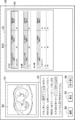
- FIG. 4 is a diagram for explaining an example of the property information derived by the sentence generation unit 22.
- the properties for each property item are "left pulmonary subpleural", “4.2 cm”, “irregular”, “enriched type”, and so on. They are “with spicula”, “mass”, “with pleural contact”, “with pleural infiltration”, “without pleural infiltration”, “without cavities” and “without calcification”.
- + is given when “yes” and-is given when there is no.
- the sentence generation unit 22 generates a finding sentence using the derived property information.
- the sentence generation unit 22 is composed of a learning model in which learning is performed so as to generate a sentence from the input property information.
- a learning model for example, a recurrent neural network can be used.
- FIG. 5 is a diagram showing a schematic configuration of a recurrent neural network.
- the recurrent neural network 40 includes an encoder 41 and a decoder 42.
- the property information derived by the sentence generation unit 22 is input to the encoder 41. For example, property information of "left pulmonary subpleuralis", “4.2 cm”, “Spicula +” and “mass" is input to the encoder 41.
- the decoder 42 is learned so as to document the character information, and generates a sentence from the input property information. Specifically, from the above-mentioned property information of "left pulmonary subpleura”, “4.2 cm”, “spicula +” and “mass”, "a 4.2 cm diameter mass having spicula under the left pulmonary pleura is recognized. Will be generated. " In FIG. 5, "EOS” indicates the end of the sentence (End Of Sentence).
- the recurrent neural network 40 learns the encoder 41 and the decoder 42 using a large amount of teacher data composed of a combination of the property information and the finding sentence. Be built.
- the generated text shown in FIG. 5 represents the findings about the lung nodule, and is generated by learning the learning model by inputting the property information of the lung nodule.
- the sentence generation unit 22 uses these learning models to generate an interpretation report including at least one finding sentence.
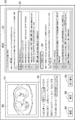
- FIG. 6 is a diagram showing an image interpretation report including an input medical image and a finding sentence generated from the medical image.
- the input medical image G0 As shown in FIG. 6, regarding the input medical image G0, "a solid nodule with a diameter of 17 mm is observed in the upper lobe S2 of the right lung, there is a spicula on the margin, and an image of pleural invagination is accompanied. Bronchial translucency image is observed. No lymphadenopathy is observed. There is no intrapulmonary metastasis. ”Interpretation report 35 has been generated.
- the acquisition unit 21 acquires the diagnostic guideline for lung cancer and stores it in the storage 13.
- 7 and 8 are diagrams showing diagnostic guidelines for lung cancer. Note that FIG. 7 shows the stage of lung cancer based on the TNM classification, and FIG. 8 shows the T factor, N factor, and M factor shown in the stage.
- stage 1 stage I
- stage II stage II
- stage III stage IV
- T factor representing cancer size and infiltration
- N factor representing lymph node metastasis
- M factor representing distant metastasis
- each factor is classified and defined in a plurality of stages.
- factor T is classified into four stages, T1 to T4, according to the size of the cancer and the state of infiltration.
- T1 and T2 are classified more finely, such as T1a, T1b, T2a, and T2b, respectively.
- N factor is classified into three stages of N1 to N3.
- N0 has no lymph node metastasis.
- M factor is classified into two stages, M0 when there is no metastasis and M1 when there is metastasis.
- M1 is classified more finely like M1a and M1b, respectively.
- the identification unit 23 identifies the corresponding part corresponding to the item included in the diagnostic guideline for the disease described in the interpretation report.
- the specific unit 23 has a learning model 23A in which machine learning is performed so as to specify a portion corresponding to an item included in the diagnostic guideline as a corresponding portion in the input sentence.
- the learning model 23A uses teacher data to perform deep learning so that when a convolutional report is input, the input convolutional report discriminates words and phrases related to items included in the diagnostic guideline as corresponding parts. It consists of a convolutional neural network (CNN).
- CNN convolutional neural network
- the teacher data for learning the learning model 23A is associated with words and phrases included in the items of the diagnostic guideline and sentences using the words and phrases included in the items of the diagnostic guideline.
- the items of the diagnostic guideline include "the maximum diameter of the tumor is 2 cm or less” as T1a of T factor, "no regional lymph node metastasis” as N0 of N factor, and M0 of M factor. Includes “no distant metastasis”.
- the words “diameter”, “lymph node”, “no metastasis”, and “no distant metastasis” included in the items of the diagnostic guideline, and "nodule with a diameter of 15 mm" are recognized. Is not admitted.
- the learning model 23A is constructed by learning a neural network using a large number of such teacher data. As a result, when the interpretation report is input, the learning model 23A outputs the corresponding portion corresponding to the item included in the diagnostic guideline for the disease described in the interpretation report. Further, when the specifying unit 23 specifies the corresponding part in the interpretation report, the specifying unit 23 specifies the item corresponding to the corresponding part in the diagnostic guideline.
- any learning model such as a support vector machine and a recurrent neural network can be used.
- the specific unit 23 is not limited to the one that specifies the corresponding portion by the learning model 23A.
- the corresponding portion may be specified by searching the interpretation report using the size of the mass and the items included in the diagnostic guideline as keywords.
- FIG. 9 is a diagram showing a display screen of an interpretation report and a diagnostic guideline.
- the display screen 50 has an image display area 51, an interpretation report display area 52, and a diagnostic guideline display area 53.
- a switching button 54, a correction button 55, and a confirmation button 56 are displayed below the interpretation report display area 52.
- the tomographic image S0 included in the medical image G0 for which the interpretation report is generated is displayed.
- the interpretation report display area 52 the interpretation report generated by the sentence generation unit 22 is displayed.
- the corresponding portion specified by the specific unit 23 is highlighted.
- the three corresponding parts of the interpretation report "17 mm in diameter”, "no lymphadenopathy” and “no intrapulmonary metastasis”, are surrounded by a frame. It is highlighted by. It should be noted that the highlighting of these three corresponding portions has different modes. In FIGS.
- the diagnostic guidelines 60A to 60C are displayed in the diagnostic guideline display area 53. Since the contents of the diagnostic guidelines 60A to 60C are diverse, the contents of the diagnostic guideline displayed in the diagnostic guideline display area 53 can be changed by selecting the switching button 54. Specifically, FIG. 9 shows diagnostic guidelines 60A for factors T and N. Further, FIG. 10 shows a diagnostic guideline 60B for the M factor. Further, in FIG. 11, the diagnostic guideline 60C for the stage is displayed. Instead of the switching display of the diagnostic guideline, the entire diagnostic guideline may be displayed so that it can be referred to by scrolling.
- the item of T1a corresponding to "diameter 17 mm” in the interpretation report is highlighted.
- the highlighting of the item of T1a is shown by surrounding it with a solid line frame 61 similar to the corresponding portion of “diameter 17 mm” in the interpretation report.
- the item N0 corresponding to "No lymphadenopathy is observed” in the interpretation report is also highlighted.
- the highlighting of the item N0 is shown by enclosing it in a broken line frame 62 similar to the corresponding part of “No lymphadenopathy is observed” in the interpretation report.
- the item of M0 corresponding to "There is no intrapulmonary metastasis" in the interpretation report is highlighted.
- the highlighting of the item M0 is shown by enclosing it in the frame 63 of the alternate long and short dash line similar to the corresponding part of “No intrapulmonary metastasis” in the interpretation report.
- the diagnostic guideline 60C shown in FIG. 11 there are three corresponding parts in the interpretation report, "diameter 17 mm", “no lymphadenopathy” and “no intrapulmonary metastasis”, and these.
- the stage derived from the item corresponding to the corresponding part is highlighted.
- the IA period is highlighted.
- the highlighting is shown by surrounding it with a solid line frame 64.
- the highlighting of items in the diagnostic guidelines is not limited to the addition of frames. Instead of adding a frame, the characters may be highlighted or the character color may be changed.
- the operator can compare and confirm the interpretation report and the diagnostic guideline on the display screen 50. If necessary, the interpretation report displayed in the interpretation report display area 52 can be modified by input from the input device 15 by selecting the correction button 55. When the confirmation or correction of the interpretation report is completed, the operator selects the confirmation button 56.
- the specific unit 23 again identifies the corresponding part corresponding to the item included in the diagnostic guideline in the modified interpretation report. In this case, if the corresponding part is added or deleted, the item in the diagnostic guideline is also added or deleted accordingly.
- the storage control unit 25 highlights the image interpretation report in which the corresponding portion is highlighted, the tomographic image S0 included in the medical image G0 referred to when generating the image interpretation report, and the items by selecting the confirmation button 56 by the operator.
- the diagnostic guideline that has been obtained is also stored in the storage 13.
- the communication unit 26 combines the image interpretation report in which the corresponding part is highlighted, the tomographic image S0 included in the medical image G0 referred to when generating the image interpretation report, and the diagnostic guideline in which the items are highlighted, and the network I / Transfer to the report server 7 via F17.
- the report server 7 also stores the image interpretation report in which the corresponding portion is highlighted, the medical image G0 referred to when generating the image interpretation report, and the diagnostic guideline in which the items are highlighted.
- FIG. 12 is a flowchart showing the processing performed in the first embodiment. It is assumed that the medical image G0 and the diagnostic guideline to be read are acquired from the image server 5 by the acquisition unit 21 and stored in the storage 13. The process is started when the interpretation doctor gives an instruction to create the interpretation report, and the sentence generation unit 22 derives the analysis result by analyzing the medical image G0 and generates the interpretation report based on the analysis result ( Step ST1). Next, the identification unit 23 identifies in the image interpretation report the corresponding part corresponding to the item included in the diagnostic guideline for the disease described in the image interpretation report (step ST2).
- the display control unit 24 associates the corresponding portion corresponding to the item of the diagnostic guideline in the image interpretation report with the item in the diagnostic guideline, and displays the image interpretation report and the diagnostic guideline on the display 14 (step ST3).
- the operator switches the diagnostic guideline displayed in the diagnostic guideline display area 53 on the display screen 50, and modifies the interpretation report displayed in the text display area 51 if necessary.
- the storage control unit 25 starts monitoring whether or not the confirmation button 56 is selected (step ST4), and when step ST4 is affirmed, the storage control unit 25 receives an image interpretation report in which the corresponding portion is highlighted.
- the tomographic image S0 included in the medical image G0 referred to when generating the interpretation report and the diagnostic guideline in which the item is highlighted are also saved in the storage 13 (save the interpretation report, etc .; step ST5).
- the communication unit 26 reports via the network I / F17 together with the image interpretation report in which the corresponding portion is highlighted, the tomographic image S0 referred to when generating the image interpretation report, and the diagnostic guideline in which the items are highlighted. Transfer to the server 7 (transfer of interpretation report, etc .; step ST6), and end the process.
- the corresponding part corresponding to the item included in the diagnostic guideline for the disease described in the image interpretation report is specified, and the above corresponding part in the image interpretation report and the item in the diagnostic guideline are specified.
- the interpretation report and the diagnostic guideline are displayed on the display 14 in association with the above. Therefore, the work of creating the interpretation report can be efficiently performed while comparing the diagnostic guideline and the interpretation report. Therefore, according to the present embodiment, it is possible to reduce the burden on the operator who creates the interpretation report by referring to the diagnostic guideline.
- the image interpretation report with the corresponding part highlighted, the tomographic image S0 included in the medical image G0 referred to when creating the image interpretation report, and the diagnostic guideline with the item highlighted are saved or transferred together. Therefore, the attending physician of the patient can refer to the correspondence between the interpretation report and the diagnostic guideline, and as a result, the diagnosis can be made efficiently.
- the interpretation report states, "A solid nodule with a diameter of 17 mm is found in the upper lobe S2 of the right lung, and there is a spicula on the margin, accompanied by a pleural invagination image.
- the interpretation report states, "A solid nodule with a diameter of 17 mm is found in the upper lobe S2 of the right lung, and there is a spicula on the margin, accompanied by a pleural invagination image.
- No intrapulmonary metastasis There is no description in the interpretation report regarding items related to lymph node metastasis.
- “lymph node metastasis (N factor)” is highlighted in the diagnostic guideline 60A.
- the highlighting is shown by surrounding it with a solid line frame 65.
- the sentence generation unit 22 analyzes the medical image G0 and generates an interpretation report, but the present invention is not limited to this.
- the operator may input the image interpretation report using the input device 15 in the image interpretation report display area 52 of the display screen 50.
- the specifying unit 23 may specify the corresponding part corresponding to the item of the diagnostic guideline in the interpretation report every time a sentence of the findings included in the interpretation report is described, and the input of the interpretation report is completed. Therefore, the corresponding portion may be specified. In this case, the sentence generation unit 22 shown in FIG. 3 is unnecessary.
- the diagnostic guideline is stored in the image server 5, but the present invention is not limited to this.
- a dedicated server for storing the diagnostic guideline may be provided in the medical information system 1 and the diagnostic guideline may be acquired from this dedicated server.
- the document creation support device 20 generates an interpretation report, but the present invention is not limited to this.
- the acquisition unit 21 acquires the image interpretation report from the report server 7, the specific unit 23 identifies the corresponding part of the acquired image interpretation report corresponding to the item of the diagnostic guideline, and the acquired image interpretation report is combined with the diagnostic guideline. It may be displayed.
- the technique of the present disclosure is applied when creating an image interpretation report using a medical image with the diagnosis target as the lung, but the diagnosis target is not limited to the lung.
- diagnosis target is not limited to the lung.
- any part of the human body such as the heart, liver, brain, and limbs can be diagnosed.
- the diagnostic guideline corresponding to the part to be diagnosed may be acquired, and the corresponding part corresponding to the item of the diagnostic guideline in the interpretation report may be specified.
- a processing unit that executes various processes such as an acquisition unit 21, a sentence generation unit 22, a specific unit 23, a display control unit 24, a storage control unit 25, and a communication unit 26.
- various processors processors shown below can be used.
- the various processors include a CPU, which is a general-purpose processor that executes software (program) and functions as various processing units, and a circuit after manufacturing an FPGA (Field Programmable Gate Array) or the like.
- Dedicated electricity which is a processor with a circuit configuration specially designed to execute specific processing such as programmable logic device (PLD), ASIC (Application Specific Integrated Circuit), which is a processor whose configuration can be changed. Circuits and the like are included.
- One processing unit may be composed of one of these various processors, or a combination of two or more processors of the same type or different types (for example, a combination of a plurality of FPGAs or a combination of a CPU and an FPGA). ) May be configured. Further, a plurality of processing units may be configured by one processor.
- one processor is configured by combining one or more CPUs and software. There is a form in which this processor functions as a plurality of processing units.
- SoC System On Chip
- the various processing units are configured by using one or more of the above-mentioned various processors as a hardware structure.
- circuitry in which circuit elements such as semiconductor elements are combined can be used.
Abstract
The present invention is provided with at least one processor, and the processor specifies a corresponding part corresponding to an item included in a diagnostic guideline for disease described in an interpretation report representing an image interpretation result. The processor associates the corresponding part in the interpretation report with the item in the diagnostic guideline, and displays, on a display, the interpretation report and the diagnostic guideline.
Description
本開示は、読影レポート等の文書の作成を支援する文書作成支援装置、方法およびプログラムに関する。
This disclosure relates to a document creation support device, method, and program that support the creation of documents such as interpretation reports.
近年、CT(Computed Tomography)装置およびMRI(Magnetic Resonance Imaging)装置等の医療機器の進歩により、より質の高い高解像度の医用画像を用いた画像診断が可能となってきている。とくに、CT画像およびMRI画像等を用いた画像診断により、病変の領域の特定および解析を精度よく行うことができるため、適切な治療を実施できるようになってきている。
In recent years, advances in medical devices such as CT (Computed Tomography) devices and MRI (Magnetic Resonance Imaging) devices have made it possible to perform diagnostic imaging using higher quality medical images. In particular, by image diagnosis using CT images, MRI images, and the like, it is possible to accurately identify and analyze the lesion area, so that appropriate treatment can be performed.
また、ディープラーニング等により機械学習がなされた学習モデルを用いたCAD(Computer-Aided Diagnosis)により医用画像を解析して、医用画像に含まれる異常陰影候補等の関心構造物の形状、濃度、位置および大きさ等の性状を判別し、これらを解析結果として取得することも行われている。CADにより取得された解析結果は、患者名、性別、年齢および医用画像を取得したモダリティ等の検査情報と対応づけられて、データベースに保存される。医用画像および解析結果は、医用画像の読影を行う読影医の端末に送信される。読影医は、自身の端末において、送信された医用画像および解析結果を参照して医用画像の読影を行い、読影レポートを作成する。
In addition, medical images are analyzed by CAD (Computer-Aided Diagnosis) using a learning model that has been machine-learned by deep learning, etc., and the shape, density, and position of structures of interest such as abnormal shadow candidates included in the medical images. It is also practiced to discriminate properties such as size and size, and obtain these as analysis results. The analysis result acquired by CAD is associated with the test information such as the patient name, gender, age, and the modality from which the medical image was acquired, and is stored in the database. The medical image and the analysis result are transmitted to the terminal of the image interpreting doctor who interprets the medical image. The image interpreting doctor interprets the medical image by referring to the transmitted medical image and the analysis result on his / her terminal, and creates an image interpretation report.
一方、上述したCT装置およびMRI装置の高性能化に伴い、読影を行う医用画像の数も増大している。しかしながら、読影医の数は医用画像の数に追いついていないことから、読影医の読影業務の負担を軽減することが望まれている。このため、読影レポート等の医療文書の作成を支援するための各種手法が提案されている。例えば、特開2019-153250号公報には、読影医が入力したキーワードおよび医用画像を解析結果に含まれる、関心構造物の性状を表す情報(以下、性状情報とする)に基づいて、読影レポートに記載するための文章を自動で生成する各種手法が提案されている。特開2019-153250号公報に記載された手法においては、入力された性状情報を表す文字から文章を生成するように学習が行われたリカレントニューラルネットワーク等の機械学習がなされた学習モデルを用いて、医療用の文章(以下、医療文章とする)が生成される。特開2019-153250号公報に記載された手法のように、医療文章を自動で生成することにより、読影レポート等の医療文書を作成する際の読影医の負担を軽減することができる。
On the other hand, the number of medical images to be interpreted is increasing with the improvement of the performance of the CT device and the MRI device described above. However, since the number of image interpreters has not kept up with the number of medical images, it is desired to reduce the burden of the image interpretation work of the image interpreters. For this reason, various methods have been proposed to support the creation of medical documents such as interpretation reports. For example, Japanese Patent Application Laid-Open No. 2019-153250 describes an interpretation report based on information representing the properties of the structure of interest (hereinafter referred to as property information), which includes keywords and medical images input by the image interpreter in the analysis results. Various methods have been proposed to automatically generate sentences to be described in. In the method described in JP-A-2019-153250, a learning model in which machine learning such as a recurrent neural network is trained so as to generate a sentence from characters representing input property information is used. , Medical texts (hereinafter referred to as medical texts) are generated. By automatically generating medical texts as in the method described in JP-A-2019-153250, it is possible to reduce the burden on the interpretation doctor when creating a medical document such as an interpretation report.
ところで、読影レポートを作成する際には、対象とする疾患に応じた診断ガイドラインに従って、必要な項目を記載する必要がある。診療ガイドラインとは、医療現場において適切な診断と治療を補助することを目的として、病気の予防、診断、治療、予後予測等診療の根拠および手順についての最新の情報を専門家の手で分かりやすくまとめた指針である。このため、診察で得られると予想される情報と病気の診断に利用される情報とを関連付けた診断ガイドラインデータベースを保持しておき、実際に診察で得られた情報の入力を受け付け、取得された診察に関する情報に基づいて、診断ガイドラインデータベースから病気の診断に利用される情報を抽出して表示する手法が提案されている(特開2005-110944号公報参照)。
By the way, when creating an interpretation report, it is necessary to describe the necessary items according to the diagnostic guidelines according to the target disease. Medical practice guidelines are easy-to-understand, easy-to-understand information on the basis and procedure of medical care such as disease prevention, diagnosis, treatment, and prognosis, with the aim of assisting appropriate diagnosis and treatment in the medical field. It is a summary guideline. For this reason, we have a diagnostic guideline database that associates the information expected to be obtained in the medical examination with the information used for diagnosing the disease, and accepts the input of the information actually obtained in the medical examination and obtained it. A method has been proposed in which information used for diagnosing a disease is extracted from a diagnostic guideline database based on information related to medical examination and displayed (see Japanese Patent Application Laid-Open No. 2005-110944).
診断ガイドラインは疾患毎に多岐に亘り、かつ絶えず改訂がなされる。このため、読影医は、すべての診断ガイドラインを覚えることが負担であり、最新版の診断ガイドラインを参照しつつ、読影レポートを記載することがある。しかしながら、診断ガイドラインと読影レポートとを見比べながら読影レポートを作成する作業は、読影医の負担が大きい。
Diagnostic guidelines are diverse and constantly revised for each disease. For this reason, it is a burden for the interpreting doctor to remember all the diagnostic guidelines, and the interpretation report may be described with reference to the latest version of the diagnostic guidelines. However, the work of creating an interpretation report while comparing the diagnostic guideline and the interpretation report places a heavy burden on the interpretation doctor.
本開示は上記事情に鑑みなされたものであり、診断ガイドラインを参照して読影レポートを作成する操作者の負担を軽減することを目的とする。
This disclosure was made in view of the above circumstances, and aims to reduce the burden on the operator who creates an interpretation report by referring to the diagnostic guidelines.
本開示による文書作成支援装置は、少なくとも1つのプロセッサを備え、
プロセッサは、画像の読影結果を表す読影レポートにおいて、読影レポートに記載された疾患についての、診断ガイドラインに含まれる項目に対応する対応部分を特定し、
読影レポートにおける対応部分と、診断ガイドラインにおける項目とを対応づけて、読影レポートおよび診断ガイドラインをディスプレイに表示するように構成される。 The document creation support device according to the present disclosure includes at least one processor.
In the interpretation report showing the interpretation result of the image, the processor identifies the corresponding part corresponding to the item included in the diagnostic guideline for the disease described in the interpretation report.
The interpretation report and the diagnostic guideline are configured to be displayed on the display by associating the corresponding part in the interpretation report with the items in the diagnostic guideline.
なお、本開示による文書作成支援装置においては、プロセッサは、対応づけた結果を保存するように構成されるものであってもよい。
In the document creation support device according to the present disclosure, the processor may be configured to store the associated result.
また、本開示による文書作成支援装置においては、プロセッサは、医用画像を解析することにより、診断ガイドラインの項目に対応する解析結果を導出し、解析結果に基づいて読影レポートを生成し、
解析結果を用いて、読影レポートにおける診断ガイドラインの項目に対応する部分を特定するように構成されるものであってもよい。 Further, in the document creation support device according to the present disclosure, the processor analyzes the medical image to derive the analysis result corresponding to the item of the diagnostic guideline, and generates an interpretation report based on the analysis result.
The analysis result may be used to identify the part corresponding to the item of the diagnostic guideline in the interpretation report.
また、本開示による文書作成支援装置においては、プロセッサは、診断ガイドラインにおいて、読影レポートにない項目を特定して表示するように構成されるものであってもよい。
Further, in the document creation support device according to the present disclosure, the processor may be configured to specify and display an item that is not included in the interpretation report in the diagnostic guideline.
また、本開示による文書作成支援装置においては、診断ガイドラインは、病期の情報を含み、
プロセッサは、読影レポートにおける診断ガイドラインの病期の情報を特定するように構成されるものであってもよい。 In addition, in the document preparation support device according to the present disclosure, the diagnostic guideline includes information on the stage of the disease.
The processor may be configured to identify stage information for diagnostic guidelines in interpretation reports.
また、本開示による文書作成支援装置においては、病期の情報は、TNM分類に基づく病期の情報であってもよい。
Further, in the document creation support device according to the present disclosure, the stage information may be the stage information based on the TNM classification.
本開示による文書作成支援方法は、画像の読影結果を表す読影レポートにおいて、読影レポートに記載された疾患についての、診断ガイドラインに含まれる項目に対応する対応部分を特定し、
読影レポートにおける対応部分と、診断ガイドラインにおける項目とを対応づけて、読影レポートおよび診断ガイドラインをディスプレイに表示する。 The document creation support method according to the present disclosure identifies the corresponding part corresponding to the item included in the diagnostic guideline for the disease described in the image interpretation report in the image interpretation report showing the image interpretation result.
The interpretation report and the diagnostic guideline are displayed on the display by associating the corresponding part in the interpretation report with the items in the diagnostic guideline.
なお、本開示による文書支援作成方法をコンピュータに実行させるためのプログラムとして提供してもよい。
Note that it may be provided as a program for causing a computer to execute the document support creation method according to the present disclosure.
本開示によれば、診断ガイドラインを参照して読影レポートを作成する操作者の負担を軽減できる。
According to this disclosure, it is possible to reduce the burden on the operator who creates the interpretation report by referring to the diagnostic guideline.
以下、図面を参照して本開示の実施形態について説明する。まず、本実施形態による文書作成支援装置を適用した医療情報システム1の構成について説明する。図1は、医療情報システム1の概略構成を示す図である。図1に示す医療情報システム1は、公知のオーダリングシステムを用いた診療科の医師からの検査オーダに基づいて、被写体の検査対象部位の撮影、撮影により取得された医用画像の保管、読影医による医用画像の読影と読影レポートの作成、および依頼元の診療科の医師による読影レポートの閲覧と読影対象の医用画像の詳細観察とを行うためのシステムである。
Hereinafter, embodiments of the present disclosure will be described with reference to the drawings. First, the configuration of the medical information system 1 to which the document creation support device according to the present embodiment is applied will be described. FIG. 1 is a diagram showing a schematic configuration of the medical information system 1. The medical information system 1 shown in FIG. 1 is based on an inspection order from a doctor in a clinical department using a known ordering system, photographs of a part to be inspected of a subject, storage of a medical image acquired by the imaging, and an interpretation doctor. It is a system for interpreting medical images and creating an interpretation report, and for viewing the interpretation report by the doctor of the requesting clinical department and observing the details of the medical image to be interpreted.
図1に示すように、医療情報システム1は、複数の撮影装置2、読影端末である複数の読影WS(WorkStation)3、診療WS4、画像サーバ5、画像データベース(以下、画像DB(DataBase)とする)6、レポートサーバ7およびレポートデータベース(以下レポートDBとする)8が、有線または無線のネットワーク10を介して互いに通信可能な状態で接続されて構成されている。
As shown in FIG. 1, the medical information system 1 includes a plurality of imaging devices 2, a plurality of image interpretation WS (WorkStation) 3 which are image interpretation terminals, a medical care WS 4, an image server 5, and an image database (hereinafter, image DB (DataBase)). 6. The report server 7 and the report database (hereinafter referred to as the report DB) 8 are connected to each other via a wired or wireless network 10 so as to be able to communicate with each other.
各機器は、医療情報システム1の構成要素として機能させるためのアプリケーションプログラムがインストールされたコンピュータである。アプリケーションプログラムは、DVD(Digital Versatile Disc)およびCD-ROM(Compact Disc Read Only Memory)等の記録媒体に記録されて配布され、その記録媒体からコンピュータにインストールされる。または、ネットワーク10に接続されたサーバコンピュータの記憶装置、若しくはネットワークストレージに、外部からアクセス可能な状態で記憶され、要求に応じてコンピュータにダウンロードされ、インストールされる。
Each device is a computer on which an application program for functioning as a component of the medical information system 1 is installed. The application program is recorded and distributed on a recording medium such as a DVD (Digital Versatile Disc) and a CD-ROM (Compact Disc Read Only Memory), and is installed on a computer from the recording medium. Alternatively, it is stored in the storage device of the server computer connected to the network 10 or in the network storage in a state of being accessible from the outside, and is downloaded and installed in the computer upon request.
撮影装置2は、被写体の診断対象となる部位を撮影することにより、診断対象部位を表す医用画像を生成する装置(モダリティ)である。具体的には、単純X線撮影装置、CT装置、MRI装置、およびPET(Positron Emission Tomography)装置等である。撮影装置2により生成された医用画像は画像サーバ5に送信され、画像DB6に保存される。
The photographing device 2 is a device (modality) that generates a medical image representing the diagnosis target part by photographing the part to be diagnosed of the subject. Specifically, it is a simple X-ray imaging apparatus, a CT apparatus, an MRI apparatus, a PET (Positron Emission Tomography) apparatus, and the like. The medical image generated by the imaging device 2 is transmitted to the image server 5 and stored in the image DB 6.
読影WS3は、例えば放射線科の読影医が、医用画像の読影および読影レポートの作成等に利用するコンピュータであり、本実施形態による文書作成支援装置20を内包する。読影WS3では、画像サーバ5に対する医用画像の閲覧要求、画像サーバ5から受信した医用画像に対する各種画像処理、医用画像の表示、および医用画像に関する所見文の入力受け付け等が行われる。また、読影WS3では、医用画像および入力された所見文に対する解析処理、解析結果に基づく読影レポートの作成の支援、レポートサーバ7に対する読影レポートの登録要求と閲覧要求、およびレポートサーバ7から受信した読影レポートの表示が行われる。これらの処理は、読影WS3が各処理のためのソフトウェアプログラムを実行することにより行われる。
The image interpretation WS3 is a computer used by, for example, an image interpretation doctor in a radiology department to interpret a medical image and create an image interpretation report, and includes a document creation support device 20 according to the present embodiment. In the image interpretation WS3, a request for viewing a medical image to the image server 5, various image processing on the medical image received from the image server 5, a display of the medical image, an input acceptance of a finding sentence related to the medical image, and the like are performed. In addition, in the interpretation WS3, analysis processing for medical images and input findings, support for creating an interpretation report based on the analysis results, a request for registration and viewing of an interpretation report for the report server 7, and an interpretation received from the report server 7 are performed. The report is displayed. These processes are performed by the interpretation WS3 executing a software program for each process.
診療WS4は、診療科の医師が、画像の詳細観察、読影レポートの閲覧、および電子カルテの作成等に利用するコンピュータであり、処理装置、ディスプレイ等の表示装置、並びにキーボードおよびマウス等の入力装置により構成される。診療WS4では、画像サーバ5に対する画像の閲覧要求、画像サーバ5から受信した画像の表示、レポートサーバ7に対する読影レポートの閲覧要求、およびレポートサーバ7から受信した読影レポートの表示が行われる。これらの処理は、診療WS4が各処理のためのソフトウェアプログラムを実行することにより行われる。
The clinical WS4 is a computer used by doctors in clinical departments for detailed observation of images, viewing of interpretation reports, creation of electronic medical records, etc., and is a processing device, a display device such as a display, and an input device such as a keyboard and a mouse. Consists of. In the medical treatment WS4, an image viewing request is made to the image server 5, an image received from the image server 5 is displayed, an image interpretation report viewing request is made to the report server 7, and an image interpretation report received from the report server 7 is displayed. These processes are performed by the medical treatment WS4 executing a software program for each process.
画像サーバ5は、汎用のコンピュータにデータベース管理システム(DataBase Management System: DBMS)の機能を提供するソフトウェアプログラムがインストールされたものである。また、画像サーバ5は画像DB6が構成されるストレージを備えている。このストレージは、画像サーバ5とデータバスとによって接続されたハードディスク装置であってもよいし、ネットワーク10に接続されているNAS(Network Attached Storage)およびSAN(Storage Area Network)に接続されたディスク装置であってもよい。また、画像サーバ5は、撮影装置2からの医用画像の登録要求を受け付けると、その医用画像をデータベース用のフォーマットに整えて画像DB6に登録する。なお、本実施形態においては、疾患に応じた診断ガイドラインも画像サーバ5に保存されているものとするが、これに限定されるものではない。
The image server 5 is a general-purpose computer in which a software program that provides a database management system (DataBase Management System: DBMS) function is installed. Further, the image server 5 includes a storage in which the image DB 6 is configured. This storage may be a hard disk device connected by the image server 5 and the data bus, or a disk device connected to NAS (Network Attached Storage) and SAN (Storage Area Network) connected to the network 10. It may be. When the image server 5 receives the medical image registration request from the imaging device 2, the image server 5 arranges the medical image in a database format and registers it in the image DB 6. In the present embodiment, it is assumed that the diagnostic guideline according to the disease is also stored in the image server 5, but the present invention is not limited to this.
画像DB6には、撮影装置2において取得された医用画像の画像データと付帯情報とが登録される。付帯情報には、例えば、個々の医用画像を識別するための画像ID(identification)、被写体を識別するための患者ID、検査を識別するための検査ID、医用画像毎に割り振られるユニークなID(UID:unique identification)、医用画像が生成された検査日、検査時刻、医用画像を取得するための検査で使用された撮影装置の種類、患者氏名、年齢、性別等の患者情報、検査部位(撮影部位)、撮影情報(撮影プロトコル、撮影シーケンス、撮像手法、撮影条件、造影剤の使用等)、1回の検査で複数の医用画像を取得した場合のシリーズ番号あるいは採取番号等の情報が含まれる。
The image data and incidental information of the medical image acquired by the imaging device 2 are registered in the image DB 6. The incidental information includes, for example, an image ID (identification) for identifying an individual medical image, a patient ID for identifying a subject, an examination ID for identifying an examination, and a unique ID assigned to each medical image ( UID: unique identification), examination date when the medical image was generated, examination time, type of imaging device used in the examination to acquire the medical image, patient information such as patient name, age, gender, examination site (imaging) Includes information such as site), imaging information (imaging protocol, imaging sequence, imaging method, imaging conditions, use of contrast medium, etc.), series number or collection number when multiple medical images are acquired in one examination. ..
また、画像サーバ5は、読影WS3および診療WS4からの閲覧要求をネットワーク10経由で受信すると、画像DB6に登録されている医用画像を検索し、検索された医用画像を要求元の読影WS3および診療WS4に送信する。
Further, when the image server 5 receives the viewing request from the image interpretation WS3 and the medical examination WS4 via the network 10, the image server 5 searches for the medical image registered in the image DB 6, and uses the searched medical image as the requesting image interpretation WS3 and the medical examination. Send to WS4.
レポートサーバ7には、汎用のコンピュータにデータベース管理システムの機能を提供するソフトウェアプログラムが組み込まれる。レポートサーバ7は、読影WS3からの読影レポートの登録要求を受け付けると、その読影レポートをデータベース用のフォーマットに整えてレポートDB8に登録する。
The report server 7 incorporates a software program that provides the functions of a database management system to a general-purpose computer. When the report server 7 receives the image interpretation report registration request from the image interpretation WS3, the report server 7 prepares the image interpretation report in a database format and registers the image interpretation report in the report DB 8.
レポートDB8には、読影医が読影WS3を用いて作成した所見文を少なくとも含む読影レポートが登録される。読影レポートは、例えば、読影対象の医用画像、医用画像を識別する画像ID、読影を行った読影医を識別するための読影医ID、病変名、病変の位置情報、特定領域を含む医用画像にアクセスするための情報、および性状情報等の情報を含んでいてもよい。
In the report DB8, an interpretation report including at least the findings created by the interpretation doctor using the interpretation WS3 is registered. The image interpretation report includes, for example, a medical image to be interpreted, an image ID for identifying the medical image, an image interpretation doctor ID for identifying the image interpretation doctor who performed the image interpretation, a lesion name, a lesion position information, and a medical image including a specific area. It may include information for access and information such as property information.
また、レポートサーバ7は、読影WS3および診療WS4からの読影レポートの閲覧要求をネットワーク10経由で受信すると、レポートDB8に登録されている読影レポートを検索し、検索された読影レポートを要求元の読影WS3および診療WS4に送信する。
Further, when the report server 7 receives a viewing request for the interpretation report from the interpretation WS3 and the medical treatment WS4 via the network 10, the report server 7 searches for the interpretation report registered in the report DB 8 and uses the searched interpretation report as the requester's interpretation. It is transmitted to WS3 and medical treatment WS4.
なお、本実施形態においては、医用画像は診断対象を肺とした、複数の断層画像からなる3次元のCT画像とし、読影WS3においてCT画像を読影することにより、肺に含まれる異常陰影についての読影レポートを作成するものとする。なお、医用画像はCT画像に限定されるものではなく、MRI画像および単純X線撮影装置により取得された単純2次元画像等の任意の医用画像を用いることができる。
In the present embodiment, the medical image is a three-dimensional CT image composed of a plurality of tomographic images with the diagnosis target as the lung, and the CT image is interpreted by the interpretation WS3 to obtain an abnormal shadow contained in the lung. An interpretation report shall be created. The medical image is not limited to the CT image, and any medical image such as an MRI image and a simple two-dimensional image acquired by a simple X-ray imaging device can be used.
ネットワーク10は、病院内の各種機器を接続する有線または無線のローカルエリアネットワークである。読影WS3が他の病院あるいは診療所に設置されている場合には、ネットワーク10は、各病院のローカルエリアネットワーク同士をインターネットまたは専用回線で接続した構成としてもよい。
Network 10 is a wired or wireless local area network that connects various devices in the hospital. When the interpretation WS3 is installed in another hospital or clinic, the network 10 may be configured such that the local area networks of each hospital are connected to each other by the Internet or a dedicated line.
次いで、本実施形態による文書作成支援装置について説明する。図2は、本実施形態による文書作成支援装置のハードウェア構成を説明する。図2に示すように、文書作成支援装置20は、CPU(Central Processing Unit)11、不揮発性のストレージ13、および一時記憶領域としてのメモリ16を含む。また、文書作成支援装置20は、液晶ディスプレイ等のディスプレイ14、キーボードとマウス等の入力デバイス15、およびネットワーク10に接続されるネットワークI/F(InterFace)17を含む。CPU11、ストレージ13、ディスプレイ14、入力デバイス15、メモリ16およびネットワークI/F17は、バス18に接続される。なお、CPU11は、本開示におけるプロセッサの一例である。
Next, the document creation support device according to this embodiment will be described. FIG. 2 illustrates the hardware configuration of the document creation support device according to the present embodiment. As shown in FIG. 2, the document creation support device 20 includes a CPU (Central Processing Unit) 11, a non-volatile storage 13, and a memory 16 as a temporary storage area. Further, the document creation support device 20 includes a display 14 such as a liquid crystal display, an input device 15 such as a keyboard and a mouse, and a network I / F (InterFace) 17 connected to the network 10. The CPU 11, the storage 13, the display 14, the input device 15, the memory 16, and the network I / F 17 are connected to the bus 18. The CPU 11 is an example of the processor in the present disclosure.
ストレージ13は、HDD(Hard Disk Drive)、SSD(Solid State Drive)、およびフラッシュメモリ等によって実現される。記憶媒体としてのストレージ13には、文書作成支援プログラム12が記憶される。CPU11は、ストレージ13から文書作成支援プログラムを読み出してからメモリ16に展開し、展開した文書作成支援プログラム12を実行する。
The storage 13 is realized by an HDD (Hard Disk Drive), an SSD (Solid State Drive), a flash memory, or the like. The document creation support program 12 is stored in the storage 13 as a storage medium. The CPU 11 reads the document creation support program from the storage 13, expands it into the memory 16, and executes the expanded document creation support program 12.
次いで、本実施形態による文書作成支援装置の機能的な構成を説明する。図3は、本実施形態による文書作成支援装置の機能的な構成を示す図である。図3に示すように文書作成支援装置20は、取得部21、文章生成部22、特定部23、表示制御部24、保存制御部25および通信部26を備える。そして、CPU11が、文書作成支援プログラムを実行することにより、CPU11は、取得部21、文章生成部22、特定部23、表示制御部24、保存制御部25および通信部26として機能する。
Next, the functional configuration of the document creation support device according to this embodiment will be described. FIG. 3 is a diagram showing a functional configuration of the document creation support device according to the present embodiment. As shown in FIG. 3, the document creation support device 20 includes an acquisition unit 21, a sentence generation unit 22, a specific unit 23, a display control unit 24, a storage control unit 25, and a communication unit 26. Then, when the CPU 11 executes the document creation support program, the CPU 11 functions as an acquisition unit 21, a sentence generation unit 22, a specific unit 23, a display control unit 24, a storage control unit 25, and a communication unit 26.
取得部21は、操作者である読影医による入力デバイス15からの指示により、画像サーバ5から読影レポートを作成するための医用画像を取得する。また、画像サーバ5から、対象とする疾患である肺がんに関して診断ガイドラインも取得する。取得した医用画像および診断ガイドラインは、ストレージ13に保存される。
The acquisition unit 21 acquires a medical image for creating an image interpretation report from the image server 5 in response to an instruction from the input device 15 by the image interpretation doctor who is the operator. In addition, diagnostic guidelines for lung cancer, which is a target disease, are also obtained from the image server 5. The acquired medical image and diagnostic guideline are stored in the storage 13.
文章生成部22は、医用画像を解析することにより解析結果を導出し、解析結果に基づいて読影レポートを生成する。このために、文章生成部22は、医用画像における異常陰影候補を判別し、判別した異常陰影候補の性状を、予め定められた複数の性状項目のそれぞれについて判別する。異常陰影について特定される性状項目の例として、異常陰影の場所、異常陰影のサイズ、境界の形状(明瞭および不整形)、吸収値の種類(充実型およびスリガラス型)、スピキュラの有無、腫瘤か結節か、胸膜接触の有無、胸膜陥入の有無、胸膜浸潤の有無、空洞の有無、および石灰化の有無等が挙げられる。
The sentence generation unit 22 derives the analysis result by analyzing the medical image, and generates an interpretation report based on the analysis result. For this purpose, the sentence generation unit 22 discriminates the abnormal shadow candidate in the medical image, and discriminates the properties of the discriminated abnormal shadow candidate for each of the plurality of predetermined property items. Examples of property items identified for abnormal shadows are the location of the abnormal shadow, the size of the abnormal shadow, the shape of the border (clear and irregular), the type of absorption value (full and pleural), the presence or absence of spicula, and the mass. These include nodules, pleural contact, pleural infiltration, pleural infiltration, cavities, and calcification.
本実施形態においては、文章生成部22は、医用画像から異常陰影候補の性状を判別するように機械学習がなされた学習モデルを有する。学習モデルは、例えば医用画像における各画素(ボクセル)が異常陰影候補を表すものであるか否かを判別し、異常陰影候補である場合には、その性状を判別するように、教師データを用いてディープラーニング(深層学習)がなされた畳み込みニューラルネットワーク(CNN(Convolutional Neural Network))からなる。
In the present embodiment, the sentence generation unit 22 has a learning model in which machine learning is performed so as to discriminate the properties of abnormal shadow candidates from the medical image. The learning model uses teacher data to determine, for example, whether or not each pixel (voxel) in a medical image represents an abnormal shadow candidate, and if it is an abnormal shadow candidate, to determine its properties. It consists of a convolutional neural network (CNN (Convolutional Neural Network)) that has undergone deep learning.
学習モデルは、例えば、異常陰影を含む医用画像と、異常陰影の性状を表す性状ラベルとの複数の組み合わせを教師データとして用いた機械学習によって学習される。学習モデルは、医用画像を入力とし、入力された医用画像に含まれる異常陰影における、性状項目毎に導出される性状スコアを出力とする。性状スコアは、各性状項目についての性状の顕著性を示すスコアである。性状スコアは例えば0以上1以下の値をとり、性状スコアの値が大きい程、その性状が顕著であることを示す。
The learning model is learned by machine learning using, for example, a plurality of combinations of a medical image including an abnormal shadow and a property label representing the property of the abnormal shadow as teacher data. The learning model takes a medical image as an input, and outputs a property score derived for each property item in the abnormal shadow included in the input medical image. The property score is a score indicating the prominence of the property for each property item. The property score takes, for example, a value of 0 or more and 1 or less, and the larger the value of the property score, the more remarkable the property.
例えば異常陰影の性状項目の1つである「スピキュラの有無」についての性状スコアが例えば0.5以上である場合、異常陰影の「スピキュラの有無」についての性状が「スピキュラ有り(陽性)」であることを特定し、「スピキュラ有無」についての性状スコアが例えば0.5未満である場合、異常陰影のスピキュラの有無についての性状が「スピキュラ無し(陰性)」であることを特定する。なお、性状判定に用いるしきい値0.5は、例示に過ぎず、性状項目毎に適切な値に設定される。
For example, when the property score for "presence or absence of spicula", which is one of the property items of abnormal shadow, is 0.5 or more, the property for "presence or absence of spicula" of abnormal shadow is "with spicula (positive)". If the property score for "presence or absence of spicula" is, for example, less than 0.5, it is specified that the property for the presence or absence of spicula in abnormal shadow is "no spicula (negative)". The threshold value 0.5 used for the property determination is merely an example, and is set to an appropriate value for each property item.
なお、医用画像を解析するための学習モデルとしては、畳み込みニューラルネットワークの他、例えばサポートベクタマシン(SVM(Support Vector Machine))等の任意の学習モデルを用いることができる。また、医用画像から異常陰影候補を検出する学習モデルと、異常陰影候補の性状情報を検出する学習モデルとを別々に構築するようにしてもよい。
As a learning model for analyzing a medical image, an arbitrary learning model such as a support vector machine (SVM (Support Vector Machine)) can be used in addition to a convolutional neural network. Further, the learning model for detecting the abnormal shadow candidate from the medical image and the learning model for detecting the property information of the abnormal shadow candidate may be constructed separately.
図4は文章生成部22が導出した性状情報の例を説明するための図である。図4に示すように文章生成部22が導出した性状情報30においては、各性状項目についての性状は、「左肺胸膜下」、「4.2cm」、「不整形」、「充実型」、「スピキュラ有」、「腫瘤」、「胸膜接触有」、「胸膜陥入有」、「胸膜浸潤無」、「空洞無」および「石灰化無」となっている。図4においては、「有り」の場合は+、無しの場合は-を付与している。
FIG. 4 is a diagram for explaining an example of the property information derived by the sentence generation unit 22. As shown in FIG. 4, in the property information 30 derived by the sentence generation unit 22, the properties for each property item are "left pulmonary subpleural", "4.2 cm", "irregular", "enriched type", and so on. They are "with spicula", "mass", "with pleural contact", "with pleural infiltration", "without pleural infiltration", "without cavities" and "without calcification". In FIG. 4, + is given when “yes” and-is given when there is no.
また、文章生成部22は、導出した性状情報を用いて所見文を生成する。本実施形態においては、このため、文章生成部22は、入力された性状情報から文章を生成するように学習が行われた学習モデルからなる。このような学習モデルとしては、例えばリカレントニューラルネットワークを用いることができる。図5はリカレントニューラルネットワークの模式的な構成を示す図である。図5に示すように、リカレントニューラルネットワーク40は、エンコーダ41およびデコーダ42からなる。エンコーダ41には、文章生成部22が導出した性状情報が入力される。例えば、エンコーダ41には、「左肺胸膜下」、「4.2cm」、「スピキュラ+」および「腫瘤」の性状情報が入力される。デコーダ42は、文字情報を文章化するように学習がなされており、入力された性状情報から文章を生成する。具体的には、上述した「左肺胸膜下」、「4.2cm」、「スピキュラ+」および「腫瘤」の性状情報から、「左肺胸膜下にスピキュラを有する4.2cm径の腫瘤が認められます。」の医療文章を生成する。なお、図5において「EOS」は文章の終わりを示す(End Of Sentence)。
In addition, the sentence generation unit 22 generates a finding sentence using the derived property information. In the present embodiment, for this reason, the sentence generation unit 22 is composed of a learning model in which learning is performed so as to generate a sentence from the input property information. As such a learning model, for example, a recurrent neural network can be used. FIG. 5 is a diagram showing a schematic configuration of a recurrent neural network. As shown in FIG. 5, the recurrent neural network 40 includes an encoder 41 and a decoder 42. The property information derived by the sentence generation unit 22 is input to the encoder 41. For example, property information of "left pulmonary subpleuralis", "4.2 cm", "Spicula +" and "mass" is input to the encoder 41. The decoder 42 is learned so as to document the character information, and generates a sentence from the input property information. Specifically, from the above-mentioned property information of "left pulmonary subpleura", "4.2 cm", "spicula +" and "mass", "a 4.2 cm diameter mass having spicula under the left pulmonary pleura is recognized. Will be generated. " In FIG. 5, "EOS" indicates the end of the sentence (End Of Sentence).
このように、性状情報の入力によって所見文を出力するために、リカレントニューラルネットワーク40は、性状情報と所見文との組み合わせからなる多数の教師データを用いてエンコーダ41およびデコーダ42を学習することにより構築されてなる。なお、図5に示す生成された文章は肺結節についての所見を表すものであり、肺結節の性状情報を入力として学習モデルを学習することにより生成される。
In this way, in order to output the finding sentence by inputting the property information, the recurrent neural network 40 learns the encoder 41 and the decoder 42 using a large amount of teacher data composed of a combination of the property information and the finding sentence. Be built. The generated text shown in FIG. 5 represents the findings about the lung nodule, and is generated by learning the learning model by inputting the property information of the lung nodule.
ここで、文章生成部22が有する性状情報を導出する学習モデルおよび所見文を生成する学習モデルを、肺結節に関するもののみならず、リンパ節腫大および肺内転移等についても用意することにより、肺結節、リンパ節腫大および肺内転移等についての所見文を生成することが可能となる。文章生成部22は、これらの学習モデルを用いて、少なくとも1つの所見文を含む読影レポートを生成する。
Here, by preparing a learning model for deriving property information possessed by the sentence generation unit 22 and a learning model for generating findings, not only for lung nodules but also for lymphadenopathy, intrapulmonary metastasis, and the like. It is possible to generate findings about lung nodules, lymphadenopathy, intrapulmonary metastases, and the like. The sentence generation unit 22 uses these learning models to generate an interpretation report including at least one finding sentence.
図6は、入力された医用画像および医用画像から生成された所見文を含む読影レポートを示す図である。図6に示すように、入力された医用画像G0について、「右肺上葉S2に直径17mmの充実型結節を認め、辺縁にはスピキュラがあり、胸膜陥入像を伴っています。内部に気管支透亮像を認めます。リンパ節腫大は認められません。肺内転移ありません。」の読影レポート35が生成されている。
FIG. 6 is a diagram showing an image interpretation report including an input medical image and a finding sentence generated from the medical image. As shown in FIG. 6, regarding the input medical image G0, "a solid nodule with a diameter of 17 mm is observed in the upper lobe S2 of the right lung, there is a spicula on the margin, and an image of pleural invagination is accompanied. Bronchial translucency image is observed. No lymphadenopathy is observed. There is no intrapulmonary metastasis. ”Interpretation report 35 has been generated.
ここで、診断ガイドラインについて説明する。上述したように、診療ガイドラインとは、医療現場において適切な診断と治療を補助することを目的として、病気の予防、診断、治療、予後予測等診療の根拠および手順についての最新の情報を専門家の手で分かりやすくまとめた指針である。本実施形態においては、肺がんについての診断ガイドラインを取得部21が取得し、ストレージ13に保存している。図7および図8は肺がんについての診断ガイドラインを示す図である。なお、図7は肺がんのTNM分類に基づく病期(ステージ)を示し、図8は病期に示されているT因子、N因子およびM因子を示す。
Here, the diagnostic guidelines will be explained. As mentioned above, clinical practice guidelines are specialists who provide the latest information on the basis and procedures of medical care such as disease prevention, diagnosis, treatment, and prognosis, with the aim of assisting appropriate diagnosis and treatment in the medical field. It is a guideline that is easy to understand by hand. In the present embodiment, the acquisition unit 21 acquires the diagnostic guideline for lung cancer and stores it in the storage 13. 7 and 8 are diagrams showing diagnostic guidelines for lung cancer. Note that FIG. 7 shows the stage of lung cancer based on the TNM classification, and FIG. 8 shows the T factor, N factor, and M factor shown in the stage.
図7に示すように、肺がんの病期は、0期、I期(IA、IB)、II期(IIA、IIB)、III期(IIIA、IIIB)およびIV期に分類される。なお、0期については図示してない。図7に示すように、各病期について、がんの大きさと浸潤を表すT因子、リンパ節への転移を表すN因子、および遠隔転移を表すM因子が示されている。
As shown in FIG. 7, the stages of lung cancer are classified into stage 0, stage I (IA, IB), stage II (IIA, IIB), stage III (IIIA, IIIB) and stage IV. The 0th period is not shown. As shown in FIG. 7, for each stage, a T factor representing cancer size and infiltration, an N factor representing lymph node metastasis, and an M factor representing distant metastasis are shown.
また、図8に示すように、各因子は複数段階に分類されて定義されている。例えば、T因子は、がんの大きさと浸潤の状態によって、T1~T4の4段階に分類されている。なお、T1,T2はそれぞれT1a,T1b,T2a,T2bのように、より細かく分類されている。また、N因子は、N1~N3の3段階に分類されている。なお、N0はリンパ節転移がないものである。また、M因子は、転移がない場合M0と転移がある場合のM1の2段階に分類されている。なお、M1はそれぞれM1a,M1bのように、より細かく分類されている。
Further, as shown in FIG. 8, each factor is classified and defined in a plurality of stages. For example, factor T is classified into four stages, T1 to T4, according to the size of the cancer and the state of infiltration. Note that T1 and T2 are classified more finely, such as T1a, T1b, T2a, and T2b, respectively. In addition, N factor is classified into three stages of N1 to N3. N0 has no lymph node metastasis. In addition, M factor is classified into two stages, M0 when there is no metastasis and M1 when there is metastasis. In addition, M1 is classified more finely like M1a and M1b, respectively.
特定部23は、読影レポートにおいて、読影レポートに記載された疾患についての診断ガイドラインに含まれる項目に対応する対応部分を特定する。このために、特定部23は、入力された文章において、診断ガイドラインに含まれる項目に対応する部分を対応部分として特定するように機械学習がなされた学習モデル23Aを有する。本実施形態においては、学習モデル23Aは、読影レポートが入力されると、入力された読影レポートにおいて、診断ガイドラインに含まれる項目に関する語句を対応部分として判別するように、教師データを用いてディープラーニングがなされた畳み込みニューラルネットワーク(CNN)からなる。
In the interpretation report, the identification unit 23 identifies the corresponding part corresponding to the item included in the diagnostic guideline for the disease described in the interpretation report. For this purpose, the specific unit 23 has a learning model 23A in which machine learning is performed so as to specify a portion corresponding to an item included in the diagnostic guideline as a corresponding portion in the input sentence. In the present embodiment, the learning model 23A uses teacher data to perform deep learning so that when a convolutional report is input, the input convolutional report discriminates words and phrases related to items included in the diagnostic guideline as corresponding parts. It consists of a convolutional neural network (CNN).
学習モデル23Aを学習するための教師データは、診断ガイドラインの項目に含まれる語句および診断ガイドラインの項目に含まれる語句を用いた文章が対応づけられてなる。ここで、診断ガイドラインの項目には、図8に示すように、T因子のT1aとして「腫瘤の最大径が2cm以下」、N因子のN0として「所属リンパ節転移なし」、M因子のM0として「遠隔転移なし」が含まれる。教師データとしては、例えば、診断ガイドラインの項目に含まれる「径」、「リンパ節」、「転移なし」、および「遠隔転移なし」の語句、並びに「直径15mmの結節を認めます。リンパ節転移は認めません。肺内転移もありません。」の文章を対応づけたものが例として挙げられる。学習モデル23Aは、このような教師データを多数用いてニューラルネットワークを学習することにより構築される。これにより、学習モデル23Aは、読影レポートが入力されると、読影レポートに記載された疾患についての診断ガイドラインに含まれる項目に対応する対応部分を出力するものとなる。また、特定部23は、読影レポートにおける上記対応部分を特定すると、診断ガイドラインにおける対応部分に対応する項目を特定する。
The teacher data for learning the learning model 23A is associated with words and phrases included in the items of the diagnostic guideline and sentences using the words and phrases included in the items of the diagnostic guideline. Here, as shown in FIG. 8, the items of the diagnostic guideline include "the maximum diameter of the tumor is 2 cm or less" as T1a of T factor, "no regional lymph node metastasis" as N0 of N factor, and M0 of M factor. Includes "no distant metastasis". As teacher data, for example, the words "diameter", "lymph node", "no metastasis", and "no distant metastasis" included in the items of the diagnostic guideline, and "nodule with a diameter of 15 mm" are recognized. Is not admitted. There is no intrapulmonary metastasis. " The learning model 23A is constructed by learning a neural network using a large number of such teacher data. As a result, when the interpretation report is input, the learning model 23A outputs the corresponding portion corresponding to the item included in the diagnostic guideline for the disease described in the interpretation report. Further, when the specifying unit 23 specifies the corresponding part in the interpretation report, the specifying unit 23 specifies the item corresponding to the corresponding part in the diagnostic guideline.
なお、学習モデル23Aとしては、畳み込みニューラルネットワークの他、例えばサポートベクタマシンおよびリカレントニューラルネットワーク等の任意の学習モデルを用いることができる。
As the learning model 23A, in addition to the convolutional neural network, any learning model such as a support vector machine and a recurrent neural network can be used.
また、特定部23は、学習モデル23Aにより上記対応部分を特定するものに限定されるものではない。腫瘤のサイズ、および診断ガイドラインに含まれる項目をキーワードとして、読影レポートを検索することにより、上記対応部分を特定するものであってもよい。
Further, the specific unit 23 is not limited to the one that specifies the corresponding portion by the learning model 23A. The corresponding portion may be specified by searching the interpretation report using the size of the mass and the items included in the diagnostic guideline as keywords.
表示制御部24は、読影レポートにおける上記対応部分と、診断ガイドラインにおける項目とを対応づけて、読影レポートおよび診断ガイドラインをディスプレイ14に表示する。図9は読影レポートおよび診断ガイドラインの表示画面を示す図である。図9に示すように、表示画面50は、画像表示領域51、読影レポート表示領域52および診断ガイドライン表示領域53を有する。読影レポート表示領域52の下方には、切り替えボタン54、修正ボタン55および確定ボタン56が表示される。
The display control unit 24 displays the image interpretation report and the diagnostic guideline on the display 14 by associating the corresponding portion in the image interpretation report with the items in the diagnostic guideline. FIG. 9 is a diagram showing a display screen of an interpretation report and a diagnostic guideline. As shown in FIG. 9, the display screen 50 has an image display area 51, an interpretation report display area 52, and a diagnostic guideline display area 53. A switching button 54, a correction button 55, and a confirmation button 56 are displayed below the interpretation report display area 52.
画像表示領域51には、読影レポートを生成する対象となった医用画像G0に含まれる断層画像S0が表示される。読影レポート表示領域52には、文章生成部22が生成した読影レポートが表示される。読影レポートにおいては、特定部23が特定した対応部分が強調表示されている。図9から図11に示すように、読影レポートにおける「直径17mm」、「リンパ節腫大は認められません。」および「肺内転移はありません。」の3つの対応部分が、枠で囲まれることにより強調表示されている。なお、これら3つの対応部分の強調表示は異なる態様となっている。図9から図11においては、「直径17mm」は実線の枠、「リンパ節腫大は認められません。」は破線の枠、「肺内転移はありません。」は一点鎖線の枠となっている。なお、枠の線種を異なるものとすることに代えて、枠の色を異なるものとしてもよい。また、枠を付与することに代えて、文字をハイライト表示してもよく、文字色を変更してもよい。
In the image display area 51, the tomographic image S0 included in the medical image G0 for which the interpretation report is generated is displayed. In the interpretation report display area 52, the interpretation report generated by the sentence generation unit 22 is displayed. In the interpretation report, the corresponding portion specified by the specific unit 23 is highlighted. As shown in FIGS. 9 to 11, the three corresponding parts of the interpretation report, "17 mm in diameter", "no lymphadenopathy" and "no intrapulmonary metastasis", are surrounded by a frame. It is highlighted by. It should be noted that the highlighting of these three corresponding portions has different modes. In FIGS. 9 to 11, "17 mm in diameter" is a solid line frame, "no lymphadenopathy is observed" is a broken line frame, and "no intrapulmonary metastasis" is a alternate long and short dash line frame. There is. Instead of using different line types for the frame, the color of the frame may be different. Further, instead of adding a frame, the characters may be highlighted or the character color may be changed.
また、診断ガイドライン表示領域53には、診断ガイドライン60A~60Cが表示される。診断ガイドライン60A~60Cは内容が多岐に亘るため、切り替えボタン54の選択することにより、診断ガイドライン表示領域53に表示される診断ガイドラインの内容を変更可能とされている。具体的には図9には、T因子およびN因子についての診断ガイドライン60Aが表示されている。また、図10にはM因子についての診断ガイドライン60Bが表示されている。また、図11には、病期についての診断ガイドライン60Cが表示されている。なお、診断ガイドラインの切り替え表示に代えて、スクロールにより診断ガイドラインの全体を参照可能に表示するようにしてもよい。
Further, the diagnostic guidelines 60A to 60C are displayed in the diagnostic guideline display area 53. Since the contents of the diagnostic guidelines 60A to 60C are diverse, the contents of the diagnostic guideline displayed in the diagnostic guideline display area 53 can be changed by selecting the switching button 54. Specifically, FIG. 9 shows diagnostic guidelines 60A for factors T and N. Further, FIG. 10 shows a diagnostic guideline 60B for the M factor. Further, in FIG. 11, the diagnostic guideline 60C for the stage is displayed. Instead of the switching display of the diagnostic guideline, the entire diagnostic guideline may be displayed so that it can be referred to by scrolling.
ここで、図9に示す診断ガイドライン60Aにおいては、読影レポートにおける「直径17mm」に対応するT1aの項目が強調表示されている。図9においては、T1aの項目の強調表示を、読影レポートにおける「直径17mm」の対応部分と同様の実線の枠61で囲むことにより示している。また、図9に示す診断ガイドライン60Aにおいては、読影レポートにおける「リンパ節腫大は認められません。」に対応するN0の項目も強調表示されている。図9においては、N0の項目の強調表示を、読影レポートにおける「リンパ節腫大は認められません。」の対応部分と同様の破線の枠62で囲むことにより示している。
Here, in the diagnostic guideline 60A shown in FIG. 9, the item of T1a corresponding to "diameter 17 mm" in the interpretation report is highlighted. In FIG. 9, the highlighting of the item of T1a is shown by surrounding it with a solid line frame 61 similar to the corresponding portion of “diameter 17 mm” in the interpretation report. In addition, in the diagnostic guideline 60A shown in FIG. 9, the item N0 corresponding to "No lymphadenopathy is observed" in the interpretation report is also highlighted. In FIG. 9, the highlighting of the item N0 is shown by enclosing it in a broken line frame 62 similar to the corresponding part of “No lymphadenopathy is observed” in the interpretation report.
また、図10に示す診断ガイドライン60Bにおいては、読影レポートにおける「肺内転移はありません。」に対応するM0の項目が強調表示されている。図11においては、M0の項目の強調表示を、読影レポートにおける「肺内転移はありません。」の対応部分と同様の一点鎖線の枠63で囲むことにより示している。
In addition, in the diagnostic guideline 60B shown in FIG. 10, the item of M0 corresponding to "There is no intrapulmonary metastasis" in the interpretation report is highlighted. In FIG. 11, the highlighting of the item M0 is shown by enclosing it in the frame 63 of the alternate long and short dash line similar to the corresponding part of “No intrapulmonary metastasis” in the interpretation report.
また、図11に示す診断ガイドライン60Cにおいては、読影レポートにおける「直径17mm」、「リンパ節腫大は認められません。」および「肺内転移はありません。」の3つの対応部分と、これらの対応部分に対応する項目から導き出される病期が強調表示されている。図11においては、IA期が強調表示されている。なお、図11においては強調表示を実線の枠64で囲むことにより表している。
In addition, in the diagnostic guideline 60C shown in FIG. 11, there are three corresponding parts in the interpretation report, "diameter 17 mm", "no lymphadenopathy" and "no intrapulmonary metastasis", and these. The stage derived from the item corresponding to the corresponding part is highlighted. In FIG. 11, the IA period is highlighted. In FIG. 11, the highlighting is shown by surrounding it with a solid line frame 64.
なお、診断ガイドラインにおける項目の強調表示は、枠の付与に限定されるものではない。枠を付与することに代えて、文字をハイライト表示したり、文字色を変更したりしてもよい。
The highlighting of items in the diagnostic guidelines is not limited to the addition of frames. Instead of adding a frame, the characters may be highlighted or the character color may be changed.
操作者は、表示画面50により、読影レポートと診断ガイドラインとを対比させて確認することができる。また、必要があれば、修正ボタン55を選択することにより、読影レポート表示領域52に表示された読影レポートを、入力デバイス15からの入力により修正することができる。読影レポートの確認または修正が完了した場合、操作者は確定ボタン56を選択する。
The operator can compare and confirm the interpretation report and the diagnostic guideline on the display screen 50. If necessary, the interpretation report displayed in the interpretation report display area 52 can be modified by input from the input device 15 by selecting the correction button 55. When the confirmation or correction of the interpretation report is completed, the operator selects the confirmation button 56.
なお、特定部23は、読影レポートが修正されると、修正された読影レポートにおいて、改めて診断ガイドラインに含まれる項目に対応する対応部分を特定する。この場合、対応部分が追加または削除されれば、これに併せて診断ガイドラインにおける項目も追加または削除されることとなる。
When the interpretation report is modified, the specific unit 23 again identifies the corresponding part corresponding to the item included in the diagnostic guideline in the modified interpretation report. In this case, if the corresponding part is added or deleted, the item in the diagnostic guideline is also added or deleted accordingly.
保存制御部25は、操作者による確定ボタン56の選択により、対応部分が強調表示された読影レポート、読影レポートを生成する際に参照した医用画像G0に含まれる断層画像S0、および項目が強調表示された診断ガイドラインを併せて、ストレージ13に保存する。
The storage control unit 25 highlights the image interpretation report in which the corresponding portion is highlighted, the tomographic image S0 included in the medical image G0 referred to when generating the image interpretation report, and the items by selecting the confirmation button 56 by the operator. The diagnostic guideline that has been obtained is also stored in the storage 13.
通信部26は、対応部分が強調表示された読影レポート、読影レポートを生成する際に参照した医用画像G0に含まれる断層画像S0、および項目が強調表示された診断ガイドラインを併せて、ネットワークI/F17を介してレポートサーバ7に転送する。レポートサーバ7は、対応部分が強調表示された読影レポート、読影レポートを生成する際に参照した医用画像G0および項目が強調表示された診断ガイドラインを併せて保存する。
The communication unit 26 combines the image interpretation report in which the corresponding part is highlighted, the tomographic image S0 included in the medical image G0 referred to when generating the image interpretation report, and the diagnostic guideline in which the items are highlighted, and the network I / Transfer to the report server 7 via F17. The report server 7 also stores the image interpretation report in which the corresponding portion is highlighted, the medical image G0 referred to when generating the image interpretation report, and the diagnostic guideline in which the items are highlighted.
次いで、本実施形態において行われる処理について説明する。図12は第1の実施形態において行われる処理を示すフローチャートである。なお、読影の対象となる医用画像G0および診断ガイドラインは、取得部21により画像サーバ5から取得されて、ストレージ13に保存されているものとする。読影レポートの作成の指示が読影医により行われることにより処理が開始され、文章生成部22は、医用画像G0を解析することにより解析結果を導出し、解析結果に基づいて読影レポートを生成する(ステップST1)。次いで、特定部23が、読影レポートにおいて、読影レポートに記載された疾患についての診断ガイドラインに含まれる項目に対応する対応部分を特定する(ステップST2)。
Next, the processing performed in this embodiment will be described. FIG. 12 is a flowchart showing the processing performed in the first embodiment. It is assumed that the medical image G0 and the diagnostic guideline to be read are acquired from the image server 5 by the acquisition unit 21 and stored in the storage 13. The process is started when the interpretation doctor gives an instruction to create the interpretation report, and the sentence generation unit 22 derives the analysis result by analyzing the medical image G0 and generates the interpretation report based on the analysis result ( Step ST1). Next, the identification unit 23 identifies in the image interpretation report the corresponding part corresponding to the item included in the diagnostic guideline for the disease described in the image interpretation report (step ST2).
続いて表示制御部24が、読影レポートにおける診断ガイドラインの項目に対応する対応部分と、診断ガイドラインにおける項目とを対応づけて、読影レポートおよび診断ガイドラインをディスプレイ14に表示する(ステップST3)。この状態において、操作者は、表示画面50における診断ガイドライン表示領域53に表示する診断ガイドラインを切り替えたり、必要であれば文章表示領域51に表示された読影レポートを修正したりする。
Subsequently, the display control unit 24 associates the corresponding portion corresponding to the item of the diagnostic guideline in the image interpretation report with the item in the diagnostic guideline, and displays the image interpretation report and the diagnostic guideline on the display 14 (step ST3). In this state, the operator switches the diagnostic guideline displayed in the diagnostic guideline display area 53 on the display screen 50, and modifies the interpretation report displayed in the text display area 51 if necessary.
次いで、保存制御部25が、確定ボタン56が選択されたか否かの監視を開始し(ステップST4)、ステップST4が肯定されると、保存制御部25は、対応部分が強調表示された読影レポート、読影レポートを生成する際に参照した医用画像G0に含まれる断層画像S0および項目が強調表示された診断ガイドラインを併せて、ストレージ13に保存する(読影レポート等保存;ステップST5)。そして、通信部26が、対応部分が強調表示された読影レポート、読影レポートを生成する際に参照した断層画像S0および項目が強調表示された診断ガイドラインを併せて、ネットワークI/F17を介してレポートサーバ7に転送し(読影レポート等転送;ステップST6)、処理を終了する。
Next, the storage control unit 25 starts monitoring whether or not the confirmation button 56 is selected (step ST4), and when step ST4 is affirmed, the storage control unit 25 receives an image interpretation report in which the corresponding portion is highlighted. , The tomographic image S0 included in the medical image G0 referred to when generating the interpretation report and the diagnostic guideline in which the item is highlighted are also saved in the storage 13 (save the interpretation report, etc .; step ST5). Then, the communication unit 26 reports via the network I / F17 together with the image interpretation report in which the corresponding portion is highlighted, the tomographic image S0 referred to when generating the image interpretation report, and the diagnostic guideline in which the items are highlighted. Transfer to the server 7 (transfer of interpretation report, etc .; step ST6), and end the process.
このように、本実施形態においては、読影レポートにおいて、読影レポートに記載された疾患についての診断ガイドラインに含まれる項目に対応する対応部分を特定し、読影レポートにおける上記対応部分と、診断ガイドラインにおける項目とを対応づけて、読影レポートおよび診断ガイドラインをディスプレイ14に表示するようにした。このため、診断ガイドラインと読影レポートとを見比べながら読影レポートを作成する作業を効率よく行うことができる。したがって、本実施形態によれば、診断ガイドラインを参照して読影レポートを作成する操作者の負担を軽減することができる。
As described above, in the present embodiment, in the image interpretation report, the corresponding part corresponding to the item included in the diagnostic guideline for the disease described in the image interpretation report is specified, and the above corresponding part in the image interpretation report and the item in the diagnostic guideline are specified. The interpretation report and the diagnostic guideline are displayed on the display 14 in association with the above. Therefore, the work of creating the interpretation report can be efficiently performed while comparing the diagnostic guideline and the interpretation report. Therefore, according to the present embodiment, it is possible to reduce the burden on the operator who creates the interpretation report by referring to the diagnostic guideline.
また、対応部分が強調表示された読影レポート、読影レポートを作成する際に参照した医用画像G0に含まれる断層画像S0および項目が強調表示された診断ガイドラインを併せて保存または転送するようにした。このため、患者の主治医が、読影レポートと診断ガイドラインとの対応づけを参照でき、その結果、効率よく診断を行うことができる。
Also, the image interpretation report with the corresponding part highlighted, the tomographic image S0 included in the medical image G0 referred to when creating the image interpretation report, and the diagnostic guideline with the item highlighted are saved or transferred together. Therefore, the attending physician of the patient can refer to the correspondence between the interpretation report and the diagnostic guideline, and as a result, the diagnosis can be made efficiently.
なお、上記実施形態においては、読影レポートおよび診断ガイドラインを表示する際に、診断ガイドラインにおいて、読影レポートに記載がない項目を特定して表示するようにしてもよい。例えば、図13に示すように、読影レポートが「右肺上葉S2に直径17mmの充実型結節を認め、辺縁にはスピキュラがあり、胸膜陥入像を伴っています。内部に気管支透亮像を認めます。肺内転移ありません。」の場合、リンパ節転移に関する項目について、読影レポートに記載がない。この場合、診断ガイドライン60Aにおいて、「リンパ節転移(N因子)」を強調表示する。なお、図13においては強調表示を実線の枠65で囲むことにより示している。これにより、操作者は、診断ガイドラインに準拠して記載すべきいずれの項目が読影レポートにおいて抜けているかを、容易に認識することができる。したがって、診断ガイドラインに準拠した読影レポートを作成することができる。
In the above embodiment, when displaying the image interpretation report and the diagnostic guideline, items not described in the image interpretation report may be specified and displayed in the diagnostic guideline. For example, as shown in FIG. 13, the interpretation report states, "A solid nodule with a diameter of 17 mm is found in the upper lobe S2 of the right lung, and there is a spicula on the margin, accompanied by a pleural invagination image. In the case of "No intrapulmonary metastasis.", There is no description in the interpretation report regarding items related to lymph node metastasis. In this case, "lymph node metastasis (N factor)" is highlighted in the diagnostic guideline 60A. In FIG. 13, the highlighting is shown by surrounding it with a solid line frame 65. As a result, the operator can easily recognize which item should be described in accordance with the diagnostic guideline in the interpretation report. Therefore, it is possible to create an interpretation report that complies with the diagnostic guidelines.
また、上記実施形態においては、文章生成部22が医用画像G0を解析して読影レポートを生成しているが、これに限定されるものではない。図14に示すように、表示画面50の読影レポート表示領域52に、操作者が入力デバイス15を用いて読影レポートを入力するようにしてもよい。この場合、特定部23は、読影レポートの含まれる所見の一文が記載される毎に、読影レポートにおける診断ガイドラインの項目に対応する対応部分を特定してもよく、読影レポートの入力が完了してから、上記対応部分を特定するようにしてもよい。なお、この場合、図3に示す文章生成部22は不要となる。
Further, in the above embodiment, the sentence generation unit 22 analyzes the medical image G0 and generates an interpretation report, but the present invention is not limited to this. As shown in FIG. 14, the operator may input the image interpretation report using the input device 15 in the image interpretation report display area 52 of the display screen 50. In this case, the specifying unit 23 may specify the corresponding part corresponding to the item of the diagnostic guideline in the interpretation report every time a sentence of the findings included in the interpretation report is described, and the input of the interpretation report is completed. Therefore, the corresponding portion may be specified. In this case, the sentence generation unit 22 shown in FIG. 3 is unnecessary.
また、上記実施形態においては、診断ガイドラインを画像サーバ5に保存しているが、これに限定されるものではない。診断ガイドラインを保存する専用のサーバを医療情報システム1に設け、この専用のサーバから診断ガイドラインを取得するようにしてもよい。
Further, in the above embodiment, the diagnostic guideline is stored in the image server 5, but the present invention is not limited to this. A dedicated server for storing the diagnostic guideline may be provided in the medical information system 1 and the diagnostic guideline may be acquired from this dedicated server.
また、上記実施形態においては、文書作成支援装置20において読影レポートを生成しているが、これに限定されるものではない。取得部21が、レポートサーバ7から読影レポートを取得し、特定部23が、取得した読影レポートにおける、診断ガイドラインの項目に対応する対応部分を特定して、取得した読影レポートを診断ガイドラインと併せて表示するようにしてもよい。
Further, in the above embodiment, the document creation support device 20 generates an interpretation report, but the present invention is not limited to this. The acquisition unit 21 acquires the image interpretation report from the report server 7, the specific unit 23 identifies the corresponding part of the acquired image interpretation report corresponding to the item of the diagnostic guideline, and the acquired image interpretation report is combined with the diagnostic guideline. It may be displayed.
また、上記実施形態においては、診断対象を肺とした医用画像を用いて読影レポートを作成する場合に本開示の技術を適用しているが、診断対象は肺に限定されるものではない。肺の他に、心臓、肝臓、脳、および四肢等の人体の任意の部位を診断対象とすることができる。この場合、診断対象の部位に応じた診断ガイドラインを取得して、読影レポートにおける診断ガイドラインの項目と対応する対応部分を特定するようにすればよい。
Further, in the above embodiment, the technique of the present disclosure is applied when creating an image interpretation report using a medical image with the diagnosis target as the lung, but the diagnosis target is not limited to the lung. In addition to the lungs, any part of the human body such as the heart, liver, brain, and limbs can be diagnosed. In this case, the diagnostic guideline corresponding to the part to be diagnosed may be acquired, and the corresponding part corresponding to the item of the diagnostic guideline in the interpretation report may be specified.
また、上記各実施形態において、例えば、取得部21、文章生成部22、特定部23、表示制御部24、保存制御部25および通信部26といった各種の処理を実行する処理部(Processing Unit)のハードウェア的な構造としては、次に示す各種のプロセッサ(Processor)を用いることができる。上記各種のプロセッサには、上述したように、ソフトウェア(プログラム)を実行して各種の処理部として機能する汎用的なプロセッサであるCPUに加えて、FPGA(Field Programmable Gate Array)等の製造後に回路構成を変更可能なプロセッサであるプログラマブルロジックデバイス(Programmable Logic Device :PLD)、ASIC(Application Specific Integrated Circuit)等の特定の処理を実行させるために専用に設計された回路構成を有するプロセッサである専用電気回路等が含まれる。
Further, in each of the above embodiments, for example, a processing unit (Processing Unit) that executes various processes such as an acquisition unit 21, a sentence generation unit 22, a specific unit 23, a display control unit 24, a storage control unit 25, and a communication unit 26. As the hardware structure, various processors (Processors) shown below can be used. As described above, the various processors include a CPU, which is a general-purpose processor that executes software (program) and functions as various processing units, and a circuit after manufacturing an FPGA (Field Programmable Gate Array) or the like. Dedicated electricity, which is a processor with a circuit configuration specially designed to execute specific processing such as programmable logic device (PLD), ASIC (Application Specific Integrated Circuit), which is a processor whose configuration can be changed. Circuits and the like are included.
1つの処理部は、これらの各種のプロセッサのうちの1つで構成されてもよいし、同種または異種の2つ以上のプロセッサの組み合わせ(例えば、複数のFPGAの組み合わせまたはCPUとFPGAとの組み合わせ)で構成されてもよい。また、複数の処理部を1つのプロセッサで構成してもよい。
One processing unit may be composed of one of these various processors, or a combination of two or more processors of the same type or different types (for example, a combination of a plurality of FPGAs or a combination of a CPU and an FPGA). ) May be configured. Further, a plurality of processing units may be configured by one processor.
複数の処理部を1つのプロセッサで構成する例としては、第1に、クライアントおよびサーバ等のコンピュータに代表されるように、1つ以上のCPUとソフトウェアとの組み合わせで1つのプロセッサを構成し、このプロセッサが複数の処理部として機能する形態がある。第2に、システムオンチップ(System On Chip:SoC)等に代表されるように、複数の処理部を含むシステム全体の機能を1つのIC(Integrated Circuit)チップで実現するプロセッサを使用する形態がある。このように、各種の処理部は、ハードウェア的な構造として、上記各種のプロセッサの1つ以上を用いて構成される。
As an example of configuring a plurality of processing units with one processor, first, as represented by a computer such as a client and a server, one processor is configured by combining one or more CPUs and software. There is a form in which this processor functions as a plurality of processing units. Second, as typified by System On Chip (SoC), there is a form that uses a processor that realizes the functions of the entire system including multiple processing units with a single IC (Integrated Circuit) chip. be. As described above, the various processing units are configured by using one or more of the above-mentioned various processors as a hardware structure.
さらに、これらの各種のプロセッサのハードウェア的な構造としては、より具体的には、半導体素子等の回路素子を組み合わせた電気回路(Circuitry)を用いることができる。
Further, as the hardware structure of these various processors, more specifically, an electric circuit (Circuitry) in which circuit elements such as semiconductor elements are combined can be used.
1 医療情報システム
2 撮影装置
3 読影WS
4 診療科WS
5 画像サーバ
6 画像DB
7 レポートサーバ
8 レポートDB
10 ネットワーク
11 CPU
12 文書作成支援プログラム
13 ストレージ
14 ディスプレイ
15 入力デバイス
16 メモリ
17 ネットワークI/F
18 バス
20 文書作成支援装置
21 取得部
22 文章生成部
23 特定部
24 表示制御部
25 保存制御部
26 通信部
30 性状情報
35 読影レポート
40 リカレントニューラルネットワーク
41 エンコーダ
42 デコーダ
50 表示画面
51 画像表示領域
52 読影レポート表示領域
53 診断ガイドライン表示領域
54 切り替えボタン
55 修正ボタン
56 確定ボタン
60A~60C 診断ガイドライン
61~65 枠 1Medical information system 2 Imaging device 3 Interpretation WS
4 Clinical department WS
5image server 6 image DB
7Report server 8 Report DB
10network 11 CPU
12 Documentcreation support program 13 Storage 14 Display 15 Input device 16 Memory 17 Network I / F
18Bus 20 Document creation support device 21 Acquisition unit 22 Sentence generation unit 23 Specific unit 24 Display control unit 25 Storage control unit 26 Communication unit 30 Property information 35 Interpretation report 40 Recurrent neural network 41 Encoder 42 Decoder 50 Display screen 51 Image display area 52 Interpretation report display area 53 Diagnostic guideline display area 54 Switch button 55 Correction button 56 Confirm button 60A-60C Diagnostic guideline 61-65 frame
2 撮影装置
3 読影WS
4 診療科WS
5 画像サーバ
6 画像DB
7 レポートサーバ
8 レポートDB
10 ネットワーク
11 CPU
12 文書作成支援プログラム
13 ストレージ
14 ディスプレイ
15 入力デバイス
16 メモリ
17 ネットワークI/F
18 バス
20 文書作成支援装置
21 取得部
22 文章生成部
23 特定部
24 表示制御部
25 保存制御部
26 通信部
30 性状情報
35 読影レポート
40 リカレントニューラルネットワーク
41 エンコーダ
42 デコーダ
50 表示画面
51 画像表示領域
52 読影レポート表示領域
53 診断ガイドライン表示領域
54 切り替えボタン
55 修正ボタン
56 確定ボタン
60A~60C 診断ガイドライン
61~65 枠 1
4 Clinical department WS
5
7
10
12 Document
18
Claims (8)
- 少なくとも1つのプロセッサを備え、
前記プロセッサは、
画像の読影結果を表す読影レポートにおいて、前記読影レポートに記載された疾患についての、診断ガイドラインに含まれる項目に対応する対応部分を特定し、
前記読影レポートにおける前記対応部分と、前記診断ガイドラインにおける項目とを対応づけて、前記読影レポートおよび前記診断ガイドラインをディスプレイに表示するように構成される文書作成支援装置。 With at least one processor
The processor
In the image interpretation report showing the image interpretation result, the corresponding part corresponding to the item included in the diagnostic guideline for the disease described in the image interpretation report is specified.
A document creation support device configured to display the interpretation report and the diagnostic guideline on a display by associating the corresponding portion in the interpretation report with the items in the diagnostic guideline. - 前記プロセッサは、前記対応づけた結果を保存するように構成される請求項1に記載の文書作成支援装置。 The document creation support device according to claim 1, wherein the processor is configured to store the associated result.
- 前記プロセッサは、医用画像を解析することにより、前記診断ガイドラインの項目に対応する解析結果を導出し、該解析結果に基づいて前記読影レポートを生成し、
前記解析結果を用いて、前記読影レポートにおける前記診断ガイドラインの項目に対応する部分を特定するように構成される請求項1または2に記載の文書作成支援装置。 The processor derives the analysis result corresponding to the item of the diagnostic guideline by analyzing the medical image, and generates the interpretation report based on the analysis result.
The document creation support device according to claim 1 or 2, which is configured to identify a part corresponding to an item of the diagnostic guideline in the interpretation report using the analysis result. - 前記プロセッサは、前記診断ガイドラインにおいて、前記読影レポートにない項目を特定して表示するように構成される請求項1から3のいずれか1項に記載の文書作成支援装置。 The document creation support device according to any one of claims 1 to 3, wherein the processor is configured to identify and display an item that is not included in the interpretation report in the diagnostic guideline.
- 前記診断ガイドラインは、病期の情報を含み、
前記プロセッサは、前記読影レポートにおける前記診断ガイドラインの病期の情報を特定するように構成される請求項1から4のいずれか1項に記載の文書作成支援装置。 The diagnostic guidelines include staging information and
The document preparation support device according to any one of claims 1 to 4, wherein the processor is configured to identify stage information of the diagnostic guideline in the interpretation report. - 前記病期の情報は、TNM分類に基づく病期の情報である請求項5に記載の文書作成支援装置。 The document creation support device according to claim 5, wherein the staging information is staging information based on the TNM classification.
- 画像の読影結果を表す読影レポートにおいて、前記読影レポートに記載された疾患についての、診断ガイドラインに含まれる項目に対応する対応部分を特定し、
前記読影レポートにおける前記対応部分と、前記診断ガイドラインにおける項目とを対応づけて、前記読影レポートおよび前記診断ガイドラインをディスプレイに表示する文書作成支援方法。 In the image interpretation report showing the image interpretation result, the corresponding part corresponding to the item included in the diagnostic guideline for the disease described in the image interpretation report is specified.
A document creation support method for displaying the interpretation report and the diagnostic guideline on a display by associating the corresponding portion in the interpretation report with the items in the diagnostic guideline. - 画像の読影結果を表す読影レポートにおいて、前記読影レポートに記載された疾患についての、診断ガイドラインに含まれる項目に対応する対応部分を特定する手順と、
前記読影レポートにおける前記対応部分と、前記診断ガイドラインにおける項目とを対応づけて、前記読影レポートおよび前記診断ガイドラインをディスプレイに表示する手順とをコンピュータに実行させる文書作成支援プログラム。 In the image interpretation report showing the image interpretation result, the procedure for identifying the corresponding part corresponding to the item included in the diagnostic guideline for the disease described in the image interpretation report, and the procedure.
A document creation support program that causes a computer to execute the interpretation report and the procedure for displaying the diagnostic guideline on a display by associating the corresponding portion in the interpretation report with the items in the diagnostic guideline.
Priority Applications (1)
| Application Number | Priority Date | Filing Date | Title |
|---|---|---|---|
| JP2022503727A JP7368592B2 (en) | 2020-02-25 | 2021-02-25 | Document creation support device, method and program |
Applications Claiming Priority (2)
| Application Number | Priority Date | Filing Date | Title |
|---|---|---|---|
| JP2020-029705 | 2020-02-25 | ||
| JP2020029705 | 2020-02-25 |
Publications (1)
| Publication Number | Publication Date |
|---|---|
| WO2021172477A1 true WO2021172477A1 (en) | 2021-09-02 |
Family
ID=77491699
Family Applications (1)
| Application Number | Title | Priority Date | Filing Date |
|---|---|---|---|
| PCT/JP2021/007207 WO2021172477A1 (en) | 2020-02-25 | 2021-02-25 | Document creation assistance device, method, and program |
Country Status (2)
| Country | Link |
|---|---|
| JP (1) | JP7368592B2 (en) |
| WO (1) | WO2021172477A1 (en) |
Citations (5)
| Publication number | Priority date | Publication date | Assignee | Title |
|---|---|---|---|---|
| JP2006260318A (en) * | 2005-03-18 | 2006-09-28 | Hitachi Medical Corp | Diagnostic reading report input support method and system |
| JP2015097127A (en) * | 2015-02-10 | 2015-05-21 | キヤノン株式会社 | Diagnosis support device, control method of diagnosis support device, program, and storage medium |
| JP2018147275A (en) * | 2017-03-07 | 2018-09-20 | 株式会社ジェイマックシステム | Treatment evaluation support device, treatment evaluation support method, and treatment evaluation support program |
| JP2018532209A (en) * | 2015-11-05 | 2018-11-01 | コーニンクレッカ フィリップス エヌ ヴェKoninklijke Philips N.V. | Long-term healthy patient profile of incidental findings |
| JP2019149005A (en) * | 2018-02-27 | 2019-09-05 | 富士フイルム株式会社 | Medical document creation support apparatus, method, and program |
Family Cites Families (1)
| Publication number | Priority date | Publication date | Assignee | Title |
|---|---|---|---|---|
| JP2005190055A (en) | 2003-12-25 | 2005-07-14 | Japan Council For Quality Health Care | Medical information providing system, management device for medical information providing system and program for the management device for medical information providing system |
-
2021
- 2021-02-25 JP JP2022503727A patent/JP7368592B2/en active Active
- 2021-02-25 WO PCT/JP2021/007207 patent/WO2021172477A1/en active Application Filing
Patent Citations (5)
| Publication number | Priority date | Publication date | Assignee | Title |
|---|---|---|---|---|
| JP2006260318A (en) * | 2005-03-18 | 2006-09-28 | Hitachi Medical Corp | Diagnostic reading report input support method and system |
| JP2015097127A (en) * | 2015-02-10 | 2015-05-21 | キヤノン株式会社 | Diagnosis support device, control method of diagnosis support device, program, and storage medium |
| JP2018532209A (en) * | 2015-11-05 | 2018-11-01 | コーニンクレッカ フィリップス エヌ ヴェKoninklijke Philips N.V. | Long-term healthy patient profile of incidental findings |
| JP2018147275A (en) * | 2017-03-07 | 2018-09-20 | 株式会社ジェイマックシステム | Treatment evaluation support device, treatment evaluation support method, and treatment evaluation support program |
| JP2019149005A (en) * | 2018-02-27 | 2019-09-05 | 富士フイルム株式会社 | Medical document creation support apparatus, method, and program |
Also Published As
| Publication number | Publication date |
|---|---|
| JPWO2021172477A1 (en) | 2021-09-02 |
| JP7368592B2 (en) | 2023-10-24 |
Similar Documents
| Publication | Publication Date | Title |
|---|---|---|
| JP2019153250A (en) | Device, method, and program for supporting preparation of medical document | |
| JP2019169049A (en) | Medical image specification device, method, and program | |
| JP7102509B2 (en) | Medical document creation support device, medical document creation support method, and medical document creation support program | |
| US20190279408A1 (en) | Medical image processing apparatus, medical image processing method, and medical image processing program | |
| WO2020209382A1 (en) | Medical document generation device, method, and program | |
| JP2024009342A (en) | Document preparation supporting device, method, and program | |
| JPWO2020129385A1 (en) | Medical document creation support devices, methods and programs | |
| US11923069B2 (en) | Medical document creation support apparatus, method and program, learned model, and learning apparatus, method and program | |
| WO2021107099A1 (en) | Document creation assistance device, document creation assistance method, and program | |
| US20230005580A1 (en) | Document creation support apparatus, method, and program | |
| WO2021193548A1 (en) | Document creation assistance device, method, and program | |
| WO2021177357A1 (en) | Information processing device, information processing method, and information processing program | |
| JP7420914B2 (en) | Information processing device, information processing method, and information processing program | |
| WO2021172477A1 (en) | Document creation assistance device, method, and program | |
| WO2022215530A1 (en) | Medical image device, medical image method, and medical image program | |
| WO2021177312A1 (en) | Device, method, and program for storing information, and device, method, and program for generating analysis records | |
| WO2022220158A1 (en) | Work assitance device, work assitance method, and work assitance program | |
| WO2021177358A1 (en) | Information processing device, information processing method, and information processing program | |
| WO2020241857A1 (en) | Medical document creation device, method, and program, learning device, method, and program, and learned model | |
| WO2023054646A1 (en) | Information processing device, information processing method, and information processing program | |
| WO2022230641A1 (en) | Document creation assisting device, document creation assisting method, and document creation assisting program | |
| WO2023054645A1 (en) | Information processing device, information processing method, and information processing program | |
| US20230281810A1 (en) | Image display apparatus, method, and program | |
| WO2021107142A1 (en) | Document creation assistance device, method, and program | |
| WO2022196106A1 (en) | Document creation device, method, and program |
Legal Events
| Date | Code | Title | Description |
|---|---|---|---|
| 121 | Ep: the epo has been informed by wipo that ep was designated in this application |
Ref document number: 21759995 Country of ref document: EP Kind code of ref document: A1 |
|
| ENP | Entry into the national phase |
Ref document number: 2022503727 Country of ref document: JP Kind code of ref document: A |
|
| NENP | Non-entry into the national phase |
Ref country code: DE |
|
| 122 | Ep: pct application non-entry in european phase |
Ref document number: 21759995 Country of ref document: EP Kind code of ref document: A1 |