WO2021193548A1 - Document creation assistance device, method, and program - Google Patents
Document creation assistance device, method, and program Download PDFInfo
- Publication number
- WO2021193548A1 WO2021193548A1 PCT/JP2021/011744 JP2021011744W WO2021193548A1 WO 2021193548 A1 WO2021193548 A1 WO 2021193548A1 JP 2021011744 W JP2021011744 W JP 2021011744W WO 2021193548 A1 WO2021193548 A1 WO 2021193548A1
- Authority
- WO
- WIPO (PCT)
- Prior art keywords
- sentence
- amount
- image
- medical
- sentences
- Prior art date
Links
Images


Classifications
-
- G—PHYSICS
- G16—INFORMATION AND COMMUNICATION TECHNOLOGY [ICT] SPECIALLY ADAPTED FOR SPECIFIC APPLICATION FIELDS
- G16H—HEALTHCARE INFORMATICS, i.e. INFORMATION AND COMMUNICATION TECHNOLOGY [ICT] SPECIALLY ADAPTED FOR THE HANDLING OR PROCESSING OF MEDICAL OR HEALTHCARE DATA
- G16H50/00—ICT specially adapted for medical diagnosis, medical simulation or medical data mining; ICT specially adapted for detecting, monitoring or modelling epidemics or pandemics
- G16H50/20—ICT specially adapted for medical diagnosis, medical simulation or medical data mining; ICT specially adapted for detecting, monitoring or modelling epidemics or pandemics for computer-aided diagnosis, e.g. based on medical expert systems
-
- G—PHYSICS
- G16—INFORMATION AND COMMUNICATION TECHNOLOGY [ICT] SPECIALLY ADAPTED FOR SPECIFIC APPLICATION FIELDS
- G16H—HEALTHCARE INFORMATICS, i.e. INFORMATION AND COMMUNICATION TECHNOLOGY [ICT] SPECIALLY ADAPTED FOR THE HANDLING OR PROCESSING OF MEDICAL OR HEALTHCARE DATA
- G16H30/00—ICT specially adapted for the handling or processing of medical images
- G16H30/40—ICT specially adapted for the handling or processing of medical images for processing medical images, e.g. editing
-
- G—PHYSICS
- G06—COMPUTING; CALCULATING OR COUNTING
- G06F—ELECTRIC DIGITAL DATA PROCESSING
- G06F40/00—Handling natural language data
- G06F40/30—Semantic analysis
-
- G—PHYSICS
- G06—COMPUTING; CALCULATING OR COUNTING
- G06F—ELECTRIC DIGITAL DATA PROCESSING
- G06F40/00—Handling natural language data
- G06F40/40—Processing or translation of natural language
- G06F40/55—Rule-based translation
- G06F40/56—Natural language generation
-
- G—PHYSICS
- G16—INFORMATION AND COMMUNICATION TECHNOLOGY [ICT] SPECIALLY ADAPTED FOR SPECIFIC APPLICATION FIELDS
- G16H—HEALTHCARE INFORMATICS, i.e. INFORMATION AND COMMUNICATION TECHNOLOGY [ICT] SPECIALLY ADAPTED FOR THE HANDLING OR PROCESSING OF MEDICAL OR HEALTHCARE DATA
- G16H15/00—ICT specially adapted for medical reports, e.g. generation or transmission thereof
-
- G—PHYSICS
- G16—INFORMATION AND COMMUNICATION TECHNOLOGY [ICT] SPECIALLY ADAPTED FOR SPECIFIC APPLICATION FIELDS
- G16H—HEALTHCARE INFORMATICS, i.e. INFORMATION AND COMMUNICATION TECHNOLOGY [ICT] SPECIALLY ADAPTED FOR THE HANDLING OR PROCESSING OF MEDICAL OR HEALTHCARE DATA
- G16H50/00—ICT specially adapted for medical diagnosis, medical simulation or medical data mining; ICT specially adapted for detecting, monitoring or modelling epidemics or pandemics
- G16H50/70—ICT specially adapted for medical diagnosis, medical simulation or medical data mining; ICT specially adapted for detecting, monitoring or modelling epidemics or pandemics for mining of medical data, e.g. analysing previous cases of other patients
Landscapes
- Engineering & Computer Science (AREA)
- Health & Medical Sciences (AREA)
- General Health & Medical Sciences (AREA)
- Public Health (AREA)
- Medical Informatics (AREA)
- Theoretical Computer Science (AREA)
- Epidemiology (AREA)
- Primary Health Care (AREA)
- Physics & Mathematics (AREA)
- Artificial Intelligence (AREA)
- Audiology, Speech & Language Pathology (AREA)
- Computational Linguistics (AREA)
- General Engineering & Computer Science (AREA)
- General Physics & Mathematics (AREA)
- Data Mining & Analysis (AREA)
- Biomedical Technology (AREA)
- Radiology & Medical Imaging (AREA)
- Nuclear Medicine, Radiotherapy & Molecular Imaging (AREA)
- Databases & Information Systems (AREA)
- Pathology (AREA)
- Medical Treatment And Welfare Office Work (AREA)
Abstract
This document creation assistance device is provided with at least one processor that generates a text about the nature of at least one structure of interest included in an image. The processor determines whether or not the text amount of said text is equal to a prescribed amount. The processor adjusts the text amount so as to be equal to the prescribed amount, on the basis of the determination result.
Description
本開示は、医療文書等の文書の作成を支援する文書作成支援装置、方法およびプログラムに関する。
This disclosure relates to a document creation support device, method and program that support the creation of documents such as medical documents.
近年、CT(Computed Tomography)装置およびMRI(Magnetic Resonance Imaging)装置等の医療機器の進歩により、より質の高い高解像度の医用画像を用いての画像診断が可能となってきている。とくに、CT画像およびMRI画像等を用いた画像診断により、病変の領域を精度よく特定することができるため、特定した結果に基づいて適切な治療が行われるようになってきている。
In recent years, advances in medical devices such as CT (Computed Tomography) devices and MRI (Magnetic Resonance Imaging) devices have made it possible to perform diagnostic imaging using higher quality medical images. In particular, since the lesion region can be accurately identified by image diagnosis using CT images, MRI images, and the like, appropriate treatment has come to be performed based on the identified results.
また、ディープラーニング等により機械学習がなされた学習モデルを用いたCAD(Computer-Aided Diagnosis)により医用画像を解析して、医用画像に含まれる異常陰影等の関心構造の形状、濃度、位置および大きさ等の性状を判別し、これらを解析結果として取得することも行われている。CADにより取得された解析結果は、患者名、性別、年齢および医用画像を取得したモダリティ等の検査情報と対応づけられて、データベースに保存される。医用画像および解析結果は、医用画像の読影を行う読影医の端末に送信される。読影医は、自身の端末において、送信された医用画像および解析結果を参照して医用画像の読影を行い、読影レポートを作成する。
In addition, the medical image is analyzed by CAD (Computer-Aided Diagnosis) using a learning model that has been machine-learned by deep learning, etc., and the shape, density, position, and size of the structure of interest such as abnormal shadows contained in the medical image. It is also practiced to discriminate properties such as sardines and obtain them as analysis results. The analysis result acquired by CAD is associated with the examination information such as the patient name, gender, age, and the modality from which the medical image was acquired, and is stored in the database. The medical image and the analysis result are transmitted to the terminal of the image interpreting doctor who interprets the medical image. The image interpreting doctor interprets the medical image by referring to the transmitted medical image and the analysis result on his / her terminal, and creates an image interpretation report.
一方、上述したCT装置およびMRI装置の高性能化に伴い、読影を行う医用画像の数も増大している。しかしながら、読影医の数は医用画像の数に追いついていないことから、読影医の読影業務の負担を軽減することが望まれている。このため、読影レポート等の医療文書の作成を支援するための各種手法が提案されている。例えば、特開2019-153250号公報には、読影医が入力したキーワードおよび医用画像を解析結果に含まれる、関心構造物の性状を表す情報(以下、性状情報とする)に基づいて、読影レポートに記載するための文章を生成する各種手法が提案されている。特開2019-153250号公報に記載された手法においては、入力された性状情報を表す文字から文章を生成するように学習が行われたリカレントニューラルネットワーク等の機械学習がなされた学習モデルを用いて、医療用の文章(以下、医療文章とする)が作成される。特開2019-153250号公報に記載された手法のように、医療文章を自動で生成することにより、読影レポート等の医療文書を作成する際の読影医の負担を軽減することができる。
On the other hand, the number of medical images to be interpreted is increasing with the improvement of the performance of the CT device and the MRI device described above. However, since the number of image interpreters has not kept up with the number of medical images, it is desired to reduce the burden of the image interpretation work of the image interpreters. For this reason, various methods have been proposed to support the creation of medical documents such as interpretation reports. For example, Japanese Patent Application Laid-Open No. 2019-153250 describes an interpretation report based on information representing the properties of the structure of interest (hereinafter referred to as property information), which includes keywords and medical images input by the image interpreter in the analysis results. Various methods have been proposed to generate sentences to be described in. In the method described in JP-A-2019-153250, a learning model in which machine learning such as a recurrent neural network is trained so as to generate a sentence from characters representing input property information is used. , Medical texts (hereinafter referred to as medical texts) are created. By automatically generating medical texts as in the method described in JP-A-2019-153250, it is possible to reduce the burden on the interpretation doctor when creating a medical document such as an interpretation report.
ところで、上述したように学習モデルにより生成された文章が長すぎると、文章を読む主治医等の読者の負担が大きくなる。逆に医療文章が短すぎると、読者は医用画像に含まれる関心構造について必要な情報が含まれているかどうか不安になる。
By the way, if the sentences generated by the learning model are too long as described above, the burden on the readers such as the attending physician who reads the sentences will increase. Conversely, if the medical text is too short, the reader is worried whether the medical image contains the necessary information about the structure of interest.
本開示は上記事情に鑑みなされたものであり、適切な情報量の文章を生成できるようにすることを目的とする。
This disclosure was made in view of the above circumstances, and an object is to enable the generation of sentences with an appropriate amount of information.
本開示による文書作成支援装置は、少なくとも1つのプロセッサを備え、
プロセッサは、
画像に含まれる少なくとも1つの関心構造の性状に関する文章を生成し、
文章の文章量が規定量であるか否かを判定し、
判定の結果に基づいて、文章量が規定量となるように文章量を調整するように構成される。 The document creation support device according to the present disclosure includes at least one processor.
The processor is
Generate a sentence about the nature of at least one structure of interest contained in the image
Judge whether the amount of sentences is the specified amount,
Based on the result of the determination, the amount of sentences is adjusted so that the amount of sentences becomes a specified amount.
「文章量」は、例えば文章の文字数、行数および段落数等を用いることができる。
For the "sentence amount", for example, the number of characters, the number of lines, the number of paragraphs, etc. of the sentence can be used.
「規定量」は、一定の値であってもよく、範囲を持つ値であってもよい。範囲としては上限値のみを持つものであってもよく、下限値のみを持つものであってもよく、上限値および下限値の双方を持つものであってもよい。
The "specified amount" may be a constant value or a value having a range. The range may have only an upper limit value, may have only a lower limit value, or may have both an upper limit value and a lower limit value.
なお、本開示による文書作成支援装置においては、プロセッサは、関心構造について少なくとも1つの性状のうちの、文章に記述すべき性状を選択することにより、文章量を調整するように構成されるものであってもよい。
In the document creation support device according to the present disclosure, the processor is configured to adjust the amount of sentences by selecting the property to be described in the sentence from at least one property of the structure of interest. There may be.
また、本開示による文書作成支援装置においては、プロセッサは、関心構造について特定した少なくとも1つの性状の各々に関する記述を含む文章を生成し、文章に含まれる複数の性状の各々に関する記述のうち、陰性の性状に関する記述を文章から削除することにより、文章量を調整するように構成されるものであってもよい。
Further, in the document creation support device according to the present disclosure, the processor generates a sentence including a description for each of at least one property specified for the structure of interest, and is negative among the descriptions for each of the plurality of properties included in the sentence. It may be configured to adjust the amount of sentences by deleting the description regarding the properties of the above from the sentences.
また、本開示による文書作成支援装置においては、プロセッサは、画像に含まれる複数の関心構造について、関心構造のそれぞれについての性状を記述した複数の文章を生成し、
Further, in the document creation support device according to the present disclosure, the processor generates a plurality of sentences describing the properties of each of the plurality of interest structures included in the image.
複数の関心構造のそれぞれについて生成された文章の総量が規定量となるように、複数の関心構造の少なくとも1つについての文章の文章量を調整するように構成されるものであってもよい。
It may be configured to adjust the amount of sentences for at least one of the plurality of interest structures so that the total amount of sentences generated for each of the plurality of interest structures is the specified amount.
また、本開示による文書作成支援装置においては、プロセッサは、文章に含まれる複数の関心構造の各々に関する記述のうち、共通する記述を統合することにより、文章量を調整するように構成されるものであってもよい。
Further, in the document creation support device according to the present disclosure, the processor is configured to adjust the amount of text by integrating the common description among the descriptions relating to each of the plurality of interest structures included in the text. It may be.
また、本開示による文書作成支援装置においては、プロセッサは、画像に含まれる複数の関心構造について、関心構造のそれぞれの性状を記述した複数の候補文章を生成し、
複数の関心構造の各々について、複数の候補文章の中から1つの候補文章を選択する組み合わせのうち、選択された候補文章を含む文章の文章量が規定量となる組み合わせを選択することにより、文章量を調整するように構成されるものであってもよい。 Further, in the document creation support device according to the present disclosure, the processor generates a plurality of candidate sentences describing the properties of each of the plurality of interest structures included in the image.
For each of the plurality of interest structures, among the combinations for selecting one candidate sentence from a plurality of candidate sentences, the sentence is selected by selecting the combination in which the sentence amount of the sentence including the selected candidate sentence is the specified amount. It may be configured to adjust the amount.
また、本開示による文書作成支援装置においては、プロセッサは、文章をディスプレイに表示するように構成されるものであってもよい。
Further, in the document creation support device according to the present disclosure, the processor may be configured to display a sentence on a display.
また、本開示による文書作成支援装置においては、画像は医用画像であり、文章は、医用画像に含まれる関心構造に関する医療文章であってもよい。
Further, in the document creation support device according to the present disclosure, the image may be a medical image, and the sentence may be a medical sentence related to the structure of interest included in the medical image.
本開示による文書作成支援方法は、画像に含まれる少なくとも1つの関心構造の性状に関する文章を生成し、
文章の文章量が規定量であるか否かを判定し、
判定の結果に基づいて、文章量が規定量となるように文章量を調整する。 The document creation support method according to the present disclosure generates a sentence regarding the nature of at least one structure of interest contained in the image.
Judge whether the amount of sentences is the specified amount,
Based on the result of the judgment, the amount of sentences is adjusted so that the amount of sentences becomes the specified amount.
なお、本開示による文書支援作成方法をコンピュータに実行させるためのプログラムとして提供してもよい。
Note that it may be provided as a program for causing a computer to execute the document support creation method according to the present disclosure.
本開示によれば、適切な情報量の医療文章を生成できる。
According to this disclosure, medical texts with an appropriate amount of information can be generated.
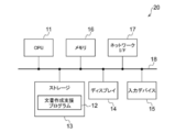
以下、図面を参照して本開示の実施形態について説明する。まず、本実施形態による文書作成支援装置を適用した医療情報システム1の構成について説明する。図1は、医療情報システム1の概略構成を示す図である。図1に示す医療情報システム1は、公知のオーダリングシステムを用いた診療科の医師からの検査オーダに基づいて、被写体の検査対象部位の撮影、撮影により取得された医用画像の保管、読影医による医用画像の読影と読影レポートの作成、および依頼元の診療科の医師による読影レポートの閲覧と読影対象の医用画像の詳細観察とを行うためのシステムである。
Hereinafter, embodiments of the present disclosure will be described with reference to the drawings. First, the configuration of the medical information system 1 to which the document creation support device according to the present embodiment is applied will be described. FIG. 1 is a diagram showing a schematic configuration of the medical information system 1. The medical information system 1 shown in FIG. 1 is based on an inspection order from a doctor in a clinical department using a known ordering system, photographs of a part to be inspected of a subject, storage of a medical image acquired by the imaging, and an interpretation doctor. It is a system for interpreting medical images and creating an interpretation report, and for viewing the interpretation report by the doctor of the requesting clinical department and observing the details of the medical image to be interpreted.
図1に示すように、医療情報システム1は、複数の撮影装置2、読影端末である複数の読影WS(WorkStation)3、診療WS4、画像サーバ5、画像データベース(以下、画像DB(DataBase)とする)6、レポートサーバ7およびレポートデータベース(以下レポートDBとする)8が、有線または無線のネットワーク10を介して互いに通信可能な状態で接続されて構成されている。
As shown in FIG. 1, the medical information system 1 includes a plurality of imaging devices 2, a plurality of image interpretation WS (WorkStation) 3 which are image interpretation terminals, a medical care WS 4, an image server 5, and an image database (hereinafter, image DB (DataBase)). 6. The report server 7 and the report database (hereinafter referred to as the report DB) 8 are connected to each other via a wired or wireless network 10 so as to be able to communicate with each other.
各機器は、医療情報システム1の構成要素として機能させるためのアプリケーションプログラムがインストールされたコンピュータである。アプリケーションプログラムは、ネットワーク10に接続されたサーバコンピュータの記憶装置、若しくはネットワークストレージに、外部からアクセス可能な状態で記憶され、要求に応じてコンピュータにダウンロードされ、インストールされる。または、DVD(Digital Versatile Disc)およびCD-ROM(Compact Disc Read Only Memory)等の記録媒体に記録されて配布され、その記録媒体からコンピュータにインストールされる。
Each device is a computer on which an application program for functioning as a component of the medical information system 1 is installed. The application program is stored in the storage device of the server computer connected to the network 10 or in the network storage in a state of being accessible from the outside, and is downloaded and installed in the computer upon request. Alternatively, it is recorded and distributed on a recording medium such as a DVD (Digital Versatile Disc) and a CD-ROM (Compact Disc Read Only Memory), and installed on a computer from the recording medium.
撮影装置2は、被写体の診断対象となる部位を撮影することにより、診断対象部位を表す医用画像を生成する装置(モダリティ)である。具体的には、単純X線撮影装置、CT装置、MRI装置、およびPET(Positron Emission Tomography)装置等である。撮影装置2により生成された医用画像は画像サーバ5に送信され、画像DB6に保存される。
The photographing device 2 is a device (modality) that generates a medical image representing the diagnosis target part by photographing the part to be diagnosed of the subject. Specifically, it is a simple X-ray imaging apparatus, a CT apparatus, an MRI apparatus, a PET (Positron Emission Tomography) apparatus, and the like. The medical image generated by the imaging device 2 is transmitted to the image server 5 and stored in the image DB 6.
読影WS3は、例えば放射線科の読影医が、医用画像の読影および読影レポートの作成等に利用するコンピュータであり、本実施形態による文書作成支援装置20を内包する。読影WS3では、画像サーバ5に対する医用画像の閲覧要求、画像サーバ5から受信した医用画像に対する各種画像処理、医用画像の表示、および医用画像に関する所見文の入力受け付け等が行われる。また、読影WS3では、医用画像および入力された所見文に対する解析処理、解析結果に基づく読影レポートの作成の支援、レポートサーバ7に対する読影レポートの登録要求と閲覧要求、およびレポートサーバ7から受信した読影レポートの表示が行われる。これらの処理は、読影WS3が各処理のためのソフトウェアプログラムを実行することにより行われる。
The image interpretation WS3 is a computer used by, for example, an image interpretation doctor in a radiology department to interpret a medical image and create an image interpretation report, and includes a document creation support device 20 according to the present embodiment. In the image interpretation WS3, a request for viewing a medical image to the image server 5, various image processing for the medical image received from the image server 5, a display of the medical image, an input acceptance of a finding sentence related to the medical image, and the like are performed. In addition, in the interpretation WS3, analysis processing for medical images and input findings, support for creating an interpretation report based on the analysis results, a request for registration and viewing of an interpretation report for the report server 7, and an interpretation received from the report server 7 are performed. The report is displayed. These processes are performed by the interpretation WS3 executing a software program for each process.
診療WS4は、診療科の医師が、画像の詳細観察、読影レポートの閲覧、および電子カルテの作成等に利用するコンピュータであり、処理装置、ディスプレイ等の表示装置、並びにキーボードおよびマウス等の入力装置により構成される。診療WS4では、画像サーバ5に対する画像の閲覧要求、画像サーバ5から受信した画像の表示、レポートサーバ7に対する読影レポートの閲覧要求、およびレポートサーバ7から受信した読影レポートの表示が行われる。これらの処理は、診療WS4が各処理のためのソフトウェアプログラムを実行することにより行われる。
The medical care WS4 is a computer used by doctors in the clinical department for detailed observation of images, viewing of interpretation reports, creation of electronic medical records, etc., and is a processing device, a display device such as a display, and an input device such as a keyboard and a mouse. Consists of. In the medical treatment WS4, an image viewing request is made to the image server 5, an image received from the image server 5 is displayed, an image interpretation report viewing request is made to the report server 7, and an image interpretation report received from the report server 7 is displayed. These processes are performed by the medical treatment WS4 executing a software program for each process.
画像サーバ5は、汎用のコンピュータにデータベース管理システム(DataBase Management System: DBMS)の機能を提供するソフトウェアプログラムがインストールされたものである。また、画像サーバ5は画像DB6が構成されるストレージを備えている。このストレージは、画像サーバ5とデータバスとによって接続されたハードディスク装置であってもよいし、ネットワーク10に接続されているNAS(Network Attached Storage)およびSAN(Storage Area Network)に接続されたディスク装置であってもよい。また、画像サーバ5は、撮影装置2からの医用画像の登録要求を受け付けると、その医用画像をデータベース用のフォーマットに整えて画像DB6に登録する。
The image server 5 is a general-purpose computer in which a software program that provides a database management system (DataBase Management System: DBMS) function is installed. Further, the image server 5 includes a storage in which the image DB 6 is configured. This storage may be a hard disk device connected by the image server 5 and the data bus, or a disk device connected to NAS (Network Attached Storage) and SAN (Storage Area Network) connected to the network 10. It may be. When the image server 5 receives the medical image registration request from the imaging device 2, the image server 5 arranges the medical image in a database format and registers it in the image DB 6.
画像DB6には、撮影装置2において取得された医用画像の画像データと付帯情報とが登録される。付帯情報には、例えば、個々の医用画像を識別するための画像ID(identification)、被写体を識別するための患者ID、検査を識別するための検査ID、医用画像毎に割り振られるユニークなID(UID:unique identification)、医用画像が生成された検査日、検査時刻、医用画像を取得するための検査で使用された撮影装置の種類、患者氏名、年齢、性別等の患者情報、検査部位(撮影部位)、撮影情報(撮影プロトコル、撮影シーケンス、撮像手法、撮影条件、造影剤の使用等)、1回の検査で複数の医用画像を取得した場合のシリーズ番号あるいは採取番号等の情報が含まれる。
The image data and incidental information of the medical image acquired by the imaging device 2 are registered in the image DB 6. The incidental information includes, for example, an image ID (identification) for identifying an individual medical image, a patient ID for identifying a subject, an examination ID for identifying an examination, and a unique ID assigned to each medical image ( UID: unique identification), examination date when the medical image was generated, examination time, type of imaging device used in the examination to acquire the medical image, patient information such as patient name, age, gender, examination site (imaging) Includes information such as site), imaging information (imaging protocol, imaging sequence, imaging method, imaging conditions, use of contrast medium, etc.), series number or collection number when multiple medical images are acquired in one examination. ..
また、画像サーバ5は、読影WS3および診療WS4からの閲覧要求をネットワーク10経由で受信すると、画像DB6に登録されている医用画像を検索し、検索された医用画像を要求元の読影WS3および診療WS4に送信する。
Further, when the image server 5 receives the viewing request from the image interpretation WS3 and the medical examination WS4 via the network 10, the image server 5 searches for the medical image registered in the image DB 6, and uses the searched medical image as the requesting image interpretation WS3 and the medical examination. Send to WS4.
レポートサーバ7には、汎用のコンピュータにデータベース管理システムの機能を提供するソフトウェアプログラムが組み込まれる。レポートサーバ7は、読影WS3からの読影レポートの登録要求を受け付けると、その読影レポートをデータベース用のフォーマットに整えてレポートDB8に登録する。
The report server 7 incorporates a software program that provides the functions of a database management system to a general-purpose computer. When the report server 7 receives the image interpretation report registration request from the image interpretation WS3, the report server 7 prepares the image interpretation report in a database format and registers the image interpretation report in the report DB 8.
レポートDB8には、読影WS3において作成された所見文を少なくとも含む読影レポートが登録される。読影レポートは、例えば、読影対象の医用画像、医用画像を識別する画像ID、読影を行った読影医を識別するための読影医ID、病変名、病変の位置情報、特定領域を含む医用画像にアクセスするための情報、および性状情報等の情報を含んでいてもよい。
In the report DB8, an interpretation report including at least the findings created in the interpretation WS3 is registered. The image interpretation report includes, for example, a medical image to be interpreted, an image ID for identifying the medical image, an image interpretation doctor ID for identifying the image interpretation doctor who performed the image interpretation, a lesion name, a lesion position information, and a medical image including a specific area. It may include information for access and information such as property information.
また、レポートサーバ7は、読影WS3および診療WS4からの読影レポートの閲覧要求をネットワーク10経由で受信すると、レポートDB8に登録されている読影レポートを検索し、検索された読影レポートを要求元の読影WS3および診療WS4に送信する。
Further, when the report server 7 receives a viewing request for the interpretation report from the interpretation WS3 and the medical treatment WS4 via the network 10, the report server 7 searches for the interpretation report registered in the report DB 8 and uses the searched interpretation report as the requester's interpretation. It is transmitted to WS3 and medical treatment WS4.
なお、本実施形態においては、医用画像は診断対象を肺とした、複数の断層画像からなる3次元のCT画像とし、CT画像を読影することにより、肺に含まれる異常陰影等の関心構造についての医療文章を所見文として含む読影レポートを作成するものとする。なお、医用画像はCT画像に限定されるものではなく、MRI画像および単純X線撮影装置により取得された単純2次元画像等の任意の医用画像を用いることができる。
In the present embodiment, the medical image is a three-dimensional CT image composed of a plurality of tomographic images with the diagnosis target as the lung, and by interpreting the CT image, the structure of interest such as abnormal shadows contained in the lung is obtained. An image interpretation report shall be prepared that includes the medical text of. The medical image is not limited to the CT image, and any medical image such as an MRI image and a simple two-dimensional image acquired by a simple X-ray imaging device can be used.
ネットワーク10は、病院内の各種機器を接続する有線または無線のローカルエリアネットワークである。読影WS3が他の病院あるいは診療所に設置されている場合には、ネットワーク10は、各病院のローカルエリアネットワーク同士をインターネットまたは専用回線で接続した構成としてもよい。
Network 10 is a wired or wireless local area network that connects various devices in the hospital. When the interpretation WS3 is installed in another hospital or clinic, the network 10 may be configured such that the local area networks of each hospital are connected to each other by the Internet or a dedicated line.
次いで、本実施形態による文書作成支援装置について説明する。図2は、本実施形態による文書作成支援装置のハードウェア構成を説明する。図2に示すように、文書作成支援装置20は、CPU(Central Processing Unit)11、不揮発性のストレージ13、および一時記憶領域としてのメモリ16を含む。また、文書作成支援装置20は、液晶ディスプレイ等のディスプレイ14、キーボードとマウス等の入力デバイス15、およびネットワーク10に接続されるネットワークI/F(InterFace)17を含む。CPU11、ストレージ13、ディスプレイ14、入力デバイス15、メモリ16およびネットワークI/F17は、バス18に接続される。なお、CPU11は、本開示におけるプロセッサの一例である。
Next, the document creation support device according to this embodiment will be described. FIG. 2 illustrates the hardware configuration of the document creation support device according to the present embodiment. As shown in FIG. 2, the document creation support device 20 includes a CPU (Central Processing Unit) 11, a non-volatile storage 13, and a memory 16 as a temporary storage area. Further, the document creation support device 20 includes a display 14 such as a liquid crystal display, an input device 15 such as a keyboard and a mouse, and a network I / F (InterFace) 17 connected to the network 10. The CPU 11, the storage 13, the display 14, the input device 15, the memory 16, and the network I / F 17 are connected to the bus 18. The CPU 11 is an example of the processor in the present disclosure.
ストレージ13は、HDD(Hard Disk Drive)、SSD(Solid State Drive)、およびフラッシュメモリ等によって実現される。記憶媒体としてのストレージ13には、文書作成支援プログラムが記憶される。CPU11は、ストレージ13から文書作成支援プログラム12を読み出してからメモリ16に展開し、展開した文書作成支援プログラム12を実行する。
The storage 13 is realized by an HDD (Hard Disk Drive), an SSD (Solid State Drive), a flash memory, or the like. A document creation support program is stored in the storage 13 as a storage medium. The CPU 11 reads the document creation support program 12 from the storage 13 and then expands the document creation support program 12 into the memory 16 to execute the expanded document creation support program 12.
次いで、本実施形態による文書作成支援装置の機能的な構成を説明する。図3は、本実施形態による文書作成支援装置の機能的な構成を示す図である。図3に示すように文書作成支援装置20は、画像取得部21、画像解析部22、文章生成部23、判定部24、表示制御部25、保存制御部26および通信部27を備える。そして、CPU11が、文書作成支援プログラム12を実行することにより、CPU11は、画像取得部21、画像解析部22、文章生成部23、判定部24、表示制御部25、保存制御部26および通信部27として機能する。
Next, the functional configuration of the document creation support device according to this embodiment will be described. FIG. 3 is a diagram showing a functional configuration of the document creation support device according to the present embodiment. As shown in FIG. 3, the document creation support device 20 includes an image acquisition unit 21, an image analysis unit 22, a sentence generation unit 23, a determination unit 24, a display control unit 25, a storage control unit 26, and a communication unit 27. Then, when the CPU 11 executes the document creation support program 12, the CPU 11 has an image acquisition unit 21, an image analysis unit 22, a sentence generation unit 23, a determination unit 24, a display control unit 25, a storage control unit 26, and a communication unit. Functions as 27.
画像取得部21は、操作者である読影医による入力デバイス15からの指示により、画像サーバ5から読影レポートを作成するための医用画像を取得する。
The image acquisition unit 21 acquires a medical image for creating an image interpretation report from the image server 5 in response to an instruction from the input device 15 by the image interpretation doctor who is the operator.
画像解析部22は、医用画像を解析することにより、医用画像に含まれる関心構造の性状を表す性状情報を導出する。このために、画像解析部22は、医用画像に含まれる異常陰影を関心構造として検出し、検出した異常陰影についての性状を、予め定められた複数の性状項目のそれぞれについて判別するように機械学習がなされた学習モデル22Aを有する。
The image analysis unit 22 analyzes the medical image to derive property information representing the property of the structure of interest contained in the medical image. For this purpose, the image analysis unit 22 detects the abnormal shadow included in the medical image as a structure of interest, and performs machine learning so as to discriminate the properties of the detected abnormal shadow for each of a plurality of predetermined property items. It has a learning model 22A that has been made.
ここで、異常陰影について特定される性状項目の例として、異常陰影の場所、異常陰影のサイズ、境界の形状(明瞭および不整形)、吸収値の種類(充実型およびスリガラス型)、スピキュラの有無、腫瘤か結節か、胸膜接触の有無、胸膜陥入の有無、胸膜浸潤の有無、空洞の有無、および石灰化の有無等が挙げられる。なお、性状項目の例はこれらに限定されるものではない。
Here, as an example of the property items specified for the abnormal shadow, the location of the abnormal shadow, the size of the abnormal shadow, the shape of the boundary (clear and irregular), the type of absorption value (full type and pleural type), and the presence or absence of spicula. , Tumor or nodule, presence or absence of pleural contact, presence or absence of pleural infiltration, presence or absence of pleural infiltration, presence or absence of cavity, presence or absence of calcification, etc. The examples of property items are not limited to these.
本実施形態においては、学習モデル22Aは、医用画像における異常陰影の性状を判別するように、教師データを用いてディープラーニング(深層学習)等により機械学習がなされた畳み込みニューラルネットワークからなる。
In the present embodiment, the learning model 22A is composed of a convolutional neural network in which machine learning is performed by deep learning or the like using teacher data so as to discriminate the properties of abnormal shadows in a medical image.
学習モデル22Aは、例えば、異常陰影を含む医用画像と、異常陰影の性状を表す性状項目との複数の組み合わせを教師データとして用いた機械学習によって構築される。学習モデル22Aは、医用画像が入力されると、医用画像に含まれる異常陰影における、性状項目毎に導出される性状スコアを出力する。性状スコアは、各性状項目についての性状の顕著性を示すスコアである。性状スコアは例えば0以上1以下の値をとり、性状スコアの値が大きい程、その性状が顕著であることを示す。
The learning model 22A is constructed by machine learning using, for example, a plurality of combinations of a medical image including an abnormal shadow and a property item representing the property of the abnormal shadow as teacher data. When the medical image is input, the learning model 22A outputs the property score derived for each property item in the abnormal shadow included in the medical image. The property score is a score indicating the prominence of the property for each property item. The property score takes, for example, a value of 0 or more and 1 or less, and the larger the value of the property score, the more remarkable the property.
例えば異常陰影の性状項目の1つである「スピキュラの有無」についての性状スコアが例えば0.5以上である場合、異常陰影の「スピキュラの有無」についての性状が「スピキュラ有り(陽性)」であることを特定し、「スピキュラの有無」についての性状スコアが例えば0.5未満である場合、異常陰影のスピキュラの有無についての性状が「スピキュラ無し(陰性)」であることを特定する。なお、性状判定に用いるしきい値0.5は、例示に過ぎず、性状項目毎に適切な値に設定される。
For example, when the property score for "presence or absence of spicula", which is one of the property items of abnormal shadow, is 0.5 or more, the property for "presence or absence of spicula" of abnormal shadow is "with spicula (positive)". If the property score for "presence or absence of spicula" is, for example, less than 0.5, it is specified that the property for the presence or absence of spicula in abnormal shadow is "no spicula (negative)". The threshold value 0.5 used for the property determination is merely an example, and is set to an appropriate value for each property item.
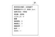
図4は画像解析部22が特定した性状情報の例を説明するための図である。図4に示すように画像解析部22が特定した性状情報30においては、各性状項目についての性状は、「左肺胸膜下」、「4.2cm」、「不整形」、「充実型」、「スピキュラ有」、「腫瘤」、「胸膜接触有」、「胸膜陥入有」、「胸膜浸潤無」、「空洞無」および「石灰化無」となっている。図4においては、「有り」すなわち陽性の場合は+、「無し」すなわち陰性の場合は-を付与している。
FIG. 4 is a diagram for explaining an example of property information specified by the image analysis unit 22. As shown in FIG. 4, in the property information 30 specified by the image analysis unit 22, the properties for each property item are "left pulmonary subpleural", "4.2 cm", "irregular", "enriched type", and so on. They are "with spicula", "mass", "with pleural contact", "with pleural invasion", "without pleural infiltration", "without cavities" and "without calcification". In FIG. 4, + is given when “yes”, that is, positive, and-is given when “no”, that is, negative.
なお、学習モデル22Aとしては、畳み込みニューラルネットワークの他、例えばサポートベクタマシン(SVM(Support Vector Machine))等の任意の学習モデルを用いることができる。
As the learning model 22A, in addition to the convolutional neural network, any learning model such as a support vector machine (SVM (Support Vector Machine)) can be used.
また、医用画像から異常陰影を検出する学習モデルと、異常陰影の性状を判別する学習モデルとを別々に構築するようにしてもよい。
Alternatively, a learning model for detecting abnormal shadows from a medical image and a learning model for discriminating the properties of abnormal shadows may be constructed separately.
文章生成部23は、画像解析部22が導出した性状情報を用いて、医用画像に含まれる異常陰影の性状に関する文章を生成する。また、後述するように、判定部24による判定結果に応じて、生成した医療文章の文章量を調整する。本実施形態においては、文章生成部23は、文章として医療文章を生成する。文章生成部23は、入力された情報から文章を生成するように学習が行われた学習モデル23Aからなる。学習モデル23Aとしては、例えばリカレントニューラルネットワークを用いることができる。図5はリカレントニューラルネットワークの模式的な構成を示す図である。図5に示すように、リカレントニューラルネットワーク40は、エンコーダ41およびデコーダ42からなる。エンコーダ41には、画像解析部22が導出した性状情報が入力される。例えば、エンコーダ41には、「左肺胸膜下」、「4.2cm」、「スピキュラ+」および「腫瘤」の性状情報が入力される。デコーダ42は、文字情報を文章化するように学習がなされており、入力された性状情報から文章を生成する。具体的には、上述した「左肺胸膜下」、「4.2cm」、「スピキュラ+」および「腫瘤」の性状情報から、「左肺胸膜下にスピキュラを有する4.2cm径の腫瘤が認められます。」の医療文章を生成する。なお、図5において「EOS」は文章の終わりを示す(End Of Sentence)。
The sentence generation unit 23 uses the property information derived by the image analysis unit 22 to generate a sentence related to the property of the abnormal shadow included in the medical image. Further, as will be described later, the amount of the generated medical text is adjusted according to the judgment result by the judgment unit 24. In the present embodiment, the sentence generation unit 23 generates a medical sentence as a sentence. The sentence generation unit 23 includes a learning model 23A in which learning is performed so as to generate a sentence from the input information. As the learning model 23A, for example, a recurrent neural network can be used. FIG. 5 is a diagram showing a schematic configuration of a recurrent neural network. As shown in FIG. 5, the recurrent neural network 40 includes an encoder 41 and a decoder 42. The property information derived by the image analysis unit 22 is input to the encoder 41. For example, property information of "left pulmonary subpleuralis", "4.2 cm", "spicula +" and "tumor" is input to the encoder 41. The decoder 42 is learned so as to document the character information, and generates a sentence from the input property information. Specifically, from the above-mentioned property information of "left pulmonary pleura", "4.2 cm", "spicula +" and "mass", "a mass having a spicula under the left pulmonary pleura and having a diameter of 4.2 cm was observed. Will be generated. " In FIG. 5, "EOS" indicates the end of the sentence (End Of Sentence).
このように、性状情報の入力によって医療文章を出力するために、リカレントニューラルネットワーク40は、性状情報と医療文章との組み合わせからなる多数の教師データを用いてエンコーダ41およびデコーダ42を学習することにより構築されてなる。
In this way, in order to output the medical text by inputting the property information, the recurrent neural network 40 learns the encoder 41 and the decoder 42 using a large amount of teacher data composed of a combination of the property information and the medical text. Be built.
なお、文章生成部23は、後述する判定部24による判定の結果に基づいて、医療文章の文章量が規定量となるように文章量を調整する。文章量の調整については後述する。
The sentence generation unit 23 adjusts the sentence amount so that the sentence amount of the medical sentence becomes the specified amount based on the result of the determination by the determination unit 24 described later. The adjustment of the amount of sentences will be described later.
判定部24は、文章生成部23が生成した医療文章の文章量が、規定量であるか否かを判定する。具体的には、判定部24は、文章の文字数、行数または段落数が規定量Th1となるか否かを判定することにより、文章量の判定を行う。規定量Th1は一定の値であってもよく、範囲を持つものであってもよい。範囲を持つ場合、上限値のみを持つものであってもよく、下限値のみを持つものであってもよく、上限値および下限値の双方を持つものであってもよい。具体的には、文字数であれば、規定量Th1を100文字としてもよく、100文字以上としてもよく、100文字以下としてもよく、90文字以上110文字以下としてもよい。本実施形態においては、規定量Th1は上限値および下限値を持つものとして説明する。また、規定量Th1は、読影医の好みに応じて変更できるようにしてもよい。
The determination unit 24 determines whether or not the sentence amount of the medical sentence generated by the sentence generation unit 23 is a specified amount. Specifically, the determination unit 24 determines the amount of sentences by determining whether or not the number of characters, the number of lines, or the number of paragraphs of the sentence is the specified amount Th1. The specified amount Th1 may be a constant value or may have a range. When it has a range, it may have only an upper limit value, it may have only a lower limit value, or it may have both an upper limit value and a lower limit value. Specifically, as long as it is the number of characters, the specified amount Th1 may be 100 characters, 100 characters or more, 100 characters or less, 90 characters or more and 110 characters or less. In the present embodiment, the specified amount Th1 will be described as having an upper limit value and a lower limit value. Further, the specified amount Th1 may be changed according to the preference of the image interpreting doctor.
そして、判定部24は、文章生成部23が生成した医療文章の文章量が規定量Th1でない場合に、判定結果に応じた指示を文章生成部23に対して行う。すなわち、文章量が規定量Th1よりも少ない場合には、文章量を多くする指示を行い、文章量が規定量Th1よりも多い場合には、文章量を少なくする指示を行う。なお、文章生成部23が生成した医療文章の文章量が規定量Th1である場合には、判定部24は何も行わない。
Then, when the sentence amount of the medical sentence generated by the sentence generation unit 23 is not the specified amount Th1, the determination unit 24 gives an instruction according to the determination result to the sentence generation unit 23. That is, when the amount of sentences is less than the specified amount Th1, an instruction to increase the amount of sentences is given, and when the amount of sentences is larger than the specified amount Th1, an instruction to decrease the amount of sentences is given. When the sentence amount of the medical sentence generated by the sentence generation unit 23 is the specified amount Th1, the determination unit 24 does nothing.
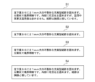
文章生成部23は、判定部24からの指示に応じて、医療文章の文章量を調整する。図6は医療文章および文章量が調整された医療文章の例を示す図である。図6に示すように、調整前の医療文章51が、「左下葉S6に21mm大の不整形な充実型結節を認めます。分葉状で境界明瞭です。内部に石灰化を認めますが、空洞や気管支透亮像は含みません。結節は胸膜と接しています。」であったとする。判定部24がこの医療文章51に関して文章量を少なくする指示を行った場合、文章生成部23は、医療文章に記述すべき性状を選択することにより、文章量を調整する。例えば、医療文章51に含まれる複数の性状のうち、形状に関する性状のみを選択することにより文章量を調整して、「左下葉S6に21mm大の不整形な充実型結節を認めます。」の医療文章52を生成する。あるいは、医療文章51における石灰化、空洞、気管支透亮像等の内部に関する性状、および胸膜接触等の他組織との接触に関する性状に関する性状以外の性状を選択するようにしてもよい。この場合、文章生成部23は、医療文章51の文章量を調整して、「左下葉S6に21mm大の不整形な充実型結節を認めます。分葉状で境界明瞭です。」の医療文章53を生成する。
The sentence generation unit 23 adjusts the amount of medical sentences according to the instruction from the determination unit 24. FIG. 6 is a diagram showing an example of a medical sentence and a medical sentence in which the amount of the sentence is adjusted. As shown in Fig. 6, the medical text 51 before adjustment states, "A 21 mm-sized irregular solid nodule is found in the lower left lobe S6. It is lobulated and has a clear boundary. Calcification is found inside, but it is hollow. And the bronchial translucent image are not included. The nodule is in contact with the pleura. " When the determination unit 24 gives an instruction to reduce the sentence amount with respect to the medical sentence 51, the sentence generation unit 23 adjusts the sentence amount by selecting the property to be described in the medical sentence. For example, the amount of sentences is adjusted by selecting only the properties related to the shape from the plurality of properties included in the medical sentence 51, and "a 21 mm-sized irregular solid nodule is recognized in the lower left lobe S6." Generate medical text 52. Alternatively, properties other than those related to the inside such as calcification, cavity, and bronchial translucency image in the medical text 51, and properties related to contact with other tissues such as pleural contact may be selected. In this case, the sentence generation unit 23 adjusts the sentence amount of the medical sentence 51, and the medical sentence 53 of "a 21 mm-sized irregular solid nodule is recognized in the lower left lobe S6. It is lobulated and has a clear boundary." To generate.
また、文章生成部23は、医療文章51における陽性の性状のみを選択することにより、文章量を調整してもよい。この場合、文章生成部23は、医療文章51の文章量を調整して、「左下葉S6に21mm大の不整形な充実型結節を認めます。分葉状で境界明瞭です。内部に石灰化を認めます。結節は胸膜と接しています。」の医療文章54を生成する。
Further, the sentence generation unit 23 may adjust the amount of sentences by selecting only the positive properties in the medical sentence 51. In this case, the sentence generation unit 23 adjusts the sentence amount of the medical sentence 51 to "recognize an irregular solid nodule of 21 mm in size in the lower left lobe S6. It is lobulated and has a clear boundary. Calcification inside. Acknowledge. The nodule is in contact with the pleura. ”Produces medical text 54.
また、医療文章が短すぎる場合には、文章生成部23は、文章量を多くするように医療文章の文章量を調整する。例えば、図7に示すように、調整前の医療文章61が、「左下葉S6に21mm大の不整形な充実型結節を認めます。」である場合、画像解析部22が導出した陽性の性状項目をすべて選択することにより医療文章61の文章量を調整する。例えば、文章生成部23は医療文章61の文章量を調整して、「左下葉S6に21mm大の不整形な充実型結節を認めます。分葉状で境界明瞭です。内部に石灰化を認めます。結節は胸膜と接しています。」の医療文章62を生成する。また、文章生成部23は、陰性の性状項目および陽性の性状項目のすべてを選択するように医療文章61の文章量を調整してもよい。例えば、文章生成部23は医療文章61の文章量を調整して、「左下葉S6に21mm大の不整形な充実型結節を認めます。分葉状で境界明瞭です。内部に石灰化を認めますが、空洞や気管支透亮像は含みません。結節は胸膜と接しています。」の医療文章63を生成する。
If the medical text is too short, the text generation unit 23 adjusts the text volume of the medical text so as to increase the text volume. For example, as shown in FIG. 7, when the medical sentence 61 before adjustment is "a 21 mm-sized irregular solid nodule is found in the lower left lobe S6", the positive properties derived by the image analysis unit 22 are positive. The amount of medical text 61 is adjusted by selecting all the items. For example, the sentence generation unit 23 adjusts the sentence amount of the medical sentence 61 to "recognize an irregular solid nodule of 21 mm in size in the lower left lobe S6. It is lobulated and has a clear boundary. Calcification is observed inside. The nodule is in contact with the pleura. ”Produces medical text 62. Further, the sentence generation unit 23 may adjust the sentence amount of the medical sentence 61 so as to select all the negative property items and the positive property items. For example, the sentence generation unit 23 adjusts the sentence amount of the medical sentence 61 to "recognize an irregular solid nodule of 21 mm in size in the lower left lobe S6. It is lobulated and has a clear boundary. Calcification is observed inside. However, it does not include cavities or bronchial translucent images. The nodules are in contact with the pleura. ”Produces medical text 63.
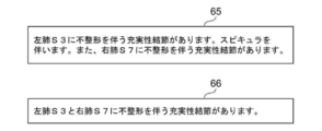
ここで、文章生成部23は、医用画像に複数の異常陰影が含まれる場合、複数の異常陰影について特定した性状の各々に関する記述を含む医療文章を生成する。このような場合において、判定部24から文章量を少なくする指示がなされた場合、文章生成部23は、複数の異常陰影について特定した性状の各々に関する記述のうち、共通する記述を統合することにより、文章量を調整してもよい。例えば、図8に示すように、調整前の医療文章65が、「左肺S3に不整形を伴う充実性結節があります。スピキュラを伴います。また、右肺S7に不整形を伴う充実性結節があります。」であって、2つの異常陰影に関する記述がなされているものとする。この場合、2つの異常陰影について、充実性結節に関する記載が共通する。このため、文章生成部23は、2つの異常陰影について共通する充実性結節に関する記述を統合することにより文章量を調整して、「左肺S3と右肺S7に不整形を伴う充実性結節があります。」の医療文章66を生成する。
Here, when the medical image contains a plurality of abnormal shadows, the sentence generation unit 23 generates a medical sentence including a description of each of the properties specified for the plurality of abnormal shadows. In such a case, when the determination unit 24 gives an instruction to reduce the amount of sentences, the sentence generation unit 23 integrates the common descriptions among the descriptions regarding each of the properties specified for the plurality of abnormal shadows. , You may adjust the amount of sentences. For example, as shown in FIG. 8, the medical sentence 65 before adjustment states, "There is a solid nodule with irregularity in the left lung S3. With spicula. Also, a solid nodule with irregularity in the right lung S7. There is. ”, And it is assumed that there is a description about two abnormal shadows. In this case, the description of the solid nodule is common for the two abnormal shadows. Therefore, the sentence generation unit 23 adjusts the amount of sentences by integrating the descriptions about the solid nodules that are common to the two abnormal shadows, and "the solid nodules with irregularities in the left lung S3 and the right lung S7 are formed. There is. ”Medical sentence 66 is generated.
また、文章生成部23は、医用画像に複数の異常陰影が含まれる場合、異常陰影のそれぞれの性状を記述した複数の候補文章を生成し、複数の関心構造の各々について、複数の候補文章の中から1つの候補文章を選択する組み合わせのうち、選択された候補文章を含む文章の文章量が規定量となる組み合わせを選択することにより、文章量を調整するようにしてもよい。
Further, when the medical image contains a plurality of abnormal shadows, the sentence generation unit 23 generates a plurality of candidate sentences describing the properties of the abnormal shadows, and for each of the plurality of interest structures, the sentence generation unit 23 generates a plurality of candidate sentences. The sentence amount may be adjusted by selecting a combination in which the sentence amount of the sentence including the selected candidate sentence is a predetermined amount from the combination of selecting one candidate sentence from the list.
例えば、医用画像に2つの異常陰影A,Bが含まれるとした場合、文章生成部23は、図9に示すように、異常陰影Aについての候補文章71A~71Cおよび異常陰影Bについての候補文章72A~72Cをそれぞれ生成する。そして、文章生成部23は、異常陰影Aの候補文章71A~71Cおよび異常陰影Bの候補文章72A~72Cからそれぞれ1つの候補文章を選択して、医療文章を生成する。例えば、異常陰影Aについての候補文章71Cと異常陰影Bについての候補文章72Cとをそれぞれ選択して、図10に示す医療文章73を生成する。
For example, assuming that the medical image contains two abnormal shadows A and B, the sentence generation unit 23 determines the candidate sentences 71A to 71C for the abnormal shadow A and the candidate sentences for the abnormal shadow B as shown in FIG. 72A to 72C are generated respectively. Then, the sentence generation unit 23 selects one candidate sentence from each of the candidate sentences 71A to 71C of the abnormal shadow A and the candidate sentences 72A to 72C of the abnormal shadow B, and generates a medical sentence. For example, the candidate sentence 71C for the abnormal shadow A and the candidate sentence 72C for the abnormal shadow B are selected to generate the medical sentence 73 shown in FIG.
医療文章73の文章量が規定量よりも多かった場合、判定部24は、文章量を短くする指示を行う。これにより、文章生成部23は、医療文章73の文章量を少なくするように調整して医療文章74を生成する。この際、文章生成部23は、異常陰影A,Bのうち、悪性度が高い方の異常陰影に関する記述が長くなるように、候補文章を選択するようにすればよい。例えば、異常陰影Aと異常陰影Bとでは、異常陰影Aの方の悪性度が高いため、医療文章73に含まれる異常陰影Bについての記述を、候補文章72Cよりも短い候補文章72Aに変更することにより、医療文章74を生成する。
When the sentence amount of the medical sentence 73 is larger than the specified amount, the determination unit 24 gives an instruction to shorten the sentence amount. As a result, the sentence generation unit 23 generates the medical sentence 74 by adjusting so as to reduce the amount of the medical sentence 73. At this time, the sentence generation unit 23 may select the candidate sentence so that the description of the abnormal shadow having the higher malignancy among the abnormal shadows A and B becomes longer. For example, in the abnormal shadow A and the abnormal shadow B, the abnormal shadow A has a higher degree of malignancy, so the description about the abnormal shadow B included in the medical sentence 73 is changed to the candidate sentence 72A shorter than the candidate sentence 72C. Thereby, the medical sentence 74 is generated.
また、上述したように、医療文章73に含まれる記述のうちの陰性の性状のみを選択したり、共通する性状の記述を統合したりすることにより、医療文章73の文章量を調整するようにしてもよい。
Further, as described above, the amount of sentences in the medical sentence 73 is adjusted by selecting only the negative properties from the descriptions included in the medical sentence 73 or by integrating the descriptions of the common properties. You may.
なお、同一患者についての過去に取得した医用画像および読影レポートを取得し、現在の読影の対象となる医用画像に含まれる異常陰影が、過去の医用画像から継続して存在するものであるか、新たに出現されたものであるか否かを判定し、新たに出現した異常陰影についての記述を長くするようにしてもよい。
It should be noted that the past medical images and interpretation reports of the same patient are acquired, and whether the abnormal shadows included in the medical images to be currently interpreted continue to exist from the past medical images. It may be determined whether or not it is a newly appearing one, and the description of the newly appearing abnormal shadow may be lengthened.
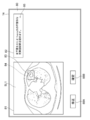
表示制御部25は、生成された医療文章をディスプレイ14に表示する。図11は医療文章の表示画面を示す図である。図11に示すように、表示画面80は画像表示領域81および文章表示領域82を含む。画像表示領域81には、画像解析部22が検出した異常陰影を最も特定しやすいスライス画像SL1が表示される。スライス画像SL1には異常陰影83が含まれ、異常陰影83は矩形領域84により囲まれている。
The display control unit 25 displays the generated medical text on the display 14. FIG. 11 is a diagram showing a display screen of medical texts. As shown in FIG. 11, the display screen 80 includes an image display area 81 and a text display area 82. In the image display area 81, the slice image SL1 that is most likely to identify the abnormal shadow detected by the image analysis unit 22 is displayed. The slice image SL1 includes an abnormal shadow 83, and the abnormal shadow 83 is surrounded by a rectangular region 84.
文章表示領域82には、文章生成部23が生成したまたは文章量を調整した医療文章85が表示されている。医療文章85は、図6に示す医療文章52と同一の「左下葉S6に21mm大の不整形な充実型結節を認めます。」である。
In the sentence display area 82, the medical sentence 85 generated by the sentence generation unit 23 or adjusted in the amount of sentences is displayed. The medical sentence 85 is the same as the medical sentence 52 shown in FIG. 6, "A 21 mm-sized irregular solid nodule is found in the lower left lobe S6."
画像表示領域81の下方には、修正ボタン88Aおよび確定ボタン88Bが表示されている。
Below the image display area 81, a correction button 88A and a confirmation button 88B are displayed.
読影医は、画像表示領域81に表示された、医用画像に含まれるスライス画像SL1を読影し、文章表示領域82に表示された医療文章85の適否を判定する。読影医は修正ボタン88Aを選択することにより、文章表示領域82に表示された医療文章85を、入力デバイス15からの入力により、手動で修正することが可能である。また、確定ボタン88Bを選択することにより、文章表示領域82に表示された医療文章85をその内容で確定することができる。
The image interpreting doctor interprets the slice image SL1 displayed in the image display area 81 and included in the medical image, and determines the suitability of the medical sentence 85 displayed in the sentence display area 82. By selecting the correction button 88A, the interpreter can manually correct the medical text 85 displayed in the text display area 82 by inputting from the input device 15. Further, by selecting the confirmation button 88B, the medical sentence 85 displayed in the sentence display area 82 can be confirmed with the content thereof.
保存制御部26は、操作者による確定ボタン88Bの選択により、文章表示領域82に記述された医療文章85を読影レポートに転記し、読影レポートおよび読影レポートを生成する際に参照したスライス画像を併せて、ストレージ13に保存する。
The storage control unit 26 transfers the medical text 85 described in the text display area 82 to the interpretation report by selecting the confirmation button 88B by the operator, and combines the slice image referred to when generating the interpretation report and the interpretation report. And save it in the storage 13.
通信部27は、文章表示領域82に記述された医療文章85が転記された読影レポート、および読影レポートを生成する際に参照したスライス画像を併せて、ネットワークI/F17を介してレポートサーバ7に転送する。レポートサーバ7は、読影レポートおよびスライス画像を併せて保存する。
The communication unit 27 combines the image interpretation report in which the medical sentence 85 described in the sentence display area 82 is transcribed and the slice image referred to when generating the image interpretation report to the report server 7 via the network I / F17. Forward. The report server 7 stores the interpretation report and the sliced image together.
次いで、本実施形態において行われる処理について説明する。図12は本実施形態において行われる処理を示すフローチャートである。なお、読影の対象となる医用画像は、画像取得部21により画像サーバ5から取得されて、ストレージ13に保存されているものとする。読影レポートの作成の指示が読影医により行われることにより処理が開始され、画像解析部22が、医用画像を解析することにより、医用画像に含まれる異常陰影等の関心構造の性状を表す性状情報を導出する(ステップST1)。次いで、文章生成部23が、性状情報に基づいて医用画像に関する医療文章を生成する(ステップST2)。続いて、判定部24が、生成された医療文章の文章量が規定量であるか否かを判定する(ステップST3)。
Next, the processing performed in this embodiment will be described. FIG. 12 is a flowchart showing the processing performed in the present embodiment. It is assumed that the medical image to be read is acquired from the image server 5 by the image acquisition unit 21 and stored in the storage 13. Processing is started when an instruction to create an image interpretation report is given by the image interpretation doctor, and the image analysis unit 22 analyzes the medical image to represent the properties of the structure of interest such as abnormal shadows contained in the medical image. Is derived (step ST1). Next, the sentence generation unit 23 generates a medical sentence related to the medical image based on the property information (step ST2). Subsequently, the determination unit 24 determines whether or not the amount of the generated medical text is the specified amount (step ST3).
ステップST3が否定されると、文章生成部23は、判定の結果に基づいて、医療文章の文章量が規定量となるように文章量を調整し(ステップST4)、ステップST3に戻る。ステップST3が肯定されると、表示制御部25が、医用画像および文章生成部23が生成した医療文章をディスプレイ14に表示する(ステップST5)。
When step ST3 is denied, the sentence generation unit 23 adjusts the sentence amount so that the sentence amount of the medical sentence becomes the specified amount based on the result of the determination (step ST4), and returns to step ST3. When step ST3 is affirmed, the display control unit 25 displays the medical image and the medical sentence generated by the sentence generation unit 23 on the display 14 (step ST5).
次いで、表示制御部25は、表示画面に表示された修正ボタン88Aが選択されたか否かを判定する(ステップST6)。ステップST6が肯定されると、表示制御部25は、文章表示領域82に表示された医療文章に対する、入力デバイス15を用いての修正を受け付け、文章生成部23は、入力デバイス15からの入力により、文章表示領域82に表示された医療文章を修正する(ステップST7)。続いて、表示制御部25は、確定ボタン88Bが選択されたか否かを判定する(ステップST8)。ステップST8が否定されると、ステップST6に戻る。
Next, the display control unit 25 determines whether or not the correction button 88A displayed on the display screen is selected (step ST6). When step ST6 is affirmed, the display control unit 25 accepts the correction of the medical text displayed in the text display area 82 using the input device 15, and the text generation unit 23 receives the input from the input device 15. , Correct the medical text displayed in the text display area 82 (step ST7). Subsequently, the display control unit 25 determines whether or not the confirmation button 88B has been selected (step ST8). If step ST8 is denied, the process returns to step ST6.
ステップST8が肯定されると、保存制御部26が、医療文章を医用画像についての読影レポートに転記し、読影レポートおよび医用画像を併せて、ストレージ13に保存する(読影レポート等保存;ステップST9)。そして、通信部27が、読影レポートおよび医用画像を併せて、ネットワークI/F17を介してレポートサーバ7に転送し(読影レポート等転送;ステップST10)、処理を終了する。
When step ST8 is affirmed, the storage control unit 26 transfers the medical text to the interpretation report for the medical image, and saves the interpretation report and the medical image together in the storage 13 (save the interpretation report, etc .; step ST9). .. Then, the communication unit 27 transfers the image interpretation report and the medical image together to the report server 7 via the network I / F17 (transfer of the image interpretation report or the like; step ST10), and ends the process.
このように、本実施形態においては、文章の文章量が規定量であるか否かを判定し、判定結果に基づいて、文章量が規定量となるように文章量を調整するようにした。このため、適切な情報量の医療文章を生成できる。
In this way, in the present embodiment, it is determined whether or not the sentence amount of the sentence is the specified amount, and the sentence amount is adjusted so that the sentence amount becomes the specified amount based on the determination result. Therefore, it is possible to generate a medical sentence with an appropriate amount of information.
なお、上記実施形態においては、表示画面80の文章表示領域82に、文章量が規定量に調整された1つの医療文章を表示しているが、これに限定されるものではない。例えば、図13に示すように、文章表示領域82に、図6に示す3つの医療文章52~53を表示し、表示された医療文章52~53から、読影医が所望する医療文章を入力デバイス15を用いて選択できるようにしてもよい。なお、図13においては、上から文章量が短い順序で医療文章52~54が表示されている。また、文章生成部23が生成した医療文章が短かかったため、多くなるように医療文章の文章量を調整した場合、文章量が多い順に複数の医療文章を表示してもよい。例えば、図14に示すように、図7に示す医療文章62,63を上から医療文章63,62の順で表示するようにしてもよい。
In the above embodiment, one medical sentence whose sentence amount is adjusted to a specified amount is displayed in the sentence display area 82 of the display screen 80, but the present invention is not limited to this. For example, as shown in FIG. 13, three medical sentences 52 to 53 shown in FIG. 6 are displayed in the sentence display area 82, and a medical sentence desired by an image interpreter is input from the displayed medical sentences 52 to 53. 15 may be used for selection. In FIG. 13, medical sentences 52 to 54 are displayed in ascending order from the top. Further, since the medical sentences generated by the sentence generation unit 23 are short, when the sentence amount of the medical sentences is adjusted to increase, a plurality of medical sentences may be displayed in descending order of the sentence amount. For example, as shown in FIG. 14, the medical sentences 62 and 63 shown in FIG. 7 may be displayed in the order of the medical sentences 63 and 62 from the top.
また、上記実施形態においては、診断対象を肺とした医用画像を用いて読影レポートを作成する場合に本開示の技術を適用しているが、診断対象は肺に限定されるものではない。肺の他に、心臓、肝臓、脳、および四肢等の人体の任意の部位を診断対象とすることができる。
Further, in the above embodiment, the technique of the present disclosure is applied when creating an image interpretation report using a medical image with the diagnosis target as the lung, but the diagnosis target is not limited to the lung. In addition to the lungs, any part of the human body such as the heart, liver, brain, and limbs can be diagnosed.
また、上記実施形態において、例えば、画像取得部21、画像解析部22、文章生成部23、判定部24、表示制御部25、保存制御部26および通信部27といった各種の処理を実行する処理部(Processing Unit)のハードウェア的な構造としては、次に示す各種のプロセッサ(Processor)を用いることができる。上記各種のプロセッサには、上述したように、ソフトウェア(プログラム)を実行して各種の処理部として機能する汎用的なプロセッサであるCPUに加えて、FPGA(Field Programmable Gate Array)等の製造後に回路構成を変更可能なプロセッサであるプログラマブルロジックデバイス(Programmable Logic Device :PLD)、ASIC(Application Specific Integrated Circuit)等の特定の処理を実行させるために専用に設計された回路構成を有するプロセッサである専用電気回路等が含まれる。
Further, in the above embodiment, for example, a processing unit that executes various processes such as an image acquisition unit 21, an image analysis unit 22, a sentence generation unit 23, a determination unit 24, a display control unit 25, a storage control unit 26, and a communication unit 27. As the hardware structure of (Processing Unit), various processors (Processors) shown below can be used. As described above, the various processors include a CPU, which is a general-purpose processor that executes software (program) and functions as various processing units, and a circuit after manufacturing an FPGA (Field Programmable Gate Array) or the like. Dedicated electricity, which is a processor with a circuit configuration specially designed to execute specific processing such as programmable logic device (PLD), ASIC (Application Specific Integrated Circuit), which is a processor whose configuration can be changed. Circuits and the like are included.
1つの処理部は、これらの各種のプロセッサのうちの1つで構成されてもよいし、同種または異種の2つ以上のプロセッサの組み合わせ(例えば、複数のFPGAの組み合わせまたはCPUとFPGAとの組み合わせ)で構成されてもよい。また、複数の処理部を1つのプロセッサで構成してもよい。
One processing unit may be composed of one of these various processors, or a combination of two or more processors of the same type or different types (for example, a combination of a plurality of FPGAs or a combination of a CPU and an FPGA). ) May be configured. Further, a plurality of processing units may be configured by one processor.
複数の処理部を1つのプロセッサで構成する例としては、第1に、クライアントおよびサーバ等のコンピュータに代表されるように、1つ以上のCPUとソフトウェアとの組み合わせで1つのプロセッサを構成し、このプロセッサが複数の処理部として機能する形態がある。第2に、システムオンチップ(System On Chip:SoC)等に代表されるように、複数の処理部を含むシステム全体の機能を1つのIC(Integrated Circuit)チップで実現するプロセッサを使用する形態がある。このように、各種の処理部は、ハードウェア的な構造として、上記各種のプロセッサの1つ以上を用いて構成される。
As an example of configuring a plurality of processing units with one processor, first, as represented by a computer such as a client and a server, one processor is configured by combining one or more CPUs and software. There is a form in which this processor functions as a plurality of processing units. Second, as typified by System On Chip (SoC), there is a form that uses a processor that realizes the functions of the entire system including multiple processing units with a single IC (Integrated Circuit) chip. be. As described above, the various processing units are configured by using one or more of the above-mentioned various processors as a hardware structure.
さらに、これらの各種のプロセッサのハードウェア的な構造としては、より具体的には、半導体素子等の回路素子を組み合わせた電気回路(Circuitry)を用いることができる。
Further, as the hardware structure of these various processors, more specifically, an electric circuit (Circuitry) in which circuit elements such as semiconductor elements are combined can be used.
1 医療情報システム
2 撮影装置
3 読影WS
4 診療科WS
5 画像サーバ
6 画像DB
7 レポートサーバ
8 レポートDB
10 ネットワーク
11 CPU
12 文書作成支援プログラム
13 ストレージ
14 ディスプレイ
15 入力デバイス
16 メモリ
17 ネットワークI/F
18 バス
20 文書作成支援装置
21 画像取得部
22 画像解析部
23 文章生成部
24 判定部
25 表示制御部
26 保存制御部
27 通信部
30 性状情報
40 リカレントニューラルネットワーク
41 エンコーダ
42 デコーダ
51~54、61~63、65、66、73、74 医療文章
71A~71C、72A~72C 候補文章
80 表示画面
81 画像表示領域
82 文章表示領域
83 異常陰影
84 矩形領域
85 医療文章
88A 修正ボタン
88B 確定ボタン
SL1 スライス画像 1Medical information system 2 Imaging device 3 Interpretation WS
4 Clinical department WS
5image server 6 image DB
7Report server 8 Report DB
10network 11 CPU
12 Documentcreation support program 13 Storage 14 Display 15 Input device 16 Memory 17 Network I / F
18Bus 20 Document creation support device 21 Image acquisition unit 22 Image analysis unit 23 Sentence generation unit 24 Judgment unit 25 Display control unit 26 Storage control unit 27 Communication unit 30 Property information 40 Recurrent neural network 41 Encoder 42 Decoder 51-54, 61- 63, 65, 66, 73, 74 Medical text 71A-71C, 72A-72C Candidate text 80 Display screen 81 Image display area 82 Text display area 83 Abnormal shadow 84 Rectangular area 85 Medical text 88A Correction button 88B Confirm button SL1 Slice image
2 撮影装置
3 読影WS
4 診療科WS
5 画像サーバ
6 画像DB
7 レポートサーバ
8 レポートDB
10 ネットワーク
11 CPU
12 文書作成支援プログラム
13 ストレージ
14 ディスプレイ
15 入力デバイス
16 メモリ
17 ネットワークI/F
18 バス
20 文書作成支援装置
21 画像取得部
22 画像解析部
23 文章生成部
24 判定部
25 表示制御部
26 保存制御部
27 通信部
30 性状情報
40 リカレントニューラルネットワーク
41 エンコーダ
42 デコーダ
51~54、61~63、65、66、73、74 医療文章
71A~71C、72A~72C 候補文章
80 表示画面
81 画像表示領域
82 文章表示領域
83 異常陰影
84 矩形領域
85 医療文章
88A 修正ボタン
88B 確定ボタン
SL1 スライス画像 1
4 Clinical department WS
5
7
10
12 Document
18
Claims (10)
- 少なくとも1つのプロセッサを備え、
前記プロセッサは、
画像に含まれる少なくとも1つの関心構造の性状に関する文章を生成し、
前記文章の文章量が規定量であるか否かを判定し、
前記判定の結果に基づいて、前記文章量が前記規定量となるように前記文章量を調整するように構成される文書作成支援装置。 With at least one processor
The processor
Generate a sentence about the nature of at least one structure of interest contained in the image
It is determined whether or not the amount of the above sentence is the specified amount, and
A document creation support device configured to adjust the sentence amount so that the sentence amount becomes the specified amount based on the result of the determination. - 前記プロセッサは、前記関心構造についての少なくとも1つの性状のうちの、前記文章に記述すべき性状を選択することにより、前記文章量を調整するように構成される請求項1に記載の文書作成支援装置。 The document creation support according to claim 1, wherein the processor is configured to adjust the amount of the sentence by selecting the property to be described in the sentence from at least one property of the structure of interest. Device.
- 前記プロセッサは、前記関心構造について特定した少なくとも1つの性状の各々に関する記述を含む前記文章を生成し、前記文章に含まれる複数の性状の各々に関する記述のうち、陰性の性状に関する記述を前記文章から削除することにより、前記文章量を調整するように構成される請求項1または2に記載の文書作成支援装置。 The processor generates the sentence including a description for each of the at least one property specified for the structure of interest, and among the descriptions for each of the plurality of properties included in the sentence, the description regarding the negative property is derived from the sentence. The document creation support device according to claim 1 or 2, wherein the amount of sentences is adjusted by deleting the document.
- 前記プロセッサは、前記画像に含まれる複数の関心構造について、前記関心構造のそれぞれについての性状を記述した複数の文章を生成し、
前記複数の関心構造のそれぞれについて生成された前記文章の総量が前記規定量となるように、前記複数の関心構造の少なくとも1つについての前記文章の文章量を調整するように構成される請求項1から3のいずれか1項に記載の文書作成支援装置。 The processor generates a plurality of sentences describing the properties of each of the plurality of interest structures contained in the image.
A claim configured to adjust the amount of text for at least one of the plurality of interest structures so that the total amount of the text generated for each of the plurality of interest structures is the specified amount. The document creation support device according to any one of 1 to 3. - 前記プロセッサは、前記文章に含まれる複数の関心構造の各々に関する記述のうち、共通する記述を統合することにより、前記文章量を調整するように構成される請求項4に記載の文書作成支援装置。 The document creation support device according to claim 4, wherein the processor is configured to adjust the amount of the sentence by integrating a common description among the descriptions relating to each of the plurality of interest structures included in the sentence. ..
- 前記プロセッサは、前記画像に含まれる複数の関心構造について、前記関心構造のそれぞれの性状を記述した複数の候補文章を生成し、
前記複数の関心構造の各々について、前記複数の候補文章の中から1つの前記候補文章を選択する組み合わせのうち、選択された候補文章を含む文章の文章量が前記規定量となる組み合わせを選択することにより、前記文章量を調整するように構成される請求項1から5のいずれか1項に記載の文書作成支援装置。 The processor generates a plurality of candidate sentences describing the properties of the plurality of interest structures contained in the image.
For each of the plurality of interest structures, among the combinations for selecting one candidate sentence from the plurality of candidate sentences, a combination in which the sentence amount of the sentence including the selected candidate sentence is the specified amount is selected. The document creation support device according to any one of claims 1 to 5, which is configured to adjust the amount of sentences. - 前記プロセッサは、前記文章をディスプレイに表示するように構成される請求項1から6のいずれか1項に記載の文書作成支援装置。 The document creation support device according to any one of claims 1 to 6, wherein the processor is configured to display the text on a display.
- 前記画像は医用画像であり、前記文章は、前記医用画像に含まれる前記関心構造に関する医療文章である請求項1から7のいずれか1項に記載の文書作成支援装置。 The document creation support device according to any one of claims 1 to 7, wherein the image is a medical image, and the sentence is a medical sentence related to the structure of interest included in the medical image.
- 画像に含まれる少なくとも1つの関心構造の性状に関する文章を生成し、
前記文章の文章量が規定量であるか否かを判定し、
前記判定の結果に基づいて、前記文章量が前記規定量となるように前記文章量を調整する文書作成支援方法。 Generate a sentence about the nature of at least one structure of interest contained in the image
It is determined whether or not the amount of the above sentence is the specified amount, and
A document creation support method for adjusting the amount of sentences so that the amount of sentences becomes the specified amount based on the result of the determination. - 画像に含まれる少なくとも1つの関心構造の性状に関する文章を生成する手順と、
前記文章の文章量が規定量であるか否かを判定する手順と、
前記判定の結果に基づいて、前記文章量が前記規定量となるように前記文章量を調整する手順とをコンピュータに実行させる文書作成支援プログラム。 A procedure for generating text about the properties of at least one structure of interest contained in an image, and
The procedure for determining whether or not the amount of the above sentence is the specified amount, and
A document creation support program that causes a computer to execute a procedure for adjusting the amount of sentences so that the amount of sentences becomes the specified amount based on the result of the determination.
Priority Applications (2)
| Application Number | Priority Date | Filing Date | Title |
|---|---|---|---|
| JP2022510491A JP7436636B2 (en) | 2020-03-23 | 2021-03-22 | Document creation support device, method and program |
| US17/901,829 US20230005601A1 (en) | 2020-03-23 | 2022-09-01 | Document creation support apparatus, method, and program |
Applications Claiming Priority (2)
| Application Number | Priority Date | Filing Date | Title |
|---|---|---|---|
| JP2020-051707 | 2020-03-23 | ||
| JP2020051707 | 2020-03-23 |
Related Child Applications (1)
| Application Number | Title | Priority Date | Filing Date |
|---|---|---|---|
| US17/901,829 Continuation US20230005601A1 (en) | 2020-03-23 | 2022-09-01 | Document creation support apparatus, method, and program |
Publications (1)
| Publication Number | Publication Date |
|---|---|
| WO2021193548A1 true WO2021193548A1 (en) | 2021-09-30 |
Family
ID=77892170
Family Applications (1)
| Application Number | Title | Priority Date | Filing Date |
|---|---|---|---|
| PCT/JP2021/011744 WO2021193548A1 (en) | 2020-03-23 | 2021-03-22 | Document creation assistance device, method, and program |
Country Status (3)
| Country | Link |
|---|---|
| US (1) | US20230005601A1 (en) |
| JP (1) | JP7436636B2 (en) |
| WO (1) | WO2021193548A1 (en) |
Families Citing this family (1)
| Publication number | Priority date | Publication date | Assignee | Title |
|---|---|---|---|---|
| WO2021107098A1 (en) * | 2019-11-29 | 2021-06-03 | 富士フイルム株式会社 | Document creation assistance device, document creation assistance method, and document creation assistance program |
Citations (5)
| Publication number | Priority date | Publication date | Assignee | Title |
|---|---|---|---|---|
| JP2001043220A (en) * | 1999-07-27 | 2001-02-16 | Sony Corp | Method and device for processing document and recording medium |
| JP2007094515A (en) * | 2005-09-27 | 2007-04-12 | Fujifilm Corp | Radiography reading report preparation device |
| JP2011087005A (en) * | 2009-10-13 | 2011-04-28 | Neikusu:Kk | Telephone call voice summary generation system, method therefor, and telephone call voice summary generation program |
| JP2018166961A (en) * | 2017-03-30 | 2018-11-01 | キヤノン株式会社 | Information processing device, method for controlling the same and program |
| JP2019153250A (en) * | 2018-03-06 | 2019-09-12 | 富士フイルム株式会社 | Device, method, and program for supporting preparation of medical document |
-
2021
- 2021-03-22 WO PCT/JP2021/011744 patent/WO2021193548A1/en active Application Filing
- 2021-03-22 JP JP2022510491A patent/JP7436636B2/en active Active
-
2022
- 2022-09-01 US US17/901,829 patent/US20230005601A1/en active Pending
Patent Citations (5)
| Publication number | Priority date | Publication date | Assignee | Title |
|---|---|---|---|---|
| JP2001043220A (en) * | 1999-07-27 | 2001-02-16 | Sony Corp | Method and device for processing document and recording medium |
| JP2007094515A (en) * | 2005-09-27 | 2007-04-12 | Fujifilm Corp | Radiography reading report preparation device |
| JP2011087005A (en) * | 2009-10-13 | 2011-04-28 | Neikusu:Kk | Telephone call voice summary generation system, method therefor, and telephone call voice summary generation program |
| JP2018166961A (en) * | 2017-03-30 | 2018-11-01 | キヤノン株式会社 | Information processing device, method for controlling the same and program |
| JP2019153250A (en) * | 2018-03-06 | 2019-09-12 | 富士フイルム株式会社 | Device, method, and program for supporting preparation of medical document |
Also Published As
| Publication number | Publication date |
|---|---|
| US20230005601A1 (en) | 2023-01-05 |
| JP7436636B2 (en) | 2024-02-21 |
| JPWO2021193548A1 (en) | 2021-09-30 |
Similar Documents
| Publication | Publication Date | Title |
|---|---|---|
| JP2019169049A (en) | Medical image specification device, method, and program | |
| WO2020209382A1 (en) | Medical document generation device, method, and program | |
| JP2023175011A (en) | Document creation assistance device, method, and program | |
| JP2024009342A (en) | Document preparation supporting device, method, and program | |
| WO2021193548A1 (en) | Document creation assistance device, method, and program | |
| JP7007469B2 (en) | Medical document creation support devices, methods and programs, trained models, and learning devices, methods and programs | |
| US20220392619A1 (en) | Information processing apparatus, method, and program | |
| US20230005580A1 (en) | Document creation support apparatus, method, and program | |
| JP7212147B2 (en) | MEDICAL DOCUMENT SUPPORT DEVICE, METHOD AND PROGRAM | |
| JP7420914B2 (en) | Information processing device, information processing method, and information processing program | |
| WO2021177357A1 (en) | Information processing device, information processing method, and information processing program | |
| JP7376715B2 (en) | Progress prediction device, method of operating the progress prediction device, and progress prediction program | |
| EP4287195A1 (en) | Information processing device, method, and program | |
| WO2021177312A1 (en) | Device, method, and program for storing information, and device, method, and program for generating analysis records | |
| JP7368592B2 (en) | Document creation support device, method and program | |
| WO2021107142A1 (en) | Document creation assistance device, method, and program | |
| WO2020241857A1 (en) | Medical document creation device, method, and program, learning device, method, and program, and learned model | |
| WO2022070528A1 (en) | Medical image processing device, method, and program | |
| WO2022153702A1 (en) | Medical image display device, method, and program | |
| WO2022064794A1 (en) | Image display device, method, and program | |
| WO2022196106A1 (en) | Document creation device, method, and program | |
| JP7371220B2 (en) | Information processing device, information processing method, and information processing program | |
| JP7361930B2 (en) | Medical image processing device, method and program | |
| WO2022113587A1 (en) | Image display device, method, and program | |
| WO2021107098A1 (en) | Document creation assistance device, document creation assistance method, and document creation assistance program |
Legal Events
| Date | Code | Title | Description |
|---|---|---|---|
| 121 | Ep: the epo has been informed by wipo that ep was designated in this application |
Ref document number: 21775801 Country of ref document: EP Kind code of ref document: A1 |
|
| ENP | Entry into the national phase |
Ref document number: 2022510491 Country of ref document: JP Kind code of ref document: A |
|
| NENP | Non-entry into the national phase |
Ref country code: DE |
|
| 122 | Ep: pct application non-entry in european phase |
Ref document number: 21775801 Country of ref document: EP Kind code of ref document: A1 |