WO2021107142A1 - Document creation assistance device, method, and program - Google Patents
Document creation assistance device, method, and program Download PDFInfo
- Publication number
- WO2021107142A1 WO2021107142A1 PCT/JP2020/044367 JP2020044367W WO2021107142A1 WO 2021107142 A1 WO2021107142 A1 WO 2021107142A1 JP 2020044367 W JP2020044367 W JP 2020044367W WO 2021107142 A1 WO2021107142 A1 WO 2021107142A1
- Authority
- WO
- WIPO (PCT)
- Prior art keywords
- sentence
- medical
- image
- content
- correction
- Prior art date
Links
Images


Classifications
-
- G—PHYSICS
- G06—COMPUTING; CALCULATING OR COUNTING
- G06F—ELECTRIC DIGITAL DATA PROCESSING
- G06F40/00—Handling natural language data
- G06F40/40—Processing or translation of natural language
- G06F40/55—Rule-based translation
- G06F40/56—Natural language generation
-
- G—PHYSICS
- G06—COMPUTING; CALCULATING OR COUNTING
- G06F—ELECTRIC DIGITAL DATA PROCESSING
- G06F40/00—Handling natural language data
- G06F40/10—Text processing
- G06F40/166—Editing, e.g. inserting or deleting
-
- G—PHYSICS
- G06—COMPUTING; CALCULATING OR COUNTING
- G06F—ELECTRIC DIGITAL DATA PROCESSING
- G06F40/00—Handling natural language data
- G06F40/20—Natural language analysis
- G06F40/274—Converting codes to words; Guess-ahead of partial word inputs
-
- G—PHYSICS
- G06—COMPUTING; CALCULATING OR COUNTING
- G06T—IMAGE DATA PROCESSING OR GENERATION, IN GENERAL
- G06T7/00—Image analysis
-
- G—PHYSICS
- G16—INFORMATION AND COMMUNICATION TECHNOLOGY [ICT] SPECIALLY ADAPTED FOR SPECIFIC APPLICATION FIELDS
- G16H—HEALTHCARE INFORMATICS, i.e. INFORMATION AND COMMUNICATION TECHNOLOGY [ICT] SPECIALLY ADAPTED FOR THE HANDLING OR PROCESSING OF MEDICAL OR HEALTHCARE DATA
- G16H15/00—ICT specially adapted for medical reports, e.g. generation or transmission thereof
-
- G—PHYSICS
- G16—INFORMATION AND COMMUNICATION TECHNOLOGY [ICT] SPECIALLY ADAPTED FOR SPECIFIC APPLICATION FIELDS
- G16H—HEALTHCARE INFORMATICS, i.e. INFORMATION AND COMMUNICATION TECHNOLOGY [ICT] SPECIALLY ADAPTED FOR THE HANDLING OR PROCESSING OF MEDICAL OR HEALTHCARE DATA
- G16H30/00—ICT specially adapted for the handling or processing of medical images
- G16H30/40—ICT specially adapted for the handling or processing of medical images for processing medical images, e.g. editing
Definitions
- This disclosure relates to a document creation support device, method and program that support the creation of documents such as medical documents.
- CT Computer Tomography
- MRI Magnetic Resonance Imaging
- medical images are analyzed by CAD (Computer-Aided Diagnosis) using a learning model that has been machine-learned by deep learning, etc., and the shape, density, and position of structures of interest such as abnormal shadow candidates included in the medical images. It is also practiced to discriminate properties such as size and size, and obtain these as analysis results.
- the analysis result acquired by CAD is associated with the examination information such as the patient name, gender, age, and the modality from which the medical image was acquired, and is stored in the database.
- the medical image and the analysis result are transmitted to the terminal of the image interpreting doctor who interprets the medical image.
- the image interpreting doctor interprets the medical image by referring to the transmitted medical image and the analysis result on his / her terminal, and creates an image interpretation report.
- Japanese Patent Application Laid-Open No. 2019-153250 describes an interpretation report based on information representing the properties of a structure of interest (hereinafter referred to as property information), which includes keywords and medical images input by an image interpreter in the analysis results.
- property information information representing the properties of a structure of interest
- 2019-153250 have been proposed (see JP-A-2019-153250).
- a learning model in which machine learning such as a recurrent neural network is trained so as to generate a sentence from characters representing input property information is used.
- Medical texts (hereinafter referred to as medical texts) are created.
- medical texts By automatically generating medical texts as in the method described in JP-A-2019-153250, it is possible to reduce the burden on the interpretation doctor when creating a medical document such as an interpretation report.
- a text document contains one or more text components, identifies an erroneous text component from one or more text components, and refers to the erroneous text component.
- a method has been proposed in which a list of alternatives is displayed based on the partial input of the desired alternative, and the wrong text component is replaced with the selected alternative in the text document.
- the document creation support device includes at least one processor.
- the processor displays a sentence containing at least one content on the display and Identify modified content based on at least some text input or specification for modified content that is added to or removed from the text. It is configured to modify the text based on the modified content.
- the processor is configured to derive property information representing the properties of the structure of interest contained in the image as content by further analyzing the image. May be good.
- the processor may be configured to generate a sentence related to an image based on the property information.
- the processor may be configured to specify the modified content based on the property information.
- the processor identifies the content included in the text and determines the content. It may be configured to modify the identified content based on the modified content.
- the processor may be configured to correct the sentence according to the style of the sentence before the correction.
- the processor generates a plurality of correction candidates, and the processor generates a plurality of correction candidates.
- the text may be modified by the correction candidates according to the style of the text before the correction.
- the processor generates a plurality of correction candidates, and the processor generates a plurality of correction candidates. Show multiple correction candidates on the display Accepts the selection of correction candidates to be used for sentences from the displayed multiple correction candidates, The selected correction candidate may be configured to correct the sentence.
- the processor may be configured to correct the text so that the corrected content and the content included in the text before the correction are consistent.
- the document creation support method displays a sentence containing at least one content on a display. Identify modified content based on at least some text input or specification for modified content that is added to or removed from the text. Correct the text based on the content.
- sentences can be corrected efficiently.
- the figure which shows the schematic structure of the document creation support apparatus by 1st Embodiment Diagram showing an example of teacher data for learning the first learning model The figure which shows the schematic structure of the recurrent neural network
- the figure which shows the example of the teacher data for learning the 3rd learning model The figure which shows the example of the display screen of the medical sentence in 1st Embodiment Diagram showing an example of a medical text display screen where correction content is being input
- the figure which shows the example of the table which shows the derivation result of property information The figure which shows the schematic structure of the recurrent neural network in the sentence correction part.
- the figure which shows the example of the teacher data for learning the 4th learning model The figure which shows the example of the display screen of the medical sentence in 1st Embodiment
- the figure which shows the other example of the medical sentence in 1st Embodiment A flowchart showing the processing performed in the first embodiment
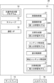
- FIG. 1 is a diagram showing a schematic configuration of a medical information system to which the document creation support device according to the first embodiment of the present disclosure is applied.
- the medical information system 1 shown in FIG. 1 is based on an examination order from a doctor in a clinical department using a known ordering system, photographs of a part to be inspected of a subject, storage of medical images obtained by photographing, and an image interpreter. It is a system for interpreting medical images and creating an interpretation report, and for viewing the interpretation report by the doctor of the requesting clinical department and observing the details of the medical image to be interpreted. As shown in FIG.
- the medical information system 1 includes a plurality of modalities (imaging devices) 2, a plurality of image interpretation workstations (WS) 3 which are image interpretation terminals, a clinical department workstation (WS) 4, an image server 5, and an image.
- the database 6, the interpretation report server 7, and the interpretation report database 8 are connected and configured so as to be able to communicate with each other via a wired or wireless network 10.
- Each device is a computer on which an application program for functioning as a component of the medical information system 1 is installed.
- the application program is stored in the storage device of the server computer connected to the network 10 or in the network storage in a state of being accessible from the outside, and is downloaded and installed in the computer upon request. Alternatively, it is recorded and distributed on a recording medium such as a DVD (Digital Versatile Disc) or a CD-ROM (Compact Disc Read Only Memory), and installed on a computer from the recording medium.
- Modality 2 is a device that generates a medical image showing the diagnosis target part by photographing the diagnosis target part of the subject. Specifically, it is a simple X-ray imaging apparatus, a CT apparatus, an MRI apparatus, a PET (Positron Emission Tomography) apparatus, and the like.
- the medical image generated by the modality 2 is transmitted to the image server 5 and stored.
- Interpretation WS3 includes a document creation support device according to this embodiment. The configuration of the interpretation WS3 will be described later.
- the clinical department WS4 is a computer used by doctors in the clinical department for detailed observation of images, viewing of interpretation reports, creation of electronic medical records, etc., and is composed of a processing device, a display, and input devices such as a keyboard and a mouse. ..
- a patient's medical record electronic medical record
- an image viewing request is made to the image server 5
- an image received from the image server 5 is displayed
- a lesion-like part in the image is automatically detected or highlighted
- an image interpretation report server is used.
- Each process such as the viewing request of the image interpretation report to the image interpretation report 7 and the display of the image interpretation report received from the image interpretation report server 7 is performed by executing the software program for each process.
- the image server 5 is a general-purpose computer in which a software program that provides a database management system (DataBase Management System: DBMS) function is installed. Further, the image server 5 includes a storage in which the image database 6 is configured. This storage may be a hard disk device connected by the image server 5 and the data bus, or a disk device connected to NAS (Network Attached Storage) and SAN (Storage Area Network) connected to the network 10. It may be.
- NAS Network Attached Storage
- SAN Storage Area Network
- the image data and incidental information of the medical image acquired in the modality 2 are registered in the image database 6.
- the incidental information includes, for example, an image ID (identification) for identifying an individual medical image, a patient ID for identifying a subject, an examination ID for identifying an examination, and a unique ID assigned to each medical image ( UID: unique identification), examination date when the medical image was generated, examination time, type of modality used in the examination to acquire the medical image, patient information such as patient name, age, gender, examination site (contrast image site) ), Imaging information (imaging protocol, imaging sequence, imaging method, imaging conditions, use of contrast medium, etc.), including information such as a series number or collection number when a plurality of medical images are acquired in one examination.
- the image server 5 When the image server 5 receives the viewing request from the image interpretation WS3 via the network 10, the image server 5 searches for the medical image registered in the image database 6 and transmits the searched medical image to the requester's image interpretation WS3.
- the interpretation report server 7 incorporates a software program that provides the functions of a database management system to a general-purpose computer.
- the image interpretation report server 7 receives the image interpretation report registration request from the image interpretation WS3, the image interpretation report server 7 arranges the image interpretation report in a database format and registers it in the image interpretation report database 8.
- the image interpretation report is searched from the image interpretation report database 8.
- the image interpretation report database 8 contains, for example, an image ID for identifying a medical image to be interpreted, an image radiologist ID for identifying an image diagnostician who has performed image interpretation, a lesion name, lesion location information, findings, and confidence in the findings. An interpretation report in which information such as the degree is recorded is registered.
- the medical image is a three-dimensional CT image composed of a plurality of tomographic images with the diagnosis target as the lung, and by interpreting the CT image, an interpretation report on abnormal shadows contained in the lung is obtained. It shall be created as a medical document.
- the medical image is not limited to the CT image, and any medical image such as an MRI image and a simple two-dimensional image acquired by a simple X-ray imaging device can be used.
- Network 10 is a wired or wireless network that connects various devices in the hospital.
- the network 10 may be configured such that the local area networks of each hospital are connected to each other by the Internet or a dedicated line.
- the image interpretation WS3 is a computer used by a medical image interpretation doctor to interpret a medical image and create an image interpretation report, and is composed of a processing device, a display, and an input device such as a keyboard and a mouse.
- the image server 5 is requested to view the medical image, various image processes for the medical image received from the image server 5, the display of the medical image, the analysis process for the medical image, the highlighting of the medical image based on the analysis result, and the analysis result.
- Each process such as creating an image interpretation report based on the above, supporting the creation of an image interpretation report, requesting the image interpretation report server 7 to register and view the image interpretation report, and displaying the image interpretation report received from the image interpretation report server 7, is for each process. It is done by executing the software program of. Of these processes, processes other than those performed by the document creation support device of the present embodiment are performed by a well-known software program, and therefore detailed description thereof will be omitted here. Further, the image interpretation WS3 does not perform any process other than the process performed by the document creation support device of the present embodiment, and a computer that performs the process is connected to the network 10 separately, and the process is requested by the image interpretation WS3. The computer may perform the requested processing.
- the interpretation WS3 includes a document creation support device according to the first embodiment. Therefore, the document creation support program according to the present embodiment is installed in the interpretation WS3.
- the document creation support program is stored in the storage device of the server computer connected to the network or in the network storage in a state of being accessible from the outside, and is downloaded and installed in the interpretation WS3 as requested. Alternatively, it is recorded on a recording medium such as a DVD or a CD-ROM, distributed, and installed on the interpretation WS3 from the recording medium.
- FIG. 2 is a diagram showing a schematic configuration of a document creation support device according to the first embodiment, which is realized by installing a document creation support program on the interpretation WS3.
- the document creation support device 20 includes a CPU (Central Processing Unit) 11, a memory 12, a storage 13, and a communication I / F (interface) 14 as a standard computer configuration. Further, a display 15 such as a liquid crystal display and an input device 16 such as a keyboard and a mouse are connected to the document creation support device 20.
- the CPU 11 corresponds to the processor.
- the storage 13 is composed of a hard disk or a storage device such as an SSD (Solid State Drive).
- the storage 13 stores various information including information necessary for processing the medical image and the document creation support device 20 acquired from the image server 5 via the network 10.
- the communication I / F 14 is a network interface that controls the transmission of various information between the external device and the document creation support device 20 via the network 10.
- the document creation support program is stored in the memory 12.
- the document creation support program is an image acquisition process for acquiring a medical image and an image analysis process for deriving property information representing the properties of the structure of interest contained in the medical image by analyzing the medical image as a process to be executed by the CPU 11.
- Sentence generation processing to generate medical sentences related to medical images based on property information
- content identification processing to identify contents representing properties related to structures of interest contained in medical sentences by analyzing medical sentences
- generated medical treatment Display control process for displaying text on display 15, modified content identification process for identifying modified content based on input or specification of at least some characters for modified content added to or deleted from medical text
- the sentence correction process for correcting medical sentences based on the corrected content is specified.
- the CPU 11 executes these processes according to the document creation support program, so that the computer has the image acquisition unit 21, the image analysis unit 22, the sentence generation unit 23, the content identification unit 24, the display control unit 25, and the modified content identification unit. It functions as 26 and the text correction unit 27.
- the image acquisition unit 21 is composed of an interface connected to the network 10, and acquires a medical image for creating an image interpretation report from the image server 5 according to an instruction from the input device 16 by the image interpretation doctor who is the operator.
- the image analysis unit 22 analyzes the medical image to derive property information representing the properties of the structure of interest such as an abnormal shadow candidate included in the medical image.
- the image analysis unit 22 has a first learning model 22A in which machine learning is performed so as to discriminate abnormal shadow candidates in a medical image and discriminate the properties of the discriminated abnormal shadow candidates.
- the first learning model 22A determines whether or not each pixel (voxel) in the medical image represents an abnormal shadow candidate, and if it is an abnormal shadow candidate, the property thereof is determined. It consists of a convolutional neural network (CNN (Convolutional Neural Network)) in which deep learning is performed using teacher data so as to discriminate.
- CNN Convolutional Neural Network
- FIG. 3 is a diagram showing an example of teacher data for learning the first learning model.
- the teacher data 30 includes a medical image 32 including the abnormal shadow 31 and property information 33 about the abnormal shadow.
- the abnormal shadow 31 is a lung nodule
- the property information 33 represents a plurality of properties of the lung nodule.
- the property information 33 includes the location of the abnormal shadow, the size of the abnormal shadow, the shape of the boundary (clear and irregular), the type of absorption value (solid and suriglass type), the presence or absence of spicula, the mass or nodule, and the pleura.
- the presence or absence of contact, the presence or absence of pleural infiltration, the presence or absence of pleural infiltration, the presence or absence of cavities, the presence or absence of fat, the presence or absence of calcification, etc. are used.
- the property information 33 shows that the location of the abnormal shadow is under the left pulmonary pleura, the size of the abnormal shadow is 4.2 cm in diameter, and the boundary is defined. Irregular shape, full absorption, with spicula, mass, with pleural contact, with pleural infiltration, no pleural infiltration, no cavities, no fat, and no calcification ..
- FIG. 1 Irregular shape, full absorption, with spicula, mass, with pleural contact, with pleural infiltration, no pleural infiltration, no cavities, no fat, and no calcification .
- the first learning model 22A is constructed by learning a neural network using a large number of teacher data as shown in FIG. For example, by using the teacher data 30 shown in FIG. 3, the first learning model 22A determines the abnormal shadow 31 included in the medical image 32 when the medical image 32 shown in FIG. 3 is input, and determines the abnormal shadow 31. With respect to 31, learning is performed so as to output the property information 33 shown in FIG.
- the property information derived by the image analysis unit 22 is stored in the storage 13 as a table showing the derivation result.
- the table showing the derivation result will be described later.
- any learning model such as a support vector machine (SVM (Support Vector Machine)) can be used.
- SVM Support Vector Machine
- the learning model for detecting the abnormal shadow candidate from the medical image and the learning model for detecting the property information of the abnormal shadow candidate may be constructed separately.
- the sentence generation unit 23 generates a medical sentence by using the property information derived by the image analysis unit 22.
- the sentence generation unit 23 includes a second learning model 23A that has been trained to generate a sentence from the input information.
- a recurrent neural network can be used as the second learning model 23A.
- FIG. 4 is a diagram showing a schematic configuration of a recurrent neural network in the sentence generation unit 23.
- the recurrent neural network 40 includes an encoder 41 and a decoder 42.
- the property information derived by the image analysis unit 22 is input to the encoder 41. For example, property information of "left pulmonary subpleural", “4.2 cm”, “Spicula +” and "mass" is input to the encoder 41.
- the decoder 42 is learned so as to document the character information, and generates a sentence from the input property information. Specifically, from the above-mentioned property information of "left pulmonary subpleura”, “4.2 cm”, “spicula +” and “mass”, "a 4.2 cm diameter tumor having spicula under the left pulmonary pleura is recognized. Will be generated. " In FIG. 4, "EOS" indicates the end of the sentence (End Of Sentence).
- the recurrent neural network 40 learns the encoder 41 and the decoder 42 using a large amount of teacher data composed of a combination of the property information and the medical text. Be built.
- the content specifying unit 24 specifies a term representing a property included in the medical sentence generated by the sentence generating unit 23 as the content of the medical sentence.
- the content specifying unit 24 has a third learning model 24A in which machine learning is performed so as to specify a term representing a property included in a medical sentence.
- the third learning model 24A is deep-learned by using the teacher data so as to discriminate the terms representing the properties contained in the input medical sentence. It consists of a convolutional neural network.
- FIG. 5 is a diagram showing an example of teacher data for learning the third learning model.
- the teacher data 50 includes the medical sentence 51 and the term 52 representing the property contained in the medical sentence 51.
- the medical sentence 51 shown in FIG. 5 is "a solid mass with a clear boundary is found in the lower lobe S6 of the left lung", and the term 52 expressing the property is "under the left lung” included in the medical sentence 51. "Leaf S6", “clear border”, “solid” and “tumor”.
- the third learning model 24A is constructed by learning a neural network using a large number of teacher data as shown in FIG. For example, by using the teacher data 50 shown in FIG. 5, the third learning model 24A outputs the term 52 shown in FIG. 5 as the content included in the sentence when the medical sentence 51 shown in FIG. 5 is input. Learning is done to do.
- the specified content is stored in the storage 13 in association with the medical image.
- any learning model such as a support vector machine and a recurrent neural network can be used.
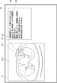
- FIG. 6 is a diagram showing an example of a medical text display screen according to the first embodiment.
- the display screen 70 includes an image display area 71 and a text display area 72.
- the slice image SL1 that is most likely to identify the abnormal shadow candidate detected by the image analysis unit 22 is displayed.
- the slice image SL1 includes an abnormal shadow candidate 73, and the abnormal shadow candidate 73 is surrounded by a rectangular region 74.
- the medical sentence 75 generated by the sentence generation unit 23 is displayed.
- Medical text 75 states, "Under the left lung pleura, an irregular tumor with a maximum lateral diameter of 4.2 cm is found. It is in contact with the chest wall and shows pleural infiltration, but no infiltration. Primary It is thought to be lung cancer. " In the medical text 75, the contents specified by the content specifying unit 26 are "left lung subpleural”, “irregular”, “4.2 cm”, “tumor”, “contacting the chest wall”, and “pleural infiltration”. , "No infiltration” and "primary lung cancer”.
- a correction button 78A and a confirmation button 78B are displayed below the image display area 71.
- the image interpreting doctor interprets the abnormal shadow candidate 73 in the slice image SL1 displayed in the image display area 71, and determines the suitability of the medical sentence 75 displayed in the sentence display area 72.
- the interpreting doctor wants to correct the medical sentence 75, he / she selects the correction button 78A using the input device 16.
- the medical text 75 displayed in the text display area 72 can be manually corrected by input from the input device 16.
- the confirmation button 78B the medical sentence 75 displayed in the sentence display area 72 can be confirmed with the content thereof.
- the medical sentence 75 is transcribed in the image interpretation report, and the image interpretation report to which the medical sentence 75 is transcribed is transmitted to the image interpretation report server 7 together with the slice image SL1 and stored.
- the interpreter When the interpreter selects the correction button 78A to correct the medical sentence 75, the interpreter is included in the abnormal shadow 31, but if there is a missing property in the medical sentence 75, the missing property is added. Make corrections to do so. In this case, the interpreter inputs the missing properties using the input device 16. For example, in the present embodiment, it is assumed that the abnormal shadow 31 has spicula, but the medical text 75 lacks the description about spicula. In this case, the interpreter inputs the characters "Spicula" using the input device 16. At that time, as shown in FIG. 7, first, the characters "su” and "pi" are input. In FIG. 7, for the sake of explanation, the input characters are displayed in the pop-up 77, but the method of inputting the characters is not limited to this. Characters may be input at the cursor position in the text.
- the modified content identification unit 26 identifies the modified content based on the input or designation of at least a part of the characters of the modified content to be added to or deleted from the medical text. Further, in the present embodiment, the modified content is specified by referring to the table showing the derivation result of the property information by the image analysis unit 22 stored in the storage 13.
- FIG. 8 is a diagram showing an example of a table showing the derivation result. As shown in FIG. 8, the table LUT1 showing the derivation result shows the location of the abnormal shadow, the size of the abnormal shadow, the shape of the boundary (clear and irregular), the type of absorption value (full type and pleural type), and the pleura.
- the modified content specifying unit 26 identifies the modified content with reference to the table LUT1 stored in the storage 13.
- the modified content specifying unit 26 recognizes the characters “su” and “pi” input by the image interpreting doctor, and further refers to the table LUT1 to select the modified content of “su” and “pi”. Identify as "spicula” containing letters. If the input character is "shu”, the modified content specifying unit 26 identifies the modified content as "tumor”, and if the input characters are "fu" and "se", the modified content specifying unit 26 Identify modified content as "irregular".
- the image interpreter uses the input device 16 to specify the property information included in the medical text 75. For example, when deleting the sentence "I admit pleural invagination", the image interpreter uses the input device 16 to move the cursor in the displayed medical sentence 75 to "chest", "" Specify the characters of the property information in the order of "membrane", "fall", and so on.
- the character designation also includes selecting a character by moving the cursor in order from the first character.
- the modified content specifying unit 26 refers to the table LUT1 by the characters “chest”, “membrane”, and “depression” designated by the interpreting doctor, and identifies the modified content as “pleural invagination”. When the designated character is "empty”, the modified content specifying unit 26 specifies the modified content as "cavity”, and when it is "stone”, the modified content is specified as "calcification”.
- the sentence correction unit 27 corrects the medical sentence 75 based on the correction content specified by the correction content identification unit 26.
- the sentence correction unit 27 is a fourth learning model in which machine learning is performed so as to output the corrected medical sentence (hereinafter referred to as the corrected medical sentence) when the medical sentence and the corrected content are input. It has 27A.
- the fourth learning model 27A for example, a recurrent neural network can be used.
- FIG. 9 is a diagram showing a schematic configuration of the recurrent neural network in the sentence correction unit 27. As shown in FIG. 9, the recurrent neural network 45 includes an encoder 46 and a decoder 47.
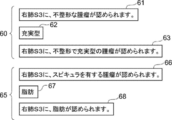
- FIG. 10 is a diagram showing an example of teacher data for learning the fourth learning model 27A.
- the teacher data 60 includes the medical text 61, the modified content 62, and the modified medical text 63.
- the medical sentence 61 is "an irregular mass is found in the right lung S3"
- the modified content 62 is "enriched”
- the modified medical sentence 63 is "an irregular mass in the right lung S3”.
- a solid mass is found in. "
- the teacher data 60 is obtained by adding a wording including the modified content 62 to the medical sentence 61 to generate the modified medical sentence 63.
- the teacher data 65 includes medical text 66, modified content 67, and modified medical text 68.
- the medical sentence 66 is "a tumor having spicula is found in the right lung S3”
- the modified content 67 is "fat”
- the modified medical sentence 68 is "fat is found in the right lung S3”. It will be.
- the teacher data 65 is obtained by adding the modified content 67 indicating benign to the medical sentence 66 in which the description indicating malignancy is added, and deleting the description indicating malignancy to generate the modified medical sentence 68. is there.
- the property information included in the medical sentence 61 of the teacher data 60 is input to the encoder 46 of the recurrent neural network 45, and the modified content 62 (“enriched type” in FIG. 9). Is entered. Then, the encoder 46 and the decoder 47 of the recurrent neural network 45 are learned so that the decoder 47 outputs the modified medical sentence 63. Further, when learning is performed using the teacher data 65, the property information included in the medical sentence 66 of the teacher data 65 is input to the encoder 46 of the recurrent neural network 45 as in the case of using the teacher data 60, and further. The modified content 67 is input. Then, the encoder 46 and the decoder 47 of the recurrent neural network 45 are learned so that the decoder 47 outputs the modified medical text 68.
- the fourth learning model 27A is constructed by learning the recurrent neural network 45 using a large number of teacher data as shown in FIG. For example, by using the teacher data 60 shown in FIG. 10, the fourth learning model 27A outputs the modified medical sentence 63 shown in FIG. 10 when the medical sentence 61 and the modified content 62 shown in FIG. 10 are input. Learning is done like this.
- the sentence correction unit 27 corrects the medical sentence 75 and " A tumor with a maximum lateral diameter of 4.2 cm with irregularity and spicula is found under the left pulmonary pleura. It is in contact with the chest wall, and pleural invagination is observed, but no invasion is observed. It is considered to be primary lung cancer. Will be generated. ”Corrected medical text. As shown in FIG. 11, the display control unit 25 displays the modified medical text 79 in the text display area 72. In FIG. 11, the corrected portion in the modified medical text 79 is underlined.
- the sentence correction unit 27 corrects the medical sentence 75 according to the style of the medical sentence 75. Specifically, if the medical sentence 75 is more and more tone, the modified medical sentence 79 is modified so that the modified medical sentence 79 is also more and more tone. Further, when the medical sentence 75 is in a definite tone, the medical sentence 75 is modified so that the modified medical sentence 79 is also in a definite tone.
- the sentence correction unit 27 generates a plurality of correction candidates based on the correction content, selects a correction candidate according to the style of the medical sentence 75 before correction from the generated correction candidates, and performs medical treatment.
- Sentence 75 may be modified. For example, in response to the above-mentioned sentence "A tumor with an irregular maximum lateral diameter of 4.2 cm is found under the left pulmonary pleura", if the modified content is "Spicula", then "Under the left pulmonary pleura". , A tumor with a maximum lateral diameter of 4.2 cm with irregularity and spicula is observed.
- the sentence correction unit 27 selects one correction candidate according to the writing style of the medical sentence 75 before correction from the generated plurality of correction candidates, and generates the corrected medical sentence 79.
- a tumor with an irregular shape and a maximum lateral diameter of 4.2 cm is found under the left pulmonary pleura
- Sentence 79 will be generated.
- the beam search method described in "https://geekyisawesome.blogspot.com/2017/10/using-beam-serach-togenrate-most.html” is used as the fourth method. It can be generated by applying it to the recurrent neural network 45 that constitutes the learning model 27A.
- the beam search method is a method of searching for a word that appears next to a certain word in consideration of the probability of occurrence of the word for a word that appears next to a certain word.
- the sentence correction unit 27 applies the beam search method to the recurrent neural network 45 to generate correction candidates for a plurality of medical sentences having a high probability of word occurrence.
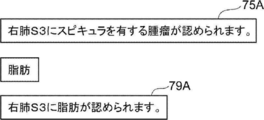
- the fourth learning model 27A of the sentence correction unit 27 is learned using the teacher data 65 shown in FIG. Therefore, for example, as shown in FIG. 12, the medical sentence 75A of "a tumor having a spicula is found in the right lung S3" is displayed in the sentence display area 72, and the modified content is "fat". To do. In this case, since spicula is content indicating malignancy and fat is content indicating benign, the medical sentence 75A is modified to delete the wording related to malignancy contained in the medical sentence 75A, and "to the right lung S3. The modified medical text 79A of "Fat is recognized.” Will be displayed.
- the sentence correction unit 27 deletes the wording including the specified content from the medical sentence.
- the modified content is specified as “pleural invagination”.
- the sentence correction unit 27 deletes the phrase "although pleural invagination is admitted”.
- the modified medical text 79 states, "A tumor with an irregular maximum lateral diameter of 4.2 cm is found under the left pulmonary pleura. It is in contact with the chest wall and no infiltration is found. It is considered to be primary lung cancer. It will be.
- the recurrent neural network 45 of the fourth learning model 27A of the sentence correction unit 27 has "left lung subpleural” and “non-pleural", excluding the content related to pleural infiltration from the contents included in the medical sentence 75. "Orthopedic”, “4.2 cm”, “mass”, “contacting the pleura”, “no infiltration” and “primary lung cancer” are entered to generate a modified medical text 79.
- the content included in the modified medical sentence 79 is different from the content included in the medical sentence 75 before the correction according to the correction of the medical sentence by the sentence correction unit 27.
- medical text 75 states, "Under the left lung pleura, an irregular tumor with a maximum lateral diameter of 4.2 cm is found. It is in contact with the chest wall and shows pleural infiltration, but no infiltration.
- the content identified by the content identification unit 26 is "left pulmonary subpleural", “irregular”, “4.2 cm”, “tumor”, “contacting the chest wall”. , “With pleural infiltration”, “No infiltration” and “Primary lung cancer”.
- the modified medical text 79 states, "Under the left lung pleura, there is an irregular mass with a maximum lateral diameter of 4.2 cm. It is in contact with the chest wall and pleural infiltration is observed, but infiltration is not observed. In the case of "I do not admit it. It is considered to be primary lung cancer.”,
- the modified medical text 79 will include "spicula" as the content, which is not included in the medical text 75 before the revision. Therefore, the sentence correction unit 27 changes the content by adding "spicula" to the content about the medical sentence 75 stored in the storage 13.
- the contents of medical texts stored in the storage 13 are "left lung subpleural”, “irregular”, “spicula”, “4.2 cm”, “tumor”, “contacting the chest wall”, and “pleura”. "With infiltration”, “No invasion” and “Primary lung cancer”.
- the content of the medical text 75 and the content of the modified medical text 79 may be included in the display screen 70 and displayed.
- FIG. 13 is a flowchart showing the processing performed in the present embodiment. It is assumed that the medical image to be read is acquired from the image server 5 by the image acquisition unit 21 and stored in the storage 13. The process is started when the image interpretation doctor gives an instruction to create an image interpretation report, and the image analysis unit 22 analyzes the medical image to show the properties of the structure of interest such as an abnormal shadow candidate included in the medical image. Derivation of property information (step ST1). Next, the sentence generation unit 23 generates a medical sentence related to the medical image based on the property information (step ST2).
- the content specifying unit 24 analyzes the medical sentence generated by the sentence generating unit 23 to specify a term representing a property related to the structure of interest included in the medical sentence as the content (step ST3). Then, the display control unit 25 displays the medical text generated by the text generation unit 23 on the text display area 72 of the display screen 70 displayed on the display 15 (step ST4).
- step ST5 determines whether or not the correction button 78A displayed on the display screen is selected.
- step ST6 affirmed, the display control unit 25 accepts the correction of the medical text displayed in the text display area 72 using the input device 16 (step ST6).
- the correction content identification unit 26 is based on the input or designation of at least a part of the characters for the correction content to be added to or deleted from the medical text. Identify the modified content (step ST7).
- the sentence correction unit 27 corrects the medical sentence based on the corrected content (step ST8). This will generate a modified medical text.
- the display control unit 25 displays the modified medical text in the text display area 72 of the display screen 70 (step ST9), and returns to the process of step ST5.
- step ST5 the display control unit 25 determines whether or not the confirmation button 78B has been selected (step ST10). If step ST10 is denied, the process returns to step ST5. When step ST10 is affirmed, the display control unit 25 transfers the medical sentence to the image interpretation report, and transmits the image interpretation report to which the medical sentence is transcribed to the image interpretation report server 7 together with the slice image SL1 (interpretation report transmission: Step ST11), the process is terminated.
- the medical text including at least one content is displayed on the display 15.
- the modified content is then identified based on the input or designation of at least some characters for the modified content that is added to or removed from the medical text.
- the text is corrected based on the corrected content. Therefore, the medical text can be modified according to the correction intention of the user, the interpretation doctor.
- the modified content is specified based on the input or designation of at least a part of the characters of the modified content, the burden on the interpreter who makes the modification can be reduced. Therefore, according to the present embodiment, the medical text can be efficiently corrected.
- the sentence correction unit 27 generates a plurality of correction candidates
- the display control unit 25 displays a plurality of correction candidates
- the sentence correction unit 27 is selected from the plurality of correction candidates. It differs from the first embodiment in that a modified medical sentence is generated depending on the modified candidate.
- FIG. 14 is a diagram showing a medical text display screen displaying a plurality of correction candidates in the second embodiment.
- FIG. 14 shows a state in which the characters “su” and “pi” are input, as in FIG. 7.
- the modified content specifying unit 26 identifies the modified content as “spicula” with reference to LUT1.
- the sentence correction unit 27 generates a plurality of correction candidates based on the correction content.
- the display control unit 25 displays a plurality of correction candidate display areas 80 below the text display area 72.
- correction candidate display area 80 "a tumor with a maximum lateral diameter of 4.2 cm with irregularity and spicula is observed under the left pulmonary pleura."
- the number of correction candidates is not limited to three.
- the interpreting doctor selects a desired correction candidate from the displayed three correction candidates using the input device 16.
- the modified medical text 79 is generated by the selected modification candidates.
- the image interpreter selects the top modification candidate from the displayed four modification candidates, the "maximum laterality having irregular and spicula under the left pulmonary pleura" is the same as in the first embodiment.
- a tumor with a diameter of 4.2 cm is observed. It is in contact with the chest wall, and pleural infiltration is observed, but no infiltration is observed. It is considered to be primary lung cancer.
- FIG. 15 is a flowchart showing the processing performed in the second embodiment.
- the process performed in the second embodiment differs from the process performed in the first embodiment shown in FIG. 13 only in the process after step ST5 is affirmed. Therefore, in FIG. 15, only the processing after the step ST5 in FIG. 13 is affirmed will be described.
- step ST5 in FIG. 13 the display control unit 25 accepts the correction of the medical text displayed in the text display area 72 using the input device 16 (step ST21).
- the correction content identification unit 26 is based on the input or designation of at least a part of the characters for the correction content to be added to or deleted from the medical text. Identify the modified content (step ST22).
- the sentence correction unit 27 generates a plurality of correction candidates for the medical sentence based on the corrected content (step ST23). Further, the display control unit 25 displays a plurality of correction candidates in the correction candidate display area 80 (step ST24).
- step ST25 when any of the plurality of correction candidates is selected (step ST25: affirmative), the sentence correction unit 27 corrects the medical sentence with the selected correction candidates (step ST26). This will generate a modified medical text.
- the display control unit 25 displays the modified medical text in the text display area 72 of the display screen 70 (step ST27), and returns to the process of step ST5.
- a plurality of correction candidates are generated, the selection of one correction candidate desired by the interpretation doctor is accepted from the plurality of correction candidates, and the medical text is corrected by the selected correction candidates. I did. Therefore, the correction medical text can be generated so as to include the correction candidates desired by the interpretation doctor, and as a result, the burden on the interpretation doctor who creates the interpretation report can be reduced.
- the medical image is analyzed and the medical text is generated in the interpretation WS3, but the present invention is not limited to this.
- the medical information system 1 in the present embodiment may be provided with an analysis server that analyzes the medical image and generates a medical sentence, and the analysis server may analyze the medical image and generate the medical sentence.
- the generated medical text is transmitted from the analysis server to the interpretation WS3 together with the table showing the derivation result of the property information, and the medical text is displayed and modified in the interpretation WS3 in the same manner as in the above embodiment.
- the modified content specifying unit 26 specifies the modified content with reference to the table LUT1 showing the derivation result shown in FIG. 8, but the modification content is not limited to this.
- a property table LUT2 in which a plurality of property information that can be detected in the lung nodule is associated with a phonetic spelling representing the property information is stored in the storage 13, and the property table LUT2 is referred to.
- the modified content may be specified.
- various property information such as clear, irregular, solid type, suriglass type, spicula, and tumor are associated with a phonetic spelling representing each property information.
- the modified content specifying unit 26 specifies the modified content with reference to the property table LUT2 stored in the storage 13. For example, the modified content specifying unit 26 identifies the modified content as "spicula” including the characters "su” and “pi” by the characters "su” and “pi” input by the interpreting doctor. If the input character is "shu”, the modified content specifying unit 26 identifies the modified content as “tumor”, and if the input characters are "fu" and "se", the modified content specifying unit 26 Identify modified content as "irregular”.
- the processing of the present disclosure is applied to the sentence generated by the sentence generation unit 23 based on the property information derived by the image analysis unit 22, but the present invention is not limited to this. Absent.
- the technique of the present disclosure can also be applied to medical texts created by an interpreter by himself / herself.
- the modified content specifying unit 26 refers to the property table LUT2 as shown in FIG. 16 to specify the modified content.
- each learning model of the image analysis unit 22, the sentence generation unit 23, the content identification unit 24, and the sentence correction unit 27 performs analysis processing, sentence generation processing, content identification processing, and sentence correction processing according to the diagnosis target. Is prepared, a learning model that performs analysis processing, sentence generation processing, content identification processing, and sentence correction processing according to the diagnosis target is selected, and medical sentence generation processing is executed.
- the technique of the present disclosure is applied when creating an interpretation report as a medical document, but it is also possible to create a medical document other than the interpretation report such as an electronic medical record and a diagnostic report.
- a medical document other than the interpretation report such as an electronic medical record and a diagnostic report.
- the technology of the present disclosure can be applied.
- various processes such as an image acquisition unit 21, an image analysis unit 22, a sentence generation unit 23, a content identification unit 24, a display control unit 25, a modified content identification unit 26, and a sentence correction unit 27 are performed.
- various processors processors shown below can be used.
- the various processors include CPUs, which are general-purpose processors that execute software (programs) and function as various processing units, as well as circuits after manufacturing FPGAs (Field Programmable Gate Arrays) and the like.
- Dedicated electricity which is a processor with a circuit configuration specially designed to execute specific processing such as programmable logic device (PLD), ASIC (Application Specific Integrated Circuit), which is a processor whose configuration can be changed. Circuits and the like are included.
- PLD programmable logic device
- ASIC Application Specific Integrated Circuit
- One processing unit may be composed of one of these various processors, or a combination of two or more processors of the same type or different types (for example, a combination of a plurality of FPGAs or a combination of a CPU and an FPGA). ) May be configured. Further, a plurality of processing units may be configured by one processor.
- one processor is configured by combining one or more CPUs and software. There is a form in which this processor functions as a plurality of processing units.
- SoC System On Chip
- the various processing units are configured by using one or more of the above-mentioned various processors as a hardware structure.
- circuitry in which circuit elements such as semiconductor elements are combined can be used.
Abstract
The present invention comprises at least one processor, wherein the processor is configured to: display a sentence containing at least one piece of content on a display; specify the modified content on the basis of the input of at least some characters to the modified content to be added to the sentence or removed from the sentence; and modify the sentence on the basis of the modified content.
Description
本開示は、医療文書等の文書の作成を支援する文書作成支援装置、方法およびプログラムに関する。
This disclosure relates to a document creation support device, method and program that support the creation of documents such as medical documents.
近年、CT(Computed Tomography)装置およびMRI(Magnetic Resonance Imaging)装置等の医療機器の進歩により、より質の高い高解像度の医用画像を用いての画像診断が可能となってきている。とくに、CT画像およびMRI画像等を用いた画像診断により、病変の領域を精度よく特定することができるため、特定した結果に基づいて適切な治療が行われるようになってきている。
In recent years, advances in medical devices such as CT (Computed Tomography) devices and MRI (Magnetic Resonance Imaging) devices have made it possible to perform diagnostic imaging using higher quality medical images. In particular, since the lesion region can be accurately identified by diagnostic imaging using CT images, MRI images, and the like, appropriate treatment has come to be performed based on the identified results.
また、ディープラーニング等により機械学習がなされた学習モデルを用いたCAD(Computer-Aided Diagnosis)により医用画像を解析して、医用画像に含まれる異常陰影候補等の関心構造物の形状、濃度、位置および大きさ等の性状を判別し、これらを解析結果として取得することも行われている。CADにより取得された解析結果は、患者名、性別、年齢および医用画像を取得したモダリティ等の検査情報と対応づけられて、データベースに保存される。医用画像および解析結果は、医用画像の読影を行う読影医の端末に送信される。読影医は、自身の端末において、送信された医用画像および解析結果を参照して医用画像の読影を行い、読影レポートを作成する。
In addition, medical images are analyzed by CAD (Computer-Aided Diagnosis) using a learning model that has been machine-learned by deep learning, etc., and the shape, density, and position of structures of interest such as abnormal shadow candidates included in the medical images. It is also practiced to discriminate properties such as size and size, and obtain these as analysis results. The analysis result acquired by CAD is associated with the examination information such as the patient name, gender, age, and the modality from which the medical image was acquired, and is stored in the database. The medical image and the analysis result are transmitted to the terminal of the image interpreting doctor who interprets the medical image. The image interpreting doctor interprets the medical image by referring to the transmitted medical image and the analysis result on his / her terminal, and creates an image interpretation report.
一方、上述したCT装置およびMRI装置の高性能化に伴い、読影を行う医用画像の数も増大している。しかしながら、読影医の数は医用画像の数に追いついていないことから、読影医の読影業務の負担を軽減することが望まれている。このため、読影レポート等の医療文書の作成を支援するための各種手法が提案されている。例えば、特開2019-153250号公報には、読影医が入力したキーワードおよび医用画像を解析結果に含まれる、関心構造物の性状を表す情報(以下、性状情報とする)に基づいて、読影レポートに記載するための文章を生成する各種手法が提案されている(特開2019-153250号公報参照)。特開2019-153250号公報に記載された手法においては、入力された性状情報を表す文字から文章を生成するように学習が行われたリカレントニューラルネットワーク等の機械学習がなされた学習モデルを用いて、医療用の文章(以下、医療文章とする)が作成される。特開2019-153250号公報に記載された手法のように、医療文章を自動で生成することにより、読影レポート等の医療文書を作成する際の読影医の負担を軽減することができる。
On the other hand, the number of medical images to be interpreted is increasing with the improvement of the performance of the CT device and the MRI device described above. However, since the number of image interpreters has not kept up with the number of medical images, it is desired to reduce the burden of the image interpretation work of the image interpreters. For this reason, various methods have been proposed to support the creation of medical documents such as interpretation reports. For example, Japanese Patent Application Laid-Open No. 2019-153250 describes an interpretation report based on information representing the properties of a structure of interest (hereinafter referred to as property information), which includes keywords and medical images input by an image interpreter in the analysis results. Various methods for generating sentences to be described in Japanese Patent Application Laid-Open No. 2019-153250 have been proposed (see JP-A-2019-153250). In the method described in Japanese Patent Application Laid-Open No. 2019-153250, a learning model in which machine learning such as a recurrent neural network is trained so as to generate a sentence from characters representing input property information is used. , Medical texts (hereinafter referred to as medical texts) are created. By automatically generating medical texts as in the method described in JP-A-2019-153250, it is possible to reduce the burden on the interpretation doctor when creating a medical document such as an interpretation report.
一方、上述したように自動で文章を生成する各種手法が提案されているものの、読影医による修正が必要な場合が多い。このため、生成された文章を簡易に修正する手法が望まれている。
On the other hand, although various methods for automatically generating sentences have been proposed as described above, there are many cases where correction by an image interpreting doctor is required. Therefore, a method for easily modifying the generated text is desired.
例えば、特開2007-117026号公報には、テキスト文書が1つまたは複数のテキスト構成要素を含み、1つまたは複数のテキスト構成要素から誤ったテキスト構成要素を識別し、誤ったテキスト構成要素に対する所望の代替物の部分入力に基づいて代替物リストを表示し、テキスト文書内で誤ったテキスト構成要素を選択した代替物で置き換える手法が提案されている。特開2007-117026号公報に記載された手法を用いることにより、ユーザに負担をかけることなく、文章における誤った箇所を修正することができる。
For example, in Japanese Patent Application Laid-Open No. 2007-117026, a text document contains one or more text components, identifies an erroneous text component from one or more text components, and refers to the erroneous text component. A method has been proposed in which a list of alternatives is displayed based on the partial input of the desired alternative, and the wrong text component is replaced with the selected alternative in the text document. By using the method described in JP-A-2007-117026, it is possible to correct an erroneous part in a sentence without imposing a burden on the user.
しかしながら、特開2007-117026号公報に記載された手法においては、ユーザは代替物リストから、修正する代替物を選択する作業を行う必要がある。また、代替物リストに所望とする内容の代替物が存在しない場合には、特開2007-117026号公報に記載された手法は、文章を効率よく修正することができない。
However, in the method described in Japanese Patent Application Laid-Open No. 2007-117026, the user needs to perform the work of selecting the alternative to be modified from the alternative list. Further, when there is no substitute having the desired content in the substitute list, the method described in Japanese Patent Application Laid-Open No. 2007-117026 cannot efficiently correct the text.
本開示は上記事情に鑑みなされたものであり、文章を効率よく修正できるようにすることを目的とする。
This disclosure was made in view of the above circumstances, and the purpose is to enable efficient correction of sentences.
本開示による文書作成支援装置は、少なくとも1つのプロセッサを備え、
プロセッサは、少なくとも1つのコンテンツを含む文章をディスプレイに表示し、
文章に対して追加するまたは文章から削除する修正コンテンツについての少なくとも一部の文字の入力または指定に基づいて、修正コンテンツを特定し、
修正コンテンツに基づいて文章を修正するように構成される。 The document creation support device according to the present disclosure includes at least one processor.
The processor displays a sentence containing at least one content on the display and
Identify modified content based on at least some text input or specification for modified content that is added to or removed from the text.
It is configured to modify the text based on the modified content.
なお、本開示による文書作成支援装置においては、プロセッサは、さらに画像を解析することにより、画像に含まれる関心構造物の性状を表す性状情報をコンテンツとして導出するように構成されるものであってもよい。
In the document creation support device according to the present disclosure, the processor is configured to derive property information representing the properties of the structure of interest contained in the image as content by further analyzing the image. May be good.
また、本開示による文書作成支援装置においては、プロセッサは、性状情報に基づいて、画像に関する文章を生成するように構成されるものであってもよい。
Further, in the document creation support device according to the present disclosure, the processor may be configured to generate a sentence related to an image based on the property information.
また、本開示による文書作成支援装置においては、プロセッサは、性状情報にも基づいて、修正コンテンツを特定するように構成されるものであってもよい。
Further, in the document creation support device according to the present disclosure, the processor may be configured to specify the modified content based on the property information.
また、本開示による文書作成支援装置においては、プロセッサは、文章に含まれるコンテンツを特定し、
修正コンテンツに基づいて、特定されたコンテンツを変更するように構成されるものであってもよい。 Further, in the document creation support device according to the present disclosure, the processor identifies the content included in the text and determines the content.
It may be configured to modify the identified content based on the modified content.
また、本開示による文書作成支援装置においては、プロセッサは、修正前の文章の文体に応じて文章を修正するように構成されるものであってもよい。
Further, in the document creation support device according to the present disclosure, the processor may be configured to correct the sentence according to the style of the sentence before the correction.
また、本開示による文書作成支援装置においては、プロセッサは、複数の修正候補を生成し、
複数の修正候補のうち、修正前の文章の文体に応じた修正候補により、文章を修正するように構成されるものであってもよい。 Further, in the document creation support device according to the present disclosure, the processor generates a plurality of correction candidates, and the processor generates a plurality of correction candidates.
Of the plurality of correction candidates, the text may be modified by the correction candidates according to the style of the text before the correction.
また、本開示による文書作成支援装置においては、プロセッサは、複数の修正候補を生成し、
複数の修正候補をディスプレイに表示し、
表示された複数の修正候補から文章に使用する修正候補の選択を受け付け、
選択された修正候補により、文章を修正するように構成されるものであってもよい。 Further, in the document creation support device according to the present disclosure, the processor generates a plurality of correction candidates, and the processor generates a plurality of correction candidates.
Show multiple correction candidates on the display
Accepts the selection of correction candidates to be used for sentences from the displayed multiple correction candidates,
The selected correction candidate may be configured to correct the sentence.
また、本開示による文書作成支援装置においては、プロセッサは、修正コンテンツと修正前の文章に含まれるコンテンツとが整合するように、文章を修正するように構成されるものであってもよい。
Further, in the document creation support device according to the present disclosure, the processor may be configured to correct the text so that the corrected content and the content included in the text before the correction are consistent.
本開示による文書作成支援方法は、少なくとも1つのコンテンツを含む文章をディスプレイに表示し、
文章に対して追加するまたは文章から削除する修正コンテンツについての少なくとも一部の文字の入力または指定に基づいて、修正コンテンツを特定し、
修正コンテンツに基づいて文章を修正する。 The document creation support method according to the present disclosure displays a sentence containing at least one content on a display.
Identify modified content based on at least some text input or specification for modified content that is added to or removed from the text.
Correct the text based on the content.
なお、本開示による文書支援作成方法をコンピュータに実行させるためのプログラムとして提供してもよい。
Note that it may be provided as a program for causing a computer to execute the document support creation method according to the present disclosure.
本開示によれば、文章を効率よく修正できる。
According to this disclosure, sentences can be corrected efficiently.
以下、図面を参照して本開示の実施形態について説明する。図1は本開示の第1の実施形態による文書作成支援装置を適用した医療情報システムの概略構成を示す図である。図1に示す医療情報システム1は、公知のオーダリングシステムを用いた診療科の医師からの検査オーダに基づいて、被写体の検査対象部位の撮影、撮影により取得された医用画像の保管、読影医による医用画像の読影と読影レポートの作成、および依頼元の診療科の医師による読影レポートの閲覧と読影対象の医用画像の詳細観察とを行うためのシステムである。図1に示すように、医療情報システム1は、複数のモダリティ(撮影装置)2、読影端末である複数の読影ワークステーション(WS)3、診療科ワークステーション(WS)4、画像サーバ5、画像データベース6、読影レポートサーバ7、および読影レポートデータベース8が、有線または無線のネットワーク10を介して互いに通信可能な状態で接続されて構成されている。
Hereinafter, embodiments of the present disclosure will be described with reference to the drawings. FIG. 1 is a diagram showing a schematic configuration of a medical information system to which the document creation support device according to the first embodiment of the present disclosure is applied. The medical information system 1 shown in FIG. 1 is based on an examination order from a doctor in a clinical department using a known ordering system, photographs of a part to be inspected of a subject, storage of medical images obtained by photographing, and an image interpreter. It is a system for interpreting medical images and creating an interpretation report, and for viewing the interpretation report by the doctor of the requesting clinical department and observing the details of the medical image to be interpreted. As shown in FIG. 1, the medical information system 1 includes a plurality of modalities (imaging devices) 2, a plurality of image interpretation workstations (WS) 3 which are image interpretation terminals, a clinical department workstation (WS) 4, an image server 5, and an image. The database 6, the interpretation report server 7, and the interpretation report database 8 are connected and configured so as to be able to communicate with each other via a wired or wireless network 10.
各機器は、医療情報システム1の構成要素として機能させるためのアプリケーションプログラムがインストールされたコンピュータである。アプリケーションプログラムは、ネットワーク10に接続されたサーバコンピュータの記憶装置、もしくはネットワークストレージに、外部からアクセス可能な状態で記憶され、要求に応じてコンピュータにダウンロードされ、インストールされる。または、DVD(Digital Versatile Disc)あるいはCD-ROM(Compact Disc Read Only Memory)等の記録媒体に記録されて配布され、その記録媒体からコンピュータにインストールされる。
Each device is a computer on which an application program for functioning as a component of the medical information system 1 is installed. The application program is stored in the storage device of the server computer connected to the network 10 or in the network storage in a state of being accessible from the outside, and is downloaded and installed in the computer upon request. Alternatively, it is recorded and distributed on a recording medium such as a DVD (Digital Versatile Disc) or a CD-ROM (Compact Disc Read Only Memory), and installed on a computer from the recording medium.
モダリティ2は、被写体の診断対象となる部位を撮影することにより、診断対象部位を表す医用画像を生成する装置である。具体的には、単純X線撮影装置、CT装置、MRI装置、およびPET(Positron Emission Tomography)装置等である。モダリティ2により生成された医用画像は画像サーバ5に送信され、保存される。
Modality 2 is a device that generates a medical image showing the diagnosis target part by photographing the diagnosis target part of the subject. Specifically, it is a simple X-ray imaging apparatus, a CT apparatus, an MRI apparatus, a PET (Positron Emission Tomography) apparatus, and the like. The medical image generated by the modality 2 is transmitted to the image server 5 and stored.
読影WS3は、本実施形態による文書作成支援装置を内包する。読影WS3の構成については後述する。
Interpretation WS3 includes a document creation support device according to this embodiment. The configuration of the interpretation WS3 will be described later.
診療科WS4は、診療科の医師が画像の詳細観察、読影レポートの閲覧、および電子カルテの作成等に利用するコンピュータであり、処理装置、ディスプレイ、並びにキーボードおよびマウス等の入力デバイスにより構成される。診療科WS4では、患者のカルテ(電子カルテ)の作成、画像サーバ5に対する画像の閲覧要求、画像サーバ5から受信した画像の表示、画像中の病変らしき部分の自動検出または強調表示、読影レポートサーバ7に対する読影レポートの閲覧要求、および読影レポートサーバ7から受信した読影レポートの表示等の各処理が、各処理のためのソフトウェアプログラムを実行することにより行われる。
The clinical department WS4 is a computer used by doctors in the clinical department for detailed observation of images, viewing of interpretation reports, creation of electronic medical records, etc., and is composed of a processing device, a display, and input devices such as a keyboard and a mouse. .. In the medical department WS4, a patient's medical record (electronic medical record) is created, an image viewing request is made to the image server 5, an image received from the image server 5 is displayed, a lesion-like part in the image is automatically detected or highlighted, and an image interpretation report server is used. Each process such as the viewing request of the image interpretation report to the image interpretation report 7 and the display of the image interpretation report received from the image interpretation report server 7 is performed by executing the software program for each process.
画像サーバ5は、汎用のコンピュータにデータベース管理システム(DataBase Management System: DBMS)の機能を提供するソフトウェアプログラムがインストールされたものである。また、画像サーバ5は画像データベース6が構成されるストレージを備えている。このストレージは、画像サーバ5とデータバスとによって接続されたハードディスク装置であってもよいし、ネットワーク10に接続されているNAS(Network Attached Storage)およびSAN(Storage Area Network)に接続されたディスク装置であってもよい。また、画像サーバ5は、モダリティ2からの医用画像の登録要求を受け付けると、その医用画像をデータベース用のフォーマットに整えて画像データベース6に登録する。
The image server 5 is a general-purpose computer in which a software program that provides a database management system (DataBase Management System: DBMS) function is installed. Further, the image server 5 includes a storage in which the image database 6 is configured. This storage may be a hard disk device connected by the image server 5 and the data bus, or a disk device connected to NAS (Network Attached Storage) and SAN (Storage Area Network) connected to the network 10. It may be. When the image server 5 receives the medical image registration request from the modality 2, the image server 5 arranges the medical image in a database format and registers it in the image database 6.
画像データベース6には、モダリティ2において取得された医用画像の画像データと付帯情報とが登録される。付帯情報には、例えば、個々の医用画像を識別するための画像ID(identification)、被写体を識別するための患者ID、検査を識別するための検査ID、医用画像毎に割り振られるユニークなID(UID:unique identification)、医用画像が生成された検査日、検査時刻、医用画像を取得するための検査で使用されたモダリティの種類、患者氏名、年齢、性別等の患者情報、検査部位(撮影部位)、撮影情報(撮影プロトコル、撮影シーケンス、撮像手法、撮影条件および造影剤の使用等)、1回の検査で複数の医用画像を取得したときのシリーズ番号あるいは採取番号等の情報が含まれる。
The image data and incidental information of the medical image acquired in the modality 2 are registered in the image database 6. The incidental information includes, for example, an image ID (identification) for identifying an individual medical image, a patient ID for identifying a subject, an examination ID for identifying an examination, and a unique ID assigned to each medical image ( UID: unique identification), examination date when the medical image was generated, examination time, type of modality used in the examination to acquire the medical image, patient information such as patient name, age, gender, examination site (contrast image site) ), Imaging information (imaging protocol, imaging sequence, imaging method, imaging conditions, use of contrast medium, etc.), including information such as a series number or collection number when a plurality of medical images are acquired in one examination.
また、画像サーバ5は、読影WS3からの閲覧要求をネットワーク10経由で受信すると、画像データベース6に登録されている医用画像を検索し、検索された医用画像を要求元の読影WS3に送信する。
When the image server 5 receives the viewing request from the image interpretation WS3 via the network 10, the image server 5 searches for the medical image registered in the image database 6 and transmits the searched medical image to the requester's image interpretation WS3.
読影レポートサーバ7には、汎用のコンピュータにデータベース管理システムの機能を提供するソフトウェアプログラムが組み込まれる。読影レポートサーバ7は、読影WS3からの読影レポートの登録要求を受け付けると、その読影レポートをデータベース用のフォーマットに整えて読影レポートデータベース8に登録する。また、読影レポートの検索要求を受け付けると、その読影レポートを読影レポートデータベース8から検索する。
The interpretation report server 7 incorporates a software program that provides the functions of a database management system to a general-purpose computer. When the image interpretation report server 7 receives the image interpretation report registration request from the image interpretation WS3, the image interpretation report server 7 arranges the image interpretation report in a database format and registers it in the image interpretation report database 8. When a search request for an image interpretation report is received, the image interpretation report is searched from the image interpretation report database 8.
読影レポートデータベース8には、例えば、読影対象の医用画像を識別する画像ID、読影を行った画像診断医を識別するための読影医ID、病変名、病変の位置情報、所見、および所見の確信度等の情報が記録された読影レポートが登録される。
The image interpretation report database 8 contains, for example, an image ID for identifying a medical image to be interpreted, an image radiologist ID for identifying an image diagnostician who has performed image interpretation, a lesion name, lesion location information, findings, and confidence in the findings. An interpretation report in which information such as the degree is recorded is registered.
なお、本実施形態においては、医用画像は診断対象を肺とした、複数の断層画像からなる3次元のCT画像とし、CT画像を読影することにより、肺に含まれる異常陰影についての読影レポートを医療文書として作成するものとする。なお、医用画像はCT画像に限定されるものではなく、MRI画像および単純X線撮影装置により取得された単純2次元画像等の任意の医用画像を用いることができる。
In the present embodiment, the medical image is a three-dimensional CT image composed of a plurality of tomographic images with the diagnosis target as the lung, and by interpreting the CT image, an interpretation report on abnormal shadows contained in the lung is obtained. It shall be created as a medical document. The medical image is not limited to the CT image, and any medical image such as an MRI image and a simple two-dimensional image acquired by a simple X-ray imaging device can be used.
ネットワーク10は、病院内の各種機器を接続する有線または無線のネットワークである。読影WS3が他の病院あるいは診療所に設置されている場合には、ネットワーク10は、各病院のローカルエリアネットワーク同士をインターネットまたは専用回線で接続した構成としてもよい。
Network 10 is a wired or wireless network that connects various devices in the hospital. When the interpretation WS3 is installed in another hospital or clinic, the network 10 may be configured such that the local area networks of each hospital are connected to each other by the Internet or a dedicated line.
以下、本実施形態による読影WS3について詳細に説明する。読影WS3は、医用画像の読影医が、医用画像の読影および読影レポートの作成に利用するコンピュータであり、処理装置、ディスプレイ、並びにキーボードおよびマウス等の入力デバイスにより構成される。読影WS3では、画像サーバ5に対する医用画像の閲覧要求、画像サーバ5から受信した医用画像に対する各種画像処理、医用画像の表示、医用画像に対する解析処理、解析結果に基づく医用画像の強調表示、解析結果に基づく読影レポートの作成、読影レポートの作成の支援、読影レポートサーバ7に対する読影レポートの登録要求と閲覧要求、並びに読影レポートサーバ7から受信した読影レポートの表示等の各処理が、各処理のためのソフトウェアプログラムを実行することにより行われる。なお、これらの処理のうち、本実施形態の文書作成支援装置が行う処理以外の処理は、周知のソフトウェアプログラムにより行われるため、ここでは詳細な説明は省略する。また、本実施形態の文書作成支援装置が行う処理以外の処理を読影WS3において行わず、別途その処理を行うコンピュータをネットワーク10に接続しておき、読影WS3からの処理の要求に応じて、そのコンピュータにおいて要求された処理を行うようにしてもよい。
Hereinafter, the interpretation WS3 according to the present embodiment will be described in detail. The image interpretation WS3 is a computer used by a medical image interpretation doctor to interpret a medical image and create an image interpretation report, and is composed of a processing device, a display, and an input device such as a keyboard and a mouse. In the interpretation WS3, the image server 5 is requested to view the medical image, various image processes for the medical image received from the image server 5, the display of the medical image, the analysis process for the medical image, the highlighting of the medical image based on the analysis result, and the analysis result. Each process, such as creating an image interpretation report based on the above, supporting the creation of an image interpretation report, requesting the image interpretation report server 7 to register and view the image interpretation report, and displaying the image interpretation report received from the image interpretation report server 7, is for each process. It is done by executing the software program of. Of these processes, processes other than those performed by the document creation support device of the present embodiment are performed by a well-known software program, and therefore detailed description thereof will be omitted here. Further, the image interpretation WS3 does not perform any process other than the process performed by the document creation support device of the present embodiment, and a computer that performs the process is connected to the network 10 separately, and the process is requested by the image interpretation WS3. The computer may perform the requested processing.
読影WS3は、第1の実施形態による文書作成支援装置が内包されている。このため、読影WS3には、本実施形態による文書作成支援プログラムがインストールされている。文書作成支援プログラムは、ネットワークに接続されたサーバコンピュータの記憶装置、もしくはネットワークストレージに、外部からアクセス可能な状態で記憶され、要求に応じて読影WS3にダウンロードされ、インストールされる。または、DVDあるいはCD-ROM等の記録媒体に記録されて配布され、その記録媒体から読影WS3にインストールされる。
The interpretation WS3 includes a document creation support device according to the first embodiment. Therefore, the document creation support program according to the present embodiment is installed in the interpretation WS3. The document creation support program is stored in the storage device of the server computer connected to the network or in the network storage in a state of being accessible from the outside, and is downloaded and installed in the interpretation WS3 as requested. Alternatively, it is recorded on a recording medium such as a DVD or a CD-ROM, distributed, and installed on the interpretation WS3 from the recording medium.
図2は、文書作成支援プログラムを読影WS3にインストールすることにより実現される、第1の実施形態による文書作成支援装置の概略構成を示す図である。図2に示すように、文書作成支援装置20は、標準的なコンピュータの構成として、CPU(Central Processing Unit)11、メモリ12、ストレージ13および通信I/F(インターフェース)14を備える。また、文書作成支援装置20には、液晶ディスプレイ等のディスプレイ15、並びにキーボードおよびマウス等の入力デバイス16が接続されている。なお、CPU11がプロセッサに相当する。
FIG. 2 is a diagram showing a schematic configuration of a document creation support device according to the first embodiment, which is realized by installing a document creation support program on the interpretation WS3. As shown in FIG. 2, the document creation support device 20 includes a CPU (Central Processing Unit) 11, a memory 12, a storage 13, and a communication I / F (interface) 14 as a standard computer configuration. Further, a display 15 such as a liquid crystal display and an input device 16 such as a keyboard and a mouse are connected to the document creation support device 20. The CPU 11 corresponds to the processor.
ストレージ13は、ハードディスクまたはSSD(Solid State Drive)等のストレージデバイスからなる。ストレージ13には、ネットワーク10を経由して画像サーバ5から取得した、医用画像および文書作成支援装置20の処理に必要な情報を含む各種情報が記憶されている。
The storage 13 is composed of a hard disk or a storage device such as an SSD (Solid State Drive). The storage 13 stores various information including information necessary for processing the medical image and the document creation support device 20 acquired from the image server 5 via the network 10.
通信I/F14は、ネットワーク10を介した外部装置と文書作成支援装置20との各種情報の伝送制御を行うネットワークインターフェースである。
The communication I / F 14 is a network interface that controls the transmission of various information between the external device and the document creation support device 20 via the network 10.
また、メモリ12には、文書作成支援プログラムが記憶されている。文書作成支援プログラムは、CPU11に実行させる処理として、医用画像を取得する画像取得処理、医用画像を解析することにより、医用画像に含まれる関心構造物の性状を表す性状情報を導出する画像解析処理、性状情報に基づいて医用画像に関する医療文章を生成する文章生成処理、医療文章を解析することにより、医療文章に含まれる関心構造物に関する性状を表すコンテンツを特定するコンテンツ特定処理、生成された医療文章をディスプレイ15に表示する表示制御処理、医療文章に対して追加するまたは医療文章から削除する修正コンテンツについての少なくとも一部の文字の入力または指定に基づいて修正コンテンツを特定する修正コンテンツ特定処理、並びに修正コンテンツに基づいて医療文章を修正する文章修正処理を規定する。
In addition, the document creation support program is stored in the memory 12. The document creation support program is an image acquisition process for acquiring a medical image and an image analysis process for deriving property information representing the properties of the structure of interest contained in the medical image by analyzing the medical image as a process to be executed by the CPU 11. , Sentence generation processing to generate medical sentences related to medical images based on property information, content identification processing to identify contents representing properties related to structures of interest contained in medical sentences by analyzing medical sentences, generated medical treatment Display control process for displaying text on display 15, modified content identification process for identifying modified content based on input or specification of at least some characters for modified content added to or deleted from medical text, In addition, the sentence correction process for correcting medical sentences based on the corrected content is specified.
そして、CPU11が文書作成支援プログラムに従いこれらの処理を実行することで、コンピュータは、画像取得部21、画像解析部22、文章生成部23、コンテンツ特定部24、表示制御部25、修正コンテンツ特定部26および文章修正部27として機能する。
Then, the CPU 11 executes these processes according to the document creation support program, so that the computer has the image acquisition unit 21, the image analysis unit 22, the sentence generation unit 23, the content identification unit 24, the display control unit 25, and the modified content identification unit. It functions as 26 and the text correction unit 27.
画像取得部21は、ネットワーク10と接続されたインターフェースからなり、操作者である読影医による入力デバイス16からの指示により、画像サーバ5から読影レポートを作成するための医用画像を取得する。
The image acquisition unit 21 is composed of an interface connected to the network 10, and acquires a medical image for creating an image interpretation report from the image server 5 according to an instruction from the input device 16 by the image interpretation doctor who is the operator.
画像解析部22は、医用画像を解析することにより、医用画像に含まれる異常陰影候補等の関心構造物の性状を表す性状情報を導出する。このために、画像解析部22は、医用画像における異常陰影候補を判別し、判別した異常陰影候補の性状を判別するように機械学習がなされた第1の学習モデル22Aを有する。本実施形態においては、第1の学習モデル22Aは、医用画像における各画素(ボクセル)が異常陰影候補を表すものであるか否かを判別し、異常陰影候補である場合には、その性状を判別するように、教師データを用いてディープラーニング(深層学習)がなされた畳み込みニューラルネットワーク(CNN(Convolutional Neural Network))からなる。
The image analysis unit 22 analyzes the medical image to derive property information representing the properties of the structure of interest such as an abnormal shadow candidate included in the medical image. For this purpose, the image analysis unit 22 has a first learning model 22A in which machine learning is performed so as to discriminate abnormal shadow candidates in a medical image and discriminate the properties of the discriminated abnormal shadow candidates. In the present embodiment, the first learning model 22A determines whether or not each pixel (voxel) in the medical image represents an abnormal shadow candidate, and if it is an abnormal shadow candidate, the property thereof is determined. It consists of a convolutional neural network (CNN (Convolutional Neural Network)) in which deep learning is performed using teacher data so as to discriminate.
図3は第1の学習モデルを学習するための教師データの例を示す図である。図3に示すように、教師データ30は、異常陰影31が含まれる医用画像32および異常陰影についての性状情報33を含む。本実施形態においては、異常陰影31は肺結節であり、性状情報33は肺結節についての複数の性状を表すものとする。例えば、性状情報33としては、異常陰影の場所、異常陰影のサイズ、境界の形状(明瞭および不整形)、吸収値の種類(充実型およびスリガラス型)、スピキュラの有無、腫瘤か結節か、胸膜接触の有無、胸膜陥入の有無、胸膜浸潤の有無、空洞の有無、脂肪の有無および石灰化の有無等が用いられる。図3に示す教師データ30に含まれる異常陰影31については、性状情報33は、図3に示すように、異常陰影の場所は左肺胸膜下、異常陰影のサイズは直径4.2cm、境界の形状は不整形、吸収値は充実型、スピキュラは有、腫瘤、胸膜接触は有、胸膜陥入は有、胸膜浸潤は無、空洞は無、脂肪は無、および石灰化は無となっている。なお、図3においては、「有り」の場合は+、無しの場合は-を付与している。第1の学習モデル22Aは、図3に示すような教師データを多数用いてニューラルネットワークを学習することにより構築される。例えば、図3に示す教師データ30を用いることにより、第1の学習モデル22Aは、図3に示す医用画像32が入力されると、医用画像32に含まれる異常陰影31を判別し、異常陰影31に関して、図3に示す性状情報33を出力するように学習がなされる。
FIG. 3 is a diagram showing an example of teacher data for learning the first learning model. As shown in FIG. 3, the teacher data 30 includes a medical image 32 including the abnormal shadow 31 and property information 33 about the abnormal shadow. In the present embodiment, the abnormal shadow 31 is a lung nodule, and the property information 33 represents a plurality of properties of the lung nodule. For example, the property information 33 includes the location of the abnormal shadow, the size of the abnormal shadow, the shape of the boundary (clear and irregular), the type of absorption value (solid and suriglass type), the presence or absence of spicula, the mass or nodule, and the pleura. The presence or absence of contact, the presence or absence of pleural infiltration, the presence or absence of pleural infiltration, the presence or absence of cavities, the presence or absence of fat, the presence or absence of calcification, etc. are used. Regarding the abnormal shadow 31 included in the teacher data 30 shown in FIG. 3, as shown in FIG. 3, the property information 33 shows that the location of the abnormal shadow is under the left pulmonary pleura, the size of the abnormal shadow is 4.2 cm in diameter, and the boundary is defined. Irregular shape, full absorption, with spicula, mass, with pleural contact, with pleural infiltration, no pleural infiltration, no cavities, no fat, and no calcification .. In FIG. 3, + is given when “yes” and − is given when there is no. The first learning model 22A is constructed by learning a neural network using a large number of teacher data as shown in FIG. For example, by using the teacher data 30 shown in FIG. 3, the first learning model 22A determines the abnormal shadow 31 included in the medical image 32 when the medical image 32 shown in FIG. 3 is input, and determines the abnormal shadow 31. With respect to 31, learning is performed so as to output the property information 33 shown in FIG.
なお、画像解析部22が導出した性状情報は、導出結果を表すテーブルとしてストレージ13に保存される。導出結果を表すテーブルについては、後述する。
The property information derived by the image analysis unit 22 is stored in the storage 13 as a table showing the derivation result. The table showing the derivation result will be described later.
また、第1の学習モデル22Aとしては、畳み込みニューラルネットワークの他、例えばサポートベクタマシン(SVM(Support Vector Machine))等の任意の学習モデルを用いることができる。
Further, as the first learning model 22A, in addition to the convolutional neural network, any learning model such as a support vector machine (SVM (Support Vector Machine)) can be used.
なお、医用画像から異常陰影候補を検出する学習モデルと、異常陰影候補の性状情報を検出する学習モデルとを別々に構築するようにしてもよい。
It should be noted that the learning model for detecting the abnormal shadow candidate from the medical image and the learning model for detecting the property information of the abnormal shadow candidate may be constructed separately.
文章生成部23は、画像解析部22が導出した性状情報を用いて、医療文章を生成する。文章生成部23は、入力された情報から文章を生成するように学習が行われた第2の学習モデル23Aからなる。第2の学習モデル23Aとしては、例えばリカレントニューラルネットワークを用いることができる。図4は文章生成部23におけるリカレントニューラルネットワークの模式的な構成を示す図である。図4に示すように、リカレントニューラルネットワーク40は、エンコーダ41およびデコーダ42からなる。エンコーダ41には、画像解析部22が導出した性状情報が入力される。例えば、エンコーダ41には、「左肺胸膜下」、「4.2cm」、「スピキュラ+」および「腫瘤」の性状情報が入力される。デコーダ42は、文字情報を文章化するように学習がなされており、入力された性状情報から文章を生成する。具体的には、上述した「左肺胸膜下」、「4.2cm」、「スピキュラ+」および「腫瘤」の性状情報から、「左肺胸膜下にスピキュラを有する4.2cm径の腫瘤が認められます。」の医療文章を生成する。なお、図4において「EOS」は文章の終わりを示す(End Of Sentence)。
The sentence generation unit 23 generates a medical sentence by using the property information derived by the image analysis unit 22. The sentence generation unit 23 includes a second learning model 23A that has been trained to generate a sentence from the input information. As the second learning model 23A, for example, a recurrent neural network can be used. FIG. 4 is a diagram showing a schematic configuration of a recurrent neural network in the sentence generation unit 23. As shown in FIG. 4, the recurrent neural network 40 includes an encoder 41 and a decoder 42. The property information derived by the image analysis unit 22 is input to the encoder 41. For example, property information of "left pulmonary subpleural", "4.2 cm", "Spicula +" and "mass" is input to the encoder 41. The decoder 42 is learned so as to document the character information, and generates a sentence from the input property information. Specifically, from the above-mentioned property information of "left pulmonary subpleura", "4.2 cm", "spicula +" and "mass", "a 4.2 cm diameter tumor having spicula under the left pulmonary pleura is recognized. Will be generated. " In FIG. 4, "EOS" indicates the end of the sentence (End Of Sentence).
このように、性状情報の入力によって医療文章を出力するために、リカレントニューラルネットワーク40は、性状情報と医療文章との組み合わせからなる多数の教師データを用いてエンコーダ41およびデコーダ42を学習することにより構築されてなる。
In this way, in order to output the medical text by inputting the property information, the recurrent neural network 40 learns the encoder 41 and the decoder 42 using a large amount of teacher data composed of a combination of the property information and the medical text. Be built.
コンテンツ特定部24は、文章生成部23が生成した医療文章に含まれる性状を表す用語を、医療文章のコンテンツとして特定する。このために、コンテンツ特定部24は、医療文章に含まれる性状を表す用語を特定するように機械学習がなされた第3の学習モデル24Aを有する。本実施形態においては、第3の学習モデル24Aは、医療文章が入力されると、入力された医療文章に含まれる性状を表す用語を判別するように、教師データを用いてディープラーニングがなされた畳み込みニューラルネットワークからなる。
The content specifying unit 24 specifies a term representing a property included in the medical sentence generated by the sentence generating unit 23 as the content of the medical sentence. For this purpose, the content specifying unit 24 has a third learning model 24A in which machine learning is performed so as to specify a term representing a property included in a medical sentence. In the present embodiment, when the medical sentence is input, the third learning model 24A is deep-learned by using the teacher data so as to discriminate the terms representing the properties contained in the input medical sentence. It consists of a convolutional neural network.
図5は第3の学習モデルを学習するための教師データの例を示す図である。図5に示すように、教師データ50は、医療文章51および医療文章51に含まれる性状を表す用語52を含む。図5に示す医療文章51は、「左肺下葉S6に、境界が明瞭な充実型の腫瘤を認めます。」であり、性状を表す用語52は、医療文章51に含まれる「左肺下葉S6」、「境界が明瞭」、「充実型」および「腫瘤」である。第3の学習モデル24Aは、図5に示すような教師データを多数用いてニューラルネットワークを学習することにより構築される。例えば、図5に示す教師データ50を用いることにより、第3の学習モデル24Aは、図5に示す医療文章51が入力されると、図5に示す用語52を、文章に含まれるコンテンツとして出力するように学習がなされる。なお、特定されたコンテンツは、医用画像と対応づけられてストレージ13に保存される。
FIG. 5 is a diagram showing an example of teacher data for learning the third learning model. As shown in FIG. 5, the teacher data 50 includes the medical sentence 51 and the term 52 representing the property contained in the medical sentence 51. The medical sentence 51 shown in FIG. 5 is "a solid mass with a clear boundary is found in the lower lobe S6 of the left lung", and the term 52 expressing the property is "under the left lung" included in the medical sentence 51. "Leaf S6", "clear border", "solid" and "tumor". The third learning model 24A is constructed by learning a neural network using a large number of teacher data as shown in FIG. For example, by using the teacher data 50 shown in FIG. 5, the third learning model 24A outputs the term 52 shown in FIG. 5 as the content included in the sentence when the medical sentence 51 shown in FIG. 5 is input. Learning is done to do. The specified content is stored in the storage 13 in association with the medical image.
また、第3の学習モデル24Aとしては、畳み込みニューラルネットワークの他、例えばサポートベクタマシンおよびリカレントニューラルネットワーク等の任意の学習モデルを用いることができる。
Further, as the third learning model 24A, in addition to the convolutional neural network, any learning model such as a support vector machine and a recurrent neural network can be used.
表示制御部25は、文章生成部23が生成した医療文章をディスプレイ15に表示する。図6は第1の実施形態における医療文章の表示画面の例を示す図である。図6に示すように、表示画面70は画像表示領域71および文章表示領域72を含む。画像表示領域71には、画像解析部22が検出した異常陰影候補を最も特定しやすいスライス画像SL1が表示される。スライス画像SL1には異常陰影候補73が含まれ、異常陰影候補73は矩形領域74により囲まれている。
The display control unit 25 displays the medical text generated by the text generation unit 23 on the display 15. FIG. 6 is a diagram showing an example of a medical text display screen according to the first embodiment. As shown in FIG. 6, the display screen 70 includes an image display area 71 and a text display area 72. In the image display area 71, the slice image SL1 that is most likely to identify the abnormal shadow candidate detected by the image analysis unit 22 is displayed. The slice image SL1 includes an abnormal shadow candidate 73, and the abnormal shadow candidate 73 is surrounded by a rectangular region 74.
文章表示領域72には、文章生成部23が生成した医療文章75が表示されている。医療文章75は、「左肺胸膜下に、不整形な最大横径4.2cmの腫瘤が認められます。胸壁に接していて、胸膜陥入を認めますが、浸潤は認めません。原発性肺癌と考えられます。」である。なお、医療文章75において、コンテンツ特定部26が特定したコンテンツは、「左肺胸膜下」、「不整形」、「4.2cm」、「腫瘤」、「胸壁に接する」、「胸膜陥入有り」、「浸潤無し」および「原発性肺癌」である。
In the sentence display area 72, the medical sentence 75 generated by the sentence generation unit 23 is displayed. Medical text 75 states, "Under the left lung pleura, an irregular tumor with a maximum lateral diameter of 4.2 cm is found. It is in contact with the chest wall and shows pleural infiltration, but no infiltration. Primary It is thought to be lung cancer. " In the medical text 75, the contents specified by the content specifying unit 26 are "left lung subpleural", "irregular", "4.2 cm", "tumor", "contacting the chest wall", and "pleural infiltration". , "No infiltration" and "primary lung cancer".
画像表示領域71の下方には、修正ボタン78Aおよび確定ボタン78Bが表示されている。
Below the image display area 71, a correction button 78A and a confirmation button 78B are displayed.
読影医は、画像表示領域71に表示されたスライス画像SL1における異常陰影候補73を読影し、文章表示領域72に表示された医療文章75の適否を判定する。
The image interpreting doctor interprets the abnormal shadow candidate 73 in the slice image SL1 displayed in the image display area 71, and determines the suitability of the medical sentence 75 displayed in the sentence display area 72.
読影医が医療文章75の修正を所望する場合、入力デバイス16を用いて修正ボタン78Aを選択する。これにより、文章表示領域72に表示された医療文章75を、入力デバイス16からの入力により、手動で修正することが可能な状態となる。また、確定ボタン78Bを選択することにより、文章表示領域72に表示された医療文章75をその内容で確定することができる。この場合、医療文章75は読影レポートに転記され、医療文章75が転記された読影レポートはスライス画像SL1と併せて読影レポートサーバ7に送信されて保管される。
When the interpreting doctor wants to correct the medical sentence 75, he / she selects the correction button 78A using the input device 16. As a result, the medical text 75 displayed in the text display area 72 can be manually corrected by input from the input device 16. Further, by selecting the confirmation button 78B, the medical sentence 75 displayed in the sentence display area 72 can be confirmed with the content thereof. In this case, the medical sentence 75 is transcribed in the image interpretation report, and the image interpretation report to which the medical sentence 75 is transcribed is transmitted to the image interpretation report server 7 together with the slice image SL1 and stored.
読影医が、修正ボタン78Aを選択して医療文章75を修正するに際し、読影医は異常陰影31に含まれるが、医療文章75において不足している性状がある場合、不足している性状を追加するように修正を行う。この場合、読影医は、不足している性状を、入力デバイス16を用いて入力する。例えば、本実施形態においては、異常陰影31にスピキュラが認められるのに、医療文章75にはスピキュラに関する記載が不足していたとする。この場合、読影医は「スピキュラ」の文字を入力デバイス16を用いて入力する。その際、図7に示すように、まず「す」、「ぴ」の文字が入力される。なお、図7においては、説明のために、入力された文字をポップアップ77に表示しているが、文字の入力のされ方はこれに限定されるものではない。文章中のカーソル位置に文字が入力されるものであってもよい。
When the interpreter selects the correction button 78A to correct the medical sentence 75, the interpreter is included in the abnormal shadow 31, but if there is a missing property in the medical sentence 75, the missing property is added. Make corrections to do so. In this case, the interpreter inputs the missing properties using the input device 16. For example, in the present embodiment, it is assumed that the abnormal shadow 31 has spicula, but the medical text 75 lacks the description about spicula. In this case, the interpreter inputs the characters "Spicula" using the input device 16. At that time, as shown in FIG. 7, first, the characters "su" and "pi" are input. In FIG. 7, for the sake of explanation, the input characters are displayed in the pop-up 77, but the method of inputting the characters is not limited to this. Characters may be input at the cursor position in the text.
修正コンテンツ特定部26は、医療文章に対して追加するまたは医療文章から削除する修正コンテンツについての少なくとも一部の文字の入力または指定に基づいて、修正コンテンツを特定する。さらに、本実施形態においては、ストレージ13に保存されている画像解析部22による性状情報の導出結果を表すテーブルを参照して、修正コンテンツを特定する。図8は導出結果を表すテーブルの例を示す図である。図8に示すように、導出結果を表すテーブルLUT1には、異常陰影の場所、異常陰影のサイズ、境界の形状(明瞭および不整形)、吸収値の種類(充実型およびスリガラス型)、スピキュラの有無、腫瘤か結節か、胸膜接触の有無、胸膜陥入の有無、胸膜浸潤の有無、空洞の有無、脂肪の有無および石灰化の有無等の複数の性状情報が含まれる。図8に示すテーブルLUT1においては、異常陰影の場所は左肺胸膜下、異常陰影のサイズは直径4.2cm、境界の形状は不整形、吸収値はスリガラス型、スピキュラは有、腫瘤、胸膜接触は有、胸膜陥入は有、胸膜浸潤は無、空洞は無、脂肪は無および石灰化は無となっている。なお、図8においても、「有り」の場合は+、無しの場合は-を付与している。
The modified content identification unit 26 identifies the modified content based on the input or designation of at least a part of the characters of the modified content to be added to or deleted from the medical text. Further, in the present embodiment, the modified content is specified by referring to the table showing the derivation result of the property information by the image analysis unit 22 stored in the storage 13. FIG. 8 is a diagram showing an example of a table showing the derivation result. As shown in FIG. 8, the table LUT1 showing the derivation result shows the location of the abnormal shadow, the size of the abnormal shadow, the shape of the boundary (clear and irregular), the type of absorption value (full type and pleural type), and the pleura. It includes multiple property information such as presence / absence, mass or nodule, presence / absence of pleural contact, presence / absence of pleural infiltration, presence / absence of pleural infiltration, presence / absence of cavity, presence / absence of fat, and presence / absence of calcification. In the table LUT1 shown in FIG. 8, the location of the abnormal shadow is under the left pulmonary pleura, the size of the abnormal shadow is 4.2 cm in diameter, the shape of the boundary is irregular, the absorption value is suriglass type, the spicula is present, the tumor is in contact with the pleura. Yes, with pleural infiltration, no pleural infiltration, no cavities, no fat and no calcification. Also in FIG. 8, + is given when “yes” and − is given when there is no.
修正コンテンツ特定部26は、ストレージ13に保存されたテーブルLUT1を参照して、修正コンテンツを特定する。本実施形態においては、修正コンテンツ特定部26は、読影医が入力した「す」および「ぴ」の文字を認識し、さらにテーブルLUT1を参照して、修正コンテンツを「す」および「ぴ」の文字を含む「スピキュラ」に特定する。なお、入力された文字が「しゅ」の場合、修正コンテンツ特定部26は、修正コンテンツを「腫瘤」に特定し、入力された文字が「ふ」および「せ」の場合、修正コンテンツ特定部26は、修正コンテンツを「不整形」に特定する。
The modified content specifying unit 26 identifies the modified content with reference to the table LUT1 stored in the storage 13. In the present embodiment, the modified content specifying unit 26 recognizes the characters “su” and “pi” input by the image interpreting doctor, and further refers to the table LUT1 to select the modified content of “su” and “pi”. Identify as "spicula" containing letters. If the input character is "shu", the modified content specifying unit 26 identifies the modified content as "tumor", and if the input characters are "fu" and "se", the modified content specifying unit 26 Identify modified content as "irregular".
一方、文章表示領域72に表示された医療文章75の一部を削除する場合には、読影医は入力デバイス16を用いて、医療文章75に含まれる性状情報を指定する。例えば、「胸膜陥入を認めますが」の文章を削除する際には、読影医は、入力デバイス16を用いて、表示された医療文章75においてカーソルを移動させることにより、「胸」、「膜」、「陥」…の順に性状情報の文字を指定する。なお、文字の指定は、先頭文字から順にカーソルを移動させて文字を選択することも含む。修正コンテンツ特定部26は、読影医が指定した「胸」、「膜」および「陥」の文字により、テーブルLUT1を参照して、修正コンテンツを「胸膜陥入」に特定する。なお、指定された文字が「空」の場合、修正コンテンツ特定部26は、修正コンテンツを「空洞」に特定し、「石」の場合には修正コンテンツを「石灰化」に特定する。
On the other hand, when deleting a part of the medical text 75 displayed in the text display area 72, the image interpreter uses the input device 16 to specify the property information included in the medical text 75. For example, when deleting the sentence "I admit pleural invagination", the image interpreter uses the input device 16 to move the cursor in the displayed medical sentence 75 to "chest", "" Specify the characters of the property information in the order of "membrane", "fall", and so on. The character designation also includes selecting a character by moving the cursor in order from the first character. The modified content specifying unit 26 refers to the table LUT1 by the characters “chest”, “membrane”, and “depression” designated by the interpreting doctor, and identifies the modified content as “pleural invagination”. When the designated character is "empty", the modified content specifying unit 26 specifies the modified content as "cavity", and when it is "stone", the modified content is specified as "calcification".
文章修正部27は、修正コンテンツ特定部26が特定した修正コンテンツに基づいて、医療文章75を修正する。このために、文章修正部27は、医療文章および修正コンテンツが入力されると、修正された医療文章(以下、修正医療文章とする)を出力するように機械学習がなされた第4の学習モデル27Aを有する。第4の学習モデル27Aとしては、例えばリカレントニューラルネットワークを用いることができる。図9は文章修正部27におけるリカレントニューラルネットワークの模式的な構成を示す図である。図9に示すようにリカレントニューラルネットワーク45は、エンコーダ46およびデコーダ47からなる。
The sentence correction unit 27 corrects the medical sentence 75 based on the correction content specified by the correction content identification unit 26. For this purpose, the sentence correction unit 27 is a fourth learning model in which machine learning is performed so as to output the corrected medical sentence (hereinafter referred to as the corrected medical sentence) when the medical sentence and the corrected content are input. It has 27A. As the fourth learning model 27A, for example, a recurrent neural network can be used. FIG. 9 is a diagram showing a schematic configuration of the recurrent neural network in the sentence correction unit 27. As shown in FIG. 9, the recurrent neural network 45 includes an encoder 46 and a decoder 47.
図10は第4の学習モデル27Aを学習するための教師データの例を示す図である。なお、図10には2種類の教師データを示している。図10に示すように、教師データ60は、医療文章61、修正コンテンツ62および修正医療文章63を含む。医療文章61は、「右肺S3に、不整形な腫瘤が認められます。」であり、修正コンテンツ62は、「充実型」であり、修正医療文章63は、「右肺S3に、不整形で充実型の腫瘤が認められます。」である。教師データ60は、医療文章61に修正コンテンツ62を含む文言が追加されることにより、修正医療文章63が生成されてなるものである。
FIG. 10 is a diagram showing an example of teacher data for learning the fourth learning model 27A. Note that FIG. 10 shows two types of teacher data. As shown in FIG. 10, the teacher data 60 includes the medical text 61, the modified content 62, and the modified medical text 63. The medical sentence 61 is "an irregular mass is found in the right lung S3", the modified content 62 is "enriched", and the modified medical sentence 63 is "an irregular mass in the right lung S3". A solid mass is found in. " The teacher data 60 is obtained by adding a wording including the modified content 62 to the medical sentence 61 to generate the modified medical sentence 63.
一方、教師データ65は、医療文章66、修正コンテンツ67および修正医療文章68を含む。医療文章66は、「右肺S3に、スピキュラを有する腫瘤が認められます。」であり、修正コンテンツ67は、「脂肪」であり、修正医療文章68は、「右肺S3に、脂肪が認められます。」である。教師データ65は、悪性を示す記述がなされた医療文章66に対して良性を示す修正コンテンツ67が追加され、かつ悪性を示す記述が削除されることにより修正医療文章68が生成されてなるものである。
On the other hand, the teacher data 65 includes medical text 66, modified content 67, and modified medical text 68. The medical sentence 66 is "a tumor having spicula is found in the right lung S3", the modified content 67 is "fat", and the modified medical sentence 68 is "fat is found in the right lung S3". It will be. " The teacher data 65 is obtained by adding the modified content 67 indicating benign to the medical sentence 66 in which the description indicating malignancy is added, and deleting the description indicating malignancy to generate the modified medical sentence 68. is there.
学習に際しては、図9に示すように、リカレントニューラルネットワーク45のエンコーダ46に、教師データ60の医療文章61に含まれる性状情報が入力され、さらに修正コンテンツ62(図9においては「充実型」)が入力される。そして、デコーダ47に、修正医療文章63を出力させるように、リカレントニューラルネットワーク45のエンコーダ46およびデコーダ47が学習される。また、教師データ65を用いて学習を行う場合も、教師データ60を用いる場合と同様に、リカレントニューラルネットワーク45のエンコーダ46に、教師データ65の医療文章66に含まれる性状情報が入力され、さらに修正コンテンツ67が入力される。そして、デコーダ47に、修正医療文章68を出力させるように、リカレントニューラルネットワーク45のエンコーダ46およびデコーダ47が学習される。
At the time of learning, as shown in FIG. 9, the property information included in the medical sentence 61 of the teacher data 60 is input to the encoder 46 of the recurrent neural network 45, and the modified content 62 (“enriched type” in FIG. 9). Is entered. Then, the encoder 46 and the decoder 47 of the recurrent neural network 45 are learned so that the decoder 47 outputs the modified medical sentence 63. Further, when learning is performed using the teacher data 65, the property information included in the medical sentence 66 of the teacher data 65 is input to the encoder 46 of the recurrent neural network 45 as in the case of using the teacher data 60, and further. The modified content 67 is input. Then, the encoder 46 and the decoder 47 of the recurrent neural network 45 are learned so that the decoder 47 outputs the modified medical text 68.
第4の学習モデル27Aは、図10に示すような教師データを多数用いてリカレントニューラルネットワーク45を学習することにより構築される。例えば、図10に示す教師データ60を用いることにより、第4の学習モデル27Aは、図10に示す医療文章61および修正コンテンツ62が入力されると、図10に示す修正医療文章63を出力するように学習がなされる。
The fourth learning model 27A is constructed by learning the recurrent neural network 45 using a large number of teacher data as shown in FIG. For example, by using the teacher data 60 shown in FIG. 10, the fourth learning model 27A outputs the modified medical sentence 63 shown in FIG. 10 when the medical sentence 61 and the modified content 62 shown in FIG. 10 are input. Learning is done like this.
その結果、図7に示すように、「す」、「ぴ」の文字が入力されて、修正コンテンツが「スピキュラ」に特定されると、文章修正部27は医療文章75を修正して、「左肺胸膜下に、不整形でスピキュラを有する最大横径4.2cmの腫瘤が認められます。胸壁に接していて、胸膜陥入を認めますが、浸潤は認めません。原発性肺癌と考えられます。」の修正医療文章を生成する。表示制御部25は、図11に示すように、文章表示領域72に修正医療文章79を表示する。なお、図11においては、修正医療文章79において修正された箇所に、下線を付与して示している。
As a result, as shown in FIG. 7, when the characters "su" and "pi" are input and the corrected content is specified as "spicula", the sentence correction unit 27 corrects the medical sentence 75 and " A tumor with a maximum lateral diameter of 4.2 cm with irregularity and spicula is found under the left pulmonary pleura. It is in contact with the chest wall, and pleural invagination is observed, but no invasion is observed. It is considered to be primary lung cancer. Will be generated. ”Corrected medical text. As shown in FIG. 11, the display control unit 25 displays the modified medical text 79 in the text display area 72. In FIG. 11, the corrected portion in the modified medical text 79 is underlined.
この際、文章修正部27は、医療文章75の文体に応じて医療文章75を修正する。具体的には、医療文章75がですます調である場合、修正医療文章79もですます調となるように医療文章75を修正する。また、医療文章75が断定調である場合、修正医療文章79も断定調となるように医療文章75を修正する。
At this time, the sentence correction unit 27 corrects the medical sentence 75 according to the style of the medical sentence 75. Specifically, if the medical sentence 75 is more and more tone, the modified medical sentence 79 is modified so that the modified medical sentence 79 is also more and more tone. Further, when the medical sentence 75 is in a definite tone, the medical sentence 75 is modified so that the modified medical sentence 79 is also in a definite tone.
また、文章修正部27は、修正コンテンツに基づいて複数の修正候補を生成し、生成された複数の修正候補のうち、修正前の医療文章75の文体に応じた修正候補を選択して、医療文章75を修正するようにしてもよい。例えば、上述した「左肺胸膜下に、不整形な最大横径4.2cmの腫瘤が認められます。」の文章に対して、修正コンテンツが「スピキュラ」である場合、「左肺胸膜下に、不整形でスピキュラを有する最大横径4.2cmの腫瘤が認められます。」の他、「左肺胸膜下に、不整形でスピキュラを有する最大横径4.2cmの腫瘤が認められる。」および「左肺胸膜下に、スピキュラがあって不整形な最大横径4.2cmの腫瘤が認められる。」等の複数の修正候補を生成する。そして、文章修正部27は、生成した複数の修正候補のうち、修正前の医療文章75の文体に応じて1つの修正候補を選択して修正医療文章79を生成する。この場合、上述した3つの修正候補のうち、ですます調の「左肺胸膜下に、不整形でスピキュラを有する最大横径4.2cmの腫瘤が認められます。」が選択されて、修正医療文章79が生成されることとなる。
Further, the sentence correction unit 27 generates a plurality of correction candidates based on the correction content, selects a correction candidate according to the style of the medical sentence 75 before correction from the generated correction candidates, and performs medical treatment. Sentence 75 may be modified. For example, in response to the above-mentioned sentence "A tumor with an irregular maximum lateral diameter of 4.2 cm is found under the left pulmonary pleura", if the modified content is "Spicula", then "Under the left pulmonary pleura". , A tumor with a maximum lateral diameter of 4.2 cm with irregularity and spicula is observed. "In addition, a mass with a maximum lateral diameter of 4.2 cm with irregularity and spicula is observed under the left pulmonary pleura." And generate multiple correction candidates such as "Under the left pulmonary pleura, there is an irregular mass with a maximum lateral diameter of 4.2 cm with spicula." Then, the sentence correction unit 27 selects one correction candidate according to the writing style of the medical sentence 75 before correction from the generated plurality of correction candidates, and generates the corrected medical sentence 79. In this case, among the three correction candidates mentioned above, "a tumor with an irregular shape and a maximum lateral diameter of 4.2 cm is found under the left pulmonary pleura" is selected, and correction medical treatment is performed. Sentence 79 will be generated.
複数の修正候補は、例えば、「https://geekyisawesome.blogspot.com/2016/10/using-beam-serach-to genrate-most.html」に記載された、ビームサーチの手法を、第4の学習モデル27Aを構成するリカレントニューラルネットワーク45に適用することにより、生成することができる。ビームサーチの手法は、ある単語の次に出現する単語について、単語の出現確率を考慮して、ある単語の次に出現する単語を探索する手法である。他の実施形態においては、文章修正部27は、ビームサーチの手法をリカレントニューラルネットワーク45に適用して、単語の出現確率が高い複数の医療文章の修正候補を生成する。
For multiple correction candidates, for example, the beam search method described in "https://geekyisawesome.blogspot.com/2016/10/using-beam-serach-togenrate-most.html" is used as the fourth method. It can be generated by applying it to the recurrent neural network 45 that constitutes the learning model 27A. The beam search method is a method of searching for a word that appears next to a certain word in consideration of the probability of occurrence of the word for a word that appears next to a certain word. In another embodiment, the sentence correction unit 27 applies the beam search method to the recurrent neural network 45 to generate correction candidates for a plurality of medical sentences having a high probability of word occurrence.
なお、上述したように文章修正部27の第4の学習モデル27Aは、図10に示す教師データ65を用いて学習がなされている。このため、例えば、図12に示すように、文章表示領域72に、「右肺S3にスピキュラを有する腫瘤が認められます。」の医療文章75Aが表示され、修正コンテンツが「脂肪」であったとする。この場合、スピキュラは悪性を表すコンテンツであり、脂肪は良性を表すコンテンツであるため、医療文章75Aは、医療文章75Aに含まれる悪性に関する文言を削除するように修正されて、「右肺S3に脂肪が認められます。」の修正医療文章79Aが表示されるようになる。
As described above, the fourth learning model 27A of the sentence correction unit 27 is learned using the teacher data 65 shown in FIG. Therefore, for example, as shown in FIG. 12, the medical sentence 75A of "a tumor having a spicula is found in the right lung S3" is displayed in the sentence display area 72, and the modified content is "fat". To do. In this case, since spicula is content indicating malignancy and fat is content indicating benign, the medical sentence 75A is modified to delete the wording related to malignancy contained in the medical sentence 75A, and "to the right lung S3. The modified medical text 79A of "Fat is recognized." Will be displayed.
一方、医療文章75に含まれるコンテンツが指定された場合には、文章修正部27は、指定されたコンテンツを含む文言を医療文章から削除する。例えば、図6に示す医療文章75において、「胸」、「膜」および「陥」が指定された場合、修正コンテンツは「胸膜陥入」に特定される。そして、文章修正部27は、「胸膜陥入を認めますが」の文言を削除する。その結果、修正医療文章79は、「左肺胸膜下に、不整形な最大横径4.2cmの腫瘤が認められます。胸壁に接していて、浸潤は認めません。原発性肺癌と考えられます。」となる。この場合、文章修正部27の第4の学習モデル27Aのリカレントニューラルネットワーク45には、医療文章75に含まれるコンテンツのうち、胸膜陥入に関するコンテンツを除いた、「左肺胸膜下」、「不整形」、「4.2cm」、「腫瘤」、「胸壁に接する」、「浸潤無し」および「原発性肺癌」が入力されて、修正医療文章79が生成される。
On the other hand, when the content included in the medical sentence 75 is specified, the sentence correction unit 27 deletes the wording including the specified content from the medical sentence. For example, in the medical text 75 shown in FIG. 6, when "chest", "membrane" and "depression" are specified, the modified content is specified as "pleural invagination". Then, the sentence correction unit 27 deletes the phrase "although pleural invagination is admitted". As a result, the modified medical text 79 states, "A tumor with an irregular maximum lateral diameter of 4.2 cm is found under the left pulmonary pleura. It is in contact with the chest wall and no infiltration is found. It is considered to be primary lung cancer. It will be. " In this case, the recurrent neural network 45 of the fourth learning model 27A of the sentence correction unit 27 has "left lung subpleural" and "non-pleural", excluding the content related to pleural infiltration from the contents included in the medical sentence 75. "Orthopedic", "4.2 cm", "mass", "contacting the pleura", "no infiltration" and "primary lung cancer" are entered to generate a modified medical text 79.
また、本実施形態においては、文章修正部27による医療文章の修正に応じて、修正医療文章79に含まれるコンテンツは、修正前の医療文章75に含まれるコンテンツと異なるものとなる。例えば、医療文章75が、「左肺胸膜下に、不整形な最大横径4.2cmの腫瘤が認められます。胸壁に接していて、胸膜陥入を認めますが、浸潤は認めません。原発性肺癌と考えられます。」である場合、コンテンツ特定部26が特定したコンテンツは、「左肺胸膜下」、「不整形」、「4.2cm」、「腫瘤」、「胸壁に接する」、「胸膜陥入有り」、「浸潤無し」および「原発性肺癌」である。
Further, in the present embodiment, the content included in the modified medical sentence 79 is different from the content included in the medical sentence 75 before the correction according to the correction of the medical sentence by the sentence correction unit 27. For example, medical text 75 states, "Under the left lung pleura, an irregular tumor with a maximum lateral diameter of 4.2 cm is found. It is in contact with the chest wall and shows pleural infiltration, but no infiltration. In the case of "It is considered to be primary lung cancer", the content identified by the content identification unit 26 is "left pulmonary subpleural", "irregular", "4.2 cm", "tumor", "contacting the chest wall". , "With pleural infiltration", "No infiltration" and "Primary lung cancer".
一方、修正医療文章79が、「左肺胸膜下に、不整形でスピキュラを有する最大横径4.2cmの腫瘤が認められます。胸壁に接していて、胸膜陥入を認めますが、浸潤は認めません。原発性肺癌と考えられます。」である場合、修正医療文章79には、修正前の医療文章75には含まれない「スピキュラ」がコンテンツとして含まれることとなる。このため、文章修正部27は、ストレージ13に保存されている医療文章75についてのコンテンツに、「スピキュラ」を加えて、コンテンツを変更する。これにより、ストレージ13に保存される医療文章についてのコンテンツは、「左肺胸膜下」、「不整形」、「スピキュラ」、「4.2cm」、「腫瘤」、「胸壁に接する」、「胸膜陥入有り」、「浸潤無し」および「原発性肺癌」となる。
On the other hand, the modified medical text 79 states, "Under the left lung pleura, there is an irregular mass with a maximum lateral diameter of 4.2 cm. It is in contact with the chest wall and pleural infiltration is observed, but infiltration is not observed. In the case of "I do not admit it. It is considered to be primary lung cancer.", The modified medical text 79 will include "spicula" as the content, which is not included in the medical text 75 before the revision. Therefore, the sentence correction unit 27 changes the content by adding "spicula" to the content about the medical sentence 75 stored in the storage 13. As a result, the contents of medical texts stored in the storage 13 are "left lung subpleural", "irregular", "spicula", "4.2 cm", "tumor", "contacting the chest wall", and "pleura". "With infiltration", "No invasion" and "Primary lung cancer".
なお、医療文章75のコンテンツおよび修正医療文章79のコンテンツを、表示画面70に含めて表示するようにしてもよい。
The content of the medical text 75 and the content of the modified medical text 79 may be included in the display screen 70 and displayed.
次いで、第1の実施形態において行われる処理について説明する。図13は本実施形態において行われる処理を示すフローチャートである。なお、読影の対象となる医用画像は、画像取得部21により画像サーバ5から取得されて、ストレージ13に保存されているものとする。読影レポートの作成の指示が読影医により行われることにより処理が開始され、画像解析部22が、医用画像を解析することにより、医用画像に含まれる異常陰影候補等の関心構造物の性状を表す性状情報を導出する(ステップST1)。次いで、文章生成部23が、性状情報に基づいて医用画像に関する医療文章を生成する(ステップST2)。続いて、コンテンツ特定部24が、文章生成部23が生成した医療文章を解析することにより、医療文章に含まれる関心構造物に関する性状を表す用語をコンテンツとして特定する(ステップST3)。そして、表示制御部25が、文章生成部23が生成した医療文章をディスプレイ15に表示された表示画面70の文章表示領域72に表示する(ステップST4)。
Next, the processing performed in the first embodiment will be described. FIG. 13 is a flowchart showing the processing performed in the present embodiment. It is assumed that the medical image to be read is acquired from the image server 5 by the image acquisition unit 21 and stored in the storage 13. The process is started when the image interpretation doctor gives an instruction to create an image interpretation report, and the image analysis unit 22 analyzes the medical image to show the properties of the structure of interest such as an abnormal shadow candidate included in the medical image. Derivation of property information (step ST1). Next, the sentence generation unit 23 generates a medical sentence related to the medical image based on the property information (step ST2). Subsequently, the content specifying unit 24 analyzes the medical sentence generated by the sentence generating unit 23 to specify a term representing a property related to the structure of interest included in the medical sentence as the content (step ST3). Then, the display control unit 25 displays the medical text generated by the text generation unit 23 on the text display area 72 of the display screen 70 displayed on the display 15 (step ST4).
次いで、表示制御部25は、表示画面に表示された修正ボタン78Aが選択されたか否かを判定する(ステップST5)。ステップST5が肯定されると、表示制御部25は、文章表示領域72に表示された医療文章に対する、入力デバイス16を用いての修正を受け付ける(ステップST6)。入力デバイス16を用いての修正が開始されると、修正コンテンツ特定部26は、医療文章に対して追加するまたは医療文章から削除する修正コンテンツについての少なくとも一部の文字の入力または指定に基づいて修正コンテンツを特定する(ステップST7)。そして、文章修正部27が、修正コンテンツに基づいて医療文章を修正する(ステップST8)。これにより、修正医療文章が生成される。次いで、表示制御部25が修正医療文章を表示画面70の文章表示領域72に表示し(ステップST9)、ステップST5の処理に戻る。
Next, the display control unit 25 determines whether or not the correction button 78A displayed on the display screen is selected (step ST5). When step ST5 is affirmed, the display control unit 25 accepts the correction of the medical text displayed in the text display area 72 using the input device 16 (step ST6). When the correction using the input device 16 is started, the correction content identification unit 26 is based on the input or designation of at least a part of the characters for the correction content to be added to or deleted from the medical text. Identify the modified content (step ST7). Then, the sentence correction unit 27 corrects the medical sentence based on the corrected content (step ST8). This will generate a modified medical text. Next, the display control unit 25 displays the modified medical text in the text display area 72 of the display screen 70 (step ST9), and returns to the process of step ST5.
ステップST5が否定されると、表示制御部25は、確定ボタン78Bが選択されたか否かを判定する(ステップST10)。ステップST10が否定されると、ステップST5に戻る。ステップST10が肯定されると、表示制御部25は、医療文章を読影レポートに転記し、医療文章が転記された読影レポートをスライス画像SL1と併せて読影レポートサーバ7に送信し(読影レポート送信:ステップST11)、処理を終了する。
When step ST5 is denied, the display control unit 25 determines whether or not the confirmation button 78B has been selected (step ST10). If step ST10 is denied, the process returns to step ST5. When step ST10 is affirmed, the display control unit 25 transfers the medical sentence to the image interpretation report, and transmits the image interpretation report to which the medical sentence is transcribed to the image interpretation report server 7 together with the slice image SL1 (interpretation report transmission: Step ST11), the process is terminated.
このように、第1の実施形態においては、少なくとも1つのコンテンツを含む医療文章をディスプレイ15に表示する。そして、医療文章に対して追加するまたは文章から削除する修正コンテンツについての少なくとも一部の文字の入力または指定に基づいて、修正コンテンツを特定する。さらに、修正コンテンツに基づいて文章を修正するようにした。このため、ユーザである読影医の修正意図に応じて医療文章を修正することができる。また、修正コンテンツについての少なくとも一部の文字の入力または指定に基づいて修正コンテンツが特定されるため、修正を行う読影医の負担も軽減できる。したがって、本実施形態によれば、医療文章を効率よく修正できる。
As described above, in the first embodiment, the medical text including at least one content is displayed on the display 15. The modified content is then identified based on the input or designation of at least some characters for the modified content that is added to or removed from the medical text. In addition, the text is corrected based on the corrected content. Therefore, the medical text can be modified according to the correction intention of the user, the interpretation doctor. In addition, since the modified content is specified based on the input or designation of at least a part of the characters of the modified content, the burden on the interpreter who makes the modification can be reduced. Therefore, according to the present embodiment, the medical text can be efficiently corrected.
次いで、本開示の第2の実施形態について説明する。なお、第2の実施形態による文書作成支援装置の構成は、図2に示す第1の実施形態による文書作成支援装置20と同一であり、行われる処理のみが異なるため、ここでは装置についての詳細な説明は省略する。第2の実施形態による文書作成支援装置は、文章修正部27が複数の修正候補を生成し、表示制御部25が複数の修正候補を表示し、文章修正部27が複数の修正候補から選択された修正候補により、修正医療文章を生成するようにした点が第1の実施形態と異なる。
Next, the second embodiment of the present disclosure will be described. The configuration of the document creation support device according to the second embodiment is the same as that of the document creation support device 20 according to the first embodiment shown in FIG. 2, and only the processing to be performed is different. The explanation will be omitted. In the document creation support device according to the second embodiment, the sentence correction unit 27 generates a plurality of correction candidates, the display control unit 25 displays a plurality of correction candidates, and the sentence correction unit 27 is selected from the plurality of correction candidates. It differs from the first embodiment in that a modified medical sentence is generated depending on the modified candidate.
図14は、第2の実施形態における複数の修正候補を表示した医療文章の表示画面を示す図である。なお、図14は図7と同様に、「す」および「ぴ」の文字が入力された状態を示している。図4に示すように読影医が「す」および「ぴ」の文字を入力デバイス16を用いて入力すると、修正コンテンツ特定部26が、LUT1を参照して修正コンテンツを「スピキュラ」に特定し、文章修正部27が、修正コンテンツに基づいて複数の修正候補を生成する。そして、表示制御部25が、文章表示領域72の下方に複数の修正候補表示領域80を表示する。そして、例えば、修正候補表示領域80には、「左肺胸膜下に、不整形でスピキュラを有する最大横径4.2cmの腫瘤が認められます。」、「左肺胸膜下に、不整形でスピキュラを有する最大横径4.2cmの腫瘤が認められる。」および「左肺胸膜下に、スピキュラがあって不整形な最大横径4.2cmの腫瘤が認められる。」の3つの修正候補が表示される。なお、修正候補の数は3に限定されるものではない。
FIG. 14 is a diagram showing a medical text display screen displaying a plurality of correction candidates in the second embodiment. Note that FIG. 14 shows a state in which the characters “su” and “pi” are input, as in FIG. 7. As shown in FIG. 4, when the interpreting doctor inputs the characters “su” and “pi” using the input device 16, the modified content specifying unit 26 identifies the modified content as “spicula” with reference to LUT1. The sentence correction unit 27 generates a plurality of correction candidates based on the correction content. Then, the display control unit 25 displays a plurality of correction candidate display areas 80 below the text display area 72. Then, for example, in the correction candidate display area 80, "a tumor with a maximum lateral diameter of 4.2 cm with irregularity and spicula is observed under the left pulmonary pleura." There are three candidates for correction: "A tumor with a maximum lateral diameter of 4.2 cm with a spicula is found." And "A mass with a maximum lateral diameter of 4.2 cm with a spicula is found under the left pulmonary pleura." Is displayed. The number of correction candidates is not limited to three.
読影医は、表示された3つの修正候補から所望とされる修正候補を入力デバイス16を用いて選択する。その結果、選択された修正候補により、修正医療文章79が生成される。例えば、読影医が、表示された4つの修正候補から一番上の修正候補を選択した場合、上記第1の実施形態と同様に、「左肺胸膜下に、不整形でスピキュラを有する最大横径4.2cmの腫瘤が認められます。胸壁に接していて、胸膜陥入を認めますが、浸潤は認めません。原発性肺癌と考えられます。」の修正医療文章を生成する。
The interpreting doctor selects a desired correction candidate from the displayed three correction candidates using the input device 16. As a result, the modified medical text 79 is generated by the selected modification candidates. For example, when the image interpreter selects the top modification candidate from the displayed four modification candidates, the "maximum laterality having irregular and spicula under the left pulmonary pleura" is the same as in the first embodiment. A tumor with a diameter of 4.2 cm is observed. It is in contact with the chest wall, and pleural infiltration is observed, but no infiltration is observed. It is considered to be primary lung cancer. "
次いで、第2の実施形態において行われる処理について説明する。図15は第2の実施形態において行われる処理を示すフローチャートである。なお、第2の実施形態において行われる処理は、図13に示す第1の実施形態において行われる処理とはステップST5が肯定された後の処理のみが異なる。このため、図15においては、図13におけるステップST5が肯定された後の処理についてのみ説明する。
Next, the processing performed in the second embodiment will be described. FIG. 15 is a flowchart showing the processing performed in the second embodiment. The process performed in the second embodiment differs from the process performed in the first embodiment shown in FIG. 13 only in the process after step ST5 is affirmed. Therefore, in FIG. 15, only the processing after the step ST5 in FIG. 13 is affirmed will be described.
図13におけるステップST5が肯定されると、表示制御部25は、文章表示領域72に表示された医療文章に対する、入力デバイス16を用いての修正を受け付ける(ステップST21)。入力デバイス16を用いての修正が開始されると、修正コンテンツ特定部26は、医療文章に対して追加するまたは医療文章から削除する修正コンテンツについての少なくとも一部の文字の入力または指定に基づいて修正コンテンツを特定する(ステップST22)。そして、文章修正部27が、修正コンテンツに基づいて、医療文章についての複数の修正候補を生成する(ステップST23)。さらに、表示制御部25が、複数の修正候補を修正候補表示領域80に表示する(ステップST24)。
When step ST5 in FIG. 13 is affirmed, the display control unit 25 accepts the correction of the medical text displayed in the text display area 72 using the input device 16 (step ST21). When the correction using the input device 16 is started, the correction content identification unit 26 is based on the input or designation of at least a part of the characters for the correction content to be added to or deleted from the medical text. Identify the modified content (step ST22). Then, the sentence correction unit 27 generates a plurality of correction candidates for the medical sentence based on the corrected content (step ST23). Further, the display control unit 25 displays a plurality of correction candidates in the correction candidate display area 80 (step ST24).
そして、複数の修正候補のうちのいずれかの修正候補が選択されると(ステップST25:肯定)、文章修正部27は、選択された修正候補により医療文章を修正する(ステップST26)。これにより、修正医療文章が生成される。次いで、表示制御部25が修正医療文章を表示画面70の文章表示領域72に表示し(ステップST27)、ステップST5の処理に戻る。
Then, when any of the plurality of correction candidates is selected (step ST25: affirmative), the sentence correction unit 27 corrects the medical sentence with the selected correction candidates (step ST26). This will generate a modified medical text. Next, the display control unit 25 displays the modified medical text in the text display area 72 of the display screen 70 (step ST27), and returns to the process of step ST5.
このように、第2の実施形態においては、複数の修正候補を生成し、複数の修正候補から読影医が所望する1つの修正候補の選択を受け付け、選択された修正候補により医療文章を修正するようにした。このため、読影医が所望とする修正候補が含まれるように修正医療文章を生成することができ、その結果、読影レポートを作成する読影医の負担を軽減することができる。
As described above, in the second embodiment, a plurality of correction candidates are generated, the selection of one correction candidate desired by the interpretation doctor is accepted from the plurality of correction candidates, and the medical text is corrected by the selected correction candidates. I did. Therefore, the correction medical text can be generated so as to include the correction candidates desired by the interpretation doctor, and as a result, the burden on the interpretation doctor who creates the interpretation report can be reduced.
なお、上記各実施形態においては、読影WS3において、医用画像を解析して医療文章を生成しているが、これに限定されるものではない。例えば、本実施形態における医療情報システム1に、医用画像を解析して医療文章を生成する解析サーバを設け、解析サーバにおいて、医用画像の解析および医療文章の生成を行うようにしてもよい。この場合、生成された医療文章は、性状情報の導出結果を表すテーブルと併せて解析サーバから読影WS3に送信され、読影WS3において、医療文章の表示および修正が上記実施形態と同様に行われる。
In each of the above embodiments, the medical image is analyzed and the medical text is generated in the interpretation WS3, but the present invention is not limited to this. For example, the medical information system 1 in the present embodiment may be provided with an analysis server that analyzes the medical image and generates a medical sentence, and the analysis server may analyze the medical image and generate the medical sentence. In this case, the generated medical text is transmitted from the analysis server to the interpretation WS3 together with the table showing the derivation result of the property information, and the medical text is displayed and modified in the interpretation WS3 in the same manner as in the above embodiment.
また、上記各実施形態においては、修正コンテンツ特定部26は、図8に示す導出結果を表すテーブルLUT1を参照して修正コンテンツを特定しているが、これに限定されるものではない。例えば、図16に示すように、肺結節において検出しうる複数の性状情報を、性状情報を表す読み仮名と対応づけた性状テーブルLUT2をストレージ13に保存しておき、性状テーブルLUT2を参照して、修正コンテンツを特定するようにしてもよい。ここで、性状テーブルLUT2には、明瞭、不整形、充実型、スリガラス型、スピキュラおよび腫瘤等の各種性状情報が、各性状情報を表す読み仮名とが対応づけられている。
Further, in each of the above embodiments, the modified content specifying unit 26 specifies the modified content with reference to the table LUT1 showing the derivation result shown in FIG. 8, but the modification content is not limited to this. For example, as shown in FIG. 16, a property table LUT2 in which a plurality of property information that can be detected in the lung nodule is associated with a phonetic spelling representing the property information is stored in the storage 13, and the property table LUT2 is referred to. , The modified content may be specified. Here, in the property table LUT2, various property information such as clear, irregular, solid type, suriglass type, spicula, and tumor are associated with a phonetic spelling representing each property information.
この場合、修正コンテンツ特定部26は、ストレージ13に保存された性状テーブルLUT2を参照して、修正コンテンツを特定する。例えば、修正コンテンツ特定部26は、読影医が入力した「す」および「ぴ」の文字により、修正コンテンツを「す」および「ぴ」の文字を含む「スピキュラ」に特定する。なお、入力された文字が「しゅ」の場合、修正コンテンツ特定部26は、修正コンテンツを「腫瘤」に特定し、入力された文字が「ふ」および「せ」の場合、修正コンテンツ特定部26は、修正コンテンツを「不整形」に特定する。
In this case, the modified content specifying unit 26 specifies the modified content with reference to the property table LUT2 stored in the storage 13. For example, the modified content specifying unit 26 identifies the modified content as "spicula" including the characters "su" and "pi" by the characters "su" and "pi" input by the interpreting doctor. If the input character is "shu", the modified content specifying unit 26 identifies the modified content as "tumor", and if the input characters are "fu" and "se", the modified content specifying unit 26 Identify modified content as "irregular".
また、上記実施形態においては、画像解析部22が導出した性状情報に基づいて文章生成部23が生成した文章に対して、本開示の処理を適用しているが、これに限定されるものではない。読影医が自ら入力することにより作成した医療文章に対しても、本開示の技術を適用することができる。この場合、画像解析部22が導出した性状情報は存在しないため、修正コンテンツ特定部26においては、図16に示すような性状テーブルLUT2が参照されて修正コンテンツが特定されることとなる。
Further, in the above embodiment, the processing of the present disclosure is applied to the sentence generated by the sentence generation unit 23 based on the property information derived by the image analysis unit 22, but the present invention is not limited to this. Absent. The technique of the present disclosure can also be applied to medical texts created by an interpreter by himself / herself. In this case, since the property information derived by the image analysis unit 22 does not exist, the modified content specifying unit 26 refers to the property table LUT2 as shown in FIG. 16 to specify the modified content.
また、上記各実施形態においては、診断対象を肺とした医用画像を用いて医療文章を生成することにより、読影レポート等の医療文書の作成支援処理を行っているが、診断対象は肺に限定されるものではない。肺の他に、心臓、肝臓、脳、および四肢等の人体の任意の部位を診断対象とすることができる。この場合、画像解析部22、文章生成部23、コンテンツ特定部24および文章修正部27の各学習モデルは、診断対象に応じた解析処理、文章生成処理、コンテンツ特定処理および文章修正処理を行うものが用意され、診断対象に応じた、解析処理、文章生成処理、コンテンツ特定処理および文章修正処理を行う学習モデルが選択され、医療文章の生成処理が実行される。
Further, in each of the above embodiments, medical texts are generated using medical images whose diagnosis target is the lungs to support the creation of medical documents such as interpretation reports, but the diagnosis target is limited to the lungs. It is not something that is done. In addition to the lungs, any part of the human body such as the heart, liver, brain, and limbs can be diagnosed. In this case, each learning model of the image analysis unit 22, the sentence generation unit 23, the content identification unit 24, and the sentence correction unit 27 performs analysis processing, sentence generation processing, content identification processing, and sentence correction processing according to the diagnosis target. Is prepared, a learning model that performs analysis processing, sentence generation processing, content identification processing, and sentence correction processing according to the diagnosis target is selected, and medical sentence generation processing is executed.
また、上記各実施形態においては、医療文書として読影レポートを作成する際に、本開示の技術を適用しているが、電子カルテおよび診断レポート等の読影レポート以外の医療文書を作成する場合にも、本開示の技術を適用できることはもちろんである。
Further, in each of the above embodiments, the technique of the present disclosure is applied when creating an interpretation report as a medical document, but it is also possible to create a medical document other than the interpretation report such as an electronic medical record and a diagnostic report. Of course, the technology of the present disclosure can be applied.
また、上記各実施形態においては、医用画像を用いて医療文章を生成しているが、これに限定されるものではない。医用画像以外の任意の画像を対象とした文章を生成する場合にも、本開示の技術を適用できることはもちろんである。
Further, in each of the above embodiments, medical texts are generated using medical images, but the present invention is not limited to this. It goes without saying that the technique of the present disclosure can be applied even when a sentence targeting an arbitrary image other than a medical image is generated.
また、上記各実施形態において、例えば、画像取得部21、画像解析部22、文章生成部23、コンテンツ特定部24、表示制御部25、修正コンテンツ特定部26および文章修正部27といった各種の処理を実行する処理部(Processing Unit)のハードウェア的な構造としては、次に示す各種のプロセッサ(Processor)を用いることができる。上記各種のプロセッサには、上述したように、ソフトウェア(プログラム)を実行して各種の処理部として機能する汎用的なプロセッサであるCPUに加えて、FPGA(Field Programmable Gate Array)等の製造後に回路構成を変更可能なプロセッサであるプログラマブルロジックデバイス(Programmable Logic Device :PLD)、ASIC(Application Specific Integrated Circuit)等の特定の処理を実行させるために専用に設計された回路構成を有するプロセッサである専用電気回路等が含まれる。
Further, in each of the above embodiments, for example, various processes such as an image acquisition unit 21, an image analysis unit 22, a sentence generation unit 23, a content identification unit 24, a display control unit 25, a modified content identification unit 26, and a sentence correction unit 27 are performed. As the hardware structure of the processing unit (Processing Unit) to be executed, various processors (Processors) shown below can be used. As described above, the various processors include CPUs, which are general-purpose processors that execute software (programs) and function as various processing units, as well as circuits after manufacturing FPGAs (Field Programmable Gate Arrays) and the like. Dedicated electricity, which is a processor with a circuit configuration specially designed to execute specific processing such as programmable logic device (PLD), ASIC (Application Specific Integrated Circuit), which is a processor whose configuration can be changed. Circuits and the like are included.
1つの処理部は、これらの各種のプロセッサのうちの1つで構成されてもよいし、同種または異種の2つ以上のプロセッサの組み合わせ(例えば、複数のFPGAの組み合わせまたはCPUとFPGAとの組み合わせ)で構成されてもよい。また、複数の処理部を1つのプロセッサで構成してもよい。
One processing unit may be composed of one of these various processors, or a combination of two or more processors of the same type or different types (for example, a combination of a plurality of FPGAs or a combination of a CPU and an FPGA). ) May be configured. Further, a plurality of processing units may be configured by one processor.
複数の処理部を1つのプロセッサで構成する例としては、第1に、クライアントおよびサーバ等のコンピュータに代表されるように、1つ以上のCPUとソフトウェアとの組み合わせで1つのプロセッサを構成し、このプロセッサが複数の処理部として機能する形態がある。第2に、システムオンチップ(System On Chip:SoC)等に代表されるように、複数の処理部を含むシステム全体の機能を1つのIC(Integrated Circuit)チップで実現するプロセッサを使用する形態がある。このように、各種の処理部は、ハードウェア的な構造として、上記各種のプロセッサの1つ以上を用いて構成される。
As an example of configuring a plurality of processing units with one processor, first, as represented by a computer such as a client and a server, one processor is configured by combining one or more CPUs and software. There is a form in which this processor functions as a plurality of processing units. Second, as typified by System On Chip (SoC), there is a form that uses a processor that realizes the functions of the entire system including multiple processing units with a single IC (Integrated Circuit) chip. is there. As described above, the various processing units are configured by using one or more of the above-mentioned various processors as a hardware structure.
さらに、これらの各種のプロセッサのハードウェア的な構造としては、より具体的には、半導体素子等の回路素子を組み合わせた電気回路(Circuitry)を用いることができる。
Further, as the hardware structure of these various processors, more specifically, an electric circuit (Circuitry) in which circuit elements such as semiconductor elements are combined can be used.
1 医療情報システム
2 モダリティ
3 読影ワークステーション
4 診療科ワークステーション
5 画像サーバ
6 画像データベース
7 読影レポートサーバ
8 読影レポートデータベース
10 ネットワーク
11 CPU
12 メモリ
13 ストレージ
14 通信I/F
15 ディスプレイ
16 入力デバイス
20 文書作成支援装置
21 画像取得部
22 画像解析部
22A 第1の学習モデル
23 文章生成部
23A 第2の学習モデル
24 コンテンツ特定部
24A 第3の学習モデル
25 表示制御部
26 修正コンテンツ特定部
27 文章修正部
27 第4の学習モデル
30 教師データ
31 異常陰影
32 医用画像
33 性状情報
40,45 リカレントニューラルネットワーク
41,46 エンコーダ
42,47 デコーダ
50 教師データ
51 医療文章
52 性状を表す用語
60,65 教師データ
61,66 医療文章
62,67 修正コンテンツ
63,68 修正医療文章
70 表示画面
71 画像表示領域
72 文章表示領域
73 異常陰影候補
74 矩形領域
75,75A 医療文章
76 性状情報
77 ポップアップ
78A 修正ボタン
78B 確定ボタン
79,79A 修正医療文章
80 修正候補表示領域
SL1 スライス画像 1Medical information system 2 Modality 3 Interpretation workstation 4 Medical department workstation 5 Image server 6 Image database 7 Interpretation report server 8 Interpretation report database 10 Network 11 CPU
12Memory 13 Storage 14 Communication I / F
15Display 16 Input device 20 Document creation support device 21 Image acquisition unit 22 Image analysis unit 22A First learning model 23 Sentence generation unit 23A Second learning model 24 Content identification unit 24A Third learning model 25 Display control unit 26 Modification Content identification part 27 Sentence correction part 27 Fourth learning model 30 Teacher data 31 Abnormal shadow 32 Medical image 33 Property information 40,45 Recurrent neural network 41,46 Encoder 42,47 Decoder 50 Teacher data 51 Medical sentence 52 60, 65 Teacher data 61, 66 Medical text 62, 67 Modified content 63, 68 Modified medical text 70 Display screen 71 Image display area 72 Text display area 73 Abnormal shadow candidate 74 Rectangular area 75, 75A Medical text 76 Property information 77 Pop-up 78A Correction button 78B Confirmation button 79,79A Correction medical text 80 Correction candidate display area SL1 Slice image
2 モダリティ
3 読影ワークステーション
4 診療科ワークステーション
5 画像サーバ
6 画像データベース
7 読影レポートサーバ
8 読影レポートデータベース
10 ネットワーク
11 CPU
12 メモリ
13 ストレージ
14 通信I/F
15 ディスプレイ
16 入力デバイス
20 文書作成支援装置
21 画像取得部
22 画像解析部
22A 第1の学習モデル
23 文章生成部
23A 第2の学習モデル
24 コンテンツ特定部
24A 第3の学習モデル
25 表示制御部
26 修正コンテンツ特定部
27 文章修正部
27 第4の学習モデル
30 教師データ
31 異常陰影
32 医用画像
33 性状情報
40,45 リカレントニューラルネットワーク
41,46 エンコーダ
42,47 デコーダ
50 教師データ
51 医療文章
52 性状を表す用語
60,65 教師データ
61,66 医療文章
62,67 修正コンテンツ
63,68 修正医療文章
70 表示画面
71 画像表示領域
72 文章表示領域
73 異常陰影候補
74 矩形領域
75,75A 医療文章
76 性状情報
77 ポップアップ
78A 修正ボタン
78B 確定ボタン
79,79A 修正医療文章
80 修正候補表示領域
SL1 スライス画像 1
12
15
Claims (11)
- 少なくとも1つのプロセッサを備え、
前記プロセッサは、
少なくとも1つのコンテンツを含む文章をディスプレイに表示し、
前記文章に対して追加するまたは該文章から削除する修正コンテンツについての少なくとも一部の文字の入力または指定に基づいて、前記修正コンテンツを特定し、
前記修正コンテンツに基づいて前記文章を修正するように構成される文書作成支援装置。 With at least one processor
The processor
Display a sentence containing at least one content on the display and
Identify the modified content based on the input or designation of at least some characters for the modified content to be added to or removed from the sentence.
A document creation support device configured to modify the sentence based on the modified content. - 前記プロセッサは、さらに画像を解析することにより、該画像に含まれる関心構造物の性状を表す性状情報を前記コンテンツとして導出するように構成される請求項1に記載の文書作成支援装置。 The document creation support device according to claim 1, wherein the processor is configured to derive property information representing the properties of the structure of interest contained in the image as the content by further analyzing the image.
- 前記プロセッサは、前記性状情報に基づいて、前記画像に関する文章を生成するように構成される請求項2に記載の文書作成支援装置。 The document creation support device according to claim 2, wherein the processor is configured to generate a sentence related to the image based on the property information.
- 前記プロセッサは、前記性状情報に基づいて、前記修正コンテンツを特定するように構成される請求項2または3に記載の文書作成支援装置。 The document creation support device according to claim 2 or 3, wherein the processor is configured to specify the modified content based on the property information.
- 前記プロセッサは、前記文章に含まれる前記コンテンツを特定し、
前記修正されたコンテンツに基づいて、前記特定されたコンテンツを変更するように構成される請求項1から4のいずれか1項に記載の文書作成支援装置。 The processor identifies the content contained in the text and
The document creation support device according to any one of claims 1 to 4, which is configured to change the specified content based on the modified content. - 前記プロセッサは、修正前の前記文章の文体に応じて前記文章を修正するように構成される請求項1から5のいずれか1項に記載の文書作成支援装置。 The document creation support device according to any one of claims 1 to 5, wherein the processor is configured to modify the sentence according to the style of the sentence before modification.
- 前記プロセッサは、複数の修正候補を生成し、
該複数の修正候補のうち、修正前の前記文章の文体に応じた修正候補により、前記文章を修正するように構成される請求項1から5のいずれか1項に記載の文書作成支援装置。 The processor generates multiple modification candidates and
The document creation support device according to any one of claims 1 to 5, which is configured to correct the sentence by the correction candidate according to the style of the sentence before the correction among the plurality of correction candidates. - 前記プロセッサは、複数の修正候補を生成し、
該複数の修正候補を前記ディスプレイに表示し、
該表示された複数の修正候補から前記文章に使用する修正候補の選択を受け付け、
該選択された修正候補により、前記文章を修正するように構成される請求項1から5のいずれか1項に記載の文書作成支援装置。 The processor generates multiple modification candidates and
The plurality of correction candidates are displayed on the display, and the plurality of correction candidates are displayed.
Accepting the selection of the correction candidate to be used for the sentence from the displayed plurality of correction candidates,
The document creation support device according to any one of claims 1 to 5, which is configured to correct the sentence by the selected correction candidate. - 前記プロセッサは、前記修正コンテンツと修正前の前記文章に含まれるコンテンツとが整合するように、前記文章を修正するように構成される請求項1から8のいずれか1項に記載の文書作成支援装置。 The document creation support according to any one of claims 1 to 8, wherein the processor modifies the text so that the modified content and the content contained in the text before modification are consistent with each other. apparatus.
- 少なくとも1つのコンテンツを含む文章をディスプレイに表示し、
前記文章に対して追加するまたは該文章から削除する修正コンテンツについての少なくとも一部の文字の入力または指定に基づいて、前記修正コンテンツを特定し、
前記修正コンテンツに基づいて前記文章を修正する文書作成支援方法。 Display a sentence containing at least one content on the display and
Identify the modified content based on the input or designation of at least some characters for the modified content to be added to or removed from the sentence.
A document creation support method for modifying the text based on the modified content. - 少なくとも1つのコンテンツを含む文章をディスプレイに表示する手順と、
前記文章に対して追加するまたは該文章から削除する修正コンテンツについての少なくとも一部の文字の入力または指定に基づいて、前記修正コンテンツを特定する手順と、
前記修正コンテンツに基づいて前記文章を修正する手順とをコンピュータに実行させる文書作成支援プログラム。 The procedure for displaying a sentence containing at least one content on the display,
A procedure for identifying the modified content based on the input or designation of at least some characters for the modified content to be added to or removed from the sentence.
A document creation support program that causes a computer to execute a procedure for modifying the text based on the modified content.
Priority Applications (2)
| Application Number | Priority Date | Filing Date | Title |
|---|---|---|---|
| JP2021561575A JPWO2021107142A1 (en) | 2019-11-29 | 2020-11-27 | |
| US17/746,978 US20220277134A1 (en) | 2019-11-29 | 2022-05-18 | Document creation support apparatus, method, and program |
Applications Claiming Priority (2)
| Application Number | Priority Date | Filing Date | Title |
|---|---|---|---|
| JP2019216046 | 2019-11-29 | ||
| JP2019-216046 | 2019-11-29 |
Related Child Applications (1)
| Application Number | Title | Priority Date | Filing Date |
|---|---|---|---|
| US17/746,978 Continuation US20220277134A1 (en) | 2019-11-29 | 2022-05-18 | Document creation support apparatus, method, and program |
Publications (1)
| Publication Number | Publication Date |
|---|---|
| WO2021107142A1 true WO2021107142A1 (en) | 2021-06-03 |
Family
ID=76129680
Family Applications (1)
| Application Number | Title | Priority Date | Filing Date |
|---|---|---|---|
| PCT/JP2020/044367 WO2021107142A1 (en) | 2019-11-29 | 2020-11-27 | Document creation assistance device, method, and program |
Country Status (3)
| Country | Link |
|---|---|
| US (1) | US20220277134A1 (en) |
| JP (1) | JPWO2021107142A1 (en) |
| WO (1) | WO2021107142A1 (en) |
Citations (3)
| Publication number | Priority date | Publication date | Assignee | Title |
|---|---|---|---|---|
| JP2002117026A (en) * | 2000-06-23 | 2002-04-19 | Microsoft Corp | Method and system for filtration and selection from candidate list generated by probabilistic input method |
| JP2005135444A (en) * | 2005-02-14 | 2005-05-26 | Just Syst Corp | Character string conversion device having proofreading support function and its method |
| JP2019153250A (en) * | 2018-03-06 | 2019-09-12 | 富士フイルム株式会社 | Device, method, and program for supporting preparation of medical document |
Family Cites Families (3)
| Publication number | Priority date | Publication date | Assignee | Title |
|---|---|---|---|---|
| US10803581B2 (en) * | 2017-11-06 | 2020-10-13 | Beijing Keya Medical Technology Co., Ltd. | System and method for generating and editing diagnosis reports based on medical images |
| EP3506279A1 (en) * | 2018-01-02 | 2019-07-03 | Koninklijke Philips N.V. | Automatic diagnosis report preparation |
| US11429779B2 (en) * | 2019-07-01 | 2022-08-30 | Microsoft Technology Licensing, Llc | Method and system for intelligently suggesting paraphrases |
-
2020
- 2020-11-27 JP JP2021561575A patent/JPWO2021107142A1/ja active Pending
- 2020-11-27 WO PCT/JP2020/044367 patent/WO2021107142A1/en active Application Filing
-
2022
- 2022-05-18 US US17/746,978 patent/US20220277134A1/en not_active Abandoned
Patent Citations (3)
| Publication number | Priority date | Publication date | Assignee | Title |
|---|---|---|---|---|
| JP2002117026A (en) * | 2000-06-23 | 2002-04-19 | Microsoft Corp | Method and system for filtration and selection from candidate list generated by probabilistic input method |
| JP2005135444A (en) * | 2005-02-14 | 2005-05-26 | Just Syst Corp | Character string conversion device having proofreading support function and its method |
| JP2019153250A (en) * | 2018-03-06 | 2019-09-12 | 富士フイルム株式会社 | Device, method, and program for supporting preparation of medical document |
Also Published As
| Publication number | Publication date |
|---|---|
| JPWO2021107142A1 (en) | 2021-06-03 |
| US20220277134A1 (en) | 2022-09-01 |
Similar Documents
| Publication | Publication Date | Title |
|---|---|---|
| JP2019153250A (en) | Device, method, and program for supporting preparation of medical document | |
| JP7102509B2 (en) | Medical document creation support device, medical document creation support method, and medical document creation support program | |
| US20190279408A1 (en) | Medical image processing apparatus, medical image processing method, and medical image processing program | |
| JP2019149005A (en) | Medical document creation support apparatus, method, and program | |
| US20220366151A1 (en) | Document creation support apparatus, method, and program | |
| US11837346B2 (en) | Document creation support apparatus, method, and program | |
| WO2020209382A1 (en) | Medical document generation device, method, and program | |
| US20230005580A1 (en) | Document creation support apparatus, method, and program | |
| JP7007469B2 (en) | Medical document creation support devices, methods and programs, trained models, and learning devices, methods and programs | |
| US20220392595A1 (en) | Information processing apparatus, information processing method, and information processing program | |
| US20230005601A1 (en) | Document creation support apparatus, method, and program | |
| WO2021167080A1 (en) | Information processing device, method, and program | |
| WO2021177357A1 (en) | Information processing device, information processing method, and information processing program | |
| WO2021107142A1 (en) | Document creation assistance device, method, and program | |
| JP7212147B2 (en) | MEDICAL DOCUMENT SUPPORT DEVICE, METHOD AND PROGRAM | |
| WO2021177312A1 (en) | Device, method, and program for storing information, and device, method, and program for generating analysis records | |
| WO2022196106A1 (en) | Document creation device, method, and program | |
| WO2021172477A1 (en) | Document creation assistance device, method, and program | |
| US20230281810A1 (en) | Image display apparatus, method, and program | |
| JP7371220B2 (en) | Information processing device, information processing method, and information processing program | |
| WO2022070528A1 (en) | Medical image processing device, method, and program | |
| WO2022215530A1 (en) | Medical image device, medical image method, and medical image program | |
| WO2022230641A1 (en) | Document creation assisting device, document creation assisting method, and document creation assisting program | |
| US20220076796A1 (en) | Medical document creation apparatus, method and program, learning device, method and program, and trained model | |
| WO2021107098A1 (en) | Document creation assistance device, document creation assistance method, and document creation assistance program |
Legal Events
| Date | Code | Title | Description |
|---|---|---|---|
| 121 | Ep: the epo has been informed by wipo that ep was designated in this application |
Ref document number: 20893816 Country of ref document: EP Kind code of ref document: A1 |
|
| ENP | Entry into the national phase |
Ref document number: 2021561575 Country of ref document: JP Kind code of ref document: A |
|
| NENP | Non-entry into the national phase |
Ref country code: DE |
|
| 122 | Ep: pct application non-entry in european phase |
Ref document number: 20893816 Country of ref document: EP Kind code of ref document: A1 |