以下、図面を参照して本開示の実施形態について説明する。まず、第1の実施形態による文書作成装置を適用した医療情報システムの構成について説明する。
Hereinafter, embodiments of the present disclosure will be described with reference to the drawings. First, the configuration of a medical information system to which the document creation device according to the first embodiment is applied will be described.
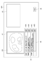
図1は、医療情報システムの概略構成を示す図である。図1に示す医療情報システム1は、公知のオーダリングシステムを用いた診療科の医師からの検査オーダに基づいて、被写体である患者の検査対象部位の撮影、撮影により取得された医用画像の保管、読影医による医用画像の読影と読影レポートの作成、および依頼元の診療科の医師による読影レポートの閲覧と読影対象の医用画像の詳細観察とを行うためのシステムである。
Fig. 1 is a diagram showing the schematic configuration of the medical information system. The medical information system 1 shown in FIG. 1 captures an examination target region of a patient as a subject, stores medical images acquired by the imaging, This is a system for interpretation of medical images and creation of an interpretation report by an interpreting doctor, and viewing of the interpretation report and detailed observation of the medical image to be interpreted by a doctor of the medical department of the requesting department.
図1に示すように、医療情報システム1は、複数の撮影装置2、読影端末である複数の読影WS(WorkStation)3、診療WS4、画像サーバ5、画像DB(DataBase)6、レポートサーバ7およびレポートDB8が、有線または無線のネットワーク10を介して互いに通信可能な状態で接続されて構成されている。
As shown in FIG. 1, a medical information system 1 includes a plurality of imaging devices 2, a plurality of interpretation WSs (WorkStations) 3 which are interpretation terminals, a medical examination WS 4, an image server 5, an image DB (DataBase) 6, a report server 7, and The report DBs 8 are connected to each other via a wired or wireless network 10 so as to be communicable with each other.
各機器は、医療情報システム1の構成要素として機能させるためのアプリケーションプログラムがインストールされたコンピュータである。アプリケーションプログラムは、DVD(Digital Versatile Disc)およびCD-ROM(Compact Disc Read Only Memory)等の記録媒体に記録されて配布され、その記録媒体からコンピュータにインストールされる。または、ネットワーク10に接続されたサーバコンピュータの記憶装置、若しくはネットワークストレージに、外部からアクセス可能な状態で記憶され、要求に応じてコンピュータにダウンロードされ、インストールされる。
Each device is a computer installed with an application program for functioning as a component of the medical information system 1. Application programs are recorded on recording media such as DVDs (Digital Versatile Discs) and CD-ROMs (Compact Discs Read Only Memory) for distribution, and are installed in computers from the recording media. Alternatively, it is stored in a storage device of a server computer connected to the network 10 or a network storage in a state accessible from the outside, and is downloaded and installed in a computer upon request.
撮影装置2は、患者の診断対象となる部位を撮影することにより、診断対象部位を表す医用画像を生成する装置(モダリティ)である。具体的には、単純X線撮影装置、CT装置、MRI装置、およびPET(Positron Emission Tomography)装置等である。撮影装置2により生成された医用画像は画像サーバ5に送信され、画像DB6に保存される。
The imaging device 2 is a device (modality) that generates a medical image representing the diagnostic target region by imaging the diagnostic target region of the patient. Specifically, they are plain X-ray equipment, CT equipment, MRI equipment, and PET (Positron Emission Tomography) equipment. A medical image generated by the imaging device 2 is transmitted to the image server 5 and stored in the image DB 6 .
読影WS3は、例えば放射線科の読影医が、医用画像の読影および読影レポートの作成等に利用するコンピュータであり、第1の実施形態に係る文書作成装置20(詳細は後述)を内包する。読影WS3では、画像サーバ5に対する医用画像の閲覧要求、画像サーバ5から受信した医用画像に対する各種画像処理、医用画像の表示、医用画像に関する所見文の入力受付が行われる。また、読影WS3では、医用画像に対する解析処理、解析結果に基づく読影レポートの作成の支援、レポートサーバ7に対する読影レポートの登録要求と閲覧要求、およびレポートサーバ7から受信した読影レポートの表示が行われる。これらの処理は、読影WS3が各処理のためのソフトウェアプログラムを実行することにより行われる。
The interpretation WS3 is a computer used by, for example, a radiology interpreting doctor to interpret medical images and create interpretation reports, and includes the document creation device 20 (details will be described later) according to the first embodiment. The interpretation WS 3 requests the image server 5 to view medical images, performs various image processing on the medical images received from the image server 5 , displays the medical images, and accepts input of remarks on the medical images. Further, the interpretation WS 3 performs analysis processing on medical images, supports creation of interpretation reports based on the analysis results, requests registration and viewing of interpretation reports to the report server 7 , and displays interpretation reports received from the report server 7 . . These processes are performed by the interpretation WS3 executing a software program for each process.
診療WS4は、例えば診療科の医師が、画像の詳細観察、読影レポートの閲覧、および電子カルテの作成等に利用するコンピュータであり、処理装置、ディスプレイ等の表示装置、並びにキーボードおよびマウス等の入力装置により構成される。診療WS4では、画像サーバ5に対する画像の閲覧要求、画像サーバ5から受信した画像の表示、レポートサーバ7に対する読影レポートの閲覧要求、およびレポートサーバ7から受信した読影レポートの表示が行われる。これらの処理は、診療WS4が各処理のためのソフトウェアプログラムを実行することにより行われる。
The clinical WS 4 is a computer used by, for example, doctors in clinical departments for detailed observation of images, viewing of interpretation reports, and preparation of electronic medical charts. It consists of a device. The medical examination WS 4 requests the image server 5 to view images, displays the images received from the image server 5 , requests the report server 7 to view interpretation reports, and displays the interpretation reports received from the report server 7 . These processes are performed by the clinical WS 4 executing a software program for each process.
画像サーバ5は、汎用のコンピュータにデータベース管理システム(DataBase Management System: DBMS)の機能を提供するソフトウェアプログラムがインストールされたものである。また、画像サーバ5は画像DB6が構成されるストレージを備えている。このストレージは、画像サーバ5とデータバスとによって接続されたハードディスク装置であってもよいし、ネットワーク10に接続されているNAS(Network Attached Storage)およびSAN(Storage Area Network)に接続されたディスク装置であってもよい。また、画像サーバ5は、撮影装置2からの医用画像の登録要求を受け付けると、その医用画像をデータベース用のフォーマットに整えて画像DB6に登録する。
The image server 5 is a general-purpose computer installed with a software program that provides the functions of a database management system (DBMS). Further, the image server 5 has a storage in which an image DB 6 is configured. This storage may be a hard disk device connected to the image server 5 by a data bus, or a disk device connected to NAS (Network Attached Storage) and SAN (Storage Area Network) connected to network 10. may be When the image server 5 receives a registration request for a medical image from the imaging device 2 , the image server 5 prepares the medical image into a database format and registers it in the image DB 6 .
画像DB6には、撮影装置2において取得された医用画像の画像データと付帯情報とが登録される。付帯情報には、例えば、個々の医用画像を識別するための画像ID(identification)、患者を識別するための患者ID、検査を識別するための検査ID、医用画像毎に割り振られるユニークなID(UID:unique identification)、医用画像が生成された検査日、検査時刻、医用画像を取得するための検査で使用された撮影装置の種類、患者氏名、年齢、性別等の患者情報、検査部位(撮影部位)、撮影情報(撮影プロトコル、撮影シーケンス、撮像手法、撮影条件、造影剤の使用等)、1回の検査で複数の医用画像を取得した場合のシリーズ番号あるいは採取番号等の情報が含まれる。
Image data of medical images acquired by the imaging device 2 and additional information are registered in the image DB 6 . The incidental information includes, for example, an image ID (identification) for identifying individual medical images, a patient ID for identifying a patient, an examination ID for identifying an examination, a unique ID assigned to each medical image ( UID: unique identification), examination date when the medical image was generated, examination time, type of imaging device used in the examination to acquire the medical image, patient information such as patient name, age, gender, examination site (imaging part), imaging information (imaging protocol, imaging sequence, imaging method, imaging conditions, use of contrast agent, etc.), and information such as series number or collection number when multiple medical images are acquired in one examination .
また、画像サーバ5は、読影WS3および診療WS4からの閲覧要求をネットワーク10経由で受信すると、画像DB6に登録されている医用画像を検索し、検索された医用画像を要求元の読影WS3および診療WS4に送信する。
When the image server 5 receives a viewing request from the interpretation WS 3 and the medical care WS 4 via the network 10 , the image server 5 searches for medical images registered in the image DB 6 and distributes the retrieved medical images to the requesting interpretation WS 3 and the medical care WS 4 . Send to WS4.
レポートサーバ7には、汎用のコンピュータにデータベース管理システムの機能を提供するソフトウェアプログラムが組み込まれる。レポートサーバ7は、読影WS3からの読影レポートの登録要求を受け付けると、その読影レポートをデータベース用のフォーマットに整えてレポートDB8に登録する。
The report server 7 incorporates a software program that provides the functions of a database management system to a general-purpose computer. When the report server 7 receives a registration request for an interpretation report from the interpretation WS 3 , the interpretation report is formatted for a database and registered in the report DB 8 .
レポートDB8には、読影医が読影WS3を用いて作成した所見文を含む読影レポートが登録される。読影レポートは、例えば、読影対象の医用画像、医用画像を識別する画像ID、読影を行った読影医を識別するための読影医ID、病変名、病変の位置情報、および病変の性状等の情報を含んでいてもよい。
In the report DB 8, an interpretation report containing the findings created by the interpretation doctor using the interpretation WS3 is registered. The interpretation report contains information such as, for example, the medical image to be interpreted, the image ID for identifying the medical image, the interpretation doctor ID for identifying the interpretation doctor who performed the interpretation, the lesion name, the position information of the lesion, and the properties of the lesion. may contain
また、レポートサーバ7は、読影WS3および診療WS4からの読影レポートの閲覧要求をネットワーク10経由で受信すると、レポートDB8に登録されている読影レポートを検索し、検索された読影レポートを要求元の読影WS3および診療WS4に送信する。
When the report server 7 receives a viewing request for an interpretation report from the interpretation WS 3 and the medical care WS 4 via the network 10, the report server 7 searches for the interpretation report registered in the report DB 8, and sends the retrieved interpretation report to the requested interpretation report. Send to WS3 and medical care WS4.
ネットワーク10は、病院内の各種機器を接続する有線または無線のローカルエリアネットワークである。読影WS3が他の病院あるいは診療所に設置されている場合には、ネットワーク10は、各病院のローカルエリアネットワーク同士をインターネットまたは専用回線で接続した構成としてもよい。
The network 10 is a wired or wireless local area network that connects various devices in the hospital. If the image interpretation WS3 is installed in another hospital or clinic, the network 10 may be configured by connecting the local area networks of each hospital via the Internet or a dedicated line.
次に、第1の実施形態による文書作成装置20について説明する。まず、図2を参照して、第1の実施形態による文書作成装置20のハードウェア構成を説明する。図2に示すように、文書作成装置20は、CPU(Central Processing Unit)11、不揮発性のストレージ13、および一時記憶領域としてのメモリ16を含む。また、文書作成装置20は、液晶ディスプレイ等のディスプレイ14、キーボードおよびマウス等のポインティングデバイス等からなる入力デバイス15、並びにネットワーク10に接続されるネットワークI/F(InterFace)17を含む。CPU11、ストレージ13、ディスプレイ14、入力デバイス15、メモリ16およびネットワークI/F17は、バス18に接続される。なお、CPU11は、本開示におけるプロセッサの一例である。
Next, the document creation device 20 according to the first embodiment will be described. First, referring to FIG. 2, the hardware configuration of the document creation device 20 according to the first embodiment will be described. As shown in FIG. 2, the document creation device 20 includes a CPU (Central Processing Unit) 11, a non-volatile storage 13, and a memory 16 as a temporary storage area. The document creation device 20 also includes a display 14 such as a liquid crystal display, an input device 15 such as a keyboard and a pointing device such as a mouse, and a network I/F (InterFace) 17 connected to the network 10 . CPU 11 , storage 13 , display 14 , input device 15 , memory 16 and network I/F 17 are connected to bus 18 . Note that the CPU 11 is an example of a processor in the present disclosure.
ストレージ13は、HDD(Hard Disk Drive)、SSD(Solid State Drive)、およびフラッシュメモリ等によって実現される。記憶媒体としてのストレージ13には、文書作成プログラム12が記憶される。CPU11は、ストレージ13から文書作成プログラム12を読み出してメモリ16に展開し、展開した文書作成プログラム12を実行する。
The storage 13 is realized by HDD (Hard Disk Drive), SSD (Solid State Drive), flash memory, and the like. A document creation program 12 is stored in the storage 13 as a storage medium. The CPU 11 reads out the document creation program 12 from the storage 13 , expands it in the memory 16 , and executes the expanded document creation program 12 .
次いで、第1の実施形態による文書作成装置の機能的な構成を説明する。図3は、第1の実施形態による文書作成装置の機能的な構成を示す図である。図3に示すように文書作成装置20は、画像取得部21、画像解析部22、作成方法決定部23、文書作成部24および表示制御部25を備える。そして、CPU11が、文書作成プログラム12を実行することにより、CPU11は、画像取得部21、画像解析部22、作成方法決定部23、文書作成部24および表示制御部25として機能する。
Next, the functional configuration of the document creation device according to the first embodiment will be described. FIG. 3 is a diagram showing the functional configuration of the document creation device according to the first embodiment. As shown in FIG. 3 , the document creation device 20 includes an image acquisition section 21 , an image analysis section 22 , a creation method determination section 23 , a document creation section 24 and a display control section 25 . By executing the document creation program 12 , the CPU 11 functions as an image acquisition unit 21 , an image analysis unit 22 , a creation method determination unit 23 , a document creation unit 24 and a display control unit 25 .
画像取得部21は、画像の一例としての医用画像G0を、ネットワークI/F17を介して画像サーバ5から取得する。第1の実施形態においては、一例として、肺のCT画像を医用画像G0として用いる。
The image acquisition unit 21 acquires a medical image G0 as an example of an image from the image server 5 via the network I/F 17. In the first embodiment, as an example, a lung CT image is used as the medical image G0.
画像解析部22は、医用画像G0を解析することにより、医用画像G0に含まれる疾病等に関する解析結果を導出する。このために、画像解析部22は、医用画像G0に含まれる病変等の異常陰影を検出し、検出した異常陰影についての所見を判別するように機械学習がなされた学習済みモデル22Aを有する。
By analyzing the medical image G0, the image analysis unit 22 derives analysis results regarding diseases and the like included in the medical image G0. For this purpose, the image analysis unit 22 has a learned model 22A that has undergone machine learning so as to detect abnormal shadows such as lesions contained in the medical image G0 and determine the findings of the detected abnormal shadows.
学習済みモデル22Aは、例えば臓器別に用意されており、臓器に含まれる異常陰影を特定し、特定した異常陰影の所見を判別する。なお、所見は複数の所見項目について判別される。例えば、臓器が肺であれば、肺に含まれる異常陰影の場所、異常陰影のサイズ、境界の形状(明瞭および不整形)、吸収値の種類(充実型およびスリガラス型)、スピキュラの有無、腫瘤か結節か、胸膜接触の有無、胸膜陥入の有無、胸膜浸潤の有無、空洞の有無、および石灰化の有無等の複数の所見項目についての所見が判別される。なお、所見項目が本開示の所見に関する属性情報の一例である。
The learned model 22A is prepared for each organ, for example, identifies abnormal shadows contained in the organ, and identifies findings of the identified abnormal shadows. A finding is determined for a plurality of finding items. For example, if the organ is the lung, the location of the abnormal shadow contained in the lung, the size of the abnormal shadow, the shape of the boundary (clear and irregular), the type of absorption value (solid type and ground glass type), the presence or absence of spicules, and the mass Findings are discriminated for a plurality of finding items such as whether it is a nodule, presence or absence of pleural contact, presence or absence of pleural indentation, presence or absence of pleural infiltration, presence or absence of cavity, and presence or absence of calcification. It should be noted that the finding item is an example of attribute information related to the finding of the present disclosure.
本実施形態においては、学習済みモデル22Aは、医用画像G0に含まれる異常陰影についての所見を判別するようにディープラーニング(深層学習)がなされた畳み込みニューラルネットワーク(CNN(Convolutional Neural Network))からなる。
In this embodiment, the trained model 22A is a convolutional neural network (CNN (Convolutional Neural Network)) that has undergone deep learning so as to discriminate findings about abnormal shadows contained in the medical image G0. .
学習済みモデル22Aは、例えば、異常陰影を含む医用画像と、異常陰影に関する各種所見項目についての所見との組み合わせを教師データとして用いた機械学習によって構築される。学習済みモデル22Aは、医用画像が入力されると、医用画像に含まれる異常陰影における、所見項目毎に導出される所見についてのスコア(以下、所見スコアとする)を出力する。所見スコアは、各所見項目についての所見の顕著性を示すスコアである。所見スコアは例えば0以上1以下の値をとり、所見スコアの値が大きい程、その所見項目についての所見が顕著であることを示す。
The learned model 22A is constructed, for example, by machine learning using a combination of medical images containing abnormal shadows and observations of various observation items related to abnormal shadows as teacher data. When a medical image is input, the trained model 22A outputs a score (hereinafter referred to as a finding score) for findings derived for each finding item in the abnormal shadow included in the medical image. The finding score is a score that indicates the salience of the finding for each finding item. The finding score takes a value of, for example, 0 or more and 1 or less.
例えば異常陰影の所見項目の1つである「スピキュラの有無」についての所見スコアが例えば0.5以上である場合、異常陰影の「スピキュラの有無」についての所見が「スピキュラ有り(陽性)」であることを特定し、「スピキュラの有無」についての所見スコアが例えば0.5未満である場合、異常陰影のスピキュラの有無についての所見が「スピキュラ無し(陰性)」であることを特定する。なお、所見の判定に用いるしきい値0.5は、例示に過ぎず、所見項目毎に適切な値に設定される。
For example, if the finding score for “presence or absence of spicules”, which is one of the findings items for abnormal shadows, is 0.5 or more, the findings for “presence or absence of spicula” for abnormal shadows are “with spicules (positive)”. If the finding score for "presence/absence of spicules" is less than, for example, 0.5, then specify that the findings for presence/absence of spicules in the abnormal shadow are "no spicules (negative)". Note that the threshold value of 0.5 used for determination of findings is merely an example, and is set to an appropriate value for each finding item.
図4は画像解析部22が導出した解析結果に含まれる所見の例を示す図である。なお、図4は医用画像G0が肺のCT画像である場合の所見の例を示す。医用画像G0が肺のCT画像である場合、画像解析部22は、例えば、複数の所見項目について、「左肺胸膜下」、「4.2cm」、「不整形」、「充実型」、「スピキュラ有り」、「腫瘤」、「胸膜接触有り」、「胸膜陥入有り」、「胸膜浸潤無し」、「空洞無し」および「石灰化無し」の所見を導出する。図4においては、「有り」すなわち陽性の場合は+、「無し」すなわち陰性の場合は-を付与している。
FIG. 4 is a diagram showing an example of findings included in the analysis results derived by the image analysis unit 22. FIG. FIG. 4 shows an example of findings when the medical image G0 is a lung CT image. When the medical image G0 is a lung CT image, the image analysis unit 22 selects, for example, “left lung subpleural”, “4.2 cm”, “irregular”, “solid type”, “ The following findings are derived: spicula, mass, pleural contact, pleural indentation, no pleural invasion, no cavity, and no calcification. In FIG. 4, “yes”, ie, positive, is given by +, and “no”, ie, negative is given by −.
なお、学習済みモデル22Aとしては、畳み込みニューラルネットワークの他、例えばサポートベクタマシン(SVM(Support Vector Machine))等の任意の学習済みモデルを用いることができる。
As the trained model 22A, any trained model such as a support vector machine (SVM (Support Vector Machine)) can be used in addition to the convolutional neural network.
作成方法決定部23は、文書の出力を決定する判断用の情報に基づいて、作成される文書の出力を決定する。第1の実施形態においては、作成方法決定部23は、医用画像G0に関する文書の作成方法を決定することにより、文書の出力を決定する。第1の実施形態においては、画像解析部22が導出した解析結果に含まれる所見項目の数に基づいて、医療文書の作成方法を決定する。所見項目の数が本開示における判断用の情報、より具体的には作成方法決定のための評価基準の一例である。また、第1の実施形態においては、文書は医用画像G0に関する所見文であるものとする。
The creation method determination unit 23 determines the output of the document to be created based on the judgment information for determining the output of the document. In the first embodiment, the creation method determining unit 23 determines the document output by determining the document creation method for the medical image G0. In the first embodiment, the medical document creation method is determined based on the number of finding items included in the analysis results derived by the image analysis unit 22 . The number of finding items is an example of judgment information in the present disclosure, more specifically, an example of evaluation criteria for determining the preparation method. Also, in the first embodiment, the document is assumed to be an observation statement regarding the medical image G0.
ここで、第1の実施形態においては、医用画像G0に含まれる部位毎に、所見項目の数と文書作成方法とが対応づけられたテーブルが用意されてストレージ13に保存されている。図5は所見項目の数と文書作成方法とが対応づけられたテーブルの例を示す図である。図5に示すテーブル30は、所見項目が1~3個の場合には、それぞれテンプレートT1,T2,T3が対応づけられている。所見項目が4個の場合には自然言語処理NLP1が対応づけられ、所見項目が5個の場合には自然言語処理NLP2が対応づけられている。また、所見項目が6個以上の場合には、3つの自然言語処理NLP1~NLP3が対応づけられている。
Here, in the first embodiment, a table is prepared and stored in the storage 13 in which the number of finding items and the document creation method are associated with each region included in the medical image G0. FIG. 5 is a diagram showing an example of a table in which the number of observation items and the document creation method are associated. In the table 30 shown in FIG. 5, templates T1, T2, and T3 are associated with one to three finding items. When there are four finding items, natural language processing NLP1 is associated, and when there are five finding items, natural language processing NLP2 is associated. Also, when there are six or more finding items, three natural language processing NLP1 to NLP3 are associated.
テンプレートT1~T3は、所見項目の数に応じて空欄を有するテキストからなる。例えばテンプレートT2は「<場所>に<所見>が見られます。」のように、2つの所見を挿入可能な空欄を有するテキストである。自然言語処理NLP1~NLP3は、ニューラルネットワークを学習することにより構築された学習済みモデルを用いることによる文書の作成方法に対応する。
Templates T1 to T3 consist of texts with blanks according to the number of finding items. For example, the template T2 is a text having blanks into which two findings can be inserted, such as "<finding> can be seen in <place>." Natural language processing NLP1-NLP3 corresponds to a document creation method using a trained model constructed by learning a neural network.
作成方法決定部23は、解析結果に含まれる所見項目の数に基づいてテーブル30を参照して、文書の作成方法を決定する。例えば、所見項目の数が2個の場合、作成方法決定部23はテンプレートT2を作成方法に決定する。また、所見項目の数が6個以上の場合には、作成方法決定部23は3つの自然言語処理NLP1~NLP3を作成方法に決定する。
The creation method determination unit 23 determines the document creation method by referring to the table 30 based on the number of finding items included in the analysis results. For example, when the number of observation items is two, the creation method determination unit 23 determines template T2 as the creation method. Further, when the number of observation items is six or more, the creation method determination unit 23 determines three natural language processing NLP1 to NLP3 as the creation method.
なお、作成方法決定部23が参照するテーブルは図5に示すものに限定されない。例えば、所見項目により特定される部位に応じて、テンプレートを使用するか、自然言語処理を使用するかを対応づけたテーブルを用いるようにしてもよい。この場合、例えば部位が肺であれば解析結果に含まれる所見項目の数が多いため、作成方法として自然言語処理を対応づけておき、部位が膵臓であれば、解析結果に含まれる所見項目の数が少ないため、作成方法としてテンプレートを対応づけておけばよい。
It should be noted that the table referred to by the creation method determination unit 23 is not limited to the one shown in FIG. For example, a table may be used that associates whether to use a template or to use natural language processing according to the site specified by the finding item. In this case, for example, if the site is the lung, the number of finding items included in the analysis results is large. Since the number is small, it is sufficient to associate a template as a creation method.
また、部位を特定するに際しては、所見項目を参照することに代えて、医用画像G0を取得した際の検査指示書を参照するようにしてもよい。検査指示書には検査を行う部位名が記載されているため、検査指示書に記載された部位名により部位を特定すればよい。また、検査指示書には検査項目が記載されている。検査項目は疾患に応じて異なるものとなる.このため、検査指示書に記載されている検査項目を参照して、疾患を特定したり、疾患から部位を特定したりしてもよい。なお、検査指示書は例えば医用画像G0と対応づけられて画像サーバ5に保存されており、画像取得部21が医用画像G0を画像サーバ5から取得する際に、医用画像G0と併せて取得するようにすればよい。
In addition, when specifying the site, instead of referring to the finding items, the inspection instruction sheet used when the medical image G0 was acquired may be referred to. Since the name of the site to be inspected is written in the inspection instruction, the site may be specified by the name of the site described in the inspection instruction. In addition, inspection items are described in the inspection instruction. The examination items differ depending on the disease. For this reason, the disease may be specified, and the site may be specified from the disease by referring to the inspection items described in the inspection instruction. Note that the inspection instruction sheet is stored in the image server 5 in association with the medical image G0, for example, and when the image acquisition unit 21 acquires the medical image G0 from the image server 5, the inspection instruction sheet is acquired together with the medical image G0. You should do it like this.
また、作成方法決定部23は、解析結果に含まれる異常陰影の数に応じて作成方法を決定してもよい。例えば、解析結果に1つの異常陰影に関する所見のみが含まれる場合にはテンプレートを作成方法に決定し、複数の異常陰影に関する所見が含まれる場合には自然言語処理を作成方法に決定するようにしてもよい。
Also, the creation method determination unit 23 may determine the creation method according to the number of abnormal shadows included in the analysis result. For example, if the analysis result contains only one abnormal shadow finding, the template is determined as the creation method, and if multiple abnormal shadow findings are included, natural language processing is determined as the creation method. good too.
また、部位および病気毎にあらかじめ定められた評価値をストレージ13に記憶しておき、評価値に応じて作成方法を決定するようにしてもよい。図6は部位および病気と評価値との関係を示すテーブルを示す図、図7は評価値と文書の作成方法とを対応づけたテーブルを示す図である。図6に示すように、テーブル31においては、部位および病気に対して評価値が対応づけられている。評価値としては、例えば文書作成の難易度を表す評価値を用いることができる。具体的には、評価値を0~1の値で定義し、文書作成の難易度が高いほど大きい評価値となるようにテーブル31を作成することができる。なお、文書作成の難易度が本開示のあらかじめ定められた作成方法決定のための評価基準の一例であり、難易度を表す評価値が本開示の判断用の情報の評価結果の一例である。
Alternatively, evaluation values predetermined for each part and disease may be stored in the storage 13, and the creation method may be determined according to the evaluation values. FIG. 6 is a diagram showing a table showing the relationship between parts and diseases and evaluation values, and FIG. 7 is a diagram showing a table in which evaluation values and document preparation methods are associated with each other. As shown in FIG. 6, in the table 31, evaluation values are associated with sites and diseases. As the evaluation value, for example, an evaluation value representing the difficulty level of document creation can be used. Specifically, the evaluation value is defined as a value between 0 and 1, and the table 31 can be created so that the higher the difficulty of document creation, the larger the evaluation value. Note that the difficulty level of document creation is an example of a predetermined evaluation criterion for determining the creation method of the present disclosure, and the evaluation value representing the difficulty level is an example of the evaluation result of information for determination of the present disclosure.
ここで、肺または肝臓の疾患については、病気が複雑になりやすいため所見文に記載すべき所見項目が多数ある。また、悪性度が高い病気は所見文に記載すべき所見項目が多くなる。さらには、異なる疾患についての所見をまとめて1つの文に記載する場合もある。このような場合、文書を作成する際の難易度が高くなる。このため、病気が複雑になりやすい部位および悪性度が高い病気等の場合には、高い値となるように評価値を設定してもよい。例えば、図6に示すテーブル31において、部位が肺および肝臓の場合には評価値を1.0に定義し、肺および肝臓以外の部位については評価値を0.5に定義すればよい。また、病気が癌の場合には評価値を1.0に定義し、がん以外の場合には0.5に定義すればよい。
Here, regarding lung or liver diseases, there are many observation items that should be described in the observation statement because the disease tends to be complicated. In addition, for diseases with high malignancy, the number of observation items to be described in the observation statement increases. In addition, findings for different diseases may be grouped together in one sentence. In such a case, the degree of difficulty in creating the document increases. Therefore, the evaluation value may be set to a high value in the case of a site where the disease is likely to become complicated or in the case of a disease with a high degree of malignancy. For example, in the table 31 shown in FIG. 6, the evaluation value may be defined as 1.0 for the lung and liver regions, and the evaluation value may be defined as 0.5 for regions other than the lung and liver. If the disease is cancer, the evaluation value should be defined as 1.0, and if the disease is other than cancer, it should be defined as 0.5.
一方、図7に示すように、テーブル32には評価値と作成方法とが対応づけられている。図7においては、評価値X1に対して自然言語処理NLP1が対応づけられ、評価値X2,X3に対してそれぞれテンプレートT1,T2が対応づけられている。また、評価値X4に対しては、テンプレートT1と自然言語処理NLP1(T1+NLP1)とが対応づけられている。
On the other hand, as shown in FIG. 7, the table 32 associates evaluation values with creation methods. In FIG. 7, a natural language processing NLP1 is associated with an evaluation value X1, and templates T1 and T2 are associated with evaluation values X2 and X3, respectively. Also, the evaluation value X4 is associated with the template T1 and the natural language processing NLP1 (T1+NLP1).
作成方法決定部23は、解析結果に基づいてテーブル31を参照して、部位および病気に応じた評価値を取得する。そして、作成方法決定部23は、テーブル32を参照して、取得した評価値に応じて文書の作成方法を決定する。この場合、部位および病気のそれぞれの評価値を加算した評価値を用いて文書の作成方法を決定してもよい。例えば、肺がんの場合には肺の評価値とがんの評価値とを加算した評価値2.0を用いて文書の作成方法を決定してもよい。
The creation method determination unit 23 refers to the table 31 based on the analysis result and acquires an evaluation value corresponding to the site and disease. Then, the creation method determining unit 23 refers to the table 32 and determines the document creation method according to the acquired evaluation value. In this case, the document creation method may be determined using an evaluation value obtained by adding the evaluation values of the site and the disease. For example, in the case of lung cancer, an evaluation value of 2.0 obtained by adding the lung evaluation value and the cancer evaluation value may be used to determine the document creation method.
なお、解析結果に含まれるすべての所見項目についての所見を所見文に記載しない場合がある。例えば、すでに作成された所見文に所見の一部が記載されている場合がある。そのような場合は、これから作成する所見文に記載すべき所見の数が減る。このような場合には、テーブル31を参照して取得された評価値により表される難易度を低くして、文書の作成方法を決定するようにしてもよい。
It should be noted that there may be cases where the observations for all observation items included in the analysis results are not described in the observations. For example, there is a case where part of the observation is described in the already created observation text. In such a case, the number of observations to be written in the observation sentences to be prepared from now on is reduced. In such a case, the difficulty level represented by the evaluation value acquired by referring to the table 31 may be lowered to determine the document creation method.
また、複数の所見を1つの所見文にまとめて記載する指示が行われる場合もある。このような場合は、テーブル31を参照して取得された評価値により表される難易度を高くして、文書の作成方法を決定するようにしてもよい。
In addition, there may be cases where instructions are given to summarize multiple findings in one statement. In such a case, the document creation method may be determined by increasing the degree of difficulty represented by the evaluation value obtained by referring to the table 31 .
また、あらかじめ定められた条件に応じて作成方法を決定するようにしてもよい。例えば、あらかじめ定められた条件に応じて、解析結果に含まれる所見項目の数による作成方法、解析結果に含まれる異常陰影の数による作成方法、並びに部位および病気毎にあらかじめ定められた評価値に基づく作成方法のいずれかの作成方法を決定するようにしてもよい。なお、予め定められた条件としては、例えば、医用画像G0に含まれる臓器が肺または肝臓等の特定の臓器であること、特定の所見を有すること、特定の撮影装置により撮影されたこと、医用画像G0を取得した患者の性別および年齢等の患者情報が特定の条件を満たすこと等が挙げられる。
Alternatively, the creation method may be determined according to predetermined conditions. For example, according to predetermined conditions, the number of finding items included in the analysis results, the number of abnormal shadows included in the analysis results, and the predetermined evaluation value for each site and disease. You may make it determine the production method of any of the production methods based on. Note that the predetermined conditions include, for example, that the organ included in the medical image G0 is a specific organ such as a lung or liver, that the medical image G0 has a specific finding, that the image was captured by a specific imaging device, that the medical image G0 Patient information such as the sex and age of the patient who acquired the image G0 satisfies specific conditions.
文書作成部24は、作成方法決定部23が決定した作成方法を用いて、解析結果を含む少なくとも1つの所見文を作成する。所見文が本開示における文書の一例である。また、文書作成部24が作成する少なくとも1つの所見文が、本開示における少なくとも1つの文書の一例である。ここで、解析結果に含まれる所見が「右肺S1」、「1cm」および「腫瘤」の3個であり、作成方法として3つの空欄を有するテンプレートT3が決定されたとする。テンプレートT3は例えば「<場所>に<大きさ>大の<種類>を認めます。」のテキストである。この場合、文書作成部24は、「<右肺S1>に<1cm>大の<腫瘤>を認めます。」の所見文を作成する。
The document creation unit 24 uses the creation method determined by the creation method determination unit 23 to create at least one remark including the analysis result. A remarks statement is an example of a document in this disclosure. Also, at least one remark sentence created by the document creating unit 24 is an example of at least one document in the present disclosure. Here, it is assumed that the three findings included in the analysis result are "right lung S1," "1 cm," and "tumor," and template T3 having three blanks is determined as the creation method. The template T3 is, for example, the text of "Allow <size> large <type> at <place>." In this case, the document creation unit 24 creates a finding sentence of "A <1 cm> large <tumor> is found in <right lung S1>."
ここで、文書作成部24は、解析結果に含まれる少なくとも1つの所見を用いて所見文を生成するように学習が行われた学習済みモデル24A~24Cを有する。学習済みモデル24A~24Cはそれぞれ自然言語処理NLP1~NLP3に対応する。
Here, the document creation unit 24 has trained models 24A to 24C that have been trained to generate observation sentences using at least one observation included in the analysis result. Trained models 24A-24C correspond to natural language processing NLP1-NLP3, respectively.
学習済みモデル24A~24Cとしては、例えばリカレントニューラルネットワークを用いることができる。画像解析部22による解析結果から所見文を出力するために、学習済みモデル24A~24Cは、解析結果に含まれる所見項目と所見文との組み合わせからなる多数の教師データを用いてリカレントニューラルネットワークを機械学習することにより構築されてなる。作成方法決定部23が決定した文書の作成方法が自然言語処理である場合、文書作成部24は、解析結果に含まれる少なくとも1つの所見を学習済みモデル24A~24Cに入力し、所見文を出力させる。
For example, a recurrent neural network can be used as the trained models 24A-24C. In order to output observation sentences from the analysis results of the image analysis unit 22, the trained models 24A to 24C run a recurrent neural network using a large amount of teacher data consisting of combinations of observation items and observation sentences included in the analysis results. It is constructed by machine learning. When the document creation method determined by the creation method determination unit 23 is natural language processing, the document creation unit 24 inputs at least one observation included in the analysis result to the learned models 24A to 24C, and outputs an observation statement. Let
例えば、画像解析部22による解析結果に「左肺S6」、「1.5cm」、「結節」、「肺転移」の4個の所見が含まれる場合、作成方法決定部23は自然言語処理NLP2を作成方法に決定する。文書作成部24は、自然言語処理NLP2に対応する学習済みモデル24Bを用いて「左肺S6に1.5cm大の結節が認められ、肺転移が疑われます。」のような所見文を作成する。
For example, when the analysis result by the image analysis unit 22 includes four findings of “left lung S6”, “1.5 cm”, “nodule”, and “lung metastasis”, the creation method determination unit 23 performs natural language processing NLP2 to the method of creation. The document creation unit 24 uses the trained model 24B corresponding to natural language processing NLP2 to create an observation sentence such as "A nodule of 1.5 cm in size is found in the left lung S6, and lung metastasis is suspected." do.
なお、作成方法決定部23が複数の自然言語処理を作成方法に決定した場合、文書作成部24は、複数の自然言語処理のそれぞれを用いて複数の所見文を作成する。
It should be noted that when the creation method determination unit 23 determines multiple natural language processes as the creation method, the document creation unit 24 creates multiple observation sentences using each of the multiple natural language processes.
例えば、医用画像G0の解析結果に「左肺上葉」、「胸膜陥入有り」、「辺縁不整有り」、「スピキュラ有り」、「4.2cm」および「腫瘤」の6個の所見項目についての所見が含まれたとする。この場合、作成方法決定部23は、3つの自然言語処理NLP1~NLP3を作成方法に決定する。自然言語処理NLP1~NLP3に対応する学習済みモデル24A~24Cは、複数の所見のうちの少なくとも1つを記述した異なる所見文を生成する。学習済みモデル24A~24Cは、複数の所見項目の各々についての所見の少なくとも1つが各所見文に記述され、かつ複数の所見文の各々に記述される所見に対応する所見項目の組み合わせが複数の所見文の間で互いに異なるように複数の所見文を生成する。この場合、複数の所見文は作成に用いた所見および作成方法の双方が異なる文書の一例である。複数の所見文は、作成に用いた所見のみが異なるものであってもよく、作成方法のみが異なるものであってもよい。
For example, the analysis result of the medical image G0 includes six findings items: "left lung upper lobe", "pleural invagination present", "marginal irregularity present", "spicule present", "4.2 cm", and "mass". Suppose that the observation about is included. In this case, the creation method determination unit 23 determines the three natural language processes NLP1 to NLP3 as the creation method. Trained models 24A-24C corresponding to natural language processing NLP1-NLP3 generate different finding sentences describing at least one of the plurality of findings. In the trained models 24A to 24C, at least one finding for each of the plurality of finding items is described in each finding sentence, and there are multiple combinations of finding items corresponding to the findings described in each of the plurality of finding sentences. A plurality of observation sentences are generated so that the observation sentences are different from each other. In this case, the plurality of observation sentences is an example of documents that differ in both the observations used for creation and the creation method. A plurality of observation sentences may differ only in the observations used for preparation, or may differ only in the preparation method.
例えば、自然言語処理NLP1に対応する学習済みモデル24Aは、複数の所見項目の各々について特定された所見のうち、例えば「左肺上葉」および「腫瘤」に基づいて「左肺上葉に腫瘤が認められます。」という所見文を生成する。また、自然言語処理NLP2に対応する学習済みモデル24Bは、複数の所見項目の各々について特定された所見のうち、例えば「左肺上葉」、「胸膜陥入有り」、「4.2cm」、「腫瘤」に基づいて「左肺上葉に胸膜陥入を伴う、4.2cm大の腫瘤が認められます。」という所見文を生成する。また、自然言語処理NLP3に対応する学習済みモデル24Cは、複数の所見項目の各々について特定された所見のうち、例えば「左肺上葉」、「胸膜陥入有り」、「スピキュラ有り」、「腫瘤」に基づいて「左肺上葉に胸膜陥入を伴うスピキュラを有する腫瘤が認められます。」という所見文を生成する。
For example, the trained model 24A corresponding to the natural language processing NLP1 is based on, for example, the "left upper lobe of the lung" and the "mass" among the findings specified for each of the plurality of findings items, "a mass in the upper lobe of the left lung is recognized.” is generated. In addition, the trained model 24B corresponding to the natural language processing NLP2, among the findings specified for each of the plurality of findings items, for example, "left lung upper lobe", "pleural invagination", "4.2 cm", Based on the "mass", a finding statement "a mass of 4.2 cm with pleural indentation is found in the upper lobe of the left lung" is generated. In addition, the trained model 24C corresponding to the natural language processing NLP3, among the findings specified for each of the plurality of findings items, for example, "left lung upper lobe", "pleural invagination", "spicule present", " Based on "mass", a finding sentence "a mass with a spicule accompanied by pleural indentation is found in the upper lobe of the left lung" is generated.
また、作成方法決定部23が、図7に示すT1+NLP1を作成方法に決定した場合、文書作成部24は、テンプレートT1と自然言語処理NLP1とを組み合わせて所見文を作成する。この場合、作成方法決定部23は、解析結果に複数の所見が含まれ、そのうちの一部の所見を用いて比較的簡単な所見文をテンプレートにより作成する。そして残りの所見については自然言語処理により所見文を作成する。例えば、解析結果が「5mm」、「結節」、「辺縁スピキュラ有り」、「内部石灰化有り」、「胸膜接触有り」、「胸膜浸潤有り」であるとする。この場合、文書作成部24は、「5mm」および「結節」の所見についてはテンプレートT1を用いて「肺に5mmの結節を認めます。」という所見文を作成する。そして、「辺縁スピキュラ有り」、「内部石灰化有り」、「胸膜接触有り」および「胸膜浸潤有り」の所見については、自然言語処理NLP1に対応する学習済みモデル24Aを用いて、「辺縁にスピキュラを認め、内部に石灰化を認めます。また、胸膜に接しており、胸膜浸潤を疑います。」という所見文を作成する。
Also, when the creation method determination unit 23 determines T1+NLP1 shown in FIG. 7 as the creation method, the document creation unit 24 creates an observation sentence by combining the template T1 and the natural language processing NLP1. In this case, the creation method determining unit 23 creates a relatively simple finding statement using a template from a plurality of findings included in the analysis result, using some of the findings. Then, for the remaining findings, a statement of findings is created by natural language processing. For example, assume that the analysis results are "5 mm", "nodule", "marginal spicule present", "internal calcification present", "pleural contact present", and "pleural infiltration present". In this case, the document creation unit 24 uses the template T1 for the findings of "5 mm" and "nodule" to create a finding sentence of "a 5 mm nodule is found in the lungs." Then, for the findings of "marginal spicule present", "internal calcification present", "pleural contact present" and "pleural infiltration present", using the trained model 24A corresponding to natural language processing NLP1, "marginal A spicula is found in the sac, and calcification is found inside.In addition, it is in contact with the pleura, and pleural infiltration is suspected.”
なお、作成方法決定部23が、図7に示すT1+NLP1を作成方法に決定した場合、メインとなる所見については、自然言語処理NLP1を用いて所見文を作成し、付加的な情報についてはテンプレートを用いて所見文を作成するようにしてもよい。例えば、解析結果が「5mm」、「結節」、「辺縁スピキュラ有り」、「内部石灰化有り」、「胸膜接触有り」、「胸膜浸潤有り」、「血管圧排像有り」であるとする。この場合、「5mm」、「結節」、「辺縁スピキュラ有り」、「内部石灰化有り」、「胸膜接触有り」および「胸膜浸潤有り」がメインとなる所見であり、「血管圧排像有り」が付加的な所見である。文書作成部24は、メインとなる所見については自然言語処理NLP1に対応する学習済みモデル24Aを用いて、「肺に5mmの結節を認めます。辺縁にスピキュラを認め、内部に石灰化を認めます。また、胸膜に接しており、胸膜浸潤を疑います。」という所見文を作成する。そして付加的な所見である「血管圧排像有り」については、テンプレートT1を用いて「血管の圧排像を認めます。」という所見文を作成する。
In addition, when the creation method determination unit 23 determines T1+NLP1 shown in FIG. You may make it create an observation sentence using it. For example, assume that the analysis results are "5 mm", "nodule", "marginal spicule present", "internal calcification present", "pleural contact present", "pleural infiltration present", and "vascular compression present". In this case, “5 mm”, “nodule”, “marginal spicule”, “internal calcification”, “pleural contact” and “pleural infiltration” were the main findings, and “vessel compression image”. are additional findings. The document creation unit 24 uses the trained model 24A corresponding to natural language processing NLP1 for the main findings, "A 5 mm nodule is recognized in the lung. In addition, it is in contact with the pleura, and pleural infiltration is suspected.” Then, for the additional finding "vessel retraction image present", a finding statement "vessel retraction image is recognized" is created using the template T1.
表示制御部25は、作成された所見文をディスプレイ14に表示する。図8は所見文の表示画面を示す図である。図8に示すように、表示画面40は、画像表示領域41と文書表示領域42とを有する。画像表示領域41には医用画像G0が表示される。なお、医用画像G0がCT画像のような3次元画像である場合、3次元画像に含まれる1つの断層面の断層画像が画像表示領域41に表示される。文書表示領域42には、文書作成部24が作成した所見文が表示される。なお、所見文は、「左肺S6に1.5cm大の結節が認められ、肺転移が疑われます。」である。
The display control unit 25 displays the created observation text on the display 14. FIG. 8 is a diagram showing a display screen for observation sentences. As shown in FIG. 8, the display screen 40 has an image display area 41 and a document display area 42 . A medical image G0 is displayed in the image display area 41 . Note that when the medical image G0 is a three-dimensional image such as a CT image, a tomographic image of one tomographic plane included in the three-dimensional image is displayed in the image display area 41 . In the document display area 42, the findings created by the document creating section 24 are displayed. The statement of findings is "A nodule of 1.5 cm in size is found in S6 of the left lung, and lung metastasis is suspected."
なお、作成方法決定部23が複数の作成方法を決定した場合の所見文の表示画面を図9に示す。複数の作成方法が決定された場合にはそれに対応して複数の所見文が作成される。このため、図9に示す表示画面40Aには、複数の所見文を表示するための所見文表示領域43が表示される。所見文表示領域43には複数(ここでは3つ)の所見文の候補が表示されている。また、3つの所見文に加えて、自由入力欄44も表示される。また、所見文の候補および自由入力欄44の右方には、それぞれ採用ボタン45A~45Dが表示されている。
FIG. 9 shows the display screen of the remark sentence when the creation method determination unit 23 has determined a plurality of creation methods. When a plurality of creation methods are determined, a plurality of observation statements are created correspondingly. Therefore, the display screen 40A shown in FIG. 9 displays a remark display area 43 for displaying a plurality of remarks. A plurality (here, three) of observation sentence candidates are displayed in the observation sentence display area 43 . In addition to the three observation sentences, a free entry field 44 is also displayed. Adoption buttons 45A to 45D are displayed on the right side of the observation sentence candidate and the free entry field 44, respectively.
読影医は、必要であれば、入力デバイス15を用いて所見文を修正することができる。また、複数の所見文が表示されている場合には、所望とする所見文に対応する採用ボタンを選択する。これにより、選択された所見文が文書表示領域42に表示される。一方、医用画像G0に含まれる病変の読影結果に適合する所見文が所見文表示領域43に表示されていない場合、読影医は自由入力欄44に自身で所見文を入力し、採用ボタン45Dを選択する。これにより、読影医が自由入力欄44に入力した所見文が文書表示領域42に表示される。
The radiologist can use the input device 15 to correct the findings if necessary. Also, when a plurality of remarks are displayed, the user selects the adoption button corresponding to the desired remarks. As a result, the selected observation sentence is displayed in the document display area 42 . On the other hand, if no finding sentence matching the interpretation result of the lesion contained in the medical image G0 is displayed in the finding sentence display area 43, the interpreting doctor enters the finding sentence in the free input field 44 by himself and presses the adoption button 45D. select. As a result, the finding text entered in the free entry field 44 by the interpreting doctor is displayed in the document display area 42 .
そして読影医が確定ボタン46を選択すると、文書表示領域42に入力された所見文を含む読影レポートが作成される。作成された読影レポートは医用画像G0および解析結果と併せてストレージ13に保存される。その後、作成された読影レポートは医用画像G0および解析結果と併せてレポートサーバ7に転送される。レポートサーバ7においては、転送された読影レポートが医用画像G0および解析結果と併せて保存される。
Then, when the interpretation doctor selects the confirm button 46, an interpretation report including the observation text entered in the document display area 42 is created. The created interpretation report is stored in the storage 13 together with the medical image G0 and the analysis results. Thereafter, the created interpretation report is transferred to the report server 7 together with the medical image G0 and the analysis results. In the report server 7, the transferred interpretation report is saved together with the medical image G0 and the analysis result.
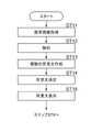
次いで、第1の実施形態において行われる処理について説明する。図10は第1の実施形態において行われる処理を示すフローチャートである。処理開始の指示が入力デバイス15から行われると、画像取得部21が、医用画像G0を画像サーバ5から取得する(ステップST1)。次いで、画像解析部22が、医用画像G0を解析することにより、少なくとも1つの所見を含む解析結果を取得する(ステップST2)。次いで、作成方法決定部23が、解析結果に基づいて医用画像G0に関する文書の作成方法を少なくとも1つ決定する(ステップST3)。
Next, the processing performed in the first embodiment will be described. FIG. 10 is a flowchart showing processing performed in the first embodiment. When an instruction to start processing is given from the input device 15, the image acquisition unit 21 acquires the medical image G0 from the image server 5 (step ST1). Next, the image analysis unit 22 acquires analysis results including at least one finding by analyzing the medical image G0 (step ST2). Next, the creation method determining unit 23 determines at least one document creation method for the medical image G0 based on the analysis result (step ST3).
そして、文書作成部24が決定された作成方法により少なくとも1つの所見文を作成し(ステップST4)、表示制御部25が所見文をディスプレイ14に表示する(ステップST5)。続いて、文書作成部24は、所見文の修正指示がなされたか否かを判定し(ステップST6)、ステップST6が肯定されると所見文を修正し(ステップST7)、ステップST6の処理に戻る。
Then, the document creation unit 24 creates at least one remark statement by the determined creation method (step ST4), and the display control unit 25 displays the remark statement on the display 14 (step ST5). Subsequently, the document creation unit 24 determines whether or not an instruction to correct the observation sentence is given (step ST6), and if step ST6 is affirmative, corrects the observation sentence (step ST7), and returns to the process of step ST6. .
ステップST6否定されると、文書作成部24は、確定ボタン46が選択されたか否かを判定し(ステップST8)、ステップST8が否定されるとステップST6に戻り、ステップST6以降の処理を繰り返す。ステップST8が肯定されると、読影レポートが作成され(ステップST9)、処理を終了する。作成された読影レポートは医用画像G0および検出結果と併せてストレージ13に保存され、さらに、作成された読影レポートが医用画像G0および検出結果と併せてレポートサーバ7に転送される。
If step ST6 is negative, the document creation unit 24 determines whether or not the enter button 46 has been selected (step ST8), and if step ST8 is negative, returns to step ST6, and repeats the processes after step ST6. If step ST8 is affirmative, an interpretation report is created (step ST9), and the process ends. The created interpretation report is stored in the storage 13 together with the medical image G0 and the detection results, and the created interpretation report is transferred to the report server 7 together with the medical image G0 and the detection results.
このように、本実施形態においては、医用画像G0についての解析結果という文書の出力を決定する判断用の情報に基づいて医用画像G0に関する所見文の作成方法を決定し、決定された作成方法により所見文を作成するようにした。このため、判断用の情報に応じて適応的に所見文の作成方法を決定することができる。例えば、判断用の情報である解析結果に含まれる所見項目の数に応じて、所見文の作成方法をテンプレートに決定したり、自然言語処理に決定したりすることができる。このため、所見文の作成方法として多数の所見項目に対応したテンプレートを用意する必要がなくなる。また、少数の所見項目に対応した自然言語処理を用意する必要もなくなる。したがって、本実施形態においては、医用画像G0に関する所見文を簡易な構成により作成することができる。
As described above, in the present embodiment, the method for creating the observation statement regarding the medical image G0 is determined based on the information for determining the output of the document, which is the analysis result of the medical image G0, and the determined creation method is used. I tried to create an opinion statement. Therefore, it is possible to adaptively determine the method of creating an observation sentence according to the information for judgment. For example, depending on the number of observation items included in the analysis result, which is the information for judgment, the method of creating the observation statement can be determined to be a template or natural language processing. Therefore, it is not necessary to prepare templates corresponding to a large number of observation items as a method of creating observation sentences. Also, there is no need to prepare natural language processing corresponding to a small number of finding items. Therefore, in the present embodiment, it is possible to create an observation statement regarding the medical image G0 with a simple configuration.
次いで、本開示の第2の実施形態について説明する。図11は第2の実施形態による文書作成装置の機能的な構成を示す図である。なお、図11において図3と同一の構成については同一の参照番号を付与し、詳細な説明は省略する。第2の実施形態による文書作成装置20Aは、第1の実施形態における文書作成装置20の作成方法決定部23に代えて、文書決定部26を備えた点が第1の実施形態と異なる。
Next, a second embodiment of the present disclosure will be described. FIG. 11 is a diagram showing the functional configuration of the document creation device according to the second embodiment. In FIG. 11, the same reference numerals are assigned to the same configurations as in FIG. 3, and detailed description thereof will be omitted. A document creation apparatus 20A according to the second embodiment differs from the first embodiment in that a document determination section 26 is provided in place of the creation method determination section 23 of the document creation apparatus 20 in the first embodiment.
第2の実施形態においては、文書作成部24は、自然言語処理NLP1~NLP3にそれぞれ対応する学習済みモデル24A~24Cを用いて、解析結果に含まれる少なくとも1つの所見を含む複数(ここでは3つ)の所見文を作成する。学習済みモデル24A~24Cは、第1の実施形態と同様に、それぞれ異なる自然言語処理NLP1~NLP3により所見文を作成するように学習がなされている。そして、第2の実施形態においては、文書決定部26が、判断用の情報に基づいて、複数の所見文のうちの少なくとも1つの所見文を出力する文書に決定する。第2の実施形態においては、判断用の情報として文書らしさを表す確信度を用いる。以下、確信度について説明する。
In the second embodiment, the document creation unit 24 uses trained models 24A to 24C corresponding to natural language processing NLP1 to NLP3, respectively, to create a plurality of (here, three (1) to create a statement of findings. As in the first embodiment, the trained models 24A-24C are trained to create observation sentences by different natural language processing NLP1-NLP3. Then, in the second embodiment, the document determination unit 26 determines a document to output at least one of the plurality of observation sentences based on the information for determination. In the second embodiment, the certainty factor representing document-likeness is used as information for determination. The degree of certainty will be described below.
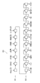
図12は、文書作成部24が有する学習済みモデル24A~24Cとして用いられるリカレントニューラルネットワークを模式的に示す図である。図12に示すようにリカレントニューラルネットワーク50は、エンコーダ51およびデコーダ52を有する。エンコーダ51は複数の入力層からなり、各入力層には解析結果に含まれる少なくとも1つの所見が入力される。図12においては、エンコーダ51には、「右上葉」、「形状」、「円形」、「境界」、「明瞭」、「吸収値」および「スリガラス型」の所見が入力される。デコーダ52は、文字情報を文章化するように学習がなされており、入力された所見の文字情報から所見文を作成する。具体的には、上述した「右上葉」、「形状」、「円形」、「境界」、「明瞭」、「吸収値」および「スリガラス型」の所見を表す文字情報から、「右上葉に円形で境界が明瞭なスリガラス型の吸収値を認めます。」の所見文を作成する。
FIG. 12 is a diagram schematically showing recurrent neural networks used as trained models 24A to 24C possessed by the document creation unit 24. As shown in FIG. As shown in FIG. 12, recurrent neural network 50 has encoder 51 and decoder 52 . The encoder 51 consists of a plurality of input layers, and at least one observation included in the analysis result is input to each input layer. In FIG. 12, the encoder 51 receives the findings of "upper right lobe", "shape", "circle", "boundary", "clear", "absorption value" and "ground glass type". The decoder 52 is trained to convert character information into sentences, and creates an opinion sentence from the input character information of an opinion. Specifically, from the character information representing the above findings of "upper right lobe", "shape", "circular", "boundary", "clear", "absorption value" and "ground glass type", "circular in upper right lobe A ground-glass type absorption value with a clear boundary is recognized.”
所見文を作成する際に、デコーダ52の各層からは、所見文に含まれるべき文字が、所見文に含まれる順に出力される。その際、各層においては、各層から出力することが可能な複数の文字についてのスコアが導出され、最も大きいスコアとなる文字がその層から出力される。例えば、「右上葉」および「に」を出力した層の次の層は、「円形」、「境界」、「明瞭」、「吸収値」および「スリガラス型」の複数の所見を表す文字を出力することが可能であるが、図12においては、これらの所見についてのスコアが導出された結果、「境界」のスコアが最大となったため、「境界」が出力された状態を示している。
When creating an observation sentence, the characters to be included in the observation sentence are output from each layer of the decoder 52 in the order in which they are included in the observation sentence. At that time, in each layer, scores are derived for a plurality of characters that can be output from each layer, and the character with the highest score is output from that layer. For example, the layer next to the layer that output "upper right lobe" and "ni" outputs characters representing multiple findings: "Circle", "Boundary", "Clear", "Absorption Value" and "Ground Glass". However, FIG. 12 shows a state in which "boundary" is output because the score for "boundary" is the highest as a result of deriving scores for these findings.
第2の実施形態においては、文書決定部26は、文書作成部24が作成した複数の所見文について、デコーダ52の複数の層のそれぞれが出力した所見を表す文字についてのスコアの代表値を文書らしさを表す確信度として導出する。代表値としては、各層から出力された文字について導出されたスコアの平均値、最大値、最小値および中間値等を用いることができる。そして、文書決定部26は作成された複数の所見文のうち、確信度が基準を満たす少なくとも1つの所見文を、出力する文書に決定する。基準としては、確信度が最大となる、確信度があらかじめ定められたしきい値以上となる、および確信度の上位n個等を用いることができる。
In the second embodiment, the document determining unit 26 determines the representative scores of the characters representing the findings output by the layers of the decoder 52 for the plurality of finding sentences created by the document creating unit 24. It is derived as a degree of certainty that represents the likelihood. As the representative value, the average value, maximum value, minimum value, intermediate value, etc. of the scores derived for the characters output from each layer can be used. Then, the document determination unit 26 determines, as a document to be output, at least one observation sentence whose certainty degree satisfies the criterion among the plurality of created observation sentences. As the criteria, the maximum certainty, the certainty equal to or higher than a predetermined threshold value, the top n certainties, and the like can be used.
例えば、文書作成部24が学習済みモデル24A~24Cにより、図9に示す候補1~候補3と同一の「左肺上葉に腫瘤が認められます。」、「左肺上葉に胸膜陥入を伴う、4.2cm大の腫瘤が認められます。」、および「左肺上葉に胸膜陥入を伴うスピキュラを有する腫瘤が認められます。」の3つの所見文を作成したとする。そして、3つの所見文の候補1~3のスコアがそれぞれ0.8.0.7.0.6であったとする。また、確信度の基準となるしきい値が0.7以上であったとする。文書決定部26は候補1~3のうち、スコアがしきい値0.7以上となる候補1,2を出力する文書に決定する。
For example, the document creation unit 24 uses the learned models 24A to 24C to determine the same candidates 1 to 3 shown in FIG. A mass of 4.2 cm in diameter accompanied by thorax is observed.” and “A mass with spicules accompanied by pleural invagination is observed in the upper lobe of the left lung.” Suppose that the scores of the three observation sentence candidates 1 to 3 are respectively 0.8.0.7.0.6. It is also assumed that the threshold, which is the criterion for confidence, is 0.7 or higher. The document determination unit 26 determines candidates 1 and 2 whose score is equal to or greater than the threshold value 0.7 among the candidates 1 to 3 as documents to be output.
なお、文書作成部24が作成した複数の所見文の流暢性を判断するように学習がなされた学習済みモデルを文書決定部26に設け、流暢性を文書らしさを表す確信度として用いて、出力する文書を決定するようにしてもよい。この場合、文書作成部24は、自然言語処理のみならず、テンプレートを用いて所見文を作成するようにしてもよい。
Note that a learned model that has been trained to determine the fluency of a plurality of remark sentences created by the document creation unit 24 is provided in the document determination unit 26, and the fluency is used as a certainty factor representing document-likeness, and output. You may make it determine the document to carry out. In this case, the document creation unit 24 may create the observation sentence using not only natural language processing but also a template.
図13は第2の実施形態における所見文の表示画面を示す図である。図13に示すように、第2の実施形態における表示画面40Bには、文書決定部26が出力する文書に決定した少なくとも1つ(図13において2つ)の所見文が所見文表示領域43Aに表示されている。また、2つの所見文のそれぞれに0.8,0.7の評価値が付与されている。
FIG. 13 is a diagram showing a display screen for remarks in the second embodiment. As shown in FIG. 13, on the display screen 40B in the second embodiment, at least one (two in FIG. 13) remarks determined for the document output by the document determination unit 26 are displayed in the remarks display area 43A. is displayed. Evaluation values of 0.8 and 0.7 are given to the two observation sentences, respectively.
読影医は表示画面40Bにおいて、表示された所見文に付与された評価値を参照しつつ、所見文表示領域43Aに表示された複数の所見文から所望とする所見文を選択することができる。なお、自由入力欄44への入力、および確定ボタン46が選択された場合等の処理は上記第1の実施形態と同一であるため、ここでは詳細な説明は省略する。
On the display screen 40B, the radiologist can refer to the evaluation values assigned to the displayed observation sentences and select a desired observation sentence from the multiple observation sentences displayed in the observation sentence display area 43A. Since the input to the free input field 44 and the processing when the confirm button 46 is selected are the same as those in the first embodiment, detailed description thereof will be omitted here.
次いで、第2の実施形態において行われる処理について説明する。図14は第2の実施形態において行われる処理を示すフローチャートである。処理開始の指示が入力デバイス15から行われると、画像取得部21が、医用画像G0を画像サーバ5から取得する(ステップST11)。次いで、画像解析部22が、医用画像G0を解析することにより、少なくとも1つの所見を含む解析結果を取得する(ステップST12)。次いで、文書作成部24が複数の所見文を作成する(ステップST13)。そして、文書決定部26が判断用の情報に基づいて複数の所見文のうちの少なくとも1つの所見文を出力する文書に決定し(ステップST14)、図10に示すステップST6の処理に進む。なお、ステップST6以降の処理は第1の実施形態と同一であるため、ここでは詳細な説明は省略する。
Next, the processing performed in the second embodiment will be described. FIG. 14 is a flow chart showing the processing performed in the second embodiment. When an instruction to start processing is given from the input device 15, the image acquisition unit 21 acquires the medical image G0 from the image server 5 (step ST11). Next, the image analysis unit 22 obtains an analysis result including at least one finding by analyzing the medical image G0 (step ST12). Next, the document creation section 24 creates a plurality of observation sentences (step ST13). Based on the information for determination, the document determination unit 26 determines a document to output at least one of the plurality of observation sentences (step ST14), and proceeds to the process of step ST6 shown in FIG. Since the processing after step ST6 is the same as in the first embodiment, detailed description is omitted here.
なお、上記第1の実施形態においては、文書の作成方法を決定するための判断用の情報として、解析結果に含まれる所見項目の数を用いているが、これに限定されるものではない。例えば所見項目の数に加えて、または所見項目の数に代えて、解析結果に含まれる所見のうちの陽性所見の数または陰性所見の数に基づいて、文書の作成方法を決定してもよい。
In the first embodiment, the number of finding items included in the analysis result is used as the judgment information for determining the document creation method, but it is not limited to this. For example, in addition to the number of finding items or instead of the number of finding items, the document preparation method may be determined based on the number of positive findings or the number of negative findings among the findings included in the analysis results. .
また、上記第1の実施形態においては、所見項目の数を判断用の情報として用いているが、これに限定されるものではない。例えば、医用画像G0において検出された異常陰影の領域の画像を判断用の情報として用いるようにしてもよい。この場合、例えば、各種医用画像から抽出された異常陰影の領域と、複数の所見または所見項目とを対応づけたデータベース(以下、管理DBとする)を用意し、このデータベースを参照することにより所見文の作成方法が決定される。
Also, in the above-described first embodiment, the number of finding items is used as information for judgment, but it is not limited to this. For example, an image of an abnormal shadow region detected in the medical image G0 may be used as information for determination. In this case, for example, a database (hereinafter referred to as a management DB) is prepared in which regions of abnormal shadows extracted from various medical images are associated with a plurality of findings or finding items. It determines how the sentence is constructed.
図15は管理DBの例を示す図である。図15に示すように、管理DB55には、複数の所見および所見項目と文書の作成方法とが対応づけられた対応情報が複数登録されている。各対応情報は、部位、疾患タイプ、疾患名、複数の疾患特徴、解剖レベル情報、サイズレベル情報および医用画像情報を含む。各対応情報においては、部位、疾患タイプまたは疾患名毎に疾患特徴、解剖レベル情報、サイズレベル情報、医用画像および作成方法が対応づけられている。
FIG. 15 is a diagram showing an example of the management DB. As shown in FIG. 15, the management DB 55 stores a plurality of correspondence information in which a plurality of observations and observation items are associated with document preparation methods. Each correspondence information includes site, disease type, disease name, multiple disease features, anatomical level information, size level information and medical image information. In each piece of correspondence information, disease features, anatomical level information, size level information, medical images, and creation methods are associated with each site, disease type, or disease name.
疾患特徴は、管理DB55を作成する際に、医用画像を解析することにより取得した所見である。なお、図15には1つの疾患タイプまたは疾患名に対して3つの疾患特徴を示しているが、疾患特徴の数は取得された所見の数に依存する。解剖レベル情報は異常陰影が存在する臓器の解剖学的区域のレベルを表す情報である。サイズレベル情報は、異常陰影のサイズの計測方向およびサイズの大小を表す情報である。
Disease characteristics are findings obtained by analyzing medical images when creating the management DB 55. Although FIG. 15 shows three disease features for one disease type or disease name, the number of disease features depends on the number of findings obtained. The anatomical level information is information representing the level of the anatomical area of the organ where the abnormal shadow exists. The size level information is information representing the measurement direction of the size of the abnormal shadow and the size of the size.
ここで、医用画像情報とは、各対応情報に登録される所見を取得した医用画像に含まれる異常陰影の領域についての局所画像である。図15においては医用画像の局所画像が登録されていることを、局所画像を表す四角形により示している。なお、図15に示すように、複数の医用画像情報が1つの対応情報に含まれる場合もある。
Here, the medical image information is a local image of an abnormal shadow area included in a medical image from which findings registered in each correspondence information are acquired. In FIG. 15, rectangles representing local images indicate that the local images of the medical image are registered. Note that, as shown in FIG. 15, a plurality of pieces of medical image information may be included in one piece of correspondence information.
管理DB55を使用するに際し、画像解析部22が医用画像G0から異常陰影の領域の局所画像を判断用の情報として抽出する。そして、作成方法決定部23が、管理DB55を参照することにより、異常陰影の局所画像と各対応情報に含まれる医用画像情報との類似度を導出する。そして、類似度が基準を満たす少なくとも1つの対応情報に登録された文書の作成方法を、対象とする医用画像G0についての所見文の作成方法に決定する。なお、この際の基準としては、類似度が最大となる、類似度があらかじめ定められたしきい値以上となる、および類似度の上位m個等を用いることができる。
When using the management DB 55, the image analysis unit 22 extracts the local image of the abnormal shadow area from the medical image G0 as information for judgment. Then, the creation method determination unit 23 refers to the management DB 55 to derive the degree of similarity between the local image of the abnormal shadow and the medical image information included in each correspondence information. Then, the method of creating a document registered in at least one piece of correspondence information whose similarity satisfies the criteria is determined as the method of creating an opinion statement for the target medical image G0. In this case, the maximum similarity, the similarity equal to or higher than a predetermined threshold value, the top m similarities, and the like can be used as the criteria.
そして、文書作成部24が、作成方法決定部23が決定した作成方法を用いて、少なくとも1つの所見文を作成する。この際、所見文に含める所見としては、類似度が基準を満たす少なくとも1つの対応情報に登録された所見または所見項目を用いるようにすればよい。
Then, the document creation unit 24 uses the creation method determined by the creation method determination unit 23 to create at least one remark sentence. At this time, as the findings to be included in the finding text, the findings or finding items registered in at least one piece of correspondence information whose similarity satisfies the standard may be used.
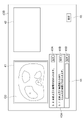
また、上記各実施形態においては、文書作成部24において、解析結果を含む所見文を作成しているが、これに限定されるものではない。解析結果についてのグラフィカルな表現を含む文書を作成してもよい。図16は解析結果についてのグラフィカルな表現を含む文書の例を示す図である。なお、ここでは、解析結果が「右肺上葉」および「肺結節」の所見を含むものとする。この場合、文書作成部24は、解析結果から肺のシェーマ60を作成方法として選択し、肺のシェーマ60における右肺上葉の位置にマーク61を付与した文書をグラフィカルな情報として生成してもよい。このようなグラフィカルな表現を含む文書を生成して表示することにより、異常部位の位置を容易に認識することが可能となる。
Also, in each of the above-described embodiments, the document creation unit 24 creates an observation sentence including the analysis result, but it is not limited to this. A document containing a graphical representation of the analysis results may be produced. FIG. 16 is a diagram showing an example of a document containing graphical representations of analysis results. Here, it is assumed that the analysis results include findings of "right upper lobe of lung" and "pulmonary nodule". In this case, the document creation unit 24 may select the lung schema 60 as the creation method from the analysis result, and generate a document as graphical information by adding a mark 61 to the position of the right upper lobe of the lung in the lung schema 60. good. By generating and displaying a document containing such graphical representations, it becomes possible to easily recognize the position of the abnormal site.
また、上記各実施形態においては、所見文の表示画面に表示された所見文と対応づけて、所見文の作成方法および判断用の情報の少なくとも一方を表示するようにしてもよい。例えば、図17に示すように、図9に対応する表示画面40Cの所見文表示領域43に、所見文の候補のそれぞれと対応づけて所見文の作成方法および判断用の情報の少なくとも一方を表示するようにしてもよい。図17においては、所見文の候補1に対して、「T2:所見項目数2」が対応づけられて表示されている。また、所見文の候補2に対して「NLP:肺、がん」が対応づけられて表示されている。また、所見文の候補3に対して「NLP」が対応づけられて表示されている。T2およびNLPは所見文の作成方法の一例であり、「所見項目数2」および「肺、がん」は判断用の情報の一例である。
Further, in each of the above embodiments, at least one of the method of creating the observation text and information for judgment may be displayed in association with the observation text displayed on the observation text display screen. For example, as shown in FIG. 17, in the observation text display area 43 of the display screen 40C corresponding to FIG. You may make it In FIG. 17 , “T2: number of observation items 2” is displayed in association with observation sentence candidate 1 . In addition, "NLP: lung, cancer" is displayed in association with the candidate 2 of the observation sentence. In addition, "NLP" is displayed in association with the observation sentence candidate 3. FIG. T2 and NLP are an example of a method of creating an observation statement, and "number of observation items 2" and "lung, cancer" are examples of information for judgment.
また、上記各実施形態においては、所見文の表示画面に表示された所見文を変更できるようにしてもよい。例えば、図18に示すように、図9に対応する表示画面40Dの所見文表示領域43に、所見文の候補のそれぞれと対応づけて変更ボタン47A~47Cを表示し、変更ボタン47A~47Cの選択により、所見文の候補を変更するようにしてもよい。この場合、変更される所見文の候補としては、例えば所見文の語尾を「認められます。」から「疑われます。」に変更した所見文の候補、所見文中に使用する所見を変更した所見文の候補、あるいは作成方法を変更した所見文の候補等を、優先順位をつけて候補1~候補3と対応づけて作成しておく。この場合、表示制御部25は、変更ボタン47A~47Cが選択されることにより、選択された候補に対する修正開始指示が受け付けられると、選択された候補について作成された候補を優先順位が高い順に、選択された候補に代えて表示するようにすればよい。
Further, in each of the above-described embodiments, the observation text displayed on the observation text display screen may be changed. For example, as shown in FIG. 18, change buttons 47A to 47C are displayed in association with each candidate of the observation text in the observation text display area 43 of the display screen 40D corresponding to FIG. The candidate of the observation sentence may be changed by selection. In this case, the candidate of the observation sentence to be changed includes, for example, the candidate of the observation sentence in which the ending of the observation sentence is changed from "perceived." to "suspicious." Candidates of sentences, or candidates of observation sentences whose creation method is changed, etc. are created in association with candidates 1 to 3 in accordance with the order of priority. In this case, when the modification start instruction for the selected candidate is received by selecting the change buttons 47A to 47C, the display control unit 25 selects the candidates created for the selected candidate in descending order of priority. It may be displayed instead of the selected candidate.
また、上記各実施形態において、医用画像G0を取得した患者と同一患者について、過去に同一部位の医用画像が取得され、読影レポートが作成されている場合がある。このような場合、作成方法決定部23は、過去の医用画像についての読影レポート作成の有無、あるいは読影レポートの内容を判断用の情報として用いて、文書の作成方法を決定するようにしてもよい。この場合、ストレージ13に図19に示すテーブル33を記憶しておく。ここで、過去の医用画像についての読影レポートが作成されている場合には、読影レポートの内容を新たに作成する読影レポートに取り込むことが好ましいため、作成する文書に記載すべき内容が多くなる。このため、過去の医用画像についての読影レポートが作成されている場合には、テーブル33を参照して、文書の作成方法をNLPに決定する。一方、読影レポートが作成されていない場合には、文書の作成方法をテンプレートT1に決定する。なお、過去の医用画像についての読影レポートが作成されている場合には、文書作成部24は、医用画像G0について画像解析部22が導出した解析結果に含まれる所見に加えて、過去の医用画像についての読影レポートに含まれている所見を用いて、NLPにより所見文を作成するようにすればよい。
In addition, in each of the above embodiments, there are cases where a medical image of the same site was acquired in the past and an interpretation report was created for the same patient as the patient who acquired the medical image G0. In such a case, the creation method determination unit 23 may determine the document creation method by using the presence or absence of creation of an interpretation report for past medical images or the content of the interpretation report as information for determination. . In this case, the table 33 shown in FIG. 19 is stored in the storage 13 . Here, when an interpretation report for past medical images has been created, it is preferable to include the contents of the interpretation report in a newly created interpretation report, so the amount of content to be described in the document to be created increases. Therefore, when an interpretation report for a past medical image has been created, the table 33 is referred to and NLP is determined as the document creation method. On the other hand, if the interpretation report has not been created, template T1 is determined as the document creation method. Note that when an interpretation report for a past medical image has been created, the document creation unit 24 adds the findings included in the analysis results derived by the image analysis unit 22 for the medical image G0 to the past medical image. Using the findings included in the interpretation report for , a statement of findings may be created by NLP.
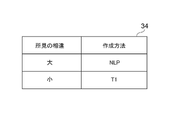
また、読影レポートの内容を判断用の情報として用いる場合、作成方法決定部23は、医用画像G0について画像解析部22が導出した解析結果に含まれる所見と、過去の医用画像についての読影レポートに含まれている所見とを比較する。そして、相違が大きい場合には、作成する文書に記載すべき内容が多くなるため、文書の作成方法をNLPに決定し、相違が小さい場合には文書の作成方法をテンプレートに決定すればよい。なお、相違が大きい場合とは、例えばサイズについての所見の相違があらかじめ定められたしきい値以上である場合、過去の読影レポートには含まれない所見が解析結果に含まれる場合、あるいは過去の読影レポートに含まれる形状を表す所見と解析結果に含まれる所見とが異なる場合等が挙げられる。なお、この場合、図20に示すテーブル34をストレージ13に記憶しておき、テーブル34を参照して、所見の相違の大小に応じて作成方法を決定するようにすればよい。
Further, when the content of the interpretation report is used as the information for determination, the creation method determination unit 23 combines the findings included in the analysis result derived by the image analysis unit 22 for the medical image G0 with the interpretation report for the past medical image. Compare with included findings. If the difference is large, the amount of content to be written in the document to be created increases. Therefore, NLP is determined as the document creation method, and if the difference is small, the template is determined as the document creation method. A large difference means, for example, that the size difference is greater than or equal to a predetermined threshold value, that the analysis results include findings that are not included in the past interpretation report, or that the past For example, the findings representing the shape included in the interpretation report are different from the findings included in the analysis results. In this case, the table 34 shown in FIG. 20 may be stored in the storage 13, and the table 34 may be referenced to determine the creation method according to the difference in findings.
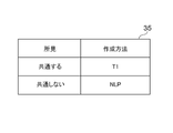
また、患者の診断を行う場合、同時に異なるモダリティにより複数種類の医用画像を取得する場合がある。例えば、CT装置およびMRI装置の双方によりCT画像およびMRI画像を医用画像として取得する場合がある。このような場合、作成方法決定部23は、CT画像の解析結果に含まれる所見とMRI画像の解析結果に含まれる所見とを比較し、比較結果に応じて文書の作成方法を決してもよい。例えば、所見が共通しない場合には、作成する文書に記載すべき内容が多くなるため、文書の作成方法をNLPに決定し、所見が共通する場合には、作成する文書に記載すべき内容がそれほど多くないため、文書の作成方法をT1に決定すればよい。なお、この場合、図21に示すテーブル35をストレージ13に記憶しておき、テーブル35を参照して、CT画像の所見とMRI画像の所見とが共通するか否かに応じて作成方法を決定するようにすればよい。
Also, when diagnosing a patient, multiple types of medical images may be acquired simultaneously using different modalities. For example, a CT image and an MRI image may be acquired as medical images by both a CT apparatus and an MRI apparatus. In such a case, the creation method determination unit 23 compares the findings included in the CT image analysis results and the findings included in the MRI image analysis results, and determines the document creation method according to the comparison results. For example, if there are no common findings, there will be more content to be written in the document to be prepared. Since there are not so many, the document creation method should be determined as T1. In this case, a table 35 shown in FIG. 21 is stored in the storage 13, and the table 35 is referenced to determine the creation method depending on whether or not the findings of the CT image and the findings of the MRI image are common. You should do it.
また、上記各実施形態においては、文書作成装置20が画像解析部22を備え、医用画像G0を解析することにより解析結果を取得しているが、これに限定されるものではない。操作者による入力デバイス15を用いた入力に基づいて解析結果を取得してもよい。
Also, in each of the above embodiments, the document creation device 20 includes the image analysis unit 22 and acquires the analysis result by analyzing the medical image G0, but the present invention is not limited to this. The analysis result may be acquired based on an input using the input device 15 by the operator.
また、上記各実施形態において、文書作成装置20における画像解析部22の処理を、例えばネットワーク10に接続された他の解析サーバ等の、外部装置で行うようにしてもよい。この場合、外部装置は、医用画像G0を画像サーバ5から取得し、医用画像G0を解析することにより解析結果を導出する。そして、文書作成装置20は、外部装置で導出された解析結果を用いて所見文を生成する。
Further, in each of the above embodiments, the processing of the image analysis unit 22 in the document creation device 20 may be performed by an external device such as another analysis server connected to the network 10, for example. In this case, the external device acquires the medical image G0 from the image server 5 and derives the analysis result by analyzing the medical image G0. Then, the document creation device 20 generates an observation sentence using the analysis result derived by the external device.
また、上記各実施形態においては、医療文書として読影レポートに記載する所見文を生成する場合に本開示の技術を適用しているが、これに限定されるものではない。例えば、電子カルテおよび診断レポート等の読影レポート以外の医療文書、並びにその他の画像に関する文字列を含む文書を作成する場合に、本開示の技術を適用してもよい。
Also, in each of the above-described embodiments, the technology of the present disclosure is applied when generating a finding sentence to be described in an interpretation report as a medical document, but it is not limited to this. For example, the technology of the present disclosure may be applied when creating medical documents other than interpretation reports such as electronic charts and diagnosis reports, and other documents including character strings related to images.
また、上記各実施形態においては、診断対象を肺とした医用画像G0を用いて各種処理を行っているが、診断対象は肺に限定されるものではない。肺の他に、心臓、肝臓、脳、および四肢等の人体の任意の部位を診断対象とすることができる。
Also, in each of the above-described embodiments, various processes are performed using the medical image G0 with the lung as the diagnostic target, but the diagnostic target is not limited to the lung. In addition to the lungs, any part of the human body such as the heart, liver, brain, and limbs can be diagnosed.
また、上記各実施形態においては、医用画像G0についての解析結果を含む文書を作成しているが、対象となる画像は医用画像G0に限定されるものではない。医用画像G0の他、写真画像を解析し、解析結果を含む文書を作成する場合にも、本実施形態を適用することができる。
Also, in each of the above embodiments, a document containing the analysis results of the medical image G0 is created, but the target image is not limited to the medical image G0. In addition to the medical image G0, the present embodiment can also be applied to the case of analyzing a photographic image and creating a document containing the analysis results.
また、上記実施形態において、例えば、画像取得部21、画像解析部22、作成方法決定部23、文書作成部24、表示制御部25および文書決定部26といった各種の処理を実行する処理部(Processing Unit)のハードウェア的な構造としては、次に示す各種のプロセッサ(Processor)を用いることができる。上記各種のプロセッサには、上述したように、ソフトウェア(プログラム)を実行して各種の処理部として機能する汎用的なプロセッサであるCPUに加えて、FPGA(Field Programmable Gate Array)等の製造後に回路構成を変更可能なプロセッサであるプログラマブルロジックデバイス(Programmable Logic Device :PLD)、ASIC(Application Specific Integrated Circuit)等の特定の処理を実行させるために専用に設計された回路構成を有するプロセッサである専用電気回路等が含まれる。
Further, in the above-described embodiment, for example, a processing unit (Processing As the hardware structure of Unit), various processors shown below can be used. As described above, the various processors include, in addition to the CPU, which is a general-purpose processor that executes software (programs) and functions as various processing units, circuits such as FPGAs (Field Programmable Gate Arrays), etc. Programmable Logic Device (PLD) which is a processor whose configuration can be changed, ASIC (Application Specific Integrated Circuit) etc. Circuits, etc. are included.
1つの処理部は、これらの各種のプロセッサのうちの1つで構成されてもよいし、同種または異種の2つ以上のプロセッサの組み合わせ(例えば、複数のFPGAの組み合わせまたはCPUとFPGAとの組み合わせ)で構成されてもよい。また、複数の処理部を1つのプロセッサで構成してもよい。複数の処理部を1つのプロセッサで構成する例としては、第1に、クライアントおよびサーバ等のコンピュータに代表されるように、1つ以上のCPUとソフトウェアとの組み合わせで1つのプロセッサを構成し、このプロセッサが複数の処理部として機能する形態がある。第2に、システムオンチップ(System On Chip:SoC)等に代表されるように、複数の処理部を含むシステム全体の機能を1つのIC(Integrated Circuit)チップで実現するプロセッサを使用する形態がある。このように、各種の処理部は、ハードウェア的な構造として、上記各種のプロセッサの1つ以上を用いて構成される。
One processing unit may be configured with one of these various processors, or a combination of two or more processors of the same or different type (for example, a combination of multiple FPGAs or a combination of a CPU and an FPGA). ). Also, a plurality of processing units may be configured by one processor. As an example of configuring a plurality of processing units in one processor, first, as represented by computers such as clients and servers, one processor is configured by combining one or more CPUs and software, There is a form in which this processor functions as a plurality of processing units. Second, as typified by System On Chip (SoC), etc., there is a form of using a processor that realizes the functions of the entire system including multiple processing units with a single IC (Integrated Circuit) chip. be. In this way, the various processing units are configured using one or more of the above various processors as a hardware structure.
さらに、これらの各種のプロセッサのハードウェア的な構造としては、より具体的には、半導体素子等の回路素子を組み合わせた電気回路(Circuitry)を用いることができる。
Furthermore, as the hardware structure of these various processors, more specifically, an electric circuit (circuitry) in which circuit elements such as semiconductor elements are combined can be used.