WO2021177312A1 - Device, method, and program for storing information, and device, method, and program for generating analysis records - Google Patents
Device, method, and program for storing information, and device, method, and program for generating analysis records Download PDFInfo
- Publication number
- WO2021177312A1 WO2021177312A1 PCT/JP2021/007977 JP2021007977W WO2021177312A1 WO 2021177312 A1 WO2021177312 A1 WO 2021177312A1 JP 2021007977 W JP2021007977 W JP 2021007977W WO 2021177312 A1 WO2021177312 A1 WO 2021177312A1
- Authority
- WO
- WIPO (PCT)
- Prior art keywords
- image
- property information
- analysis record
- information
- medical
- Prior art date
Links
- 238000004458 analytical method Methods 0.000 title claims description 111
- 238000000034 method Methods 0.000 title claims description 49
- 238000010191 image analysis Methods 0.000 claims abstract description 56
- 238000012937 correction Methods 0.000 claims abstract description 42
- 238000012986 modification Methods 0.000 claims description 4
- 230000004048 modification Effects 0.000 claims description 4
- 238000012217 deletion Methods 0.000 claims description 2
- 230000037430 deletion Effects 0.000 claims description 2
- 230000002159 abnormal effect Effects 0.000 description 41
- 230000008595 infiltration Effects 0.000 description 26
- 238000001764 infiltration Methods 0.000 description 26
- 230000008569 process Effects 0.000 description 21
- 238000012545 processing Methods 0.000 description 21
- 238000010586 diagram Methods 0.000 description 18
- 238000003384 imaging method Methods 0.000 description 14
- 238000012790 confirmation Methods 0.000 description 13
- 238000004891 communication Methods 0.000 description 12
- 230000001788 irregular Effects 0.000 description 12
- 210000000779 thoracic wall Anatomy 0.000 description 12
- 206010028980 Neoplasm Diseases 0.000 description 11
- 210000004224 pleura Anatomy 0.000 description 9
- 230000002685 pulmonary effect Effects 0.000 description 9
- 238000013528 artificial neural network Methods 0.000 description 8
- 238000002591 computed tomography Methods 0.000 description 8
- 210000004072 lung Anatomy 0.000 description 8
- 230000006870 function Effects 0.000 description 7
- 230000000306 recurrent effect Effects 0.000 description 7
- 238000003745 diagnosis Methods 0.000 description 6
- 238000002595 magnetic resonance imaging Methods 0.000 description 6
- 238000012546 transfer Methods 0.000 description 5
- 230000002308 calcification Effects 0.000 description 4
- 238000013527 convolutional neural network Methods 0.000 description 4
- 238000010521 absorption reaction Methods 0.000 description 3
- 238000004195 computer-aided diagnosis Methods 0.000 description 3
- 230000003902 lesion Effects 0.000 description 3
- 239000007787 solid Substances 0.000 description 3
- 238000012706 support-vector machine Methods 0.000 description 3
- 206010056342 Pulmonary mass Diseases 0.000 description 2
- 102100029860 Suppressor of tumorigenicity 20 protein Human genes 0.000 description 2
- 230000005540 biological transmission Effects 0.000 description 2
- 150000001875 compounds Chemical class 0.000 description 2
- 238000013135 deep learning Methods 0.000 description 2
- 238000009795 derivation Methods 0.000 description 2
- 238000002059 diagnostic imaging Methods 0.000 description 2
- 238000005516 engineering process Methods 0.000 description 2
- 238000010801 machine learning Methods 0.000 description 2
- 238000002600 positron emission tomography Methods 0.000 description 2
- 102100035353 Cyclin-dependent kinase 2-associated protein 1 Human genes 0.000 description 1
- 101001139126 Homo sapiens Krueppel-like factor 6 Proteins 0.000 description 1
- 101000585359 Homo sapiens Suppressor of tumorigenicity 20 protein Proteins 0.000 description 1
- 125000002066 L-histidyl group Chemical group [H]N1C([H])=NC(C([H])([H])[C@](C(=O)[*])([H])N([H])[H])=C1[H] 0.000 description 1
- 210000004556 brain Anatomy 0.000 description 1
- 239000002872 contrast media Substances 0.000 description 1
- 125000000753 cycloalkyl group Chemical group 0.000 description 1
- 230000005611 electricity Effects 0.000 description 1
- 210000003414 extremity Anatomy 0.000 description 1
- 125000000524 functional group Chemical group 0.000 description 1
- 210000002216 heart Anatomy 0.000 description 1
- 230000006872 improvement Effects 0.000 description 1
- 238000007689 inspection Methods 0.000 description 1
- 230000009545 invasion Effects 0.000 description 1
- 239000004973 liquid crystal related substance Substances 0.000 description 1
- 210000004185 liver Anatomy 0.000 description 1
- 238000004519 manufacturing process Methods 0.000 description 1
- 230000004044 response Effects 0.000 description 1
- 239000004065 semiconductor Substances 0.000 description 1
- 239000000126 substance Substances 0.000 description 1
- 238000012360 testing method Methods 0.000 description 1
Images


Classifications
-
- G—PHYSICS
- G06—COMPUTING; CALCULATING OR COUNTING
- G06F—ELECTRIC DIGITAL DATA PROCESSING
- G06F40/00—Handling natural language data
- G06F40/40—Processing or translation of natural language
-
- G—PHYSICS
- G06—COMPUTING; CALCULATING OR COUNTING
- G06F—ELECTRIC DIGITAL DATA PROCESSING
- G06F40/00—Handling natural language data
- G06F40/40—Processing or translation of natural language
- G06F40/55—Rule-based translation
- G06F40/56—Natural language generation
-
- A—HUMAN NECESSITIES
- A61—MEDICAL OR VETERINARY SCIENCE; HYGIENE
- A61B—DIAGNOSIS; SURGERY; IDENTIFICATION
- A61B6/00—Apparatus for radiation diagnosis, e.g. combined with radiation therapy equipment
- A61B6/02—Devices for diagnosis sequentially in different planes; Stereoscopic radiation diagnosis
- A61B6/03—Computerised tomographs
-
- G—PHYSICS
- G06—COMPUTING; CALCULATING OR COUNTING
- G06F—ELECTRIC DIGITAL DATA PROCESSING
- G06F40/00—Handling natural language data
- G06F40/10—Text processing
- G06F40/166—Editing, e.g. inserting or deleting
- G06F40/169—Annotation, e.g. comment data or footnotes
-
- G—PHYSICS
- G06—COMPUTING; CALCULATING OR COUNTING
- G06F—ELECTRIC DIGITAL DATA PROCESSING
- G06F40/00—Handling natural language data
- G06F40/40—Processing or translation of natural language
- G06F40/42—Data-driven translation
- G06F40/44—Statistical methods, e.g. probability models
-
- G—PHYSICS
- G06—COMPUTING; CALCULATING OR COUNTING
- G06T—IMAGE DATA PROCESSING OR GENERATION, IN GENERAL
- G06T11/00—2D [Two Dimensional] image generation
- G06T11/60—Editing figures and text; Combining figures or text
-
- G—PHYSICS
- G16—INFORMATION AND COMMUNICATION TECHNOLOGY [ICT] SPECIALLY ADAPTED FOR SPECIFIC APPLICATION FIELDS
- G16H—HEALTHCARE INFORMATICS, i.e. INFORMATION AND COMMUNICATION TECHNOLOGY [ICT] SPECIALLY ADAPTED FOR THE HANDLING OR PROCESSING OF MEDICAL OR HEALTHCARE DATA
- G16H10/00—ICT specially adapted for the handling or processing of patient-related medical or healthcare data
- G16H10/60—ICT specially adapted for the handling or processing of patient-related medical or healthcare data for patient-specific data, e.g. for electronic patient records
-
- G—PHYSICS
- G16—INFORMATION AND COMMUNICATION TECHNOLOGY [ICT] SPECIALLY ADAPTED FOR SPECIFIC APPLICATION FIELDS
- G16H—HEALTHCARE INFORMATICS, i.e. INFORMATION AND COMMUNICATION TECHNOLOGY [ICT] SPECIALLY ADAPTED FOR THE HANDLING OR PROCESSING OF MEDICAL OR HEALTHCARE DATA
- G16H15/00—ICT specially adapted for medical reports, e.g. generation or transmission thereof
-
- G—PHYSICS
- G16—INFORMATION AND COMMUNICATION TECHNOLOGY [ICT] SPECIALLY ADAPTED FOR SPECIFIC APPLICATION FIELDS
- G16H—HEALTHCARE INFORMATICS, i.e. INFORMATION AND COMMUNICATION TECHNOLOGY [ICT] SPECIALLY ADAPTED FOR THE HANDLING OR PROCESSING OF MEDICAL OR HEALTHCARE DATA
- G16H30/00—ICT specially adapted for the handling or processing of medical images
- G16H30/40—ICT specially adapted for the handling or processing of medical images for processing medical images, e.g. editing
-
- G—PHYSICS
- G16—INFORMATION AND COMMUNICATION TECHNOLOGY [ICT] SPECIALLY ADAPTED FOR SPECIFIC APPLICATION FIELDS
- G16H—HEALTHCARE INFORMATICS, i.e. INFORMATION AND COMMUNICATION TECHNOLOGY [ICT] SPECIALLY ADAPTED FOR THE HANDLING OR PROCESSING OF MEDICAL OR HEALTHCARE DATA
- G16H50/00—ICT specially adapted for medical diagnosis, medical simulation or medical data mining; ICT specially adapted for detecting, monitoring or modelling epidemics or pandemics
- G16H50/20—ICT specially adapted for medical diagnosis, medical simulation or medical data mining; ICT specially adapted for detecting, monitoring or modelling epidemics or pandemics for computer-aided diagnosis, e.g. based on medical expert systems
-
- G—PHYSICS
- G06—COMPUTING; CALCULATING OR COUNTING
- G06T—IMAGE DATA PROCESSING OR GENERATION, IN GENERAL
- G06T2200/00—Indexing scheme for image data processing or generation, in general
- G06T2200/24—Indexing scheme for image data processing or generation, in general involving graphical user interfaces [GUIs]
-
- G—PHYSICS
- G06—COMPUTING; CALCULATING OR COUNTING
- G06T—IMAGE DATA PROCESSING OR GENERATION, IN GENERAL
- G06T2211/00—Image generation
- G06T2211/40—Computed tomography
- G06T2211/441—AI-based methods, deep learning or artificial neural networks
Definitions
- This disclosure relates to an information storage device, a method and a program, and an analysis record generation device, a method and a program.
- CT Computer Tomography
- MRI Magnetic Resonance Imaging
- medical images are analyzed by CAD (Computer-Aided Diagnosis) using a learning model that has been machine-learned by deep learning, etc., and the shape, density, and position of structures of interest such as abnormal shadow candidates included in the medical images. It is also practiced to discriminate properties such as size and size, and obtain these as analysis results.
- the analysis result acquired by CAD is associated with the test information such as the patient name, gender, age, and the modality from which the medical image was acquired, and is stored in the database.
- the medical image and the analysis result are transmitted to the terminal of the image interpreting doctor who interprets the medical image.
- the image interpreting doctor interprets the medical image by referring to the transmitted medical image and the analysis result on his / her terminal, and creates an image interpretation report.
- JP-A-2019-153250 a learning model in which machine learning such as a recurrent neural network is trained so as to generate a sentence from characters representing input property information is used.
- Medical texts (hereinafter referred to as medical texts) are created.
- medical texts By automatically generating medical texts as in the method described in JP-A-2019-153250, it is possible to reduce the burden on the interpretation doctor when creating a medical document such as an interpretation report.
- the automatically generated interpretation report may be modified by the interpretation doctor.
- the past medical image interpretation report is often referred to. Therefore, a method of extracting the corrected part of the corrected image interpretation report (see Japanese Patent Application Laid-Open No. 2011-125402) and a method of extracting the difference between the past medical image interpretation report and the latest medical image interpretation report (see Japanese Patent Application Laid-Open No. 2011-125402). JP-A-2007-122679) has been proposed.
- a learning model that generates an image analysis record such as a sentence from an image
- a learning model that generates an interpretation report from a medical image there is a user's preference for the content and expression of the analysis record, and that preference. It is desired to build a learning model that reflects the above.
- the user's preference for example, regarding the property information analyzed from the image, which property information should be reflected in the final analysis record and the like can be mentioned.
- the generated image analysis record may not match the user's preference. In such a case, the user needs to modify the generated image analysis record.
- the image analysis record is a sentence
- the sentences before and after the modification are compared using the methods described in JP-A-2011-125402 and JP-A-2007-122679, any part of the sentence can be compared. Can be recognized as having been modified.
- the present disclosure has been made in view of the above circumstances, and when the image analysis record generated from the image is modified, it is possible to recognize which property information derived by analyzing the image is modified.
- the purpose is.
- the information storage device includes at least one processor.
- the processor derives a plurality of property information representing the properties of the structure of interest contained in the image.
- Generate an image analysis record that contains at least a portion of multiple property information Accepts user corrections to property information It is configured to distinguish and store the derived property information and the modified property information.
- the processor may be configured to display the image analysis record on the display.
- the processor is configured to accept at least one of the deletion of the property information included in the displayed image analysis record and the addition of the property information not included in the image analysis record as a modification. It may be what is done.
- the processor displays all or a part of the derived property information on the display. It may be configured to accept corrections based on the user's selection of the displayed property information.
- the information storage device may further include a learning model that has been trained to output an image analysis record when property information is input.
- the processor may generate a sentence including at least a part of the property information as an image analysis record.
- the image may be a medical image
- the text may be a medical text relating to the structure of interest included in the medical image.
- the analysis record generator comprises at least one processor.
- the processor derives a plurality of property information representing the properties of the structure of interest contained in the target image to be analyzed. It is configured to generate a target image analysis record containing at least a part of the property information by referring to the information stored by the information storage device according to the present disclosure.
- the processor identifies stored information including property information that matches the property information derived from the target image, and performs image analysis associated with the specified stored information.
- the recording may be generated as a target image analysis record.
- the processor is further configured to generate other target image analysis records including at least a part of the derived property information without reference to the stored information. It may be one.
- Unreferenced ... Generate another target image analysis record means to generate another target image analysis record without referring to the saved information.
- the processor may be configured to display the target image analysis record and other target image analysis records on the display.
- the processor may be configured to accept the selection of either the displayed target image analysis record or another target image analysis record.
- the processor may be configured to generate a sentence including at least a part of the property information as a target image analysis record.
- the image may be a medical image
- the text may be a medical text relating to a structure of interest included in the medical image.
- the information storage method derives a plurality of property information representing the properties of the structure of interest contained in the image by analyzing the image. Generate an image analysis record that contains at least a portion of multiple property information Accepts user corrections to property information The derived property information and the modified property information are stored separately.
- the analysis record generation method derives a plurality of property information representing the properties of the structure of interest contained in the target image to be analyzed. With reference to the information stored by the information storage device according to the present disclosure, a target image analysis record including at least a part of the property information is generated.
- the information storage method and the analysis record generation method according to the present disclosure may be provided as a program for executing the computer.
- the image analysis record generated from the image is modified, it is possible to recognize which property information derived by analyzing the image is modified.
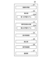
- Functional configuration diagram of the information storage device and analysis record generation device according to this embodiment Diagram showing an example of teacher data for learning the first learning model Diagram for explaining the property information derived by the analysis department The figure which shows the schematic structure of the recurrent neural network Diagram showing an example of a medical text display screen Diagram showing an example of a display screen for modified medical text Diagram for explaining saved information showing the saved result of property information Diagram showing an example of the target medical text and the display screen of another plan Flowchart showing information storage processing performed in this embodiment Flowchart of analysis record generation processing performed in this embodiment Diagram showing an example of a medical text and collation result display screen
- FIG. 1 is a diagram showing a schematic configuration of the medical information system 1.
- the medical information system 1 shown in FIG. 1 is based on an inspection order from a doctor in a clinical department using a known ordering system, photographs of a part to be inspected of a subject, storage of a medical image acquired by the imaging, and an image interpreter. It is a system for interpreting medical images and creating an interpretation report, and for viewing the interpretation report by the doctor of the requesting clinical department and observing the details of the medical image to be interpreted.
- the medical information system 1 includes a plurality of imaging devices 2, a plurality of image interpretation WS (WorkStation) 3 which are image interpretation terminals, a medical care WS 4, an image server 5, and an image database (hereinafter, image DB (DataBase)).
- the report server 7 and the report database (hereinafter referred to as the report DB) 8 are connected to each other via a wired or wireless network 10 so as to be able to communicate with each other.
- Each device is a computer on which an application program for functioning as a component of the medical information system 1 is installed.
- the application program is recorded and distributed on a recording medium such as a DVD (Digital Versatile Disc) and a CD-ROM (Compact Disc Read Only Memory), and is installed on a computer from the recording medium.
- a recording medium such as a DVD (Digital Versatile Disc) and a CD-ROM (Compact Disc Read Only Memory)
- it is stored in the storage device of the server computer connected to the network 10 or in the network storage in a state of being accessible from the outside, and is downloaded and installed in the computer upon request.
- the photographing device 2 is a device (modality) that generates a medical image representing the diagnosis target part by photographing the part to be diagnosed of the subject. Specifically, it is a simple X-ray imaging apparatus, a CT apparatus, an MRI apparatus, a PET (Positron Emission Tomography) apparatus, and the like.
- the medical image generated by the imaging device 2 is transmitted to the image server 5 and stored in the image DB 6.
- the image interpretation WS3 is a computer used by, for example, an image interpretation doctor in a radiology department to interpret a medical image and create an image interpretation report, and is represented by an information storage device and an analysis record generation device (hereinafter referred to as an information storage device) according to the present embodiment. ) 20 is included.
- an information storage device an analysis record generation device (hereinafter referred to as an information storage device) according to the present embodiment. ) 20 is included.
- an information storage device an analysis record generation device
- the medical care WS4 is a computer used by doctors in the clinical department for detailed observation of images, viewing of interpretation reports, creation of electronic medical records, etc., and is a processing device, a display device such as a display, and an input device such as a keyboard and a mouse. Consists of.
- an image viewing request is made to the image server 5
- an image received from the image server 5 is displayed
- an image interpretation report viewing request is made to the report server 7
- an image interpretation report received from the report server 7 is displayed.
- the image server 5 is a general-purpose computer in which a software program that provides a database management system (DataBase Management System: DBMS) function is installed. Further, the image server 5 includes a storage in which the image DB 6 is configured. This storage may be a hard disk device connected by the image server 5 and the data bus, or a disk device connected to NAS (Network Attached Storage) and SAN (Storage Area Network) connected to the network 10. It may be.
- NAS Network Attached Storage
- SAN Storage Area Network
- the image data and incidental information of the medical image acquired by the imaging device 2 are registered in the image DB 6.
- the incidental information includes, for example, an image ID (identification) for identifying an individual medical image, a patient ID for identifying a subject, an examination ID for identifying an examination, and a unique ID assigned to each medical image ( UID: unique identification), examination date when the medical image was generated, examination time, type of imaging device used in the examination to acquire the medical image, patient information such as patient name, age, gender, examination site (imaging) Includes information such as site), imaging information (imaging protocol, imaging sequence, imaging method, imaging conditions, use of contrast medium, etc.), series number or collection number when multiple medical images are acquired in one examination. ..
- the image server 5 when the image server 5 receives the viewing request from the image interpretation WS3 and the medical examination WS4 via the network 10, the image server 5 searches for the medical image registered in the image DB 6, and uses the searched medical image as the requesting image interpretation WS3 and the medical examination. Send to WS4.
- the report server 7 incorporates a software program that provides the functions of a database management system to a general-purpose computer.
- the report server 7 receives the image interpretation report registration request from the image interpretation WS3, the report server 7 prepares the image interpretation report in a database format and registers the image interpretation report in the report DB 8.
- the image interpretation report includes, for example, a medical image to be interpreted, an image ID for identifying the medical image, an image interpretation doctor ID for identifying the image interpretation doctor who performed the image interpretation, a lesion name, a lesion position information, and a medical image including a specific area. It may include information for access and information such as property information.
- the report server 7 when the report server 7 receives a viewing request for the interpretation report from the interpretation WS3 and the medical treatment WS4 via the network 10, the report server 7 searches for the interpretation report registered in the report DB 8 and uses the searched interpretation report as the requester's interpretation. It is transmitted to WS3 and medical treatment WS4.
- the medical image is a three-dimensional CT image composed of a plurality of tomographic images with the diagnosis target as the lung, and the CT image is interpreted by the interpretation WS3 to obtain an abnormal shadow contained in the lung.
- An interpretation report shall be prepared as a medical document.
- the medical image is not limited to the CT image, and any medical image such as an MRI image and a two-dimensional image acquired by a simple X-ray imaging device can be used.
- Network 10 is a wired or wireless local area network that connects various devices in the hospital.
- the network 10 may be configured such that the local area networks of each hospital are connected to each other by the Internet or a dedicated line.
- FIG. 2 describes the hardware configuration of the information storage device and the analysis record generation device according to the present embodiment.
- the information storage device 20 represents the information storage device and the analysis record generation device.
- the information storage device 20 includes a CPU (Central Processing Unit) 11, a non-volatile storage 13, and a memory 16 as a temporary storage area.
- the information storage device 20 includes a display 14 such as a liquid crystal display, an input device 15 such as a keyboard and a mouse, and a network I / F (InterFace) 17 connected to the network 10.
- the CPU 11, the storage 13, the display 14, the input device 15, the memory 16, and the network I / F 17 are connected to the bus 18.
- the CPU 11 is an example of the processor in the present disclosure.
- the storage 13 is realized by an HDD (Hard Disk Drive), an SSD (Solid State Drive), a flash memory, or the like.
- the information storage program 12A and the analysis record generation program 12B are stored in the storage 13 as a storage medium.
- the CPU 11 reads the information storage program 12A and the analysis record generation program 12B from the storage 13 and then expands the information storage program 12A and the analysis record generation program 12B into the memory 16 to execute the expanded information storage program 12A and the analysis record generation program 12B.
- FIG. 3 is a diagram showing a functional configuration of the information storage device and the analysis record generation device according to the present embodiment.
- the information storage device (and analysis record generation device) 20 includes an image acquisition unit 21, an analysis unit 22, an analysis record generation unit 23, a display control unit 24, a correction unit 25, a storage control unit 26, and communication.
- a unit 27 is provided.
- the CPU 11 executes the information storage program 12A and the analysis record generation program 12B, the CPU 11 has the image acquisition unit 21, the analysis unit 22, the analysis record generation unit 23, the display control unit 24, the correction unit 25, and the storage control. It functions as a unit 26 and a communication unit 27.
- the image acquisition unit 21, the analysis unit 22, the analysis record generation unit 23, the display control unit 24, and the communication unit 27 have a common configuration in the information storage program 12A and the analysis record generation program 12B.
- the image acquisition unit 21 acquires a medical image for creating an image interpretation report from the image server 5 in response to an instruction from the input device 15 by the image interpretation doctor who is the operator.
- the medical image also includes a target medical image to be analyzed, which will be described later.
- the analysis unit 22 analyzes the medical image to derive property information representing the properties of the structure of interest such as an abnormal shadow candidate included in the medical image.
- the analysis unit 22 has a first learning model 22A in which machine learning is performed so as to discriminate the abnormal shadow candidates in the medical image and discriminate the properties of the discriminated abnormal shadow candidates.
- the first learning model 22A determines whether or not each pixel (voxel) in the medical image represents an abnormal shadow candidate, and if it is an abnormal shadow candidate, the property thereof is determined. It consists of a convolutional neural network (CNN (Convolutional Neural Network)) in which deep learning is performed using teacher data so as to discriminate.
- CNN Convolutional Neural Network
- FIG. 4 is a diagram showing an example of teacher data for learning the first learning model.
- the teacher data 30 includes a medical image 32 including the abnormal shadow 31 and property information 33 about the abnormal shadow.
- the abnormal shadow 31 is a lung nodule
- the property information 33 represents a plurality of properties of the lung nodule.
- the property information 33 includes the location of the abnormal shadow, the size of the abnormal shadow, the shape of the boundary (clear and irregular), the type of absorption value (solid and suriglass type), the presence or absence of spicula, the mass or nodule, and the pleura.
- the presence or absence of contact, the presence or absence of pleural invasion, the presence or absence of pleural infiltration, the presence or absence of cavities, the presence or absence of calcification, etc. are used.
- the property information 33 shows that the location of the abnormal shadow is under the left pulmonary pleura, the size of the abnormal shadow is 4.2 cm in diameter, and the boundary is defined as shown in FIG. Irregular shape, full absorption, with spicula, mass, with pleural contact, with pleural infiltration, no pleural infiltration, no cavities, and no calcification.
- + is given when “yes” and ⁇ is given when there is no.
- the first learning model 22A is constructed by learning a neural network using a large number of teacher data as shown in FIG. For example, by using the teacher data 30 shown in FIG. 4, the first learning model 22A determines the abnormal shadow 31 included in the medical image 32 when the medical image 32 shown in FIG. 4 is input, and determines the abnormal shadow 31. With respect to 31, learning is performed so as to output the property information 33 shown in FIG.
- any learning model such as a support vector machine (SVM (Support Vector Machine)) can be used.
- SVM Support Vector Machine
- FIG. 5 is a diagram for explaining the property information derived by the analysis unit 22.
- the property information 35 derived by the analysis unit 22 is "left pulmonary subpleural", “4.2 cm”, “irregular”, “enriched”, “no spicula”, “tumor”, “mass”. It shall be "with pleural contact”, “with pleural invagination”, “without pleural infiltration”, “without cavities” and “without calcification”.
- the analysis record generation unit 23 generates an image analysis record using the property information derived by the analysis unit 22.
- the medical text is generated as an image analysis record.
- the analysis record generation unit 23 includes a second learning model 23A that has been trained to generate a sentence from the input information.
- a recurrent neural network can be used as the second learning model 23A.
- FIG. 6 is a diagram showing a schematic configuration of a recurrent neural network. As shown in FIG. 6, the recurrent neural network 40 includes an encoder 41 and a decoder 42. The property information derived by the analysis unit 22 is input to the encoder 41.
- property information of "left pulmonary subpleuralis”, “4.2 cm”, “Spicula +” and “mass” is input to the encoder 41.
- the decoder 42 is learned so as to document the character information, and generates a sentence from the input property information. Specifically, from the above-mentioned property information of "left pulmonary subpleura”, “4.2 cm”, “spicula +” and “mass”, "a 4.2 cm diameter mass having spicula under the left pulmonary pleura is recognized. Will be generated. " In FIG. 6, "EOS” indicates the end of the sentence (End Of Sentence).
- the recurrent neural network 40 learns the encoder 41 and the decoder 42 using a large amount of teacher data composed of a combination of the property information and the medical text. Be built.
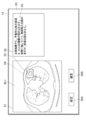
- FIG. 7 is a diagram showing an example of a medical text display screen according to the present embodiment.
- the display screen 50 includes an image display area 51 and a text display area 52.
- the slice image SL1 that is most likely to identify the abnormal shadow candidate detected by the analysis unit 22 is displayed.
- the slice image SL1 includes an abnormal shadow candidate 53, and the abnormal shadow candidate 53 is surrounded by a rectangular region 54.
- the medical text 55 generated by the analysis record generation unit 23 is displayed in the text display area 52.
- Medical text 55 states, "Under the left lung pleura, an irregular tumor with a maximum lateral diameter of 4.2 cm is found. It is in contact with the chest wall, and pleural infiltration is observed, but infiltration is not observed.” be.
- the property information used in the medical text 55 is "left pulmonary subpleural”, “irregular”, “4.2 cm”, “tumor”, and “chest wall” among the property information derived by the analysis unit 22. "With contact”, “With pleural infiltration” and “Without pleural infiltration”.
- a correction button 58A and a confirmation button 58B are displayed below the image display area 51.
- the image interpreting doctor interprets the abnormal shadow candidate 53 in the slice image SL1 displayed in the image display area 51, and determines the suitability of the medical sentence 55 displayed in the sentence display area 52.
- the correction unit 25 receives corrections by the image interpreting doctor for the property information. That is, the medical text 55 displayed in the text display area 52 can be manually corrected by input from the input device 15. Further, by selecting the confirmation button 58B, the medical sentence 55 displayed in the sentence display area 52 can be confirmed with its contents. In this case, the medical text 55 is transcribed into the interpretation report, and the interpretation report to which the medical text 55 is transcribed is transmitted to the report server 7 together with the slice image SL1 and stored.
- the interpreter When the interpreter selects the correction button 58A to correct the medical sentence 55, the interpreter is included in the abnormal shadow 31, but if there is a missing property in the medical sentence 55, the missing property is added. Modify medical text 55 to do so.
- the interpreter inputs the missing properties using the input device 15.
- the interpreting doctor inputs the property information of "Spicula” using the input device 15.
- the medical text 55 is modified so that the correction unit 25 adds the property information of "Spicula".
- the image interpreter modifies the medical text 55 to remove unwanted properties. For example, in the present embodiment, when the property of being in contact with the chest wall is unnecessary, the image interpreter deletes the property information of "being in contact with the chest wall" using the input device 15. As a result, the medical sentence 55 is modified so that the correction unit 25 deletes the property information "in contact with the chest wall".
- FIG. 8 is a diagram showing an example of a modified medical text display screen. As shown in FIG. 8, in the sentence display area 72, the modified medical sentence 59 obtained by modifying the medical sentence 55 is displayed.
- the modified medical text 59 states, "Under the left pleura, a tumor with an irregular shape and a maximum lateral diameter of 4.2 cm is observed. Pleural infiltration is observed, but infiltration is not observed.”
- the medical sentence 55 displayed in the sentence display area 52 can be confirmed with the contents.
- the medical text 55 is transcribed into the interpretation report, and the interpretation report to which the medical text 55 is transcribed is transmitted to the report server 7 by the communication unit 27 together with the slice image SL1 and stored.
- the interpretation doctor selects the confirmation button 58B after the correction, the correction medical sentence 59 can be confirmed with the content thereof.
- the modified medical text 59 is transcribed in the interpretation report, and the interpretation report to which the modified medical text 59 is transcribed is transmitted to the report server 7 by the communication unit 27 together with the slice image SL1 and the stored information 45 described later. Will be stored.
- the interpretation report and the stored information 45 are associated and stored.
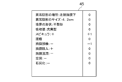
- FIG. 9 is a diagram for explaining the stored information showing the stored result of the property information.
- the stored information 45 is corrected from "without spicula” to "with spicula” and “with chest wall contact” to “without chest wall contact” in the property information derived by the analysis unit 22 shown in FIG. It has been done.
- the property information corrected from "none” to "yes” has a +1 flag
- the property information corrected from "yes” to "no” has a -1 flag, but there is no correction.
- a flag of 0 is added to each of the property information.
- the stored information 45 the property information derived by the analysis unit 22 and the modified property information can be distinguished by a flag.
- the stored information 45 stored in the storage 13 is transmitted to the report server 7 together with the interpretation report and stored as described above.
- the analysis unit 22 analyzes the target medical image to characterize the target medical image. Derive information. Further, the analysis record generation unit 23 generates the target medical text from the property information of the target medical image with the stored information 45 stored in the report server 7 as a non-reference, that is, without referring to the stored information 45. Specifically, the analysis record generation unit 23 generates a target medical sentence by using only the property information derived by the analysis unit 22. The target medical text without reference to the stored information 45 corresponds to the other target image analysis records of the present disclosure. Further, the analysis record generation unit 23 refers to the stored information 45 stored in the report server 7 and generates a target medical sentence as an alternative from the property information of the target medical image.
- the analysis record generation unit 23 identifies the stored information 45 including the property information that matches the property information of the target medical image derived by the analysis unit 22 by searching the report server 7.
- the property information and the stored information 45 include the location and size of the abnormal shadow, but specify the stored information 45 in which the property items excluding the location and size of the abnormal shadow match. For example, when the property information of the target medical image is "tumor", "pleural infiltration” and “no infiltration", the analysis record generation unit 23 determines "tumor", "pleural infiltration” and "no infiltration”.
- the stored information 45 including the property information of "" is searched. Then, the analysis record generation unit 23 acquires an interpretation report associated with the stored information 45 that matches the property information of the target medical image from the report server 7.
- the fact that the property information matches is not limited to the case where all the property information except for the location and size of the abnormal shadow among the plurality of property information matches, and the majority of the plurality of property information, for example, a plurality. It also includes the case where 80% or more and even 90% or more of the property information of the above matches.
- the stored information 45 that match the property information of the target medical image can be selected according to the criteria such as the one with a new creation date or the one created by the interpretation doctor who is operating the interpretation WS3.
- the number of interpretation reports to be acquired may be limited to search for stored information that matches the property information.
- the analysis record generation unit 23 rewrites the location and size of the abnormal shadow in the acquired interpretation report to the location and size of the abnormal shadow included in the property information of the target medical image, and generates another plan.
- the stored information 45 that matches the property information of the target medical image is not stored in the report server 7. In such a case, no alternative plan shall be generated in this embodiment.
- FIG. 10 is a diagram showing a target medical text and an alternative display screen. Note that FIG. 10 shows only one alternative. As shown in FIG. 10, the display screen 70 includes an image display area 71 and a text display area 72. In the image display area 71, the slice image SL2, which is the easiest to identify the abnormal shadow candidate detected by the analysis unit 22 from the target medical image, is displayed. The slice image SL2 includes an abnormal shadow candidate 73, and the abnormal shadow candidate 73 is surrounded by a rectangular region 74.
- the target medical text 75 and the alternative plan 76 generated by the analysis record generation unit 23 are displayed.
- the target medical text 75 is "Under the left pulmonary pleura, a tumor with an irregular shape and a maximum lateral diameter of 4.2 cm is observed. Pleural infiltration is observed, but infiltration is not observed.”
- Alternative 76 says, "Under the left lung pleura, an irregular tumor with a maximum lateral diameter of 4.2 cm is found. It is in contact with the chest wall, and pleural infiltration is observed, but infiltration is not observed.” be.
- a correction button 78A and a confirmation button 78B are displayed below the image display area 71.
- the image interpreting doctor interprets the abnormal shadow candidate 73 in the slice image SL2 displayed in the image display area 71, and determines the suitability of the target medical sentence 75 and the alternative plan 76 displayed in the sentence display area 72.
- the correction button 78A is selected using the input device 15.
- the medical text 75 displayed in the text display area 72 can be corrected in the same manner as described above by inputting from the input device 15.
- the interpreter when the interpreter adopts either the target medical text 75 or the alternative 76, the interpreter selects either the target medical text 75 or the alternative 76 using the input device 15 and presses the confirm button. By selecting 78B, either the target medical text 75 or the alternative 76 can be determined by its contents. In this case, any of the selected target medical text 75 and the alternative 76 is transcribed in the interpretation report, and the interpretation report to which the text is transcribed is transmitted to the report server 7 together with the slice image SL2 and stored.
- the communication unit 27 exchanges information between the information storage device 20 and the external device via the network I / F17.
- FIG. 11 is a flowchart of the information storage process performed in the present embodiment. It is assumed that the medical image to be read is acquired from the image server 5 by the image acquisition unit 21 and stored in the storage 13. The process is started when an instruction to create an image interpretation report is given by the image interpretation doctor, and the analysis unit 22 analyzes the medical image to represent the properties of the structure of interest such as an abnormal shadow candidate included in the medical image. Derivation of information (step ST1). Next, the analysis record generation unit 23 generates a medical sentence related to the medical image as an image analysis record based on the property information (step ST2). Subsequently, the display control unit 24 displays the medical text generated by the analysis record generation unit 23 on the text display area 52 of the display screen 50 displayed on the display 14 (step ST3).
- step ST4 determines whether or not the correction button 58A displayed on the display screen 50 is selected.
- step ST4 affirmed, the correction unit 25 accepts correction using the input device 15 for the property information included in the medical text displayed in the text display area 52 (step ST5).
- step ST6 determines whether or not the confirmation button 58B has been selected (step ST6). If step ST6 is denied, the process returns to step ST5 and the correction is continuously accepted.
- step ST6 is affirmed, the storage control unit 26 distinguishes between the derived property information and the modified property information and stores the modified property information in the storage 13 (step ST7).
- the display control unit 24 transfers the corrected medical text to the interpretation report, and the communication unit 27 transmits the interpretation report to which the corrected medical text is transcribed to the report server 7 together with the slice image SL1 (transmission of the interpretation report: Step ST8), the process is terminated.
- step ST4 determines whether or not the confirmation button 58B has been selected (step ST9). If step ST9 is denied, the process returns to step ST4. When step ST9 is affirmed, the process proceeds to step ST8, the display control unit 24 transfers the medical text to the interpretation report, and the communication unit 27 transfers the medical text transcribed interpretation report together with the slice image SL1 to the report server 7. And finish the process.
- FIG. 12 is a flowchart of the analysis record generation process performed in the present embodiment. It is assumed that the medical image to be read is acquired from the image server 5 by the image acquisition unit 21 and stored in the storage 13. Further, it is assumed that the storage information 45, which is stored separately from the derived property information and the modified property information, is also stored in the storage 13.
- the process is started when an instruction to create an image interpretation report is given by the image interpretation doctor, and the analysis unit 22 analyzes the medical image to represent the properties of the structure of interest such as an abnormal shadow candidate included in the medical image. Derivation of information (step ST11).
- the analysis record generation unit 23 generates the target medical text related to the target medical image as an image analysis record based on the property information derived by the analysis unit 22 without referring to the stored information 45, that is, without referring to the stored information 45. (Step ST12). Further, the analysis record generation unit 23 refers to the stored information 45 stored in the report server 7 and generates a target medical text regarding the target medical image as an alternative plan (step ST13).
- the display control unit 24 displays the target medical text 75 and the alternative 76 generated by the analysis record generation unit 23 in the text display area 52 of the display screen 50 displayed on the display 14 (medical text display: step ST14). ).
- the display control unit 24 accepts the selection of either the target medical text 75 or the alternative 76 (step ST15). Further, the display control unit 24 determines whether or not the correction button 78A displayed on the display screen is selected (step ST16). When step ST16 is affirmed, the correction unit 25 accepts correction of the property information contained in the selected medical text using the input device 15 (step ST17). Subsequently, the correction unit 25 determines whether or not the confirmation button 78B has been selected (step ST18). If step ST18 is denied, the process returns to step ST17 and the correction is continuously accepted. When step ST18 is affirmed, the storage control unit 26 distinguishes between the derived property information and the modified property information and stores the modified property information in the storage 13 (step ST19).
- Step ST20 Interpretation report transmission: Step ST20
- step ST16 determines whether or not the confirmation button 78B has been selected (step ST21). If step ST21 is denied, the process returns to step ST16. When step ST21 is affirmed, the process proceeds to step ST20, the display control unit 24 transfers the selected medical text to the interpretation report, and the communication unit 27 combines the interpretation report in which the medical text is transcribed with the slice image SL1. Is sent to the report server 7 to end the process.
- a plurality of property information representing the properties of the structure of interest included in the image is derived, an image analysis record including at least a part of the plurality of property information is generated, and the user for the property information Accepted the correction by, and saved the derived property information and the corrected property information separately. Therefore, by referring to the stored stored information, when the image analysis record generated from the image is modified, it is possible to recognize which property information derived by analyzing the image is modified.
- the interpretation report is displayed by displaying an alternative plan referring to the stored information in addition to the target medical text generated based on the property information derived from the target medical image without referring to the stored information 45. You can increase the choices of sentences to be posted to. Therefore, the image interpreter can transfer the medical text describing the desired property information to the image interpretation report.
- the target medical text is generated using only the property information derived by the analysis unit 22 for the target medical image, but the present invention is not limited to this.
- An alternative plan generated by referring only to the stored information 45 may be used as the target medical text without generating the target medical text using only the property information derived by the analysis unit 22 for the target medical image.
- FIG. 13 is a diagram showing another example of the medical text display screen in the present embodiment.
- the display screen 80 includes an image display area 81, a property information display area 82, and a text display area 83.
- the slice image SL3 which is the easiest to identify the abnormal shadow candidate detected by the analysis unit 22, is displayed.
- the slice image SL3 includes an abnormal shadow candidate 84, and the abnormal shadow candidate 84 is surrounded by a rectangular region 85.
- the shape of the boundary (clear and irregular), the type of absorption value (solid type and suriglass type), the presence or absence of spicula, the presence or absence of a mass or nodule, the presence or absence of pleural contact, the presence or absence of pleural infiltration, Buttons 82A-82I are displayed to specify the presence / absence of pleural infiltration, the presence / absence of cavities, and the presence / absence of calcification, respectively.
- the medical text 86 generated by the analysis record generation unit 23 is displayed in the text display area 83.
- the medical text 86 states, "Under the left lung pleura, an irregular tumor with a maximum lateral diameter of 4.2 cm is found. It is in contact with the chest wall, and pleural infiltration is observed, but infiltration is not observed.” be.
- the property information used in the medical text 86 is "left pulmonary subpleural”, “irregular”, “4.2 cm”, “tumor”, and “chest wall” among the property information derived by the analysis unit 22. "With contact”, “With pleural infiltration” and "Without pleural infiltration”.
- a correction button 88A and a confirmation button 88B are displayed below the image display area 81. Since the functions of the correction button 88A and the confirmation button 88B are the same as those of the correction buttons 58A and 78A and the confirmation buttons 58B and 78B described above, detailed description thereof will be omitted here.
- the image interpreter can modify the medical text 86 by selecting the desired button for the property information displayed in the property information display area 82. For example, by selecting the button 82C, it is possible to correct the absence of spicula to the presence of spicula. Further, by selecting the button 82E, it is possible to correct the presence of pleural contact to the absence of pleural contact. As a result, in the text display area 83, "A tumor with an irregular shape and a maximum lateral diameter of 4.2 cm is observed under the left pulmonary pleura. Pleural infiltration is observed, but infiltration is not observed. "Corrected medical text will be displayed.
- medical texts are generated using medical images whose diagnosis target is the lungs to support the creation of medical documents such as interpretation reports, but the diagnosis target is limited to the lungs. It's not something.
- diagnosis target is limited to the lungs. It's not something.
- any part of the human body such as the heart, liver, brain, and limbs can be diagnosed.
- each learning model of the analysis unit 22 and the analysis record generation unit 23 is prepared to perform analysis processing and analysis record processing according to the diagnosis target, and analysis processing and analysis record generation processing according to the diagnosis target.
- the learning model to perform is selected, and the analysis record generation process is executed.
- the technique of the present disclosure is applied when creating an interpretation report as an analysis record, but when a medical document other than the interpretation report such as an electronic medical record and a diagnostic report is created as an analysis record.
- a medical document other than the interpretation report such as an electronic medical record and a diagnostic report is created as an analysis record.
- the technology of the present disclosure can also be applied.
- the image analysis record is generated using the medical image, but the present invention is not limited to this. It goes without saying that the technique of the present disclosure can also be applied when generating an image analysis record for an arbitrary image other than a medical image. For example, when analyzing an image of the chemical formula of a compound, deriving the type of cyclic hydrocarbon and the type of functional group as property information, and generating the name of the compound as an image analysis record from the derived property information, this book is also available. The disclosed technology can be applied.
- a process of executing various processes such as an image acquisition unit 21, an analysis unit 22, an analysis record generation unit 23, a display control unit 24, a correction unit 25, a storage control unit 26, and a communication unit 27.
- various processors processors shown below can be used.
- the various processors include a CPU, which is a general-purpose processor that executes software (program) and functions as various processing units, and a circuit after manufacturing an FPGA (Field Programmable Gate Array) or the like.
- Dedicated electricity which is a processor with a circuit configuration specially designed to execute specific processing such as programmable logic device (PLD), ASIC (Application Specific Integrated Circuit), which is a processor whose configuration can be changed. Circuits and the like are included.
- One processing unit may be composed of one of these various processors, or a combination of two or more processors of the same type or different types (for example, a combination of a plurality of FPGAs or a combination of a CPU and an FPGA). ) May be configured. Further, a plurality of processing units may be configured by one processor.
- one processor is configured by combining one or more CPUs and software. There is a form in which this processor functions as a plurality of processing units.
- SoC System On Chip
- the various processing units are configured by using one or more of the above-mentioned various processors as a hardware structure.
- circuitry in which circuit elements such as semiconductor elements are combined can be used.
Abstract
In the present invention, at least one processor is provided, and the processor analyzes an image and derives multiple pieces of aspect information indicating aspects of a structure of interest included in the image, generates an image analysis record that contains at least some of the multiple pieces of aspect information, accepts a correction made to the aspect information by a user, and stores the derived aspect information and the corrected aspect information separately.
Description
本開示は、情報保存装置、方法およびプログラム、並びに解析記録生成装置、方法およびプログラムに関する。
This disclosure relates to an information storage device, a method and a program, and an analysis record generation device, a method and a program.
近年、CT(Computed Tomography)装置およびMRI(Magnetic Resonance Imaging)装置等の医療機器の進歩により、より質の高い高解像度の医用画像を用いての画像診断が可能となってきている。とくに、CT画像およびMRI画像等を用いた画像診断により、病変の領域を精度よく解析することができるため、解析した結果に基づいて適切な治療が行われるようになってきている。
In recent years, advances in medical devices such as CT (Computed Tomography) devices and MRI (Magnetic Resonance Imaging) devices have made it possible to perform diagnostic imaging using higher quality medical images. In particular, since the lesion region can be analyzed accurately by diagnostic imaging using CT images, MRI images, and the like, appropriate treatment has come to be performed based on the analysis results.
また、ディープラーニング等により機械学習がなされた学習モデルを用いたCAD(Computer-Aided Diagnosis)により医用画像を解析して、医用画像に含まれる異常陰影候補等の関心構造物の形状、濃度、位置および大きさ等の性状を判別し、これらを解析結果として取得することも行われている。CADにより取得された解析結果は、患者名、性別、年齢および医用画像を取得したモダリティ等の検査情報と対応づけられて、データベースに保存される。医用画像および解析結果は、医用画像の読影を行う読影医の端末に送信される。読影医は、自身の端末において、送信された医用画像および解析結果を参照して医用画像の読影を行い、読影レポートを作成する。
In addition, medical images are analyzed by CAD (Computer-Aided Diagnosis) using a learning model that has been machine-learned by deep learning, etc., and the shape, density, and position of structures of interest such as abnormal shadow candidates included in the medical images. It is also practiced to discriminate properties such as size and size, and obtain these as analysis results. The analysis result acquired by CAD is associated with the test information such as the patient name, gender, age, and the modality from which the medical image was acquired, and is stored in the database. The medical image and the analysis result are transmitted to the terminal of the image interpreting doctor who interprets the medical image. The image interpreting doctor interprets the medical image by referring to the transmitted medical image and the analysis result on his / her terminal, and creates an image interpretation report.
一方、上述したCT装置およびMRI装置の高性能化に伴い、読影を行う医用画像の数も増大している。しかしながら、読影医の数は医用画像の数に追いついていないことから、読影医の読影業務の負担を軽減することが望まれている。このため、読影レポート等の医療文書の作成を支援するための各種手法が提案されている。例えば、特開2019-153250号公報には、読影医が入力したキーワードおよび医用画像の解析結果に含まれる、関心構造物の性状を表す情報(以下、性状情報とする)に基づいて、読影レポートに記載するための文章を自動で生成する手法が提案されている。特開2019-153250号公報に記載された手法においては、入力された性状情報を表す文字から文章を生成するように学習が行われたリカレントニューラルネットワーク等の機械学習がなされた学習モデルを用いて、医療用の文章(以下、医療文章とする)が作成される。特開2019-153250号公報に記載された手法のように、医療文章を自動で生成することにより、読影レポート等の医療文書を作成する際の読影医の負担を軽減することができる。
On the other hand, the number of medical images to be interpreted is increasing with the improvement of the performance of the CT device and the MRI device described above. However, since the number of image interpreters has not kept up with the number of medical images, it is desired to reduce the burden of the image interpretation work of the image interpreters. For this reason, various methods have been proposed to support the creation of medical documents such as interpretation reports. For example, Japanese Patent Application Laid-Open No. 2019-153250 provides an interpretation report based on information representing the properties of the structure of interest (hereinafter referred to as property information) included in the keyword input by the image interpreter and the analysis result of the medical image. A method has been proposed in which a sentence to be described in is automatically generated. In the method described in JP-A-2019-153250, a learning model in which machine learning such as a recurrent neural network is trained so as to generate a sentence from characters representing input property information is used. , Medical texts (hereinafter referred to as medical texts) are created. By automatically generating medical texts as in the method described in JP-A-2019-153250, it is possible to reduce the burden on the interpretation doctor when creating a medical document such as an interpretation report.
ところで、自動で生成された読影レポートは、読影医により修正される場合がある。また、経時比較を行う場合には、最新の医用画像の読影レポートを記載する際に、過去の医用画像の読影レポートを参照することが多い。このため、修正された読影レポートの修正箇所を抽出する手法(特開2011-125402号公報参照)、および過去の医用画像の読影レポートと最新の医用画像の読影レポートとの差分を抽出する手法(特開2007-122679号公報参照)が提案されている。
By the way, the automatically generated interpretation report may be modified by the interpretation doctor. In addition, when comparing with time, when describing the latest medical image interpretation report, the past medical image interpretation report is often referred to. Therefore, a method of extracting the corrected part of the corrected image interpretation report (see Japanese Patent Application Laid-Open No. 2011-125402) and a method of extracting the difference between the past medical image interpretation report and the latest medical image interpretation report (see Japanese Patent Application Laid-Open No. 2011-125402). JP-A-2007-122679) has been proposed.
ところで、医用画像から読影レポートを生成する学習モデルのように、画像から文章等の画像解析記録を生成する学習モデルにおいては、解析記録の内容および表現に対して使用者の好みがあり、その好みを反映させた学習モデルを構築することが望まれている。使用者の好みとしては、例えば画像から解析される性状情報に関して、どの性状情報を最終的な解析記録に反映させるか等が挙げられる。
By the way, in a learning model that generates an image analysis record such as a sentence from an image, such as a learning model that generates an interpretation report from a medical image, there is a user's preference for the content and expression of the analysis record, and that preference. It is desired to build a learning model that reflects the above. As the user's preference, for example, regarding the property information analyzed from the image, which property information should be reflected in the final analysis record and the like can be mentioned.
しかしながら、学習モデルの学習に使用した教師データの内容に依存して、あるいは学習モデルの学習の限界に依存して、生成された画像解析記録が、使用者の好みと一致しない場合がある。このような場合、使用者は、生成された画像解析記録を修正する必要がある。ここで、画像解析記録が文章である場合、上記特開2011-125402号公報および特開2007-122679号公報に記載された手法を用いて修正前後の文章を比較すれば、文章においていずれの部分が修正されたのかを認識することができる。
However, depending on the content of the teacher data used for learning the learning model, or depending on the learning limit of the learning model, the generated image analysis record may not match the user's preference. In such a case, the user needs to modify the generated image analysis record. Here, when the image analysis record is a sentence, if the sentences before and after the modification are compared using the methods described in JP-A-2011-125402 and JP-A-2007-122679, any part of the sentence can be compared. Can be recognized as having been modified.
しかしながら、特開2011-125402号公報および特開2007-122679号公報に記載された手法の場合、画像解析記録の修正箇所は分かるが、画像を解析することにより取得された性状情報のいずれが修正されたかを認識することができない。学習モデルは性状情報から画像解析記録を生成するものであるため、いずれの性状情報が修正されたかが分からないと、使用者の好みに応じた学習モデルを構築することは困難である。
However, in the case of the methods described in JP-A-2011-125402 and JP-A-2007-122679, although the corrected part of the image analysis record can be known, any of the property information obtained by analyzing the image is corrected. I can't recognize what was done. Since the learning model generates an image analysis record from the property information, it is difficult to construct a learning model according to the user's preference unless it is known which property information has been modified.
本開示は上記事情に鑑みなされたものであり、画像から生成された画像解析記録を修正した場合に、画像を解析することにより導出されるいずれの性状情報が修正されたかを認識できるようにすることを目的とする。
The present disclosure has been made in view of the above circumstances, and when the image analysis record generated from the image is modified, it is possible to recognize which property information derived by analyzing the image is modified. The purpose is.
本開示による情報保存装置は、少なくとも1つのプロセッサを備え、
プロセッサは、画像を解析することにより、画像に含まれる関心構造物の性状を表す複数の性状情報を導出し、
複数の性状情報の少なくとも一部を含む画像解析記録を生成し、
性状情報に対するユーザによる修正を受け付け、
導出された性状情報と修正された性状情報とを区別して保存するように構成される。 The information storage device according to the present disclosure includes at least one processor.
By analyzing the image, the processor derives a plurality of property information representing the properties of the structure of interest contained in the image.
Generate an image analysis record that contains at least a portion of multiple property information
Accepts user corrections to property information
It is configured to distinguish and store the derived property information and the modified property information.
なお、本開示による情報保存装置においては、プロセッサは、画像解析記録をディスプレイに表示するように構成されるものであってもよい。
In the information storage device according to the present disclosure, the processor may be configured to display the image analysis record on the display.
また、本開示による情報保存装置においては、プロセッサは、表示された画像解析記録に含まれる性状情報の削除および画像解析記録に含まれない性状情報の追加の少なくとも一方を、修正として受け付けるように構成されるものであってもよい。
Further, in the information storage device according to the present disclosure, the processor is configured to accept at least one of the deletion of the property information included in the displayed image analysis record and the addition of the property information not included in the image analysis record as a modification. It may be what is done.
また、本開示による情報保存装置においては、プロセッサは、導出された性状情報の全部または一部をディスプレイに表示し、
表示された性状情報のユーザによる選択に基づいて、修正を受け付けるように構成されるものであってもよい。 Further, in the information storage device according to the present disclosure, the processor displays all or a part of the derived property information on the display.
It may be configured to accept corrections based on the user's selection of the displayed property information.
また、本開示による情報保存装置においては、性状情報が入力されると画像解析記録を出力するように学習がなされた学習モデルをさらに備えるものであってもよい。
Further, the information storage device according to the present disclosure may further include a learning model that has been trained to output an image analysis record when property information is input.
また、本開示による情報保存装置においては、プロセッサは、性状情報の少なくとも一部を含む文章を、画像解析記録として生成するものであってもよい。
Further, in the information storage device according to the present disclosure, the processor may generate a sentence including at least a part of the property information as an image analysis record.
また、本開示による情報保存装置においては、画像は医用画像であり、文章は、医用画像に含まれる関心構造物に関する医療文章であってもよい。
Further, in the information storage device according to the present disclosure, the image may be a medical image, and the text may be a medical text relating to the structure of interest included in the medical image.
本開示による解析記録生成装置は、少なくとも1つのプロセッサを備え、
プロセッサは、解析対象となる対象画像に含まれる関心構造物の性状を表す複数の性状情報を導出し、
本開示による情報保存装置により保存された情報を参照して、性状情報の少なくとも一部を含む対象画像解析記録を生成するように構成される。 The analysis record generator according to the present disclosure comprises at least one processor.
The processor derives a plurality of property information representing the properties of the structure of interest contained in the target image to be analyzed.
It is configured to generate a target image analysis record containing at least a part of the property information by referring to the information stored by the information storage device according to the present disclosure.
なお、本開示による解析記録生成装置においては、プロセッサは、対象画像から導出した性状情報と一致する性状情報を含む保存された情報を特定し、特定された保存された情報に関連付けられた画像解析録を対象画像解析記録として生成するものであってもよい。
In the analysis record generator according to the present disclosure, the processor identifies stored information including property information that matches the property information derived from the target image, and performs image analysis associated with the specified stored information. The recording may be generated as a target image analysis record.
また、本開示による解析記録生成装置においては、プロセッサは、さらに保存された情報を非参照とした、導出された性状情報の少なくとも一部を含む他の対象画像解析記録を生成するように構成されるものであってもよい。
Further, in the analysis record generation device according to the present disclosure, the processor is further configured to generate other target image analysis records including at least a part of the derived property information without reference to the stored information. It may be one.
「非参照とした…他の対象画像解析記録を生成する」とは、保存された情報を参照しないで、他の対象画像解析記録を生成することを意味する。
"Unreferenced ... Generate another target image analysis record" means to generate another target image analysis record without referring to the saved information.
また、本開示による解析記録生成装置においては、プロセッサは、対象画像解析記録および他の対象画像解析記録をディスプレイに表示するように構成されるものであってもよい。
Further, in the analysis record generation device according to the present disclosure, the processor may be configured to display the target image analysis record and other target image analysis records on the display.
また、本開示による解析記録生成装置においては、プロセッサは、表示された対象画像解析記録および他の対象画像解析記録のいずれかの選択を受け付けるように構成されるものであってもよい。
Further, in the analysis record generation device according to the present disclosure, the processor may be configured to accept the selection of either the displayed target image analysis record or another target image analysis record.
また、本開示による解析記録生成装置においては、プロセッサは、性状情報の少なくとも一部を含む文章を、対象画像解析記録として生成するように構成されるものであってもよい。
Further, in the analysis record generation device according to the present disclosure, the processor may be configured to generate a sentence including at least a part of the property information as a target image analysis record.
また、本開示による解析記録生成装置においては、画像は医用画像であり、文章は、医用画像に含まれる関心構造物に関する医療文章であってもよい。
Further, in the analysis record generation device according to the present disclosure, the image may be a medical image, and the text may be a medical text relating to a structure of interest included in the medical image.
本開示による情報保存方法は、画像を解析することにより、画像に含まれる関心構造物の性状を表す複数の性状情報を導出し、
複数の性状情報の少なくとも一部を含む画像解析記録を生成し、
性状情報に対するユーザによる修正を受け付け、
導出された性状情報と修正された性状情報とを区別して保存する。 The information storage method according to the present disclosure derives a plurality of property information representing the properties of the structure of interest contained in the image by analyzing the image.
Generate an image analysis record that contains at least a portion of multiple property information
Accepts user corrections to property information
The derived property information and the modified property information are stored separately.
本開示による解析記録生成方法は、解析対象となる対象画像に含まれる関心構造物の性状を表す複数の性状情報を導出し、
本開示による情報保存装置により保存された情報を参照して、性状情報の少なくとも一部を含む対象画像解析記録を生成する。 The analysis record generation method according to the present disclosure derives a plurality of property information representing the properties of the structure of interest contained in the target image to be analyzed.
With reference to the information stored by the information storage device according to the present disclosure, a target image analysis record including at least a part of the property information is generated.
なお、本開示による情報保存方法および解析記録生成方法をコンピュータに実行させるためのプログラムとして提供してもよい。
It should be noted that the information storage method and the analysis record generation method according to the present disclosure may be provided as a program for executing the computer.
本開示によれば、画像から生成された画像解析記録を修正した場合に、画像を解析することにより導出されるいずれの性状情報が修正されたかを認識できる。
According to the present disclosure, when the image analysis record generated from the image is modified, it is possible to recognize which property information derived by analyzing the image is modified.
以下、図面を参照して本開示の実施形態について説明する。まず、本実施形態による情報保存装置および解析記録生成装置を適用した医療情報システム1の構成について説明する。図1は、医療情報システム1の概略構成を示す図である。図1に示す医療情報システム1は、公知のオーダリングシステムを用いた診療科の医師からの検査オーダに基づいて、被写体の検査対象部位の撮影、撮影により取得された医用画像の保管、読影医による医用画像の読影と読影レポートの作成、および依頼元の診療科の医師による読影レポートの閲覧と読影対象の医用画像の詳細観察とを行うためのシステムである。
Hereinafter, embodiments of the present disclosure will be described with reference to the drawings. First, the configuration of the medical information system 1 to which the information storage device and the analysis record generation device according to the present embodiment are applied will be described. FIG. 1 is a diagram showing a schematic configuration of the medical information system 1. The medical information system 1 shown in FIG. 1 is based on an inspection order from a doctor in a clinical department using a known ordering system, photographs of a part to be inspected of a subject, storage of a medical image acquired by the imaging, and an image interpreter. It is a system for interpreting medical images and creating an interpretation report, and for viewing the interpretation report by the doctor of the requesting clinical department and observing the details of the medical image to be interpreted.
図1に示すように、医療情報システム1は、複数の撮影装置2、読影端末である複数の読影WS(WorkStation)3、診療WS4、画像サーバ5、画像データベース(以下、画像DB(DataBase)とする)6、レポートサーバ7およびレポートデータベース(以下レポートDBとする)8が、有線または無線のネットワーク10を介して互いに通信可能な状態で接続されて構成されている。
As shown in FIG. 1, the medical information system 1 includes a plurality of imaging devices 2, a plurality of image interpretation WS (WorkStation) 3 which are image interpretation terminals, a medical care WS 4, an image server 5, and an image database (hereinafter, image DB (DataBase)). 6. The report server 7 and the report database (hereinafter referred to as the report DB) 8 are connected to each other via a wired or wireless network 10 so as to be able to communicate with each other.
各機器は、医療情報システム1の構成要素として機能させるためのアプリケーションプログラムがインストールされたコンピュータである。アプリケーションプログラムは、DVD(Digital Versatile Disc)およびCD-ROM(Compact Disc Read Only Memory)等の記録媒体に記録されて配布され、その記録媒体からコンピュータにインストールされる。または、ネットワーク10に接続されたサーバコンピュータの記憶装置、若しくはネットワークストレージに、外部からアクセス可能な状態で記憶され、要求に応じてコンピュータにダウンロードされ、インストールされる。
Each device is a computer on which an application program for functioning as a component of the medical information system 1 is installed. The application program is recorded and distributed on a recording medium such as a DVD (Digital Versatile Disc) and a CD-ROM (Compact Disc Read Only Memory), and is installed on a computer from the recording medium. Alternatively, it is stored in the storage device of the server computer connected to the network 10 or in the network storage in a state of being accessible from the outside, and is downloaded and installed in the computer upon request.
撮影装置2は、被写体の診断対象となる部位を撮影することにより、診断対象部位を表す医用画像を生成する装置(モダリティ)である。具体的には、単純X線撮影装置、CT装置、MRI装置、およびPET(Positron Emission Tomography)装置等である。撮影装置2により生成された医用画像は画像サーバ5に送信され、画像DB6に保存される。
The photographing device 2 is a device (modality) that generates a medical image representing the diagnosis target part by photographing the part to be diagnosed of the subject. Specifically, it is a simple X-ray imaging apparatus, a CT apparatus, an MRI apparatus, a PET (Positron Emission Tomography) apparatus, and the like. The medical image generated by the imaging device 2 is transmitted to the image server 5 and stored in the image DB 6.
読影WS3は、例えば放射線科の読影医が、医用画像の読影および読影レポートの作成等に利用するコンピュータであり、本実施形態による情報保存装置および解析記録生成装置(以下、情報保存装置で代表させる)20を内包する。読影WS3では、画像サーバ5に対する医用画像の閲覧要求、画像サーバ5から受信した医用画像に対する各種画像処理、医用画像の表示、および医用画像に関する所見文の入力受け付け等が行われる。また、読影WS3では、医用画像および入力された所見文に対する解析処理、解析結果に基づく読影レポートの作成の支援、レポートサーバ7に対する読影レポートの登録要求と閲覧要求、およびレポートサーバ7から受信した読影レポートの表示が行われる。これらの処理は、読影WS3が各処理のためのソフトウェアプログラムを実行することにより行われる。
The image interpretation WS3 is a computer used by, for example, an image interpretation doctor in a radiology department to interpret a medical image and create an image interpretation report, and is represented by an information storage device and an analysis record generation device (hereinafter referred to as an information storage device) according to the present embodiment. ) 20 is included. In the image interpretation WS3, a request for viewing a medical image to the image server 5, various image processing for the medical image received from the image server 5, a display of the medical image, an input acceptance of a finding sentence related to the medical image, and the like are performed. In addition, in the interpretation WS3, analysis processing for medical images and input findings, support for creating an interpretation report based on the analysis results, a request for registration and viewing of an interpretation report for the report server 7, and an interpretation received from the report server 7 are performed. The report is displayed. These processes are performed by the interpretation WS3 executing a software program for each process.
診療WS4は、診療科の医師が、画像の詳細観察、読影レポートの閲覧、および電子カルテの作成等に利用するコンピュータであり、処理装置、ディスプレイ等の表示装置、並びにキーボードおよびマウス等の入力装置により構成される。診療WS4では、画像サーバ5に対する画像の閲覧要求、画像サーバ5から受信した画像の表示、レポートサーバ7に対する読影レポートの閲覧要求、およびレポートサーバ7から受信した読影レポートの表示が行われる。これらの処理は、診療WS4が各処理のためのソフトウェアプログラムを実行することにより行われる。
The medical care WS4 is a computer used by doctors in the clinical department for detailed observation of images, viewing of interpretation reports, creation of electronic medical records, etc., and is a processing device, a display device such as a display, and an input device such as a keyboard and a mouse. Consists of. In the medical treatment WS4, an image viewing request is made to the image server 5, an image received from the image server 5 is displayed, an image interpretation report viewing request is made to the report server 7, and an image interpretation report received from the report server 7 is displayed. These processes are performed by the medical treatment WS4 executing a software program for each process.
画像サーバ5は、汎用のコンピュータにデータベース管理システム(DataBase Management System: DBMS)の機能を提供するソフトウェアプログラムがインストールされたものである。また、画像サーバ5は画像DB6が構成されるストレージを備えている。このストレージは、画像サーバ5とデータバスとによって接続されたハードディスク装置であってもよいし、ネットワーク10に接続されているNAS(Network Attached Storage)およびSAN(Storage Area Network)に接続されたディスク装置であってもよい。また、画像サーバ5は、撮影装置2からの医用画像の登録要求を受け付けると、その医用画像をデータベース用のフォーマットに整えて画像DB6に登録する。
The image server 5 is a general-purpose computer in which a software program that provides a database management system (DataBase Management System: DBMS) function is installed. Further, the image server 5 includes a storage in which the image DB 6 is configured. This storage may be a hard disk device connected by the image server 5 and the data bus, or a disk device connected to NAS (Network Attached Storage) and SAN (Storage Area Network) connected to the network 10. It may be. When the image server 5 receives the medical image registration request from the imaging device 2, the image server 5 arranges the medical image in a database format and registers it in the image DB 6.
画像DB6には、撮影装置2において取得された医用画像の画像データと付帯情報とが登録される。付帯情報には、例えば、個々の医用画像を識別するための画像ID(identification)、被写体を識別するための患者ID、検査を識別するための検査ID、医用画像ごとに割り振られるユニークなID(UID:unique identification)、医用画像が生成された検査日、検査時刻、医用画像を取得するための検査で使用された撮影装置の種類、患者氏名、年齢、性別等の患者情報、検査部位(撮影部位)、撮影情報(撮影プロトコル、撮影シーケンス、撮像手法、撮影条件、造影剤の使用等)、1回の検査で複数の医用画像を取得した場合のシリーズ番号あるいは採取番号等の情報が含まれる。
The image data and incidental information of the medical image acquired by the imaging device 2 are registered in the image DB 6. The incidental information includes, for example, an image ID (identification) for identifying an individual medical image, a patient ID for identifying a subject, an examination ID for identifying an examination, and a unique ID assigned to each medical image ( UID: unique identification), examination date when the medical image was generated, examination time, type of imaging device used in the examination to acquire the medical image, patient information such as patient name, age, gender, examination site (imaging) Includes information such as site), imaging information (imaging protocol, imaging sequence, imaging method, imaging conditions, use of contrast medium, etc.), series number or collection number when multiple medical images are acquired in one examination. ..
また、画像サーバ5は、読影WS3および診療WS4からの閲覧要求をネットワーク10経由で受信すると、画像DB6に登録されている医用画像を検索し、検索された医用画像を要求元の読影WS3および診療WS4に送信する。
Further, when the image server 5 receives the viewing request from the image interpretation WS3 and the medical examination WS4 via the network 10, the image server 5 searches for the medical image registered in the image DB 6, and uses the searched medical image as the requesting image interpretation WS3 and the medical examination. Send to WS4.
レポートサーバ7には、汎用のコンピュータにデータベース管理システムの機能を提供するソフトウェアプログラムが組み込まれる。レポートサーバ7は、読影WS3からの読影レポートの登録要求を受け付けると、その読影レポートをデータベース用のフォーマットに整えてレポートDB8に登録する。
The report server 7 incorporates a software program that provides the functions of a database management system to a general-purpose computer. When the report server 7 receives the image interpretation report registration request from the image interpretation WS3, the report server 7 prepares the image interpretation report in a database format and registers the image interpretation report in the report DB 8.
レポートDB8には、読影医が読影WS3を用いて作成した所見文を少なくとも含む読影レポートが登録される。読影レポートは、例えば、読影対象の医用画像、医用画像を識別する画像ID、読影を行った読影医を識別するための読影医ID、病変名、病変の位置情報、特定領域を含む医用画像にアクセスするための情報、および性状情報等の情報を含んでいてもよい。
In the report DB8, an interpretation report including at least the findings created by the interpretation doctor using the interpretation WS3 is registered. The image interpretation report includes, for example, a medical image to be interpreted, an image ID for identifying the medical image, an image interpretation doctor ID for identifying the image interpretation doctor who performed the image interpretation, a lesion name, a lesion position information, and a medical image including a specific area. It may include information for access and information such as property information.
また、レポートサーバ7は、読影WS3および診療WS4からの読影レポートの閲覧要求をネットワーク10経由で受信すると、レポートDB8に登録されている読影レポートを検索し、検索された読影レポートを要求元の読影WS3および診療WS4に送信する。
Further, when the report server 7 receives a viewing request for the interpretation report from the interpretation WS3 and the medical treatment WS4 via the network 10, the report server 7 searches for the interpretation report registered in the report DB 8 and uses the searched interpretation report as the requester's interpretation. It is transmitted to WS3 and medical treatment WS4.
なお、本実施形態においては、医用画像は診断対象を肺とした、複数の断層画像からなる3次元のCT画像とし、読影WS3においてCT画像を読影することにより、肺に含まれる異常陰影についての読影レポートを医療文書として作成するものとする。なお、医用画像はCT画像に限定されるものではなく、MRI画像および単純X線撮影装置により取得された2次元画像等の任意の医用画像を用いることができる。
In the present embodiment, the medical image is a three-dimensional CT image composed of a plurality of tomographic images with the diagnosis target as the lung, and the CT image is interpreted by the interpretation WS3 to obtain an abnormal shadow contained in the lung. An interpretation report shall be prepared as a medical document. The medical image is not limited to the CT image, and any medical image such as an MRI image and a two-dimensional image acquired by a simple X-ray imaging device can be used.
ネットワーク10は、病院内の各種機器を接続する有線または無線のローカルエリアネットワークである。読影WS3が他の病院あるいは診療所に設置されている場合には、ネットワーク10は、各病院のローカルエリアネットワーク同士をインターネットまたは専用回線で接続した構成としてもよい。
Network 10 is a wired or wireless local area network that connects various devices in the hospital. When the interpretation WS3 is installed in another hospital or clinic, the network 10 may be configured such that the local area networks of each hospital are connected to each other by the Internet or a dedicated line.
次いで、本実施形態による情報保存装置および解析記録生成装置について説明する。図2は、本実施形態による情報保存装置および解析記録生成装置のハードウェア構成を説明する。なお、図2においては、情報保存装置および解析記録生成装置を情報保存装置20にて代表させている。図2に示すように、情報保存装置20は、CPU(Central Processing Unit)11、不揮発性のストレージ13、および一時記憶領域としてのメモリ16を含む。また、情報保存装置20は、液晶ディスプレイ等のディスプレイ14、キーボードとマウス等の入力デバイス15、およびネットワーク10に接続されるネットワークI/F(InterFace)17を含む。CPU11、ストレージ13、ディスプレイ14、入力デバイス15、メモリ16およびネットワークI/F17は、バス18に接続される。なお、CPU11は、本開示におけるプロセッサの一例である。
Next, the information storage device and the analysis record generation device according to the present embodiment will be described. FIG. 2 describes the hardware configuration of the information storage device and the analysis record generation device according to the present embodiment. In FIG. 2, the information storage device 20 represents the information storage device and the analysis record generation device. As shown in FIG. 2, the information storage device 20 includes a CPU (Central Processing Unit) 11, a non-volatile storage 13, and a memory 16 as a temporary storage area. Further, the information storage device 20 includes a display 14 such as a liquid crystal display, an input device 15 such as a keyboard and a mouse, and a network I / F (InterFace) 17 connected to the network 10. The CPU 11, the storage 13, the display 14, the input device 15, the memory 16, and the network I / F 17 are connected to the bus 18. The CPU 11 is an example of the processor in the present disclosure.
ストレージ13は、HDD(Hard Disk Drive)、SSD(Solid State Drive)、およびフラッシュメモリ等によって実現される。記憶媒体としてのストレージ13には、情報保存プログラム12Aおよび解析記録生成プログラム12Bが記憶される。CPU11は、ストレージ13から情報保存プログラム12Aおよび解析記録生成プログラム12Bを読み出してからメモリ16に展開し、展開した情報保存プログラム12Aおよび解析記録生成プログラム12Bを実行する。
The storage 13 is realized by an HDD (Hard Disk Drive), an SSD (Solid State Drive), a flash memory, or the like. The information storage program 12A and the analysis record generation program 12B are stored in the storage 13 as a storage medium. The CPU 11 reads the information storage program 12A and the analysis record generation program 12B from the storage 13 and then expands the information storage program 12A and the analysis record generation program 12B into the memory 16 to execute the expanded information storage program 12A and the analysis record generation program 12B.
次いで、本実施形態による情報保存装置および解析記録生成装置の機能的な構成を説明する。図3は、本実施形態による情報保存装置および解析記録生成装置の機能的な構成を示す図である。図3に示すように情報保存装置装置(および解析記録生成装置)20は、画像取得部21、解析部22、解析記録生成部23、表示制御部24、修正部25、保存制御部26および通信部27を備える。そして、CPU11が、情報保存プログラム12Aおよび解析記録生成プログラム12Bを実行することにより、CPU11は、画像取得部21、解析部22、解析記録生成部23、表示制御部24、修正部25、保存制御部26および通信部27として機能する。
Next, the functional configurations of the information storage device and the analysis record generation device according to the present embodiment will be described. FIG. 3 is a diagram showing a functional configuration of the information storage device and the analysis record generation device according to the present embodiment. As shown in FIG. 3, the information storage device (and analysis record generation device) 20 includes an image acquisition unit 21, an analysis unit 22, an analysis record generation unit 23, a display control unit 24, a correction unit 25, a storage control unit 26, and communication. A unit 27 is provided. Then, when the CPU 11 executes the information storage program 12A and the analysis record generation program 12B, the CPU 11 has the image acquisition unit 21, the analysis unit 22, the analysis record generation unit 23, the display control unit 24, the correction unit 25, and the storage control. It functions as a unit 26 and a communication unit 27.
なお、本実施形態においては、画像取得部21、解析部22、解析記録生成部23表示制御部24および通信部27は、情報保存プログラム12Aおよび解析記録生成プログラム12Bで共通の構成となる。
In the present embodiment, the image acquisition unit 21, the analysis unit 22, the analysis record generation unit 23, the display control unit 24, and the communication unit 27 have a common configuration in the information storage program 12A and the analysis record generation program 12B.
画像取得部21は、操作者である読影医による入力デバイス15からの指示により、画像サーバ5から読影レポートを作成するための医用画像を取得する。なお、医用画像には後述する解析対象となる対象医用画像も含む。
The image acquisition unit 21 acquires a medical image for creating an image interpretation report from the image server 5 in response to an instruction from the input device 15 by the image interpretation doctor who is the operator. The medical image also includes a target medical image to be analyzed, which will be described later.
解析部22は、医用画像を解析することにより、医用画像に含まれる異常陰影候補等の関心構造物の性状を表す性状情報を導出する。このために、解析部22は、医用画像における異常陰影候補を判別し、判別した異常陰影候補の性状を判別するように機械学習がなされた第1の学習モデル22Aを有する。本実施形態においては、第1の学習モデル22Aは、医用画像における各画素(ボクセル)が異常陰影候補を表すものであるか否かを判別し、異常陰影候補である場合には、その性状を判別するように、教師データを用いてディープラーニング(深層学習)がなされた畳み込みニューラルネットワーク(CNN(Convolutional Neural Network))からなる。
The analysis unit 22 analyzes the medical image to derive property information representing the properties of the structure of interest such as an abnormal shadow candidate included in the medical image. For this purpose, the analysis unit 22 has a first learning model 22A in which machine learning is performed so as to discriminate the abnormal shadow candidates in the medical image and discriminate the properties of the discriminated abnormal shadow candidates. In the present embodiment, the first learning model 22A determines whether or not each pixel (voxel) in the medical image represents an abnormal shadow candidate, and if it is an abnormal shadow candidate, the property thereof is determined. It consists of a convolutional neural network (CNN (Convolutional Neural Network)) in which deep learning is performed using teacher data so as to discriminate.
図4は第1の学習モデルを学習するための教師データの例を示す図である。図4に示すように、教師データ30は、異常陰影31が含まれる医用画像32および異常陰影についての性状情報33を含む。本実施形態においては、異常陰影31は肺結節であり、性状情報33は肺結節についての複数の性状を表すものとする。例えば、性状情報33としては、異常陰影の場所、異常陰影のサイズ、境界の形状(明瞭および不整形)、吸収値の種類(充実型およびスリガラス型)、スピキュラの有無、腫瘤か結節か、胸膜接触の有無、胸膜陥入の有無、胸膜浸潤の有無、空洞の有無、および石灰化の有無等が用いられる。図4に示す教師データ30に含まれる異常陰影31については、性状情報33は、図4に示すように、異常陰影の場所は左肺胸膜下、異常陰影のサイズは直径4.2cm、境界の形状は不整形、吸収値は充実型、スピキュラは有、腫瘤、胸膜接触は有、胸膜陥入は有、胸膜浸潤は無、空洞は無、および石灰化は無となっている。なお、図4においては、「有り」の場合は+、無しの場合は-を付与している。第1の学習モデル22Aは、図4に示すような教師データを多数用いてニューラルネットワークを学習することにより構築される。例えば、図4に示す教師データ30を用いることにより、第1の学習モデル22Aは、図4に示す医用画像32が入力されると、医用画像32に含まれる異常陰影31を判別し、異常陰影31に関して、図4に示す性状情報33を出力するように学習がなされる。
FIG. 4 is a diagram showing an example of teacher data for learning the first learning model. As shown in FIG. 4, the teacher data 30 includes a medical image 32 including the abnormal shadow 31 and property information 33 about the abnormal shadow. In the present embodiment, the abnormal shadow 31 is a lung nodule, and the property information 33 represents a plurality of properties of the lung nodule. For example, the property information 33 includes the location of the abnormal shadow, the size of the abnormal shadow, the shape of the boundary (clear and irregular), the type of absorption value (solid and suriglass type), the presence or absence of spicula, the mass or nodule, and the pleura. The presence or absence of contact, the presence or absence of pleural invasion, the presence or absence of pleural infiltration, the presence or absence of cavities, the presence or absence of calcification, etc. are used. Regarding the abnormal shadow 31 included in the teacher data 30 shown in FIG. 4, the property information 33 shows that the location of the abnormal shadow is under the left pulmonary pleura, the size of the abnormal shadow is 4.2 cm in diameter, and the boundary is defined as shown in FIG. Irregular shape, full absorption, with spicula, mass, with pleural contact, with pleural infiltration, no pleural infiltration, no cavities, and no calcification. In FIG. 4, + is given when “yes” and − is given when there is no. The first learning model 22A is constructed by learning a neural network using a large number of teacher data as shown in FIG. For example, by using the teacher data 30 shown in FIG. 4, the first learning model 22A determines the abnormal shadow 31 included in the medical image 32 when the medical image 32 shown in FIG. 4 is input, and determines the abnormal shadow 31. With respect to 31, learning is performed so as to output the property information 33 shown in FIG.
また、第1の学習モデル22Aとしては、畳み込みニューラルネットワークの他、例えばサポートベクタマシン(SVM(Support Vector Machine))等の任意の学習モデルを用いることができる。
Further, as the first learning model 22A, in addition to the convolutional neural network, any learning model such as a support vector machine (SVM (Support Vector Machine)) can be used.
なお、医用画像から異常陰影候補を検出する学習モデルと、異常陰影候補の性状情報を検出する学習モデルとを別々に構築するようにしてもよい。また、解析部22が導出した性状情報は、ストレージ13に保存される。図5は解析部22が導出した性状情報を説明するための図である。図5に示すように解析部22が導出した性状情報35は、「左肺胸膜下」、「4.2cm」、「不整形」、「充実型」、「スピキュラ無」、「腫瘤」、「胸膜接触有」、「胸膜陥入有」、「胸膜浸潤無」、「空洞無」および「石灰化無」であるものとする。
It should be noted that the learning model for detecting the abnormal shadow candidate from the medical image and the learning model for detecting the property information of the abnormal shadow candidate may be constructed separately. Further, the property information derived by the analysis unit 22 is stored in the storage 13. FIG. 5 is a diagram for explaining the property information derived by the analysis unit 22. As shown in FIG. 5, the property information 35 derived by the analysis unit 22 is "left pulmonary subpleural", "4.2 cm", "irregular", "enriched", "no spicula", "tumor", "mass". It shall be "with pleural contact", "with pleural invagination", "without pleural infiltration", "without cavities" and "without calcification".
解析記録生成部23は、解析部22が導出した性状情報を用いて画像解析記録を生成する。本実施形態においては、医療文章を画像解析記録として生成する。解析記録生成部23は、入力された情報から文章を生成するように学習が行われた第2の学習モデル23Aからなる。第2の学習モデル23Aとしては、例えばリカレントニューラルネットワークを用いることができる。図6はリカレントニューラルネットワークの模式的な構成を示す図である。図6に示すように、リカレントニューラルネットワーク40は、エンコーダ41およびデコーダ42からなる。エンコーダ41には、解析部22が導出した性状情報が入力される。例えば、エンコーダ41には、「左肺胸膜下」、「4.2cm」、「スピキュラ+」および「腫瘤」の性状情報が入力される。デコーダ42は、文字情報を文章化するように学習がなされており、入力された性状情報から文章を生成する。具体的には、上述した「左肺胸膜下」、「4.2cm」、「スピキュラ+」および「腫瘤」の性状情報から、「左肺胸膜下にスピキュラを有する4.2cm径の腫瘤が認められます。」の医療文章を生成する。なお、図6において「EOS」は文章の終わりを示す(End Of Sentence)。
The analysis record generation unit 23 generates an image analysis record using the property information derived by the analysis unit 22. In this embodiment, the medical text is generated as an image analysis record. The analysis record generation unit 23 includes a second learning model 23A that has been trained to generate a sentence from the input information. As the second learning model 23A, for example, a recurrent neural network can be used. FIG. 6 is a diagram showing a schematic configuration of a recurrent neural network. As shown in FIG. 6, the recurrent neural network 40 includes an encoder 41 and a decoder 42. The property information derived by the analysis unit 22 is input to the encoder 41. For example, property information of "left pulmonary subpleuralis", "4.2 cm", "Spicula +" and "mass" is input to the encoder 41. The decoder 42 is learned so as to document the character information, and generates a sentence from the input property information. Specifically, from the above-mentioned property information of "left pulmonary subpleura", "4.2 cm", "spicula +" and "mass", "a 4.2 cm diameter mass having spicula under the left pulmonary pleura is recognized. Will be generated. " In FIG. 6, "EOS" indicates the end of the sentence (End Of Sentence).
このように、性状情報の入力によって医療文章を出力するために、リカレントニューラルネットワーク40は、性状情報と医療文章との組み合わせからなる多数の教師データを用いてエンコーダ41およびデコーダ42を学習することにより構築されてなる。
In this way, in order to output the medical text by inputting the property information, the recurrent neural network 40 learns the encoder 41 and the decoder 42 using a large amount of teacher data composed of a combination of the property information and the medical text. Be built.
表示制御部24は、解析記録生成部23が生成した医療文章をディスプレイ14に表示する。図7は本実施形態における医療文章の表示画面の例を示す図である。図7に示すように、表示画面50は画像表示領域51および文章表示領域52を含む。画像表示領域51には、解析部22が検出した異常陰影候補を最も特定しやすいスライス画像SL1が表示される。スライス画像SL1には異常陰影候補53が含まれ、異常陰影候補53は矩形領域54により囲まれている。
The display control unit 24 displays the medical text generated by the analysis record generation unit 23 on the display 14. FIG. 7 is a diagram showing an example of a medical text display screen according to the present embodiment. As shown in FIG. 7, the display screen 50 includes an image display area 51 and a text display area 52. In the image display area 51, the slice image SL1 that is most likely to identify the abnormal shadow candidate detected by the analysis unit 22 is displayed. The slice image SL1 includes an abnormal shadow candidate 53, and the abnormal shadow candidate 53 is surrounded by a rectangular region 54.
文章表示領域52には、解析記録生成部23が生成した医療文章55が表示されている。医療文章55は、「左肺胸膜下に、不整形な最大横径4.2cmの腫瘤が認められます。胸壁に接していて、胸膜陥入を認めますが、浸潤は認めません。」である。なお、医療文章55において使用されている性状情報は、解析部22が導出した性状情報のうちの、「左肺胸膜下」、「不整形」、「4.2cm」、「腫瘤」、「胸壁接触有」、「胸膜陥入有」および「胸膜浸潤無」である。
The medical text 55 generated by the analysis record generation unit 23 is displayed in the text display area 52. Medical text 55 states, "Under the left lung pleura, an irregular tumor with a maximum lateral diameter of 4.2 cm is found. It is in contact with the chest wall, and pleural infiltration is observed, but infiltration is not observed." be. The property information used in the medical text 55 is "left pulmonary subpleural", "irregular", "4.2 cm", "tumor", and "chest wall" among the property information derived by the analysis unit 22. "With contact", "With pleural infiltration" and "Without pleural infiltration".
画像表示領域51の下方には、修正ボタン58Aおよび確定ボタン58Bが表示されている。
Below the image display area 51, a correction button 58A and a confirmation button 58B are displayed.
読影医は、画像表示領域51に表示されたスライス画像SL1における異常陰影候補53を読影し、文章表示領域52に表示された医療文章55の適否を判定する。
The image interpreting doctor interprets the abnormal shadow candidate 53 in the slice image SL1 displayed in the image display area 51, and determines the suitability of the medical sentence 55 displayed in the sentence display area 52.
読影医が医療文章55の修正を所望する場合、入力デバイス15を用いて修正ボタン58Aを選択する。これにより、修正部25が性状情報に対する読影医による修正を受け付ける。すなわち、文章表示領域52に表示された医療文章55を、入力デバイス15からの入力により、手動で修正することが可能な状態となる。また、確定ボタン58Bを選択することにより、文章表示領域52に表示された医療文章55をその内容で確定することができる。この場合、医療文章55は読影レポートに転記され、医療文章55が転記された読影レポートはスライス画像SL1と併せてレポートサーバ7に送信されて保管される。
When the interpreting doctor wants to correct the medical sentence 55, he / she selects the correction button 58A using the input device 15. As a result, the correction unit 25 receives corrections by the image interpreting doctor for the property information. That is, the medical text 55 displayed in the text display area 52 can be manually corrected by input from the input device 15. Further, by selecting the confirmation button 58B, the medical sentence 55 displayed in the sentence display area 52 can be confirmed with its contents. In this case, the medical text 55 is transcribed into the interpretation report, and the interpretation report to which the medical text 55 is transcribed is transmitted to the report server 7 together with the slice image SL1 and stored.
読影医が、修正ボタン58Aを選択して医療文章55を修正するに際し、読影医は異常陰影31に含まれるが、医療文章55において不足している性状がある場合、不足している性状を追加するように医療文章55を修正する。この場合、読影医は、不足している性状を、入力デバイス15を用いて入力する。例えば、本実施形態においては、異常陰影31にスピキュラが認められるのに、医療文章55にはスピキュラに関する記載が不足していたとする。この場合、読影医は「スピキュラ」の性状情報を入力デバイス15を用いて入力する。これにより、修正部25が「スピキュラ」の性状情報を追加するように、医療文章55を修正する。
When the interpreter selects the correction button 58A to correct the medical sentence 55, the interpreter is included in the abnormal shadow 31, but if there is a missing property in the medical sentence 55, the missing property is added. Modify medical text 55 to do so. In this case, the interpreter inputs the missing properties using the input device 15. For example, in the present embodiment, it is assumed that the abnormal shadow 31 has spicula, but the medical text 55 lacks the description about spicula. In this case, the interpreting doctor inputs the property information of "Spicula" using the input device 15. As a result, the medical text 55 is modified so that the correction unit 25 adds the property information of "Spicula".
また、医療文章55において医用画像には見られない不要な性状がある場合、または医用画像には見られるものの、読影医としては医療文章55に記載が不要であると考えている性状がある場合、読影医は不要な性状を削除するように医療文章55を修正する。例えば、本実施形態においては、胸壁に接している旨の性状が不要である場合、読影医は「胸壁に接していて」の性状情報を入力デバイス15を用いて削除する。これにより、修正部25が「胸壁に接していて」の性状情報を削除するように、医療文章55を修正する。
In addition, when there are unnecessary properties in the medical text 55 that are not seen in the medical image, or when there are properties that are seen in the medical image but are considered by the interpreter to be unnecessary in the medical text 55. , The image interpreter modifies the medical text 55 to remove unwanted properties. For example, in the present embodiment, when the property of being in contact with the chest wall is unnecessary, the image interpreter deletes the property information of "being in contact with the chest wall" using the input device 15. As a result, the medical sentence 55 is modified so that the correction unit 25 deletes the property information "in contact with the chest wall".
図8は修正された医療文章の表示画面の例を示す図である。図8に示すように、文章表示領域72には、医療文章55を修正した修正医療文章59が表示される。修正医療文章59は、「左肺胸膜下に、不整形でスピキュラを有する最大横径4.2cmの腫瘤が認められます。胸膜陥入を認めますが、浸潤は認めません。」である。
FIG. 8 is a diagram showing an example of a modified medical text display screen. As shown in FIG. 8, in the sentence display area 72, the modified medical sentence 59 obtained by modifying the medical sentence 55 is displayed. The modified medical text 59 states, "Under the left pleura, a tumor with an irregular shape and a maximum lateral diameter of 4.2 cm is observed. Pleural infiltration is observed, but infiltration is not observed."
ここで、図8に示すように医療文章55が修正された場合、性状情報は、「スピキュラ無」が「スピキュラ有」に、「胸壁接触有」が「胸壁接触無」に修正されることとなる。
Here, when the medical sentence 55 is modified as shown in FIG. 8, the property information is modified so that "without spicula" is corrected to "with spicula" and "with chest wall contact" is corrected to "without chest wall contact". Become.
なお、読影医が、修正を行うこと無く確定ボタン58Bを選択すると、文章表示領域52に表示された医療文章55をその内容で確定することができる。この場合、医療文章55は読影レポートに転記され、医療文章55が転記された読影レポートはスライス画像SL1と併せて、通信部27によりレポートサーバ7に送信されて保管される。また、修正後に読影医が確定ボタン58Bを選択すると、修正医療文章59をその内容で確定することができる。この場合、修正医療文章59は読影レポートに転記され、修正医療文章59が転記された読影レポートは、スライス画像SL1および後述する保存情報45と併せて、通信部27によりレポートサーバ7に送信されて保管される。レポートサーバ7においては、読影レポートと保存情報45とが対応づけられて保存される。
If the image interpreter selects the confirmation button 58B without making corrections, the medical sentence 55 displayed in the sentence display area 52 can be confirmed with the contents. In this case, the medical text 55 is transcribed into the interpretation report, and the interpretation report to which the medical text 55 is transcribed is transmitted to the report server 7 by the communication unit 27 together with the slice image SL1 and stored. Further, when the interpretation doctor selects the confirmation button 58B after the correction, the correction medical sentence 59 can be confirmed with the content thereof. In this case, the modified medical text 59 is transcribed in the interpretation report, and the interpretation report to which the modified medical text 59 is transcribed is transmitted to the report server 7 by the communication unit 27 together with the slice image SL1 and the stored information 45 described later. Will be stored. In the report server 7, the interpretation report and the stored information 45 are associated and stored.
保存制御部26は、解析部22により導出された性状情報と、修正部25が受け付けた修正された性状情報とを区別してストレージ13に保存する。図9は性状情報の保存結果を表す保存情報を説明するための図である。図9に示すように、保存情報45は、図5に示す解析部22が導出した性状情報において、「スピキュラ無」が「スピキュラ有」に、「胸壁接触有」が「胸壁接触無」に修正されたものとなっている。なお、保存情報45においては、「無」から「有」に修正された性状情報に+1のフラグが、「有」から「無」に修正された性状情報に-1のフラグが、修正がなかった性状情報に0のフラグがそれぞれ付与されている。この結果、保存情報45においては、解析部22により導出された性状情報と、修正された性状情報とがフラグによって区別可能なものとなっている。なお、ストレージ13に保存された保存情報45は、上述したように読影レポートと併せてレポートサーバ7に送信されて保存される。
The storage control unit 26 distinguishes between the property information derived by the analysis unit 22 and the modified property information received by the correction unit 25, and stores the property information in the storage 13. FIG. 9 is a diagram for explaining the stored information showing the stored result of the property information. As shown in FIG. 9, the stored information 45 is corrected from "without spicula" to "with spicula" and "with chest wall contact" to "without chest wall contact" in the property information derived by the analysis unit 22 shown in FIG. It has been done. In the stored information 45, the property information corrected from "none" to "yes" has a +1 flag, and the property information corrected from "yes" to "no" has a -1 flag, but there is no correction. A flag of 0 is added to each of the property information. As a result, in the stored information 45, the property information derived by the analysis unit 22 and the modified property information can be distinguished by a flag. The stored information 45 stored in the storage 13 is transmitted to the report server 7 together with the interpretation report and stored as described above.
一方、情報保存装置20の画像取得部21が、解析対象となる医用画像(以下、対象医用画像とする)を取得すると、解析部22は、対象医用画像を解析することにより対象医用画像の性状情報を導出する。また、解析記録生成部23は、レポートサーバ7に保存された保存情報45を非参照として、すなわち保存情報45を参照しないで、対象医用画像の性状情報から対象医療文章を生成する。具体的には、解析記録生成部23は、解析部22が導出した性状情報のみを用いてして、対象医療文章を生成する。保存情報45を非参照とした対象医療文章が、本開示の他の対象画像解析記録に対応する。さらに、解析記録生成部23は、レポートサーバ7に保存された保存情報45を参照して、対象医用画像の性状情報から対象医療文章を別案として生成する。
On the other hand, when the image acquisition unit 21 of the information storage device 20 acquires the medical image to be analyzed (hereinafter referred to as the target medical image), the analysis unit 22 analyzes the target medical image to characterize the target medical image. Derive information. Further, the analysis record generation unit 23 generates the target medical text from the property information of the target medical image with the stored information 45 stored in the report server 7 as a non-reference, that is, without referring to the stored information 45. Specifically, the analysis record generation unit 23 generates a target medical sentence by using only the property information derived by the analysis unit 22. The target medical text without reference to the stored information 45 corresponds to the other target image analysis records of the present disclosure. Further, the analysis record generation unit 23 refers to the stored information 45 stored in the report server 7 and generates a target medical sentence as an alternative from the property information of the target medical image.
以下、別案の生成について説明する。まず、解析記録生成部23は、解析部22が導出した対象医用画像の性状情報と一致する性状情報を含む保存情報45を、レポートサーバ7において検索することにより特定する。なお、性状情報および保存情報45は、異常陰影の場所およびサイズを含むが、異常陰影の場所およびサイズを除いた性状の項目が一致する保存情報45を特定するものとする。例えば、対象医用画像の性状情報が、「腫瘤」、「胸膜陥入有」および「浸潤無」である場合、解析記録生成部23は、「腫瘤」、「胸膜陥入有」および「浸潤無」の性状情報を含む保存情報45を検索する。そして、解析記録生成部23は、対象医用画像の性状情報と一致する保存情報45と対応づけられた読影レポートをレポートサーバ7から取得する。
The generation of alternative plans will be explained below. First, the analysis record generation unit 23 identifies the stored information 45 including the property information that matches the property information of the target medical image derived by the analysis unit 22 by searching the report server 7. The property information and the stored information 45 include the location and size of the abnormal shadow, but specify the stored information 45 in which the property items excluding the location and size of the abnormal shadow match. For example, when the property information of the target medical image is "tumor", "pleural infiltration" and "no infiltration", the analysis record generation unit 23 determines "tumor", "pleural infiltration" and "no infiltration". The stored information 45 including the property information of "" is searched. Then, the analysis record generation unit 23 acquires an interpretation report associated with the stored information 45 that matches the property information of the target medical image from the report server 7.
ここで、性状情報が一致するとは、複数の性状情報のうちの、異常陰影の場所およびサイズを除くすべての性状情報が一致する場合のみならず、複数の性状情報のうちの大多数、例えば複数の性状情報のうちの80%以上さらには90%以上が一致する場合も含む。
Here, the fact that the property information matches is not limited to the case where all the property information except for the location and size of the abnormal shadow among the plurality of property information matches, and the majority of the plurality of property information, for example, a plurality. It also includes the case where 80% or more and even 90% or more of the property information of the above matches.
なお、対象医用画像の性状情報と一致する保存情報45が複数ある場合、作成した日付が新しいものあるいは読影WS3を操作している読影医が作成したもの等の基準に従って選択することができる。または、取得する読影レポートの数を制限して、性状情報と一致する保存情報を検索するようにしてもよい。そして、解析記録生成部23は、取得した読影レポートにおいて、異常陰影の場所およびサイズを対象医用画像の性状情報に含まれる異常陰影の場所およびサイズに書き換えて、別案を生成する。なお、対象医用画像の性状情報と一致する保存情報45がレポートサーバ7に保存されていない場合がある。このような場合は、本実施形態においては別案を生成しないものとする。
If there are a plurality of stored information 45 that match the property information of the target medical image, it can be selected according to the criteria such as the one with a new creation date or the one created by the interpretation doctor who is operating the interpretation WS3. Alternatively, the number of interpretation reports to be acquired may be limited to search for stored information that matches the property information. Then, the analysis record generation unit 23 rewrites the location and size of the abnormal shadow in the acquired interpretation report to the location and size of the abnormal shadow included in the property information of the target medical image, and generates another plan. In some cases, the stored information 45 that matches the property information of the target medical image is not stored in the report server 7. In such a case, no alternative plan shall be generated in this embodiment.
表示制御部24は、対象医療文章および対象医療文章の別案をディスプレイ14に表示する。図10は対象医療文章および別案の表示画面を示す図である。なお、図10においては1つの別案のみを示している。図10に示すように、表示画面70は画像表示領域71および文章表示領域72を含む。画像表示領域71には、解析部22が対象医用画像から検出した異常陰影候補を最も特定しやすいスライス画像SL2が表示される。スライス画像SL2には異常陰影候補73が含まれ、異常陰影候補73は矩形領域74により囲まれている。
The display control unit 24 displays the target medical text and alternative plans for the target medical text on the display 14. FIG. 10 is a diagram showing a target medical text and an alternative display screen. Note that FIG. 10 shows only one alternative. As shown in FIG. 10, the display screen 70 includes an image display area 71 and a text display area 72. In the image display area 71, the slice image SL2, which is the easiest to identify the abnormal shadow candidate detected by the analysis unit 22 from the target medical image, is displayed. The slice image SL2 includes an abnormal shadow candidate 73, and the abnormal shadow candidate 73 is surrounded by a rectangular region 74.
文章表示領域72には、解析記録生成部23が生成した対象医療文章75および別案76が表示されている。対象医療文章75は、「左肺胸膜下に、不整形でスピキュラを有する最大横径4.2cmの腫瘤が認められます。胸膜陥入を認めますが、浸潤は認めません。」である。別案76は、「左肺胸膜下に、不整形な最大横径4.2cmの腫瘤が認められます。胸壁に接していて、胸膜陥入を認めますが、浸潤は認めません。」である。
In the text display area 72, the target medical text 75 and the alternative plan 76 generated by the analysis record generation unit 23 are displayed. The target medical text 75 is "Under the left pulmonary pleura, a tumor with an irregular shape and a maximum lateral diameter of 4.2 cm is observed. Pleural infiltration is observed, but infiltration is not observed." Alternative 76 says, "Under the left lung pleura, an irregular tumor with a maximum lateral diameter of 4.2 cm is found. It is in contact with the chest wall, and pleural infiltration is observed, but infiltration is not observed." be.
画像表示領域71の下方には、修正ボタン78Aおよび確定ボタン78Bが表示されている。
Below the image display area 71, a correction button 78A and a confirmation button 78B are displayed.
読影医は、画像表示領域71に表示されたスライス画像SL2における異常陰影候補73を読影し、文章表示領域72に表示された対象医療文章75および別案76の適否を判定する。
The image interpreting doctor interprets the abnormal shadow candidate 73 in the slice image SL2 displayed in the image display area 71, and determines the suitability of the target medical sentence 75 and the alternative plan 76 displayed in the sentence display area 72.
読影医が対象医療文章75および別案76のいずれにも同意せずに修正を所望する場合、入力デバイス15を用いて修正ボタン78Aを選択する。これにより、文章表示領域72に表示された医療文章75を、入力デバイス15からの入力により、上記と同様に修正することが可能な状態となる。
If the interpreting doctor wishes to make corrections without agreeing to either the target medical text 75 or the alternative 76, the correction button 78A is selected using the input device 15. As a result, the medical text 75 displayed in the text display area 72 can be corrected in the same manner as described above by inputting from the input device 15.
一方、読影医が対象医療文章75および別案76のいずれかを採用する場合には、読影医は入力デバイス15を用いて対象医療文章75および別案76のいずれかを選択して、確定ボタン78Bを選択することにより、対象医療文章75および別案76のいずれかをその内容で確定することができる。この場合、選択した対象医療文章75および別案76のいずれかの文章は読影レポートに転記され、文章が転記された読影レポートはスライス画像SL2と併せてレポートサーバ7に送信されて保管される。
On the other hand, when the interpreter adopts either the target medical text 75 or the alternative 76, the interpreter selects either the target medical text 75 or the alternative 76 using the input device 15 and presses the confirm button. By selecting 78B, either the target medical text 75 or the alternative 76 can be determined by its contents. In this case, any of the selected target medical text 75 and the alternative 76 is transcribed in the interpretation report, and the interpretation report to which the text is transcribed is transmitted to the report server 7 together with the slice image SL2 and stored.
通信部27は、ネットワークI/F17を介して、情報保存装置20と外部装置との情報のやりとりを行う。
The communication unit 27 exchanges information between the information storage device 20 and the external device via the network I / F17.
次いで、本実施形態において行われる処理について説明する。図11は本実施形態において行われる情報保存処理のフローチャートである。なお、読影の対象となる医用画像は、画像取得部21により画像サーバ5から取得されて、ストレージ13に保存されているものとする。読影レポートの作成の指示が読影医により行われることにより処理が開始され、解析部22が、医用画像を解析することにより、医用画像に含まれる異常陰影候補等の関心構造物の性状を表す性状情報を導出する(ステップST1)。次いで、解析記録生成部23が、性状情報に基づいて医用画像に関する医療文章を画像解析記録として生成する(ステップST2)。続いて、表示制御部24が、解析記録生成部23が生成した医療文章をディスプレイ14に表示された表示画面50の文章表示領域52に表示する(ステップST3)。
Next, the processing performed in this embodiment will be described. FIG. 11 is a flowchart of the information storage process performed in the present embodiment. It is assumed that the medical image to be read is acquired from the image server 5 by the image acquisition unit 21 and stored in the storage 13. The process is started when an instruction to create an image interpretation report is given by the image interpretation doctor, and the analysis unit 22 analyzes the medical image to represent the properties of the structure of interest such as an abnormal shadow candidate included in the medical image. Derivation of information (step ST1). Next, the analysis record generation unit 23 generates a medical sentence related to the medical image as an image analysis record based on the property information (step ST2). Subsequently, the display control unit 24 displays the medical text generated by the analysis record generation unit 23 on the text display area 52 of the display screen 50 displayed on the display 14 (step ST3).
次いで、表示制御部24は、表示画面50に表示された修正ボタン58Aが選択されたか否かを判定する(ステップST4)。ステップST4が肯定されると、修正部25が、文章表示領域52に表示された医療文章に含まれる性状情報に対する、入力デバイス15を用いての修正を受け付ける(ステップST5)。続いて、修正部25は、確定ボタン58Bが選択されたか否かを判定する(ステップST6)。ステップST6が否定されるとステップST5に戻り、引き続き修正を受け付ける。ステップST6が肯定されると、保存制御部26が、導出された性状情報と修正された性状情報とを区別してストレージ13に保存する(ステップST7)。そして、表示制御部24が、修正医療文章を読影レポートに転記し、通信部27が、修正医療文章が転記された読影レポートをスライス画像SL1と併せてレポートサーバ7に送信し(読影レポート送信:ステップST8)、処理を終了する。
Next, the display control unit 24 determines whether or not the correction button 58A displayed on the display screen 50 is selected (step ST4). When step ST4 is affirmed, the correction unit 25 accepts correction using the input device 15 for the property information included in the medical text displayed in the text display area 52 (step ST5). Subsequently, the correction unit 25 determines whether or not the confirmation button 58B has been selected (step ST6). If step ST6 is denied, the process returns to step ST5 and the correction is continuously accepted. When step ST6 is affirmed, the storage control unit 26 distinguishes between the derived property information and the modified property information and stores the modified property information in the storage 13 (step ST7). Then, the display control unit 24 transfers the corrected medical text to the interpretation report, and the communication unit 27 transmits the interpretation report to which the corrected medical text is transcribed to the report server 7 together with the slice image SL1 (transmission of the interpretation report: Step ST8), the process is terminated.
一方、ステップST4が否定されると、表示制御部24は、確定ボタン58Bが選択されたか否かを判定する(ステップST9)。ステップST9が否定されると、ステップST4に戻る。ステップST9が肯定されると、ステップST8に進み、表示制御部24が医療文章を読影レポートに転記し、通信部27が、医療文章が転記された読影レポートをスライス画像SL1と併せてレポートサーバ7に送信し、処理を終了する。
On the other hand, if step ST4 is denied, the display control unit 24 determines whether or not the confirmation button 58B has been selected (step ST9). If step ST9 is denied, the process returns to step ST4. When step ST9 is affirmed, the process proceeds to step ST8, the display control unit 24 transfers the medical text to the interpretation report, and the communication unit 27 transfers the medical text transcribed interpretation report together with the slice image SL1 to the report server 7. And finish the process.
次いで、導出された性状情報と修正された性状情報とを区別して保存した保存情報がストレージ13に保存されている場合において行われる処理について説明する。図12は、本実施形態において行われる解析記録生成処理のフローチャートである。なお、読影の対象となる医用画像は、画像取得部21により画像サーバ5から取得されて、ストレージ13に保存されているものとする。また、導出された性状情報と修正された性状情報とを区別して保存した保存情報45もストレージ13に保存されているものとする。
Next, the processing performed when the stored information saved by distinguishing between the derived property information and the modified property information is stored in the storage 13 will be described. FIG. 12 is a flowchart of the analysis record generation process performed in the present embodiment. It is assumed that the medical image to be read is acquired from the image server 5 by the image acquisition unit 21 and stored in the storage 13. Further, it is assumed that the storage information 45, which is stored separately from the derived property information and the modified property information, is also stored in the storage 13.
読影レポートの作成の指示が読影医により行われることにより処理が開始され、解析部22が、医用画像を解析することにより、医用画像に含まれる異常陰影候補等の関心構造物の性状を表す性状情報を導出する(ステップST11)。次いで、解析記録生成部23が、保存情報45を非参照として、すなわち保存情報45を参照しないで、解析部22が導出した性状情報に基づいて対象医用画像に関する対象医療文章を画像解析記録として生成する(ステップST12)。また、解析記録生成部23は、レポートサーバ7に保存された保存情報45を参照して、対象医用画像に関する対象医療文章を別案として生成する(ステップST13)。続いて、表示制御部24が、解析記録生成部23が生成した対象医療文章75および別案76をディスプレイ14に表示された表示画面50の文章表示領域52に表示する(医療文章表示:ステップST14)。
The process is started when an instruction to create an image interpretation report is given by the image interpretation doctor, and the analysis unit 22 analyzes the medical image to represent the properties of the structure of interest such as an abnormal shadow candidate included in the medical image. Derivation of information (step ST11). Next, the analysis record generation unit 23 generates the target medical text related to the target medical image as an image analysis record based on the property information derived by the analysis unit 22 without referring to the stored information 45, that is, without referring to the stored information 45. (Step ST12). Further, the analysis record generation unit 23 refers to the stored information 45 stored in the report server 7 and generates a target medical text regarding the target medical image as an alternative plan (step ST13). Subsequently, the display control unit 24 displays the target medical text 75 and the alternative 76 generated by the analysis record generation unit 23 in the text display area 52 of the display screen 50 displayed on the display 14 (medical text display: step ST14). ).
次いで、表示制御部24は、対象医療文章75および別案76のいずれかの選択を受け付ける(ステップST15)。さらに、表示制御部24は、表示画面に表示された修正ボタン78Aが選択されたか否かを判定する(ステップST16)。ステップST16が肯定されると、修正部25が、選択された医療文章に含まれる性状情報に対する、入力デバイス15を用いての修正を受け付ける(ステップST17)。続いて、修正部25は、確定ボタン78Bが選択されたか否かを判定する(ステップST18)。ステップST18が否定されるとステップST17に戻り,引き続き修正を受け付ける。ステップST18が肯定されると、保存制御部26が、導出された性状情報と修正された性状情報とを区別してストレージ13に保存する(ステップST19)。そして、表示制御部24が、選択されかつ修正された医療文章を読影レポートに転記し、通信部27が、医療文章が転記された読影レポートをスライス画像SL1と併せてレポートサーバ7に送信し(読影レポート送信:ステップST20)、処理を終了する。
Next, the display control unit 24 accepts the selection of either the target medical text 75 or the alternative 76 (step ST15). Further, the display control unit 24 determines whether or not the correction button 78A displayed on the display screen is selected (step ST16). When step ST16 is affirmed, the correction unit 25 accepts correction of the property information contained in the selected medical text using the input device 15 (step ST17). Subsequently, the correction unit 25 determines whether or not the confirmation button 78B has been selected (step ST18). If step ST18 is denied, the process returns to step ST17 and the correction is continuously accepted. When step ST18 is affirmed, the storage control unit 26 distinguishes between the derived property information and the modified property information and stores the modified property information in the storage 13 (step ST19). Then, the display control unit 24 posts the selected and corrected medical text to the interpretation report, and the communication unit 27 transmits the interpretation report to which the medical text is transcribed to the report server 7 together with the slice image SL1 ( Interpretation report transmission: Step ST20), the process is completed.
一方、ステップST16が否定されると、表示制御部24は、確定ボタン78Bが選択されたか否かを判定する(ステップST21)。ステップST21が否定されると、ステップST16に戻る。ステップST21が肯定されると、ステップST20に進み、表示制御部24は、選択された医療文章を読影レポートに転記し、通信部27が、医療文章が転記された読影レポートをスライス画像SL1と併せてレポートサーバ7に送信し、処理を終了する。
On the other hand, when step ST16 is denied, the display control unit 24 determines whether or not the confirmation button 78B has been selected (step ST21). If step ST21 is denied, the process returns to step ST16. When step ST21 is affirmed, the process proceeds to step ST20, the display control unit 24 transfers the selected medical text to the interpretation report, and the communication unit 27 combines the interpretation report in which the medical text is transcribed with the slice image SL1. Is sent to the report server 7 to end the process.
このように、本実施形態においては、画像に含まれる関心構造物の性状を表す複数の性状情報を導出し、複数の性状情報の少なくとも一部を含む画像解析記録を生成し、性状情報に対するユーザによる修正を受け付け、導出された性状情報と修正された性状情報とを区別して保存するようにした。このため、保存された保存情報を参照することにより、画像から生成された画像解析記録を修正した場合に、画像を解析することにより導出されるいずれの性状情報が修正されたかを認識できる。
As described above, in the present embodiment, a plurality of property information representing the properties of the structure of interest included in the image is derived, an image analysis record including at least a part of the plurality of property information is generated, and the user for the property information Accepted the correction by, and saved the derived property information and the corrected property information separately. Therefore, by referring to the stored stored information, when the image analysis record generated from the image is modified, it is possible to recognize which property information derived by analyzing the image is modified.
また、対象医用画像に関して、保存情報45を参照しないで、対象医用画像から導出した性状情報に基づいて生成した対象医療文章に加えて、保存情報を参照した別案を表示することにより、読影レポートに転記する文章の選択肢を増やすことができる。このため、読影医は所望とする性状情報について記載された医療文章を読影レポートに転記することができる。
In addition, regarding the target medical image, the interpretation report is displayed by displaying an alternative plan referring to the stored information in addition to the target medical text generated based on the property information derived from the target medical image without referring to the stored information 45. You can increase the choices of sentences to be posted to. Therefore, the image interpreter can transfer the medical text describing the desired property information to the image interpretation report.
なお、上記実施形態においては、対象医用画像について解析部22が導出した性状情報のみを用いて対象医療文章を生成しているが、これに限定されるものではない。対象医用画像について解析部22が導出した性状情報のみを用いて対象医療文章を生成すること無く、保存情報45のみを参照して生成した別案を、対象医療文章として用いてもよい。
In the above embodiment, the target medical text is generated using only the property information derived by the analysis unit 22 for the target medical image, but the present invention is not limited to this. An alternative plan generated by referring only to the stored information 45 may be used as the target medical text without generating the target medical text using only the property information derived by the analysis unit 22 for the target medical image.
なお、上記実施形態においては、表示画面50の文章表示領域52に表示された医療文章55に対して、入力デバイス15を用いての修正を受け付けているが、これに限定されるものではない。図13は本実施形態における医療文章の表示画面の他の例を示す図である。図13に示すように、表示画面80は画像表示領域81、性状情報表示領域82および文章表示領域83を含む。画像表示領域51には、解析部22が検出した異常陰影候補を最も特定しやすいスライス画像SL3が表示される。スライス画像SL3には異常陰影候補84が含まれ、異常陰影候補84は矩形領域85により囲まれている。
In the above embodiment, the medical text 55 displayed in the text display area 52 of the display screen 50 is modified by using the input device 15, but the present invention is not limited to this. FIG. 13 is a diagram showing another example of the medical text display screen in the present embodiment. As shown in FIG. 13, the display screen 80 includes an image display area 81, a property information display area 82, and a text display area 83. In the image display area 51, the slice image SL3, which is the easiest to identify the abnormal shadow candidate detected by the analysis unit 22, is displayed. The slice image SL3 includes an abnormal shadow candidate 84, and the abnormal shadow candidate 84 is surrounded by a rectangular region 85.
性状情報表示領域82には、境界の形状(明瞭および不整形)、吸収値の種類(充実型およびスリガラス型)、スピキュラの有無、腫瘤か結節か、胸膜接触の有無、胸膜陥入の有無、胸膜浸潤の有無、空洞の有無、および石灰化の有無をそれぞれ指定するためのボタン82A~82Iが表示されている。
In the property information display area 82, the shape of the boundary (clear and irregular), the type of absorption value (solid type and suriglass type), the presence or absence of spicula, the presence or absence of a mass or nodule, the presence or absence of pleural contact, the presence or absence of pleural infiltration, Buttons 82A-82I are displayed to specify the presence / absence of pleural infiltration, the presence / absence of cavities, and the presence / absence of calcification, respectively.
文章表示領域83には、解析記録生成部23が生成した医療文章86が表示されている。医療文章86は、「左肺胸膜下に、不整形な最大横径4.2cmの腫瘤が認められます。胸壁に接していて、胸膜陥入を認めますが、浸潤は認めません。」である。なお、医療文章86において使用されている性状情報は、解析部22が導出した性状情報のうちの、「左肺胸膜下」、「不整形」、「4.2cm」、「腫瘤」、「胸壁接触有」、「胸膜陥入有」および「胸膜浸潤無」である。
The medical text 86 generated by the analysis record generation unit 23 is displayed in the text display area 83. The medical text 86 states, "Under the left lung pleura, an irregular tumor with a maximum lateral diameter of 4.2 cm is found. It is in contact with the chest wall, and pleural infiltration is observed, but infiltration is not observed." be. The property information used in the medical text 86 is "left pulmonary subpleural", "irregular", "4.2 cm", "tumor", and "chest wall" among the property information derived by the analysis unit 22. "With contact", "With pleural infiltration" and "Without pleural infiltration".
なお、画像表示領域81の下方には、修正ボタン88Aおよび確定ボタン88Bが表示されている。修正ボタン88Aおよび確定ボタン88Bの機能は、上述した修正ボタン58A,78Aおよび確定ボタン58B,78Bと同一であるため、ここでは詳細な説明は省略する。
A correction button 88A and a confirmation button 88B are displayed below the image display area 81. Since the functions of the correction button 88A and the confirmation button 88B are the same as those of the correction buttons 58A and 78A and the confirmation buttons 58B and 78B described above, detailed description thereof will be omitted here.
読影医は、性状情報表示領域82に表示された性状情報について所望とするボタンを選択することにより、医療文章86を修正することができる。例えば、ボタン82Cを選択することにより、スピキュラ無をスピキュラ有に修正することができる。また、ボタン82Eを選択することにより、胸膜接触有を胸膜接触無に修正することができる。その結果、文章表示領域83には、「左肺胸膜下に、不整形でスピキュラを有する最大横径4.2cmの腫瘤が認められます。胸膜陥入を認めますが、浸潤は認めません。」の修正医療文章が表示されることとなる。
The image interpreter can modify the medical text 86 by selecting the desired button for the property information displayed in the property information display area 82. For example, by selecting the button 82C, it is possible to correct the absence of spicula to the presence of spicula. Further, by selecting the button 82E, it is possible to correct the presence of pleural contact to the absence of pleural contact. As a result, in the text display area 83, "A tumor with an irregular shape and a maximum lateral diameter of 4.2 cm is observed under the left pulmonary pleura. Pleural infiltration is observed, but infiltration is not observed. "Corrected medical text will be displayed.
このような表示画面における医療文章に含まれる性状情報の修正により、上記実施形態と同様に、導出された性状情報と修正された性状情報とを区別して保存することが可能となる。
By modifying the property information included in the medical text on such a display screen, it is possible to distinguish and store the derived property information and the modified property information as in the above embodiment.
なお、上記実施形態においては、診断対象を肺とした医用画像を用いて医療文章を生成することにより、読影レポート等の医療文書の作成支援処理を行っているが、診断対象は肺に限定されるものではない。肺の他に、心臓、肝臓、脳、および四肢等の人体の任意の部位を診断対象とすることができる。この場合、解析部22および解析記録生成部23の各学習モデルは、診断対象に応じた解析処理および解析記録処理を行うものが用意され、診断対象に応じた、解析処理および解析記録生成処理理を行う学習モデルが選択され、解析記録の生成処理が実行される。
In the above embodiment, medical texts are generated using medical images whose diagnosis target is the lungs to support the creation of medical documents such as interpretation reports, but the diagnosis target is limited to the lungs. It's not something. In addition to the lungs, any part of the human body such as the heart, liver, brain, and limbs can be diagnosed. In this case, each learning model of the analysis unit 22 and the analysis record generation unit 23 is prepared to perform analysis processing and analysis record processing according to the diagnosis target, and analysis processing and analysis record generation processing according to the diagnosis target. The learning model to perform is selected, and the analysis record generation process is executed.
また、上記実施形態においては、解析記録として読影レポートを作成する際に、本開示の技術を適用しているが、電子カルテおよび診断レポート等の読影レポート以外の医療文書を解析記録として作成する場合にも、本開示の技術を適用できることはもちろんである。
Further, in the above embodiment, the technique of the present disclosure is applied when creating an interpretation report as an analysis record, but when a medical document other than the interpretation report such as an electronic medical record and a diagnostic report is created as an analysis record. Of course, the technology of the present disclosure can also be applied.
また、上記実施形態においては、医用画像を用いて画像解析記録を生成しているが、これに限定されるものではない。医用画像以外の任意の画像を対象とした画像解析記録を生成する場合にも、本開示の技術を適用できることはもちろんである。例えば、化合物の化学式の画像を解析し、環状炭化水素の種類および官能基の種類を性状情報として導出し、導出した性状情報から化合物の名称を画像解析記録として生成するような場合にも,本開示の技術を適用することができる。
Further, in the above embodiment, the image analysis record is generated using the medical image, but the present invention is not limited to this. It goes without saying that the technique of the present disclosure can also be applied when generating an image analysis record for an arbitrary image other than a medical image. For example, when analyzing an image of the chemical formula of a compound, deriving the type of cyclic hydrocarbon and the type of functional group as property information, and generating the name of the compound as an image analysis record from the derived property information, this book is also available. The disclosed technology can be applied.
また、上記各実施形態において、例えば、画像取得部21、解析部22、解析記録生成部23、表示制御部24、修正部25、保存制御部26および通信部27といった各種の処理を実行する処理部(Processing Unit)のハードウェア的な構造としては、次に示す各種のプロセッサ(Processor)を用いることができる。上記各種のプロセッサには、上述したように、ソフトウェア(プログラム)を実行して各種の処理部として機能する汎用的なプロセッサであるCPUに加えて、FPGA(Field Programmable Gate Array)等の製造後に回路構成を変更可能なプロセッサであるプログラマブルロジックデバイス(Programmable Logic Device :PLD)、ASIC(Application Specific Integrated Circuit)等の特定の処理を実行させるために専用に設計された回路構成を有するプロセッサである専用電気回路等が含まれる。
Further, in each of the above embodiments, for example, a process of executing various processes such as an image acquisition unit 21, an analysis unit 22, an analysis record generation unit 23, a display control unit 24, a correction unit 25, a storage control unit 26, and a communication unit 27. As the hardware structure of the unit (Processing Unit), various processors (Processors) shown below can be used. As described above, the various processors include a CPU, which is a general-purpose processor that executes software (program) and functions as various processing units, and a circuit after manufacturing an FPGA (Field Programmable Gate Array) or the like. Dedicated electricity, which is a processor with a circuit configuration specially designed to execute specific processing such as programmable logic device (PLD), ASIC (Application Specific Integrated Circuit), which is a processor whose configuration can be changed. Circuits and the like are included.
1つの処理部は、これらの各種のプロセッサのうちの1つで構成されてもよいし、同種または異種の2つ以上のプロセッサの組み合わせ(例えば、複数のFPGAの組み合わせまたはCPUとFPGAとの組み合わせ)で構成されてもよい。また、複数の処理部を1つのプロセッサで構成してもよい。
One processing unit may be composed of one of these various processors, or a combination of two or more processors of the same type or different types (for example, a combination of a plurality of FPGAs or a combination of a CPU and an FPGA). ) May be configured. Further, a plurality of processing units may be configured by one processor.
複数の処理部を1つのプロセッサで構成する例としては、第1に、クライアントおよびサーバ等のコンピュータに代表されるように、1つ以上のCPUとソフトウェアとの組み合わせで1つのプロセッサを構成し、このプロセッサが複数の処理部として機能する形態がある。第2に、システムオンチップ(System On Chip:SoC)等に代表されるように、複数の処理部を含むシステム全体の機能を1つのIC(Integrated Circuit)チップで実現するプロセッサを使用する形態がある。このように、各種の処理部は、ハードウェア的な構造として、上記各種のプロセッサの1つ以上を用いて構成される。
As an example of configuring a plurality of processing units with one processor, first, as represented by a computer such as a client and a server, one processor is configured by combining one or more CPUs and software. There is a form in which this processor functions as a plurality of processing units. Second, as typified by System On Chip (SoC), there is a form that uses a processor that realizes the functions of the entire system including multiple processing units with a single IC (Integrated Circuit) chip. be. As described above, the various processing units are configured by using one or more of the above-mentioned various processors as a hardware structure.
さらに、これらの各種のプロセッサのハードウェア的な構造としては、より具体的には、半導体素子等の回路素子を組み合わせた電気回路(Circuitry)を用いることができる。
Further, as the hardware structure of these various processors, more specifically, an electric circuit (Circuitry) in which circuit elements such as semiconductor elements are combined can be used.
1 医療情報システム
2 モダリティ
3 読影WS
4 診療科WS
5 画像サーバ
6 画像DB
7 レポートサーバ
8 レポートDB
10 ネットワーク
11 CPU
12A 情報保存プログラム
12B 解析記録生成プログラム
13 ストレージ
14 ディスプレイ
15 入力デバイス
16 メモリ
17 ネットワークI/F
18 バス
20 情報保存装置
21 画像取得部
22 解析部
22A 第1の学習モデル
23 解析記録生成部
23A 第2の学習モデル
24 表示制御部
25 修正部
26 保存制御部
27 通信部
30 教師データ
31 異常陰影
32 医用画像
33,35 性状情報
40 リカレントニューラルネットワーク
41 エンコーダ
42 デコーダ
45 保存情報
50,70,80 表示画面
51,71,81 画像表示領域
52,72,83 文章表示領域
53,73,84 異常陰影候補
54,74,85 矩形領域
55,86 医療文章
58A,78A,88A 修正ボタン
58B,78B,88B 確定ボタン
59 修正医療文章
75 対象医療文章
76 別案
82 性状情報表示領域
82A~82I ボタン
SL1~SL3 スライス画像 1Medical information system 2 Modality 3 Interpretation WS
4 Clinical department WS
5image server 6 image DB
7Report server 8 Report DB
10network 11 CPU
12AInformation storage program 12B Analysis record generation program 13 Storage 14 Display 15 Input device 16 Memory 17 Network I / F
18Bus 20 Information storage device 21 Image acquisition unit 22 Analysis unit 22A First learning model 23 Analysis record generation unit 23A Second learning model 24 Display control unit 25 Correction unit 26 Storage control unit 27 Communication unit 30 Teacher data 31 Abnormal shadow 32 Medical image 33,35 Property information 40 Recurrent neural network 41 Encoder 42 Decoder 45 Saved information 50, 70, 80 Display screen 51, 71, 81 Image display area 52, 72, 83 Text display area 53, 73, 84 Abnormal shadow candidates 54, 74, 85 Rectangular area 55,86 Medical text 58A, 78A, 88A Correction button 58B, 78B, 88B Confirmation button 59 Correction medical text 75 Target medical text 76 Alternative 82 Property information display area 82A to 82I Button SL1-SL3 Slice image
2 モダリティ
3 読影WS
4 診療科WS
5 画像サーバ
6 画像DB
7 レポートサーバ
8 レポートDB
10 ネットワーク
11 CPU
12A 情報保存プログラム
12B 解析記録生成プログラム
13 ストレージ
14 ディスプレイ
15 入力デバイス
16 メモリ
17 ネットワークI/F
18 バス
20 情報保存装置
21 画像取得部
22 解析部
22A 第1の学習モデル
23 解析記録生成部
23A 第2の学習モデル
24 表示制御部
25 修正部
26 保存制御部
27 通信部
30 教師データ
31 異常陰影
32 医用画像
33,35 性状情報
40 リカレントニューラルネットワーク
41 エンコーダ
42 デコーダ
45 保存情報
50,70,80 表示画面
51,71,81 画像表示領域
52,72,83 文章表示領域
53,73,84 異常陰影候補
54,74,85 矩形領域
55,86 医療文章
58A,78A,88A 修正ボタン
58B,78B,88B 確定ボタン
59 修正医療文章
75 対象医療文章
76 別案
82 性状情報表示領域
82A~82I ボタン
SL1~SL3 スライス画像 1
4 Clinical department WS
5
7
10
12A
18
Claims (18)
- 少なくとも1つのプロセッサを備え、
前記プロセッサは、画像を解析することにより、該画像に含まれる関心構造物の性状を表す複数の性状情報を導出し、
前記複数の性状情報の少なくとも一部を含む画像解析記録を生成し、
前記性状情報に対するユーザによる修正を受け付け、
前記導出された性状情報と前記修正された性状情報とを区別して保存するように構成される情報保存装置。 With at least one processor
By analyzing the image, the processor derives a plurality of property information representing the properties of the structure of interest contained in the image.
An image analysis record containing at least a part of the plurality of property information is generated.
Accepting user corrections to the property information
An information storage device configured to distinguish and store the derived property information and the modified property information. - 前記プロセッサは、前記画像解析記録をディスプレイに表示するように構成される請求項1に記載の情報保存装置。 The information storage device according to claim 1, wherein the processor is configured to display the image analysis record on a display.
- 前記プロセッサは、前記表示された画像解析記録に含まれる性状情報の削除および前記画像解析記録に含まれない性状情報の追加の少なくとも一方を、前記修正として受け付けるように構成される請求項2に記載の情報保存装置。 The second aspect of the present invention, wherein the processor is configured to accept at least one of the deletion of the property information included in the displayed image analysis record and the addition of the property information not included in the image analysis record as the modification. Information storage device.
- 前記プロセッサは、前記導出された性状情報の全部または一部を前記ディスプレイに表示し、
前記表示された性状情報の前記ユーザによる選択に基づいて、前記修正を受け付けるように構成される請求項2に記載の情報保存装置。 The processor displays all or part of the derived property information on the display.
The information storage device according to claim 2, wherein the modification is accepted based on the selection of the displayed property information by the user. - 前記性状情報が入力されると前記画像解析記録を出力するように学習がなされた学習モデルをさらに備えた請求項1から4のいずれか1項に記載の情報保存装置。 The information storage device according to any one of claims 1 to 4, further comprising a learning model trained to output the image analysis record when the property information is input.
- 前記プロセッサは、前記性状情報の少なくとも一部を含む文章を、前記画像解析記録として生成する請求項1から5のいずれか1項に記載の情報保存装置。 The information storage device according to any one of claims 1 to 5, wherein the processor generates a sentence including at least a part of the property information as the image analysis record.
- 前記画像は医用画像であり、前記文章は、前記医用画像に含まれる前記関心構造物に関する医療文章である請求項6に記載の情報保存装置。 The information storage device according to claim 6, wherein the image is a medical image, and the text is a medical text relating to the structure of interest included in the medical image.
- 少なくとも1つのプロセッサを備え、
前記プロセッサは、解析対象となる対象画像に含まれる関心構造物の性状を表す複数の性状情報を導出し、
請求項1から7のいずれか1項に記載の情報保存装置により保存された情報を参照して、前記性状情報の少なくとも一部を含む対象画像解析記録を生成するように構成される解析記録生成装置。 With at least one processor
The processor derives a plurality of property information representing the properties of the structure of interest contained in the target image to be analyzed.
Analysis record generation configured to generate a target image analysis record including at least a part of the property information by referring to the information stored by the information storage device according to any one of claims 1 to 7. Device. - 前記プロセッサは、前記対象画像から導出した前記性状情報と一致する性状情報を含む前記保存された情報を特定し、特定された前記保存された情報に関連付けられた画像解析録を前記対象画像解析記録として生成する請求項8に記載の解析記録生成装置。 The processor identifies the stored information including the property information that matches the property information derived from the target image, and records the image analysis record associated with the identified stored information as the target image analysis record. The analysis record generator according to claim 8.
- 前記プロセッサは、さらに前記保存された情報を非参照とした、前記導出された性状情報の少なくとも一部を含む他の対象画像解析記録を生成するように構成される請求項8または9に記載の解析記録生成装置。 8. Analysis record generator.
- 前記プロセッサは、前記対象画像解析記録および前記他の対象画像解析記録をディスプレイに表示するように構成される請求項10に記載の解析記録生成装置。 The analysis record generation device according to claim 10, wherein the processor is configured to display the target image analysis record and the other target image analysis record on a display.
- 前記プロセッサは、前記表示された対象画像解析記録および他の対象画像解析記録のいずれかの選択を受け付けるように構成される請求項11に記載の解析記録生成装置。 The analysis record generation device according to claim 11, wherein the processor is configured to accept selection of either the displayed target image analysis record or another target image analysis record.
- 前記プロセッサは、前記性状情報の少なくとも一部を含む文章を、前記対象画像解析記録として生成するように構成される請求項8から12のいずれか1項に記載の解析記録生成装置。 The analysis record generation device according to any one of claims 8 to 12, wherein the processor is configured to generate a sentence including at least a part of the property information as the target image analysis record.
- 前記画像は医用画像であり、前記文章は、前記医用画像に含まれる前記関心構造物に関する医療文章である請求項13に記載の解析記録生成装置。 The analysis record generation device according to claim 13, wherein the image is a medical image, and the text is a medical text relating to the structure of interest included in the medical image.
- 画像を解析することにより、該画像に含まれる関心構造物の性状を表す複数の性状情報を導出し、
前記複数の性状情報の少なくとも一部を含む画像解析記録を生成し、
前記性状情報に対するユーザによる修正を受け付け、
前記導出された性状情報と前記修正された性状情報とを区別して保存する情報保存方法。 By analyzing the image, a plurality of property information representing the properties of the structure of interest contained in the image can be derived.
An image analysis record containing at least a part of the plurality of property information is generated.
Accepting user corrections to the property information
An information storage method for distinguishing and storing the derived property information and the modified property information. - 解析対象となる対象画像に含まれる関心構造物の性状を表す複数の性状情報を導出し、
請求項1から7のいずれか1項に記載の情報保存装置により保存された情報を参照して、前記性状情報の少なくとも一部を含む対象画像解析記録を生成する解析記録生成方法。 A plurality of property information representing the properties of the structure of interest contained in the target image to be analyzed is derived, and a plurality of property information is derived.
An analysis record generation method for generating a target image analysis record including at least a part of the property information by referring to the information stored by the information storage device according to any one of claims 1 to 7. - 画像を解析することにより、該画像に含まれる関心構造物の性状を表す複数の性状情報を導出する手順と、
前記複数の性状情報の少なくとも一部を含む画像解析記録を生成する手順と、
前記性状情報に対するユーザによる修正を受け付ける手順と、
前記導出された性状情報と前記修正された性状情報とを区別して保存する手順とをコンピュータに実行させるための情報保存プログラム。 A procedure for deriving a plurality of property information representing the properties of the structure of interest contained in the image by analyzing the image, and a procedure for deriving the property information.
A procedure for generating an image analysis record containing at least a part of the plurality of property information, and
The procedure for accepting user corrections to the property information and
An information storage program for causing a computer to execute a procedure for distinguishing and storing the derived property information and the modified property information. - 解析対象となる対象画像に含まれる関心構造物の性状を表す複数の性状情報を導出する手順と、
請求項1から7のいずれか1項に記載の情報保存装置により保存された情報を参照して、前記性状情報の少なくとも一部を含む対象画像解析記録を生成する手順とをコンピュータに実行させるための解析記録生成プログラム。 A procedure for deriving multiple property information representing the properties of the structure of interest contained in the target image to be analyzed, and a procedure for deriving multiple property information.
To make a computer execute a procedure of generating a target image analysis record including at least a part of the property information by referring to the information stored by the information storage device according to any one of claims 1 to 7. Analysis record generation program.
Priority Applications (3)
| Application Number | Priority Date | Filing Date | Title |
|---|---|---|---|
| JP2022504406A JP7436628B2 (en) | 2020-03-03 | 2021-03-02 | Information storage device, method and program, and analysis record generation device, method and program |
| US17/890,270 US20220391599A1 (en) | 2020-03-03 | 2022-08-18 | Information saving apparatus, method, and program and analysis record generation apparatus, method, and program |
| JP2023195440A JP2024012644A (en) | 2020-03-03 | 2023-11-16 | Information saving device, method, and program, and analysis record generation device, method, and program |
Applications Claiming Priority (2)
| Application Number | Priority Date | Filing Date | Title |
|---|---|---|---|
| JP2020035800 | 2020-03-03 | ||
| JP2020-035800 | 2020-03-03 |
Related Child Applications (1)
| Application Number | Title | Priority Date | Filing Date |
|---|---|---|---|
| US17/890,270 Continuation US20220391599A1 (en) | 2020-03-03 | 2022-08-18 | Information saving apparatus, method, and program and analysis record generation apparatus, method, and program |
Publications (1)
| Publication Number | Publication Date |
|---|---|
| WO2021177312A1 true WO2021177312A1 (en) | 2021-09-10 |
Family
ID=77612638
Family Applications (1)
| Application Number | Title | Priority Date | Filing Date |
|---|---|---|---|
| PCT/JP2021/007977 WO2021177312A1 (en) | 2020-03-03 | 2021-03-02 | Device, method, and program for storing information, and device, method, and program for generating analysis records |
Country Status (3)
| Country | Link |
|---|---|
| US (1) | US20220391599A1 (en) |
| JP (2) | JP7436628B2 (en) |
| WO (1) | WO2021177312A1 (en) |
Citations (6)
| Publication number | Priority date | Publication date | Assignee | Title |
|---|---|---|---|---|
| JPH0731591A (en) * | 1993-07-19 | 1995-02-03 | Toshiba Corp | Image reading report preparation support device |
| JP2009082443A (en) * | 2007-09-28 | 2009-04-23 | Canon Inc | Diagnosis support device and control method thereof |
| JP2009259000A (en) * | 2008-04-16 | 2009-11-05 | Fujifilm Corp | Document creation support device, document creation support method, and document creation support program |
| JP2016040688A (en) * | 2014-08-12 | 2016-03-24 | 株式会社東芝 | Image reading report preparation support device |
| JP2019149130A (en) * | 2018-02-28 | 2019-09-05 | 富士フイルム株式会社 | Medical image display device, method, and program |
| JP2019153250A (en) * | 2018-03-06 | 2019-09-12 | 富士フイルム株式会社 | Device, method, and program for supporting preparation of medical document |
-
2021
- 2021-03-02 WO PCT/JP2021/007977 patent/WO2021177312A1/en active Application Filing
- 2021-03-02 JP JP2022504406A patent/JP7436628B2/en active Active
-
2022
- 2022-08-18 US US17/890,270 patent/US20220391599A1/en active Pending
-
2023
- 2023-11-16 JP JP2023195440A patent/JP2024012644A/en active Pending
Patent Citations (6)
| Publication number | Priority date | Publication date | Assignee | Title |
|---|---|---|---|---|
| JPH0731591A (en) * | 1993-07-19 | 1995-02-03 | Toshiba Corp | Image reading report preparation support device |
| JP2009082443A (en) * | 2007-09-28 | 2009-04-23 | Canon Inc | Diagnosis support device and control method thereof |
| JP2009259000A (en) * | 2008-04-16 | 2009-11-05 | Fujifilm Corp | Document creation support device, document creation support method, and document creation support program |
| JP2016040688A (en) * | 2014-08-12 | 2016-03-24 | 株式会社東芝 | Image reading report preparation support device |
| JP2019149130A (en) * | 2018-02-28 | 2019-09-05 | 富士フイルム株式会社 | Medical image display device, method, and program |
| JP2019153250A (en) * | 2018-03-06 | 2019-09-12 | 富士フイルム株式会社 | Device, method, and program for supporting preparation of medical document |
Also Published As
| Publication number | Publication date |
|---|---|
| JP7436628B2 (en) | 2024-02-21 |
| US20220391599A1 (en) | 2022-12-08 |
| JPWO2021177312A1 (en) | 2021-09-10 |
| JP2024012644A (en) | 2024-01-30 |
Similar Documents
| Publication | Publication Date | Title |
|---|---|---|
| JP2019153250A (en) | Device, method, and program for supporting preparation of medical document | |
| JP2019169049A (en) | Medical image specification device, method, and program | |
| JP7102509B2 (en) | Medical document creation support device, medical document creation support method, and medical document creation support program | |
| US20190279408A1 (en) | Medical image processing apparatus, medical image processing method, and medical image processing program | |
| JP2024009342A (en) | Document preparation supporting device, method, and program | |
| US11837346B2 (en) | Document creation support apparatus, method, and program | |
| JP7007469B2 (en) | Medical document creation support devices, methods and programs, trained models, and learning devices, methods and programs | |
| US20230005580A1 (en) | Document creation support apparatus, method, and program | |
| WO2019193983A1 (en) | Medical document display control device, medical document display control method, and medical document display control program | |
| US20220392619A1 (en) | Information processing apparatus, method, and program | |
| US20220392595A1 (en) | Information processing apparatus, information processing method, and information processing program | |
| WO2021193548A1 (en) | Document creation assistance device, method, and program | |
| WO2021177357A1 (en) | Information processing device, information processing method, and information processing program | |
| WO2021177312A1 (en) | Device, method, and program for storing information, and device, method, and program for generating analysis records | |
| WO2021107142A1 (en) | Document creation assistance device, method, and program | |
| WO2021172477A1 (en) | Document creation assistance device, method, and program | |
| JP7371220B2 (en) | Information processing device, information processing method, and information processing program | |
| US20230197253A1 (en) | Medical image processing apparatus, method, and program | |
| US20230281810A1 (en) | Image display apparatus, method, and program | |
| JP7376715B2 (en) | Progress prediction device, method of operating the progress prediction device, and progress prediction program | |
| WO2022230641A1 (en) | Document creation assisting device, document creation assisting method, and document creation assisting program | |
| WO2022215530A1 (en) | Medical image device, medical image method, and medical image program | |
| WO2020241857A1 (en) | Medical document creation device, method, and program, learning device, method, and program, and learned model | |
| WO2022196106A1 (en) | Document creation device, method, and program | |
| WO2021107098A1 (en) | Document creation assistance device, document creation assistance method, and document creation assistance program |
Legal Events
| Date | Code | Title | Description |
|---|---|---|---|
| 121 | Ep: the epo has been informed by wipo that ep was designated in this application |
Ref document number: 21764028 Country of ref document: EP Kind code of ref document: A1 |
|
| ENP | Entry into the national phase |
Ref document number: 2022504406 Country of ref document: JP Kind code of ref document: A |
|
| NENP | Non-entry into the national phase |
Ref country code: DE |
|
| 122 | Ep: pct application non-entry in european phase |
Ref document number: 21764028 Country of ref document: EP Kind code of ref document: A1 |