WO2020183916A1 - 血糖値算出プログラム、血糖値算出方法及び血糖値測定装置 - Google Patents
血糖値算出プログラム、血糖値算出方法及び血糖値測定装置 Download PDFInfo
- Publication number
- WO2020183916A1 WO2020183916A1 PCT/JP2020/001985 JP2020001985W WO2020183916A1 WO 2020183916 A1 WO2020183916 A1 WO 2020183916A1 JP 2020001985 W JP2020001985 W JP 2020001985W WO 2020183916 A1 WO2020183916 A1 WO 2020183916A1
- Authority
- WO
- WIPO (PCT)
- Prior art keywords
- blood glucose
- glucose level
- sample
- reagent
- blood
- Prior art date
- Legal status (The legal status is an assumption and is not a legal conclusion. Google has not performed a legal analysis and makes no representation as to the accuracy of the status listed.)
- Ceased
Links
Images


Classifications
-
- G—PHYSICS
- G01—MEASURING; TESTING
- G01N—INVESTIGATING OR ANALYSING MATERIALS BY DETERMINING THEIR CHEMICAL OR PHYSICAL PROPERTIES
- G01N33/00—Investigating or analysing materials by specific methods not covered by groups G01N1/00 - G01N31/00
- G01N33/48—Biological material, e.g. blood, urine; Haemocytometers
- G01N33/50—Chemical analysis of biological material, e.g. blood, urine; Testing involving biospecific ligand binding methods; Immunological testing
- G01N33/66—Chemical analysis of biological material, e.g. blood, urine; Testing involving biospecific ligand binding methods; Immunological testing involving blood sugars, e.g. galactose
Definitions
- the first base material 1 and the second base material 2 may be colored if necessary.
- the sample may be formed in a structure provided with an optical path for the measurement light to pass through. More specifically, the optical path can be secured by forming only the portion serving as the optical path with the transparent member or by cutting out a part of the colored member. Further, a reflective member may be provided on either one of the first base material 1 and the second base material 2 to reflect the light transmitted through the material to be measured for measurement.
- each of the above other components may be used alone or in combination of two or more.
- each of the above other components may be used alone or in combination of two or more.
- q is 1 or 2, preferably 1.
- R 3 is a methyl group or an ethyl group, and is preferably a methyl group.
- the substitution position of -OR 3 is a substituent of benzothiazolyl group present in the 2-position of the tetrazole skeleton is not particularly limited, 4, 5, one of the 6-position, or 7 It may be.
- the substitution position of -OR 3 is preferably attached to the 6-position of the benzothiazolyl group.
- R 3 is a hydrogen atom, a methyl group or an ethyl group, and at least one is a methyl group or an ethyl group.
- the substitution position of R 2 is not particularly limited, but is the 2-position, 3-position, 4-position, 5-position or 6-position of the substituted sulfophenyl group existing at the 3-position of the tetrazole skeleton.
- at least one R 2 is in the 2nd and / or 4th position.
- 4-methoxy-5-sulfophenyl group is preferable, 4-methoxy-3-sulfophenyl group, 2-methoxy-5-sulfophenyl group, 3-carboxy-4-methoxyphenyl group, or 4-methoxy-5- It is more preferably a sulfophenyl group, preferably a 4-methoxy-3-sulfophenyl group, a 4-methoxy-5-sulfophenyl group, or a 2-methoxy-5-sulfophenyl group. Of these, a 4-methoxy-3-sulfophenyl group is particularly preferable.
- a buffer to the blood glucose level measuring reagent is not particularly limited, and a buffer generally used for measuring the concentration of biological components can be used in the same manner. Specifically, phosphate buffer, citrate buffer, citrate-phosphate buffer, trishydroxymethylaminomethane-HCl buffer (tris hydrochloride buffer), MES buffer (2-morpholinoetan sulfonic acid buffer).
- the concentration of the buffering agent is not particularly limited, but is preferably 0.01 to 1.0 M at the time of preparing the blood glucose level measuring reagent solution.
- the concentration of the buffering agent means the concentration (M, mol / L) of the buffering agent contained in the aqueous solution.
- the pH of the buffer solution has no effect on biological components (for example, blood cells 62).
- a pH adjuster may be added to the blood glucose level measuring reagent.
- the type of pH adjuster is not particularly limited.
- an acid hydroochloric acid, sulfuric acid, phosphoric acid, etc.
- a base potassium hydroxide, sodium hydroxide, potassium carbonate, etc.
- the pH adjuster may be used as it is or may be used in the form of an aqueous solution.
- the amount of the pH adjuster is not particularly limited. The blood glucose level measuring reagent solution is adjusted so that the pH is near neutral, for example, about pH 5.0 to 8.0.
- a solvent such as a surfactant or alcohol, metal ions, or salts may be added as appropriate according to the purpose when adjusting the blood glucose level measuring reagent.
- the blood glucose level measuring system 500 includes a blood glucose level measuring chip 100 and a blood glucose level measuring device 110.
- the blood collected from the subject can be introduced into the blood glucose level measuring chip 100.
- the blood glucose level measuring device 110 measures the amount of glucose contained in the sample.
- the amount of glucose in the sample can be measured by mixing and dissolving the blood glucose level measuring reagent described later in the sample. Thereby, when the sample is blood, the blood glucose level can be calculated.
- a mixture or mixed solution of a sample (blood) and a blood glucose level measuring reagent is referred to as a sample.
- the blood glucose level measuring chip 100 is attached to the tip of the blood glucose level measuring device 110.
- the blood glucose level measuring chip 100 is removed from the blood glucose level measuring device 110 and discarded.
- the blood glucose level measuring device 110 removes the display unit 111 that displays the measurement result or the operation content, the power button 112 that instructs the start and end of the blood glucose level measuring device 110, the operation button 113, and the blood glucose level measuring chip 100.
- a removal lever 114 is provided.
- the display unit 111 may be composed of a liquid crystal, an LED, or the like.
- the blood glucose level measuring device 110 includes a mounting portion 22 for mounting the blood glucose level measuring chip 100.
- the mounting unit 22 has an opening 21 located at the tip of the blood glucose level measuring device 110, and an accommodating portion 23 accommodating the blood glucose level measuring chip 100.
- the opening 21 and the accommodating portion 23 are partitioned by an inner wall provided inside the mounting portion 22.
- the blood glucose level measuring device 110 includes an optical measuring unit 24 that irradiates the sample with the measuring light and detects the measuring light transmitted through the sample.
- the blood glucose level measuring device 110 includes a control unit 25 that calculates the blood glucose level of the sample based on the detection result of the optical measuring unit 24, and an eject pin 26 that interlocks with the removal lever 114 to remove the blood glucose level measuring chip 100. ..
- the light receiving unit 32 may be composed of, for example, a photodiode (PD).
- the light receiving unit 32 may be composed of another light receiving device such as a CCD (Charged Coupled Device) or a CMOS (Complementary Metal Oxide semiconductor) image sensor.
- CCD Charge Coupled Device
- CMOS Complementary Metal Oxide semiconductor
- the irradiation unit 31 includes a first light emitting element 51 that emits light having a first wavelength, and a second light emitting element 52 that emits light having a second wavelength different from the first wavelength.
- the first wavelength is a wavelength for detecting the amount of color development of the blood glucose level measuring reagent according to the amount of glucose (blood glucose level), and is selected from, for example, a wavelength included in the wavelength band of 600 to 920 nm.
- the second wavelength is a wavelength for detecting the concentration of red blood cells in blood, and is selected from, for example, a wavelength included in the wavelength band of 510 to 590 nm.
- the irradiation unit 31 may have a light emitting element having a wavelength selected from the above wavelength bands depending on the measurement method, or may have a light emitting element having a wavelength other than the above wavelength band. Good.
- the blood glucose level measuring device 110 has a first space 41 and a second space 42 formed inside.
- the irradiation unit 31 and the light receiving unit 32 are arranged in the first space 41 and the second space 42, respectively.
- the first space 41 and the second space 42 face each other with the accommodating portion 23 interposed therebetween.
- the first space 41 and the second space 42 sandwich the position where the reagent unit 30 on the blood glucose level measuring chip 100 is held. Oppose with.
- the irradiation unit 31 may be arranged at a position where the irradiation light 33 can irradiate the bottom surface of the blood glucose level measuring chip 100 substantially perpendicularly. By doing so, the energy loss of the irradiation light 33 can be reduced. In order to suppress noise in the transmitted light measurement, it is preferable that the microbeads or the porous film are not arranged in the portion of the blood glucose level measuring chip 100 that corresponds to the optical path of the measurement light.
- the irradiation unit 31 When the irradiation unit 31 is composed of a halogen lamp that irradiates white, the irradiation unit 31 may further include a spectroscopic filter that extracts only a specific wavelength as the irradiation light 33.
- the irradiation unit 31 may include a condenser lens.
- the blood glucose level measuring device 110 may further include a temperature sensor.
- the temperature sensor measures the temperature of the sample.
- the blood glucose level may be calculated by using this sample temperature as a correction value.
- the control unit 25 acquires a detection signal from the light receiving unit 32 and calculates the blood glucose level of the sample (sample) based on the detection signal.
- the control unit 25 may include, for example, a processor.
- the control unit 25 may have a storage unit for storing various information and programs.
- the storage unit may include, for example, a semiconductor memory.
- the storage unit may be configured separately from the control unit 25.
- the blood glucose level measuring device 110 can measure the blood glucose level of a sample by the following procedure, for example.
- the measurement worker operates the power button 112 to activate the blood glucose level measuring device 110.
- the measurement worker attaches the blood glucose level measuring chip 100 to the tip of the blood glucose level measuring device 110.
- the blood glucose level measuring device 110 detects that the blood glucose level measuring chip 100 is attached, and measures the initial state in which the sample is not introduced by the irradiation unit 31 and the light receiving unit 32.
- the measurement worker causes the blood of the subject to flow into the blood glucose level measuring chip 100 as a sample.
- the blood of the subject collected by the blood glucose level measuring chip 100 is also referred to as a whole blood sample.
- the blood glucose level measuring reagent and glucose in plasma 61 react at a rate corresponding to the temperature of the sample. Therefore, the blood glucose level measuring device 110 further calculates the blood glucose level of the sample based on the temperature of the sample, so that the measurement accuracy of the blood glucose level is improved.
- the blood glucose level measuring device 110 may measure the temperature of the sample by a temperature sensor.
- the blood glucose level measuring device 110 can calculate the blood glucose level based on the parameters including the amount of color development, the hematocrit value, and the temperature of the sample.
- the relationship between the elapsed time after the reaction between glucose in plasma 61 and the blood glucose level measuring reagent starts and the amount of color development due to the reaction is non-linear.
- the relationship between the elapsed time and the amount of color development becomes non-linear based on the following reasons (a) to (d).
- B Glucose in the blood cells 62 flows out of the blood cells 62 (that is, in the plasma 61) as the glucose in the plasma 61 decreases.
- C) Glucose in plasma 61 has at least two anomers present in equilibrium.
- the reaction rate changes depending on the temperature of the sample.
- the blood glucose level measuring system 500 is required to calculate the blood glucose level of the sample in the shortest possible time after collecting the sample on the blood glucose level measuring chip 100 and notify the measuring operator.
- the blood glucose measuring device 110 is required to calculate the blood glucose level based on a non-linear relationship in which the elapsed time after the start of the reaction is short.
- the blood glucose level measuring device 110 calculates the blood glucose level by solving an equation having a non-linear relationship for each measurement, the blood glucose level measuring device 110 requires a great deal of computing power. However, the blood glucose level measuring device 110 has a limited computing power, which is less than the required computing power, due to device cost or size restrictions. Therefore, it is difficult for the blood glucose measuring device 110 to solve the equation having a non-linear relationship for each measurement.
- a calibration curve for associating the color development amount with the blood glucose level is prepared in advance based on the measured values of the color development amount and the blood glucose level. May be good.
- the calibration curve may be expressed as a mathematical formula or as a table.
- the sample has various parameters.
- the sample can have at least a hematocrit value as a parameter.
- the sample may have its temperature at the time of measurement as a parameter.
- the hematocrit value and the temperature of the sample can be various values.
- the blood glucose level calculation method can calculate a calibration curve that associates the color development amount with the blood glucose level for a sample for which parameters other than the blood glucose level are specified, based on a limited number of actually measured values.
- a calibration curve that associates the color development amount with the blood glucose level for a sample in which parameters other than the blood glucose level are specified is also called a simple calibration curve.
- the simple calibration curve may be calculated based on an intermediate relational expression that can be calculated with limited computing power. By calculating the simple calibration curve based on an intermediate relational expression, the amount of data prepared in advance can be reduced.
- the blood glucose level calculation method can calculate an intermediate relational expression based on a limited number of actually measured values.
- the intermediate relational expression may be an expression that can be solved algebraically.
- a simple calibration curve can be easily calculated because the intermediate relational expression is an expression that can be solved algebraically.
- the blood glucose level measuring device 110 may acquire the calculation result of the simple calibration curve in advance.
- the blood glucose level measuring device 110 may acquire parameters including the hematocrit value or temperature of the sample collected on the blood glucose level measuring chip 100, and select a simple calibration curve based on the acquired contents.
- the blood glucose level measuring device 110 may calculate the blood glucose level of the sample by measuring the color development amount of the sample in the blood glucose level measuring chip 100 and applying the measurement result to the selected simple calibration curve.
- the blood glucose level measuring device 110 may acquire the calculation result of the intermediate relational expression in advance.
- the blood glucose level measuring device 110 may acquire parameters including the hematocrit value or temperature of the sample collected on the blood glucose level measuring chip 100, and calculate a simple calibration curve from an intermediate relational expression based on the acquired contents. ..
- the blood glucose level measuring device 110 may calculate the blood glucose level of the sample by measuring the color development amount of the sample in the blood glucose level measuring chip 100 and applying the measurement result to the calculated simple calibration curve.
- the blood glucose level calculation method according to the present embodiment may be executed to calculate a calibration curve.
- the blood glucose level measuring chip 100 is represented by a predetermined model exemplified in FIG.
- the blood glucose level calculation method is a simple calibration curve or an intermediate by substituting the measured value of the color development value and the measured value of each parameter into the simultaneous equations describing a predetermined model and specifying the parameters of the simultaneous equations. It is possible to calculate various relational expressions.
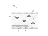
- the predetermined model represents the blood glucose level measuring chip 100 as a one-dimensional model specified by the coordinates of the Z axis along the chip thickness direction.
- the first base material 1, the flow path 20, and the second base material 2 are laminated along the Z-axis direction.
- the first base material 1 and the second base material 2 are positioned so as to face each other, thereby partitioning the flow path 20.
- the surfaces of the first base material 1 and the second base material 2 on the flow path 20 side are orthogonal to the Z axis.
- the distance between the first base material 1 and the second base material 2 is represented by L.
- the state at the time when the sample flows into the blood glucose level measuring chip 100 is represented by the model shown in FIG.
- the region corresponding to the flow path 20 in FIG. 5 is filled with the sample.
- the specimen is whole blood and contains at least plasma 61 and blood cells 62.
- the time when the sample flows in is also called the initial time.
- the time is represented by t.
- the sample flowing into the blood glucose level measuring chip 100 is mixed with the reagent unit 30 and reacts with the blood glucose level measuring reagent.
- the blood glucose level measuring reagent dissolves in plasma 61 from the reagent section 30 and diffuses.
- the arrow DA schematically represents the diffusion of the blood glucose level measuring reagent.
- the blood glucose level measuring reagent reacts with glucose in the plasma 61.
- the blood glucose level measuring reagent generates the colored dye 63.
- the molecule of the colored dye 63 is present in the plasma 61 in a dissolved state.
- the colored dye 63 diffuses into plasma 61 according to a concentration gradient.
- the arrow DC schematically represents the diffusion of the colored dye 63.
- the colored dye 63 in FIG. 7 exemplifies a portion (dark) in which a particularly large amount of dissolved colored dye 63 is present.
- glucose is consumed by the reaction between the blood glucose level measuring reagent dissolved in the plasma 61 from the reagent unit 30 and glucose, and the amount of glucose in the plasma 61 is reduced.
- Glucose diffuses in plasma 61 based on the concentration gradient.
- Arrow DG1 schematically represents the diffusion of glucose.
- the blood cell 62 has a transport mechanism (transporter, mainly GLUT-1) on the blood cell membrane that regulates the glucose concentration inside and outside the blood cell 62. By the action of this transport mechanism, glucose in the blood cells 62 flows out into the plasma 61.
- the arrow DG2 schematically represents the transfer of glucose from blood cells 62 to plasma 61.
- a given model can represent diffusion in the Z-axis direction, the concentration of each substance distributed in the Z-axis direction, and the change in concentration due to the reaction of each substance. That is, it can be said that the predetermined model is represented as a diffusion reaction model of a one-dimensional coordinate system.
- the blood glucose level measuring reagent diffuses in plasma 61 as the elapsed time from the initial time increases. It is assumed that the blood glucose level measuring reagent contained in the reagent section 30 has a viscosity sufficiently higher than the viscosity of the sample at the initial time. That is, the reagent unit 30 is considered to be a solid.
- the plasma 61 permeates the inside of the reagent section 30 from the surface thereof to reduce the viscosity of the blood glucose level measuring reagent contained in the reagent section 30.
- the viscosity ( ⁇ ) of the blood glucose level measuring reagent contained in the reagent section 30 is represented by the following formula (1) as a general formula.
- Equation (1) represents a so-called sigmoid function.
- ⁇ is represented as a function of z and t.
- K mS and n are constants that determine the shape of the sigmoid function.
- ⁇ represents the maximum viscosity of the blood glucose level measuring reagent part applied to the reagent part 30, and is set to a viscosity sufficiently higher than the viscosity of the sample so that the reagent part 30 is regarded as a solid.
- a and B are variables representing the concentration of the component contained in the blood glucose level measuring reagent, and are expressed as a function of z and t.
- the blood glucose level measuring reagent contains a component having a relatively high molecular weight (for example, a molecular weight of 10,000 or more) such as an enzyme and a component having a relatively low molecular weight (for example, a molecular weight of less than 10,000) such as a dye.
- A represents the concentration of the enzyme contained in the blood glucose level measuring reagent.
- B is assumed to represent the concentration of a component other than the enzyme contained in the blood glucose level measuring reagent.
- a and b are constants for weighting based on the molecular weight of the components represented by A and B.
- the concentration of the high molecular weight component has a greater effect on the viscosity than the concentration of the low molecular weight component.
- a is set to a value greater than b.
- the components contained in the blood glucose level measuring reagent are divided into two types, a component represented by A and a component represented by B, but may be further divided into other components.
- the components contained in the blood glucose level measuring reagent are increased, the number of terms contained in the portion represented by (a, A + b, B + 7) In the formula (1) is increased to more than two.
- ⁇ Diffusion of glucose> In a stable aqueous glucose solution, a plurality of isomers (anomers) are present in an equilibrium state. Similarly, in plasma 61, glucose has a plurality of isomers in an equilibrium state. However, depending on the substrate specificity of the enzyme (GDH) used for the blood glucose level measuring reagent, the equilibrium state of these isomers is biased, so that the influence on the blood glucose level measuring reaction may not be negligible.
- the enzyme (GDH) of the present embodiment uses ⁇ -glucose as a substrate to develop a color of a blood glucose level measuring reagent to produce a colored dye 63.
- isomers other than ⁇ -glucose do not serve as a substrate for the enzyme (GDH) (do not react). Therefore, when ⁇ -glucose is consumed by the enzymatic reaction, isomerization of ⁇ -glucose to ⁇ -glucose proceeds in plasma 61, and the total number of molecules of glucose in plasma 61 decreases. As the number of glucose molecules in plasma 61 decreases, glucose in blood cells 62 flows out into plasma 61 by the transport mechanism present in the blood cell membrane. Glucose in the blood cell 62 flows out into the plasma 61 when the blood glucose level measuring reagent and the sample are mixed. In this embodiment, ⁇ -glucose and ⁇ -glucose are considered as glucose isomers having a large abundance ratio.
- ⁇ -glucose flowing out of blood cells 62 is not considered in the present embodiment for simplification of the calculation formula, but may be considered.
- D G represents the diffusion coefficient of glucose in pure water of 25 ° C..
- the arrow with DG1 in FIG. 7 qualitatively represents the Z-axis diffusion of ⁇ -glucose in plasma 61.
- the arrow with DG2 in FIG. 7 qualitatively represents the Z-axis outflow of ⁇ -glucose from blood cells 62 to plasma 61.
- ⁇ 0 represents the viscosity of the sample.
- the term represented by ( ⁇ + ⁇ 0 ) corrects the diffusion coefficient by the viscosity. That is, the higher the viscosity, the smaller the effective diffusion coefficient.
- Vmax M represents the maximum outflow rate of glucose from blood cells 62 to plasma 61.
- K mM is the Michaelis-Menten constant for outflow from inside blood cells 62 to plasma 61.
- Vmax ⁇ represents the maximum rate of isomerization reaction between ⁇ -glucose and ⁇ -glucose.
- K ⁇ is the rate constant of the isomerization reaction from ⁇ -glucose to ⁇ -glucose.
- K ⁇ is the rate constant of the isomerization reaction from ⁇ glucose to ⁇ glucose.
- Km ⁇ is the Michaelis-Menten constant of mutarotase, a type of enzyme. Mutarotase is contained in the sample when the sample is whole blood.
- the first term on the right side of the formula (2) is a term representing the diffusion of ⁇ -glucose present in plasma 61 in the z-axis direction at the initial time.
- the first term on the right side of the formula (3) is a term representing the diffusion of ⁇ -glucose flowing out of the blood cells 62 in the z-axis direction.
- the first term on the right side of the formula (4) is a term representing the diffusion of ⁇ -glucose in plasma 61 in the z-axis direction.
- the term for diffusion is described based on Fick's second law, which is the basic law of diffusion of matter.
- the third term on the right side of the formula (2), the third term on the right side of the formula (3), and the second term on the right side of the formula (4) are terms representing the rate of the isomerization reaction between ⁇ -glucose and ⁇ -glucose. is there. ⁇ -glucose and ⁇ -glucose exist in an equilibrium state. The third term on the right side of equations (2) and (3) becomes a positive value when ⁇ -glucose increases. The second term on the right-hand side of equation (4) is given a minus sign so that it becomes a positive value when ⁇ -glucose increases.
- the diffusion of the blood glucose level measuring reagent is represented by the following formula (5).
- A is a variable representing the concentration of the component represented by A in the blood glucose level measuring reagent, and is represented as a function of z and t.
- DA represents the diffusion coefficient of the component represented by A in the blood glucose level measuring reagent with respect to pure water at 25 ° C.
- U A of the second term on the right side of the formula (5) is a reaction term representing the reaction rate of the component represented by A and ⁇ -glucose.
- the reaction between the blood glucose level measuring reagent and ⁇ -glucose produces the colored dye 63.
- the amount (C) of the colored dye 63 produced is calculated by the following formula (8).
- C is represented as a function of t.
- ⁇ C is the molar absorption constant of the colored dye 63.
- Ht represents the hematocrit value of the sample.
- R v represents the ratio of the solution in the blood cells 62.
- the predetermined model is represented by the partial differential simultaneous equations described by the equations (1) to (8).
- the blood glucose level calculation method includes, for example, the following procedure.
- the operator flows a sample whose parameters such as blood glucose level, hematocrit value, and temperature are known into the blood glucose level measuring chip 100 mounted on the blood glucose level measuring device 110, and measures the absorbance (color development amount) (No. 1). 1 step).
- the blood glucose level measuring device 110 measures the time change of the absorbance of the sample (third step).
- the blood glucose level measuring device 110 may measure the absorbance until the time change of the absorbance becomes sufficiently small. When the time change of the absorbance becomes sufficiently small, it is considered that the reaction between the sample and the blood glucose level measuring reagent has proceeded sufficiently.
- the absorbance at each time after the start of the reaction may be used to prepare a simple calibration curve by associating it with the parameters of the sample.
- the parameters of the sample include parameters derived from the sample (blood) itself and parameters of the sample (mixture of blood and reagent).
- a computer that calculates a simple calibration curve applies the measured data of the time change of the absorbance of a sample having known parameters obtained in the above procedure to a predetermined model, and thereby performs a simple calibration curve for a sample having a combination of unmeasured parameters.
- the line can be calculated.
- the blood glucose level measuring device 110 acquires the hematocrit value and temperature of the sample even when measuring the sample specified by the combination of unmeasured parameters, and matches the acquired value from the simple calibration curve calculated by the computer. You can select a simple calibration curve to be used.
- the blood glucose level measuring device 110 can calculate the blood glucose level of the sample specified by the combination of unmeasured parameters by applying the measured value of the absorbance acquired in the first step to the selected simple calibration curve ( Second step).
- known parameters include temperature, blood glucose level, and hematocrit value.
- the third step may be performed prior to the first step and / or the second step.
- the computer that calculates the simple calibration curve predicts the change in the absorbance of the sample when the unmeasured sample is flowed into the blood glucose level measuring chip 100 in the form of a modeling formula based on a predetermined model based on the measured data. it can.
- a component having a relatively low molecular weight such as a dye is also referred to as a low molecular weight component.
- the concentration of the low molecular weight component is expressed as W.
- W is represented by the following equation (9).
- W is represented as a function of z and t.
- D W represents the diffusion coefficient of the low molecular weight component with respect to pure water at 25 ° C.
- a component having a relatively high molecular weight such as a molecular weight of 10,000 or more
- the concentration of the high molecular weight component is expressed as E.
- E is represented by the following equation (10).
- E is represented as a function of z and t.
- DE represents the diffusion coefficient of the high molecular weight component with respect to pure water at 25 ° C.
- ⁇ E is a coefficient that corrects the interaction of high molecular weight components.
- the change in the concentration of ⁇ -glucose (G ⁇ ) in plasma 61 is described by the following formula (11).
- the formula (11) is the same as the above formula (4).
- the explanation of the variables included in the equation (11) is omitted.
- the reaction in which an intermediate is produced by the reaction between the coloring reagent and ⁇ -glucose can also be considered.
- the sample is whole blood, it is necessary to measure the amount of color development at the measurement wavelength in the wavelength range that is not easily affected by the hemoglobin in the sample.
- the blood glucose level measuring reagent further contains a transition metal ion to chelate the formazan (intermediate) generated by the enzymatic reaction and the transition metal ion to measure a longer wavelength.
- the colored dye 63 can be detected by the wavelength.
- an intermediate (formazan before chelation formation) is produced as a precursor of the colored dye 63 by the reaction between the blood glucose level measuring reagent and ⁇ -glucose.
- the concentration (F 1 ) of the intermediate produced by the reaction of ⁇ -glucose present in plasma 61 from the time of the initial time is represented by the following formula (15).
- the concentration (F 2 ) of the intermediate produced by the reaction of ⁇ -glucose flowing out from the blood cells 62 is represented by the following formula (16).
- F 1 and F 2 are represented as functions of z and t.
- C 1 represents the concentration of the colored dye 63 that changes from the intermediate produced by the reaction of ⁇ -glucose present in the plasma 61 from the time of the initial time.
- C 2 represents the concentration of the colored dye 63 that changes from the intermediate produced by the reaction of ⁇ -glucose flowing out of the blood cells 62.
- D F represents the diffusion coefficient of the intermediate in pure water of 25 ° C..
- the second term on the right side is a reaction term representing the reaction rate between the enzyme and ⁇ -glucose. The reaction rate is based on the concentration of the enzyme, the proportion of active enzyme, and the concentration of glucose.
- K Ni is a constant representing the rate of change from the intermediate to the colored dye 63.
- K F is a constant representing the rate of change from the colored dye 63 to the intermediate.
- At least part of the intermediate changes to the colored dye 63.
- the change in the concentration (C 1 ) of the colored dye 63 that changes from the intermediate produced by the reaction of ⁇ -glucose present in the plasma 61 from the time of the initial time is represented by the following formula (17).
- the change in the concentration (C 2 ) of the colored dye 63 that changes from the intermediate produced by the reaction of ⁇ -glucose flowing out from the blood cells 62 is represented by the following formula (18).
- C 1 and C 2 are represented as functions of z and t.
- D C represents the diffusion coefficient of the color already dye 63 with respect to pure water of 25 ° C..
- the reaction between the blood glucose level measuring reagent and ⁇ -glucose produces the colored dye 63.
- W 0 is a constant for determining the initial value of W.
- E 0 is a constant for determining the initial value of E.
- BG represents the blood glucose level (unit: mg / dL) of the sample.
- E d represents the concentration of the enzyme after 3 days of treatment at 60 ° C.
- RM is a constant that converts the amount of enzyme activity into weight concentration. Since it is difficult to directly measure the following values, they are empirically adjusted values in order to fit the result of the above formula (19) to the color development amount (C) of some known samples.
- Temp represents the temperature (unit: ° C.) of the sample. For ⁇ 0 , a directly measured value may be used.
- the calculation method of the relational expression is not particularly limited, and a well-known method such as optimization by a polynomial can be used.
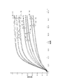
- the temporal change of absorbance exemplified in FIG. A modeling equation was calculated to predict.
- the horizontal axis represents time.
- the vertical axis represents the absorbance of the sample.
- the broken line represents a modeling formula that predicts the time change of absorbance.
- a modeling formula in which the hematocrit value is used as a variable is shown for a sample having a blood glucose level of 400 mg / dL and a temperature of 25 ° C. The hematocrit value is changed from 0% to 70% at 10% intervals.
- the graphs shown by solid lines represent the data obtained by actually measuring the time change of the absorbance of the sample having the parameters that are the premise of the prediction. Regardless of the hematocrit value from 0% to 70%, the error of the measured absorbance with respect to the absorbance of the modeling formula is within 10%. Therefore, the blood glucose level calculation method according to the present embodiment is a sample specified by a combination of unmeasured parameters by modeling the reaction between the blood glucose level measuring reagent and the sample in the blood glucose level measuring chip 100 with a predetermined model. The reaction in was predicted with high accuracy.
- the blood glucose measuring device 110 can perform blood glucose in a short time without waiting from the start of the reaction between the sample and the blood glucose measuring reagent until the reaction sufficiently proceeds.
- the value can be calculated. For example, even if it takes 10 seconds or more for the reaction between the sample and the blood glucose level measuring reagent to proceed sufficiently, the blood glucose level measuring device 110 measures the blood glucose level calculation result in a predetermined time of less than 10 seconds. Can be informed. As a result, the convenience of the blood glucose level measuring device 110 is improved.
- the modeling formula calculated by the computer for calculating the simple calibration curve can be used as an intermediate relational expression for calculating the simple calibration curve for a sample of a combination of unmeasured parameters.
- the blood glucose level calculation method it is possible to calculate a simple calibration curve for measuring the blood glucose level based on a small amount of actual measurement data. As a result, the development period of the blood glucose level measuring device 110 can be shortened and the development cost can be reduced.
- each component or each step can be reconfigured so as not to be logically inconsistent, and a plurality of components or steps can be combined or divided into one. is there.
Landscapes
- Health & Medical Sciences (AREA)
- Life Sciences & Earth Sciences (AREA)
- Engineering & Computer Science (AREA)
- Hematology (AREA)
- Molecular Biology (AREA)
- Urology & Nephrology (AREA)
- Biomedical Technology (AREA)
- Chemical & Material Sciences (AREA)
- Immunology (AREA)
- Food Science & Technology (AREA)
- Diabetes (AREA)
- Microbiology (AREA)
- Cell Biology (AREA)
- Biotechnology (AREA)
- Medicinal Chemistry (AREA)
- Physics & Mathematics (AREA)
- Analytical Chemistry (AREA)
- Biochemistry (AREA)
- General Health & Medical Sciences (AREA)
- General Physics & Mathematics (AREA)
- Pathology (AREA)
- Investigating Or Analysing Biological Materials (AREA)
Abstract
血糖値算出プログラムは、血漿と血球とを含む検体に含まれるグルコースと反応して発色色素を発色させる試薬を有する血糖値測定チップにおける発色色素の発色量の測定結果に基づいて検体の血糖値を算出するために、所定のモデルに基づいて測定結果と検体の血糖値との関係式を算出する第1ステップをプロセッサに実行させる。検体は、少なくとも血糖値を含む所定のパラメータを有する。所定のモデルは、一次元の座標系の拡散反応モデルとして表されている。拡散反応モデルは、試薬の粘度の時間変化と、血漿に含まれるグルコースの濃度の時間変化と、試薬の濃度の時間変化と、試薬の発色量の時間変化とによって表される。
Description
本発明は、血糖値算出プログラム、血糖値算出方法及び血糖値測定装置に関する。
従来、被検者から採取した血液の血糖値を測定する装置が知られている。例えば特許文献1には、血液中のグルコースと反応して発色する試薬の発色量を検出し、検出結果に基づいて血糖値を算出する装置が開示されている。
サンプルを特定するパラメータの組合せの数が多い場合、全ての組合せについて実測データを取得することによって、試薬の発色量と血糖値とを対応づける検量線を準備することは難しい。
本開示の目的は、上記問題に鑑み、パラメータの組合せの数が多くても検量線を容易に算出できる血糖値算出プログラム、血糖値算出方法及び血糖値測定装置を提供することである。
本発明の第1の態様としての血糖値算出プログラムは、
血漿と血球とを含む検体に含まれるグルコースの量に応じて発色する発色色素を含む試薬と検体とを反応させて得られる前記発色色素の発色量を測定する第1ステップと、
前記発色量に基づいて前記検体の血糖値を算出するために、所定のモデルに基づいて前記発色量と前記検体の血糖値との関係式を算出する第2ステップと、
をプロセッサに実行させ、
前記検体は、少なくとも前記血糖値を含む所定のパラメータを有し、
前記所定のモデルは、一次元の座標系の拡散反応モデルとして表されており、
前記拡散反応モデルは、前記試薬の粘度の時間変化と、前記血漿に含まれるグルコースの濃度の時間変化と、前記試薬の濃度の時間変化と、前記試薬の発色量の時間変化とによって表される、血糖値算出プログラム。
血漿と血球とを含む検体に含まれるグルコースの量に応じて発色する発色色素を含む試薬と検体とを反応させて得られる前記発色色素の発色量を測定する第1ステップと、
前記発色量に基づいて前記検体の血糖値を算出するために、所定のモデルに基づいて前記発色量と前記検体の血糖値との関係式を算出する第2ステップと、
をプロセッサに実行させ、
前記検体は、少なくとも前記血糖値を含む所定のパラメータを有し、
前記所定のモデルは、一次元の座標系の拡散反応モデルとして表されており、
前記拡散反応モデルは、前記試薬の粘度の時間変化と、前記血漿に含まれるグルコースの濃度の時間変化と、前記試薬の濃度の時間変化と、前記試薬の発色量の時間変化とによって表される、血糖値算出プログラム。
本発明の1つの実施形態として、
前記試薬の発色量は、検体の血漿中のグルコースと、前記試薬と検体が混合した際に血球内から血漿中へ流出するグルコースと、に基づいてよい。
前記試薬の発色量は、検体の血漿中のグルコースと、前記試薬と検体が混合した際に血球内から血漿中へ流出するグルコースと、に基づいてよい。
本発明の1つの実施形態として、
血糖値算出プログラムは、前記所定のパラメータとして既知のパラメータを有する前記検体を前記試薬と反応させた場合における前記試薬の発色量の時間変化の測定結果を取得する第3ステップを前記プロセッサにさらに実行させ、
前記第2ステップにおいて、前記第3ステップで取得した前記試薬の発色量の時間変化の測定結果と既知のパラメータとの関係に基づいて、前記関係式を算出してよい。
血糖値算出プログラムは、前記所定のパラメータとして既知のパラメータを有する前記検体を前記試薬と反応させた場合における前記試薬の発色量の時間変化の測定結果を取得する第3ステップを前記プロセッサにさらに実行させ、
前記第2ステップにおいて、前記第3ステップで取得した前記試薬の発色量の時間変化の測定結果と既知のパラメータとの関係に基づいて、前記関係式を算出してよい。
本発明の1つの実施形態として、
前記所定のパラメータは、前記検体の中の前記血球の比率を表すヘマトクリット値と、前記検体の温度とをさらに含んでよい。
前記所定のパラメータは、前記検体の中の前記血球の比率を表すヘマトクリット値と、前記検体の温度とをさらに含んでよい。
本発明の第2の態様としての血糖値算出方法は、
血漿と血球とを含む検体に含まれるグルコースの量に応じて発色する発色色素を含む試薬と検体とを反応させて得られる前記発色色素の発色量を測定する第1ステップと、
前記発色量に基づいて前記検体の血糖値を算出するために、所定のモデルに基づいて前記発色量と前記検体の血糖値との関係式を算出する第2ステップと、
を含み、
前記検体は、少なくとも前記血糖値を含む所定のパラメータを有し、
前記所定のモデルは、一次元の座標系の拡散反応モデルとして表されており、
前記拡散反応モデルは、前記試薬の粘度の時間変化と、前記血漿に含まれるグルコースの濃度の時間変化と、前記試薬の濃度の時間変化と、前記試薬の発色量の時間変化とによって表される。
血漿と血球とを含む検体に含まれるグルコースの量に応じて発色する発色色素を含む試薬と検体とを反応させて得られる前記発色色素の発色量を測定する第1ステップと、
前記発色量に基づいて前記検体の血糖値を算出するために、所定のモデルに基づいて前記発色量と前記検体の血糖値との関係式を算出する第2ステップと、
を含み、
前記検体は、少なくとも前記血糖値を含む所定のパラメータを有し、
前記所定のモデルは、一次元の座標系の拡散反応モデルとして表されており、
前記拡散反応モデルは、前記試薬の粘度の時間変化と、前記血漿に含まれるグルコースの濃度の時間変化と、前記試薬の濃度の時間変化と、前記試薬の発色量の時間変化とによって表される。
本発明の第3の態様としての血糖値測定装置は、
血漿と血球とを含む検体に含まれるグルコースの量に応じて発色する発色色素を含む試薬と検体とを反応させ得られる前記発色色素の発色量の測定結果に基づいて前記検体の血糖値を算出する制御部を備え、
前記制御部は、所定のモデルに基づいて前記測定結果と前記検体の血糖値との関係式を算出し、前記関係式に基づいて前記検体の血糖値を算出し、
前記検体は、少なくとも前記血糖値を含む所定のパラメータを有し、
前記所定のモデルは、一次元の座標系の拡散反応モデルとして表されており、
前記拡散反応モデルは、前記試薬の粘度の時間変化と、前記血漿に含まれるグルコースの濃度の時間変化と、前記試薬の濃度の時間変化と、前記試薬の発色量の時間変化とによって表される。
血漿と血球とを含む検体に含まれるグルコースの量に応じて発色する発色色素を含む試薬と検体とを反応させ得られる前記発色色素の発色量の測定結果に基づいて前記検体の血糖値を算出する制御部を備え、
前記制御部は、所定のモデルに基づいて前記測定結果と前記検体の血糖値との関係式を算出し、前記関係式に基づいて前記検体の血糖値を算出し、
前記検体は、少なくとも前記血糖値を含む所定のパラメータを有し、
前記所定のモデルは、一次元の座標系の拡散反応モデルとして表されており、
前記拡散反応モデルは、前記試薬の粘度の時間変化と、前記血漿に含まれるグルコースの濃度の時間変化と、前記試薬の濃度の時間変化と、前記試薬の発色量の時間変化とによって表される。
本発明に係る血糖値算出プログラム、血糖値算出方法及び血糖値測定装置によれば、パラメータの組合せの数が多くても検量線が容易に算出されうる。
以下、本開示に係る実施形態が、図面を参照しながら説明される。各図中、同一符号で表されている構成要素は、同一又は同等である。一実施形態に係る血糖値測定システム500(図3参照)は、血糖値測定チップ100(図1参照)と、血糖値測定装置110(図3参照)とを備える。血糖値測定システム500は、血糖値測定チップ100で採取した被検者の血液を血糖値測定装置110で解析することによって、被検者の血液中の血糖量を測定する。
(血糖値測定チップ)
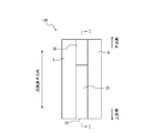
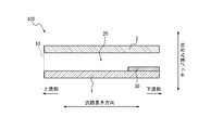
図1及び図2に示されるように、本実施形態における血糖値測定チップ100は、供給口10と、流路20と、試薬部30とを備える。
図1及び図2に示されるように、本実施形態における血糖値測定チップ100は、供給口10と、流路20と、試薬部30とを備える。
血糖値測定チップ100は、底面部を形成する第1基材1と、天面部を形成する第2基材2とを備える。血糖値測定チップ100は、第1基材1及び第2基材2の間であって、チップ厚み方向及び流路長手方向に対して直交する方向(幅方向とも称される)の両端に設けられている接着部3及び接着部4をさらに備える。血糖値測定チップ100は、接着部3、4において、第1基材1及び第2基材2の間に所定の厚みを有するスペーサをさらに備えてもよい。第1基材1及び第2基材2がスペーサを挟んで接着されることで、第1基材1と第2基材2との間に、所定の大きさの空隙として流路20が形成される。流路20は、上流側の端部に供給口10を有する。血糖値測定チップ100が採取する血液は、供給口10から流路20に流入できる。血糖値測定チップ100は、流路20内に、試薬部30を備える。試薬部30は、第1基材1上に形成されているが、これに限定されるものではなく、流路20を閉塞することなく、流路20内に設けられていればよい。
第1基材1の材質は、特に制限されず、測定光の照射及び受光を妨げないように適宜選択されてよい。第1基材1は、例えば、ポリエチレンテレフタレート(PET)、ポリメチルメタアクリレート、ポリスチレン、環状ポリオレフィン、環状オレフィンコポリマー、又はポリカーボネート等の透明な有機樹脂材料で構成されてよい。第1基材1は、ガラス又は石英等の透明な無機材料で構成されてよい。
第2基材2の材質は、特に制限されず、測定光の照射及び受光を妨げないように適宜選択されてよい。第2基材2は、例えば、親水処理ポリエステルフィルム(3M親水化フィルム)等の透明な有機樹脂材料で構成されてよい。
第1基材1、第2基材2は、必要に応じて着色されてもよい。この場合、サンプルを測定光が通過するための光路を備える構造に形成されていればよい。より具体的には、光路となる箇所のみを透明部材で形成したり、着色部材の一部を切り欠くことで、光路を確保することができる。また、第1基材1、第2基材2のいずれか一方に反射部材を設けて、被測定物資を透過した光を反射させて測定してもよい。
接着部3、4の厚みは、流路20のチップ厚み方向の長さを所望の値にするために、適宜調整される。例えば、第1基材1と第2基材2とは、それらの間に任意の厚みを有するスペーサが配置された状態で接着されたり融着されたりしてよい。第1基材1と第2基材2とは、スペーサの機能を兼ねる両面テープによって接着されてもよい。
(血糖値測定試薬)
試薬部30は、血糖値測定試薬を含む。より具体的には、試薬部30は、後述の成分を含むように調製した血糖値測定試薬溶液を、第1基材1上に塗布し、乾燥させることで形成される。血糖値測定試薬は、単に試薬とも称される。使用形態は、固体、ゲル状、ゾル状、又は液体のいずれの形態であってもよい。血糖値測定試薬は、酵素と、発色色素とを含むが、上記に加えて、さらに他の成分を含んでもよい。ここで、他の成分としては、通常、測定対象である生体成分の種類に応じて適切に選択され、生体成分濃度を測定するために添加される成分が同様にして使用できる。具体的には、電子受容体(メディエータ)、緩衝剤、pH調整剤、界面活性剤、水(例えば、RO水)、金属イオン、塩などが挙げられる。ここで、上記他の成分は、それぞれ、1種を単独で使用してもよいし、2種以上を組み合わせて使用してもよい。また、上記他の成分のそれぞれは、1種を単独で使用してもよいし、2種以上を併用してもよい。
試薬部30は、血糖値測定試薬を含む。より具体的には、試薬部30は、後述の成分を含むように調製した血糖値測定試薬溶液を、第1基材1上に塗布し、乾燥させることで形成される。血糖値測定試薬は、単に試薬とも称される。使用形態は、固体、ゲル状、ゾル状、又は液体のいずれの形態であってもよい。血糖値測定試薬は、酵素と、発色色素とを含むが、上記に加えて、さらに他の成分を含んでもよい。ここで、他の成分としては、通常、測定対象である生体成分の種類に応じて適切に選択され、生体成分濃度を測定するために添加される成分が同様にして使用できる。具体的には、電子受容体(メディエータ)、緩衝剤、pH調整剤、界面活性剤、水(例えば、RO水)、金属イオン、塩などが挙げられる。ここで、上記他の成分は、それぞれ、1種を単独で使用してもよいし、2種以上を組み合わせて使用してもよい。また、上記他の成分のそれぞれは、1種を単独で使用してもよいし、2種以上を併用してもよい。
<酵素>
酵素は、血液中のグルコースと反応して、グルコースから電子を引き抜く。ここで引き抜かれた電子は、発色色素に受け渡されることで、発色色素が呈色する。酵素は、グルコースを基質とする限り、特に制限されず、目的に応じて適宜選択されてよい。酵素は、例えば、グルコースデヒドロゲナーゼ(GDH)又はグルコースオキシダーゼ(GOD)等を含んでよい。酵素は、1種類の物質だけで構成されてもよいし、2種類以上の物質で構成されてもよい。酵素は、FADを補酵素とするGDH(FAD-GDH)を用いるのが好ましい。
酵素は、血液中のグルコースと反応して、グルコースから電子を引き抜く。ここで引き抜かれた電子は、発色色素に受け渡されることで、発色色素が呈色する。酵素は、グルコースを基質とする限り、特に制限されず、目的に応じて適宜選択されてよい。酵素は、例えば、グルコースデヒドロゲナーゼ(GDH)又はグルコースオキシダーゼ(GOD)等を含んでよい。酵素は、1種類の物質だけで構成されてもよいし、2種類以上の物質で構成されてもよい。酵素は、FADを補酵素とするGDH(FAD-GDH)を用いるのが好ましい。
酵素の血液検体反応時の濃度は、特に制限されず、目的に応じて適宜選択されてよいが、例えば1U/μL以上とされてよい。
<電子受容体(メディエータ)>
電子受容体(メディエータ)は、酵素と血液中のグルコースとの反応を促進するとともに、酵素がグルコースから引き抜いた電子を一旦受け取って発色色素に渡す。電子受容体は、特に制限されず、目的に応じて適宜選択されてよい。電子受容体は、例えば、5-メチルフェナジニウムメチルスルファート(PMS)、1-メトキシ-5-メチルフェナジニウムメチルスルファート(mPMS)、NAD、FAD、PQQ、又はフェリシアン化カリウム等で構成されてよい。電子受容体は、1種類の物質だけで構成されてもよいし、2種類以上の物質で構成されてもよい。
電子受容体(メディエータ)は、酵素と血液中のグルコースとの反応を促進するとともに、酵素がグルコースから引き抜いた電子を一旦受け取って発色色素に渡す。電子受容体は、特に制限されず、目的に応じて適宜選択されてよい。電子受容体は、例えば、5-メチルフェナジニウムメチルスルファート(PMS)、1-メトキシ-5-メチルフェナジニウムメチルスルファート(mPMS)、NAD、FAD、PQQ、又はフェリシアン化カリウム等で構成されてよい。電子受容体は、1種類の物質だけで構成されてもよいし、2種類以上の物質で構成されてもよい。
電子受容体の含有量は、特に制限されず、目的に応じて適宜選択されてよいが、例えば0.01mol%以上とされてよい。電子受容体の血液検体反応時の濃度は、特に制限されず、目的に応じて適宜選択されてよいが、例えば0.1mM以上とされてよい。なお、電子受容体は必ずしも含む必要はなく、使用する酸化還元酵素の種類によっては、電子受容体を配合しなくともよい
<発色色素>
発色色素(還元系発色試薬)は、酵素とグルコースとの反応によって生じた電子又は過酸化水素を受け取って(還元されて)発色する。発色した発色色素は、発色済色素63(図7参照)と称されるとする。発色色素は、特に制限されないが、目的に応じて適宜選択されてよい。発色色素は、例えば、テトラゾリウム塩、リンモリブデン酸ナトリウム、インディゴカルメン、ジクロロインドフェノール、又はレサズリン、テトラゾリウム塩を用いてもよいが、このうち、テトラゾリウム塩が好ましい。発色色素は、1種類の物質だけで構成されてもよいし、2種類以上の物質で構成されてもよい。本実施形態において、発色済色素63は、青色を呈色する。発色済色素63は、青色に限られず、他の色を呈色してもよいが、血色素の主な吸収帯と重ならない波長域(600nm以上、特に650nm以上)に極大吸収波長を有するものが好ましい。
発色色素(還元系発色試薬)は、酵素とグルコースとの反応によって生じた電子又は過酸化水素を受け取って(還元されて)発色する。発色した発色色素は、発色済色素63(図7参照)と称されるとする。発色色素は、特に制限されないが、目的に応じて適宜選択されてよい。発色色素は、例えば、テトラゾリウム塩、リンモリブデン酸ナトリウム、インディゴカルメン、ジクロロインドフェノール、又はレサズリン、テトラゾリウム塩を用いてもよいが、このうち、テトラゾリウム塩が好ましい。発色色素は、1種類の物質だけで構成されてもよいし、2種類以上の物質で構成されてもよい。本実施形態において、発色済色素63は、青色を呈色する。発色済色素63は、青色に限られず、他の色を呈色してもよいが、血色素の主な吸収帯と重ならない波長域(600nm以上、特に650nm以上)に極大吸収波長を有するものが好ましい。
血糖値測定試薬における発色色素の含有量は、特に制限されず、目的に応じて適宜選択されてよいが、例えば5mol%以上とされてよい。発色色素の血液検体反応時の濃度は、特に制限されず、目的に応じて適宜選択されてよいが、例えば10mM以上とされてよい。
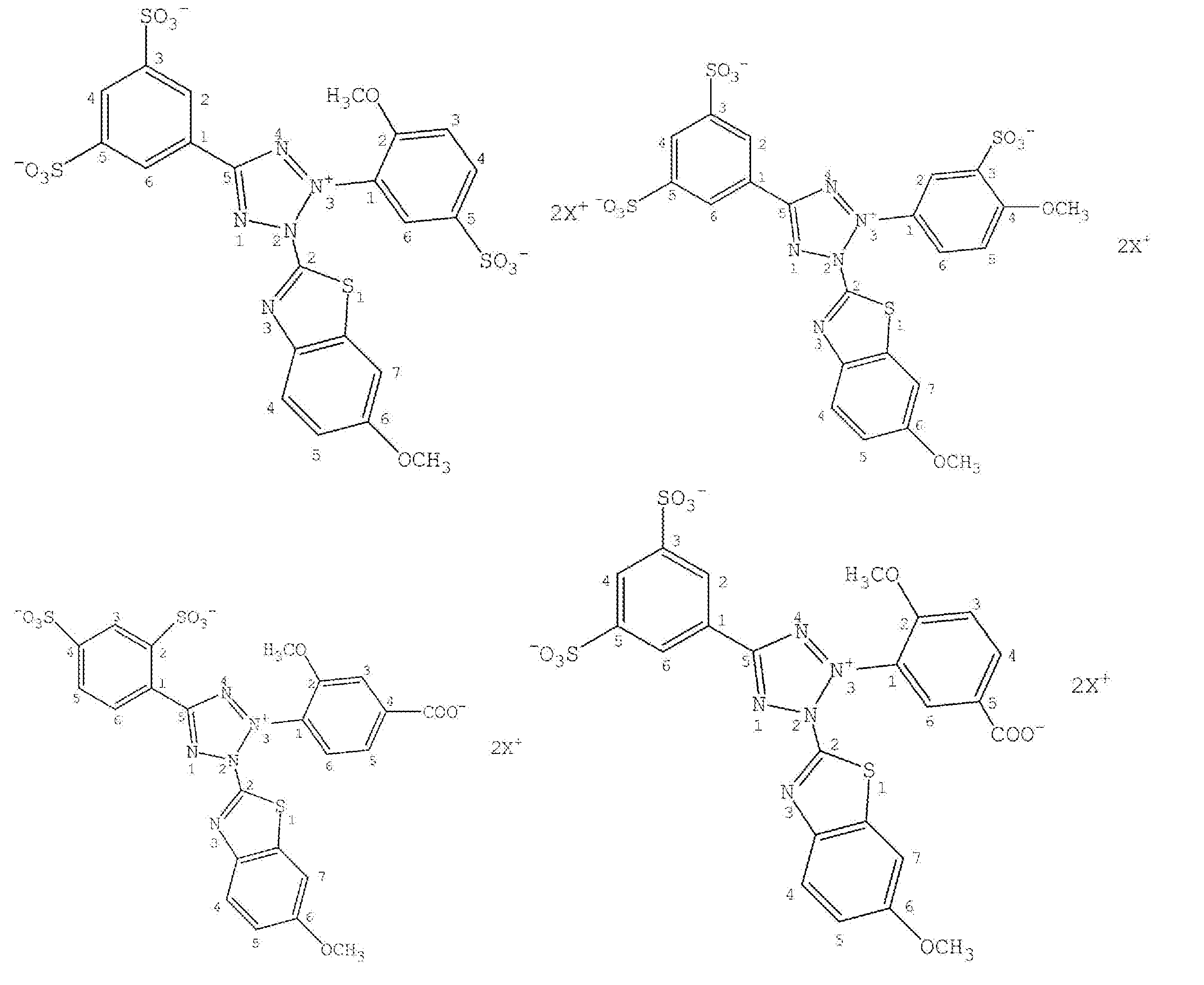
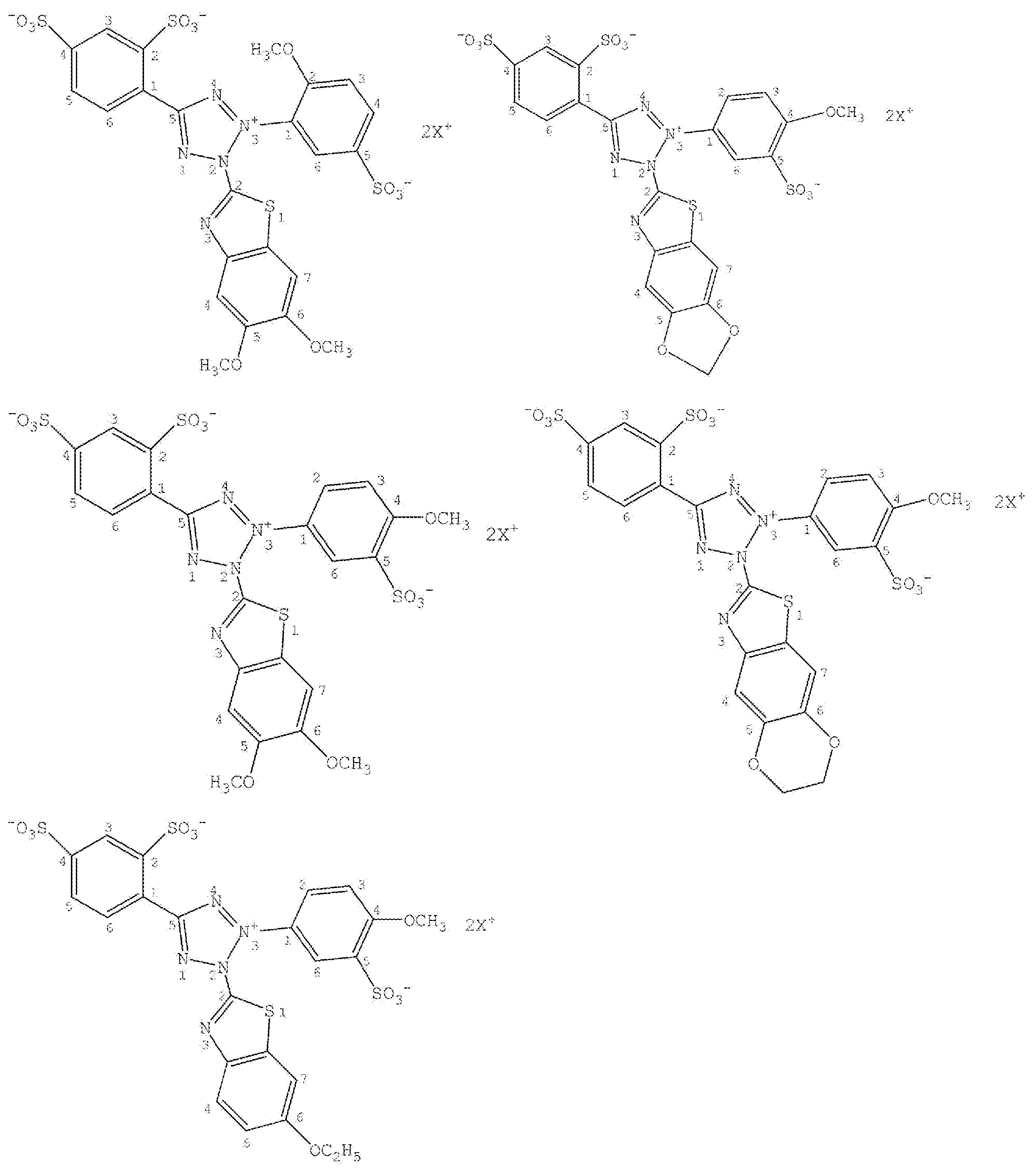
発色色素として用いられるテトラゾリウム塩は、酸化還元酵素及びメディエータ機能を有するテトラゾリウム塩であってもよい。テトラゾリウム塩としては、例えば、3,3’-[3,3’-Dimethoxy-(1,1’-biphenyl)-4,4’-diyl]-bis[2-(4-nitrophenyl)-5-phenyl-2H-tetrazoliumchloride](Nitro-TB)、2-(4-Iodophenyl)-3-(4-nitrophenyl)-5-(2,4-disulfophenyl)-2H-tetrazolium,monosodiumsalt(WST-1)、2-(4-Iodophenyl)-3-(2,4-dinitrophenyl)-5-(2,4-disulfophenyl)-2H-tetrazolium,monosodiumsalt(WST-3)、2-benzothiazolyl-3-(4-carboxy-2-methoxyphenyl)-5-[4-(2-sulfoethylcarbamoyl)phenyl]-2H-tetrazolium(WST-4)、2,2’-dibenzothiazolyl-5,5’-bis[4-di(2-sulfophenyl)carbamoylphenyl]-3,3’-(3,3’-dimethoxy-4,4’-biphenylene)ditetrazoliumdisodium salt(WST-5)、2-(2-Methoxy-4-nitrophenyl)-3-(4-nitrophenyl)-5-(2,4-disulfophenyl)-2H-tetrazolium,monosodiumsalt(WST-8)等を用いてもよい。本実施形態では、発色色素は、テトラゾリウム塩の中でも水溶性が高く、血球62に含まれる血色素の影響が低減された測定波長で、発色量を測定することができる下記化学式(1):
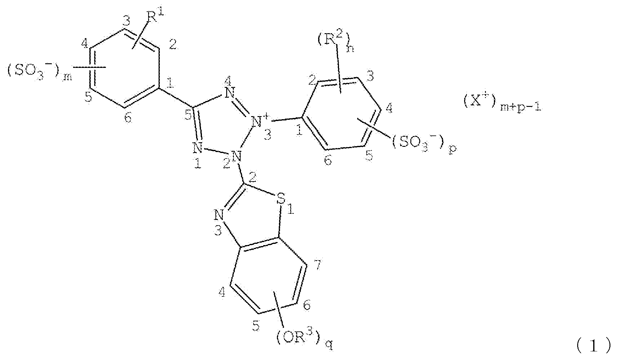
で示される、2-置換ベンゾチアゾリル-3-置換フェニル-5-置換スルホ化フェニル-2H-テトラゾリウム塩であることが好ましい。
上記化学式(1)において、テトラゾール環の2位にベンゾチアゾリル基が存在することにより、例えば、遷移金属化合物をさらに使用する場合には、遷移金属化合物と効率よくかつ速やかにキレート化合物を形成し、ホルマザン化合物の極大吸収波長を長波長域にシフトできる。そして、テトラゾール骨格の2位のベンゾチアゾリル基に、少なくとも1のメトキシ基又はエトキシ基(-OR3)が導入されることで、さらに生成するホルマザンとNi2+等の遷移金属イオンとのキレート時の極大吸収波長が長波長側にシフトする。これにより、血球62に含まれる血色素の影響が低減された測定波長で、発色量を測定できる。
ここで、qは1又は2であり、1であることが好ましい。ここで、q=1の場合、R3は、メチル基又はエチル基であり、メチル基であることが好ましい。q=1の場合、テトラゾール骨格の2位に存在するベンゾチアゾリル基の置換基である-OR3の置換位置は特に限定されるものではなく、4位、5位、6位、又は7位のいずれであってもよい。-OR3の置換位置は、ベンゾチアゾリル基の6位に結合することが好ましい。q=2の場合、R3は、水素原子、メチル基又はエチル基であり、少なくとも一がメチル基、又はエチル基である。また、qが2である場合、各OR3は隣接して配置され、各OR3が互いに環を形成してもよい。この際、好適な組み合わせとしては、R3が、水素原子及びメチル基の組み合わせ、メチル基及びメチル基の組み合わせである。q=2の場合、テトラゾール骨格の2位に存在するベンゾチアゾリル基の置換基である-OR3の置換位置は2つの-OR3が隣接して配置されれば特に限定されるものではなく、4,5位、5,6位、又は6,7位のいずれであってもよい。少なくとも一の-OR3の置換位置は、ベンゾチアゾリル基の6位に結合することが好ましく、すなわち、2つの-OR3の置換位置は、5,6位、又は6,7位である。
化学式(1)において、テトラゾール骨格の5位には置換スルホ化フェニル基が存在する。該スルホ化フェニル基の置換基であるR1は、水素原子、水酸基、メトキシ基、及びエトキシ基からなる群から選択されるいずれか一つである。
テトラゾール骨格の5位には、スルホ基(-SO3
-)が少なくとも1個存在する(m=1又は2)。化学式(1)において、mは、スルホ基(-SO3
-)がテトラゾール骨格の5位のフェニル基に結合する数であり、1又は2である。m=2であり、R1が水素原子であることがより好ましい。ここで、m=2である場合には、スルホ基(-SO3
-)がテトラゾール骨格の3位のフェニル基に結合する数であるpが1であることが好ましい。
なお、下記(1)~(4):(1)m=2かつp=1である、(2)m=1かつn=0である、(3)テトラゾール骨格の5位に存在するフェニル基において、R1が水酸基であり、この際、スルホ基(SO3-)及び水酸基が2,4位、又は4,6位にある、(4)p=0かつ少なくとも一のR2がカルボキシル基である、のいずれかを満たすことが好ましく、(1)m=2かつp=1又は、(4)p=0かつ少なくとも一のR2がカルボキシル基であることがより好ましい。
ここで、テトラゾール骨格の5位に存在するフェニル基へのスルホ基(-SO3
-)の結合位置は特に制限されない。m=2の場合には、スルホ基(-SO3
-)は、フェニル基の、2,4位、3,5位に存在することが好ましい。フェニル基の2,4位にスルホ基が存在することが特に好ましい。化学式(1)においてテトラゾール骨格の5位のフェニル基が、スルホ基(-SO3
-)が2,4位に存在するフェニル基であることが好ましい。
式(2)において、テトラゾール骨格の3位には置換フェニル基が存在する。フェニル基は必須に置換されるため、n+pは1以上である。
テトラゾール骨格の3位のフェニル基の置換基としてのR2は、ニトロ基、-OR4、及びカルボキシル基からなる群から選択されるいずれか一つである。R2は、ニトロ基又は-OR4であることが好ましく、カルボキシル基であることが好ましい。また、nは、R2がテトラゾール骨格の3位のフェニル基に結合する数であり、0~2の整数である。n=1又は2であることが好ましく、n=1であることがより好ましい。R2が2個存在する場合、すなわち、n=2である場合、R2は同じであっても異なるものであってもよい。n=1又は2である場合、少なくとも1のR2が-OR4基であることが好ましい。R4は、メチル基又はエチル基である。
nが1又は2である場合の、R2の置換位置は特に限定されないが、テトラゾール骨格の3位に存在する置換スルホフェニル基の2位、3位、4位、5位又は6位であることが好ましく、少なくとも一のR2が2位及び/又は4位である。
pは、スルホ基(-SO3
-)がテトラゾール骨格の3位のフェニル基に結合する数であり、pが1である、又はp=0かつ少なくとも一のR2がカルボキシル基である。より好適には、m=2かつp=1である、又はp=0かつ少なくとも一のR2がカルボキシル基である。p=1である場合、テトラゾール骨格の3位のフェニル基を置換するスルホ基(-SO3
-)の置換位置は特に限定されるものではないが、3位又は5位であることが好ましく、3位であることがより好ましい。
また、式(2)において、テトラゾール骨格の3位に存在する置換基が、4-メトキシ-3-スルホフェニル基、2-メトキシ-5-スルホフェニル基、2-メトキシ-4-ニトロ-5-スルホフェニル基、2-メトキシ-4-ニトロフェニル基、4-スルホフェニル基、4-カルボキシ-2-メトキシフェニル基、5-カルボキシ-2-メトキシフェニル基、3-カルボキシ-4-メトキシフェニル基、又は4-メトキシ-5-スルホフェニル基が好ましく、4-メトキシ-3-スルホフェニル基、2-メトキシ-5-スルホフェニル基、3-カルボキシ-4-メトキシフェニル基、又は4-メトキシ-5-スルホフェニル基であることがより好ましく、4-メトキシ-3-スルホフェニル基、4-メトキシ-5-スルホフェニル基、又は、2-メトキシ-5-スルホフェニル基であることが好ましい。このうち、4-メトキシ-3-スルホフェニル基であることが特に好ましい。
化学式(1)に存在するスルホ基の総数(m+p)は、2以上である。
化学式(1)において、Xは、水素原子又はアルカリ金属を表わす。アルカリ金属の種類は特に制限されず、リチウム、ナトリウム、カリウム、ルビジウム、セシウムのいずれでもよい。
発色色素の好ましい例としては、以下のものが挙げられる。
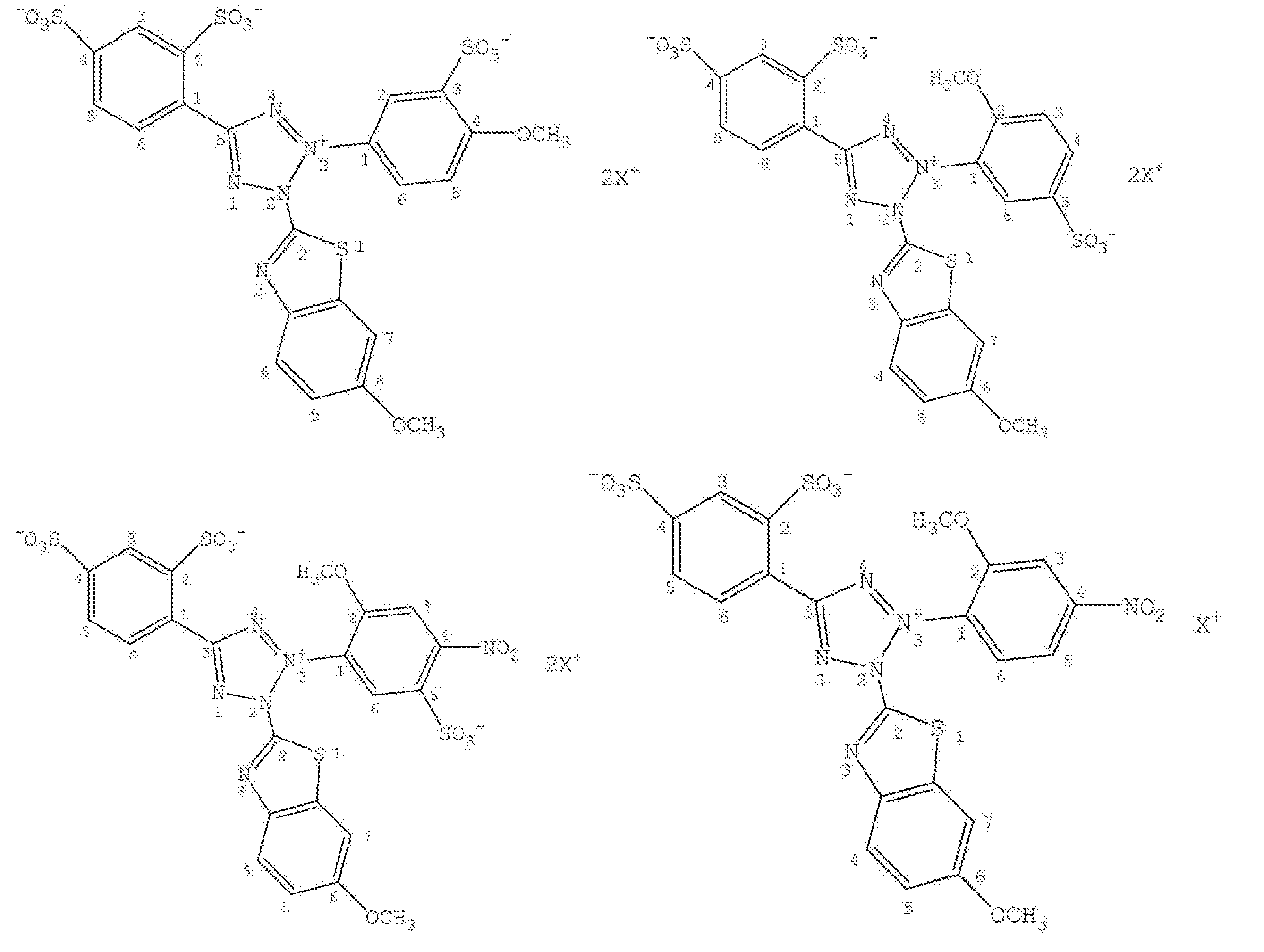


血糖値測定試薬への緩衝剤(バッファー)の添加は、特に制限されず、一般的に生体成分濃度を測定する際に使用されるバッファーが同様にして使用できる。具体的には、リン酸緩衝剤、クエン酸緩衝剤、クエン酸-リン酸緩衝剤、トリスヒドロキシメチルアミノメタン-HCl緩衝剤(トリス塩酸緩衝剤)、MES緩衝剤(2-モルホリノエタンスルホン酸緩衝剤)、TES緩衝剤(N-トリス(ヒドロキシメチル)メチル-2-アミノエタンスルホン酸緩衝剤)、酢酸緩衝剤、MOPS緩衝剤(3-モルホリノプロパンスルホン酸緩衝剤)、MOPS-NaOH緩衝剤、HEPES緩衝剤(4-(2-ヒドロキシエチル)-1-ピペラジンエタンスルホン酸緩衝剤)、HEPES-NaOH緩衝剤などのGOOD緩衝剤、グリシン-塩酸緩衝剤、グリシン-NaOH緩衝剤、グリシルグリシン-NaOH緩衝剤、グリシルグリシン-KOH緩衝剤などのアミノ酸系緩衝剤、トリス-ホウ酸緩衝剤、ホウ酸-NaOH緩衝剤、ホウ酸緩衝剤などのホウ酸系緩衝剤、又はイミダゾール緩衝剤などが用いられる。
緩衝剤の濃度としては、特に制限されないが、血糖値測定試薬溶液の調整時において、0.01~1.0Mであるのが好ましい。なお、ここでは、緩衝剤の濃度とは、水性溶液中に含まれる緩衝剤の濃度(M、mol/L)をいう。また、緩衝液のpHは、生体成分(たとえば、血球62)に対して作用を及ぼさないことが好ましい。
血糖値測定試薬は、芳香族炭化水素を含有している。芳香族炭化水素は、少なくとも1個のスルホン酸基を有する芳香族炭化水素である限り、特に制限はなく、目的に応じて適宜選択されてよい。芳香族炭化水素は、例えば、ベンゼンスルホン酸ナトリウム、1,3-ベンゼンスルホン酸二ナトリウム、1,3,5-ベンゼンスルホン酸三ナトリウム、ナフタレン-1,3,6-トリスルホン酸三ナトリウム、又はアントラセン-1,3,6-トリスルホン酸三ナトリウム等で構成されてよい。芳香族炭化水素は、1種類の物質だけで構成されてもよいし、2種類以上の物質で構成されてもよい。
芳香族炭化水素の含有量は、特に制限されず、目的に応じて適宜選択されてよいが、例えば30mol%以上とされてよい。芳香族炭化水素の血液検体反応時の濃度は、特に制限されず、目的に応じて適宜選択されてよいが、例えば50mM以上とされてよい。
血糖値測定試薬に、pH調整剤が添加されてもよい。pH調整剤の種類としては、特に制限されない。pH調整剤としては、一般的に生体成分濃度を測定する際に適用されるpHとなるように、酸(塩酸、硫酸、リン酸など)又は塩基(水酸化カリウム、水酸化ナトリウム、炭酸カリウム、炭酸ナトリウムなど)が適宜選択・使用される。また、pH調整剤は、そのまま使用されても又は水溶液の形態で使用されてもよい。また、pH調整剤の量は、特に制限されない。血糖値測定試薬溶液は、pHが中性付近、例えば、pH5.0~8.0程度となるように調整される。
また、界面活性剤若しくはアルコール等の溶剤、金属イオン、又は、塩類が、血糖値測定試薬調整時に適宜目的に応じて添加されてもよい。
(血糖値測定装置)
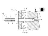
図3に示されるように、血糖値測定システム500は、血糖値測定チップ100と、血糖値測定装置110とを備える。被検者から採取した血液は、血糖値測定チップ100の内部に導入可能である。血糖値測定装置110は、検体に含まれるグルコース量を測定する。検体に後述の血糖値測定試薬が混合、溶解することで、検体中のグルコース量を測定できる。これにより、検体が血液の場合、血糖値を算出することができる。ここで、検体(血液)と血糖値測定試薬との混合物もしくは混合溶液をサンプルと称する。血糖値測定チップ100は、血糖値測定装置110の先端部に装着される。血糖値測定チップ100は、測定後、血糖値測定装置110から取り外されて廃棄される。血糖値測定装置110は、測定結果又は操作内容等を表示する表示部111と、血糖値測定装置110の起動と終了を指示する電源ボタン112と、操作ボタン113と、血糖値測定チップ100を取り外す取外レバー114とを備える。表示部111は、液晶又はLED等で構成されてよい。
図3に示されるように、血糖値測定システム500は、血糖値測定チップ100と、血糖値測定装置110とを備える。被検者から採取した血液は、血糖値測定チップ100の内部に導入可能である。血糖値測定装置110は、検体に含まれるグルコース量を測定する。検体に後述の血糖値測定試薬が混合、溶解することで、検体中のグルコース量を測定できる。これにより、検体が血液の場合、血糖値を算出することができる。ここで、検体(血液)と血糖値測定試薬との混合物もしくは混合溶液をサンプルと称する。血糖値測定チップ100は、血糖値測定装置110の先端部に装着される。血糖値測定チップ100は、測定後、血糖値測定装置110から取り外されて廃棄される。血糖値測定装置110は、測定結果又は操作内容等を表示する表示部111と、血糖値測定装置110の起動と終了を指示する電源ボタン112と、操作ボタン113と、血糖値測定チップ100を取り外す取外レバー114とを備える。表示部111は、液晶又はLED等で構成されてよい。
図4に示されるように、血糖値測定装置110は、血糖値測定チップ100を装着する装着部22を備える。装着部22は、血糖値測定装置110の先端に位置する開口部21と、血糖値測定チップ100を収容する収容部23とを有する。開口部21及び収容部23は、装着部22の内部に設けられている内壁によって区画されている。
血糖値測定装置110は、測定光をサンプルに照射するとともにサンプルを透過した測定光を検出する光学測定部24を備える。血糖値測定装置110は、光学測定部24の検出結果に基づいて、サンプルの血糖値を算出する制御部25と、取外レバー114と連動し血糖値測定チップ100を取り外すイジェクトピン26とを備える。
測定の際は血糖値測定チップ100が収容部23に装着される。装着作業はユーザにより手作業で行われる。血糖値測定装置110は、血糖値測定チップ100を収容部23内の所定位置に固定するための適当なロック機構等を備えてよい。このようにすることで、手作業によって生じうる装着位置のバラツキが低減されうる。
光学測定部24は、照射部31と、受光部32とを備える。照射部31は、血糖値測定試薬と血液との反応生成物に照射光33(測定光)を照射する。照射部31は、発光ダイオード(LED)で構成されてよい。照射部31は、ハロゲンランプ又はレーザー等の他の光源で構成されてもよい。受光部32は、血糖値測定試薬と血液との反応生成物を透過した測定光を受光する。血糖値測定試薬は、酵素によって、検体中のグルコース量に応じて発色するため、受光部32は、発色量に基づく信号を受取ることができる。発色量に基づく信号は、検出信号とも称される。受光部32は、例えば、フォトダイオード(PD)で構成されてよい。受光部32は、CCD(Charged Coupled Device)又はCMOS(Complementary Metal Oxide semiconductor)イメージセンサ等の他の受光デバイスで構成されてもよい。
照射部31は、第1の波長を有する光を発する第1の発光素子51と、第1の波長と異なる第2の波長を有する光を発する第2の発光素子52とを含む。第1の波長は、血糖値測定試薬の、グルコース量(血糖値)に応じた発色量を検出するための波長であり、例えば600~920nmの波長帯に含まれる波長から選択される。第2の波長は、血液中の赤血球濃度を検出するための波長であり、例えば510~590nmの波長帯に含まれる波長から選択される。なお、照射部31は、測定方法に応じて、上記の波長帯の中から選択される波長の発光素子を有していてもよいし、上記波長帯以外の波長の発光素子を有してもよい。
図4を参照して、照射部31と受光部32の配置及び両者の位置関係が説明される。血糖値測定装置110は、内部に形成されている、第1の空間41と第2の空間42とを有する。照射部31及び受光部32はそれぞれ、第1の空間41及び第2の空間42に配置されている。血糖値測定チップ100が血糖値測定装置110に装着されていない状態で、第1の空間41と第2の空間42とは、収容部23を挟んで対向する。血糖値測定チップ100が血糖値測定装置110に装着された状態で、第1の空間41と第2の空間42とは、血糖値測定チップ100上の試薬部30が保持されている位置を挟んで対向する。照射部31は、照射光33が血糖値測定チップ100の底面にほぼ垂直に照射できる位置に配置されていてよい。このようにすることで、照射光33のエネルギーロスが低減されうる。なお、透過光測定におけるノイズを抑制するため、血糖値測定チップ100における、測定光の光路にあたる部分に、マイクロビーズ又は多孔質膜は配置しないのが好ましい。
照射部31は、白色を照射するハロゲンランプで構成される場合、特定の波長のみを照射光33として抽出する分光フィルタをさらに備えてもよい。照射部31は、集光レンズを備えてもよい。
血糖値測定装置110は、温度センサをさらに備えてよい。温度センサは、サンプルの温度を測定する。このサンプル温度を補正値として利用して、血糖値が算出されてもよい。
制御部25は、受光部32から検出信号を取得し、検出信号に基づいてサンプル(検体)の血糖値を算出する。制御部25は、例えばプロセッサを含んでよい。制御部25は、種々の情報及びプログラムを格納する記憶部を有してよい。記憶部は、例えば、半導体メモリを含んでよい。記憶部は、制御部25と別個に構成されてよい。
以上説明してきた血糖値測定装置110は、血液と、発色色素を含む血糖値測定試薬との反応生成物を含むサンプルを透過した光を測定する透過型の装置である。血糖値測定装置110は、これに限定されず、例えば、反応生成物を含むサンプルから反射した光を測定する反射型の装置であってもよい。反射型の装置の場合、照射部31と受光部32は血糖値測定チップ100の挿入位置に対して同じ側に設けられる。
血糖値測定装置110は、例えば以下の手順でサンプルの血糖値を測定できる。測定作業者は、電源ボタン112を操作して、血糖値測定装置110を起動する。測定作業者は、血糖値測定装置110の先端部に血糖値測定チップ100を装着する。血糖値測定装置110は、血糖値測定チップ100が装着されたことを検出し、照射部31と受光部32とによってサンプルが導入されていない初期状態を測定する。測定作業者は、被検者の血液を、検体として血糖値測定チップ100内に流入させる。血糖値測定チップ100で採取される被検者の血液は、全血検体とも称される。全血検体は、毛細管現象によって、血糖値測定チップ100の供給口10から流路20に流入し、試薬部30に到達し、検体に測定試薬が溶解することで、血糖値測定試薬との反応を開始する。血糖値測定装置110は、サンプル中に含まれるグルコースと測定試薬との反応によって生成する発色済色素63の量を、照射部31から照射した光量と、受光部32で受けた光量とに基づく吸光度として検出する。血糖値測定装置110は、検体の流入から所定時間の経過後における吸光度に基づいて、サンプルの血糖値を算出する。言い換えれば、血糖値測定装置110は、発色済色素63の量に基づいて、検体の血糖値を算出できる。発色済色素63の量は、発色量とも称される。このとき、測定時の温度測定値が取得されてもよい。なお、測定作業者は、被検者と同一であってもよい。
(発色量と血糖値との関係)
全血検体は、赤血球等の血球62(図6参照)と、血漿61(図6参照)とを含む。試薬部30に含まれる血糖値測定試薬は、血漿61中のグルコースと反応することによって発色済色素63を生成する。したがって、血糖値測定装置110がサンプル中の血球62の比率(血漿61の比率)にさらに基づくことで、サンプルの血糖値を算出する。サンプル中の血球比率は、ヘマトクリット値とも称され、Ht(単位:%)で表される。血糖値測定装置110は、第2の発光素子52が射出する第2の波長を有する光の吸光度を測定することによって、サンプルのヘマトクリット値を測定してよい。血糖値測定試薬と血漿61中のグルコースとは、サンプルの温度に応じた速度で反応する。したがって、血糖値測定装置110がサンプルの温度にさらに基づいてサンプルの血糖値を算出することで、血糖値の測定精度が高められる。血糖値測定装置110は、温度センサによってサンプルの温度を測定してよい。血糖値測定装置110は、発色量と、ヘマトクリット値と、サンプルの温度とを含むパラメータに基づいて、血糖値を算出できる。
全血検体は、赤血球等の血球62(図6参照)と、血漿61(図6参照)とを含む。試薬部30に含まれる血糖値測定試薬は、血漿61中のグルコースと反応することによって発色済色素63を生成する。したがって、血糖値測定装置110がサンプル中の血球62の比率(血漿61の比率)にさらに基づくことで、サンプルの血糖値を算出する。サンプル中の血球比率は、ヘマトクリット値とも称され、Ht(単位:%)で表される。血糖値測定装置110は、第2の発光素子52が射出する第2の波長を有する光の吸光度を測定することによって、サンプルのヘマトクリット値を測定してよい。血糖値測定試薬と血漿61中のグルコースとは、サンプルの温度に応じた速度で反応する。したがって、血糖値測定装置110がサンプルの温度にさらに基づいてサンプルの血糖値を算出することで、血糖値の測定精度が高められる。血糖値測定装置110は、温度センサによってサンプルの温度を測定してよい。血糖値測定装置110は、発色量と、ヘマトクリット値と、サンプルの温度とを含むパラメータに基づいて、血糖値を算出できる。
血漿61中のグルコースと血糖値測定試薬との反応が開始した後の経過時間と、反応による発色量との関係は、非線形となる。特に、反応開始後の経過時間が例えば10秒以下である段階において、以下の(a)から(d)までの理由に基づいて、経過時間と発色量との関係が非線形となる。
(a)血糖値測定チップ100に流入した検体(サンプル)に含まれる全てのグルコースが、発色色素と反応し終えていない。
(b)血球62内のグルコースは、血漿61中のグルコースの減少に伴い、血球62外(すなわち、血漿61中)へ流出する。
(c)血漿61中のグルコースは、少なくとも2つのアノマーが平衡状態で存在している。
(d)反応の速度がサンプルの温度に応じて変化する。
(a)血糖値測定チップ100に流入した検体(サンプル)に含まれる全てのグルコースが、発色色素と反応し終えていない。
(b)血球62内のグルコースは、血漿61中のグルコースの減少に伴い、血球62外(すなわち、血漿61中)へ流出する。
(c)血漿61中のグルコースは、少なくとも2つのアノマーが平衡状態で存在している。
(d)反応の速度がサンプルの温度に応じて変化する。
血糖値測定システム500は、血糖値測定チップ100に検体を採取した後、できるだけ短い時間でサンプルの血糖値を算出し、測定作業者に知らせることが求められる。血糖値測定装置110は、反応開始後の経過時間が短い段階の非線形な関係に基づいて、血糖値を算出することが求められる。
仮に血糖値測定装置110が、非線形の関係を有する式を測定毎に解くことによって血糖値を算出する場合、血糖値測定装置110は、多大な演算能力を必要とする。しかし、血糖値測定装置110は、装置コスト又はサイズの制約によって、必要な演算能力より少ない、限られた演算能力しか有しない。したがって、測定毎に、血糖値測定装置110が非線形の関係を有する式を解くことは難しい。
非線形の関係を有する式を測定毎に解かずに血糖値を算出するために、発色量及び血糖値の実測値に基づいて、発色量と血糖値とを対応づける検量線が予め準備されていてもよい。検量線は、数式として表されていてもよいし、テーブルとして表されていてもよい。ここで、サンプルは、種々のパラメータを有する。サンプルは、少なくともヘマトクリット値をパラメータとして有しうる。サンプルは、測定時におけるその温度をパラメータとして有しうる。ヘマトクリット値及びサンプルの温度は、種々の値となりうる。各パラメータを変化させた場合を仮定し、それぞれの場合において発色量と血糖値とを対応づける検量線を準備する場合、仮に2つのパラメータそれぞれを100通りに変化させるとして、1万通りの検量線を準備する必要がある。つまり、発色量及び血糖値が1万通りのパラメータで測定される必要がある。パラメータ数がさらに増える場合、又は、各パラメータがとりうる値の組合せ数がさらに増える場合、パラメータの組合せを網羅する検量線を準備することは困難になる。
非線形の関係を有する式が予め解かれることによって、血糖値以外のパラメータが特定されているサンプルに関して発色量と血糖値とを対応づける検量線が準備されてもよい。しかし、多くのパラメータ数と各パラメータがとりうる値の多くのバリエーションとを組み合わせた大量のパターン毎に非線形の関係を有する式を解くために、過大な演算能力が必要とされる。
本実施形態に係る血糖値算出方法は、血糖値以外のパラメータが特定されているサンプルに関して発色量と血糖値とを対応づける検量線を、限られた数の実測値に基づいて算出できる。血糖値以外のパラメータが特定されているサンプル(検体)に関して発色量と血糖値とを対応づける検量線は、簡易検量線とも称される。簡易検量線は、限られた演算能力で算出できる中間的な関係式に基づいて算出されてもよい。簡易検量線が中間的な関係式に基づいて算出されることによって、予め準備されるデータ量が低減されうる。本実施形態に係る血糖値算出方法は、中間的な関係式を限られた数の実測値に基づいて算出できる。中間的な関係式は、代数的に解くことができる式であってよい。中間的な関係式が代数的に解くことができる式であることによって、簡易検量線が容易に算出される。
本実施形態に係る血糖値算出方法は、プロセッサに実行させる血糖値算出プログラムとして実現されてもよい。血糖値算出プログラムは、例えば、高い演算能力を有するサーバ等によって実行されてよい。サーバは、スーパーコンピュータ等を含んでよい。
血糖値測定装置110は、簡易検量線の算出結果を予め取得しておいてよい。血糖値測定装置110は、血糖値測定チップ100に採取した検体のヘマトクリット値又は温度等を含むパラメータを取得し、取得した内容に基づいて簡易検量線を選択してよい。血糖値測定装置110は、血糖値測定チップ100内のサンプルの発色量を測定するとともに、選択した簡易検量線に測定結果を当てはめることによって、サンプルの血糖値を算出してよい。
血糖値測定装置110は、中間的な関係式の算出結果を予め取得しておいてよい。血糖値測定装置110は、血糖値測定チップ100に採取したサンプルのヘマトクリット値又は温度等を含むパラメータを取得し、取得した内容に基づいて、中間的な関係式から簡易検量線を算出してよい。血糖値測定装置110は、血糖値測定チップ100内のサンプルの発色量を測定するとともに、算出した簡易検量線に測定結果を当てはめることによって、サンプルの血糖値を算出してよい。
血糖値測定装置110は、必要とされる演算能力を有する場合、本実施形態に係る血糖値算出方法を実行し、検量線を算出してもよい。
(一次元モデル)
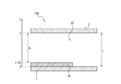
本実施形態において、血糖値測定チップ100は、図5に例示される所定のモデルによって表されるとする。血糖値算出方法は、所定のモデルを記述する連立方程式に対して、発色値の実測値と各パラメータの実測値とを代入し、連立方程式のパラメータを特定することによって、簡易検量線又は中間的な関係式を算出することを可能にする。
本実施形態において、血糖値測定チップ100は、図5に例示される所定のモデルによって表されるとする。血糖値算出方法は、所定のモデルを記述する連立方程式に対して、発色値の実測値と各パラメータの実測値とを代入し、連立方程式のパラメータを特定することによって、簡易検量線又は中間的な関係式を算出することを可能にする。
所定のモデルは、チップ厚み方向に沿うZ軸の座標で特定される一次元モデルとして血糖値測定チップ100を表す。所定のモデルにおいて、Z軸方向に沿って、第1基材1と、流路20と、第2基材2とが積層している。第1基材1及び第2基材2は、対向して位置することによって、流路20を区画している。第1基材1及び第2基材2それぞれの流路20側の面は、Z軸に直交している。第1基材1と第2基材2との間隔は、Lで表される。Z座標は、第1基材1の流路20側の面上に位置する点を原点(Z=0)とし、第2基材2の流路20側の面上に位置する点をZ=Lとして表される。
血糖値測定チップ100が流路20内に有する試薬部30は、モデルにおいて、第1基材1の流路20側の面に沿って配置されている。試薬部30は、第1面と第2面とを有する板状であるとする。試薬部30は、第1面で第1基材1の流路20側の面に接しており、第2面で第2基材2の流路20側の面に対向している。試薬部30の第2面と第2基材2の流路20側の面との間隔は、CLで表されるとする。試薬部30の第1面は、Z=0で表される座標に位置する。試薬部30の第2面は、Z=L-CLで表される座標に位置する。つまり、試薬部30の厚みは、L-CLで表される。
血糖値測定チップ100に検体が流入した時点における状態は、図6に示されるモデルによって表されるとする。図5における流路20に対応する領域は、検体によって満たされている。検体は全血であり、少なくとも血漿61と血球62とを含む。検体が流入した時点の時刻は、初期時刻とも称される。時刻は、tで表されるとする。初期時刻は、t=0で表される時刻に対応する。
血糖値測定チップ100に流入した検体は、試薬部30と混合し、血糖値測定試薬と反応する。サンプル中で、検体と血糖値測定試薬との反応によって生じる物質の拡散は、図7に示されるモデルを参照して定性的に説明される。血糖値測定試薬は、試薬部30から血漿61に溶解し、拡散する。矢印DAは、血糖値測定試薬の拡散を模式的に表している。血糖値測定試薬の拡散にともない、血糖値測定試薬が、血漿61中のグルコースと反応する。血糖値測定試薬とグルコースとの反応によって、血糖値測定試薬は、発色済色素63を発生させる。発色済色素63の分子は、血漿61中において溶解状態で存在する。発色済色素63は、血漿61に対して、濃度勾配に従って拡散する。矢印DCは、発色済色素63の拡散を模式的に表している。なお、図7の発色済色素63は、溶解した発色済色素63が特に多く存在する(濃い)部分を例示する。実際は、試薬部30から血漿61中へ溶解した血糖値測定試薬とグルコースとの反応によって、グルコースが消費され、血漿61中のグルコース量が減少する。その結果、血漿61中においてグルコースの濃度勾配が発生する。グルコースは、濃度勾配に基づいて血漿61中で拡散する。矢印DG1は、グルコースの拡散を模式的に表している。血漿61中のグルコースが減少すると、血球62中のグルコース濃度と血漿61中のグルコース濃度との間に差が生じる。血球62は、血球62内及び血球62外のグルコース濃度を調節する輸送機構(トランスポータ、主にGLUT-1)を血球膜上に有する。この輸送機構の働きにより、血球62内のグルコースが血漿61中に流出する。矢印DG2は、血球62から血漿61へのグルコースの移動を模式的に表している。所定のモデルは、Z軸方向への拡散と、Z軸方向に分布する各物質の濃度と、各物質の反応による濃度の変化とを表すことができる。つまり、所定のモデルは、一次元の座標系の拡散反応モデルとして表されるといえる。
<血糖値測定試薬の粘度変化>
血糖値測定試薬は、初期時刻からの経過時間が大きくなるほど、血漿61中を拡散していく。試薬部30に含まれる血糖値測定試薬は、初期時刻において、サンプルの粘度よりも十分に大きい粘度を有すると仮定する。つまり、試薬部30は、固体であるとみなされる。血漿61は、試薬部30の表面からその内部に浸透することによって、試薬部30に含まれる血糖値測定試薬の粘度を低下させる。試薬部30に含まれる血糖値測定試薬の粘度(μ)は、一般式として、以下の式(1)で表されるとする。

式(1)は、いわゆるシグモイド関数を表している。μは、z及びtの関数として表される。KmS及びnは、シグモイド関数の形状を決定する定数である。αは、試薬部30に塗布された血糖値測定試薬部の最大粘度を表しており、試薬部30が固体とみなされるように、サンプルの粘度よりも十分に大きい粘度に設定される。A及びBは、血糖値測定試薬に含まれる成分の濃度を表す変数であり、z及びtの関数として表される。血糖値測定試薬は、酵素等の比較的高い分子量(例えば、分子量1万以上)の成分と、色素等の比較的低い分子量(例えば、分子量1万未満)の成分とを含む。本実施形態において、Aは、血糖値測定試薬に含まれる酵素の濃度を表すとする。Bは、血糖値測定試薬に含まれる酵素以外の成分の濃度を表すとする。a及びbは、A及びBで表される成分の分子量に基づく重みづけのための定数である。高分子量の成分の濃度は、低分子量の成分の濃度よりも、粘度に大きく影響を及ぼす。Aによって表される成分の分子量がBによって表される成分の分子量より大きい場合、aは、bよりも大きい値に設定される。このようにすることで、酵素の濃度が適切に試薬部30の粘度に反映されうる。
血糖値測定試薬は、初期時刻からの経過時間が大きくなるほど、血漿61中を拡散していく。試薬部30に含まれる血糖値測定試薬は、初期時刻において、サンプルの粘度よりも十分に大きい粘度を有すると仮定する。つまり、試薬部30は、固体であるとみなされる。血漿61は、試薬部30の表面からその内部に浸透することによって、試薬部30に含まれる血糖値測定試薬の粘度を低下させる。試薬部30に含まれる血糖値測定試薬の粘度(μ)は、一般式として、以下の式(1)で表されるとする。
本実施形態において、血糖値測定試薬に含まれる成分は、Aで表される成分とBで表される成分との2種類に分けられるが、さらに他の成分に分けられてもよい。血糖値測定試薬に含まれる成分が増やされる場合、式(1)において、(a・A+b・B+・・・)として表されている部分に含まれている項数が2つより大きくされる。
<グルコースの拡散>
安定なグルコース水溶液中では、複数の異性体(アノマー)が平衡状態で存在する。同様に、血漿61中でも、グルコースは複数の異性体が平衡状態で存在する。しかしながら、血糖値測定試薬に使用する酵素(GDH)の基質特異性によっては、これら異性体の平衡状態に偏りが生じるため、血糖値測定反応に及ぼす影響を無視できない場合がある。本実施形態の酵素(GDH)は、βグルコースを基質として、血糖値測定試薬を発色させ、発色済色素63を生成する。一方で、βグルコース以外の異性体(αグルコース又はグルコースの鎖状体等)は、酵素(GDH)の基質とはならない(反応しない)。ゆえに、酵素反応によってβグルコースが消費されると、血漿61中では、αグルコースからβグルコースへの異性化が進むとともに、血漿61中のグルコースの総分子数は減少する。血漿61中のグルコース分子が減少するにつれ、血球62内のグルコースは、血球膜に存在する輸送機構によって、血漿61中へ流出する。血球62内のグルコースは、血糖値測定試薬と検体とが混合した際に、血漿61中へ流出する。なお、本実施形態では、存在比が大きいグルコース異性体として、αグルコースと、βグルコースとを考慮する。
安定なグルコース水溶液中では、複数の異性体(アノマー)が平衡状態で存在する。同様に、血漿61中でも、グルコースは複数の異性体が平衡状態で存在する。しかしながら、血糖値測定試薬に使用する酵素(GDH)の基質特異性によっては、これら異性体の平衡状態に偏りが生じるため、血糖値測定反応に及ぼす影響を無視できない場合がある。本実施形態の酵素(GDH)は、βグルコースを基質として、血糖値測定試薬を発色させ、発色済色素63を生成する。一方で、βグルコース以外の異性体(αグルコース又はグルコースの鎖状体等)は、酵素(GDH)の基質とはならない(反応しない)。ゆえに、酵素反応によってβグルコースが消費されると、血漿61中では、αグルコースからβグルコースへの異性化が進むとともに、血漿61中のグルコースの総分子数は減少する。血漿61中のグルコース分子が減少するにつれ、血球62内のグルコースは、血球膜に存在する輸送機構によって、血漿61中へ流出する。血球62内のグルコースは、血糖値測定試薬と検体とが混合した際に、血漿61中へ流出する。なお、本実施形態では、存在比が大きいグルコース異性体として、αグルコースと、βグルコースとを考慮する。
βグルコースのうち、初期時刻の時点から血漿61中に存在しているβグルコースの濃度(G1)の変化は、z軸方向における血漿61への拡散と、時間の経過に基づく変化と、αグルコースからβグルコースへの異性化とを考慮した、以下の式(2)によって記述される。

βグルコースのうち、血球62から流出するβグルコースの濃度(G2)の変化は、以下の式(3)によって記述される。

血漿中のαグルコースの濃度(Gα)の変化は、以下の式(4)によって記述される。

G1、G2及びGαは、z及びtの関数として表される。所定のモデルにおいて、血球62から流出するαグルコースは、本実施形態では計算式を簡略化するため考慮されていないが、考慮されてもよい。DGは、25℃の純水に対するグルコースの拡散係数を表す。図7のDG1が付された矢印は、血漿61中におけるβグルコースのZ軸方向の拡散を定性的に表している。図7のDG2が付された矢印は、血球62から血漿61へのβグルコースのZ軸方向の流出を定性的に表している。μ0は、サンプルの粘度を表す。(μ+μ0)で表される項は、拡散係数を粘度によって補正している。つまり、粘度が高いほど実効的な拡散係数が小さくなることを表している。VmaxMは、血球62中から血漿61へのグルコースの最大流出速度を表す。KmMは、血球62内部から血漿61への流出に関するミカエリス-メンテン定数である。Vmaxαは、αグルコースとβグルコースとの間の異性化反応の最大速度を表す。Kαは、αグルコースからβグルコースへの異性化反応の速度定数である。Kβは、βグルコースからαグルコースへの異性化反応の速度定数である。Kmαは、酵素の一種であるムタロターゼのミカエリス-メンテン定数である。ムタロターゼは、検体が全血の場合に、サンプル中に含まれている。
式(2)の右辺第1項は、初期時刻において、血漿61中に存在しているβグルコースのz軸方向への拡散を表す項である。式(3)の右辺第1項は、血球62中から流出するβグルコースのz軸方向への拡散を表す項である。式(4)の右辺第1項は、血漿61中のαグルコースのz軸方向への拡散を表す項である。拡散を表す項は、物質の拡散に関する基本法則であるフィックの第2法則に基づいて記述されている。
式(2)の右辺第3項、式(3)の右辺第3項、及び式(4)の右辺第2項は、αグルコースとβグルコースとの間の異性化反応の速度を表す項である。αグルコースとβグルコースは、平衡状態で存在している。式(2)及び(3)の右辺第3項は、βグルコースが増加する場合に正の値となる。式(4)の右辺第2項は、αグルコースが増加する場合に正の値となるように、マイナス符号が付されている。
式(2)及び(3)の右辺第2項のUG1及びUG2は、血糖値測定試薬とβグルコースとの反応速度を表す反応項で、ミカエリス-メンテンの式が適応される。反応様式又は試薬の特性に応じ、様々なミカエリス-メンテンの式のバリエーションから適したものを選択してよい。反応項は、酵素の濃度とグルコースの濃度とをパラメータとして反応速度を表す。反応項は、酵素の失活率を係数として含んでもよい。酵素の失活率が係数として含まれることによって、酵素の濃度が粘度に及ぼす影響と、反応に及ぼす影響とが独立に記述されうる。
<血糖値測定試薬の反応・拡散>
血糖値測定試薬の拡散は、以下の式(5)によって表される。

Aは、血糖値測定試薬のうち、Aで表される成分の濃度を表す変数であり、z及びtの関数として表される。DAは、血糖値測定試薬のうちAで表される成分の、25℃の純水に対する拡散係数を表す。式(5)の右辺第2項のUAは、Aで表される成分と、βグルコースとの反応速度を表す反応項である。Aで表される成分が酵素に対応する場合、Aで表される成分の量は、反応によって変化しない。したがって、UAは、無視されるとする。
血糖値測定試薬の拡散は、以下の式(5)によって表される。
<発色済色素の生成>
発色色素とβグルコースの反応によって色素は発色する(最大吸収波長がより長波長側に移行する)。初期時刻の時点から血漿61中に存在しているβグルコースの反応によって生成する発色済色素63の濃度(C1)の変化は、以下の式(6)によって表される。

血球62から流出するβグルコースの反応によって生成する発色済色素63の濃度(C2)の変化は、以下の式(7)によって表される。

C1及びC2は、z及びtの関数として表される。DCは、25℃の純水に対する発色済色素63の拡散係数を表す。UC1及びUC2は、発色済色素63が生成する反応の反応速度を表す項である。反応速度の算出には、ミカエリス-メンテン式を適宜立てればよい。なお、用いる試薬の反応様式に応じ、阻害因子等を考慮してもよい。
発色色素とβグルコースの反応によって色素は発色する(最大吸収波長がより長波長側に移行する)。初期時刻の時点から血漿61中に存在しているβグルコースの反応によって生成する発色済色素63の濃度(C1)の変化は、以下の式(6)によって表される。
<発色量>
血糖値測定試薬とβグルコースとの反応は、発色済色素63を生成する。発色済色素63の生成量(C)は、以下の式(8)によって算出される。

Cは、tの関数として表される。εCは、発色済色素63のモル吸光定数である。Htは、サンプル(検体)のヘマトクリット値を表す。Rvは、血球62内の溶液の比率を表す。式(8)は、Z=0からZ=Lまでの範囲における発色済色素63の濃度の積分を表している。
血糖値測定試薬とβグルコースとの反応は、発色済色素63を生成する。発色済色素63の生成量(C)は、以下の式(8)によって算出される。
以上述べてきたように、所定のモデルは、式(1)から式(8)までの各式によって記述される偏微分連立方程式によって表される。
<簡易検量線及び中間的な関係式の算出>
本実施形態に係る血糖値算出方法は、例えば以下の手順を含む。作業者は、血糖値、ヘマトクリット値及び温度等のパラメータが既知である検体を、血糖値測定装置110に装着されている血糖値測定チップ100に流入させ、吸光度(発色量)を測定する(第1ステップ)。血糖値測定装置110は、そのサンプルの吸光度の時間変化を測定する(第3ステップ)。血糖値測定装置110は、吸光度の時間変化が十分小さくなるまで吸光度を測定してよい。吸光度の時間変化が十分小さくなった場合、検体と血糖値測定試薬との反応が十分に進んだとみなされる。反応開始後の各時刻における吸光度は、サンプルのパラメータと対応づけられることによって、簡易検量線の作成に利用されてよい。サンプルのパラメータは、検体(血液)そのものに由来するパラメータと、サンプル(血液と試薬との混合物)のパラメータとが含まれる。
本実施形態に係る血糖値算出方法は、例えば以下の手順を含む。作業者は、血糖値、ヘマトクリット値及び温度等のパラメータが既知である検体を、血糖値測定装置110に装着されている血糖値測定チップ100に流入させ、吸光度(発色量)を測定する(第1ステップ)。血糖値測定装置110は、そのサンプルの吸光度の時間変化を測定する(第3ステップ)。血糖値測定装置110は、吸光度の時間変化が十分小さくなるまで吸光度を測定してよい。吸光度の時間変化が十分小さくなった場合、検体と血糖値測定試薬との反応が十分に進んだとみなされる。反応開始後の各時刻における吸光度は、サンプルのパラメータと対応づけられることによって、簡易検量線の作成に利用されてよい。サンプルのパラメータは、検体(血液)そのものに由来するパラメータと、サンプル(血液と試薬との混合物)のパラメータとが含まれる。
簡易検量線を算出するコンピュータは、上記手順で得られた、既知のパラメータを有するサンプルの吸光度の時間変化の実測データを所定のモデルに当てはめることによって、未実測のパラメータの組合せのサンプルに関する簡易検量線を算出できる。血糖値測定装置110は、未実測のパラメータの組合せで特定されるサンプルを測定する場合でも、サンプルのヘマトクリット値と温度とを取得し、コンピュータで算出済みの簡易検量線から、取得した値に適合する簡易検量線を選択できる。血糖値測定装置110は、選択した簡易検量線に、第1ステップで取得した吸光度の測定値を当てはめることによって、未実測のパラメータの組合せで特定されるサンプル(検体)の血糖値を算出できる(第2ステップ)。ここで、既知のパラメータとして、温度、血糖値、及びヘマトクリット値があげられる。なお、第3ステップを、第1ステップ及びまたは第2ステップに先立って行ってもよい。
簡易検量線を算出するコンピュータは、実測されていないサンプルを血糖値測定チップ100に流入させた場合のサンプルの吸光度の変化を、実測データに基づいて、所定のモデルに基づくモデリング式の形式で予測できる。
(実施例)
以下、血糖値測定チップ100の一次元モデルを表す偏微分連立方程式に基づいて吸光度の時間変化を予測するモデリング式を算出する具体例が説明される。
以下、血糖値測定チップ100の一次元モデルを表す偏微分連立方程式に基づいて吸光度の時間変化を予測するモデリング式を算出する具体例が説明される。
血糖値測定試薬に含まれる成分のうち、色素等の比較的低い分子量(例えば、分子量1万未満)の成分は、低分子量成分とも称される。低分子量成分の濃度は、Wとして表されるとする。この場合、Wは、以下の式(9)によって表される。

Wは、z及びtの関数として表される。DWは、低分子量成分の、25℃の純水に対する拡散係数を表す。
血糖値測定試薬に含まれる成分のうち、酵素等の比較的高い分子量(例えば、分子量1万以上)の成分は、高分子量成分とも称される。高分子量成分の濃度は、Eとして表されるとする。この場合、Eは、以下の式(10)によって表される。
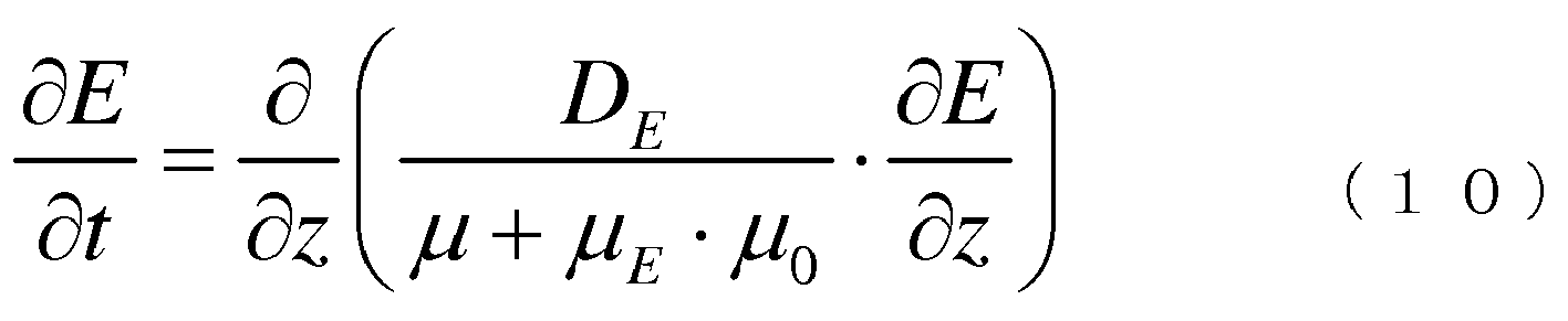
Eは、z及びtの関数として表される。DEは、高分子量成分の、25℃の純水に対する拡散係数を表す。μEは、高分子量成分の相互作用を補正する係数である。
血漿61中のαグルコースの濃度(Gα)の変化は、以下の式(11)によって記述される。

式(11)は、上述の式(4)と同一である。式(11)に含まれる変数の説明は省略される。
βグルコースのうち、初期時刻の時点から血漿61中に存在しているβグルコースの濃度(G1)の変化は、z軸方向における血漿61への拡散と、時間の経過に基づく変化と、αグルコースからβグルコースへの異性化とを考慮した、以下の式(12)によって記述される。

式(12)の右辺第2項において、上述の式(2)におけるUG1の項が具体的に表されている。REは、酵素Eの残存活性比を表し、RE?1である。S0は、発色色素(テトラゾリウム化合物)が血漿に溶解して均一になったときの濃度(単位:mM)を表している。
KiTは、S0において、酵素とグルコースの反応に対する阻害量を表す係数である。KmGは、グルコースを基質とした場合のミカエリス―メンテン定数である。
KiTは、S0において、酵素とグルコースの反応に対する阻害量を表す係数である。KmGは、グルコースを基質とした場合のミカエリス―メンテン定数である。
血球62から流出するβグルコースの濃度(GB)は、以下の式(13)によって記述される。

式(13)の右辺は、上述の式(3)の右辺第3項に対応する。G1は、初期時刻の時点から血漿61中に存在しているβグルコースの濃度を表す。G2は、血球62から流出するβグルコースの濃度を表す。VmaxMは、血球62中から血漿61へのグルコースの最大流出速度を表す。KmMは、血球62内部から血漿61への流出に関するミカエリス-メンテン定数である。
βグルコースのうち、血球62から流出するβグルコースの濃度(G2)の変化は、以下の式(14)によって記述される。

本実施形態では、発色試薬とβグルコースとの反応によって中間体が生成する反応についても考慮することができる。検体が全血である場合、サンプル中の血色素の影響を受けにくい波長領域の測定波長で発色量を測定する必要がある。発色色素としてテトラゾリウム塩が用いられる場合、血糖値測定試薬が遷移金属イオンをさらに含むことによって、酵素反応によって生成するホルマザン(中間体)と、遷移金属イオンとをキレート形成させ、より長波長の測定波長で発色済色素63を検出することができる。このように、本実施例では、発色済色素63の前駆体として、血糖値測定試薬とβグルコースとの反応によって中間体(キレート形成前のホルマザン)が生成する。中間体のうち、初期時刻の時点から血漿61中に存在しているβグルコースの反応によって生成する中間体の濃度(F1)は、以下の式(15)によって表される。

中間体のうち、血球62から流出するβグルコースの反応によって生成する中間体の濃度(F2)は、以下の式(16)によって表される。

F1及びF2は、z及びtの関数として表される。C1は、初期時刻の時点から血漿61中に存在しているβグルコースの反応によって生成する中間体から変化する発色済色素63の濃度を表す。C2は、血球62から流出するβグルコースの反応によって生成する中間体から変化する発色済色素63の濃度を表す。DFは、25℃の純水に対する中間体の拡散係数を表す。右辺第2項は、酵素とβグルコースとの反応速度を表す反応項である。反応速度は、酵素の濃度と、活性である酵素の割合と、グルコースの濃度とに基づく。KNiは、中間体から発色済色素63へ変化する速度を表す定数である。KFは、発色済色素63から中間体へ変化する速度を表す定数である。
中間体の少なくとも一部は、発色済色素63に変化する。初期時刻の時点から血漿61中に存在しているβグルコースの反応によって生成する中間体から変化する発色済色素63の濃度(C1)の変化は、以下の式(17)によって表される。

血球62から流出するβグルコースの反応によって生成する中間体から変化する発色済色素63の濃度(C2)の変化は、以下の式(18)によって表される。

C1及びC2は、z及びtの関数として表される。DCは、25℃の純水に対する発色済色素63の拡散係数を表す。
血糖値測定試薬とβグルコースとの反応は、発色済色素63を生成する。発色済色素63の生成量(C)は、以下の式(19)によって算出される。なお、100/1000は単位を揃えるための係数である。

Cは、tの関数として表される。εCは、発色済色素63のモル吸光定数である。Htは、サンプル(検体)のヘマトクリット値を表す。Rvは、血球62内の溶液の比率を表す。式(19)は、Z=0からZ=Lまでの範囲における発色済色素63の濃度の積分を表している。
Wの初期値は、以下の式(20)によって表されるとする。
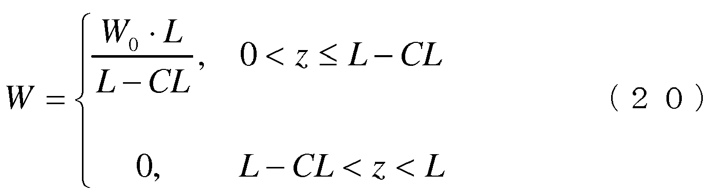
W0は、Wの初期値を決定するための定数である。
Eの初期値は、以下の式(21)によって表されるとする。
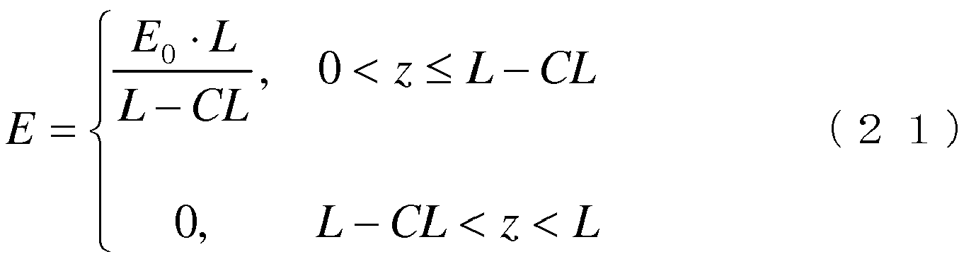
E0は、Eの初期値を決定するための定数である。
G1の初期値は、以下の式(22)によって表されるとする。

BGは、サンプルの血糖値(単位:mg/dL)を表している。
Gαの初期値は、以下の式(23)によって表されるとする。
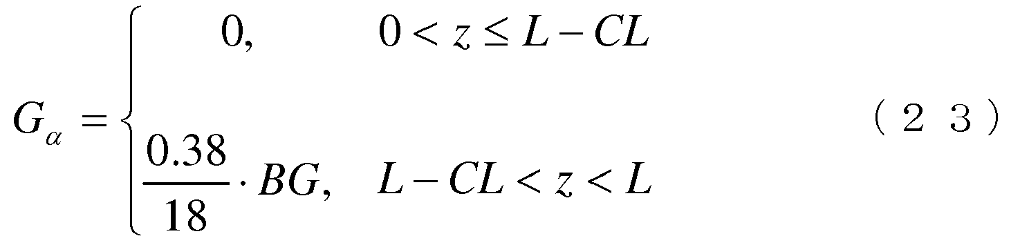
GBの初期値は、以下の式(24)によって表されるとする。
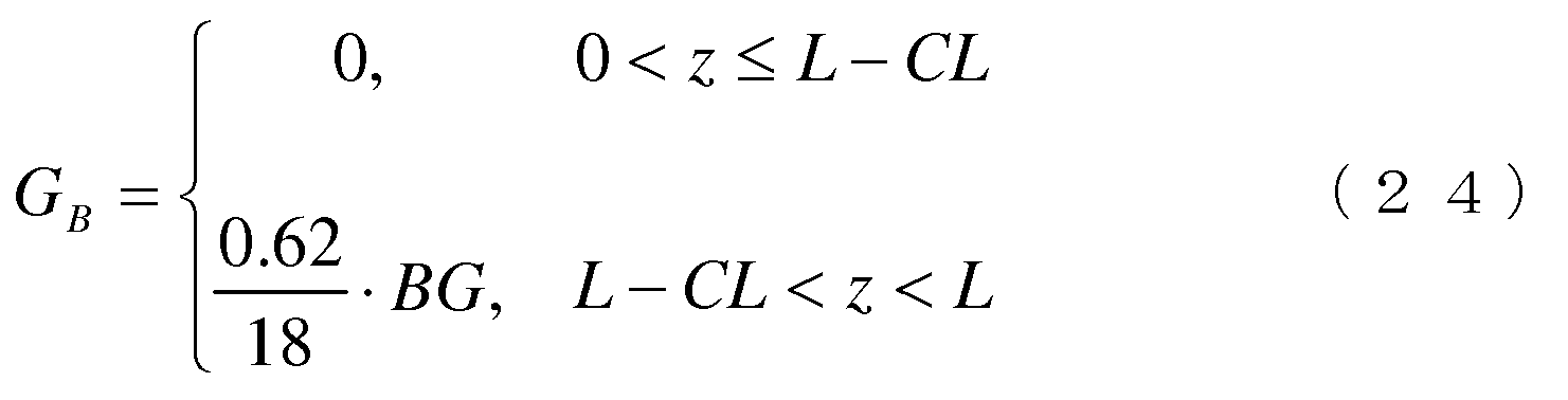
G2、F1、F2、C1及びC2の初期値は、zの値にかかわらず、0であるとする。
境界条件は、以下の式(25)で表されるとする。

定数は、以下の通りであるとする。
DW=6.0×10⁻¹⁰,DE=3.2×10⁻¹¹,DG=6.6×10⁻¹⁰,
DF=2.9×10⁻¹⁰,DC=1.9×10⁻¹⁰,Rv=0.8,
KmM=0.9,Kβ/Kα=0.61
これらの値は、公知の値を採用することができる。
また次の定数は、配合する試薬成分に固有の物性値、調製した血糖値測定試薬および血糖値測定チップを、実際に測定することより求めた値である。
L=46.5×10⁻⁶,CL=43.0×10⁻⁶,W0=165,E0=30,
Ed=5,S0=50,KmG=5,KiT=200,εC=47000,RM=0.83
Edは、60℃で3日間処理後の酵素の濃度を表している。
RMは、酵素活性量を重量濃度に換算する定数である。
下記の値については、直接測定することが困難なため、いつくかの既知検体の発色量(C)に上記式(19)の結果をフィッティングさせるため経験則的に調整した値である。

Tempは、サンプルの温度(単位:℃)を表している。
μ0は、直接測定した値を使用してもよい。測定が困難である場合は、検体の血糖値、ヘマトクリット値および測定温度を別途測定し、上記式(9)から式(19)までの関係式を使用して、算出した値と検体のヘマトクリット値および測定温度より関係式を見出したうえで、得られた関係式より予測した値を使用してもよい。関係式の算出方法は特に限定されることなく、多項式による最適化等、周知の方法が使用できる。
DW=6.0×10⁻¹⁰,DE=3.2×10⁻¹¹,DG=6.6×10⁻¹⁰,
DF=2.9×10⁻¹⁰,DC=1.9×10⁻¹⁰,Rv=0.8,
KmM=0.9,Kβ/Kα=0.61
これらの値は、公知の値を採用することができる。
また次の定数は、配合する試薬成分に固有の物性値、調製した血糖値測定試薬および血糖値測定チップを、実際に測定することより求めた値である。
L=46.5×10⁻⁶,CL=43.0×10⁻⁶,W0=165,E0=30,
Ed=5,S0=50,KmG=5,KiT=200,εC=47000,RM=0.83
Edは、60℃で3日間処理後の酵素の濃度を表している。
RMは、酵素活性量を重量濃度に換算する定数である。
下記の値については、直接測定することが困難なため、いつくかの既知検体の発色量(C)に上記式(19)の結果をフィッティングさせるため経験則的に調整した値である。
μ0は、直接測定した値を使用してもよい。測定が困難である場合は、検体の血糖値、ヘマトクリット値および測定温度を別途測定し、上記式(9)から式(19)までの関係式を使用して、算出した値と検体のヘマトクリット値および測定温度より関係式を見出したうえで、得られた関係式より予測した値を使用してもよい。関係式の算出方法は特に限定されることなく、多項式による最適化等、周知の方法が使用できる。
本実施例において、式(9)から式(19)までを含む偏微分連立方程式を上述の初期値と境界条件と定数とに基づいて解くことによって、図8に例示される、吸光度の時間変化を予測するモデリング式が算出された。横軸は、時間を表している。縦軸は、サンプルの吸光度を表している。破線は、吸光度の時間変化を予測するモデリング式を表している。図8の例において、血糖値が400mg/dLであり、且つ、温度が25℃であるサンプルについて、ヘマトクリット値を変数とするモデリング式が示されている。ヘマトクリット値は、0%から70%まで10%間隔で変更されている。これらの予測を表すグラフに対して、実線で示されるグラフは、予測の前提となるパラメータを実際に有するサンプルについて吸光度の時間変化を実測したデータを表している。ヘマトクリット値が0%から70%までのいずれの値であっても、モデリング式の吸光度に対する、実測した吸光度の誤差は、10%以内に収まっている。したがって、本実施形態に係る血糖値算出方法は、血糖値測定チップ100における血糖値測定試薬と検体との反応を所定のモデルでモデリングすることによって、未実測のパラメータの組合せで特定される、サンプルにおける反応を高精度で予測できた。
吸光度の時間変化を表すモデリング式が高精度で予測されることによって、血糖値測定装置110は、サンプルと血糖値測定試薬との反応開始から反応が十分に進むまで待つことなく、短時間で血糖値を算出できる。例えば、サンプルと血糖値測定試薬との反応が十分に進むまで10秒以上かかる場合であっても、血糖値測定装置110は、10秒未満の所定の時間で血糖値の算出結果を測定作業者に知らせることができる。その結果、血糖値測定装置110の利便性が向上する。
簡易検量線を算出するコンピュータによって算出されるモデリング式は、未実測のパラメータの組合せのサンプルに関する簡易検量線を算出するための中間的な関係式として用いられうる。
本実施形態に係る血糖値算出方法によれば、少ない実測データに基づいて血糖値を測定するための簡易検量線を算出することが可能になる。その結果、血糖値測定装置110の開発期間の短縮、及び、開発コストの低減が実現される。
本開示に係る構成は、上述した実施形態で特定された構成に限定されず、特許請求の範囲に記載した発明の要旨を逸脱しない範囲内で種々の変形が可能である。例えば、各構成部又は各ステップなどに含まれる機能等は論理的に矛盾しないように再構成可能であり、複数の構成部又はステップ等を1つに組み合わせたり、分割したりすることが可能である。
1、2 第1基材、第2基材
3、4 接着部
10 供給口
20 流路
21 開口部
22 装着部
23 収容部
24 光学測定部
25 制御部
26 イジェクトピン
30 試薬部
31 照射部
32 受光部
33 照射光
41、42 第1の空間、第2の空間
51、52 第1の発光素子、第2の発光素子
61 血漿
62 血球
63 発色済色素
100 血糖値測定チップ
110 血糖値測定装置
111 表示部
112 電源ボタン
113 操作ボタン
114 取外レバー
500 血糖値測定システム
3、4 接着部
10 供給口
20 流路
21 開口部
22 装着部
23 収容部
24 光学測定部
25 制御部
26 イジェクトピン
30 試薬部
31 照射部
32 受光部
33 照射光
41、42 第1の空間、第2の空間
51、52 第1の発光素子、第2の発光素子
61 血漿
62 血球
63 発色済色素
100 血糖値測定チップ
110 血糖値測定装置
111 表示部
112 電源ボタン
113 操作ボタン
114 取外レバー
500 血糖値測定システム
Claims (6)
- 血漿と血球とを含む検体に含まれるグルコースの量に応じて発色する発色色素を含む試薬と検体とを反応させて得られる前記発色色素の発色量を測定する第1ステップと、
前記発色量に基づいて前記検体の血糖値を算出するために、所定のモデルに基づいて前記発色量と前記検体の血糖値との関係式を算出する第2ステップと、をプロセッサに実行させ、
前記検体は、少なくとも前記血糖値を含む所定のパラメータを有し、
前記所定のモデルは、一次元の座標系の拡散反応モデルとして表されており、
前記拡散反応モデルは、前記試薬の粘度の時間変化と、前記血漿に含まれるグルコースの濃度の時間変化と、前記試薬の濃度の時間変化と、前記試薬の発色量の時間変化とによって表される、血糖値算出プログラム。 - 前記試薬の発色量は、検体の血漿中のグルコースと、前記試薬と検体が混合した際に血球内から血漿中へ流出するグルコースと、に基づく、請求項1に記載の血糖値算出プログラム。
- 前記所定のパラメータとして既知のパラメータを有する前記検体を前記試薬と反応させた場合における前記試薬の発色量の時間変化の測定結果を取得する第3ステップを前記プロセッサにさらに実行させ、
前記第2ステップにおいて、前記第3ステップで取得した前記試薬の発色量の時間変化の測定結果と既知のパラメータとの関係に基づいて、前記関係式を算出する、請求項1又は2に記載の血糖値算出プログラム。 - 前記所定のパラメータは、前記検体の中の前記血球の比率を表すヘマトクリット値と、前記検体の温度とをさらに含む、請求項1乃至3に記載の血糖値算出プログラム。
- 血漿と血球とを含む検体に含まれるグルコースの量に応じて発色する発色色素を含む試薬と検体とを反応させて得られる前記発色色素の発色量を測定する第1ステップと、
前記発色量に基づいて前記検体の血糖値を算出するために、所定のモデルに基づいて前記発色量と前記検体の血糖値との関係式を算出する第2ステップと、
を含み、
前記検体は、少なくとも前記血糖値を含む所定のパラメータを有し、
前記所定のモデルは、一次元の座標系の拡散反応モデルとして表されており、
前記拡散反応モデルは、前記試薬の粘度の時間変化と、前記血漿に含まれるグルコースの濃度の時間変化と、前記試薬の濃度の時間変化と、前記試薬の発色量の時間変化とによって表される、血糖値算出方法。 - 血漿と血球とを含む検体に含まれるグルコースの量に応じて発色する発色色素を含む試薬と検体とを反応させ得られる前記発色色素の発色量の測定結果に基づいて前記検体の血糖値を算出する制御部を備え、
前記制御部は、所定のモデルに基づいて前記測定結果と前記検体の血糖値との関係式を算出し、前記関係式に基づいて前記検体の血糖値を算出し、
前記検体は、少なくとも前記血糖値を含む所定のパラメータを有し、
前記所定のモデルは、一次元の座標系の拡散反応モデルとして表されており、
前記拡散反応モデルは、前記試薬の粘度の時間変化と、前記血漿に含まれるグルコースの濃度の時間変化と、前記試薬の濃度の時間変化と、前記試薬の発色量の時間変化とによって表される、血糖値測定装置。
Priority Applications (1)
| Application Number | Priority Date | Filing Date | Title |
|---|---|---|---|
| JP2021505557A JP7372310B2 (ja) | 2019-03-11 | 2020-01-21 | 血糖値算出プログラム、血糖値算出方法及び血糖値測定装置 |
Applications Claiming Priority (2)
| Application Number | Priority Date | Filing Date | Title |
|---|---|---|---|
| JP2019-044205 | 2019-03-11 | ||
| JP2019044205 | 2019-03-11 |
Publications (1)
| Publication Number | Publication Date |
|---|---|
| WO2020183916A1 true WO2020183916A1 (ja) | 2020-09-17 |
Family
ID=72427256
Family Applications (1)
| Application Number | Title | Priority Date | Filing Date |
|---|---|---|---|
| PCT/JP2020/001985 Ceased WO2020183916A1 (ja) | 2019-03-11 | 2020-01-21 | 血糖値算出プログラム、血糖値算出方法及び血糖値測定装置 |
Country Status (2)
| Country | Link |
|---|---|
| JP (1) | JP7372310B2 (ja) |
| WO (1) | WO2020183916A1 (ja) |
Citations (5)
| Publication number | Priority date | Publication date | Assignee | Title |
|---|---|---|---|---|
| JP2000262298A (ja) * | 1999-03-15 | 2000-09-26 | Fuji Photo Film Co Ltd | 全血中のグルコース濃度もしくはコレステロール濃度の定量方法 |
| US6168957B1 (en) * | 1997-06-25 | 2001-01-02 | Lifescan, Inc. | Diagnostic test strip having on-strip calibration |
| WO2009081790A1 (ja) * | 2007-12-20 | 2009-07-02 | Terumo Kabushiki Kaisha | 血糖測定値管理システム及び血糖値測定装置 |
| JP2015179038A (ja) * | 2014-03-19 | 2015-10-08 | テルモ株式会社 | 測定用チップ |
| WO2017122485A1 (ja) * | 2016-01-12 | 2017-07-20 | テルモ株式会社 | 成分測定装置、成分測定方法及び成分測定プログラム |
-
2020
- 2020-01-21 JP JP2021505557A patent/JP7372310B2/ja active Active
- 2020-01-21 WO PCT/JP2020/001985 patent/WO2020183916A1/ja not_active Ceased
Patent Citations (5)
| Publication number | Priority date | Publication date | Assignee | Title |
|---|---|---|---|---|
| US6168957B1 (en) * | 1997-06-25 | 2001-01-02 | Lifescan, Inc. | Diagnostic test strip having on-strip calibration |
| JP2000262298A (ja) * | 1999-03-15 | 2000-09-26 | Fuji Photo Film Co Ltd | 全血中のグルコース濃度もしくはコレステロール濃度の定量方法 |
| WO2009081790A1 (ja) * | 2007-12-20 | 2009-07-02 | Terumo Kabushiki Kaisha | 血糖測定値管理システム及び血糖値測定装置 |
| JP2015179038A (ja) * | 2014-03-19 | 2015-10-08 | テルモ株式会社 | 測定用チップ |
| WO2017122485A1 (ja) * | 2016-01-12 | 2017-07-20 | テルモ株式会社 | 成分測定装置、成分測定方法及び成分測定プログラム |
Also Published As
| Publication number | Publication date |
|---|---|
| JP7372310B2 (ja) | 2023-10-31 |
| JPWO2020183916A1 (ja) | 2020-09-17 |
Similar Documents
| Publication | Publication Date | Title |
|---|---|---|
| CN108368472B (zh) | 血糖值测定试剂、血糖值测定芯片和血糖值测定装置套组 | |
| US11150193B2 (en) | Method, composition, and chip for detecting analyte in blood sample | |
| JPS6361147A (ja) | 分析方法 | |
| JP7208219B2 (ja) | 生体試料中の分析対象物濃度を測定するための、方法、試薬およびチップ | |
| JP2022179961A (ja) | アンモニア又は尿素測定用試験片 | |
| JP2022186748A (ja) | 血糖値測定チップ及び血糖値測定装置セット | |
| US11739083B2 (en) | Disposable sensor chip with reagent including 2-substituted benzothiazolyl-3-substituted phenyl-5-substituted sulfonated phenyl-2H-tetrazolium salt | |
| JP2013165709A (ja) | 乾燥試薬の製造方法、乾燥試薬、およびそれを用いた分析用具 | |
| EP4086613A1 (en) | Component measurement device, component measurement device set, and information processing method | |
| WO2009119213A1 (ja) | ヘマトクリット値または血液成分濃度の測定方法および測定装置 | |
| JP7372310B2 (ja) | 血糖値算出プログラム、血糖値算出方法及び血糖値測定装置 | |
| WO2023112442A1 (ja) | 生体成分濃度測定試薬、測定方法およびセンサ | |
| JP2023087330A (ja) | 生体成分濃度測定試薬、測定方法およびセンサ | |
| JP2023087327A (ja) | 生体成分濃度測定試薬、測定方法およびセンサ | |
| US12339221B2 (en) | Component measurement device, component measurement device set, and information processing method | |
| EP4644875A1 (en) | Component measurement device and component measurement method | |
| HK1257848B (en) | Blood-sugar-level measurement reagent, blood-sugar-level measurement chip, and blood-sugar-level measurement device set | |
| HK40012315B (en) | Blood sugar level measurement chip and blood sugar level measurement device set | |
| HK40012315A (en) | Blood sugar level measurement chip and blood sugar level measurement device set | |
| JP2004317307A (ja) | 試薬部対面分析用具およびこの分析用具の製造方法 | |
| JP2017142199A (ja) | 血糖値測定試薬、血糖値測定チップ、及び血糖値測定装置セット | |
| JP2004317308A (ja) | 小ギャップ比色分析用具 |
Legal Events
| Date | Code | Title | Description |
|---|---|---|---|
| 121 | Ep: the epo has been informed by wipo that ep was designated in this application |
Ref document number: 20770128 Country of ref document: EP Kind code of ref document: A1 |
|
| ENP | Entry into the national phase |
Ref document number: 2021505557 Country of ref document: JP Kind code of ref document: A |
|
| NENP | Non-entry into the national phase |
Ref country code: DE |
|
| 122 | Ep: pct application non-entry in european phase |
Ref document number: 20770128 Country of ref document: EP Kind code of ref document: A1 |