WO2020032221A1 - アルギン酸中空マイクロファイバ - Google Patents
アルギン酸中空マイクロファイバ Download PDFInfo
- Publication number
- WO2020032221A1 WO2020032221A1 PCT/JP2019/031538 JP2019031538W WO2020032221A1 WO 2020032221 A1 WO2020032221 A1 WO 2020032221A1 JP 2019031538 W JP2019031538 W JP 2019031538W WO 2020032221 A1 WO2020032221 A1 WO 2020032221A1
- Authority
- WO
- WIPO (PCT)
- Prior art keywords
- cells
- producing
- cho cells
- alginate gel
- alginate
- Prior art date
- Legal status (The legal status is an assumption and is not a legal conclusion. Google has not performed a legal analysis and makes no representation as to the accuracy of the status listed.)
- Ceased
Links
Images


Classifications
-
- C—CHEMISTRY; METALLURGY
- C12—BIOCHEMISTRY; BEER; SPIRITS; WINE; VINEGAR; MICROBIOLOGY; ENZYMOLOGY; MUTATION OR GENETIC ENGINEERING
- C12M—APPARATUS FOR ENZYMOLOGY OR MICROBIOLOGY; APPARATUS FOR CULTURING MICROORGANISMS FOR PRODUCING BIOMASS, FOR GROWING CELLS OR FOR OBTAINING FERMENTATION OR METABOLIC PRODUCTS, i.e. BIOREACTORS OR FERMENTERS
- C12M25/00—Means for supporting, enclosing or fixing the microorganisms, e.g. immunocoatings
- C12M25/10—Hollow fibers or tubes
- C12M25/12—Hollow fibers or tubes the culture medium flowing outside the fiber or tube
-
- C—CHEMISTRY; METALLURGY
- C07—ORGANIC CHEMISTRY
- C07K—PEPTIDES
- C07K16/00—Immunoglobulins [IG], e.g. monoclonal or polyclonal antibodies
-
- C—CHEMISTRY; METALLURGY
- C07—ORGANIC CHEMISTRY
- C07K—PEPTIDES
- C07K16/00—Immunoglobulins [IG], e.g. monoclonal or polyclonal antibodies
- C07K16/18—Immunoglobulins [IG], e.g. monoclonal or polyclonal antibodies against material from animals or humans
- C07K16/28—Immunoglobulins [IG], e.g. monoclonal or polyclonal antibodies against material from animals or humans against receptors, cell surface antigens or cell surface determinants
- C07K16/2866—Immunoglobulins [IG], e.g. monoclonal or polyclonal antibodies against material from animals or humans against receptors, cell surface antigens or cell surface determinants against receptors for cytokines, lymphokines, interferons
-
- C—CHEMISTRY; METALLURGY
- C12—BIOCHEMISTRY; BEER; SPIRITS; WINE; VINEGAR; MICROBIOLOGY; ENZYMOLOGY; MUTATION OR GENETIC ENGINEERING
- C12M—APPARATUS FOR ENZYMOLOGY OR MICROBIOLOGY; APPARATUS FOR CULTURING MICROORGANISMS FOR PRODUCING BIOMASS, FOR GROWING CELLS OR FOR OBTAINING FERMENTATION OR METABOLIC PRODUCTS, i.e. BIOREACTORS OR FERMENTERS
- C12M25/00—Means for supporting, enclosing or fixing the microorganisms, e.g. immunocoatings
- C12M25/14—Scaffolds; Matrices
-
- C—CHEMISTRY; METALLURGY
- C12—BIOCHEMISTRY; BEER; SPIRITS; WINE; VINEGAR; MICROBIOLOGY; ENZYMOLOGY; MUTATION OR GENETIC ENGINEERING
- C12M—APPARATUS FOR ENZYMOLOGY OR MICROBIOLOGY; APPARATUS FOR CULTURING MICROORGANISMS FOR PRODUCING BIOMASS, FOR GROWING CELLS OR FOR OBTAINING FERMENTATION OR METABOLIC PRODUCTS, i.e. BIOREACTORS OR FERMENTERS
- C12M25/00—Means for supporting, enclosing or fixing the microorganisms, e.g. immunocoatings
- C12M25/16—Particles; Beads; Granular material; Encapsulation
-
- C—CHEMISTRY; METALLURGY
- C12—BIOCHEMISTRY; BEER; SPIRITS; WINE; VINEGAR; MICROBIOLOGY; ENZYMOLOGY; MUTATION OR GENETIC ENGINEERING
- C12N—MICROORGANISMS OR ENZYMES; COMPOSITIONS THEREOF; PROPAGATING, PRESERVING, OR MAINTAINING MICROORGANISMS; MUTATION OR GENETIC ENGINEERING; CULTURE MEDIA
- C12N5/00—Undifferentiated human, animal or plant cells, e.g. cell lines; Tissues; Cultivation or maintenance thereof; Culture media therefor
- C12N5/0068—General culture methods using substrates
-
- C—CHEMISTRY; METALLURGY
- C07—ORGANIC CHEMISTRY
- C07K—PEPTIDES
- C07K2317/00—Immunoglobulins specific features
- C07K2317/10—Immunoglobulins specific features characterized by their source of isolation or production
- C07K2317/14—Specific host cells or culture conditions, e.g. components, pH or temperature
-
- C—CHEMISTRY; METALLURGY
- C07—ORGANIC CHEMISTRY
- C07K—PEPTIDES
- C07K2317/00—Immunoglobulins specific features
- C07K2317/20—Immunoglobulins specific features characterized by taxonomic origin
- C07K2317/24—Immunoglobulins specific features characterized by taxonomic origin containing regions, domains or residues from different species, e.g. chimeric, humanized or veneered
-
- C—CHEMISTRY; METALLURGY
- C12—BIOCHEMISTRY; BEER; SPIRITS; WINE; VINEGAR; MICROBIOLOGY; ENZYMOLOGY; MUTATION OR GENETIC ENGINEERING
- C12N—MICROORGANISMS OR ENZYMES; COMPOSITIONS THEREOF; PROPAGATING, PRESERVING, OR MAINTAINING MICROORGANISMS; MUTATION OR GENETIC ENGINEERING; CULTURE MEDIA
- C12N2533/00—Supports or coatings for cell culture, characterised by material
- C12N2533/70—Polysaccharides
- C12N2533/74—Alginate
Definitions
- the present invention relates to an alginate gel fiber for producing an antibody, a method for producing the gel fiber, and a method for producing an antibody using the gel fiber.
- Antibody production is being performed using genetic recombination technology. Until now, various antibodies have been produced using CHO cells, Sp2 / 0 cells, NS0 cells, Escherichia coli and the like as antibody-producing cells.
- CHO cells derived from Chinese hamster ovaries
- CHO cells are cells that can be cultured in suspension, have a high cell growth rate, and can easily mass-produce the target protein by mass culture of CHO cells. Frequently used for antibody culture.
- Patent Document 1 International Publication No. 2011/046105 pamphlet
- Patent Document 2 International Publication No. 2015/178427 pamphlet
- Patent Document 3 JP-A-2014-236698
- Patent Document 4 Japanese Patent Application Laid-Open No. 2016-77229)
- Patent Document 5 Japanese Patent Application Laid-Open No. 2017-77473
- Non-Patent Document 1 A method for producing a meter-length core-shell hydrogel microfiber encapsulating extracellular matrix (ECM) proteins and differentiated cells or somatic stem cells using a microfluidic device having double coaxial laminar flow is known.
- ECM extracellular matrix
- Non-Patent Document 2 Cell-containing hydrogel microfibers having improved mechanical properties and handleability using a double network (DN) hydrogel composed of alginate and polyacrylamide are known.
- Patent Documents 1 to 5 and Non-patent Documents 1 and 2 disclose that a core layer containing antibody-producing cells is formed of an alginate gel having a high mechanical strength (preferably, an alginate gel having a higher mechanical strength than the core layer). Alginate gel fibers having high mechanical strength covered with a shell layer containing the gel), and a method for producing an antibody using the alginate gel fibers are not disclosed or suggested.
- a core layer containing antibody-producing cells (for example, CHO cells and the like) and a substrate (for example, a solution or a gel of collagen, a medium, alginic acid, etc.) has been cross-linked to a cross-linked alginate gel (for example, a naturally occurring cross-linked alginate gel)
- a cross-linked alginate gel for example, a naturally occurring cross-linked alginate gel
- alginate gel fibers for antibody production coated with a shell layer containing, and production of recombinant antibodies using the gel fibers Furthermore, there has not been known a method of forming the alginate gel fiber having a certain length or a method of continuously producing an antibody using the alginate gel fiber.
- the core layer containing the antibody-producing cells and the substrate has a high core layer.
- a substrate for example, a substrate selected from the group consisting of a solution or a gel of collagen, a medium, alginic acid, etc.
- an alginate gel fiber for producing antibodies can be prepared by coating with a shell layer containing an alginate gel having mechanical strength (preferably, an alginate gel having higher mechanical strength than the core layer). Further, when the antibody-producing cells were cultured using the alginate gel fiber thus produced, they found that antibodies could be produced continuously for a long period of time, and completed the present invention.
- a CHO cell incorporating an antibody gene as an antibody-producing cell and a core layer containing a collagen gel as a substrate were coated with a calcium-crosslinked alginate gel having high mechanical strength (preferably, a mechanical layer having a higher mechanical strength than the core layer).
- Alginate gel fiber with high mechanical strength is produced by coating with a shell layer containing high-strength calcium cross-linked alginate gel), and culturing the gel fiber to produce antibodies (genetical recombination) continuously for a long period of time I knew I could do it.
- the produced alginate gel fiber provides an environment suitable for continuous production of antibody for antibody-producing CHO cells, and the antibody produced in the core layer continuously penetrates the shell layer and out of the gel fiber. was released.
- the core layer comprising the antibody-producing cells and the substrate is coated with a shell layer comprising an alginate gel having high mechanical strength (preferably, an alginate gel having higher mechanical strength than the core layer). By doing so, an alginate gel fiber having high mechanical strength is provided.
- the core layer containing the antibody-producing cells is coated with a calcium cross-linked alginate gel formed from a sodium alginate solution (A-2 or B-2) to form a shell with high mechanical strength.
- the alginate gel fiber of the preferred embodiment provides an environment suitable for producing antibodies. Since the cells are encapsulated in the core layer, there is little physical stress on the antibody-producing cells in the culture solution, and the encapsulated antibody-producing cells are expected to continue producing antibodies for a long period of time. Therefore, the method for producing an antibody using such an alginate gel fiber can be expected to dramatically improve the antibody production efficiency. For example, unlike suspension culture of an antibody that requires a large-scale culture tank, it is expected that the antibody will be produced by a small-scale production facility. It is also expected to be a continuous production technology for next-generation antibody drugs that is suitable for the production of small and large numbers of antibody drugs.
- FIG. 3 is a schematic diagram illustrating a cross section of an alginate gel fiber containing antibody-producing cells in a core layer, and that the produced antibody, metabolites, waste products, culture solution (nutrient source), and oxygen permeate the shell layer. is there. It is a microscope picture after culture
- a first aspect is an alginate gel fiber formed by covering a core layer containing antibody-producing cells and a substrate with a shell layer containing alginate gel having high mechanical strength.
- the antibody-producing cells contained in the core layer are, for example, CHO cells, CHO cell sublines, COS cells, Sp2 / 0 cells, NS0 cells, SP2 cells, PERC6 cells, YB2 / 0 cells, YE2 / 0 cells, 1R983F cells, Namalwa cells, Wil-2 cells, Jurkat cells, Vera cells, Molt-4 cells, 293-HEK cells, BHK cells, KGH6 cells, P3X63Ag8.653 cells, C127 cells, A cell selected from the group consisting of JC cells, LA7 cells, ZR-45-30 cells, hTERT cells, NM2C5 cells, UACC-812 cells and the like; preferably, CHO cells, CHO cell sublines, COS cells, Sp2 / 0 cells, NS0 cells, SP2 cells, and PERC6 cells. In it; more preferably, CHO cells, a cell that is selected from the group consisting of
- the antibody-producing cells contained in the core layer of the alginate gel fiber are CHO cells, and the CHO cells are, for example, muromonab-CD3-producing CHO cells, trastuzumab Producing CHO cells, rituximab producing CHO cells, palivizumab producing CHO cells, infliximab producing CHO cells, basiliximab producing CHO cells, tocilizumab producing CHO cells, gemtuzumab ozogamicin producing CHO cells, bevacizumab producing CHO cells, ibritumomab utixalim CHO cell producing CHO cells Cetuximab producing CHO cells, ranibizumab producing CHO cells, omalizumab producing CHO cells, eculizumab producing CHO cells, panitumumab producing CHO cells, ustekinumab producing CHO cells, Rimumab-producing CHO cells, canakinum
- the base material contained in the core layer may be, for example, a collagen solution, a collagen gel, a medium (or culture solution), an alginic acid solution (A substrate selected from the group consisting of, for example, a sodium alginate solution), an alginate gel, and a mixture thereof; preferably, a collagen solution, a collagen gel, a medium (or a culture solution), an alginate solution, or an alginate gel.
- a substrate selected from the group more preferably, a collagen solution or a collagen gel.
- a second aspect is that the raw material of the alginate gel contained in the shell layer is sodium alginate, and the M / G ratio of the sodium alginate is in the range of 0.4 to 1.8 or 0.1 to 0.4.
- a third aspect is that the raw material of the alginate gel contained in the shell layer is sodium alginate, and the molecular weight (GPC) of the sodium alginate is in the range of 700,000 to 1,000,000 or 800,000 to 1,000.
- the raw material of the alginate gel contained in the shell layer is sodium alginate, and the viscosity of 1 w / w% of sodium alginate is in the range of 50 to 150 (mPa ⁇ s) or 70 to 150 (mPa ⁇ s).
- a fifth aspect is the alginate gel fiber according to any one of the aspects [1] to [4], wherein the outer diameter of the alginate gel fiber is, for example, in the range of 0.2 ⁇ m to 2000 ⁇ m. is there.
- the outer diameter of the alginate gel fiber is, for example, in a range of 50 ⁇ m to 1000 ⁇ m; preferably, 300 ⁇ m.
- the diameter of the core layer of the alginate gel fiber is, for example, in the range of 0.1 ⁇ m to 1000 ⁇ m, according to any one of the above aspects [1] to [5-1]. Alginate gel fiber.
- the diameter of the core layer of the alginate gel fiber is, for example, in the range of 10 ⁇ m to 150 ⁇ m; preferably 100 ⁇ m.
- a seventh aspect is the above-mentioned aspect [1], wherein the alginate gel fiber has an outer diameter in the range of 0.2 ⁇ m to 2000 ⁇ m, and the core layer of the alginate gel fiber has a diameter in the range of 0.1 ⁇ m to 1000 ⁇ m. -The alginate gel fiber according to any one of [6-1].
- the outer diameter of the alginate gel fiber is in a range of 50 ⁇ m to 1000 ⁇ m, and the diameter of the core layer of the alginate gel fiber is in a range of 10 ⁇ m to 150 ⁇ m; preferably The outer diameter of the alginate gel fiber is 300 ⁇ m, and the diameter of the core layer of the alginate gel fiber is 100 ⁇ m.
- An eighth aspect is a method for producing an alginate gel fiber having high mechanical strength formed by coating a core layer containing an antibody-producing cell and a substrate with a shell layer containing an alginate gel.
- the microfluidic device 10 including the outlet 50 of the inlet tube 40 located downstream of (1) a step of introducing and injecting the antibody-producing cells 6 and the substrate from the inlet 1 to form a first laminar flow of the antibody-producing cells 6 and the substrate in the introduction tube 40; (2) forming a second laminar flow of the sodium alginate solution, which introduces and injects the sodium alginate solution from the inlet 2 and covers the outer periphery of the first laminar flow; (3) A step of introducing and ejecting a solution containing divalent metal ions from the inlet 3 to form a third laminar flow of the solution containing divalent metal ions that covers the outer periphery of the second laminar flow.
- the antibody-producing cell 6 is, for example, the cell according to the aspect [1-1], that is, for example, CHO cells, CHO cell sublines, COS cells, Sp2 / 0 cells, NS0 cells, SP2 cells, PERC6 cells, YB2 / 0 cells, YE2 / 0 cells, 1R983F cells, Namalwa cells, Wil-2 cells, Jurkat cells, Vera cells, Molt-4 cells, 293-HEK cells, A cell selected from the group consisting of BHK cells, KGH6 cells, P3X63Ag8.653 cells, C127 cells, JC cells, LA7 cells, ZR-45-30 cells, hTERT cells, NM2C5 cells, UACC-812 cells, and the like; preferably Are CHO cells, CHO cell sublines, COS cells, Sp2 / 0 cells, NS0 cells, SP2 cells, and In there cell selected from the group consisting of ERC6 cell; more
- the antibody-producing cells contained in the core layer of the alginate gel fiber are CHO cells, and the CHO cells are, for example, muromonab-CD3-producing CHO cells, trastuzumab Producing CHO cells, rituximab producing CHO cells, palivizumab producing CHO cells, infliximab producing CHO cells, basiliximab producing CHO cells, tocilizumab producing CHO cells, gemtuzumab ozogamicin producing CHO cells, bevacizumab producing CHO cells, ibritumomab utixalim CHO cell producing CHO cells Cetuximab producing CHO cells, ranibizumab producing CHO cells, omalizumab producing CHO cells, eculizumab producing CHO cells, panitumumab producing CHO cells, ustekinumab producing CHO cells, Rimumab-producing CHO cells, canaki
- the base material contained in the core layer may be:
- a substrate selected from the group consisting of a collagen solution, a collagen gel, a medium (or a culture solution), an alginate solution (eg, a sodium alginate solution), an alginate gel, and a mixture thereof;
- the divalent metal ion is selected from the group consisting of, for example, calcium ion, magnesium ion, barium ion, strontium ion, zinc ion and the like.
- the ion of choice preferably a calcium ion.
- the solution containing a divalent metal ion is, for example, a group consisting of an aqueous solution of calcium chloride, an aqueous solution of calcium carbonate, an aqueous solution of calcium gluconate, and the like. And an aqueous solution of calcium chloride.
- the concentration of the divalent metal ion in the solution containing the divalent metal ion is, for example, in the range of 1 mM to 1 M, or 50 to 50%. In the range of 500 mM; preferably 100 mM.
- the flow rates of the antibody-producing cells 6 and the base material ejected from the inlet 1 of the microfluidic device 10 are, for example, 10 to The range is 500 ⁇ L / min.
- the flow rate of the sodium alginate solution ejected from the inlet 2 of the microfluidic device 10 is, for example, 10 to 500 ⁇ L / min. Range.
- the flow rate of the solution containing divalent metal ions ejected from the inlet 3 of the microfluidic device 10 is, for example, 1 to The range is 10 mL / min.
- the flow rate of the antibody-producing cells 6 and the base material ejected from the inlet 1 of the microfluidic device 10 is 10 to
- the flow rate of the sodium alginate solution ejected from the inlet 2 of the microfluidic device 10 is in the range of 500 ⁇ L / min
- the flow rate of the sodium alginate solution ejected from the inlet 3 of the microfluidic device 10 is in the range of 10 to 500 ⁇ L / min.
- the flow rate of the solution containing valent metal ions is in the range of 1 to 10 mL / min.
- the temperature at the time of producing the alginate gel fiber is, for example, in the range of 4 to 25 ° C.
- a ninth aspect is a method for producing an antibody using an alginate gel fiber formed by coating a core layer containing antibody-producing cells and a substrate with a shell layer containing an alginate gel.
- a method for producing an antibody by placing the alginate gel fiber according to any one of [7-1] in a culture vessel, adding a medium to impregnate the alginate gel fiber, and performing shaking culture. It is.
- the culture vessel is a vessel selected from the group consisting of, for example, an Erlenmeyer flask, a T-flask, a spinner flask, and the like; preferably, an Erlenmeyer flask.
- the conditions for the shaking culture are, for example, 125 rpm in a 5% CO 2 incubator at 37 ° C.
- the period for shaking culture is, for example, 30 days; or 12 days; or 28 days.
- the antibody-producing cell is, for example, the cell according to the embodiment [1-1], that is, for example, a CHO cell CHO cell sublines, COS cells, Sp2 / 0 cells, NS0 cells, SP2 cells, PERC6 cells, YB2 / 0 cells, YE2 / 0 cells, 1R983F cells, Namalwa cells, Wil-2 cells, Jurkat cells, Vera cells, It consists of Molt-4 cells, 293-HEK cells, BHK cells, KGH6 cells, P3X63Ag8.653 cells, C127 cells, JC cells, LA7 cells, ZR-45-30 cells, hTERT cells, NM2C5 cells, UACC-812 cells, etc.
- a CHO cell CHO cell sublines COS cells, Sp2 / 0 cells, NS0 cells, SP2 cells, PERC6 cells, YB2 / 0 cells, YE2 / 0 cells, 1R983F cells, Namal
- Cells selected from the group preferably, CHO cells, CHO cell sublines, COS cells, Sp2 / 0 cells, NS A cell selected from the group consisting of cells, SP2 cells, and PERC6 cells; more preferably, a cell selected from the group consisting of CHO cells, Sp2 / 0 cells, and NS0; even more preferably, CHO cells. It is.
- the antibody-producing cell is a CHO cell
- the CHO cell is, for example, muromonab-CD3-producing CHO cell, trastuzumab-producing CHO cell, Rituximab-producing CHO cells, palivizumab-producing CHO cells, infliximab-producing CHO cells, basiliximab-producing CHO cells, tocilizumab-producing CHO cells, gemtuzumab ozogamicin-producing CHO cells, bevacizumab-producing CHO cells, ibritumomab tiuxetane-producing CHO cells, Odalimumab-O Cells, ranibizumab producing CHO cells, omalizumab producing CHO cells, eculizumab producing CHO cells, panitumumab producing CHO cells, ustekinumab producing CHO cells, golimumab producing CHO cells, Quinum
- a tenth aspect is an antibody produced in the core layer of the alginate gel fiber obtained by the method for producing an antibody according to any one of the aspects [9] to [9-5] and permeating the shell layer.
- the isotype of the antibody produced in the core layer and permeating the shell layer is preferably IgG or IgE; more preferably IgG.
- An eleventh aspect is an antibody produced in the core layer of the alginate gel fiber obtained by the method for producing an antibody according to any one of the aspects [9] to [9-5] and permeating the shell layer. Is an antibody whose molecular weight ranges from 45,000 to 900,000.
- the molecular weight of the antibody produced in the core layer and permeating the shell layer is preferably in the range of 45,000 to 160,000; more than 140,000 to 150,000. Range.
- a twelfth aspect is an antibody obtained by the method for producing an antibody according to any one of the aspects [9] to [9-5], For example, muromonab-CD3 using muromonab-CD3-producing CHO cells, trastuzumab using trastuzumab-producing CHO cells, rituximab using rituximab-producing CHO cells, palivizumab using palivizumab-producing CHO cells, and infliximab using infliximab-producing CHO cells.
- Basiliximab using basiliximab-producing CHO cells Basiliximab using basiliximab-producing CHO cells, tocilizumab using tocilizumab-producing CHO cells, gemtuzumab using ozogamicin-producing CHO cells gemtuzumab ozogamicin using bevacizumab-producing CHO cells, bevacizumab using the CHO cells producing ibitsumumab Adalimumab using adalimumab producing CHO cells, cetuxima using cetuximab producing CHO cells B, ranibizumab using ranibizumab-producing CHO cells, omalizumab using omalizumab-producing CHO cells, eculizumab using eculizumab-producing CHO cells, panitumumab using panitumumab-producing CHO cells, ustekinumab CH using ustekinumab-CH using ustekinumab-producing CHO cells,
- daratumumab averumab-producing CHO cells, averumab, using dupirumab-producing CHO cells, using dupirumab, using atezolizumab-producing CHO cells, using atezolizumab, using benralizumab-producing CHO cells, using benralizumab, and inotuzumab ozomabozozumab ozomabozozumab ozomabozinomycin.
- ozogamicin and emicellizumab-producing CHO cells for emisizumab and guselkumab-producing CHO cells Deyurubarumabu using Deyurubarumabu producing CHO cell, a Vedolizumab with Obinutsuzumabu or Vedolizumab producing CHO cells using Obinutsuzumabu producing CHO cells.
- the antibody that can be produced by the method for producing an antibody according to any one of the aspects [9] to [9-5] is trastuzumab-producing CHO cell.
- trastuzumab-producing CHO cell Using rituximab using trastuzumab, rituximab producing CHO cells, infliximab using infliximab producing CHO cells, tocilizumab using tocilizumab producing CHO cells, adalimumab using adalimumab producing CHO cells using nivolumab using nivolumab producing CHO cells. Yes; more preferably tocilizumab using tocilizumab producing CHO cells.
- alginic acid means at least one kind of alginic acid (sometimes referred to as “alginic acids”) selected from the group consisting of alginic acid, alginic acid esters, and salts thereof (eg, sodium alginate). I do.
- alginic acids sometimes referred to as “alginic acids” selected from the group consisting of alginic acid, alginic acid esters, and salts thereof (eg, sodium alginate). I do.
- alginic acid used may be of natural or synthetic origin, but is preferably of natural origin.
- Alginic acids that are preferably used are bioabsorbable polysaccharides extracted from brown algae such as Lessonia, Macrocystis, Laminaria, Ascophyllum, Davilia, Sculpin, Alame and Kombu, and D-mannuronic acid (M) And L-guluronic acid (G) are linearly polymerized two types of uronic acids. More specifically, a homopolymer fraction of D-mannuronic acid (MM fraction), a homopolymer fraction of L-guluronic acid (GG fraction), and a random arrangement of D-mannuronic acid and L-guluronic acid This is a block copolymer in which the fractions (M / G fraction) are arbitrarily combined.
- sodium alginate commercially available sodium alginate can be used.
- sodium alginate can be obtained by using sodium alginate of A-1, A-2, A-3, B-1, B-2, or B-3 shown in the following table (released by Mochida Pharmaceutical Co., Ltd.). Good.
- Table 1 shows the viscosity, weight average molecular weight, and M / G lamp of a 1 w / w% (mass%) aqueous solution of each sodium alginate.
- the physical properties of the sodium alginate A-1, A-2 and A-3 are measured by the following various methods, but the measuring methods are not limited to the methods.
- Viscosity measurement of sodium alginate According to the viscosity measurement method of the Japanese Pharmacopoeia (16th edition), the viscosity was measured using a rotational viscometer (cone plate type rotational viscometer). Specific measurement conditions are as follows. Preparation of the sample solution was performed using MilliQ water. As a measuring device, a cone plate type rotary viscometer (visco-viscoelasticity measuring device Rheostress RS600 (Thermo Haake GmbH) sensor: 35/1) was used. The rotation speed was 1 rpm when measuring a 1 w / w% (mass%) sodium alginate solution. The reading time was measured for 2 minutes, and was the average value from 1 minute to 2 minutes from the start. The average of the three measurements was taken as the measured value. The measurement temperature was 20 ° C.
- Da Dalton
- alginic acid alginic acid derivatives, cross-linked alginic acid, and cross-linked alginic acid.
- the composition ratio (M / G ratio) of D-mannuronic acid and L-guluronic acid in alginic acids differs mainly depending on the type of living organisms such as seaweeds, and is affected by the place where the organisms grow and the season. , M / G ratios of about 0.2 to high M types with an M / G ratio of about 5 range from high G types.
- the gelling ability of alginic acids and the properties of the formed gel are affected by the M / G ratio, and it is generally known that when the G ratio is high, the gel strength of ionically crosslinked alginic acid increases.
- the M / G ratio also affects the hardness, fragility, water absorption, flexibility, etc. of the gel.
- the M / G ratio of the alginic acids and / or salts used is, for example, in the range of 0.4 to 1.8, or in the range of 0.1 to 0.4.
- a numerical range indicated by using “to” indicates a range including numerical values described before and after “to” as a minimum value and a maximum value, respectively.
- alginate and “alginate” used are not particularly limited, but need to have no functional group that inhibits the crosslinking reaction in order to react with the crosslinking agent.
- alginate ester preferably, propylene glycol alginate and the like are mentioned.
- Alginates include, for example, monovalent salts of alginic acid and divalent salts of alginic acid.
- Preferred examples of the monovalent salt of alginic acid include sodium alginate, potassium alginate, ammonium alginate, etc., more preferably sodium alginate or potassium alginate, and particularly preferably sodium alginate.
- the divalent salt of alginic acid preferably includes calcium alginate, magnesium alginate, barium alginate, strontium alginate, and the like.
- Alginic acid is a high molecular weight polysaccharide, and it is difficult to determine the molecular weight accurately.
- the weight average molecular weight is 10 to 10,000,000, preferably 10,000 to 8,000,000, more preferably 20,000 to 3,000,000. Range. It is known that in the measurement of the molecular weight of a polymer derived from a natural product, the value may differ depending on the measurement method.
- the weight average molecular weight of the alginic acid measured by gel permeation chromatography (GPC) or gel filtration chromatography is, for example, from 300,000 to 400,000. Range, range from 700,000 to 1,000,000, range from 1,100,000 to 1,700,000, range from 400,000 to 500,000, range from 800,000 to 1,000,000, Or, it is in the range of 1,500,000 to 1,900,000.
- the weight average molecular weight measured by gel permeation chromatography (GPC) or gel filtration chromatography is, for example, alginate A-1 with a weight average molecular weight of 300,000 to 400,000.
- Alginate A-2 in the range of 700,000 to 1,000,000
- alginate A-3 in the range of 1,100,000 to 1,700,000
- alginate B-1 In the range of 400,000 to 500,000
- for alginic acid B-2 in the range of 800,000 to 1,000,000
- for alginic acid B-3 in the range of 1,500,000 to 1,900, 000.
- the weight average molecular weight of alginic acid measured by gel permeation chromatography (GPC) or gel filtration chromatography is in the range of 700,000 to 1,000,000, Or it is in the range of 800,000 to 1,000,000.
- GPC gel permeation chromatography
- size exclusion chromatography is in the range of 800,000 to 1,000,000.
- the absolute weight average molecular weight can be measured.
- the weight average molecular weight (absolute molecular weight) measured by the GPC-MALS method is, for example, in the range of 60,000 to 80,000, 100,000 to 200,000, 200,000 to 400, 000, 70,000 to 90,000, 100,000 to 200,000, or 200,000 to 350,000.
- the weight average molecular weight (absolute molecular weight) measured by the GPC-MALS method is, for example, in the range of 60,000 to 80,000 for alginic acid A-1; and 100,000 to 200, for alginic acid A-2.
- Alginate A-3 in the range of 200,000 to 400,000; alginate B-1, in the range of 70,000 to 90,000; alginate B-2, 100,000. ⁇ 200,000; alginate B-3, ranging from 200,000 to 350,000.
- the weight average molecular weight (absolute molecular weight) measured by the GPC-MALS method is in the range of 100,000 to 200,000. Preferably, it is in the range of 100,000 to 200,000 for alginate A-2 or 100,000 to 200,000 for alginate B-2.
- a measurement error of 10% to 20% may occur.
- a value variation of about 320,000 to 480,000 for 400,000, a variation of 400,000 to 600,000 for 500,000, and a variation of about 800,000 to 1.2 million for 1,000,000 can occur.
- the molecular weight of alginic acids can be measured according to a conventional method.
- Typical conditions when using gel filtration chromatography for molecular weight measurement are as described above.
- the column for example, Superose 6 Increase 10/300 GL column (GE Healthcare Science) can be used, and as a developing solvent, for example, 10 mmol / L phosphate buffer (pH 7.4) containing 0.15 mol / L NaCl is used.
- a developing solvent for example, 10 mmol / L phosphate buffer (pH 7.4) containing 0.15 mol / L NaCl is used.
- blue dextran, thyroglobulin, ferritin, aldolase, conalbumin, ovalbumin, ribonuclease A and aprotinin can be used as molecular weight standards.
- the viscosity of alginic acid used in the present specification is not particularly limited.
- the viscosity of alginic acid A-1 is 10 to 40 (mPa ⁇ s).
- Alginate A-2 in the range of 50 to 150 (mPa ⁇ s);
- alginate A-3 in the range of 300 to 600 (mPa ⁇ s);
- alginate B-1 10 to 150 (mPa ⁇ s).
- alginic acid B-2 in the range of 70 to 150 (mPa ⁇ s); and in the case of alginic acid B-3, in the range of 400 to 600 (mPa ⁇ s).
- the range is 50 to 150 (mPa ⁇ s) for alginate A-2, or the range is 70 to 150 (mPa ⁇ s) for alginate B-2.
- the viscosity of the aqueous solution of alginic acid can be measured according to a conventional method. For example, using a coaxial double cylindrical rotational viscometer, a single cylindrical rotational viscometer (Brookfield viscometer), a cone-plate rotational viscometer (cone plate viscometer), etc. of the rotational viscometer method. Can be measured. Preferably, it follows the viscosity measurement method of the Japanese Pharmacopoeia (16th edition). More preferably, a cone plate type viscometer is used.
- Alginic acids initially have a high molecular weight and a high viscosity when extracted from brown algae, but have a low molecular weight and a low viscosity in the course of drying and purification by heat.
- Alginic acids having different molecular weights can be produced by techniques such as control of conditions such as temperature in the production process, selection of brown algae as a raw material, and fractionation of molecular weight in the production process. Furthermore, by mixing with another lot of alginic acids having different molecular weights or viscosities, it is possible to obtain alginic acids having the target molecular weight.
- alginic acid (sodium alginate) used for forming the core layer or the shell layer of the alginate gel fiber is not particularly limited, and for example, sodium alginate A- as shown in Table 1 above. 1, A-2, A-3, B-1, B-2, or B-3.
- the concentration of the sodium alginate solution adjusted using the sodium alginate is, for example, in the range of 0.1 to 2.0% by mass (w / w%).
- the sodium alginate used for forming the shell layer is preferably A-2 or B-2, and the concentration of the sodium alginate solution adjusted using the sodium alginate is preferably 1.5 mass%. % (W / w%).
- the alginic acid (sodium alginate) used to form the core layer or shell layer of the alginate gel fiber is not particularly limited, but may be a mixture of a collagen solution, a medium (or a culture solution), or the like. It is also possible to use it.
- the solvents used for the sodium alginate solution, the collagen solution and the like are as described below.
- Alginate gel (shell layer)
- the alginic acid solution is partially crosslinked by divalent metal ions to form an alginic acid gel (ionically crosslinked alginic acid).
- the time for forming the alginate gel by contact with the divalent metal ion is, for example, from an instant (for example, 1 to 5 seconds) to several hours (for example, 1 to 3 hours).
- the divalent metal ion used to obtain the alginate gel is not particularly limited, and examples thereof include a calcium ion, a magnesium ion, a barium ion, a strontium ion, and a zinc ion, and a calcium ion is preferable.
- the solution containing a divalent metal ion is not particularly limited, and includes, for example, a solution containing a calcium ion (for example, an aqueous solution such as an aqueous solution of calcium chloride, an aqueous solution of calcium carbonate, or an aqueous solution of calcium gluconate). It is an aqueous solution.
- a solution containing a calcium ion for example, an aqueous solution such as an aqueous solution of calcium chloride, an aqueous solution of calcium carbonate, or an aqueous solution of calcium gluconate. It is an aqueous solution.
- the divalent metal ion concentration (eg, calcium ion concentration) of the solution containing divalent metal ions is not particularly limited, but is, for example, in the range of 1 mM to 1 M, or in the range of 5 mM to 500 mM, and more preferably 100 mM. .
- ⁇ ⁇ Alginic acid used in the above method has different physical properties such as gel strength as described above depending on the molecular weight, M / G ratio, alginic acid concentration and calcium ion concentration as a solution. Accordingly, a desired gel having high mechanical strength, for example, a gel having higher mechanical strength than the core layer, can be produced by adjusting these.
- the solvent in the alginic acid solution in the shell layer, the solvent containing divalent metal ions, and the like are not particularly limited, but each is independently, for example, tap water, pure water (eg, distilled water, ion-exchanged water, RO water, RO-EDI). Water, etc.), ultrapure water (MilliQ water), medium (that is, cell culture medium (or culture solution)), phosphate buffered saline (PBS), and physiological saline, and the like. Pure water.
- Alginate gel fiber refers to a fibrous structure that includes a core layer and a shell layer containing alginate gel.
- FIG. 1 shows a cross-sectional view of an example of an alginate gel fiber formed as a fiber having a core-shell structure.
- the alginate gel fiber has an outer diameter c, and includes a core layer 5 having a diameter a and a shell layer 4 having a thickness c.
- the core layer 5 includes an antibody-producing cell 6 and a base material. 4 contains alginate gel.
- the base material included in the core layer of the alginate gel fiber and the base material constituting the shell layer may be different base materials or the same base material.
- the core layer containing the antibody-producing cells and collagen is formed by being covered with a shell layer containing the alginate gel, that is, the substrate contained in the core layer.
- the base material constituting the shell layer is different.
- the “alginate gel fiber” has the above-mentioned core-shell structure (hollow portion structure passing through the central axis), and the outer diameter of the alginate gel fiber is, for example, about 0.2 ⁇ m to 2000 ⁇ m (where the outer diameter is the diameter However, the fiber is a fibrous structure, and may be referred to herein as “alginate hollow microfiber”.
- the cross-sectional shape in the direction perpendicular to the central axis of the alginate gel fiber may be a variety of shapes such as a circle, an ellipse, or a polygon (for example, a square, a pentagon, etc.), and is preferably shown in FIG. Such a circular cross-sectional shape.
- the diameter of the core layer (hollow portion) of the alginate gel fiber is not particularly limited, but is, for example, in the range of 0.1 ⁇ m to 1000 ⁇ m, or may be in the range of 10 ⁇ m to 150 ⁇ m, and is preferably 100 ⁇ m. Note that the inner diameter of the shell layer is equal to the diameter of the core layer.
- the thickness of the shell layer of the alginate gel fiber can be determined by “(outer diameter of alginate gel fiber ⁇ diameter of core layer) / 2”.
- the outer diameter of the alginate gel fiber is not particularly limited, but is, for example, in the range of 0.2 ⁇ m to 2000 ⁇ m, or may be in the range of 50 ⁇ m to 1000 ⁇ m, and is preferably 300 ⁇ m.
- the diameter of the core layer of the alginate gel fiber is 100 ⁇ m and the outer diameter of the alginate gel fiber is 300 ⁇ m. At this time, the thickness of the shell layer is 100 ⁇ m.
- the diameter of the core layer (hollow portion) of the alginate gel fiber, the inner diameter of the shell layer, and the outer diameter of the alginate gel fiber are, for example, measured values from an image obtained by a phase-contrast optical microscope. Is represented as the average of the measured values at.
- the core and shell layers of the alginate gel fibers described above typically have a substantially uniform thickness, and preferably each layer has a thickness uniformity in the range of ⁇ 5%.
- the length of the alginate gel fiber is not particularly limited, but is, for example, 1 mm to 200 m, for example, may be 1 cm to 50 m, and is preferably 1 to 30 m.
- the substrate on which the core layer is formed is not particularly limited as long as it does not have cytotoxicity.
- the core layer is formed from a solution in which the substrate is made to contain antibody-producing cells and the whole is made into a solution having an appropriate concentration.
- a sodium alginate solution for example, a 0.1 to 2.0% by mass (w / w%) solution, preferably a 1.5% by mass (w / w%) solution; a collagen solution is used. In this case, for example, it is a 0.1 to 2.0% by mass (w / w%) solution, and preferably a 0.2% by mass (w / w%) solution.
- the solvent used for the base material of the core layer is not particularly limited.
- tap water pure water (eg, distilled water, ion exchange water, RO water, RO-EDI water, etc.), ultrapure water (MilliQ water)
- pure water eg, distilled water, ion exchange water, RO water, RO-EDI water, etc.
- ultrapure water MilliQ water
- examples include a cell culture medium (or culture solution), phosphate buffered saline (PBS), and physiological saline, and preferably ultrapure water.
- PBS phosphate buffered saline
- physiological saline preferably ultrapure water.
- a commercially available medium substrate, a prepared medium, or a self-made medium can be used as the cell culture medium.
- a natural medium for example, LB medium, Nutrient Broth (NB) medium, soybean-casein digest medium (SCD medium) and the like
- a synthetic medium a medium in which various nutrients necessary for growth are all supplemented with chemicals.
- the medium is not particularly limited, but may be a basic medium containing components (inorganic salts, carbohydrates, hormones, essential amino acids, non-essential amino acids, vitamins, etc.) necessary for the survival and growth of cells.
- DMEM Minimum Essential Medium
- MEM Minimum Essential Medium
- BME Basal Medium Eagle
- Dulbecco's Modified Eagle's Medium Nutrient Mixture F-12 (DMEM / F-12), Glasgow Minimum Essential Medium ( Glasgow @ MEM), G016 medium, and the like.
- the medium may further contain serum.
- serum include, but are not particularly limited to, FBS / FCS (Fetal Bovine / Calf Serum), NCS (Newborn Calf Serum), CS (Calf Serum), and HS (Horse Serum).
- the concentration of the serum contained in the medium is, for example, 2% by mass or more and 10% by mass or less.
- the antibody-producing cells that can be contained in the core layer of the alginate gel fiber are not particularly limited.
- the antibody-producing cells that can be included in the core layer of the alginate gel fiber are preferably CHO cells, CHO cell sublines, COS cells, Sp2 / 0 cells, NS0 cells, SP2 cells, or PERC6 cells, more preferably , CHO cells, Sp2 / 0 cells or NS0, more preferably CHO cells.
- the CHO cells which can be contained in the core layer of the alginate gel fiber are not particularly limited. Producing CHO cells, infliximab producing CHO cells, basiliximab producing CHO cells, tocilizumab producing CHO cells, gemtuzumab ozogamicin producing CHO cells, bevacizumab producing CHO cells, ibritumomab tiuxetane producing CHO cells, adalimumab producing CHO cells, cetuximab CHO producing cells, cetuximab producing CHO cells , Omalizumab producing CHO cells, eculizumab producing CHO cells, panitumumab producing CHO cells, ustekinumab producing CHO cells, golimumab producing CHO cells Kanakinumab-producing CHO cells, denosumab-producing CHO cells, mogamulizumab-producing CHO cells, certolizumab-pegol-producing CHO cells, ofatumumum
- the CHO cells that can be contained in the core layer of the alginate gel fiber more preferably, trastuzumab-producing CHO cells, rituximab-producing CHO cells, infliximab-producing CHO cells, tocilizumab-producing CHO cells, adalimumab-producing CHO cells, and CHO cells selected from the group consisting of nivolumab-producing CHO cells, and more preferably tocilizumab-producing CHO cells.
- the shell layer of the alginate gel fiber comprises a crosslinked alginate gel (eg, a naturally occurring crosslinked alginate gel).
- the alginate gel may be a gel having a higher or equivalent mechanical strength as compared to the core layer containing the antibody-producing cells to be coated, and preferably, the alginate gel is used for the antibody-producing cells to be coated. Is a gel having higher mechanical strength than a core layer containing. In addition, it has sufficient permeability to components such as a culture solution (nutrition source) and oxygen existing outside the alginate gel fiber during culture.
- the tensile strength and the load strength can be measured by a method using a tensile tester in water according to a method well known to those skilled in the art.
- Biological components and non-biological components can be added to the crosslinked alginate gel as needed.
- the alginate gel of some embodiments is an alginate gel that gels by an external stimulus.
- the external stimulus include, but are not limited to, addition of a divalent metal ion (addition of a solution containing a divalent metal ion), enzyme treatment, pH fluctuation, heating, UV irradiation, and radiation irradiation.
- a divalent metal ion a divalent metal ion.
- the divalent metal ion is not particularly limited, but includes, for example, a calcium ion, a magnesium ion, a barium ion, a strontium ion, and a zinc ion, and is preferably a calcium ion.
- the solution containing the calcium ion is not particularly limited, and examples thereof include aqueous solutions of an aqueous solution of calcium chloride, an aqueous solution of calcium carbonate, an aqueous solution of calcium gluconate, and the like, and an aqueous solution of calcium chloride is preferred.
- the alginate gel fiber can be infiltrated with the culture solution at an early stage after the formation of the gel fiber, and the culture can be started. More preferably, it is possible to provide a gel fiber having a large core layer diameter without causing necrosis of antibody-producing cells contained in the core layer. That is, a gel fiber having a core layer containing a certain number of antibody-producing cells can be easily obtained by using the alginate gel fiber.
- the present inventors use alginic acid (eg, naturally occurring alginic acid) for the shell layer covering the core layer, and crosslink and harden the calcium layer with calcium ions, thereby providing sufficient strength to cover the core layer. It has been found that an alginate gel fiber which has a culture solution (nutrient source) and oxygen can be obtained.
- the alginate gel forming the shell layer is an alginate gel having higher mechanical strength than the core layer.
- the alginate gel fiber is an alginate gel fiber formed by covering the core layer containing the antibody-producing cells and the substrate with a shell layer containing alginate gel having high mechanical strength.
- the alginate gel fiber may be referred to as "alginate gel fiber formed by coating a core layer containing antibody-producing cells and a substrate with a shell layer containing a calcium ion-crosslinked alginate gel". .
- the “alginate gel having higher mechanical strength than the core layer” refers to a substrate (eg, collagen solution, collagen gel, medium (or A gel having substantially the same or higher mechanical strength as compared to a culture medium), an alginic acid solution (eg, a sodium alginate solution), an alginate gel, and a substrate selected from the group consisting of mixtures thereof, etc. Alginic acid gel or agarose gel, but is not limited to these). By using the alginate gel, the shell layer is less likely to be crushed or broken at the stage of coating the core layer. means.
- an alginate gel having a property of gelling in the presence of a metal ion such as a calcium ion is used, so that a mechanical property higher than that of the core layer is obtained.
- a metal ion such as a calcium ion (for example, a calcium ion crosslinked alginate gel)
- Alginate gel fibers having strength can also be obtained.
- the mechanical strength of the gel the tensile strength, load strength, and the like can be measured by a method using a tensile tester in water according to a method well known to those skilled in the art.
- the shell layer has higher mechanical strength than the core layer.
- the Young's modulus of alginic acid which is one of the shell layers, is 3.6 kPa for 1% by weight alginic acid and 6.0 kPa for 2% by weight alginic acid (alginic acid used in Examples is 1.5% by weight).
- the shell layer is an algin gel
- the shell layer has higher mechanical strength than the core layer.
- the Young's modulus (also called longitudinal modulus or longitudinal modulus) is a value defined as the slope of (the linear part of) an elastic region in a stress-strain diagram obtained by a tensile test of a material, and is an index of mechanical strength. Can be As the Young's modulus, for example, values for the following materials are known.
- the alginate gel fiber may be a gel fiber whose both ends are sealed with alginate gel or the like. By sealing both ends of the gel fiber, it is possible to prevent the core layer from leaking out of the alginate gel fiber during the culture period.
- Method for producing alginate gel fiber there is provided a method for producing an alginate gel fiber formed by coating a core layer containing an antibody-producing cell and a substrate with a shell layer containing alginate gel, the method comprising using a microfluidic device. Is done.
- the method for producing the alginate gel fiber is not particularly limited, but is performed using a microfluidic device.
- the microfluidic device here is a device preferably used for producing an alginate gel fiber.
- the microfluidic device is a device for forming a fine channel having three inlets and one outlet, and the first inlet is a first liquid and the second inlet is a second liquid.
- the third liquid flows at an appropriate speed through the third liquid and the third inlet, the first liquid and the second liquid intersect to form a single flow path with the first liquid.
- the two liquids do not mix, forming a two-layer clean laminar flow.
- the first liquid and the first liquid are mixed.
- the three liquids become a clean laminar flow without mixing the second liquid and the third liquid.
- a double coaxial microfluidic device 10 as shown in FIG. 2 can be mentioned.
- a microfluidic device 10 capable of injecting two fluids separately into a core portion and a shell portion so as to be coaxial is described in, for example, Wonje ⁇ Jeong, ⁇ et ⁇ al. , Hydrodynamic microfabrication via "on the fly” photopolymerization of microscale fibers fibers and tubes, Lab Chip, 2004, 4, 4, p576-580. 1 or, for example, those specifically described in FIG. 1 or FIG. 2 of Japanese Patent Application Laid-Open No. 2016-77229 (applicant: National University Corporation: The University of Tokyo).
- alginate gel fibers can be manufactured using similar examples of the microfluidic device 10 described herein or similar devices under similar fabrication conditions.
- the microfluidic device 10 includes an inlet pipe 40, an inlet 1 of the inlet pipe 40, an inlet 2 of the inlet pipe 40 located downstream of the inlet 1, and a downstream port of the inlet 2. It includes an inlet 3 of the inlet pipe 40 located therein and an outlet 50 of the inlet pipe 40 located downstream of the inlet 2.
- FIG. 2 is a schematic diagram illustrating one embodiment of a process of manufacturing an alginate gel fiber.
- a manufacturing method using a collagen solution as a base material of a core layer and a sodium alginate solution as a base material of a shell portion will be described.
- the alginate gel fiber can be manufactured, for example, by a method including the following steps (1) to (4).
- the sodium alginate solution in the shell layer gels the shell layer 4 contains the calcium cross-linked alginate gel, and the core layer 5 produces the alginate gel fiber 20 containing the antibody-producing cells 6 and the base material. it can.
- the alginate gel fiber obtained by the above method may be heated, for example, at about 37 ° C. for several minutes (for example, 2 to 5 minutes) to about 1 hour, whereby the antibody-producing cells 6 of the core layer 5 may be heated.
- the containing collagen solution may be gelled.
- the injection speed of the solution at the inlets 2 and 3 is not particularly limited, but when the diameter of the inlet 1 of the microfluidic device 10 is about 50 ⁇ m to 2 mm, for example, it may be about 10 to 500 ⁇ L / min. .
- the diameter of the core layer and the coating thickness of the shell layer can be appropriately adjusted. For example, if the injection speed of the solution at the inlets 2 and 3 is increased, the diameter of the core layer and the coating thickness of the shell layer are reduced, while if the injection speed is reduced, the diameter of the core layer and the coating thickness of the shell layer are increased. Become.
- a collagen solution containing the antibody-producing cells 6 injected from the inlet 1 is prepared as follows.
- a buffer composed of HBSS, Hepes and NaHCO 3 is added at a ratio of 4: 1 to the collagen acidic solution I-PC (Koken Co., cat # .IPC-50) to adjust the collagen concentration to 4 mg / mL.
- the cell suspension is mixed 1: 1 with a cell suspension adjusted to a predetermined concentration in a medium to prepare a collagen solution containing antibody-producing cells 6 having a final collagen concentration of 2 mg / mL (0.2%).
- the flow rate (injection speed) between the antibody-producing cells 6 and the substrate injected from the inlet 1 of the microfluidic device 10 is not particularly limited.
- the diameter of the microfluidic device 10 is about 50 ⁇ m to 2000 ⁇ m, It may be about 10 to 500 ⁇ L / min.
- the sodium alginate solution injected from the inlet 2 is prepared by adding a culture solution (for example, a CHO culture medium) using sodium alginate shown in Table 1 to a predetermined concentration (for example, 1.5 mass%). % (W / w%)) sodium alginate solution.
- the flow rate of the sodium alginate solution ejected from the inlet 2 of the microfluidic device 10 is not particularly limited. For example, when the diameter of the microfluidic device 10 is about 50 ⁇ m to 2000 ⁇ m, the flow rate is about 10 to 500 ⁇ L / min. It may be a range.
- the solution containing divalent metal ions ejected from the inlet 3 is prepared by adding MilliQ water using, for example, calcium chloride to prepare a calcium chloride aqueous solution having a predetermined concentration (for example, 100 mM).
- the flow rate of the solution containing divalent metal ions ejected from the inlet 3 of the microfluidic device 10 is not particularly limited, but may be, for example, in the range of about 1 to 10 mL / min.
- the outer diameter of the manufactured alginate gel fiber 20 is not particularly limited, but is as described above, and may be, for example, in the range of 0.2 ⁇ m to 2000 ⁇ m, or in the range of 50 ⁇ m to 1000 ⁇ m, and preferably in the range of 300 ⁇ m. is there.
- the length of the hollow microfiber 200 is not particularly limited and is as described above, and may be, for example, about several mm to several meters.
- the cross-sectional shape of the cell fiber 200 is as described above, and examples thereof include a circle, an ellipse, and a polygon such as a quadrangle and a pentagon.
- culturing the alginate gel fiber in a culture solution allows the antibody-producing cells to be cultured to produce the antibody.
- the alginate gel fiber allows continuous culturing of antibodies for several months by appropriately changing the culture medium.
- Method for producing an antibody which comprises using an alginate gel fiber formed by coating a core layer containing an antibody-producing cell and a substrate with a shell layer containing an alginate gel.
- the method for producing an antibody may be referred to as “the method for culturing antibody-producing cells”.
- the method for culturing antibody-producing cells after producing an alginate gel fiber formed by coating the core layer containing the antibody-producing cells and the substrate with a shell layer containing alginate gel, immediately produce the antibody. Initiate cell culture. Thereby, as shown in FIG. 3, the supply of the culture solution (nutrition source) and oxygen to the core layer can be performed immediately.
- the culture solution nutrition source
- oxygen to the core layer can be performed immediately.
- antibodies can be produced while sufficiently preventing necrosis of antibody-producing cells in the core layer.
- Alginate gel fibers containing antibody-producing cells in the core layer were placed in a 125 mL polycarbonate Erlenmeyer flask, a medium (30 mL) having the composition shown in Table 3 described below was added, and the gel fibers were impregnated. Culture is performed while shaking at 125 rpm in a 5% CO 2 incubator. For the passage, the medium is not changed at all during the culture period, but once every two or three days, 1.8 mL of the medium is withdrawn and fed (Irvine, JX Feed 003, consumed during cell culture). 1.8 mL of a medium containing a high concentration of necessary nutrients such as glucose and glutamine) is added, and the total amount of the medium is kept at 30 mL.
- the antibody production technology using alginate gel fiber in a preferred embodiment is based on the fact that the antibody-producing cells contained in the core layer do not proliferate to a certain number or more, resulting in less physical stress on the cells. This is excellent in that cells have a possibility of producing antibodies for a long period of time.
- the production / purification efficiency of the antibody may be dramatically improved (for example, by using a microgel fiber of the preferred embodiment, unlike a suspension culture requiring a large-scale culture tank, It is also possible to cultivate antibodies at production facilities in the United States), and is expected as a continuous production technology for next-generation antibody drugs suitable for the production of small and large numbers of antibody drugs (specifically, antibody drugs, etc.) it can.
- the antibody (for example, tocilizumab) produced by the culture may be stored in the core layer of the alginate gel fiber, and is preferably transmitted through the shell layer of the alginate gel fiber and stored in a culture solution outside the alginate gel fiber. .
- the antibody can be collected and purified by referring to the description below.
- the antibodies produced in the core layer are sequentially released through the shell layer and out of the alginate gel fiber, so that a cycle capable of continuously culturing the antibodies can be formed. is there. At this time, metabolites and waste products may also be released out of the alginate gel fiber.
- the produced antibody tocilizumab was used in the core layer and the culture solution. It was confirmed that it was stored inside.
- a culture vessel for culturing the alginate gel fiber formed by covering the core layer containing the antibody-producing cells with the alginate gel for example, an Erlenmeyer flask, a T-flask, a spinner flask and the like can be mentioned.
- the flask is an Erlenmeyer flask, and more preferably, an Erlenmeyer flask made of polycarbonate.
- Antibodies are classified into classes (isotypes) and subclasses as shown in Table 2 below, depending on the structural difference of the constant region.
- the antibody produced in the core layer of the alginate gel fiber by culturing the antibody-producing cells, the antibody that can pass through the shell layer is not particularly limited, for example, An antibody having a class (isotype) selected from the group consisting of IgG, IgA, IgM, IgD, IgE and the like can be mentioned.
- the antibody produced in the core layer of the alginate gel fiber by culturing the antibody-producing cells and capable of permeating the shell layer is preferably an antibody of the IgG or IgE class (isotype), more preferably an IgG class. (Isotype) antibodies.
- the molecular weight of the antibody that can be produced in the core layer of the alginate gel fiber and penetrate the shell layer is not particularly limited, For example, antibodies in the range of 45,000 to 900,000.
- the molecular weight of the antibody produced in the core layer of the alginate gel fiber and permeating the shell layer in the method for producing an antibody of some embodiments is preferably in the range of 45,000 to 160,000, more preferably It ranges from 140,000 to 150,000.
- an antibody corresponding to the antibody-producing CHO cells used is produced.
- muromonab-CD3-producing CHO cells muromonab-CD3 is produced as an antibody.
- Examples of the produced antibody include muromonab-CD3 (IgG; 150,000) using muromonab-CD3-producing CHO cells, trastuzumab (IgG; 148,000), and rituximab-producing CHO cells using trastuzumab-producing CHO cells.
- Rituximab (IgG; 144,510), palivizumab using palivizumab producing CHO cells, palivizumab (IgG; “147,700”), infliximab producing CHO cells using infliximab (IgG; “149,000”), basiliximab producing CHO Using cells for basiliximab (IgG; 147,000), using tocilizumab-producing CHO cells for tocilizumab (IgG; 148,000), and using gemtuzumab @ ozogamicin-producing CHO cells for gemtuzumab @ ozogamicin IgG; “153,000”), bevacizumab (IgG; 149,000) using bevacizumab-producing CHO cells, and ibritumomab utixetane (IgG; 148,000) using ibritumomab utixetane-producing CHO cells, and adalimumab-producing CHO
- Adalimumab (IgG; 148,000), cetuximab (IgG; 151,800) using cetuximab producing CHO cells, ranibizumab (IgG; 48,000) using ranibizumab producing CHO cells, and omalizumab using omalizumab producing CHO cells ( IgE; 149,000), eculizumab (IgG; 145, 235) using eculizumab-producing CHO cells, panitumumab (IgG; 147,000) using panitumumab-producing CHO cells, ustekinuma Ustekinumab (IgG; 148, 079-149, 690) using production CHO cells, golimumab (IgG; 149, 802-151, 064) using golimumab production CHO cells, and canakinumumab (IgG; 148,000), denosumab (IgG; 150,000) using denosumab-producing CHO cells,
- nivolumab (IgG; 145,000) using nivolumab-producing CHO cells, alemtuzumab (IgG; 150,000) using alemtuzumab-producing CHO cells, and secukinumab (IgG; 151,000) using Secukinumab-producing CHO cells.
- Ramucirumab (IgG; 147,000) using rhamcilumab-producing CHO cells, ipilimumab (IgG; 148,000) using ipilimumab-producing CHO cells, and evolocumab using evolocumab-producing CHO cells ( gG; 141,789), mepolizumab (IgG using Mepolizumab producing CHO cells; 149,000), Arirokumabu (IgG using Arirokumabu producing CHO cells; 145,892.049.
- Ixekizumab (IgG; 149,000) using ixekizumab-producing CHO cells, brodalumab (IgG; 147,000) using brodalumab-producing CHO cells, and idalcizumab (IgG; 47,782) using idalcizumab-producing CHO cells.
- Elotuzumab (IgG; 148,000) using Erotuzumab-producing CHO cells Pembrolizumab (IgG; 149,000) using Pembrolizumab-producing CHO cells, Salilumab (IgG; 150,000), and Vezlotoxumab using salilumab-producing CHO cells Vezrotoxumab (IgG; 148,000) using CHO cells, berimumab (IgG; 147,000) using berimumab-producing CHO cells, and daratumumab (IgG; 48,000), averumab (IgG; 147,000) using averumab-producing CHO cells, dupirumab (IgG; 152,000) using dupirumab-producing CHO cells, and atezolizumab (IgG; 144) using atezolizumab-producing CHO cells.
- venralizumab (IgG; 148,000) using benralizumab-producing CHO cells, inotuzumab ozogamicin (IgG; 159,000) using inotuzumab ozogamicin-producing CHO cells, and Emicizumab (Ig.
- guselkumab (IgG; 146,000) using guselkumab-producing CHO cells
- durvalumab IgG; 149,000
- obinutuzumab Obinutuzumab (IgG; 148,000 to 150,000) using CHO cells
- Vedolizumab (IgG; 150,000) using vedolizumab-producing CHO cells (the parentheses after the antibody name indicate the class of the antibody). (Isotype) and molecular weight).
- the produced antibody is purified, for example, through the following three steps.
- Step 1 In order to substantially remove proteins and solids other than antibodies contained in the medium, centrifugation or filtration using a filter is performed.
- Step 2 For example, the target antibody is extracted by affinity chromatography using Protein A or Protein G, or chromatography such as ion exchange chromatography.
- Step 3 In order to remove the contaminants mixed in step 2, gel filtration chromatography is performed to purify the target antibody with high purity.
- Affinity chromatography using Protein A or Protein G As a method for purifying IgG, for example, a method for purifying an antibody using Protein A or Protein G is known. As an example of a method for purifying an antibody using Protein A, the following method is given. (1) Using a column filled with beads to which Protein A is immobilized, the solution obtained by adding serum to the solution obtained by the method of the above [Step 1] is filtered, so that IgG can be contained in the beads in the column. And other serum components are discharged out of the column. (2) Thereafter, by passing the acidic solution through the column, the IgG bound to the beads is cut off and eluted out of the column to obtain IgG. Since the binding strength of Ig to Protein A and Protein G differs depending on the animal species and subclass, Protein A or Protein G can be used depending on the purpose.
- Ion exchange chromatography This is a method of separating proteins using the electrical properties (charges) of the proteins.
- Basic proteins with a positive charge are ionically bound to a cation exchanger (carrier) with a negative charge
- acidic proteins with a negative charge are bound to an anion exchanger with a positive charge.
- the protein is bound to the ion exchanger by passing the contained sample through a column filled with the ion exchanger. After that, by increasing the salt concentration of the solvent passing through the column, the ionic bond between the protein and the ion exchanger is weakened, and the proteins with weaker binding strengths are sequentially separated from the ion exchanger and flow out of the column. Will come.
- the selection of the cation exchanger or the anion exchanger should be made based on the charge of the protein used as the sample.
- Gel filtration chromatography This is a method of separating proteins using the difference in the molecular weight of the protein.
- proteins with a small molecular weight flow out while entering the pores, and proteins with a large molecular weight flow out without entering the pores. Therefore, a protein having a low molecular weight passes through the column late, and a protein having a high molecular weight passes earlier, so that it is possible to separate proteins with a time difference.
- Example 1 Method for Producing Alginate Gel Fiber A Production of alginate gel fiber described in Japanese Patent Application Laid-Open No. 2016-77229 (applicant: University of Tokyo) in accordance with the production process schematically shown in FIG. Alginate gel fibers A were produced under the same conditions. As the cells, tocilizumab-producing CHO cells were used.
- Example 2 Manufacturing method of alginate gel fiber B Manufacturing of alginate gel fiber described in JP-A-2016-77229 (applicant: University of Tokyo) according to the manufacturing process schematically shown in FIG. Alginate gel fibers B were produced under the same conditions. As the cells, tocilizumab-producing CHO cells were used.
- a 0.2% collagen solution containing cells at a cell concentration of 5 ⁇ 10 6 cells / mL (1 ⁇ 10 6 cells / one 25 m fiber) (collagen acidic solution I-PC 5 mg, pH 3.0, sterile Atelocolllag (Koken Co., Ltd., cat # .IPC-50) solvent was introduced, and 1.5 mass% (w / w%) sodium alginate (B-2 Mochida Pharmaceutical) was introduced through the inlet 2.
- Example 3 Cultivation of Tocilizumab-Producing CHO Cells in Alginate Gel Fiber A
- the alginate gel fiber A obtained in Example 1 (outer diameter 300 ⁇ m ⁇ length 25 m, fiber volume (outer shape) Part) (approximately 1.8 mL) was added, and a medium (30 mL) having the composition shown in Table 3 below was added to impregnate the fiber.
- the culture was performed for 12 days with shaking at 125 rpm in a 5% CO 2 incubator at 37 ° C. After culturing for 12 days, it was confirmed that an antibody (tocilizumab) was produced and released into the external solution of the fiber.
- Example 4 Cultivation of Tocilizumab-Producing CHO Cells in Alginate Gel Fiber B
- the alginate gel fiber B obtained in Example 2 (outside diameter 300 ⁇ m ⁇ length 25 m, fiber volume (outer shape) Part) (approximately 1.8 mL) was added, and a medium (30 mL) having the composition shown in Table 3 was added to impregnate the fiber.
- the culture was performed for 28 days with shaking at 125 rpm in a 5% CO 2 incubator at 37 ° C. After culturing for 28 days, 4.77 mg of an antibody (tocilizumab) was produced.
- FIG. 5 shows a micrograph of the alginate gel fiber after culturing for 28 days.
- an alginate gel fiber for producing an antibody, in which the antibody-producing cells are contained in the core layer.
- a method for producing the alginate gel fiber and a method for culturing an antibody using the alginate gel fiber can be provided.
- a diameter of core layer (hollow portion) of alginate gel fiber
- b thickness of shell layer of alginate gel fiber
- c outer diameter of alginate gel fiber 4: shell layer 5: core layer 6: antibody-producing cells
- microfluidic device 1 inlet for substrate containing antibody-producing cells 6
- 2 inlet for sodium alginate solution 3
- inlet for calcium chloride solution 20 alginate gel fiber 40: inlet tube 50: outlet
Landscapes
- Health & Medical Sciences (AREA)
- Chemical & Material Sciences (AREA)
- Organic Chemistry (AREA)
- Life Sciences & Earth Sciences (AREA)
- Engineering & Computer Science (AREA)
- Bioinformatics & Cheminformatics (AREA)
- Wood Science & Technology (AREA)
- Zoology (AREA)
- Genetics & Genomics (AREA)
- Immunology (AREA)
- Biochemistry (AREA)
- General Health & Medical Sciences (AREA)
- Biotechnology (AREA)
- Biomedical Technology (AREA)
- General Engineering & Computer Science (AREA)
- Microbiology (AREA)
- Sustainable Development (AREA)
- Molecular Biology (AREA)
- Medicinal Chemistry (AREA)
- Biophysics (AREA)
- Proteomics, Peptides & Aminoacids (AREA)
- Cell Biology (AREA)
- Micro-Organisms Or Cultivation Processes Thereof (AREA)
- Peptides Or Proteins (AREA)
- Apparatus Associated With Microorganisms And Enzymes (AREA)
- Preparation Of Compounds By Using Micro-Organisms (AREA)
- Immobilizing And Processing Of Enzymes And Microorganisms (AREA)
Abstract
抗体産生細胞(例えば、抗体産生CHO細胞)をコア層に含ませた抗体生産用のアルギン酸ゲルファイバ、及び当該アルギン酸ゲルファイバを用いる抗体の製造方法を提供する。これにより、さらなる抗体の生産方法が提供される。
Description
本発明は、抗体生産用のアルギン酸ゲルファイバ、当該ゲルファイバの製造方法、及び当該ゲルファイバを用いる抗体の製造方法に関する。
遺伝子組換え技術を利用した抗体生産が行われている。これ迄に、抗体産生細胞として、CHO細胞、Sp2/0細胞、NS0細胞又は大腸菌等が用いられ種々の抗体が産生されている。
とりわけ、CHO細胞(チャイニーズハムスター卵巣由来)は、浮遊培養が可能な細胞であり、又細胞の増殖速度が速く、CHO細胞の大量培養により、目的のタンパク質を大量生産することが容易なことから、抗体の培養に頻用されている。
近年、抗体医薬品の開発・製造において、抗体医薬品の安定生産、低コスト化への取組みが求められており、それらを達成するべく、より生産性の高い効率的な生産システム(例えば、連続生産方法、小規模生産設備による必要な量の抗体産生の為の新規な培養技術、等)の開発が注目されている。
抗体産生細胞の培養は、抗体産生細胞株をスピナーフラスコ等で細胞を起こした後に、培地組成・温度・攪拌条件・ガス交換・pH等の培養条件を制御しながら拡大培養を行い、最終的に数千から1万Lスケールの大規模な生産培養タンクにて培養が行われる。
抗体産生細胞を高密度で連続培養する場合、細胞と培養液の分離方法、効果的な酸素の供給方法等が問題になる場合がある。前者に関しては、重力沈降管や連続遠心分離機を用いて分離する方法等が検討されており、後者に関しては、培養タンク内に入れた多孔質のチューブを通した酸素拡散法、酸素溶解度の高いフッ素化炭素を培養液に添加する等の種々の方法が検討されており、種々の改善が図られている。しかし、抗体を効率的に生産させるためには、未だ種々の問題を解決することが必要とされている。
コア部に各種細胞が含まれるコア・シェル構造を有するマイクロファイバが知られている(特許文献1:国際公開第2011/046105号パンフレット)。
細胞層を含む中空マイクロファイバ、当該マイクロファイバの製造方法が知られている。(特許文献2:国際公開第2015/178427号パンフレット)
アルギン酸ゲル中空ファイバの中空部にて、生体適合性ポリマー含有水溶液中に懸濁した細胞を培養することにより線状細胞凝集塊の作製方法が知られている。(特許文献3:特開2014-236698号公報)
培養液を組織の隅々にまで供給することが可能な3次元細胞構造体の製造方法及び培養方法が知られている。(特許文献4:特開2016-77229号公報)
内部に細胞又は水溶性化学物質を含み、最外層に疎水性ポリマーを含む半透膜を備えることを特徴とするマイクロチューブが知られている。(特許文献5:特開2017-77473号公報)
二重同軸層流を有するマイクロ流体装置を用いて、細胞外マトリックス(ECM)タンパク質と分化細胞又は体細胞幹細胞をカプセル化したメーター長のコア-シェルヒドロゲル微小繊維の製造方法が知られている。(非特許文献1)
アルギン酸塩とポリアクリルアミドからなるダブルネットワーク(DN)ハイドロゲルを用いて、機械的特性と取扱性を向上させた、細胞含有ハイドロゲルマイクロファイバーが知られている。(非特許文献2)
しかし、特許文献1~5及び非特許文献1及び2には、抗体産生細胞が含まれるコア層を、高い機械的強度を有するアルギン酸ゲル(好ましくは、コア層よりも高い機械的強度を有するアルギン酸ゲル)を含むシェル層で被覆した高い機械的強度を有するアルギン酸ゲルファイバ、及び当該アルギン酸ゲルファイバを用いる抗体の製造方法が開示されていないし、示唆もされていない。
Nature Materials.,12, p584-590, 2013年.
ACS Biomater. Sci. Eng., 3 (3), p392-398, 2017年.
これまで、抗体産生細胞(例えば、CHO細胞等)及び基材(例えば、コラーゲン、培地、アルギン酸等の溶液又はゲル)が含まれるコア層を、架橋アルギン酸ゲル(例えば、天然由来の架橋アルギン酸ゲル)を含むシェル層で被覆した抗体生産用のアルギン酸ゲルファイバ、及び当該ゲルファイバを用いて、組み換え抗体生産を行うことは知られていなかった。又、これまで、一定の長さを有する前記アルギン酸ゲルファイバを形成することや、当該アルギン酸ゲルファイバを用いた連続的な抗体の生産方法も知られていない。
このような状況の下、さらなる抗体の生産方法が求められていた。
本発明者らは、鋭意研究を重ねた結果、抗体産生細胞及び基材(例えば、コラーゲン、培地、アルギン酸等の溶液又はゲルからなる群から選択される基材)が含まれるコア層を、高い機械的強度を有するアルギン酸ゲル(好ましくは、コア層よりも高い機械的強度を有するアルギン酸ゲル)を含むシェル層で被覆することで抗体産生用のアルギン酸ゲルファイバを作製できることを見出した。又、このように作製したアルギン酸ゲルファイバを用いて抗体産生細胞の培養を行ったところ、抗体を長期間連続して産生できることを見出し、本発明を完成するに至った。
後述の実施例から、抗体産生細胞として抗体遺伝子を組み込んだCHO細胞及び基材としてコラーゲンゲルを含むコア層を、高い機械的強度を有するカルシウム架橋アルギン酸ゲル(好ましくは、コア層よりも高い機械的強度を有するカルシウム架橋アルギン酸ゲル)を含むシェル層で被覆し、高い機械的強度を有するアルギン酸ゲルファイバを作製し、当該ゲルファイバを培養したところ、長期間連続して抗体(遺伝子組換え)を産生できることが分かった。このように、作製したアルギン酸ゲルファイバは、抗体産生CHO細胞にとって抗体の連続産生に適した環境を提供しており、コア層で産生された抗体は、シェル層を連続的に透過しゲルファイバ外に放出された。
本発明により、さらなる抗体の生産方法が提供される。
いくつかの態様では、抗体産生細胞及び基材が含まれるコア層を、高い機械的強度を有するアルギン酸ゲル(好ましくは、コア層よりも高い機械的強度を有するアルギン酸ゲル)を含むシェル層で被覆することで、高い機械的強度を有するアルギン酸ゲルファイバが提供される。後述の実施例では、抗体産生細胞を含むコア層を、高い機械的強度を有するシェルを形成するために、アルギン酸ナトリウム溶液(A-2又はB-2)から形成されるカルシウム架橋アルギン酸ゲルで被覆したアルギン酸ゲルファイバを作製し、抗体産生細胞を培養することにより、コア層で長期間抗体が連続産生され、その抗体がシェル層を透過して、アルギン酸ゲルファイバ外に連続的に放出される特徴を有する。
いくつかの態様では、抗体産生細胞及び基材が含まれるコア層を、高い機械的強度を有するアルギン酸ゲル(好ましくは、コア層よりも高い機械的強度を有するアルギン酸ゲル)を含むシェル層で被覆することで、高い機械的強度を有するアルギン酸ゲルファイバが提供される。後述の実施例では、抗体産生細胞を含むコア層を、高い機械的強度を有するシェルを形成するために、アルギン酸ナトリウム溶液(A-2又はB-2)から形成されるカルシウム架橋アルギン酸ゲルで被覆したアルギン酸ゲルファイバを作製し、抗体産生細胞を培養することにより、コア層で長期間抗体が連続産生され、その抗体がシェル層を透過して、アルギン酸ゲルファイバ外に連続的に放出される特徴を有する。
好ましい態様のアルギン酸ゲルファイバは、抗体の生産に適した環境を提供する。細胞がコア層に封入されているため、培養液中の抗体産生細胞への物理的ストレスが少なく、封入した抗体産生細胞が長期間に渡り抗体を産生し続けることが期待される。従って、このようなアルギン酸ゲルファイバを用いた抗体の製造方法は、抗体の生産効率を飛躍的に向上させることが期待できる。例えば、大規模な培養タンクを要する抗体の浮遊培養とは異なり、小規模の生産設備にて抗体を製造することも期待される。少量・多種品目の抗体医薬品製造にも適した次世代型抗体医薬品の連続生産技術としても期待される。
[具体的態様]
ここでは、アルギン酸ゲルファイバ、当該ゲルファイバの製造方法、及び当該ゲルファイバを用いる抗体の製造方法の具体的態様について説明する。より具体的には、以下の態様[1]~[12-1]に記載の通りである。
ここでは、アルギン酸ゲルファイバ、当該ゲルファイバの製造方法、及び当該ゲルファイバを用いる抗体の製造方法の具体的態様について説明する。より具体的には、以下の態様[1]~[12-1]に記載の通りである。
[1]第1の態様は、抗体産生細胞及び基材が含まれるコア層を、高い機械的強度を有するアルギン酸ゲルを含むシェル層で被覆して形成されるアルギン酸ゲルファイバである。
[1-1]前記態様[1]において、コア層に含まれる抗体産生細胞が、例えば、CHO細胞、CHO細胞亜株、COS細胞、Sp2/0細胞、NS0細胞、SP2細胞、PERC6細胞、YB2/0細胞、YE2/0細胞、1R983F細胞、Namalwa細胞、Wil-2細胞、Jurkat細胞、Vera細胞、Molt-4細胞、293-HEK細胞、BHK細胞、KGH6細胞、P3X63Ag8.653細胞、C127細胞、JC細胞、LA7細胞、ZR-45-30細胞、hTERT細胞、NM2C5細胞、UACC-812細胞等からなる群から選択される細胞であり;好ましくは、CHO細胞、CHO細胞亜株、COS細胞、Sp2/0細胞、NS0細胞、SP2細胞、及びPERC6細胞からなる群から選択される細胞であり;より好ましくは、CHO細胞、Sp2/0細胞、及びNS0細胞からなる群から選択される細胞であり;更に好ましくは、CHO細胞である。
[1-2]前記態様[1]又は[1-1]において、アルギン酸ゲルファイバのコア層に含まれる抗体産生細胞がCHO細胞であり、CHO細胞が、例えば、ムロモナブ-CD3産生CHO細胞、トラスツズマブ産生CHO細胞、リツキシマブ産生CHO細胞、パリビズマブ産生CHO細胞、インフリキシマブ産生CHO細胞、バシリキシマブ産生CHO細胞、トシリズマブ産生CHO細胞、ゲムツズマブ オゾガマイシン産生CHO細胞、ベバシズマブ産生CHO細胞、イブリツモマブ チウキセタン産生CHO細胞、アダリムマブ産生CHO細胞、セツキシマブ産生CHO細胞、ラニビズマブ産生CHO細胞、オマリズマブ産生CHO細胞、エクリズマブ産生CHO細胞、パニツムマブ産生CHO細胞、ウステキヌマブ産生CHO細胞、ゴリムマブ産生CHO細胞、カナキヌマブ産生CHO細胞、デノスマブ産生CHO細胞、モガムリズマブ産生CHO細胞、セルトリズマブ ペゴル産生CHO細胞、オファツムマブ産生CHO細胞、ペルツズマブ産生CHO細胞、トラスツズマブ エムタンシン産生CHO細胞、ブレンツキシマブ ベドチン産生CHO細胞、ナタリズマブ産生CHO細胞、ニボルマブ産生CHO細胞、アレムツズマブ産生CHO細胞、セクキヌマブ産生CHO細胞、ラムシルマブ産生CHO細胞、イピリムマブ産生CHO細胞、エボロクマブ産生CHO細胞、メポリズマブ産生CHO細胞、アリロクマブ産生CHO細胞、イキセキズマブ産生CHO細胞、ブロダルマブ産生CHO細胞、イダルシズマブ産生CHO細胞、エロツズマブ産生CHO細胞、ペムブロリズマブ産生CHO細胞、サリルマブ産生CHO細胞、ベズロトクスマブ産生CHO細胞、ベリムマブ産生CHO細胞、ダラツムマブ産生CHO細胞、アベルマブ産生CHO細胞、デュピルマブ産生CHO細胞、アテゾリズマブ産生CHO細胞、ベンラリズマブ産生CHO細胞、イノツズマブ オゾガマイシン産生CHO細胞、エミシズマブ産生CHO細胞、グセルクマブ産生CHO細胞、デュルバルマブ産生CHO細胞、オビヌツズマブ産生CHO細胞、ベドリズマブ産生CHO細胞等からなる群から選択されるCHO細胞であり;好ましくは、トラスツズマブ産生CHO細胞、リツキシマブ産生CHO細胞、インフリキシマブ産生CHO細胞、トシリズマブ産生CHO細胞、アダリムマブ産生CHO細胞、及びニボルマブ産生CHO細胞からなる群から選択されるCHO細胞であり;より好ましくは、トシリズマブ産生CHO細胞である。
[1-3]前記態様[1]~[1-2]のいずれか1つにおいて、コア層に含まれる基材は、例えば、コラーゲン溶液、コラーゲンゲル、培地(または培養液)、アルギン酸溶液(例えば、アルギン酸ナトリウム溶液)、アルギン酸ゲル、又はそれらの混合物等からなる群から選択される基材であり;好ましくは、コラーゲン溶液、コラーゲンゲル、培地(または培養液)、アルギン酸溶液、アルギン酸ゲルからなる群から選択される基材であり;より好ましくは、コラーゲン溶液又はコラーゲンゲルである。
[2]第2の態様は、シェル層に含まれるアルギン酸ゲルの原料がアルギン酸ナトリウムであり、アルギン酸ナトリウムのM/G比が0.4~1.8の範囲又は0.1~0.4の範囲である、前記態様[1]~[1-3]のいずれか1つに記載のアルギン酸ゲルファイバである。
[3]第3の態様は、シェル層に含まれるアルギン酸ゲルの原料がアルギン酸ナトリウムであり、アルギン酸ナトリウムの分子量(GPC)が700,000~1,000,000の範囲又は800,000~1,000,000の範囲である、前記態様[1]~[2]のいずれか1つに記載のアルギン酸ゲルファイバである。
[4]第4の態様はシェル層に含まれるアルギン酸ゲルの原料がアルギン酸ナトリウムであり、アルギン酸ナトリウムの1w/w%の粘度が50~150(mPa・s)の範囲又は70~150(mPa・s)の範囲である、前記態様[1]~[3]のいずれか1つに記載のアルギン酸ゲルファイバである。
[5]第5の態様は、アルギン酸ゲルファイバの外径が、例えば、0.2μm~2000μmの範囲である、前記態様[1]~[4]のいずれか1つに記載のアルギン酸ゲルファイバである。
[5-1]前記態様[5]において、アルギン酸ゲルファイバの外径は、例えば、50μm~1000μmの範囲であり;好ましくは、300μmである。
[6]第6の態様は、アルギン酸ゲルファイバのコア層の直径が、例えば、0.1μm~1000μmの範囲である、前記態様[1]~[5-1]のいずれか1つに記載のアルギン酸ゲルファイバである。
[6-1]前記態様[6]において、アルギン酸ゲルファイバのコア層の直径は、例えば、10μm~150μmの範囲であり;好ましくは100μmである。
[7]第7の態様は、アルギン酸ゲルファイバの外径が0.2μm~2000μmの範囲であり、アルギン酸ゲルファイバのコア層の直径が0.1μm~1000μmの範囲である、前記態様[1]~[6-1]のいずれか1つに記載のアルギン酸ゲルファイバである。
[7-1]前記態様[7]において、例えば、アルギン酸ゲルファイバの外径が、50μm~1000μmの範囲であり、アルギン酸ゲルファイバのコア層の直径が、10μm~150μmの範囲であり;好ましくは、アルギン酸ゲルファイバの外径が、300μmであり、アルギン酸ゲルファイバのコア層の直径が、100μmである。
[8]第8の態様は、抗体産生細胞及び基材が含まれるコア層を、アルギン酸ゲルを含むシェル層で被覆して形成される高い機械的強度を有するアルギン酸ゲルファイバの製造方法であって、
導入管40と、導入管40の導入口1と、導入口1の下流に位置する導入管40の導入口2と、導入口2の下流に位置する導入管40の導入口3と、導入口2の下流に位置する導入管40の出口50を含む、マイクロ流体装置10を用い、
(1)導入口1から、抗体産生細胞6と基材を導入して射出して、導入管40内に抗体産生細胞6と基材の第1の層流を形成する工程、
(2)導入口2から、アルギン酸ナトリウム溶液を導入して射出して、第1の層流の外周を覆う、アルギン酸ナトリウム溶液の第2の層流を形成する工程、
(3)導入口3から、2価金属イオンを含む溶液を導入して射出して、第2の層流の外周を覆う、2価金属イオンを含む溶液の第3の層流を形成する工程、
(4)出口50から射出される、抗体産生細胞及び基材が含まれるコア層を、アルギン酸ゲルを含むシェル層で被覆して形成されるアルギン酸ゲルファイバを得る工程
を含むことを特徴とする、アルギン酸ゲルファイバの製造方法である。
導入管40と、導入管40の導入口1と、導入口1の下流に位置する導入管40の導入口2と、導入口2の下流に位置する導入管40の導入口3と、導入口2の下流に位置する導入管40の出口50を含む、マイクロ流体装置10を用い、
(1)導入口1から、抗体産生細胞6と基材を導入して射出して、導入管40内に抗体産生細胞6と基材の第1の層流を形成する工程、
(2)導入口2から、アルギン酸ナトリウム溶液を導入して射出して、第1の層流の外周を覆う、アルギン酸ナトリウム溶液の第2の層流を形成する工程、
(3)導入口3から、2価金属イオンを含む溶液を導入して射出して、第2の層流の外周を覆う、2価金属イオンを含む溶液の第3の層流を形成する工程、
(4)出口50から射出される、抗体産生細胞及び基材が含まれるコア層を、アルギン酸ゲルを含むシェル層で被覆して形成されるアルギン酸ゲルファイバを得る工程
を含むことを特徴とする、アルギン酸ゲルファイバの製造方法である。
[8-1]前記態様[8]において、抗体産生細胞6は、例えば、前記態様[1-1]に記載の細胞であり、すなわち、例えば、CHO細胞、CHO細胞亜株、COS細胞、Sp2/0細胞、NS0細胞、SP2細胞、PERC6細胞、YB2/0細胞、YE2/0細胞、1R983F細胞、Namalwa細胞、Wil-2細胞、Jurkat細胞、Vera細胞、Molt-4細胞、293-HEK細胞、BHK細胞、KGH6細胞、P3X63Ag8.653細胞、C127細胞、JC細胞、LA7細胞、ZR-45-30細胞、hTERT細胞、NM2C5細胞、UACC-812細胞等からなる群から選択される細胞であり;好ましくは、CHO細胞、CHO細胞亜株、COS細胞、Sp2/0細胞、NS0細胞、SP2細胞、及びPERC6細胞からなる群から選択される細胞であり;より好ましくは、CHO細胞、Sp2/0細胞、及びNS0からなる群から選択される細胞であり;更に好ましくは、CHO細胞である。
[8-2]前記態様[8]又は[8-1]において、アルギン酸ゲルファイバのコア層に含まれる抗体産生細胞がCHO細胞であり、CHO細胞が、例えば、ムロモナブ-CD3産生CHO細胞、トラスツズマブ産生CHO細胞、リツキシマブ産生CHO細胞、パリビズマブ産生CHO細胞、インフリキシマブ産生CHO細胞、バシリキシマブ産生CHO細胞、トシリズマブ産生CHO細胞、ゲムツズマブ オゾガマイシン産生CHO細胞、ベバシズマブ産生CHO細胞、イブリツモマブ チウキセタン産生CHO細胞、アダリムマブ産生CHO細胞、セツキシマブ産生CHO細胞、ラニビズマブ産生CHO細胞、オマリズマブ産生CHO細胞、エクリズマブ産生CHO細胞、パニツムマブ産生CHO細胞、ウステキヌマブ産生CHO細胞、ゴリムマブ産生CHO細胞、カナキヌマブ産生CHO細胞、デノスマブ産生CHO細胞、モガムリズマブ産生CHO細胞、セルトリズマブ ペゴル産生CHO細胞、オファツムマブ産生CHO細胞、ペルツズマブ産生CHO細胞、トラスツズマブ エムタンシン産生CHO細胞、ブレンツキシマブ ベドチン産生CHO細胞、ナタリズマブ産生CHO細胞、ニボルマブ産生CHO細胞、アレムツズマブ産生CHO細胞、セクキヌマブ産生CHO細胞、ラムシルマブ産生CHO細胞、イピリムマブ産生CHO細胞、エボロクマブ産生CHO細胞、メポリズマブ産生CHO細胞、アリロクマブ産生CHO細胞、イキセキズマブ産生CHO細胞、ブロダルマブ産生CHO細胞、イダルシズマブ産生CHO細胞、エロツズマブ産生CHO細胞、ペムブロリズマブ産生CHO細胞、サリルマブ産生CHO細胞、ベズロトクスマブ産生CHO細胞、ベリムマブ産生CHO細胞、ダラツムマブ産生CHO細胞、アベルマブ産生CHO細胞、デュピルマブ産生CHO細胞、アテゾリズマブ産生CHO細胞、ベンラリズマブ産生CHO細胞、イノツズマブ オゾガマイシン産生CHO細胞、エミシズマブ産生CHO細胞、グセルクマブ産生CHO細胞、デュルバルマブ産生CHO細胞、オビヌツズマブ産生CHO細胞、ベドリズマブ産生CHO細胞等からなる群から選択されるCHO細胞であり;好ましくは、トラスツズマブ産生CHO細胞、リツキシマブ産生CHO細胞、インフリキシマブ産生CHO細胞、トシリズマブ産生CHO細胞、アダリムマブ産生CHO細胞、及びニボルマブ産生CHO細胞からなる群から選択されるCHO細胞であり;より好ましくは、トシリズマブ産生CHO細胞である。
[8-3]前記態様[8]~[8-2]のいずれか1つにおいて、コア層に含まれる基材が、
例えば、コラーゲン溶液、コラーゲンゲル、培地(または培養液)、アルギン酸溶液(例えば、アルギン酸ナトリウム溶液)、アルギン酸ゲル、及びそれらの混合物等からなる群から選択される基材であり;
好ましくは、コラーゲン溶液、コラーゲンゲル、培地(または培養液)、アルギン酸溶液、及びアルギン酸ゲルからなる群から選択される基材であり;
より好ましくは、コラーゲン溶液又はコラーゲンゲルである。
例えば、コラーゲン溶液、コラーゲンゲル、培地(または培養液)、アルギン酸溶液(例えば、アルギン酸ナトリウム溶液)、アルギン酸ゲル、及びそれらの混合物等からなる群から選択される基材であり;
好ましくは、コラーゲン溶液、コラーゲンゲル、培地(または培養液)、アルギン酸溶液、及びアルギン酸ゲルからなる群から選択される基材であり;
より好ましくは、コラーゲン溶液又はコラーゲンゲルである。
[8-4]前記態様[8]~[8-3]のいずれか1つにおいて、2価金属イオンが、例えば、カルシウムイオン、マグネシウムイオン、バリウムイオン、ストロンチウムイオン、亜鉛イオン等からなる群から選択されるイオンであり;好ましくは、カルシウムイオンである。
[8-5]前記態様[8]~[8-4]のいずれか1つにおいて、2価金属イオンを含む溶液は、例えば、塩化カルシウム水溶液、炭酸カルシウム水溶液、グルコン酸カルシウム水溶液等からなる群から選択される水溶液であり;好ましくは、塩化カルシウム水溶液である。
[8-6]前記態様[8]~[8-5]のいずれか1つにおいて、2価金属イオンを含む溶液の2価金属イオンの濃度が、例えば、1mM~1Mの範囲、又は50~500mMの範囲であり;好ましくは、100mMである。
[8-7]前記態様[8]~[8-6]のいずれか1つにおいて、マイクロ流体装置10の導入口1から射出される抗体産生細胞6と基材の流速は、例えば、10~500μL/分の範囲である。
[8-8]前記態様[8]~[8-7]のいずれか1つにおいて、マイクロ流体装置10の導入口2から射出されるアルギン酸ナトリウム溶液の流速は、例えば、10~500μL/分の範囲である。
[8-9]前記態様[8]~[8-8]のいずれか1つにおいて、マイクロ流体装置10の導入口3から射出される2価金属イオンを含む溶液の流速は、例えば、1~10mL/分の範囲である。
[8-10]前記態様[8]~[8-9]のいずれか1つにおいて、例えば、マイクロ流体装置10の導入口1から射出される抗体産生細胞6と基材の流速が、10~500μL/分の範囲であり、マイクロ流体装置10の導入口2から射出されるアルギン酸ナトリウム溶液の流速が、10~500μL/分の範囲であり、マイクロ流体装置10の導入口3から射出される2価金属イオンを含む溶液の流速が、1~10mL/分の範囲である。
[8-11]前記態様[8]~[8-10]のいずれか1つにおいて、アルギン酸ゲルファイバの製法時の温度は、例えば、4~25℃の範囲である。
[9]第9の態様は、抗体産生細胞及び基材が含まれるコア層を、アルギン酸ゲルを含むシェル層で被覆して形成されるアルギン酸ゲルファイバを用いる抗体の製造方法であり、前記態様[1]~[7-1]のいずれか1つに記載のアルギン酸ゲルファイバを培養容器に入れ、培地を添加して前記アルギン酸ゲルファイバを含浸させ、振とう培養を行うことによる、抗体の製造方法である。
[9-1]前記態様[9]において、培養容器は、例えば、三角フラスコ、T-フラスコ、スピナーフラスコ等からなる群から選択される容器であり;好ましくは三角フラスコである。
[9-2]前記態様[9]又は[9-1]において、振とう培養の条件は、例えば、37℃、5%CO2インキュベータ内で125rpmである。
[9-3]前記態様[9]~[9-2]のいずれか1つにおいて、振とう培養する期間は、例えば、30日であり;又は12日であり;又は28日である。
[9-4]前記態様[9]~[9-3]のいずれか1つにおいて、抗体産生細胞は、例えば、前記態様[1-1]に記載の細胞であり、すなわち、例えば、CHO細胞、CHO細胞亜株、COS細胞、Sp2/0細胞、NS0細胞、SP2細胞、PERC6細胞、YB2/0細胞、YE2/0細胞、1R983F細胞、Namalwa細胞、Wil-2細胞、Jurkat細胞、Vera細胞、Molt-4細胞、293-HEK細胞、BHK細胞、KGH6細胞、P3X63Ag8.653細胞、C127細胞、JC細胞、LA7細胞、ZR-45-30細胞、hTERT細胞、NM2C5細胞、UACC-812細胞等からなる群から選択される細胞であり;好ましくは、CHO細胞、CHO細胞亜株、COS細胞、Sp2/0細胞、NS0細胞、SP2細胞、及びPERC6細胞からなる群から選択される細胞であり;より好ましくは、CHO細胞、Sp2/0細胞、及びNS0からなる群から選択される細胞であり;更に好ましくは、CHO細胞である。
[9-5]前記態様[9]~[9-4]のいずれか1つにおいて、抗体産生細胞がCHO細胞であり、CHO細胞が、例えば、ムロモナブ-CD3産生CHO細胞、トラスツズマブ産生CHO細胞、リツキシマブ産生CHO細胞、パリビズマブ産生CHO細胞、インフリキシマブ産生CHO細胞、バシリキシマブ産生CHO細胞、トシリズマブ産生CHO細胞、ゲムツズマブ オゾガマイシン産生CHO細胞、ベバシズマブ産生CHO細胞、イブリツモマブ チウキセタン産生CHO細胞、アダリムマブ産生CHO細胞、セツキシマブ産生CHO細胞、ラニビズマブ産生CHO細胞、オマリズマブ産生CHO細胞、エクリズマブ産生CHO細胞、パニツムマブ産生CHO細胞、ウステキヌマブ産生CHO細胞、ゴリムマブ産生CHO細胞、カナキヌマブ産生CHO細胞、デノスマブ産生CHO細胞、モガムリズマブ産生CHO細胞、セルトリズマブ ペゴル産生CHO細胞、オファツムマブ産生CHO細胞、ペルツズマブ産生CHO細胞、トラスツズマブ エムタンシン産生CHO細胞、ブレンツキシマブ ベドチン産生CHO細胞、ナタリズマブ産生CHO細胞、ニボルマブ産生CHO細胞、アレムツズマブ産生CHO細胞、セクキヌマブ産生CHO細胞、ラムシルマブ産生CHO細胞、イピリムマブ産生CHO細胞、エボロクマブ産生CHO細胞、メポリズマブ産生CHO細胞、アリロクマブ産生CHO細胞、イキセキズマブ産生CHO細胞、ブロダルマブ産生CHO細胞、イダルシズマブ産生CHO細胞、エロツズマブ産生CHO細胞、ペムブロリズマブ産生CHO細胞、サリルマブ産生CHO細胞、ベズロトクスマブ産生CHO細胞、ベリムマブ産生CHO細胞、ダラツムマブ産生CHO細胞、アベルマブ産生CHO細胞、デュピルマブ産生CHO細胞、アテゾリズマブ産生CHO細胞、ベンラリズマブ産生CHO細胞、イノツズマブ オゾガマイシン産生CHO細胞、エミシズマブ産生CHO細胞、グセルクマブ産生CHO細胞、デュルバルマブ産生CHO細胞、オビヌツズマブ産生CHO細胞、ベドリズマブ産生CHO細胞等からなる群から選択されるCHO細胞であり;好ましくは、トラスツズマブ産生CHO細胞、リツキシマブ産生CHO細胞、インフリキシマブ産生CHO細胞、トシリズマブ産生CHO細胞、アダリムマブ産生CHO細胞、及びニボルマブ産生CHO細胞からなる群から選択される細胞であり;より好ましくは、トシリズマブ産生CHO細胞である。
[10]第10の態様は、前記態様[9]~[9-5]のいずれか1つに記載の抗体の製造方法で得られるアルギン酸ゲルファイバのコア層で産生されシェル層を透過する抗体が、IgG、IgA、IgM、IgD、及びIgE等からなる群から選択されるアイソタイプを有する抗体である。
[10-1]前記態様[10]において、コア層で産生されシェル層を透過する抗体が有するアイソタイプが、好ましくは、IgG又はIgEであり;より好ましくは、IgGである。
[11]第11の態様は、前記態様[9]~[9-5]のいずれか1つに記載の抗体の製造方法で得られるアルギン酸ゲルファイバのコア層で産生されシェル層を透過する抗体の分子量が、45,000~900,000の範囲である抗体である。
[11-1]前記態様[11]において、コア層で産生されシェル層を透過する抗体の分子量が、好ましくは、45,000~160,000の範囲であり;より140,000~150,000の範囲である。
[12]第12の態様は、前記態様[9]~[9-5]のいずれか1つに記載の抗体の製造方法にて得られる抗体が、
例えば、ムロモナブ-CD3産生CHO細胞を用いてムロモナブ-CD3、トラスツズマブ産生CHO細胞を用いてトラスツズマブ、リツキシマブ産生CHO細胞を用いてリツキシマブ、パリビズマブ産生CHO細胞を用いてパリビズマブ、インフリキシマブ産生CHO細胞を用いてインフリキシマブ、バシリキシマブ産生CHO細胞を用いてバシリキシマブ、トシリズマブ産生CHO細胞を用いてトシリズマブ、ゲムツズマブ オゾガマイシン産生CHO細胞を用いてゲムツズマブ オゾガマイシン、ベバシズマブ産生CHO細胞を用いてベバシズマブ、イブリツモマブ チウキセタン産生CHO細胞を用いてイブリツモマブ チウキセタン、アダリムマブ産生CHO細胞を用いてアダリムマブ、セツキシマブ産生CHO細胞を用いてセツキシマブ、ラニビズマブ産生CHO細胞を用いてラニビズマブ、オマリズマブ産生CHO細胞を用いてオマリズマブ、エクリズマブ産生CHO細胞を用いてエクリズマブ、パニツムマブ産生CHO細胞を用いてパニツムマブ、ウステキヌマブ産生CHO細胞を用いてウステキヌマブ、ゴリムマブ産生CHO細胞を用いてゴリムマブ、カナキヌマブ産生CHO細胞を用いてカナキヌマブ、デノスマブ産生CHO細胞を用いてデノスマブ、モガムリズマブ産生CHO細胞を用いてモガムリズマブ、セルトリズマブ ペゴル産生CHO細胞を用いてセルトリズマブ ペゴル、オファツムマブ産生CHO細胞を用いてオファツムマブ、ペルツズマブ産生CHO細胞を用いてペルツズマブ、トラスツズマブ エムタンシン産生CHO細胞を用いてトラスツズマブ エムタンシン、ブレンツキシマブ ベドチン産生CHO細胞を用いてブレンツキシマブ ベドチン、ナタリズマブ産生CHO細胞を用いてナタリズマブ、ニボルマブ産生CHO細胞を用いてニボルマブ、アレムツズマブ産生CHO細胞を用いてアレムツズマブ、セクキヌマブ産生CHO細胞を用いてセクキヌマブ、ラムシルマブ産生CHO細胞を用いてラムシルマブ、イピリムマブ産生CHO細胞を用いてイピリムマブ、エボロクマブ産生CHO細胞を用いてエボロクマブ、メポリズマブ産生CHO細胞を用いてメポリズマブ、アリロクマブ産生CHO細胞を用いてアリロクマブ、イキセキズマブ産生CHO細胞を用いてイキセキズマブ、ブロダルマブ産生CHO細胞を用いてブロダルマブ、イダルシズマブ産生CHO細胞を用いてイダルシズマブ、エロツズマブ産生CHO細胞を用いてエロツズマブ、ペムブロリズマブ産生CHO細胞を用いてペムブロリズマブ、サリルマブ産生CHO細胞を用いてサリルマブ、ベズロトクスマブ産生CHO細胞を用いてベズロトクスマブ、ベリムマブ産生CHO細胞を用いてベリムマブ、ダラツムマブ産生CHO細胞を用いてダラツムマブ、アベルマブ産生CHO細胞を用いてアベルマブ、デュピルマブ産生CHO細胞を用いてデュピルマブ、アテゾリズマブ産生CHO細胞を用いてアテゾリズマブ、ベンラリズマブ産生CHO細胞を用いてベンラリズマブ、イノツズマブ オゾガマイシン産生CHO細胞を用いてイノツズマブ オゾガマイシン、エミシズマブ産生CHO細胞を用いてエミシズマブ、グセルクマブ産生CHO細胞を用いてグセルクマブ、デュルバルマブ産生CHO細胞を用いてデュルバルマブ、オビヌツズマブ産生CHO細胞を用いてオビヌツズマブ、又はベドリズマブ産生CHO細胞を用いてベドリズマブである。
例えば、ムロモナブ-CD3産生CHO細胞を用いてムロモナブ-CD3、トラスツズマブ産生CHO細胞を用いてトラスツズマブ、リツキシマブ産生CHO細胞を用いてリツキシマブ、パリビズマブ産生CHO細胞を用いてパリビズマブ、インフリキシマブ産生CHO細胞を用いてインフリキシマブ、バシリキシマブ産生CHO細胞を用いてバシリキシマブ、トシリズマブ産生CHO細胞を用いてトシリズマブ、ゲムツズマブ オゾガマイシン産生CHO細胞を用いてゲムツズマブ オゾガマイシン、ベバシズマブ産生CHO細胞を用いてベバシズマブ、イブリツモマブ チウキセタン産生CHO細胞を用いてイブリツモマブ チウキセタン、アダリムマブ産生CHO細胞を用いてアダリムマブ、セツキシマブ産生CHO細胞を用いてセツキシマブ、ラニビズマブ産生CHO細胞を用いてラニビズマブ、オマリズマブ産生CHO細胞を用いてオマリズマブ、エクリズマブ産生CHO細胞を用いてエクリズマブ、パニツムマブ産生CHO細胞を用いてパニツムマブ、ウステキヌマブ産生CHO細胞を用いてウステキヌマブ、ゴリムマブ産生CHO細胞を用いてゴリムマブ、カナキヌマブ産生CHO細胞を用いてカナキヌマブ、デノスマブ産生CHO細胞を用いてデノスマブ、モガムリズマブ産生CHO細胞を用いてモガムリズマブ、セルトリズマブ ペゴル産生CHO細胞を用いてセルトリズマブ ペゴル、オファツムマブ産生CHO細胞を用いてオファツムマブ、ペルツズマブ産生CHO細胞を用いてペルツズマブ、トラスツズマブ エムタンシン産生CHO細胞を用いてトラスツズマブ エムタンシン、ブレンツキシマブ ベドチン産生CHO細胞を用いてブレンツキシマブ ベドチン、ナタリズマブ産生CHO細胞を用いてナタリズマブ、ニボルマブ産生CHO細胞を用いてニボルマブ、アレムツズマブ産生CHO細胞を用いてアレムツズマブ、セクキヌマブ産生CHO細胞を用いてセクキヌマブ、ラムシルマブ産生CHO細胞を用いてラムシルマブ、イピリムマブ産生CHO細胞を用いてイピリムマブ、エボロクマブ産生CHO細胞を用いてエボロクマブ、メポリズマブ産生CHO細胞を用いてメポリズマブ、アリロクマブ産生CHO細胞を用いてアリロクマブ、イキセキズマブ産生CHO細胞を用いてイキセキズマブ、ブロダルマブ産生CHO細胞を用いてブロダルマブ、イダルシズマブ産生CHO細胞を用いてイダルシズマブ、エロツズマブ産生CHO細胞を用いてエロツズマブ、ペムブロリズマブ産生CHO細胞を用いてペムブロリズマブ、サリルマブ産生CHO細胞を用いてサリルマブ、ベズロトクスマブ産生CHO細胞を用いてベズロトクスマブ、ベリムマブ産生CHO細胞を用いてベリムマブ、ダラツムマブ産生CHO細胞を用いてダラツムマブ、アベルマブ産生CHO細胞を用いてアベルマブ、デュピルマブ産生CHO細胞を用いてデュピルマブ、アテゾリズマブ産生CHO細胞を用いてアテゾリズマブ、ベンラリズマブ産生CHO細胞を用いてベンラリズマブ、イノツズマブ オゾガマイシン産生CHO細胞を用いてイノツズマブ オゾガマイシン、エミシズマブ産生CHO細胞を用いてエミシズマブ、グセルクマブ産生CHO細胞を用いてグセルクマブ、デュルバルマブ産生CHO細胞を用いてデュルバルマブ、オビヌツズマブ産生CHO細胞を用いてオビヌツズマブ、又はベドリズマブ産生CHO細胞を用いてベドリズマブである。
[12-1]前記態様[12]において、前記態様[9]~[9-5]のいずれか1つに記載の抗体の製造方法にて産生可能な抗体として、好ましくは、トラスツズマブ産生CHO細胞を用いてトラスツズマブ、リツキシマブ産生CHO細胞を用いてリツキシマブ、インフリキシマブ産生CHO細胞を用いてインフリキシマブ、トシリズマブ産生CHO細胞を用いてトシリズマブ、アダリムマブ産生CHOを用いてアダリムマブ、又はニボルマブ産生CHO細胞を用いてニボルマブであり;より好ましくは、トシリズマブ産生CHO細胞を用いてトシリズマブである。
以下、詳細に説明する。
1.アルギン酸:
本明細書中、アルギン酸と記載する場合、アルギン酸、アルギン酸エステル、及びそれらの塩(例えば、アルギン酸ナトリウム)からなる群から選択される少なくとも1種のアルギン酸(「アルギン酸類」という場合がある)を意味する。用いられるアルギン酸は、天然由来でも合成物であってもよいが、天然由来であるのが好ましい。好ましく用いられるアルギン酸類は、レッソニア、マクロシスティス、ラミナリア、アスコフィラム、ダービリア、カジカ、アラメ、コンブなどの褐藻類から抽出される生体内吸収性の多糖類であって、D-マンヌロン酸(M)とL-グルロン酸(G)という2種類のウロン酸が直鎖状に重合したポリマーである。より具体的には、D-マンヌロン酸のホモポリマー画分(MM画分)、L-グルロン酸のホモポリマー画分(GG画分)、及びD-マンヌロン酸とL-グルロン酸がランダムに配列した画分(M/G画分)が任意に結合したブロック共重合体である。
1.アルギン酸:
本明細書中、アルギン酸と記載する場合、アルギン酸、アルギン酸エステル、及びそれらの塩(例えば、アルギン酸ナトリウム)からなる群から選択される少なくとも1種のアルギン酸(「アルギン酸類」という場合がある)を意味する。用いられるアルギン酸は、天然由来でも合成物であってもよいが、天然由来であるのが好ましい。好ましく用いられるアルギン酸類は、レッソニア、マクロシスティス、ラミナリア、アスコフィラム、ダービリア、カジカ、アラメ、コンブなどの褐藻類から抽出される生体内吸収性の多糖類であって、D-マンヌロン酸(M)とL-グルロン酸(G)という2種類のウロン酸が直鎖状に重合したポリマーである。より具体的には、D-マンヌロン酸のホモポリマー画分(MM画分)、L-グルロン酸のホモポリマー画分(GG画分)、及びD-マンヌロン酸とL-グルロン酸がランダムに配列した画分(M/G画分)が任意に結合したブロック共重合体である。
アルギン酸ナトリウムは、市販品のアルギン酸ナトリウムを用いることができる。例えば、アルギン酸ナトリウムは、下表に記載したA-1、A-2、A-3、B-1、B-2、又はB-3のアルギン酸ナトリウム(発売元 持田製薬株式会社)を用いてもよい。各アルギン酸ナトリウムの1w/w%(質量%)の水溶液の粘度、重量平均分子量及びM/G灯を表1に示す。

前記アルギン酸ナトリウムA-1、A-2及びA-3の各物性値は、下記の各種方法により測定をしているが、測定方法は、当該方法に限定されるものではない。
[アルギン酸ナトリウムの粘度測定]
日本薬局方(第16版)の粘度測定法に従い、回転粘度計法(コーンプレート型回転粘度計)を用いて測定した。具体的な測定条件は以下のとおりである。試料溶液の調製は、MilliQ水を用いて行った。測定機器は、コーンプレート型回転粘度計(粘度粘弾性測定装置レオストレスRS600(Thermo Haake GmbH)センサー:35/1)を用いた。回転数は、1w/w%(質量%)アルギン酸ナトリウム溶液測定時は1rpmとした。読み取り時間は、2分間測定し、開始1分から2分までの平均値とした。3回の測定の平均値を測定値とした。測定温度は20℃とした。
日本薬局方(第16版)の粘度測定法に従い、回転粘度計法(コーンプレート型回転粘度計)を用いて測定した。具体的な測定条件は以下のとおりである。試料溶液の調製は、MilliQ水を用いて行った。測定機器は、コーンプレート型回転粘度計(粘度粘弾性測定装置レオストレスRS600(Thermo Haake GmbH)センサー:35/1)を用いた。回転数は、1w/w%(質量%)アルギン酸ナトリウム溶液測定時は1rpmとした。読み取り時間は、2分間測定し、開始1分から2分までの平均値とした。3回の測定の平均値を測定値とした。測定温度は20℃とした。
[アルギン酸ナトリウムの重量平均分子量測定]
(1)ゲル浸透クロマトグラフィー(GPC)と、(2)GPC-MALSの2種類の測定法で測定した。測定条件は以下のとおりである。
(1)ゲル浸透クロマトグラフィー(GPC)と、(2)GPC-MALSの2種類の測定法で測定した。測定条件は以下のとおりである。
[前処理方法]
試料に溶離液を加え溶解後、0.45μmメンブランフィルターろ過したものを測定溶液とした。
(1)ゲル浸透クロマトグラフィー(GPC)測定
[測定条件(相対分子量分布測定)]
カラム:TSKgel GMPW-XL×2+G2500PW-XL(7.8mm I.D.×300mm×3本)
溶離液:200mM硝酸ナトリウム水溶液
流量:1.0mL/min
濃度:0.05%
検出器:RI検出器
カラム温度:40℃
注入量:200μL
分子量標準:標準プルラン、グルコース
試料に溶離液を加え溶解後、0.45μmメンブランフィルターろ過したものを測定溶液とした。
(1)ゲル浸透クロマトグラフィー(GPC)測定
[測定条件(相対分子量分布測定)]
カラム:TSKgel GMPW-XL×2+G2500PW-XL(7.8mm I.D.×300mm×3本)
溶離液:200mM硝酸ナトリウム水溶液
流量:1.0mL/min
濃度:0.05%
検出器:RI検出器
カラム温度:40℃
注入量:200μL
分子量標準:標準プルラン、グルコース
(2)GPC-MALS測定
[屈折率増分(dn/dc)測定(測定条件)]
示唆屈折率計:Optilab T-rEX
測定波長:658nm
測定温度:40℃
溶媒:200mM硝酸ナトリウム水溶液
試料濃度:0.5~2.5mg/mL(5濃度)
[屈折率増分(dn/dc)測定(測定条件)]
示唆屈折率計:Optilab T-rEX
測定波長:658nm
測定温度:40℃
溶媒:200mM硝酸ナトリウム水溶液
試料濃度:0.5~2.5mg/mL(5濃度)
[測定条件(絶対分子量分布測定)]
カラム:TSKgel GMPW-XL×2+G2500PW-XL(7.8mm I.D.×300mm×3本)
溶離液:200mM硝酸ナトリウム水溶液
流量:1.0mL/min
濃度:0.05%
検出器:RI検出器、光散乱検出器(MALS)
カラム温度:40℃
注入量:200μL
カラム:TSKgel GMPW-XL×2+G2500PW-XL(7.8mm I.D.×300mm×3本)
溶離液:200mM硝酸ナトリウム水溶液
流量:1.0mL/min
濃度:0.05%
検出器:RI検出器、光散乱検出器(MALS)
カラム温度:40℃
注入量:200μL
本明細書中、アルギン酸、アルギン酸誘導体、架橋アルギン酸、及び架橋アルギン酸の分子量において、単位としてDa(ダルトン)を付記する場合がある。
アルギン酸類のD-マンヌロン酸とL-グルロン酸の構成比(M/G比)は、主に海藻等の由来となる生物の種類によって異なり、また、その生物の生育場所や季節による影響を受け、M/G比が約0.2の高G型からM/G比が約5の高M型まで高範囲にわたる。アルギン酸類のゲル化能力及び生成したゲルの性質は、M/G比によって影響を受け、一般的に、G比率が高い場合にはイオン架橋アルギン酸のゲル強度が高くなることが知られている。M/G比は、その他にも、ゲルの硬さ、もろさ、吸水性、柔軟性などにも影響を与える。用いるアルギン酸類及び/又はその塩のM/G比は、例えば、0.4~1.8の範囲であり、又は0.1~0.4の範囲である。
本明細書中、「~」を用いて示された数値範囲は、「~」の前後に記載される数値をそれぞれ最小値及び最大値として含む範囲を示す。
本明細書中、「~」を用いて示された数値範囲は、「~」の前後に記載される数値をそれぞれ最小値及び最大値として含む範囲を示す。
用いられる「アルギン酸エステル」、「アルギン酸塩」とは、特に限定されないが、架橋剤と反応させるため、架橋反応を阻害する官能基を有していないことが必要である。アルギン酸エステルとしては、好ましくは、アルギン酸プロピレングリコール、等が挙げられる。
アルギン酸塩としては、例えば、アルギン酸の1価の塩、アルギン酸の2価の塩が挙げられる。アルギン酸の1価の塩としては、好ましくは、アルギン酸ナトリウム、アルギン酸カリウム、アルギン酸アンモニウム、等が挙げられ、より好ましくは、アルギン酸ナトリウム又はアルギン酸カリウムであり、特に好ましくは、アルギン酸ナトリウムである。アルギン酸の2価の塩としては、好ましくは、アルギン酸カルシウム、アルギン酸マグネシウム、アルギン酸バリウム、アルギン酸ストロンチウム、等が挙げられる。
アルギン酸は、高分子多糖類であり、分子量を正確に定めることは困難であるが、一般的に重量平均分子量で1000~1000万、好ましくは1万~800万、より好ましくは2万~300万の範囲である。天然物由来の高分子物質の分子量測定では、測定方法により値に違いが生じうることが知られている。
いくつかの態様では、ゲル浸透クロマトグラフィー(GPC)又はゲルろ過クロマトグラフィー(これらを合わせてサイズ排除クロマトグラフィーともいう)により測定したアルギン酸の重量平均分子量は、例えば、300,000~400,000の範囲、700,000~1,000,000の範囲、1,100,000~1,700,000の範囲、400,000~500,000の範囲、800,000~1,000,000の範囲、又は1,500,000~1,900,000の範囲である。
例えば、ゲル浸透クロマトグラフィー(GPC)又はゲルろ過クロマトグラフィー(これらを合わせてサイズ排除クロマトグラフィーともいう)により測定した重量平均分子量は、例えば、アルギン酸A-1で、300,000~400,000の範囲であり;アルギン酸A-2で、700,000~1,000,000の範囲であり;アルギン酸A-3で、1,100,000~1,700,000の範囲であり;アルギン酸B-1で、400,000~500,000の範囲であり;アルギン酸B-2で、800,000~1,000,000の範囲であり;アルギン酸B-3で、1,500,000~1,900,000の範囲である。
例えば、ゲル浸透クロマトグラフィー(GPC)又はゲルろ過クロマトグラフィー(これらを合わせてサイズ排除クロマトグラフィーともいう)により測定した重量平均分子量は、例えば、アルギン酸A-1で、300,000~400,000の範囲であり;アルギン酸A-2で、700,000~1,000,000の範囲であり;アルギン酸A-3で、1,100,000~1,700,000の範囲であり;アルギン酸B-1で、400,000~500,000の範囲であり;アルギン酸B-2で、800,000~1,000,000の範囲であり;アルギン酸B-3で、1,500,000~1,900,000の範囲である。
好ましい態様では、ゲル浸透クロマトグラフィー(GPC)又はゲルろ過クロマトグラフィー(これらを合わせてサイズ排除クロマトグラフィーともいう)により測定したアルギン酸の重量平均分子量は、700,000~1,000,000の範囲、又は800,000~1,000,000の範囲である。
好ましくは、アルギン酸A-2での、700,000~1,000,000の範囲、又は、アルギン酸B-2での、800,000~1,000,000の範囲である。
好ましくは、アルギン酸A-2での、700,000~1,000,000の範囲、又は、アルギン酸B-2での、800,000~1,000,000の範囲である。
また、例えば、GPC-MALS法によれば、絶対重量平均分子量を測定することができる。
いくつかの態様では、GPC-MALS法により測定した重量平均分子量(絶対分子量)は、例えば、60,000~80,000の範囲、100,000~200,000の範囲、200,000~400,000の範囲、70,000~90,000の範囲、100,000~200,000の範囲、又は200,000~350,000の範囲である。
例えば、GPC-MALS法により測定した重量平均分子量(絶対分子量)は、例えば、アルギン酸A-1で、60,000~80,000の範囲であり;アルギン酸A-2で、100,000~200,000の範囲であり;アルギン酸A-3で、200,000~400,000の範囲であり;アルギン酸B-1で、70,000~90,000の範囲であり;アルギン酸B-2で100,000~200,000の範囲であり;アルギン酸B-3で、200,000~350,000の範囲である。
いくつかの態様では、GPC-MALS法により測定した重量平均分子量(絶対分子量)は、例えば、60,000~80,000の範囲、100,000~200,000の範囲、200,000~400,000の範囲、70,000~90,000の範囲、100,000~200,000の範囲、又は200,000~350,000の範囲である。
例えば、GPC-MALS法により測定した重量平均分子量(絶対分子量)は、例えば、アルギン酸A-1で、60,000~80,000の範囲であり;アルギン酸A-2で、100,000~200,000の範囲であり;アルギン酸A-3で、200,000~400,000の範囲であり;アルギン酸B-1で、70,000~90,000の範囲であり;アルギン酸B-2で100,000~200,000の範囲であり;アルギン酸B-3で、200,000~350,000の範囲である。
好ましい態様では、GPC-MALS法により測定した重量平均分子量(絶対分子量)は、100,000~200,000の範囲である。
好ましくは、アルギン酸A-2での100,000~200,000の範囲、又は、アルギン酸B-2での100,000~200,000の範囲である。
好ましくは、アルギン酸A-2での100,000~200,000の範囲、又は、アルギン酸B-2での100,000~200,000の範囲である。
通常、高分子多糖類の分子量を上記のような手法で算出する場合、10%~20%の測定誤差を生じうる。例えば、40万であれば32万~48万、50万であれば40万~60万、100万であれば80万~120万程度の範囲で値の変動が生じうる。
アルギン酸類の分子量の測定は、常法に従い測定することができる。
分子量測定にゲルろ過クロマトグラフィーを用いる場合の代表的な条件は、前述のとおりである。カラムは、例えば、Superose6 Increase10/300 GLカラム(GEヘルスケアサイエンス社)を用いることができ、展開溶媒として、例えば、0.15mol/L NaClを含む10mmol/Lリン酸緩衝液(pH7.4)を使用することができ、分子量標準としてブルーデキストラン、チログロブリン、フェリチン、アルドラーゼ、コンアルブミン、オブアルブミン、リボヌクレアーゼA及びアプロチニンを用いることができる。
本明細書中で用いられるアルギン酸の粘度は、特に限定されないが、1w/w%のアルギン酸類の水溶液として粘度を測定した場合、例えば、アルギン酸A-1で、10~40(mPa・s)の範囲であり;アルギン酸A-2で、50~150(mPa・s)の範囲であり;アルギン酸A-3で、300~600(mPa・s)の範囲であり;アルギン酸B-1で、10~40(mPa・s)の範囲であり;アルギン酸B-2で、70~150(mPa・s)の範囲であり;アルギン酸B-3で、400~600(mPa・s)の範囲である。
好ましくは、アルギン酸A-2での、50~150(mPa・s)の範囲、又は、アルギン酸B-2での、70~150(mPa・s)の範囲である。
アルギン酸の水溶液の粘度の測定は、常法に従い測定することができる。例えば、回転粘度計法の、共軸二重円筒形回転粘度計、単一円筒形回転粘度計(ブルックフィールド型粘度計)、円すい-平板形回転粘度計(コーンプレート型粘度計)等を用いて測定することができる。好ましくは、日本薬局方(第16版)の粘度測定法に従うことが望ましい。より好ましくは、コーンプレート型粘度計を用いる。
アルギン酸類は、褐藻類から抽出された当初は、分子量が大きく、粘度が高めだが、熱による乾燥、精製などの過程で、分子量が小さくなり、粘度は低めとなる。製造工程の温度等の条件管理、原料とする褐藻類の選択、製造工程における分子量の分画などの手法により分子量の異なるアルギン酸類を製造することができる。さらに、異なる分子量あるいは粘度を持つ別ロットのアルギン酸類と混合することにより、目的とする分子量を有するアルギン酸類とすることも可能である。
別のいくつかの態様において、アルギン酸ゲルファイバのコア層又はシェル層の形成に用いられるアルギン酸(アルギン酸ナトリウム)は、特に限定されることは無いが、例えば、前記表1に記載のアルギン酸ナトリウムA-1、A-2、A-3、B-1、B-2、又はB-3から選択することが可能である。前記アルギン酸ナトリウムを用いて調整されたアルギン酸ナトリウム溶液の濃度は、例えば、0.1~2.0質量%(w/w%)の範囲である。又、シェル層形成に用いられるアルギン酸ナトリウムは、好ましくは、A-2又はB-2であり、又、当該アルギン酸ナトリウムを用いて調整されたアルギン酸ナトリウム溶液の濃度は、好ましくは、1.5質量%(w/w%)である。
いくつかの態様において、アルギン酸ゲルファイバのコア層又はシェル層の形成に用いられるアルギン酸(アルギン酸ナトリウム)は、特に限定されることは無いが、コラーゲン溶液、又は培地(または培養液)等を、混合して用いることも可能である。
尚、アルギン酸ナトリウム溶液、コラーゲン溶液等に用いられる溶媒は、後述の通りである。
尚、アルギン酸ナトリウム溶液、コラーゲン溶液等に用いられる溶媒は、後述の通りである。
2.アルギン酸ゲル(シェル層)
アルギン酸溶液は2価金属イオンにより部分的に架橋が形成されアルギン酸ゲル(イオン架橋アルギン酸)となる。
アルギン酸溶液は2価金属イオンにより部分的に架橋が形成されアルギン酸ゲル(イオン架橋アルギン酸)となる。
2価金属イオンと接触することにより、アルギン酸ゲルが形成される時間は、例えば、瞬時(例えば、1~5秒)~数時間(例えば、1~3時間)である。
前記アルギン酸ゲルを得る為に用いられる2価金属イオンとしては、特に限定されないが、例えば、カルシウムイオン、マグネシウムイオン、バリウムイオン、ストロンチウムイオン、亜鉛イオン等が挙げられ、好ましくはカルシウムイオンである。
2価金属イオンを含む溶液としては、特に限定されないが、例えば、カルシウムイオンを含む溶液(例えば、塩化カルシウム水溶液、炭酸カルシウム水溶液、グルコン酸カルシウム水溶液、等の水溶液)が挙げられ、好ましくは塩化カルシウム水溶液である。
2価金属イオンを含む溶液の2価金属イオン濃度(例えば、カルシウムイオン濃度)は、特に限定されないが、例えば、1mM~1Mの範囲、又は5mM~500mMの範囲であり、より好ましくは100mMである。
前記方法にて用いるアルギン酸は、前述の通り、その分子量やM/G比、また、溶液としてのアルギン酸濃度、カルシウムイオン濃度によりゲル強度等の物性が異なる。従って、高い機械的強度を有する所望のゲル、例えばコア層よりも高い機械的強度を有する所望のゲルは、これらを調節することにより製造することができる。
シェル層におけるアルギン酸溶液、2価金属イオンを含む溶液等における溶媒も特に限定されないが、それぞれ独立して、例えば、水道水、純水(例えば、蒸留水、イオン交換水、RO水、RO-EDI水、等)、超純水(MilliQ水)、培地(すなわち、細胞培養用培地(または培養液))、リン酸緩衝生理食塩水(PBS)、及び生理食塩水等が挙げられ、好ましくは超純水である。
3.アルギン酸ゲルファイバ
「アルギン酸ゲルファイバ」は、コア層と、アルギン酸ゲルを含むシェル層を含む線維状の構造体を意味する。図1に、コア・シェル構造を有するファイバとして形成されたアルギン酸ゲルファイバの一例の断面図を示す。このアルギン酸ゲルファイバは、その外径がcであり、直径aのコア層5と厚さcのシェル層4を含み、コア層5には、抗体産生細胞6と基材が含まれ、シェル層4はアルギン酸ゲルを含む。
「アルギン酸ゲルファイバ」は、コア層と、アルギン酸ゲルを含むシェル層を含む線維状の構造体を意味する。図1に、コア・シェル構造を有するファイバとして形成されたアルギン酸ゲルファイバの一例の断面図を示す。このアルギン酸ゲルファイバは、その外径がcであり、直径aのコア層5と厚さcのシェル層4を含み、コア層5には、抗体産生細胞6と基材が含まれ、シェル層4はアルギン酸ゲルを含む。
ここで、アルギン酸ゲルファイバのコア層に含まれる基材及びシェル層を構成する基材(すなわち、アルギン酸ゲル)は、異なる基材であっても良く又は同一の基材であっても良い。いくつかの態様のアルギン酸ゲルファイバでは、抗体産生細胞及びコラーゲン(溶媒又はゲル)を含むコア層が、アルギン酸ゲルを含むシェル層により被覆して形成されたもの、すなわち、コア層に含まれる基材及びシェル層を構成する基材が異なるものである。
「アルギン酸ゲルファイバ」は、上記のコア・シェル構造(中心軸を通る中空部分構造)を有し、アルギン酸ゲルファイバの外径が例えば0.2μm~2000μm程度であり(但し、外径は当該径に限定されない)、繊維状の構造体で有ることから、ここでは、「アルギン酸中空マイクロファイバ」と言うこともある。
アルギン酸ゲルファイバの中心軸に対する垂直方向の断面形状としては、円形、楕円系、又は多角形(例えば、四角形、五角形等)等の多様な形状であっても良く、好ましくは、図1に示されるような円形の断面形状である。
アルギン酸ゲルファイバのコア層(中空部)の直径は、特に限定されないが、例えば0.1μm~1000μmの範囲であり、又は10μm~150μmの範囲であっても良く、好ましくは100μmである。尚、シェル層の内径は、コア層の直径と等しい。
アルギン酸ゲルファイバのシェル層の厚さは、シェル層の厚さは、「(アルギン酸ゲルファイバの外径-コア層の直径)/2」により求めることができる。
アルギン酸ゲルファイバの外径は、特に限定されないが、例えば、0.2μm~2000μmの範囲であり、又は50μm~1000μmの範囲であっても良く、好ましくは、300μmである。
いくつかの形態において、アルギン酸ゲルファイバのコア層の直径は100μmであり、そしてアルギン酸ゲルファイバの外径は300μmである。このとき、シェル層の厚さは、100μmである。
上記のアルギン酸ゲルファイバのコア層(中空部)の直径、並びにシェル層の内径及びアルギン酸ゲルファイバ外径は、例えば、位相差光学顕微鏡による画像からの計測値であって、当該アルギン酸ゲルファイバの数カ所における計測値の平均値として表される。上記のアルギン酸ゲルファイバのコア層及びシェル層は、通常、実質的に均一な厚みを有しており、好ましくは、各層は、±5%の範囲内の厚さ均一性を有する。
アルギン酸ゲルファイバの長さは、特に限定されないが、例えば1mm~200mであり、例えば1cm~50mであっても良く、好ましくは、1~30mである。
本明細書中、アルギン酸ゲルファイバにおいて、コア層を形成する基材としては、細胞毒性を有さないものであれば特に限定されないが、例えば、コラーゲン溶液、コラーゲンゲル、培地(または培養液)、アルギン酸溶液(例えば、アルギン酸ナトリウム溶液)、アルギン酸ゲル、及びそれらの混合物等からなる群から選択される基材であり;好ましくは、コラーゲン溶液、コラーゲンゲル、培地(または培養液)、アルギン酸溶液、及びアルギン酸ゲルからなる群から選択される基材であり;より好ましくは、コラーゲン溶液又はコラーゲンゲルである。
本明細書中、コア層は、前記基材に抗体産生細胞を含ませ、全体を適切な濃度の溶液としたものから形成される。アルギン酸ナトリウム溶液を用いる場合、例えば、0.1~2.0質量%(w/w%)溶液であり、好ましくは、1.5質量%(w/w%)溶液であり;コラーゲン溶液を用いる場合、例えば、0.1~2.0質量%(w/w%)溶液であり、好ましくは、0.2質量%(w/w%)溶液である。
コア層の基材に用いる溶媒も特に限定されないが、例えば、水道水、純水(例えば、蒸留水、イオン交換水、RO水、RO-EDI水、等)、超純水(MilliQ水)、細胞培養用培地(または培養液)、リン酸緩衝生理食塩水(PBS)、及び生理食塩水等が挙げられ、好ましくは超純水である。
本明細書中、細胞培養用培地には、市販の培地基材又は調製済み培地、若しくは自製した培地を使用することができる。又、天然培地(例えば、LB培地、Nutrient Broth(NB)培地、ソイビーン-カゼインダイジェスト培地(SCD培地)等がある)又は合成培地(増殖に必要な各種栄養素を全て化学薬品にて補う培地である)を使用することもできる。又、当該培地は、特に限定されることは無いが、細胞の生存増殖に必要な成分(無機塩、炭水化物、ホルモン、必須アミノ酸、非必須アミノ酸、ビタミン等)が含まれる基本培地であれば良く、例えば、DMEM、Minimum Essential Medium(MEM)、RPMI-1640、Basal Medium Eagle(BME)、Dulbecco’s Modified Eagle’s Medium:Nutrient Mixture F-12(DMEM/F-12)、Glasgow Minimum Essential Medium(Glasgow MEM)、G016培地、等が挙げられる。
又、前記培地には、更に血清が含まれていても良い。前記血清としては、特に限定されることは無いが、例えば、FBS/FCS(Fetal Bovine/Calf Serum)、NCS(Newborn Calf serum)、CS(Calf Serum)、HS(Horse Serum)等が挙げられる。培地に含まれる血清の濃度は、例えば、2質量%以上10質量%以下である。
アルギン酸ゲルファイバのコア層に含むことのできる抗体産生細胞としては、特に限定されることは無いが、例えば、CHO細胞、CHO細胞亜株、COS細胞、Sp2/0細胞、NS0細胞、SP2細胞、PERC6細胞、YB2/0細胞、YE2/0細胞、1R983F細胞、Namalwa細胞、Wil-2細胞、Jurkat細胞、Vera細胞、Molt-4細胞、293-HEK細胞、BHK細胞、KGH6細胞、P3X63Ag8.653細胞、C127細胞、JC細胞、LA7細胞、ZR-45-30細胞、hTERT細胞、NM2C5細胞、又はUACC-812細胞等(これらの細胞は、American Type Culture Collectionから入手可能なATCC細胞系カタログに記載されているものもある)から、適宜選択することができる。
アルギン酸ゲルファイバのコア層に含むことのできる抗体産生細胞は、好ましくは、CHO細胞、CHO細胞亜株、COS細胞、Sp2/0細胞、NS0細胞、SP2細胞、又はPERC6細胞であり、より好ましくは、CHO細胞、Sp2/0細胞、又はNS0であり、更に好ましくは、CHO細胞である。
本明細書中、アルギン酸ゲルファイバのコア層に含むことのできるCHO細胞としては、特に限定されることは無いが、例えば、ムロモナブ-CD3産生CHO細胞、トラスツズマブ産生CHO細胞、リツキシマブ産生CHO細胞、パリビズマブ産生CHO細胞、インフリキシマブ産生CHO細胞、バシリキシマブ産生CHO細胞、トシリズマブ産生CHO細胞、ゲムツズマブ オゾガマイシン産生CHO細胞、ベバシズマブ産生CHO細胞、イブリツモマブ チウキセタン産生CHO細胞、アダリムマブ産生CHO細胞、セツキシマブ産生CHO細胞、ラニビズマブ産生CHO細胞、オマリズマブ産生CHO細胞、エクリズマブ産生CHO細胞、パニツムマブ産生CHO細胞、ウステキヌマブ産生CHO細胞、ゴリムマブ産生CHO細胞、カナキヌマブ産生CHO細胞、デノスマブ産生CHO細胞、モガムリズマブ産生CHO細胞、セルトリズマブ ペゴル産生CHO細胞、オファツムマブ産生CHO細胞、ペルツズマブ産生CHO細胞、トラスツズマブ エムタンシン産生CHO細胞、ブレンツキシマブ ベドチン産生CHO細胞、ナタリズマブ産生CHO細胞、ニボルマブ産生CHO細胞、アレムツズマブ産生CHO細胞、セクキヌマブ産生CHO細胞、ラムシルマブ産生CHO細胞、イピリムマブ産生CHO細胞、エボロクマブ産生CHO細胞、メポリズマブ産生CHO細胞、アリロクマブ産生CHO細胞、イキセキズマブ産生CHO細胞、ブロダルマブ産生CHO細胞、イダルシズマブ産生CHO細胞、エロツズマブ産生CHO細胞、ペムブロリズマブ産生CHO細胞、サリルマブ産生CHO細胞、ベズロトクスマブ産生CHO細胞、ベリムマブ産生CHO細胞、ダラツムマブ産生CHO細胞、アベルマブ産生CHO細胞、デュピルマブ産生CHO細胞、アテゾリズマブ産生CHO細胞、ベンラリズマブ産生CHO細胞、イノツズマブ オゾガマイシン産生CHO細胞、エミシズマブ産生CHO細胞、グセルクマブ産生CHO細胞、デュルバルマブ産生CHO細胞、オビヌツズマブ産生CHO細胞、又はベドリズマブ産生CHO細胞等が挙げられる。
本明細書中、アルギン酸ゲルファイバのコア層に含むことのできるCHO細胞として、より好ましくは、トラスツズマブ産生CHO細胞、リツキシマブ産生CHO細胞、インフリキシマブ産生CHO細胞、トシリズマブ産生CHO細胞、アダリムマブ産生CHO細胞、及びニボルマブ産生CHO細胞からなる群から選択されるCHO細胞であり、更に好ましくは、トシリズマブ産生CHO細胞である。
アルギン酸ゲルファイバのシェル層は、架橋アルギン酸ゲル(例えば、天然由来の架橋アルギン酸ゲル)を含む。当該アルギン酸ゲルは、被覆される抗体産生細胞を含むコア層に比べて、より高い又は同等な機械的強度を有するゲルであっても良く、好ましくは、当該アルギン酸ゲルは、被覆される抗体産生細胞を含むコア層に比べて、より高い機械的強度を有するゲルである。又、培養時にアルギン酸ゲルファイバ外に存在している培養液(栄養源)及び酸素等の成分に対して十分な透過性を有しているものである。
前記架橋アルギン酸ゲルの機械的強度については、当業者に周知の方法に従って、引っ張り試験機を水中で用いる方法などにより引っ張り強度や荷重強度などを測定することができる。前記架橋アルギン酸ゲル中には、生体成分や非生体成分を必要に応じて添加することもできる。
いくつかの態様のアルギン酸ゲルは、外的刺激によりゲル化するアルギン酸ゲルである。外的刺激としては、例えば、2価金属イオンの添加(2価金属イオンを含む溶液の添加)、酵素処理、pH変動、加熱、UV照射、放射線照射などが挙げられるがこれらに限定されない。好ましくは、2価金属イオンである。
前記2価金属イオンとしては、特に限定されないが、例えば、カルシウムイオン、マグネシウムイオン、バリウムイオン、ストロンチウムイオン、亜鉛イオン等が挙げられ、好ましくはカルシウムイオンである。
前記カルシウムイオンを含む溶液としては、特に限定されないが、例えば、塩化カルシウム水溶液、炭酸カルシウム水溶液、グルコン酸カルシウム水溶液、等の水溶液が挙げられ、好ましくは塩化カルシウム水溶液である。
好ましくは、アルギン酸ゲルファイバは、当該ゲルファイバを形成した後の早い段階で、培養液に浸潤させ培養を開始することができる。より好ましくは、コア層に含まれる、抗体産生細胞を壊死させることなく、コア層の直径が大きいなゲルファイバを提供することができる。即ち、アルギン酸ゲルファイバにより、抗体産生細胞がある一定数含まれたコア層を有するゲルファイバを容易に得ることができる。
コア層を被覆するシェル層によっては、十分な機械的強度が得られないことにより、コア層を被覆する段階でシェル層が潰れる場合や、壊れてしまう場合があり得る。しかし、本発明者らは、コア層を被覆するシェル層にアルギン酸(例えば、天然由来のアルギン酸)を用いて、カルシウムイオンにより架橋し硬化させることで、コア層を被覆するのに十分な強度を有しており、又、培養液(栄養源)及び酸素を供給することができるアルギン酸ゲルファイバが得られることを見出した。好ましい態様では、シェル層を形成する、アルギン酸ゲルは、コア層よりも高い機械的強度を有するアルギン酸ゲルとなっている。
従って、アルギン酸ゲルファイバは、抗体産生細胞及び基材が含まれるコア層を、高い機械的強度を有するアルギン酸ゲルを含むシェル層で被覆して形成されるアルギン酸ゲルファイバである。又、ここでは、このアルギン酸ゲルファイバを、「抗体産生細胞及び基材が含まれるコア層を、カルシウムイオン架橋アルギン酸ゲルを含むシェル層で被覆して形成されるアルギン酸ゲルファイバ」と言うこともある。
ここで、「コア層よりも高い機械的強度を有するアルギン酸ゲル」とは、アルギン酸ゲルファイバのシェル層に、被覆されるコア層を形成する基材(例えば、コラーゲン溶液、コラーゲンゲル、培地(または培養液)、アルギン酸溶液(例えば、アルギン酸ナトリウム溶液)、アルギン酸ゲル、及びそれらの混合物等からなる群から選択される基材)と比べて実質的に同一又はより高い機械的強度を有するゲル(例えば、アルギン酸ゲルやアガロースゲルを挙げることができるが、これらに限定されることはない)を用いることで、コア層を被覆する段階でシェル層が潰れたり、壊れたりする恐れが少ない、アルギン酸ゲルを意味する。
シェル層を形成するゲルとして、好ましくは、カルシウムイオンなどの金属イオンの存在下でゲル化する性質を有するアルギン酸ゲル(例えば、カルシウムイオン架橋アルギン酸ゲル)を用いることで、コア層よりも高い機械的強度を有するアルギン酸ゲルファイバを得ることもできる。
ゲルの機械的強度については、当業者に周知の方法に従って、引っ張り試験機を水中で用いる方法などにより引っ張り強度や荷重強度などを測定することができる。
シェル層を形成するゲルとして、好ましくは、カルシウムイオンなどの金属イオンの存在下でゲル化する性質を有するアルギン酸ゲル(例えば、カルシウムイオン架橋アルギン酸ゲル)を用いることで、コア層よりも高い機械的強度を有するアルギン酸ゲルファイバを得ることもできる。
ゲルの機械的強度については、当業者に周知の方法に従って、引っ張り試験機を水中で用いる方法などにより引っ張り強度や荷重強度などを測定することができる。
又、シェル層を形成するゲル及びコア層を形成する基材のヤング率を比較することにより、シェル層がコア層よりも高い機械的強度を有することが確認できる。例えば、シェル層の1つであるアルギン酸のヤング率は、1質量%アルギン酸で3.6 kPa、2質量%アルギン酸で6.0 kPaであり(実施例で用いられているアルギン酸は1.5質量%であり、3.6kPa~6.0kPa間の値と推定できる)、コア層の基材の1つであるコラーゲンのヤング率は0.13 kPaであることから、シェル層がアルギンゲルであり、コア層を形成する基材がコラーゲン溶液又はコラーゲンゲルであるアルギン酸ゲルファイバにおいては、シェル層がコア層よりも高い機械的強度を有することとなる。
ヤング率(縦弾性係数又は縦弾性率とも言う)は、材料の引張試験により得られた応力ひずみ線図における弾性域(の線形部)の傾きとして定義される値であり、機械的強度の指標となり得る。ヤング率として、例えば、以下の各材料に関する値が知られている。1%アルギン酸:3.6kPa、2質量%アルギン酸:6.0kPa、0.2質量%アガロース:0.7kPa、0.5質量%アガロース: 2.4kPa、1質量%アガロース:3.6kPa、2質量%アガロース:10.6kPa、5質量%PEG-DA(Polyethylene glycol diacrylate):0.5kPa、10質量%PEG-DA:1.1kPa、PDMS(Polydimethylsiloxane):1783kPa、コラーゲン:0.13kPa(Annals of Biomedical Engineering,36(7),pp.1254-1267,2008年.又はLab Chip,16,pp.1757-1776,2016年を参照)。
アルギン酸ゲルファイバは、その両端がアルギン酸ゲル等で封止されたゲルファイバであっても良い。ゲルファイバの両端を封止することにより、培養期間中にコア層がアルギン酸ゲルファイバ外へ漏れだすことの防止に繋がる。
[アルギン酸ゲルファイバの製造方法]
ここでは、抗体産生細胞及び基材が含まれるコア層を、アルギン酸ゲルを含むシェル層で被覆して形成されるアルギン酸ゲルファイバの製造方法であって、マイクロ流体装置を用いることを含む方法が提供される。
ここでは、抗体産生細胞及び基材が含まれるコア層を、アルギン酸ゲルを含むシェル層で被覆して形成されるアルギン酸ゲルファイバの製造方法であって、マイクロ流体装置を用いることを含む方法が提供される。
以下に、抗体産生細胞及び基材が含まれるコア層を、アルギン酸ゲルを含むシェル層で被覆して形成されるアルギン酸ゲルファイバの作製方法について説明する。
前記アルギン酸ゲルファイバの作製方法は特に限定されないが、マイクロ流体装置を用いて行う。ここでのマイクロ流体装置は、アルギン酸ゲルファイバを作製するのに好ましく用いられる装置である。具体的には、マイクロ流体装置は、導入口が3つ、出口が1つの微細流路を作るための装置であり、第1の導入口に第1の液、第2の導入口に第2の液、及び第3の導入口に第3の液を適当な速さで流すと、第1の液と第2の液が交差して一本となった流路では、第1の液と第2の液が混ざることなく、2層のきれいな層流になり、さらに、その下流で第3の液と交差して3つの液が一本となった流路では、第1の液と第2の液と第3の液が混ざることなく、3層のきれいな層流になる。マイクロ流体装置としては、例えば、図2に示すような二重の同軸マイクロ流体装置(coaxial microfluidic device)10を挙げることができる。
2つの流体を同軸となるようにコア部及びシェル部に分けて射出することができるマイクロ流体装置10は、例えば、Wonje Jeong, et al., Hydrodynamic microfabrication via “on the fly” photopolymerization of microscale fibers and tubes, Lab Chip, 2004, 4, p576-580 のFig.1、又は、例えば、特開2016-77229号公報(出願人:国立大学法人 東京大学)の図1又は図2に具体的に記載されているものを挙げることができる。いくつかの態様では、これらに記載されているマイクロ流体装置10の具体例またはそれと同様な装置を用いて、同様な作製条件にて、アルギン酸ゲルファイバを製造することができる。
図2に示すように、マイクロ流体装置10は、導入管40と、導入管40の導入口1と、導入口1の下流に位置する導入管40の導入口2と、導入口2の下流に位置する導入管40の導入口3と、導入口2の下流に位置する導入管40の出口50を含む。
図2は、アルギン酸ゲルファイバの製造過程の1つの態様を説明する模式図である。一例として、コア層の基材にコラーゲン溶液を用い、シェル部の基材にアルギン酸ナトリウム溶液を用いる作製方法について説明する。
アルギン酸ゲルファイバは、例えば下記工程(1)~(4)を含む方法により製造することができる。
(1)導入口1から、抗体産生細胞6と基材を導入して射出して、導入管40内に抗体産生細胞6と基材の第1の層流を形成する工程、
(2)導入口2から、アルギン酸ナトリウム溶液を導入して射出して、第1の層流の外周を覆う、アルギン酸ナトリウム溶液の第2の層流を形成する工程、
(3)導入口3から、2価金属イオンを含む溶液を導入して射出して、第2の層流の外周を覆う、2価金属イオンを含む溶液の第3の層流を形成する工程、
(4)出口50から射出される、抗体産生細胞及び基材が含まれるコア層を、アルギン酸ゲルを含むシェル層で被覆して形成されるアルギン酸ゲルファイバを得る工程
(1)~(4)の工程を行うことにより、シェル層のアルギン酸ナトリウム溶液がゲル化し、シェル層4にカルシウム架橋アルギン酸ゲルを含み、またコア層5に抗体産生細胞6と基材を含むアルギン酸ゲルファイバ20を製造することができる。
(1)導入口1から、抗体産生細胞6と基材を導入して射出して、導入管40内に抗体産生細胞6と基材の第1の層流を形成する工程、
(2)導入口2から、アルギン酸ナトリウム溶液を導入して射出して、第1の層流の外周を覆う、アルギン酸ナトリウム溶液の第2の層流を形成する工程、
(3)導入口3から、2価金属イオンを含む溶液を導入して射出して、第2の層流の外周を覆う、2価金属イオンを含む溶液の第3の層流を形成する工程、
(4)出口50から射出される、抗体産生細胞及び基材が含まれるコア層を、アルギン酸ゲルを含むシェル層で被覆して形成されるアルギン酸ゲルファイバを得る工程
(1)~(4)の工程を行うことにより、シェル層のアルギン酸ナトリウム溶液がゲル化し、シェル層4にカルシウム架橋アルギン酸ゲルを含み、またコア層5に抗体産生細胞6と基材を含むアルギン酸ゲルファイバ20を製造することができる。
また、前記方法により得られたアルギン酸ゲルファイバを、例えば37℃程度で数分(例えば、2~5分)~約1時間程度加熱してもよく、これによりコア層5の抗体産生細胞6を含むコラーゲン溶液をゲル化させてもよい。
導入口2及び3における溶液の射出速度は特に限定されないが、マイクロ流体装置10の導入口1の口径が50μm~2mm程度である場合には、例えば、10~500μL/分程度であってもよい。導入口2及び3における溶液の射出速度を調節することにより、コア層の直径及びシェル層の被覆厚みを適宜調節できる。例えば、導入口2及び3における溶液の射出速度を速くすると、コア層の直径及びシェル層の被覆厚みが小さくなり、一方、射出速度を遅くすると、コア層の直径及びシェル層の被覆厚みが大きくなる。
導入口1から射出される抗体産生細胞6を含むコラーゲン溶液は以下の通り調製する。
コラーゲン酸性溶液I-PC(株式会社高研、cat#.IPC-50)にHBSS、Hepes、NaHCO3で構成されるバッファーを4:1で添加し、コラーゲン濃度を4mg/mLに調製する。その後、培地で所定の濃度に調製した細胞懸濁液と1:1で混合し、コラーゲン終濃度2mg/mL(0.2%)の抗体産生細胞6を含むコラーゲン溶液を調製する。
コラーゲン酸性溶液I-PC(株式会社高研、cat#.IPC-50)にHBSS、Hepes、NaHCO3で構成されるバッファーを4:1で添加し、コラーゲン濃度を4mg/mLに調製する。その後、培地で所定の濃度に調製した細胞懸濁液と1:1で混合し、コラーゲン終濃度2mg/mL(0.2%)の抗体産生細胞6を含むコラーゲン溶液を調製する。
マイクロ流体装置10の導入口1から射出される抗体産生細胞6と基材の流速(射出速度)は特に限定されないが、例えば、マイクロ流体装置10の口径が50μm~2000μm程度である場合には、10~500μL/分程度であってもよい。
導入口2から射出されるアルギン酸ナトリウム溶液は、例えば、表1に記載されたアルギン酸ナトリウムを用いて、培養液(例えば、CHO培養培地)を添加して、所定の濃度(例えば、1.5質量%(w/w%))のアルギン酸ナトリウム溶液を調製する。
マイクロ流体装置10の導入口2から射出されるアルギン酸ナトリウム溶液の流速は、特に限定されないが、例えば、マイクロ流体装置10の口径が50μm~2000μm程度である場合には、10~500μL/分程度の範囲であってもよい。
マイクロ流体装置10の導入口2から射出されるアルギン酸ナトリウム溶液の流速は、特に限定されないが、例えば、マイクロ流体装置10の口径が50μm~2000μm程度である場合には、10~500μL/分程度の範囲であってもよい。
導入口3から射出される2価金属イオンを含む溶液は、例えば、塩化カルシウムを用いて、MilliQ水を添加して、所定の濃度(例えば、100mM)の塩化カルシウム水溶液を調製する。
マイクロ流体装置10の導入口3から射出される2価金属イオンを含む溶液の流速は、特に限定されないが、例えば1~10mL/分程度の範囲であってもよい。
マイクロ流体装置10の導入口3から射出される2価金属イオンを含む溶液の流速は、特に限定されないが、例えば1~10mL/分程度の範囲であってもよい。
作製されるアルギン酸ゲルファイバ20の外径は、特に限定されないが、前述の通りであり、例えば、0.2μm~2000μmの範囲、又は50μm~1000μmの範囲であっても良く、好ましくは、300μmである。中空マイクロファイバ200の長さは特に限定されず、前述の通りであり、例えば数mm~数m程度であってもよい。細胞ファイバ200の断面形状としては、前述の通りであり、例えば、円形、楕円系、四角形や五角形等の多角形等が挙げられる。
いくつかの態様では、アルギン酸ゲルファイバを培養液中で培養することにより、抗体産生細胞が培養され、抗体を産生することができる。アルギン酸ゲルファイバは、培養液を適切に交換することにより、抗体を数か月の連続培養することが可能となる。
[抗体産生細胞の培養方法]
ここでは、抗体産生細胞及び基材が含まれるコア層を、アルギン酸ゲルを含むシェル層で被覆して形成されるアルギン酸ゲルファイバを用いることを含む、抗体の製造方法が提供される。以下、「抗体の製造方法」を「抗体産生細胞の培養方法」という場合がある。
ここでは、抗体産生細胞及び基材が含まれるコア層を、アルギン酸ゲルを含むシェル層で被覆して形成されるアルギン酸ゲルファイバを用いることを含む、抗体の製造方法が提供される。以下、「抗体の製造方法」を「抗体産生細胞の培養方法」という場合がある。
好ましい態様の抗体産生細胞の培養方法によれば、抗体産生細胞及び基材が含まれるコア層を、アルギン酸ゲルを含むシェル層で被覆して形成されるアルギン酸ゲルファイバを製造した後、直ぐに抗体産生細胞の培養を開始する。これにより、図3に示すように、コア層への培養液(栄養源)及び酸素の供給をすぐに行うことができる。特に好ましい態様では、コア層での抗体産生細胞の壊死を十分に防ぎつつ、抗体を産生することができる。
以下に、抗体産生細胞の培養方法の一例について、具体的に説明するが、これに限定されない。125 mLポリカーボネート製三角フラスコに、コア層に抗体産生細胞を含むアルギン酸ゲルファイバを入れ、後述する表3の組成である培地(30 mL)を添加し、ゲルファイバを含浸させた後、37℃、5%CO2インキュベータ内で125 rpmで振とうしながら培養を行う。継代については、培養期間中の培地交換は一切行わないが、2~3日に一回、培地1.8 mLを抜き取り、Feed(Irvine社製、JX Feed 003、細胞培養中に消費されるグルコースやグルタミンなど必要な栄養成分を高濃度に含んだ培地)1.8 mLを添加し、培地の総量を30 mLに保つ。
又、好ましい態様のアルギン酸ゲルファイバを用いる抗体の製造技術は、コア層に含まれる抗体産生細胞がある一定数以上には増殖しないことにより、細胞への物理的ストレスが少ないため、封入した抗体産生細胞が長期間に渡り抗体を産生し続ける可能性を有している点で優れている。
特に好ましい態様では、抗体の生産・精製効率を飛躍的に向上させる可能性があり(例えば、好ましい態様のマイクロゲルファイバを用いることで、大規模な培養タンクを要する浮遊培養とは異なり、小規模の生産設備にて抗体を培養することも可能となる)、少量・多種品目の抗体医薬品(具体的には、抗体医薬品等)の製造にも適した次世代型抗体医薬品の連続生産技術として期待できる。
培養により産生された抗体(例えば、トシリズマブ)は、アルギン酸ゲルファイバのコア層に貯留されても良く、好ましくは、アルギン酸ゲルファイバのシェル層を透過しアルギン酸ゲルファイバ外の培養液中に貯留される。尚、抗体の回収・精製は、後述の記載を参照し、行うことが可能である。
好ましい態様では、図3に示すように、コア層内で産生された抗体がシェル層を透過してアルギン酸ゲルファイバ外へ順次放出されることとなり、抗体の連続培養が可能なサイクルが形成可能である。尚、このとき、代謝物及び老廃物もアルギン酸ゲルファイバ外へ放出されてもよい。
実際に、後述の実施例では、シェル層の原料であるアルギン酸ナトリウムに、前述の表1中のA-2及びB-2を用いた場合、産生された抗体(トシリズマブ)がコア層及び培養液中に貯留された事が確認できた。
実際に、後述の実施例では、シェル層の原料であるアルギン酸ナトリウムに、前述の表1中のA-2及びB-2を用いた場合、産生された抗体(トシリズマブ)がコア層及び培養液中に貯留された事が確認できた。
抗体産生細胞が含まれるコア層をアルギン酸ゲルで被覆して形成されるアルギン酸ゲルファイバを培養する、培養容器としては、例えば、三角フラスコ、T-フラスコ、スピナーフラスコ等が挙げられる。好ましくは、三角フラスコであり、より好ましくはポリカーボネート製三角フラスコである。
抗体は、定常領域の構造上の違いにより、下記表2に示されるようなクラス(アイソタイプ)やサブクラスに分類される。
ヒトIgの分類
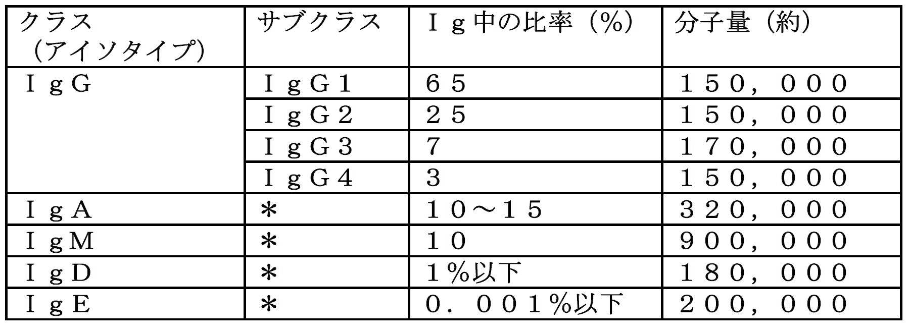
いくつかの態様の抗体の製造方法にて、抗体産生細胞を培養することよってアルギン酸ゲルファイバのコア層で産生されシェル層を透過し得る抗体としては、特に限定されることは無いが、例えば、IgG、IgA、IgM、IgD、IgE等からなる群から選択されるクラス(アイソタイプ)を有する抗体が挙げられる。抗体産生細胞を培養することによってアルギン酸ゲルファイバのコア層で産生されシェル層を透過し得る抗体は、好ましくは、IgG、又はIgEのクラス(アイソタイプ)の抗体であり、より好ましくは、IgGのクラス(アイソタイプ)の抗体である。
いくつかの態様の抗体の製造方法にて、抗体産生細胞を培養することによって、アルギン酸ゲルファイバのコア層で産生されシェル層を透過し得る抗体の分子量は、特に限定されることは無いが、例えば、45,000~900,000の範囲にある抗体である。いくつかの態様の抗体の製造方法にてアルギン酸ゲルファイバのコア層で産生されシェル層を透過し得る抗体の分子量は、好ましくは、45,000~160,000の範囲であり、より好ましくは、140,000~150,000の範囲である。
本明細書中、前記記載の各抗体産生CHO細胞を用いて前記記載の抗体の製造方法にて培養を行う場合、用いた抗体産生CHO細胞に対応する抗体が産生される。例えば、ムロモナブ-CD3産生CHO細胞を用いる場合、抗体としてムロモナブ-CD3が産生される。
産生される抗体としては、例えば、ムロモナブ-CD3産生CHO細胞を用いてムロモナブ-CD3(IgG;150,000)、トラスツズマブ産生CHO細胞を用いてトラスツズマブ(IgG;148,000)、リツキシマブ産生CHO細胞を用いてリツキシマブ(IgG;144,510)、パリビズマブ産生CHO細胞を用いてパリビズマブ(IgG;“147,700”)、インフリキシマブ産生CHO細胞を用いてインフリキシマブ(IgG;“149,000”)、バシリキシマブ産生CHO細胞を用いてバシリキシマブ(IgG;147,000)、トシリズマブ産生CHO細胞を用いてトシリズマブ(IgG;148,000)、ゲムツズマブ オゾガマイシン産生CHO細胞を用いてゲムツズマブ オゾガマイシン(IgG;“153,000”)、ベバシズマブ産生CHO細胞を用いてベバシズマブ(IgG;149,000)、イブリツモマブ チウキセタン産生CHO細胞を用いてイブリツモマブ チウキセタン(IgG;148,000)、アダリムマブ産生CHO細胞を用いてアダリムマブ(IgG;148,000)、セツキシマブ産生CHO細胞を用いてセツキシマブ(IgG;151,800)、ラニビズマブ産生CHO細胞を用いてラニビズマブ(IgG;48,000)、オマリズマブ産生CHO細胞を用いてオマリズマブ(IgE;149,000)、エクリズマブ産生CHO細胞を用いてエクリズマブ(IgG;145,235)、パニツムマブ産生CHO細胞を用いてパニツムマブ(IgG;147,000)、ウステキヌマブ産生CHO細胞を用いてウステキヌマブ(IgG;148,079~149,690)、ゴリムマブ産生CHO細胞を用いてゴリムマブ(IgG;149,802~151,064)、カナキヌマブ産生CHO細胞を用いてカナキヌマブ(IgG;148,000)、デノスマブ産生CHO細胞を用いてデノスマブ(IgG;150,000)、モガムリズマブ産生CHO細胞を用いてモガムリズマブ(IgG;149,000)、セルトリズマブ ペゴル産生CHO細胞を用いてセルトリズマブ ペゴル(IgG;90,000)、オファツムマブ産生CHO細胞を用いてオファツムマブ(IgG;149,000)、ペルツズマブ産生CHO細胞を用いてペルツズマブ(IgG;148,000)、トラスツズマブ エムタンシン産生CHO細胞を用いてトラスツズマブ エムタンシン(IgG;151,000)、ブレンツキシマブ ベドチン産生CHO細胞を用いてブレンツキシマブ ベドチン(IgG;153,000)、ナタリズマブ産生CHO細胞を用いてナタリズマブ(IgG;146,178)、ニボルマブ産生CHO細胞を用いてニボルマブ(IgG;145,000)、アレムツズマブ産生CHO細胞を用いてアレムツズマブ(IgG;150,000)、セクキヌマブ産生CHO細胞を用いてセクキヌマブ(IgG;151,000)、ラムシルマブ産生CHO細胞を用いてラムシルマブ(IgG;147,000)、イピリムマブ産生CHO細胞を用いてイピリムマブ(IgG;148,000)、エボロクマブ産生CHO細胞を用いてエボロクマブ(IgG;141,789)、メポリズマブ産生CHO細胞を用いてメポリズマブ(IgG;149,000)、アリロクマブ産生CHO細胞を用いてアリロクマブ(IgG;145892.049.)、イキセキズマブ産生CHO細胞を用いてイキセキズマブ(IgG;149,000)、ブロダルマブ産生CHO細胞を用いてブロダルマブ(IgG;147,000)、イダルシズマブ産生CHO細胞を用いてイダルシズマブ(IgG;47,782)、エロツズマブ産生CHO細胞を用いてエロツズマブ(IgG;148,000)、ペムブロリズマブ産生CHO細胞を用いてペムブロリズマブ(IgG;149,000)、サリルマブ産生CHO細胞を用いてサリルマブ(IgG;150,000)、ベズロトクスマブ産生CHO細胞を用いてベズロトクスマブ(IgG;148,000)、ベリムマブ産生CHO細胞を用いてベリムマブ(IgG;147,000)、ダラツムマブ産生CHO細胞を用いてダラツムマブ(IgG;148,000)、アベルマブ産生CHO細胞を用いてアベルマブ(IgG;147,000)、デュピルマブ産生CHO細胞を用いてデュピルマブ(IgG;152,000)、アテゾリズマブ産生CHO細胞を用いてアテゾリズマブ(IgG;144,611)、ベンラリズマブ産生CHO細胞を用いてベンラリズマブ(IgG;148,000)、イノツズマブ オゾガマイシン産生CHO細胞を用いてイノツズマブ オゾガマイシン(IgG;159,000)、エミシズマブ産生CHO細胞を用いてエミシズマブ(IgG;148,000)、グセルクマブ産生CHO細胞を用いてグセルクマブ(IgG;146,000)、デュルバルマブ産生CHO細胞を用いてデュルバルマブ(IgG;149,000)、オビヌツズマブ産生CHO細胞を用いてオビヌツズマブ(IgG;148,000~150,000)、又はベドリズマブ産生CHO細胞を用いてベドリズマブ(IgG;150,000)が挙げられる(抗体名後のカッコ内は、当該抗体のクラス(アイソタイプ)及び分子量を示す)。
産生された抗体は、例えば、下記の3工程を経て精製が行われる。
〔工程1〕培地中に含まれる、抗体以外のタンパク質及び固形物をほぼ取り除く為に、遠心分離法又はフィルターによる濾過等を行う。
〔工程2〕例えば、Protein A又はProtein Gを用いたアフィニティークロマトグラフィー、又はイオン交換クロマトグラフィー等のクロマトグラフィーにて目的とする抗体を取り出す。
〔工程3〕工程2で混入してきた夾雑物を除去する為に、ゲルろ過クロマトグラフィーを行い、目的とする抗体を高純度精製する。
〔工程1〕培地中に含まれる、抗体以外のタンパク質及び固形物をほぼ取り除く為に、遠心分離法又はフィルターによる濾過等を行う。
〔工程2〕例えば、Protein A又はProtein Gを用いたアフィニティークロマトグラフィー、又はイオン交換クロマトグラフィー等のクロマトグラフィーにて目的とする抗体を取り出す。
〔工程3〕工程2で混入してきた夾雑物を除去する為に、ゲルろ過クロマトグラフィーを行い、目的とする抗体を高純度精製する。
Protein A又はProtein Gを用いたアフィニティークロマトグラフィー:
IgGの精製法としては、例えば、Protein A又はProtein Gを用いた抗体の精製方法が知られている。Protein Aを用いた抗体の精製法として、下記方法が1例として挙げられる。(1)Protein Aが固定されたビーズが充填されたカラムを用いて、前記〔ステップ1〕の方法で得られてくる溶液に血清を添加した溶液をろ過することで、IgGがカラム中のビーズに結合して、他の血清成分がカラム外へ流出がされる。(2)その後、カラムに酸性溶液を通過させることにより、ビーズに結合していたIgGが切れて、カラム外へ溶出されてIgGが得られる。尚、IgのProtein AとProtein Gへの結合力が、動物種やサブクラスによって違うことから、目的によって、Protein A又はProtein Gを使い分けることができる。
IgGの精製法としては、例えば、Protein A又はProtein Gを用いた抗体の精製方法が知られている。Protein Aを用いた抗体の精製法として、下記方法が1例として挙げられる。(1)Protein Aが固定されたビーズが充填されたカラムを用いて、前記〔ステップ1〕の方法で得られてくる溶液に血清を添加した溶液をろ過することで、IgGがカラム中のビーズに結合して、他の血清成分がカラム外へ流出がされる。(2)その後、カラムに酸性溶液を通過させることにより、ビーズに結合していたIgGが切れて、カラム外へ溶出されてIgGが得られる。尚、IgのProtein AとProtein Gへの結合力が、動物種やサブクラスによって違うことから、目的によって、Protein A又はProtein Gを使い分けることができる。
イオン交換クロマトグラフィー:
タンパク質が有する電気的な性質(電荷)を利用してタンパク質を分離する方法である。正電荷を示す塩基性タンパク質は、負電荷をもつ陽イオン交換体(担体)にイオン結合し、負電荷を示す酸性タ ンパク質は正電荷を持つ陰イオン交換体に結合することから、タンパク質が含まれる試料をイオン交換体が充填されたカラムを通すことで、タンパク質がイオン交換体に結合する。その後、カラムを通す溶媒の塩濃度を高濃度にすることで、タンパク質とイオン交換体とのイオン結合が弱くなり、結合力の弱いタンパク質から順番に、イオン交換体から外れて、カラム外へ流出してくる。陽イオン交換体又は陰イオン交換体の選択は、試料として用いるタンパク質の電荷から選択するものとする。
タンパク質が有する電気的な性質(電荷)を利用してタンパク質を分離する方法である。正電荷を示す塩基性タンパク質は、負電荷をもつ陽イオン交換体(担体)にイオン結合し、負電荷を示す酸性タ ンパク質は正電荷を持つ陰イオン交換体に結合することから、タンパク質が含まれる試料をイオン交換体が充填されたカラムを通すことで、タンパク質がイオン交換体に結合する。その後、カラムを通す溶媒の塩濃度を高濃度にすることで、タンパク質とイオン交換体とのイオン結合が弱くなり、結合力の弱いタンパク質から順番に、イオン交換体から外れて、カラム外へ流出してくる。陽イオン交換体又は陰イオン交換体の選択は、試料として用いるタンパク質の電荷から選択するものとする。
ゲルろ過クロマトグラフィー:
タンパク質の分子量の違いを利用してタンパク質を分離する方法である。小孔が付いている担体が充填されたカラムに試料を流すことで、分子量の小さいタンパク質は、前記小孔に入り込みながら流出してき、分子量の大きいタンパク質は前記小孔に入らずに流出してくる為、カラムを通過する時間が分子量の小さいタンパク質は遅く、分子量の大きいタンパク質は早くなることから、時間差的にタンパク質を分離することが可能となる。
タンパク質の分子量の違いを利用してタンパク質を分離する方法である。小孔が付いている担体が充填されたカラムに試料を流すことで、分子量の小さいタンパク質は、前記小孔に入り込みながら流出してき、分子量の大きいタンパク質は前記小孔に入らずに流出してくる為、カラムを通過する時間が分子量の小さいタンパク質は遅く、分子量の大きいタンパク質は早くなることから、時間差的にタンパク質を分離することが可能となる。
尚、本明細書において引用した全ての文献、及び公開公報、特許公報、その他の特許文献は、参照として本明細書に組み込むものとする。本明細書は、本願の優先権主張の基礎となる日本国特許出願である特願2018-151574号(2018年8月10日出願)の特許請求の範囲、明細書、および図面の開示内容を包含する。
以下、実施例により本発明を説明するが、本発明は以下の実施例に限定されるものではない。
[実施例1]アルギン酸ゲルファイバAの製法
図2に模式的に示す製造過程にしたがって、特開2016-77229号公報(出願人:国立大学法人 東京大学)に記載されているアルギン酸ゲルファイバの作製条件と同様にして、アルギン酸ゲルファイバAを作製した。細胞は、トシリズマブ産生CHO細胞を用いた。マイクロ流体装置10の導入口1から、細胞濃度1×10⁶細胞/mL(2×105細胞/25mファイバ1本)の細胞を含む0.2質量%コラーゲン溶液(コラーゲン酸性溶液I-PC 5mg、pH3.0、無菌Atelocollag(株式会社高研、cat#.IPC-50)溶媒)を導入し、導入口2から、1.5質量%(w/w%)アルギン酸ナトリウム(A-2 持田製薬)溶液(溶媒:下記表3の組成である培地)を導入し、導入口3から100mM塩化カルシウム水溶液を導入して射出することにより、コア層直径約100μm、外径約300μm、全長25mのアルギン酸ゲルファイバAを得た。
図2に模式的に示す製造過程にしたがって、特開2016-77229号公報(出願人:国立大学法人 東京大学)に記載されているアルギン酸ゲルファイバの作製条件と同様にして、アルギン酸ゲルファイバAを作製した。細胞は、トシリズマブ産生CHO細胞を用いた。マイクロ流体装置10の導入口1から、細胞濃度1×10⁶細胞/mL(2×105細胞/25mファイバ1本)の細胞を含む0.2質量%コラーゲン溶液(コラーゲン酸性溶液I-PC 5mg、pH3.0、無菌Atelocollag(株式会社高研、cat#.IPC-50)溶媒)を導入し、導入口2から、1.5質量%(w/w%)アルギン酸ナトリウム(A-2 持田製薬)溶液(溶媒:下記表3の組成である培地)を導入し、導入口3から100mM塩化カルシウム水溶液を導入して射出することにより、コア層直径約100μm、外径約300μm、全長25mのアルギン酸ゲルファイバAを得た。
[実施例2]アルギン酸ゲルファイバBの製法
図2に模式的に示す製造過程にしたがって、特開2016-77229号公報(出願人:国立大学法人 東京大学)に記載されているアルギン酸ゲルファイバの作製条件と同様にして、アルギン酸ゲルファイバBを作製した。細胞は、トシリズマブ産生CHO細胞を用いた。マイクロ流体装置10の導入口1から、細胞濃度5×10⁶細胞/mL(1×106細胞/25mファイバ1本)の細胞を含む0.2%コラーゲン溶液(コラーゲン酸性溶液I-PC 5mg、pH3.0 、無菌Atelocollag(株式会社高研、cat#.IPC-50)溶媒)を導入し、導入口2から、1.5質量%(w/w%)アルギン酸ナトリウム(B-2 持田製薬)溶液(溶媒:下記表3の組成である培地)を導入し、導入口3から100mM塩化カルシウム水溶液を導入して射出することにより、コア層直径約100μm、外径約300μm、全長25mのアルギン酸ゲルファイバBを得た。
図2に模式的に示す製造過程にしたがって、特開2016-77229号公報(出願人:国立大学法人 東京大学)に記載されているアルギン酸ゲルファイバの作製条件と同様にして、アルギン酸ゲルファイバBを作製した。細胞は、トシリズマブ産生CHO細胞を用いた。マイクロ流体装置10の導入口1から、細胞濃度5×10⁶細胞/mL(1×106細胞/25mファイバ1本)の細胞を含む0.2%コラーゲン溶液(コラーゲン酸性溶液I-PC 5mg、pH3.0 、無菌Atelocollag(株式会社高研、cat#.IPC-50)溶媒)を導入し、導入口2から、1.5質量%(w/w%)アルギン酸ナトリウム(B-2 持田製薬)溶液(溶媒:下記表3の組成である培地)を導入し、導入口3から100mM塩化カルシウム水溶液を導入して射出することにより、コア層直径約100μm、外径約300μm、全長25mのアルギン酸ゲルファイバBを得た。
[実施例3]アルギン酸ゲルファイバA中でのトシリズマブ産生CHO細胞の培養
125 mLポリカーボネート製三角フラスコに、実施例1で得られたアルギン酸ゲルファイバA(外径300μm×長さ25m、ファイバ体積(外形部分)約1.8 mL分)を入れ、下記表3の組成である培地(30 mL)を添加し、ファイバを含浸させた。37℃、5%CO2インキュベータ内で125 rpmで振とうしながら培養を、12日間実施した。12日間培養を行うことで、抗体(トシリズマブ)が産生され、ファイバ外液中に放出されたことを確認した。産生された抗体がトシリズマブであることは、ヒトIL-6 Receptor a(Peprotech, Cat# 200-06RC)を用いたELISA測定により確認した。又、本実験結果より、産生された抗体がファイバ外に透過できることを見い出せた。12日培養後のアルギン酸ゲルファイバの顕微鏡写真を図4に示す。
125 mLポリカーボネート製三角フラスコに、実施例1で得られたアルギン酸ゲルファイバA(外径300μm×長さ25m、ファイバ体積(外形部分)約1.8 mL分)を入れ、下記表3の組成である培地(30 mL)を添加し、ファイバを含浸させた。37℃、5%CO2インキュベータ内で125 rpmで振とうしながら培養を、12日間実施した。12日間培養を行うことで、抗体(トシリズマブ)が産生され、ファイバ外液中に放出されたことを確認した。産生された抗体がトシリズマブであることは、ヒトIL-6 Receptor a(Peprotech, Cat# 200-06RC)を用いたELISA測定により確認した。又、本実験結果より、産生された抗体がファイバ外に透過できることを見い出せた。12日培養後のアルギン酸ゲルファイバの顕微鏡写真を図4に示す。
培地組成:G016培地に下表の各種添加物を加え、全量を1000mLに調製した。

[実施例4]アルギン酸ゲルファイバB中でのトシリズマブ産生CHO細胞の培養
125 mLポリカーボネート製三角フラスコに、実施例2で得られたアルギン酸ゲルファイバB(外径300μm×長さ25m、ファイバ体積(外形部分)約1.8 mL分)を入れ、上記表3の組成である培地(30 mL)を添加し、ファイバを含浸させた。37℃、5%CO2インキュベータ内で125 rpmで振とうしながら培養を、28日間実施した。28日間培養を行うことで、抗体(トシリズマブ)が、4.77mg産生された。産生された抗体がトシリズマブであることは、ヒトIL-6 Receptor a(Peprotech, Cat# 200-06RC)を用いたELISA測定により確認した。又、本実験結果より、産生された抗体がファイバ外に透過できることを見い出せた。28日培養後のアルギン酸ゲルファイバの顕微鏡写真を図5に示す。
125 mLポリカーボネート製三角フラスコに、実施例2で得られたアルギン酸ゲルファイバB(外径300μm×長さ25m、ファイバ体積(外形部分)約1.8 mL分)を入れ、上記表3の組成である培地(30 mL)を添加し、ファイバを含浸させた。37℃、5%CO2インキュベータ内で125 rpmで振とうしながら培養を、28日間実施した。28日間培養を行うことで、抗体(トシリズマブ)が、4.77mg産生された。産生された抗体がトシリズマブであることは、ヒトIL-6 Receptor a(Peprotech, Cat# 200-06RC)を用いたELISA測定により確認した。又、本実験結果より、産生された抗体がファイバ外に透過できることを見い出せた。28日培養後のアルギン酸ゲルファイバの顕微鏡写真を図5に示す。
ここでは、抗体産生細胞がコア層に含まれた抗体生産用のアルギン酸ゲルファイバ(アルギン酸中空マイクロファイバ)が提供される。また、当該アルギン酸ゲルファイバの製造方法、及び当該アルギン酸ゲルファイバを用いた抗体の培養方法を提供することができる。
a:アルギン酸ゲルファイバのコア層(中空部)の直径
b:アルギン酸ゲルファイバのシェル層の厚さ
c:アルギン酸ゲルファイバの外径
4:シェル層
5:コア層
6:抗体産生細胞
b:アルギン酸ゲルファイバのシェル層の厚さ
c:アルギン酸ゲルファイバの外径
4:シェル層
5:コア層
6:抗体産生細胞
10:マイクロ流体装置
1:抗体産生細胞6を含む基材の導入口
2:アルギン酸ナトリウム溶液の導入口
3:塩化カルシウム溶液の導入口
20: アルギン酸ゲルファイバ
40:導入管
50:出口
1:抗体産生細胞6を含む基材の導入口
2:アルギン酸ナトリウム溶液の導入口
3:塩化カルシウム溶液の導入口
20: アルギン酸ゲルファイバ
40:導入管
50:出口
Claims (14)
- 抗体産生細胞及び基材が含まれるコア層をコア層よりも高い機械的強度を有するアルギン酸ゲルを含むシェル層で被覆して形成されるアルギン酸ゲルファイバ。
- コア層に含まれる抗体産生細胞が、CHO細胞、CHO細胞亜株、COS細胞、Sp2/0細胞、NS0細胞、SP2細胞、PERC6細胞、YB2/0細胞、YE2/0細胞、1R983F細胞、Namalwa細胞、Wil-2細胞、Jurkat細胞、Vera細胞、Molt-4細胞、293-HEK細胞、BHK細胞、KGH6細胞、P3X63Ag8.653細胞、C127細胞、JC細胞、LA7細胞、ZR-45-30細胞、hTERT細胞、NM2C5細胞、及びUACC-812細胞からなる群から選択される細胞である、請求項1に記載のアルギン酸ゲルファイバ。
- コア層に含まれる基材がコラーゲン溶液、コラーゲンゲル、培地(または培養液)、アルギン酸溶液(例えば、アルギン酸ナトリウム溶液)、アルギン酸ゲル、又はそれらの混合物等からなる群から選択される基材である、請求項1~2のいずれか1項に記載のアルギン酸ゲルファイバ。
- シェル層に含まれるアルギン酸ゲルの原料がアルギン酸ナトリウムであり、アルギン酸ナトリウムのM/G比が0.4~1.8の範囲又は0.1~0.4の範囲である、請求項1~3のいずれか1項に記載のアルギン酸ゲルファイバ。
- シェル層に含まれるアルギン酸ゲルの原料がアルギン酸ナトリウムであり、アルギン酸ナトリウムの分子量(GPC)が700,000~1,000,000の範囲又は800,000~1,000,000の範囲である、請求項1~4のいずれか1項に記載のアルギン酸ゲルファイバ。
- シェル層に含まれるアルギン酸ゲルの原料がアルギン酸ナトリウムであり、アルギン酸ナトリウムの1w/w%の粘度が50~150(mPa・s)の範囲又は70~150(mPa・s)の範囲である、請求項1~5のいずれか1項に記載のアルギン酸ゲルファイバ。
- アルギン酸ゲルファイバの外径が0.2μm~2000μmの範囲であり、アルギン酸ゲルファイバのコア層の直径が0.1μm~1000μmの範囲である、請求項1~6のいずれか1項に記載のアルギン酸ゲルファイバ。
- 抗体産生細胞及び基材が含まれるコア層を、コア層よりも高い機械的強度を有するアルギン酸ゲルを含むシェル層で被覆して形成されるアルギン酸ゲルファイバの製造方法であって、
導入管40と、導入管40の導入口1と、導入口1の下流に位置する導入管40の導入口2と、導入口2の下流に位置する導入管40の導入口3と、導入口2の下流に位置する導入管40の出口50を含む、マイクロ流体装置10を用い、
(1)導入口1から、抗体産生細胞6と基材を導入して射出して、導入管40内に抗体産生細胞6と基材の第1の層流を形成する工程、
(2)導入口2から、アルギン酸ナトリウム溶液を導入して射出して、第1の層流の外周を覆う、アルギン酸ナトリウム溶液の第2の層流を形成する工程、
(3)導入口3から、2価金属イオンを含む溶液を導入して射出して、第2の層流の外周を覆う、2価金属イオンを含む溶液の第3の層流を形成する工程、
(4)出口50から射出される、抗体産生細胞及び基材が含まれるコア層を、アルギン酸ゲルを含むシェル層で被覆して形成されるアルギン酸ゲルファイバを得る工程
を含むことを特徴とする、アルギン酸ゲルファイバの製造方法。 - 抗体産生細胞6が、請求項2に記載の細胞である、請求項8に記載のアルギン酸ゲルファイバの製造方法。
- コア層に含まれる基材がコラーゲン溶液、コラーゲンゲル、培地(または培養液)、アルギン酸溶液(例えば、アルギン酸ナトリウム溶液)、アルギン酸ゲル、又はそれらの混合物等からなる群から選択される基材である請求項8または9に記載のアルギン酸ゲルファイバの製造方法。
- 2価金属イオンが、カルシウムイオン、マグネシウムイオン、バリウムイオン、ストロンチウムイオン、及び亜鉛イオンからなる群から選択されるイオンである、請求項8~10のいずれか1項に記載のアルギン酸ゲルファイバの製造方法。
- 2価金属イオンを含む溶液が、塩化カルシウム水溶液、炭酸カルシウム水溶液、及びグルコン酸カルシウム水溶液なる群から選択される水溶液である、請求項8~11のいずれか1項に記載のアルギン酸ゲルファイバの製造方法。
- アルギン酸ゲルファイバの製法時の温度が、4~25℃の範囲である、請求項8~12のいずれか1項に記載のアルギン酸ゲルファイバの製造方法。
- 抗体産生細胞及び基材が含まれるコア層を、コア層よりも高い機械的強度を有するアルギン酸ゲルを含むシェル層で被覆して形成されるアルギン酸ゲルファイバを用いる抗体の製造方法であり、請求項1~7のいずれか1項に記載のアルギン酸ゲルファイバを培養容器に入れ、培地を添加して前記アルギン酸ゲルファイバを含浸させ、振とう培養を行うことによる、抗体の製造方法。
Priority Applications (4)
| Application Number | Priority Date | Filing Date | Title |
|---|---|---|---|
| CN201980053240.9A CN112513245A (zh) | 2018-08-10 | 2019-08-09 | 海藻酸中空微纤维 |
| US17/267,320 US20210340481A1 (en) | 2018-08-10 | 2019-08-09 | Alginate hollow microfiber |
| JP2020535894A JP7388722B2 (ja) | 2018-08-10 | 2019-08-09 | アルギン酸中空マイクロファイバ |
| EP19846840.7A EP3835409A4 (en) | 2018-08-10 | 2019-08-09 | HOLLOW ALGINATE MICROFIBER |
Applications Claiming Priority (2)
| Application Number | Priority Date | Filing Date | Title |
|---|---|---|---|
| JP2018151574 | 2018-08-10 | ||
| JP2018-151574 | 2018-08-10 |
Publications (1)
| Publication Number | Publication Date |
|---|---|
| WO2020032221A1 true WO2020032221A1 (ja) | 2020-02-13 |
Family
ID=69414919
Family Applications (1)
| Application Number | Title | Priority Date | Filing Date |
|---|---|---|---|
| PCT/JP2019/031538 Ceased WO2020032221A1 (ja) | 2018-08-10 | 2019-08-09 | アルギン酸中空マイクロファイバ |
Country Status (5)
| Country | Link |
|---|---|
| US (1) | US20210340481A1 (ja) |
| EP (1) | EP3835409A4 (ja) |
| JP (1) | JP7388722B2 (ja) |
| CN (1) | CN112513245A (ja) |
| WO (1) | WO2020032221A1 (ja) |
Cited By (7)
| Publication number | Priority date | Publication date | Assignee | Title |
|---|---|---|---|---|
| WO2021125279A1 (ja) * | 2019-12-18 | 2021-06-24 | 持田製薬株式会社 | 化学架橋アルギン酸ゲルファイバ |
| WO2021177455A1 (ja) * | 2020-03-05 | 2021-09-10 | 株式会社セルファイバ | 収容体及び凍結保存方法 |
| WO2022145420A1 (ja) | 2020-12-28 | 2022-07-07 | 持田製薬株式会社 | 新規な多層ポリマーコーティング架橋アルギン酸ゲルファイバ |
| WO2022270549A1 (ja) | 2021-06-23 | 2022-12-29 | 持田製薬株式会社 | 新規なポリマーコーティング架橋アルギン酸ゲルファイバ |
| WO2023210691A1 (ja) * | 2022-04-27 | 2023-11-02 | 国立大学法人東京大学 | 活性成分含有溶液処理装置および体外活性成分含有溶液循環システム |
| WO2025004984A1 (ja) | 2023-06-26 | 2025-01-02 | 株式会社セルファイバ | 細胞カプセルの製造方法、細胞集団又は細胞産生物の製造方法、溶液及び細胞集団 |
| WO2026042871A1 (ja) * | 2024-08-23 | 2026-02-26 | 国立大学法人千葉大学 | ハイドロゲル構造体及びその製造方法 |
Families Citing this family (1)
| Publication number | Priority date | Publication date | Assignee | Title |
|---|---|---|---|---|
| CN115262028A (zh) * | 2022-09-13 | 2022-11-01 | 南京鼓楼医院 | 一种仿生载种子细胞多组分纤维的制备方法及应用 |
Citations (6)
| Publication number | Priority date | Publication date | Assignee | Title |
|---|---|---|---|---|
| WO2011046105A1 (ja) | 2009-10-14 | 2011-04-21 | 国立大学法人 東京大学 | 被覆されたマイクロゲルファイバ |
| JP2014236698A (ja) | 2013-06-07 | 2014-12-18 | 国立大学法人 鹿児島大学 | 細胞凝集塊作製法 |
| WO2015178427A1 (ja) | 2014-05-20 | 2015-11-26 | 国立大学法人 東京大学 | 中空マイクロファイバ |
| JP2016077229A (ja) | 2014-10-17 | 2016-05-16 | 国立大学法人 東京大学 | ファイバ状基材、3次元細胞構造体及びその製造方法、並びに3次元細胞構造体の培養方法 |
| JP2017077473A (ja) | 2015-10-21 | 2017-04-27 | 国立大学法人 東京大学 | マイクロチューブ、マイクロチューブの製造方法、及びマイクロチューブの製造装置 |
| JP2018151574A (ja) | 2017-03-14 | 2018-09-27 | 株式会社Jvcケンウッド | 電子機器及び表示パネルの移動方法 |
Family Cites Families (17)
| Publication number | Priority date | Publication date | Assignee | Title |
|---|---|---|---|---|
| JP2006238841A (ja) * | 2005-03-07 | 2006-09-14 | Fuji Photo Film Co Ltd | 細胞培養担体 |
| KR101142159B1 (ko) * | 2010-06-11 | 2012-05-10 | 연세대학교 산학협력단 | 알지네이트 비드의 연속적인 생산 및 포획이 가능한 미세 관류 배양 시스템 |
| CN103301788B (zh) * | 2012-03-09 | 2015-09-02 | 中国科学院大连化学物理研究所 | Peg接枝改性的海藻酸盐-壳聚糖微胶囊及制备和应用 |
| WO2014037942A1 (en) * | 2012-09-04 | 2014-03-13 | Technion Research & Development Foundation Limited | Use of decellularized extracellular matrix for encapsulating cells |
| CN102974410B (zh) * | 2012-11-06 | 2015-06-24 | 中国科学院大连化学物理研究所 | 基于微流控芯片制备微米级海藻酸钙丝的方法及专用芯片 |
| US20140127290A1 (en) * | 2012-11-08 | 2014-05-08 | Ohio State Innovation Foundation | Microcapsules Encapsulating Living Cells |
| WO2014126228A1 (ja) * | 2013-02-16 | 2014-08-21 | 国立大学法人千葉大学 | 細胞評価用ハイドロゲル基材および細胞評価手法 |
| KR101472045B1 (ko) * | 2013-08-16 | 2014-12-23 | 단국대학교 산학협력단 | 코어-쉘 섬유상 세포전달체 및 이를 포함하는 골조직 또는 연골조직 재생용 조성물 |
| US9795714B2 (en) * | 2013-12-24 | 2017-10-24 | Ares Trading S.A. | FGF-18 formulation in alginate/collagen hydrogels |
| KR101741815B1 (ko) * | 2014-05-23 | 2017-06-16 | 서강대학교산학협력단 | 세포 공동-배양을 위한 하이드로젤 기반 미세유체칩 |
| CN105624832B (zh) * | 2014-11-07 | 2017-09-22 | 中国科学院大连化学物理研究所 | 基于微流控技术制备具球棒结构的海藻酸钙微纤维的方法 |
| JP2019503203A (ja) * | 2016-01-29 | 2019-02-07 | アカオジェン インコーポレイテッド | 細胞表面エピトープと結合する抗体を同定するためのスクリーニング方法 |
| CN105641743B (zh) * | 2016-03-16 | 2019-05-17 | 宁波瑞柏思生物材料科技有限公司 | 一种微流控装置及利用该装置制备微凝胶的方法 |
| TWI734775B (zh) * | 2016-04-26 | 2021-08-01 | 美商美國泰福生技股份有限公司 | 細胞培養基 |
| CN108149342B (zh) * | 2016-12-05 | 2020-05-19 | 中国科学院大连化学物理研究所 | 基于微流控技术的复合空腔微纤维的制备方法 |
| CN107930542B (zh) * | 2017-11-13 | 2020-11-20 | 深圳华诺生物科技有限公司 | 一步法连续制备海藻酸钙微凝胶的微流控技术 |
| CN108360088B (zh) * | 2018-02-28 | 2020-03-03 | 清华大学深圳研究生院 | 制备海藻酸钙纤维的方法和装置 |
-
2019
- 2019-08-09 CN CN201980053240.9A patent/CN112513245A/zh active Pending
- 2019-08-09 JP JP2020535894A patent/JP7388722B2/ja active Active
- 2019-08-09 WO PCT/JP2019/031538 patent/WO2020032221A1/ja not_active Ceased
- 2019-08-09 EP EP19846840.7A patent/EP3835409A4/en not_active Withdrawn
- 2019-08-09 US US17/267,320 patent/US20210340481A1/en not_active Abandoned
Patent Citations (6)
| Publication number | Priority date | Publication date | Assignee | Title |
|---|---|---|---|---|
| WO2011046105A1 (ja) | 2009-10-14 | 2011-04-21 | 国立大学法人 東京大学 | 被覆されたマイクロゲルファイバ |
| JP2014236698A (ja) | 2013-06-07 | 2014-12-18 | 国立大学法人 鹿児島大学 | 細胞凝集塊作製法 |
| WO2015178427A1 (ja) | 2014-05-20 | 2015-11-26 | 国立大学法人 東京大学 | 中空マイクロファイバ |
| JP2016077229A (ja) | 2014-10-17 | 2016-05-16 | 国立大学法人 東京大学 | ファイバ状基材、3次元細胞構造体及びその製造方法、並びに3次元細胞構造体の培養方法 |
| JP2017077473A (ja) | 2015-10-21 | 2017-04-27 | 国立大学法人 東京大学 | マイクロチューブ、マイクロチューブの製造方法、及びマイクロチューブの製造装置 |
| JP2018151574A (ja) | 2017-03-14 | 2018-09-27 | 株式会社Jvcケンウッド | 電子機器及び表示パネルの移動方法 |
Non-Patent Citations (7)
| Title |
|---|
| ACS BIOMATER. SCI. ENG., vol. 3, no. 3, 2017, pages 392 - 398 |
| ANNALS OF BIOMEDICAL ENGINEERING, vol. 36, no. 7, 2008, pages 1254 - 1267 |
| LAB CHIP, vol. 16, 2016, pages 1757 - 1776 |
| NATURE MATERIALS, vol. 12, 2013, pages 584 - 590 |
| ONOE, H. ET AL.: "Metre-long cell -laden microfibres exhibit tissue morphologies and functions", NATURE MATERIALS, vol. 12, 31 March 2013 (2013-03-31), pages 584 - 590, XP055058191 * |
| See also references of EP3835409A4 |
| WONJE JEONG ET AL.: "Hydrodynamic micro fabrication via ''on the fly'' photopolymerization of microscale fibers and tubes", LAB CHIP, vol. 4, 2004, pages 576 - 580 |
Cited By (10)
| Publication number | Priority date | Publication date | Assignee | Title |
|---|---|---|---|---|
| WO2021125279A1 (ja) * | 2019-12-18 | 2021-06-24 | 持田製薬株式会社 | 化学架橋アルギン酸ゲルファイバ |
| WO2021177455A1 (ja) * | 2020-03-05 | 2021-09-10 | 株式会社セルファイバ | 収容体及び凍結保存方法 |
| WO2022145420A1 (ja) | 2020-12-28 | 2022-07-07 | 持田製薬株式会社 | 新規な多層ポリマーコーティング架橋アルギン酸ゲルファイバ |
| KR20230127997A (ko) | 2020-12-28 | 2023-09-01 | 모찌다 세이야쿠 가부시끼가이샤 | 신규의 다층 폴리머 코팅 가교 알긴산 겔 파이버 |
| WO2022270549A1 (ja) | 2021-06-23 | 2022-12-29 | 持田製薬株式会社 | 新規なポリマーコーティング架橋アルギン酸ゲルファイバ |
| KR20240024839A (ko) | 2021-06-23 | 2024-02-26 | 모찌다 세이야쿠 가부시끼가이샤 | 신규의 폴리머 코팅 가교 알긴산 겔 파이버 |
| US12312574B2 (en) | 2021-06-23 | 2025-05-27 | Mochida Pharmaceutical Co., Ltd. | Polymer-coated crosslinked alginate gel fiber |
| WO2023210691A1 (ja) * | 2022-04-27 | 2023-11-02 | 国立大学法人東京大学 | 活性成分含有溶液処理装置および体外活性成分含有溶液循環システム |
| WO2025004984A1 (ja) | 2023-06-26 | 2025-01-02 | 株式会社セルファイバ | 細胞カプセルの製造方法、細胞集団又は細胞産生物の製造方法、溶液及び細胞集団 |
| WO2026042871A1 (ja) * | 2024-08-23 | 2026-02-26 | 国立大学法人千葉大学 | ハイドロゲル構造体及びその製造方法 |
Also Published As
| Publication number | Publication date |
|---|---|
| JPWO2020032221A1 (ja) | 2021-08-10 |
| EP3835409A1 (en) | 2021-06-16 |
| EP3835409A4 (en) | 2022-05-04 |
| CN112513245A (zh) | 2021-03-16 |
| JP7388722B2 (ja) | 2023-11-29 |
| US20210340481A1 (en) | 2021-11-04 |
Similar Documents
| Publication | Publication Date | Title |
|---|---|---|
| JP7388722B2 (ja) | アルギン酸中空マイクロファイバ | |
| Sharma et al. | Three‐dimensional supermacroporous carrageenan‐gelatin cryogel matrix for tissue engineering applications | |
| CN101632841B (zh) | 含海藻酸盐、糖胺聚糖和胶原的组织工程支架及其制备方法 | |
| BR112017000813B1 (pt) | sistemas de gel microporoso, usos de uma pluralidade de partículas de microgel em uma solução aquosa e um agente de recozimento, de camadas de sequestrantes covalentemente estabilizados de partículas de microgel, e de um sequestrante covalentemente estabilizado de partículas de microgel com espaços intersticiais, bem como método para a preparação de partículas de microgel | |
| US6023009A (en) | Artificial pancreas | |
| Ding et al. | Zinc-mineralized diatom biosilica/hydroxybutyl chitosan composite hydrogel for diabetic chronic wound healing | |
| CN102294049B (zh) | 生物活性玻璃与壳聚糖复合骨修复材料及其制法和用途 | |
| FR2501715A1 (fr) | Procede de culture de cellules necessitant une fixation pour la fabrication de substances therapeutiques | |
| Wang et al. | Ligand‐selective targeting of macrophage hydrogel elicits bone immune‐stem cell endogenous self‐healing program to promote bone regeneration | |
| CN102399370B (zh) | 一种壳聚糖聚合物及其制备方法 | |
| US10744228B2 (en) | Methacrylated devitalized cartilage and devitalized cartilage particles | |
| WO2022270549A1 (ja) | 新規なポリマーコーティング架橋アルギン酸ゲルファイバ | |
| JP2001505809A (ja) | 組織処理用の改善されたヒドロゲル | |
| Chu et al. | Cross-linking N-succinyl chitosan-oxidated hyaluronic acid-based hydrogel loaded with bone marrow mesenchymal stem cell-derived exosomes induce bone regeneration in cranial defects | |
| KR101862457B1 (ko) | 다공성 실리카 입자에 의해 기계적 강도가 향상된 하이브리드 하이드로겔 및 이의 제조 방법 | |
| DeVolder et al. | Microfabrication of proangiogenic cell-laden alginate-g-pyrrole hydrogels | |
| Iurciuc et al. | Gellan. Pharmaceutical, medical and cosmetic applications | |
| Vaithilingam et al. | In Vitro and In Vivo Biocompatibility Evaluation of Polyallylamine and Macromolecular Heparin Conjugates Modified Alginate Microbeads | |
| Hachinohe et al. | Bone formation on murine cranial bone by injectable cross-linked hyaluronic acid containing nano-hydroxyapatite and bone morphogenetic protein | |
| US11213607B2 (en) | Cell purification and delivery using shear thinning gel | |
| Matole et al. | A Brief Review on Hydrogel | |
| Neuhäusler et al. | Cell-instructive microfibers enable programmable alignment of bioprinted hMSC | |
| CN101401959B (zh) | 明胶-壳聚糖-β-磷酸三钙球形多孔颗粒材料的制备方法 | |
| Bae et al. | RGD island spacing controls phenotype of primary human fibroblasts adhered to ligand-organized hydrogels | |
| RU2819701C1 (ru) | Гидрогелевый биокомпозит на основе бактериальных полисахаридов для использования в тканевой инженерии |
Legal Events
| Date | Code | Title | Description |
|---|---|---|---|
| 121 | Ep: the epo has been informed by wipo that ep was designated in this application |
Ref document number: 19846840 Country of ref document: EP Kind code of ref document: A1 |
|
| ENP | Entry into the national phase |
Ref document number: 2020535894 Country of ref document: JP Kind code of ref document: A |
|
| NENP | Non-entry into the national phase |
Ref country code: DE |
|
| ENP | Entry into the national phase |
Ref document number: 2019846840 Country of ref document: EP Effective date: 20210310 |