WO2019150862A1 - 血流計測装置 - Google Patents
血流計測装置 Download PDFInfo
- Publication number
- WO2019150862A1 WO2019150862A1 PCT/JP2018/047841 JP2018047841W WO2019150862A1 WO 2019150862 A1 WO2019150862 A1 WO 2019150862A1 JP 2018047841 W JP2018047841 W JP 2018047841W WO 2019150862 A1 WO2019150862 A1 WO 2019150862A1
- Authority
- WO
- WIPO (PCT)
- Prior art keywords
- unit
- blood flow
- optical system
- image
- movement
- Prior art date
Links
Images


Classifications
-
- A—HUMAN NECESSITIES
- A61—MEDICAL OR VETERINARY SCIENCE; HYGIENE
- A61B—DIAGNOSIS; SURGERY; IDENTIFICATION
- A61B3/00—Apparatus for testing the eyes; Instruments for examining the eyes
- A61B3/10—Objective types, i.e. instruments for examining the eyes independent of the patients' perceptions or reactions
- A61B3/12—Objective types, i.e. instruments for examining the eyes independent of the patients' perceptions or reactions for looking at the eye fundus, e.g. ophthalmoscopes
- A61B3/1225—Objective types, i.e. instruments for examining the eyes independent of the patients' perceptions or reactions for looking at the eye fundus, e.g. ophthalmoscopes using coherent radiation
- A61B3/1233—Objective types, i.e. instruments for examining the eyes independent of the patients' perceptions or reactions for looking at the eye fundus, e.g. ophthalmoscopes using coherent radiation for measuring blood flow, e.g. at the retina
-
- A—HUMAN NECESSITIES
- A61—MEDICAL OR VETERINARY SCIENCE; HYGIENE
- A61B—DIAGNOSIS; SURGERY; IDENTIFICATION
- A61B3/00—Apparatus for testing the eyes; Instruments for examining the eyes
- A61B3/0016—Operational features thereof
- A61B3/0033—Operational features thereof characterised by user input arrangements
-
- A—HUMAN NECESSITIES
- A61—MEDICAL OR VETERINARY SCIENCE; HYGIENE
- A61B—DIAGNOSIS; SURGERY; IDENTIFICATION
- A61B3/00—Apparatus for testing the eyes; Instruments for examining the eyes
- A61B3/10—Objective types, i.e. instruments for examining the eyes independent of the patients' perceptions or reactions
- A61B3/102—Objective types, i.e. instruments for examining the eyes independent of the patients' perceptions or reactions for optical coherence tomography [OCT]
Definitions
- This invention relates to a blood flow measuring device for measuring blood flow dynamics of the fundus.
- OCT optical coherence tomography
- the incident light may be offset with respect to the optical axis of the eye.
- the amount of offset increases, light can be incident on the blood vessel more obliquely, while the possibility that the incident light or its return light will be vignetted at the pupil increases. Therefore, it is desirable to find a position where the maximum offset amount is obtained in a range where no vignetting occurs in the pupil.
- One object of the present invention is to optimize the offset position of incident light for fundus blood flow measurement.
- Another object of the present invention is to shorten the operation for optimizing the offset position of incident light for fundus blood flow measurement.
- the blood flow measurement device includes a blood flow measurement unit, a moving mechanism, a control unit, and a determination unit.
- the blood flow measurement unit includes an optical system for applying an optical coherence tomography (OCT) scan to the fundus of the subject's eye, and acquires blood flow information based on data collected by the OCT scan.
- OCT optical coherence tomography
- the moving mechanism moves the optical system.
- the control unit applies, to the moving mechanism, first movement control for moving the optical system by a predetermined distance from the optical axis in a first direction orthogonal to the optical axis of the optical system.
- a determination part determines the presence or absence of vignetting based on the detection result of the return light of the light incident on the eye to be examined via the optical system after the first movement control.
- the control unit applies the second movement control for further moving the optical system to the movement mechanism based on the determination result obtained by the determination unit.
- the blood flow measurement device includes a scan optical system, a moving mechanism, an image forming unit, a direction setting unit, a control unit, and a blood flow information acquisition unit.
- the scanning optical system applies optical coherence tomography (OCT) scanning to the fundus of the subject's eye.
- OCT optical coherence tomography
- the moving mechanism moves the scanning optical system.
- the image forming unit forms an image from the first data collected by the scanning optical system.
- the direction setting unit analyzes the image and sets a first direction orthogonal to the optical axis of the scan optical system.
- the control unit applies first movement control for moving the scanning optical system in the first direction to the moving mechanism.
- the blood flow information acquisition unit acquires blood flow information from the second data collected by the scan optical system after the first movement control.
- the blood flow measurement device 1 includes a fundus camera unit 2, an OCT unit 100, and an arithmetic control unit 200.
- the fundus camera unit 2 is provided with an optical system and a mechanism for acquiring a front image of the eye to be examined.
- the OCT unit 100 is provided with a part of an optical system and a mechanism for performing OCT. Another part of the optical system and mechanism for performing OCT is provided in the fundus camera unit 2.
- the arithmetic control unit 200 includes one or more processors that execute various types of arithmetic operations and controls.
- a member for supporting the subject's face chin rest, forehead rest, etc.
- a lens unit for switching the OCT target site for example, anterior segment OCT attachment
- a unit may be provided in the blood flow measurement device 1.
- the “processor” is, for example, a CPU (Central Processing Unit), a GPU (Graphics Processing Unit), an ASIC (Application Specific Integrated Circuit), a programmable logic device (eg, SPLD (Simple ProGLD). It means a circuit such as Programmable Logic Device (FPGA) or Field Programmable Gate Array (FPGA).
- the processor implements the functions according to the embodiment by reading and executing a program stored in a storage circuit or a storage device.
- the fundus camera unit 2 includes an illumination optical system 10 and a photographing optical system 30.
- the illumination optical system 10 irradiates the eye E with illumination light.
- the imaging optical system 30 detects the return light of the illumination light from the eye E.
- the measurement light from the OCT unit 100 is guided to the eye E through the optical path in the fundus camera unit 2, and the return light is guided to the OCT unit 100 through the same optical path.
- the light (observation illumination light) output from the observation light source 11 of the illumination optical system 10 is reflected by the concave mirror 12, passes through the condenser lens 13, passes through the visible cut filter 14, and becomes near infrared light. Further, the observation illumination light is once converged in the vicinity of the photographing light source 15, reflected by the mirror 16, and passes through the relay lens system 17, the relay lens 18, the stop 19, and the relay lens system 20. The observation illumination light is reflected by the peripheral part of the perforated mirror 21 (region around the hole part), passes through the dichroic mirror 46, and is refracted by the objective lens 22 to illuminate the eye E (fundus Ef). To do.
- Liquid crystal display (LCD) 39 displays a fixation target (fixation target image).
- a part of the light beam output from the LCD 39 is reflected by the half mirror 33 ⁇ / b> A, reflected by the mirror 32, passes through the hole of the perforated mirror 21 through the photographing focusing lens 31 and the dichroic mirror 55.
- the light beam that has passed through the aperture of the aperture mirror 21 passes through the dichroic mirror 46, is refracted by the objective lens 22, and is projected onto the fundus oculi Ef.
- fixation positions include a fixation position for acquiring an image centered on the macula, a fixation position for acquiring an image centered on the optic nerve head, and between the macula and the optic nerve head.
- fixation position for acquiring an image centered on the center of the fundus
- fixation position for acquiring an image of a part (a fundus peripheral portion) far away from the macula
- GUI graphical user interface
- a GUI or the like for manually moving the fixation position (display position of the fixation target) can be provided.
- the alignment optical system 50 generates an alignment index used for alignment of the optical system with respect to the eye E.
- the alignment light output from the light emitting diode (LED) 51 passes through the diaphragm 52, the diaphragm 53, and the relay lens 54, is reflected by the dichroic mirror 55, passes through the hole of the perforated mirror 21, and passes through the dichroic mirror 46.
- the light passes through and is projected onto the eye E through the objective lens 22.
- Return light (corneal reflection light or the like) of the alignment light from the eye E is guided to the image sensor 35 through the same path as the return light of the observation illumination light.
- Manual alignment and auto-alignment can be executed based on the received light image (alignment index image).
- the light is once imaged and reflected on the reflection surface. Further, the focus light passes through the relay lens 20, is reflected by the perforated mirror 21, passes through the dichroic mirror 46, and is projected onto the eye E through the objective lens 22.
- the return light (fundus reflection light or the like) of the focus light from the eye E is guided to the image sensor 35 through the same path as the return light of the alignment light.
- Manual focusing and autofocusing can be executed based on the received light image (split index image).
- the dichroic mirror 46 combines the fundus imaging optical path and the OCT optical path (measurement arm).
- the dichroic mirror 46 reflects light in a wavelength band used for OCT and transmits light for fundus photographing.
- a collimator lens unit 40, a retroreflector 41, a dispersion compensation member 42, an OCT focusing lens 43, an optical scanner 44, and a relay lens 45 are provided in this order from the OCT unit 100 side.
- the dispersion compensation member 42 works together with the dispersion compensation member 113 (described later) disposed on the reference arm so as to match the dispersion characteristic of the measurement light LS and the dispersion characteristic of the reference light LR.
- the OCT focusing lens 43 is moved along the measurement arm in order to adjust the focus of the measurement arm.
- the movement of the photographing focusing lens 31, the movement of the focus optical system 60, and the movement of the OCT focusing lens 43 can be controlled in a coordinated manner.
- the optical scanner 44 is substantially disposed at a position optically conjugate with the pupil of the eye E to be examined.
- the optical scanner 44 deflects the measurement light LS guided by the measurement arm.
- the optical scanner 44 is, for example, a galvano scanner capable of two-dimensional scanning.
- the reference light LR that has passed through the retro-reflector 114 is converted from a parallel light beam into a focused light beam by the collimator 116 via the dispersion compensation member 113 and the optical path length correction member 112, and enters the optical fiber 117.
- the reference light LR incident on the optical fiber 117 is guided to the polarization controller 118 and its polarization state is adjusted.
- the reference light LR is guided to the attenuator 120 through the optical fiber 119 and the amount of light is adjusted. Led.
- the measurement light LS generated by the fiber coupler 105 is guided by the optical fiber 127 and converted into a parallel light beam by the collimator lens unit 40, and the retroreflector 41, the dispersion compensation member 42, the OCT focusing lens 43, and the optical scanner 44. Then, the light is reflected by the dichroic mirror 46 via the relay lens 45, refracted by the objective lens 22, and projected onto the eye E.
- the measurement light LS is scattered and reflected at various depth positions of the eye E.
- the return light from the eye E to be measured LS travels in the opposite direction on the same path as the forward path, is guided to the fiber coupler 105, and reaches the fiber coupler 122 via the optical fiber 128.
- the fiber coupler 122 superimposes the measurement light LS incident through the optical fiber 128 and the reference light LR incident through the optical fiber 121 to generate interference light.
- the fiber coupler 122 generates a pair of interference light LC by branching the generated interference light at a predetermined branching ratio (for example, 1: 1).
- the pair of interference lights LC are guided to the detector 125 through optical fibers 123 and 124, respectively.
- the detector 125 includes, for example, a balanced photodiode.
- the balanced photodiode has a pair of photodetectors that respectively detect the pair of interference lights LC, and outputs a difference between a pair of detection results obtained by these.
- the detector 125 sends this output (detection signal) to the data acquisition system (DAQ) 130.
- DAQ data acquisition system
- the clock KC is supplied from the light source unit 101 to the data collection system 130.
- the clock KC is generated in synchronization with the output timing of each wavelength that is swept within a predetermined wavelength range by the wavelength variable light source in the light source unit 101.
- the light source unit 101 divides the light L0 of each output wavelength to generate two branched lights, optically delays one of the branched lights, synthesizes the branched lights, and combines the obtained synthesized lights.
- the clock KC is generated based on the detection result.
- the data acquisition system 130 performs sampling of the detection signal input from the detector 125 based on the clock KC.
- the data collection system 130 sends the sampling result to the arithmetic and control unit 200.
- both an element for changing the optical path length of the measurement arm for example, the retroreflector 41
- an element for changing the optical path length of the reference arm for example, the retroreflector 114 or the reference mirror
- only one element may be provided.
- elements for changing the difference between the optical path length of the measurement arm and the optical path length of the reference arm are not limited to these, and may be arbitrary elements (optical member, mechanism, etc.). .
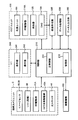
- Control system> A configuration example of the control system of the blood flow measuring device 1 is shown in FIGS.
- the control unit 210, the image forming unit 220, and the data processing unit 230 are provided in the arithmetic control unit 200.
- the control unit 210 executes various controls.
- the control unit 210 includes a main control unit 211 and a storage unit 212.
- the main control unit 211 includes a processor and controls each unit (including the elements shown in FIGS. 1 to 4) of the blood flow measurement device 1.
- the imaging focusing lens 31 arranged in the imaging optical path and the focus optical system 60 arranged in the illumination optical path are moved synchronously by an imaging focusing drive unit (not shown) under the control of the main control unit 211.
- the retro-reflector 41 provided in the measurement arm is moved by the retro-reflector (RR) drive unit 41A under the control of the main control unit 211.
- the OCT focusing lens 43 arranged on the measurement arm is moved by the OCT focusing driving unit 43A under the control of the main control unit 211.
- the optical scanner 44 provided in the measurement arm operates under the control of the main control unit 211.
- the retro-reflector 114 arranged on the reference arm is moved by the retro-reflector (RR) driving unit 114A under the control of the main control unit 211.
- Each of these drive units includes an actuator such as a pulse motor that operates under the control of the main control unit 211.
- the main control unit 211 controls the LCD 39.
- the main controller 211 displays a fixation target at a preset position on the screen of the LCD 39.
- the main control unit 211 can change the display position (fixation position) of the fixation target displayed on the LCD 39.
- the movement of the fixation target can be performed in any manner such as continuous movement, intermittent movement, and discrete movement. The movement mode of the fixation position in this embodiment will be described later.
- the fixation position is expressed by, for example, the display position (pixel coordinates) of the fixation target image on the LCD 39.
- This coordinate is, for example, a coordinate represented by a two-dimensional coordinate system defined in advance on the display screen of the LCD 39.
- the fixation position is expressed by, for example, the position (coordinates) of the light emitting unit that is lit.
- This coordinate is, for example, a coordinate represented by a two-dimensional coordinate system defined in advance on the array surface of the plurality of light emitting units.
- the storage unit 212 stores various data. Examples of data stored in the storage unit 212 include OCT images, fundus images, and eye information.
- the eye information includes subject information such as patient ID and name, left / right eye identification information, electronic medical record information, and the like.
- the image forming unit 220 forms OCT image data of the fundus oculi Ef based on a signal (sampling data) input from the data acquisition system 130.
- the image forming unit 220 can form B-scan image data (two-dimensional tomographic image data) of the fundus oculi Ef and phase image data. These OCT image data will be described later.
- the image forming unit 220 includes, for example, a circuit board and a microprocessor. In the present specification, unless otherwise specified, “image data” and “image” based thereon are not distinguished.
- a cross section (target cross section) intersecting the target blood vessel of the fundus oculi Ef is repeatedly scanned with the measurement light LS.
- a predetermined cross section (supplementary cross section) is scanned with the measurement light LS in order to estimate the inclination of the blood vessel of interest in the cross section of interest.
- the supplementary cross section may be, for example, a cross section that intersects the target blood vessel and is located in the vicinity of the target cross section (first supplemental cross section), or a cross section that intersects the target cross section and is along the target blood vessel (second supplemental cross section). .
- FIG. 5A shows an example in which the first supplementary cross section is applied.
- one attention section C0 located near the optic disc Da of the fundus oculi Ef and two supplementary sections C1 and C2 located near the intersection intersect the attention blood vessel Db.
- One of the two supplementary cross sections C1 and C2 is positioned upstream of the target blood vessel Db with respect to the target cross section C0, and the other is positioned downstream.
- the target cross section C0 and the supplementary cross sections C1 and C2 are oriented so as to be substantially orthogonal to the traveling direction of the target blood vessel Db.
- FIG. 5B shows an example in which the second supplementary cross section is applied.
- the target cross section C0 similar to the example shown in FIG. 5A is set so as to be substantially orthogonal to the target blood vessel Db
- the supplementary cross section Cp is set so as to be substantially orthogonal to the target cross section C0.
- the supplementary cross section Cp is set along the target blood vessel Db.
- the supplementary cross section Cp may be set so as to pass through the central axis of the target blood vessel Db at the position of the target cross section C0.
- the image forming unit 220 includes a tomographic image forming unit 221 and a phase image forming unit 222.
- the tomographic image forming unit 221 forms a tomographic image (main tomographic image) representing a time-series change in the form of the cross section of interest based on the sampling data obtained from the data acquisition system 130 in the main scanning. This process will be described in more detail.
- the cross section of interest C0 is repeatedly scanned as described above. Sampling data is sequentially input from the data collection system 130 to the tomographic image forming unit 221 in accordance with this repeated scanning.
- the tomographic image forming unit 221 forms one main tomographic image corresponding to the target section C0 based on the sampling data corresponding to each scan of the target section C0.
- the tomographic image forming unit 221 forms a series of main tomographic images along a time series by repeating this process as many times as the main scanning is repeated.
- these main tomographic images may be divided into a plurality of groups, and the main tomographic image groups included in each group may be superimposed to improve image quality (image averaging process).
- the tomographic image forming unit 221 forms a tomographic image (supplemental tomographic image) representing the shape of the supplemental cross section based on the sampling data obtained by the data acquisition system 130 in the supplementary scan for the supplemental cross section.
- the process for forming the supplemental tomographic image is executed in the same manner as the process for forming the main tomographic image.
- the main tomographic image is a series of tomographic images along time series, but the supplementary tomographic image may be a single tomographic image.
- the supplemental tomographic image may be an image obtained by superimposing a plurality of tomographic images obtained by scanning the supplemental cross section a plurality of times to improve the image quality (addition averaging process of images).
- the tomographic image forming unit 221 forms a supplemental tomographic image corresponding to the supplemental cross section C1 and a supplemental tomographic image corresponding to the supplementary cross section C2.
- the tomographic image forming unit 221 forms a supplemental tomographic image corresponding to the supplementary cross section Cp.
- the processing for forming a tomographic image as exemplified above includes noise removal (noise reduction), filter processing, fast Fourier transform (FFT), and the like, as in the conventional Fourier domain OCT.
- noise removal noise reduction
- filter processing filter processing
- FFT fast Fourier transform
- the tomographic image forming unit 221 executes a known process corresponding to the type.
- the phase image forming unit 222 forms a phase image representing a time-series change of the phase difference in the cross section of interest based on the sampling data obtained by the data acquisition system 130 in the main scanning.
- the sampling data used for forming the phase image is the same as the sampling data used for forming the main tomographic image by the tomographic image forming unit 221. Therefore, it is possible to align the main tomographic image and the phase image. That is, it is possible to set a natural correspondence between the pixels of the main tomographic image and the pixels of the phase image.
- phase image forming method An example of a phase image forming method will be described.
- the phase image of this example is obtained by calculating the phase difference between adjacent A-line complex signals (that is, signals corresponding to adjacent scanning points).
- the phase image in this example is formed based on a time-series change in pixel values (luminance values) of the main tomographic image.
- the phase image forming unit 222 creates a graph of the time-series change in luminance value of the pixel.
- the phase difference ⁇ is defined as the phase difference ⁇ (t1) at the time point t1 (more generally, an arbitrary time point between the time point t1 and the time point t2).
- the phase image represents the value of the phase difference at each time point of each pixel as an image.
- This imaging process can be realized, for example, by expressing the value of the phase difference with the display color or brightness.
- the display color for example, red
- the display color for example, blue
- the magnitude of the phase change amount can be expressed by the density of the display color.
- the data processing unit 230 executes various data processing.
- the data processing unit 230 performs various types of image processing and analysis processing on the image formed by the image forming unit 220.
- the data processing unit 230 executes various correction processes such as image brightness correction and dispersion correction.
- the data processing unit 230 can perform various types of image processing and analysis processing on images (fundus image, anterior eye image, etc.) obtained by the fundus camera unit 2 and images input from the outside. it can.
- Stack data is image data obtained by three-dimensionally arranging a plurality of tomographic images obtained along a plurality of scanning lines based on the positional relationship of the scanning lines. That is, the stack data is an image obtained by expressing a plurality of tomographic images originally defined by individual two-dimensional coordinate systems using one three-dimensional coordinate system (that is, embedding in one three-dimensional space). It is data.
- the main tomographic image and the supplemental tomographic image have sufficient resolution as an object of analysis processing, but the phase image may not have enough resolution to identify the boundary of the blood vessel region.
- the phase image may not have enough resolution to identify the boundary of the blood vessel region.
- it is necessary to specify a blood vessel region included in the blood flow information with high accuracy and high accuracy. Therefore, for example, by performing the following processing, the blood vessel region in the phase image can be specified more accurately.
- the blood vessel region specifying unit 231 obtains a blood vessel region by analyzing a main tomographic image, specifies an image region in a phase image corresponding to the blood vessel region based on the correspondence, and specifies the specified image region as a phase image. Adopt as middle blood vessel region. Thereby, the blood vessel region of the phase image can be specified with high accuracy and high accuracy.
- the blood flow information generation unit 232 generates blood flow information related to the target blood vessel Db. As described above, the blood flow information generation unit 232 includes the inclination estimation unit 233, the blood flow velocity calculation unit 234, the blood vessel diameter calculation unit 235, and the blood flow rate calculation unit 236.
- the inclination estimation unit 233 obtains an estimated value of the inclination of the blood vessel of interest based on supplementary cross-section data (cross-section data, supplemental tomographic image) collected by supplementary scanning.
- This estimated inclination value may be, for example, a measured value of the inclination of the target blood vessel in the target section or an approximate value thereof.
- the inclination estimation unit 233 specifies the positional relationship between the cross section of interest C0, the supplemental cross section C1, and the supplemental cross section C2, and specifies the blood vessel region by the blood vessel region specifying unit 231. Based on the result, the inclination of the target blood vessel Db in the target cross section C0 can be calculated.
- Reference numerals G0, G1, and G2 respectively indicate a main tomographic image at the target section C0, a supplementary tomographic image at the supplementary section C1, and a supplementary tomographic image at the supplementary section C2.
- Symbols V0, V1, and V2 indicate a blood vessel region in the main tomographic image G0, a blood vessel region in the supplemental tomographic image G1, and a blood vessel region in the supplemental tomographic image G2, respectively.
- the z coordinate axis shown in FIG. 6A substantially coincides with the incident direction of the measurement light LS.
- the blood vessel region in three cross sections is considered, but it is also possible to obtain the inclination in consideration of the two cross sections.
- the inclination of the first line segment or the second line segment can be set as a target inclination.
- one inclination is obtained, but the inclination may be obtained for each of two or more positions (or areas) in the blood vessel region V0.
- two or more obtained slope values can be used separately, or values obtained by statistically processing these slope values (for example, average value, maximum value, minimum value, intermediate value) , Mode value, etc.) can be used as the slope A.
- the blood flow rate calculation unit 236 substitutes the calculation result w of the blood vessel diameter by the blood vessel diameter calculation unit 235 and the maximum value Vm based on the calculation result of the blood flow velocity by the blood flow velocity calculation unit 234 into this equation.
- a target blood flow rate Q is calculated.
- the main control unit 211 causes the display unit 241 to display a front image of the fundus oculi Ef.
- This front image may be an arbitrary type of image, and may be any one of an observation image, a captured image, a fluorescence image, an OCT angiography image, an OCT projection image, and an OCT shadowgram, for example.
- the user can designate one or more cross sections of interest with respect to the displayed front image of the fundus oculi Ef by operating the operation unit 242.
- the cross section of interest is designated to intersect the blood vessel of interest.
- the cross-section setting unit 237 can set one or more supplemental cross-sections for each of the one or more attention sections based on the specified one or more attention sections and the front image of the fundus oculi Ef. Note that the supplementary cross section may be set manually.
- the cross-section setting unit 237 may be configured to identify one or more blood vessels of interest by analyzing a front image of the fundus oculi Ef.
- the target blood vessel is identified based on, for example, the thickness of the blood vessel, the positional relationship with respect to a predetermined part of the fundus (for example, the optic nerve head and the macula), the type of blood vessel (for example, artery, vein), and the like.
- the cross-section setting unit 237 can set one or more target cross sections and one or more supplemental cross sections for each of the specified one or more target blood vessels.
- the attention cross section and the supplemental cross section illustrated in FIG. 5A or 5B are set for the fundus oculi Ef by the cross section setting section 237 or by the user and the cross section setting section 237 in cooperation with the user.
- the vignetting determination unit 238 determines the presence or absence of vignetting based on the detection result of the return light of the light incident on the eye E via the optical system mounted on the blood flow measurement device 1.
- the type, application, and intensity (light quantity) of light incident on the eye E for vignetting determination may be arbitrary.
- the type of incident light may be, for example, infrared light or visible light.
- the use of incident light may be, for example, imaging, inspection or measurement.
- the incident light that can be used in the present embodiment may be, for example, measurement light LS for OCT, observation illumination light for photographing the fundus, photographing illumination light, or excitation light.
- the detection result of the incident light return light may be, for example, an OCT image, an observation image, a captured image, or a fluorescence image.
- “Vignetting” in this embodiment includes vignetting (decrease in brightness) that occurs when incident light for fundus blood flow measurement or part or all of its return light is blocked by the pupil. Further, the “presence / absence of vignetting” in the present embodiment includes any of the following: whether or not vignetting has occurred; whether or not the degree of vignetting exceeds a predetermined level.
- incident light for vignetting determination is measurement light LS, and a predetermined OCT scan is applied.
- the aspect of the OCT scan in this example may be, for example, an OCT scan for the cross section of interest C0 (or supplementary cross section C1 and / or C2) shown in FIG. 5A or an OCT scan for the supplementary cross section Cp shown in FIG. 5B.
- the OCT scan in this example is a repeated scan with respect to the same cross section, for example.
- a tomographic image Gp1 (section data) representing the form of the supplementary section Cp is obtained as shown in FIG. 7A.
- a tomographic image Gp2 (cross-sectional data) in which the form of the supplementary cross-section Cp is not depicted is obtained as shown in FIG. 7B.
- the optical scanner 44 is disposed at a position substantially conjugate with the pupil of the eye E, so that the deflection center of the measurement light LS substantially coincides with the pupil. Therefore, when the measurement light LS is vignetted in the pupil, a tomographic image Gp2 (cross-section data) in which the shape of the supplementary cross-section Cp is not drawn is obtained.
- the incident light for vignetting determination is illumination light for fundus photographing, and may be observation illumination light or photographing illumination light.
- the vignetting determination unit 238 analyzes the fundus image and determines whether or not a light amount decrease due to the vignetting has occurred. For example, the vignetting determination unit 238 determines whether there is a difference between the brightness of the center portion of the fundus image and the brightness of the peripheral portion (that is, whether the peripheral light amount has decreased). This determination is performed with reference to the values (typically luminance distribution) of the pixels constituting the fundus image, and includes image processing such as threshold processing and labeling, for example.
- the degree of vignetting can be easily determined.
- the degree of vignetting is evaluated based on, for example, the characteristics of an image area where the peripheral light amount is reduced in the fundus image.
- the vignetting determination unit 238 can calculate the evaluation value (for example, the size (area) of the peripheral light amount reduction region) in this way, and can determine the presence or absence of vignetting based on the size of the evaluation value.
- the “presence / absence of vignetting” in this example corresponds to, for example, whether or not the degree of vignetting exceeds a predetermined level.
- the vignetting determination unit 238 compares the calculated evaluation value with a threshold value, determines that “there is vignetting” when the evaluation value exceeds the threshold value, and when the evaluation value is equal to or less than the threshold value. It is determined that “no vignetting”.
- the user interface (UI) 240 includes a display unit 241 and an operation unit 242.
- the display unit 241 includes the display device 3 shown in FIG. 1 and other display devices.
- the operation unit 242 includes an arbitrary operation device.
- the user interface 240 may include a device having both a display function and an operation function, such as a touch panel.
- the main control unit 211 controls the moving mechanism 150 to move the optical system by a predetermined distance in a predetermined direction on the xy plane.
- the main control unit 211 applies the first movement control for moving the optical system by a predetermined distance from the optical axis of the optical system to the moving mechanism 150 in a predetermined direction orthogonal to the optical axis of the optical system.
- the optical axis of the optical system is moved from a position H0 substantially coincident with the corneal apex (or pupil center) in the xy direction to a position H1 that is separated by a predetermined distance in the upper left direction.
- the movement direction (initial movement direction) of the optical system in the first movement control may be arbitrary and is set in advance.
- the movement distance (initial movement amount) of the optical system in the first movement control may be arbitrary, for example, may be a default distance.
- the initial movement amount may be, for example, a distance set according to the presence or absence of mydriasis, or a distance set according to the pupil diameter of the eye E to be examined. Also good.
- step S6 Fine movement of the optical system
- the main control unit 211 finely adjusts the position of the optical system based on the determination result obtained in step S5.
- the second movement control is applied to the movement mechanism 150.
- the main control unit 211 controls the moving mechanism 150 to move the optical system by a predetermined distance (predetermined fine movement amount) in the initial movement direction.
- a predetermined distance predetermined fine movement amount
- the optical axis of the optical system is in the direction toward the outer edge of the pupil Ep (that is, in the direction away from the optical axis position H0 immediately after auto-alignment). Is moved.
- the main control unit 211 controls the moving mechanism 150 so as to move the optical system by a predetermined distance (predetermined fine movement amount) in the direction opposite to the initial movement direction.
- a predetermined distance predetermined fine movement amount
- the determination of whether or not the movement completion condition of this example is satisfied is not limited to these examples. Further, the movement completion condition is not limited to that in this example.
- step S9 Execute blood flow measurement
- the blood flow measurement device 1 performs blood flow measurement of the cross section of interest C0 as described above, and generates blood flow information.
- the display unit 241 displays the measurement angle value (estimated value) obtained by the inclination estimation unit 233 and a preset suitable measurement angle (target angle) value, thereby allowing the measurement angle and the target value to be displayed. It is possible to present the angle to the user.
- This display control is executed by the main control unit 211.
- the target angle is set, for example, in the range of 78 degrees to 85 degrees, and more preferably in the range of 80 degrees to 85 degrees.
- the target angle may be a single value or a range defined by an upper limit and / or a lower limit.
- the target angle is, for example, a default value, a default range, a value or range set by a user or another person (for example, a maintenance service person), a blood flow measurement device 1 or another device (for example, a medical institution server, and It may be either a value or a range set by the device that monitors the operation of the blood flow measuring device 1.
- a target angle previously applied to the eye E is set again, a target angle is set according to the attributes of the subject, The target angle can be set according to the attribute of the eye E.
- the main control unit 211 can cause the display unit 241 to display the measurement angle obtained by the inclination estimation unit 233 at an arbitrary timing after the first movement control together with the target angle.
- the main control unit 211 causes the display unit 241 to display the measurement angle obtained by the tilt estimation unit 233 together with the target angle after the first movement control, or the tilt estimation unit 233 after the second movement control.
- the measurement angle obtained by the above can be displayed on the display unit 241 together with the target angle.
- the user can input an instruction with reference to the measurement value of the measurement angle and the value of the target angle. For example, when a measurement angle that is (substantially) coincident with the target angle is obtained, the user can input a blood flow measurement start instruction using the operation unit 242. Further, the main control unit 211 compares the measurement value of the measurement angle with the target angle, executes blood flow measurement start control when they are (almost) coincident, and if they do not coincide with each other, further control is performed. Two movement control may be performed.
- the main control unit 211 can execute the first movement control again. For example, in response to the change of the target angle, the main control unit 211 can re-execute step S4 of the operation shown in FIG. 8 and re-execute the processes after step S5.
- the main control unit 211 can start blood flow measurement and acquire blood flow information.
- the main control unit 211 can start blood flow measurement.
- the blood flow measuring device 1 can be used as follows. Assume that the initial value of the target angle is set to 80 degrees. If the measurement angle of 80 degrees is not achieved even when the second movement control is repeatedly performed, the user changes the target angle to 78 degrees, for example.
- the blood flow measurement device 1 performs the first movement control again (S4), and further performs vignetting determination (S5), the second movement control, and the like.
- the user checks the value of the measurement angle obtained after the second movement control on the display unit 241 and inputs a blood flow measurement start instruction when the measurement angle of 78 degrees is achieved. If the measurement angle of 78 degrees is not achieved even after repeating the second movement control, for example, the user can select one of re-change of the target angle, re-measurement, and cancellation of measurement.
- the blood flow measurement device (1) of the embodiment includes a blood flow measurement unit, a moving mechanism, a control unit, and a determination unit.
- the blood flow measurement unit includes an optical system for applying an optical coherence tomography (OCT) scan, and acquires blood flow information based on data collected by the OCT scan.
- OCT optical coherence tomography
- the optical system of the blood flow measurement unit includes the measurement arm shown in FIG. 1 and the optical system shown in FIG.
- the blood flow measuring unit includes an image forming unit 220 and a data processing unit 230 (blood flow information generating unit 232).
- the moving mechanism has a configuration for moving the optical system of the blood flow measurement unit.
- the moving mechanism includes the moving mechanism 150.
- the determination unit determines the presence or absence of vignetting based on the detection result of the return light of the light incident on the eye to be examined via the optical system of the blood flow measurement unit after the first movement control.
- the determination unit includes the vignetting determination unit 238.
- the optical system can be configured.
- the light path for blood flow measurement and the light path for vignetting determination should be known or recognizable.
- the control unit applies the second movement control for further moving the optical system of the blood flow measurement unit to the movement mechanism based on the determination result obtained by the determination unit.
- the user can recognize the measurement angle of the blood vessel of interest, and can further compare the measurement angle with the target angle. Thereby, it is possible to arbitrarily input an instruction to start or stop blood flow measurement.
- the user can change the target angle using the operation unit (242).
- the control unit can execute the first movement control again.
- the user can arbitrarily change the target angle. Furthermore, in response to the change of the target angle, the offset operation of the measurement arm for obtaining an optimal Doppler signal in fundus blood flow measurement can be performed again. As a result, when a suitable offset state cannot be obtained even by repeating the second movement control, it is possible to reset the target angle and search for a suitable offset position again.
- the optical axis of the measurement arm is considered to be located inside the pupil of the eye to be examined.Therefore, there is room for increasing the offset amount of the measurement arm in order to obtain an optimal Doppler signal in fundus blood flow measurement. is there. Therefore, in the embodiment, the optical system of the blood flow measurement unit is further moved in the same first direction as the movement direction in the first movement control to search for a more suitable offset position.
- the optical axis of the measurement arm is considered to be located outside the pupil of the eye to be inspected, so that the blood flow measurement unit has a direction opposite to the movement direction in the first movement control.
- the offset position is adjusted so that the optical axis of the measurement arm passes through the inside of the pupil by moving the optical system.
- control unit determines that there is no vignetting by the determination unit, and continues until the optical system is arranged at a position where the displacement from the position of the optical axis immediately before the first movement control is maximized.
- the movement control can be repeatedly executed.
- the data processing unit 230A includes a motion detection unit 251 and an image evaluation unit 252 in addition to the elements in the data processing unit 230 shown in FIG.
- the main control unit 211 can execute the second movement control similar to the first embodiment based on the determination result obtained by the vignetting determination unit 238 and the evaluation result obtained by the image evaluation unit 252. . Also in this embodiment, the main control unit 211 applies the first movement control for moving the measurement arm by a predetermined distance from the optical axis to the movement mechanism 150 in the first direction orthogonal to the optical axis of the measurement arm. Second movement control for further moving the measurement arm based on the determination result obtained by the vignetting determination unit 238 is applied to the moving mechanism 150.
- the main control unit 211 performs optical in the same first direction as the first movement control.
- the second movement control can be executed so as to further move the system (measurement arm).
- the main control unit 211 performs a direction opposite to the first direction.
- the second movement control can be executed so as to move the optical system (measurement arm).
- the main control unit 211 determines that there is no vignetting by the vignetting determination unit 238, the image evaluation unit 252 evaluates that the infrared observation image is good, and the position of the optical axis immediately before the first movement control. It is possible to repeatedly execute the second movement control until the optical system (the objective lens 22 of the measurement arm) is disposed at a position where the displacement from (H0) is maximized.
- the present embodiment it is possible to execute the offset operation of the measurement arm for obtaining an optimal Doppler signal in fundus blood flow measurement while suitably performing tracking. Therefore, it is possible to avoid an error in the offset operation due to the tracking failure and to further reduce the burden on the subject.
- the blood flow measurement device can set an initial movement amount (for example, a distance between the position H0 and the position H1 in FIG. 9A) that is the movement distance of the optical system in the first movement control. is there.
- an initial movement amount for example, a distance between the position H0 and the position H1 in FIG. 9A
- Whether or not a mydriatic is applied to the eye E is input by the user using the operation unit 242, for example. Or you may make it determine whether a mydriatic agent was applied with reference to a test subject's electronic medical record etc.
- FIG. 242 Whether or not a mydriatic is applied to the eye E is input by the user using the operation unit 242, for example. Or you may make it determine whether a mydriatic agent was applied with reference to a test subject's electronic medical record etc.
- the blood flow measurement device can set an initial movement amount (for example, a distance between the position H0 and the position H1 in FIG. 9A) that is the movement distance of the optical system in the first movement control. is there.
- the initial movement amount is set according to whether or not the mydriatic is applied, but in this embodiment, the initial movement amount is set according to the actual pupil diameter of the eye E.
- the pupil diameter information acquisition unit 260 acquires the value of the pupil diameter of the eye E.
- the pupil diameter information acquisition unit 260 includes an element for measuring the pupil diameter of the eye E.
- the pupil diameter information acquisition unit 260 analyzes the anterior segment image of the eye E acquired by the illumination optical system 10 and the imaging optical system 30, identifies the pupil region, and calculates the diameter thereof. May be included.
- the specification of the pupil region may include processing such as threshold processing and edge detection.
- the calculation of the diameter of the pupil region may include processing such as elliptical approximation and circular approximation.
- the pupil diameter information acquisition unit 260 acquires a measured value of the pupil diameter of the eye E acquired in the past from the electronic medical record of the subject.
- the pupil diameter information acquisition unit 260 of this example includes a communication device for accessing an apparatus in which an electronic medical record or the like is stored.
- the control unit 210B includes a movement distance setting unit 214 in addition to the elements in the control unit 210 shown in FIG.
- the movement distance setting unit 214 can set the initial movement amount in the first movement control based on the pupil diameter value acquired by the pupil diameter information acquisition unit 260.
- the movement distance setting unit 214 can set, for example, a half value of the pupil diameter value acquired by the pupil diameter information acquisition unit 260 as the initial movement amount. This initial movement amount corresponds to the value of the radius of the pupil of the eye E or the approximate value thereof.
- the movement distance setting unit 214 is an example of a second setting unit.
- the main control unit 211 executes the first movement control by applying the initial movement amount selected by the movement distance setting unit 214.
- the initial movement amount in the first movement control can be set based on the actual measurement value of the pupil diameter of the eye E, in order to obtain an optimal Doppler signal in fundus blood flow measurement.
- the offset operation of the measurement arm it is possible to further shorten the time until the optimum offset position is achieved. Thereby, it is possible to further reduce the burden on the subject.
- the blood flow measurement device 1 ⁇ / b> A includes a fundus camera unit 2, an OCT unit 100, and an arithmetic control unit 200.
- the fundus camera unit 2 is provided with an optical system and a mechanism for acquiring a front image of the eye to be examined.
- the OCT unit 100 is provided with a part of an optical system and a mechanism for performing OCT. Another part of the optical system and mechanism for performing OCT is provided in the fundus camera unit 2.
- the arithmetic control unit 200 includes one or more processors that execute various types of arithmetic operations and controls.
- optional elements such as a member for supporting the subject's face (chin rest, forehead rest, etc.) and a lens unit for switching the OCT target site (for example, anterior segment OCT attachment) Or a unit may be provided in the blood flow measuring apparatus 1A.
- the “processor” is, for example, a CPU (Central Processing Unit), a GPU (Graphics Processing Unit), an ASIC (Application Specific Integrated Circuit), a programmable logic device (eg, SPLD (Simple ProGLD). It means a circuit such as Programmable Logic Device (FPGA) or Field Programmable Gate Array (FPGA).
- the processor implements the functions according to the embodiment by reading and executing a program stored in a storage circuit or a storage device.
- the fundus camera unit 2 is provided with an optical system for photographing the fundus oculi Ef of the eye E to be examined.
- the acquired image of the fundus oculi Ef (referred to as a fundus oculi image, a fundus oculi photo or the like) is a front image such as an observation image or a captured image.
- the observation image is obtained by moving image shooting using near infrared light.
- the photographed image is a still image using flash light.
- the fundus camera unit 2 includes an illumination optical system 10 and a photographing optical system 30.
- the illumination optical system 10 irradiates the eye E with illumination light.
- the imaging optical system 30 detects the return light of the illumination light from the eye E.
- the measurement light from the OCT unit 100 is guided to the eye E through the optical path in the fundus camera unit 2, and the return light is guided to the OCT unit 100 through the same optical path.
- the light (observation illumination light) output from the observation light source 11 of the illumination optical system 10 is reflected by the concave mirror 12, passes through the condenser lens 13, passes through the visible cut filter 14, and becomes near infrared light. Further, the observation illumination light is once converged in the vicinity of the photographing light source 15, reflected by the mirror 16, and passes through the relay lens system 17, the relay lens 18, the stop 19, and the relay lens system 20. The observation illumination light is reflected by the peripheral part of the perforated mirror 21 (region around the hole part), passes through the dichroic mirror 46, and is refracted by the objective lens 22 to illuminate the eye E (fundus Ef). To do.
- the return light of the observation illumination light from the eye E is refracted by the objective lens 22, passes through the dichroic mirror 46, passes through the hole formed in the central region of the perforated mirror 21, and passes through the dichroic mirror 55.
- the light is reflected by the mirror 32 via the photographing focusing lens 31. Further, the return light passes through the half mirror 33A, is reflected by the dichroic mirror 33, and forms an image on the light receiving surface of the image sensor 35 by the condenser lens.
- the image sensor 35 detects return light at a predetermined frame rate. Note that the focus of the photographing optical system 30 is adjusted to match the fundus oculi Ef or the anterior eye segment.
- the light (imaging illumination light) output from the imaging light source 15 is irradiated onto the fundus oculi Ef through the same path as the observation illumination light.
- the return light of the imaging illumination light from the eye E is guided to the dichroic mirror 33 through the same path as the return light of the observation illumination light, passes through the dichroic mirror 33, is reflected by the mirror 36, and is reflected by the condenser lens 37.
- An image is formed on the light receiving surface of the image sensor 38.
- Liquid crystal display (LCD) 39 displays a fixation target (fixation target image).
- a part of the light beam output from the LCD 39 is reflected by the half mirror 33 ⁇ / b> A, reflected by the mirror 32, passes through the hole of the perforated mirror 21 through the photographing focusing lens 31 and the dichroic mirror 55.
- the light beam that has passed through the aperture of the aperture mirror 21 passes through the dichroic mirror 46, is refracted by the objective lens 22, and is projected onto the fundus oculi Ef.
- the configuration for presenting a fixation target that can change the fixation position to the eye E is not limited to a display device such as an LCD.
- a fixation matrix in which a plurality of light emitting units (light emitting diodes or the like) are arranged in a matrix (array) can be used instead of the display device.
- the fixation position of the eye E by the fixation target can be changed by selectively turning on the plurality of light emitting units.
- a fixation target whose fixation position can be changed can be generated by one or more movable light emitting units.
- the alignment optical system 50 generates an alignment index used for alignment of the optical system with respect to the eye E.
- the alignment light output from the light emitting diode (LED) 51 passes through the diaphragm 52, the diaphragm 53, and the relay lens 54, is reflected by the dichroic mirror 55, passes through the hole of the perforated mirror 21, and passes through the dichroic mirror 46.
- the light passes through and is projected onto the eye E through the objective lens 22.
- Return light (corneal reflection light or the like) of the alignment light from the eye E is guided to the image sensor 35 through the same path as the return light of the observation illumination light.
- Manual alignment and auto-alignment can be executed based on the received light image (alignment index image).
- the alignment index image of this example is composed of two bright spot images whose positions change depending on the alignment state.
- the two bright spot images are integrally displaced in the xy direction.
- the relative position between the eye E and the optical system changes in the z direction
- the relative position (distance) between the two bright spot images changes.
- the two bright spot images overlap.
- the position of the eye E and the position of the optical system coincide with each other in the xy direction
- two bright spot images are presented in or near a predetermined alignment target. If the distance between the eye E and the optical system in the z direction matches the working distance, and the position of the eye E and the position of the optical system in the xy direction match, the two bright spot images overlap and align. Presented in the target.
- the focus optical system 60 generates a split index used for focus adjustment on the eye E.
- the focus optical system 60 is moved along the optical path (illumination optical path) of the illumination optical system 10 in conjunction with the movement of the imaging focusing lens 31 along the optical path (imaging optical path) of the imaging optical system 30.
- the reflector 67 is inserted into and removed from the illumination optical path.
- the reflecting surface of the reflecting bar 67 is inclinedly arranged in the illumination optical path.
- the focus light output from the LED 61 passes through the relay lens 62, is separated into two light beams by the split indicator plate 63, passes through the two-hole aperture 64, is reflected by the mirror 65, and is reflected by the condenser lens 66 as a reflecting rod 67.
- the dispersion compensation member 42 works together with the dispersion compensation member 113 (described later) disposed on the reference arm so as to match the dispersion characteristic of the measurement light LS and the dispersion characteristic of the reference light LR.
- the optical scanner 44 is substantially disposed at a position optically conjugate with the pupil of the eye E to be examined.
- the optical scanner 44 deflects the measurement light LS guided by the measurement arm.
- the optical scanner 44 is, for example, a galvano scanner capable of two-dimensional scanning.
- the reference light LR is guided to the collimator 111 by the optical fiber 110 and converted into a parallel light beam, and is guided to the retro-reflector 114 via the optical path length correction member 112 and the dispersion compensation member 113.
- the optical path length correction member 112 acts to match the optical path length of the reference light LR and the optical path length of the measurement light LS.
- the dispersion compensation member 113 together with the dispersion compensation member 42 disposed on the measurement arm, acts to match the dispersion characteristics between the reference light LR and the measurement light LS.
- the retro-reflector 114 is movable along the optical path of the reference light LR incident thereon, thereby changing the length of the reference arm. The change of the optical path length of the reference arm is used, for example, for optical path length correction according to the eye axis length, adjustment of the interference state, or the like.
- the measurement light LS generated by the fiber coupler 105 is guided by the optical fiber 127 and converted into a parallel light beam by the collimator lens unit 40, and the retroreflector 41, the dispersion compensation member 42, the OCT focusing lens 43, and the optical scanner 44. Then, the light is reflected by the dichroic mirror 46 via the relay lens 45, refracted by the objective lens 22, and projected onto the eye E.
- the measurement light LS is scattered and reflected at various depth positions of the eye E.
- the return light from the eye E to be measured LS travels in the opposite direction on the same path as the forward path, is guided to the fiber coupler 105, and reaches the fiber coupler 122 via the optical fiber 128.
- the control unit 210, the image forming unit 220, and the data processing unit 230 are provided in the arithmetic control unit 200.
- the main control unit 211 includes a processor that can operate according to a control program, and controls each unit (including the elements shown in FIGS. 13 to 16) of the blood flow measurement device 1A.
- the moving mechanism 150 moves, for example, at least the fundus camera unit 2 three-dimensionally.
- the moving mechanism 150 includes an x stage that can move in the ⁇ x direction (left and right direction), an x moving mechanism that moves the x stage, and a y stage that can move in the ⁇ y direction (up and down direction).
- Each of these moving mechanisms includes an actuator such as a pulse motor that operates under the control of the main control unit 211.
- the image forming unit 220 includes a tomographic image forming unit 221 and a phase image forming unit 222.
- the data processing unit 230 includes a blood vessel region specifying unit 231, a blood flow information generating unit 232, and a cross-section setting unit 237 as exemplary elements for obtaining blood flow information.
- the blood flow information generation unit 232 includes an inclination estimation unit 233, a blood flow velocity calculation unit 234, a blood vessel diameter calculation unit 235, and a blood flow rate calculation unit 236.
- the blood vessel region specifying unit 231 obtains a blood vessel region by analyzing a main tomographic image, specifies an image region in a phase image corresponding to the blood vessel region based on the correspondence, and specifies the specified image region as a phase image. Adopt as middle blood vessel region. Thereby, the blood vessel region of the phase image can be specified with high accuracy and high accuracy.
- the inclination estimation unit 233 specifies the positional relationship between the target cross section C0, the supplementary cross section C1, and the supplementary cross section C2, and specifies the blood vessel region by the blood vessel region specifying unit 231. Based on the result, the inclination of the target blood vessel Db in the target cross section C0 can be calculated.
- the inclination estimation unit 233 may analyze a supplemental tomographic image corresponding to the supplementary cross section Cp and calculate an approximate value of the inclination of the target blood vessel Db in the target cross section C0. it can.
- the inclination estimation unit 233 can analyze the supplemental tomographic image Gp and specify an image region corresponding to a predetermined tissue of the fundus oculi Ef.
- the inclination estimation unit 233 can specify an image region (inner boundary membrane region) M corresponding to the inner boundary membrane (ILM) that is the surface tissue of the retina.
- ILM inner boundary membrane
- a known segmentation process is used to specify the image area.
- the slope A shown in FIGS. 18A and 18B is a vector representing the direction of the blood vessel Db of interest, and the definition of the value may be arbitrary.
- the value of the inclination A can be defined as an angle formed by the inclination (vector) A and the z axis.
- the slope A app shown in FIG. 18B is a vector representing the direction of the inner boundary membrane region M, and the definition of the value may be arbitrary.
- the value of the slope A app can be defined as the angle formed by the slope (vector) A app and the z-axis. Note that the direction of the z-axis is substantially the same as the incident direction of the measurement light LS.
- the blood flow velocity calculation unit 234 calculates the blood flow velocity in the target cross section C0 of the blood flowing in the target blood vessel Db based on the time series change of the phase difference obtained as the phase image.
- This calculation target may be a blood flow velocity at a certain point in time, or a time-series change (blood flow velocity change information) of this blood flow velocity.
- the time range in the latter is the entire time or arbitrary part of the time when the target cross section C0 is scanned.
- ⁇ f represents the Doppler shift received by the scattered light of the measurement light LS
- n represents the refractive index of the medium
- v represents the flow velocity (blood flow velocity) of the medium
- ⁇ represents the angle formed by the irradiation direction of the measurement light LS and the flow vector of the medium
- ⁇ represents the center wavelength of the measurement light LS.
- the blood vessel diameter calculation unit 235 determines the relationship between the scale on the image and the scale in the real space, such as the shooting angle of view (shooting magnification), working distance, and information on the eyeball optical system. Set the scale.
- This scale represents the length in real space.
- the blood flow in the blood vessel is a Hagen-Poiseuille flow.
- the blood flow rate Q is expressed by the following equation.
- the blood flow rate calculation unit 236 substitutes the calculation result w of the blood vessel diameter by the blood vessel diameter calculation unit 235 and the maximum value Vm based on the calculation result of the blood flow velocity by the blood flow velocity calculation unit 234 into this equation.
- a target blood flow rate Q is calculated.
- the main control unit 211 causes the display unit 241 to display a front image of the fundus oculi Ef.
- This front image may be an arbitrary type of image, and may be any one of an observation image, a captured image, a fluorescence image, an OCT angiography image, an OCT projection image, and an OCT shadowgram, for example.
- the cross-section setting unit 237 may be configured to identify one or more blood vessels of interest by analyzing a front image of the fundus oculi Ef.
- the target blood vessel is identified based on, for example, the thickness of the blood vessel, the positional relationship with respect to a predetermined part of the fundus (for example, the optic nerve head and the macula), the type of blood vessel (for example, artery, vein), and the like.
- the cross-section setting unit 237 can set one or more target cross sections and one or more supplemental cross sections for each of the specified one or more target blood vessels.
- the attention cross section and the supplemental cross section illustrated in FIG. 17A or FIG. 17B are set for the fundus oculi Ef by the cross section setting section 237 or by the user and the cross section setting section 237 in cooperation with the user.
- ⁇ Movement condition setting unit 260A> As described above, in order to obtain an optimal Doppler signal in fundus blood flow measurement, the measurement light LS needs to be incident at a suitable angle with respect to the traveling direction of the blood vessel of interest (blood flow direction). Then, this is realized by offsetting the optical axis of the optical system (the optical axis of the objective lens 22) with respect to the optical axis of the eye E to be examined.
- the movement condition setting unit 260A sets the movement condition of the optical system with respect to the optical axis of the eye E.
- the movement condition setting unit 260A includes a direction setting unit 261 and a distance setting unit 262.
- the moving direction set by the direction setting unit 261 is a direction orthogonal to the optical axis of the optical system (the optical axis of the measurement arm, the optical axis of the objective lens 22).
- the moving direction set by the direction setting unit 261 is a direction defined in the xy plane, and has only one or both of the x-direction component and the y-direction component. That is, the moving direction set by the direction setting unit 261 does not have a z-direction component.
- the direction setting unit 261 analyzes the supplemental tomographic image G1 to identify the blood vessel region V1, and analyzes the supplemental tomographic image G2 to identify the blood vessel region V2.
- the direction setting unit 261 specifies a feature position from each of the two specified blood vessel regions V1 and V2, and estimates the inclination of the target blood vessel Db from the difference between the z coordinate values of the two specified feature positions. Ask for.
- the direction setting unit 261 can set the initial movement direction of the optical system based on the estimated value of the inclination of the target blood vessel Db.
- the direction setting unit 261 can also set the initial movement direction of the optical system based on the difference between the z coordinate values of the two feature positions specified from the two blood vessel regions V1 and V2. It is.
- the direction setting unit 261 analyzes the supplemental tomographic image G1 to identify the inner boundary membrane region, and analyzes the supplemental tomographic image G2 to identify the inner boundary membrane region. Next, the direction setting unit 261 specifies a characteristic position (for example, a position immediately above the target blood vessel Db) from each of the two specified inner boundary membrane regions. The direction setting unit 261 sets the initial movement direction of the optical system based on the difference between the z coordinate values of the two specified feature positions (or the estimated value of the inclination of the target blood vessel Db calculated from this difference). Can do.
- the direction setting unit 261 analyzes the supplemental tomographic image Gp and identifies the inner boundary membrane region M. Next, the direction setting unit 261 calculates an estimated value of the inclination of the target blood vessel Db based on the identified inner boundary membrane region M. The direction setting unit 261 can set the initial movement direction of the optical system based on the estimated value of the inclination of the target blood vessel Db.
- the direction setting unit 261 analyzes the supplemental tomographic image Gp and identifies the blood vessel region corresponding to the target blood vessel Db. Next, the direction setting unit 261 obtains an estimated value of the inclination of the target blood vessel Db based on the identified blood vessel region. The direction setting unit 261 can set the initial movement direction of the optical system based on the estimated value of the inclination of the target blood vessel Db.
- the target blood vessel Db and the inner boundary membrane are depicted.
- the symbol Ma indicates the intersection position between the inner boundary membrane region M and the target cross section C0 (see FIG. 18B and the like), and is at the calculated position of the inner boundary membrane region M (calculated position of the estimated estimated value of the blood vessel Db).
- the straight line indicated by the symbol Mh is a line (inclination line) indicating the inclination direction of the inner boundary membrane region M at the inclination calculation position Ma.
- the straight line indicated by the symbol Mn is a line (inclination normal) indicating the normal direction of the inclination line Mh at the inclination calculation position Ma.
- the straight line indicated by the symbol P0 is the path (scan position) of the measurement light LS before the optical system moves (initial position).
- the initial position of the optical system is, for example, in a state where alignment is aligned with the pupil center of the eye E, that is, in a state where the optical axis of the optical system (the optical axis of the objective lens 22) is the pupil center of the eye E. Equivalent to.
- the measurement path P0 passes through the inclination calculation position Ma and is parallel to the z direction.
- the straight line indicated by the symbol P0n is a line (measurement normal) indicating the normal direction of the measurement path P0 at the inclination calculation position Ma.
- the angle between the measurement path P0 and the tilt normal Mn is indicated by ⁇ 0.
- the angle ⁇ 0 is an incident angle of the measurement light LS with respect to the inner boundary film of the eye E when the optical system is disposed at the initial position. That is, the angle ⁇ 0 is used as the incident angle of the measurement light LS with respect to the target blood vessel Db when the optical system is disposed at the initial position.
- the angle formed by the measurement normal line P0n and the inclined line Mh is also ⁇ 0.
- the inclination of the target blood vessel Db may be defined by an angle “ ⁇ ” formed by the measurement path and the inclination normal line, or may be defined by an angle “ ⁇ ” formed by the measurement normal line and the inclination line.
- the inclination of the target blood vessel Db may be defined by an angle “90 degrees ⁇ ” formed by the measurement path and the inclination line, or an angle “90 degrees ⁇ ” formed by the measurement normal line and the inclination normal line. May be defined.
- the angle ⁇ is an incident angle of the measurement light LS with respect to the inner boundary membrane (target blood vessel Db).
- the residual angle 90 ° ⁇ of the angle ⁇ is represented by “ ⁇ ”.
- the direction setting unit 261 has the incident angle ⁇
- the moving direction of the optical system is set so that it is included in the target angle range (that is, the incident angle ⁇ is increased).
- the moving direction at this time is the left direction in FIG. 19A.
- the straight line indicated by reference sign P1n is a line (measurement normal) indicating the normal direction of the measurement path P1 at the inclination calculation position Ma.
- the direction setting unit 261 makes the optical system so that the incident angle ⁇ is included in the target angle range (that is, the incident angle ⁇ becomes small). Set the direction of movement.
- ⁇ Distance setting unit 262> As described above, the optical system is moved in the initial movement direction set by the direction setting unit 261.
- the movement distance (initial movement amount) at this time may be set in advance.
- the initial movement amount is set by the distance setting unit 262.
- the distance setting unit 262 may be capable of setting a movement amount applied to the second (or third and subsequent) movement of the optical system.
- the distance setting unit 262 of this example can set the initial movement amount according to the actual pupil diameter of the eye E.
- the pupil diameter information acquisition unit includes an element for measuring the pupil diameter of the eye E.
- the pupil diameter information acquisition unit analyzes a anterior segment image of the eye E acquired by the illumination optical system 10 and the imaging optical system 30, identifies a pupil region, and calculates a diameter thereof. May contain.
- the specification of the pupil region may include processing such as threshold processing and edge detection.
- the calculation of the diameter of the pupil region may include processing such as elliptical approximation and circular approximation.
- the pupil diameter information acquisition unit may be configured to acquire a measurement value of the pupil diameter of the eye E acquired in the past from an electronic medical record of the subject.
- the pupil diameter information acquisition unit of this example includes a communication device (data input / output unit 290) for accessing an apparatus in which an electronic medical record or the like is stored.
- the distance setting unit 262 can set the initial movement amount based on the value of the pupil diameter acquired by the pupil diameter information acquisition unit.
- the distance setting unit 262 can set, for example, a half value of the pupil diameter value acquired by the pupil diameter information acquisition unit as the initial movement amount. This initial movement amount corresponds to the value of the radius of the pupil of the eye E or the approximate value thereof.
- the initial movement amount can be set based on the actual measurement value of the pupil diameter of the eye E
- the optimum offset in the offset operation of the measurement arm for obtaining the optimum Doppler signal in the fundus blood flow measurement The time until the position is achieved can be shortened. Thereby, the burden placed on the subject can be reduced.
- a second example of processing that can be executed by the distance setting unit 262 will be described.
- the eye E is a small pupil eye or a cataract eye, or when the blood vessel of interest is located in the periphery of the fundus oculi Ef, a drug (mydriatic agent) for dilating the pupil is examined. E may apply.
- the distance setting unit 262 of this example can set the initial movement amount depending on whether or not a mydriatic is applied to the eye E.
- the distance setting unit 262 of this example previously determines the initial movement amount (first initial movement amount) when the mydriatic agent is not applied and the movement distance (second initial movement amount) when the mydriatic agent is applied.
- the first initial movement amount is set to, for example, a half value of a standard pupil diameter (a radius of a standard size pupil).
- the second initial movement amount is set to a value that is half the diameter of the dilated pupil when a mydriatic agent is applied to an eye with a standard pupil diameter.
- Whether or not a mydriatic is applied to the eye E is input by the user using the operation unit 242, for example. Alternatively, it may be determined whether or not a mydriatic is applied to the eye E by referring to the electronic medical record or the like of the subject via the data input / output unit 290.
- the distance setting unit 262 can select one of a plurality of stored initial movement amounts based on information indicating whether or not a mydriatic is applied to the eye E.
- the vignetting determination unit 270 determines the presence or absence of vignetting based on the detection result of the return light of the light incident on the eye E through the optical system mounted on the blood flow measurement device 1A.
- the type, application, and intensity (light quantity) of light incident on the eye E for vignetting determination may be arbitrary.
- the type of incident light may be, for example, infrared light or visible light.
- the use of incident light may be, for example, imaging, inspection or measurement.
- the incident light that can be used in the present embodiment may be, for example, measurement light LS for OCT, observation illumination light for photographing the fundus, photographing illumination light, or excitation light.
- the detection result of the incident light return light may be, for example, an OCT reflection intensity profile, an OCT image, an observation image, a captured image, or a fluorescence image.
- “Vignetting” in this embodiment includes vignetting (decrease in brightness) that occurs when incident light for fundus blood flow measurement or part or all of its return light is blocked by the pupil. Further, the “presence / absence of vignetting” in the present embodiment includes any of the following: whether or not vignetting has occurred; whether or not the degree of vignetting exceeds a predetermined level.
- incident light for vignetting determination is measurement light LS, and a predetermined OCT scan is applied.
- the aspect of the OCT scan in this example may be, for example, an OCT scan with respect to the cross section of interest C0 (or supplementary cross section C1 and / or C2) shown in FIG. 17A or an OCT scan with respect to the supplementary cross section Cp shown in FIG.
- the OCT scan in this example is a repeated scan with respect to the same cross section, for example.
- the supplemental section Cp When repetitive scanning is applied to the supplemental section Cp (or supplementary sections C1 and / or C2), it is possible to estimate the inclination of the blood vessel Db in parallel with the vignetting determination, or from the vignetting determination to the blood vessel of interest. A smooth transition to Db slope estimation is possible.
- the movement condition setting (movement direction setting, movement distance setting) may be performed in parallel with the vignetting determination.
- a tomographic image Gp1 (section data) representing the form of the supplementary section Cp is obtained as shown in FIG. 20A.
- a tomographic image Gp2 (cross-sectional data) in which the form of the supplementary cross-section Cp is not depicted is obtained as shown in FIG. 20B.
- the optical scanner 44 is disposed at a position substantially conjugate with the pupil of the eye E, so that the deflection center of the measurement light LS substantially coincides with the pupil. Therefore, when the measurement light LS is vignetted in the pupil, a tomographic image Gp2 (cross-section data) in which the shape of the supplementary cross-section Cp is not drawn is obtained.
- the vignetting determination unit 270 analyzes the tomographic image obtained by the OCT scan for the supplementary cross section Cp, and determines whether or not the form of the supplementary cross section Cp is drawn.
- the vignetting determination unit 270 may be configured to determine whether or not the inner boundary membrane region (M) is depicted in the tomographic image.
- the determination result that “the inner boundary membrane region is depicted in the tomographic image” corresponds to the determination result that “there is no vignetting”. Conversely, the determination result that “the inner boundary membrane region is not depicted in the tomographic image” corresponds to the determination result that “there is vignetting”.
- the incident light for vignetting determination is illumination light for fundus photographing, and may be observation illumination light or photographing illumination light.
- the vignetting determination unit 270 analyzes the fundus oculi image and determines whether or not a light amount decrease due to vignetting has occurred. For example, the vignetting determination unit 270 determines whether there is a difference between the brightness of the center portion of the fundus image and the brightness of the peripheral portion (that is, whether the peripheral light amount has decreased). This determination is performed with reference to the values (typically luminance distribution) of the pixels constituting the fundus image, and includes image processing such as threshold processing and labeling, for example.
- the degree of vignetting can be easily determined.
- the degree of vignetting is evaluated based on, for example, the characteristics of an image area where the peripheral light amount is reduced in the fundus image.
- the vignetting determination unit 270 calculates the evaluation value (for example, the size (area) of the peripheral light amount reduction region) in this way, and can determine the presence or absence of vignetting based on the size of the evaluation value.
- the “presence / absence of vignetting” in this example corresponds to, for example, whether or not the degree of vignetting exceeds a predetermined level.
- the vignetting determination unit 270 compares the calculated evaluation value with a threshold value, determines that there is vignetting when the evaluation value exceeds the threshold value, and if the evaluation value is equal to or less than the threshold value. It is determined that “no vignetting”.
- the data processing unit 230 that functions as described above includes, for example, a processor, a RAM, a ROM, a hard disk drive, and the like.
- a storage device such as a hard disk drive stores in advance a computer program that causes the processor to execute the above functions.
- the user interface (UI) 240 includes a display unit 241 and an operation unit 242.
- the display unit 241 includes the display device 3 shown in FIG. 13 and other display devices.
- the operation unit 242 includes an arbitrary operation device.
- the user interface 240 may include a device having both a display function and an operation function, such as a touch panel.
- the data input / output unit 290 outputs data from the blood flow measurement device 1A and inputs data to the blood flow measurement device 1A.
- the data input / output unit 290 has a function for communicating with an external device (not shown), for example.
- the communication unit 290 includes a communication interface corresponding to a connection form with an external device.
- the external device is, for example, any ophthalmic device.
- the external device may be any information processing device such as a hospital information system (HIS) server, DICOM (Digital Imaging and Communication in Medicine) server, doctor terminal, mobile terminal, personal terminal, cloud server, or the like.
- HIS hospital information system
- DICOM Digital Imaging and Communication in Medicine
- the data input / output unit 290 may include a device that reads information from a recording medium (data reader), a device that writes information to the recording medium (data writer), and the like.
- the blood flow measurement device 1A acquires the pupil diameter information of the eye E by the pupil diameter information acquisition unit described above.
- the blood flow measuring device 1A starts tracking for moving the optical system in accordance with the movement of the eye E (fundus Ef). Tracking includes, for example, an operation of acquiring an observation image of the fundus oculi Ef using infrared light, an operation of analyzing the observation image to detect the movement of the fundus oculi Ef, and an optical system in accordance with the detected movement of the fundus oculi Ef. This is realized by a combination with the movement of moving.
- the tracking may include an operation for controlling the optical scanner 44 in accordance with the movement of the fundus oculi Ef.
- setting of fundus imaging conditions such as focus adjustment for the fundus oculi Ef
- setting of OCT measurement conditions such as optical path length adjustment and focus adjustment, and the like
- selection of a target blood vessel, setting of a target cross section, setting of a supplemental cross section, and the like can be performed at this stage or at any other timing.
- the target blood vessel Db, the target cross section C0, and the supplementary cross section Cp shown in FIG. 17B are set.
- the blood flow measuring device 1A applies the OCT scan to the supplementary cross section Cp.
- the direction setting unit 261 analyzes the supplemental tomographic image Gp representing the supplementary cross section Cp and sets the movement direction (initial movement direction) of the optical system (at least the measurement arm).
- the distance setting unit 262 sets the movement distance (initial movement amount) of the optical system based on the pupil diameter information acquired in step S23. It is also possible to set the movement distance according to whether or not the mydriatic is applied.
- the pupil center (or corneal apex) in the xy direction substantially coincides with the optical axis of the optical system, and the eye E and the optical system (objective lens 22) are in the z direction.
- the distance is almost equal to the working distance.
- the main control unit 211 controls the moving mechanism 150 so as to move the optical system by the initial moving amount set in step S26 in the initial moving direction set in step S25.
- the main control unit 211 applies the first movement control for moving the optical system from the optical axis by the initial movement amount in the initial movement direction orthogonal to the optical axis of the optical system to the moving mechanism 150.
- the optical axis of the optical system is moved from the position H0 substantially coincident with the center of the pupil (or the apex of the cornea) in the xy direction to a position H1 separated by a predetermined distance in the upper left direction. Is moved.
- the movement direction (initial movement direction) of the optical system in the first movement control is set based on the OCT image of the fundus oculi Ef.
- the movement distance (initial movement amount) of the optical system in the first movement control may be arbitrary, for example, a distance set based on pupil diameter information and / or the presence or absence of application of a mydriatic agent. Alternatively, a default distance may be used.
- step S28 Determine the presence or absence of vignetting
- the main control unit 211 After the first movement control (first movement of the optical system) in step S27, the main control unit 211 starts scanning the supplementary cross section Cp shown in FIG. 17B as an OCT scan for vignetting determination.
- the vignetting determination unit 270 determines the presence or absence of vignetting based on the cross-sectional data acquired by the OCT scan.
- the image forming unit 220 forms a tomographic image from the data acquired by the OCT scan for the supplementary slice Cp, and the vignetting determination unit 270 adds the supplementary slice Cp to this tomographic image.
- the presence or absence of vignetting is determined by determining whether or not the form is drawn.
- step S29: Is there vignetting If it is determined by the vignetting determination in step S28 that there is vignetting (S29: Yes), the operation proceeds to step S30. On the other hand, when it is determined that there is no vignetting (S29: No), the operation proceeds to step S31.
- step S30 Fine movement of the optical system
- step S ⁇ b> 29 that there is vignetting
- step S ⁇ b> 29: Yes the main control unit 211 applies second movement control for finely adjusting the position of the optical system to the moving mechanism 150.
- the movement distance in the second movement control is shorter than the movement distance (initial movement amount) in the first movement control in step S27.
- the movement direction in the second movement control is the direction opposite to the initial movement direction in the first movement control in step S27.
- the movement distance in the second movement control may be a distance set by default or a distance set by the distance setting unit 262, for example.
- step S29 If it is determined in step S29 that vignetting has occurred, for example, as shown in FIG. 22B, when the optical axis position H1 after the first movement control is outside the pupil Ep, the measurement light LS is chromatic. A tomographic image in which the shape of the supplementary cross section Cp is not depicted as shown in FIG. 20B is obtained. As a result, the vignetting determination unit 270 obtains a determination result that vignetting has occurred. As indicated by the symbol J in FIG. 22B, the main control unit 211 moves so as to move the optical system by a predetermined distance (predetermined fine movement amount) in the opposite direction to the initial movement direction (direction from the position H0 to the position H1). The mechanism 150 is controlled. Thus, if it is determined in step S29 that vignetting has occurred, the optical axis of the optical system is moved in the direction toward the optical axis position H0 immediately after auto-alignment.
- predetermined distance predetermined fine movement amount
- Step S28 to S30 are repeated until “Yes” is determined in step S29. It should be noted that an error determination can be output when steps S28 to S30 are repeated a predetermined number of times, or when steps S28 to S30 are repeated for a predetermined time.
- step S31 Estimating the inclination of the blood vessel of interest
- step S29 No
- the blood flow measuring device 1A estimates the inclination of the blood vessel Db of interest in the manner described above.
- the cross-sectional data of the supplemental cross-section Cp is used for the inclination estimation in this example, as in the vignetting determination.
- step S29 If it is determined in step S29 that no vignetting has occurred, for example, as shown in FIG. 22C, the optical axis position H1 after the first movement control is inside the pupil Ep.
- step S32 Execute blood flow measurement
- the blood flow measurement device 1A performs blood flow measurement of the cross section of interest C0 as described above, and generates blood flow information (END).
- the main control unit 211 can cause the display unit 241 to display information indicating the initial movement direction set in step S25. Further, the main control unit 211 can cause the display unit 241 to display information indicating the initial movement amount set in step S26. Further, the main control unit 211 causes the display unit 241 to display information indicating the estimated tilt value obtained by the tilt estimating unit 233, or displays information indicating the difference between the estimated tilt value and the target angle range. 241 can be displayed.
- the target angle range is set to a range of 80 to 85 degrees, for example.
- the target angle range may be a single value or a range defined by an upper limit and / or a lower limit.
- the target angle range is, for example, a default value, a default range, a value or range set by a user or another person (for example, a maintenance serviceman), a blood flow measurement device 1A or another device (for example, a medical institution server, and
- the device or the device that monitors the operation of the blood flow measurement device 1A may be set to any one of a value or a range.
- a target angle range that has been applied to the eye E in the past is set again, or a target angle range is set according to the attributes of the subject.
- the target angle range can be set according to the attribute of the eye E.
- the user can input an instruction regarding the moving direction and an instruction regarding the moving distance with reference to the displayed information. These instructions are given, for example, when a suitable measurement angle cannot be obtained even if the second movement control is repeatedly performed.
- the blood flow measurement device (1) of the exemplary embodiment includes a scanning optical system, a moving mechanism, an image forming unit, a direction setting unit, a control unit, and a blood flow information acquisition unit.
- the scan optical system applies an OCT scan to the fundus of the subject's eye.
- the optical system including the OCT unit 100 and the measurement arm in the fundus camera unit 2 is an example of this scanning optical system.
- the moving mechanism moves the scanning optical system.
- the moving mechanism 150 of this embodiment is an example of this moving mechanism.
- the image forming unit forms an image from the first data collected by the scanning optical system.
- the image formed from the first data may be a reflection intensity profile, or may be an image (image data) generated therefrom.
- the image forming unit 220 of this embodiment is an example of this image forming unit.
- the direction setting unit analyzes the image formed by the image forming unit and sets a first direction (initial movement direction) orthogonal to the optical axis of the scanning optical system.
- the direction setting unit 261 of this embodiment is an example of this direction setting unit.
- the control unit applies the first movement control for moving the scanning optical system in the first direction to the moving mechanism.
- the control unit 210 main control unit 211) of this embodiment is an example of this control unit.
- the blood flow information acquisition unit acquires blood flow information from the second data collected by the scanning optical system after the first movement control.
- the image forming unit 220 and the data processing unit 230 (the blood vessel region specifying unit 231 and the blood flow information generating unit 232) of the present embodiment are examples of this blood flow information acquiring unit.
- the scanning optical system may be configured to collect the first data by applying an OCT scan to a cross section along the blood vessel of interest at the fundus.
- the direction setting unit may be configured to set the first direction by analyzing an image of the cross section formed from the first data. After the first movement control, the scan optical system can collect the second data by applying the OCT scan to the target blood vessel.
- the direction setting unit is configured to obtain an estimated value of the inclination of the target blood vessel by analyzing a cross-sectional image along the target blood vessel, and to set the first direction based on the estimated inclination value. It may be.
- the direction setting unit analyzes an image of a cross section along the blood vessel of interest, identifies an image region (inner boundary membrane region) corresponding to the fundus surface, and obtains an estimated tilt value based on the image region. It may be constituted as follows.
- the direction setting unit is configured to analyze an image of a cross section along the target blood vessel, identify an image region corresponding to the target blood vessel, and obtain an inclination estimation value based on the image region. Good.
- the direction setting unit may be configured to set the first direction so that the incident angle of the OCT scan light with respect to the blood vessel of interest is included in a predetermined range.
- control unit may be configured to execute the first movement control so as to move the scanning optical system in the first direction by a preset distance (initial movement amount).
- the blood flow measurement device further includes a pupil diameter information acquisition unit that acquires pupil diameter information of the eye to be examined, and a first setting unit that sets an initial movement amount based on the acquired pupil diameter information.
- a pupil diameter information acquisition unit that acquires pupil diameter information of the eye to be examined
- a first setting unit that sets an initial movement amount based on the acquired pupil diameter information.
- the illumination optical system 10, the imaging optical system 30, and the processor are one example of this pupil diameter information acquisition unit.
- the data input / output unit 290 is another example of the pupil diameter information acquisition unit.
- the distance setting unit 262 of the present embodiment is an example of the first setting unit.
- the blood flow measurement device may further include a second setting unit that sets an initial movement amount according to whether or not a mydriatic is applied to the eye to be examined.
- the distance setting unit 262 of this embodiment is an example of this second setting unit.
- the blood flow measurement device includes a determination unit that determines the presence or absence of vignetting based on the detection result of the return light of the light incident on the eye to be examined via the scanning optical system after the first movement control. Further, it may be included. Furthermore, the control unit may be configured to apply second movement control for further moving the scan optical system to the moving mechanism in accordance with the determination result obtained by the determination unit.
- the vignetting determination unit 270 of this embodiment is an example of this determination unit.
- the scanning optical system may be configured to apply an OCT scan for collecting the second data to the fundus when the determination unit determines that there is no vignetting. That is, the scan optical system is configured to apply an OCT scan for collecting the second data to the fundus without performing further movement control when the determination unit determines that there is no vignetting. Good.
- the control unit when the determination unit determines that vignetting is present, the control unit is a first direction (initial movement direction) by a distance shorter than the movement distance (initial movement amount) in the first movement control.
- the second movement control for moving the scanning optical system in the second direction opposite to the first direction may be applied to the moving mechanism.
- the blood is measured after the measurement arm is offset in the direction set based on the OCT image.
- Flow measurement can be performed. Therefore, the offset direction of the measurement arm with respect to the optical axis of the eye to be examined can be suitably set, and the offset position of the incident light for fundus blood flow measurement can be optimized.
- the present embodiment it is possible to present information for assisting the user when performing part of the operation of optimizing the offset position of incident light for fundus blood flow measurement manually.
Abstract
実施形態の血流計測装置は、血流計測部、移動機構、制御部及び判定部を含む。血流計測部は、眼底にOCTスキャンを適用するための光学系を含み、OCTスキャンにより収集されたデータに基づいて血流情報を取得する。移動機構は光学系を移動する。制御部は、光学系の光軸に直交する第1方向に光軸から所定距離だけ光学系を移動させるための第1移動制御を移動機構に適用する。判定部は、第1移動制御の後に、光学系を介して被検眼に入射した光の戻り光の検出結果に基づいてケラレの有無を判定する。制御部は、判定部により得られた判定結果に基づいて光学系を更に移動させるための第2移動制御を移動機構に適用する。
Description
この発明は、眼底の血流動態を計測するための血流計測装置に関する。
眼科分野において、光コヒーレンストモグラフィ(Optical Coherence Tomography:OCT)を利用して眼底の血流動態を計測する装置の開発が進められている。
眼底血流計測において最適なドップラー信号を得るためには、血流方向(血管の走行方向)に対して斜めに光を入射する必要がある。そのために、眼の光軸に対して入射光をオフセットさせることがある。オフセット量が増加するほど、血管に対してより斜めに光を入射できる一方、入射光又はその戻り光が瞳孔にてケラレる可能性がより高くなる。したがって、瞳孔でケラレない範囲において最大のオフセット量になる位置を探すことが望ましい。この動作を自動で行う場合、ケラレが生じないようにオフセット量を少しずつ増加していく方法も考えられるが、最適なオフセット位置を達成するまでに長時間を要し、被検者に多大な負担をかけることが想定される。
また、オフセットの方向及び量に応じて血管に対する入射角が変化するため、血流方向(血管の走行方向)に対して好適な入射角を実現するには、オフセット位置を最適化することが要求される。
この発明の1つの目的は、眼底血流計測のための入射光のオフセット位置を最適化することにある。
この発明の他の目的は、眼底血流計測のための入射光のオフセット位置の最適化動作を短時間化することにある。
幾つかの例示的な態様に係る血流計測装置は、血流計測部と、移動機構と、制御部と、判定部とを含む。血流計測部は、被検眼の眼底に光コヒーレンストモグラフィ(OCT)スキャンを適用するための光学系を含み、OCTスキャンにより収集されたデータに基づいて血流情報を取得する。移動機構は、光学系を移動する。制御部は、光学系の光軸に直交する第1方向に光軸から所定距離だけ光学系を移動させるための第1移動制御を移動機構に適用する。判定部は、第1移動制御の後に、光学系を介して被検眼に入射した光の戻り光の検出結果に基づいてケラレの有無を判定する。制御部は、判定部により得られた判定結果に基づいて光学系を更に移動させるための第2移動制御を移動機構に適用する。
幾つかの例示的な態様に係る血流計測装置は、スキャン光学系と、移動機構と、画像形成部と、方向設定部と、制御部と、血流情報取得部とを含む。スキャン光学系は、被検眼の眼底に光コヒーレンストモグラフィ(OCT)スキャンを適用する。移動機構は、スキャン光学系を移動する。画像形成部は、スキャン光学系により収集された第1データから画像を形成する。方向設定部は、画像を解析して、スキャン光学系の光軸に直交する第1方向を設定する。制御部は、スキャン光学系を第1方向に移動させるための第1移動制御を移動機構に適用する。血流情報取得部は、第1移動制御よりも後にスキャン光学系により収集された第2データから血流情報を取得する。
幾つかの実施形態によれば、眼底血流計測のための入射光のオフセット位置を最適化することが可能である。
幾つかの実施形態によれば、眼底血流計測のための入射光のオフセット位置の最適化動作を短時間化することが可能である。
例示的な実施形態に係る血流計測装置について図面を参照しながら詳細に説明する。実施形態の血流計測装置は、OCTを利用して眼底のデータを収集し、血流動態を表す情報(血流情報)を生成する。本明細書にて引用された文献の開示内容を含む任意の公知技術を、実施形態に援用することが可能である。
以下の実施形態では、フーリエドメインOCT(例えば、スウェプトソースOCT)を用いて生体眼の眼底を計測することが可能な血流計測装置について説明する。OCTのタイプはスウェプトソースOCTには限定されず、例えばスペクトラルドメインOCT又はタイムドメインOCTであってもよい。実施形態の血流計測装置はOCT装置と眼底カメラを組み合わせた装置であるが、眼底カメラ以外の眼底撮影装置とOCT装置とを組み合わせてもよい。そのような眼底撮影装置の例として、走査型レーザー検眼鏡(SLO)、スリットランプ顕微鏡、眼科手術用顕微鏡などがある。
〈第1の実施形態〉
〈構成〉
図1に示すように、血流計測装置1は、眼底カメラユニット2、OCTユニット100及び演算制御ユニット200を含む。眼底カメラユニット2には、被検眼の正面画像を取得するための光学系や機構が設けられている。OCTユニット100には、OCTを実行するための光学系や機構の一部が設けられている。OCTを実行するための光学系や機構の他の一部は、眼底カメラユニット2に設けられている。演算制御ユニット200は、各種の演算や制御を実行する1以上のプロセッサを含む。これらに加え、被検者の顔を支持するための部材(顎受け、額当て等)や、OCTの対象部位を切り替えるためのレンズユニット(例えば、前眼部OCT用アタッチメント)等の任意の要素やユニットが血流計測装置1に設けられてもよい。
本明細書において「プロセッサ」は、例えば、CPU(Central Processing Unit)、GPU(Graphics Processing Unit)、ASIC(Application Specific Integrated Circuit)、プログラマブル論理デバイス(例えば、SPLD(Simple Programmable Logic Device)、CPLD(Complex Programmable Logic Device)、FPGA(Field Programmable Gate Array))等の回路を意味する。プロセッサは、例えば、記憶回路や記憶装置に格納されているプログラムを読み出し実行することで、実施形態に係る機能を実現する。
〈眼底カメラユニット2〉
眼底カメラユニット2には、被検眼Eの眼底Efを撮影するための光学系が設けられている。取得される眼底Efの画像(眼底像、眼底写真等と呼ばれる)は、観察画像、撮影画像等の正面画像である。観察画像は、近赤外光を用いた動画撮影により得られる。撮影画像は、フラッシュ光を用いた静止画像である。
眼底カメラユニット2には、被検眼Eの眼底Efを撮影するための光学系が設けられている。取得される眼底Efの画像(眼底像、眼底写真等と呼ばれる)は、観察画像、撮影画像等の正面画像である。観察画像は、近赤外光を用いた動画撮影により得られる。撮影画像は、フラッシュ光を用いた静止画像である。
眼底カメラユニット2は、照明光学系10と撮影光学系30とを含む。照明光学系10は被検眼Eに照明光を照射する。撮影光学系30は、被検眼Eからの照明光の戻り光を検出する。OCTユニット100からの測定光は、眼底カメラユニット2内の光路を通じて被検眼Eに導かれ、その戻り光は、同じ光路を通じてOCTユニット100に導かれる。
照明光学系10の観察光源11から出力された光(観察照明光)は、凹面鏡12により反射され、集光レンズ13を経由し、可視カットフィルタ14を透過して近赤外光となる。更に、観察照明光は、撮影光源15の近傍にて一旦集束し、ミラー16により反射され、リレーレンズ系17、リレーレンズ18、絞り19、及びリレーレンズ系20を経由する。そして、観察照明光は、孔開きミラー21の周辺部(孔部の周囲の領域)にて反射され、ダイクロイックミラー46を透過し、対物レンズ22により屈折されて被検眼E(眼底Ef)を照明する。観察照明光の被検眼Eからの戻り光は、対物レンズ22により屈折され、ダイクロイックミラー46を透過し、孔開きミラー21の中心領域に形成された孔部を通過し、ダイクロイックミラー55を透過し、撮影合焦レンズ31を経由し、ミラー32により反射される。更に、この戻り光は、ハーフミラー33Aを透過し、ダイクロイックミラー33により反射され、集光レンズ34によりイメージセンサ35の受光面に結像される。イメージセンサ35は、所定のフレームレートで戻り光を検出する。なお、撮影光学系30のフォーカスは、眼底Ef又は前眼部に合致するように調整される。
撮影光源15から出力された光(撮影照明光)は、観察照明光と同様の経路を通って眼底Efに照射される。被検眼Eからの撮影照明光の戻り光は、観察照明光の戻り光と同じ経路を通ってダイクロイックミラー33まで導かれ、ダイクロイックミラー33を透過し、ミラー36により反射され、集光レンズ37によりイメージセンサ38の受光面に結像される。
液晶ディスプレイ(LCD)39は固視標(固視標画像)を表示する。LCD39から出力された光束は、その一部がハーフミラー33Aに反射され、ミラー32に反射され、撮影合焦レンズ31及びダイクロイックミラー55を経由し、孔開きミラー21の孔部を通過する。孔開きミラー21の孔部を通過した光束は、ダイクロイックミラー46を透過し、対物レンズ22により屈折されて眼底Efに投射される。
LCD39の画面上における固視標画像の表示位置を変更することにより、固視標による被検眼Eの固視位置を変更できる。固視位置の例として、黄斑部を中心とする画像を取得するための固視位置や、視神経乳頭を中心とする画像を取得するための固視位置や、黄斑部と視神経乳頭との間の眼底中心を中心とする画像を取得するための固視位置や、黄斑から大きく離れた部位(眼底周辺部)の画像を取得するための固視位置などがある。このような典型的な固視位置の少なくとも1つを指定するためのグラフィカルユーザーインターフェース(GUI)等を設けることができる。また、固視位置(固視標の表示位置)をマニュアルで移動するためのGUI等を設けることができる。
固視位置を変更可能な固視標を被検眼Eに提示するための構成はLCD等の表示デバイスには限定されない。例えば、複数の発光部(発光ダイオード等)がマトリクス状(アレイ状)に配列された固視マトリクスを表示デバイスの代わりに採用することができる。この場合、複数の発光部を選択的に点灯させることにより、固視標による被検眼Eの固視位置を変更することができる。他の例として、移動可能な1以上の発光部によって、固視位置を変更可能な固視標を生成することができる。
アライメント光学系50は、被検眼Eに対する光学系のアライメントに用いられるアライメント指標を生成する。発光ダイオード(LED)51から出力されたアライメント光は、絞り52、絞り53、及びリレーレンズ54を経由し、ダイクロイックミラー55により反射され、孔開きミラー21の孔部を通過し、ダイクロイックミラー46を透過し、対物レンズ22を介して被検眼Eに投射される。アライメント光の被検眼Eからの戻り光(角膜反射光等)は、観察照明光の戻り光と同じ経路を通ってイメージセンサ35に導かれる。その受光像(アライメント指標像)に基づいてマニュアルアライメントやオートアライメントを実行できる。
従来と同様に、本例のアライメント指標像は、アライメント状態により位置が変化する2つの輝点像からなる。被検眼Eと光学系との相対位置がxy方向に変化すると、2つの輝点像が一体的にxy方向に変位する。被検眼Eと光学系との相対位置がz方向に変化すると、2つの輝点像の間の相対位置(距離)が変化する。z方向における被検眼Eと光学系との間の距離が既定のワーキングディスタンスに一致すると、2つの輝点像が重なり合う。xy方向において被検眼Eの位置と光学系の位置とが一致すると、所定のアライメントターゲット内又はその近傍に2つの輝点像が提示される。z方向における被検眼Eと光学系との間の距離がワーキングディスタンスに一致し、且つ、xy方向における被検眼Eの位置と光学系の位置とが一致すると、2つの輝点像が重なり合ってアライメントターゲット内に提示される。
オートアライメントでは、データ処理部230が、2つの輝点像の位置を検出し、主制御部211が、2つの輝点像とアライメントターゲットとの位置関係に基づいて後述の移動機構150を制御する。マニュアルアライメントでは、主制御部211が、被検眼Eの観察画像とともに2つの輝点像を表示部241に表示させ、ユーザーが、表示された2つの輝点像を参照しながら操作部242を用いて移動機構150を動作させる。
フォーカス光学系60は、被検眼Eに対するフォーカス調整に用いられるスプリット指標を生成する。撮影光学系30の光路(撮影光路)に沿った撮影合焦レンズ31の移動に連動して、フォーカス光学系60は照明光学系10の光路(照明光路)に沿って移動される。反射棒67は、照明光路に対して挿脱される。フォーカス調整を行う際には、反射棒67の反射面が照明光路に傾斜配置される。LED61から出力されたフォーカス光は、リレーレンズ62を通過し、スプリット指標板63により2つの光束に分離され、二孔絞り64を通過し、ミラー65により反射され、集光レンズ66により反射棒67の反射面に一旦結像されて反射される。更に、フォーカス光は、リレーレンズ20を経由し、孔開きミラー21に反射され、ダイクロイックミラー46を透過し、対物レンズ22を介して被検眼Eに投射される。フォーカス光の被検眼Eからの戻り光(眼底反射光等)は、アライメント光の戻り光と同じ経路を通ってイメージセンサ35に導かれる。その受光像(スプリット指標像)に基づいてマニュアルフォーカシングやオートフォーカシングを実行できる。
孔開きミラー21とダイクロイックミラー55との間の撮影光路に、視度補正レンズ70及び71を選択的に挿入することができる。視度補正レンズ70は、強度遠視を補正するためのプラスレンズ(凸レンズ)である。視度補正レンズ71は、強度近視を補正するためのマイナスレンズ(凹レンズ)である。
ダイクロイックミラー46は、眼底撮影用光路とOCT用光路(測定アーム)とを合成する。ダイクロイックミラー46は、OCTに用いられる波長帯の光を反射し、眼底撮影用の光を透過させる。測定アームには、OCTユニット100側から順に、コリメータレンズユニット40、リトロリフレクタ41、分散補償部材42、OCT合焦レンズ43、光スキャナ44、及びリレーレンズ45が設けられている。
リトロリフレクタ41は、図1に示す矢印の方向に移動可能とされ、それにより測定アームの長さが変更される。測定アームの光路長の変更は、例えば、眼軸長に応じた光路長補正や、干渉状態の調整などに利用される。
分散補償部材42は、参照アームに配置された分散補償部材113(後述)とともに、測定光LSの分散特性と参照光LRの分散特性とを合わせるよう作用する。
OCT合焦レンズ43は、測定アームのフォーカス調整を行うために測定アームに沿って移動される。撮影合焦レンズ31の移動、フォーカス光学系60の移動、及びOCT合焦レンズ43の移動を連係的に制御することができる。
光スキャナ44は、実質的に、被検眼Eの瞳孔と光学的に共役な位置に配置される。光スキャナ44は、測定アームにより導かれる測定光LSを偏向する。光スキャナ44は、例えば、2次元走査が可能なガルバノスキャナである。
〈OCTユニット100〉
図2に例示するように、OCTユニット100には、スウェプトソースOCTを実行するための光学系が設けられている。この光学系は干渉光学系を含む。この干渉光学系は、波長可変光源(波長掃引型光源)からの光を測定光と参照光とに分割し、被検眼Eからの測定光の戻り光と参照光路を経由した参照光とを干渉させて干渉光を生成し、この干渉光を検出する。干渉光学系により得られた検出結果(検出信号)は、干渉光のスペクトルを表す信号であり、演算制御ユニット200に送られる。
図2に例示するように、OCTユニット100には、スウェプトソースOCTを実行するための光学系が設けられている。この光学系は干渉光学系を含む。この干渉光学系は、波長可変光源(波長掃引型光源)からの光を測定光と参照光とに分割し、被検眼Eからの測定光の戻り光と参照光路を経由した参照光とを干渉させて干渉光を生成し、この干渉光を検出する。干渉光学系により得られた検出結果(検出信号)は、干渉光のスペクトルを表す信号であり、演算制御ユニット200に送られる。
光源ユニット101は、例えば、出射光の波長を高速で変化させる近赤外波長可変レーザーを含む。光源ユニット101から出力された光L0は、光ファイバ102により偏波コントローラ103に導かれてその偏光状態が調整される。更に、光L0は、光ファイバ104によりファイバカプラ105に導かれて測定光LSと参照光LRとに分割される。測定光LSの光路は測定アームなどと呼ばれ、参照光LRの光路は参照アームなどと呼ばれる。
参照光LRは、光ファイバ110によりコリメータ111に導かれて平行光束に変換され、光路長補正部材112及び分散補償部材113を経由し、リトロリフレクタ114に導かれる。光路長補正部材112は、参照光LRの光路長と測定光LSの光路長とを合わせるよう作用する。分散補償部材113は、測定アームに配置された分散補償部材42とともに、参照光LRと測定光LSとの間の分散特性を合わせるよう作用する。リトロリフレクタ114は、これに入射する参照光LRの光路に沿って移動可能であり、それにより参照アームの長さが変更される。参照アームの光路長の変更は、例えば、眼軸長に応じた光路長補正や、干渉状態の調整などに利用される。
リトロリフレクタ114を経由した参照光LRは、分散補償部材113及び光路長補正部材112を経由し、コリメータ116によって平行光束から集束光束に変換され、光ファイバ117に入射する。光ファイバ117に入射した参照光LRは、偏波コントローラ118に導かれてその偏光状態が調整され、光ファイバ119を通じてアッテネータ120に導かれてその光量が調整され、光ファイバ121を通じてファイバカプラ122に導かれる。
一方、ファイバカプラ105により生成された測定光LSは、光ファイバ127により導かれてコリメータレンズユニット40により平行光束に変換され、リトロリフレクタ41、分散補償部材42、OCT合焦レンズ43、光スキャナ44及びリレーレンズ45を経由し、ダイクロイックミラー46により反射され、対物レンズ22により屈折されて被検眼Eに投射される。測定光LSは、被検眼Eの様々な深さ位置において散乱・反射される。測定光LSの被検眼Eからの戻り光は、往路と同じ経路を逆向きに進行してファイバカプラ105に導かれ、光ファイバ128を経由してファイバカプラ122に到達する。
ファイバカプラ122は、光ファイバ128を介して入射された測定光LSと、光ファイバ121を介して入射された参照光LRとを重ね合わせて干渉光を生成する。ファイバカプラ122は、生成された干渉光を所定の分岐比(例えば1:1)で分岐することで一対の干渉光LCを生成する。一対の干渉光LCは、それぞれ光ファイバ123及び124を通じて検出器125に導かれる。
検出器125は、例えばバランスドフォトダイオードを含む。バランスドフォトダイオードは、一対の干渉光LCをそれぞれ検出する一対のフォトディテクタを有し、これらにより得られた一対の検出結果の差分を出力する。検出器125は、この出力(検出信号)をデータ収集システム(DAQ)130に送る。
データ収集システム130には、光源ユニット101からクロックKCが供給される。クロックKCは、光源ユニット101において、波長可変光源により所定の波長範囲内で掃引される各波長の出力タイミングに同期して生成される。光源ユニット101は、例えば、各出力波長の光L0を分岐して2つの分岐光を生成し、これら分岐光の一方を光学的に遅延させ、これら分岐光を合成し、得られた合成光を検出し、その検出結果に基づいてクロックKCを生成する。データ収集システム130は、検出器125から入力される検出信号のサンプリングをクロックKCに基づいて実行する。データ収集システム130は、このサンプリングの結果を演算制御ユニット200に送る。
本例では、測定アームの光路長を変更するための要素(例えば、リトロリフレクタ41)と、参照アームの光路長を変更するための要素(例えば、リトロリフレクタ114、又は参照ミラー)との双方が設けられているが、一方の要素のみが設けられていてもよい。また、測定アームの光路長と参照アームの光路長との間の差(光路長差)を変更するための要素はこれらに限定されず、任意の要素(光学部材、機構など)であってよい。
〈制御系〉
血流計測装置1の制御系の構成例を図3及び図4に示す。制御部210、画像形成部220及びデータ処理部230は、演算制御ユニット200に設けられる。
血流計測装置1の制御系の構成例を図3及び図4に示す。制御部210、画像形成部220及びデータ処理部230は、演算制御ユニット200に設けられる。
〈制御部210〉
制御部210は、各種の制御を実行する。制御部210は、主制御部211と記憶部212とを含む。
制御部210は、各種の制御を実行する。制御部210は、主制御部211と記憶部212とを含む。
〈主制御部211〉
主制御部211は、プロセッサを含み、血流計測装置1の各部(図1~図4に示された要素を含む)を制御する。
主制御部211は、プロセッサを含み、血流計測装置1の各部(図1~図4に示された要素を含む)を制御する。
撮影光路に配置された撮影合焦レンズ31と照明光路に配置されたフォーカス光学系60とは、主制御部211の制御の下に、図示しない撮影合焦駆動部によって同期的に移動される。測定アームに設けられたリトロリフレクタ41は、主制御部211の制御の下に、リトロリフレクタ(RR)駆動部41Aによって移動される。測定アームに配置されたOCT合焦レンズ43は、主制御部211の制御の下に、OCT合焦駆動部43Aによって移動される。測定アームに設けられた光スキャナ44は、主制御部211の制御の下に動作する。参照アームに配置されたリトロリフレクタ114は、主制御部211の制御の下に、リトロリフレクタ(RR)駆動部114Aによって移動される。これら駆動部のそれぞれは、主制御部211の制御の下に動作するパルスモータ等のアクチュエータを含む。
移動機構150は、例えば、少なくとも眼底カメラユニット2を3次元的に移動する。典型的な例において、移動機構150は、±x方向(左右方向)に移動可能なxステージと、xステージを移動するx移動機構と、±y方向(上下方向)に移動可能なyステージと、yステージを移動するy移動機構と、±z方向(奥行き方向)に移動可能なzステージと、zステージを移動するz移動機構とを含む。これら移動機構のそれぞれは、主制御部211の制御の下に動作するパルスモータ等のアクチュエータを含む。
主制御部211は、LCD39を制御する。例えば、主制御部211は、LCD39の画面において予め設定された位置に固視標を表示させる。また、主制御部211は、LCD39に表示されている固視標の表示位置(固視位置)を変更することができる。固視標の移動は、連続的移動、断続的移動、離散的移動など、任意の態様で行うことが可能である。本実施形態における固視位置の移動態様については後述する。
固視位置は、例えば、LCD39における固視標画像の表示位置(画素の座標)によって表現される。この座標は、例えば、LCD39の表示画面において予め定義された2次元座標系で表される座標である。固視マトリクスが用いられる場合、固視位置は、例えば、点灯された発光部の位置(座標)によって表現される。この座標は、例えば、複数の発光部の配列面において予め定義された2次元座標系で表される座標である。
〈記憶部212〉
記憶部212は各種のデータを記憶する。記憶部212に記憶されるデータとしては、OCT画像や眼底像や被検眼情報などがある。被検眼情報は、患者IDや氏名などの被検者情報や、左眼/右眼の識別情報や、電子カルテ情報などを含む。
記憶部212は各種のデータを記憶する。記憶部212に記憶されるデータとしては、OCT画像や眼底像や被検眼情報などがある。被検眼情報は、患者IDや氏名などの被検者情報や、左眼/右眼の識別情報や、電子カルテ情報などを含む。
〈画像形成部220〉
画像形成部220は、データ収集システム130から入力された信号(サンプリングデータ)に基づいて、眼底EfのOCT画像データを形成する。画像形成部220は、眼底EfのBスキャン画像データ(2次元断層像データ)と、位相画像データとを形成することができる。これらOCT画像データについては後述する。画像形成部220は、例えば、回路基板やマイクロプロセッサを含む。なお、本明細書では、特に言及しない限り、「画像データ」と、それに基づく「画像」とを区別しない。
画像形成部220は、データ収集システム130から入力された信号(サンプリングデータ)に基づいて、眼底EfのOCT画像データを形成する。画像形成部220は、眼底EfのBスキャン画像データ(2次元断層像データ)と、位相画像データとを形成することができる。これらOCT画像データについては後述する。画像形成部220は、例えば、回路基板やマイクロプロセッサを含む。なお、本明細書では、特に言及しない限り、「画像データ」と、それに基づく「画像」とを区別しない。
本実施形態の血流計測では、眼底Efに対して2種類の走査(主走査及び補足走査)が実行される。
主走査では、位相画像データを取得するために、眼底Efの注目血管に交差する断面(注目断面)を測定光LSで反復的に走査する。
補足走査では、注目断面における注目血管の傾きを推定するために、所定の断面(補足断面)を測定光LSで走査する。補足断面は、例えば、注目血管に交差し且つ注目断面の近傍に位置する断面(第1補足断面)、又は、注目断面に交差し且つ注目血管に沿う断面(第2補足断面)であってよい。
第1補足断面が適用される場合の例を図5Aに示す。本例では、眼底像Dに示すように、眼底Efの視神経乳頭Daの近傍に位置する1つの注目断面C0と、その近傍に位置する2つの補足断面C1及びC2とが、注目血管Dbに交差するように設定される。2つの補足断面C1及びC2の一方は、注目断面C0に対して注目血管Dbの上流側に位置し、他方は下流側に位置する。注目断面C0及び補足断面C1及びC2は、例えば、注目血管Dbの走行方向に対して略直交するように向き付けられる。
第2補足断面が適用される場合の例を図5Bに示す。本例では、図5Aに示す例と同様の注目断面C0が注目血管Dbに略直交するように設定され、且つ、注目断面C0に略直交するように補足断面Cpが設定される。補足断面Cpは、注目血管Dbに沿って設定される。一例として、補足断面Cpは、注目断面C0の位置において注目血管Dbの中心軸を通過するように設定されてよい。
例示的な血流計測において、主走査は、患者の心臓の少なくとも1心周期を含む期間にわたって繰り返し実行される。それにより、全ての心時相における血流動態を求めることが可能となる。なお、主走査を実行する時間は、予め設定された一定の時間であってもよいし、患者ごとに又は検査ごとに設定された時間であってもよい。前者の場合、標準的な心周期よりも長い時間が設定される(例えば2秒間)。後者の場合、患者の心電図等の生体データを参照することができる。ここで、心周期以外のファクターを考慮することも可能である。このファクターの例としては、検査に掛かる時間(患者への負担)、光スキャナ44の応答時間(走査時間間隔)、検出器125の応答時間(走査時間間隔)などがある。
画像形成部220は、断層像形成部221と、位相画像形成部222とを含む。
〈断層像形成部221〉
断層像形成部221は、主走査においてデータ収集システム130より得られたサンプリングデータに基づいて、注目断面における形態の時系列変化を表す断層像(主断層像)を形成する。この処理についてより詳しく説明する。主走査は、上記のように注目断面C0を繰り返し走査するものである。断層像形成部221には、この繰り返し走査に応じて、データ収集システム130からサンプリングデータが逐次に入力される。断層像形成部221は、注目断面C0の各走査に対応するサンプリングデータに基づいて、注目断面C0に対応する1枚の主断層像を形成する。断層像形成部221は、この処理を主走査の反復回数だけ繰り返すことで、時系列に沿った一連の主断層像を形成する。ここで、これら主断層像を複数の群に分割し、各群に含まれる主断層像群を重ね合わせて画質の向上を図ってもよい(画像の加算平均処理)。
断層像形成部221は、主走査においてデータ収集システム130より得られたサンプリングデータに基づいて、注目断面における形態の時系列変化を表す断層像(主断層像)を形成する。この処理についてより詳しく説明する。主走査は、上記のように注目断面C0を繰り返し走査するものである。断層像形成部221には、この繰り返し走査に応じて、データ収集システム130からサンプリングデータが逐次に入力される。断層像形成部221は、注目断面C0の各走査に対応するサンプリングデータに基づいて、注目断面C0に対応する1枚の主断層像を形成する。断層像形成部221は、この処理を主走査の反復回数だけ繰り返すことで、時系列に沿った一連の主断層像を形成する。ここで、これら主断層像を複数の群に分割し、各群に含まれる主断層像群を重ね合わせて画質の向上を図ってもよい(画像の加算平均処理)。
更に、断層像形成部221は、補足断面に対する補足走査においてデータ収集システム130により得られたサンプリングデータに基づいて、補足断面の形態を表す断層像(補足断層像)を形成する。補足断層像を形成する処理は、主断層像を形成する処理と同じ要領で実行される。ここで、主断層像は時系列に沿う一連の断層像であるが、補足断層像は1枚の断層像であってよい。また、補足断層像は、補足断面を複数回走査して得られた複数の断層像を重ね合わせて画質の向上を図ったものであってもよい(画像の加算平均処理)。
図5Aに例示する補足断面C1及びC2が適用される場合、断層像形成部221は、補足断面C1に対応する補足断層像と、補足断面C2に対応する補足断層像とを形成する。図5Bに例示する補足断面Cpが適用される場合、断層像形成部221は、補足断面Cpに対応する補足断層像を形成する。
以上に例示したような断層像を形成する処理は、従来のフーリエドメインOCTと同様に、ノイズ除去(ノイズ低減)、フィルタ処理、高速フーリエ変換(FFT)などを含む。他のタイプのOCT装置の場合、断層像形成部221は、そのタイプに応じた公知の処理を実行する。
〈位相画像形成部222〉
位相画像形成部222は、主走査においてデータ収集システム130により得られたサンプリングデータに基づいて、注目断面における位相差の時系列変化を表す位相画像を形成する。位相画像の形成に用いられるサンプリングデータは、断層像形成部221による主断層像の形成に用いられるサンプリングデータと同じである。よって、主断層像と位相画像との間の位置合わせをすることが可能である。つまり、主断層像の画素と位相画像の画素との間に自然な対応関係を設定することが可能である。
位相画像形成部222は、主走査においてデータ収集システム130により得られたサンプリングデータに基づいて、注目断面における位相差の時系列変化を表す位相画像を形成する。位相画像の形成に用いられるサンプリングデータは、断層像形成部221による主断層像の形成に用いられるサンプリングデータと同じである。よって、主断層像と位相画像との間の位置合わせをすることが可能である。つまり、主断層像の画素と位相画像の画素との間に自然な対応関係を設定することが可能である。
位相画像の形成方法の一例を説明する。この例の位相画像は、隣り合うAライン複素信号(つまり、隣接する走査点に対応する信号)の位相差を算出することにより得られる。換言すると、この例の位相画像は、主断層像の画素値(輝度値)の時系列変化に基づいて形成される。主断層像の任意の画素について、位相画像形成部222は、その画素の輝度値の時系列変化のグラフを作成する。位相画像形成部222は、このグラフにおいて所定の時間間隔Δtだけ離れた2つの時点t1及びt2(t2=t1+Δt)の間における位相差Δφを求める。そして、この位相差Δφを時点t1(より一般に、時点t1と時点t2との間の任意の時点)における位相差Δφ(t1)として定義する。予め設定された多数の時点のそれぞれについてこの処理を実行することにより、当該画素における位相差の時系列変化が得られる。
位相画像は、各画素の各時点における位相差の値を画像として表現したものである。この画像化処理は、例えば、位相差の値を表示色や輝度で表現することで実現できる。このとき、時系列に沿って位相が増加した場合の表示色(例えば赤色)と、減少した場合の表示色(例えば青色)とを変更することができる。また、位相の変化量の大きさを表示色の濃度で表現することもできる。このような表現方法を採用することで、血流の向きや大きさを表示色で明示することが可能となる。以上の処理を各画素について実行することにより位相画像が形成される。
なお、位相差の時系列変化は、上記の時間間隔Δtを十分に小さくして位相の相関を確保することにより得られる。このとき、測定光LSの走査において断層像の分解能に相当する時間未満の値に時間間隔Δtを設定したオーバーサンプリングが実行される。
〈データ処理部230〉
データ処理部230は、各種のデータ処理を実行する。例えば、データ処理部230は、画像形成部220により形成された画像に対して各種の画像処理や解析処理を施す。その具体例として、データ処理部230は、画像の輝度補正や分散補正等の各種補正処理を実行する。更に、データ処理部230は、眼底カメラユニット2により得られた画像(眼底像、前眼部像等)や、外部から入力された画像に対して、各種の画像処理や解析処理を施すことができる。
データ処理部230は、各種のデータ処理を実行する。例えば、データ処理部230は、画像形成部220により形成された画像に対して各種の画像処理や解析処理を施す。その具体例として、データ処理部230は、画像の輝度補正や分散補正等の各種補正処理を実行する。更に、データ処理部230は、眼底カメラユニット2により得られた画像(眼底像、前眼部像等)や、外部から入力された画像に対して、各種の画像処理や解析処理を施すことができる。
データ処理部230は、眼底Efの3次元画像データを形成することができる。3次元画像データとは、3次元座標系により画素の位置が定義された画像データを意味する。3次元画像データの例として、スタックデータやボリュームデータがある。
スタックデータは、複数の走査線に沿って得られた複数の断層像を、走査線の位置関係に基づいて3次元的に配列させて得られた画像データである。すなわち、スタックデータは、元々個別の2次元座標系により定義されていた複数の断層像を、1つの3次元座標系により表現する(つまり、1つの3次元空間に埋め込む)ことにより得られた画像データである。
ボリュームデータは、3次元的に配列されたボクセルを画素とする画像データであり、ボクセルデータとも呼ばれる。ボリュームデータは、スタックデータに補間処理やボクセル化処理などを適用することで形成される。
データ処理部230は、3次元画像データに対してレンダリングを施すことで、表示用の画像を形成することができる。適用可能なレンダリング法の例として、ボリュームレンダリング、サーフェスレンダリング、最大値投影(MIP)、最小値投影(MinIP)、多断面再構成(MPR)などがある。
データ処理部230は、血流情報を求めるための例示的な要素として、血管領域特定部231と、血流情報生成部232と、断面設定部237と、ケラレ判定部238とを含む。血流情報生成部232は、傾き推定部233と、血流速度算出部234と、血管径算出部235と、血流量算出部236とを含む。
〈血管領域特定部231〉
血管領域特定部231は、主断層像、補足断層像、及び位相画像のそれぞれについて、注目血管Dbに対応する血管領域を特定する。この処理は、各画像の画素値を解析することによって実行することが可能である(例えば閾値処理)。
血管領域特定部231は、主断層像、補足断層像、及び位相画像のそれぞれについて、注目血管Dbに対応する血管領域を特定する。この処理は、各画像の画素値を解析することによって実行することが可能である(例えば閾値処理)。
なお、主断層像と補足断層像は解析処理の対象として十分な解像度を持っているが、位相画像は血管領域の境界を特定できるほどの解像度を持っていない場合がある。しかし、位相画像に基づき血流情報を生成する以上、それに含まれる血管領域を高精度且つ高確度で特定する必要がある。そこで、例えば次のような処理を行うことで、位相画像中の血管領域をより正確に特定することができる。
前述のように、主断層像と位相画像は同じサンプリングデータに基づいて形成されるため、主断層像の画素と位相画像の画素との間の自然な対応関係を定義することが可能である。血管領域特定部231は、例えば、主断層像を解析して血管領域を求め、この血管領域に対応する位相画像中の画像領域を当該対応関係に基づき特定し、特定された画像領域を位相画像中の血管領域として採用する。これにより、位相画像の血管領域を高精度且つ高確度で特定することができる。
〈血流情報生成部232〉
血流情報生成部232は、注目血管Dbに関する血流情報を生成する。前述のように、血流情報生成部232は、傾き推定部233と、血流速度算出部234と、血管径算出部235と、血流量算出部236とを含む。
血流情報生成部232は、注目血管Dbに関する血流情報を生成する。前述のように、血流情報生成部232は、傾き推定部233と、血流速度算出部234と、血管径算出部235と、血流量算出部236とを含む。
〈傾き推定部233〉
傾き推定部233は、補足走査により収集された補足断面のデータ(断面データ、補足断層像)に基づいて、注目血管の傾きの推定値を求める。この傾き推定値は、例えば、注目断面における注目血管の傾きの測定値、又はその近似値であってよい。
傾き推定部233は、補足走査により収集された補足断面のデータ(断面データ、補足断層像)に基づいて、注目血管の傾きの推定値を求める。この傾き推定値は、例えば、注目断面における注目血管の傾きの測定値、又はその近似値であってよい。
注目血管の傾きの値を実際に測定する場合の例を説明する(傾き推定の第1の例)。図5Aに示す補足断面C1及びC2が適用された場合、傾き推定部233は、注目断面C0と補足断面C1と補足断面C2との間の位置関係と、血管領域特定部231による血管領域の特定結果とに基づいて、注目断面C0における注目血管Dbの傾きを算出することができる。
注目血管Dbの傾きの算出方法について図6Aを参照しつつ説明する。符号G0、G1及びG2は、それぞれ、注目断面C0における主断層像、補足断面C1における補足断層像、及び補足断面C2における補足断層像を示す。また、符号V0、V1及びV2は、それぞれ、主断層像G0内の血管領域、補足断層像G1内の血管領域、及び補足断層像G2内の血管領域を示す。図6Aに示すz座標軸は、測定光LSの入射方向と実質的に一致する。また、主断層像G0(注目断面C0)と補足断層像G1(補足断面C1)との間の距離をdとし、主断層像G0(注目断面C0)と補足断層像G2(補足断面C2)との間の距離を同じくdとする。隣接する断層像の間隔、つまり隣接する断面の間隔を、断面間距離と呼ぶ。
傾き推定部233は、3つの血管領域V0、V1及びV2の間の位置関係に基づいて、注目断面C0における注目血管Dbの傾きAを算出することができる。この位置関係は、例えば、3つの血管領域V0、V1及びV2を接続することによって求められる。その具体例として、傾き推定部233は、3つの血管領域V0、V1及びV2のそれぞれの特徴位置を特定し、これら特徴位置を接続することができる。この特徴位置としては、中心位置、重心位置、最上部(z座標値が最小の位置)、最下部(z座標値が最大の位置)などがある。また、これら特徴位置の接続方法としては、線分で結ぶ方法、近似曲線(スプライン曲線、ベジェ曲線等)で結ぶ方法などがある。
更に、傾き推定部233は、3つの血管領域V0、V1及びV2から特定された特徴位置の間を接続する線に基づいて傾きAを算出する。線分で接続する場合、例えば、注目断面C0の特徴位置と補足断面C1の特徴位置とを結ぶ第1線分の傾きと、注目断面C0の特徴位置と補足断面C2の特徴位置とを結ぶ第2線分の傾きとに基づき傾きAを算出することができる。この算出処理の例として、2つの線分の傾きの平均値を求めることが可能である。また、近似曲線で結ぶ場合の例として、近似曲線が注目断面C0に交差する位置におけるこの近似曲線の傾きを求めることができる。なお、断面間距離dは、例えば、線分や近似曲線を求める処理において、断層像G0~G2をxyz座標系に埋め込むときに用いられる。
この例では、3つの断面における血管領域を考慮しているが、2つの断面を考慮して傾きを求めるように構成することも可能である。その具体例として、上記第1線分又は第2線分の傾きを目的の傾きとすることができる。また、この例では1つの傾きを求めているが、血管領域V0中の2つ以上の位置(又は領域)についてそれぞれ傾きを求めるようにしてもよい。この場合、得られた2つ以上の傾きの値を別々に用いることもできるし、これら傾きの値を統計的に処理して得られる値(例えば、平均値、最大値、最小値、中間値、最頻値など)を傾きAとして用いることもできる。
注目血管の傾きの近似値を求める場合の例を説明する(傾き推定の第2の例)。図5Bに示す補足断面Cpが適用された場合、傾き推定部233は、補足断面Cpに対応する補足断層像を解析して、注目断面C0における注目血管Dbの傾きの近似値を算出することができる。
注目血管Dbの傾きの近似方法について図6Bを参照しつつ説明する。符号Gpは、補足断面Cpにおける補足断層像を示す。符号Aは、図6Aに示す例と同様に、注目血管Dbの傾きを示す。
本例において、傾き推定部233は、補足断層像Gpを解析して、眼底Efの所定組織に相当する画像領域を特定することができる。例えば、傾き推定部233は、網膜の表層組織である内境界膜(ILM)に相当する画像領域(内境界膜領域)Mを特定することができる。画像領域の特定には、例えば、公知のセグメンテーション処理が利用される。
内境界膜と眼底血管とは互いに略平行であることが知られている。傾き推定部233は、注目断面C0における内境界膜領域Mの傾きAappを算出する。注目断面C0における内境界膜領域Mの傾きAappは、注目断面C0における注目血管Dbの傾きAの近似値として用いられる。
なお、図6A及び図6Bに示す傾きAは、注目血管Dbの向きを表すベクトルであり、その値の定義は任意であってよい。一例として、傾き(ベクトル)Aとz軸とが成す角度として傾きAの値を定義することが可能である。同様に、図6Bに示す傾きAappは、内境界膜領域Mの向きを表すベクトルであり、その値の定義は任意であってよい。例えば、傾き(ベクトル)Aappとz軸とが成す角度として傾きAappの値を定義することが可能である。なお、z軸の向きは、測定光LSの入射方向と実質的に同一である。
傾き推定部233が実行する処理は上記の例には限定されず、眼底Efの断面にOCTスキャンを適用して収集された断面データに基づいて注目血管Dbの傾きの推定値(例えば、注目血管Db自体の傾き値、その近似値など)を求めることが可能な任意の処理であってよい。
〈血流速度算出部234〉
血流速度算出部234は、位相画像として得られる位相差の時系列変化に基づいて、注目血管Db内を流れる血液の注目断面C0における血流速度を算出する。この算出対象は、或る時点における血流速度でもよいし、この血流速度の時系列変化(血流速度変化情報)でもよい。前者の場合、例えば心電図の所定の時相(例えばR波の時相)における血流速度を選択的に取得することが可能である。また、後者における時間の範囲は、注目断面C0を走査した時間の全体又は任意の一部である。
血流速度算出部234は、位相画像として得られる位相差の時系列変化に基づいて、注目血管Db内を流れる血液の注目断面C0における血流速度を算出する。この算出対象は、或る時点における血流速度でもよいし、この血流速度の時系列変化(血流速度変化情報)でもよい。前者の場合、例えば心電図の所定の時相(例えばR波の時相)における血流速度を選択的に取得することが可能である。また、後者における時間の範囲は、注目断面C0を走査した時間の全体又は任意の一部である。
血流速度変化情報が得られた場合、血流速度算出部234は、計測期間における血流速度の統計値を算出することができる。この統計値としては、平均値、標準偏差、分散、中央値、最頻値、最大値、最小値、極大値、極小値などがある。また、血流速度の値に関するヒストグラムを作成することもできる。
血流速度算出部234は、ドップラーOCTの手法を用いて血流速度を算出する。このとき、傾き推定部233により算出された注目断面C0における注目血管Dbの傾きA(又は、その近似値Aapp)が考慮される。具体的には、血流速度算出部234は、次式を用いることができる。
ここで:
Δfは、測定光LSの散乱光が受けるドップラーシフトを表す;
nは、媒質の屈折率を表す;
vは、媒質の流速(血流速度)を表す;
θは、測定光LSの照射方向と媒質の流れベクトルとが成す角度を表す;
λは、測定光LSの中心波長を表す。
Δfは、測定光LSの散乱光が受けるドップラーシフトを表す;
nは、媒質の屈折率を表す;
vは、媒質の流速(血流速度)を表す;
θは、測定光LSの照射方向と媒質の流れベクトルとが成す角度を表す;
λは、測定光LSの中心波長を表す。
本実施形態では、nとλは既知であり、Δfは位相差の時系列変化から得られ、θは傾きA(又は、その近似値Aapp)から得られる。典型的には、θは、傾きA(又は、その近似値Aapp)に等しい。これらの値を上記の式に代入することにより、血流速度vが算出される。
〈血管径算出部235〉
血管径算出部235は、注目断面C0における注目血管Dbの径を算出する。この算出方法の例として、眼底像(正面画像)を用いた第1の算出方法と、断層像を用いた第2の算出方法がある。
血管径算出部235は、注目断面C0における注目血管Dbの径を算出する。この算出方法の例として、眼底像(正面画像)を用いた第1の算出方法と、断層像を用いた第2の算出方法がある。
第1の算出方法が適用される場合、注目断面C0の位置を含む眼底Efの部位の撮影が予め行われる。それにより得られる眼底像は、観察画像(のフレーム)でもよいし、撮影画像でもよい。撮影画像がカラー画像である場合には、これを構成する画像(例えばレッドフリー画像)を用いてもよい。また、撮影画像は、眼底蛍光造影撮影(フルオレセイン蛍光造影撮影など)により得られた蛍光画像でもよいし、OCT血管造影(OCTアンジオグラフィ)により得られた血管強調画像(アンジオグラム、モーションコントラスト画像)でもよい。
血管径算出部235は、撮影画角(撮影倍率)、ワーキングディスタンス、眼球光学系の情報など、画像上のスケールと実空間でのスケールとの関係を決定する各種ファクターに基づいて、眼底像におけるスケールを設定する。このスケールは実空間における長さを表す。具体例として、このスケールは、隣接する画素の間隔と、実空間におけるスケールとを対応付けたものである(例えば画素の間隔=10μm)。なお、上記ファクターの様々な値と、実空間でのスケールとの関係を予め算出し、この関係をテーブル形式やグラフ形式で表現した情報を記憶しておくことも可能である。この場合、血管径算出部235は、上記ファクターに対応するスケールを選択的に適用する。
更に、血管径算出部235は、このスケールと血管領域V0に含まれる画素とに基づいて、注目断面C0における注目血管Dbの径、つまり血管領域V0の径を算出する。具体例として、血管径算出部235は、血管領域V0の様々な方向の径の最大値や平均値を求める。また、血管領域235は、血管領域V0の輪郭を円近似又は楕円近似し、その円又は楕円の径を求めることができる。なお、血管径が決まれば血管領域V0の面積を(実質的に)決定することができるので(つまり両者を実質的に一対一に対応付けることができるので)、血管径を求める代わりに当該面積を算出するようにしてもよい。
第2の算出方法について説明する。第2の算出方法では、典型的には、注目断面C0における断層像が用いられる。この断層像は、主断層像でもよいし、これとは別個に取得されたものでもよい。
この断層像におけるスケールは、OCTの計測条件などに基づき決定される。本実施形態では、図5A又は図5Bに示すように注目断面C0を走査する。注目断面C0の長さは、ワーキングディスタンス、眼球光学系の情報など、画像上のスケールと実空間でのスケールとの関係を決定する各種ファクターに基づいて決定される。血管径算出部235は、例えば、この長さに基づいて隣接する画素の間隔を求め、第1の算出方法と同様にして注目断面C0における注目血管Dbの径を算出する。
〈血流量算出部236〉
血流量算出部236は、血流速度の算出結果と血管径の算出結果とに基づいて、注目血管Db内を流れる血液の流量を算出する。この処理の一例を以下に説明する。
血流量算出部236は、血流速度の算出結果と血管径の算出結果とに基づいて、注目血管Db内を流れる血液の流量を算出する。この処理の一例を以下に説明する。
血管内における血流がハーゲン・ポアズイユ流(Hagen-Poiseuille flow)と仮定する。また、血管径をwとし、血流速度の最大値をVmとすると、血流量Qは次式で表される。
血流量算出部236は、血管径算出部235による血管径の算出結果wと、血流速度算出部234による血流速度の算出結果に基づく最大値Vmとを、この数式に代入することにより、目的の血流量Qを算出する。
〈断面設定部237〉
主制御部211は、表示部241に眼底Efの正面画像を表示させる。この正面画像は、任意種別の画像であってよく、例えば、観察画像、撮影画像、蛍光画像、OCT血管造影画像、OCTプロジェクション画像、及びOCTシャドウグラムのうちのいずれかであってよい。
主制御部211は、表示部241に眼底Efの正面画像を表示させる。この正面画像は、任意種別の画像であってよく、例えば、観察画像、撮影画像、蛍光画像、OCT血管造影画像、OCTプロジェクション画像、及びOCTシャドウグラムのうちのいずれかであってよい。
ユーザーは、操作部242を操作することで、表示された眼底Efの正面画像に対して1以上の注目断面を指定することができる。注目断面は、注目血管に交差するように指定される。断面設定部237は、指定された1以上の注目断面と、眼底Efの正面画像とに基づいて、1以上の注目断面のそれぞれに関する1以上の補足断面を設定することができる。なお、補足断面の設定を手動で行うようにしてもよい。
他の例において、断面設定部237は、眼底Efの正面画像を解析して1以上の注目血管を特定するように構成されていてよい。注目血管の特定は、例えば、血管の太さや、眼底の所定部位(例えば、視神経乳頭、黄斑)に対する位置関係や、血管の種別(例えば、動脈、静脈)などに基づいて実行される。更に、断面設定部237は、特定された1以上の注目血管のそれぞれに関する1以上の注目断面と1以上の補足断面とを設定することができる。
このように、ユーザーにより、断面設定部237により、又は、ユーザーと断面設定部237との協働により、図5A又は図5Bに例示するような注目断面及び補足断面が眼底Efに対して設定される。
〈ケラレ判定部238〉
ケラレ判定部238は、血流計測装置1に搭載された光学系を介して被検眼Eに入射した光の戻り光の検出結果に基づいて、ケラレの有無を判定する。
ケラレ判定部238は、血流計測装置1に搭載された光学系を介して被検眼Eに入射した光の戻り光の検出結果に基づいて、ケラレの有無を判定する。
ケラレ判定のために被検眼Eに入射する光の種別や用途や強度(光量)は任意であってよい。入射光の種別は、例えば赤外光又は可視光であってよい。入射光の用途は、例えば撮影、検査又は測定であってよい。
本実施形態において利用可能な入射光は、例えば、OCT用の測定光LS、又は、眼底撮影用の観察照明光、撮影照明光若しくは励起光であってよい。この入射光の戻り光の検出結果は、例えば、OCT画像、観察画像、撮影画像、又は蛍光画像であってよい。
本実施形態における「ケラレ」は、眼底血流計測のための入射光又はその戻り光の一部又は全部が瞳孔により遮られて発生するケラレ(明るさの低下)を含む。また、本実施形態における「ケラレの有無」は、次のいずれかを含む:ケラレが発生しているか否か;ケラレの程度が所定の程度を超えるか否か。
ケラレ判定部238が実行する処理の例を説明する。以下、ケラレ判定手法に関する第1の例及び第2の例を説明するが、手法はこれらに限定されない。
第1の例において、ケラレ判定のための入射光は測定光LSであり、所定のOCTスキャンが適用される。本例におけるOCTスキャンの態様は、例えば、図5Aに示す注目断面C0(或いは、補足断面C1及び/又はC2)に対するOCTスキャン、又は、図5Bに示す補足断面Cpに対するOCTスキャンであってよい。本例におけるOCTスキャンは、例えば、同一断面に対する繰り返し走査である。
補足断面Cp(或いは、補足断面C1及び/又はC2)に対する繰り返し走査が適用される場合、ケラレ判定と並行して注目血管Dbの傾き推定を行うことが可能であり、又は、ケラレ判定から注目血管Dbの傾き推定への円滑な移行が可能である。
補足断面Cpが適用される場合において、ケラレが無い場合には、図7Aに示すように補足断面Cpの形態を表す断層像Gp1(断面データ)が得られる。一方、ケラレがある場合には、図7Bに示すように補足断面Cpの形態が描出されていない断層像Gp2(断面データ)が得られる。なお、アライメントが好適な状態において、光スキャナ44は被検眼Eの瞳孔と略共役な位置に配置されるので、測定光LSの偏向中心は瞳孔にほぼ一致する。したがって、測定光LSが瞳孔にケラレている場合には、補足断面Cpの形態が描出されていない断層像Gp2(断面データ)が得られる。
ケラレ判定部238は、補足断面Cpに対するOCTスキャンで得られた断層像を解析し、補足断面Cpの形態が描出されているか否か判定する。例えば、ケラレ判定部238は、断層像に内境界膜領域(M)が描出されているか否か判定するように構成されてよい。「内境界膜領域が断層像に描出されている」との判定結果は、「ケラレが無い」との判定結果に相当する。逆に、「内境界膜領域が断層像に描出されていない」との判定結果は、「ケラレが有る」との判定結果に相当する。
ケラレ判定部238が実行する処理の第2の例を説明する。本例において、ケラレ判定のための入射光は眼底撮影用の照明光であり、観察照明光又は撮影照明光であってよい。ケラレ判定部238は、眼底像を解析して、ケラレの影響である光量低下が生じているか判定する。例えば、ケラレ判定部238は、眼底像の中心部の明るさと周辺部の明るさとの間に差が発生しているか(つまり、周辺光量の低下が発生しているか)判定する。この判定は、眼底像を構成する画素の値(典型的には、輝度分布)を参照して実行され、例えば閾値処理、ラベリングなどの画像処理を含む。
第2の例によれば、ケラレの程度を容易に判定することができる。ケラレの程度は、例えば、眼底像において周辺光量が低下している画像領域の特性に基づいて評価される。ケラレ判定部238は、このようにして評価値(例えば、周辺光量低下領域のサイズ(面積))を算出し、この評価値の大きさに基づいてケラレの有無を判定することができる。本例における「ケラレの有無」は、例えば、ケラレの程度が所定の程度を超えるか否かに相当する。典型的には、ケラレ判定部238は、算出された評価値と閾値とを比較し、評価値が閾値を超える場合には「ケラレ有り」と判定し、評価値が閾値以下である場合には「ケラレ無し」と判定する。
以上のように機能するデータ処理部230は、例えば、プロセッサ、RAM、ROM、ハードディスクドライブ等を含んで構成される。ハードディスクドライブ等の記憶装置には、上記機能をプロセッサに実行させるコンピュータプログラムが予め格納されている。
〈ユーザーインターフェイス240〉
ユーザーインターフェイス(UI)240は、表示部241と操作部242とを含む。表示部241は、図1に示す表示装置3や他の表示デバイスを含む。操作部242は、任意の操作デバイスを含む。ユーザーインターフェイス240は、例えばタッチパネルのように、表示機能と操作機能の双方を備えたデバイスを含んでいてもよい。
ユーザーインターフェイス(UI)240は、表示部241と操作部242とを含む。表示部241は、図1に示す表示装置3や他の表示デバイスを含む。操作部242は、任意の操作デバイスを含む。ユーザーインターフェイス240は、例えばタッチパネルのように、表示機能と操作機能の双方を備えたデバイスを含んでいてもよい。
〈動作〉
血流計測装置1の動作の例を説明する。血流計測装置1の動作の一例を図8に示す。なお、患者IDの入力などの準備的処理は既に行われたとする。
血流計測装置1の動作の例を説明する。血流計測装置1の動作の一例を図8に示す。なお、患者IDの入力などの準備的処理は既に行われたとする。
(S1:オートアライメント開始)
まず、オートアライメントを実行する。オートアライメントは、例えば、前述したようにアライメント指標を用いて実行される。或いは、特開2013-248376号公報に開示されたように、2以上の前眼部カメラを利用してオートアライメントを実行することも可能である。オートアライメントの手法はこれらに限定されない。
まず、オートアライメントを実行する。オートアライメントは、例えば、前述したようにアライメント指標を用いて実行される。或いは、特開2013-248376号公報に開示されたように、2以上の前眼部カメラを利用してオートアライメントを実行することも可能である。オートアライメントの手法はこれらに限定されない。
(S2:オートアライメント完了)
被検眼Eと光学系との間の相対位置がx方向、y方向及びz方向の全てにおいて合致すると、オートアライメントが完了する。
被検眼Eと光学系との間の相対位置がx方向、y方向及びz方向の全てにおいて合致すると、オートアライメントが完了する。
(S3:トラッキング開始)
オートアライメントが完了したら、血流計測装置1は、被検眼E(眼底Ef)の動きに合わせて光学系を移動するためのトラッキングを開始する。トラッキングは、例えば、赤外光を用いて眼底Efの観察画像を取得する動作と、観察画像を解析して眼底Efの動きを検知する動作と、検知された眼底Efの動きに合わせて光学系を移動する動作との組み合わせによって実現される。
オートアライメントが完了したら、血流計測装置1は、被検眼E(眼底Ef)の動きに合わせて光学系を移動するためのトラッキングを開始する。トラッキングは、例えば、赤外光を用いて眼底Efの観察画像を取得する動作と、観察画像を解析して眼底Efの動きを検知する動作と、検知された眼底Efの動きに合わせて光学系を移動する動作との組み合わせによって実現される。
ここで、眼底Efに対するフォーカス調整などの眼底撮影条件の設定や、光路長調整などのOCT計測条件の設定などを実行することができる。また、注目血管の選択、注目断面の設定、補足断面の設定などを、この段階又は他の任意のタイミングで実行することが可能である。本動作例では、図5Bに示す注目血管Db、注目断面C0、及び、補足断面Cpが設定されたとする。
(S4:所定距離だけ光学系を移動)
この段階では、xy方向において角膜頂点(又は、瞳孔中心)と光学系の光軸とがほぼ一致し、z方向において被検眼Eと光学系(対物レンズ22)との間の距離がワーキングディスタンスにほぼ一致している。
この段階では、xy方向において角膜頂点(又は、瞳孔中心)と光学系の光軸とがほぼ一致し、z方向において被検眼Eと光学系(対物レンズ22)との間の距離がワーキングディスタンスにほぼ一致している。
主制御部211は、xy面における所定方向に所定距離だけ光学系を移動するように移動機構150を制御する。換言すると、主制御部211は、光学系の光軸に直交する所定方向に、光学系の光軸から所定距離だけ光学系を移動させるための第1移動制御を移動機構150に適用する。それにより、xy方向において角膜頂点(又は、瞳孔中心)にほぼ一致した位置H0から、左上方向に所定距離だけ離れた位置H1に、光学系の光軸が移動される。
第1移動制御における光学系の移動方向(初期移動方向)は任意であってよく、予め設定される。第1移動制御における光学系の移動距離(初期移動量)は任意であってよく、例えば、デフォルト設定された距離であってよい。なお、詳細については後述するが、初期移動量は、例えば、散瞳の有無に応じて設定された距離であってもよいし、被検眼Eの瞳孔径に応じて設定された距離であってもよい。
(S5:ケラレの有無を判定)
ステップS4の後、主制御部211は、ケラレ判定のためのOCTスキャンとして、
図5Bに示す補足断面Cpのスキャンを開始する。ケラレ判定部238は、このOCTスキャンにより取得された断面データに基づいて、ケラレの有無を判定する。例えば、画像形成部220(断層像形成部221)が、このOCTスキャンによって取得されたデータから補足断面Cpの断層像を形成し、ケラレ判定部238が、この断層像に補足断面Cpの形態が描出されているか否か判定することでケラレの有無を判定する。
ステップS4の後、主制御部211は、ケラレ判定のためのOCTスキャンとして、
図5Bに示す補足断面Cpのスキャンを開始する。ケラレ判定部238は、このOCTスキャンにより取得された断面データに基づいて、ケラレの有無を判定する。例えば、画像形成部220(断層像形成部221)が、このOCTスキャンによって取得されたデータから補足断面Cpの断層像を形成し、ケラレ判定部238が、この断層像に補足断面Cpの形態が描出されているか否か判定することでケラレの有無を判定する。
(S6:移動完了?)
所定の条件(移動完了条件)が満足された場合、光学系の移動は完了したと判断され(S6:Yes)、処理はステップS8に移行する。移動完了条件が満足されていない場合(S6:No)、処理はステップS7に移行する。移動完了条件については後述する。
所定の条件(移動完了条件)が満足された場合、光学系の移動は完了したと判断され(S6:Yes)、処理はステップS8に移行する。移動完了条件が満足されていない場合(S6:No)、処理はステップS7に移行する。移動完了条件については後述する。
(S7:光学系を微動)
ステップS6において移動完了条件が満足されていないと判定された場合(S6:No)、主制御部211は、ステップS5で得られた判定結果に基づいて、光学系の位置を微調整するための第2移動制御を移動機構150に適用する。
ステップS6において移動完了条件が満足されていないと判定された場合(S6:No)、主制御部211は、ステップS5で得られた判定結果に基づいて、光学系の位置を微調整するための第2移動制御を移動機構150に適用する。
一例として、ステップS5においてケラレが発生していないと判定された場合、主制御部211は、初期移動方向に更に光学系を移動させるように第2移動制御を実行する。例えば、図9Bに示すように、第1移動制御の後の光軸位置H1が瞳孔Epの内部にある場合、補足断面CpのOCTスキャンが好適に実行され、図7Aに示すように補足断面Cpの形態が描出された断層像が得られる。その結果、ケラレが発生していないとケラレ判定部238によって判定される。図9Bに符号J1で示すように、主制御部211は、初期移動方向に所定距離(所定の微動量)だけ光学系を移動するように移動機構150を制御する。このように、ステップS5においてケラレが発生していないと判定された場合には、瞳孔Epの外縁に向かう方向に(つまり、オートアライメント直後の光軸位置H0から離れる方向に)光学系の光軸が移動される。
逆に、ステップS5においてケラレが発生していると判定された場合、主制御部211は、初期移動方向とは反対の方向に光学系を移動させるように第2移動制御を実行する。例えば、図9Cに示すように、第1移動制御の後の光軸位置H1が瞳孔Epの外部にある場合、測定光LSは光彩等により遮られ、図7Bに示すように補足断面Cpの形態が描出されていない断層像が得られる。その結果、ケラレが発生しているとケラレ判定部238によって判定される。図9Cに符号J2で示すように、主制御部211は、初期移動方向の反対方向に所定距離(所定の微動量)だけ光学系を移動するように移動機構150を制御する。このように、ステップS5においてケラレが発生していると判定された場合には、オートアライメント直後の光軸位置H0に向かう方向に光学系の光軸が移動される。
光学系が微動された後、処理はステップS5に戻る。ステップS5~S7は、ステップS6において「Yes」と判定されるまで繰り返される。なお、ステップS5~S7が所定回数繰り返されたとき、又は、ステップS5~S7の繰り返しが所定時間続いたとき、エラー判定を出力することが可能である。
ステップS6における移動完了条件について説明する。例えば、移動完了条件は、ケラレが発生していない状態であって、第1移動制御の直前における光軸の位置からの変位が最大となる位置に光学系が配置された状態である。この場合、主制御部211は、ケラレ判定部238によりケラレが無いと判定され、且つ、第1移動制御の直前における光軸の位置からの変位が最大となる位置に光学系が配置されるまで、ステップS5~S7(第2移動制御)を繰り返し実行させる。
一例として、初期移動方向への微動の後にケラレが有ると判定された場合、この微動の前の光軸位置に光学系を戻すことで、本例の移動完了条件が満足される。他の例として、初期移動方向の反対方向への微動の後にケラレが無いと判定された場合、この微動の後の光学系の位置は本例の移動完了条件を満足する。
なお、本例の移動完了条件が満足されたか否かの判定は、これらの例に限定されない。また、移動完了条件は、本例のそれに限定されない。
(S8:注目血管の傾きを推定)
ステップS6において光学系の移動は完了したと判断されると(S6:Yes)、血流計測装置1は、注目血管Dbの傾きを推定する。本例の傾き推定には、例えば、ケラレ判定と同じく補足断面Cpの断面データが利用される。
ステップS6において光学系の移動は完了したと判断されると(S6:Yes)、血流計測装置1は、注目血管Dbの傾きを推定する。本例の傾き推定には、例えば、ケラレ判定と同じく補足断面Cpの断面データが利用される。
(S9:血流計測を実行)
ステップS8で注目血管Dbの傾きの推定値が得られたら、血流計測装置1は、前述した要領で、注目断面C0の血流計測を実行し、血流情報を生成する。
ステップS8で注目血管Dbの傾きの推定値が得られたら、血流計測装置1は、前述した要領で、注目断面C0の血流計測を実行し、血流情報を生成する。
以上に説明した動作の変形例を説明する。前述したように、OCTを利用した眼底血流計測では、注目血管Dbの向きと測定光LSの入射方向とが成す角度(計測角度)が好適であることが重要である。
そこで、傾き推定部233により求められた計測角度の値(推定値)と、予め設定された好適な計測角度(目標角度)の値とを、表示部241に表示させることで、計測角度と目標角度とをユーザーに提示することが可能である。この表示制御は主制御部211によって実行される。
目標角度は、例えば、78度~85度の範囲において設定され、より好適には80度~85度の範囲において設定される。目標角度は、単一の値であってもよいし、上限及び/又は下限により定義される範囲であってもよい。目標角度は、例えば、デフォルト値、デフォルト範囲、ユーザー又は他の者(例えば、メンテナンスサービスマン)が設定した値又は範囲、血流計測装置1又は他の装置(例えば、医療機関内サーバー、及び、血流計測装置1の動作を監視する装置)が設定した値又は範囲のうちのいずれかであってよい。血流計測装置1等が設定する場合の例として、被検眼Eに対して過去に適用された目標角度を再度設定することや、被検者の属性に応じて目標角度を設定することや、被検眼Eの属性に応じて目標角度を設定することが可能である。
本例では、主制御部211は、第1移動制御よりも後の任意のタイミングで傾き推定部233により求められた計測角度を目標角度とともに表示部241に表示させることができる。典型的には、主制御部211は、第1移動制御の後に傾き推定部233により求められた計測角度を目標角度とともに表示部241に表示させることや、第2移動制御の後に傾き推定部233により求められた計測角度を目標角度とともに表示部241に表示させることができる。
ユーザーは、計測角度の測定値と目標角度の値とを参照して指示を入力することができる。例えば、目標角度と(ほぼ)一致する計測角度が得られた場合、ユーザーは、血流計測開始指示を操作部242を用いて入力することができる。また、主制御部211は、計測角度の測定値と目標角度とを比較し、これらが(ほぼ)一致する場合には血流計測開始制御を実行し、これらが一致しない場合には更なる第2移動制御を実行するように構成されていてもよい。
第2移動制御を繰り返し行っても好適な計測角度が得られない可能性もある。この場合、ユーザーは、操作部242を用いて目標角度を変更することができる。また、目標角度の変更を血流計測装置1が実行することも可能である。例えば、第2移動制御が所定回数繰り返されたとき、又は、第2移動制御が所定時間実行されたとき、主制御部211は、目標角度を変更することができる。なお、目標角度を変更可能な範囲が予め設定されていてもよい。
目標角度が変更されたとき、主制御部211は、第1移動制御を再度実行することができる。例えば、目標角度が変更されたことを受けて、主制御部211は、図8に示す動作のステップS4を再度実行させ、更にステップS5以降の処理を再度実行させることが可能である。
傾き推定部233により求められた計測角度の値が目標角度に(ほぼ)一致したとき、主制御部211は、血流計測を開始させて血流情報を取得させることができる。典型的には、第2移動制御の後に傾き推定部233により求められた計測角度の値が目標角度に(ほぼ)一致したときに、主制御部211は、血流計測を開始させることができる。
このような変形例によれば、血流計測装置1を次のように使用することができる。目標角度の初期値が80度に設定されたとする。第2移動制御を繰り返し行っても計測角度80度が達成されない場合、ユーザーは、目標角度を例えば78度に変更する。血流計測装置1は、第1移動制御を再度行い(S4)、更に、ケラレ判定(S5)、第2移動制御などを実行する。ユーザーは、第2移動制御の後に求められた計測角度の値を表示部241にて確認し、計測角度78度が達成されたら血流計測開始指示を入力する。第2移動制御を繰り返しても計測角度78度が達成されない場合、ユーザーは、例えば、目標角度の再変更、計測のやり直し、及び、計測の中止のいずれかを選択することができる。
〈作用・効果〉
実施形態に係る血流計測装置の作用及び効果について説明する。
実施形態に係る血流計測装置の作用及び効果について説明する。
実施形態の血流計測装置(1)は、血流計測部と、移動機構と、制御部と、判定部とを含む。
血流計測部は、光コヒーレンストモグラフィ(OCT)スキャンを適用するための光学系を含み、OCTスキャンにより収集されたデータに基づいて血流情報を取得する。上記の実施形態において、血流計測部の光学系は、図1に示す測定アーム、及び、図2に示す光学系を含む。更に、血流計測部は、画像形成部220、及び、データ処理部230(血流情報生成部232)を含む。
移動機構は、血流計測部の光学系を移動するための構成を有する。上記の実施形態において、移動機構は、移動機構150を含む。
制御部は、血流計測部の光学系の光軸に直交する第1方向に光軸から所定距離だけ光学系を移動させるための第1移動制御を移動機構に適用する。上記の実施形態では、制御部は主制御部211を含む。図9Aに例示するように、主制御部211は、オートアライメント直後の光軸位置H0から左上方向に所定距離だけ離れた位置H1に測定アーム(その光軸)を移動させるための第1移動制御を移動機構150に適用する。
判定部は、第1移動制御の後に、血流計測部の光学系を介して被検眼に入射した光の戻り光の検出結果に基づいてケラレの有無を判定する。上記の実施形態では、判定部はケラレ判定部238を含む。
ケラレ判定のために被検眼に入射される光は、OCTスキャンのための光(例えば、測定光LS)でもよいし、眼底撮影のための光(例えば、観察照明光、撮影照明光)でもよいし、ケラレ判定専用の光でもよいし、これら以外の任意の光でもよい。
ケラレ判定のために被検眼に入射される光は、典型的には、血流計測のための光(OCTスキャンのための光)、又はそれと同軸に入射する光であってよい。
血流計測のための光と非同軸に入射する光をケラレ判定に用いる場合において、瞳孔又はその近傍において血流計測のための光とケラレ判定のための光とが実質的に同じ位置を通過するように光学系を構成することができる。
或いは、血流計測のための光と非同軸に入射する光をケラレ判定に用いる場合において、血流計測のための光の経路とケラレ判定のための光の経路とが既知又は認識可能であれば、これら経路の相対的位置関係と、ケラレ判定のための光のケラレ状態とに基づいて、血流計測のための光のケラレ判定を行うことが可能である。
制御部は、判定部により得られた判定結果に基づいて、血流計測部の光学系を更に移動させるための第2移動制御を移動機構に適用する。
このような実施形態によれば、眼底血流計測において最適なドップラー信号を得るための測定アームのオフセット動作において、第1移動制御により測定アームを所定距離だけオフセットさせてからケラレ判定を行い、その結果に応じて第2移動制御を行って測定アームの位置を調整することができる。したがって、瞳孔によるケラレが生じないように被検眼の光軸に対する測定アームのオフセット量を少しずつ増加していく方法と比較して、最適なオフセット位置を達成するまでの時間の短縮を図ることができる。それにより、被検者に係る負担の軽減を図ることが可能になる。
実施形態において、第1移動制御の後に、血流計測部の光学系が、眼底の注目血管に沿う断面にOCTスキャンを適用して断面データを収集してもよい。更に、判定部が、この断面データに基づいてケラレの有無を判定してもよい。上記の実施形態では、図5Bに示す補足断面CpにOCTスキャンを適用して断面データ(補足断層像)を取得し、この断面データに基づきケラレの有無を判定することができる(図7A、図7Bを参照)。
実施形態において、眼底の注目血管に沿う断面にOCTスキャンを適用して収集された断面データに基づいて注目血管の傾きの推定値を求めてもよい(傾き推定部)。上記の実施形態では、傾き推定部233が、注目血管Db自体の傾き、又は、これを近似する内境界膜の傾きを求めることができる。
このような構成によれば、ケラレ判定のためのOCTスキャンと血管傾き推定のためのOCTスキャンとの双方に、眼底の注目血管に沿う断面にOCTスキャンが適用されるので、ケラレ判定と並行して血管傾き推定を行うことが可能であり、また、ケラレ判定から血管傾き推定への円滑な移行が可能である。それにより、計測時間の短縮を図ることができ、被検者に係る負担の軽減を図ることが可能になる。
実施形態において、制御部は、第2移動制御の後に傾き推定部により求められた注目血管の傾きの推定値と、予め設定された傾きの目標値とを表示手段に表示させる。上記の実施形態では、主制御部211は、第1移動制御の後の任意のタイミング(例えば、第2移動制御の後)に傾き推定部233により求められた注目血管Dbの傾きの推定値(計測角度)と、予め設定された傾きの目標値(目標角度)とを表示部241に表示させることができる。
このような構成によれば、ユーザーは、注目血管の計測角度を認識することができ、更に、計測角度と目標角度とを比較することができる。それにより、血流計測の開始や中止といった指示を任意に入力することが可能になる。
実施形態において、ユーザーは、操作部(242)を用いて目標角度が変更することができる。目標角度が変更されたとき、制御部は第1移動制御を再度実行することが可能である。
このような構成によれば、まず、ユーザーが目標角度を任意に変更することができる。更に、目標角度が変更されたことを受けて、眼底血流計測において最適なドップラー信号を得るための測定アームのオフセット動作を再度行うことができる。それにより、第2移動制御を繰り返しても好適なオフセット状態が得られない場合などにおいて、目標角度を再設定して好適なオフセット位置を再度探索することが可能になる。
実施形態において、第2移動制御の後に傾き推定部により求められた注目血管の傾きの推定値が、予め設定された傾きの目標値に略一致する場合、制御部は、血流計測部を制御して血流情報を取得させることができる。上記の実施形態では、主制御部211は、計測角度が目標角度に(ほぼ)一致したことを受けて血流計測を開始させることが可能である。
このような構成によれば、好適な計測角度が得られたことに対応して血流計測を自動で開始することができる。それにより、好適な計測角度が得られたタイミングを逃すことが無くなり、計測時間の短縮を図ることが可能となる。
実施形態において、判定部によりケラレが無いと判定された場合、制御部は、第1移動制御における移動方向と同じ第1方向に血流計測部の光学系を更に移動させるように、第2移動制御を実行することができる。逆に、判定部によりケラレが有ると判定された場合、制御部は、第1方向の反対方向に血流計測部の光学系を移動させるように、第2移動制御を実行することができる。
ケラレが無いと判定された場合、測定アームの光軸は被検眼の瞳孔内部に位置すると考えられるので、眼底血流計測において最適なドップラー信号を得るために測定アームのオフセット量を増加させる余地がある。したがって、実施形態では、第1移動制御における移動方向と同じ第1方向に血流計測部の光学系を更に移動させて、より好適なオフセット位置を探索する。
逆に、ケラレが有ると判定された場合には、測定アームの光軸は被検眼の瞳孔外部に位置すると考えられるので、第1移動制御における移動方向とは反対の方向に血流計測部の光学系を移動させて、測定アームの光軸が瞳孔内部を通過するようにオフセット位置を調整する。
このような構成によれば、ケラレが生じない範囲において測定アームのオフセット量を最大化することが可能となる。
実施形態において、制御部は、判定部によりケラレが無いと判定され、且つ、第1移動制御の直前における光軸の位置からの変位が最大となる位置に光学系が配置されるまで、第2移動制御を繰り返し実行することができる。
上記の実施形態では、主制御部211は、ケラレ判定部238によりケラレが無いと判定され、且つ、第1移動制御の直前における測定アームの光軸の位置H0からのオフセット量が最大となる位置に測定アームの光軸が配置されるまで、第2移動制御を繰り返し実行することができる。更に、ケラレが生じない範囲において測定アームのオフセット量が最大化されたときに、主制御部211は血流計測を開始させることができる。
〈第2の実施形態〉
第1の実施形態の動作例にて説明したように、被検眼(眼底)の動きに合わせて光学系を移動するためのトラッキングが眼底血流計測に適用される場合がある。眼底血流計測ではOCTスキャンが数秒程度にわたって実行されるので、眼球運動の影響を低減するためにトラッキングを適用するのが一般的である。
第1の実施形態の動作例にて説明したように、被検眼(眼底)の動きに合わせて光学系を移動するためのトラッキングが眼底血流計測に適用される場合がある。眼底血流計測ではOCTスキャンが数秒程度にわたって実行されるので、眼球運動の影響を低減するためにトラッキングを適用するのが一般的である。
前述したように、トラッキングは、典型的には、眼底の赤外観察画像を取得する動作と、この赤外観察画像を解析して眼底の動きを検知する動作と、検知された眼底の動きに合わせて移動機構を制御する動作(つまり、眼底の動きに光学系を追従させる動作)との組み合わせによって実現される。
本実施形態に係る血流計測装置の構成の例について図10を参照しつつ説明する。データ処理部230Aは、第1の実施形態のデータ処理部230(図3、図4)の代わりに適用される。なお、第1の実施形態に係る血流計測装置1の要素のうちデータ処理部230以外の任意の要素を本実施形態に適用することが可能である。以下、第1の実施形態における要素を必要に応じ準用して説明を行う。
データ処理部230Aは、図4に示すデータ処理部230内の要素群に加え、動き検知部251と画像評価部252とを含む。
本実施形態に係る血流計測装置は、観察系(照明光学系10及び撮影光学系30)を用いて眼底Efの赤外観察画像を取得することができる。赤外観察画像は、所定の時間間隔で取得された時系列画像群(フレーム群)を含む。
動き検知部251は、観察系により取得されたフレームを解析して眼底Efの動きを検知する。この動き検知は、観察系により取得された全てのフレームに対して適用されてもよいし、それらの一部のみに対して適用されてもよい。
動き検知部251は、例えば、眼底Efの特徴部位(例えば、視神経乳頭、黄斑、血管、病変、レーザー治療痕など)に相当する画像領域(特徴領域)をフレーム中から特定する第1処理と、複数のフレームから特定された複数の特徴領域の位置の時系列変化(時系列画像群における特徴領域の描出位置の時系列変化)を求める第2処理とを実行する。
一例において、第1処理及び第2処理は観察系による赤外観察画像の取得と並行してリアルタイムに実行される。第1処理は、観察系により逐次に取得されるフレームを逐次に解析して特徴領域を特定するように実行される。更に、第2処理は、時間的に隣接する2つのフレームの間における特徴領域の描出位置の変位を逐次に算出するように実行される。
主制御部211は、動き検知部251により検知された眼底Efの動きに合わせて光学系(少なくとも測定アームを含む)を移動するための制御を移動機構150に適用する。それにより、トラッキングが実現される。
画像評価部252は、観察系により取得された赤外観察画像(時系列画像群、フレーム群)を評価する。この評価は、赤外観察画像がトラッキングのために好適か否かの評価であってよく、典型的には、赤外観察画像を取得するための光(例えば、観察照明光及び/又はその戻り光)のケラレの有無の判定を含む。ケラレが有る場合、赤外観察画像の周辺光量の低下が検出される。本実施形態における赤外観察画像の評価は、例えば、第1の実施形態にて説明した「ケラレ判定部238が実行する処理の第2の例」と同じ要領で実行されてよい。
主制御部211は、ケラレ判定部238により得られた判定結果と画像評価部252により得られた評価結果とに基づいて、第1の実施形態と同様の第2移動制御を実行することができる。本実施形態においても、主制御部211は、測定アームの光軸に直交する第1方向に光軸から所定距離だけ測定アームを移動させるための第1移動制御を移動機構150に適用し、更に、ケラレ判定部238により得られた判定結果に基づいて測定アームを更に移動させるための第2移動制御を移動機構150に適用する。
すなわち、本実施形態では、ケラレ判定と赤外観察画像の評価とを考慮して第2移動制御が実行される。このような処理の例を以下に説明する。
ケラレ判定部238によりケラレが無いと判定され、且つ、画像評価部252により赤外観察画像が良好であると評価された場合、主制御部211は、第1移動制御と同じ第1方向に光学系(測定アーム)を更に移動させるように第2移動制御を実行することができる。
逆に、ケラレ判定部238によりケラレが有ると判定された場合、又は、画像評価部252により前記赤外観察画像が良好でないと評価された場合、主制御部211は、第1方向の反対方向に光学系(測定アーム)を移動させるように第2移動制御を実行することができる。
更に、主制御部211は、ケラレ判定部238によりケラレが無いと判定され、画像評価部252により赤外観察画像が良好であると評価され、且つ、第1移動制御の直前における光軸の位置(H0)からの変位が最大となる位置に光学系(測定アームの対物レンズ22)が配置されるまで、第2移動制御を繰り返し実行することが可能である。
本実施形態によれば、トラッキングを好適に行いながら、眼底血流計測において最適なドップラー信号を得るための測定アームのオフセット動作を実行することが可能である。したがって、トラッキングの失敗に起因するオフセット動作のエラーを回避することができ、被検者に係る負担の更なる軽減を図ることが可能になる。
〈第3の実施形態〉
本実施形態に係る血流計測装置は、第1移動制御における光学系の移動距離である初期移動量(例えば、図9Aにおける位置H0と位置H1との間の距離)を設定することが可能である。
本実施形態に係る血流計測装置は、第1移動制御における光学系の移動距離である初期移動量(例えば、図9Aにおける位置H0と位置H1との間の距離)を設定することが可能である。
本実施形態に係る血流計測装置の構成の例について図11を参照しつつ説明する。制御部210Aは、第1の実施形態の制御部210(図3、図4)の代わりに適用される。なお、第1の実施形態に係る血流計測装置1の要素のうち制御部210以外の任意の要素を本実施形態に適用することが可能である。以下、第1の実施形態における要素を必要に応じ準用して説明を行う。なお、本実施形態に係る血流計測装置は、データ処理部230及びデータ処理部230Aのいずれか一方を含んでいてよい。
制御部210Aは、図3に示す制御部210内の要素群に加え、移動距離設定部213を含む。
被検眼Eが小瞳孔眼である場合や白内障眼である場合や、注目血管が眼底Efの周辺部に位置する場合などにおいて、瞳孔を散大させるための薬剤(散瞳剤)を被検眼Eに適用することがある。移動距離設定部213は、被検眼Eに散瞳剤が適用されたか否かに応じて初期移動量を設定するように構成されている。移動距離設定部213は、第1設定部の例である。
移動距離設定部213の例を説明する。例示的な移動距離設定部213は、散瞳剤が適用されない場合の初期移動量(第1初期移動量)と、散瞳剤が適用される場合の移動距離(第2初期移動量)とを予め記憶している。第1初期移動量は、例えば、標準的な瞳孔径の半分の値(標準的なサイズの瞳孔の半径)に設定される。第2初期移動量は、標準的な瞳孔径の眼に散瞳剤を適用したときの、散大した瞳孔の直径の半分の値に設定される。なお、被検者の属性(年齢、性別等)に応じた初期移動量や、被検眼の属性に応じた初期移動量を、予め記憶するようにしてもよい。
被検眼Eに散瞳剤が適用されたか否かは、例えば、操作部242を用いてユーザーが入力する。或いは、被検者の電子カルテ等を参照して散瞳剤が適用されたか否かを判定するようにしてもよい。
移動距離設定部213は、被検眼Eに散瞳剤が適用されたか否かを示す情報に基づいて、記憶された複数の初期移動量のうちから1つを選択する。主制御部211は、移動距離設定部213により選択された初期移動量を適用して第1移動制御を実行する。
本実施形態によれば、被検眼Eの瞳孔のサイズ(特に、散瞳剤の適用の有無に応じた瞳孔径)に応じて第1移動制御における初期移動量を設定することができるので、眼底血流計測において最適なドップラー信号を得るための測定アームのオフセット動作において、最適なオフセット位置を達成するまでの時間の更なる短縮を図ることができる。それにより、被検者に係る負担の更なる軽減を図ることが可能になる。
〈第4の実施形態〉
本実施形態に係る血流計測装置は、第1移動制御における光学系の移動距離である初期移動量(例えば、図9Aにおける位置H0と位置H1との間の距離)を設定することが可能である。第3の実施形態では散瞳剤の適用の有無に応じて初期移動量を設定しているが、本実施形態では被検眼Eの実際の瞳孔径に応じて初期移動量を設定する。
本実施形態に係る血流計測装置は、第1移動制御における光学系の移動距離である初期移動量(例えば、図9Aにおける位置H0と位置H1との間の距離)を設定することが可能である。第3の実施形態では散瞳剤の適用の有無に応じて初期移動量を設定しているが、本実施形態では被検眼Eの実際の瞳孔径に応じて初期移動量を設定する。
本実施形態に係る血流計測装置の構成の例について図12を参照しつつ説明する。本実施形態に係る血流計測装置は、瞳孔径情報取得部260を含む。また、制御部210Bは、第1の実施形態の制御部210(図3、図4)の代わりに適用される。なお、第1の実施形態に係る血流計測装置1の要素のうち制御部210以外の任意の要素を本実施形態に適用することが可能である。以下、第1の実施形態における要素を必要に応じ準用して説明を行う。なお、本実施形態に係る血流計測装置は、データ処理部230及びデータ処理部230Aのいずれか一方を含んでいてよい。
瞳孔径情報取得部260は、被検眼Eの瞳孔径の値を取得する。例えば、瞳孔径情報取得部260は、被検眼Eの瞳孔径を測定するための要素を含む。その具体例として、瞳孔径情報取得部260は、照明光学系10及び撮影光学系30により取得された被検眼Eの前眼部像を解析して瞳孔領域を特定してその径を算出するプロセッサを含んでいてよい。瞳孔領域の特定は、閾値処理、エッジ検出などの処理を含んでいてよい。また、瞳孔領域の径の算出は、楕円近似、円近似などの処理を含んでいてよい。
他の例において、瞳孔径情報取得部260は、過去に取得された被検眼Eの瞳孔径の測定値を、被検者の電子カルテ等から取得する。本例の瞳孔径情報取得部260は、電子カルテ等が格納された装置にアクセスするための通信デバイスを含む。
制御部210Bは、図3に示す制御部210内の要素群に加え、移動距離設定部214を含む。移動距離設定部214は、瞳孔径情報取得部260により取得された瞳孔径の値に基づいて、第1移動制御における初期移動量を設定することができる。移動距離設定部214は、例えば、瞳孔径情報取得部260により取得された瞳孔径の値の半分の値を初期移動量として設定することができる。この初期移動量は、被検眼Eの瞳孔の半径の値又はその近似値に相当する。移動距離設定部214は、第2設定部の例である。
主制御部211は、移動距離設定部214により選択された初期移動量を適用して第1移動制御を実行する。
本実施形態によれば、被検眼Eの瞳孔径の実際の測定値に基づいて、第1移動制御における初期移動量を設定することができるので、眼底血流計測において最適なドップラー信号を得るための測定アームのオフセット動作において、最適なオフセット位置を達成するまでの時間の更なる短縮を図ることができる。それにより、被検者に係る負担の更なる軽減を図ることが可能になる。
以上に説明した実施形態は、この発明の例示的な態様に過ぎない。よって、この発明の要旨の範囲内における任意の変形(省略、置換、付加等)を施すことが可能である。
〈第5の実施形態〉
本実施形態において説明される要素のうち、第1~第4の実施形態のいずれかで説明した要素と同一又は同様の要素には、同じ符号が付与されている。
本実施形態において説明される要素のうち、第1~第4の実施形態のいずれかで説明した要素と同一又は同様の要素には、同じ符号が付与されている。
〈構成〉
図13に示すように、血流計測装置1Aは、眼底カメラユニット2、OCTユニット100及び演算制御ユニット200を含む。眼底カメラユニット2には、被検眼の正面画像を取得するための光学系や機構が設けられている。OCTユニット100には、OCTを実行するための光学系や機構の一部が設けられている。OCTを実行するための光学系や機構の他の一部は、眼底カメラユニット2に設けられている。演算制御ユニット200は、各種の演算や制御を実行する1以上のプロセッサを含む。これらに加え、被検者の顔を支持するための部材(顎受け、額当て等)や、OCTの対象部位を切り替えるためのレンズユニット(例えば、前眼部OCT用アタッチメント)等の任意の要素やユニットが血流計測装置1Aに設けられてもよい。
図13に示すように、血流計測装置1Aは、眼底カメラユニット2、OCTユニット100及び演算制御ユニット200を含む。眼底カメラユニット2には、被検眼の正面画像を取得するための光学系や機構が設けられている。OCTユニット100には、OCTを実行するための光学系や機構の一部が設けられている。OCTを実行するための光学系や機構の他の一部は、眼底カメラユニット2に設けられている。演算制御ユニット200は、各種の演算や制御を実行する1以上のプロセッサを含む。これらに加え、被検者の顔を支持するための部材(顎受け、額当て等)や、OCTの対象部位を切り替えるためのレンズユニット(例えば、前眼部OCT用アタッチメント)等の任意の要素やユニットが血流計測装置1Aに設けられてもよい。
本明細書において「プロセッサ」は、例えば、CPU(Central Processing Unit)、GPU(Graphics Processing Unit)、ASIC(Application Specific Integrated Circuit)、プログラマブル論理デバイス(例えば、SPLD(Simple Programmable Logic Device)、CPLD(Complex Programmable Logic Device)、FPGA(Field Programmable Gate Array))等の回路を意味する。プロセッサは、例えば、記憶回路や記憶装置に格納されているプログラムを読み出し実行することで、実施形態に係る機能を実現する。
〈眼底カメラユニット2〉
眼底カメラユニット2には、被検眼Eの眼底Efを撮影するための光学系が設けられている。取得される眼底Efの画像(眼底像、眼底写真等と呼ばれる)は、観察画像、撮影画像等の正面画像である。観察画像は、近赤外光を用いた動画撮影により得られる。撮影画像は、フラッシュ光を用いた静止画像である。
眼底カメラユニット2には、被検眼Eの眼底Efを撮影するための光学系が設けられている。取得される眼底Efの画像(眼底像、眼底写真等と呼ばれる)は、観察画像、撮影画像等の正面画像である。観察画像は、近赤外光を用いた動画撮影により得られる。撮影画像は、フラッシュ光を用いた静止画像である。
眼底カメラユニット2は、照明光学系10と撮影光学系30とを含む。照明光学系10は被検眼Eに照明光を照射する。撮影光学系30は、被検眼Eからの照明光の戻り光を検出する。OCTユニット100からの測定光は、眼底カメラユニット2内の光路を通じて被検眼Eに導かれ、その戻り光は、同じ光路を通じてOCTユニット100に導かれる。
照明光学系10の観察光源11から出力された光(観察照明光)は、凹面鏡12により反射され、集光レンズ13を経由し、可視カットフィルタ14を透過して近赤外光となる。更に、観察照明光は、撮影光源15の近傍にて一旦集束し、ミラー16により反射され、リレーレンズ系17、リレーレンズ18、絞り19、及びリレーレンズ系20を経由する。そして、観察照明光は、孔開きミラー21の周辺部(孔部の周囲の領域)にて反射され、ダイクロイックミラー46を透過し、対物レンズ22により屈折されて被検眼E(眼底Ef)を照明する。観察照明光の被検眼Eからの戻り光は、対物レンズ22により屈折され、ダイクロイックミラー46を透過し、孔開きミラー21の中心領域に形成された孔部を通過し、ダイクロイックミラー55を透過し、撮影合焦レンズ31を経由し、ミラー32により反射される。更に、この戻り光は、ハーフミラー33Aを透過し、ダイクロイックミラー33により反射され、集光レンズ34によりイメージセンサ35の受光面に結像される。イメージセンサ35は、所定のフレームレートで戻り光を検出する。なお、撮影光学系30のフォーカスは、眼底Ef又は前眼部に合致するように調整される。
撮影光源15から出力された光(撮影照明光)は、観察照明光と同様の経路を通って眼底Efに照射される。被検眼Eからの撮影照明光の戻り光は、観察照明光の戻り光と同じ経路を通ってダイクロイックミラー33まで導かれ、ダイクロイックミラー33を透過し、ミラー36により反射され、集光レンズ37によりイメージセンサ38の受光面に結像される。
液晶ディスプレイ(LCD)39は固視標(固視標画像)を表示する。LCD39から出力された光束は、その一部がハーフミラー33Aに反射され、ミラー32に反射され、撮影合焦レンズ31及びダイクロイックミラー55を経由し、孔開きミラー21の孔部を通過する。孔開きミラー21の孔部を通過した光束は、ダイクロイックミラー46を透過し、対物レンズ22により屈折されて眼底Efに投射される。
LCD39の画面上における固視標画像の表示位置を変更することにより、固視標による被検眼Eの固視位置を変更できる。固視位置の例として、黄斑部を中心とする画像を取得するための固視位置や、視神経乳頭を中心とする画像を取得するための固視位置や、黄斑部と視神経乳頭との間の眼底中心を中心とする画像を取得するための固視位置や、黄斑から大きく離れた部位(眼底周辺部)の画像を取得するための固視位置などがある。このような典型的な固視位置の少なくとも1つを指定するためのグラフィカルユーザーインターフェース(GUI)等を設けることができる。また、固視位置(固視標の表示位置)をマニュアルで移動するためのGUI等を設けることができる。
固視位置を変更可能な固視標を被検眼Eに提示するための構成はLCD等の表示デバイスには限定されない。例えば、複数の発光部(発光ダイオード等)がマトリクス状(アレイ状)に配列された固視マトリクスを表示デバイスの代わりに採用することができる。この場合、複数の発光部を選択的に点灯させることにより、固視標による被検眼Eの固視位置を変更することができる。他の例として、移動可能な1以上の発光部によって、固視位置を変更可能な固視標を生成することができる。
アライメント光学系50は、被検眼Eに対する光学系のアライメントに用いられるアライメント指標を生成する。発光ダイオード(LED)51から出力されたアライメント光は、絞り52、絞り53、及びリレーレンズ54を経由し、ダイクロイックミラー55により反射され、孔開きミラー21の孔部を通過し、ダイクロイックミラー46を透過し、対物レンズ22を介して被検眼Eに投射される。アライメント光の被検眼Eからの戻り光(角膜反射光等)は、観察照明光の戻り光と同じ経路を通ってイメージセンサ35に導かれる。その受光像(アライメント指標像)に基づいてマニュアルアライメントやオートアライメントを実行できる。
従来と同様に、本例のアライメント指標像は、アライメント状態により位置が変化する2つの輝点像からなる。被検眼Eと光学系との相対位置がxy方向に変化すると、2つの輝点像が一体的にxy方向に変位する。被検眼Eと光学系との相対位置がz方向に変化すると、2つの輝点像の間の相対位置(距離)が変化する。z方向における被検眼Eと光学系との間の距離が既定のワーキングディスタンスに一致すると、2つの輝点像が重なり合う。xy方向において被検眼Eの位置と光学系の位置とが一致すると、所定のアライメントターゲット内又はその近傍に2つの輝点像が提示される。z方向における被検眼Eと光学系との間の距離がワーキングディスタンスに一致し、且つ、xy方向における被検眼Eの位置と光学系の位置とが一致すると、2つの輝点像が重なり合ってアライメントターゲット内に提示される。
オートアライメントでは、データ処理部230が、2つの輝点像の位置を検出し、主制御部211が、2つの輝点像とアライメントターゲットとの位置関係に基づいて後述の移動機構150を制御する。マニュアルアライメントでは、主制御部211が、被検眼Eの観察画像とともに2つの輝点像を表示部241に表示させ、ユーザーが、表示された2つの輝点像を参照しながら操作部242を用いて移動機構150を動作させる。
フォーカス光学系60は、被検眼Eに対するフォーカス調整に用いられるスプリット指標を生成する。撮影光学系30の光路(撮影光路)に沿った撮影合焦レンズ31の移動に連動して、フォーカス光学系60は照明光学系10の光路(照明光路)に沿って移動される。反射棒67は、照明光路に対して挿脱される。フォーカス調整を行う際には、反射棒67の反射面が照明光路に傾斜配置される。LED61から出力されたフォーカス光は、リレーレンズ62を通過し、スプリット指標板63により2つの光束に分離され、二孔絞り64を通過し、ミラー65により反射され、集光レンズ66により反射棒67の反射面に一旦結像されて反射される。更に、フォーカス光は、リレーレンズ20を経由し、孔開きミラー21に反射され、ダイクロイックミラー46を透過し、対物レンズ22を介して被検眼Eに投射される。フォーカス光の被検眼Eからの戻り光(眼底反射光等)は、アライメント光の戻り光と同じ経路を通ってイメージセンサ35に導かれる。その受光像(スプリット指標像)に基づいてマニュアルフォーカシングやオートフォーカシングを実行できる。
孔開きミラー21とダイクロイックミラー55との間の撮影光路に、視度補正レンズ70及び71を選択的に挿入することができる。視度補正レンズ70は、強度遠視を補正するためのプラスレンズ(凸レンズ)である。視度補正レンズ71は、強度近視を補正するためのマイナスレンズ(凹レンズ)である。
ダイクロイックミラー46は、眼底撮影用光路とOCT用光路(測定アーム)とを合成する。ダイクロイックミラー46は、OCTに用いられる波長帯の光を反射し、眼底撮影用の光を透過させる。測定アームには、OCTユニット100側から順に、コリメータレンズユニット40、リトロリフレクタ41、分散補償部材42、OCT合焦レンズ43、光スキャナ44、及びリレーレンズ45が設けられている。
リトロリフレクタ41は、図13に示す矢印の方向に移動可能とされ、それにより測定アームの長さが変更される。測定アームの光路長の変更は、例えば、眼軸長に応じた光路長補正や、干渉状態の調整などに利用される。
分散補償部材42は、参照アームに配置された分散補償部材113(後述)とともに、測定光LSの分散特性と参照光LRの分散特性とを合わせるよう作用する。
OCT合焦レンズ43は、測定アームのフォーカス調整を行うために測定アームに沿って移動される。撮影合焦レンズ31の移動、フォーカス光学系60の移動、及びOCT合焦レンズ43の移動を連係的に制御することができる。
光スキャナ44は、実質的に、被検眼Eの瞳孔と光学的に共役な位置に配置される。光スキャナ44は、測定アームにより導かれる測定光LSを偏向する。光スキャナ44は、例えば、2次元走査が可能なガルバノスキャナである。
〈OCTユニット100〉
図14に例示するように、OCTユニット100には、スウェプトソースOCTを実行するための光学系が設けられている。この光学系は干渉光学系を含む。この干渉光学系は、波長可変光源(波長掃引型光源)からの光を測定光と参照光とに分割し、被検眼Eからの測定光の戻り光と参照光路を経由した参照光とを干渉させて干渉光を生成し、この干渉光を検出する。干渉光学系により得られた検出結果(検出信号)は、干渉光のスペクトルを表す信号であり、演算制御ユニット200に送られる。
図14に例示するように、OCTユニット100には、スウェプトソースOCTを実行するための光学系が設けられている。この光学系は干渉光学系を含む。この干渉光学系は、波長可変光源(波長掃引型光源)からの光を測定光と参照光とに分割し、被検眼Eからの測定光の戻り光と参照光路を経由した参照光とを干渉させて干渉光を生成し、この干渉光を検出する。干渉光学系により得られた検出結果(検出信号)は、干渉光のスペクトルを表す信号であり、演算制御ユニット200に送られる。
光源ユニット101は、例えば、出射光の波長を高速で変化させる近赤外波長可変レーザーを含む。光源ユニット101から出力された光L0は、光ファイバ102により偏波コントローラ103に導かれてその偏光状態が調整される。更に、光L0は、光ファイバ104によりファイバカプラ105に導かれて測定光LSと参照光LRとに分割される。測定光LSの光路は測定アームなどと呼ばれ、参照光LRの光路は参照アームなどと呼ばれる。
参照光LRは、光ファイバ110によりコリメータ111に導かれて平行光束に変換され、光路長補正部材112及び分散補償部材113を経由し、リトロリフレクタ114に導かれる。光路長補正部材112は、参照光LRの光路長と測定光LSの光路長とを合わせるよう作用する。分散補償部材113は、測定アームに配置された分散補償部材42とともに、参照光LRと測定光LSとの間の分散特性を合わせるよう作用する。リトロリフレクタ114は、これに入射する参照光LRの光路に沿って移動可能であり、それにより参照アームの長さが変更される。参照アームの光路長の変更は、例えば、眼軸長に応じた光路長補正や、干渉状態の調整などに利用される。
リトロリフレクタ114を経由した参照光LRは、分散補償部材113及び光路長補正部材112を経由し、コリメータ116によって平行光束から集束光束に変換され、光ファイバ117に入射する。光ファイバ117に入射した参照光LRは、偏波コントローラ118に導かれてその偏光状態が調整され、光ファイバ119を通じてアッテネータ120に導かれてその光量が調整され、光ファイバ121を通じてファイバカプラ122に導かれる。
一方、ファイバカプラ105により生成された測定光LSは、光ファイバ127により導かれてコリメータレンズユニット40により平行光束に変換され、リトロリフレクタ41、分散補償部材42、OCT合焦レンズ43、光スキャナ44及びリレーレンズ45を経由し、ダイクロイックミラー46により反射され、対物レンズ22により屈折されて被検眼Eに投射される。測定光LSは、被検眼Eの様々な深さ位置において散乱・反射される。測定光LSの被検眼Eからの戻り光は、往路と同じ経路を逆向きに進行してファイバカプラ105に導かれ、光ファイバ128を経由してファイバカプラ122に到達する。
ファイバカプラ122は、光ファイバ128を介して入射された測定光LSと、光ファイバ121を介して入射された参照光LRとを重ね合わせて干渉光を生成する。ファイバカプラ122は、生成された干渉光を所定の分岐比(例えば1:1)で分岐することで一対の干渉光LCを生成する。一対の干渉光LCは、それぞれ光ファイバ123及び124を通じて検出器125に導かれる。
検出器125は、例えばバランスドフォトダイオードを含む。バランスドフォトダイオードは、一対の干渉光LCをそれぞれ検出する一対のフォトディテクタを有し、これらにより得られた一対の検出結果の差分を出力する。検出器125は、この出力(検出信号)をデータ収集システム(DAQ)130に送る。
データ収集システム130には、光源ユニット101からクロックKCが供給される。クロックKCは、光源ユニット101において、波長可変光源により所定の波長範囲内で掃引される各波長の出力タイミングに同期して生成される。光源ユニット101は、例えば、各出力波長の光L0を分岐して2つの分岐光を生成し、これら分岐光の一方を光学的に遅延させ、これら分岐光を合成し、得られた合成光を検出し、その検出結果に基づいてクロックKCを生成する。データ収集システム130は、検出器125から入力される検出信号のサンプリングをクロックKCに基づいて実行する。データ収集システム130は、このサンプリングの結果を演算制御ユニット200に送る。
本例では、測定アームの光路長を変更するための要素(例えば、リトロリフレクタ41)と、参照アームの光路長を変更するための要素(例えば、リトロリフレクタ114、又は参照ミラー)との双方が設けられているが、一方の要素のみが設けられていてもよい。また、測定アームの光路長と参照アームの光路長との間の差(光路長差)を変更するための要素はこれらに限定されず、任意の要素(光学部材、機構など)であってよい。
〈処理系〉
血流計測装置1Aの処理系(演算制御系)の構成例を図15及び図16に示す。制御部210、画像形成部220及びデータ処理部230は、演算制御ユニット200に設けられる。
血流計測装置1Aの処理系(演算制御系)の構成例を図15及び図16に示す。制御部210、画像形成部220及びデータ処理部230は、演算制御ユニット200に設けられる。
〈制御部210〉
制御部210は、各種の制御を実行する。制御部210は、主制御部211と記憶部212とを含む。
制御部210は、各種の制御を実行する。制御部210は、主制御部211と記憶部212とを含む。
〈主制御部211〉
主制御部211は、制御プログラムにしたがって動作可能なプロセッサを含み、血流計測装置1Aの各部(図13~図16に示された要素を含む)を制御する。
主制御部211は、制御プログラムにしたがって動作可能なプロセッサを含み、血流計測装置1Aの各部(図13~図16に示された要素を含む)を制御する。
撮影光路に配置された撮影合焦レンズ31と照明光路に配置されたフォーカス光学系60とは、主制御部211の制御の下に、図示しない撮影合焦駆動部によって同期的に移動される。測定アームに設けられたリトロリフレクタ41は、主制御部211の制御の下に、リトロリフレクタ(RR)駆動部41Aによって移動される。測定アームに配置されたOCT合焦レンズ43は、主制御部211の制御の下に、OCT合焦駆動部43Aによって移動される。測定アームに設けられた光スキャナ44は、主制御部211の制御の下に動作する。参照アームに配置されたリトロリフレクタ114は、主制御部211の制御の下に、リトロリフレクタ(RR)駆動部114Aによって移動される。これら駆動部のそれぞれは、主制御部211の制御の下に動作するパルスモータ等のアクチュエータを含む。
移動機構150は、例えば、少なくとも眼底カメラユニット2を3次元的に移動する。典型的な例において、移動機構150は、±x方向(左右方向)に移動可能なxステージと、xステージを移動するx移動機構と、±y方向(上下方向)に移動可能なyステージと、yステージを移動するy移動機構と、±z方向(奥行き方向)に移動可能なzステージと、zステージを移動するz移動機構とを含む。これら移動機構のそれぞれは、主制御部211の制御の下に動作するパルスモータ等のアクチュエータを含む。
主制御部211は、LCD39を制御する。例えば、主制御部211は、LCD39の画面において予め設定された位置に固視標を表示させる。また、主制御部211は、LCD39に表示されている固視標の表示位置(固視位置)を変更することができる。固視標の移動は、連続的移動、断続的移動、離散的移動など、任意の態様で行うことが可能である。本実施形態における固視位置の移動態様については後述する。
固視位置は、例えば、LCD39における固視標画像の表示位置(画素の座標)によって表現される。この座標は、例えば、LCD39の表示画面において予め定義された2次元座標系で表される座標である。固視マトリクスが用いられる場合、固視位置は、例えば、点灯された発光部の位置(座標)によって表現される。この座標は、例えば、複数の発光部の配列面において予め定義された2次元座標系で表される座標である。
〈記憶部212〉
記憶部212は各種のデータを記憶する。記憶部212に記憶されるデータとしては、OCT画像や眼底像や被検眼情報などがある。被検眼情報は、患者IDや氏名などの被検者情報や、左眼/右眼の識別情報や、電子カルテ情報などを含む。
記憶部212は各種のデータを記憶する。記憶部212に記憶されるデータとしては、OCT画像や眼底像や被検眼情報などがある。被検眼情報は、患者IDや氏名などの被検者情報や、左眼/右眼の識別情報や、電子カルテ情報などを含む。
〈画像形成部220〉
画像形成部220は、データ収集システム130から入力された信号(サンプリングデータ)に基づいて、眼底EfのOCT画像データを形成する。画像形成部220は、眼底EfのBスキャン画像データ(2次元断層像データ)と、位相画像データとを形成することができる。これらOCT画像データについては後述する。画像形成部220は、例えば、画像形成プログラムにしたがって動作可能なプロセッサを含む。なお、本明細書では、特に言及しない限り、「画像データ」と、それに基づく「画像」とを区別しない。
画像形成部220は、データ収集システム130から入力された信号(サンプリングデータ)に基づいて、眼底EfのOCT画像データを形成する。画像形成部220は、眼底EfのBスキャン画像データ(2次元断層像データ)と、位相画像データとを形成することができる。これらOCT画像データについては後述する。画像形成部220は、例えば、画像形成プログラムにしたがって動作可能なプロセッサを含む。なお、本明細書では、特に言及しない限り、「画像データ」と、それに基づく「画像」とを区別しない。
本実施形態の血流計測では、眼底Efに対して2種類の走査(主走査及び補足走査)が実行される。
主走査では、位相画像データを取得するために、眼底Efの注目血管に交差する断面(注目断面)を測定光LSで反復的に走査する。
補足走査では、注目断面における注目血管の傾きを推定するために、所定の断面(補足断面)を測定光LSで走査する。補足断面は、例えば、注目血管に交差し、且つ、注目断面の近傍に位置する断面(第1補足断面)であってよい。或いは、補足断面は、注目断面に交差し、且つ、注目血管に沿う断面(第2補足断面)であってよい。
第1補足断面が適用される場合の例を図17Aに示す。本例では、眼底像Dに示すように、眼底Efの視神経乳頭Daの近傍に位置する1つの注目断面C0と、その近傍に位置する2つの補足断面C1及びC2とが、注目血管Dbに交差するように設定される。2つの補足断面C1及びC2の一方は、注目断面C0に対して注目血管Dbの上流側に位置し、他方は下流側に位置する。注目断面C0及び補足断面C1及びC2は、例えば、注目血管Dbの走行方向に対して略直交するように向き付けられる。
第2補足断面が適用される場合の例を図17Bに示す。本例では、図17Aに示す例と同様の注目断面C0が注目血管Dbに略直交するように設定され、且つ、注目断面C0に略直交するように補足断面Cpが設定される。補足断面Cpは、注目血管Dbに沿って設定される。一例として、補足断面Cpは、注目断面C0の位置において注目血管Dbの中心軸を通過するように設定されてよい。
例示的な血流計測において、主走査は、患者の心臓の少なくとも1心周期を含む期間にわたって繰り返し実行される。それにより、全ての心時相における血流動態を求めることが可能となる。なお、主走査を実行する時間は、予め設定された一定の時間であってもよいし、患者ごとに又は検査ごとに設定された時間であってもよい。前者の場合、標準的な心周期よりも長い時間が設定される(例えば2秒間)。後者の場合、患者の心電図等の生体データを参照することができる。ここで、心周期以外のファクターを考慮することも可能である。このファクターの例としては、検査に掛かる時間(患者への負担)、光スキャナ44の応答時間(走査時間間隔)、検出器125の応答時間(走査時間間隔)などがある。
画像形成部220は、断層像形成部221と、位相画像形成部222とを含む。
〈断層像形成部221〉
断層像形成部221は、主走査においてデータ収集システム130より得られたサンプリングデータに基づいて、注目断面における形態の時系列変化を表す断層像(主断層像)を形成する。この処理についてより詳しく説明する。主走査は、上記のように注目断面C0を繰り返し走査するものである。断層像形成部221には、この繰り返し走査に応じて、データ収集システム130からサンプリングデータが逐次に入力される。断層像形成部221は、注目断面C0の各走査に対応するサンプリングデータに基づいて、注目断面C0に対応する1枚の主断層像を形成する。断層像形成部221は、この処理を主走査の反復回数だけ繰り返すことで、時系列に沿った一連の主断層像を形成する。ここで、これら主断層像を複数の群に分割し、各群に含まれる主断層像群を重ね合わせて画質の向上を図ってもよい(画像の加算平均処理)。
断層像形成部221は、主走査においてデータ収集システム130より得られたサンプリングデータに基づいて、注目断面における形態の時系列変化を表す断層像(主断層像)を形成する。この処理についてより詳しく説明する。主走査は、上記のように注目断面C0を繰り返し走査するものである。断層像形成部221には、この繰り返し走査に応じて、データ収集システム130からサンプリングデータが逐次に入力される。断層像形成部221は、注目断面C0の各走査に対応するサンプリングデータに基づいて、注目断面C0に対応する1枚の主断層像を形成する。断層像形成部221は、この処理を主走査の反復回数だけ繰り返すことで、時系列に沿った一連の主断層像を形成する。ここで、これら主断層像を複数の群に分割し、各群に含まれる主断層像群を重ね合わせて画質の向上を図ってもよい(画像の加算平均処理)。
更に、断層像形成部221は、補足断面に対する補足走査においてデータ収集システム130により得られたサンプリングデータに基づいて、補足断面の形態を表す断層像(補足断層像)を形成する。補足断層像を形成する処理は、主断層像を形成する処理と同じ要領で実行される。ここで、主断層像は時系列に沿う一連の断層像であるが、補足断層像は1枚の断層像であってよい。また、補足断層像は、補足断面を複数回走査して得られた複数の断層像を重ね合わせて画質の向上を図ったものであってもよい(画像の加算平均処理)。
図17Aに例示する補足断面C1及びC2が適用される場合、断層像形成部221は、補足断面C1に対応する補足断層像と、補足断面C2に対応する補足断層像とを形成する。図17Bに例示する補足断面Cpが適用される場合、断層像形成部221は、補足断面Cpに対応する補足断層像を形成する。
以上に例示したような断層像を形成する処理は、従来のフーリエドメインOCTと同様に、ノイズ除去(ノイズ低減)、フィルタ処理、高速フーリエ変換(FFT)などを含む。高速フーリエ変換により、データ収集システム130により得られたサンプリングデータ(干渉信号、インターフェログラム)が、Aラインプロファイル(z方向に沿った反射強度プロファイル)に変換される。Aラインプロファイルを画像化することで(つまり、反射強度値に画素値を割り当てることで)、Aスキャン画像が得られる。複数のAスキャン画像をスキャンパターンにしたがって配列することにより、Bスキャン画像やサークルスキャン画像などの2次元断層像が得られる。他のタイプのOCT装置の場合、断層像形成部221は、そのタイプに応じた公知の処理を実行する。
〈位相画像形成部222〉
位相画像形成部222は、主走査においてデータ収集システム130により得られたサンプリングデータに基づいて、注目断面における位相差の時系列変化を表す位相画像を形成する。位相画像の形成に用いられるサンプリングデータは、断層像形成部221による主断層像の形成に用いられるサンプリングデータと同じである。よって、主断層像と位相画像との間の位置合わせをすることが可能である。つまり、主断層像の画素と位相画像の画素との間に自然な対応関係を設定することが可能である。
位相画像形成部222は、主走査においてデータ収集システム130により得られたサンプリングデータに基づいて、注目断面における位相差の時系列変化を表す位相画像を形成する。位相画像の形成に用いられるサンプリングデータは、断層像形成部221による主断層像の形成に用いられるサンプリングデータと同じである。よって、主断層像と位相画像との間の位置合わせをすることが可能である。つまり、主断層像の画素と位相画像の画素との間に自然な対応関係を設定することが可能である。
位相画像の形成方法の一例を説明する。この例の位相画像は、隣り合うAライン複素信号(つまり、隣接する走査点に対応する信号)の位相差を算出することにより得られる。換言すると、この例の位相画像は、主断層像の画素値(輝度値)の時系列変化に基づいて形成される。主断層像の任意の画素について、位相画像形成部222は、その画素の輝度値の時系列変化のグラフを作成する。位相画像形成部222は、このグラフにおいて所定の時間間隔Δtだけ離れた2つの時点t1及びt2(t2=t1+Δt)の間における位相差Δφを求める。そして、この位相差Δφを時点t1(より一般に、時点t1と時点t2との間の任意の時点)における位相差Δφ(t1)として定義する。予め設定された多数の時点のそれぞれについてこの処理を実行することにより、当該画素における位相差の時系列変化が得られる。
位相画像は、各画素の各時点における位相差の値を画像として表現したものである。この画像化処理は、例えば、位相差の値を表示色や輝度で表現することで実現できる。このとき、時系列に沿って位相が増加した場合の表示色(例えば赤色)と、減少した場合の表示色(例えば青色)とを変更することができる。また、位相の変化量の大きさを表示色の濃度で表現することもできる。このような表現方法を採用することで、血流の向きや大きさを表示色で明示することが可能となる。以上の処理を各画素について実行することにより位相画像が形成される。
なお、位相差の時系列変化は、上記の時間間隔Δtを十分に小さくして位相の相関を確保することにより得られる。このとき、測定光LSの走査において断層像の分解能に相当する時間未満の値に時間間隔Δtを設定したオーバーサンプリングが実行される。
〈データ処理部230〉
データ処理部230は、各種のデータ処理を実行する。例えば、データ処理部230は、画像形成部220により形成された画像に対して各種の画像処理や解析処理を施す。その具体例として、データ処理部230は、画像の輝度補正や分散補正等の各種補正処理を実行する。更に、データ処理部230は、眼底カメラユニット2により得られた画像(眼底像、前眼部像等)や、外部から入力された画像に対して、各種の画像処理や解析処理を施すことができる。
データ処理部230は、各種のデータ処理を実行する。例えば、データ処理部230は、画像形成部220により形成された画像に対して各種の画像処理や解析処理を施す。その具体例として、データ処理部230は、画像の輝度補正や分散補正等の各種補正処理を実行する。更に、データ処理部230は、眼底カメラユニット2により得られた画像(眼底像、前眼部像等)や、外部から入力された画像に対して、各種の画像処理や解析処理を施すことができる。
データ処理部230は、眼底Efの3次元画像データを形成することができる。3次元画像データとは、3次元座標系により画素の位置が定義された画像データを意味する。3次元画像データの例として、スタックデータやボリュームデータがある。
スタックデータは、複数の走査線に沿って得られた複数の断層像を、走査線の位置関係に基づいて3次元的に配列させて得られた画像データである。すなわち、スタックデータは、元々個別の2次元座標系により定義されていた複数の断層像を、1つの3次元座標系により表現する(つまり、1つの3次元空間に埋め込む)ことにより得られた画像データである。
ボリュームデータは、3次元的に配列されたボクセルを画素とする画像データであり、ボクセルデータとも呼ばれる。ボリュームデータは、スタックデータに補間処理やボクセル化処理などを適用することで形成される。
データ処理部230は、3次元画像データに対してレンダリングを施すことで、表示用の画像を形成することができる。適用可能なレンダリング法の例として、ボリュームレンダリング、サーフェスレンダリング、最大値投影(MIP)、最小値投影(MinIP)、多断面再構成(MPR)などがある。
データ処理部230は、血流情報を求めるための例示的な要素として、血管領域特定部231と、血流情報生成部232と、断面設定部237とを含む。血流情報生成部232は、傾き推定部233と、血流速度算出部234と、血管径算出部235と、血流量算出部236とを含む。
〈血管領域特定部231〉
血管領域特定部231は、主断層像、補足断層像、及び位相画像のそれぞれについて、注目血管Dbに対応する血管領域を特定する。この処理は、各画像の画素値を解析することによって実行することが可能である(例えば閾値処理)。
血管領域特定部231は、主断層像、補足断層像、及び位相画像のそれぞれについて、注目血管Dbに対応する血管領域を特定する。この処理は、各画像の画素値を解析することによって実行することが可能である(例えば閾値処理)。
なお、主断層像と補足断層像は解析処理の対象として十分な解像度を持っているが、位相画像は血管領域の境界を特定できるほどの解像度を持っていない場合がある。しかし、位相画像に基づき血流情報を生成する以上、それに含まれる血管領域を高精度且つ高確度で特定する必要がある。そこで、例えば次のような処理を行うことで、位相画像中の血管領域をより正確に特定することができる。
前述のように、主断層像と位相画像は同じサンプリングデータに基づいて形成されるため、主断層像の画素と位相画像の画素との間の自然な対応関係を定義することが可能である。血管領域特定部231は、例えば、主断層像を解析して血管領域を求め、この血管領域に対応する位相画像中の画像領域を当該対応関係に基づき特定し、特定された画像領域を位相画像中の血管領域として採用する。これにより、位相画像の血管領域を高精度且つ高確度で特定することができる。
〈血流情報生成部232〉
血流情報生成部232は、注目血管Dbに関する血流情報を生成する。前述のように、血流情報生成部232は、傾き推定部233と、血流速度算出部234と、血管径算出部235と、血流量算出部236とを含む。
血流情報生成部232は、注目血管Dbに関する血流情報を生成する。前述のように、血流情報生成部232は、傾き推定部233と、血流速度算出部234と、血管径算出部235と、血流量算出部236とを含む。
〈傾き推定部233〉
傾き推定部233は、補足走査により収集された補足断面のデータ(断面データ、補足断層像)に基づいて、注目血管の傾きの推定値を求める。この傾き推定値は、例えば、注目断面における注目血管の傾きの測定値、又はその近似値であってよい。
傾き推定部233は、補足走査により収集された補足断面のデータ(断面データ、補足断層像)に基づいて、注目血管の傾きの推定値を求める。この傾き推定値は、例えば、注目断面における注目血管の傾きの測定値、又はその近似値であってよい。
注目血管の傾きの値を実際に測定する場合の例を説明する(傾き推定の第1の例)。図17Aに示す補足断面C1及びC2が適用された場合、傾き推定部233は、注目断面C0と補足断面C1と補足断面C2との間の位置関係と、血管領域特定部231による血管領域の特定結果とに基づいて、注目断面C0における注目血管Dbの傾きを算出することができる。
注目血管Dbの傾きの算出方法について図18Aを参照しつつ説明する。符号G0、G1及びG2は、それぞれ、注目断面C0における主断層像、補足断面C1における補足断層像、及び補足断面C2における補足断層像を示す。また、符号V0、V1及びV2は、それぞれ、主断層像G0内の血管領域、補足断層像G1内の血管領域、及び補足断層像G2内の血管領域を示す。図18Aに示すz座標軸は、測定光LSの入射方向と実質的に一致する。また、主断層像G0(注目断面C0)と補足断層像G1(補足断面C1)との間の距離をdとし、主断層像G0(注目断面C0)と補足断層像G2(補足断面C2)との間の距離を同じくdとする。隣接する断層像の間隔、つまり隣接する断面の間隔を、断面間距離と呼ぶ。
傾き推定部233は、3つの血管領域V0、V1及びV2の間の位置関係に基づいて、注目断面C0における注目血管Dbの傾きAを算出することができる。この位置関係は、例えば、3つの血管領域V0、V1及びV2を接続することによって求められる。その具体例として、傾き推定部233は、3つの血管領域V0、V1及びV2のそれぞれの特徴位置を特定し、これら特徴位置を接続することができる。この特徴位置としては、中心位置、重心位置、最上部(z座標値が最小の位置)、最下部(z座標値が最大の位置)などがある。これら特徴位置のうちでは、最上部の特定が最も簡便な処理と考えられる。また、特徴位置の接続方法としては、線分で結ぶ方法、近似曲線(スプライン曲線、ベジェ曲線等)で結ぶ方法などがある。
更に、傾き推定部233は、3つの血管領域V0、V1及びV2から特定された特徴位置の間を接続する線に基づいて傾きAを算出する。線分で接続する場合、例えば、注目断面C0の特徴位置と補足断面C1の特徴位置とを結ぶ第1線分の傾きと、注目断面C0の特徴位置と補足断面C2の特徴位置とを結ぶ第2線分の傾きとに基づき傾きAを算出することができる。この算出処理の例として、2つの線分の傾きの平均値を求めることが可能である。また、近似曲線で結ぶ場合の例として、近似曲線が注目断面C0に交差する位置におけるこの近似曲線の傾きを求めることができる。なお、断面間距離dは、例えば、線分や近似曲線を求める処理において、断層像G0~G2をxyz座標系に埋め込むときに用いられる。
上記の例では、3つの断面における血管領域を考慮しているが、2つの断面を考慮して傾きを求めるように構成することも可能である。その具体例として、上記第1線分又は第2線分の傾きを目的の傾きとすることができる。また、2つの補足断層像G1及びG2に基づいて注目断面C0における注目血管Dbの傾きAを算出することができる。
上記の例では1つの傾きを求めているが、血管領域V0中の2つ以上の位置(又は領域)についてそれぞれ傾きを求めるようにしてもよい。この場合、得られた2つ以上の傾きの値を別々に用いることもできるし、これら傾きの値を統計的に処理して得られる値(例えば、平均値、最大値、最小値、中間値、最頻値など)を傾きAとして用いることもできる。
注目血管の傾きの近似値を求める場合の例を説明する(傾き推定の第2の例)。図17Bに示す補足断面Cpが適用された場合、傾き推定部233は、補足断面Cpに対応する補足断層像を解析して、注目断面C0における注目血管Dbの傾きの近似値を算出することができる。
注目血管Dbの傾きの近似方法について図18Bを参照しつつ説明する。符号Gpは、補足断面Cpにおける補足断層像を示す。符号Aは、図18Aに示す例と同様に、注目断面C0における注目血管Dbの傾きを示す。
本例において、傾き推定部233は、補足断層像Gpを解析して、眼底Efの所定組織に相当する画像領域を特定することができる。例えば、傾き推定部233は、網膜の表層組織である内境界膜(ILM)に相当する画像領域(内境界膜領域)Mを特定することができる。画像領域の特定には、例えば、公知のセグメンテーション処理が利用される。
内境界膜と眼底血管とは互いに略平行であることが知られている。傾き推定部233は、注目断面C0における内境界膜領域Mの傾きAappを算出する。注目断面C0における内境界膜領域Mの傾きAappは、注目断面C0における注目血管Dbの傾きAの近似値として用いられる。
なお、図18A及び図18Bに示す傾きAは、注目血管Dbの向きを表すベクトルであり、その値の定義は任意であってよい。一例として、傾き(ベクトル)Aとz軸とが成す角度として傾きAの値を定義することが可能である。同様に、図18Bに示す傾きAappは、内境界膜領域Mの向きを表すベクトルであり、その値の定義は任意であってよい。例えば、傾き(ベクトル)Aappとz軸とが成す角度として傾きAappの値を定義することが可能である。なお、z軸の向きは、測定光LSの入射方向と実質的に同一である。
注目血管の傾き推定の第3の例として、傾き推定部233は、図18Bに示す補足断層像Gpを解析して、注目血管Dbに相当する画像領域を特定し、注目断面C0に相当する位置における当該画像領域の傾きを求めることができる。このとき、傾き推定部233は、例えば、注目血管Dbに相当する画像領域の境界又は中心軸を曲線近似することができ、注目断面C0に相当する位置における当該近似曲線の傾きを求めるようにしてもよい。前述した眼底Efの所定組織に相当する画像領域(例えば内境界膜領域M)に対して同様の曲線近似を適用することも可能である。
傾き推定部233が実行する処理は上記の例には限定されず、眼底Efの断面にOCTスキャンを適用して収集された断面データに基づいて注目血管Dbの傾きの推定値(例えば、注目血管Db自体の傾き値、その近似値など)を求めることが可能な任意の処理であってよい。
〈血流速度算出部234〉
血流速度算出部234は、位相画像として得られる位相差の時系列変化に基づいて、注目血管Db内を流れる血液の注目断面C0における血流速度を算出する。この算出対象は、或る時点における血流速度でもよいし、この血流速度の時系列変化(血流速度変化情報)でもよい。前者の場合、例えば心電図の所定の時相(例えばR波の時相)における血流速度を選択的に取得することが可能である。また、後者における時間の範囲は、注目断面C0を走査した時間の全体又は任意の一部である。
血流速度算出部234は、位相画像として得られる位相差の時系列変化に基づいて、注目血管Db内を流れる血液の注目断面C0における血流速度を算出する。この算出対象は、或る時点における血流速度でもよいし、この血流速度の時系列変化(血流速度変化情報)でもよい。前者の場合、例えば心電図の所定の時相(例えばR波の時相)における血流速度を選択的に取得することが可能である。また、後者における時間の範囲は、注目断面C0を走査した時間の全体又は任意の一部である。
血流速度変化情報が得られた場合、血流速度算出部234は、計測期間における血流速度の統計値を算出することができる。この統計値としては、平均値、標準偏差、分散、中央値、最頻値、最大値、最小値、極大値、極小値などがある。また、血流速度の値に関するヒストグラムを作成することもできる。
血流速度算出部234は、ドップラーOCTの手法を用いて血流速度を算出する。このとき、傾き推定部233により算出された注目断面C0における注目血管Dbの傾きA(又は、その近似値Aapp)が考慮される。具体的には、血流速度算出部234は、次式を用いることができる。
ここで:
Δfは、測定光LSの散乱光が受けるドップラーシフトを表す;
nは、媒質の屈折率を表す;
vは、媒質の流速(血流速度)を表す;
θは、測定光LSの照射方向と媒質の流れベクトルとが成す角度を表す;
λは、測定光LSの中心波長を表す。
Δfは、測定光LSの散乱光が受けるドップラーシフトを表す;
nは、媒質の屈折率を表す;
vは、媒質の流速(血流速度)を表す;
θは、測定光LSの照射方向と媒質の流れベクトルとが成す角度を表す;
λは、測定光LSの中心波長を表す。
本実施形態では、nとλは既知であり、Δfは位相差の時系列変化から得られ、θは傾きA(又は、その近似値Aapp)から得られる。典型的には、θは、傾きA(又は、その近似値Aapp)に等しい。これらの値を上記の式に代入することにより、血流速度vが算出される。
〈血管径算出部235〉
血管径算出部235は、注目断面C0における注目血管Dbの径を算出する。この算出方法の例として、眼底像(正面画像)を用いた第1の算出方法と、断層像を用いた第2の算出方法がある。
血管径算出部235は、注目断面C0における注目血管Dbの径を算出する。この算出方法の例として、眼底像(正面画像)を用いた第1の算出方法と、断層像を用いた第2の算出方法がある。
第1の算出方法が適用される場合、注目断面C0の位置を含む眼底Efの部位の撮影が予め行われる。それにより得られる眼底像は、観察画像(のフレーム)でもよいし、撮影画像でもよい。撮影画像がカラー画像である場合には、これを構成する画像(例えばレッドフリー画像)を用いてもよい。また、撮影画像は、眼底蛍光造影撮影(フルオレセイン蛍光造影撮影など)により得られた蛍光画像でもよいし、OCT血管造影(OCTアンジオグラフィ)により得られた血管強調画像(アンジオグラム、モーションコントラスト画像)でもよい。
血管径算出部235は、撮影画角(撮影倍率)、ワーキングディスタンス、眼球光学系の情報など、画像上のスケールと実空間でのスケールとの関係を決定する各種ファクターに基づいて、眼底像におけるスケールを設定する。このスケールは実空間における長さを表す。具体例として、このスケールは、隣接する画素の間隔と、実空間におけるスケールとを対応付けたものである(例えば画素の間隔=10μm)。なお、上記ファクターの様々な値と、実空間でのスケールとの関係を予め算出し、この関係をテーブル形式やグラフ形式で表現した情報を記憶しておくことも可能である。この場合、血管径算出部235は、上記ファクターに対応するスケールを選択的に適用する。
更に、血管径算出部235は、このスケールと血管領域V0に含まれる画素とに基づいて、注目断面C0における注目血管Dbの径、つまり血管領域V0の径を算出する。具体例として、血管径算出部235は、血管領域V0の様々な方向の径の最大値や平均値を求める。また、血管領域235は、血管領域V0の輪郭を円近似又は楕円近似し、その円又は楕円の径を求めることができる。なお、血管径が決まれば血管領域V0の面積を(実質的に)決定することができるので(つまり両者を実質的に一対一に対応付けることができるので)、血管径を求める代わりに当該面積を算出するようにしてもよい。
第2の算出方法について説明する。第2の算出方法では、典型的には、注目断面C0における断層像が用いられる。この断層像は、主断層像でもよいし、これとは別個に取得されたものでもよい。
この断層像におけるスケールは、OCTの計測条件などに基づき決定される。本実施形態では、図17A又は図17Bに示すように注目断面C0を走査する。注目断面C0の長さは、ワーキングディスタンス、眼球光学系の情報など、画像上のスケールと実空間でのスケールとの関係を決定する各種ファクターに基づいて決定される。血管径算出部235は、例えば、この長さに基づいて隣接する画素の間隔を求め、第1の算出方法と同様にして注目断面C0における注目血管Dbの径を算出する。
〈血流量算出部236〉
血流量算出部236は、血流速度の算出結果と血管径の算出結果とに基づいて、注目血管Db内を流れる血液の流量を算出する。この処理の一例を以下に説明する。
血流量算出部236は、血流速度の算出結果と血管径の算出結果とに基づいて、注目血管Db内を流れる血液の流量を算出する。この処理の一例を以下に説明する。
血管内における血流がハーゲン・ポアズイユ流(Hagen-Poiseuille flow)と仮定する。また、血管径をwとし、血流速度の最大値をVmとすると、血流量Qは次式で表される。
血流量算出部236は、血管径算出部235による血管径の算出結果wと、血流速度算出部234による血流速度の算出結果に基づく最大値Vmとを、この数式に代入することにより、目的の血流量Qを算出する。
〈断面設定部237〉
主制御部211は、表示部241に眼底Efの正面画像を表示させる。この正面画像は、任意種別の画像であってよく、例えば、観察画像、撮影画像、蛍光画像、OCT血管造影画像、OCTプロジェクション画像、及びOCTシャドウグラムのうちのいずれかであってよい。
主制御部211は、表示部241に眼底Efの正面画像を表示させる。この正面画像は、任意種別の画像であってよく、例えば、観察画像、撮影画像、蛍光画像、OCT血管造影画像、OCTプロジェクション画像、及びOCTシャドウグラムのうちのいずれかであってよい。
ユーザーは、操作部242を操作することで、表示された眼底Efの正面画像に対して1以上の注目断面を指定することができる。注目断面は、注目血管に交差するように指定される。断面設定部237は、指定された1以上の注目断面と、眼底Efの正面画像とに基づいて、1以上の注目断面のそれぞれに関する1以上の補足断面を設定することができる。なお、補足断面の設定を手動で行うようにしてもよい。
他の例において、断面設定部237は、眼底Efの正面画像を解析して1以上の注目血管を特定するように構成されていてよい。注目血管の特定は、例えば、血管の太さや、眼底の所定部位(例えば、視神経乳頭、黄斑)に対する位置関係や、血管の種別(例えば、動脈、静脈)などに基づいて実行される。更に、断面設定部237は、特定された1以上の注目血管のそれぞれに関する1以上の注目断面と1以上の補足断面とを設定することができる。
このように、ユーザーにより、断面設定部237により、又は、ユーザーと断面設定部237との協働により、図17A又は図17Bに例示するような注目断面及び補足断面が眼底Efに対して設定される。
〈移動条件設定部260A〉
前述したように、眼底血流計測において最適なドップラー信号を得るためには、注目血管の走行方向(血流方向)に対して好適な角度で測定光LSを入射させる必要があり、本実施形態では、被検眼Eの光軸に対して光学系の光軸(対物レンズ22の光軸)をオフセットさせることでこれを実現する。移動条件設定部260Aは、被検眼Eの光軸に対する光学系の移動条件を設定する。移動条件設定部260Aは、方向設定部261と距離設定部262とを含む。
前述したように、眼底血流計測において最適なドップラー信号を得るためには、注目血管の走行方向(血流方向)に対して好適な角度で測定光LSを入射させる必要があり、本実施形態では、被検眼Eの光軸に対して光学系の光軸(対物レンズ22の光軸)をオフセットさせることでこれを実現する。移動条件設定部260Aは、被検眼Eの光軸に対する光学系の移動条件を設定する。移動条件設定部260Aは、方向設定部261と距離設定部262とを含む。
〈方向設定部261〉
方向設定部261は、眼底EfのOCT画像を解析して光学系の移動方向を設定する。なお、以下に説明するように、方向設定部261は、他の要素(血管領域特定部231、傾き推定部233など)と同様の処理を実行可能である。その場合、方向設定部261は、対応する要素とは別に当該機能を有していてもよいし、対応する要素により実行された処理の結果を利用してもよいし、対応する要素に当該処理を依頼するようにしてもよいし、対応する要素を含んでいてもよい。また、方向設定部261が実行可能な処理のうち、他の要素と同様の処理については、詳細な説明を再度行うことはしない。
方向設定部261は、眼底EfのOCT画像を解析して光学系の移動方向を設定する。なお、以下に説明するように、方向設定部261は、他の要素(血管領域特定部231、傾き推定部233など)と同様の処理を実行可能である。その場合、方向設定部261は、対応する要素とは別に当該機能を有していてもよいし、対応する要素により実行された処理の結果を利用してもよいし、対応する要素に当該処理を依頼するようにしてもよいし、対応する要素を含んでいてもよい。また、方向設定部261が実行可能な処理のうち、他の要素と同様の処理については、詳細な説明を再度行うことはしない。
方向設定部261により設定される移動方向は、光学系の光軸(測定アームの光軸、対物レンズ22の光軸)に対して直交する方向である。換言すると、方向設定部261により設定される移動方向は、xy面内において定義される方向であり、x方向成分及びy方向成分のいずれか一方のみ又は双方のみを有する。つまり、方向設定部261により設定される移動方向は、z方向成分を有しない。
典型的には、方向設定部261により設定される移動方向は、光学系の最初の移動に適用される移動方向(初期移動方向)である。なお、方向設定部261は、光学系の2回目(又は3回目以降)の移動に適用される移動方向を設定可能であってもよい。
方向設定部261により解析されるOCT画像は、任意のOCT画像であってよい。例えば、方向設定部261は、図18Aに示す主断層像G0、補足断層像G1、及び補足断層像G2のうちの少なくとも2つの断層像を解析することによって初期移動方向を設定することができる。より一般に、方向設定部261は、注目断面に交差する2以上の断面にそれぞれOCTスキャンを適用して構築された2以上の断層像を解析することによって初期移動方向を設定することができる。
他の例として、方向設定部261は、図18Bに示す補足断層像Gpを解析することによって初期移動方向を設定することができる。より一般に、方向設定部261は、注目断面に沿う断面にOCTスキャンを適用して構築された断層像を解析することによって初期移動方向を設定することができる。なお、初期移動方向の設定において解析されるOCT画像はこれら例示に限定されない。
第1の例として、光学系の初期移動方向を設定するために、図18Aに示す主断層像G0、補足断層像G1、及び補足断層像G2のうちの少なくとも2つの断層像を解析する場合を説明する。本例では補足断層像G1及びG2を解析するが、他の組み合わせが採用される場合にも同様の解析が可能である。
本例において、方向設定部261は、補足断層像G1を解析して血管領域V1を特定し、且つ、補足断層像G2を解析して血管領域V2を特定する。次に、方向設定部261は、特定された2つの血管領域V1及びV2のそれぞれから特徴位置を特定し、特定された2つの特徴位置のz座標値の差から注目血管Dbの傾きの推定値を求める。方向設定部261は、注目血管Dbの傾きの推定値に基づいて光学系の初期移動方向を設定することができる。
第1の例の変形において、方向設定部261は、2つの血管領域V1及びV2から特定された2つの特徴位置のz座標値の差に基づいて光学系の初期移動方向を設定することも可能である。
第1の例の他の変形において、方向設定部261は、補足断層像G1を解析して内境界膜領域を特定し、且つ、補足断層像G2を解析して内境界膜領域を特定する。次に、方向設定部261は、特定された2つの内境界膜領域のそれぞれから特徴位置(例えば、注目血管Dbの直上の位置)を特定する。方向設定部261は、特定された2つの特徴位置のz座標値の差(又は、この差から算出された注目血管Dbの傾きの推定値)に基づいて光学系の初期移動方向を設定することができる。
第2の例として、光学系の移動方向を設定するために、図18Bに示す補足断層像Gpを解析する場合を説明する。
本例において、方向設定部261は、補足断層像Gpを解析して内境界膜領域Mを特定する。次に、方向設定部261は、特定された内境界膜領域Mに基づいて注目血管Dbの傾きの推定値を求める。方向設定部261は、注目血管Dbの傾きの推定値に基づいて光学系の初期移動方向を設定することができる。
第2の例の変形において、方向設定部261は、補足断層像Gpを解析して、注目血管Dbに相当する血管領域を特定する。次に、方向設定部261は、特定された血管領域に基づいて注目血管Dbの傾きの推定値を求める。方向設定部261は、注目血管Dbの傾きの推定値に基づいて光学系の初期移動方向を設定することができる。
注目血管Dbの傾きに基づき初期移動方向を設定する処理について説明する。注目血管Dbの傾きと同値なパラメータについても同様の処理を適用可能である。そのようなパラメータの例として、内境界膜領域Mの傾き、注目血管Dbの2以上の断面における血管領域のz座標値の差、注目血管Dbの2以上の断面における内境界膜領域のz座標値の差などがある。以下、図19A及び図19Bを参照する。
図19Aに示す補足断層像Gpには、注目血管Dbと内境界膜(内境界膜領域M)とが描出されている。符号Maは、内境界膜領域Mと注目断面C0(図18B等を参照)との交差位置を示し、内境界膜領域Mの傾きの算出位置(注目血管Dbの傾き推定値の算出位置)に相当する。符号Mhで示す直線は、傾き算出位置Maにおける内境界膜領域Mの傾斜方向を示す線(傾斜線)である。符号Mnで示す直線は、傾き算出位置Maにおける傾斜線Mhの法線方向を示す線(傾斜法線)である。
符号P0で示す直線は、光学系の移動前(初期位置)における測定光LSの経路(スキャン位置)である。光学系の初期位置は、例えば、被検眼Eの瞳孔中心にアライメントが合致している状態、つまり、光学系の光軸(対物レンズ22の光軸)が被検眼Eの瞳孔中心をする状態に相当する。本例では、測定経路P0は、傾き算出位置Maを通過し、且つ、z方向に平行である。符号P0nで示す直線は、傾き算出位置Maにおける測定経路P0の法線方向を示す線(測定法線)である。
測定経路P0と傾斜法線Mnとがなす角の角度をα0で示す。角度α0は、光学系が初期位置に配置されている場合における、被検眼Eの内境界膜に対する測定光LSの入射角である。すなわち、角度α0は、光学系が初期位置に配置されている場合における、注目血管Dbに対する測定光LSの入射角として用いられる。また、測定法線P0nと傾斜線Mhとがなす角の角度もα0である。
注目血管Dbの傾きは、測定経路と傾斜法線とがなす角「α」で定義されてもよいし、又は、測定法線と傾斜線とがなす角「α」で定義されてもよい。或いは、注目血管Dbの傾きは、測定経路と傾斜線とがなす角「90度-α」で定義されてもよいし、又は、測定法線と傾斜法線とがなす角「90度-α」で定義されてもよい。血流計測のために好適な角度の範囲(目標角度範囲)は、例えば、「α=5度~10度」、すなわち「90度-α=80度~85度」に設定される。ここで、角αは、内境界膜(注目血管Db)に対する測定光LSの入射角である。また、角αの余角90度-αを「β」で表す。
方向設定部261は、入射角αが目標角度範囲(例えば、α=5度~10度)に含まれるように、光学系の移動方向を設定する。これと同値な条件として、方向設定部261は、余角βが目標角度範囲(例えば、β=80度~85度)に含まれるように、光学系の移動方向を設定する。
特開2017-42602号公報(特許文献1)の図19等から明らかなように、図19Aに示す入射角α=α0が目標角度範囲よりも小さい場合、方向設定部261は、入射角αが目標角度範囲に含まれるように(つまり、入射角αが大きくなるように)、光学系の移動方向を設定する。このときの移動方向は、図19Aにおける左方向である。これにより、例えば図19Bに示すように、入射角α=α1(α1>α0)となる測定経路P1を通る測定光LSによって注目断面C0のスキャンを行うことができる。ここで、符号P1nで示す直線は、傾き算出位置Maにおける測定経路P1の法線方向を示す線(測定法線)である。
逆に、入射角α=α0が目標角度範囲よりも大きい場合、方向設定部261は、入射角αが目標角度範囲に含まれるように(つまり、入射角αが小さくなるように)、光学系の移動方向を設定する。このときの移動方向は、図19Aにおける右方向である。図示は省略するが、このような光学系の移動により、入射角α=α2(α2<α0)となる測定経路P2を通る測定光LSによって注目断面C0のスキャンを行うことができる。
〈距離設定部262〉
上記のように、光学系は、方向設定部261により設定された初期移動方向に移動される。このときの移動距離(初期移動量)は予め設定されてよい。本実施形態では、初期移動量は距離設定部262によって設定される。なお、距離設定部262は、光学系の2回目(又は3回目以降)の移動に適用される移動量を設定可能であってもよい。
上記のように、光学系は、方向設定部261により設定された初期移動方向に移動される。このときの移動距離(初期移動量)は予め設定されてよい。本実施形態では、初期移動量は距離設定部262によって設定される。なお、距離設定部262は、光学系の2回目(又は3回目以降)の移動に適用される移動量を設定可能であってもよい。
距離設定部262が実行可能な処理の第1の例を説明する。本例の距離設定部262は、被検眼Eの実際の瞳孔径に応じて初期移動量を設定することができる。
被検眼Eの瞳孔径を示す情報(瞳孔径情報)は瞳孔径情報取得部によって取得される。例えば、瞳孔径情報取得部は、被検眼Eの瞳孔径を測定するための要素を含む。その具体例として、瞳孔径情報取得部は、照明光学系10及び撮影光学系30により取得された被検眼Eの前眼部像を解析して瞳孔領域を特定してその径を算出するプロセッサを含んでいてよい。瞳孔領域の特定は、閾値処理、エッジ検出などの処理を含んでいてよい。また、瞳孔領域の径の算出は、楕円近似、円近似などの処理を含んでいてよい。
瞳孔径情報取得部は、過去に取得された被検眼Eの瞳孔径の測定値を、被検者の電子カルテ等から取得するように構成されていてよい。本例の瞳孔径情報取得部は、電子カルテ等が格納された装置にアクセスするための通信デバイス(データ入出力部290)を含む。
距離設定部262は、瞳孔径情報取得部により取得された瞳孔径の値に基づいて初期移動量を設定することができる。距離設定部262は、例えば、瞳孔径情報取得部により取得された瞳孔径の値の半分の値を初期移動量として設定することができる。この初期移動量は、被検眼Eの瞳孔の半径の値又はその近似値に相当する。
本例によれば、被検眼Eの瞳孔径の実際の測定値に基づき初期移動量を設定できるので、眼底血流計測において最適なドップラー信号を得るための測定アームのオフセット動作において、最適なオフセット位置を達成するまでの時間の短縮を図ることができる。それにより、被検者に掛かる負担を軽減できる。
距離設定部262が実行可能な処理の第2の例を説明する。被検眼Eが小瞳孔眼である場合や白内障眼である場合、又は、注目血管が眼底Efの周辺部に位置する場合などにおいて、瞳孔を散大させるための薬剤(散瞳剤)を被検眼Eに適用することがある。本例の距離設定部262は、被検眼Eに散瞳剤が適用されたか否かに応じて初期移動量を設定することができる。
本例の距離設定部262は、散瞳剤が適用されない場合の初期移動量(第1初期移動量)と、散瞳剤が適用される場合の移動距離(第2初期移動量)とを予め記憶している。第1初期移動量は、例えば、標準的な瞳孔径の半分の値(標準的なサイズの瞳孔の半径)に設定される。第2初期移動量は、標準的な瞳孔径の眼に散瞳剤を適用したときの、散大した瞳孔の直径の半分の値に設定される。なお、被検者の属性(年齢、性別等)に応じた初期移動量や、被検眼の属性に応じた初期移動量を、予め記憶するようにしてもよい。
被検眼Eに散瞳剤が適用されたか否かは、例えば、操作部242を用いてユーザーが入力する。或いは、データ入出力部290を介して被検者の電子カルテ等を参照することによって、被検眼Eに散瞳剤が適用されたか否かを判定するようにしてもよい。
距離設定部262は、被検眼Eに散瞳剤が適用されたか否かを示す情報に基づいて、記憶された複数の初期移動量のうちから1つを選択することができる。
本例によれば、被検眼Eの瞳孔のサイズ(特に、散瞳剤の適用の有無に応じた瞳孔径)に応じて初期移動量を設定できるので、眼底血流計測において最適なドップラー信号を得るための測定アームのオフセット動作において、最適なオフセット位置を達成するまでの時間の短縮を図ることができる。それにより、被検者に掛かる負担を軽減できる。
〈ケラレ判定部270〉
ケラレ判定部270は、血流計測装置1Aに搭載された光学系を介して被検眼Eに入射した光の戻り光の検出結果に基づいて、ケラレの有無を判定する。
ケラレ判定部270は、血流計測装置1Aに搭載された光学系を介して被検眼Eに入射した光の戻り光の検出結果に基づいて、ケラレの有無を判定する。
ケラレ判定のために被検眼Eに入射する光の種別や用途や強度(光量)は任意であってよい。入射光の種別は、例えば赤外光又は可視光であってよい。入射光の用途は、例えば撮影、検査又は測定であってよい。
本実施形態において利用可能な入射光は、例えば、OCT用の測定光LS、又は、眼底撮影用の観察照明光、撮影照明光若しくは励起光であってよい。この入射光の戻り光の検出結果は、例えば、OCT反射強度プロファイル、OCT画像、観察画像、撮影画像、又は蛍光画像であってよい。
本実施形態における「ケラレ」は、眼底血流計測のための入射光又はその戻り光の一部又は全部が瞳孔により遮られて発生するケラレ(明るさの低下)を含む。また、本実施形態における「ケラレの有無」は、次のいずれかを含む:ケラレが発生しているか否か;ケラレの程度が所定の程度を超えるか否か。
ケラレ判定部270が実行する処理の例を説明する。以下、ケラレ判定手法に関する第1の例及び第2の例を説明するが、手法はこれらに限定されない。
第1の例において、ケラレ判定のための入射光は測定光LSであり、所定のOCTスキャンが適用される。本例におけるOCTスキャンの態様は、例えば、図17Aに示す注目断面C0(或いは、補足断面C1及び/又はC2)に対するOCTスキャン、又は、図17Bに示す補足断面Cpに対するOCTスキャンであってよい。本例におけるOCTスキャンは、例えば、同一断面に対する繰り返し走査である。
補足断面Cp(或いは、補足断面C1及び/又はC2)に対する繰り返し走査が適用される場合、ケラレ判定と並行して注目血管Dbの傾き推定を行うことが可能であり、又は、ケラレ判定から注目血管Dbの傾き推定への円滑な移行が可能である。また、ケラレ判定と並行して移動条件設定(移動方向設定、移動距離設定)を行うようにしてもよい。
補足断面Cpが適用される場合において、ケラレが無い場合には、図20Aに示すように補足断面Cpの形態を表す断層像Gp1(断面データ)が得られる。一方、ケラレがある場合には、図20Bに示すように補足断面Cpの形態が描出されていない断層像Gp2(断面データ)が得られる。なお、アライメントが好適な状態において、光スキャナ44は被検眼Eの瞳孔と略共役な位置に配置されるので、測定光LSの偏向中心は瞳孔にほぼ一致する。したがって、測定光LSが瞳孔にケラレている場合には、補足断面Cpの形態が描出されていない断層像Gp2(断面データ)が得られる。
ケラレ判定部270は、補足断面Cpに対するOCTスキャンで得られた断層像を解析し、補足断面Cpの形態が描出されているか否か判定する。例えば、ケラレ判定部270は、断層像に内境界膜領域(M)が描出されているか否か判定するように構成されてよい。「内境界膜領域が断層像に描出されている」との判定結果は、「ケラレが無い」との判定結果に相当する。逆に、「内境界膜領域が断層像に描出されていない」との判定結果は、「ケラレが有る」との判定結果に相当する。
ケラレ判定部270が実行する処理の第2の例を説明する。本例において、ケラレ判定のための入射光は眼底撮影用の照明光であり、観察照明光又は撮影照明光であってよい。ケラレ判定部270は、眼底像を解析して、ケラレの影響である光量低下が生じているか判定する。例えば、ケラレ判定部270は、眼底像の中心部の明るさと周辺部の明るさとの間に差が発生しているか(つまり、周辺光量の低下が発生しているか)判定する。この判定は、眼底像を構成する画素の値(典型的には、輝度分布)を参照して実行され、例えば閾値処理、ラベリングなどの画像処理を含む。
第2の例によれば、ケラレの程度を容易に判定することができる。ケラレの程度は、例えば、眼底像において周辺光量が低下している画像領域の特性に基づいて評価される。ケラレ判定部270は、このようにして評価値(例えば、周辺光量低下領域のサイズ(面積))を算出し、この評価値の大きさに基づいてケラレの有無を判定することができる。本例における「ケラレの有無」は、例えば、ケラレの程度が所定の程度を超えるか否かに相当する。典型的には、ケラレ判定部270は、算出された評価値と閾値とを比較し、評価値が閾値を超える場合には「ケラレ有り」と判定し、評価値が閾値以下である場合には「ケラレ無し」と判定する。
以上のように機能するデータ処理部230は、例えば、プロセッサ、RAM、ROM、ハードディスクドライブ等を含んで構成される。ハードディスクドライブ等の記憶装置には、上記機能をプロセッサに実行させるコンピュータプログラムが予め格納されている。
〈ユーザーインターフェイス240〉
ユーザーインターフェイス(UI)240は、表示部241と操作部242とを含む。表示部241は、図13に示す表示装置3や他の表示デバイスを含む。操作部242は、任意の操作デバイスを含む。ユーザーインターフェイス240は、例えばタッチパネルのように、表示機能と操作機能の双方を備えたデバイスを含んでいてもよい。
ユーザーインターフェイス(UI)240は、表示部241と操作部242とを含む。表示部241は、図13に示す表示装置3や他の表示デバイスを含む。操作部242は、任意の操作デバイスを含む。ユーザーインターフェイス240は、例えばタッチパネルのように、表示機能と操作機能の双方を備えたデバイスを含んでいてもよい。
〈データ入出力部290〉
データ入出力部290は、血流計測装置1Aからのデータの出力と、血流計測装置1Aへのデータの入力とを行う。
データ入出力部290は、血流計測装置1Aからのデータの出力と、血流計測装置1Aへのデータの入力とを行う。
データ入出力部290は、例えば、図示しない外部装置と通信するための機能を有する。通信部290は、外部装置との接続形態に応じた通信インターフェイスを備える。外部装置は、例えば、任意の眼科装置である。また、外部装置は、病院情報システム(HIS)サーバー、DICOM(Digital Imaging and COmmunication in Medicine)サーバー、医師端末、モバイル端末、個人端末、クラウドサーバーなど、任意の情報処理装置であってもよい。
データ入出力部290は、記録媒体から情報を読み取る装置(データリーダ)、記録媒体に情報を書き込む装置(データライタ)などを含んでいてもよい。
〈動作〉
血流計測装置1Aの動作の例を説明する。血流計測装置1Aの動作の一例を図21に示す。なお、患者IDの入力などの準備的処理は既に行われたとする。
血流計測装置1Aの動作の例を説明する。血流計測装置1Aの動作の一例を図21に示す。なお、患者IDの入力などの準備的処理は既に行われたとする。
(S1:オートアライメント開始)
まず、オートアライメントを実行する。オートアライメントは、例えば、前述したようにアライメント指標を用いて実行される。或いは、特開2013-248376号公報に開示されたように、2以上の前眼部カメラを利用してオートアライメントを実行することも可能である。オートアライメントの手法はこれらに限定されない。
まず、オートアライメントを実行する。オートアライメントは、例えば、前述したようにアライメント指標を用いて実行される。或いは、特開2013-248376号公報に開示されたように、2以上の前眼部カメラを利用してオートアライメントを実行することも可能である。オートアライメントの手法はこれらに限定されない。
(S22:オートアライメント完了)
被検眼Eと光学系との間の相対位置がx方向、y方向及びz方向の全てにおいて合致すると、オートアライメントが完了する。
被検眼Eと光学系との間の相対位置がx方向、y方向及びz方向の全てにおいて合致すると、オートアライメントが完了する。
(S23:瞳孔径情報を取得)
血流計測装置1Aは、前述した瞳孔径情報取得部により、被検眼Eの瞳孔径情報を取得する。
血流計測装置1Aは、前述した瞳孔径情報取得部により、被検眼Eの瞳孔径情報を取得する。
(S24:トラッキング開始)
オートアライメントの完了後、血流計測装置1Aは、被検眼E(眼底Ef)の動きに合わせて光学系を移動するためのトラッキングを開始する。トラッキングは、例えば、赤外光を用いて眼底Efの観察画像を取得する動作と、観察画像を解析して眼底Efの動きを検知する動作と、検知された眼底Efの動きに合わせて光学系を移動する動作との組み合わせによって実現される。また、トラッキングは、眼底Efの動きに合わせて光スキャナ44を制御する動作を含んでもよい。
オートアライメントの完了後、血流計測装置1Aは、被検眼E(眼底Ef)の動きに合わせて光学系を移動するためのトラッキングを開始する。トラッキングは、例えば、赤外光を用いて眼底Efの観察画像を取得する動作と、観察画像を解析して眼底Efの動きを検知する動作と、検知された眼底Efの動きに合わせて光学系を移動する動作との組み合わせによって実現される。また、トラッキングは、眼底Efの動きに合わせて光スキャナ44を制御する動作を含んでもよい。
ここで、眼底Efに対するフォーカス調整などの眼底撮影条件の設定や、光路長調整やフォーカス調整などのOCT計測条件の設定などを実行することができる。また、注目血管の選択、注目断面の設定、補足断面の設定などを、この段階又は他の任意のタイミングで実行することが可能である。本動作例では、図17Bに示す注目血管Db、注目断面C0、及び、補足断面Cpが設定されたとする。
(S25:移動方向を設定)
血流計測装置1Aは、補足断面Cpに対してOCTスキャンを適用する。方向設定部261は、補足断面Cpを表す補足断層像Gpを解析して光学系(少なくとも測定アーム)の移動方向(初期移動方向)を設定する。
血流計測装置1Aは、補足断面Cpに対してOCTスキャンを適用する。方向設定部261は、補足断面Cpを表す補足断層像Gpを解析して光学系(少なくとも測定アーム)の移動方向(初期移動方向)を設定する。
(S26:移動距離を設定)
距離設定部262は、ステップS23で取得された瞳孔径情報に基づいて光学系の移動距離(初期移動量)を設定する。なお、散瞳剤の適用の有無に応じて移動距離を設定することも可能である。
距離設定部262は、ステップS23で取得された瞳孔径情報に基づいて光学系の移動距離(初期移動量)を設定する。なお、散瞳剤の適用の有無に応じて移動距離を設定することも可能である。
(S27:光学系を移動)
この段階では、典型的には、xy方向において瞳孔中心(又は、角膜頂点)と光学系の光軸とがほぼ一致し、z方向において被検眼Eと光学系(対物レンズ22)との間の距離がワーキングディスタンスにほぼ一致している。
この段階では、典型的には、xy方向において瞳孔中心(又は、角膜頂点)と光学系の光軸とがほぼ一致し、z方向において被検眼Eと光学系(対物レンズ22)との間の距離がワーキングディスタンスにほぼ一致している。
主制御部211は、ステップS25で設定された初期移動方向にステップS26で設定された初期移動量だけ光学系を移動するように移動機構150を制御する。換言すると、主制御部211は、光学系の光軸に直交する初期移動方向に光軸から初期移動量だけ光学系を移動させるための第1移動制御を移動機構150に適用する。
第1移動制御により、例えば図22Aに示すように、xy方向において瞳孔中心(又は、角膜頂点)にほぼ一致した位置H0から、左上方向に所定距離だけ離れた位置H1に、光学系の光軸が移動される。
本実施形態では、第1移動制御における光学系の移動方向(初期移動方向)は、眼底EfのOCT画像に基づき設定される。一方、第1移動制御における光学系の移動距離(初期移動量)は任意であってよく、例えば、瞳孔径情報及び/又は散瞳剤の適用の有無に基づき設定された距離であってもよいし、デフォルト設定された距離であってもよい。
(S28:ケラレの有無を判定)
ステップS27の第1移動制御(光学系の第1回目の移動)の後、主制御部211は、ケラレ判定のためのOCTスキャンとして、図17Bに示す補足断面Cpのスキャンを開始する。ケラレ判定部270は、このOCTスキャンにより取得された断面データに基づいて、ケラレの有無を判定する。
ステップS27の第1移動制御(光学系の第1回目の移動)の後、主制御部211は、ケラレ判定のためのOCTスキャンとして、図17Bに示す補足断面Cpのスキャンを開始する。ケラレ判定部270は、このOCTスキャンにより取得された断面データに基づいて、ケラレの有無を判定する。
例えば、本ステップでは、画像形成部220(断層像形成部221)が、補足断面Cpに対するOCTスキャンにより取得されたデータから断層像を形成し、ケラレ判定部270が、この断層像に補足断面Cpの形態が描出されているか否か判定することでケラレの有無を判定する。
(S29:ケラレ有り?)
ステップS28のケラレ判定により、ケラレが有ると判定された場合(S29:Yes)、動作はステップS30に移行する。これに対し、ケラレが無いと判定された場合(S29:No)、動作はステップS31に移行する。
ステップS28のケラレ判定により、ケラレが有ると判定された場合(S29:Yes)、動作はステップS30に移行する。これに対し、ケラレが無いと判定された場合(S29:No)、動作はステップS31に移行する。
(S30:光学系を微動)
ステップS29においてケラレが有ると判定された場合(S29:Yes)、主制御部211は、光学系の位置を微調整するための第2移動制御を移動機構150に適用する。
ステップS29においてケラレが有ると判定された場合(S29:Yes)、主制御部211は、光学系の位置を微調整するための第2移動制御を移動機構150に適用する。
典型的には、第2移動制御における移動距離は、ステップS27の第1移動制御での移動距離(初期移動量)よりも短い。更に、第2移動制御における移動方向は、ステップS27の第1移動制御での初期移動方向とは反対の方向である。ここで、第2移動制御における移動距離は、例えば、デフォルト設定された距離、又は、距離設定部262により設定された距離であってよい。
ステップS29においてケラレが発生していると判定された場合には、例えば図22Bに示すように、第1移動制御の後の光軸位置H1が瞳孔Epの外部にある場合、測定光LSは光彩等により遮られ、図20Bに示すように補足断面Cpの形態が描出されていない断層像が得られる。その結果、ケラレが発生しているとの判定結果がケラレ判定部270により得られる。図22Bに符号Jで示すように、主制御部211は、初期移動方向(位置H0から位置H1に向かう方向)の反対方向に所定距離(所定の微動量)だけ光学系を移動するように移動機構150を制御する。このように、ステップS29においてケラレが発生していると判定された場合には、オートアライメント直後の光軸位置H0に向かう方向に光学系の光軸が移動される。
光学系が微動された後、処理はステップS28に戻る。ステップS28~S30は、ステップS29において「Yes」と判定されるまで繰り返される。なお、ステップS28~S30が所定回数繰り返されたとき、又は、ステップS28~S30の繰り返しが所定時間続いたとき、エラー判定を出力することが可能である。
(S31:注目血管の傾きを推定)
ステップS29においてケラレが無いと判定された場合(S29:No)、血流計測装置1Aは、前述した要領で、注目血管Dbの傾きを推定する。本例の傾き推定には、例えば、ケラレ判定と同じく補足断面Cpの断面データが利用される。
ステップS29においてケラレが無いと判定された場合(S29:No)、血流計測装置1Aは、前述した要領で、注目血管Dbの傾きを推定する。本例の傾き推定には、例えば、ケラレ判定と同じく補足断面Cpの断面データが利用される。
なお、ステップS29においてケラレが発生していないと判定された場合には、例えば図22Cに示すように、第1移動制御の後の光軸位置H1が瞳孔Epの内部にある。
(S32:血流計測を実行)
ステップS31で注目血管Dbの傾きの推定値が得られたら、血流計測装置1Aは、前述した要領で、注目断面C0の血流計測を実行し、血流情報を生成する(エンド)。
ステップS31で注目血管Dbの傾きの推定値が得られたら、血流計測装置1Aは、前述した要領で、注目断面C0の血流計測を実行し、血流情報を生成する(エンド)。
以上に説明した動作の変形例を説明する。主制御部211は、ステップS25で設定された初期移動方向を示す情報を表示部241に表示させることができる。また、主制御部211は、ステップS26で設定された初期移動量を示す情報を表示部241に表示させることができる。また、主制御部211は、傾き推定部233により求められた傾き推定値を示す情報を表示部241に表示させることや、傾き推定値と目標角度範囲との間の差を示す情報を表示部241に表示させることができる。
前述したように、目標角度範囲は、例えば80度~85度の範囲に設定される。目標角度範囲は、単一の値であってもよいし、上限及び/又は下限により定義される範囲であってもよい。目標角度範囲は、例えば、デフォルト値、デフォルト範囲、ユーザー又は他の者(例えば、メンテナンスサービスマン)が設定した値又は範囲、血流計測装置1A又は他の装置(例えば、医療機関内サーバー、及び、血流計測装置1Aの動作を監視する装置)が設定した値又は範囲のうちのいずれかであってよい。血流計測装置1A等が設定する場合の例として、被検眼Eに対して過去に適用された目標角度範囲を再度設定することや、被検者の属性に応じて目標角度範囲を設定することや、被検眼Eの属性に応じて目標角度範囲を設定することが可能である。
ユーザーは、表示された情報を参照して、移動方向に関する指示や移動距離に関する指示を入力することができる。これらの指示は、例えば、第2移動制御を繰り返し行っても好適な計測角度が得られない場合などに行われる。
〈作用・効果〉
例示的な実施形態に係る血流計測装置の作用及び効果について説明する。
例示的な実施形態に係る血流計測装置の作用及び効果について説明する。
例示的な実施形態の血流計測装置(1)は、スキャン光学系と、移動機構と、画像形成部と、方向設定部と、制御部と、血流情報取得部とを含む。
スキャン光学系は、被検眼の眼底にOCTスキャンを適用する。本実施形態では、OCTユニット100と眼底カメラユニット2内の測定アームとを含む光学系は、このスキャン光学系の例である。
移動機構は、スキャン光学系を移動する。本実施形態の移動機構150は、この移動機構の例である。
画像形成部は、スキャン光学系により収集された第1データから画像を形成する。第1データから形成される画像は、反射強度プロファイルでもよいし、これから生成された画像(画像データ)であってもよい。本実施形態の画像形成部220は、この画像形成部の例である。
方向設定部は、画像形成部により形成された画像を解析して、スキャン光学系の光軸に直交する第1方向(初期移動方向)を設定する。本実施形態の方向設定部261は、この方向設定部の例である。
制御部は、スキャン光学系を第1方向に移動させるための第1移動制御を移動機構に適用する。本実施形態の制御部210(主制御部211)は、この制御部の例である。
血流情報取得部は、第1移動制御よりも後にスキャン光学系により収集された第2データから血流情報を取得する。本実施形態の画像形成部220及びデータ処理部230(血管領域特定部231、血流情報生成部232)は、この血流情報取得部の例である。
例示的な実施形態において、スキャン光学系は、眼底の注目血管に沿う断面にOCTスキャンを適用することで第1データを収集するように構成されていてよい。更に、方向設定部は、第1データから形成された当該断面の画像を解析することで第1方向を設定するように構成されていてよい。第1移動制御よりも後に、スキャン光学系は、注目血管にOCTスキャンを適用して第2データを収集することができる。
例示的な実施形態において、方向設定部は、注目血管に沿う断面の画像を解析することによって注目血管の傾きの推定値を求め、この傾き推定値に基づき第1方向を設定するように構成されていてよい。
例示的な実施形態において、方向設定部は、注目血管に沿う断面の画像を解析して眼底表面に相当する画像領域(内境界膜領域)を特定し、この画像領域に基づき傾き推定値を求めるように構成されていてよい。
例示的な実施形態において、方向設定部は、注目血管に沿う断面の画像を解析して注目血管に相当する画像領域を特定し、この画像領域に基づき傾き推定値を求めるように構成されていてよい。
例示的な実施形態において、方向設定部は、注目血管に対するOCTスキャン光の入射角が所定範囲に含まれるように第1方向を設定するように構成されていてよい。
例示的な実施形態において、制御部は、予め設定された距離(初期移動量)だけスキャン光学系を第1方向に移動させるように第1移動制御を実行するように構成されていてよい。
例示的な実施形態に係る血流計測装置は、被検眼の瞳孔径情報を取得する瞳孔径情報取得部と、取得された瞳孔径情報に基づき初期移動量を設定する第1設定部とを更に含んでいてよい。本実施形態において、照明光学系10、撮影光学系30及びプロセッサは、この瞳孔径情報取得部の一つの例である。また、本実施形態において、データ入出力部290は、この瞳孔径情報取得部の他の例である。更に、本実施形態の距離設定部262は、この第1設定部の例である。
例示的な実施形態に係る血流計測装置は、被検眼に散瞳剤が適用されたか否かに応じて初期移動量を設定する第2設定部を更に含んでいてよい。本実施形態の距離設定部262は、この第2設定部の例である。
例示的な実施形態に係る血流計測装置は、第1移動制御の後に、スキャン光学系を介して被検眼に入射した光の戻り光の検出結果に基づいてケラレの有無を判定する判定部を更に含んでいてよい。更に、制御部は、判定部により得られた判定結果に応じて、スキャン光学系を更に移動させるための第2移動制御を移動機構に適用するように構成されていてよい。本実施形態のケラレ判定部270は、この判定部の例である。
例示的な実施形態において、スキャン光学系は、判定部によりケラレが無いと判定されたときに、第2データを収集するためのOCTスキャンを眼底に適用するように構成されていてよい。すなわち、スキャン光学系は、判定部によりケラレが無いと判定されたときに、更なる移動制御を行うことなく、第2データを収集するためのOCTスキャンを眼底に適用するように構成されていてよい。
例示的な実施形態において、制御部は、判定部によりケラレが有ると判定されたときに、第1移動制御での移動距離(初期移動量)よりも短い距離だけ第1方向(初期移動方向)とは反対の第2方向にスキャン光学系を移動させるための第2移動制御を移動機構に適用するように構成されていてよい。
このような実施形態によれば、眼底血流計測において最適なドップラー信号を得るための光学系(測定アーム)のオフセット動作において、OCT画像に基づき設定された方向に測定アームをオフセットさせてから血流計測を行うことができる。したがって、被検眼の光軸に対する測定アームのオフセット方向を好適に設定することができ、眼底血流計測のための入射光のオフセット位置の最適化を図ることが可能である。
本実施形態によれば、眼底血流計測のための入射光のオフセット位置の最適化を自動で行うことが可能である。
本実施形態によれば、眼底血流計測のための入射光のオフセット位置の最適化の一部の動作を手動で行う場合に、ユーザーを支援するための情報を提示することが可能である。
以上に説明した実施形態は、この発明の例示的な態様に過ぎない。よって、この発明の要旨の範囲内における任意の変形(省略、置換、付加等)を施すことが可能である。
1、1A 血流計測装置
210 制御部
211 主制御部
210A 制御部
213 移動距離設定部
210B 制御部
214 移動距離設定部
220 画像形成部
230 データ処理部
232 血流情報生成部
233 傾き推定部
238 ケラレ判定部
230A データ処理部
251 動き検知部
252 画像評価部
260 瞳孔径情報取得部
260A 移動条件設定部
261 方向設定部
262 距離設定部
270 ケラレ判定部
290 データ入出力部
210 制御部
211 主制御部
210A 制御部
213 移動距離設定部
210B 制御部
214 移動距離設定部
220 画像形成部
230 データ処理部
232 血流情報生成部
233 傾き推定部
238 ケラレ判定部
230A データ処理部
251 動き検知部
252 画像評価部
260 瞳孔径情報取得部
260A 移動条件設定部
261 方向設定部
262 距離設定部
270 ケラレ判定部
290 データ入出力部
Claims (26)
- 被検眼の眼底に光コヒーレンストモグラフィ(OCT)スキャンを適用するための光学系を含み、前記OCTスキャンにより収集されたデータに基づいて血流情報を取得する血流計測部と、
前記光学系を移動するための移動機構と、
前記光学系の光軸に直交する第1方向に前記光軸から所定距離だけ前記光学系を移動させるための第1移動制御を前記移動機構に適用する制御部と、
前記第1移動制御の後に、前記光学系を介して前記被検眼に入射した光の戻り光の検出結果に基づいてケラレの有無を判定する判定部と
を含み、
前記制御部は、前記判定部により得られた判定結果に基づいて前記光学系を更に移動させるための第2移動制御を前記移動機構に適用する、
血流計測装置。 - 前記第1移動制御の後に、前記光学系は、前記眼底の注目血管に沿う断面にOCTスキャンを適用して断面データを収集し、
前記判定部は、前記断面データに基づいて前記ケラレの有無を判定する
ことを特徴とする請求項1の血流計測装置。 - 前記断面データに基づいて前記注目血管の傾きの推定値を求める傾き推定部を更に含む
ことを特徴とする請求項2の血流計測装置。 - 前記制御部は、前記第2移動制御の後に前記傾き推定部により求められた前記傾きの推定値と、予め設定された傾きの目標値とを表示手段に表示させる
ことを特徴とする請求項3の血流計測装置。 - 操作部を更に含み、
前記操作部を用いて前記目標値が変更されたとき、前記制御部は前記第1移動制御を再度実行する
ことを特徴とする請求項4の血流計測装置。 - 前記第2移動制御の後に前記傾き推定部により求められた前記傾きの推定値が、予め設定された傾きの目標値に略一致する場合、前記制御部は、前記血流計測部を制御して前記血流情報を取得させる
ことを特徴とする請求項3~5のいずれかの血流計測装置。 - 前記判定部により前記ケラレが無いと判定された場合、前記制御部は、前記光学系を前記第1方向に更に移動させるように前記第2移動制御を実行し、
前記判定部により前記ケラレが有ると判定された場合、前記制御部は、前記第1方向の反対方向に前記光学系を移動させるように前記第2移動制御を実行する
ことを特徴とする請求項1~6のいずれかの血流計測装置。 - 前記制御部は、前記判定部により前記ケラレが無いと判定され、且つ、前記第1移動制御の直前における前記光軸の位置からの変位が最大となる位置に前記光学系が配置されるまで、前記第2移動制御を繰り返し実行する
ことを特徴とする請求項7の血流計測装置。 - 前記眼底の赤外観察画像を取得するための観察系と、
前記赤外観察画像を評価する評価部と
を更に含み、
前記制御部は、前記判定部により得られた判定結果と前記評価部により得られた評価結果とに基づいて前記第2移動制御を実行する
ことを特徴とする請求項1~6のいずれかの血流計測装置。 - 前記判定部により前記ケラレが無いと判定され、且つ、前記評価部により前記赤外観察画像が良好であると評価された場合、前記制御部は、前記光学系を前記第1方向に更に移動させるように前記第2移動制御を実行し、
前記判定部により前記ケラレが有ると判定された場合、又は、前記評価部により前記赤外観察画像が良好でないと評価された場合、前記制御部は、前記第1方向の反対方向に前記光学系を移動させるように前記第2移動制御を実行する
ことを特徴とする請求項9の血流計測装置。 - 前記制御部は、前記判定部により前記ケラレが無いと判定され、前記評価部により前記赤外観察画像が良好であると評価され、且つ、前記第1移動制御の直前における前記光軸の位置からの変位が最大となる位置に前記光学系が配置されるまで、前記第2移動制御を繰り返し実行する
ことを特徴とする請求項10の血流計測装置。 - 前記赤外観察画像を解析して前記眼底の動きを検知する動き検知部を更に含み、
前記制御部は、前記動き検知部により検知された前記眼底の動きに合わせて前記光学系を移動するためのトラッキング制御を前記移動機構に適用する
ことを特徴とする請求項9~11のいずれかの血流計測装置。 - 前記被検眼に散瞳剤が適用されたか否かに応じて前記第1移動制御における前記所定距離を設定する第1設定部を更に含む
ことを特徴とする請求項1~12のいずれかの血流計測装置。 - 前記被検眼の瞳孔径の値を取得する瞳孔径情報取得部と、
前記瞳孔径の値に基づいて前記第1移動制御における前記所定距離を設定する第2設定部と
を更に含む
ことを特徴とする請求項1~12のいずれかの血流計測装置。 - 被検眼の眼底に光コヒーレンストモグラフィ(OCT)スキャンを適用するスキャン光学系と、
前記スキャン光学系を移動するための移動機構と、
前記スキャン光学系により収集された第1データから画像を形成する画像形成部と、
前記画像を解析して、前記スキャン光学系の光軸に直交する第1方向を設定する方向設定部と、
前記スキャン光学系を前記第1方向に移動させるための第1移動制御を前記移動機構に適用する制御部と、
前記第1移動制御よりも後に前記スキャン光学系により収集された第2データから血流情報を取得する血流情報取得部と
を含む血流計測装置。 - 前記スキャン光学系は、前記眼底の注目血管に沿う断面にOCTスキャンを適用して前記第1データを収集し、
前記方向設定部は、前記第1データから形成された前記断面の画像を解析して前記第1方向を設定し、
前記第1移動制御よりも後に、前記スキャン光学系は、前記注目血管にOCTスキャンを適用して前記第2データを収集する
ことを特徴とする、請求項15の血流計測装置。 - 前記方向設定部は、
前記断面の画像を解析して前記注目血管の傾きの推定値を求め、
前記傾きの推定値に基づき前記第1方向を設定する
ことを特徴とする、請求項16の血流計測装置。 - 前記方向設定部は、
前記断面の画像を解析して前記眼底の表面に相当する画像領域を特定し、
前記画像領域に基づき前記傾きの推定値を求める
ことを特徴とする、請求項17の血流計測装置。 - 前記方向設定部は、
前記断面の画像を解析して前記注目血管に相当する画像領域を特定し、
前記画像領域に基づき前記傾きの推定値を求める
ことを特徴とする、請求項17の血流計測装置。 - 前記方向設定部は、前記注目血管に対するOCTスキャン光の入射角が所定範囲に含まれるように前記第1方向を設定する
ことを特徴とする、請求項16~19のいずれかの血流計測装置。 - 前記制御部は、予め設定された距離だけ前記スキャン光学系を前記第1方向に移動させるように前記第1移動制御を実行する
ことを特徴とする、請求項15~20のいずれかの血流計測装置。 - 前記被検眼の瞳孔径情報を取得する瞳孔径情報取得部と、
前記瞳孔径情報に基づいて前記距離を設定する第1設定部と
を更に含む
ことを特徴とする、請求項21の血流計測装置。 - 前記被検眼に散瞳剤が適用されたか否かに応じて前記距離を設定する第2設定部を更に含む
ことを特徴とする、請求項21の血流計測装置。 - 前記第1移動制御の後に、前記スキャン光学系を介して前記被検眼に入射した光の戻り光の検出結果に基づいてケラレの有無を判定する判定部を更に含み、
前記制御部は、前記判定部により得られた判定結果に応じて、前記スキャン光学系を更に移動させるための第2移動制御を前記移動機構に適用する
ことを特徴とする、請求項15~23のいずれかの血流計測装置。 - 前記判定部により前記ケラレが無いと判定されたとき、前記スキャン光学系は、前記第2データを収集するためのOCTスキャンを前記眼底に適用する
ことを特徴とする、請求項24の血流計測装置。 - 前記判定部により前記ケラレが有ると判定されたとき、前記制御部は、前記第1移動制御での移動距離よりも短い距離だけ前記第1方向とは反対の第2方向に前記スキャン光学系を移動させるための第2移動制御を前記移動機構に適用する
ことを特徴とする、請求項24又は25の血流計測装置。
Priority Applications (2)
| Application Number | Priority Date | Filing Date | Title |
|---|---|---|---|
| US16/966,480 US11911108B2 (en) | 2018-01-31 | 2018-12-26 | Blood flow measurement apparatus |
| CN201880087949.6A CN111787843B (zh) | 2018-01-31 | 2018-12-26 | 血流测量装置 |
Applications Claiming Priority (4)
| Application Number | Priority Date | Filing Date | Title |
|---|---|---|---|
| JP2018-014959 | 2018-01-31 | ||
| JP2018014959A JP6991075B2 (ja) | 2018-01-31 | 2018-01-31 | 血流計測装置 |
| JP2018-179660 | 2018-09-26 | ||
| JP2018179660A JP7096116B2 (ja) | 2018-09-26 | 2018-09-26 | 血流計測装置 |
Publications (1)
| Publication Number | Publication Date |
|---|---|
| WO2019150862A1 true WO2019150862A1 (ja) | 2019-08-08 |
Family
ID=67479250
Family Applications (1)
| Application Number | Title | Priority Date | Filing Date |
|---|---|---|---|
| PCT/JP2018/047841 WO2019150862A1 (ja) | 2018-01-31 | 2018-12-26 | 血流計測装置 |
Country Status (3)
| Country | Link |
|---|---|
| US (1) | US11911108B2 (ja) |
| CN (1) | CN111787843B (ja) |
| WO (1) | WO2019150862A1 (ja) |
Cited By (1)
| Publication number | Priority date | Publication date | Assignee | Title |
|---|---|---|---|---|
| WO2024004455A1 (ja) * | 2022-06-27 | 2024-01-04 | 株式会社トプコン | 眼科情報処理装置、眼科装置、眼科情報処理方法、及びプログラム |
Citations (3)
| Publication number | Priority date | Publication date | Assignee | Title |
|---|---|---|---|---|
| US20120274897A1 (en) * | 2011-04-27 | 2012-11-01 | Carl Zeiss Meditec, Inc. | Systems and methods for improved ophthalmic imaging |
| US20160287071A1 (en) * | 2015-04-03 | 2016-10-06 | Oregon Health & Science University | Methods of measuring total retinal blood flow using en face doppler oct |
| JP2017169602A (ja) * | 2016-03-18 | 2017-09-28 | 株式会社トプコン | 眼科装置 |
Family Cites Families (16)
| Publication number | Priority date | Publication date | Assignee | Title |
|---|---|---|---|---|
| JP3332535B2 (ja) * | 1993-12-14 | 2002-10-07 | キヤノン株式会社 | 眼科測定装置 |
| US6726325B2 (en) * | 2002-02-26 | 2004-04-27 | Carl Zeiss Meditec, Inc. | Tracking assisted optical coherence tomography |
| JP4869757B2 (ja) | 2006-03-24 | 2012-02-08 | 株式会社トプコン | 眼底観察装置 |
| JP5138977B2 (ja) | 2007-05-24 | 2013-02-06 | 株式会社トプコン | 光画像計測装置 |
| CN101156769A (zh) * | 2007-08-02 | 2008-04-09 | 太原特玛茹电子科技有限公司 | 激光散斑法眼底血液流量测量仪 |
| JP5850637B2 (ja) | 2011-04-27 | 2016-02-03 | キヤノン株式会社 | 眼底撮像装置、眼底撮像装置の制御方法、およびプログラム |
| US20130010259A1 (en) * | 2011-07-05 | 2013-01-10 | Escalon Digital Vision, Inc. | Region based vision tracking system for imaging of the eye for use in optical coherence tomography |
| JP5989523B2 (ja) | 2012-05-01 | 2016-09-07 | 株式会社トプコン | 眼科装置 |
| JP2014045868A (ja) * | 2012-08-30 | 2014-03-17 | Canon Inc | インタラクティブ制御装置 |
| JP5955193B2 (ja) * | 2012-10-18 | 2016-07-20 | キヤノン株式会社 | 眼科装置および眼科装置の制御方法並びにプログラム |
| JP6173169B2 (ja) * | 2012-11-26 | 2017-08-02 | キヤノン株式会社 | 眼科装置及び眼科装置の制御方法 |
| JP6550745B2 (ja) * | 2014-12-19 | 2019-07-31 | 国立大学法人旭川医科大学 | 血流計測装置 |
| JP6633468B2 (ja) | 2015-08-26 | 2020-01-22 | 株式会社トプコン | 血流計測装置 |
| JP2017143994A (ja) | 2016-02-16 | 2017-08-24 | 株式会社トプコン | 眼科撮影装置 |
| JP2017158836A (ja) * | 2016-03-10 | 2017-09-14 | キヤノン株式会社 | 眼科装置および撮像方法 |
| JP6736356B2 (ja) * | 2016-05-31 | 2020-08-05 | 株式会社トプコン | 眼科装置 |
-
2018
- 2018-12-26 WO PCT/JP2018/047841 patent/WO2019150862A1/ja active Application Filing
- 2018-12-26 US US16/966,480 patent/US11911108B2/en active Active
- 2018-12-26 CN CN201880087949.6A patent/CN111787843B/zh active Active
Patent Citations (3)
| Publication number | Priority date | Publication date | Assignee | Title |
|---|---|---|---|---|
| US20120274897A1 (en) * | 2011-04-27 | 2012-11-01 | Carl Zeiss Meditec, Inc. | Systems and methods for improved ophthalmic imaging |
| US20160287071A1 (en) * | 2015-04-03 | 2016-10-06 | Oregon Health & Science University | Methods of measuring total retinal blood flow using en face doppler oct |
| JP2017169602A (ja) * | 2016-03-18 | 2017-09-28 | 株式会社トプコン | 眼科装置 |
Cited By (1)
| Publication number | Priority date | Publication date | Assignee | Title |
|---|---|---|---|---|
| WO2024004455A1 (ja) * | 2022-06-27 | 2024-01-04 | 株式会社トプコン | 眼科情報処理装置、眼科装置、眼科情報処理方法、及びプログラム |
Also Published As
| Publication number | Publication date |
|---|---|
| CN111787843B (zh) | 2023-07-21 |
| CN111787843A (zh) | 2020-10-16 |
| US11911108B2 (en) | 2024-02-27 |
| US20210038075A1 (en) | 2021-02-11 |
Similar Documents
| Publication | Publication Date | Title |
|---|---|---|
| JP6685151B2 (ja) | 眼科装置 | |
| JP7106728B2 (ja) | 眼科装置 | |
| JP7368568B2 (ja) | 眼科装置 | |
| JP2020044027A (ja) | 眼科装置、その制御方法、プログラム、及び記録媒体 | |
| JP2023076659A (ja) | 眼科装置 | |
| JP7348374B2 (ja) | 眼科情報処理装置、眼科撮影装置、眼科情報処理方法、及びプログラム | |
| JP7096116B2 (ja) | 血流計測装置 | |
| JP7090438B2 (ja) | 眼科撮影装置、その制御方法、プログラム、及び記録媒体 | |
| JP6736734B2 (ja) | 眼科撮影装置及び眼科情報処理装置 | |
| JP2023038280A (ja) | 血流計測装置 | |
| JP2023014190A (ja) | 眼科撮影装置 | |
| WO2019150862A1 (ja) | 血流計測装置 | |
| JP2022173339A (ja) | 眼科撮影装置、その制御方法、プログラム、及び記録媒体 | |
| JP7281906B2 (ja) | 眼科装置、その制御方法、プログラム、及び記録媒体 | |
| JP2019054993A (ja) | 血流計測装置 | |
| JP6991075B2 (ja) | 血流計測装置 | |
| JP2021013491A (ja) | 光コヒーレンストモグラフィ装置、その制御方法、光計測方法、プログラム、及び記憶媒体 | |
| JP2020031873A (ja) | 眼科装置、その制御方法、プログラム、及び記録媒体 | |
| JP2019154987A (ja) | 眼科装置、その制御方法、プログラム、及び記録媒体 | |
| JP7216514B2 (ja) | 血管解析装置 | |
| JP6942627B2 (ja) | 眼科撮影装置、その制御方法、プログラム、及び記録媒体 | |
| JP2023066499A (ja) | 眼科情報処理装置、眼科装置、眼科情報処理方法、及びプログラム | |
| JP2019034237A (ja) | 血流計測装置 | |
| JP2019130046A (ja) | 眼科装置 | |
| JP2019118422A (ja) | 眼科撮影装置、その制御方法、プログラム、及び記録媒体 |
Legal Events
| Date | Code | Title | Description |
|---|---|---|---|
| 121 | Ep: the epo has been informed by wipo that ep was designated in this application |
Ref document number: 18904376 Country of ref document: EP Kind code of ref document: A1 |
|
| NENP | Non-entry into the national phase |
Ref country code: DE |
|
| 122 | Ep: pct application non-entry in european phase |
Ref document number: 18904376 Country of ref document: EP Kind code of ref document: A1 |