WO2018194161A1 - 振盪浮遊培養を用いた間葉系幹細胞の未分化性維持方法 - Google Patents
振盪浮遊培養を用いた間葉系幹細胞の未分化性維持方法 Download PDFInfo
- Publication number
- WO2018194161A1 WO2018194161A1 PCT/JP2018/016321 JP2018016321W WO2018194161A1 WO 2018194161 A1 WO2018194161 A1 WO 2018194161A1 JP 2018016321 W JP2018016321 W JP 2018016321W WO 2018194161 A1 WO2018194161 A1 WO 2018194161A1
- Authority
- WO
- WIPO (PCT)
- Prior art keywords
- cells
- culture
- cell
- msc
- cell mass
- Prior art date
- Legal status (The legal status is an assumption and is not a legal conclusion. Google has not performed a legal analysis and makes no representation as to the accuracy of the status listed.)
- Ceased
Links
Images



Classifications
-
- C—CHEMISTRY; METALLURGY
- C12—BIOCHEMISTRY; BEER; SPIRITS; WINE; VINEGAR; MICROBIOLOGY; ENZYMOLOGY; MUTATION OR GENETIC ENGINEERING
- C12N—MICROORGANISMS OR ENZYMES; COMPOSITIONS THEREOF; PROPAGATING, PRESERVING, OR MAINTAINING MICROORGANISMS; MUTATION OR GENETIC ENGINEERING; CULTURE MEDIA
- C12N5/00—Undifferentiated human, animal or plant cells, e.g. cell lines; Tissues; Cultivation or maintenance thereof; Culture media therefor
- C12N5/06—Animal cells or tissues; Human cells or tissues
- C12N5/0602—Vertebrate cells
- C12N5/0652—Cells of skeletal and connective tissues; Mesenchyme
- C12N5/0662—Stem cells
- C12N5/0663—Bone marrow mesenchymal stem cells (BM-MSC)
-
- C—CHEMISTRY; METALLURGY
- C12—BIOCHEMISTRY; BEER; SPIRITS; WINE; VINEGAR; MICROBIOLOGY; ENZYMOLOGY; MUTATION OR GENETIC ENGINEERING
- C12N—MICROORGANISMS OR ENZYMES; COMPOSITIONS THEREOF; PROPAGATING, PRESERVING, OR MAINTAINING MICROORGANISMS; MUTATION OR GENETIC ENGINEERING; CULTURE MEDIA
- C12N5/00—Undifferentiated human, animal or plant cells, e.g. cell lines; Tissues; Cultivation or maintenance thereof; Culture media therefor
- C12N5/06—Animal cells or tissues; Human cells or tissues
- C12N5/0602—Vertebrate cells
- C12N5/0652—Cells of skeletal and connective tissues; Mesenchyme
- C12N5/0662—Stem cells
- C12N5/0668—Mesenchymal stem cells from other natural sources
-
- C—CHEMISTRY; METALLURGY
- C12—BIOCHEMISTRY; BEER; SPIRITS; WINE; VINEGAR; MICROBIOLOGY; ENZYMOLOGY; MUTATION OR GENETIC ENGINEERING
- C12M—APPARATUS FOR ENZYMOLOGY OR MICROBIOLOGY; APPARATUS FOR CULTURING MICROORGANISMS FOR PRODUCING BIOMASS, FOR GROWING CELLS OR FOR OBTAINING FERMENTATION OR METABOLIC PRODUCTS, i.e. BIOREACTORS OR FERMENTERS
- C12M3/00—Tissue, human, animal or plant cell, or virus culture apparatus
-
- C—CHEMISTRY; METALLURGY
- C12—BIOCHEMISTRY; BEER; SPIRITS; WINE; VINEGAR; MICROBIOLOGY; ENZYMOLOGY; MUTATION OR GENETIC ENGINEERING
- C12N—MICROORGANISMS OR ENZYMES; COMPOSITIONS THEREOF; PROPAGATING, PRESERVING, OR MAINTAINING MICROORGANISMS; MUTATION OR GENETIC ENGINEERING; CULTURE MEDIA
- C12N2527/00—Culture process characterised by the use of mechanical forces, e.g. strain, vibration
Definitions
- the present invention relates to a mesenchymal stem cell and a culture method thereof.
- Non-Patent Documents 1 and 2 In the medical field / dental field, reconstruction / implant treatment or autologous bone transplantation using artificial materials such as ⁇ -TCP, titanium, etc. is performed for bone defects.
- treatments using artificial materials including autologous bone grafts resorption of regenerated bone that occurs after treatment is a problem (Non-Patent Documents 1 and 2).
- membranes that prevent scaffolding and granulation tissue invasion are often required for space making during surgery, and their influence on the regeneration site must also be considered.
- MSC mesenchymal stem cell
- MSCs are cells originally defined as cells that have the ability to proliferate by seeding bone marrow cells in plastic culture dishes and to differentiate into mesodermal tissues and cells such as fat, cartilage, and bone. Yes (Non-Patent Document 3).
- the mouse bone marrow MSC marker was searched, and in 2009, Morikawa et al. Used a flow cytometer to enrich the mouse MSC in the PI ⁇ / CD45 ⁇ / Ter119 ⁇ / Sca-1 + / PDGFR ⁇ + fraction. Have been reported (Non-Patent Documents 4 and 5). Details of this purification technique have been made public by Houlihan et al. (Non-patent Document 6). It has been reported by Mabuchi et al. That human bone marrow MSC is contained in a high concentration in the CD271 (LNGFR) + / CD90 (Thy-1) + fraction (Non-patent Document 7).
- MSC exists in various tissues, but generally a tissue that can stably acquire many MSCs is bone marrow.
- MSC is defined as a cell that can adhere to and proliferate by seeding cells in bone marrow in a plastic culture dish and differentiate into osteoblasts, chondrocytes, and adipocytes (Non-patent Document 3). It is attracting attention as a source.
- the proliferation ability / differentiation ability is reduced by repeating adhesion culture over a long period of time, there is a problem in that there is a difference in results between facilities and patients in clinical practice.
- Non-patent Documents 8 and 9 it has been found that conventional MSCs have a limited ability to proliferate in an adhesion culture environment and lose their differentiation ability accordingly. It is known that purified MSC also gradually loses its ability to grow in an adherent culture environment (see Non-Patent Document 5, FIG. 1G).
- Non-patent Document 10 a three-dimensional cell mass (sphere or spheroid) of MSC can be formed in a floating state without adhering to the culture dish.
- this method cannot be cultivated for a long period of time, and no report has been made as to what kind of change occurs in cells when cultured in a floating environment.
- the problem to be solved by the present invention is to culture MSC for a long time while maintaining undifferentiation.
- the present inventors have conducted intensive research, and as a result, it is possible to induce the formation of cell mass by shaking culture of MSC, and that MSC can be cultured for a long time while maintaining undifferentiation. I found.
- the present invention is based on such novel findings.
- the present invention provides the method and cell mass shown in the following section.
- Item 1 A method of culturing MSCs while maintaining undifferentiation, comprising culturing MSCs with shaking.
- Item 2. The method according to Item 1, wherein the shaking culture is performed at a rotation speed of 20 to 200 rpm, preferably 60 to 120 rpm, more preferably 85 to 95 rpm.
- Item 3. The method according to Item 1 or 2, wherein the shaking culture is performed with an amplitude of 10 to 40 mm, preferably 25 to 40 mm, more preferably 30 to 40 mm.
- Item 4 The method according to any one of Items 1 to 3, wherein the subculture is performed at least twice.
- Item 5 Any one of Items 1 to 4, wherein the MSC subjected to shaking culture is a cell that has lost the ability to differentiate into a predetermined cell, and the ability to differentiate into the predetermined cell is restored by shaking culture. The method according to item.
- Item 6. A cell mass obtained by the method according to any one of Items 1 to 5.
- Item 7. A cell differentiation method comprising the step of culturing the cell mass according to Item 6 in a differentiation induction medium.
- Item 8 A cell mass differentiated into a predetermined cell by the method according to Item 7.
- MSC by performing shaking culture, MSC can be led to the formation of a cell mass, and can be cultured for a long time while maintaining its undifferentiation.
- the differentiation ability can be recovered by shaking culture.
- it has been thought that physical stimulation rather induces differentiation. Therefore, the above-mentioned effect of the present invention that the undifferentiation can be maintained for a long period of time by performing shaking culture on MSC is unexpected, which cannot be expected from the prior art.
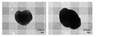
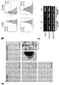
- FIG. 1 left mouse MSC. Used after 12 adhesion subcultures.
- FIG. 1 right human MSC. Used after 6 adherent subcultures. It shows about the influence which shaking culture has on differentiation ability maintenance of mouse MSC.
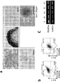
- Fig. 2a top Adhesive subculture 11 times (Fig. 2a top left osteoblasts, upper middle chondrocytes, upper right adipocytes).
- Fig. 2a bottom shaking culture after 36 subcultures of adhesion (Fig. 2a bottom left osteoblast, lower right fat cell).
- Figure 2b Left Adhesive subculture 8 times.
- Fig. 2b, right Shaking culture after 44 adhesion subcultures.
- Fig. 1 left mouse MSC. Used after 12 adhesion subcultures.
- FIG. 1 right human MSC. Used after 6 adherent subcultures. It shows about the influence which shaking culture has on differentiation ability maintenance of mouse MSC.
- Fig. 2a top Adhesive subculture 11 times (Fig. 2a top left osteoblast
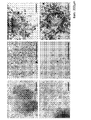
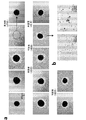
- FIG. 3 left column osteoblasts.
- FIG. 3 middle row chondrocytes.
- FIG. 3 right column fat cells.
- FIG. 3 top 9 months of adherence subculture and 1 month of shaking culture.
- FIG. 3 bottom 1 month of shaking culture after 30 adhesion subcultures.
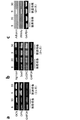
- 2 shows gene expression analysis (RT-PCR) after induction of differentiation into mesoderm cells.
- Fig. 4a After induction of bone differentiation.
- FIG. 4b After induction of cartilage differentiation.
- FIG. 4c after induction of fat differentiation.
- the numbers described above FIG. 4a, FIG. 4b and FIG. 4c indicate the number of adherent subcultures (times). It shows about the influence which shaking culture has on differentiation ability maintenance of human MSC.
- FIG. 5a left column osteoblasts.
- FIG. 5a right column adipocytes.
- Fig. 5a top Adhesive subculture 20 times.
- FIG. 5a middle shaking culture after 6 adherent subcultures.
- FIG. 5a bottom 20 subcultures of adhesion without induction of bone differentiation.
- FIG. 5b top 21 adherent subcultures.
- FIG. 5b bottom shaking culture after 7 adherent subcultures.
- FIG. 5c The numbers on FIG.
- FIG. 5c indicate the number of adherent subcultures (times). It shows about the cartilage differentiation ability of human MSC.
- Fig. 6a top cartilage (toluidine blue staining). Adhesive subculture 9 times.
- FIG. 6a bottom cartilage (toluidine blue staining).
- FIG. 6b top cartilage (toluidine blue staining).
- FIG. 6b bottom cartilage (toluidine blue staining). Shake culture after 19 adhesion subcultures. It shows about the adhesion culture result of the MSC cell mass using the three-dimensional suspension culture container which is a prior art.
- FIG. 6a top cartilage (toluidine blue staining). Adhesive subculture 9 times.
- FIG. 6a bottom cartilage (toluidine blue staining).
- FIG. 6b top cartilage (toluidine blue staining).
- FIG. 7a Human MSC (used after 9 adherent passages). 7 days in culture.
- FIG. 7b Mouse MSC.
- Fig. 7b Upper: Used after 25 times of adhesion subculture (second from left: three-dimensional suspension culture vessel, immediately after seeding. Third from left: three-dimensional suspension culture vessel, culture day 7. Fourth from left: culture dish Re-adhesion, re-adhesion day 7. Fifth from left: re-adhesion to culture dish, induction of fat differentiation).
- Fig. 7b bottom used after 41 times of adhesion subculture (second from the left: three-dimensional suspension culture vessel, immediately after seeding. Third from the left: three-dimensional suspension culture vessel, day 7 of culture.
- FIG. 8a Used after 25 passages of adhesive subculture (Fig. 8a left: bone. Fig. 8a right: fat).
- Fig. 8b Used after 41 times of adhesion subculture (Fig. 8b left: osteoblast.
- FIG. 9a first from the left in the upper row: seeding of the cell mass in an adherent culture dish, day 0. 2nd from the left in the upper part of FIG. 9a: one day.
- FIG. 9a third from the top left: 7th.
- Fig. 9a 4th and 5th from the upper left Adhesion at the time of cell mass (first time).
- Second from the left in the middle of FIG. 9a 15th.
- FIG. 9a lower left: 57 days.
- FIG. 9a lower right 67 days.
- Fig. 9b Differentiation of migrating cells after the third re-adhesion (Fig. 9b left: osteoblasts; Fig. 9b right: adipocytes).
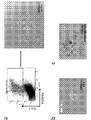
- FIG. 10a right mouse purified MSC.
- FIGS. 10b and c Shaking culture was carried out after two times of adhesion subculture, and after the formation of the cell mass, the cell mass was reattached to the culture dish.
- FIG. 10b shows the first day of bonding.
- FIG. 10 c shows adipocytes (fat droplets) after differentiation induction. It shows cell mass formation using a medium for neural stem cells of accoll TM mouse MSC.
- FIG. 10b shows the first day of bonding.
- FIG. 10 c shows adipocytes (fat droplets) after differentiation induction. It shows cell mass formation using a medium for neural stem cells of accoll TM mouse MSC.
- FIG. 11a 9 months of adherent subculture and 1 month of shaking culture.
- Fig. 11b Nine passages after subculture for 9 months after shaking culture (first from the left in Fig. 11b: bone differentiation; second from the left in Fig. 11b: adipose differentiation; third from the left in Fig. 11b: cartilage differentiation)
- Fig. 11c neural differentiation .
- FIG. 11 d shaking culture for 1 month after 39 times of adhesion subculture.
- Fig. 11e RT-PCR after 1 month of shaking culture (numbers on Fig. 11d are passage numbers)
- the present invention provides a method for culturing MSC in a cell mass while maintaining undifferentiation, characterized by culturing MSC with shaking.
- the type of MSC used in the present invention is not particularly limited, and examples include bone marrow-derived cells, dental pulp-derived cells, and adipose tissue-derived cells.
- the animal species of MSC are not particularly limited, and examples include those derived from mammals such as primates (eg, humans, monkeys, preferably humans), rodents (eg, mice, rats, rabbits, etc.). It is done.
- the present invention is characterized in that such MSCs are cultured by shaking.
- the culture vessel is not particularly limited, and can be appropriately set according to the amount of culture solution required, the type of shaking method, and the like.
- Examples of the shape of the culture vessel include an Erlenmeyer flask and a flask for a seesaw type bioreactor (for example, a flask having a polygonal bottom surface (4 to 8 corners)) as shown in the drawing of WO2015 / 064705.
- Examples of the capacity of the culture vessel include 125 to 3000 ml, preferably 125 to 500 ml.
- a non-adhesive culture container for example, a non-adhesive culture dish, a non-adhesive well, a non-adhesive flask, etc.
- a normal culture vessel not particularly subjected to such treatment may be used.
- the type of shaking method is not particularly limited, and examples include swiveling, figure eight, reciprocation, and seesaw type.
- swirl is preferred, but other shaking modes can be employed.
- the number of rotations is not particularly limited, but can typically be set in the range of, for example, 20 to 200 rpm, preferably 60 to 120 rpm, more preferably 85 to 95 rpm.
- the swirl type is taken as an example to explain the shaking conditions such as the rotation speed and the amplitude.
- the same physical stimulation can be applied to the stem cells.
- an 8-shaped, reciprocating, seesaw type or the like can be adopted.
- the culture vessel is shaken through two substantially circular orbits that touch at one point.
- the conditions can be set so as to be amplitude.
- the number of reciprocations per minute can be set to 20 to 250 reciprocations / minute, and the amplitude can be appropriately set within a range of 10 to 40 mm.
- the shaking culture angle can be appropriately set within the range of 2 ° to 12 ° and the shaking cycle in the range of 5 to 60 rpm.
- the medium to be used is not particularly limited as long as it is a liquid medium suitable for stem cell culture.
- a medium include Minimum Essential Medium (MEM) medium.
- MEM Minimum Essential Medium
- the medium may contain an additive that is included in the normal stem cell culture, if necessary.
- Specific examples of the additive include fetal bovine serum, amino acids (eg, L-glutamine, L-alanyl-L-glutamine, etc.), antibiotics (eg, penicillin, streptomycin, etc.), and the like.
- a preferred embodiment of the culture of the present invention includes subculture.
- stem cells are collected before reaching the confluent state.
- about 1 ⁇ 10 3 to 1 ⁇ 10 7 cells (cells) / ml, preferably 1 ⁇ 10 5 to 1 ⁇ Cells are seeded in a new medium so as to be about 10 6 cells / ml.
- it is preferable to change the medium appropriately for example, every 1 to 5 days, preferably every 3 to 4 days).
- MSC can be subcultured in an undifferentiated state without losing its differentiation ability.
- the subculture can be performed by shaking culture, for example, once or more, preferably 3 times or more, preferably 5 times or more.
- the upper limit of subculture by shaking culture is not particularly limited.
- subculture can be performed 3 times or less, preferably 2 times or less, preferably 1 time or less.
- the time for shaking culture is, for example, preferably about 1 to 90 days (about 24 to 2160 hours), more preferably 10 to 75 days (about 240 to 1800 hours), and 14 to 60 days (about 336 to 1440 hours). Is more preferable.
- the shaking culture temperature in this step is not particularly limited, and is preferably 30 to 42 ° C, for example, and more preferably 35 to 39 ° C. Such culturing is preferably performed in an atmosphere of 3 to 10% CO 2 .
- MSC can be cultured while containing an undifferentiated state and a state having differentiation ability.
- the shaking culture MSCs proliferate and aggregate to form cell clusters. Therefore, in the present invention, the MSC is cultured by shaking culture of MSC, a method for forming a (undifferentiated) cell mass; the MSC is cultured by shaking, a method for inhibiting differentiation of MSC, etc.
- the cell mass obtained by the method of the present invention has the ability to differentiate into various cells belonging to the mesodermal system, in a preferred embodiment, the cell mass is osteoblast, adipocyte or the like, chondrocyte, or more. Preferably, it has the ability to differentiate into adipocytes. As described in the Examples, in the conventional adhesion culture, the ability to differentiate into adipocytes is lost, and thus this embodiment is preferable.
- undifferentiated means that the cell is in an undifferentiated state and has the ability to differentiate.
- the undifferentiated nature of MSC can typically be confirmed by, for example, whether or not it has the ability to differentiate into both osteoblasts and adipocytes, as shown in the Examples of the present application.
- “maintaining undifferentiated” includes not only the state in which MSC originally maintains without losing the differentiation ability, but also MSC can originally differentiate (as described above). Although the differentiation ability to at least one of the cells once decreased or lost in the culturing process before the shaking culture, the state in which the differentiation ability was recovered by shaking culture is also included.
- a predetermined cell for example, fat cell
- MSCs that have lost their differentiation ability are also included in the range of “maintaining undifferentiation” by restoring the differentiation ability to the cells by shaking culture. Therefore, the present invention also provides a method for recovering the differentiation ability to the predetermined cell, characterized in that, in the embodiment, MSC that has lost the differentiation ability to the predetermined cell is cultured by shaking.
- the method of the present invention is characterized by culturing MSCs with shaking
- static culture may be combined as long as the effects of the present invention are obtained.
- the cell mass may be subjected to stationary culture.
- Such stationary culture may be performed by adhesion culture or non-adhesion culture.
- the medium, culture temperature, CO 2 concentration, and the like in the stationary culture can be appropriately selected from the same conditions as in the shake culture described above.
- a non-adhesive culture container for example, a non-adhesive culture dish, a non-adhesive well, a non-adhesive flask, etc.
- a normal culture vessel not particularly subjected to such treatment may be used.
- the number of subcultures in static culture is not particularly limited as long as the effects of the present invention are obtained.
- the number of subcultures is 2 times or more, preferably 3 times or more, preferably 5 times or more, It can be cultured.
- the upper limit of the subculture in the static culture is not particularly limited.
- the subculture can be performed 5 times or less, preferably 3 times or less, preferably 1 time or less.
- the time for stationary culture is, for example, preferably about 0.1 to 60 days, more preferably 1 to 30 days, and more preferably 3 to 14 days.
- the stationary culture time is preferably about 2.4 to 1440 hours, more preferably about 24 to 720 hours, and more preferably about 72 to 336 hours.
- the present invention provides a cell mass obtained by the culture method of the present invention described above.
- the cell mass means a mass of cells having a three-dimensional spread rather than a group of cells grown in a plane along the bottom surface and / or wall surface of the inner wall of the culture vessel by adhesion culture or the like.
- a cell mass can also be called a sphere, a spheroid, etc.
- the cell mass of the present invention exhibits an effect that it is in an undifferentiated state and has not lost its differentiation ability even after long-term culture.
- all cells constituting the cell cluster may be maintained undifferentiated, but used as a stem cell pool that continues to supply stem cells that retain undifferentiatedness; As long as it can be used for applications such as application, cells that are maintained undifferentiated may be contained in the cell mass.
- the preferred shaking method in shaking culture for obtaining a cell mass has been exemplified by the swirling shaking culture.
- “by swirling shaking culture” In the range of ⁇ cell mass obtained '', not only the cell mass actually obtained by swirling shaking culture, but also a cell mass that is undifferentiated and has not lost its differentiation ability, Naturally, those cultured by shaking in other methods can also be included.
- the cell mass of the present invention may be positive for markers such as PDGFR ⁇ , Sca-1, both of these, preferably PDGFR ⁇ .
- the cell mass of the present invention may be positive for markers such as CD90 and CD106. That the marker is positive means, for example, that the proportion of the positive fraction in the cell mass is 50% or more when measured according to the method described in the Examples of the present application.
- the present invention provides a cell differentiation method including the step of culturing the above-described cell mass of the present invention in a differentiation induction medium.
- differentiation from MSC to cells belonging to the mesodermal system can be induced.
- cells belonging to the mesodermal system or mesenchymal system include adipocytes, osteoblasts, chondrocytes, bone cells, cardiomyocytes, tendon cells, dental pulp cells, odontoblasts, and preferably osteoblasts.
- examples include cells and fat cells.
- the cell mass of the present invention can also be applied to the culture of purified MSCs containing neural crest-derived cells (Non-Patent Document 4), and cells belonging to the neural crest, such as nerve cells, glial cells, smooth muscle cells, Examples include dental pulp cells, odontoblasts, cementoblasts, periodontal ligament cells and the like (preferably nerve cells, odontoblasts, more preferably nerve cells).
- a medium suitable for inducing differentiation into a desired type of cells can be appropriately used.
- a medium suitable for inducing differentiation into a desired type of cells include a DMEM medium (eg, Nacalai Tesque, sodium pyruvate-free DMEM medium, etc.); ⁇ MEM medium (eg, gibco, MEM Alpha (1 ⁇ ) medium, etc.) Takara Bio, Mesenchymal stem cell growth media; LONZA Co., MSCGM TM Mesenchymal stem cell growth medium, hMSC-Human Mesenchymal stem cell osteogenic Differentiation medium BullkKit TM, hMSC-Human Mesenchymal stem cell Chondrogenic Differentiation medium BullkKit TM, hMSC-Huma Mesenchymal Stem cell Adipogenic Differentiation Medium BullkKit TM, and the like. These media may be used alone or in combination of two or more.
- the medium may be blended with a differentiation induction promoter for the type of cells desired.
- adipogenic differentiation induction promoter examples include insulin, dexamethasone, 3-isobutyl-1-methylxanthine, and the like. These agents for promoting differentiation into adipocytes may be used singly or in combination of two or more.
- ascorbic acid for example, ascorbic acid-2-phosphate, ⁇ -glycerophosphate, dexamethasone, hydrocortisone hemisuccinate, statin, isoflavone derivative, 3-benzothiepine derivative TAK-778 (( 2R, 4S)-( ⁇ )-N- [4- (diethoxyphosphorylmethyl) phenyl] -1,2,4,5-tetrahydro-4-methyl-7,8-methylenedioxy-5-oxo-3 -Benzothiepin-2-carboxamide, helioxanthin derivative TH (4- (4-methoxyphenyl) pyrido [40,30: 4,5] thieno [2,3-b] pyridine-2-carboxamide), phenamyl (3,5 -Diamino-6-chloro-N- [imino (phenylamino) methyl] pyrazine-2-carboxamide Harmin and its analogs,
- differentiation induction promoter for chondrocytes examples include BMP-6, TGF- ⁇ 3, dexamethasone, ascorbic acid and the like. These chondrocyte differentiation induction promoters may be used alone or in combination of two or more.
- Examples of the differentiation induction promoter for cardiomyocytes include KY03I, KY02111, ⁇ FGF, and the like. These cardiomyocyte differentiation induction promoters may be used alone or in combination of two or more.
- Examples of the differentiation-inducing promoter for tendon cells include PDGF, VEGF, and the like. These tendon cell differentiation induction promoters may be used alone or in combination of two or more.
- the present invention provides a cell mass differentiated into a predetermined cell by the differentiation induction method of the present invention described above.
- the differentiation-induced cell mass according to the present invention uses an undifferentiated cell mass obtained by the above-described method of the present invention as a raw material.
- the undifferentiated cell mass of the present invention as a raw material is different from the conventional MSC cell mass, and is in an undifferentiated state and has lost its differentiation ability even after a long-term subculture. It has the unexpected effect of not being. Therefore, it is clear that the cell mass induced to differentiate into a desired cell using such an undifferentiated cell mass as a raw material is also different from the conventional cell mass.
- Non-Patent Document 6 C57 / BL6 male 4 weeks old (CLEA JAPAN) 5 femurs and tibias were crushed with a mortar pestle, 2% FBS (Cat. # SH3099103: Hyclone), 10 mM HEPES (Cat. # 346-01373: Dojindo) ), And 1% Penicillin / Streptomycin (P / S: Cat. # 168-23191, Wako) and suspended in HBSS + (Cat. # 14025134: Gibco) (hereinafter referred to as HBSS + adjustment solution) was removed. Shredded bones were crushed for 1 hour at 37 ° C. with 0.2% collagenase (Cat.
- the collected 1 ⁇ 10 7 cells were suspended in 1 ml of HBSS + adjustment solution, and PE-conjugated CD45 (30-F11: Cat. # 12-0451-83, 0.2 mg / ml, eBioscience), TER119 (TER- 119: Cat. # 12-5921-83, 0.2 mg / ml, eBioscience), APC-conjugated PDGFR ⁇ (APA5: Cat. # 17-1401-81, 0.2 mg / ml, eBioscience) and FITC-conjugated Sca- 1 (Ly6A / E: Cat.
- Cell samples subcultured in FACS analysis adhesion culture of mouse MSC were washed twice with PBS, and then 1 ml of cell dissociation buffer. Cells were detached using (Cat. # 13151014: Gibco), HBSS + adjustment solution was added, centrifuged at 280 G for 5 minutes at 4 ° C., the supernatant was removed, and the precipitated cells were collected.
- Cell mass sample formed by shaking culture Considering that it is difficult to separate and collect single cells without damaging each cell or cell surface antigen of the cell mass, once an average of 10 cell masses was allowed to stand in a 10 cm culture dish (Cat. # 664160-013: CELLSTAR) for 7 days, and cells migrated from the cell mass adhered to the culture dish were washed twice with PBS. After removing the PBS, 1 ml of Cell dissociation buffer (Cat. # 13151014: Gibco) was added to the culture dish and allowed to act for 2 to 3 minutes to detach the cells.
- Cell dissociation buffer Cat. # 13151014: Gibco
- the mixture was centrifuged at 280 G for 7 minutes at 4 ° C., adjusted with a HBSS + adjustment solution to obtain a cell suspension of 1 ⁇ 10 7 cells / ml, and analyzed using Aria III (BD Bioscience). In the analysis of cell mass-derived cells, no subculture was performed, and cells that migrated from the cell mass were directly used for analysis.
- Non-Patent Document 7 Human MSCs handed over from Tokyo Medical and Dental University were PE-conjugated CD271 (LNGFR: Cat. # 130-091-885, Miltenyi Biotec), FITC-conjugated CD90 (Thy-1: Cat. #) From a 19-year-old male femur bone marrow. (328110, Biolegend) antibody and sorted with MoFlo (BECKMAN COULTER) (Non-patent Document 7). PI was used for the labeling of dead cells, and after the negative fraction was sorted as a living cell, cells that had been passaged twice were donated.
- the mixture was centrifuged at 280 G for 5 minutes at 4 ° C., adjusted with HBSS + adjustment solution so as to obtain a cell suspension of 1 ⁇ 10 7 cells / ml, and analyzed using Aria III (BD Bioscience).
- Human MSC is a 20% FBS (Cat. # SH309910: Hyclone), 1% P / S (Cat. # 168-23191: Wako), 10 mM HEPES (Cat. # 346-01373: Dojindo) as a growth maintenance medium. , And 20 ng / ml ⁇ FGF (Cat. # 084-05384: WAKO) and DMEM (Cat. # 0859-64: Nacalai tesque).
- Mouse or human MSCs were seeded at a concentration of 1 ⁇ 10 6 cells / dish in a 10 cm culture dish (Cat. # 664160-013: CELLSTAR) in an environment of 37 ° C. and 5% CO 2 for 4-7.
- the cells were cultured for a day and subcultured in a state of 80% confluence.
- the serum component was removed with 1 ⁇ PBS after the medium was aspirated, and the cells were treated with 0.25% trypsin EDTA (Cat. # 201-16945: Wako) for 2 minutes.
- the dish was gently tapped, and as soon as the cells came off, a growth maintenance medium was added and the cells were collected.
- the growth maintenance medium was added to the precipitated cells from which the supernatant was removed by suction, and the cells were seeded at 1 ⁇ 10 6 cells / dish in a new 10 cm culture dish.
- OriCell TM Strain C57BL / 6 mouse MSCs (MUBMX-01001: Cyagen) have already been subcultured 6 times by the manufacturer at the time of purchase. Purchased cells are shipped after confirmation of CD29, CD44, CD31, Sca-1 positive (> 70%) and CD117 negative ( ⁇ 5%) by the manufacturer. In addition, the manufacturer recommends the use of subcultures of 10 times or less, and subsequent subcultures do not guarantee the properties of the cells at the time of purchase. In addition, the use of OriCell TM Mouse MSC Growth Medium (MUXMX90001: Cyagen) is recommended for cell maintenance culture.
- MSITEC shake suspension culture TAITEC BR-40LF bio-shaker was used for shake culture.
- a cell suspension of mouse or human MSC prepared with 20 ml of growth maintenance medium was prepared in a 125 ml erlenmeyer flask (Cat. # 431405: Corning).
- 20 ng / ml ⁇ FGF (Cat. # 064-05384: WAKO) was added to the growth maintenance medium.
- the number of cells per flask at the start of shaking culture was 1.0 ⁇ 10 7 cells with OriCell TM mouse MSC, 5.0 ⁇ 10 5 cells with purified mouse MSC, 1.0 ⁇ 10 6 cells with human MSC, or 1 It adjusted so that it might become 0.0 * 10 ⁇ 7 > pieces.
- the shaking culture was performed while rotating at 37 ° C., 5% CO 2 , 85 to 95 r / min, and amplitude of 40 mm, and the medium was changed once every 3 to 4 days. In the medium exchange, first, the whole medium containing cells was transferred to a 50 ml centrifuge tube (Cat. # TR2004: True Line) and centrifuged at 280 G for 5 minutes at 4 ° C.
- the medium for neural stem cells contains 1% P / S (Cat. # 168). -23191: Wako) and 10 mM HEPES (Cat. # 346-01373: Dojindo), 1 ⁇ N2 (Cat. # 17502-048: Gibco), 20 ng / ml EGF (Cat. # 059-07873: WAKO), 20 ng / Advanced DMEM / F12 (Cat. 12491015: Gibco) containing ml FGF (Cat. # 064-05384: WAKO), 1 ⁇ B27 (Cat.
- a cell suspension of mouse MSC (thoughll TM mouse MSC: 1.0 ⁇ 10 7 cells) was prepared with 20 ml of neural stem cell medium and prepared in a 125 ml erlenmeyer flask (Cat. # 431405: Corning). These cell suspensions were subjected to shaking culture using a TAITEC BR-40LF bio-shaker while swirling at 37 ° C., 5% CO2, 85 to 95 r / min, and amplitude of 40 mm. The medium was changed once every 3 to 4 days. For medium exchange, first, the whole medium containing cells was transferred to a 50 ml centrifuge tube (Cat.
- Differentiation induction method staining method (see Non-Patent Document 6) Differentiation induction into osteoblasts and adipocytes was performed by seeding cells in a 12-well plate (Cat. # 665-180: CELLSTAR) at a concentration of 1 ⁇ 10 5 cells / well in adhesion culture. For cell mass-derived migrating cells, 2 to 3 cell masses were seeded in each well of the same 12-well plate, and cells migrated and amplified from the cell mass were used. After seeding, the cell adhesion state was confirmed, and osteoblast differentiation inducing medium (Cat. # PT3002: Lonza) and adipocyte differentiation inducing medium (Cat. # PT3004: 60-70% confluent (cultured for about 7 days).
- osteoblast differentiation inducing medium Cat. # PT3002: Lonza
- adipocyte differentiation inducing medium Cat. # PT3004: 60-70% confluent (cultured for about 7 days).
- medium was changed once every 3 to 4 days to induce differentiation for a maximum of 21 days.
- adherent cells and formed lipid droplets sometimes floated and peeled off. In that case, the culture was terminated at that time.
- Alkaline phosphatase (ALP) staining was used to confirm osteoblast differentiation, and oil red O staining was used to confirm adipocyte differentiation. In both staining methods, hematoxylin staining was not performed.
- chondrocytes For differentiation induction into chondrocytes, cells were pelleted at 150 G in a 15 ml centrifuge tube, 10 ng / ml transforming growth factor- ⁇ 3 (Cat. # PT4124: LONZA), 500 ng / ml bone morphogenic protein-6 (Cat. #) Culturing was performed using a chondrocyte induction medium (Cat. # PT3003: Lonza) supplemented with 6325-BM-020: R & D Systems. The medium was changed twice a week and cultured for 21 days. Differentiation of the induced chondrocytes was confirmed by toluidine blue staining (Cat. # 209-14545: Wako). Hematoxylin staining was not performed.
- ALP staining see Non-Patent Document 6 Histofine assay kit (Cat. # 415161: Nichirei) was used for staining. PFA was washed with PBS, the reagents in the kit were mixed and adjusted immediately before staining, stained with a staining solution filtered with a syringe filter (Cat. # SLGP033RS: Merck Millipore) having a pore size of 0.22 ⁇ m, and washed with water. As the negative control (NC), MSC before induction of osteoblast differentiation was simultaneously ALP-stained.
- Toluidine blue staining (see Non-Patent Document 6) The cell pellet after differentiation induction was embedded in paraffin, and a section was prepared on a slide glass. Toluidine blue staining solution (Cat. # 209) filtered with a syringe filter (Cat. # SLGP033RS: Merck Millipore) having a pore size of 0.22 ⁇ m after deparaffinization with xylene and dexylene with 100% alcohol and treatment with 80% alcohol. -14545: Wako) for 30 minutes. After dyeing, it was dehydrated with 100% alcohol, and after xylene treatment, it was sealed with marinol (Cat. # 2009-3: Muto pure Chemicals).
- Nerve cell induction (see Non-Patent Document 4)
- the cell mass shake-cultured in the neural stem cell medium was removed from the neural stem cell medium, removed from EGF, FGF, 1 ⁇ B27, and transferred to a medium supplemented with 10% FBS (Cat. # SH309910: Hyclone).
- Chamber slide (Cat. # SCS-NO8 treated with fibronectin (Cat. # 062-05701: WAKO; 37 ° C, 12 hours) after treatment with L-ornithine (Cat. # 163-27421: WAKO; 37 ° C, 12 hours) : MATUNAMI) (2-3 cell clumps / well).
- Cells migrated from the cell mass 7 days after seeding were fixed with 4% PFA (Cat. # 163-20145: WAKO) and used for observation of fluorescent immunostaining.
- Fluorescent immunostaining (see Non-Patent Document 4) The sample fixed with PFA was washed with 1 ⁇ PBS, then treated with 0.3% Triton-X100 (Cat. # 160-24751: WAKO) at room temperature for 5 minutes, and washed again with 1 ⁇ PBS. Washed cells were treated with blocking buffer (0.01% Triton-X100 (Cat. # 160-24751: WAKO), 5% Bovine Serum Albumin (Cat. # 23208: Thermoscientific) 1 ⁇ PBS) for 30 minutes. Thereafter, primary staining (4 ° C., overnight) was performed using an anti- ⁇ III-Tublin antibody (Cat. # Ab18207: abcom) diluted 500-fold.
- Triton-X100 Cat. # 160-24751: WAKO
- Bovine Serum Albumin Cat. # 23208: Thermoscientific
- the cells were washed with 1 ⁇ PBS and subjected to secondary staining (1 hour at room temperature) using Donkey Anti-Rabbit IgG H & L (Alexa Floor (registered trademark) 488; Cat # ab150073: abcom) diluted 1000 times.
- the cell sample was washed with 1 ⁇ PBS, encapsulated using VECTASHIELD mounting medium with DAPI (Cat # H-1200: VECTOR), and observed using a confocal laser scanning microscope (LSM780: Zeiss).
- RNA extraction Trizol (Cat. # 15596108: Invitrogen) and RNeasy Mini Kit (Cat. # 74106: Quiagen) were used (extracted according to the Qiagen Kit protocol), and DNase I (Cat. # AM2222: Ambion) was used. DNA was removed. Reverse transcription system (Cat. # A3500: Promega) was used for the reverse transcription reaction, and random primers (Cat. # C118B: Promega), AMV reverse transcriptase (Cat. # M900B: Promega) and MgCl2 (Cat.) Were used according to the protocol procedure. # A351H: Promega). In the PCR reaction, cDNA was amplified using Go Taq Green Master Mix (Cat.
- FIG. 1 Mouse and human MSC cell mass formation by shaking culture OriCell TM mouse MSC (MUBMX-01001: Cyagen) and human MSC (MSC purified from 19-year-old male bone marrow) were subcultured in adhesion culture, Cells having a small number and a large number of subcultures were prepared and subjected to shaking culture.
- Mouse MSCs could be subcultured by adhesion culture up to 50 times after purchase (56 counts before shipment).
- Human MSCs could be subcultured by adhesion culture up to 20 times after donation (22 counts from before donation).
- the OriCell TM mouse MSC used this time has already been subcultured 6 times by the manufacturer at the time of purchase, and the human-derived MSC has already been subcultured twice at the time of donation. It has been broken.
- the number of subcultures indicates the number of counts continued before shipment and before donation (including the number of subcultures before shipment and before donation).
- the number of cell masses formed by shaking culture by human MSC is 14 months later when the number of cells at the start of shaking culture is 1.0 ⁇ 10 6. 3 ⁇ 2.0 and 20.7 ⁇ 2.1 in 2 months.
- the number of cells at the start of shaking culture was 1.0 ⁇ 10 7 cells, it was 15.0 ⁇ 1.0 after 1 month and 11.7 ⁇ 2.5 after 2 months (3 The average value of the results of the experiment ⁇ standard deviation). That is, even when the number of cells at the start of shaking culture was increased from 1.0 ⁇ 10 6 to 10 times, there was no significant difference in the number of cells formed.
- the size of the cell mass formed by shaking culture of human MSC with 7 subcultures is 433.3 ⁇ in one month when the number of cells at the start of shaking culture is 1.0 ⁇ 10 6. It was 103.3 ⁇ m and 533.3 ⁇ 150.6 ⁇ m in 2 months. When the number of cells at the start of the shaking culture was 1.0 ⁇ 10 7 cells, it was 783.3 ⁇ 248.3 ⁇ m after 1 month and 833.3 ⁇ 186.1 ⁇ m after 2 months (6 times Mean value ⁇ standard deviation of experimental results).
- the number of cell masses formed by shaking culture for one month with a large number of subcultures (19 times) was 14 when the number of cells at the start of shaking culture was 1.0 ⁇ 10 6. In the case of 1.0 ⁇ 10 7 , it was 15 (result of one experiment).
- the size of the cell mass formed by shaking culture of human MSC with 19 passage cultures for one month is 500 ⁇ when the number of cells at the start of shaking culture is 1.0 ⁇ 10 6. In the case of 126.5 ⁇ m and 1.0 ⁇ 10 7 cells, it was 766.7 ⁇ 206.6 ⁇ m (average value of 6 cell mass ⁇ standard deviation).
- Fig. 2 Influence of shaking culture on maintenance of differentiation ability of mouse MSC The influence of shaking culture on maintenance of differentiation ability of mouse MSC was examined.
- OriCell TM mouse MSCs maintained differentiation potential into osteoblasts (positive ALP staining) and differentiation into chondrocytes (positive toluidine blue staining) at a relatively low stage of 11 subcultures. However, it had already lost its ability to differentiate into fat and was negative for oil red O staining (FIG. 2-a: upper panel).
- Non-patent Documents 4 and 5 It is known that mouse MSCs co-expressing PDGFR ⁇ and Sca-1 on the cell surface are high-quality MSCs that maintain an undifferentiated state.
- OriCell TM mouse MSCs subcultured 7 times in adherent culture retained about 90% of PDGFR ⁇ + / Sca-1 + (co-positive fraction) (not shown), but when subculture was repeated as is This co-positive fraction decreased to 46.1% after 8 passages (FIG. 2-b: left figure), and decreased to 25.0% after 23 passages (not shown). ).
- OriCell TM mouse MSCs cultured in adhesion culture expressed high levels of the PDGFR ⁇ gene when the number of subcultures was 9, but the number of subcultures was as high as 25 or 41 times. As a result, the expression of the PDGFR ⁇ gene markedly decreases, suggesting the possibility of loss of undifferentiated ability.
- FIG. 3 shows the results of induction of differentiation into osteoblasts, chondrocytes, and adipocytes, which are mesodermal cells, after one month of shaking culture.
- Both the cells with low passage number (9 passages) and the cells with many passages (30 passages) induced to differentiate after shaking culture are osteoblasts (ALP positive), chondrocytes (toluidine blue positive), adipocytes The ability to differentiate into (oil red O positive) was shown.
- FIG. 4 Tissue-specific gene expression analysis during differentiation induction into mesoderm cells (RT-PCR) Using purelyll TM mouse MSC, cells with a low number of adherent passages (passage number 8-9) and cells with a high passage number (passage number 30) were prepared. Using RT-PCR, we analyzed the expression of genes related to osteoblasts, chondrocytes, and adipocytes in cells that had undergone differentiation induction immediately after adhesion culture and in cells that had undergone shaking culture after adhesion culture. did.
- Fig. 5 Influence of shaking culture on maintenance of differentiation ability of human MSC The influence of shaking culture on maintenance of differentiation ability of human MSC was examined. In adhesion culture, human MSCs showed differentiation ability into osteoblasts (positive ALP staining) after 20 subcultures, but lost the differentiation ability into adipocytes (oil red O staining negative) ( Fig. 5-a: Top).
- Non-patent Document 7 which is a cell surface marker of high-quality MSC
- the expression of human MSC cells subcultured 21 times in adhesion culture is 6.0 to 7.5%.
- FIG. 5-b upper right figure
- the expression was 59 in cells that had migrated and proliferated from the cell mass formed by shaking culture of human MSC that had been subcultured 7 times in adhesion culture for 2 months. It was maintained at 6% to 87.1% (FIG. 5-b: lower right figure).
- Fig. 6 Cartilage differentiation ability of human MSC Using human MSC, cells with a small number of adhesion subcultures (9 passages) and cells (19 passages) were prepared and differentiated into chondrocytes. Performance analysis was performed.
- FIG. 7 Adhesion culture of MSC cell mass using 3D suspension culture vessel (prior art) Low adhesion 3D culture vessel (Kuraray Elplasia RB 500 400) used in the existing method of forming MSC cell mass NA Plate) was inoculated with 3 ⁇ 10 6 cells / ml of human MSC (number of subcultures: 9 times) or OriCell TM mouse MSC (number of subcultures: 25 times and 41 times) maintained in adhesion culture. . As a result, human MSCs adhered to the culture dish, and no cell mass was formed even after 7 days (FIG. 7-a). On the other hand, in mouse MSC, as reported by Baraniak et al.
- Non-Patent Document 10 a cell mass was formed after 7 days of culture in an incubator (FIG. 7-b). The cell mass collected at this time was transferred again to the culture dish and allowed to stand in an adhesion culture environment. As a result, the cell mass adhered to the culture dish and cells migrated around, but the cell mass did not maintain its shape. It disappeared after a day, and the ability to differentiate into adipocytes was lost (oil red O staining was negative and no lipid droplets were observed) (FIG. 7-b).
- Figure 8 Differentiation analysis of accoma TM mouse MSC cell mass using 3D suspension culture vessel (prior art). Adhesion to low adhesion 3D suspension culture vessel (Kuraray Elplasia RB 500 400 NA Plate). accomboll TM mouse MSC (number of subcultures: 25 times and 41 times) maintained in culture was seeded at 3 ⁇ 10 6 cells / ml. The cell mass formed after 7 days was seeded on a 12-well plate (Cat. # 665-180: CELLSTAR, greiner bio-one) to shift to adhesion culture, and osteoblasts of cells migrated from the adhered cell mass and Differentiation into adipocytes was verified.
- Fig. 9 Morphology retention and cell supply ability of human MSC cell mass cultured by shaking As a result of leaving the cell mass formed by shaking culture of human MSC subcultured 7 times in adhesion culture for 2 months on a culture dish, On the next day, cell migration around the cell mass was observed, and after 7 days, the cells grew to fill the culture dish (FIG. 9-a: upper panel). Moreover, the reattached cell mass maintained its form after 7 days, and it was possible to mechanically peel off the cell mass and leave it in a new culture dish (first reattachment).
- FIG. 9-a two photos on the left in the middle row. Even if this cell mass is mechanically peeled off and left to stand in a new culture dish four more times (reattachment 2 to 5), cell migration around the cell mass is also observed and within 14 days. In addition, the cells grew to fill the culture dish, and the cell mass retained its morphology (FIG. 9-a: middle three right photos to lower).
- the MSC cell mass according to the present invention has a strong adhesive force between cells, and even if the cells are returned to the adhesion culture environment, the MSC cell mass continues to supply undifferentiated MSC to the culture dish without destroying the shape of the cell mass. I understood.
- FIG. 10 Cell mass formed by shaking culture of mouse-purified MSC It was examined whether or not a cell mass purified by shaking culture was also subjected to shaking culture to form a cell cluster similar to that of OriCell TM mouse MSC.
- the mouse purified MSC after sorting was subcultured twice by adhesion culture in order to secure the number of cells.
- shaking culture of mouse purified MSC 5.0 ⁇ 10 5 cells / flask
- a cell mass similar to that of OriCell TM mouse MSC was formed (not shown).
- FIG. 10 Cell mass formed by shaking culture of mouse-purified MSC
- Figure 11 Cell mass formation using — — — — — — — — — — ⁇ ⁇ ⁇ ⁇ ⁇ ⁇ ⁇ ⁇ ⁇ ⁇ ⁇ ⁇ ⁇ ⁇ ⁇ ⁇ ⁇ ⁇ ⁇ ⁇ ⁇ ⁇ ⁇ ⁇ ⁇ ⁇ ⁇ ⁇ ⁇ ⁇ ⁇ ⁇ ⁇ ⁇ ⁇ ⁇ ⁇ ⁇ ⁇ ⁇ ⁇ ⁇ ⁇ ⁇ ⁇ ⁇ ⁇ ⁇ ⁇ ⁇ ⁇ ⁇ ⁇ ⁇ ⁇ ⁇ ⁇ ⁇ ⁇ ⁇ ⁇ ⁇ ⁇ ⁇ ⁇ ⁇ ⁇ ⁇ ⁇ ⁇ ⁇ ⁇ ⁇ ⁇ ⁇ ⁇ ⁇ ⁇ ⁇ ⁇ ⁇ ⁇ ⁇ ⁇ ⁇ ⁇ ⁇ ⁇ ⁇ ⁇ ⁇ ⁇ ⁇ ⁇ ⁇ ⁇ ⁇ ⁇ ⁇ ⁇ ⁇ ⁇ ⁇ ⁇ ⁇ ⁇ ⁇ ⁇ ⁇ ⁇ ⁇ ⁇ ⁇
- FIG. 11-a As a result of culturing cells with few adhesion subcultures (9 passages) in a neural stem cell culture medium, formation of cell clusters was observed within one month (FIG. 11-a).
- cell clusters could be formed using the neural stem cell medium (FIG. 11-d).
- the cells of this cell cluster markedly expressed neural crest stem cell markers Nestin and Twist and mesenchymal stem cell marker PDGFR ⁇ (FIG. 11-e).
- MSCs that had lost undifferentiation could be restored again to a cell mass of a highly undifferentiated MSC population.
- the MSC cell mass formed by shaking culture retains undifferentiated properties, and at the same time has a strong form-retaining ability, and supplies undifferentiated MSCs by re-adhering to a new culture dish. Can do.
- a strong three-dimensional MSC mass (sphere) that maintains its differentiation ability and maintains its form can be formed by culturing MSC from an adhesion environment by a completely new stirring and shaking culture method.
- stem cell pool that continues to supply stem cells that retain undifferentiated properties.
- the cell mass itself is expected to promote the regeneration of stem cells by making a space at the defect site as a scaffold.
Landscapes
- Health & Medical Sciences (AREA)
- Life Sciences & Earth Sciences (AREA)
- Engineering & Computer Science (AREA)
- Biomedical Technology (AREA)
- Zoology (AREA)
- Developmental Biology & Embryology (AREA)
- Organic Chemistry (AREA)
- Biotechnology (AREA)
- Chemical & Material Sciences (AREA)
- Bioinformatics & Cheminformatics (AREA)
- Genetics & Genomics (AREA)
- Wood Science & Technology (AREA)
- Microbiology (AREA)
- Cell Biology (AREA)
- Biochemistry (AREA)
- General Engineering & Computer Science (AREA)
- General Health & Medical Sciences (AREA)
- Rheumatology (AREA)
- Hematology (AREA)
- Immunology (AREA)
- Micro-Organisms Or Cultivation Processes Thereof (AREA)
- Measuring Or Testing Involving Enzymes Or Micro-Organisms (AREA)
Abstract
間葉系幹細胞を振盪培養することを特徴とする、未分化性を維持したまま間葉系幹細胞を細胞塊で培養する方法。
Description
本発明は、間葉系幹細胞及びその培養方法に関する。
医科領域・歯科領域において骨組織の実質欠損に対しては、β-TCPなどの人工材料やチタンによる再建・インプラント治療や自家骨移植が行われる。自家骨移植を含めた人工材料を用いた治療では、治療後に起こる再生骨の吸収が問題となっている(非特許文献1、2)。また骨再生の分野では、手術時のスペースメーキングのためにスキャホールドや肉芽組織の侵入を防ぐメンブレンが必要なことが多く、再生の場におけるそれらの影響も考慮しなければならない。
また近年では、自己由来細胞を用いた研究・治療も行われてきており、その代表が間葉系幹細胞(MSC)である(以降も、本明細書において、間葉系幹細胞を略してMSCと示す)。
MSCは、元々、骨髄細胞をプラスチック培養皿に播種することで張り付き増殖し、脂肪・軟骨・骨という中胚葉系の組織・細胞に分化する能力を有している細胞として定義付けされた細胞である(非特許文献3)。
マウス骨髄MSCのマーカー探索が行われ、2009年に森川らによりフローサイトメーターを用い、PI-/CD45-/Ter119-/Sca-1+/PDGFRα+分画中にマウスMSCが濃縮されていることが報告された(非特許文献4、5)。この純化技術はHoulihanらにより詳細が公にされている(非特許文献6)。ヒト骨髄MSCは馬渕らにより、CD271(LNGFR)+/CD90(Thy-1)+分画中に高濃度に含まれることが報告されている(非特許文献7)。
MSCは様々な組織中に存在するが、一般的に安定的に多くのMSCを獲得可能な組織は骨髄である。MSCは骨髄中の細胞をプラスチック培養皿に播種することで、貼り着き増殖し、骨芽細胞・軟骨細胞・脂肪細胞へ分化可能な細胞と定義され(非特許文献3)、再生医療の細胞供給源として着目されている。しかしながら長期に及ぶ接着培養を繰り返すことで増殖能・分化能が低下することから、臨床においては施設間や患者間で結果に差が生じることが問題となっている。
具体的には、従来のMSCは接着培養環境下でその増殖能に限界があり、それに応じて分化能も喪失することが分かっている(非特許文献8、9)。純化MSCも接着培養環境では徐々に増殖能が落ちることが知られている(非特許文献5 Fig 1G参照)。
近年、MSCを特殊な培養皿上で培養すると、培養皿に接着することなく浮遊状態でMSCの3次元的な細胞塊(スフェアあるいはスフェロイド)を形成できることが報告されている(非特許文献10)。しかしながら、この方法では長期間培養することはできず、浮遊環境で培養することで細胞にどのような変化が起こるかについては報告がされていない。
Hatano et al. Clin Oral Impl Res, vol.15, p339~345, 2004
Verhoeven et al. Clin Oral Impl Res, vol.11, p583~594, 2000
Pittenger et al. Science, vol 284 2 April, 1999
Morikawa et al. BBRC, vol.379, p1114~1119, 2009.
Morikawa et al. JEM, vol.206, p2483~2496, 2009.
Houlihan et al. Nature Protocol, vol.7(12), p2103~2111, 2012.
Mabuchi et al. Stem cell reports, vol.1(2), p152~165, 2013
Bonab et al. BMC Cell Biol, vol.7, p14, 2006.
Bork et al. Aging Cell, vol.9(1), p54~63, 2010.
Baraniak et al. Cell Tissue Res, vol.347(3), p701~711, 2012
Doetsch et al. Cell, 97 (6), p703~716, 1999.
Laura et al. Protoc Exch, Doi:10.1038/nprot.2006.215, 2006
本発明が解決しようとする課題は、未分化性を維持したままMSCを長期間培養することにある。
かかる状況の下、本発明者らは、鋭意研究した結果、MSCを振盪培養することによって細胞塊の形成を導くことが可能であり、未分化性を維持したままMSCを長期間培養することできることを見出した。本発明は、かかる新規の知見に基づくものである。
従って、本発明は以下の項に示す方法及び細胞塊を提供する。
項1.MSCを振盪培養することを特徴とする、未分化性を維持したままMSCを培養する方法。
項2.前記振盪培養が回転数20~200rpm、好ましくは60~120rpm、より好ましくは85~95rpmで行われる、項1に記載の方法。
項3.前記振盪培養が振幅10~40mm、好ましくは25~40mmより好ましくは30~40mmで行われる、項1又は2に記載の方法。
項4.2回以上、継代培養を行う、項1~3のいずれか1項に記載の方法。
項5.振盪培養に供する前記MSCが所定の細胞への分化能を喪失している細胞であり、かつ該所定の細胞への分化能が振盪培養により回復する、項1~4のいずれか1項に記載の方法。
項6.項1~5のいずれか1項に記載の方法により得られる細胞塊。
項7.分化誘導培地中で項6に記載の細胞塊を培養する工程を含む、細胞分化方法。
項8.項7に記載の方法により所定の細胞に分化した細胞塊。
本発明によれば、振盪培養を行うことにより、MSCを細胞塊の形成に導き、その未分化性を維持したまま長期間培養することができる。また、本発明によれば、従来の接着培養等を長期間行うことにより分化能を失ったMSCを用いた場合、これを振盪培養することにより、その分化能を回復することもできる。本発明が属する幹細胞の分野においては、物理的刺激はむしろ分化を誘導すると考えられてきた。従って、MSCに対し振盪培養を行うことによって未分化性を長期間維持できるという本発明の上記効果は、従来技術から予想し得ない意外なものである。
未分化性を維持したままMSCを培養する方法
本発明は、MSCを振盪培養することを特徴とする、未分化性を維持したまま細胞塊でMSCを培養する方法を提供する。
本発明は、MSCを振盪培養することを特徴とする、未分化性を維持したまま細胞塊でMSCを培養する方法を提供する。
本発明において用いるMSCの種類としては、特に限定されず、骨髄由来の細胞、歯髄由来の細胞、脂肪組織由来の細胞等が挙げられる。また、MSCの動物種も特に限定されず、例えば、霊長類(例えば、ヒト、サル等、好ましくはヒト)、げっ歯類(例えば、マウス、ラット、ウサギ等)等の哺乳類由来のものが挙げられる。
本発明は、かかるMSCを振盪培養することを特徴とする。培養容器としては特に限定されず、必要とされる培養液の量、振盪方法の種類等に応じて適宜設定することができる。培養容器の形状としては、例えば、三角フラスコ、WO2015/064705の図に示されるような、シーソー型バイオリアクター用フラスコ(例えば、底面が多角形(4~8角形)のフラスコ)等が挙げられる。培養容器の容量としては、例えば、125~3000ml、好ましくは125~500ml等のものが挙げられる。培養容器としては、容器内壁の底面等への細胞の接着が抑制されるような処理がされた非接着性培養容器(例えば、非接着性培養皿、非接着性ウェル、非接着性フラスコ等)を用いてもよいし、かかる処理を特に行っていない通常の培養容器を用いてもよい。
振盪方法の種類も特に限定されず、旋回、8の字、往復、シーソー型等が挙げられる。本発明においては、旋回が好ましいが、他の振盪様式を採用することができる。旋回による振盪培養の場合、回転数は特に限定されないが、典型的には、例えば、20~200rpm、好ましくは60~120rpm、より好ましくは85~95rpmの範囲で設定することができる。上記回転数の範囲で振盪培養することにより、培養容器内部の側面にも、底面(床部)にもスフェアが張り付きにくくなるため、好ましい。
本明細書においては、好ましい実施形態として旋回型を例に挙げて回転数、振幅等の振盪条件の説明を行ったが、幹細胞に対しこれと同様の物理的刺激を与えることができるものであれば、8の字型、往復、シーソー型等を採用することができる。例えば、8の字型の振盪培養の場合、一点で接する2つの略円形の軌道を通って培養容器が振盪されるため、8の字を形成する各円について、上記旋回型で例示した回転数、振幅となるよう条件を設定することができる。また、例えば、往復型の場合、1分間当たりの往復回数を、20~250往復/分とし、振幅を10~40mmの範囲で適宜設定することができる。例えば、シーソー型の場合、振盪培養の角度を2°~12°、振盪の周期を5~60rpmの範囲で適宜設定することができる。
用いる培地は、幹細胞の培養に適した液体培地であれば特に限定されるものではない。このような培地として、Minimum Essential Medium(MEM)培地等が挙げられる。培地は、必要に応じて通常の幹細胞の培養の際に含める添加剤を含むものであってもよい。添加剤の具体例として、牛胎児血清(fetal bovine serum)、アミノ酸(例えば、L-グルタミン、L-アラニル-L-グルタミン等)、抗生物質(例えば、ペニシリン、ストレプトマイシン等)などが挙げられる。
本発明の培養の好ましい態様として、継代培養が挙げられる。継代培養においては、コンフルエント状態に到達する前に幹細胞を回収し、例えば、MSCの場合は1×10³~1×10⁷細胞(個)/ml程度、好ましくは1×105~1×106細胞(個)/ml程度となるように細胞を新しい培地に播種する。また、本発明の培養において、培地を適宜(例えば、1~5日毎に、好ましくは3~4日毎に)交換することが好ましい。本発明の方法によれば、MSCを、未分化の状態でかつその分化能を失うことなく継代培養することができる。本発明においては、振盪培養により、例えば、1回以上、好ましくは3回以上、好ましくは5回以上、継代培養することができる。また、本発明において振盪培養による継代培養の上限も特に限定されないが、例えば、3回以下、好ましくは2回以下、好ましくは1回以下、継代培養することができる。
振盪培養の時間としては、例えば、約1~90日間(約24~2160時間)が好ましく、10~75日間(約240~1800時間)がより好ましく、14~60日間(約336~1440時間)がより好ましい。本工程における振盪培養温度は特に限定されず、例えば、30~42℃が好ましく、35~39℃がより好ましい。かかる培養は、3~10%CO2の雰囲気下で行うことが好ましい。
当該本発明の方法により、未分化の状態であり、かつ分化能を有した状態のものを含んだままMSCを培養することができる。また、当該振盪培養により、MSCが増殖及び凝集をして、細胞塊が形成される。従って、本発明は、MSCの培養は、MSCを振盪培養することを特徴とする、(未分化の)細胞塊の形成方法;MSCを振盪培養することを特徴とする、MSCの分化抑制方法等も提供する。尚、本発明の方法により得られる細胞塊は、中胚葉系に属する種々の細胞への分化能を有するが、好ましい実施形態において、当該細胞塊は骨芽細胞、脂肪細胞等、軟骨細胞、より好ましくは脂肪細胞等への分化能を有する。実施例に記載のように、従来の接着培養においては、脂肪細胞への分化能を喪失してしまうため、かかる実施形態が好ましい。
尚、本発明において、未分化性とは、細胞が未分化の状態でかつその分化能を有していることを意味する。本発明において、MSCの未分化性は、典型的には、例えば、本願実施例に示すように、骨芽細胞及び脂肪細胞の両方への分化能を示すか否か等により確認することができる。また、本発明において、「未分化性の維持」には、MSCが当初有していた分化能を失うことなく維持している状態だけでなく、MSCが本来分化し得る(前述したような)細胞のうち少なくとも1種への分化能が、振盪培養以前の培養過程において、一旦、低下し又は失われたものの、振盪培養をすることにより当該分化能が回復した状態も包含される。従って、本発明においては、例えば、本願実施例に示すように、振盪培養以外の方法(例えば、接着培養等)により継代培養を重ねた結果、所定の細胞(例えば、脂肪細胞等)への分化能を喪失したMSCに対し、振盪培養することによって、当該細胞への分化能を回復した状態も「未分化性の維持」の範囲に包含される。従って、本発明は、当該実施形態において、所定の細胞への分化能を喪失したMSCを、振盪培養することを特徴とする、当該所定の細胞への分化能を回復する方法も提供する。
また、本発明の方法は、MSCを振盪培養することを特徴とするが、本発明の効果が得られる限りにおいて、静置培養を組み合わせてもよい。例えば、本願実施例に示すように、一定期間、振盪培養を行うことにより未分化性を保持した細胞塊を形成した後、かかる細胞塊を静置培養に供してもよい。かかる静置培養は、接着培養で行っても、非接着培養で行ってもよい。かかる静置培養における培地、培養温度、CO2濃度等は、前述の振盪培養と同様の条件を適宜採用できる。培養容器としては、容器内壁の底面等への細胞の接着が抑制されるような処理がされた非接着性培養容器(例えば、非接着性培養皿、非接着性ウェル、非接着性フラスコ等)を用いてもよいし、かかる処理を特に行っていない通常の培養容器を用いてもよい。静置培養における継代培養の回数は、本発明の効果が得られる限りにおいて特に限定されないが、例えば、本発明においては、2回以上、好ましくは3回以上、好ましくは5回以上、継代培養することができる。また、静置培養を行う場合、静置培養における継代培養の上限も特に限定されないが、例えば、5回以下、好ましくは3回以下、好ましくは1回以下、継代培養することができる。かかる実施形態において、静置培養の時間としては、例えば、約0.1~60日間が好ましく、1~30日間がより好ましく、3~14日間がより好ましい。また、本発明の当該実施形態において、静置培養の時間としては、例えば、約2.4~1440時間が好ましく、約24~720時間がより好ましく、約72~336時間がより好ましい。
未分化性を保持した細胞塊
本発明は、前述した本発明の培養方法により得られる細胞塊を提供する。本明細書において、細胞塊とは、接着培養等により培養容器内壁の底面及び/又は壁面にそって平面的に増殖した一群の細胞ではなく、3次元的な広がりを有する細胞の塊を意味する。細胞塊は、スフェア、スフェロイド等と言い換えることもできる。本発明の細胞塊は、長期間の培養を経ても未分化の状態でかつその分化能を失っていないという効果を示す。ここで、本発明においては、細胞塊を構成する全ての細胞が未分化性を維持していてもよいが、未分化性を保持した幹細胞を供給し続ける幹細胞プールとしての使用;再生医療への応用等の用途に用いることができる範囲で、未分化性が維持された細胞が細胞塊に含まれていればよい。
本発明は、前述した本発明の培養方法により得られる細胞塊を提供する。本明細書において、細胞塊とは、接着培養等により培養容器内壁の底面及び/又は壁面にそって平面的に増殖した一群の細胞ではなく、3次元的な広がりを有する細胞の塊を意味する。細胞塊は、スフェア、スフェロイド等と言い換えることもできる。本発明の細胞塊は、長期間の培養を経ても未分化の状態でかつその分化能を失っていないという効果を示す。ここで、本発明においては、細胞塊を構成する全ての細胞が未分化性を維持していてもよいが、未分化性を保持した幹細胞を供給し続ける幹細胞プールとしての使用;再生医療への応用等の用途に用いることができる範囲で、未分化性が維持された細胞が細胞塊に含まれていればよい。
本願発明にかかる細胞塊が長期間の培養を経ても未分化性を維持するという効果は、上述した振盪培養を行うことにより獲得されたものであることは本願実施例等から明らかである。しかし、かかる細胞塊が有する特徴を、上記工程の記載を用いずに細胞塊自体の構造又は特性により文言をもって特定することは非常に困難である。
尚、前述した本発明の方法に関する説明において、細胞塊を得るための振盪培養における好ましい振盪方法の種類として、旋回式の振盪培養を挙げたが、例えば、本発明における「旋回式の振盪培養により得られる細胞塊」の範囲には、実際に旋回式の振盪培養により得られた細胞塊だけでなく、これと同様に未分化の状態でかつその分化能を失っていない細胞塊であれば、他の方法で振盪培養がされたものも当然に包含され得る。
また、本発明の細胞塊は、例えば、マウスMSCの場合、PDGFRα、Sca-1、これらの両方等、好ましくはPDGFRα等といったマーカーが陽性であってもよい。また、本発明の細胞塊は、例えば、ヒトMSCの場合、CD90、CD106等のマーカーが陽性であってもよい。かかるマーカーが陽性であるとは、例えば、本願実施例に記載の方法に準じて測定した場合、細胞塊のうち陽性画分の割合が5割以上であることを意味する。
分化誘導方法
本発明は、分化誘導培地中で前述した本発明の細胞塊を培養する工程を含む、細胞分化方法を提供する。
本発明は、分化誘導培地中で前述した本発明の細胞塊を培養する工程を含む、細胞分化方法を提供する。
本発明の方法により、MSCから中胚葉系に属する細胞への分化を誘導することができる。中胚葉系や間葉系に属する細胞としては、例えば、脂肪細胞、骨芽細胞、軟骨細胞、骨細胞、心筋細胞、腱細胞、歯髄細胞、象牙芽細胞等が挙げられ、好ましくは、骨芽細胞、脂肪細胞等が挙げられる。また、本発明の細胞塊は神経堤由来細胞を含む純化MSC(非特許文献4)の培養にも応用が可能であり、神経堤に属する細胞、例えば、神経細胞、グリア細胞、平滑筋細胞、歯髄細胞、象牙芽細胞、セメント芽細胞、歯根膜細胞等(好ましくは、神経細胞、象牙芽細胞、より好ましくは神経細胞)が挙げられる。
当該工程に用いる培地としては、求める種類の細胞への分化誘導に適した培地を適宜使用することができる。かかる培地としては、例えば、DMEM培地(例えば、ナカライテスク社製、sodium pyruvate非含有DMEM培地等);αMEM培地(例えばgibco社製、MEM Alpha(1×)培地等)タカラバイオ社製、Mesenchymal stem cell growth培地;LONZA社製、MSCGMTM Mesenchymal stem cell growth培地、hMSC-Human Mesenchymal Stem cell Osteogenic Differentiation Medium BullkKitTM、hMSC-Human Mesenchymal Stem cell Chondrogenic Differentiation Medium BullkKitTM、hMSC-Human Mesenchymal Stem cell Adipogenic Differentiation Medium BullkKitTM等が挙げられる。これらの培地は、1種単独で用いても、2種以上を組み合わせもよい。
本工程において、培地には、求める種類の細胞への分化誘導促進剤等を配合してもよい。
脂肪細胞への分化誘導促進剤としては、例えば、インシュリン、デキサメタゾン、3-イソブチル-1-メチルキサンシン等が挙げられる。これらの脂肪細胞への分化誘導促進剤は、1種単独で用いても、2種以上を組み合わせもよい。
骨芽細胞への分化誘導促進剤としては、例えば、アスコルビン酸、アスコルビン酸-2-リン酸、β-グリセロリン酸、デキサメタゾン、ヘミコハク酸ヒドロコルチゾン、スタチン、イソフラボン誘導体、3-ベンゾチエピン誘導体TAK-778((2R,4S)-(-)-N-〔4-(ジエトキシホスホリルメチル)フェニル〕-1,2,4,5-テトラヒドロ-4-メチル-7,8-メチレンジオキシ-5-オキソ-3-ベンゾチエピン-2-カルボキサミド、ヘリオキサンチン誘導体TH(4-(4-メトキシフェニル)ピリド[40,30:4,5]チエノ[2,3-b]ピリジン-2-カルボキサミド)、フェナミル(3,5-ジアミノ-6-クロロ-N-[イミノ(フェニルアミノ)メチル]ピラジン-2-カルボキサミド、ハルミン及びその類縁体、アセロゲニン及び類縁体、レスベラトロール等の化合物;骨形成に関与するタンパク質〔BMP(Bone morphogenic Protein)-2、BMP-4、IGF(Insulin-like growth factor)‐1、βFGF(basic fibroblast growth factor)、TGF(Transforming Growth Factor)-β1、PTH(parathyroid hormone)、Wnts等〕等が挙げられる。これらの骨芽細胞への分化誘導促進剤は、1種単独で用いても、2種以上を組み合わせもよい。
軟骨細胞への分化誘導促進剤としては、例えば、BMP-6、TGF-β3、デキサメタゾン、アスコルビン酸等が挙げられる。これらの軟骨細胞への分化誘導促進剤は、1種単独で用いても、2種以上を組み合わせもよい。
心筋細胞への分化誘導促進剤としては、例えば、KY03I、KY02111、βFGF等が挙げられる。これらの心筋細胞への分化誘導促進剤は、1種単独で用いても、2種以上を組み合わせもよい。
腱細胞等への分化誘導促進剤としては、例えば、PDGF、VEGF等が挙げられる。これらの腱細胞への分化誘導促進剤は、1種単独で用いても、2種以上を組み合わせもよい。
分化誘導された細胞塊
本発明は、前述した本発明の分化誘導方法により所定の細胞に分化した細胞塊を提供する。
本発明は、前述した本発明の分化誘導方法により所定の細胞に分化した細胞塊を提供する。
本発明にかかる分化誘導された細胞塊は、原料として前述した本発明の方法により得られた未分化の細胞塊を用いる。前述したように、原料となる本発明の未分化細胞塊は、従来のMSCの細胞塊とは相違し、かつ長期間の継代培養を経ても未分化の状態でかつその分化能を失っていないという予想外の効果を奏している。従って、かかる未分化の細胞塊を原料として、所望の細胞に分化誘導された細胞塊も、従来の細胞塊と異なるものであることは明らかである。
以下実施例を挙げて、本発明の実施の形態をさらに具体的に説明するとともに本発明による作用効果を例証する。これらの実施例は例示及び具体的説明のためのものであり、本発明はこれらの実施例に限定されない。
実験方法
以下の方法に従い、実験を行った。
以下の方法に従い、実験を行った。
マウスMSCの純化ソーティング(非特許文献6参照)
C57/BL6雄4週齢(CLEA JAPAN)5匹の大腿骨・脛骨を乳鉢乳棒にて砕き、2%FBS(Cat.#SH30910.03:Hyclone),10mM HEPES(Cat.#346-01373:Dojindo),及び1%Penicillin/Streptomycin(P/S:Cat.#168-23191,Wako)を添加したHBSS+(Cat.#14025134:Gibco)(以下HBSS+調整溶液とする)にて懸濁し、赤血球を除去した。粉々に砕いた骨を1時間37℃で0.2%collagenase(Cat.#034-10533:Wako)の10mM HEPES(Cat.#346-01373:Dojindo)及び1%P/S(Cat.#168-23191:Wako)含有DMEM(Cat.#08459-64:Nacalai tesque)溶液にて酵素処理した。酵素処理した骨を40μm径のcell strainer (Cat.#352340:Falcon) に通し、280Gで7分、4℃で遠心分離し、上清を除去し、細胞を回収した。標準的には1匹から1×10⁷~3×10⁷個の細胞が回収できる。回収した細胞1×10⁷個を1mlのHBSS+調整溶液に懸濁し、PE-conjugated CD45(30-F11:Cat.#12-0451-83, 0.2mg/ml, eBioscience),TER119 (TER-119:Cat.#12-5921-83,0.2mg/ml, eBioscience),APC-conjugated PDGFRα(APA5:Cat.#17-1401-81, 0.2mg/ml, eBioscience)及びFITC-conjugated Sca-1(Ly6A/E:Cat.#11-5981-85, 0.5mg/ml, eBioscience)抗体を各2μl加え、遮光、4℃の環境で30分間静置した。次に、280Gで7分、4℃で遠心分離し、1×10⁷個/1mlの細胞懸濁液となるようHBSS+調整溶液で調整し、最終濃度1μg/mlのヨウ化プロピジウム(PI:Cat.#169-26281,WAKO)染色により死細胞を標識後、Aria III flow-cytometer(BD Bioscience)を用いてPI-/CD45-/Ter119-/PDGFRα+/Sca-1+の細胞をソーティングした(図6-a)。
C57/BL6雄4週齢(CLEA JAPAN)5匹の大腿骨・脛骨を乳鉢乳棒にて砕き、2%FBS(Cat.#SH30910.03:Hyclone),10mM HEPES(Cat.#346-01373:Dojindo),及び1%Penicillin/Streptomycin(P/S:Cat.#168-23191,Wako)を添加したHBSS+(Cat.#14025134:Gibco)(以下HBSS+調整溶液とする)にて懸濁し、赤血球を除去した。粉々に砕いた骨を1時間37℃で0.2%collagenase(Cat.#034-10533:Wako)の10mM HEPES(Cat.#346-01373:Dojindo)及び1%P/S(Cat.#168-23191:Wako)含有DMEM(Cat.#08459-64:Nacalai tesque)溶液にて酵素処理した。酵素処理した骨を40μm径のcell strainer (Cat.#352340:Falcon) に通し、280Gで7分、4℃で遠心分離し、上清を除去し、細胞を回収した。標準的には1匹から1×10⁷~3×10⁷個の細胞が回収できる。回収した細胞1×10⁷個を1mlのHBSS+調整溶液に懸濁し、PE-conjugated CD45(30-F11:Cat.#12-0451-83, 0.2mg/ml, eBioscience),TER119 (TER-119:Cat.#12-5921-83,0.2mg/ml, eBioscience),APC-conjugated PDGFRα(APA5:Cat.#17-1401-81, 0.2mg/ml, eBioscience)及びFITC-conjugated Sca-1(Ly6A/E:Cat.#11-5981-85, 0.5mg/ml, eBioscience)抗体を各2μl加え、遮光、4℃の環境で30分間静置した。次に、280Gで7分、4℃で遠心分離し、1×10⁷個/1mlの細胞懸濁液となるようHBSS+調整溶液で調整し、最終濃度1μg/mlのヨウ化プロピジウム(PI:Cat.#169-26281,WAKO)染色により死細胞を標識後、Aria III flow-cytometer(BD Bioscience)を用いてPI-/CD45-/Ter119-/PDGFRα+/Sca-1+の細胞をソーティングした(図6-a)。
マウスMSCのFACS解析
接着培養で継代培養した細胞試料:10cm培養皿(Cat.#664160-013:CELLSTAR)に接着培養した細胞を、2度PBSを用いて洗浄した後、1mlのCell dissociation Buffer(Cat.#13151014:Gibco)を用い細胞を剥がし、HBSS+調整溶液を加え、280Gで5分間、4℃で遠心分離し、上清を除去し、沈殿した細胞を回収した。
接着培養で継代培養した細胞試料:10cm培養皿(Cat.#664160-013:CELLSTAR)に接着培養した細胞を、2度PBSを用いて洗浄した後、1mlのCell dissociation Buffer(Cat.#13151014:Gibco)を用い細胞を剥がし、HBSS+調整溶液を加え、280Gで5分間、4℃で遠心分離し、上清を除去し、沈殿した細胞を回収した。
振盪培養によって形成した細胞塊試料:細胞塊の各細胞または細胞表面抗原に障害を与えずにsingle cellに分離して回収することが困難であることを考慮し、一旦、平均10個の細胞塊を10cm培養皿(Cat.#664160-013:CELLSTAR)に7日間静置し、培養皿に接着した細胞塊から遊走した細胞を、2度PBSを用いて洗浄した。PBSを除去後、培養皿に1mlのCell dissociation Buffer(Cat.#13151014:Gibco)を添加し、2~3分間作用させて細胞を剥がした。続いて、9mlのHBSS+調整溶液を加え、280Gで5分、4℃で遠心分離し、上清を破棄し、沈殿した細胞を回収した。回収した細胞1×10⁷個を1mlのHBSS+調整溶液に懸濁し、APC-conjugated Sca-1(Ly6A/E:Cat.#11-5981-85, 0.5mg/ml, eBioscience)抗体を各2μl加え、遮光、4℃の環境で30分間静置した。次に、280Gで7分、4℃で遠心分離し、1×10⁷個/1mlの細胞懸濁液となるようHBSS+調整溶液で調整し、Aria III(BD Bioscience)を用いて解析した。なお、細胞塊由来細胞の解析では継代培養は1度も行わず、細胞塊から遊走した細胞を直接解析に用いた。
供与されたヒトMSCの純化ソーティング(非特許文献7参照)
東京医科歯科大学より譲り受けたヒトMSCは、19歳男性大腿骨骨髄よりPE-conjugated CD271 (LNGFR:Cat.#130-091-885,Miltenyi Biotec)、FITC-conjugated CD90 (Thy-1:Cat.#328110,Biolegend)抗体を用いて、MoFlo(BECKMAN COULTER)でソーティングされたものである(非特許文献7)。死細胞の標識にはPIを用い、陰性分画を生細胞としてソーティング後、2度継代した細胞を供与された。
東京医科歯科大学より譲り受けたヒトMSCは、19歳男性大腿骨骨髄よりPE-conjugated CD271 (LNGFR:Cat.#130-091-885,Miltenyi Biotec)、FITC-conjugated CD90 (Thy-1:Cat.#328110,Biolegend)抗体を用いて、MoFlo(BECKMAN COULTER)でソーティングされたものである(非特許文献7)。死細胞の標識にはPIを用い、陰性分画を生細胞としてソーティング後、2度継代した細胞を供与された。
ヒトMSCのFACS解析
マウス細胞の解析と同様、接着細胞は10cm培養皿(Cat.#664160-013:CELLSTAR)で培養した細胞を2度PBSで洗浄し、1mlのCell dissociation Buffer(Cat.#13151014:Gibco)を用いて細胞を剥がし、9mlのHBSS+調整溶液を加え、280Gで5分、4℃で遠心分離し上清を除去し、細胞を回収した。細胞塊の解析では、平均10個の細胞塊を10cm培養皿(Cat.#664160-013:CELLSTAR)に7日間静置し、培養皿に接着した細胞塊から遊走した細胞を2度PBSで洗浄し、1mlのCell dissociation Buffer(Cat.#13151014:Gibco)を用いて細胞を剥がし、9mlのHBSS+調整溶液を加え、280Gで5分、4℃で遠心分離し上清を除去し、細胞を回収した。なお、細胞塊由来細胞の解析では継代培養は1度も行わず、細胞塊から遊走した細胞を直接解析に用いた。接着培養した場合、及び細胞塊由来の接着培養の場合共に、10cm培養皿1枚から7×10⁶~1×10⁷個の細胞が回収可能であった。回収した細胞1×10⁷個を1mlのHBSS+調整溶液に懸濁し、(PE-conjugated LNGFR:Cat.#130-091-885,5.5μg/ml,Miltenyi Biotec),FITC-conjugated Thy-1:Cat.#328110, 0.2mg/ml, Biolegend)、及びAPC-conjugated CD106(VCAM-1):Cat.#305810,0.5mg/ml, Biolegend)抗体を各2μl加え、遮光、4℃の環境で30分間静置した。次に、280Gで5分、4℃で遠心分離し、1×10⁷個/1mlの細胞懸濁液となるようHBSS+調整溶液で調整し、Aria III(BD Bioscience)を用いて解析した。
マウス細胞の解析と同様、接着細胞は10cm培養皿(Cat.#664160-013:CELLSTAR)で培養した細胞を2度PBSで洗浄し、1mlのCell dissociation Buffer(Cat.#13151014:Gibco)を用いて細胞を剥がし、9mlのHBSS+調整溶液を加え、280Gで5分、4℃で遠心分離し上清を除去し、細胞を回収した。細胞塊の解析では、平均10個の細胞塊を10cm培養皿(Cat.#664160-013:CELLSTAR)に7日間静置し、培養皿に接着した細胞塊から遊走した細胞を2度PBSで洗浄し、1mlのCell dissociation Buffer(Cat.#13151014:Gibco)を用いて細胞を剥がし、9mlのHBSS+調整溶液を加え、280Gで5分、4℃で遠心分離し上清を除去し、細胞を回収した。なお、細胞塊由来細胞の解析では継代培養は1度も行わず、細胞塊から遊走した細胞を直接解析に用いた。接着培養した場合、及び細胞塊由来の接着培養の場合共に、10cm培養皿1枚から7×10⁶~1×10⁷個の細胞が回収可能であった。回収した細胞1×10⁷個を1mlのHBSS+調整溶液に懸濁し、(PE-conjugated LNGFR:Cat.#130-091-885,5.5μg/ml,Miltenyi Biotec),FITC-conjugated Thy-1:Cat.#328110, 0.2mg/ml, Biolegend)、及びAPC-conjugated CD106(VCAM-1):Cat.#305810,0.5mg/ml, Biolegend)抗体を各2μl加え、遮光、4℃の環境で30分間静置した。次に、280Gで5分、4℃で遠心分離し、1×10⁷個/1mlの細胞懸濁液となるようHBSS+調整溶液で調整し、Aria III(BD Bioscience)を用いて解析した。
マウスおよびヒトMSCの接着培養
OriCellTM Strain C57BL/6 マウスMSCs(MUBMX-01001:CYAGEN)およびマウス純化MSCは、増殖維持培地として10%FBS(Cat.#SH30910.03:Hyclone),1%P/S(Cat.#168-23191:Wako)及び10mM HEPES(Cat.#346-01373:Dojindo)を含むMEM-α+GlutaMAX-I(Cat.#32561-102:Gibco)を用いて培養した。
OriCellTM Strain C57BL/6 マウスMSCs(MUBMX-01001:CYAGEN)およびマウス純化MSCは、増殖維持培地として10%FBS(Cat.#SH30910.03:Hyclone),1%P/S(Cat.#168-23191:Wako)及び10mM HEPES(Cat.#346-01373:Dojindo)を含むMEM-α+GlutaMAX-I(Cat.#32561-102:Gibco)を用いて培養した。
ヒトMSCは、増殖維持培地として20%FBS(Cat.#SH30910.03:Hyclone),1%P/S(Cat.#168-23191:Wako),10mM HEPES(Cat.#346-01373:Dojindo),及び20ng/ml βFGF(Cat.#064-05384:WAKO)を含むDMEM(Cat.#08459-64:Nacalai tesque)を用いて培養した。
マウスあるいはヒトMSCは、37℃、5%CO2の環境下で10cm培養皿(Cat.#664160-013:CELLSTAR)に1×10⁶細胞(個)/皿の濃度で播種して4~7日間培養し、80%コンフルエントの状態で継代培養を行なった。継代培養は、培地を吸引後1×PBSにて血清成分を除去し、0.25%トリプシンEDTA(Cat.#201-16945:Wako)にて2分間細胞を処理した。優しくディッシュをタッピングし、細胞が剥がれてきたら直ちに増殖維持培地を加え、細胞を回収した。250×gにて5分間遠心後、上清を吸引除去した沈殿細胞に増殖維持培地を加え、新たな10cm培養皿に細胞を1×106個/皿で播種した。
尚、OriCellTM Strain C57BL/6 mouse MSCs(MUBMX-01001:Cyagen)は購入時にすでに製造元にて継代培養を6回行われたものである。また、購入細胞は製造元にてCD29,CD44,CD31,Sca-1陽性(>70%)CD117陰性(<5%)を確認し出荷されている。また、製造者は継代培養数10回以下での使用を推奨しており、それ以降の継代培養は購入時の細胞の性質を保証していない。また、細胞の維持培養にはOriCellTMMouse MSC Growth Medium(MUXMX90011:Cyagen)の使用が推奨されている。
MSC振盪浮遊培養
振盪培養にはTAITEC BR-40LF bio-shakerを用いた。125ml erlenmeyerフラスコ(Cat.#431405:Corning)の中に、20mlの増殖維持培地で調整したマウスあるいはヒトMSCの細胞懸濁液を用意した。マウスMSC(OriCellTM マウスMSC及び純化したマウスMSC)の振盪培養時には、増殖維持培地に20ng/ml βFGF(Cat.#064-05384:WAKO)を添加した。振盪培養開始時のフラスコあたりの細胞数は、OriCellTM マウスMSCで1.0×10⁷個、純化したマウスMSCで5.0×10⁵個、ヒトMSCで1.0×10⁶個あるいは1.0×10⁷個になるように調整した。振盪培養は37℃,5%CO2,85~95r/min,振幅40mmで旋回しながら行い、3~4日に1度培地交換を行った。培地交換は、まず細胞を含む全培地を50ml遠沈管(Cat.#TR2004:True Line)に移し、280Gで5分、4℃で遠心分離した。上清を除去後、25mlピペット(Cat.#760180:greiner bio-one)を用いて新たな培地を20ml加え、2~3回ピペッティングを行いフラスコに移した。振盪培養を開始して10~14日後には肉眼による細胞塊の形成が確認できた。
振盪培養にはTAITEC BR-40LF bio-shakerを用いた。125ml erlenmeyerフラスコ(Cat.#431405:Corning)の中に、20mlの増殖維持培地で調整したマウスあるいはヒトMSCの細胞懸濁液を用意した。マウスMSC(OriCellTM マウスMSC及び純化したマウスMSC)の振盪培養時には、増殖維持培地に20ng/ml βFGF(Cat.#064-05384:WAKO)を添加した。振盪培養開始時のフラスコあたりの細胞数は、OriCellTM マウスMSCで1.0×10⁷個、純化したマウスMSCで5.0×10⁵個、ヒトMSCで1.0×10⁶個あるいは1.0×10⁷個になるように調整した。振盪培養は37℃,5%CO2,85~95r/min,振幅40mmで旋回しながら行い、3~4日に1度培地交換を行った。培地交換は、まず細胞を含む全培地を50ml遠沈管(Cat.#TR2004:True Line)に移し、280Gで5分、4℃で遠心分離した。上清を除去後、25mlピペット(Cat.#760180:greiner bio-one)を用いて新たな培地を20ml加え、2~3回ピペッティングを行いフラスコに移した。振盪培養を開始して10~14日後には肉眼による細胞塊の形成が確認できた。
神経幹細胞用培地を用いたマウスMSC振盪浮遊培養
神経幹細胞用培地(非特許文献4、非特許文献11、非特許文献12参照)には、1%P/S(Cat.#168-23191:Wako)及び10mM HEPES(Cat.#346-01373:Dojindo)、1×N2(Cat.#17502-048:Gibco)、20ng/ml EGF(Cat.#059-07873:WAKO)、20ng/ml FGF(Cat.#064-05384:WAKO)、1×B27(Cat.#17504-044:Gibco)を含むAdvanced DMEM/F12(Cat.12491015:Gibco)を用いた。
マウスMSC(OricellTM マウスMSC:1.0×10⁷個)の細胞懸濁液を20mlの神経幹細胞用培地で調整し、125ml erlenmeyerフラスコ(Cat.#431405:Corning)の中に用意した。これら細胞懸濁液を、TAITEC BR-40LF bio-shakerを用いて37℃,5%CO2,85~95r/min,振幅40mmで旋回しながら振盪培養を行った。培地交換は3~4日に1度行った。培地交換は、まず細胞を含む全培地を50ml遠沈管(Cat.#TR2004:True Line)に移し、280Gで5分、4℃で細胞を遠心分離した。上清を除去後、25mlピペット(Cat.#760180:Greiner bio-one)を用いて新たな培地を20ml加え、2~3回ピペッティングを行いフラスコに移した。振盪培養を開始して10~14日後には肉眼による細胞塊の形成が確認された。
既存の三次元浮遊培養容器を用いたMSC細胞塊の形成
接着培養したマウスあるいはヒトMSCを、3×10⁶細胞(個)/mlの細胞懸濁濃度でKuraray三次元培養容器(Elplasia Cat.#RB 500 400 NA Plate:Kuraray)に播種した。培養には、上述のMSC振盪浮遊培養と同様の増殖維持培地を用い、3~4日に1度の培地交換を行った。
接着培養したマウスあるいはヒトMSCを、3×10⁶細胞(個)/mlの細胞懸濁濃度でKuraray三次元培養容器(Elplasia Cat.#RB 500 400 NA Plate:Kuraray)に播種した。培養には、上述のMSC振盪浮遊培養と同様の増殖維持培地を用い、3~4日に1度の培地交換を行った。
MSC細胞塊の再接着と細胞増幅
マウスあるいはヒトMSCを振盪培養することで形成された細胞塊を、増殖維持培地の入ったプラスチック培養皿へ静置して接着させた。この際、ヒトMSCでは上述の増殖培地からβFGF除いた培地を用いた。その結果、マウスおよびヒトMSCの細胞増幅が可能であった。培養皿に接着した細胞塊の周囲からは多くの細胞が遊走し、増幅して培養皿の培養表面に広がった。細胞の遊走・増幅後、培養皿に接着した細胞塊の本体は、200μlピペットチップの先端を用いて、細胞塊の底面を剥ぎとることで容易に回収が可能であった。回収した細胞塊を別の培養皿へ再度静置すると同様の細胞遊走による細胞増幅が可能であった。10cm培養皿(Cat.#664160-013:CELLSTAR)あるいは12wellプレート(Cat.#665-180:CELLSTAR)に遊走・増幅させた細胞を、それぞれFrow-cytometory解析あるいは分化誘導解析に用いた。分化誘導解析では12wellプレートの各ウェルに2~3個の細胞塊を播種した(分化誘導法参照)。10cm培養皿での培養では、直径200μmを超える8~12個の細胞塊を、培養皿全体に均等な距離を保つ様静置した。
マウスあるいはヒトMSCを振盪培養することで形成された細胞塊を、増殖維持培地の入ったプラスチック培養皿へ静置して接着させた。この際、ヒトMSCでは上述の増殖培地からβFGF除いた培地を用いた。その結果、マウスおよびヒトMSCの細胞増幅が可能であった。培養皿に接着した細胞塊の周囲からは多くの細胞が遊走し、増幅して培養皿の培養表面に広がった。細胞の遊走・増幅後、培養皿に接着した細胞塊の本体は、200μlピペットチップの先端を用いて、細胞塊の底面を剥ぎとることで容易に回収が可能であった。回収した細胞塊を別の培養皿へ再度静置すると同様の細胞遊走による細胞増幅が可能であった。10cm培養皿(Cat.#664160-013:CELLSTAR)あるいは12wellプレート(Cat.#665-180:CELLSTAR)に遊走・増幅させた細胞を、それぞれFrow-cytometory解析あるいは分化誘導解析に用いた。分化誘導解析では12wellプレートの各ウェルに2~3個の細胞塊を播種した(分化誘導法参照)。10cm培養皿での培養では、直径200μmを超える8~12個の細胞塊を、培養皿全体に均等な距離を保つ様静置した。
分化誘導法、染色法(非特許文献6参照)
骨芽細胞及び脂肪細胞への分化誘導は、接着培養では細胞を12wellプレート(Cat.#665-180:CELLSTAR)へ1×10⁵細胞(個)/wellの濃度で播種して行った。細胞塊由来遊走細胞では、同様の12wellプレートの各ウェルに2~3個の細胞塊を播種し、細胞塊からから遊走・増幅した細胞を用いた。播種後細胞の接着状態を確認し、60-70%コンフルエントの状態(約7日培養)で骨芽細胞分化誘導培地(Cat.#PT3002:Lonza)及び脂肪細胞分化誘導培地(Cat.#PT3004:Lonza)に交換し、以降3~4日に1度培地交換を行い、最大21日間の分化誘導を行った。誘導期間中約2週間で接着細胞及び形成された脂肪滴が浮遊し剥がれる場合があった。その場合は、その時点で培養を終了した。骨芽細胞分化の確認にはアルカリフォスファターゼ(ALP)染色を、脂肪細胞分化の確認にはオイルレッドO染色を用いた。どちらの染色法においても、ヘマトキシリン染色は行わなかった。
骨芽細胞及び脂肪細胞への分化誘導は、接着培養では細胞を12wellプレート(Cat.#665-180:CELLSTAR)へ1×10⁵細胞(個)/wellの濃度で播種して行った。細胞塊由来遊走細胞では、同様の12wellプレートの各ウェルに2~3個の細胞塊を播種し、細胞塊からから遊走・増幅した細胞を用いた。播種後細胞の接着状態を確認し、60-70%コンフルエントの状態(約7日培養)で骨芽細胞分化誘導培地(Cat.#PT3002:Lonza)及び脂肪細胞分化誘導培地(Cat.#PT3004:Lonza)に交換し、以降3~4日に1度培地交換を行い、最大21日間の分化誘導を行った。誘導期間中約2週間で接着細胞及び形成された脂肪滴が浮遊し剥がれる場合があった。その場合は、その時点で培養を終了した。骨芽細胞分化の確認にはアルカリフォスファターゼ(ALP)染色を、脂肪細胞分化の確認にはオイルレッドO染色を用いた。どちらの染色法においても、ヘマトキシリン染色は行わなかった。
軟骨細胞への分化誘導は15mlの遠沈管に細胞を150Gでペレットを形成し、10ng/ml transforming growth factor-β3(Cat.#PT4124:LONZA)、500ng/ml bone morphogenetic protein-6(Cat.#6325-BM-020:R&D Systems)を添加した軟骨細胞誘導培地(Cat.#PT3003:Lonza)を用いて培養した。1週間に2回培地交換を行い21日間の培養を行った。誘導した軟骨細胞はトルイジンブルー染色(Cat.#209-14545:Wako)にて分化の確認を行った。ヘマトキシリン染色は行わなかった。
染色細胞は全て4%PFA(パラホルムアルデヒド)(Cat.#163-20145:Wako)を用いた固定を行った。
ALP染色(非特許文献6参照)
染色にはHistofine assay Kit(Cat.#415161:ニチレイ)を用いた。PFAをPBSで洗浄し、染色直前にkit中の試薬を混合調整し、孔径0.22μmのシリンジフィルター(Cat.#SLGP033RS:Merk Millipore)でろ過した染色液で30分染色し、水洗した。ネガティブコントロール(NC)は骨芽細胞分化誘導前のMSCを同時にALP染色したものを用いた。
染色にはHistofine assay Kit(Cat.#415161:ニチレイ)を用いた。PFAをPBSで洗浄し、染色直前にkit中の試薬を混合調整し、孔径0.22μmのシリンジフィルター(Cat.#SLGP033RS:Merk Millipore)でろ過した染色液で30分染色し、水洗した。ネガティブコントロール(NC)は骨芽細胞分化誘導前のMSCを同時にALP染色したものを用いた。
オイルレッドO染色(非特許文献6参照)
PFAをPBSで洗浄後、60%イソプロピルアルコールで1分処理し、孔径0.22μmのシリンジフィルター(Cat.#SLGP033RS:Merk Millipore)でろ過したオイルレッドO染色液(Cat.#40492:Muto Pure Chemicals)にて30分染色した。染色後染色液を除去し、再び60%イソプロピルアルコールで1分処理した後、水洗した。
PFAをPBSで洗浄後、60%イソプロピルアルコールで1分処理し、孔径0.22μmのシリンジフィルター(Cat.#SLGP033RS:Merk Millipore)でろ過したオイルレッドO染色液(Cat.#40492:Muto Pure Chemicals)にて30分染色した。染色後染色液を除去し、再び60%イソプロピルアルコールで1分処理した後、水洗した。
トルイジンブルー染色(非特許文献6参照)
分化誘導後の細胞ペレットをパラフィン包埋し、スライドガラス上に切片を作製した。キシレンによる脱パラフィン、100%アルコールによる脱キシレンを行い、80%アルコールで処理した後、孔径0.22μmのシリンジフィルター(Cat.#SLGP033RS:Merk Millipore)でろ過したトルイジンブルー染色液(Cat.#209-14545: Wako)にて30分染色した。染色後、100%アルコールにて脱水し、キシレン処理後マリノール(Cat.#2009-3: Muto pure Chemicals)にて封入した。
分化誘導後の細胞ペレットをパラフィン包埋し、スライドガラス上に切片を作製した。キシレンによる脱パラフィン、100%アルコールによる脱キシレンを行い、80%アルコールで処理した後、孔径0.22μmのシリンジフィルター(Cat.#SLGP033RS:Merk Millipore)でろ過したトルイジンブルー染色液(Cat.#209-14545: Wako)にて30分染色した。染色後、100%アルコールにて脱水し、キシレン処理後マリノール(Cat.#2009-3: Muto pure Chemicals)にて封入した。
神経細胞誘導(非特許文献4参照)
神経幹細胞用培地で振盪培養した細胞塊を、神経幹細胞用培地中からEGF、FGF、1×B27を取り除き、10%FBS(Cat.#SH30910.03:Hyclone)を加えた培地に移し、ポリ-L-オルニチン(Cat.#163-27421:WAKO;37℃,12時間)処理後、フィブロネクチン(Cat.#062-05701:WAKO;37℃,12時間)処理したチャンバースライド(Cat.#SCS-NO8:MATSUNAMI)へ播種した(2~3個の細胞塊/well)。播種7日後に細胞塊から遊走した細胞を4%PFA(Cat.#163-20145:WAKO)を用いて固定し、蛍光免疫染色観察に用いた。
神経幹細胞用培地で振盪培養した細胞塊を、神経幹細胞用培地中からEGF、FGF、1×B27を取り除き、10%FBS(Cat.#SH30910.03:Hyclone)を加えた培地に移し、ポリ-L-オルニチン(Cat.#163-27421:WAKO;37℃,12時間)処理後、フィブロネクチン(Cat.#062-05701:WAKO;37℃,12時間)処理したチャンバースライド(Cat.#SCS-NO8:MATSUNAMI)へ播種した(2~3個の細胞塊/well)。播種7日後に細胞塊から遊走した細胞を4%PFA(Cat.#163-20145:WAKO)を用いて固定し、蛍光免疫染色観察に用いた。
蛍光免疫染色(非特許文献4参照)
PFAで固定した試料を1×PBSで洗浄後、0.3%Triton-X100(Cat.#160-24751:WAKO)を用いて室温で5分間処理し、再度1×PBSで洗浄した。洗浄した細胞はブロッキングバッファー(0.01%Triton-X100(Cat.#160-24751:WAKO)、5%Bovine Serum Albumin(Cat.#23208:Thermoscientific)を添加した1×PBS)にて30分間処理後、500倍に希釈した抗βIII-Tublin抗体(Cat.#ab18207:abcom)を用いて一次染色(4℃,オーバーナイト)を行った。その後1×PBSにて洗浄し、1000倍に希釈したDonkey Anti-Rabbit IgG H&L(Alexa Flour(登録商標)488;Cat#ab150073:abcom)を用いて二次染色(室温1時間)を行った。細胞試料を1×PBSにて洗浄し、VECTASHIELD mounting medium with DAPI (Cat#H-1200:VECTOR)を用いて封入し、共焦点レーザー走査顕微鏡(LSM780:Zeiss)を用いて観察した。
PFAで固定した試料を1×PBSで洗浄後、0.3%Triton-X100(Cat.#160-24751:WAKO)を用いて室温で5分間処理し、再度1×PBSで洗浄した。洗浄した細胞はブロッキングバッファー(0.01%Triton-X100(Cat.#160-24751:WAKO)、5%Bovine Serum Albumin(Cat.#23208:Thermoscientific)を添加した1×PBS)にて30分間処理後、500倍に希釈した抗βIII-Tublin抗体(Cat.#ab18207:abcom)を用いて一次染色(4℃,オーバーナイト)を行った。その後1×PBSにて洗浄し、1000倍に希釈したDonkey Anti-Rabbit IgG H&L(Alexa Flour(登録商標)488;Cat#ab150073:abcom)を用いて二次染色(室温1時間)を行った。細胞試料を1×PBSにて洗浄し、VECTASHIELD mounting medium with DAPI (Cat#H-1200:VECTOR)を用いて封入し、共焦点レーザー走査顕微鏡(LSM780:Zeiss)を用いて観察した。
RT-PCR
Total RNA抽出にはTrizol(Cat.#15596108:Invitrogen)とRNeasy Mini Kit(Cat.#74106:Quiagen)を使用し(Quiagen Kitプロトコールに従い抽出)、DNase I (Cat.#AM2222:Ambion)にてgenomic DNAを除去した。逆転写反応にはReverse Transcription System(Cat.#A3500:Promega)を用い、プロトコール手順に従いランダムプライマー(Cat.#C118B:Promega)、AMVリバーストランスクリプターゼ(Cat.#M900B:Promega)及びMgCl2(Cat.#A351H:Promega)を使用した。PCR反応はcDNAをGo Taq Green Master Mix(Cat.#M7123:Promega)を用いて増幅した(Promega プロトコール手順に従った)。PCR productsは1.0~1.5%アガロースゲルに泳動した:
マウス
PDGFRα;270bp,60℃,35サイクル
F: 5'-TACATCATCCCCCTGCCAGA-3'(配列番号1)
R: 5'-AAGGTTATCCCGAGGAGGCT-3'(配列番号2)
GAPDH;418bp,アニーリング67℃,26サイクル
F: 5'-CACCATGGAGAAGGCCGGGG-3'(配列番号3)
R: 5’-GACGGACACATTGGGGGTAG-3’(配列番号4)
OPN;437bp,62℃,35サイクル
F: 5’-TCACCATTCGGATGAGTCTG-3’(配列番号11)
R: 5’-ACTTGTGGCTCTGATGTTCC-3’ (配列番号12)
OCN;292bp,62℃,35サイクル
F: 5’-AAGCAGGAGGGCAATAAGGT-3’ (配列番号13)
R: 5’-AGCTGCTGTGACATCCATAC-3’ (配列番号14)
Adipsin;433bp,64℃,40サイクル
F: 5’-ACTCCCTGTCCGCCCCTGAACC-3’ (配列番号15)
R: 5’-CGAGAGCCCCACGTAACCACACCT-3’ (配列番号16)
PPARγ;460bp,56℃,35サイクル
F: 5’-GTGCGATCAAAGTAGAACCTGC-3’(配列番号17)
R: 5’-CCTATCATAAATAAGCTTCAATCG-3’(配列番号18)
Agrican;146bp,64℃,35サイクル
F: 5’-CGCCACTTTCATGACCGAGA-3’(配列番号19)
R: 5’-TCATTCAGACCGATCCACTGGTAG-3’(配列番号20)
Sox9;132bp,61℃,35サイクル
F: 5’-CCTTCAACCTTCCTCACTACAGC-3’ (配列番号21)
R: 5’-GGTGGAGTAGAGCCCTGAGC-3’(配列番号22)
Col2A1;121bp,61℃,35サイクル
F: 5’-CCTCCGTCTACTGTCCACTGA-3’(配列番号23)
R: 5’-ATTGGAGCCCTGGATGAGCA-3’(配列番号24)
Nestin;492bp,60℃,35サイクル
F: 5’-AATGGGAGGATGGAGAATGGAC-3’(配列番号25)
R: 5’-TAGACAGGCAGGGCTAGCAAG-3’(配列番号26)
Twist;225bp,64 ℃,35サイクル
F: 5’-GGAGGATGGAGGGGGCCTGG-3’(配列番号27)
R: 5’-TGTGCCCCACGCCCTGATTC-3’(配列番号28)
ヒト
Sox2;151bp,64℃,40サイクル
F: 5'-GGGAAATGGGAGGGGTGCAAAAGAGG-3'(配列番号5)
R: 5'-TTGCGTGAGTGTGGATGGGATTGGTG-3'(配列番号6)
Oct3/4:144bp,68℃,40サイクル
F: 5'-GACAGGGGGAGGGGAGGAGCTAGG-3'(配列番号7)
R: 5'-CTTCCCTCCAACCAGTTGCCCCAAAC-3'(配列番号8)
GAPDH;613bp,56℃,35サイクル
F: 5'-GTCAAGGCCGAGAATGGGAA-3'(配列番号9)
R: 5'-GCTTCACCACCTTCTTGATG-3(配列番号10)
結果
図1:振盪培養によるマウス及びヒトMSCの細胞塊形成
OriCellTM マウスMSC(MUBMX-01001:Cyagen)及びヒトMSC(19歳男性骨髄から純化したMSC)を接着培養にて継代培養を重ね、継代培養数の少ない細胞及び多い細胞を用意して振盪培養に供した。マウスMSCは購入後最大50回(出荷前からのカウントでは56回)まで接着培養による継代培養が可能であった。ヒトMSCは供与後20回(供与前からのカウントで22回)まで接着培養による継代培養が可能であった。
図1:振盪培養によるマウス及びヒトMSCの細胞塊形成
OriCellTM マウスMSC(MUBMX-01001:Cyagen)及びヒトMSC(19歳男性骨髄から純化したMSC)を接着培養にて継代培養を重ね、継代培養数の少ない細胞及び多い細胞を用意して振盪培養に供した。マウスMSCは購入後最大50回(出荷前からのカウントでは56回)まで接着培養による継代培養が可能であった。ヒトMSCは供与後20回(供与前からのカウントで22回)まで接着培養による継代培養が可能であった。
フラスコ内に1.0×10⁶個のマウスMSC(出荷前からの合計継代培養数:12回)、あるいは1.0×10⁷個のヒトMSC(供与前からの合計継代培養数:6回)を用意し、振盪培養を2カ月間行った結果、マウス及びヒトMSCはどちらも細胞塊を形成した(図1-a)。また、同様の振盪培養条件で、出荷前からの継代培養数が44回のマウスMSC、及び供与前からの継代培養数が22回のヒトMSCを用いて振盪培養を2カ月間行った結果、マウスおよびヒトMSCどちらも細胞塊を形成した(図示せず)。
尚、前述したように、今回使用したOriCellTM マウスMSCは購入時にすでに製造元にて継代培養が6回行われており、ヒト由来MSCは供与された時点ですでに継代培養が2回行われている。本実施例においては、以降、継代培養数は出荷前及び供与前から継続したカウント数(出荷前及び供与前の継代培養数を含む数)を示す。
ヒトMSC(継代培養数:6~7回)が振盪培養によって形成した細胞塊の数は、振盪培養開始時の細胞数が1.0×10⁶個の場合には、1ヶ月後で14.3±2.0個、2ヶ月で10.7±2.1個であった。振盪培養開始時の細胞数が1.0×10⁷個の場合には、1ヶ月後で15.0±1.0個、2ヶ月後で11.7±2.5個であった(3回の実験結果の平均値±標準偏差)。つまり、振盪培養開始時の細胞数を1.0×10⁶個から10倍に増やしても細胞塊の形成数に大きな差は生じなかった。
継代培養数が7回のヒトMSCが振盪培養によって形成した細胞塊の大きさは、振盪培養開始時の細胞数が1.0×10⁶個の場合には、1ヶ月で433.3±103.3μm、2ヶ月で533.3±150.6μmであった。振盪培養開始時の細胞数が1.0×10⁷個の場合には、1ヶ月後で783.3±248.3μm、2ヶ月後で833.3±186.1μmであった(6回の実験結果の平均値±標準偏差)。
継代培養数が多い(19回)ヒトMSCが1ヶ月間の振盪培養によって形成した細胞塊の数は、振盪培養開始時の細胞数が1.0×10⁶個の場合には、14個、1.0×10⁷個の場合には15個であった(1回の実験結果)。また、継代培養数が19回のヒトMSCが1ヶ月間の振盪培養によって形成した細胞塊の大きさは、振盪培養開始時の細胞数が1.0×10⁶個の場合には500±126.5μm、1.0×10⁷個の場合には766.7±206.6μmであった(6個の細胞塊の平均値±標準偏差)。
図2:振盪培養がマウスMSCの分化能維持に及ぼす影響
振盪培養がマウスMSCの分化能維持に及ぼす影響を検討した。接着培養においてOriCellTM マウスMSCは、継代培養数が11回という比較的少ない段階では、骨芽細胞への分化能(ALP染色陽性)及び軟骨細胞への分化能(トルイジンブルー染色陽性)を保っているが、すでに脂肪への分化能は喪失しており、オイルレッドO染色に対して陰性を示した(図2-a:上段)。
振盪培養がマウスMSCの分化能維持に及ぼす影響を検討した。接着培養においてOriCellTM マウスMSCは、継代培養数が11回という比較的少ない段階では、骨芽細胞への分化能(ALP染色陽性)及び軟骨細胞への分化能(トルイジンブルー染色陽性)を保っているが、すでに脂肪への分化能は喪失しており、オイルレッドO染色に対して陰性を示した(図2-a:上段)。
しかしながら、接着培養で多くの継代培養(36回)を重ねたOriCellTM マウスMSCであっても、振盪培養に2カ月間供して細胞塊を形成させることで、細胞塊から遊走・増殖した細胞は骨芽細胞への分化能(ALP染色陽性)だけでなく、脂肪細胞への分化能(図2-aの矢印に示される脂肪滴)を保持・回復していた(図2-a:下段)。
細胞表面にPDGFRα及びSca-1を共発現しているマウスMSCは未分化な状態を維持した良質のMSCであることが知られている(非特許文献4、5)。接着培養で7回継代培養したOriCellTM マウスMSCは、PDGFRα+/Sca-1+(共陽性分画)を約90%保持していた(図示せず)が、そのまま継代培養を繰り返すと、この共陽性分画は8回の継代培養で46.1%に減少し(図2-b:左図)、23回の継代培養では25.0%にまで低下した(図示せず)。
しかしながら接着培養で多くの継代培養(44回)を重ねたOriCellTM マウスMSCであっても、振盪培養に2カ月間供して細胞塊を形成させることで、細胞塊から遊走・増殖した細胞の共陽性分画は68.3%に回復した(図2-b:右図)。
RT-PCR解析の結果、接着培養で培養したOriCellTM マウスMSCは、継代培養数が9回の時点ではPDGFRα遺伝子を高発現しているが、継代培養数が25回、41回と多くなるにつれてPDGFRα遺伝子の発現は著明に低下し、未分化能が喪失している可能性が示唆される。
しかしながら、継代培養を37回重ねたOriCellTM マウスMSCであっても、振盪培養に2カ月間供して細胞塊を形成させることで、PDGFRα遺伝子の発現は高い状態に回復することが確認された(図2-C)。
図3:振盪培養を行なったOricell
TM
マウスMSCの中胚葉系分化
OricellTM マウスMSCを用い、接着培養継代数の少ない細胞(継代数9回)及び多い細胞(継代数30回)を用意し、振盪培養を1か月行った後に中胚葉系の細胞である骨芽細胞、軟骨細胞、脂肪細胞へ分化誘導した結果を図3に示す。
OricellTM マウスMSCを用い、接着培養継代数の少ない細胞(継代数9回)及び多い細胞(継代数30回)を用意し、振盪培養を1か月行った後に中胚葉系の細胞である骨芽細胞、軟骨細胞、脂肪細胞へ分化誘導した結果を図3に示す。
振盪培養後に分化誘導した接着培養継代数の少ない細胞(継代数9回)及び多い細胞(継代数30回)のどちらも、骨芽細胞(ALP陽性)、軟骨細胞(トルイジンブルー陽性)、脂肪細胞(オイルレッドO陽性)への分化能を示した。
図4:中胚葉系細胞への分化誘導時における組織特異的遺伝子発現解析(RT-PCR)
OricellTM マウスMSCを用い、接着継代数の少ない細胞(継代数8~9回)及び多い細胞(継代数30回)を準備した。接着培養後すぐに分化誘導を行なった細胞と接着培養後に振盪培養し分化誘導を行なった細胞で、それぞれ骨芽細胞、軟骨細胞、脂肪細胞に関連する遺伝子の発現をRT-PCRを用いて解析した。
骨芽細胞に21日間分化誘導した場合、接着培養継代数の少ない細胞(継代数8~9)では、振盪培養細胞の方が接着培養細胞と比較して骨芽細胞マーカーであるOCN、OPNの発現が高かった。接着継代培養数の多い細胞(継代数30回)では、振盪培養細胞の方が接着培養細胞と比較してOPNの発現が高かった(図4-a)。
軟骨細胞に21日間分化誘導した場合、接着培養継代数の少ない細胞(継代数8~9)では、振盪培養細胞の方が接着培養細胞と比較して軟骨細胞マーカーであるAgrican、Sox9、Col2A1の発現が高かった。接着継代培養数の多い細胞(継代数30回)では、振盪培養細胞の方が接着培養細胞と比較してAgrican、Sox9の発現が高かった(図4-b)。
脂肪細胞に21日間分化誘導した場合、接着培養した細胞は継代数にかかわらず脂肪細胞マーカーであるAdipsinおよびPPARγを発現しなかった。一方、接着継代数の少ない細胞(継代数9回)を振盪培養するとAdipsinおよびPPARγの著明な発現を認め、接着継代数の多い細胞(継代数30回)細胞を振盪培養するとPPARγの発現を認めた(図4-c)。
図5:振盪培養がヒトMSCの分化能維持に及ぼす影響
振盪培養がヒトMSCの分化能維持に及ぼす影響を検討した。接着培養においてヒトMSCは、継代培養を20回重ねると骨芽細胞への分化能(ALP染色陽性)は示すが、脂肪細胞への分化能は喪失していた(オイルレッドO染色陰性)(図5-a:上段)。
振盪培養がヒトMSCの分化能維持に及ぼす影響を検討した。接着培養においてヒトMSCは、継代培養を20回重ねると骨芽細胞への分化能(ALP染色陽性)は示すが、脂肪細胞への分化能は喪失していた(オイルレッドO染色陰性)(図5-a:上段)。
しかしながら、接着培養で6回継代培養したヒトMSCを振盪培養に2カ月間供して細胞塊を形成させることで、細胞塊から遊走・増殖した細胞は骨芽細胞への分化能(ALP染色陽性)だけでなく、脂肪細胞への高い分化能(オイルレッドO染色陽性)を保持していた(図5-a:中段)。特に培養皿に接着した細胞塊の周囲に遊走した細胞は脂肪滴を多く形成し、未分化性が高い可能性が示された。尚、6回継代培養したヒトMSCに対して、振盪培養と同様の2ヵ月間を接着培養した場合、その継代培養数は12回に達し、脂肪への分化能はすでに喪失していた(オイルレッドO染色陰性:図示せず)。
細胞表面にCD271(LNGFR)、CD90(Thy-1)及びCD106(VCAM-1)を発現しているヒトMSCは未分化な状態を維持した良質のMSCであることが知られている(非特許文献7)。接着培養で21回継代培養したヒトMSCはCD90を高発現していた(図5-b:左上図)。同様に、接着培養にて7回継代培養したヒトMSCを2ヵ月間振盪培養して形成した細胞塊から遊走・増殖した細胞も、CD90の発現を97.9%~98.0%の高さで維持していた(図5-b:左下図)。一方、接着培養で7回継代培養したヒトMSCにおけるCD271の発現はすでに低下し、振盪培養においてもその発現が回復することはなかった(図示せず)。
しかしながら、高品質なMSCの細胞表面マーカーであるCD106(非特許文献7)についてFACS解析した結果、接着培養で21回継代培養したヒトMSC細胞ではその発現は6.0~7.5%まで低下していたが(図5-b:右上図)、接着培養にて7回継代培養したヒトMSCを2ヵ月間振盪培養して形成した細胞塊から遊走・増殖した細胞ではその発現は59.6%~87.1%に維持されていた(図5-b:右下図)。
RT-PCR解析の結果、接着培養で培養したヒトMSCは、継代培養数が7回の時点では未分化な幹細胞マーカーとして知られるOct3/4遺伝子を発現しているが、継代培養数が19回と多くなるとその発現は低下し、未分化能が喪失している可能性が示唆される。しかしながら、継代培養を19回重ねたヒトMSCであっても、振盪培養に2カ月間供して細胞塊を形成させることで、幹細胞関連マーカーとして知られるSox2やOct3/4遺伝子の発現は高い状態に回復することが確認された(図5-C)。
図6:ヒトMSCの軟骨分化能
ヒトMSCを用い、接着継代培養数の少ない細胞(継代数9回)と多い細胞(継代数19回)を準備し、軟骨細胞への分化能解析を行なった。
ヒトMSCを用い、接着継代培養数の少ない細胞(継代数9回)と多い細胞(継代数19回)を準備し、軟骨細胞への分化能解析を行なった。
接着培養後すぐに分化誘導を行った接着継代培養数の少ない細胞(継代数9回)を21日間軟骨へ分化誘導した場合、ペレット形成は認めたが軟骨基質を染めるトルイジンブルーに陽性の細胞はわずかであった。一方、接着継代培養数の少ない細胞(継代数9回)を振盪培養した後に軟骨へ分化誘導した場合、トルイジンブルー陽性細胞によるペレット形成を認めた(図6-a)。
接着培養後すぐに分化誘導を行った接着継代培養数の多い細胞(継代数19回)を軟骨へ21日間分化誘導した場合、ペレット形成を認めなかった。一方、接着継代培養数の多い細胞(継代数19回)を振盪培養した後に軟骨へ分化誘導した場合、トルイジンブルー陽性細胞によるペレット形成を認めた(図6-b)。
図7:三次元浮遊培養容器(先行技術)を用いたMSC細胞塊の接着培養
MSCの細胞塊を形成する既存の方法に用いられている低接着性の三次元培養容器(Kuraray Elplasia RB 500 400 NA Plate)に、接着培養で維持培養したヒトMSC(継代培養数:9回)あるいはOriCellTM マウスMSC(継代培養数:25回および41回)を3×10⁶個/mlで播種した。結果、ヒトMSCにおいては培養皿に接着してしまい、7日経っても細胞塊の形成は得られなかった(図7-a)。一方マウスMSCにおいては、Baraniakらの報告(非特許文献10)と同様、培養器中で培養7日後には細胞塊を形成した(図7-b)。この時点で回収した細胞塊を再度培養皿に移して接着培養環境下に静置した結果、細胞塊は培養皿に接着し周囲に細胞が遊走したが、細胞塊はその形態を保つことなく7日後には消失し、脂肪細胞への分化能は失われていた(オイルレッドO染色陰性かつ脂肪滴を認めず)(図7-b)。
MSCの細胞塊を形成する既存の方法に用いられている低接着性の三次元培養容器(Kuraray Elplasia RB 500 400 NA Plate)に、接着培養で維持培養したヒトMSC(継代培養数:9回)あるいはOriCellTM マウスMSC(継代培養数:25回および41回)を3×10⁶個/mlで播種した。結果、ヒトMSCにおいては培養皿に接着してしまい、7日経っても細胞塊の形成は得られなかった(図7-a)。一方マウスMSCにおいては、Baraniakらの報告(非特許文献10)と同様、培養器中で培養7日後には細胞塊を形成した(図7-b)。この時点で回収した細胞塊を再度培養皿に移して接着培養環境下に静置した結果、細胞塊は培養皿に接着し周囲に細胞が遊走したが、細胞塊はその形態を保つことなく7日後には消失し、脂肪細胞への分化能は失われていた(オイルレッドO染色陰性かつ脂肪滴を認めず)(図7-b)。
図8:三次元浮遊培養容器(先行技術)を用いたOricell
TM
マウスMSC細胞塊の分化能解析
低接着性の三次元浮遊培養容器(Kuraray Elplasia RB 500 400 NA Plate)に、接着培養で維持培養したOricellTM マウスMSC(継代培養数:25回および41回)を3×10⁶個/mlで播種した。7日後に形成された細胞塊を12well plate(Cat.#665-180:CELLSTAR,greiner bio-one)に播種することで接着培養へ移行し、接着した細胞塊から遊走した細胞の骨芽細胞および脂肪細胞への分化を検証した。
低接着性の三次元浮遊培養容器(Kuraray Elplasia RB 500 400 NA Plate)に、接着培養で維持培養したOricellTM マウスMSC(継代培養数:25回および41回)を3×10⁶個/mlで播種した。7日後に形成された細胞塊を12well plate(Cat.#665-180:CELLSTAR,greiner bio-one)に播種することで接着培養へ移行し、接着した細胞塊から遊走した細胞の骨芽細胞および脂肪細胞への分化を検証した。
接着継代培養25回後及び41回後のOricellTM マウスMSCを骨芽細胞へ21日間分化誘導した結果、どちらもALP陽性を示し、骨芽細胞への分化能が確認された(図8-a)。一方、接着継代培養25回後及び41回後のOricellTM マウスMSCを脂肪細胞へ21日間分化誘導した結果、オイルレッドO陽性の脂肪滴の形成は認めなかった(図8-b)。
図9:振盪培養したヒトMSC細胞塊の形態保持及び細胞供給能力
接着培養にて7回継代培養したヒトMSCを2ヵ月間振盪培養して形成した細胞塊を培養皿に静置した結果、翌日には細胞塊周囲への細胞の遊走を認め、7日後には細胞は培養皿を満たすように増殖した(図9-a:上段)。また、再接着した細胞塊は7日後にもその形態を保持しており、細胞塊を機械的に剥がして新たな培養皿に静置することが可能であった(再接着1回目)。再度細胞塊を静置した翌日には、先述同様に細胞塊周囲への細胞の遊走を認め、10日以内に細胞は培養皿を満たすように増殖し、細胞塊はその形態を保持していた(図9-a:中段左2枚の写真)。この細胞塊を機械的に剥がして新たな培養皿に静置することをさらに4回繰り返しても(再接着2~5回目)、同様に細胞塊周囲への細胞の遊走を認め、14日以内に細胞は培養皿を満たすように増殖し、細胞塊はその形態を保持していた(図9-a:中段右3枚の写真~下段)。
接着培養にて7回継代培養したヒトMSCを2ヵ月間振盪培養して形成した細胞塊を培養皿に静置した結果、翌日には細胞塊周囲への細胞の遊走を認め、7日後には細胞は培養皿を満たすように増殖した(図9-a:上段)。また、再接着した細胞塊は7日後にもその形態を保持しており、細胞塊を機械的に剥がして新たな培養皿に静置することが可能であった(再接着1回目)。再度細胞塊を静置した翌日には、先述同様に細胞塊周囲への細胞の遊走を認め、10日以内に細胞は培養皿を満たすように増殖し、細胞塊はその形態を保持していた(図9-a:中段左2枚の写真)。この細胞塊を機械的に剥がして新たな培養皿に静置することをさらに4回繰り返しても(再接着2~5回目)、同様に細胞塊周囲への細胞の遊走を認め、14日以内に細胞は培養皿を満たすように増殖し、細胞塊はその形態を保持していた(図9-a:中段右3枚の写真~下段)。
また、この細胞塊を剥がして再度培養皿に接着させる工程を3回行うことで遊走した細胞は、骨芽細胞への分化能(ALP染色陽性)および脂肪細胞への分化能(脂肪滴およびオイルレッドO染色陽性)を示し、従って、未分化性を維持していることが明らかとなった(図9-b)。従って、本発明によるMSC細胞塊は、細胞同士が強靭な接着力を持ち、再度接着培養環境に戻しても、その細胞塊の形態を崩すことなく培養皿に未分化なMSCを供給し続けることが分かった。
図10:マウス純化MSCの振盪培養によって形成された細胞塊
純化したマウスMSCにおいても振盪培養に供することによってOriCellTM マウスMSCの場合と同様の細胞塊を形成するか否かを検討した。ソーティング後のマウス純化MSCは、細胞数を確保するため、接着培養にて2回継代培養を行った。この2回継代したマウス純化MSC(5.0×10⁵個/フラスコ)を2ヶ月間振盪培養した結果、OriCellTM マウスMSCの場合と同様の細胞塊を形成した(図示せず)。形成した細胞塊を新たな培養皿に静置した結果、翌日には細胞塊周囲への細胞の遊走を認め(図10-b:左図)、遊走した細胞は10日以内に培養皿を満たすように増殖した。なお、この際も細胞塊は形を崩すことはなくその形態を保持していた。また、遊走・増殖した細胞は、脂肪細胞へ分化誘導すると14日後には脂肪滴を産生したことから(図10-c)、脂肪細胞への分化能を保持していることが明らかとなった。
純化したマウスMSCにおいても振盪培養に供することによってOriCellTM マウスMSCの場合と同様の細胞塊を形成するか否かを検討した。ソーティング後のマウス純化MSCは、細胞数を確保するため、接着培養にて2回継代培養を行った。この2回継代したマウス純化MSC(5.0×10⁵個/フラスコ)を2ヶ月間振盪培養した結果、OriCellTM マウスMSCの場合と同様の細胞塊を形成した(図示せず)。形成した細胞塊を新たな培養皿に静置した結果、翌日には細胞塊周囲への細胞の遊走を認め(図10-b:左図)、遊走した細胞は10日以内に培養皿を満たすように増殖した。なお、この際も細胞塊は形を崩すことはなくその形態を保持していた。また、遊走・増殖した細胞は、脂肪細胞へ分化誘導すると14日後には脂肪滴を産生したことから(図10-c)、脂肪細胞への分化能を保持していることが明らかとなった。
図11:OricellTM マウスMSCの神経幹細胞用培地を用いた細胞塊形成
OricellTMマウスMSCを用い、神経幹細胞用培地中で同様の振盪培養を行った場合にも細胞塊が形成されるか否かを検証した。また、得られた細胞塊における中胚葉系細胞(骨、脂肪、軟骨)および神経細胞への分化能について検証した。さらに、RT-PCRにて神経堤幹細胞マーカーであるNestin、Twistの発現および間葉系幹細胞マーカーであるPDGFRαの発現を解析した。
OricellTMマウスMSCを用い、神経幹細胞用培地中で同様の振盪培養を行った場合にも細胞塊が形成されるか否かを検証した。また、得られた細胞塊における中胚葉系細胞(骨、脂肪、軟骨)および神経細胞への分化能について検証した。さらに、RT-PCRにて神経堤幹細胞マーカーであるNestin、Twistの発現および間葉系幹細胞マーカーであるPDGFRαの発現を解析した。
接着継代培養の少ない細胞(継代数9回)を神経幹細胞用培地中で振盪培養した結果、1か月以内に細胞塊の形成を認めた(図11-a)。
この細胞塊を用いて骨芽細胞、軟骨細胞、脂肪細胞へ分化誘導した結果、ALP陽性の骨芽細胞、オイルレッドO陽性の脂肪滴を示す脂肪細胞、トルイジンブルー陽性細胞による軟骨ペレットの形成を認めた(図11-b)。また、この細胞塊を用いて神経細胞へ分化誘導した結果、βIII-Tublin陽性の神経細胞を認めた(図11-c)。
この細胞塊を用いて骨芽細胞、軟骨細胞、脂肪細胞へ分化誘導した結果、ALP陽性の骨芽細胞、オイルレッドO陽性の脂肪滴を示す脂肪細胞、トルイジンブルー陽性細胞による軟骨ペレットの形成を認めた(図11-b)。また、この細胞塊を用いて神経細胞へ分化誘導した結果、βIII-Tublin陽性の神経細胞を認めた(図11-c)。
さらに、接着継代培養の多い細胞(継代数39回)であっても、神経幹細胞用培地を用いることで細胞塊の形成は可能であった(図11-d)。RT-PCR解析の結果、この細胞塊の細胞は神経堤幹細胞マーカーであるNestin、Twistおよび間葉系幹細胞マーカーであるPDGFRαを著明に発現していた(図11-e)。
まとめ
上記結果から明らかなように、振盪培養を用いることにより、未分化性を失ったMSCを再び高い未分化性MSC集団の細胞塊へと回復させることができた。また、振盪培養にて形成されたMSC細胞塊は未分化性を保持すると同時に、強靭な形態保持能力を有しており、新たな培養皿に再度接着させることで未分化なMSCを供給することができる。
上記結果から明らかなように、振盪培養を用いることにより、未分化性を失ったMSCを再び高い未分化性MSC集団の細胞塊へと回復させることができた。また、振盪培養にて形成されたMSC細胞塊は未分化性を保持すると同時に、強靭な形態保持能力を有しており、新たな培養皿に再度接着させることで未分化なMSCを供給することができる。
本発明の方法によれば、MSCを接着環境から全く新しい攪拌振盪培養法にて培養することで、分化能を維持させ形態を維持した強靭な三次元的MSC塊(スフェア)を形成できる。
従って、未分化性を保持した幹細胞を供給し続ける幹細胞プールとしての有用性があることが示唆される。
また再生医療への応用の観点では、この細胞塊はそれ自体がスキャホールドとして欠損部位のスペースメーキングし、幹細胞の組織再生を促すことが期待される。
また再生医療への応用の観点では、この細胞塊はそれ自体がスキャホールドとして欠損部位のスペースメーキングし、幹細胞の組織再生を促すことが期待される。
Claims (8)
- 間葉系幹細胞を振盪培養することを特徴とする、未分化性を維持したまま間葉系幹細胞を培養する方法。
- 前記振盪培養が回転数20~200rpmで行われる、請求項1に記載の方法。
- 前記振盪培養が振幅10~40mmで行われる、請求項1に記載の方法。
- 2回以上、継代培養を行う、請求項1に記載の方法。
- 振盪培養に供する前記間葉系幹細胞が所定の細胞への分化能を喪失している細胞であり、かつ該所定の細胞への分化能が振盪培養により回復する、請求項1に記載の方法。
- 請求項1に記載の方法により得られる細胞塊。
- 分化誘導培地中で請求項6に記載の細胞塊を培養する工程を含む、細胞分化方法。
- 請求項7に記載の方法により所定の細胞に分化した細胞塊。
Priority Applications (4)
| Application Number | Priority Date | Filing Date | Title |
|---|---|---|---|
| JP2019513703A JP7588806B2 (ja) | 2017-04-20 | 2018-04-20 | 振盪浮遊培養を用いた間葉系幹細胞の未分化性維持方法 |
| US16/606,081 US11661584B2 (en) | 2017-04-20 | 2018-04-20 | Method for maintaining undifferentiation potency of mesenchymal stem cell employing shaking suspension culture |
| JP2022183757A JP2023009245A (ja) | 2017-04-20 | 2022-11-16 | 振盪浮遊培養を用いた間葉系幹細胞の未分化性維持方法 |
| JP2024112960A JP7874337B2 (ja) | 2017-04-20 | 2024-07-12 | 振盪浮遊培養を用いた間葉系幹細胞の未分化性維持方法 |
Applications Claiming Priority (2)
| Application Number | Priority Date | Filing Date | Title |
|---|---|---|---|
| JP2017-083483 | 2017-04-20 | ||
| JP2017083483 | 2017-04-20 |
Publications (1)
| Publication Number | Publication Date |
|---|---|
| WO2018194161A1 true WO2018194161A1 (ja) | 2018-10-25 |
Family
ID=63856757
Family Applications (1)
| Application Number | Title | Priority Date | Filing Date |
|---|---|---|---|
| PCT/JP2018/016321 Ceased WO2018194161A1 (ja) | 2017-04-20 | 2018-04-20 | 振盪浮遊培養を用いた間葉系幹細胞の未分化性維持方法 |
Country Status (3)
| Country | Link |
|---|---|
| US (1) | US11661584B2 (ja) |
| JP (3) | JP7588806B2 (ja) |
| WO (1) | WO2018194161A1 (ja) |
Cited By (3)
| Publication number | Priority date | Publication date | Assignee | Title |
|---|---|---|---|---|
| WO2020175592A1 (ja) * | 2019-02-26 | 2020-09-03 | 国立大学法人東北大学 | iPS細胞を用いた骨芽細胞塊の作製法 |
| CN112725276A (zh) * | 2021-01-14 | 2021-04-30 | 香港再生医学有限公司 | 提高msc分化为神经前体细胞产量的悬浮摇动培养方法 |
| JP2023071331A (ja) * | 2021-11-11 | 2023-05-23 | ロート製薬株式会社 | 間葉系幹細胞、間葉系幹細胞の培養上清及び医薬組成物 |
Families Citing this family (1)
| Publication number | Priority date | Publication date | Assignee | Title |
|---|---|---|---|---|
| KR102319735B1 (ko) * | 2020-04-28 | 2021-11-01 | 건국대학교 산학협력단 | 3차원 배양된 줄기세포로부터 세포외 소포체를 제조하는 방법 |
Citations (3)
| Publication number | Priority date | Publication date | Assignee | Title |
|---|---|---|---|---|
| WO2004065586A1 (ja) * | 2003-01-17 | 2004-08-05 | The University Of Tokyo | 細胞の三次元構造体及びその作製方法 |
| JP2005278910A (ja) * | 2004-03-30 | 2005-10-13 | Nipro Corp | 組織または器官再生用材料 |
| JP2013536696A (ja) * | 2010-09-07 | 2013-09-26 | テクニオン リサーチ アンド ディベロップメント ファウンデーション リミテッド | 多能性幹細胞を培養するための新規な方法および培養培地 |
-
2018
- 2018-04-20 JP JP2019513703A patent/JP7588806B2/ja active Active
- 2018-04-20 WO PCT/JP2018/016321 patent/WO2018194161A1/ja not_active Ceased
- 2018-04-20 US US16/606,081 patent/US11661584B2/en active Active
-
2022
- 2022-11-16 JP JP2022183757A patent/JP2023009245A/ja active Pending
-
2024
- 2024-07-12 JP JP2024112960A patent/JP7874337B2/ja active Active
Patent Citations (3)
| Publication number | Priority date | Publication date | Assignee | Title |
|---|---|---|---|---|
| WO2004065586A1 (ja) * | 2003-01-17 | 2004-08-05 | The University Of Tokyo | 細胞の三次元構造体及びその作製方法 |
| JP2005278910A (ja) * | 2004-03-30 | 2005-10-13 | Nipro Corp | 組織または器官再生用材料 |
| JP2013536696A (ja) * | 2010-09-07 | 2013-09-26 | テクニオン リサーチ アンド ディベロップメント ファウンデーション リミテッド | 多能性幹細胞を培養するための新規な方法および培養培地 |
Non-Patent Citations (3)
| Title |
|---|
| ALIMPERTI, S. ET AL.: "Serum free spheroid suspension culture maintains mesenchymal stem cell proliferation and differentiation potential", BIOTECHNOLOGY PROGRESS, vol. 30, no. 4, 2014, pages 974 - 983, XP009193361 * |
| LINY, M. ET AL.: "Expansion in microcarrier-spinner cultures improves the chondrogenic potential of human early mesenchymal stromal cells", CYTOTHERAPY, vol. 18, no. 6, 2016, pages 740 - 753, XP055541714 * |
| SHEKARAN, A. ET AL.: "Enhanced in vitro osteogenic differentiation of human fetal MSCs attached to 3D microcarriers versus harvested from 2D monolayers", BMC BIOTECHNOLOGY, vol. 15, no. 102, 2015, pages 1 - 13, XP055541715 * |
Cited By (3)
| Publication number | Priority date | Publication date | Assignee | Title |
|---|---|---|---|---|
| WO2020175592A1 (ja) * | 2019-02-26 | 2020-09-03 | 国立大学法人東北大学 | iPS細胞を用いた骨芽細胞塊の作製法 |
| CN112725276A (zh) * | 2021-01-14 | 2021-04-30 | 香港再生医学有限公司 | 提高msc分化为神经前体细胞产量的悬浮摇动培养方法 |
| JP2023071331A (ja) * | 2021-11-11 | 2023-05-23 | ロート製薬株式会社 | 間葉系幹細胞、間葉系幹細胞の培養上清及び医薬組成物 |
Also Published As
| Publication number | Publication date |
|---|---|
| JP7874337B2 (ja) | 2026-06-16 |
| JPWO2018194161A1 (ja) | 2020-02-27 |
| JP7588806B2 (ja) | 2024-11-25 |
| JP2023009245A (ja) | 2023-01-19 |
| US20210139855A1 (en) | 2021-05-13 |
| JP2024133719A (ja) | 2024-10-02 |
| US11661584B2 (en) | 2023-05-30 |
Similar Documents
| Publication | Publication Date | Title |
|---|---|---|
| Nesti et al. | Differentiation potential of multipotent progenitor cells derived from war-traumatized muscle tissue | |
| US9192695B2 (en) | Allografts combined with tissue derived stem cells for bone healing | |
| Ferraro et al. | Human adipose CD34+ CD90+ stem cells and collagen scaffold constructs grafted in vivo fabricate loose connective and adipose tissues | |
| JP5927080B2 (ja) | 分化多能性増殖間葉系前駆細胞子孫(memp)およびその使用 | |
| Hicok et al. | Human adipose-derived adult stem cells produce osteoid in vivo | |
| Kuhbier et al. | Isolation, characterization, differentiation, and application of adipose-derived stem cells | |
| JP2023009245A (ja) | 振盪浮遊培養を用いた間葉系幹細胞の未分化性維持方法 | |
| Tran et al. | Culture and differentiation of osteoblasts on coral scaffold from human bone marrow mesenchymal stem cells | |
| JP2017506511A (ja) | 無血清培地 | |
| EP2295541A1 (en) | Pluripotent stem cell derived from cardiac tissue | |
| Shetty et al. | Gingival mesenchymal stem cells: A periodontal regenerative substitute | |
| Rivkin et al. | High-yield isolation, expansion, and differentiation of murine bone marrow-derived mesenchymal stem cells using fibrin microbeads (FMB) | |
| KR101389849B1 (ko) | 위장관, 말초신경, 또는 혈관 기원의 중간엽줄기세포의 배양방법 및 그 용도 | |
| Ninu et al. | Isolation, proliferation, characterization and in vivo osteogenic potential of bone-marrow derived mesenchymal stem cells (rBMSC) in rabbit model | |
| Zupan et al. | Bone-marrow-derived mesenchymal stromal cells: From basic biology to applications in bone tissue engineering and bone regeneration | |
| JP6525282B2 (ja) | 骨分化能を有する脂肪由来幹細胞シート及びその作製方法 | |
| US20080241111A1 (en) | Pluripotent Stem Cell Derived from Cardiac Tissue | |
| US20240052313A1 (en) | Chondrogenic human mesenchymal stem cell (msc) sheets | |
| Semyari et al. | Isolation and culture of mesenchymal stem cells from rabbit scapular subcutaneous adipose tissue and their ability to differentiate into osteoblasts | |
| English et al. | Mesenchymal Stem Cells: Use in Cartilage Repair | |
| AU2005287872B2 (en) | Multipotential expanded mesenchymal precursor cell progeny (MEMP) and uses thereof | |
| KHANNA-JAIN | Isolation, characterization and osteogenic differentiation of dental stem cells in vitro for bone tissue engineering | |
| Lindroos | Characterization and optimization of in vitro culture conditions of adult stem cells for clinical cell therapy | |
| Chow | Growth and differentiation of bone marrow-derived mesenchymal stem cells (BM-MSCs) seeded on calcium sulfate (CaSO4) based scaffolds | |
| KYLLÖNEN | Effects of Biochemical and Mechanical Signals on Osteogenic Differentiation of Adipose Stem Cells in Vitro |
Legal Events
| Date | Code | Title | Description |
|---|---|---|---|
| 121 | Ep: the epo has been informed by wipo that ep was designated in this application |
Ref document number: 18788221 Country of ref document: EP Kind code of ref document: A1 |
|
| ENP | Entry into the national phase |
Ref document number: 2019513703 Country of ref document: JP Kind code of ref document: A |
|
| NENP | Non-entry into the national phase |
Ref country code: DE |
|
| 122 | Ep: pct application non-entry in european phase |
Ref document number: 18788221 Country of ref document: EP Kind code of ref document: A1 |