WO2018151071A1 - 蛍光標識法 - Google Patents
蛍光標識法 Download PDFInfo
- Publication number
- WO2018151071A1 WO2018151071A1 PCT/JP2018/004802 JP2018004802W WO2018151071A1 WO 2018151071 A1 WO2018151071 A1 WO 2018151071A1 JP 2018004802 W JP2018004802 W JP 2018004802W WO 2018151071 A1 WO2018151071 A1 WO 2018151071A1
- Authority
- WO
- WIPO (PCT)
- Prior art keywords
- aminocoumarin compound
- particles
- compound
- aminocoumarin
- encapsulated
- Prior art date
- Legal status (The legal status is an assumption and is not a legal conclusion. Google has not performed a legal analysis and makes no representation as to the accuracy of the status listed.)
- Ceased
Links
Classifications
-
- C—CHEMISTRY; METALLURGY
- C09—DYES; PAINTS; POLISHES; NATURAL RESINS; ADHESIVES; COMPOSITIONS NOT OTHERWISE PROVIDED FOR; APPLICATIONS OF MATERIALS NOT OTHERWISE PROVIDED FOR
- C09B—ORGANIC DYES OR CLOSELY-RELATED COMPOUNDS FOR PRODUCING DYES, e.g. PIGMENTS; MORDANTS; LAKES
- C09B57/00—Other synthetic dyes of known constitution
- C09B57/02—Coumarine dyes
-
- C—CHEMISTRY; METALLURGY
- C09—DYES; PAINTS; POLISHES; NATURAL RESINS; ADHESIVES; COMPOSITIONS NOT OTHERWISE PROVIDED FOR; APPLICATIONS OF MATERIALS NOT OTHERWISE PROVIDED FOR
- C09B—ORGANIC DYES OR CLOSELY-RELATED COMPOUNDS FOR PRODUCING DYES, e.g. PIGMENTS; MORDANTS; LAKES
- C09B67/00—Influencing the physical, e.g. the dyeing or printing properties of dyestuffs without chemical reactions, e.g. by treating with solvents grinding or grinding assistants, coating of pigments or dyes; Process features in the making of dyestuff preparations; Dyestuff preparations of a special physical nature, e.g. tablets, films
- C09B67/0001—Post-treatment of organic pigments or dyes
- C09B67/0004—Coated particulate pigments or dyes
- C09B67/0008—Coated particulate pigments or dyes with organic coatings
- C09B67/0013—Coated particulate pigments or dyes with organic coatings with polymeric coatings
-
- C—CHEMISTRY; METALLURGY
- C09—DYES; PAINTS; POLISHES; NATURAL RESINS; ADHESIVES; COMPOSITIONS NOT OTHERWISE PROVIDED FOR; APPLICATIONS OF MATERIALS NOT OTHERWISE PROVIDED FOR
- C09B—ORGANIC DYES OR CLOSELY-RELATED COMPOUNDS FOR PRODUCING DYES, e.g. PIGMENTS; MORDANTS; LAKES
- C09B67/00—Influencing the physical, e.g. the dyeing or printing properties of dyestuffs without chemical reactions, e.g. by treating with solvents grinding or grinding assistants, coating of pigments or dyes; Process features in the making of dyestuff preparations; Dyestuff preparations of a special physical nature, e.g. tablets, films
- C09B67/0071—Process features in the making of dyestuff preparations; Dehydrating agents; Dispersing agents; Dustfree compositions
- C09B67/0084—Dispersions of dyes
- C09B67/0085—Non common dispersing agents
- C09B67/009—Non common dispersing agents polymeric dispersing agent
-
- C—CHEMISTRY; METALLURGY
- C09—DYES; PAINTS; POLISHES; NATURAL RESINS; ADHESIVES; COMPOSITIONS NOT OTHERWISE PROVIDED FOR; APPLICATIONS OF MATERIALS NOT OTHERWISE PROVIDED FOR
- C09B—ORGANIC DYES OR CLOSELY-RELATED COMPOUNDS FOR PRODUCING DYES, e.g. PIGMENTS; MORDANTS; LAKES
- C09B67/00—Influencing the physical, e.g. the dyeing or printing properties of dyestuffs without chemical reactions, e.g. by treating with solvents grinding or grinding assistants, coating of pigments or dyes; Process features in the making of dyestuff preparations; Dyestuff preparations of a special physical nature, e.g. tablets, films
- C09B67/0097—Dye preparations of special physical nature; Tablets, films, extrusion, microcapsules, sheets, pads, bags with dyes
-
- G—PHYSICS
- G01—MEASURING; TESTING
- G01N—INVESTIGATING OR ANALYSING MATERIALS BY DETERMINING THEIR CHEMICAL OR PHYSICAL PROPERTIES
- G01N33/00—Investigating or analysing materials by specific methods not covered by groups G01N1/00 - G01N31/00
- G01N33/48—Biological material, e.g. blood, urine; Haemocytometers
- G01N33/50—Chemical analysis of biological material, e.g. blood, urine; Testing involving biospecific ligand binding methods; Immunological testing
- G01N33/53—Immunoassay; Biospecific binding assay; Materials therefor
-
- G—PHYSICS
- G01—MEASURING; TESTING
- G01N—INVESTIGATING OR ANALYSING MATERIALS BY DETERMINING THEIR CHEMICAL OR PHYSICAL PROPERTIES
- G01N33/00—Investigating or analysing materials by specific methods not covered by groups G01N1/00 - G01N31/00
- G01N33/48—Biological material, e.g. blood, urine; Haemocytometers
- G01N33/50—Chemical analysis of biological material, e.g. blood, urine; Testing involving biospecific ligand binding methods; Immunological testing
- G01N33/53—Immunoassay; Biospecific binding assay; Materials therefor
- G01N33/531—Production of immunochemical test materials
- G01N33/532—Production of labelled immunochemicals
- G01N33/533—Production of labelled immunochemicals with fluorescent label
Definitions
- the present invention relates to an immunostaining method and a fluorescent labeling method such as FISH, in which an aminocoumarin compound is used as a dye and labeling is performed with aminocoumarin compound-encapsulating resin particles encapsulating the aminocoumarin compound.
- an antigen is fluorescently labeled by specifically binding a fluorescently labeled antibody to an in vivo molecule (antigen) whose expression level is increased or decreased depending on the presence or absence of the disease, and the amount of fluorescent signal is used to determine the disease. Quantifying the amount of antigen associated with.
- an antibody is directly or indirectly bound to a nanoparticle encapsulating a fluorescent dye and bound to an antigen. Yes.
- the fluorescent dye a dye that emits light in each of the green region, the red region, the orange region, and the far infrared region is used.
- Examples of the dye that emits light in the green region include Pyrromethene 556 described in Patent Document 2.
- fluorescent labeling is performed using nanoparticles containing Pyrromethene 556, the green bright spot is unclear, and in the case of multiple labels, leakage into other color regions is large, and effective observation cannot be performed. There was a problem.
- the present invention seeks to solve the problems associated with the prior art as described above, and has a clear green luminescent spot, and in the case of multiple labels, a fluorescent labeling method with little leakage into other color regions.
- the purpose is to provide.
- the present inventors have used an aminocoumarin compound having a specific structure as a green pigment, and by labeling with an aminocoumarin compound-encapsulated particle encapsulating this aminocoumarin compound.
- the present inventors have found that the above problems can be solved and have completed the present invention.
- labeling is performed using aminocoumarin compound-encapsulated particles in which an aminocoumarin compound having a structure represented by the following formula (1) or (2) or a salt thereof is encapsulated in base particles.
- This is a fluorescent labeling method.
- each R independently represents a hydrogen atom or a methyl group
- Q represents a sulfur atom, an oxygen atom or N—R 1
- R 1 represents a hydrogen atom or a methyl group.
- A independently represents a hydrogen atom or a methyl group
- Q represents a sulfur atom, an oxygen atom or N—R 1
- R 1 represents a hydrogen atom or a methyl group.
- the average particle size of the aminocoumarin compound-containing particles is preferably 80 to 200 nm.
- the fluorescent labeling method can perform multiple labeling including labeling using the aminocoumarin compound-encapsulated particles. Examples of the fluorescent labeling method include immunostaining and FISH.
- Preferable embodiments of the immunostaining method include PDL1, CTLA4, CD8, CD30, CD48, CD59, IDO, TDO, CSF-1R, HDAC, CXCR4, FLT-3, TIGIT, INF- ⁇ , INF- ⁇ , INF- ⁇ , INF- ⁇ , INF- ⁇ , INF- ⁇ CSF, EPO, EGF, FGF, PDGF, HGF, TGF, CD3, CD4, CD25, CD28, CD80, CD86, CD160, CD57, OX40 (CD134) ), OX40L (CD252), ICOS (CD278), ICOSL (CD275), CD155, CD226, CD112, CD27, CD70, 4-1BB (CD137), 4-1BBL (CD137L), GITR (CD357), GITRL, BTLA ( CD272), HVEM (CD270), TIM-3, Galle Cutin-9 (Galectin-9), LAG-3 (CD223), B7-H3
- a green bright spot is clear, and in the case of multiple labels, leakage into other color areas, for example, a red area is small, and a green bright spot and a red bright spot are detected. A good balance with the points is obtained.
- the fluorescent labeling method of the present invention is a fluorescent label for labeling using an aminocoumarin compound-encapsulated particle in which an aminocoumarin compound having a structure represented by the following formula (1) or (2) or a salt thereof is encapsulated in a base particle. Is the law.
- R's each independently represent a hydrogen atom or a methyl group.
- six A's each independently represent a hydrogen atom or a methyl group.
- Q represents a sulfur atom, an oxygen atom or N—R 1 .
- R 1 represents a hydrogen atom or a methyl group.
- the aminocoumarin compound of the present invention has a benzothiazole structure when Q in formula (1) or (2) is a sulfur atom, and has a benzoxazole structure when Q is an oxygen atom, and N—R 1 In some cases, it will have a benzimidazole structure.
- the sulfonic acid group SO 3 H contained in the formulas (1) and (2) is any carbon atom among the four carbon atoms that can be bonded to the benzene ring contained in the benzothiazole structure, benzoxazole structure or benzimidazole structure. May be bonded to.
- the aminocoumarin compound having the structure represented by the formula (1) and the aminocoumarin compound having the structure represented by the formula (2) have a sulfonated benzothiazole residue, benzoxazole residue or benzimidazole residue. Common in that it has an aminocoumarin structure.
- the nitrogen atom bonded to the coumarin structure forms two 6-membered rings together with the four carbon atoms of the benzene ring contained in the coumarin structure, That is, the structure is different from a known sulfonated coumarin compound in that the amino group of aminocoumarin has a julolidine structure.
- the aminocoumarin compound having the structure represented by the formula (1) has an excitation wavelength longer than that of a known sulfonated coumarin compound, and a wavelength giving a maximum excitation intensity is 475 nm or more, for example, at 475 to 510 nm. is there.
- the aminocoumarin compound of the present invention also has a longer emission wavelength than known sulfonated coumarin compounds, and the wavelength that gives the maximum emission intensity is 510 nm or more, for example, 510 to 540 nm.
- the aminocoumarin compound represented by the formula (1) has a feature that the emission intensity is higher than that of an aminocoumarin compound formed by substituting the sulfone group of the aminocoumarin compound with a hydrogen atom.
- An aminocoumarin compound having a structure represented by the formula (1) can be produced, for example, by a method of sulfonating a coumarin compound having a structure represented by the following formula (3). Specifically, it can be produced by adding 1 ml of fuming sulfuric acid to 0.1 g of the coumarin compound represented by the formula (3) and reacting at 0 to 140 ° C. for 1 to 8 hours.
- An aminocoumarin compound having a structure represented by the formula (2) can be produced, for example, by a method of sulfonating a coumarin compound having a structure represented by the following formula (4). Specifically, it can be produced by adding 1 ml of fuming sulfuric acid to 0.1 g of the coumarin compound represented by formula (4) and reacting at 0 to 140 ° C. for 1 to 8 hours.
- the aminocoumarin compound-encapsulated particles have an aminocoumarin compound having a structure represented by formula (1) or formula (2) and base particles encapsulating the aminocoumarin compound.
- the base particles encapsulating the aminocoumarin compound are organic particles or inorganic particles, and are not particularly limited as long as the aminocoumarin compound can be encapsulated.
- the organic particles are preferably thermosetting resins. Since the thermosetting resin has a three-dimensional network structure, the aminocoumarin compound encapsulated in the thermosetting resin is not easily detached from the resin particles, and is suitable for fluorescent labeling such as immunostaining.
- the thermosetting resin include melamine resin, urea resin, aniline resin, guanamine resin, phenol resin, xylene resin, and furan resin. Among these, amino resins such as melamine resin and urea resin are particularly preferable because they can more effectively suppress the release of the pigment from the resin particles.
- the inorganic particles include silica particles and glass particles.
- the amount of the aminocoumarin compound encapsulated in the base particle is not particularly limited, and may be an amount that can ensure a detectable luminance when the aminocoumarin compound-encapsulated particle is used for a fluorescent label such as immunostaining.
- the average particle diameter of the aminocoumarin compound-encapsulated particles is not particularly limited, but when used for fluorescent labeling such as immunostaining, it is usually 20 to 500 nm, preferably 80 to 200 nm. If the average particle size of the aminocoumarin compound-containing particles exceeds 200 nm, there may be a problem with labeling properties, and if it is less than 80 nm, there may be problems with visibility.
- the average particle size is calculated as an average value obtained by measuring the particle size of 1000 aminocoumarin compound-encapsulated particles by SEM observation.
- the production method of the aminocoumarin compound-encapsulated particles is not particularly limited, and a known method can be adopted. In general, a method of forming a matrix such as a resin or silica in the presence of an aminocoumarin compound and encapsulating the aminocoumarin compound in the matrix particles can be used.
- an aminocoumarin compound is added while (co) polymerizing the (co) monomer for synthesizing the base particle by emulsion polymerization, and the (co) polymer is added.
- a method of incorporating an aminocoumarin compound into the inside or the surface of can be used.
- the base particles are inorganic particles such as silica
- the method for synthesizing FITC-encapsulated silica nanoparticles described in Langmuir Vol. 8, Vol. 9, page 2921 (1992) can be referred to.
- Aminocoumarin compound-encapsulated silica nanoparticles can be synthesized by using an aminocoumarin compound instead of FITC.
- the wavelength that gives the maximum excitation intensity is preferably 475 to 510 nm, and the wavelength that gives the maximum emission intensity is preferably 510 to 540 nm.
- the aminocoumarin compound-encapsulating resin particle produced by encapsulating the aminocoumarin compound represented by the formula (1) in the resin is obtained by replacing the sulfone group of the aminocoumarin compound with a hydrogen atom.
- the emission intensity tends to be strong. This is because the aminocoumarin compound represented by the formula (1) is more easily included in the resin than the aminocoumarin compound formed by replacing the sulfone group of the aminocoumarin compound with a hydrogen atom. It is presumed that it is taken in.
- fluorescent labeling method of the present invention labeling is performed using the aminocoumarin compound-encapsulated particles.
- the fluorescent labeling method include immunostaining and FISH.
- the specific operation method of immunostaining and FISH is not particularly limited, and a known method can be used.
- the aminocoumarin compound-encapsulated particles may be used as the dye particles.
- the aminocoumarin compound-encapsulated particles can be used to stain not only HER2 and Ki67 but also proteins to be stained such as PDL1, CTLA4, CD8, CD30, CD48, and CD59.
- the fluorescent labeling method of the present invention may be a multiple label including a label using the aminocoumarin compound-encapsulated particles. That is, multiple labeling can be performed on two or more labeling targets using different dyes, and at least one of the staining targets can be labeled using the aminocoumarin compound-encapsulated particles. For example, for a plurality of labeling targets, some of the labeling targets are labeled using the aminocoumarin compound-containing particles, and particles containing a dye that emits light other than green are used for other labeling targets. Thus, a plurality of objects to be labeled can be separately labeled with green and a color other than green.
- multiple staining is performed on at least two proteins to be stained selected from PDL1, CTLA4, CD8, CD30, CD48 and CD59 using different dyes, and at least one of the proteins to be stained is selected.
- PDL1 can be stained green with aminocoumarin compound-encapsulated particles
- CTLA4 can be stained with red
- PDL1 and CTLA4 can be labeled with different colors.
- the aminocoumarin compound has a light emitting region close to a red region as compared with a coumarin compound other than the aminocoumarin compound.
- a coumarin other than the aminocoumarin compound is used. The effect that the leakage into the red region is rather smaller than that of the pigment particles containing the compound is obtained.
- the recovered precipitate was dispersed with ethanol, the dispersion was centrifuged, and the supernatant was removed to obtain an aminocoumarin compound I represented by the following formula (I) as a precipitate.
- the yield of aminocoumarin compound I was 80%.
- the obtained powder was added to pure water, then neutralized with an aqueous NaOH solution to dissolve the precipitate, and the pH of the solution was adjusted to 7-8.
- This solution was dried with a freeze dryer to obtain Na salt of aminocoumarin compound I. It was confirmed that the aminocoumarin compound I dissolves quickly in water by using Na salt, whereas the sulfonic acid form has poor solubility in water.
- the resulting organoalkoxysilane compound solution (0.3 mL) was mixed with 99% ethanol (24 mL), tetraethoxysilane (TEOS) (0.3 mL), ultrapure water (0.75 mL), and 28% by mass of ammonia water (0.75 mL) at 25 ° C. Mixed for hours.
- TEOS tetraethoxysilane
- the mixed solution prepared in the above step was centrifuged at 10,000 G for 20 minutes, and the supernatant was removed. To this precipitate, ethanol was added to disperse the precipitate, and rinsing was performed again by centrifugation. Further, similar rinsing was repeated twice to obtain aminocoumarin compound-encapsulated particles I. When 1000 particles of the obtained particles were observed with an SEM and the average particle size was measured, the average particle size was 60 nm.
- Aminocoumarin compound-encapsulated particles VI were obtained in the same manner as in Production Example 3 except that aminocoumarin compound II was used instead of aminocoumarin compound I. When 1000 particles of the obtained particles were observed with an SEM and the average particle size was measured, the average particle size was 150 nm.
- the solution was heated to 70 ° C. while stirring on a hot stirrer, and then 0.65 g of melamine resin raw material Nicalak MX-035 (manufactured by Nippon Carbide Industries Co., Ltd.) was added to the solution.
- melamine resin raw material Nicalak MX-035 manufactured by Nippon Carbide Industries Co., Ltd.
- To this solution was added 1000 ⁇ L of a 10% aqueous solution of dodecylbenzenesulfonic acid (manufactured by Kanto Chemical Co., Ltd.) as a reaction initiator, and the mixture was heated and stirred at 70 ° C. for 50 minutes, then heated to 90 ° C. and stirred for 20 minutes.
- aminocoumarin compound-encapsulated particles VII were obtained.
- the obtained dispersion liquid of aminocoumarin compound-encapsulating resin particles VII was washed with pure water to remove impurities such as excess resin material and aminocoumarin compound. Specifically, the mixture was centrifuged at 20000 G for 15 minutes in a centrifuge (Kubota Micro Cooling Centrifuge 3740), and after removing the supernatant, ultrapure water was added and ultrasonically irradiated to redisperse. Centrifugation, supernatant removal, and washing by redispersion in ultrapure water were repeated 5 times. When 1000 particles of aminocoumarin compound-encapsulated particles VII were observed with SEM and the average particle size was measured, the average particle size was 150 nm.
- Dye-containing particles i were obtained in the same manner as in Production Example 1 except that Pyrromethene 556, which is a green pigment, was used instead of aminocoumarin compound I. When 1000 particles of the obtained particles were observed with an SEM and the average particle size was measured, the average particle size was 150 nm.
- Dye-encapsulated particles ii were obtained in the same manner as in Production Example 7 except that Pyrromethene 556, which is a green pigment, was used instead of aminocoumarin compound I. When 1000 particles of the obtained particles were observed with an SEM and the average particle size was measured, the average particle size was 150 nm.
- Example 1 Immunostaining was performed by the following method. (Modification of dye-encapsulated particles with streptavidin) Aminocoumarin compound-encapsulated particles I were adjusted to 3 nM using PBS (phosphate buffered saline) containing 2 mM of EDTA (ethylenediaminetetraacetic acid), and SM (PEG) was added to this solution to a final concentration of 10 mM. ) 12 (manufactured by Thermo Scientific, succinimidyl-[(N-maleimidopropionamid) -dodecaneethyleneglycol] ester) was mixed and reacted at 5 ° C. for 1 hour.
- the mixture was centrifuged at 10,000 G for 20 minutes, the supernatant was removed, PBS containing 2 mM of EDTA was added, the precipitate was dispersed, and the mixture was centrifuged again. By performing washing by the same procedure three times, aminocoumarin compound-encapsulated particles I having a maleimide group at the end were obtained.
- streptavidin solution was desalted with a gel filtration column (Zaba Spin Desaling Columns: Funakoshi) to obtain streptavidin capable of binding to the silica particles.
- This total amount of streptavidin (containing 0.04 mg) was mixed with 740 ⁇ L of the silica-based particles adjusted to 0.67 nM using PBS containing 2 mM of EDTA, and reacted at room temperature for 1 hour.
- the reaction solution was purified by subjecting it to a desalting column “Zeba Desalt Spin Spin Columns” (Thermo Scientific Cat. # 89882). Absorption at a wavelength of 300 nm of the desalted reaction solution was measured with a spectrophotometer (Hitachi “F-7000”) to calculate the amount of protein contained in the reaction solution.
- the reaction solution was adjusted to 250 ⁇ g / mL with a 50 mM Tris solution, and this solution was used as a biotinylated secondary antibody solution.
- Specimen processing step (1-1) Deparaffinization processing step HER2 (3+) and HER2 (-) tissue array slides ("CB-A712 series” manufactured by Cosmo Bio) are used as tissue sections for staining. It was. The tissue array slide was deparaffinized.
- Activation treatment step Washing was performed by replacing the deparaffinized tissue array slide with water.
- the washed tissue array slide was autoclaved at 121 ° C. for 15 minutes in 10 mM citrate buffer (pH 6.0) to activate the antigen.
- the tissue array slide after the activation treatment was washed with PBS, and the washed tissue array slide was subjected to blocking treatment with PBS containing 1% BSA for 1 hour.
- tissue sections that had undergone the immunostaining step and the morphological observation staining step were immersed in pure ethanol for 5 minutes four times, and washed and dehydrated. Subsequently, the operation of immersing in xylene for 5 minutes was carried out 4 times to perform clearing. Finally, a tissue section slide of a sample for observation was prepared by enclosing a tissue section using an encapsulant (“Enteran New” manufactured by Merck).
- the tissue section after the immobilization treatment step was irradiated with predetermined excitation light to emit fluorescence.
- the tissue sections in this state were observed and imaged with a fluorescence microscope (OLYMPUS "BX-53") and a digital camera for microscope (OLYMPUS "DP73").
- the excitation light was set to 575 to 600 nm by passing through an optical filter.
- the range of the wavelength (nm) of fluorescence to be observed was also set to 612 to 692 nm by passing through an optical filter.
- the conditions of the excitation wavelength during microscopic observation and image acquisition were such that the irradiation energy near the center of the field of view was 900 W / cm 2 for excitation at 580 nm.
- the exposure time at the time of image acquisition was arbitrarily set so as not to saturate the brightness of the image (for example, set to 4000 ⁇ sec) and imaged.
- the number of bright spots of the HER2 (3+) tissue was an average value of 1000 cells measured by the ImageJ FindMaxims method based on an image captured at 400 times.
- the number S of bright spots on the cell membrane in the field of view and the number N of bright spots outside the field in the field of view were measured, and S / N was calculated. S / N is shown in Table 1.
- Examples 2 to 5, 7, 8 (Modification of dye-encapsulated particles with streptavidin)
- streptavidin-linked amino acid was prepared in the same manner as in Example 1 except that aminocoumarin compound-encapsulated particles II to VI and VIII were used instead of aminocoumarin compound-encapsulated particle I, respectively.
- Coumarin compound-encapsulated particles II to VI and VIII were obtained, respectively.
- Examples 2 to 5, 7, and 8 were the same as Example 1 except that streptavidin-conjugated aminocoumarin compound-encapsulated particles I were used instead of streptavidin-conjugated aminocoumarin compound-encapsulated particles I, respectively.
- S / N was calculated by the method. S / N is shown in Table 1.
- Example 6 Modification of dye-encapsulated particles with streptavidin
- Streptavidin-linked aminocoumarin compound-encapsulated particles III were obtained in the same manner as in Example 1 except that aminocoumarin compound-encapsulated particles III were used instead of aminocoumarin compound-encapsulated particles I.
- a biotinylated secondary antibody solution was obtained in the same manner as in Example 1.
- tissue array slide of PDL1 was used as a tissue section for staining.
- the tissue array slide was deparaffinized.
- Activation treatment step Washing was performed by replacing the deparaffinized tissue array slide with water.
- the washed tissue array slide was autoclaved in a 10 mM citrate buffer (pH 6.0) at 121 ° C. for 15 minutes to activate the antigen.
- the tissue array slide after the activation treatment was washed with PBS, and the washed tissue array slide was subjected to blocking treatment with PBS containing 1% BSA for 1 hour.
- tissue sections that had undergone the immunostaining step and the morphological observation staining step were immersed in pure ethanol for 5 minutes four times, and washed and dehydrated. Subsequently, the operation of immersing in xylene for 5 minutes was carried out 4 times to perform clearing. Finally, a tissue section slide of a sample for observation was prepared by enclosing a tissue section using an encapsulant (“Enteran New” manufactured by Merck).
- Example 9 (Modification of dye-encapsulated particles with streptavidin) Streptavidin-linked aminocoumarin compound-encapsulated particles VIII were obtained in the same manner as in Example 1 except that aminocoumarin compound-encapsulated particles VIII were used instead of aminocoumarin compound-encapsulated particles I.
- Step (1) The tissue specimen was immersed in a container containing xylene for 15 minutes. The xylene was changed twice during the process.
- Step (2) The tissue specimen was immersed in a container containing ethanol for 10 minutes. The ethanol was changed twice during the process.
- Step (3) The tissue specimen was immersed in a container containing water for 10 minutes.
- Step (4) The tissue specimen was immersed in a 10 mM citrate buffer (pH 6.0).
- Process (5) The autoclave process was performed for 5 minutes at 121 degreeC.
- Step (6) The tissue specimen after the autoclave treatment was immersed in a container containing PBS for 15 minutes. The PBS was changed three times during the process.
- Step (7) PBS containing 1% BSA was placed on the tissue specimen and allowed to stand for 1 hour.
- Step (10) Anti-Ki67 antibody-binding dye-encapsulated particles adjusted to 0.1 nM with PBS containing 1% BSA were placed on a tissue specimen and allowed to stand overnight to label Ki67.
- Step (11) The labeled tissue specimen was immersed in a container containing PBS for 30 minutes.
- Step (12) The tissue specimen was fixed with a 4% neutral paraformaldehyde solution for 10 minutes, and then stained with HE.
- Step (13) After dropping Aquack made by Merck, a cover glass was placed and a tissue specimen was enclosed.
- the tissue specimen after immunostaining was placed on the stage, and each time the filter set was switched while switching between two types of filter sets for green and red, the number of fluorescent luminescent spots in the fluorescence image of the tissue specimen was measured. The results are shown in Table 3.
- Examples 11 and 12 In Examples 11 and 12, multiple immunostaining was performed in the same manner as in Example 10 except that aminocoumarin compound-encapsulated particles IX and aminocoumarin compound-encapsulated particles VIII were used instead of aminocoumarin compound-encapsulated resin particles VII, respectively. went. The results are shown in Table 3.
- Example 13 Multiple immunostaining of green and red was performed by the following method.
- Aminocoumarin compound-encapsulated particle VIII is introduced with maleimide using NHS-PEG (polyethylene glycol) -maleimide reagent at the end, and thiolated anti-CTLA4 antibody is bound thereto to produce an anti-CTLA4 antibody-bound aminocoumarin compound-encapsulated particle did.
- maleimide was introduced into the end of the dye-encapsulated particle iii, and a thiolated anti-PDL1 antibody was bound thereto to produce an anti-CTLA4 antibody-bound dye-encapsulated particle.
- Step (1) The tissue specimen was immersed in a container containing xylene for 15 minutes. The xylene was changed twice during the process.
- Step (2) The tissue specimen was immersed in a container containing ethanol for 10 minutes. The ethanol was changed twice during the process.
- Step (3) The tissue specimen was immersed in a container containing water for 10 minutes.
- Step (4) The tissue specimen was immersed in a 10 mM citrate buffer (pH 6.0).
- Process (5) The autoclave process was performed for 5 minutes at 121 degreeC.
- Step (6) The tissue specimen after the autoclave treatment was immersed in a container containing PBS for 15 minutes. The PBS was changed three times during the process.
- Step (7) PBS containing 1% BSA was placed on the tissue specimen and allowed to stand for 1 hour.
- Step (9) The labeled tissue specimen was immersed in a container containing PBS for 15 minutes.
- Step (10) Anti-CTLA4 antibody-binding dye-encapsulated particles adjusted to 0.1 nM with PBS containing 1% BSA were placed on a tissue specimen and allowed to stand overnight to label PDL1.
- Step (11) The labeled tissue specimen was immersed in a container containing PBS for 30 minutes.
- Step (12) The tissue specimen was fixed with a 4% neutral paraformaldehyde solution for 10 minutes, and then stained with HE.
- Step (13) After dropping Aquack made by Merck, a cover glass was placed and a tissue specimen was enclosed. (Microscopic observation) Microscopic observation was performed in the same manner as in Example 10. The results are shown in Table 4.
- Example 14 Multiple immunostaining of green and red was performed by the following method.
- Maleimide was introduced into the end of the aminocoumarin compound-encapsulated particles VIII using NHS-PEG (polyethylene glycol) -maleimide reagent, and thiolated anti-CD8 antibody ("DCD” anti-CD8 mouse monoclonal antibody (C8 / 144B )]) was bound to produce anti-CD8 antibody-bound aminocoumarin compound-encapsulated particles.
- Step (1) The tissue specimen was immersed in a container containing xylene for 15 minutes. The xylene was changed twice during the process.
- Step (2) The tissue specimen was immersed in a container containing ethanol for 10 minutes. The ethanol was changed twice during the process.
- Step (3) The tissue specimen was immersed in a container containing water for 10 minutes.
- Step (4) The tissue specimen was immersed in a 10 mM citrate buffer (pH 6.0).
- Process (5) The autoclave process was performed for 5 minutes at 121 degreeC.
- Step (6) The tissue specimen after the autoclave treatment was immersed in a container containing PBS for 15 minutes. The PBS was changed three times during the process.
- Step (7) PBS containing 1% BSA was placed on the tissue specimen and allowed to stand for 1 hour.
- Step (10) Anti-PDL1 antibody-binding dye-encapsulated particles adjusted to 0.1 nM with PBS containing 1% BSA were placed on a tissue specimen and allowed to stand overnight to label PDL1.
- Step (11) The labeled tissue specimen was immersed in a container containing PBS for 30 minutes.
- Step (12) The tissue specimen was fixed with a 4% neutral paraformaldehyde solution for 10 minutes, and then stained with HE.
- Step (13) After dropping Aquack made by Merck, a cover glass was placed and a tissue specimen was enclosed. (Microscopic observation) Microscopic observation was performed in the same manner as in Example 10. The results are shown in Table 4.
- Anti-CD30 antibody manufactured by Dako, “Anti-CD30 mouse monoclonal antibody (BerH2) obtained by introducing maleimide into the end of aminocoumarin compound-encapsulated particles VIII using NHS-PEG (polyethylene glycol) -maleimide reagent and thiolating it. Were bound to produce anti-CD30 antibody-bound aminocoumarin compound-encapsulated particles.
- Step (1) The tissue specimen was immersed in a container containing xylene for 15 minutes. The xylene was changed twice during the process.
- Step (2) The tissue specimen was immersed in a container containing ethanol for 10 minutes. The ethanol was changed twice during the process.
- Step (3) The tissue specimen was immersed in a container containing water for 10 minutes.
- Step (4) The tissue specimen was immersed in a 10 mM citrate buffer (pH 6.0).
- Process (5) The autoclave process was performed for 5 minutes at 121 degreeC.
- Step (6) The tissue specimen after the autoclave treatment was immersed in a container containing PBS for 15 minutes. The PBS was changed three times during the process.
- Step (7) PBS containing 1% BSA was placed on the tissue specimen and allowed to stand for 1 hour.
- Step (10) Anti-PDL1 antibody-binding dye-encapsulated particles adjusted to 0.1 nM with PBS containing 1% BSA were placed on a tissue specimen and allowed to stand overnight to label PDL1.
- Step (11) The labeled tissue specimen was immersed in a container containing PBS for 30 minutes.
- Step (12) The tissue specimen was fixed with a 4% neutral paraformaldehyde solution for 10 minutes, and then stained with HE.
- Step (13) After dropping Aquack made by Merck, a cover glass was placed and a tissue specimen was enclosed. (Microscopic observation) Microscopic observation was performed in the same manner as in Example 10. The results are shown in Table 4.
Landscapes
- Health & Medical Sciences (AREA)
- Chemical & Material Sciences (AREA)
- Immunology (AREA)
- Life Sciences & Earth Sciences (AREA)
- Engineering & Computer Science (AREA)
- Hematology (AREA)
- Urology & Nephrology (AREA)
- Organic Chemistry (AREA)
- Biomedical Technology (AREA)
- Molecular Biology (AREA)
- Chemical Kinetics & Catalysis (AREA)
- Microbiology (AREA)
- Analytical Chemistry (AREA)
- Biotechnology (AREA)
- Pathology (AREA)
- Food Science & Technology (AREA)
- Medicinal Chemistry (AREA)
- Physics & Mathematics (AREA)
- Cell Biology (AREA)
- Biochemistry (AREA)
- General Health & Medical Sciences (AREA)
- General Physics & Mathematics (AREA)
- Dispersion Chemistry (AREA)
- Measuring Or Testing Involving Enzymes Or Micro-Organisms (AREA)
- Investigating Or Analysing Biological Materials (AREA)
Abstract
本発明は、下記式(1)または(2)で示される構造を有するアミノクマリン化合物またはその塩を母体粒子に内包してなるアミノクマリン化合物内包粒子を用いて標識を行う蛍光標識法である。式(1)中、Rは、それぞれ独立に水素原子またはメチル基を表わし、はイオウ原子、酸素原子またはN-R1を表わし、R1は水素原子またはメチル基を表わす。式(2)中、Aは、それぞれ独立に水素原子またはメチル基を表わし、Qはイオウ原子、酸素原子またはN-R1を表わし、R1は水素原子またはメチル基を表わす。本発明の蛍光標識法によると、緑色の輝点が明瞭であり、多色同時染色の場合、他の色領域、たとえば赤色領域への漏れ込みが小さく、緑色の輝点と赤色の輝点との良好なバランスが得られる。
Description
本発明は、色素としてアミノクマリン化合物を用い、このアミノクマリン化合物が内包されたアミノクマリン化合物内包樹脂粒子により標識を行う、免疫染色法およびFISH等の蛍光標識法に関する。
現在、免疫染色法およびFISH等の蛍光標識法が広く利用されている。
たとえば、医療においては、被験者が対象疾患に罹患しているか否かを判断するためのデータを提供するために、被験者の組織切片等について免疫染色が広く行われている。この免疫染色では、例えば、前記罹患の有無によって発現量が増減する生体内の分子(抗原)に、蛍光標識した抗体を特異的に結合させることにより抗原を蛍光標識し、蛍光シグナルの量から疾患に関連する抗原の量を定量することが行われる。蛍光標識した抗体を抗原に結合させる技術として、蛍光色素を粒子に内包させたナノ粒子に抗体を直接的または間接的に結合させ、これを抗原に結合させる技術がたとえば特許文献1に開示されている。
たとえば、医療においては、被験者が対象疾患に罹患しているか否かを判断するためのデータを提供するために、被験者の組織切片等について免疫染色が広く行われている。この免疫染色では、例えば、前記罹患の有無によって発現量が増減する生体内の分子(抗原)に、蛍光標識した抗体を特異的に結合させることにより抗原を蛍光標識し、蛍光シグナルの量から疾患に関連する抗原の量を定量することが行われる。蛍光標識した抗体を抗原に結合させる技術として、蛍光色素を粒子に内包させたナノ粒子に抗体を直接的または間接的に結合させ、これを抗原に結合させる技術がたとえば特許文献1に開示されている。
蛍光色素としては、緑色領域、赤色領域、オレンジ色領域および遠赤外線領域の各領域において発光を呈する色素がそれぞれ用いられている。異なる領域において発光する2種以上の色素を用いて、2つ以上の領域において同時に標識を行う多重標識は、きわめて有効な標識手段であって、その技術の発展が期待されている。
緑色領域で発光する色素としては、たとえば特許文献2に記載されたPyrromethene556が挙げられる。
しかし、Pyrromethene556を内包したナノ粒子を用いて蛍光標識を行うと、緑色の輝点が不明瞭であり、また多重標識の場合、他の色領域への漏れ込みが大きく、効果的な観察ができないという問題があった。
しかし、Pyrromethene556を内包したナノ粒子を用いて蛍光標識を行うと、緑色の輝点が不明瞭であり、また多重標識の場合、他の色領域への漏れ込みが大きく、効果的な観察ができないという問題があった。
本発明は、上記のような従来技術に伴う問題を解決しようとするものであって、緑色の輝点が明瞭であり、多重標識の場合、他の色領域への漏れ込みが小さい蛍光標識法を提供することを目的とする。
本発明者らは、上記問題点を解決すべく鋭意研究した結果、緑色色素として特定構造を有するアミノクマリン化合物を用い、このアミノクマリン化合物が内包されたアミノクマリン化合物内包粒子により標識を行うことにより上記の課題を解決できることを見出し、本発明を完成するに至った。
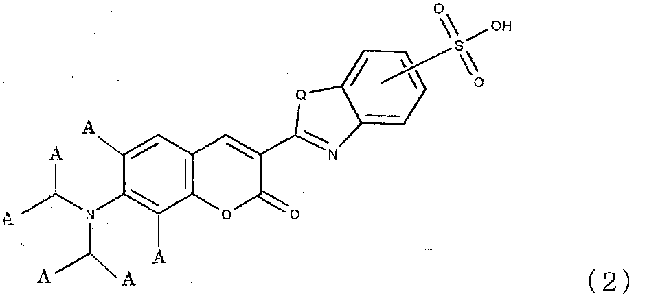
すなわち、本発明の蛍光標識法は、下記式(1)または(2)で示される構造を有するアミノクマリン化合物またはその塩を母体粒子に内包してなるアミノクマリン化合物内包粒子を用いて標識を行う蛍光標識法である。

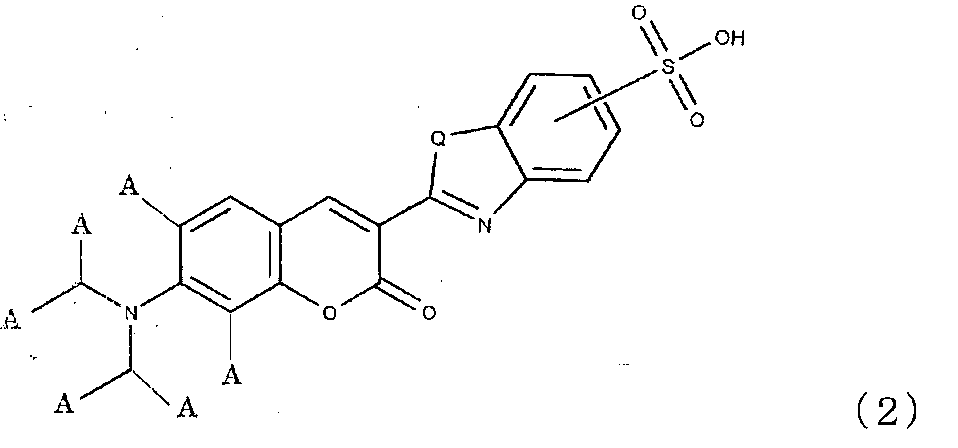
前記蛍光標識法において、前記アミノクマリン化合物内包粒子の平均粒径が80~200nmであることが好ましい。
前記蛍光標識法は、前記アミノクマリン化合物内包粒子を用いた標識を含む多重標識を行うことができる。
前記蛍光標識法は、たとえば免疫染色法およびFISHである。
前記蛍光標識法は、前記アミノクマリン化合物内包粒子を用いた標識を含む多重標識を行うことができる。
前記蛍光標識法は、たとえば免疫染色法およびFISHである。
前記免疫染色法の好適な態様として、PDL1、CTLA4、CD8、CD30、CD48、CD59、IDO、 TDO、CSF-1R、HDAC、CXCR4、FLT-3、TIGIT、INF-α、INF-β、INF-ω、INF-ε、INF-κ、INF-γ、INF-λ CSF、EPO、EGF、FGF、PDGF、HGF、TGF、CD3、CD4、CD25、CD28、CD80、CD86、CD160、CD57、OX40(CD134)、OX40L(CD252)、ICOS(CD278)、ICOSL(CD275)、CD155、CD226、CD112、CD27、CD70、4-1BB(CD137)、4-1BBL(CD137L)、GITR(CD357)、GITRL、BTLA(CD272)、HVEM(CD270)、TIM-3、ガレクチン-9(Galectin-9)、LAG-3(CD223)、B7-H3(CD276)、B7-H4、B7-H5、CD40、CD40L、PD-1、PD-L2、2B4(CD244)、KLRG-1、E-Cadherin、N-Cadherin、R-Cadherin、CD68、CD163およびCSF1-R から選択される少なくとも2つの染色対象タンパク質に対してそれぞれ異なる色素を用いて多重染色を行い、前記染色対象タンパク質の少なくとも1つを、前記アミノクマリン化合物内包粒子を用いて染色する蛍光標識法を挙げることができる。
本発明の蛍光標識法により蛍光標識を行うと、緑色の輝点が明瞭であり、多重標識の場合、他の色領域、たとえば赤色領域への漏れ込みが小さく、緑色の輝点と赤色の輝点との良好なバランスが得られる。
本発明の蛍光標識法は、下記式(1)または(2)で示される構造を有するアミノクマリン化合物またはその塩を母体粒子に内包してなるアミノクマリン化合物内包粒子を用いて標識を行う蛍光標識法である。


式(2)中、6個のAは、それぞれ独立に水素原子またはメチル基を表わす。
式(1)および(2)中、Qはイオウ原子、酸素原子またはN-R1を表わす。前記R1は水素原子またはメチル基を表わす。本発明のアミノクマリン化合物は、式(1)または(2)のQがイオウ原子である場合、ベンゾチアゾール構造を有し、酸素原子である場合、ベンゾオキサゾール構造を有し、N-R1である場合、ベンゾイミダゾール構造を有することになる。
式(1)および(2)に含まれるスルホン酸基SO3Hは、前記ベンゾチアゾール構造、ベンゾオキサゾール構造またはベンゾイミダゾール構造に含まれるベンゼン環が有する結合可能な4つの炭素原子のうちどの炭素原子に結合していてもよい。
式(1)で示される構造を有するアミノクマリン化合物と式(2)で示される構造を有するアミノクマリン化合物とは、スルホン化されたベンゾチアゾール残基、ベンゾオキサゾール残基またはベンゾイミダゾール残基を有するアミノクマリン構造を有する点において共通する。
式(1)で示される構造を有するアミノクマリン化合物は、クマリン構造に結合する窒素原子が、クマリン構造に含まれるベンゼン環の4つの炭素原子とともに、2つの6員環を形成している点、すなわちアミノクマリンのアミノ基がジュロリジン構造となっている点で公知のスルホン化クマリン系化合物と構造が相違する。
式(1)で示される構造を有するアミノクマリン化合物は、公知のスルホン化クマリン系化合物よりも、励起波長が長波長であり、最大励起強度を与える波長が475nm以上であり、たとえば475~510nmである。また、本発明のアミノクマリン化合物は、発光波長も公知のスルホン化クマリン系化合物より長波長であり、最大発光強度を与える波長が510nm以上であり、たとえば510~540nmである。
式(1)で表わされるアミノクマリン化合物は、該アミノクマリン化合物のスルホン基を水素原子で置換して形成されるアミノクマリン化合物に比較して、発光強度が強いという特徴を有する。
式(1)で示される構造を有するアミノクマリン化合物は、たとえば、下記式(3)で示される構造を有するクマリン化合物をスルホン化する方法により製造することができる。具体的には、式(3)で示されるクマリン化合物0.1gに対して発煙硫酸を1ml加えて、0~140℃で、1~8時間反応させることにより製造することができる。

式(2)で示される構造を有するアミノクマリン化合物は、たとえば、下記式(4)で示される構造を有するクマリン化合物をスルホン化する方法により製造することができる。具体的には、式(4)で示されるクマリン化合物0.1gに対して発煙硫酸を1ml加えて、0~140℃で、1~8時間反応させることにより製造することができる。

前記アミノクマリン化合物内包粒子は、式(1)または式(2)で示される構造を有するアミノクマリン化合物と該アミノクマリン化合物を内包する母体粒子とを有する。
アミノクマリン化合物を内包する母体粒子は、有機粒子または無機粒子であり、アミノクマリン化合物を内包できる限り特に制限はない。
前記有機粒子としては、熱硬化性樹脂であることが好ましい。熱硬化性樹脂は三次元的な網目構造を有するので、これに包み込まれたアミノクマリン化合物は樹脂粒子から離脱しにくく、免疫染色等の蛍光標識において好適である。熱硬化性樹脂としては、メラミン樹脂、尿素樹脂、アニリン樹脂、グアナミン樹脂、フェノール樹脂、キシレン樹脂およびフラン樹脂等を挙げることができる。これらの中でも、メラミン樹脂、尿素樹脂等のアミノ樹脂は、色素の樹脂粒子からの離脱をより効果的に抑止できる点で、特に好ましい。
アミノクマリン化合物を内包する母体粒子は、有機粒子または無機粒子であり、アミノクマリン化合物を内包できる限り特に制限はない。
前記有機粒子としては、熱硬化性樹脂であることが好ましい。熱硬化性樹脂は三次元的な網目構造を有するので、これに包み込まれたアミノクマリン化合物は樹脂粒子から離脱しにくく、免疫染色等の蛍光標識において好適である。熱硬化性樹脂としては、メラミン樹脂、尿素樹脂、アニリン樹脂、グアナミン樹脂、フェノール樹脂、キシレン樹脂およびフラン樹脂等を挙げることができる。これらの中でも、メラミン樹脂、尿素樹脂等のアミノ樹脂は、色素の樹脂粒子からの離脱をより効果的に抑止できる点で、特に好ましい。
前記無機粒子としては、シリカ粒子、ガラス粒子等を例示できる。
母体粒子に内包されるアミノクマリン化合物の量は、特に制限はなく、アミノクマリン化合物内包粒子を免疫染色等の蛍光標識に用いる場合に、検出可能な輝度を確保できる量であればよい。
母体粒子に内包されるアミノクマリン化合物の量は、特に制限はなく、アミノクマリン化合物内包粒子を免疫染色等の蛍光標識に用いる場合に、検出可能な輝度を確保できる量であればよい。
アミノクマリン化合物内包粒子の平均粒径は、特に制限はないが、免疫染色等の蛍光標識に用いる場合には、通常20~500nm、好ましくは80~200nmである。アミノクマリン化合物内包粒子の平均粒径が200nmを超えると標識性に問題が生じる場合があり、80nm未満であると、視認性に問題が生じる場合がある。
上記平均粒径は、SEM観察でアミノクマリン化合物内包粒子の1000個について粒径を測定し、その平均値として算出される。
アミノクマリン化合物内包粒子の製造方法は、特に制限されず、公知の方法を採用することができる。一般的には、アミノクマリン化合物の存在下に樹脂またはシリカ等の母体を形成し、アミノクマリン化合物を母体粒子の中に内包させる方法を用いることができる。
アミノクマリン化合物内包粒子の製造方法は、特に制限されず、公知の方法を採用することができる。一般的には、アミノクマリン化合物の存在下に樹脂またはシリカ等の母体を形成し、アミノクマリン化合物を母体粒子の中に内包させる方法を用いることができる。
母体粒子が有機粒子である場合には、たとえば、乳化重合法により、母体粒子を合成するための(コ)モノマーを(共)重合させながら、アミノクマリン化合物を添加し、当該(共)重合体の内部または表面にアミノクマリン化合物を取り込ませる方法を用いることができる。
母体粒子がシリカ等の無機粒子である場合には、たとえば、ラングミュア 8巻 2921ページ(1992)に記載されているFITC内包シリカナノ粒子の合成方法を参考にすることができる。FITCの代わりにアミノクマリン化合物を用いることでアミノクマリン化合物内包シリカナノ粒子を合成することができる。
アミノクマリン化合物内包樹脂粒子は、最大励起強度を与える波長が475~510nmであり、最大発光強度を与える波長が510~540nmであることが好ましい。
式(1)で表わされるアミノクマリン化合物を樹脂に内包させて製造されたアミノクマリン化合物内包樹脂粒子は、そのアミノクマリン化合物のスルホン基を水素原子で置換して形成されるアミノクマリン化合物を樹脂に内包させて製造されたアミノクマリン化合物内包樹脂粒子に比較して、発光強度が強い傾向がある。これは、式(1)で表わされるアミノクマリン化合物は、該アミノクマリン化合物のスルホン基を水素原子で置換して形成されるアミノクマリン化合物よりも、樹脂に内包されやすい性質があり、樹脂により多く取り込まれるからであると推測される。
式(1)で表わされるアミノクマリン化合物を樹脂に内包させて製造されたアミノクマリン化合物内包樹脂粒子は、そのアミノクマリン化合物のスルホン基を水素原子で置換して形成されるアミノクマリン化合物を樹脂に内包させて製造されたアミノクマリン化合物内包樹脂粒子に比較して、発光強度が強い傾向がある。これは、式(1)で表わされるアミノクマリン化合物は、該アミノクマリン化合物のスルホン基を水素原子で置換して形成されるアミノクマリン化合物よりも、樹脂に内包されやすい性質があり、樹脂により多く取り込まれるからであると推測される。
本発明の蛍光標識法は、前記アミノクマリン化合物内包粒子を用いて標識を行う。前記蛍光標識法としては、免疫染色法およびFISH等を挙げることができる。免疫染色法およびFISHの具体的な操作方法は特に限定されず、公知の方法を用いることができる。色素粒子にて標識を行う従来の免疫染色法またはFISHにおいて、前記色素粒子として前記アミノクマリン化合物内包粒子を使用すればよい。
免疫染色法の場合、前記アミノクマリン化合物内包粒子を用いて、HER2およびKi67の他、PDL1、CTLA4、CD8、CD30、CD48およびCD59などの染色対象タンパク質に対しても染色することができる。
本発明の蛍光標識法は、前記アミノクマリン化合物内包粒子を用いた標識を含む多重標識であってもよい。すなわち、2つ以上の標識対象に対してそれぞれ異なる色素を用いて多重標識を行い、その染色対象の少なくとも1つを、前記アミノクマリン化合物内包粒子を用いて標識することができる。たとえば、複数の標識対象について、そのうちの一部の標識対象に対して前記アミノクマリン化合物内包粒子を用いて標識を行い、他の標識対象に対して緑色以外の発光を示す色素を含む粒子を用いて標識を行って、複数の標識対象を緑色と緑色以外の色とで別々に標識化することができる。
たとえば、免疫染色法においては、PDL1、CTLA4、CD8、CD30、CD48およびCD59から選択される少なくとも2つの染色対象タンパク質に対してそれぞれ異なる色素を用いて多重染色を行い、前記染色対象タンパク質の少なくとも1つを、前記アミノクマリン化合物内包粒子を用いて染色することができる。そうすれば、たとえば、PDL1をアミノクマリン化合物内包粒子によって緑色に染色し、CTLA4を赤色で染色して、PDL1とCTLA4とを異なる色で標識化するということが可能になる。
この多重染色においては、PDL1、CTLA4、CD8、CD30、CD48、CD59、IDO、 TDO、CSF-1R、HDAC、CXCR4、FLT-3、TIGIT、INF-α、INF-β、INF-ω、INF-ε、INF-κ、INF-γ、INF-λ CSF、EPO、EGF、FGF、PDGF、HGF、TGF、CD3、CD4、CD25、CD28、CD80、CD86、CD160、CD57、OX40(別名CD134)、OX40L(別名CD252)、ICOS(別名CD278)、ICOSL(別名CD275)、CD155、CD226、CD112、CD27、CD70、4-1BB(別名CD137)、4-1BBL(別名CD137L)、GITR(別名CD357)、GITRL、BTLA(別名CD272)、HVEM(別名CD270)、TIM-3、Galectin-9、LAG-3(別名CD223)、B7-H3(別名CD276)、B7-H4、B7-H5、CD40、CD40L、PD-1、PD-L2、2B4(別名CD244)、KLRG-1、E-Cadherin、N-Cadherin、R-Cadherin、CD68、CD163およびCSF1-R から選択される少なくとも2つの染色対象タンパク質に対してそれぞれ異なる色素を用いて多重染色を行い、前記染色対象タンパク質の少なくとも1つを、前記アミノクマリン化合物内包粒子を用いて染色することができる。
この多重染色においては、PDL1、CTLA4、CD8、CD30、CD48、CD59、IDO、 TDO、CSF-1R、HDAC、CXCR4、FLT-3、TIGIT、INF-α、INF-β、INF-ω、INF-ε、INF-κ、INF-γ、INF-λ CSF、EPO、EGF、FGF、PDGF、HGF、TGF、CD3、CD4、CD25、CD28、CD80、CD86、CD160、CD57、OX40(別名CD134)、OX40L(別名CD252)、ICOS(別名CD278)、ICOSL(別名CD275)、CD155、CD226、CD112、CD27、CD70、4-1BB(別名CD137)、4-1BBL(別名CD137L)、GITR(別名CD357)、GITRL、BTLA(別名CD272)、HVEM(別名CD270)、TIM-3、Galectin-9、LAG-3(別名CD223)、B7-H3(別名CD276)、B7-H4、B7-H5、CD40、CD40L、PD-1、PD-L2、2B4(別名CD244)、KLRG-1、E-Cadherin、N-Cadherin、R-Cadherin、CD68、CD163およびCSF1-R から選択される少なくとも2つの染色対象タンパク質に対してそれぞれ異なる色素を用いて多重染色を行い、前記染色対象タンパク質の少なくとも1つを、前記アミノクマリン化合物内包粒子を用いて染色することができる。
前記アミノクマリン化合物内包粒子を用いて標識を行うと、明瞭な緑色の輝点が確認でき、赤色領域などの他の色領域への漏れ込みが小さい。このため、特定の標識対象に対して前記アミノクマリン化合物内包粒子を用いて緑色の標識を行い、他の染色対象に対して赤色色素を含む色素粒子を用いて赤色の標識を行うと、得られる緑色の輝点は赤色領域への漏れ込みが小さく、緑色の輝点と赤色の輝点との良好なバランスが得られる。
前記アミノクマリン化合物は、前記アミノクマリン化合物以外のクマリン化合物に比較して、赤色領域に近い発光領域を有するが、前記アミノクマリン化合物内包粒子を用いて標識を行うと、前記アミノクマリン化合物以外のクマリン化合物を含む色素粒子よりも、赤色領域への漏れ込みがむしろ小さいという効果が得られる。
[合成例1]
20mLバイアル管瓶に下記式(5)で表わされる化合物 600mgを入れ、発煙硫酸6mLを加えて、25℃にて4時間撹拌し、反応を行った。反応の進行はTLCにて確認した。具体的には、反応液の一部をNaOH水溶液にて中和した後、反応液にエタノールを加え、CHCl3 を2、MeOHを3の割合で混合した溶液を用いてTLCを行った。原料のRf値0.88に対し、目的物のRf値0.73であり、このTLCのデータより、反応の収束および目的物の生成を確認した。
20mLバイアル管瓶に下記式(5)で表わされる化合物 600mgを入れ、発煙硫酸6mLを加えて、25℃にて4時間撹拌し、反応を行った。反応の進行はTLCにて確認した。具体的には、反応液の一部をNaOH水溶液にて中和した後、反応液にエタノールを加え、CHCl3 を2、MeOHを3の割合で混合した溶液を用いてTLCを行った。原料のRf値0.88に対し、目的物のRf値0.73であり、このTLCのデータより、反応の収束および目的物の生成を確認した。
50mLバイアル管瓶に氷を8分目(30mL)まで入れ、この中に反応液を少しずつ加えた。生成した色素が懸濁した懸濁液が得られた。この懸濁液を遠心分離し、上澄み液を除去して色素を沈殿として回収した。沈殿を純水10mLで分散し、この分散液を遠心分離して、上澄み液を除去して、沈殿を回収し、再度、純水10mLで分散し、この分散液を遠心分離して上澄み液を除去して、沈殿を回収した。回収した沈殿をエタノールで分散し、この分散液を遠心分離し、上澄み液を除去して、沈殿として下記式(I)で表わされるアミノクマリン化合物Iを得た。アミノクマリン化合物Iの収率は80%であった。
得られた沈殿物を乾燥後、得られた粉末を純水に加えた後、NaOH水溶液で中和し、沈殿を溶解し、溶液のpHを7~8とした。この溶液を凍結乾燥機にて乾燥する事により、アミノクマリン化合物IのNa塩を得た。アミノクマリン化合物Iは、スルホン酸体では水への溶解性が悪いのに対し、Na塩とする事で、水に速やかに溶解することを確認した。


[合成例2]
式(5)で表わされる化合物の代わりに下記式(6)で表わされるアミノクマリン化合物iを用いたこと以外は製造例1と同様の方法により、下記式(II)で表わされるアミノクマリン化合物IIを得た。
式(5)で表わされる化合物の代わりに下記式(6)で表わされるアミノクマリン化合物iを用いたこと以外は製造例1と同様の方法により、下記式(II)で表わされるアミノクマリン化合物IIを得た。


[合成例3]
式(5)で表わされる化合物の代わりに下記式(7)で表わされるアミノクマリン化合物iを用いたこと以外は製造例1と同様の方法により、下記式(III)で表わされるアミノクマリン化合物IIIを得た。
式(5)で表わされる化合物の代わりに下記式(7)で表わされるアミノクマリン化合物iを用いたこと以外は製造例1と同様の方法により、下記式(III)で表わされるアミノクマリン化合物IIIを得た。


[製造例1]
アミノクマリン化合物I 3.4mgに塩化チオニル0.1mLを加え、65℃4時間、加熱混合した後、真空乾燥を行なって余剰の塩化チオニルを除去した。得られたアミノクマリン化合物と塩化チオニルの反応物と3-アミノプロピルトリメトキシシラン(3-aminopropyltrimetoxysilane、信越シリコーン社製、KBM903)3μLを1.2mLのN,N-ジメチルホルムアミド(DMF)の中で混合し、オルガノアルコキシシラン化合物を得た。
アミノクマリン化合物I 3.4mgに塩化チオニル0.1mLを加え、65℃4時間、加熱混合した後、真空乾燥を行なって余剰の塩化チオニルを除去した。得られたアミノクマリン化合物と塩化チオニルの反応物と3-アミノプロピルトリメトキシシラン(3-aminopropyltrimetoxysilane、信越シリコーン社製、KBM903)3μLを1.2mLのN,N-ジメチルホルムアミド(DMF)の中で混合し、オルガノアルコキシシラン化合物を得た。
得られたオルガノアルコキシシラン化合物液0.3mLを、99%エタノール24mL、テトラエトキシシラン(TEOS)0.3mL、超純水0.75mL、および28質量%のアンモニア水0.75mLと25℃で3時間混合した。
上記工程で作製した混合液を10000Gで20分間遠心分離し、上澄みを除去した。この沈殿に対して、エタノールを加えて、沈殿物を分散させ、再度遠心分離をするリンスを行った。さらに同様のリンスを2回繰り返し、アミノクマリン化合物内包粒子Iを得た。得られた粒子の1000個についてSEM観察を行い、平均粒径を測定したところ、平均粒径は60nmであった。
[製造例2]
超純水0.85mL、アンモニア水0.85mLとしたこと以外は製造例1と同様の方法で、アミノクマリン化合物内包粒子IIを得た。得られた粒子の1000個についてSEM観察を行い、平均粒径を測定したところ、平均粒径は80nmであった。
超純水0.85mL、アンモニア水0.85mLとしたこと以外は製造例1と同様の方法で、アミノクマリン化合物内包粒子IIを得た。得られた粒子の1000個についてSEM観察を行い、平均粒径を測定したところ、平均粒径は80nmであった。
[製造例3]
超純水1.10mL、アンモニア水1.10mLとしたこと以外は製造例1と同様の方法で、アミノクマリン化合物内包粒子IIIを得た。得られた粒子の1000個についてSEM観察を行い、平均粒径を測定したところ、平均粒径は150nmであった。
超純水1.10mL、アンモニア水1.10mLとしたこと以外は製造例1と同様の方法で、アミノクマリン化合物内包粒子IIIを得た。得られた粒子の1000個についてSEM観察を行い、平均粒径を測定したところ、平均粒径は150nmであった。
[製造例4]
超純水1.15mL、アンモニア水1.15mLとしたこと以外は製造例1と同様の方法で、アミノクマリン化合物内包粒子IVを得た。得られた粒子の1000個についてSEM観察を行い、平均粒径を測定したところ、平均粒径は195nmであった。
超純水1.15mL、アンモニア水1.15mLとしたこと以外は製造例1と同様の方法で、アミノクマリン化合物内包粒子IVを得た。得られた粒子の1000個についてSEM観察を行い、平均粒径を測定したところ、平均粒径は195nmであった。
[製造例5]
超純水1.20mL、アンモニア水1.20mLとしたこと以外は製造例1と同様の方法で、アミノクマリン化合物内包粒子Vを得た。得られた粒子の1000個についてSEM観察を行い、平均粒径を測定したところ、平均粒径は220nmであった。
超純水1.20mL、アンモニア水1.20mLとしたこと以外は製造例1と同様の方法で、アミノクマリン化合物内包粒子Vを得た。得られた粒子の1000個についてSEM観察を行い、平均粒径を測定したところ、平均粒径は220nmであった。
[製造例6]
アミノクマリン化合物Iの代わりにアミノクマリン化合物IIを用いたこと以外は製造例3と同様の方法で、アミノクマリン化合物内包粒子VIを得た。得られた粒子の1000個についてSEM観察を行い、平均粒径を測定したところ、平均粒径は150nmであった。
アミノクマリン化合物Iの代わりにアミノクマリン化合物IIを用いたこと以外は製造例3と同様の方法で、アミノクマリン化合物内包粒子VIを得た。得られた粒子の1000個についてSEM観察を行い、平均粒径を測定したところ、平均粒径は150nmであった。
[製造例7]
アミノクマリン化合物I 14.4mgを水22mLに加えて溶解させた。この溶液に乳化重合用乳化剤のエマルジョン(登録商標)430(ポリオキシエチレンオレイルエーテル、花王社製)の5%水溶液を2mL加えた。
アミノクマリン化合物I 14.4mgを水22mLに加えて溶解させた。この溶液に乳化重合用乳化剤のエマルジョン(登録商標)430(ポリオキシエチレンオレイルエーテル、花王社製)の5%水溶液を2mL加えた。
この溶液をホットスターラー上で撹拌しながら70℃まで昇温させた後、この溶液にメラミン樹脂原料ニカラックMX-035(日本カーバイド工業社製)を0.65g加えた。この溶液に反応開始剤としてドデシルベンゼンスルホン酸(関東化学社製)の10%水溶液を1000μL加え、70℃で50分間加熱撹拌し、その後、90℃に昇温して20分間加熱撹拌した。以上の操作により、アミノクマリン化合物内包粒子VIIを得た。
得られたアミノクマリン化合物内包樹脂粒子VIIの分散液から、純水による洗浄を行い、余剰の樹脂原料やアミノクマリン化合物などの不純物を除いた。具体的には、遠心分離機(クボタ社製マイクロ冷却遠心機3740)にて20000Gで15分間、遠心分離し、上澄み除去後、超純水を加えて超音波照射して再分散した。遠心分離、上澄み除去および超純水への再分散による洗浄を5回繰り返した。
アミノクマリン化合物内包粒子VIIの1000個についてSEM観察を行い、平均粒径を測定したところ、平均粒径は150nmであった。
アミノクマリン化合物内包粒子VIIの1000個についてSEM観察を行い、平均粒径を測定したところ、平均粒径は150nmであった。
[製造例8]
アミノクマリン化合物Iの代わりにアミノクマリン化合物IIを使用したこと以外は製造例7と同様の方法で、アミノクマリン化合物内包粒子VIIIを得た。
アミノクマリン化合物内包粒子VIIIの1000個についてSEM観察を行い、平均粒径を測定したところ、平均粒径は150nmであった。
アミノクマリン化合物Iの代わりにアミノクマリン化合物IIを使用したこと以外は製造例7と同様の方法で、アミノクマリン化合物内包粒子VIIIを得た。
アミノクマリン化合物内包粒子VIIIの1000個についてSEM観察を行い、平均粒径を測定したところ、平均粒径は150nmであった。
[製造例9]
アミノクマリン化合物Iの代わりにアミノクマリン化合物IIIを使用したこと以外は製造例7と同様の方法で、アミノクマリン化合物内包粒子IXを得た。
アミノクマリン化合物内包粒子IXの1000個についてSEM観察を行い、平均粒径を測定したところ、平均粒径は150nmであった。
アミノクマリン化合物Iの代わりにアミノクマリン化合物IIIを使用したこと以外は製造例7と同様の方法で、アミノクマリン化合物内包粒子IXを得た。
アミノクマリン化合物内包粒子IXの1000個についてSEM観察を行い、平均粒径を測定したところ、平均粒径は150nmであった。
[製造例10]
アミノクマリン化合物Iの代わりに緑色色素であるPyrromethene556を用いたこと以外は製造例1と同様の方法で、色素内包粒子iを得た。得られた粒子の1000個についてSEM観察を行い、平均粒径を測定したところ、平均粒径は150nmであった。
アミノクマリン化合物Iの代わりに緑色色素であるPyrromethene556を用いたこと以外は製造例1と同様の方法で、色素内包粒子iを得た。得られた粒子の1000個についてSEM観察を行い、平均粒径を測定したところ、平均粒径は150nmであった。
[製造例11]
アミノクマリン化合物Iの代わりに緑色色素であるPyrromethene556を用いたこと以外は製造例7と同様の方法で、色素内包粒子iiを得た。得られた粒子の1000個についてSEM観察を行い、平均粒径を測定したところ、平均粒径は150nmであった。
アミノクマリン化合物Iの代わりに緑色色素であるPyrromethene556を用いたこと以外は製造例7と同様の方法で、色素内包粒子iiを得た。得られた粒子の1000個についてSEM観察を行い、平均粒径を測定したところ、平均粒径は150nmであった。
[製造例12]
アミノクマリン化合物Iの代わりに赤色色素であるスルホローダミン101を用いたこと以外は製造例7と同様の方法で、色素内包粒子iiiを得た。得られた粒子の1000個についてSEM観察を行い、平均粒径を測定したところ、平均粒径は150nmであった。
アミノクマリン化合物Iの代わりに赤色色素であるスルホローダミン101を用いたこと以外は製造例7と同様の方法で、色素内包粒子iiiを得た。得られた粒子の1000個についてSEM観察を行い、平均粒径を測定したところ、平均粒径は150nmであった。
[実施例1]
下記の方法により免疫染色を行った。
(色素内包粒子のストレプトアビジン修飾)
アミノクマリン化合物内包粒子Iを、EDTA(エチレンジアミン四酢酸)を2mM含有するPBS(リン酸緩衝液生理的食塩水)を用いて3nMに調整し、この溶液に最終濃度10mMとなるようにSM(PEG)12(サーモサイエンティフィック社製、succinimidyl-[(N-maleimidopropionamid)-dodecanethyleneglycol]ester)を混合し、5℃で1時間反応させた。
下記の方法により免疫染色を行った。
(色素内包粒子のストレプトアビジン修飾)
アミノクマリン化合物内包粒子Iを、EDTA(エチレンジアミン四酢酸)を2mM含有するPBS(リン酸緩衝液生理的食塩水)を用いて3nMに調整し、この溶液に最終濃度10mMとなるようにSM(PEG)12(サーモサイエンティフィック社製、succinimidyl-[(N-maleimidopropionamid)-dodecanethyleneglycol]ester)を混合し、5℃で1時間反応させた。
この混合液を、10000Gで20分遠心分離を行い、上澄みを除去した後に、EDTAを2mM含有したPBSを加え、沈降物を分散させ、再度遠心分離を行った。同様の手順による洗浄を3回行うことで末端にマレイミド基がついたアミノクマリン化合物内包粒子Iを得た。
1mg/mLに調整したストレプトアビジン(和光純薬工業社製)40μLを210μLのボレートバッファーに加えた後、64mg/mLに調整した2-イミノチオラン塩酸塩(シグマアルドリッチ社製)70μLを加え、室温で1時間反応させた。これにより、ストレプトアビジンのアミノ基に対してチオール基(-NH-C(=NH2
+Cl-)-CH2-CH2-CH2-SH)を導入した。
このストレプトアビジン溶液をゲルろ過カラム(Zaba Spin Desalting Columns:フナコシ)により脱塩し、上記シリカ系粒子に結合可能なストレプトアビジンを得た。このストレプトアビジン全量(0.04mg含有)とEDTAを2mM含有したPBSを用いて上記0.67nMに調整したシリカ系粒子740μLとを混合し、室温で1時間反応させた。
10mMメルカプトエタノールを添加し、反応を停止させた。得られた溶液を遠心フィルターで濃縮後、精製用ゲルろ過カラムを用いて未反応ストレプトアビジン等を除去し、ストレプトアビジン結合アミノクマリン化合物内包粒子Iを得た。
(ビオチン修飾された2次抗体の作製)
50mMTris-HCl溶液(pH7.5)に抗ウサギIgG抗体50μgを溶解した。該溶液に、最終濃度3mMとなるようにDTT(dithiothretol)溶液を混合した。その後、該溶液を37℃で30分間反応させた。その後、脱塩カラムを用いてDTTで還元化した2次抗体を精製した。精製した抗体全量のうち200μLを50mMTris-HCl溶液(pH7.5)に溶解して抗体溶液を得た。その一方で、スペーサーの長さが30オングストロームであるリンカー試薬「(+)-Biotin-PEG6‐NH‐Mal」(PurePEG社製,製品番号2461006-250)を、DMSOを用いて0.4mMとなるように調整した。この溶液8.5μLを前記抗体溶液に添加し、混和して37℃で30分間反応させた。
50mMTris-HCl溶液(pH7.5)に抗ウサギIgG抗体50μgを溶解した。該溶液に、最終濃度3mMとなるようにDTT(dithiothretol)溶液を混合した。その後、該溶液を37℃で30分間反応させた。その後、脱塩カラムを用いてDTTで還元化した2次抗体を精製した。精製した抗体全量のうち200μLを50mMTris-HCl溶液(pH7.5)に溶解して抗体溶液を得た。その一方で、スペーサーの長さが30オングストロームであるリンカー試薬「(+)-Biotin-PEG6‐NH‐Mal」(PurePEG社製,製品番号2461006-250)を、DMSOを用いて0.4mMとなるように調整した。この溶液8.5μLを前記抗体溶液に添加し、混和して37℃で30分間反応させた。
この反応溶液を脱塩カラム「Zeba Desalt Spin Columns」(サーモサイエンティフィック社製,Cat.#89882)に供して精製した。脱塩した反応溶液の波長300nmの吸収を分光高度計(日立製「F-7000」)により計測して反応溶液に含まれるタンパク質の量を算出した。50mMTris溶液により反応溶液を250μg/mLに調整し、該溶液をビオチン化2次抗体の溶液とした。
(染色)
(1)標本処理工程
(1-1)脱パラフィン処理工程
染色用の組織切片として、HER2(3+)とHER2(-)の組織アレイスライド(コスモバイオ社製「CB-A712のシリーズ」)を用いた。この組織アレイスライドを脱パラフィン処理した。
(1)標本処理工程
(1-1)脱パラフィン処理工程
染色用の組織切片として、HER2(3+)とHER2(-)の組織アレイスライド(コスモバイオ社製「CB-A712のシリーズ」)を用いた。この組織アレイスライドを脱パラフィン処理した。
(1-2)賦活化処理工程
脱パラフィン処理した組織アレイスライドを水に置換する洗浄を行った。洗浄した組織アレイスライドを10mMクエン酸緩衝液中(pH6.0)中で121℃、15分間オートクレーブ処理することで、抗原の賦活化処理を行った。賦活化処理後の組織アレイスライドをPBSにより洗浄し、洗浄した組織アレイスライドに対してBSAを1%含有するPBSを用いて1時間ブロッキング処理を行った。
脱パラフィン処理した組織アレイスライドを水に置換する洗浄を行った。洗浄した組織アレイスライドを10mMクエン酸緩衝液中(pH6.0)中で121℃、15分間オートクレーブ処理することで、抗原の賦活化処理を行った。賦活化処理後の組織アレイスライドをPBSにより洗浄し、洗浄した組織アレイスライドに対してBSAを1%含有するPBSを用いて1時間ブロッキング処理を行った。
(2)免疫染色処理工程
(2-1)1次反応
BSAを1%含有するPBSを用いて、ベンタナ社製「抗HER2ウサギモノクロナール抗体(4B5)」を0.05nMに調整し、該1次抗体の溶液を上述のブロッキング処理した組織アレイスライドに対して4℃で1晩反応させた。
(2-1)1次反応
BSAを1%含有するPBSを用いて、ベンタナ社製「抗HER2ウサギモノクロナール抗体(4B5)」を0.05nMに調整し、該1次抗体の溶液を上述のブロッキング処理した組織アレイスライドに対して4℃で1晩反応させた。
(2-2)2次反応
1次反応を行った組織アレイスライドをPBSで洗浄した後、1%BSA含有のPBSで6μg/mLに希釈した上記ビオチン化2次抗体と室温30分間反応させた。
1次反応を行った組織アレイスライドをPBSで洗浄した後、1%BSA含有のPBSで6μg/mLに希釈した上記ビオチン化2次抗体と室温30分間反応させた。
(2-3)蛍光標識処理
2次反応を行った組織アレイスライドに対して、1%BSA含有のPBSで0.02nMに希釈したストレプトアビジン結合アミノクマリン化合物内包粒子Iを、中性のpH環境(pH6.9~7.4)室温の条件下で3時間反応させた。該反応後の組織アレイスライドをPBSで洗浄した。
2次反応を行った組織アレイスライドに対して、1%BSA含有のPBSで0.02nMに希釈したストレプトアビジン結合アミノクマリン化合物内包粒子Iを、中性のpH環境(pH6.9~7.4)室温の条件下で3時間反応させた。該反応後の組織アレイスライドをPBSで洗浄した。
(3)形態観察染色工程
免疫染色後、ヘマトキシリン・エオシン染色(HE染色)を行った。免疫染色した切片をマイヤーヘマトキシリン液で5分間染色してヘマトキシリン染色を行った。その後、該組織切片を45℃の流水で3分間洗浄した。次に、1%エオシン液で5分間染色してエオシン染色を行った。
免疫染色後、ヘマトキシリン・エオシン染色(HE染色)を行った。免疫染色した切片をマイヤーヘマトキシリン液で5分間染色してヘマトキシリン染色を行った。その後、該組織切片を45℃の流水で3分間洗浄した。次に、1%エオシン液で5分間染色してエオシン染色を行った。
(4)固定処理工程
免疫染色工程および形態観察染色工程を終えた組織切片に対して、純エタノールに5分間浸漬する操作を4回行い、洗浄・脱水を行った。続いて、キシレンに5分間浸漬する操作を4回行い、透徹を行った。最後に、封入剤(メルク社製「エンテランニュー」)を用いて、組織切片を封入して観察用のサンプルの組織アレイスライドとした。
免疫染色工程および形態観察染色工程を終えた組織切片に対して、純エタノールに5分間浸漬する操作を4回行い、洗浄・脱水を行った。続いて、キシレンに5分間浸漬する操作を4回行い、透徹を行った。最後に、封入剤(メルク社製「エンテランニュー」)を用いて、組織切片を封入して観察用のサンプルの組織アレイスライドとした。
(5)観察・計測工程
固定化処理工程を終えた組織切片に対して所定の励起光を照射して、蛍光を発光させた。その状態の組織切片を蛍光顕微鏡(オリンパス社製「BX-53」)、顕微鏡用デジタルカメラ(オリンパス社製「DP73」)により観察および撮像を行った。上記励起光は、光学フィルターに通すことで575~600nmに設定した。また、観察する蛍光の波長(nm)の範囲についても、光学フィルターを通すことで612~692nmに設定した。顕微鏡観察、画像取得時の励起波長の条件は、580nmの励起では視野中心部付近の照射エネルギーが900W/cm2となるようにした。画像取得時の露光時間は、画像の輝度が飽和しないように任意に設定(例えば4000μ秒に設定)して撮像した。HER2(3+)の組織の輝点数は、400倍で撮像した画像をもとにImageJ FindMaxims法により計測した1000細胞の平均値とした。
視野内の細胞膜上の輝点数Sおよび視野内の細胞外の輝点数Nを測定し、S/Nを算出した。S/Nを表1に示す。
固定化処理工程を終えた組織切片に対して所定の励起光を照射して、蛍光を発光させた。その状態の組織切片を蛍光顕微鏡(オリンパス社製「BX-53」)、顕微鏡用デジタルカメラ(オリンパス社製「DP73」)により観察および撮像を行った。上記励起光は、光学フィルターに通すことで575~600nmに設定した。また、観察する蛍光の波長(nm)の範囲についても、光学フィルターを通すことで612~692nmに設定した。顕微鏡観察、画像取得時の励起波長の条件は、580nmの励起では視野中心部付近の照射エネルギーが900W/cm2となるようにした。画像取得時の露光時間は、画像の輝度が飽和しないように任意に設定(例えば4000μ秒に設定)して撮像した。HER2(3+)の組織の輝点数は、400倍で撮像した画像をもとにImageJ FindMaxims法により計測した1000細胞の平均値とした。
視野内の細胞膜上の輝点数Sおよび視野内の細胞外の輝点数Nを測定し、S/Nを算出した。S/Nを表1に示す。
[実施例2~5、7、8]
(色素内包粒子のストレプトアビジン修飾)
実施例2~5、7、8においては、アミノクマリン化合物内包粒子Iの代わりにアミノクマリン化合物内包粒子II~VI、VIIIをそれぞれ使用したこと以外は実施例1と同様の方法でストレプトアビジン結合アミノクマリン化合物内包粒子II~VI、VIIIをそれぞれ得た。
(色素内包粒子のストレプトアビジン修飾)
実施例2~5、7、8においては、アミノクマリン化合物内包粒子Iの代わりにアミノクマリン化合物内包粒子II~VI、VIIIをそれぞれ使用したこと以外は実施例1と同様の方法でストレプトアビジン結合アミノクマリン化合物内包粒子II~VI、VIIIをそれぞれ得た。
(ビオチン修飾された2次抗体の作製)
実施例2~5、7、8においては、実施例1と同様の方法でビオチン化2次抗体の溶液を得た。
実施例2~5、7、8においては、実施例1と同様の方法でビオチン化2次抗体の溶液を得た。
(染色)
実施例2~5、7、8においては、ストレプトアビジン結合アミノクマリン化合物内包粒子Iの代わりにストレプトアビジン結合アミノクマリン化合物内包粒子II~VI、VIIIをそれぞれ使用したこと以外は実施例1と同様の方法で、S/Nを算出した。S/Nを表1に示す。
実施例2~5、7、8においては、ストレプトアビジン結合アミノクマリン化合物内包粒子Iの代わりにストレプトアビジン結合アミノクマリン化合物内包粒子II~VI、VIIIをそれぞれ使用したこと以外は実施例1と同様の方法で、S/Nを算出した。S/Nを表1に示す。
[実施例6]
(色素内包粒子のストレプトアビジン修飾)
アミノクマリン化合物内包粒子Iの代わりにアミノクマリン化合物内包粒子IIIを使用したこと以外は実施例1と同様の方法でストレプトアビジン結合アミノクマリン化合物内包粒子IIIを得た。
(ビオチン修飾された2次抗体の作製)
実施例1と同様の方法でビオチン化2次抗体の溶液を得た。
(色素内包粒子のストレプトアビジン修飾)
アミノクマリン化合物内包粒子Iの代わりにアミノクマリン化合物内包粒子IIIを使用したこと以外は実施例1と同様の方法でストレプトアビジン結合アミノクマリン化合物内包粒子IIIを得た。
(ビオチン修飾された2次抗体の作製)
実施例1と同様の方法でビオチン化2次抗体の溶液を得た。
(染色)
(1)標本処理工程
(1-1)脱パラフィン処理工程
染色用の組織切片としてPDL1の組織アレイスライドを用いた。この組織アレイスライドを脱パラフィン処理した。
(1)標本処理工程
(1-1)脱パラフィン処理工程
染色用の組織切片としてPDL1の組織アレイスライドを用いた。この組織アレイスライドを脱パラフィン処理した。
(1-2)賦活化処理工程
脱パラフィン処理した組織アレイスライドを水に置換する洗浄を行った。洗浄した組織アレイスライドを10mMクエン酸緩衝液(pH6.0)中で121℃、15分間オートクレーブ処理することで、抗原の賦活化処理を行った。賦活化処理後の組織アレイスライドをPBSにより洗浄し、洗浄した組織アレイスライドに対してBSAを1%含有するPBSを用いて1時間ブロッキング処理を行った。
脱パラフィン処理した組織アレイスライドを水に置換する洗浄を行った。洗浄した組織アレイスライドを10mMクエン酸緩衝液(pH6.0)中で121℃、15分間オートクレーブ処理することで、抗原の賦活化処理を行った。賦活化処理後の組織アレイスライドをPBSにより洗浄し、洗浄した組織アレイスライドに対してBSAを1%含有するPBSを用いて1時間ブロッキング処理を行った。
(2)免疫染色処理工程
(2-1)1次反応
BSAを1%含有するPBSを用いて、Cell Signaling Technology社製「抗PD-L1ウサギモノクロナール抗体(E1L3N)」を0.05nMに調整し、該1次抗体の溶液を上述のブロッキング処理した組織アレイスライドに対して4℃で1晩反応させた。
(2-1)1次反応
BSAを1%含有するPBSを用いて、Cell Signaling Technology社製「抗PD-L1ウサギモノクロナール抗体(E1L3N)」を0.05nMに調整し、該1次抗体の溶液を上述のブロッキング処理した組織アレイスライドに対して4℃で1晩反応させた。
(2-2)2次反応
1次反応を行った組織アレイスライドをPBSで洗浄した後、1%BSA含有のPBSで6μg/mLに希釈した上記ビオチン化2次抗体と室温30分間反応させた。
1次反応を行った組織アレイスライドをPBSで洗浄した後、1%BSA含有のPBSで6μg/mLに希釈した上記ビオチン化2次抗体と室温30分間反応させた。
(2-3)蛍光標識処理
2次反応を行った組織アレイスライドに対して、1%BSA含有のPBSで0.02nMに希釈したストレプトアビジン結合アミノクマリン化合物内包粒子IIIを、中性のpH環境(pH6.9~7.4)室温の条件下で3時間反応させた。該反応後の組織アレイスライドをPBSで洗浄した。
2次反応を行った組織アレイスライドに対して、1%BSA含有のPBSで0.02nMに希釈したストレプトアビジン結合アミノクマリン化合物内包粒子IIIを、中性のpH環境(pH6.9~7.4)室温の条件下で3時間反応させた。該反応後の組織アレイスライドをPBSで洗浄した。
(3)形態観察染色工程
免疫染色後、ヘマトキシリン・エオシン染色(HE染色)を行った。免疫染色した切片をマイヤーヘマトキシリン液で5分間染色してヘマトキシリン染色を行った。その後、該組織切片を45℃の流水で3分間洗浄した。次に、1%エオシン液で5分間染色してエオシン染色を行った。
免疫染色後、ヘマトキシリン・エオシン染色(HE染色)を行った。免疫染色した切片をマイヤーヘマトキシリン液で5分間染色してヘマトキシリン染色を行った。その後、該組織切片を45℃の流水で3分間洗浄した。次に、1%エオシン液で5分間染色してエオシン染色を行った。
(4)固定処理工程
免疫染色工程および形態観察染色工程を終えた組織切片に対して、純エタノールに5分間浸漬する操作を4回行い、洗浄・脱水を行った。続いて、キシレンに5分間浸漬する操作を4回行い、透徹を行った。最後に、封入剤(メルク社製「エンテランニュー」)を用いて、組織切片を封入して観察用のサンプルの組織アレイスライドとした。
免疫染色工程および形態観察染色工程を終えた組織切片に対して、純エタノールに5分間浸漬する操作を4回行い、洗浄・脱水を行った。続いて、キシレンに5分間浸漬する操作を4回行い、透徹を行った。最後に、封入剤(メルク社製「エンテランニュー」)を用いて、組織切片を封入して観察用のサンプルの組織アレイスライドとした。
(5)観察・計測工程
実施例1と同様の方法で、S/Nを算出した。S/Nを表1に示す。
実施例1と同様の方法で、S/Nを算出した。S/Nを表1に示す。
[実施例9]
(色素内包粒子のストレプトアビジン修飾)
アミノクマリン化合物内包粒子Iの代わりにアミノクマリン化合物内包粒子VIIIを使用したこと以外は実施例1と同様の方法でストレプトアビジン結合アミノクマリン化合物内包粒子VIIIを得た。
(色素内包粒子のストレプトアビジン修飾)
アミノクマリン化合物内包粒子Iの代わりにアミノクマリン化合物内包粒子VIIIを使用したこと以外は実施例1と同様の方法でストレプトアビジン結合アミノクマリン化合物内包粒子VIIIを得た。
(ビオチン修飾された2次抗体の作製)
実施例1と同様の方法でビオチン化2次抗体の溶液を得た。
(染色)
ストレプトアビジン結合アミノクマリン化合物内包シリカナノ粒子IIIの代わりにストレプトアビジン結合アミノクマリン化合物内包粒子VIIIを使用したこと以外は実施例6と同様の方法で、S/Nを算出した。S/Nを表1に示す。
実施例1と同様の方法でビオチン化2次抗体の溶液を得た。
(染色)
ストレプトアビジン結合アミノクマリン化合物内包シリカナノ粒子IIIの代わりにストレプトアビジン結合アミノクマリン化合物内包粒子VIIIを使用したこと以外は実施例6と同様の方法で、S/Nを算出した。S/Nを表1に示す。
[比較例1]
(色素内包粒子のストレプトアビジン修飾)
アミノクマリン化合物内包粒子Iの代わりに色素内包粒子iを使用したこと以外は実施例1と同様の方法でストレプトアビジン結合色素内包粒子iを得た。
(色素内包粒子のストレプトアビジン修飾)
アミノクマリン化合物内包粒子Iの代わりに色素内包粒子iを使用したこと以外は実施例1と同様の方法でストレプトアビジン結合色素内包粒子iを得た。
(ビオチン修飾された2次抗体の作製)
実施例1と同様の方法でビオチン化2次抗体の溶液を得た。
(染色)
ストレプトアビジン結合アミノクマリン化合物内包粒子Iの代わりにストレプトアビジン結合色素内包粒子iを使用したこと以外は実施例1と同様の方法で、S/Nを算出した。S/Nを表1に示す。
実施例1と同様の方法でビオチン化2次抗体の溶液を得た。
(染色)
ストレプトアビジン結合アミノクマリン化合物内包粒子Iの代わりにストレプトアビジン結合色素内包粒子iを使用したこと以外は実施例1と同様の方法で、S/Nを算出した。S/Nを表1に示す。
[比較例2]
(色素内包粒子のストレプトアビジン修飾)
アミノクマリン化合物内包粒子Iの代わりに色素内包粒子iを使用したこと以外は実施例1と同様の方法でストレプトアビジン結合色素内包粒子iを得た。
(色素内包粒子のストレプトアビジン修飾)
アミノクマリン化合物内包粒子Iの代わりに色素内包粒子iを使用したこと以外は実施例1と同様の方法でストレプトアビジン結合色素内包粒子iを得た。
(ビオチン修飾された2次抗体の作製)
実施例1と同様の方法でビオチン化2次抗体の溶液を得た。
(染色)
ストレプトアビジン結合アミノクマリン化合物内包粒子IIIの代わりにストレプトアビジン結合色素内包粒子iを使用したこと以外は実施例6と同様の方法で、S/Nを算出した。S/Nを表1に示す。
実施例1と同様の方法でビオチン化2次抗体の溶液を得た。
(染色)
ストレプトアビジン結合アミノクマリン化合物内包粒子IIIの代わりにストレプトアビジン結合色素内包粒子iを使用したこと以外は実施例6と同様の方法で、S/Nを算出した。S/Nを表1に示す。
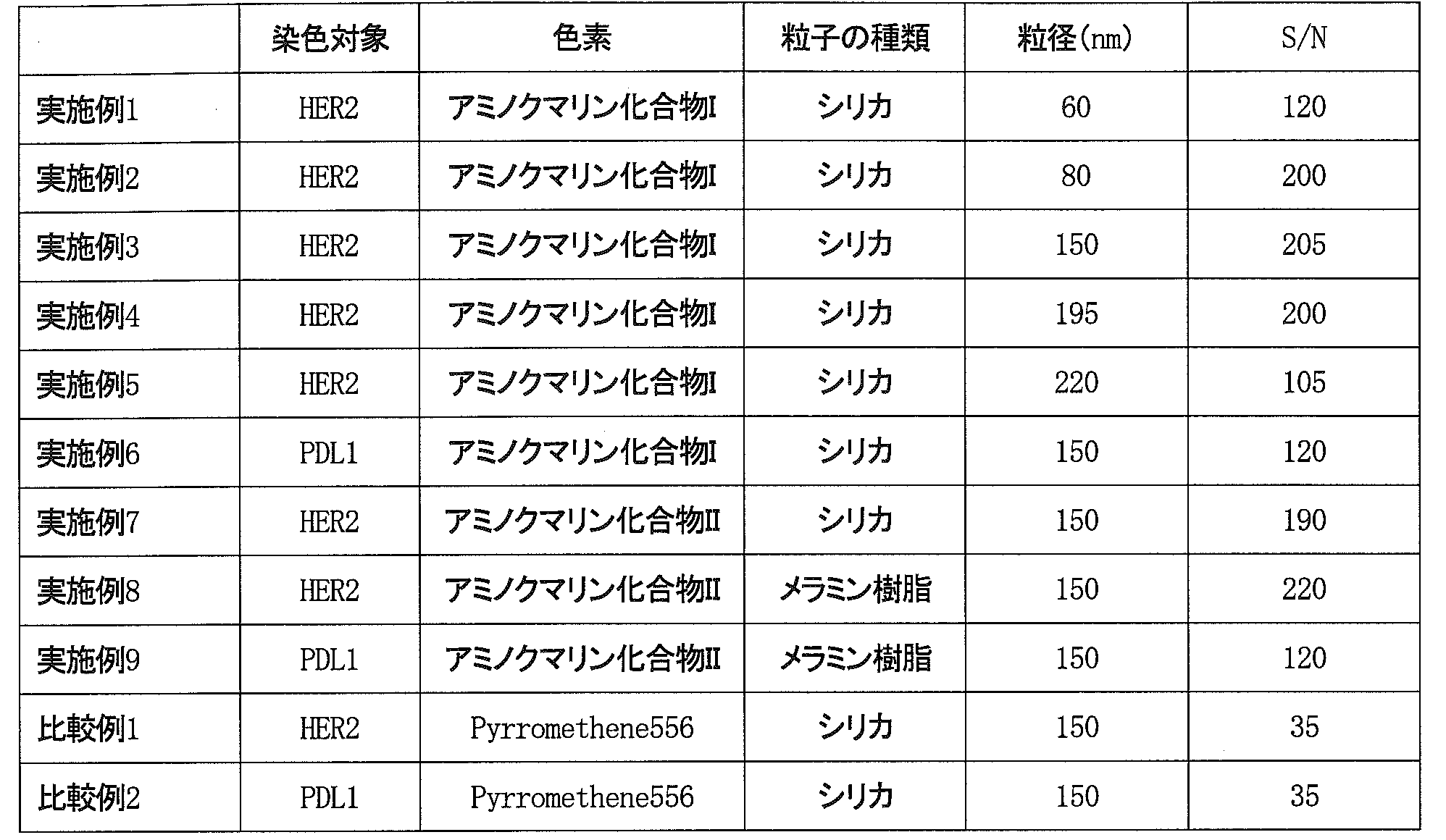
表1より、式(1)または(2)で示される構造を有するアミノクマリン化合物I~IIIを内包したアミノクマリン化合物内包粒子を用いて免疫染色を行うと、式(1)または(2)で示される構造を有するアミノクマリン化合物以外の色素であるPyrromethene556を内包した色素内包粒子を用いた場合に比較して、S/Nが向上することが確認された。
[実施例10]
下記の方法により緑色および赤色の多重免疫染色を行った。
(色素内包粒子の修飾)
アミノクマリン化合物内包樹脂粒子VIIの末端にNHS-PEG(polyethylene glycol)-マレイミド試薬を用いてマレイミドを導入し、これにチオール化した抗HER2抗体を結合させ、抗HER2抗体結合アミノクマリン化合物内包樹脂粒子を作製した。
上記と同様に、色素内包粒子iiiの末端にマレイミドを導入し、これにチオール化した抗Ki67抗体を結合させ、抗Ki67抗体結合色素内包粒子を作製した。
下記の方法により緑色および赤色の多重免疫染色を行った。
(色素内包粒子の修飾)
アミノクマリン化合物内包樹脂粒子VIIの末端にNHS-PEG(polyethylene glycol)-マレイミド試薬を用いてマレイミドを導入し、これにチオール化した抗HER2抗体を結合させ、抗HER2抗体結合アミノクマリン化合物内包樹脂粒子を作製した。
上記と同様に、色素内包粒子iiiの末端にマレイミドを導入し、これにチオール化した抗Ki67抗体を結合させ、抗Ki67抗体結合色素内包粒子を作製した。
(組織標本の免疫染色)
下記工程(1)~(13)によりヒト乳房組織標本の免疫染色(IHC法)を行った。
工程(1):キシレンを入れた容器に組織標本を15分浸漬させた。途中2回キシレンを交換した。
工程(2):エタノールを入れた容器に組織標本を10分浸漬させた。途中2回エタノールを交換した。
工程(3):水を入れた容器に組織標本を10分浸漬させた。
工程(4):10mMクエン酸緩衝液(pH6.0)に組織標本を浸漬させた。
工程(5):121℃で5分間オートクレーブ処理を行った。
工程(6):PBSを入れた容器に、オートクレーブ処理後の組織標本を15分浸漬させた。途中3回PBSを交換した。
工程(7):1%BSA含有PBSを組織標本に載せて、1時間放置した。
工程(8):1%BSA含有PBSで0.1nMに調整した抗HER2抗体結合アミノクマリン化合物内包樹脂粒子を組織標本に載せて一晩放置し、HER2を標識した。
工程(9):PBSを入れた容器に標識後の組織標本を15分浸漬させた。
工程(10):1%BSA含有PBSで0.1nMに調整した抗Ki67抗体結合色素内包粒子を、組織標本に載せて一晩放置し、Ki67を標識した。
工程(11):PBSを入れた容器に標識後の組織標本を30分浸漬させた。
工程(12):組織標本を4%中性パラホルムアルデヒド溶液で10分間固定処理した後、HE染色を行った。
工程(13):Merck社製Aquatexを滴下後、カバーガラスを載せ、組織標本を封入した。
下記工程(1)~(13)によりヒト乳房組織標本の免疫染色(IHC法)を行った。
工程(1):キシレンを入れた容器に組織標本を15分浸漬させた。途中2回キシレンを交換した。
工程(2):エタノールを入れた容器に組織標本を10分浸漬させた。途中2回エタノールを交換した。
工程(3):水を入れた容器に組織標本を10分浸漬させた。
工程(4):10mMクエン酸緩衝液(pH6.0)に組織標本を浸漬させた。
工程(5):121℃で5分間オートクレーブ処理を行った。
工程(6):PBSを入れた容器に、オートクレーブ処理後の組織標本を15分浸漬させた。途中3回PBSを交換した。
工程(7):1%BSA含有PBSを組織標本に載せて、1時間放置した。
工程(8):1%BSA含有PBSで0.1nMに調整した抗HER2抗体結合アミノクマリン化合物内包樹脂粒子を組織標本に載せて一晩放置し、HER2を標識した。
工程(9):PBSを入れた容器に標識後の組織標本を15分浸漬させた。
工程(10):1%BSA含有PBSで0.1nMに調整した抗Ki67抗体結合色素内包粒子を、組織標本に載せて一晩放置し、Ki67を標識した。
工程(11):PBSを入れた容器に標識後の組織標本を30分浸漬させた。
工程(12):組織標本を4%中性パラホルムアルデヒド溶液で10分間固定処理した後、HE染色を行った。
工程(13):Merck社製Aquatexを滴下後、カバーガラスを載せ、組織標本を封入した。
(顕微鏡観察)
蛍光顕微鏡としてCarl Zeiss社製蛍光顕微鏡を、フィルターセットとしてSemrock製フィルターセットを使用した。フィルターセットは免疫染色剤(緑色用および赤色用)に対応する下記2種類を使用した。
蛍光顕微鏡としてCarl Zeiss社製蛍光顕微鏡を、フィルターセットとしてSemrock製フィルターセットを使用した。フィルターセットは免疫染色剤(緑色用および赤色用)に対応する下記2種類を使用した。
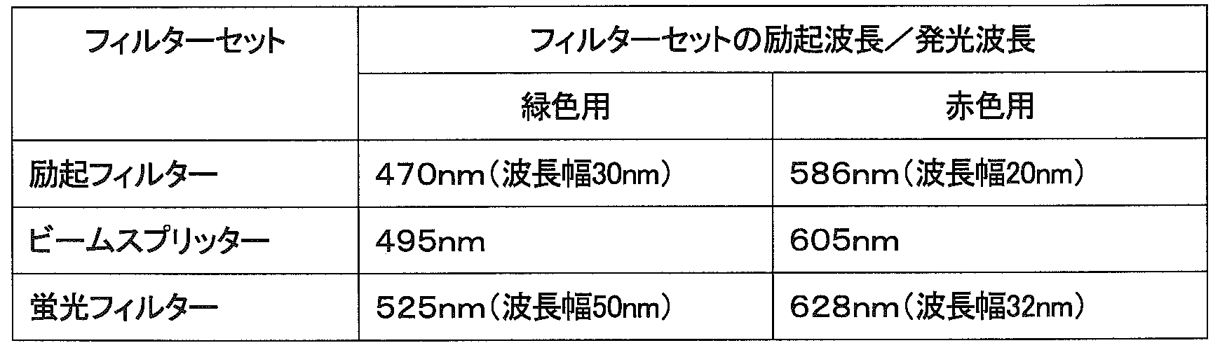
免疫染色後の組織標本をステージに設置し、緑色用および赤色用の2種類のフィルターセットを切り替えながら、フィルターセットを切り替えるごとに、組織標本の蛍光像の蛍光輝点数を計測した。結果を表3に示す。
[実施例11、12]
実施例11および12においては、アミノクマリン化合物内包樹脂粒子VIIの代わりにアミノクマリン化合物内包粒子IXおよびアミノクマリン化合物内包粒子VIIIをそれぞれ使用したこと以外は実施例10と同様の方法により多重免疫染色を行った。結果を表3に示す。
実施例11および12においては、アミノクマリン化合物内包樹脂粒子VIIの代わりにアミノクマリン化合物内包粒子IXおよびアミノクマリン化合物内包粒子VIIIをそれぞれ使用したこと以外は実施例10と同様の方法により多重免疫染色を行った。結果を表3に示す。
[比較例3]
アミノクマリン化合物内包樹脂粒子VIIの代わりに色素内包粒子iiを使用したこと以外は実施例10と同様の方法により多重免疫染色を行った。結果を表3に示す。
アミノクマリン化合物内包樹脂粒子VIIの代わりに色素内包粒子iiを使用したこと以外は実施例10と同様の方法により多重免疫染色を行った。結果を表3に示す。
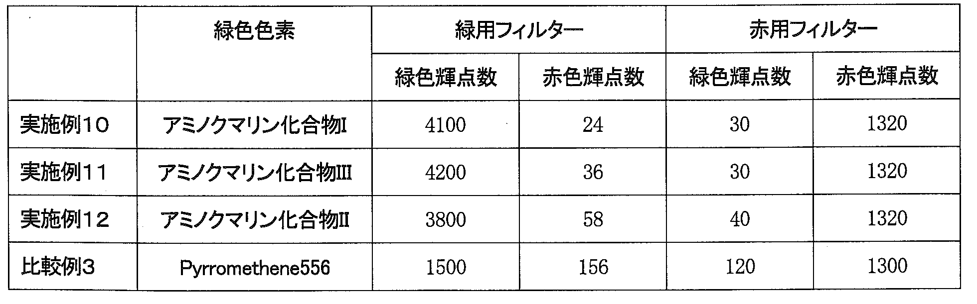
表3より、HER2およびKi67の二重染色の結果、式(1)または(2)で示される構造を有するアミノクマリン化合物I~IIIを内包したアミノクマリン化合物内包粒子を用いて免疫染色を行うと、式(1)または(2)で示される構造を有するアミノクマリン化合物以外の色素であるPyrromethene556を内包した色素内包粒子を用いた場合に比較して、緑色輝点の赤色輝点への漏れ込みが少ないことが確認された。
さらに、式(1)または(2)で示される構造を有するアミノクマリン化合物I~IIIを内包したアミノクマリン化合物内包粒子を用いて免疫染色を行った場合、二重染色の赤輝点数への影響はほとんどないことが表3から確認された。
[実施例13]
下記の方法により緑色および赤色の多重免疫染色を行った。
(色素内包粒子の修飾)
アミノクマリン化合物内包粒子VIIIの末端にNHS-PEG(polyethylene glycol)-マレイミド試薬を用いてマレイミドを導入し、これにチオール化した抗CTLA4抗体を結合させ、抗CTLA4抗体結合アミノクマリン化合物内包粒子を作製した。
上記と同様に、色素内包粒子iiiの末端にマレイミドを導入し、これにチオール化した抗PDL1抗体を結合させ、抗CTLA4抗体結合色素内包粒子を作製した。
下記の方法により緑色および赤色の多重免疫染色を行った。
(色素内包粒子の修飾)
アミノクマリン化合物内包粒子VIIIの末端にNHS-PEG(polyethylene glycol)-マレイミド試薬を用いてマレイミドを導入し、これにチオール化した抗CTLA4抗体を結合させ、抗CTLA4抗体結合アミノクマリン化合物内包粒子を作製した。
上記と同様に、色素内包粒子iiiの末端にマレイミドを導入し、これにチオール化した抗PDL1抗体を結合させ、抗CTLA4抗体結合色素内包粒子を作製した。
(組織標本の免疫染色)
下記工程(1)~(13)によりヒト乳房組織標本の免疫染色(IHC法)を行った。
工程(1):キシレンを入れた容器に組織標本を15分浸漬させた。途中2回キシレンを交換した。
工程(2):エタノールを入れた容器に組織標本を10分浸漬させた。途中2回エタノールを交換した。
工程(3):水を入れた容器に組織標本を10分浸漬させた。
工程(4):10mMクエン酸緩衝液(pH6.0)に組織標本を浸漬させた。
工程(5):121℃で5分間オートクレーブ処理を行った。
工程(6):PBSを入れた容器に、オートクレーブ処理後の組織標本を15分浸漬させた。途中3回PBSを交換した。
工程(7):1%BSA含有PBSを組織標本に載せて、1時間放置した。
工程(8):1%BSA含有PBSで0.1nMに調整した抗CTLA4抗体結合アミノクマリン化合物内包粒子を組織標本に載せて一晩放置し、CTLA4を標識した。
工程(9):PBSを入れた容器に標識後の組織標本を15分浸漬させた。
工程(10):1%BSA含有PBSで0.1nMに調整した抗CTLA4抗体結合色素内包粒子を、組織標本に載せて一晩放置し、PDL1を標識した。
工程(11):PBSを入れた容器に標識後の組織標本を30分浸漬させた。
工程(12):組織標本を4%中性パラホルムアルデヒド溶液で10分間固定処理した後、HE染色を行った。
工程(13):Merck社製Aquatexを滴下後、カバーガラスを載せ、組織標本を封入した。
(顕微鏡観察)
実施例10と同様の方法で顕微鏡観察を行った。結果を表4に示す。
下記工程(1)~(13)によりヒト乳房組織標本の免疫染色(IHC法)を行った。
工程(1):キシレンを入れた容器に組織標本を15分浸漬させた。途中2回キシレンを交換した。
工程(2):エタノールを入れた容器に組織標本を10分浸漬させた。途中2回エタノールを交換した。
工程(3):水を入れた容器に組織標本を10分浸漬させた。
工程(4):10mMクエン酸緩衝液(pH6.0)に組織標本を浸漬させた。
工程(5):121℃で5分間オートクレーブ処理を行った。
工程(6):PBSを入れた容器に、オートクレーブ処理後の組織標本を15分浸漬させた。途中3回PBSを交換した。
工程(7):1%BSA含有PBSを組織標本に載せて、1時間放置した。
工程(8):1%BSA含有PBSで0.1nMに調整した抗CTLA4抗体結合アミノクマリン化合物内包粒子を組織標本に載せて一晩放置し、CTLA4を標識した。
工程(9):PBSを入れた容器に標識後の組織標本を15分浸漬させた。
工程(10):1%BSA含有PBSで0.1nMに調整した抗CTLA4抗体結合色素内包粒子を、組織標本に載せて一晩放置し、PDL1を標識した。
工程(11):PBSを入れた容器に標識後の組織標本を30分浸漬させた。
工程(12):組織標本を4%中性パラホルムアルデヒド溶液で10分間固定処理した後、HE染色を行った。
工程(13):Merck社製Aquatexを滴下後、カバーガラスを載せ、組織標本を封入した。
(顕微鏡観察)
実施例10と同様の方法で顕微鏡観察を行った。結果を表4に示す。
[実施例14]
下記の方法により緑色および赤色の多重免疫染色を行った。
(色素内包粒子の修飾)
アミノクマリン化合物内包粒子VIIIの末端にNHS-PEG(polyethylene glycol)-マレイミド試薬を用いてマレイミドを導入し、これにチオール化した抗CD8抗体(Dako社製「抗CD8マウスモノクロナール抗体(C8/144B)」)を結合させ、抗CD8抗体結合アミノクマリン化合物内包粒子を作製した。
上記と同様に、色素内包粒子iiiの末端にマレイミドを導入し、これにチオール化した抗PDL1抗体を結合させ、抗PDL1抗体結合色素内包粒子を作製した。
下記の方法により緑色および赤色の多重免疫染色を行った。
(色素内包粒子の修飾)
アミノクマリン化合物内包粒子VIIIの末端にNHS-PEG(polyethylene glycol)-マレイミド試薬を用いてマレイミドを導入し、これにチオール化した抗CD8抗体(Dako社製「抗CD8マウスモノクロナール抗体(C8/144B)」)を結合させ、抗CD8抗体結合アミノクマリン化合物内包粒子を作製した。
上記と同様に、色素内包粒子iiiの末端にマレイミドを導入し、これにチオール化した抗PDL1抗体を結合させ、抗PDL1抗体結合色素内包粒子を作製した。
(組織標本の免疫染色)
下記工程(1)~(13)によりヒト乳房組織標本の免疫染色(IHC法)を行った。
工程(1):キシレンを入れた容器に組織標本を15分浸漬させた。途中2回キシレンを交換した。
工程(2):エタノールを入れた容器に組織標本を10分浸漬させた。途中2回エタノールを交換した。
工程(3):水を入れた容器に組織標本を10分浸漬させた。
工程(4):10mMクエン酸緩衝液(pH6.0)に組織標本を浸漬させた。
工程(5):121℃で5分間オートクレーブ処理を行った。
工程(6):PBSを入れた容器に、オートクレーブ処理後の組織標本を15分浸漬させた。途中3回PBSを交換した。
工程(7):1%BSA含有PBSを組織標本に載せて、1時間放置した。
工程(8):1%BSA含有PBSで0.1nMに調整した抗CD8抗体結合アミノクマリン化合物内包粒子を組織標本に載せて一晩放置し、CD8を標識した。
工程(9):PBSを入れた容器に標識後の組織標本を15分浸漬させた。
工程(10):1%BSA含有PBSで0.1nMに調整した抗PDL1抗体結合色素内包粒子を組織標本に載せて一晩放置し、PDL1を標識した。
工程(11):PBSを入れた容器に標識後の組織標本を30分浸漬させた。
工程(12):組織標本を4%中性パラホルムアルデヒド溶液で10分間固定処理した後、HE染色を行った。
工程(13):Merck社製Aquatexを滴下後、カバーガラスを載せ、組織標本を封入した。
(顕微鏡観察)
実施例10と同様の方法で顕微鏡観察を行った。結果を表4に示す。
下記工程(1)~(13)によりヒト乳房組織標本の免疫染色(IHC法)を行った。
工程(1):キシレンを入れた容器に組織標本を15分浸漬させた。途中2回キシレンを交換した。
工程(2):エタノールを入れた容器に組織標本を10分浸漬させた。途中2回エタノールを交換した。
工程(3):水を入れた容器に組織標本を10分浸漬させた。
工程(4):10mMクエン酸緩衝液(pH6.0)に組織標本を浸漬させた。
工程(5):121℃で5分間オートクレーブ処理を行った。
工程(6):PBSを入れた容器に、オートクレーブ処理後の組織標本を15分浸漬させた。途中3回PBSを交換した。
工程(7):1%BSA含有PBSを組織標本に載せて、1時間放置した。
工程(8):1%BSA含有PBSで0.1nMに調整した抗CD8抗体結合アミノクマリン化合物内包粒子を組織標本に載せて一晩放置し、CD8を標識した。
工程(9):PBSを入れた容器に標識後の組織標本を15分浸漬させた。
工程(10):1%BSA含有PBSで0.1nMに調整した抗PDL1抗体結合色素内包粒子を組織標本に載せて一晩放置し、PDL1を標識した。
工程(11):PBSを入れた容器に標識後の組織標本を30分浸漬させた。
工程(12):組織標本を4%中性パラホルムアルデヒド溶液で10分間固定処理した後、HE染色を行った。
工程(13):Merck社製Aquatexを滴下後、カバーガラスを載せ、組織標本を封入した。
(顕微鏡観察)
実施例10と同様の方法で顕微鏡観察を行った。結果を表4に示す。
[実施例15]
下記の方法により緑色および赤色の多重免疫染色を行った。
(色素内包粒子の修飾)
アミノクマリン化合物内包粒子VIIIの末端にNHS-PEG(polyethylene glycol)-マレイミド試薬を用いてマレイミドを導入し、これにチオール化した抗CD30抗体(Dako社製「抗CD30マウスモノクロナール抗体(BerH2))を結合させ、抗CD30抗体結合アミノクマリン化合物内包粒子を作製した。
上記と同様に、色素内包粒子iiiの末端にマレイミドを導入し、これにチオール化した抗PDL1抗体を結合させ、抗PDL1抗体結合色素内包粒子を作製した。
下記の方法により緑色および赤色の多重免疫染色を行った。
(色素内包粒子の修飾)
アミノクマリン化合物内包粒子VIIIの末端にNHS-PEG(polyethylene glycol)-マレイミド試薬を用いてマレイミドを導入し、これにチオール化した抗CD30抗体(Dako社製「抗CD30マウスモノクロナール抗体(BerH2))を結合させ、抗CD30抗体結合アミノクマリン化合物内包粒子を作製した。
上記と同様に、色素内包粒子iiiの末端にマレイミドを導入し、これにチオール化した抗PDL1抗体を結合させ、抗PDL1抗体結合色素内包粒子を作製した。
(組織標本の免疫染色)
下記工程(1)~(13)によりヒト乳房組織標本の免疫染色(IHC法)を行った。
工程(1):キシレンを入れた容器に組織標本を15分浸漬させた。途中2回キシレンを交換した。
工程(2):エタノールを入れた容器に組織標本を10分浸漬させた。途中2回エタノールを交換した。
工程(3):水を入れた容器に組織標本を10分浸漬させた。
工程(4):10mMクエン酸緩衝液(pH6.0)に組織標本を浸漬させた。
工程(5):121℃で5分間オートクレーブ処理を行った。
工程(6):PBSを入れた容器に、オートクレーブ処理後の組織標本を15分浸漬させた。途中3回PBSを交換した。
工程(7):1%BSA含有PBSを組織標本に載せて、1時間放置した。
工程(8):1%BSA含有PBSで0.1nMに調整した抗CD30抗体結合アミノクマリン化合物内包粒子を組織標本に載せて一晩放置し、CD30を標識した。
工程(9):PBSを入れた容器に標識後の組織標本を15分浸漬させた。
工程(10):1%BSA含有PBSで0.1nMに調整した抗PDL1抗体結合色素内包粒子を組織標本に載せて一晩放置し、PDL1を標識した。
工程(11):PBSを入れた容器に標識後の組織標本を30分浸漬させた。
工程(12):組織標本を4%中性パラホルムアルデヒド溶液で10分間固定処理した後、HE染色を行った。
工程(13):Merck社製Aquatexを滴下後、カバーガラスを載せ、組織標本を封入した。
(顕微鏡観察)
実施例10と同様の方法で顕微鏡観察を行った。結果を表4に示す。
下記工程(1)~(13)によりヒト乳房組織標本の免疫染色(IHC法)を行った。
工程(1):キシレンを入れた容器に組織標本を15分浸漬させた。途中2回キシレンを交換した。
工程(2):エタノールを入れた容器に組織標本を10分浸漬させた。途中2回エタノールを交換した。
工程(3):水を入れた容器に組織標本を10分浸漬させた。
工程(4):10mMクエン酸緩衝液(pH6.0)に組織標本を浸漬させた。
工程(5):121℃で5分間オートクレーブ処理を行った。
工程(6):PBSを入れた容器に、オートクレーブ処理後の組織標本を15分浸漬させた。途中3回PBSを交換した。
工程(7):1%BSA含有PBSを組織標本に載せて、1時間放置した。
工程(8):1%BSA含有PBSで0.1nMに調整した抗CD30抗体結合アミノクマリン化合物内包粒子を組織標本に載せて一晩放置し、CD30を標識した。
工程(9):PBSを入れた容器に標識後の組織標本を15分浸漬させた。
工程(10):1%BSA含有PBSで0.1nMに調整した抗PDL1抗体結合色素内包粒子を組織標本に載せて一晩放置し、PDL1を標識した。
工程(11):PBSを入れた容器に標識後の組織標本を30分浸漬させた。
工程(12):組織標本を4%中性パラホルムアルデヒド溶液で10分間固定処理した後、HE染色を行った。
工程(13):Merck社製Aquatexを滴下後、カバーガラスを載せ、組織標本を封入した。
(顕微鏡観察)
実施例10と同様の方法で顕微鏡観察を行った。結果を表4に示す。
[比較例4]
アミノクマリン化合物内包粒子VIIIの代わりに色素内包粒子iiを使用したこと以外は実施例13と同様の方法により緑色および赤色の多重免疫染色を行った。結果を表4に示す。
アミノクマリン化合物内包粒子VIIIの代わりに色素内包粒子iiを使用したこと以外は実施例13と同様の方法により緑色および赤色の多重免疫染色を行った。結果を表4に示す。
[比較例5]
アミノクマリン化合物内包粒子VIIIの代わりに色素内包粒子iiを使用したこと以外は実施例14と同様の方法により緑色および赤色の多重免疫染色を行った。結果を表4に示す。
アミノクマリン化合物内包粒子VIIIの代わりに色素内包粒子iiを使用したこと以外は実施例14と同様の方法により緑色および赤色の多重免疫染色を行った。結果を表4に示す。
[比較例6]
アミノクマリン化合物内包粒子VIIIの代わりに色素内包粒子iiを使用したこと以外は実施例15と同様の方法により緑色および赤色の多重免疫染色を行った。結果を表4に示す。
アミノクマリン化合物内包粒子VIIIの代わりに色素内包粒子iiを使用したこと以外は実施例15と同様の方法により緑色および赤色の多重免疫染色を行った。結果を表4に示す。
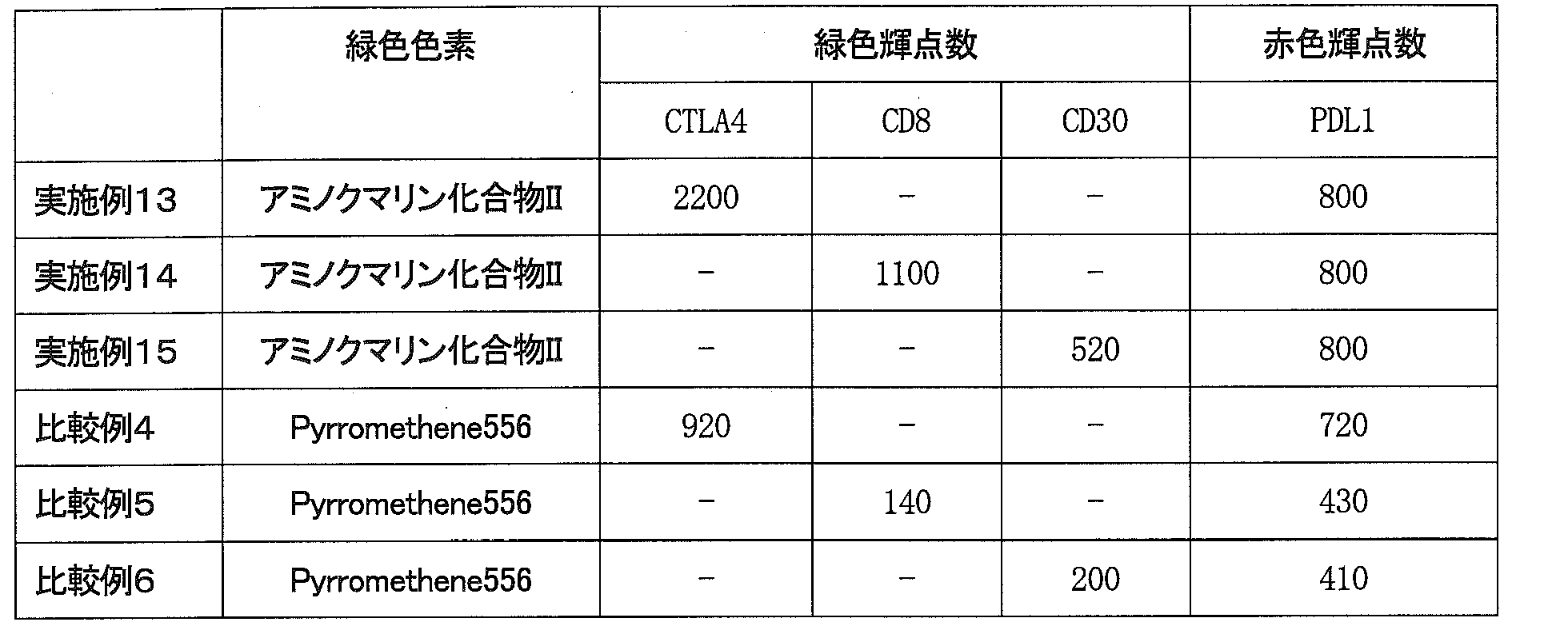
表4より、PDL1とCTLA4、CD8またはCD30との二重染色の結果、式(2)で示される構造を有するアミノクマリン化合物IIを内包したアミノクマリン化合物内包粒子を用いて免疫染色を行うと、式(1)または(2)で示される構造を有するアミノクマリン化合物以外の色素であるPyrromethene556を内包した色素内包粒子を用いた場合に比較して、緑色輝点の赤色輝点への漏れ込みが少ないことが確認された。
さらに、式(2)で示される構造を有するアミノクマリン化合物IIを内包したアミノクマリン化合物内包粒子を用いて免疫染色を行った場合、HER2およびKi67の二重染色の場合と同様に、二重染色の赤輝点数への影響はほとんどないことが表4から確認された。
Claims (5)
- 下記式(1)または(2)で示される構造を有するアミノクマリン化合物またはその塩を母体粒子に内包してなるアミノクマリン化合物内包粒子を用いて標識を行う蛍光標識法。

- 前記アミノクマリン化合物内包粒子の平均粒径が80~200nmである請求項1に記載の蛍光標識法。
- 前記アミノクマリン化合物内包粒子を用いた標識を含む多重標識を行う請求項1または2に記載の蛍光標識法。
- 免疫染色法またはFISHである請求項1~3のいずれかに記載の蛍光標識法。
- 前記免疫染色法は、PDL1、CTLA4、CD8、CD30、CD48、CD59、IDO、 TDO、CSF-1R、HDAC、CXCR4、FLT-3、TIGIT、INF-α、INF-β、INF-ω、INF-ε、INF-κ、INF-γ、INF-λ CSF、EPO、EGF、FGF、PDGF、HGF、TGF、CD3、CD4、CD25、CD28、CD80、CD86、CD160、CD57、OX40、OX40L、ICOS、ICOSL、CD155、CD226、CD112、CD27、CD70、4-1BB、4-1BBL、GITR、GITRL、BTLA、HVEM、TIM-3、Galectin-9、LAG-3、B7-H3、B7-H4、B7-H5、CD40、CD40L、PD-1、PD-L2、2B4、KLRG-1、E-Cadherin、N-Cadherin、R-Cadherin、CD68、CD163およびCSF1-Rから選択される少なくとも2つの染色対象タンパク質に対してそれぞれ異なる色素を用いて多重染色を行い、前記染色対象タンパク質の少なくとも1つを、前記アミノクマリン化合物内包粒子を用いて染色する請求項4に記載の蛍光標識法。
Priority Applications (1)
| Application Number | Priority Date | Filing Date | Title |
|---|---|---|---|
| JP2018568515A JP7095603B2 (ja) | 2017-02-14 | 2018-02-13 | 蛍光標識法 |
Applications Claiming Priority (2)
| Application Number | Priority Date | Filing Date | Title |
|---|---|---|---|
| JP2017-024875 | 2017-02-14 | ||
| JP2017024875 | 2017-02-14 |
Publications (1)
| Publication Number | Publication Date |
|---|---|
| WO2018151071A1 true WO2018151071A1 (ja) | 2018-08-23 |
Family
ID=63170319
Family Applications (1)
| Application Number | Title | Priority Date | Filing Date |
|---|---|---|---|
| PCT/JP2018/004802 Ceased WO2018151071A1 (ja) | 2017-02-14 | 2018-02-13 | 蛍光標識法 |
Country Status (2)
| Country | Link |
|---|---|
| JP (1) | JP7095603B2 (ja) |
| WO (1) | WO2018151071A1 (ja) |
Cited By (1)
| Publication number | Priority date | Publication date | Assignee | Title |
|---|---|---|---|---|
| JP2019172968A (ja) * | 2018-03-28 | 2019-10-10 | 日本化薬株式会社 | クマリン化合物、及びこれを含んだ顔料組成物 |
Citations (8)
| Publication number | Priority date | Publication date | Assignee | Title |
|---|---|---|---|---|
| JPH069892A (ja) * | 1992-06-22 | 1994-01-18 | Nippon Kanko Shikiso Kenkyusho:Kk | クマリン誘導体 |
| JPH06271599A (ja) * | 1993-01-29 | 1994-09-27 | Bayer Ag | スルホクマリン−含有ヌクレオチド及び核酸検出法におけるそれらの利用 |
| JPH07508309A (ja) * | 1992-05-13 | 1995-09-14 | モレキュラー・プロウブズ・インコーポレーテッド | 制御可能な強化ストークスシフトを有する蛍光微小粒子 |
| JP2004309458A (ja) * | 2003-03-27 | 2004-11-04 | Institute Of Physical & Chemical Research | 時間分解蛍光顕微鏡 |
| JP2006045314A (ja) * | 2004-08-03 | 2006-02-16 | Fuji Electric Holdings Co Ltd | 蛍光性陰イオンおよび蛍光性陽イオンからなる塩およびそれを用いた色変換膜 |
| WO2015045961A1 (ja) * | 2013-09-26 | 2015-04-02 | コニカミノルタ株式会社 | 蛍光ナノ粒子標識体、多重免疫染色剤キットおよび多重免疫染色法 |
| JP2015093878A (ja) * | 2013-11-08 | 2015-05-18 | コニカミノルタ株式会社 | 蛍光色素内包樹脂粒子、該蛍光色素内包樹脂粒子を含む組織多重染色用蛍光色素内包樹脂粒子セット、及び該蛍光色素内包樹脂粒子を用いた組織多重染色法 |
| WO2016129444A1 (ja) * | 2015-02-12 | 2016-08-18 | コニカミノルタ株式会社 | 抗体結合蛍光体集積ナノ粒子、抗体結合蛍光体集積ナノ粒子の製造方法および免疫染色キット |
Family Cites Families (2)
| Publication number | Priority date | Publication date | Assignee | Title |
|---|---|---|---|---|
| TWI551597B (zh) | 2012-03-29 | 2016-10-01 | 住友化學股份有限公司 | 染料用化合物 |
| JP6463005B2 (ja) | 2013-05-30 | 2019-01-30 | キヤノン株式会社 | マクロファージ識別剤、該マクロファージ識別剤を用いた識別方法、分取方法、評価方法、スクリーニング方法、およびキット |
-
2018
- 2018-02-13 WO PCT/JP2018/004802 patent/WO2018151071A1/ja not_active Ceased
- 2018-02-13 JP JP2018568515A patent/JP7095603B2/ja active Active
Patent Citations (8)
| Publication number | Priority date | Publication date | Assignee | Title |
|---|---|---|---|---|
| JPH07508309A (ja) * | 1992-05-13 | 1995-09-14 | モレキュラー・プロウブズ・インコーポレーテッド | 制御可能な強化ストークスシフトを有する蛍光微小粒子 |
| JPH069892A (ja) * | 1992-06-22 | 1994-01-18 | Nippon Kanko Shikiso Kenkyusho:Kk | クマリン誘導体 |
| JPH06271599A (ja) * | 1993-01-29 | 1994-09-27 | Bayer Ag | スルホクマリン−含有ヌクレオチド及び核酸検出法におけるそれらの利用 |
| JP2004309458A (ja) * | 2003-03-27 | 2004-11-04 | Institute Of Physical & Chemical Research | 時間分解蛍光顕微鏡 |
| JP2006045314A (ja) * | 2004-08-03 | 2006-02-16 | Fuji Electric Holdings Co Ltd | 蛍光性陰イオンおよび蛍光性陽イオンからなる塩およびそれを用いた色変換膜 |
| WO2015045961A1 (ja) * | 2013-09-26 | 2015-04-02 | コニカミノルタ株式会社 | 蛍光ナノ粒子標識体、多重免疫染色剤キットおよび多重免疫染色法 |
| JP2015093878A (ja) * | 2013-11-08 | 2015-05-18 | コニカミノルタ株式会社 | 蛍光色素内包樹脂粒子、該蛍光色素内包樹脂粒子を含む組織多重染色用蛍光色素内包樹脂粒子セット、及び該蛍光色素内包樹脂粒子を用いた組織多重染色法 |
| WO2016129444A1 (ja) * | 2015-02-12 | 2016-08-18 | コニカミノルタ株式会社 | 抗体結合蛍光体集積ナノ粒子、抗体結合蛍光体集積ナノ粒子の製造方法および免疫染色キット |
Cited By (2)
| Publication number | Priority date | Publication date | Assignee | Title |
|---|---|---|---|---|
| JP2019172968A (ja) * | 2018-03-28 | 2019-10-10 | 日本化薬株式会社 | クマリン化合物、及びこれを含んだ顔料組成物 |
| JP7261609B2 (ja) | 2018-03-28 | 2023-04-20 | 日本化薬株式会社 | クマリン化合物、及びこれを含んだ顔料組成物 |
Also Published As
| Publication number | Publication date |
|---|---|
| JP7095603B2 (ja) | 2022-07-05 |
| JPWO2018151071A1 (ja) | 2019-12-12 |
Similar Documents
| Publication | Publication Date | Title |
|---|---|---|
| US10551386B2 (en) | Biological substance detection method | |
| JP7147724B2 (ja) | 組織染色用染色剤、組織染色用染色剤の製造方法および組織染色用染色剤を含む組織染色用キット | |
| US10458983B2 (en) | Method for detecting biological material | |
| CN108473505A (zh) | 聚芴并[4,5-cde]氧杂环庚三烯偶联物及其在分析物检测方法中的用途 | |
| JPWO2019088266A1 (ja) | 凝集ナノ粒子および蛍光標識材 | |
| WO2018151071A1 (ja) | 蛍光標識法 | |
| JP7151489B2 (ja) | アミノクマリン化合物およびアミノクマリン化合物内包樹脂粒子 | |
| JP6187170B2 (ja) | 蛍光色素内包樹脂粒子、該蛍光色素内包樹脂粒子を含む組織多重染色用蛍光色素内包樹脂粒子セット、及び該蛍光色素内包樹脂粒子を用いた組織多重染色法 | |
| WO2023192653A1 (en) | Probes for fluorescence imaging | |
| WO2018150450A1 (ja) | アミノクマリン化合物およびアミノクマリン化合物内包樹脂粒子 | |
| JP6447679B2 (ja) | 蛍光色素内包樹脂粒子および該蛍光色素内包樹脂粒子を含む組織多重染色用蛍光色素内包樹脂粒子セット | |
| KR102066344B1 (ko) | 핵산표지를 위한 신규한 형광화합물 및 이의 제조방법 | |
| WO2021230134A1 (ja) | 画像形成方法 | |
| WO2023244963A2 (en) | Bicyclononyne reagents for cell imaging | |
| DIAZ et al. | Aminoacridine dyes and the secondary structure of DNA in situ | |
| US20190033181A1 (en) | Staining agent for staining tissue, production method for staining agent for staining tissue and tissue staining kit including staining agent for staining tissue |
Legal Events
| Date | Code | Title | Description |
|---|---|---|---|
| 121 | Ep: the epo has been informed by wipo that ep was designated in this application |
Ref document number: 18754494 Country of ref document: EP Kind code of ref document: A1 |
|
| ENP | Entry into the national phase |
Ref document number: 2018568515 Country of ref document: JP Kind code of ref document: A |
|
| NENP | Non-entry into the national phase |
Ref country code: DE |
|
| 122 | Ep: pct application non-entry in european phase |
Ref document number: 18754494 Country of ref document: EP Kind code of ref document: A1 |