WO2017010381A1 - N末端標識剤、これを用いた蛍光標識タンパク質及びその製造方法 - Google Patents
N末端標識剤、これを用いた蛍光標識タンパク質及びその製造方法 Download PDFInfo
- Publication number
- WO2017010381A1 WO2017010381A1 PCT/JP2016/070058 JP2016070058W WO2017010381A1 WO 2017010381 A1 WO2017010381 A1 WO 2017010381A1 JP 2016070058 W JP2016070058 W JP 2016070058W WO 2017010381 A1 WO2017010381 A1 WO 2017010381A1
- Authority
- WO
- WIPO (PCT)
- Prior art keywords
- group
- protein
- fluorescent
- antibody
- labeling agent
- Prior art date
- Legal status (The legal status is an assumption and is not a legal conclusion. Google has not performed a legal analysis and makes no representation as to the accuracy of the status listed.)
- Ceased
Links
Images



Classifications
-
- C—CHEMISTRY; METALLURGY
- C07—ORGANIC CHEMISTRY
- C07D—HETEROCYCLIC COMPOUNDS
- C07D311/00—Heterocyclic compounds containing six-membered rings having one oxygen atom as the only hetero atom, condensed with other rings
- C07D311/02—Heterocyclic compounds containing six-membered rings having one oxygen atom as the only hetero atom, condensed with other rings ortho- or peri-condensed with carbocyclic rings or ring systems
- C07D311/78—Ring systems having three or more relevant rings
- C07D311/80—Dibenzopyrans; Hydrogenated dibenzopyrans
-
- C—CHEMISTRY; METALLURGY
- C07—ORGANIC CHEMISTRY
- C07K—PEPTIDES
- C07K1/00—General methods for the preparation of peptides, i.e. processes for the organic chemical preparation of peptides or proteins of any length
- C07K1/13—Labelling of peptides
-
- G—PHYSICS
- G01—MEASURING; TESTING
- G01N—INVESTIGATING OR ANALYSING MATERIALS BY DETERMINING THEIR CHEMICAL OR PHYSICAL PROPERTIES
- G01N33/00—Investigating or analysing materials by specific methods not covered by groups G01N1/00 - G01N31/00
- G01N33/48—Biological material, e.g. blood, urine; Haemocytometers
- G01N33/50—Chemical analysis of biological material, e.g. blood, urine; Testing involving biospecific ligand binding methods; Immunological testing
- G01N33/53—Immunoassay; Biospecific binding assay; Materials therefor
- G01N33/531—Production of immunochemical test materials
- G01N33/532—Production of labelled immunochemicals
- G01N33/533—Production of labelled immunochemicals with fluorescent label
-
- G—PHYSICS
- G01—MEASURING; TESTING
- G01N—INVESTIGATING OR ANALYSING MATERIALS BY DETERMINING THEIR CHEMICAL OR PHYSICAL PROPERTIES
- G01N33/00—Investigating or analysing materials by specific methods not covered by groups G01N1/00 - G01N31/00
- G01N33/48—Biological material, e.g. blood, urine; Haemocytometers
- G01N33/50—Chemical analysis of biological material, e.g. blood, urine; Testing involving biospecific ligand binding methods; Immunological testing
- G01N33/53—Immunoassay; Biospecific binding assay; Materials therefor
- G01N33/536—Immunoassay; Biospecific binding assay; Materials therefor with immune complex formed in liquid phase
- G01N33/542—Immunoassay; Biospecific binding assay; Materials therefor with immune complex formed in liquid phase with steric inhibition or signal modification, e.g. fluorescent quenching
-
- G—PHYSICS
- G01—MEASURING; TESTING
- G01N—INVESTIGATING OR ANALYSING MATERIALS BY DETERMINING THEIR CHEMICAL OR PHYSICAL PROPERTIES
- G01N33/00—Investigating or analysing materials by specific methods not covered by groups G01N1/00 - G01N31/00
- G01N33/48—Biological material, e.g. blood, urine; Haemocytometers
- G01N33/50—Chemical analysis of biological material, e.g. blood, urine; Testing involving biospecific ligand binding methods; Immunological testing
- G01N33/58—Chemical analysis of biological material, e.g. blood, urine; Testing involving biospecific ligand binding methods; Immunological testing involving labelled substances
Definitions
- the present invention relates to an N-terminal labeling agent for simply labeling a protein, a fluorescent-labeled protein labeled with such an N-terminal labeling agent, and a method for producing the same, and a fluorescent-labeled antibody used in a fluorescence immunoassay and the like is prepared.
- an N-terminal labeling agent for simply labeling a protein
- a fluorescent-labeled protein labeled with such an N-terminal labeling agent and a method for producing the same
- a fluorescent-labeled antibody used in a fluorescence immunoassay and the like is prepared. Useful as technology.
- a fluorescently labeled antibody is quenched in a state where no antigen is bound (in this specification, it may be referred to as quenching or quenching), whereas when the antigen is bound, the quenching is performed.
- quenching or quenching There is known a “homogeneous fluorescence immunoassay method” using a phenomenon in which is canceled (for example, see Patent Document 1 below). This method is carried out by mixing a fluorescently labeled antibody fragment or a fluorescently labeled single-chain antibody with a test sample solution for examining whether or not an antigen is contained.
- the presence of the antigen is concluded when the fluorescence intensity of the fluorescent dye and the antigen concentration are positively correlated, and the concentration of the antigen can be measured based on the degree of correlation.
- the main cause of such quenching and its release is that the highly conserved tryptophan residue in the antibody molecule interacts with the fluorescent dye to quench the fluorescent dye and bind to the antigen in an antigen-dependent manner. Is to be resolved.
- VL the antibody light chain variable region
- VH the antibody heavy chain variable region
- a single chain antibody (abbreviated as “scFv”) formed by binding a fluorescently labeled VL polypeptide and a VH polypeptide. May be used).
- the above two types of fluorescently labeled antibodies may be collectively referred to as “Quenchbody (registered trademark, hereinafter the same)” or “Q-body (registered trademark, hereinafter the same)”.
- the former when two antibody fragments are used
- the latter when a single chain antibody is used
- scFv type Q-body May be called.
- Patent Document 1 The features of the method described in Patent Document 1 are mainly in the following three points.
- the first feature is that no cleaning process is required. Thus, since the measurement is completed simply by mixing a small amount of sample and Q-body and measuring the fluorescence intensity, this method can be said to be an extremely simple measurement technique.
- the second feature is that tryptophan having a high degree of preservation present in the antibody is used for quenching. That is, since the quenching effect can be obtained even if the type of antibody is changed, it can be said that this technique is excellent in versatility in that it can be applied to detection of various substances using various antibodies.
- the third feature is that only one antigenic site is required. For this reason, it can be applied to a low molecular compound in principle.
- this method is a simple measurement method that does not require a washing step. For this reason, it is possible to reduce the size of the measurement device based on this method to a portable size, for example, and by using this measurement device, ordinary people who have not received specialized education can make measurements on-site. It is assumed that this is possible.
- a Fab antibody Fram, antigen binding
- a fluorescent labeling complex has been developed in which the same or different fluorescent dyes are labeled on each of VL and VH (Patent Document 2).
- the quenching effect (H-dimer) between the dyes is added in addition to the quenching due to the interaction between the fluorescent dye and the tryptophan residue, which is high.
- Detection sensitivity can be obtained (FIG. 7, FIG. 8, etc. of Patent Document 2).
- fluorescent labeling complex in which each of VL and VH is labeled with a different color fluorescent dye, in addition to quenching effect due to interaction between the fluorescent dye and tryptophan residue and quenching effect between the dyes, a quenching effect due to the FRET effect is added.
- High detection sensitivity can be obtained (FIGS. 9 and 10 in Patent Document 2).
- Such fluorescently labeled Fab antibody complexes are sometimes collectively referred to as “UQ-body (registered trademark, the same applies hereinafter)”.
- Quenching when antigens and ligands are not bound, and releasing the quenching when bound is popular not only in the medical field but also in various fields such as environmental pesticide detection and toxin detection It is expected that.
- a technique for easily producing a fluorescently labeled protein such as Q-body or UQ-body using a protein containing an existing antibody or receptor.
- an object of the present invention is to provide a fluorescently labeled protein such as Q-body or UQ-body in a short time and in a short time using a protein containing an existing antibody or receptor.
- the present inventors have made it easy to use fluorescent dyes for proteins such as existing antibodies by binding a fluorescent group to an aldehyde group via a linker having a certain length. It was found that this can be used for fluorescent immunoassay and the like.
- the present invention has been completed by further studies based on such knowledge.
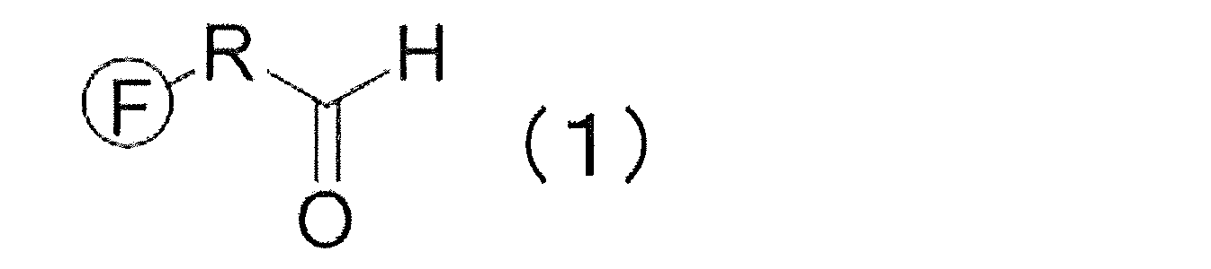
- the present invention [1] The following general formula (1): (In the formula, F is a fluorescent group, and R is a linear soft segment composed of a linear hydrocarbon having 5 or more carbon atoms, which may have a substituent, or a part of the main chain carbon. A linker group having a linear soft segment substituted with -O-, -N-, -S-, -CO-, -CONH-, -COO- or -POO-).
- the fluorescent group is a rhodamine type, coumarin type, oxazine type, carbopyronine type, cyanine type, pyromesene type, naphthalene type, biphenyl type, anthracene type, phenanthrene type, pyrene type, carbazole type, Cy type, EvoBlue type,
- a fluorescent group can be bound to the N-terminus in a short time using a protein such as an existing antibody in a short time. Since the existing protein can be used, it is not necessary to prepare a hybridoma or analyze a gene sequence, and a protein that can be used for a homogeneous fluorescent immunoassay can be easily and efficiently produced.
- N-terminal labeling agent The present invention provides the following general formula (1): (In the formula, F is a fluorescent group, and R is a linear soft segment composed of a linear hydrocarbon having 5 or more carbon atoms, which may have a substituent, or a part of the main chain carbon. A linker group having a linear soft segment substituted with -O-, -N-, -S-, -CO-, -CONH-, -COO- or -POO-). It is a protein N-terminal labeling agent.
- the N-terminal labeling agent in the present invention has an aldehyde group at the terminal, and is bonded to a fluorescent group represented by F via a linker group represented by R.
- the linker group may have a linear soft segment and other linking groups.
- the linear soft segment refers to a segment having a linear group in the main chain and having flexibility.
- a linear group is preferably a group in which the main chain rotation and flexibility are not easily inhibited.
- the straight chain hydrocarbon include an alkylene group, an alkenylene group, and an alkynylene group.
- the straight chain hydrocarbon has 5 or more carbon atoms, preferably 6 or more, more preferably 7 or more, still more preferably 8 or more, and even more preferably 9 or more.
- the above is more preferable, 11 or more is more preferable, 12 or more is particularly preferable, and 13 or more is most preferable.
- the carbon number in the straight chain hydrocarbon can be 100 or less, preferably 90 or less, more preferably 80 or less, and 70 or less. More preferably.
- the straight chain soft segment may have a part of the main chain carbon substituted with —O—, —N—, —S—, —CO—, —CONH—, —COO— or —POO—. This includes those having a repeating unit such as an alkylene group.
- the tryptophan residue and the fluorescent group interact to quench the fluorescent group.
- the protein is an antibody
- fluorescent groups can interact with tryptophan residues in the antigen binding pocket.
- the principle of quenching is not limited to the interaction between tryptophan residues and fluorescent groups, but the structure and length of linker groups can affect the interaction between tryptophan residues and fluorescent groups. is assumed. For example, if the linker group is too short or too long, the tryptophan residue and the fluorescent group cannot sufficiently interact.
- the linker group includes a plurality of cyclic structures, it is assumed that the rotation and mobility of the linker group are inhibited and the tryptophan residue and the fluorescent group cannot sufficiently interact.
- the linear soft segment made of a linear hydrocarbon having 5 or more carbon atoms may have a substituent.
- the substituent is not limited as long as the rotation and flexibility of the main chain are not hindered.
- a methyl group, an ethyl group, an n-propyl group, an i-propyl group, a butyl group are not limited.
- the linker group may contain another linking group as long as the function of the linear soft segment is not easily inhibited.
- another linking group for example, an arylene group, a heteroarylene group, C 4 -C 10 Selected from the group consisting of cycloalkyl groups and combinations thereof.
- the linker group may have a substituent, imino group, succinylamino group, acetamide group, 2-aminopentanamide, 2 (2 ′)-alkyl-aminoacetyl group, carbamoyl group, Aminoalkyl group, aminoalkylol group, glutaramide group, ornitino group, theopropanoyl group, N- (mercaptomethyl) propionamide group, 2- (1,2-dihydroxy-3-mercaptopropylthio) propanoyl group, It may contain a linking group such as aminoethanethiol group, mercaptopropanol group, (hydroxypropylthio) propanenoyl group, oxy group, succinyl group, acetyl group or oxopentanoyl group.
- a linking group such as aminoethanethiol group, mercaptopropanol group, (hydroxypropylthio) propanenoyl group, oxy
- the linker group may have a substituent, and may include a binding group such as an amino acid linker, a disulfide linker, a phosphate ester linker, or the like containing one or more amino acid repeating units. Good.
- the linker group includes an alkylene group
- it is not limited.
- a C 2 to C 20 alkylene group is preferable, and a C 3 to C 18 alkylene group is preferable. Is more preferable, and a C 4 to C 16 alkylene group is more preferable.
- the linker group includes a PEG group
- the number of repeating units of the PEG group is preferably 3 to 20, more preferably 4 to 18, more preferably 5 to 15 is more preferable, 6 to 12 is particularly preferable, and 2 to 10 is most preferable.
- the protein labeled with the N-terminal labeling agent is not particularly limited, but is preferably a protein that can bind to a target substance, and is not limited, but examples thereof include antibodies, protein tags, receptors, and cell adhesion molecules. It is done.
- an antibody is a glycoprotein produced by B cells and has a function of recognizing and binding to an antigen.
- An antibody also called an immunoglobulin, has two long polypeptide chains (also referred to herein as antibody heavy chains) and two short polypeptide chains (also referred to herein as antibody light chains). Are connected by a disulfide bond.
- Such an antibody having two antibody heavy chains and two antibody light chains is referred to as a complete antibody in the present specification.
- an antibody when an antibody is used as a protein labeled with an N-terminal labeling agent, either a polyclonal antibody or a monoclonal antibody can be used. Also good.
- an antibody isotype any of IgG, IgA, IgM, IgE, and IgD can be used, and IgG is preferable from the viewpoint of availability.
- the origin of the antibody is not particularly limited, but is preferably derived from a mammal, more preferably from a human, mouse, rat, dog, cat, monkey, pig, cow, sheep, goat, horse, or dolphin. More preferably derived from human, mouse, rat, sheep or goat, particularly preferably derived from human or mouse, and most preferably derived from human.
- the antibody used may be an antibody against various antigens, and examples of the target antigen include proteins, peptides, carbohydrates, lipids, glycolipids, low molecular compounds, and the like. Protozoa, fungi, bacteria, mycoplasma, rickettsia, chlamydia, viruses, animal tissues and the like containing these substances can also be targeted. In addition, chemical substances including low-molecular compounds such as narcotics, explosives, agricultural chemicals, fragrances, and pollutants can be targeted.
- cannabinoids such as tetrahydrocannabinol (THC), tetrahydrocannabinolic acid (THC-A), cannabinol (CBN), cannabidiol (CBD); amphetamine, methamphetamine, morphine , Heroin, codeine and other stimulants and narcotics; fungal toxins such as aflatoxin, sterigmatocystin, neosolanil, nivalenol, fumonisin, ochratoxin, and endophyte-producing toxins; sex hormones such as testosterone and estradiol; feeds such as clenbuterol and ractopamine Additives illegally used in foods; PCBs, gossypol, histamine, benzpyrene, melamine, acrylamide, dioxins and other harmful substances; acetamiprid, imidaclopri Chlorfenapyr
- a complete antibody can be used by cleaving a part thereof by known means.
- a complete antibody can be separated into two Fab portions and one Fc portion by cleaving with, for example, papain.
- a complete antibody can be separated into one F (ab ′) 2 portion and one Fc ′ portion by cleaving with, for example, pepsin.
- a part of a complete antibody is referred to as an antibody fragment, and a single chain antibody [single-chain Fv (scFv)], Fab part, F (ab ′) 2 part, variable region (Fv) part, Fc part , Fc ′ portion and the like.
- the fragment of the antibody is preferably scFv, Fab portion, F (ab ′) 2 portion or Fv portion from the viewpoint that it can bind to the antigen. Further, from the viewpoint of being able to bind to an Fc receptor, the antibody fragment is preferably an Fc portion or an Fc ′ portion. Antibody fragments can also be synthesized and used by known means.
- the scFv part and the Fab part consist of one polypeptide containing the antibody light chain variable region and one polypeptide containing the antibody heavy chain variable region, and the F (ab ′) 2 part and the complete antibody contain the antibody light chain variable region. It consists of two polypeptides containing and two polypeptides containing antibody heavy chain variable regions.
- Antibody light chain variable region polypeptides, antibody light chain variable region polypeptides, and single chain antibody polypeptides comprising both antibody light chain variable regions and antibody light chain variable regions are known chemical synthesis methods, gene sets It can be prepared using a replacement technique, a method for degrading antibody molecules with proteolytic enzymes, etc., but among them, it is preferable to prepare by a gene recombination technique that can be prepared in large quantities by a relatively easy operation. .
- the desired polypeptide can be expressed by an expression system using bacteria, yeast, insects, animal or plant cells as a host, or a cell-free translation system.
- a cell-free translation system for example, in a reaction solution in which nucleotide triphosphate and various amino acids are added to a cell-free extract such as Escherichia coli, wheat germ, rabbit reticulocyte, etc., the polypeptide is used. Can be expressed.
- the antibody light chain variable region is the number 35 in the Kabat numbering system. It is preferred to have an amino acid sequence in which the second amino acid is tryptophan.
- the antibody heavy chain variable region preferably has an amino acid sequence in which the 36th, 47th, or 103rd amino acid is tryptophan in the Kabat numbering system.
- a protein tag refers to a peptide or protein that utilizes affinity with other molecules.
- the protein tag is not particularly limited as long as it is a protein tag having affinity with other molecules.
- Histidine tag His tag
- glutathione-S-transferase GST
- MBP maltose binding protein
- influenza virus examples include hemagglutinin peptide sequence tags (HA tags), myc tags, FLAG tags, Biotin Carboxyl Carrier Protein (BCCP) tags (biotinylated peptides), and the like. From the viewpoint of remarkably exhibiting the quenching effect of the fluorescent group, His tag, HA tag, and FLAG tag are preferable.
- the protein tag may be commercially available or may be obtained by synthesis.
- a receptor refers to a protein having a binding property to a ligand.
- the receptor is not particularly limited as long as it is a receptor capable of binding to a ligand, and any of a secretory receptor, a membrane-bound receptor, and an intracellular receptor can be used. Such receptors may be naturally occurring or modified.
- the receptor may be a commercially available product or a synthesized product.
- the origin of the receptor is not particularly limited, but is preferably derived from mammals, and more preferably derived from humans, mice, rats, dogs, cats, monkeys, pigs, cows, sheep, goats, horses, or dolphins.
- the receptor is derived from human, mouse, rat, sheep or goat, more preferably derived from human or mouse, and most preferably derived from human.
- a fragment of the receptor can be used as long as it has binding ability to the ligand.
- the secretory receptor refers to a receptor having a structure obtained by removing a transmembrane region and an intracellular region from a transmembrane receptor, and is not particularly limited as long as it has a binding property to a ligand.
- the transmembrane receptor is not particularly limited as long as it has a binding property to a ligand.
- a ligand for example, an ion channel-linked receptor, a receptor not linked to an ion channel, a kinase type receptor, Examples include kinase type receptors.
- the nuclear receptor is not particularly limited as long as it has a binding property with a ligand, and examples thereof include a thyroid hormone receptor type, a retinoid X receptor type, an estrogen receptor type, and a steroid hormone receptor type.
- the cell adhesion molecule refers to a molecule involved in adhesion between cells or between a cell and an extracellular matrix.
- the cell adhesion molecule is not particularly limited as long as it is a receptor having a binding property with a ligand, and examples thereof include an immunoglobulin superfamily, a cadherin superfamily, an integrin superfamily, a selectin family, and a MAM superfamily.
- Such cell adhesion molecules may be naturally occurring or modified.
- Cell adhesion molecules may be commercially available or synthesized.
- the origin of the cell adhesion molecule is not particularly limited, but is preferably derived from a mammal, and preferably derived from a human, mouse, rat, dog, cat, monkey, pig, cow, sheep, goat, horse or dolphin. More preferably, it is derived from human, mouse, rat, sheep or goat, particularly preferably derived from human or mouse, and most preferably derived from human.
- a cell adhesion molecule a fragment of a cell adhesion molecule can be used as long as it has binding ability to a ligand.
- the fluorescent group is not limited as long as the effects of the present invention are exhibited, but the rhodamine system, coumarin system, oxazine system, carbopyronine system, cyanine system, pyromesene system, naphthalene system, biphenyl system, anthracene system, phenanthrene system , Pyrene series, carbazole series, Cy series, EvoBlue series, fluorescein series or derivatives thereof.
- the fluorescent group is preferably a rhodamine fluorescent group, an oxazine fluorescent group, a Cy system, or a fluorescein system.
- the fluorescent group is a fluorescent group having, for example, rhodamine, coumarin, oxazine, carbopyronine, cyanine, pyromesene, naphthalene, biphenyl, anthracene, phenanthrene, pyrene, carbazole, Cy, EvoBlue, fluorescein or the like as a basic skeleton, or Derivatives of these fluorescent groups can be mentioned.
- the fluorescent group is, for example, CR110: carboxyrhodamine 110: Rhodamine Green (trade name), TAMRA: carbocytetremethlrhodamine: TMR, Carboxyrhodamine 6G: CR6G, ATTO655 (trade name), BODIPY FL (trade name): 4, 4-difluoro-5,7-dimethyl-4-bora-3a, 4a-diaza-s-indancene-3-propionic acid, BODIPY 493/503 (trade name): 4,4-difluoro-1,3,5, 7-tetramethyl-4-bora-3a, 4a-diaza-s-indancene-8-propionicacid, BODIPY R6G (trade name): 4,4-difluoro-5- (4-phenyl-1,3-butadienyl) -4 -bora-3a, 4a-diaza-s-indancene-3-propionic acid,
- the fluorescent group has a rhodamine-based fluorescent group TAMRA, CR110, Rhodamine ⁇ Green (trade name), an oxazine-based fluorescent group, ATTO, and a Cy-based fluorescent group, Cy3, from the viewpoint of remarkably exhibiting the quenching effect of the fluorescent group.
- TAMRA rhodamine-based fluorescent group
- CR110 Rhodamine ⁇ Green
- ATTO oxazine-based fluorescent group
- Cy3 Cy-based fluorescent group
- Cy3 Cy-based fluorescent group, Cy3 from the viewpoint of remarkably exhibiting the quenching effect of the fluorescent group.
- carboxyfluorescein which is a fluorescein series, are preferable.
- 5 (6) -TAMRA-X (trade name), 5TAMRA (trade name), ATTO 655 (trade name), Cy3 (trade name) ), Cy5 (trade name), 5-carboxyfluorescein, 6-carboxyfluorescein, and 5,6-dicarboxyfluorescein are more preferable.
- a reaction between the reactive end of the compound having the fluorescent group and the end of the compound having the linker group examples include amidation by a condensation reaction.
- a compound having a linker group may have an aldehyde group at the other end, but after reacting in the form of a hydroxyl group or the like, it may be aldehyded by a reaction such as desmartin oxidation. Is preferred.
- the present invention relates to a fluorescently labeled protein in which the fluorescent group is introduced at the N-terminus of the protein using the N-terminal labeling agent. Since the N-terminal labeling agent of the present invention has an aldehyde group at one end, it can cause a reductive amination reaction with the N-terminus of the protein in the presence of a reducing agent.
- a quencher capable of quenching the fluorescence of the labeled fluorescent group may be further added to the fluorescently labeled protein.
- a quencher a combination in which the quencher effectively quenches the fluorescent group in the absence of a target substance such as an antigen and does not inhibit the emission of the fluorescent group in the presence of the target substance is appropriately selected.
- examples of such quenchers include NBD: 7-nitrobenzofurazane, DABCYL, BHQ, ATTO, QXL, QSY, Cy, Lowa 'Black, IRDYE, etc., quenching dyes, derivatives of these quenching dyes, and the like. It is done.
- NBD DABCYL
- BHQ-1 trade name
- BHQ-2 trade name
- BHQ-3 trade name
- ATTO 540Q trade name
- ATTO 580Q trade name
- ATTO 612Q trade name
- QXL490 trade name
- QXL520 trade name
- QXL 570 trade name
- QXL 610 trade name
- QXL 670 trade name
- QXL 680 trade name
- QSY-35 trade name
- QSY-7 Trade name
- QSY-9 trade name
- QSY-21 trade name
- Cy5Q trade name
- Cy7Q trade name
- Lowa Black Black FQ trade name
- Lowa Black Black RQ trade name
- IRDYE QC -1 trade name
- NBD is preferable from the viewpoint of remarkably exhibiting the effect as a quencher.
- a combination with NBD can be exemplified.
- a fluorescent group when a fluorescent group is introduced into an antibody using an N-terminal labeling agent, in the absence of an antigen, the fluorescent group is caused by the interaction between the fluorescent group and a tryptophan residue conserved in the antibody variable region. Quenching occurs. When multiple fluorescent groups of the same color are introduced into the antibody, a quenching effect between the fluorescent groups can be obtained. In addition, when a plurality of differently colored fluorescent groups are introduced into an antibody, in addition to quenching by the above tryptophan residues and quenching between fluorescent groups, there is a quenching effect due to the fluorescence resonance energy transfer (FRET) effect. can get.
- FRET fluorescence resonance energy transfer
- FIG. 1 shows an example in which a fluorescent group is introduced into a complete antibody using an N-terminal labeling agent.
- fluorescence is quenched (middle) in the absence of antigen (middle).
- the quench is eliminated and the fluorescent group is exposed from the protein (right side).
- the generated fluorescence intensity can be positively correlated with the antigen concentration.
- the hydrophobic group in the protein tag, receptor or cell adhesion molecule is hydrophobically or electrically fluorescent in the absence of the target substance.
- the part capable of interacting with the fluorescent substance acts as a quencher, and quenching of the fluorescent group can occur.
- a quenching effect between the fluorescent groups can be obtained.
- FRET fluorescence resonance energy transfer
- quenching of the fluorescent group may occur in the presence of the target substance.
- the target substance can interact with the fluorescent group mainly hydrophobicly and electrically, it can be caused by more quenching in the presence of the target substance.
- the generated fluorescence intensity can be negatively correlated with the antigen concentration.
- the production method of the N-terminal labeling agent can be a known method.
- the protective group for the fluorescent group is a compound containing the above linker group.
- the terminal can be substituted with an aldehyde group by causing desmartin oxidation using a desmartin reagent, for example. Specifically, it will be described in detail in Examples.
- a light source and a measuring device used for fluorescence detection can be usually used for measurement of fluorescence.
- the light source any light source capable of irradiating the excitation light wavelength may be used. Specific examples include a mercury lamp, a xenon lamp, an LED (light emitting diode), and laser light. At this time, excitation light having a specific wavelength can be obtained using an appropriate fluorescent filter.
- the fluorescence measuring apparatus for example, a fluorescence microscope equipped with a light source of excitation light and its irradiation system, a fluorescence image acquisition system, and the like can be used.
- MF20 / FluoroPoint-Light manufactured by Olympus
- FMBIO- III manufactured by Hitachi Software Engineering Co., Ltd.
- the fluorescence detection may be detection of a fluorescence spectrum or detection of fluorescence intensity at a specific wavelength.
- a fluorescently labeled protein when administered to a non-human animal, in addition to collecting the body fluid, tissue, etc., irradiate the detection target region of the non-human animal with BR> Ano excitation light, and the fluorescence of the fluorescent dye 2 Measurement and / or detection can be performed three-dimensionally or three-dimensionally.
- examples using a fluorescence microscope, a fluorescence image analyzer, an endoscope equipped with a light source, and the like can be given.
- an image showing the structure of an individual, tissue, or cell of a non-human animal can also be acquired using an endoscope, X-ray, CT, MRI, ultrasound, a microscope, or the like. preferable.
- the localization (position) of the target substance and / or based on the two-dimensional or three-dimensional image of the detected fluorescence The amount can be known and compared with an image showing the structure.
- a sample for measurement that does not contain a fluorescently labeled protein or does not contain a sample is prepared as a negative control and measured together. And / or can be detected.
- the amount of target substance can be measured using the fluorescence intensity ratio obtained by dividing the fluorescence measurement value in the negative control by the fluorescence measurement value in the measurement sample.
- the excitation light to be irradiated and the wavelength of fluorescence to be measured and / or detected may be appropriately selected according to the type of fluorescent group to be used.
- the type of fluorescent group to be used for example, when TAMRA is used for the fluorescent group, an excitation light wavelength of 530 nm and a fluorescence wavelength of 580 nm can be used, and when ATTO655 is used for the fluorescent group, an excitation light wavelength of 630 nm and a fluorescence wavelength of 680 nm can be used.
- the combination of an excitation light wavelength and a fluorescence wavelength can be selected suitably, and can be used.
- the method for producing a fluorescently labeled protein of the present invention includes a step (a) of reacting the N-terminal labeling agent with the N-terminus of the protein.
- the N-terminal labeling agent can bond the terminal aldehyde group to the N-terminal of the protein by causing a reductive amination reaction.
- the pH of the solution is preferably pH 3-7.
- the pH of the above solution is more preferably pH 3.5 to 6, further preferably pH 4 to pH 5.5, and pH 4.5 to pH 5. It is particularly preferable to do this.
- the temperature condition can be, for example, 1 to 30 ° C., preferably 2 to 10 ° C., more preferably 3 to 5 ° C.
- the reaction time can be, for example, 1 minute to 3 days, preferably 30 minutes to 2 days, and more preferably 1 hour to 1 day.
- the reducing agent used in step (a) is not limited as long as it can cause a reductive amination reaction.
- sodium cyanoborohydride, sodium borohydride, sodium triacetoxyborohydride , 2-picoline borane and the like are preferred. From the viewpoint of efficient reaction, sodium cyanoborohydride and 2-picoline borane are preferred.
- the method for producing a fluorescently labeled protein can further include a purification step (b).
- a known method can be used as a purification method of the fluorescently labeled protein, and is not limited. For example, gel filtration chromatography, ultrafiltration, ion exchange chromatography, hydrophobic chromatography, affinity chromatography, adsorption Chromatography, reverse phase chromatography and the like can be mentioned. From the viewpoint of efficient reaction, it is preferable to use gel filtration chromatography and ultrafiltration.
- the present invention also relates to a kit for producing a fluorescently labeled protein, the kit having the above N-terminal labeling agent.
- a kit for producing a fluorescently labeled protein the kit having the above N-terminal labeling agent.
- Such a kit can further contain usual reagents (reducing agent, pH adjuster, etc.) used for the reductive amination reaction, instruments (equipment relating to purification, etc.), instruction manuals, and the like.
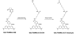
- Example 1 Synthesis of N-terminal labeling agent using fluorescent group TAMRA-X 1.5 Synthesis of 5 (6) -TAMRA-X-C8-OH 5 (6) -TAMRA- Using X-SE, 5 (6) -TAMRA-X-C8-OH was synthesized. Specifically, it is as follows.
- 5 (6) -TAMRA-X-C7-Aldehyde As shown in FIG. 2, using the obtained 5 (6) -TAMRA-X-C8-OH, 5 (6) -TAMRA- X-C7-Aldehyde was synthesized. Specifically, it is as follows. Add 175 ⁇ L of 2 mM 5 (6) -TAMRA-X-C8-OH, 175 ⁇ L of DMSO and 350 ⁇ L of 500 mM Dess-Martin reagent (manufactured by Tokyo Chemical Industry Co., Ltd.) to a 1.5 mL low adsorption tube and shake at 37 ° C. for 1 hour. Incubated while allowing.
- Dess-Martin reagent manufactured by Tokyo Chemical Industry Co., Ltd.
- Example 2 Fluorescent labeling of protein Fluorescence labeling reaction (reductive amination reaction) Using the N-terminal labeling agent synthesized in Example 1, fluorescent labeling of the N-terminal of the antibody was performed. 9 ⁇ L of 1.0 mg / mL antibody solution, 4.5 ⁇ L of 0.96 mM 5 (6) -TAMRA-X-C7-aldehyde (50% DMSO solution), 4.5 ⁇ L of 192 mM picoline borane (18% DMSO solution), 9 ⁇ L of 50 mM quencher Sodium acid buffer (pH 4.8) was added to a 1.5 mL low adsorption tube. This solution was incubated at 4 ° C. for 24 hours to perform a reductive alkylation reaction.
- Anti-thyroxine antibody Anti-Thyroxine: HyTest, clone # 1H1
- Anti-influenza hemagglutinin antibody Anti-Influenza Hemagglutinin: MBL, clone # 5D8
- Anti-influenza hemagglutinin antibody Anti-Influenza Hemagglutinin: MBL, clone # TANA2
- Anti-FLAG antibody Sigma, clone # M2
- Anti-His Tag antibody Novagen
- scFv derived from anti-BGP antibody expressed in E. coli with MetAla (methionine and alanine) added to the N-terminus
- Thyroxine antigen Sigma
- Influenza hemagglutinin tag peptide (Wako Pure Chemical Industries, Ltd.) (The same antigen was used for the fluorescence-labeled proteins using anti-influenza hemagglutinin antibodies clone # 5D8 and TANA2)
- FLAG peptide antigen (Wako Pure Chemical Industries, Ltd.)
- His peptide antigen (Wako Pure Chemical Industries, Ltd.) 5) BGP peptide antigen (Genscript)
- Fluorescence measurement These fluorescently labeled protein samples were subjected to fluorescence intensity measurement using a fluorescence spectrophotometer (Fluorolog-3; manufactured by Horiba, Ltd.).
- the excitation wavelength (Ex) was set to 550 nm, and the fluorescence intensity at the fluorescence wavelength (Em) of 565 to 700 nm was measured.
- the fluorescence intensity at 580 nm which is the maximum wavelength, was used.
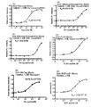
- the measurement results of the fluorescence intensity of fluorescently labeled protein samples using various antibodies are shown in FIG.
- the horizontal axis represents the antigen concentration
- the vertical axis represents the ratio of the fluorescence intensity at each antigen concentration to the fluorescence intensity without the antigen as the fluorescence intensity ratio.
- the fluorescent group was quenched in the absence of the antigen, but it was confirmed that the quenching of the fluorescent group was canceled and the fluorescence was emitted as the antigen concentration increased.
- the amplification rate of the fluorescence intensity after addition of the antigen is 1.75 times for the fluorescence-labeled protein sample using the anti-thyroxine antibody, and the anti-influenza hemagglutinin antibody (clone # 5D8).
- Example 3 Measurement using N-terminal labeling agents having various linker lengths
- n in the linker group was changed to 2, 5, 7, 9 by the same method as in Example 1.
- Various N-terminal labeling agents were prepared.
- n The N-terminal labeling agent for 7 is described as 5 (6) -TAMRA-X-C7-CHO
- Fluorescent labeled protein samples of each linker length were reacted with an antigen, irradiated with excitation light having a wavelength of 550 nm, and the fluorescence spectrum was measured using a fluorescence spectrophotometer (Fluorolog-3).
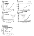
- FIG. 4 or FIG. 5 shows the measurement results of the fluorescence intensity of the fluorescently labeled protein samples of each linker length.
- FIG. 4 shows the results of using anti-FLAG antibody as an antibody solution and FLAG peptide antigen as an antigen
- FIG. 5 shows the results of using anti-His tag antibody as an antibody solution and His peptide antigen as an antigen. .
- Example 4 Measurement using N-terminal labeling agent having various linker groups N-terminal labeling agents having a PEG group as a linker group were synthesized. This synthesis was the same as the synthesis of TAMRA-X-C2-CHO in Example 3 except that TAMRA-PEO8-SE (manufactured by Biotium) was used instead of 5 (6) -TAMRA-X-SE. I went there.
- the protein was fluorescently labeled using an N-terminal labeling agent having a PEG group as a linker group.
- the antibody solution used was an anti-thyroxine antibody, and the antigen used was a thyroxine antigen.
- a fluorescently labeled protein sample having a PEG group as a linker group was reacted with an antigen, irradiated with excitation light having a wavelength of 550 nm, and a fluorescence spectrum was measured using a fluorescence spectrophotometer (Fluorolog-3).
- Microspin® G25 column was equilibrated with a citric acid equilibration buffer (50 mM sodium citrate buffer (pH 4.8), 100 mM KCl, 0.1% PEG8000, 0.1% Brij35). 300 ⁇ L of HKM equilibration buffer was added to the column and centrifuged at 3000 rpm for 1 minute at room temperature. This operation was performed 4 times in total. After that, 1 mg / mL anti-FLAG antibody / 9FLAG ⁇ L and 50 mM sodium citrate buffer (pH 4.8) 9 ⁇ L mixed solution was added to the column and centrifuged at 3000rpm for 1 minute to exchange the buffer. .
- a citric acid equilibration buffer 50 mM sodium citrate buffer (pH 4.8), 100 mM KCl, 0.1% PEG8000, 0.1% Brij35.
- 300 ⁇ L of HKM equilibration buffer was added to the column and centrifuged at 3000 rpm for 1 minute
- Fluorescently labeled protein samples with various fluorescent groups are reacted with antigens, and the maximum absorption wavelength of each fluorescent group (TAMRA 550nm, FAM 495nm, RhodamineRed 570nm, RhodamineGreen 495nm, BODIPY FL 495nm, IC3 540nm, ATTO655 650nm, Cy3 540nm ) And the fluorescence spectrum was measured using a fluorescence spectrophotometer (Fluorolog-3).
- the horizontal axis represents the antigen concentration
- the vertical axis represents the ratio of the fluorescence intensity at each antigen concentration to the fluorescence intensity without the antigen as the fluorescence intensity ratio.
- the fluorescent group was quenched in the absence of antigen, but as the antigen concentration increased, the quenching of the fluorescent group was released, It was confirmed to emit fluorescence.
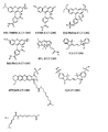
- N-terminal labeling agents having various fluorescent groups shown in FIG. 8 were used, fluorescently labeled protein samples were obtained.
Landscapes
- Health & Medical Sciences (AREA)
- Life Sciences & Earth Sciences (AREA)
- Chemical & Material Sciences (AREA)
- Immunology (AREA)
- Engineering & Computer Science (AREA)
- Molecular Biology (AREA)
- Hematology (AREA)
- Urology & Nephrology (AREA)
- Biomedical Technology (AREA)
- Organic Chemistry (AREA)
- General Health & Medical Sciences (AREA)
- Biochemistry (AREA)
- Analytical Chemistry (AREA)
- Medicinal Chemistry (AREA)
- Physics & Mathematics (AREA)
- Food Science & Technology (AREA)
- Microbiology (AREA)
- Cell Biology (AREA)
- General Physics & Mathematics (AREA)
- Pathology (AREA)
- Biotechnology (AREA)
- Genetics & Genomics (AREA)
- Proteomics, Peptides & Aminoacids (AREA)
- Biophysics (AREA)
- Peptides Or Proteins (AREA)
- Investigating Or Analysing Biological Materials (AREA)
- Pyrane Compounds (AREA)
- Investigating, Analyzing Materials By Fluorescence Or Luminescence (AREA)
Abstract
下記一般式(1): (式中、Fは蛍光基であり、Rは、置換基を有していてもよい、炭素数が5以上の直鎖炭化水素からなる直鎖ソフトセグメント又はその主鎖の炭素の一部が-O-、-N-、-S-、-CO-、-CONH-、-COO-又は-POO-で置換された直鎖ソフトセグメントを有するリンカー基である)で表される化合物を含む、タンパク質のN末端標識剤。
Description
本発明はタンパク質の標識を簡便に行うN末端標識剤や、かかるN末端標識剤を用いて標識した蛍光標識タンパク質及びその製造方法に関し、蛍光免疫測定法等に使用する蛍光標識抗体等を調製する技術として有用である。
免疫学的測定法として、抗原が結合していない状態には蛍光標識された抗体が消光(本明細書において、クエンチ、クエンチングとも呼ぶことがある)する一方、抗原が結合した場合に前記消光が解除される現象を用いた「均一系蛍光免疫測定法」が知られている(例えば、下記特許文献1参照)。この方法は、蛍光標識した抗体の断片、又は蛍光標識した一本鎖抗体を、抗原が含まれているか否かを検査する被検試料溶液に混合することにより行われる。この方法によれば、蛍光色素の蛍光強度と抗原濃度が正の相関関係にあることをもって、抗原の存在が結論付けられ、また、その相関度合いに基づいて抗原の濃度測定を行うことができる。このような消光とその解除の主たる要因は、抗体分子内の保存性の高いトリプトファン残基と蛍光色素が相互作用して蛍光色素を消光すると共に、抗原と結合することによって抗原依存的にかかる消光が解消されることにある。
測定に際し、蛍光標識した抗体の断片を用いる場合には、より詳細には、抗体軽鎖可変領域ポリペプチドと抗体重鎖可変領域ポリペプチドのどちらか一方を蛍光標識した2本の抗体断片が用いられる。ここで、抗体軽鎖可変領域は「VL」と略記されることがあり、抗体重鎖可変領域は「VH」と略記されることがある。
また、測定に際し、蛍光標識した一本鎖抗体を用いる場合には、より詳細には、蛍光標識したVLポリペプチドとVHポリペプチドとが結合してなる一本鎖抗体(「scFv」と略記されることがある。)が用いられる。
上記2種類の蛍光標識した抗体を総称して、「Quenchbody(登録商標、以下同じ)」又は「Q-body(登録商標、以下同じ)」と呼ぶことがある。また、特に両者を区別する場合においては、前者(2本の抗体断片を用いる場合)を「VH+VL型Q-body」と称し、後者(一本鎖抗体を用いる場合)を「scFv型Q-body」と称することがある。
特許文献1に記載された手法の特徴は、大きく以下の3点にある。
第一の特徴は、洗浄工程が不要である点にある。これにより、少量のサンプルとQ-bodyを混合して蛍光強度を測定するだけで測定が完了するため、この手法は極めて簡便な測定技術であるといえる。第二の特徴は、抗体中に存在する保存性の高いトリプトファンを消光に利用する点にある。すなわち、抗体の種類を変えても消光効果が得られるため、様々な抗体を用いて種々の物質を検出することに適用できる点において、この手法は汎用性に優れているといえる。第三の特徴は、抗原部位が1ヶ所でよい点にある。このため、原理的に低分子化合物に対しても適用できる。
このように、本手法は、洗浄工程を必要とせず、簡便な測定方法である。このため、本手法に基づく測定デバイスを、例えば携帯可能なサイズにまで小型化することが可能であり、また、この測定デバイスを用いることで、専門教育を受けていない一般の人が現場で測定することも可能であると想定される。
また、VL及び抗体軽鎖定常領域からなるポリペプチドと、VH及び抗体重鎖定常領域からなるポリペプチドとが、ジスルフィド結合で結合した1分子のヘテロダイマータンパク質からなるFab抗体(Fragment, antigen binding)を作成し、VL及びVHのそれぞれに同色又は異色の蛍光色素を標識した蛍光標識複合体が開発されている(特許文献2)。VL及びVHのそれぞれに同色の蛍光色素を標識した蛍光標識複合体では、上述した蛍光色素とトリプトファン残基との相互作用による消光に加え、色素間の消光効果(H-dimer)が加わり、高い検出感度を得ることが可能である(特許文献2の図7、図8等)。VL及びVHのそれぞれに異色の蛍光色素を標識した蛍光標識複合体では、蛍光色素とトリプトファン残基との相互作用による消光と、色素間の消光効果に加え、FRET効果による消光効果が加わり、さらに高い検出感度を得ることが可能である(特許文献2の図9、図10等)。このような蛍光標識Fab抗体複合体を総称して「UQ-body(登録商標、以下同じ)」と呼ぶことがある。
抗原やリガンドが結合していない場合は消光し、結合がなされると消光が解除されるシステムは、医療の分野だけではなく、環境中の農薬検出や、毒素検出等の様々な分野で普及することが期待されている。既存の抗体やレセプターを含むタンパク質を用いて、容易にQ-bodyやUQ-bodyのような蛍光標識タンパク質を作成する技術が求められている。
そこで、本発明の目的は、既存の抗体やレセプターを含むタンパク質を用いて、短行程で短時間のうちにQ-bodyやUQ-bodyのような蛍光標識タンパク質を提供することにある。
本発明者らは、上記課題を解決すべく鋭意検討した結果、アルデヒド基に一定の長さのリンカーを介して蛍光基を結合させることで、既存の抗体等のタンパク質に対して蛍光色素を簡便に結合可能であり、これが蛍光免疫測定法等に利用可能であることを見出した。本発明は、このような知見に基づいて、更に検討を重ねることによって完成したものである。
すなわち、本発明は、
[1]下記一般式(1):

(式中、Fは蛍光基であり、Rは、置換基を有していてもよい、炭素数が5以上の直鎖炭化水素からなる直鎖ソフトセグメント又はその主鎖の炭素の一部が-O-、-N-、-S-、-CO-、-CONH-、-COO-又は-POO-で置換された直鎖ソフトセグメントを有するリンカー基である)で表される化合物を含む、タンパク質のN末端標識剤;
[2]前記蛍光基が、ローダミン系、クマリン系、オキサジン系、カルボピロニン系、シアニン系、ピロメセン系、ナフタレン系、ビフェニル系、アントラセン系、フェナントレン系、ピレン系、カルバゾール系、Cy系、EvoBlue系、フルオレセイン系及びこれらの誘導体からなる群より選択される少なくとも1種である、前記[1]に記載のN末端標識剤;
[3]前記蛍光基が、カルボキシテトラメチルローダミン、カルボキシフルオレセイン、ATTO655(登録商標)及びローダミングリーンからなる群より選択される少なくとも1種を含む、前記[2]に記載のN末端標識剤;
[4]前記タンパク質が完全抗体である、前記[1]~[3]のいずれかに記載のN末端標識剤;
[5]前記タンパク質が抗体のフラグメントである、前記[1]~[3]のいずれか1項に記載のN末端標識剤;
[6]タンパク質のN末端に前記[1]~[5]のいずれかに記載のN末端標識剤を用いて、前記蛍光基が導入された蛍光標識タンパク質;
[7]蛍光標識タンパク質の製造方法であって、前記[1]~[5]のいずれかに記載のN末端標識剤とタンパク質のN末端とを反応させる工程(a)、
を含む、蛍光標識タンパク質の製造方法;
[8]前記工程(a)が溶液中で前記N末端標識剤と前記タンパク質とを接触させるものであり、該溶液のpHが、pH3~7である、前記[7]に記載の方法;
[9]前記工程(a)における前記タンパク質が完全抗体又はそのフラグメントである、前記[7]又は[8]のいずれかに記載の方法;
[10]蛍光標識タンパク質を作成するためのキットであって、
前記[1]~[5]のいずれかに記載のN末端標識剤を有する、キット;に関する。
[1]下記一般式(1):
[2]前記蛍光基が、ローダミン系、クマリン系、オキサジン系、カルボピロニン系、シアニン系、ピロメセン系、ナフタレン系、ビフェニル系、アントラセン系、フェナントレン系、ピレン系、カルバゾール系、Cy系、EvoBlue系、フルオレセイン系及びこれらの誘導体からなる群より選択される少なくとも1種である、前記[1]に記載のN末端標識剤;
[3]前記蛍光基が、カルボキシテトラメチルローダミン、カルボキシフルオレセイン、ATTO655(登録商標)及びローダミングリーンからなる群より選択される少なくとも1種を含む、前記[2]に記載のN末端標識剤;
[4]前記タンパク質が完全抗体である、前記[1]~[3]のいずれかに記載のN末端標識剤;
[5]前記タンパク質が抗体のフラグメントである、前記[1]~[3]のいずれか1項に記載のN末端標識剤;
[6]タンパク質のN末端に前記[1]~[5]のいずれかに記載のN末端標識剤を用いて、前記蛍光基が導入された蛍光標識タンパク質;
[7]蛍光標識タンパク質の製造方法であって、前記[1]~[5]のいずれかに記載のN末端標識剤とタンパク質のN末端とを反応させる工程(a)、
を含む、蛍光標識タンパク質の製造方法;
[8]前記工程(a)が溶液中で前記N末端標識剤と前記タンパク質とを接触させるものであり、該溶液のpHが、pH3~7である、前記[7]に記載の方法;
[9]前記工程(a)における前記タンパク質が完全抗体又はそのフラグメントである、前記[7]又は[8]のいずれかに記載の方法;
[10]蛍光標識タンパク質を作成するためのキットであって、
前記[1]~[5]のいずれかに記載のN末端標識剤を有する、キット;に関する。
本発明によれば、既存の抗体等のタンパク質を用いて、短工程で短時間のうちに、N末端に蛍光基を結合させることができる。既存のタンパク質を利用可能となったことで、ハイブリドーマの作成や遺伝子配列の解析も不要となり、均一系蛍光免疫測定法に利用可能なタンパク質を簡便に効率良く製造可能となる。
以下、本発明を詳細に説明する。
[N末端標識剤]
本発明は、下記一般式(1):

(式中、Fは蛍光基であり、Rは、置換基を有していてもよい、炭素数が5以上の直鎖炭化水素からなる直鎖ソフトセグメント又はその主鎖の炭素の一部が-O-、-N-、-S-、-CO-、-CONH-、-COO-又は-POO-で置換された直鎖ソフトセグメントを有するリンカー基である)で表される化合物を含む、タンパク質のN末端標識剤である。すなわち、本発明におけるN末端標識剤は、末端にアルデヒド基を有し、Rで示されるリンカー基を介して、Fで示される蛍光基と結合している。また、リンカー基は、直鎖ソフトセグメントとその他の結合基を有していてもよい。
[N末端標識剤]
本発明は、下記一般式(1):
本明細書において、直鎖ソフトセグメントとは、主鎖に直鎖状基を有し、柔軟性を有するセグメントをいう。このような直鎖状基は、主鎖の回転性や屈曲性が阻害されにくい基が好ましい。直鎖炭化水素としては、例えば、アルキレン基、アルケニレン基、アルキニレン基等が挙げられる。
主鎖の回転性や屈曲性の観点から、直鎖炭化水素における炭素数は、5以上であり、6以上が好ましく、7以上がより好ましく、8以上がさらに好ましく、9以上がさらに好ましく、10以上がさらに好ましく、11以上がさらに好ましく、12以上が特に好ましく、13以上が最も好ましい。また、主鎖の回転性や屈曲性の観点から、直鎖炭化水素における炭素数は、100以下とすることができ、90以下とすることが好ましく、80以下とすることがより好ましく、70以下とすることがさらに好ましい。直鎖ソフトセグメントは、主鎖の炭素の一部が-O-、-N-、-S-、-CO-、-CONH-、-COO-又は-POO-で置換されていてもよく、オキシアルキレン基等の繰り返し単位を有するものもこれに含まれる。
N末端標識剤により標識されたタンパク質において、蛍光基がトリプトファン残基の近傍に位置している場合、トリプトファン残基と蛍光基とが相互作用して、蛍光基がクエンチされ得る。タンパク質が抗体の場合は、抗原結合ポケットにおけるトリプトファン残基との間で、蛍光基が相互作用し得る。クエンチングの原理は、トリプトファン残基と蛍光基との相互作用に限定されるものではないが、リンカー基の構造や長さは、トリプトファン残基と蛍光基との相互作用に影響を及ぼすことが想定される。例えば、リンカー基が短すぎる場合や、長すぎる場合には、トリプトファン残基と蛍光基とが十分に相互作用することができない。また、例えば、リンカー基に環状構造が複数含まれている場合は、リンカー基の回転性や可動性が阻害され、トリプトファン残基と蛍光基とが十分に相互作用することができないことが想定される。
本明細書において、炭素数が5以上の直鎖炭化水素からなる直鎖ソフトセグメントは、置換基を有していてもよい。本明細書において、置換基としては、主鎖の回転性や屈曲性が阻害されにくい基であれば限定はされないが、例えば、メチル基、エチル基、n-プロピル基、i-プロピル基、ブチル基、ペンチル基、ヘキシル基、シクロヘキシレン基、エテニル基、プロペニル基、ブテニル基、フェニル基、トルイル基、ナフチル基、ピリジル基、フラニル基、メトキシ基、エトキシ基、プロポキシ基、アセチル基、プロパノイル基、シクロヘキサンカルボニル基、ベンゾイル基、メトキシカルボニル基、エトキシカルボニル基、ヒドロキシル基、メチルアミノ基、エチルアミノ基、ジメチルアミノ基、メチルスルホニル基、エチルスルホニル基、メチルシリル基、ジメチルシリル基、フルオロ基、クロロ基、トリフルオロメチル基、アミン基、アセタール基、ケタール基、アルデヒド基、ケトン基、イミン基、ニトリル基、カルボキシ基、エーテル基、エステル基、ニトロ基、スルホ基等が挙げられる。合成の容易さの観点から、置換基を有する場合は、メチル基、エチル基等が好ましい。
本明細書において、リンカー基は、直鎖ソフトセグメントの機能が阻害されにくい構造であれば、さらに別の結合基を含んでいてもよく、例えば、アリーレン基、ヘテロアリーレン基、C4~C10シクロアルキル基及びそれらの組合せからなる群より選択される。
限定はされないが、リンカー基は、置換基を有していてもよい、イミノ基、スクシニルアミノ基、アセタミド基、2-アミノペンタンアミド、2(2’)-アルキル-アミノアセチル基、カルバモイル基、アミノアルキル基、アミノアルキルオル基、グルタルアミド基、オルニチノ基、テオプロパノアイル基、N-(メルカプトメチル)プロピオンアミド基、2-(1,2-ジヒドロキシ-3-メルカプトプロピルチオ)プロパノイル基、アミノエタンチオール基、メルカプトプロパノール基、(ヒドロキシプロピルチオ)プロパンノアイル基、オキシ基、スクシニル基、アセチル基、オキソペンタノイル基等の結合基を含んでいてもよい。
また、限定はされないが、リンカー基は、置換基を有していてもよい、1個以上のアミノ酸の繰り返し単位を含むアミノ酸リンカー、ジスルフィドリンカー、リン酸エステルリンカー等の結合基を含んでいてもよい。
リンカー基がアルキレン基を含む場合は、限定はされないが、蛍光基のクエンチ効果を顕著に奏する観点から、C2~C20アルキレン基であることが好ましく、C3~C18アルキレン基であることがより好ましく、C4~C16アルキレン基であることがさらに好ましい。
リンカー基がPEG基を含む場合は、限定はされないが、蛍光基のクエンチ効果を顕著に奏する観点から、PEG基の繰り返し単位数は、3~20が好ましく、4~18がより好ましく、5~15がさらに好ましく、6~12が特に好ましく、2~10が最も好ましい。
N末端標識剤により標識されるタンパク質は、特に制限されないが、標的物質と結合可能なタンパク質であることが好ましく、限定はされないが、例えば、抗体、タンパク質タグ、受容体、細胞接着分子等が挙げられる。
本明細書において、抗体とは、B細胞が産生する糖タンパク質であり、抗原を認識して結合する働きを有するものをいう。抗体は、免疫グロブリンとも呼ばれ、長いポリペプチド鎖(本明細書において抗体重鎖ともいう)と、短いポリペプチド鎖(本明細書において抗体軽鎖ともいう)をそれぞれ二本ずつ有し、これらがジスルフィド結合で繋がっている。このように抗体重鎖と抗体軽鎖をそれぞれ二本ずつ有している抗体を、本明細書においては、完全抗体という。
本明細書において、N末端標識剤により標識されるタンパク質として抗体を用いる場合は、ポリクローナル抗体又はモノクローナル抗体のいずれを用いることも可能であり、市販のものであっても、合成したものであってもよい。抗体のアイソタイプとしては、IgG、IgA、IgM、IgE又はIgDのいずれも用いることができ、入手のし易さの観点からIgGが好ましい。抗体の由来は、特に制限されないが、哺乳動物由来であることが好ましく、ヒト、マウス、ラット、イヌ、ネコ、サル、ブタ、ウシ、ヒツジ、ヤギ、ウマ、又はイルカ由来であることがより好ましく、ヒト、マウス、ラット、ヒツジ又はヤギ由来であることがさらに好ましく、ヒト又はマウス由来であることが特に好ましく、ヒト由来であることが最も好ましい。
用いられる抗体は、様々な抗原に対する抗体であってよく、対象となる抗原としては、タンパク質、ペプチド、糖質、脂質、糖脂質、低分子化合物等が挙げられる。これらの物質を含む原生動物、真菌、細菌、マイコプラズマ、リケッチア、クラミジア、ウイルス、動物組織等も対象となり得る。また、麻薬、爆薬、農薬、香料、公害物質等の低分子化合物を含む化学物質も対象となり得る。このような物質としては、例えば、テトラヒドロカンナビノール(THC)、テトラヒドロカンナビノール酸(THC-A)、カンナビノール(CBN)、カンナビジオール(CBD)等のカンナビノイドと呼ばれる大麻成分;アンフェタミン、メタンフェタミン、モルヒネ、ヘロイン、コデインなどの覚醒剤や麻薬類;アフラトキシン、ステリグマトシスチン、ネオソラニール、ニバレノール、フモニシン、オクラトキシン、エンドファイト産生毒素などのカビ毒;テストステロンやエストラジオールなどの性ホルモン;クレンブテロールやラクトパミンなどの飼料に不正に用いられる添加物;PCB、ゴシポール、ヒスタミン、ベンツピレン、メラミン、アクリルアミド、ダイオキシンなどの有害物質;アセタミプリド、イミダクロプリド、クロルフェナピル、マラチオン、カルバリル、クロチアニジン、トリフルミゾール、クロロタロニル、スピノサド、ランネート、メタミドホス、クロルピリホスなどの残留農薬;ビスフェノールAなどの環境ホルモンなどを挙げることができる。上記の物質は各物質の誘導体も含む。
完全抗体は、公知の手段により、その一部分を切断して用いることも可能である。完全抗体は、例えば、パパインにより切断することで、2つのFab部分と1つのFc部分に分離することが可能である。また、完全抗体は、例えば、ペプシンにより切断することで、1つのF(ab’)2部分と、1つのFc’部分に分離することが可能である。本明細書において、完全抗体の一部分を抗体のフラグメントといい、1本鎖抗体[single-chain Fv(scFv)]、Fab部分、F(ab’)2部分、可変領域(Fv)部分、Fc部分、Fc’部分等が含まれる。抗体のフラグメントは、抗原との結合が可能である観点から、scFv、Fab部分、F(ab’)2部分又はFv部分が好ましい。また、Fc受容体と結合が可能である観点から、抗体のフラグメントは、Fc部分またはFc’部分が好ましい。また、抗体のフラグメントは、公知の手段により、合成して用いることも可能である。
scFv部分及びFab部分は、抗体軽鎖可変領域を含むポリペプチド1つと抗体重鎖可変領域を含むポリペプチド1つからなり、F(ab’)2部分及び完全抗体は、抗体軽鎖可変領域を含むポリペプチド2つと抗体重鎖可変領域を含むポリペプチド2つからなる。
抗体軽鎖可変領域ポリペプチド、抗体軽鎖可変領域ポリペプチド、及び、抗体軽鎖可変領域と抗体軽鎖可変領域の両方を含む一本鎖抗体のポリペプチドは、公知の化学合成法、遺伝子組換え技術、抗体分子のタンパク質分解酵素による分解方法等を用いて調製することができるが、中でも、比較的容易な操作でかつ大量に調製することが可能な遺伝子組換え技術により調製することが好ましい。遺伝子組換え技術により上記ポリペプチドを調製する場合には、抗体軽鎖可変領域又は抗体軽鎖可変領域に特異的なアミノ酸配列をコードする塩基配列を含むDNAを好適なベクターに導入して発現ベクターを作製し、バクテリア、酵母、昆虫、動植物細胞などを宿主として用いた発現系や、無細胞翻訳系により目的のポリペプチドを発現させることができる。無細胞翻訳系においてポリペプチドの発現を行う場合は、例えば、大腸菌、小麦胚芽、ウサギ網状赤血球等の無細胞抽出液に、ヌクレオチド3リン酸や各種アミノ酸を加えた反応液中で、ポリペプチドを発現させることができる。
N末端標識剤により標識されるタンパク質として抗体を用いる場合、蛍光基と抗体の可変領域におけるトリプトファンとの相互作用の観点から、抗体軽鎖可変領域は、カバット(Kabat)の番号付け系で第35番目のアミノ酸がトリプトファンであるアミノ酸配列を有することが好ましい。また、抗体重鎖可変領域は、カバット(Kabat)の番号付け系で第36番目、第47番目、又は第103番目のアミノ酸がトリプトファンであるアミノ酸配列を有することが好ましい。
本明細書において、タンパク質タグとは、他の分子とのアフィニティを利用したペプチドやタンパク質をいう。タンパク質タグは、他の分子とのアフィニティを有するタンパク質タグであれば特に制限されないが、例えば、ヒスチジンタグ(His tag)、グルタチオン-S-トランスフェラーゼ(GST)、マルトース結合タンパク質(MBP)、インフルエンザウイルスのヘマグルチニンペプチド配列タグ(HAタグ)、mycタグ、FLAGタグ、Biotin Carboxyl Carrier Protein(BCCP)タグ(ビオチン化ペプチド)等が挙げられる。蛍光基のクエンチ効果を顕著に奏する観点から、His tag、HAタグ、FLAGタグが好ましい。タンパク質タグは、市販のものであっても、合成して得たものであってもよい。
本明細書において、受容体とは、リガンドとの結合性を有するタンパク質をいう。受容体は、リガンドとの結合性を有する受容体であれば特に制限されないが、分泌型受容体、膜結合型受容体、細胞内受容体のいずれも用いることができる。このような受容体は、天然に存在するものであっても、改変されたものであってもよい。受容体は、市販のものであっても、合成して得たものであってもよい。受容体の由来は、特に制限されないが、哺乳動物由来であることが好ましく、ヒト、マウス、ラット、イヌ、ネコ、サル、ブタ、ウシ、ヒツジ、ヤギ、ウマ、又はイルカ由来であることがより好ましく、ヒト、マウス、ラット、ヒツジ又はヤギ由来であることがさらに好ましく、ヒト又はマウス由来であることが特に好ましく、ヒト由来であることが最も好ましい。受容体は、リガンドとの結合性を有する限り、受容体のフラグメントを用いることも可能である。
本明細書において、分泌型受容体とは、膜貫通型受容体から膜貫通領域及び細胞内領域を除いた構造を有するものをいい、リガンドとの結合性を有するものであれば特に制限されない。
膜貫通型受容体としては、リガンドとの結合性を有するものであれば特に制限されないが、例えば、イオンチャンネル連結型受容体、イオンチャンネルとは連結していない受容体、キナーゼタイプ受容体、非キナーゼタイプ受容体等が挙げられる。
核内受容体としては、リガンドとの結合性を有するものであれば特に制限されないが、例えば、甲状腺ホルモン受容体型、レチノイドX受容体型、エストロゲン受容体型、ステロイドホルモン受容体型等が挙げられる。
本明細書において、細胞接着分子とは、細胞同士又は細胞と細胞外マトリックスとの接着に関与する分子をいう。細胞接着分子としては、リガンドとの結合性を有する受容体であれば特に制限されないが、例えば、免疫グロブリンスーパーファミリー、カドヘリンスーパーファミリー、インテグリンスーパーファミリー、セレクチンファミリ一、MAMスーパーファミリー等が挙げられる。このような細胞接着分子は、天然に存在するものであっても、改変されたものであってもよい。細胞接着分子は、市販のものであっても、合成して得たものであってもよい。細胞接着分子の由来は、特に制限されないが、哺乳動物由来であることが好ましく、ヒト、マウス、ラット、イヌ、ネコ、サル、ブタ、ウシ、ヒツジ、ヤギ、ウマ、又はイルカ由来であることがより好ましく、ヒト、マウス、ラット、ヒツジ又はヤギ由来であることがさらに好ましく、ヒト又はマウス由来であることが特に好ましく、ヒト由来であることが最も好ましい。細胞接着分子としては、リガンドとの結合性を有する限り、細胞接着分子のフラグメントを用いることも可能である。
本明細書において、蛍光基は、本発明の効果を奏する限り限定はされないが、ローダミン系、クマリン系、オキサジン系、カルボピロニン系、シアニン系、ピロメセン系、ナフタレン系、ビフェニル系、アントラセン系、フェナントレン系、ピレン系、カルバゾール系、Cy系、EvoBlue系、フルオレセイン系又はこれらの誘導体等が挙げられる。中でも、蛍光基のクエンチ効果を顕著に奏する観点から、蛍光基は、ローダミン系蛍光基、オキサジン系蛍光基、Cy系、フルオレセイン系が好ましい。
蛍光基は、具体的には、例えば、ローダミン、クマリン、オキサジン、カルボピロニン、シアニン、ピロメセン、ナフタレン、ビフェニル、アントラセン、フェナントレン、ピレン、カルバゾール、Cy、EvoBlue、フルオレセイン等を基本骨格として有する蛍光基、又はこれらの蛍光基の誘導体が挙げられる。
蛍光基は、より具体的には、例えば、CR110:carboxyrhodamine 110:Rhodamine Green(商標名)、TAMRA:carbocytetremethlrhodamine:TMR、Carboxyrhodamine 6G:CR6G、ATTO655(商標名)、BODIPY FL(商標名):4,4-difluoro-5,7-dimethyl-4-bora-3a,4a-diaza-s-indancene-3-propionic acid、BODIPY 493/503(商標名):4,4-difluoro-1,3,5,7-tetramethyl-4-bora-3a,4a-diaza-s-indancene-8-propionicacid、BODIPY R6G(商標名):4,4-difluoro-5-(4-phenyl-1,3-butadienyl)-4-bora-3a,4a-diaza-s-indancene-3-propionic acid、BODIPY 558/568(商標名):4,4-difluoro-5-(2-thienyl)-4-bora-3a,4a-diaza-s-indancene-3-propionic acid、BODIPY 564/570(商標名):4,4-difluoro-5-styryl-4-bora-3a,4a-diaza-s-indancene-3-propionic acid、BODIPY 576/589(商標名):4,4-difluoro-5-(2-pyrrolyl)-4-bora-3a,4a-diaza-s-indancene-3-propionic acid、BODIPY 581/591(商標名):4,4-difluoro-5-(4-phenyl-1, 3-butadienyl)-4-bora-3a,4a-diaza-s-indancene-3-propionic acid、Cy3(商標名)、Cy3B(商標名)、Cy3.5(商標名)、Cy5(商標名)、Cy5.5(商標名)、EvoBlue10(商標名)、EvoBlue30(商標名)、MR121、ATTO 390(商標名)、ATTO 425(商標名)、ATTO 465(商標名)、ATTO 488(商標名)、ATTO 495(商標名)、ATTO 520(商標名)、ATTO 532(商標名)、ATTO Rho6G(商標名)、ATTO 550(商標名)、ATTO 565(商標名)、ATTO Rho3B(商標名)、ATTO Rho11(商標名)、ATTO Rho12(商標名)、ATTO Thio12(商標名)、ATTO 610(商標名)、ATTO 611X(商標名)、ATTO 620(商標名)、ATTO Rho14(商標名)、ATTO 633(商標名)、ATTO 647(商標名)、ATTO 647N(商標名)、ATTO 655(商標名)、ATTO Oxa12(商標名)、ATTO 700(商標名)、ATTO 725(商標名)、ATTO 740(商標名)、Alexa Fluor 350(商標名)、Alexa Fluor 405(商標名)、Alexa Fluor 430(商標名)、Alexa Fluor 488(商標名)、Alexa Fluor 532(商標名)、Alexa Fluor 546(商標名)、Alexa Fluor 555(商標名)、Alexa Fluor 568(商標名)、Alexa Fluor 594(商標名)、Alexa Fluor 633(商標名)、Alexa Fluor 647(商標名)、Alexa Fluor 680(商標名)、Alexa Fluor 700(商標名)、Alexa Fluor 750(商標名)、Alexa Fluor 790(商標名)、Rhodamine Red-X(商標名)、Texas Red-X(商標名)、5(6)-TAMRA-X(商標名)、5TAMRA(商標名)、SFX(商標名)、5-カルボキシフルオレセイン(FAM)、6-カルボキシフルオレセイン、5,6-ジカルボキシフルオレセイン、6-カルボキシ-2',4,4',5',7,7'-ヘキサクロロフルオレセイン、6-カルボキシ-2',4,7,7'-テトラクロロフルオレセイン、6-カルボキシ-4',5'-ジクロロ-2',7'-ジメトキシフルオレセイン、フルオレセイン-5-イソシアネート(FITC)、ナフトフルオレセイン等が挙げられる。中でも、蛍光基は、蛍光基のクエンチ効果を顕著に奏する観点から、ローダミン系蛍光基であるTAMRA、CR110、Rhodamine Green(商標名)、オキサジン系蛍光基であるATTO、Cy系蛍光基であるCy3(商標名)、Cy5(商標名)、フルオレセイン系であるカルボキシフルオレセインが好ましく、5(6)-TAMRA-X(商標名)、5TAMRA(商標名)、ATTO 655(商標名)、Cy3(商標名)、Cy5(商標名)、5-カルボキシフルオレセイン、6-カルボキシフルオレセイン、5,6-ジカルボキシフルオレセインがより好ましい。
蛍光基とリンカー基を結合する際には、蛍光基を有する化合物の反応性末端と、リンカー基を有する化合物の末端との反応を行うことが可能である。このような反応としては、縮合反応によるアミド化等が挙げられる。このような反応において、リンカー基を有する化合物は、他方の末端にアルデヒド基を有していてもよいが、水酸基等の形で反応させた後に、デスマーチン酸化等の反応により、アルデヒド化することが好ましい。
[蛍光標識タンパク質]
本発明は、上記タンパク質のN末端に、上記N末端標識剤を用いて、上記蛍光基が導入された蛍光標識タンパク質に関する。本発明のN末端標識剤は、一方端にアルデヒド基を有するため、還元剤の存在下で、タンパク質のN末端と還元的アミノ化反応を起こすことが可能である。
本発明は、上記タンパク質のN末端に、上記N末端標識剤を用いて、上記蛍光基が導入された蛍光標識タンパク質に関する。本発明のN末端標識剤は、一方端にアルデヒド基を有するため、還元剤の存在下で、タンパク質のN末端と還元的アミノ化反応を起こすことが可能である。
蛍光標識タンパク質には、標識された蛍光基の蛍光を、消光することができるクエンチャーをさらに付加していてもよい。このようなクエンチャーとしては、抗原等の標的物質の非存在下では、クエンチャーが蛍光基を効果的に消光し、標的物質存在下においては、蛍光基の発光を阻害しない組合せを適宜選択することができる。このようなクエンチャーとしては、例えば、NBD:7-nitrobenzofurazan、DABCYL、BHQ、ATTO、QXL、QSY、Cy、Lowa Black、IRDYE等を基本骨格とする消光色素やこれらの消光色素の誘導体等が挙げられる。より具体的には、NBD、DABCYL、BHQ-1(商標名)、BHQ-2(商標名)、BHQ-3(商標名)、ATTO540Q(商標名)、ATTO580Q(商標名)、ATTO612Q(商標名)、QXL490(商標名)、QXL520(商標名)、QXL570(商標名)、QXL610(商標名)、QXL670(商標名)、QXL680(商標名)、QSY-35(商標名)、QSY-7(商標名)、QSY-9(商標名)、QSY-21(商標名)、Cy5Q(商標名)、Cy7Q(商標名)、Lowa Black FQ(商標名)、Lowa Black RQ(商標名)、IRDYE QC-1(商標名)等が挙げられ、クエンチャーとしての効果を顕著に奏する観点から、NBDが好ましい。例えば、蛍光基としてローダミン系蛍光基を用いた場合には、NBDとの組合せを例示することができる。
本明細書において、N末端標識剤を用いて抗体に蛍光基を導入した場合、抗原の非存在下では、上記の蛍光基と抗体可変領域において保存されたトリプトファン残基との相互作用による蛍光基の消光が起こる。抗体に対して、同色の蛍光基が複数導入された場合、さらに蛍光基間のクエンチング効果が得られる。また、抗体に対して、異色の蛍光基が複数導入された場合、上記のトリプトファン残基によるクエンチング、蛍光基間のクエンチングに加え、蛍光共鳴エネルギー転移(FRET)効果によるクエンチングの効果が得られる。さらに、蛍光基とその蛍光基を消光するクエンチャーが共存する場合には、蛍光基とクエンチャー間のクエンチング効果により、ダイナミックレンジを増大することができる。図1では、N末端標識剤を用いて完全抗体に蛍光基を導入した場合の一例を示す。この実施形態では、抗原非存在下では蛍光がクエンチ(消光)される(真ん中)。抗原存在下では、クエンチが解消され、蛍光基がタンパク質から露出する(右側)。このような場合、発生する蛍光強度は、抗原濃度と正の相関関係となり得る。
N末端標識剤を用いてタンパク質タグ、受容体又は細胞接着分子に蛍光基を導入した場合、標的物質の非存在下では、タンパク質タグ、受容体又は細胞接着分子における疎水的、電気的に蛍光基と相互作用が可能な部分がクエンチャーとして働き、蛍光基の消光が起こり得る。タンパク質タグ、受容体又は細胞接着分子に対して、同色の蛍光基が複数導入された場合、さらに蛍光基間のクエンチング効果が得られる。また、タンパク質タグ、受容体又は細胞接着分子に対して、異色の蛍光基が複数導入された場合、上記のトリプトファン残基によるクエンチング、蛍光基間のクエンチングに加え、蛍光共鳴エネルギー転移(FRET)効果によるクエンチングの効果が得られる。さらに、蛍光基とその蛍光基を消光するクエンチャーが共存する場合には、蛍光基とクエンチャー間のクエンチング効果により、ダイナミックレンジを増大することができる。
別の実施態様として、蛍光標識タンパク質では、標的物質の存在下において、蛍光基の消光が起こる場合もあり得る。標的物質が蛍光基と、主として疎水的、電気的に相互作用が可能な場合には、標的物質の存在下において、より消光が強まることにより生じ得る。このような場合、発生する蛍光強度は、抗原濃度と負の相関関係となり得る。
本明細書において、N末端標識剤の製造方法は、公知の方法によることが可能であるが、例えば、市販の蛍光基を用いる場合は、蛍光基の保護基を上記のリンカー基を含む化合物で置換する。置換された反応物が末端にヒドロキシ基を有する場合、例えば、デスマーチン試薬を用いて、デスマーチン酸化を起こすことにより、末端をアルデヒド基に置換することができる。具体的には、実施例において詳述する。
本明細書において、蛍光の測定には、通常、蛍光検出に用いる光源や測定装置を用いることができる。光源としては励起光波長を照射できるものであればよく、具体的には、水銀ランプ、キセノンランプ、LED(発光ダイオード)、レーザー光等が挙げられる。この際、適当な蛍光フィルターを用いて特定の波長の励起光を得ることができる。蛍光測定装置としては、例えば、励起光の光源及びその照射システム、蛍光画像取得システム等を備えた蛍光顕微鏡等を利用することができ、例えば、MF20/FluoroPoint-Light(オリンパス社製)やFMBIO-III(日立ソフトウェアエンジニアリング社製)等が挙げられる。また、光源、照射システム、測定システムを備えた小型で持ち運び可能な蛍光検出装置を用いてもよい。このような小型の装置を用いることにより、被験試料の採取現場で測定が可能となる。なお、蛍光の検出は、蛍光スペクトルの検出であっても、特定の波長の蛍光強度の検出であってもよい。
また、蛍光標識タンパク質を非ヒト動物に投与した場合は、その体液や組織等を採取する他、非ヒト動物の検出対象領・BR>謔ノ励起光を照射して、蛍光色素の蛍光を2次元又は3次元的に測定及び/又は検出することもでき、この場合、蛍光顕微鏡や蛍光イメージアナライザー、光源を備えた内視鏡等を使用する例を挙げることができる。また、検出の際には、内視鏡、X線、CT、MRI、超音波、顕微鏡等を用いて、非ヒト動物の個体、組織、又は細胞の構造を示す画像も合わせて取得することが好ましい。測定及び/又は検出された蛍光強度と標的物質量とが正の相関関係にある場合、検出された蛍光の2次元又は3次元的画像に基づいて、標的物質の局在(位置)及び/又は量を知ることができ、この際前記構造を示す画像と比較することもできる。蛍光強度と標的物質量とが正の相関関係にある場合、これらの蛍光の検出に際しては、蛍光標識タンパク質を含まない、又は検体を含まない測定用試料等をネガティブコントロールとして調製し、合わせて測定及び/又は検出することができる。また、上記ネガティブコントロールにおける蛍光測定値で、測定用試料における蛍光測定値を除した、蛍光強度比を用いて、標的物質量の測定等を行うこともできる。あるいは、蛍光強度と標的物質量とが正の相関関係にある場合、適宜設定した閾値を越える蛍光強度が得られた場合に、測定試料中に標的物質が存在すると判定することもできる。
本明細書において、照射する励起光及び、測定及び/又は検出する蛍光の波長は、使用する蛍光基の種類に応じて適宜選択すればよい。例えば、蛍光基にTAMRAを用いた場合は、励起光波長530nmと蛍光波長580nmを用い、蛍光基にATTO655を用いた場合は、励起光波長630nmと蛍光波長680nmを用いることができる。また、2種類の異なる蛍光基を用いる場合も、励起光波長及び蛍光波長の組合せを適宜選択して用いることができる。
[蛍光標識タンパク質の製造方法]
本発明の蛍光標識タンパク質の製造方法は、上記N末端標識剤と上記タンパク質のN末端とを反応させる工程(a)を含む。工程(a)において、上記N末端標識剤は、末端のアルデヒド基が、タンパク質のN末端に対して、還元的アミノ化反応を起こして結合することができる。工程(a)において、溶液中で上記N末端標識剤と上記タンパク質とを接触させる場合は、上記溶液のpHは、pH3~7とすることが好ましい。タンパク質のαアミノ基を効率的に反応させる観点から、上記溶液のpHは、pH3.5~6とすることがより好ましく、pH4~pH5.5とすることがさらに好ましく、pH4.5~pH5とすることが特に好ましい。温度条件は、例えば、1~30℃とすることができ、2~10℃が好ましく、3~5℃がより好ましい。反応時間は、例えば1分~3日とすることができ、30分~2日が好ましく、1時間~1日がより好ましい。
本発明の蛍光標識タンパク質の製造方法は、上記N末端標識剤と上記タンパク質のN末端とを反応させる工程(a)を含む。工程(a)において、上記N末端標識剤は、末端のアルデヒド基が、タンパク質のN末端に対して、還元的アミノ化反応を起こして結合することができる。工程(a)において、溶液中で上記N末端標識剤と上記タンパク質とを接触させる場合は、上記溶液のpHは、pH3~7とすることが好ましい。タンパク質のαアミノ基を効率的に反応させる観点から、上記溶液のpHは、pH3.5~6とすることがより好ましく、pH4~pH5.5とすることがさらに好ましく、pH4.5~pH5とすることが特に好ましい。温度条件は、例えば、1~30℃とすることができ、2~10℃が好ましく、3~5℃がより好ましい。反応時間は、例えば1分~3日とすることができ、30分~2日が好ましく、1時間~1日がより好ましい。
本明細書において、工程(a)で用いる還元剤としては、還元的アミノ化反応を起こすことができる限り制限されないが、例えば、シアノ水素化ホウ素ナトリウム、水素化ホウ素ナトリウム、水素化トリアセトキシホウ素ナトリウム、2-ピコリンボラン等が挙げられる。効率的に反応させる観点から、シアノ水素化ホウ素ナトリウム、2-ピコリンボランが好ましい。
蛍光標識タンパク質の製造方法は、さらに精製工程(b)を含むことができる。蛍光標識タンパク質の精製方法としては、公知の方法を用いることが可能であり限定はされないが、例えば、ゲルろ過クロマトグラフィー、限外ろ過、イオン交換クロマトグラフィー、疎水性クロマトグラフィー、アフィニティクロマトグラフィー、吸着クロマトグラフィー、逆相クロマトグラフィー等が挙げられる。効率的に反応させる観点から、ゲルろ過クロマトグラフィー、限外ろ過を用いることが好ましい。
[蛍光標識タンパク質を作成するためのキット]
また、本発明は、蛍光標識タンパク質を作成するためのキットであって、上記のN末端標識剤を有するキットに関する。このようなキットには、さらに還元的アミノ化反応に用いられる通常の試薬(還元剤、pH調整剤等)、器具(精製に関する器具等)、取扱説明書等を含むことができる。
また、本発明は、蛍光標識タンパク質を作成するためのキットであって、上記のN末端標識剤を有するキットに関する。このようなキットには、さらに還元的アミノ化反応に用いられる通常の試薬(還元剤、pH調整剤等)、器具(精製に関する器具等)、取扱説明書等を含むことができる。
本発明を以下の実施例によって具体的に説明するが、本発明はこれらの実施例によって限定されるものではない。
[実施例1]蛍光基TAMRA-Xを用いたN末端標識剤の合成
1.5(6)-TAMRA-X-C8-OHの合成
図2に示されるように、5(6)-TAMRA-X-SEを用いて、5(6)-TAMRA-X-C8-OHを合成した。具体的には以下のとおりである。
10μLの100mM 5(6)-TAMRA-X-SE(6-(Tetramethylrhodamine-5-(and-6)-carboxamido)hexanoic acid, succinimidyl ester;Molecular Probes社製)、10μLのDMSO(Dimethyl sulfoxide)、100μLの100mM octanolamine及び20μLの100mM NaHCO3水和物を1.5mL低吸着チューブに加え、氷上で混和することで反応させた。この反応液に2μLの1M 酢酸を加えて中和し、その後140μLの0.1%トリフルオロ酢酸(TFA)を加えた。この溶液から下記分取条件で、高速液体クロマトグラフィー(HPLC)により合成物を分取し、乾燥させた。この乾燥物を所定の濃度となるようにDMSOに溶かした。671.38 (MH+, monoisotopic)の5(6)-TAMRA-X-C8-OHを得た。質量分析条件は以下のとおりである。
1.5(6)-TAMRA-X-C8-OHの合成
図2に示されるように、5(6)-TAMRA-X-SEを用いて、5(6)-TAMRA-X-C8-OHを合成した。具体的には以下のとおりである。
10μLの100mM 5(6)-TAMRA-X-SE(6-(Tetramethylrhodamine-5-(and-6)-carboxamido)hexanoic acid, succinimidyl ester;Molecular Probes社製)、10μLのDMSO(Dimethyl sulfoxide)、100μLの100mM octanolamine及び20μLの100mM NaHCO3水和物を1.5mL低吸着チューブに加え、氷上で混和することで反応させた。この反応液に2μLの1M 酢酸を加えて中和し、その後140μLの0.1%トリフルオロ酢酸(TFA)を加えた。この溶液から下記分取条件で、高速液体クロマトグラフィー(HPLC)により合成物を分取し、乾燥させた。この乾燥物を所定の濃度となるようにDMSOに溶かした。671.38 (MH+, monoisotopic)の5(6)-TAMRA-X-C8-OHを得た。質量分析条件は以下のとおりである。
(HPLC分取条件)
検出器:SPD-20A(島津製作所社製)、260nm吸光度検出
カラム:Xbridge C18 5μm 10x50mm column (Waters社製)
移動相A:0.1% TFA
移動相B:アセトニトリル(MeCN)
カラム温度:30℃
グラジエント条件:0-100% B in A for 15min
流量:3.0mL/min
検出器:SPD-20A(島津製作所社製)、260nm吸光度検出
カラム:Xbridge C18 5μm 10x50mm column (Waters社製)
移動相A:0.1% TFA
移動相B:アセトニトリル(MeCN)
カラム温度:30℃
グラジエント条件:0-100% B in A for 15min
流量:3.0mL/min
(質量分析条件)
分析装置:Voyager DE Pro(Applied Biosystems社製)
マトリックス:α-CHCA in 0.1%TFA/MeCN (1:1)溶液 (10 mg/ml)
測定モード:ポジティブ
外部標準物質:angiotensin II (1046.5)
分析装置:Voyager DE Pro(Applied Biosystems社製)
マトリックス:α-CHCA in 0.1%TFA/MeCN (1:1)溶液 (10 mg/ml)
測定モード:ポジティブ
外部標準物質:angiotensin II (1046.5)
2.5(6)-TAMRA-X-C7-Aldehydeの合成
図2に示されるように、得られた5(6)-TAMRA-X-C8-OHを用いて、5(6)-TAMRA-X-C7-Aldehydeを合成した。具体的には以下のとおりである。
175μLの2mM 5(6)-TAMRA-X-C8-OH、175μLのDMSO及び350μLの500mM Dess-Martin試薬(東京化成工業社製)を1.5mL低吸着チューブに加え、37℃、1時間振とうさせながらインキュベートした。750μLの0.1% TFA溶液を加え、上記分取条件でHPLCにより合成物を分取し、乾燥させた。この乾燥物を100μLのトリフルオロ酢酸/アセトニトリル(1:1)に溶かし、このうちの1μLを用いて定量分析した。定量分析は下記定量・分析条件によるHPLCにて行った。再度、乾燥させ、この乾燥物を所定の濃度となるようDMSOに溶かした。N末端標識剤として、669.36 (MH+, monoisotopic)の5(6)-TAMRA-X-C7-Aldehydeを得た。質量分析条件は上記のとおりである。
図2に示されるように、得られた5(6)-TAMRA-X-C8-OHを用いて、5(6)-TAMRA-X-C7-Aldehydeを合成した。具体的には以下のとおりである。
175μLの2mM 5(6)-TAMRA-X-C8-OH、175μLのDMSO及び350μLの500mM Dess-Martin試薬(東京化成工業社製)を1.5mL低吸着チューブに加え、37℃、1時間振とうさせながらインキュベートした。750μLの0.1% TFA溶液を加え、上記分取条件でHPLCにより合成物を分取し、乾燥させた。この乾燥物を100μLのトリフルオロ酢酸/アセトニトリル(1:1)に溶かし、このうちの1μLを用いて定量分析した。定量分析は下記定量・分析条件によるHPLCにて行った。再度、乾燥させ、この乾燥物を所定の濃度となるようDMSOに溶かした。N末端標識剤として、669.36 (MH+, monoisotopic)の5(6)-TAMRA-X-C7-Aldehydeを得た。質量分析条件は上記のとおりである。
(HPLC定量・分析条件)
検出器:SPD-20A(島津製作所社製)、260nm吸光度検出
カラム:Xbridge C18 2.5μm 4.6x20mm IS column (Waters社製)
移動相A:0.1% TFA
移動相B:アセトニトリル(MeCN)
カラム温度:30℃
グラジエント条件:0-100% D in C for 10min
流量:1.5mL/min
検出器:SPD-20A(島津製作所社製)、260nm吸光度検出
カラム:Xbridge C18 2.5μm 4.6x20mm IS column (Waters社製)
移動相A:0.1% TFA
移動相B:アセトニトリル(MeCN)
カラム温度:30℃
グラジエント条件:0-100% D in C for 10min
流量:1.5mL/min
[実施例2]タンパク質の蛍光標識化
1.蛍光標識化反応(還元的アミノ化反応)
実施例1で合成したN末端標識剤を用いて、抗体のN末端の蛍光標識化を行った。
9μLの1.0mg/mL 抗体溶液、4.5μLの0.96mM 5(6)-TAMRA-X-C7-aldehyde (50% DMSO溶液)、4.5μLの192mM ピコリンボラン (18% DMSO溶液)、9μLの50mM クエン酸ナトリウムバッファー(pH4.8)を1.5mL低吸着チューブに加えた。この溶液を、4℃、24時間インキュベートすることで還元的アルキル化反応を行った。
1.蛍光標識化反応(還元的アミノ化反応)
実施例1で合成したN末端標識剤を用いて、抗体のN末端の蛍光標識化を行った。
9μLの1.0mg/mL 抗体溶液、4.5μLの0.96mM 5(6)-TAMRA-X-C7-aldehyde (50% DMSO溶液)、4.5μLの192mM ピコリンボラン (18% DMSO溶液)、9μLの50mM クエン酸ナトリウムバッファー(pH4.8)を1.5mL低吸着チューブに加えた。この溶液を、4℃、24時間インキュベートすることで還元的アルキル化反応を行った。
抗体溶液としては、以下のものを使用した。
1)抗サイロキシン抗体(Anti-Thyroxine: HyTest社製、clone#1H1)
2)抗インフルエンザ ヘマグルチニン抗体(Anti-Influenza Hemagglutinin: MBL社製、clone# 5D8)
3)抗インフルエンザ ヘマグルチニン抗体(Anti-Influenza Hemagglutinin: MBL社製、clone# TANA2)
4)抗FLAG抗体(Sigma社製、clone# M2)
5)抗His Tag抗体(Novagen社製)
6)抗BGP抗体由来scFv(N末端にMetAla(メチオニン及びアラニン)を付加して大腸菌により発現)
1)抗サイロキシン抗体(Anti-Thyroxine: HyTest社製、clone#1H1)
2)抗インフルエンザ ヘマグルチニン抗体(Anti-Influenza Hemagglutinin: MBL社製、clone# 5D8)
3)抗インフルエンザ ヘマグルチニン抗体(Anti-Influenza Hemagglutinin: MBL社製、clone# TANA2)
4)抗FLAG抗体(Sigma社製、clone# M2)
5)抗His Tag抗体(Novagen社製)
6)抗BGP抗体由来scFv(N末端にMetAla(メチオニン及びアラニン)を付加して大腸菌により発現)
2.蛍光標識タンパク質のゲルろ過
Microspin G25 column (GEヘルスケア・ジャパン社製)を用いて、得られた反応液から蛍光標識タンパク質を溶出した。
具体的には、始めにMicrospin G25 columnを、HKM平衡化バッファー(HKMバッファー(25 mM HEPES-KOH(pH7.4), 5 mM MgCl2, 100 mM KCl)、0.1% PEG8000(Sigma社製)、0.05% Brij(非イオン性界面活性剤、和光純薬社製))で平衡化した。カラムの平衡化には、300μLのHKM平衡化バッファーをカラムに添加し、室温、3000rpmにて1分間遠心分離した。この操作を合計4回行った。その後、上記の蛍光標識化反応で得られた反応液全量をカラムに添加して、室温、3000rpmにて1分間遠心分離することで蛍光標識タンパク質を溶出した。
Microspin G25 column (GEヘルスケア・ジャパン社製)を用いて、得られた反応液から蛍光標識タンパク質を溶出した。
具体的には、始めにMicrospin G25 columnを、HKM平衡化バッファー(HKMバッファー(25 mM HEPES-KOH(pH7.4), 5 mM MgCl2, 100 mM KCl)、0.1% PEG8000(Sigma社製)、0.05% Brij(非イオン性界面活性剤、和光純薬社製))で平衡化した。カラムの平衡化には、300μLのHKM平衡化バッファーをカラムに添加し、室温、3000rpmにて1分間遠心分離した。この操作を合計4回行った。その後、上記の蛍光標識化反応で得られた反応液全量をカラムに添加して、室温、3000rpmにて1分間遠心分離することで蛍光標識タンパク質を溶出した。
3.蛍光標識タンパク質の蛍光測定(抗原滴定)
(抗原溶液の作成)
抗原を純水若しくはDMSOで溶解し、10-1~10-2溶液を作成した。その後、HKMバッファー若しくはDMSOを用いて、10-9溶液まで段階的に希釈した。各種抗体に対する抗原は以下のものを使用した。
1)サイロキシン抗原(Sigma社製)
2)インフルエンザ ヘマグルチニンタグペプチド(和光純薬社製)
(抗インフルエンザ ヘマグルチニン抗体のclone# 5D8とTANA2を用いた蛍光標識タンパク質では同一の抗原を使用した)
3)FLAGペプチド抗原(和光純薬社製)
4)Hisペプチド抗原(和光純薬社製)
5)BGPペプチド抗原(Genscript社製)
(抗原溶液の作成)
抗原を純水若しくはDMSOで溶解し、10-1~10-2溶液を作成した。その後、HKMバッファー若しくはDMSOを用いて、10-9溶液まで段階的に希釈した。各種抗体に対する抗原は以下のものを使用した。
1)サイロキシン抗原(Sigma社製)
2)インフルエンザ ヘマグルチニンタグペプチド(和光純薬社製)
(抗インフルエンザ ヘマグルチニン抗体のclone# 5D8とTANA2を用いた蛍光標識タンパク質では同一の抗原を使用した)
3)FLAGペプチド抗原(和光純薬社製)
4)Hisペプチド抗原(和光純薬社製)
5)BGPペプチド抗原(Genscript社製)
(蛍光測定溶液の作製)
18.9μLのゲルろ過後サンプルと926.1μLのHKM蛍光バッファー(HKMバッファー、0.1% PEG8000、0.005% Brij)とを混合し、1.5mL低吸着チューブに100μLずつ計9本に分注した。その後、抗原溶液を加えないサンプルにはHKMバッファーもしくはDMSOのみを1.01μL添加し、残りのサンプルには段階希釈した抗原溶液1.01μLを添加し、軽くボルテックスにより混和し、スピンダウンし、25°C、1時間インキュベートした。その後、各蛍光標識タンパク質サンプルの95μLを蛍光測定に使用した。
18.9μLのゲルろ過後サンプルと926.1μLのHKM蛍光バッファー(HKMバッファー、0.1% PEG8000、0.005% Brij)とを混合し、1.5mL低吸着チューブに100μLずつ計9本に分注した。その後、抗原溶液を加えないサンプルにはHKMバッファーもしくはDMSOのみを1.01μL添加し、残りのサンプルには段階希釈した抗原溶液1.01μLを添加し、軽くボルテックスにより混和し、スピンダウンし、25°C、1時間インキュベートした。その後、各蛍光標識タンパク質サンプルの95μLを蛍光測定に使用した。
(蛍光測定)
これらの蛍光標識タンパク質サンプルを蛍光分光光度計(Fluorolog-3; 堀場製作所製)を用いて蛍光強度測定を行った。蛍光基として、TAMRAを用いた場合には、励起波長(Ex)は550nmにセットし、蛍光波長(Em)565~700nmでの蛍光強度を測定した。蛍光強度を比較する際は、極大波長となる580nmでの蛍光強度を使用した。
これらの蛍光標識タンパク質サンプルを蛍光分光光度計(Fluorolog-3; 堀場製作所製)を用いて蛍光強度測定を行った。蛍光基として、TAMRAを用いた場合には、励起波長(Ex)は550nmにセットし、蛍光波長(Em)565~700nmでの蛍光強度を測定した。蛍光強度を比較する際は、極大波長となる580nmでの蛍光強度を使用した。
各種の抗体を用いた蛍光標識タンパク質サンプルの蛍光強度の測定結果を図3に示す。各グラフにおいて、横軸は抗原濃度を示し、縦軸は抗原なしの場合の蛍光強度に対する、各抗原濃度における蛍光強度の比を蛍光強度比として示した。
図3に示すように、抗原の非存在下では、蛍光基はクエンチされていたが、抗原濃度が高くなるにつれ、蛍光基のクエンチが解除され、蛍光を発することが確認された。抗原未添加時の蛍光強度を1とした場合に対する、抗原添加後の蛍光強度の増幅率は、抗サイロキシン抗体を用いた蛍光標識タンパク質サンプルでは1.75倍、抗インフルエンザ ヘマグルチニン抗体(clone# 5D8)を用いた蛍光標識タンパク質サンプルでは1.4倍、抗インフルエンザ ヘマグルチニン抗体(clone# TANA2)を用いた蛍光標識タンパク質サンプルでは1.2倍、抗FLAG抗体を用いた蛍光標識タンパク質サンプルでは1.6倍、抗His Tag抗体を用いた蛍光標識タンパク質サンプルでは1.2倍、抗BGP抗体由来scFvを用いた蛍光標識タンパク質サンプルでは2.7倍だった。
[実施例3]種々のリンカー長を有するN末端標識剤を用いた測定
実施例1と同様の方法により、下記式(2)中、リンカー基内におけるnを2、5、7、9と変更した種々のN末端標識剤を調製した。n=2の場合のN末端標識剤は5(6)-TAMRA-X-C2-CHO、n=5の場合のN末端標識剤は5(6)-TAMRA-X-C5-CHO、n=7の場合のN末端標識剤は5(6)-TAMRA-X-C7-CHO、n=9の場合のN末端標識剤は5(6)-TAMRA-X-C9-CHOと記載する。
実施例1と同様の方法により、下記式(2)中、リンカー基内におけるnを2、5、7、9と変更した種々のN末端標識剤を調製した。n=2の場合のN末端標識剤は5(6)-TAMRA-X-C2-CHO、n=5の場合のN末端標識剤は5(6)-TAMRA-X-C5-CHO、n=7の場合のN末端標識剤は5(6)-TAMRA-X-C7-CHO、n=9の場合のN末端標識剤は5(6)-TAMRA-X-C9-CHOと記載する。

実施例2と同様の方法により、種々のリンカー長を有するN末端標識剤を用いてタンパク質の蛍光標識化を行った。
各リンカー長の蛍光標識タンパク質サンプルを抗原と反応させ、波長550nmの励起光を照射し、蛍光分光光度計(Fluorolog-3)を用いて蛍光スペクトルを測定した。
各リンカー長の蛍光標識タンパク質サンプルの蛍光強度の測定結果を図4又は図5に示す。図4は、抗体溶液として抗FLAG抗体を用い、抗原としてFLAGペプチド抗原を用いた結果であり、図5は、抗体溶液として抗His Tag抗体を用い、抗原としてHisペプチド抗原を用いた結果である。
図4に示すように、抗体溶液として抗FLAG抗体を用いた場合、上記の化学式(2)においてn=2、5、7又は9である場合、抗原存在下で、蛍光標識タンパク質サンプルの蛍光強度変化を検出できることが確認され、特にn=5、7である場合に大きな蛍光強度変化が検出された。
図5に示すように、抗体溶液として抗His Tag抗体を用いた場合、上記化学式(2)においてn=5、7である場合に、抗原存在下で、蛍光標識タンパク質サンプルの高い蛍光強度変化が検出された。
[実施例4]種々のリンカー基を有するN末端標識剤を用いた測定
リンカー基にPEG基を有するN末端標識剤を合成した。この合成は、5(6)-TAMRA-X-SEの代わりに、TAMRA-PEO8-SE(Biotium社製)を使用したことを除き、実施例3におけるTAMRA-X-C2-CHOの合成と同様にして行った。
リンカー基にPEG基を有するN末端標識剤を合成した。この合成は、5(6)-TAMRA-X-SEの代わりに、TAMRA-PEO8-SE(Biotium社製)を使用したことを除き、実施例3におけるTAMRA-X-C2-CHOの合成と同様にして行った。
実施例2と同様の方法により、リンカー基にPEG基を有するN末端標識剤を用いてタンパク質の蛍光標識化を行った。抗体溶液は抗サイロキシン抗体を使用し、抗原はサイロキシン抗原を使用した。
リンカー基にPEG基を有する蛍光標識タンパク質サンプルを抗原と反応させ、波長550nmの励起光を照射し、蛍光分光光度計(Fluorolog-3)を用いて蛍光スペクトルを測定した。
図6に示すように、リンカー基にPEG基を有する蛍光標識タンパク質サンプルにおいても、抗原存在下で、蛍光標識タンパク質サンプルの高い蛍光強度変化を検出することが確認された。
[実施例5]種々の蛍光基を有するN末端標識剤を用いた測定
蛍光基にFAM(Molecular Probes社製)、RhodamineRed(Molecular Probes社製)、RhodamineGreen (Molecular Probes社製)、BODIPY FL(Molecular Probes社製)、IC3(同仁化学社製またはBiosearch Technologies社製)、ATTO655(ATTO Tech社製)又はCy3(GE Lifescience社製)を用いたN末端標識剤を合成した。抗体溶液は抗FLAG抗体を用い、抗原はFLAGペプチド抗原を用いた。FAMの場合は、アルデヒドへの酸化反応の際に、Dess-Martin試薬の濃度を500mMから15mMに減じて、反応時間を4時間に延長した。その他の条件は、上記の実施例1に準じる。
蛍光基にFAM(Molecular Probes社製)、RhodamineRed(Molecular Probes社製)、RhodamineGreen (Molecular Probes社製)、BODIPY FL(Molecular Probes社製)、IC3(同仁化学社製またはBiosearch Technologies社製)、ATTO655(ATTO Tech社製)又はCy3(GE Lifescience社製)を用いたN末端標識剤を合成した。抗体溶液は抗FLAG抗体を用い、抗原はFLAGペプチド抗原を用いた。FAMの場合は、アルデヒドへの酸化反応の際に、Dess-Martin試薬の濃度を500mMから15mMに減じて、反応時間を4時間に延長した。その他の条件は、上記の実施例1に準じる。
Microspin G25 columnを、クエン酸平衡化バッファー(50mMクエン酸ナトリウムバッファー(pH4.8)、100mM KCl、0.1% PEG8000、0.1% Brij35)で平衡化した。300μLのHKM平衡化バッファーをカラムに添加し、室温、3000rpmにて1分間遠心分離した。この操作を合計4回行った。その後、1 mg/mL 抗FLAG抗体 9 μLと50 mM クエン酸ナトリウムバッファー(pH 4.8) 9 μLの混合溶液をカラムに添加して、室温、3000rpmにて1分間遠心分離してバッファー交換を行った。溶出液に、0.96 mM の各蛍光基を有するN末端標識剤(50% DMSO溶液)4.5μLと192 mM ピコリンボラン(18% DMSO溶液)4.5μLを添加して、4℃、24時間インキュベートすることで還元的アルキル化反応を行った。続いて、実施例2と同様にしてゲルろ過により蛍光標識タンパク質を溶出した。
種々の蛍光基を有する蛍光標識タンパク質サンプルを抗原と反応させ、それぞれの蛍光基の吸収極大の波長(TAMRA 550nm、FAM 495nm、RhodamineRed 570nm、RhodamineGreen 495nm、BODIPY FL 495nm、IC3 540nm、ATTO655 650nm、Cy3 540nm)の励起光を照射し、蛍光分光光度計(Fluorolog-3)を用いて蛍光スペクトルを測定した。各グラフにおいて、横軸は抗原濃度を示し、縦軸は抗原なしの場合の蛍光強度に対する、各抗原濃度における蛍光強度の比を蛍光強度比として示した。
図7に示すように、種々の蛍光基を有する蛍光標識タンパク質サンプルにおいて、抗原の非存在下では、蛍光基はクエンチされていたが、抗原濃度が高くなるにつれ、蛍光基のクエンチが解除され、蛍光を発することが確認された。
図8に示す種々の蛍光基を有するN末端標識剤を用いた場合も、蛍光標識タンパク質サンプルが得られた。
Claims (10)
- 下記一般式(1):
- 前記蛍光基が、ローダミン系、クマリン系、オキサジン系、カルボピロニン系、シアニン系、ピロメセン系、ナフタレン系、ビフェニル系、アントラセン系、フェナントレン系、ピレン系、カルバゾール系、Cy系、EvoBlue系、フルオレセイン系及びこれらの誘導体からなる群より選択される少なくとも1種である、請求項1に記載のN末端標識剤。
- 前記蛍光基が、カルボキシテトラメチルローダミン、カルボキシフルオレセイン、ATTO655(登録商標)及びローダミングリーンからなる群より選択される少なくとも1種を含む、請求項2に記載のN末端標識剤。
- 前記タンパク質が完全抗体である、請求項1~3のいずれか1項に記載のN末端標識剤。
- 前記タンパク質が抗体のフラグメントである、請求項1~3のいずれか1項に記載のN末端標識剤。
- タンパク質のN末端に請求項1~5のいずれか1項に記載のN末端標識剤を用いて、前記蛍光基が導入された蛍光標識タンパク質。
- 蛍光標識タンパク質の製造方法であって、請求項1~5のいずれか1項に記載のN末端標識剤とタンパク質のN末端とを反応させる工程(a)、
を含む、蛍光標識タンパク質の製造方法。 - 前記工程(a)が溶液中で前記N末端標識剤と前記タンパク質とを接触させるものであり、該溶液のpHが、pH3~7である、請求項7に記載の方法。
- 前記工程(a)における前記タンパク質が完全抗体又はそのフラグメントである、請求項7又は8に記載の方法。
- 蛍光標識タンパク質を作成するためのキットであって、
請求項1~5のいずれか1項に記載のN末端標識剤を有する、キット。
Applications Claiming Priority (2)
| Application Number | Priority Date | Filing Date | Title |
|---|---|---|---|
| JP2015138743A JP2017020896A (ja) | 2015-07-10 | 2015-07-10 | N末端標識剤、これを用いた蛍光標識タンパク質及びその製造方法 |
| JP2015-138743 | 2015-07-10 |
Publications (1)
| Publication Number | Publication Date |
|---|---|
| WO2017010381A1 true WO2017010381A1 (ja) | 2017-01-19 |
Family
ID=57757313
Family Applications (1)
| Application Number | Title | Priority Date | Filing Date |
|---|---|---|---|
| PCT/JP2016/070058 Ceased WO2017010381A1 (ja) | 2015-07-10 | 2016-07-07 | N末端標識剤、これを用いた蛍光標識タンパク質及びその製造方法 |
Country Status (2)
| Country | Link |
|---|---|
| JP (1) | JP2017020896A (ja) |
| WO (1) | WO2017010381A1 (ja) |
Cited By (2)
| Publication number | Priority date | Publication date | Assignee | Title |
|---|---|---|---|---|
| WO2018147018A1 (ja) * | 2017-02-08 | 2018-08-16 | 国立大学法人東京工業大学 | 抗原検出又は測定用キット |
| IT202100013634A1 (it) * | 2021-05-25 | 2022-11-25 | Alda S R L | Nuovi reagenti per microscopia ottica |
Citations (8)
| Publication number | Priority date | Publication date | Assignee | Title |
|---|---|---|---|---|
| JPH06172312A (ja) * | 1992-03-04 | 1994-06-21 | Wako Pure Chem Ind Ltd | 1級アミノ基を有する化合物の蛍光標識化方法 |
| JP2005501228A (ja) * | 2001-06-04 | 2005-01-13 | アマシャム バイオサイエンス ユーケイ リミテッド | 生物材料の蛍光検出用標識試薬としてのキナクリドン誘導体 |
| JP2005013119A (ja) * | 2003-06-26 | 2005-01-20 | Japan Science & Technology Agency | Rna−ペプチド複合体を用いた蛍光センサー |
| US20090149631A1 (en) * | 2007-11-09 | 2009-06-11 | Massachusetts Institute Of Technology | Methods and compositions for protein labeling using lipoic acid ligases |
| JP4414654B2 (ja) * | 2001-05-14 | 2010-02-10 | ザ インスティチュート フォー システムズ バイオロジー | サンプル分子を単離および標識するための方法 |
| WO2013065314A1 (ja) * | 2011-11-02 | 2013-05-10 | ウシオ電機株式会社 | 蛍光標識抗体可変領域含有ポリペプチド複合体を用いた蛍光免疫測定方法 |
| JP2013528185A (ja) * | 2010-05-27 | 2013-07-08 | ジーイー・ヘルスケア・バイオサイエンス・アクチボラグ | タンパク質の標識法及びキット |
| JP2014156428A (ja) * | 2013-02-15 | 2014-08-28 | Univ Of Tokyo | 抗体結合タンパク質 |
-
2015
- 2015-07-10 JP JP2015138743A patent/JP2017020896A/ja active Pending
-
2016
- 2016-07-07 WO PCT/JP2016/070058 patent/WO2017010381A1/ja not_active Ceased
Patent Citations (8)
| Publication number | Priority date | Publication date | Assignee | Title |
|---|---|---|---|---|
| JPH06172312A (ja) * | 1992-03-04 | 1994-06-21 | Wako Pure Chem Ind Ltd | 1級アミノ基を有する化合物の蛍光標識化方法 |
| JP4414654B2 (ja) * | 2001-05-14 | 2010-02-10 | ザ インスティチュート フォー システムズ バイオロジー | サンプル分子を単離および標識するための方法 |
| JP2005501228A (ja) * | 2001-06-04 | 2005-01-13 | アマシャム バイオサイエンス ユーケイ リミテッド | 生物材料の蛍光検出用標識試薬としてのキナクリドン誘導体 |
| JP2005013119A (ja) * | 2003-06-26 | 2005-01-20 | Japan Science & Technology Agency | Rna−ペプチド複合体を用いた蛍光センサー |
| US20090149631A1 (en) * | 2007-11-09 | 2009-06-11 | Massachusetts Institute Of Technology | Methods and compositions for protein labeling using lipoic acid ligases |
| JP2013528185A (ja) * | 2010-05-27 | 2013-07-08 | ジーイー・ヘルスケア・バイオサイエンス・アクチボラグ | タンパク質の標識法及びキット |
| WO2013065314A1 (ja) * | 2011-11-02 | 2013-05-10 | ウシオ電機株式会社 | 蛍光標識抗体可変領域含有ポリペプチド複合体を用いた蛍光免疫測定方法 |
| JP2014156428A (ja) * | 2013-02-15 | 2014-08-28 | Univ Of Tokyo | 抗体結合タンパク質 |
Non-Patent Citations (5)
| Title |
|---|
| KEISUKE FUKUNAGA: "Monoclonal IgG no N-mattan Sentakuteki na Keiko Hyoshiki to Kogen no Keiko Kenshutsu", ANNUAL MEETING OF THE CHEMICAL SOCIETY OF JAPAN IN SPRING KOEN YOKOSHU, vol. 96 th, 10 March 2016 (2016-03-10), pages ROMBUNNO.4c5 - 10 * |
| KENSUKE YOSHIKOSHI: "Shuju no Keikoki o Mochiita Niju Keiko Hyoshiki Ipponsa Kotai ni yoru Kogen no Ratio Kenshutsu", CSJ: THE CHEMICAL SOCIETY OF JAPAN KOEN YOKOSHU, vol. 95 th, no. 3, 11 March 2015 (2015-03-11), pages 822 1J3 - 33 * |
| MASANORI MIURA: "Kaishi Codon no Kakucho ni yoru Shuju no Keiko Hyoshiki Karubonsan no Tanpakushitsu N-mattan eno Donyu", PROTEIN SCIENCE SOCIETY OF JAPAN NENKAI PROGRAM . YOSHISHU, vol. 9 th, 2009, pages 79 1P-123 * |
| TAKAYOSHI WATANABE: "Hi Tennen Amino-san Donyu Gijutsu o Riyo shita Tanpakushitsu no Bui Tokuiteki Shushoku Gijutsu no Kaihatsu", CSJ: THE CHEMICAL SOCIETY OF JAPAN KOEN YOKOSHU, vol. 94, no. 3, 2014, pages 938 4G5 - 12 * |
| TAKAYOSHI WATANABE: "Keiko Hyoshiki Hydroxy-san o Mochiita Shinki N-mattan Tokuiteki Tanpakushitsu Keiko Hyoshikiho no Kaihatsu", JOURNAL OF JAPANESE BIOCHEMICAL SOCIETY, 2007, pages 2P-1533 * |
Cited By (4)
| Publication number | Priority date | Publication date | Assignee | Title |
|---|---|---|---|---|
| WO2018147018A1 (ja) * | 2017-02-08 | 2018-08-16 | 国立大学法人東京工業大学 | 抗原検出又は測定用キット |
| JPWO2018147018A1 (ja) * | 2017-02-08 | 2019-12-12 | 国立大学法人東京工業大学 | 抗原検出又は測定用キット |
| US11499965B2 (en) | 2017-02-08 | 2022-11-15 | Tokyo Institute Of Technology | Kit for detecting antigen or measuring its amount |
| IT202100013634A1 (it) * | 2021-05-25 | 2022-11-25 | Alda S R L | Nuovi reagenti per microscopia ottica |
Also Published As
| Publication number | Publication date |
|---|---|
| JP2017020896A (ja) | 2017-01-26 |
Similar Documents
| Publication | Publication Date | Title |
|---|---|---|
| JP6070769B2 (ja) | 蛍光標識抗体可変領域含有ポリペプチド複合体を用いた蛍光免疫測定キット | |
| Prakashan et al. | Design and fabrication of a competitive lateral flow assay using gold nanoparticle as capture probe for the rapid and on-site detection of penicillin antibiotic in food samples | |
| Zhang et al. | A broad-spectrum sensing strategy for the tetracycline family of antibiotics based on an ovalbumin-stabilized gold nanocluster and its application in a pump-free microfluidic sensing platform | |
| CN102667480B (zh) | 荧光免疫测定方法 | |
| Viirlaid et al. | Immunoassay for rapid on-site detection of glyphosate herbicide | |
| JP7837518B2 (ja) | 蛍光偏光免疫分析法 | |
| Yang et al. | Fluorescent immunosorbent assay for the detection of alpha lactalbumin in dairy products with monoclonal antibody bioconjugated with CdSe/ZnS quantum dots | |
| Kong et al. | An ultrasensitive ELISA for medroxyprogesterone residues in fish tissues based on a structure-specific hapten | |
| Zhou et al. | Gold nanoparticle probe-based immunoassay as a new tool for tetrodotoxin detection in puffer fish tissues | |
| Wu et al. | Sensitive enzyme‐linked immunosorbent assay and gold nanoparticle immunochromatocgraphic strip for rapid detecting chloramphenicol in food | |
| CN100552452C (zh) | 使用荧光化抗体的荧光分析方法 | |
| JP5949890B2 (ja) | 蛍光標識された抗体可変領域を含むポリペプチドを含む抗原結合タンパク質を用いた蛍光免疫測定方法 | |
| JP2017020896A (ja) | N末端標識剤、これを用いた蛍光標識タンパク質及びその製造方法 | |
| WO2018147018A1 (ja) | 抗原検出又は測定用キット | |
| JP2016200545A (ja) | 検査対象物質を識別して検出する方法 | |
| Yoohanngoh et al. | Carbon dots-based fluorescent immunosensor for highly sensitive detection of leucomalachite green in fish and shrimp samples | |
| CN106018793B (zh) | 一种六次甲基四胺的检测方法 | |
| Xi et al. | Preparation and characterization of artificial antigens for cadmium and lead | |
| He et al. | Sample-to-answer Aggregation-Induced Emission Lateral Flow Immunoassay (ALFIA) for rapid and sensitive on-site detection of Toxoplasma gondii antibodies across multiple host species | |
| Du et al. | Determination of diethylstilbestrol based on biotin–streptavidin‐amplified time‐resolved fluoro‐immunoassay | |
| JP7461661B2 (ja) | 複数の蛍光色素を結合させたペプチドを用いるホモジニアス免疫測定法 | |
| JP2017049184A (ja) | 抗体のヌクレオチド結合部位(nbs)を利用して蛍光標識された抗体 | |
| US20230393123A1 (en) | Concentration estimation kit and concentration estimation method | |
| WO2016208466A1 (ja) | 検出対象物質の検出方法 | |
| Perez | Development of Fast Photochemical Oxidation of Proteins for in Vivo Modification in Caenorhabditis elegans |
Legal Events
| Date | Code | Title | Description |
|---|---|---|---|
| 121 | Ep: the epo has been informed by wipo that ep was designated in this application |
Ref document number: 16824361 Country of ref document: EP Kind code of ref document: A1 |
|
| NENP | Non-entry into the national phase |
Ref country code: DE |
|
| 122 | Ep: pct application non-entry in european phase |
Ref document number: 16824361 Country of ref document: EP Kind code of ref document: A1 |