WO2016194552A1 - Detection kit for multiple target nucleic acids and detection method using same - Google Patents

Detection kit for multiple target nucleic acids and detection method using same Download PDF

Info

Publication number
WO2016194552A1
WO2016194552A1 PCT/JP2016/063831 JP2016063831W WO2016194552A1 WO 2016194552 A1 WO2016194552 A1 WO 2016194552A1 JP 2016063831 W JP2016063831 W JP 2016063831W WO 2016194552 A1 WO2016194552 A1 WO 2016194552A1
Authority
WO
WIPO (PCT)
Prior art keywords
target nucleic
nucleic acid
target
temperature
value
Prior art date
Application number
PCT/JP2016/063831
Other languages
French (fr)
Japanese (ja)
Inventor
長野隆志
宮島兼佑
楢原謙次
Original Assignee
株式会社ミズホメディー
Priority date (The priority date is an assumption and is not a legal conclusion. Google has not performed a legal analysis and makes no representation as to the accuracy of the date listed.)
Filing date
Publication date
Priority to CN201680031912.2A priority Critical patent/CN107849617B/en
Priority to US15/576,923 priority patent/US20180155764A1/en
Application filed by 株式会社ミズホメディー filed Critical 株式会社ミズホメディー
Priority to MYPI2017704387A priority patent/MY182821A/en
Priority to CA2983428A priority patent/CA2983428C/en
Priority to BR112017026001-8A priority patent/BR112017026001A2/en
Priority to MX2017015673A priority patent/MX2017015673A/en
Priority to ES16802989T priority patent/ES2899826T3/en
Priority to DK16802989.0T priority patent/DK3305912T3/en
Priority to JP2017521758A priority patent/JP6720161B2/en
Priority to EP16802989.0A priority patent/EP3305912B1/en
Priority to KR1020177033935A priority patent/KR102077577B1/en
Priority to SG11201708920PA priority patent/SG11201708920PA/en
Priority to AU2016269875A priority patent/AU2016269875B2/en
Publication of WO2016194552A1 publication Critical patent/WO2016194552A1/en
Priority to PH12017502015A priority patent/PH12017502015A1/en
Priority to US17/205,364 priority patent/US20210238653A1/en

Links

Images

Classifications

    • CCHEMISTRY; METALLURGY
    • C12BIOCHEMISTRY; BEER; SPIRITS; WINE; VINEGAR; MICROBIOLOGY; ENZYMOLOGY; MUTATION OR GENETIC ENGINEERING
    • C12QMEASURING OR TESTING PROCESSES INVOLVING ENZYMES, NUCLEIC ACIDS OR MICROORGANISMS; COMPOSITIONS OR TEST PAPERS THEREFOR; PROCESSES OF PREPARING SUCH COMPOSITIONS; CONDITION-RESPONSIVE CONTROL IN MICROBIOLOGICAL OR ENZYMOLOGICAL PROCESSES
    • C12Q1/00Measuring or testing processes involving enzymes, nucleic acids or microorganisms; Compositions therefor; Processes of preparing such compositions
    • C12Q1/68Measuring or testing processes involving enzymes, nucleic acids or microorganisms; Compositions therefor; Processes of preparing such compositions involving nucleic acids
    • C12Q1/6844Nucleic acid amplification reactions
    • C12Q1/686Polymerase chain reaction [PCR]
    • CCHEMISTRY; METALLURGY
    • C12BIOCHEMISTRY; BEER; SPIRITS; WINE; VINEGAR; MICROBIOLOGY; ENZYMOLOGY; MUTATION OR GENETIC ENGINEERING
    • C12QMEASURING OR TESTING PROCESSES INVOLVING ENZYMES, NUCLEIC ACIDS OR MICROORGANISMS; COMPOSITIONS OR TEST PAPERS THEREFOR; PROCESSES OF PREPARING SUCH COMPOSITIONS; CONDITION-RESPONSIVE CONTROL IN MICROBIOLOGICAL OR ENZYMOLOGICAL PROCESSES
    • C12Q1/00Measuring or testing processes involving enzymes, nucleic acids or microorganisms; Compositions therefor; Processes of preparing such compositions
    • C12Q1/68Measuring or testing processes involving enzymes, nucleic acids or microorganisms; Compositions therefor; Processes of preparing such compositions involving nucleic acids
    • C12Q1/6813Hybridisation assays
    • C12Q1/6816Hybridisation assays characterised by the detection means
    • CCHEMISTRY; METALLURGY
    • C12BIOCHEMISTRY; BEER; SPIRITS; WINE; VINEGAR; MICROBIOLOGY; ENZYMOLOGY; MUTATION OR GENETIC ENGINEERING
    • C12QMEASURING OR TESTING PROCESSES INVOLVING ENZYMES, NUCLEIC ACIDS OR MICROORGANISMS; COMPOSITIONS OR TEST PAPERS THEREFOR; PROCESSES OF PREPARING SUCH COMPOSITIONS; CONDITION-RESPONSIVE CONTROL IN MICROBIOLOGICAL OR ENZYMOLOGICAL PROCESSES
    • C12Q1/00Measuring or testing processes involving enzymes, nucleic acids or microorganisms; Compositions therefor; Processes of preparing such compositions
    • C12Q1/68Measuring or testing processes involving enzymes, nucleic acids or microorganisms; Compositions therefor; Processes of preparing such compositions involving nucleic acids
    • C12Q1/6876Nucleic acid products used in the analysis of nucleic acids, e.g. primers or probes
    • C12Q1/6888Nucleic acid products used in the analysis of nucleic acids, e.g. primers or probes for detection or identification of organisms
    • C12Q1/689Nucleic acid products used in the analysis of nucleic acids, e.g. primers or probes for detection or identification of organisms for bacteria
    • CCHEMISTRY; METALLURGY
    • C12BIOCHEMISTRY; BEER; SPIRITS; WINE; VINEGAR; MICROBIOLOGY; ENZYMOLOGY; MUTATION OR GENETIC ENGINEERING
    • C12QMEASURING OR TESTING PROCESSES INVOLVING ENZYMES, NUCLEIC ACIDS OR MICROORGANISMS; COMPOSITIONS OR TEST PAPERS THEREFOR; PROCESSES OF PREPARING SUCH COMPOSITIONS; CONDITION-RESPONSIVE CONTROL IN MICROBIOLOGICAL OR ENZYMOLOGICAL PROCESSES
    • C12Q2527/00Reactions demanding special reaction conditions
    • C12Q2527/101Temperature
    • CCHEMISTRY; METALLURGY
    • C12BIOCHEMISTRY; BEER; SPIRITS; WINE; VINEGAR; MICROBIOLOGY; ENZYMOLOGY; MUTATION OR GENETIC ENGINEERING
    • C12QMEASURING OR TESTING PROCESSES INVOLVING ENZYMES, NUCLEIC ACIDS OR MICROORGANISMS; COMPOSITIONS OR TEST PAPERS THEREFOR; PROCESSES OF PREPARING SUCH COMPOSITIONS; CONDITION-RESPONSIVE CONTROL IN MICROBIOLOGICAL OR ENZYMOLOGICAL PROCESSES
    • C12Q2527/00Reactions demanding special reaction conditions
    • C12Q2527/107Temperature of melting, i.e. Tm
    • CCHEMISTRY; METALLURGY
    • C12BIOCHEMISTRY; BEER; SPIRITS; WINE; VINEGAR; MICROBIOLOGY; ENZYMOLOGY; MUTATION OR GENETIC ENGINEERING
    • C12QMEASURING OR TESTING PROCESSES INVOLVING ENZYMES, NUCLEIC ACIDS OR MICROORGANISMS; COMPOSITIONS OR TEST PAPERS THEREFOR; PROCESSES OF PREPARING SUCH COMPOSITIONS; CONDITION-RESPONSIVE CONTROL IN MICROBIOLOGICAL OR ENZYMOLOGICAL PROCESSES
    • C12Q2563/00Nucleic acid detection characterized by the use of physical, structural and functional properties
    • C12Q2563/107Nucleic acid detection characterized by the use of physical, structural and functional properties fluorescence

Landscapes

  • Chemical & Material Sciences (AREA)
  • Life Sciences & Earth Sciences (AREA)
  • Organic Chemistry (AREA)
  • Proteomics, Peptides & Aminoacids (AREA)
  • Zoology (AREA)
  • Health & Medical Sciences (AREA)
  • Engineering & Computer Science (AREA)
  • Wood Science & Technology (AREA)
  • Analytical Chemistry (AREA)
  • Microbiology (AREA)
  • Physics & Mathematics (AREA)
  • Molecular Biology (AREA)
  • Immunology (AREA)
  • Biotechnology (AREA)
  • Biophysics (AREA)
  • Biochemistry (AREA)
  • Bioinformatics & Cheminformatics (AREA)
  • General Engineering & Computer Science (AREA)
  • General Health & Medical Sciences (AREA)
  • Genetics & Genomics (AREA)
  • Chemical Kinetics & Catalysis (AREA)
  • Measuring Or Testing Involving Enzymes Or Micro-Organisms (AREA)

Abstract

Provided is a detection kit for multiple target nucleic acids whereby multiple genes can be simultaneously amplified and detected using a single reaction container containing a single kind of reaction solution and a single label. The detection kit for multiple target nucleic acids comprises a solution, which is suspected to contain a first target nucleic acid 10 and a second target nucleic acid 20, each target nucleic acid dissociating at a denaturation temperature T0, and to which a DNA polymerase 30, a primer 13 for first target capable of binding to a single strand originating from the first target nucleic acid at an annealing temperature T1, a primer 23 for second target capable of binding to a single strand originating from the second target nucleic acid at the annealing temperature T1, a probe 15 for first target capable of binding to a single strand originating from the first target nucleic acid at the annealing temperature T1, and a probe 25 for second target capable of binding to a single strand originating from the second target nucleic acid at a second target detection temperature T3 that is lower than the annealing temperature T1 and an elongation temperature T2 are added, wherein the requirement T0>T2≥T1>T3 is satisfied.

Description

複数の標的核酸の検出キット及びそれを用いる検出方法Multiple target nucleic acid detection kit and detection method using the same
 本発明は、PCR法を改善し、複数の標的核酸を測定する検出キット及びその関連技術に関する。 The present invention relates to a detection kit for improving a PCR method and measuring a plurality of target nucleic acids and related techniques.
 遺伝子検査において、複数の標的遺伝子の測定が必要なことも多い。たとえばインフルエンザのA型とB型、インフルエンザとRSV、さらにはヒトメタニューモウイルス、性感染症のクラミジアと淋菌、マイコプラズマとその耐性因子などである。 In genetic testing, it is often necessary to measure multiple target genes. Examples include influenza A and B, influenza and RSV, human metapneumovirus, chlamydia and gonorrhea of sexually transmitted diseases, mycoplasma and its resistance factors.
 病状が似ている上に同じ時期に同時に感染が拡大する場合などは、これらを鑑別して診断し、治療方針を立てることも考えられる。また、耐性因子の存在は投薬の選定判断などにも使われる。 If the disease is similar and the infection spreads simultaneously at the same time, it may be possible to make a diagnosis by distinguishing these and making a treatment policy. In addition, the presence of resistance factors is also used to make decisions on medication selection.
 一方、単独の測定項目においても、その測定が本当にうまく進んだかどうかを確認する手段として内部コントロールを設定することも有用である。これは検出目的である標的遺伝子とは関係ない核酸配列を増幅するようにあらかじめ準備しておき、標的遺伝子の有無にかかわらず反応がうまく進んだかどうかを確認できるものである。このことで測定試薬や装置が有効に作用したかどうかを測定内で確認できる。 On the other hand, it is also useful to set an internal control as a means of confirming whether or not the measurement has progressed really well even for a single measurement item. This is prepared in advance so as to amplify a nucleic acid sequence unrelated to the target gene to be detected, and it can be confirmed whether or not the reaction has proceeded successfully regardless of the presence or absence of the target gene. This makes it possible to confirm in the measurement whether or not the measuring reagent or device has worked effectively.
 この様に、複数の標的核酸を検出するには、別々の測定を実施するか、或いは、それぞれの標的核酸の検出に異なる標識色素などを用いて識別する手段が考えられる。 In this way, in order to detect a plurality of target nucleic acids, it is conceivable to carry out separate measurements or to identify each target nucleic acid using a different labeling dye or the like.
 または、同時に増幅反応を行った後に、緩やかに温度を上昇(または下降)させて、蛍光シグナルの変化率がピークを示す温度と標的核酸の融解温度から識別判断すること(熱融解曲線分析法)も可能である。 Or, after performing the amplification reaction at the same time, gradually increase (or decrease) the temperature, and discriminate and judge from the temperature at which the change rate of the fluorescence signal shows a peak and the melting temperature of the target nucleic acid (thermal melting curve analysis method) Is also possible.
 しかしながら、別々の測定では時間やコストがかかり、異なる標識物を識別することも、複数の標識試薬の準備のみならず分析装置の波長設定などでコストがかかってしまう。 However, it takes time and cost for separate measurements, and identifying different labels is costly not only for preparing a plurality of labeling reagents but also for setting the wavelength of the analyzer.
 熱融解曲線分析法を用いる場合には、コストは低いが緩やかに温度の変化をさせる必要があるため、正確な分析を行うためには5~10分ほどの時間をかけて温度変化を行わなければならない。 When using the thermal melting curve analysis method, it is necessary to change the temperature slowly, but the temperature must be changed over a period of about 5 to 10 minutes for accurate analysis. I must.
 特許文献1(日本国特開2002-136300号公報)では、異なる反応液が入った複数の反応容器を用意し、それぞれ増幅検出する。項目ごとに試薬調製、分注作業があり手間がかかるという問題点がある。 In Patent Document 1 (Japanese Patent Laid-Open No. 2002-136300), a plurality of reaction containers containing different reaction solutions are prepared, and each is amplified and detected. There is a problem in that reagent preparation and dispensing work are required for each item, which is troublesome.
 特許文献2(日本国特開2004-203号公報)では、1種類の反応液が入った1つの反応容器で複数の遺伝子を同時増幅する。遺伝子ごとに標識色素を変えておくことで識別する。用いる標識色素の数だけ、検出する装置の光学系が複数必要になってくるため装置のコストが高くなってしまうという問題点がある。 In Patent Document 2 (Japanese Patent Laid-Open No. 2004-203), a plurality of genes are simultaneously amplified in one reaction vessel containing one kind of reaction solution. Identification is made by changing the labeling dye for each gene. There is a problem that the cost of the apparatus is increased because a plurality of optical systems for the detection apparatus are required as many as the number of labeling dyes to be used.
 特許文献3(日本国特開2008-173127号公報)では、1種類の反応液が入った1つの反応容器で複数の遺伝子を同時増幅する。項目ごとに、PCR産物や標識プローブの解離温度を変え、増幅後に低温側から高温側に徐々に温度を上昇させ蛍光値をモニタリングする解離曲線分析(「熱融解曲線分析」と同義。)を行い、塩基配列に依存するそれぞれの解離温度におけるピークの有無をモニタリングすることによって識別する。 In Patent Document 3 (Japanese Unexamined Patent Application Publication No. 2008-173127), a plurality of genes are simultaneously amplified in one reaction container containing one kind of reaction solution. For each item, dissociation curve analysis (synonymous with “thermal melting curve analysis”) is performed in which the dissociation temperature of the PCR product and the labeled probe is changed, and after amplification, the temperature is gradually increased from the low temperature side to the high temperature side to monitor the fluorescence value. The identification is performed by monitoring the presence or absence of a peak at each dissociation temperature depending on the base sequence.
 融解曲線分析時の温度変化の速度を上げると各項目の融解温度を識別するのが困難になるため、PCR反応後5~10分ほど時間が余計にかかってしまうという問題点がある。 If the speed of temperature change during melting curve analysis is increased, it becomes difficult to identify the melting temperature of each item, so that it takes 5 to 10 minutes after the PCR reaction.
 特許文献4(日本国特表2012-513215号公報)では、1個の反応液容器で複数の遺伝子を同時増幅する。遺伝子ごとに、PCR産物の解離温度とプライマーの融解温度を変え、それぞれの融解温度で蛍光をモニタリングして識別する。 In Patent Document 4 (Japanese Patent Special Publication No. 2012-513215), a plurality of genes are simultaneously amplified in one reaction solution container. For each gene, the dissociation temperature of the PCR product and the melting temperature of the primer are changed, and fluorescence is monitored and identified at each melting temperature.
 通常のPCRプロファイルは1サイクル中に、変性、アニーリング・伸長ステップを設けるが、特許文献4では、項目ごとにPCR産物の融解温度を変えるため、考慮すべき条件が多く、プロファイルの設計が困難であり、測定時間も長くなる。温度変化が激しいため、装置の負担も大きいという問題点がある。 A normal PCR profile is provided with denaturation, annealing, and extension steps in one cycle. However, in Patent Document 4, since the melting temperature of the PCR product is changed for each item, there are many conditions to be considered and it is difficult to design the profile. Yes, the measurement time becomes longer. Since the temperature change is severe, there is a problem that the burden on the apparatus is heavy.
日本国特開2002-136300号公報Japanese Unexamined Patent Publication No. 2002-136300 日本国特開2004-203号公報Japanese Unexamined Patent Publication No. 2004-203 日本国特開2008-173127号公報Japanese Unexamined Patent Publication No. 2008-173127 日本国特表2012-513215号公報Japan Special Table 2012-513215 日本国特許第4724380号公報Japanese Patent No. 4724380
 そこで本発明は、1種類の反応液が入った1つの反応容器及び単一の標識を用いて、複数の遺伝子を同時に増幅・検出できる、複数の標的核酸の検出キットを提供することを目的とする。 Accordingly, an object of the present invention is to provide a plurality of target nucleic acid detection kits capable of simultaneously amplifying and detecting a plurality of genes using a single reaction container containing a single type of reaction solution and a single label. To do.
 第1の発明に係る複数の標的核酸の検出キットは、変性温度をT0、アニーリング温度をT1、伸長温度をT2、第2標的検出温度をT3とすると、T0>T2≧T1>T3を満たすように温度設定される複数の標的核酸の検出キットであって、変性温度T0において、2本鎖の水素結合が切断され、それぞれ2本の1本鎖に解離する、第1標的核酸及び第2標的核酸を含み得る溶液に、
 アニーリング温度T1において、第1標的核酸が解離した2本の1本鎖のいずれかに特異的に結合する第1標的用プライマーと、アニーリング温度T1において、第2標的核酸が解離した2本の1本鎖のいずれかに特異的に結合する第2標的用プライマーと、アニーリング温度T1において、第1標的核酸が解離した2本の1本鎖のいずれかに特異的に結合する第1標的用プローブであって、結合により蛍光シグナルが変化する第1標識物を有するものと、DNAポリメラーゼと、伸長温度T2において、DNAポリメラーゼの作用により、第1標的核酸が解離した2本の1本鎖及び第2標的核酸が解離した2本の1本鎖に結合するデオキシリボヌクレオチド3リン酸と、アニーリング温度T1においては、第1標的核酸が解離した2本の1本鎖及び第2標的核酸が解離した2本の1本鎖のいずれにも結合せず、第2標的検出温度T3において、第2標的核酸が解離した2本の1本鎖のいずれかに特異的に結合する第2標的用プローブであって、結合により蛍光シグナルが変化する第2標識物を有するものとを含有する。
The detection kit for a plurality of target nucleic acids according to the first invention satisfies T0> T2 ≧ T1> T3, where T0 is the denaturation temperature, T1 is the annealing temperature, T2 is the extension temperature, and T3 is the second target detection temperature. A plurality of target nucleic acid detection kits that are temperature-set at the first target nucleic acid and the second target, each having a double-strand hydrogen bond cleaved and dissociated into two single-strands at the denaturation temperature T0. In a solution that may contain nucleic acids,
A first target primer that specifically binds to one of the two single strands from which the first target nucleic acid has been dissociated at the annealing temperature T1, and two ones from which the second target nucleic acid has been dissociated at the annealing temperature T1. A second target primer that specifically binds to one of the strands, and a first target probe that specifically binds to one of the two single strands from which the first target nucleic acid is dissociated at the annealing temperature T1 The first target nucleic acid whose fluorescence signal changes upon binding, the DNA polymerase, and the two single strands from which the first target nucleic acid is dissociated by the action of the DNA polymerase at the extension temperature T2. Two deoxyribonucleotide triphosphates that bind to two single strands from which the target nucleic acid has been dissociated, and two 1s from which the first target nucleic acid has been dissociated at the annealing temperature T1. It does not bind to any of the two single strands from which the second target nucleic acid has been dissociated, and is specific to one of the two single strands from which the second target nucleic acid has dissociated at the second target detection temperature T3 And a second target probe that binds to a molecule having a second label whose fluorescence signal changes upon binding.
 好ましくは、第1、第2標識物は、QProbe(登録商標)プローブ、Eprobe(登録商標)プローブ及びTaqMan(登録商標)プローブからなる群から選択される。 Preferably, the first and second labeled substances are selected from the group consisting of a QProbe (registered trademark) probe, an Eprobe (registered trademark) probe, and a TaqMan (registered trademark) probe.
 蛍光シグナルは、アニールすると消光するもの、アニールすると発光するもののいずれであってもよい。 Fluorescent signal may be either one that quenches when annealed or one that emits light when annealed.
 第1標的核酸及び第2標的核酸は、好ましくは、マイコプラズマP1遺伝子と内部コントロール、或いは、クラミジア内在性プラスミド遺伝子と淋菌CMT遺伝子である。 The first target nucleic acid and the second target nucleic acid are preferably mycoplasma P1 gene and internal control, or Chlamydia endogenous plasmid gene and Neisseria gonorrhoeae CMT gene.
 以上述べたように、本発明によれば、1種類の標識物を用いて複数の標的核酸を、1種類の混合液で検出できる。熱融解曲線分析なしで複数の標的核酸の識別が可能であるため、測定時間を大幅に短縮でき、実用的効果が高い。 As described above, according to the present invention, a plurality of target nucleic acids can be detected with a single mixed solution using a single label. Since a plurality of target nucleic acids can be identified without thermal melting curve analysis, the measurement time can be greatly shortened and the practical effect is high.
 本発明者らは、PCR法における、プローブのアニーリング温度の条件設定と温度変化プロファイルを組み合わせた、1種類の反応液が入った1つの反応容器で単一蛍光標識を用いた複数核酸を検出するキットを開発した。 In the PCR method, the present inventors detect a plurality of nucleic acids using a single fluorescent label in a single reaction vessel containing a single type of reaction solution, which combines a probe annealing temperature condition setting and a temperature change profile. A kit was developed.
 本キットでは、図35に示すように、4つの温度、即ち、変性温度T0(95℃)、アニーリング温度T1(70℃)、伸長温度T2(72℃)及び第2標的検出温度T3(55℃)を使用する。これらの温度は、T0>T2≧T1>T3を満たすように温度設定される。勿論、これらの温度数値は、例示に過ぎず、後述するように、種々変更することができる。 In this kit, as shown in FIG. 35, four temperatures, namely, a denaturation temperature T0 (95 ° C.), an annealing temperature T1 (70 ° C.), an extension temperature T2 (72 ° C.), and a second target detection temperature T3 (55 ° C.). ). These temperatures are set so as to satisfy T0> T2 ≧ T1> T3. Of course, these temperature values are merely examples, and can be variously changed as will be described later.
 複数の標的核酸とは、第1標的核酸及び第2標的核酸であるが、必要とあれば、第3標的検出温度T4(T3>T4)等の設定温度を増やすことにより、第3標的核酸その他の標的核酸を追加することもできる。 The plurality of target nucleic acids are the first target nucleic acid and the second target nucleic acid. If necessary, the third target nucleic acid and others can be increased by increasing the set temperature such as the third target detection temperature T4 (T3> T4). The target nucleic acid can also be added.
 図36を参照しながら、本キットの内容について説明する。まず、本キットでは、第1標的核酸10と、第2標的核酸20とを検出対象とする。以下説明の便宜のため、容器1内の溶液2(詳細は後述する。)には、第1標的核酸10と第2標的核酸20の両方が含まれている場合(つまり、陽性と陽性である場合)のみを説明する。 The contents of this kit will be described with reference to FIG. First, in this kit, the first target nucleic acid 10 and the second target nucleic acid 20 are to be detected. For convenience of explanation below, the solution 2 in the container 1 (details will be described later) contains both the first target nucleic acid 10 and the second target nucleic acid 20 (that is, positive and positive). Only).
 勿論、片方又は両方が陰性である場合には、第1標的核酸10と第2標的核酸20の少なくとも一方又は両方が溶液2に含まれていないことになり、以下の説明において、含まれていない標的核酸に関する、蛍光シグナル及び増幅が行われないことになる。 Of course, when one or both are negative, at least one or both of the first target nucleic acid 10 and the second target nucleic acid 20 are not included in the solution 2, and are not included in the following description. There will be no fluorescence signal and amplification for the target nucleic acid.
 図37(a)に示すように、溶液2の温度を上昇させ変性温度T0になると、第1標的核酸10及び第2標的核酸20は、いずれも2本鎖の水素結合が切断され、それぞれ2本の1本鎖(第1標的核酸10は、第1の1本鎖11と第2の1本鎖12に、第2標的核酸20は、第1の1本鎖21と第2の1本鎖22に)解離する。 As shown in FIG. 37 (a), when the temperature of the solution 2 is increased to reach the denaturation temperature T0, the first target nucleic acid 10 and the second target nucleic acid 20 are both cleaved by double-stranded hydrogen bonds, Single strands (the first target nucleic acid 10 is the first single strand 11 and the second single strand 12, the second target nucleic acid 20 is the first single strand 21 and the second single strand Dissociate into chain 22).
 図37(b)に示すように、変性温度T0から温度を下げてアニーリング温度T1になると、第1標的核酸10について、第1標的用Fプライマー13が第1の1本鎖11の相補的な配列に特異的に結合し、第1標的用Rプライマー14が第2の1本鎖12の相補的な配列に特異的に結合する。同様に、第2標的核酸20について、第2標的用Fプライマー23が第1の1本鎖21の相補的な配列に特異的に結合し、第2標的用Rプライマー24が第2の1本鎖22の相補的な配列に特異的に結合する。 As shown in FIG. 37 (b), when the temperature is lowered from the denaturation temperature T0 to the annealing temperature T1, the first target F primer 13 is complementary to the first single strand 11 for the first target nucleic acid 10. Specifically, the first target R primer 14 specifically binds to a complementary sequence of the second single strand 12. Similarly, for the second target nucleic acid 20, the second target F primer 23 specifically binds to the complementary sequence of the first single strand 21, and the second target R primer 24 has the second one. It binds specifically to the complementary sequence of strand 22.
 また、第1標識物16により標識された第1標的用プローブ15が、図37(b)に示すアニーリング温度T1において、第1標的核酸10由来の第1の1本鎖11の特定部位に特異的に結合し、第1標識物16は蛍光サインを発生する。しかしながら、第2標識物26により標識された第2標的用プローブ25は、アニーリング温度T1では第2標的検出温度T3よりも温度が高いため、第2標的核酸20由来の第1の1本鎖21には結合せず、第2標識物26は蛍光サインを発生しない。 Further, the first target probe 15 labeled with the first label 16 is specific to the specific site of the first single strand 11 derived from the first target nucleic acid 10 at the annealing temperature T1 shown in FIG. The first label 16 generates a fluorescent signature. However, since the second target probe 25 labeled with the second label 26 has a higher temperature at the annealing temperature T1 than the second target detection temperature T3, the first single strand 21 derived from the second target nucleic acid 20 is used. The second label 26 does not generate a fluorescent signature.
 図37(c)に示すように、温度をアニーリング温度T1から伸長温度T2に上げると、溶液2に、PCRサイクルを繰り返すために十分な量の、DNAポリメラーゼ30とデオキシリボヌクレオチド3リン酸31とが含まれているため、次のように増幅反応が進行する。 As shown in FIG. 37 (c), when the temperature is raised from the annealing temperature T1 to the extension temperature T2, sufficient amounts of DNA polymerase 30 and deoxyribonucleotide triphosphate 31 are added to the solution 2 to repeat the PCR cycle. Since it is contained, the amplification reaction proceeds as follows.
 即ち、第1標的核酸10について、第1の1本鎖11に結合する第1標的用Fプライマー13から、また、第2の1本鎖12に結合する第1標的用Rプライマー14から、それぞれデオキシリボヌクレオチド3リン酸31が結合し伸長してゆく。 That is, for the first target nucleic acid 10, from the first target F primer 13 that binds to the first single strand 11, and from the first target R primer 14 that binds to the second single strand 12, respectively. Deoxyribonucleotide triphosphate 31 binds and extends.
 また、第2標的核酸20について、第1の1本鎖21に結合する第2標的用Fプライマー23から、また、第2の1本鎖22に結合する第2標的用Rプライマー24から、それぞれデオキシリボヌクレオチド3リン酸31が結合し伸長してゆく。 For the second target nucleic acid 20, from the second target F primer 23 that binds to the first single strand 21, and from the second target R primer 24 that binds to the second single strand 22, respectively. Deoxyribonucleotide triphosphate 31 binds and extends.
 そして、増幅反応が完了すると、図37(d)を図36と比較すると分かるように、第1標的核酸10及び第2標的核酸20の両方が、2倍に増えたことになる。 When the amplification reaction is completed, both the first target nucleic acid 10 and the second target nucleic acid 20 are doubled, as can be seen by comparing FIG. 37 (d) with FIG.
 再び、図37(a)に示すステップに戻り、以上のステップ(図37(a)~図37(d))を繰り返すと、第1標的物16による蛍光シグナルの変化と増幅反応を繰り返すことができる。 Again, returning to the step shown in FIG. 37 (a) and repeating the above steps (FIG. 37 (a) to FIG. 37 (d)), the change of the fluorescence signal by the first target 16 and the amplification reaction may be repeated. it can.
 次に、図38を参照しながら、変性温度T0から温度を下げて第2標的温度検出温度T3となる場合について説明する。 Next, a case where the temperature is lowered from the denaturation temperature T0 to become the second target temperature detection temperature T3 will be described with reference to FIG.
 変性温度T0では、図38(a)に示すように、図37(a)と同様の状態となる。ところが、温度を第2標的温度検出温度T3に下げると、図37(b)とは異なり、図38(b)に示す状態となる。 At the denaturation temperature T0, as shown in FIG. 38 (a), the state is the same as in FIG. 37 (a). However, when the temperature is lowered to the second target temperature detection temperature T3, unlike FIG. 37 (b), the state shown in FIG. 38 (b) is obtained.
 即ち、図38(b)に示すように、第1標識物16により標識された第1標的用プローブ15が、第1標的核酸10由来の第1の1本鎖11の特定部位に特異的に結合し、第1標識物16の蛍光シグナルが変化する点では、図37(b)と同様であるが、第2標識物26により標識された第2標的用プローブ25も、第2標的検出温度T3となっているため、第2標的核酸20由来の第1の1本鎖21に結合し、第2標識物26の蛍光シグナルが変化する。なお、第1標識物16と第2標識物26は同一であるのが好ましい。 That is, as shown in FIG. 38 (b), the first target probe 15 labeled with the first label 16 specifically binds to a specific site of the first single strand 11 derived from the first target nucleic acid 10. The second target probe 25 labeled with the second label 26 is also the second target detection temperature in the same manner as FIG. 37B in that it binds and the fluorescence signal of the first label 16 changes. Since it is T3, it binds to the first single strand 21 derived from the second target nucleic acid 20, and the fluorescence signal of the second label 26 changes. The first labeled object 16 and the second labeled object 26 are preferably the same.
 その結果、第2標識物26による蛍光シグナルの変化による差分を捉えれば、第2標識物20の有無(陽性/陰性)を判定できることになる。 As a result, the presence or absence (positive / negative) of the second label 20 can be determined by capturing the difference due to the change in the fluorescence signal by the second label 26.
 しかも、以上説明したところから明らかなように、1種類の反応液が入った1つの反応容器を用いて一連でとぎれないステップ中に複数の標的核酸を検出できることが理解されよう。 In addition, as will be apparent from the above description, it will be understood that a plurality of target nucleic acids can be detected during a series of steps using a single reaction vessel containing a single type of reaction solution.
 以下、さらに具体的に、QProbe(登録商標)法、Eprobe(登録商標)法、TaqMan(登録商標)法による、3種類のプローブを用いた複数核酸を検出する実施の形態を示す。実施するのに必要なプライマー配列及び各プローブの塩基配列、核酸試料の情報もあわせて示す。 Hereinafter, more specifically, embodiments in which a plurality of nucleic acids using three types of probes are detected by the QProbe (registered trademark) method, the Probe (registered trademark) method, and the TaqMan (registered trademark) method will be described. The primer sequences necessary for the implementation, the base sequences of the probes, and information on the nucleic acid sample are also shown.
 (実施の形態1)
 <QProbe法におけるマイコプラズマP1遺伝子と内部コントロールの検出>
(Embodiment 1)
<Detection of Mycoplasma P1 Gene and Internal Control in QProbe Method>
 QProbe法におけるマイコプラズマP1遺伝子と内部コントロールの検出を行った。 Detected mycoplasma P1 gene and internal control in QProbe method.
 <材料及び方法>
 (プライマー)
 実施の形態1におけるPCR法に用いたプライマー対は、(表1)に示すとおりである。
<Materials and methods>
(Primer)
Primer pairs used in the PCR method in Embodiment 1 are as shown in (Table 1).
Figure JPOXMLDOC01-appb-T000001
 配列番号1、配列番号2は、Ievenらの報告(The Journal Of Infectious Diseases,1996;173;1445-52)に記載されている(P1アドへシン遺伝子に対するプライマー対)配列を使用した。 SEQ ID NO: 1 and SEQ ID NO: 2 used the sequences described in Ieven et al. (The Journal of Infectious Diseases, 1996; 173; 1445-52) (primer pair for the P1 adhesin gene).
 (核酸試料)
 実施の形態1でPCR法に用いた核酸試料を以下に示す。
(Nucleic acid sample)
The nucleic acid sample used in the PCR method in Embodiment 1 is shown below.
 (pMYC)
 マイコプラズマ肺炎の病原体であるマイコプラズマ・ニューモニエ由来の膜タンパク質であるP1タンパク質をコードする遺伝子断片(配列番号1と2のプライマー対で増幅される配列)を人工合成後、pMD20Tベクターに組み込んだプラスミドDNAである。このプラスミドDNAは、タカラバイオ株式会社に合成を委託して作製した。
(PMYC)
A gene fragment (sequence amplified by the primer pair of SEQ ID NO: 1 and 2) encoding a P1 protein that is a membrane protein derived from Mycoplasma pneumoniae, a pathogen of Mycoplasma pneumonia, is artificially synthesized, and then inserted into a pMD20T vector. is there. This plasmid DNA was produced by consigning synthesis to Takara Bio Inc.
 (pICM5)
 配列番号1と2のプライマー対を共通プライマーとして増幅できるように、プライマー対と相補的な塩基配列を含む配列を人工合成後、pMD20Tベクターに組み込んだプラスミドDNAである。このプラスミドDNAはタカラバイオ株式会社に合成を委託して作製した。
(PICM5)
A plasmid DNA in which a sequence containing a base sequence complementary to a primer pair is artificially synthesized and then incorporated into a pMD20T vector so that the primer pair of SEQ ID NOs: 1 and 2 can be amplified as a common primer. This plasmid DNA was prepared by consigning synthesis to Takara Bio Inc.
 (核酸試料の調製)
 上記のプラスミドの長さは、pMYCは2942bp、pICM5は2886bpである。
(Preparation of nucleic acid sample)
The length of the above plasmid is 2942 bp for pMYC and 2886 bp for pICM5.
 各プラスミドの長さとプラスミド溶液の濃度(μg/μl)から1μLあたりのコピー数を計算した後、TE緩衝溶液(10mM Tris-HCl、1.0mM EDTA pH8.0)を用い、pMYCは1×10^5copies/μl、pICM5は1×10^3copies/μlとなるように希釈した。 After calculating the number of copies per 1 μL from the length of each plasmid and the concentration (μg / μl) of the plasmid solution, a TE buffer solution (10 mM Tris-HCl, 1.0 mM EDTA pH 8.0) was used, and pMYC was 1 × 10. ^ 5 copies / μl, pICM5 was diluted to 1 × 10 ^ 3 copies / μl.
 (プローブ)
 実施の形態1におけるPCR法に用いるプローブの情報は、(表2)に示すとおりである。
(probe)
The information on the probe used for the PCR method in Embodiment 1 is as shown in (Table 2).
Figure JPOXMLDOC01-appb-T000002
 (表1)のプライマー対で増幅し得るPCR産物の塩基配列から、配列番号3及び配列番号4は、J-Bio21センターのホームページ上のQProbe設計支援ツールを用い計算したTm値を参考にし、特定領域を選択した。 From the base sequences of PCR products that can be amplified with the primer pairs in (Table 1), SEQ ID NO: 3 and SEQ ID NO: 4 are identified with reference to the Tm value calculated using the QProbe design support tool on the J-Bio21 Center homepage. Selected area.
 pMYCに特異的にアニールするQProbeであるMYC QPと、pICM5に特異的にアニールするQProbeであるIC QPの蛍光色素にはBODIPY(登録商標)FLを使用し、それぞれ3’末端のCを標識した。QProbeは日鉄住金環境株式会社に合成を委託して作製した。 BODIPY (registered trademark) FL was used as the fluorescent dye of MYC QP, which is a QProbe that specifically anneals to pMYC, and IC QP, which is a QProbe that specifically anneals to pICM5, and labeled with C at the 3 ′ end. . QProbe was produced by consigning synthesis to Nippon Steel & Sumikin Environment Co., Ltd.
 (PCR・蛍光測定条件)
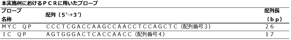
 実施の形態1では、1種類の反応液が入った1つの反応容器中で増幅する2つの標的核酸について、第1標的であるpMYCに特異的にアニールするQProbe(MYC QP)のTm値をプライマーのTm値よりも高い温度に設定し、増幅反応中に検出した。
(PCR / fluorescence measurement conditions)
In the first embodiment, for two target nucleic acids to be amplified in one reaction vessel containing one kind of reaction solution, the Tm value of QProbe (MYC QP) that specifically anneals to the first target pMYC is used as a primer. The temperature was set higher than the Tm value of and detected during the amplification reaction.
 次に、第2標的であるpICM5に特異的にアニールするQProbe(IC QP)のTm値を増幅反応中の変化温度(70℃―95℃)よりも低い温度に設定し、増幅反応後に検出した。 Next, the Tm value of QProbe (IC QP) that specifically anneals to the second target, pICM5, was set to a temperature lower than the change temperature (70 ° C.-95 ° C.) during the amplification reaction, and was detected after the amplification reaction. .
 PCR反応液組成、反応条件を(表3)と(表4)に示し、また、混合液の温度変化をグラフ化したものを図1に示す。 The PCR reaction solution composition and reaction conditions are shown in (Table 3) and (Table 4), and the temperature change of the mixed solution is shown in FIG.
Figure JPOXMLDOC01-appb-T000003
Figure JPOXMLDOC01-appb-T000004
 PCR及び蛍光測定には、LightCycler(登録商標)nano(ロシュ・ダイアグノスティックス株式会社)を使用した。 LightCycler (registered trademark) nano (Roche Diagnostics Co., Ltd.) was used for PCR and fluorescence measurement.
 蛍光測定時の励起波長と蛍光波長の組み合わせは510-528nmを選択した。各サイクルの変性ステップ(95℃)と伸長ステップ(72℃)で蛍光値を測定し、更に、増幅反応後に第2標的を検出するためのステップ(95℃と55℃)において蛍光測定を行った。 The combination of excitation wavelength and fluorescence wavelength at the time of fluorescence measurement was selected to be 510-528 nm. The fluorescence value was measured at the denaturation step (95 ° C.) and the extension step (72 ° C.) of each cycle, and further the fluorescence measurement was performed at the steps (95 ° C. and 55 ° C.) for detecting the second target after the amplification reaction. .
 増幅反応の最終サイクルと、第2標的検出ステップにおける蛍光測定条件を図2に示す。 Fig. 2 shows the final cycle of the amplification reaction and the fluorescence measurement conditions in the second target detection step.
 (データの処理方法)
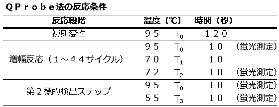
 QProbeを用いた際の解析方法は、LightCycler nanoに備え付けの解析ソフトが対応していないため、特許文献5(日本国特許第4724380号公報)に記載されている方法を参考に、得られた生データに対して補正演算処理を行った。
(Data processing method)
The analysis method when using QProbe is not compatible with the analysis software provided in LightCycler nano, so the raw data obtained with reference to the method described in Patent Document 5 (Japanese Patent No. 4724380) is referred to. Correction calculation processing was performed on the data.
 増幅反応と第2標的検出ステップの全てのサイクル毎に、次の数式による演算を行った。
fn=fhyb.n/fden.n (数1)
fe=fhyb.e/fden.e (数1’)
fn:(数1)により算出されたnサイクルにおける蛍光強度値
fhyb.n:nサイクル目の伸長ステップの蛍光強度値
fden.n:nサイクル目の変性ステップの蛍光強度値
fe:(数1’)により算出された第2標的検出ステップにおける蛍光強度値
fhyb.e:第2標的検出ステップ55℃の蛍光強度値
fden.e:第2標的検出ステップ95℃の蛍光強度値
For each cycle of the amplification reaction and the second target detection step, the calculation according to the following equation was performed.
fn = fhyb. n / fden. n (Equation 1)
fe = fhyb. e / fden. e (Equation 1 ')
fn: Fluorescence intensity value fhyb. in n cycles calculated by (Equation 1) n: fluorescence intensity value fden. n: Fluorescence intensity value fhyb. in the second target detection step calculated by the fluorescence intensity value fe of the denaturation step in the n-th cycle: (Equation 1 ′) e: Second target detection step Fluorescence intensity value fden. e: Second target detection step Fluorescence intensity value at 95 ° C.
 次に、増幅反応の全てのサイクルと第2標的検出ステップについて、次の数式で演算を行った。
Fn=fn/f10 (数2)
Fe=fe/f10 (数2’)
Fn:10サイクル目の(数1)により得られた蛍光強度値を1とした時のnサイクル目の相対値
Fe:10サイクル目の(数1’)により得られた蛍光強度値を1とした時の第2標的検出ステップの相対値
Next, for all cycles of the amplification reaction and the second target detection step, calculation was performed using the following formula.
Fn = fn / f10 (Equation 2)
Fe = fe / f10 (Equation 2 ′)
Fn: relative value of the nth cycle when the fluorescence intensity value obtained by (Equation 1) of the 10th cycle is 1, Fe: the fluorescence intensity value obtained by (
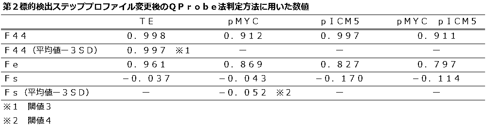
Equation 1 ′) of the 10th cycle is 1. Relative value of the second target detection step when
 増幅反応の最終サイクルのFnの値であるF44を、第1標的核酸の有無の判定に用いた。 F44, which is the value of Fn in the final cycle of the amplification reaction, was used to determine the presence or absence of the first target nucleic acid.
 更に、次の数式による演算を行った。
Fs=Fe-F44  (数3)
Fs:第2標的の測定値
Fsの値を、第2標的核酸の有無の判定に用いた。
Furthermore, the calculation by the following formula was performed.
Fs = Fe-F44 (Equation 3)
Fs: The measured value Fs of the second target was used to determine the presence or absence of the second target nucleic acid.
 QProbe法における判定は下記に示す方法で行った。
 第1標的核酸の有無は、測定試料のF44の値と閾値1を比較して[測定試料のF44<閾値1]を陽性と判定した。
The determination in the QProbe method was performed by the method shown below.
The presence or absence of the first target nucleic acid was determined by comparing the value of F44 of the measurement sample with the threshold 1 [F44 of measurement sample <threshold 1] as positive.
 閾値1は、陰性対照(DNAの代わりにTE緩衝液を添加した試薬)のF44の平均値-標準偏差の3倍(以下mean-3SD)の値を用いた。 Threshold value 1 was a negative control (reagent added with TE buffer instead of DNA) F44 average value-three times the standard deviation (hereinafter mean-3SD).
 第2標的核酸の有無は、第1標的核酸が陽性、陰性に関わらず、Fsの値と閾値2を比較して[Fs<閾値2]を陽性と判定した。 The presence or absence of the second target nucleic acid was determined as [Fs <threshold 2] positive by comparing the Fs value with the threshold 2 regardless of whether the first target nucleic acid was positive or negative.
 閾値2は、pMYCだけを添加した試料のFsのmean-3SDの値を用いた。判定方法のフローチャートは、図3に示すとおりである。 As the threshold 2, the value of mean-3SD of Fs of the sample to which only pMYC was added was used. The flowchart of the determination method is as shown in FIG.
 マイコプラズマP1遺伝子はpMYC、内部コントロールはpICM5を、それぞれ核酸試料に用いてPCRを実施した。 PCR was performed using pMYC for the mycoplasma P1 gene and pICM5 for the internal control, respectively, as nucleic acid samples.
 MYC QPの配列はTm値を72.5℃、IC QPの配列はTm値を57.5℃に設定した。そのため、PCR中の温度範囲(70℃から95℃)では、プローブはMYC QPだけがアニールすると推測された。 The MYC QP array had a Tm value of 72.5 ° C, and the IC QP array had a Tm value of 57.5 ° C. Therefore, in the temperature range during PCR (70 ° C. to 95 ° C.), it was speculated that only MYC QP annealed as a probe.
 第2標的検出ステップの蛍光測定は、95℃とIC QPのTm値よりも低い55℃で行うため、第2標的検出ステップの測定値はMYC QPとIC QPの消光を同時に検出すると推測された。 Since the fluorescence measurement in the second target detection step is performed at 95 ° C. and 55 ° C., which is lower than the Tm value of IC QP, it was estimated that the measurement value in the second target detection step detects quenching of MYC QP and IC QP simultaneously. .
 陰性対照の増幅曲線は、PCRが終了する44サイクル目まで消光が見られず(F44の値0.998が閾値1の値0.997よりも低くないため)、この時のmean-3SDは0.997となり、実施の形態1の第1標的検出のための閾値1とし、(表5)に示す。増幅曲線を図4に示す。 The amplification curve of the negative control shows no quenching until the 44th cycle when PCR ends (because the F44 value of 0.998 is not lower than the threshold 1 value of 0.997) and the mean-3SD at this time is 0 It is set to the threshold 1 for the first target detection of the first embodiment, and is shown in (Table 5). The amplification curve is shown in FIG.
 第1標的核酸であるpMYCの増幅曲線は、28サイクル付近からMYC QPのアニーリングに伴う消光が観察された。 In the amplification curve of pMYC, the first target nucleic acid, quenching accompanying annealing of MYC QP was observed from around 28 cycles.
 F44の値0.913は閾値1より低く、pMYCの目的の領域が増幅したことが示された。この時のFsのmean-3SDは-0.056であり、第2標的検出のための閾値2とし、(表5)に示す。増幅曲線を図5に示す。 The F44 value of 0.913 was lower than the threshold value 1, indicating that the target region of pMYC was amplified. At this time, mean-3SD of Fs is −0.056, and is set as a threshold value 2 for detecting the second target, as shown in (Table 5). The amplification curve is shown in FIG.
 第2標的核酸であるpICM5の増幅曲線は、増幅反応中は全く消光が見られず(F44の値0.997が閾値1の値0.997よりも低くないため)Fsの値-0.118は、閾値2より低かった。 The amplification curve of the second target nucleic acid pICM5 shows no quenching during the amplification reaction (because the F44 value of 0.997 is not lower than the threshold 1 value of 0.997), and the Fs value is −0.118. Was lower than threshold 2.
 この結果より、pMYCの目的の領域が増幅しなかったことと、pICM5の目的の領域が増幅したことが示された。 From this result, it was shown that the target region of pMYC was not amplified and the target region of pICM5 was amplified.
 Fsの値を(表5)に示す。増幅曲線を図6に示す。 The values of Fs are shown in (Table 5). The amplification curve is shown in FIG.
Figure JPOXMLDOC01-appb-T000005
 第1標的核酸であるpMYCと第2標的核酸であるpICM5の両方を添加した際の増幅曲線は、pMYCの増幅曲線と同様に、28サイクル付近からMYC QPのアニーリングに伴う消光が観察され、F44の値0.912は閾値1より低く、pMYCの目的の領域が増幅したことが示された。 The amplification curve when adding both the first target nucleic acid pMYC and the second target nucleic acid pICM5, as in the case of the pMYC amplification curve, was observed to be quenched by annealing of MYC QP from around 28 cycles. A value of 0.912 was lower than threshold 1, indicating that the target region of pMYC was amplified.
 Fsの値-0.116は、閾値2より低く、pICM5の目的の領域が増幅したことが示された。Fsの値を(表5)に示す。増幅曲線を図7に示す。 The value of Fs−0.116 was lower than threshold 2, indicating that the target region of pICM5 was amplified. The value of Fs is shown in (Table 5). The amplification curve is shown in FIG.
 これらのPCR産物はPCR終了後にアガロース電気泳動し、pMYCを添加した試料では206bp付近、pICM5を添加した試料では150bp付近に目的のPCR産物が確認された。 These PCR products were subjected to agarose electrophoresis after the completion of PCR, and the target PCR product was confirmed around 206 bp in the sample added with pMYC and around 150 bp in the sample added with pICM5.
 以上の結果より、QProbe法において、プローブの設計方法と温度変化プロファイルを組み合わせ、1種類の反応液が入った1つの反応容器で単一蛍光標識を用い、マイコプラズマP1遺伝子と内部コントロールを検出できることが示された。 From the above results, the QProbe method can detect the mycoplasma P1 gene and the internal control by combining a probe design method and a temperature change profile and using a single fluorescent label in one reaction vessel containing one type of reaction solution. Indicated.
 (実施の形態2)
 <第2検出ステップのプロファイルを変更したQProbe法におけるマイコプラズマP1遺伝子と内部コントロールの検出>
(Embodiment 2)
<Detection of Mycoplasma P1 Gene and Internal Control in QProbe Method with Modified Profile of Second Detection Step>
 実施の形態1の第2標的検出ステップのプロファイルを変更した方法でQProbe法におけるマイコプラズマP1遺伝子と内部コントロールの検出を行った。 The mycoplasma P1 gene and internal control in the QProbe method were detected by the method of changing the profile of the second target detection step of the first embodiment.
 PCRに用いたプライマー、プローブ、反応液組成と核酸試料は実施の形態1と同じものを用いた。本形態の反応条件は、(表6)に示すとおりである。 The same primer, probe, reaction solution composition and nucleic acid sample used in PCR as those in Embodiment 1 were used. The reaction conditions of this form are as shown in (Table 6).
Figure JPOXMLDOC01-appb-T000006
 実施の形態2では、第2標的検出ステップで55℃において蛍光測定を行った。増幅反応の最終サイクルと、第2標的検出ステップにおける蛍光測定条件を図8に示す。 In Embodiment 2, fluorescence measurement was performed at 55 ° C. in the second target detection step. FIG. 8 shows the final cycle of the amplification reaction and the fluorescence measurement conditions in the second target detection step.
 実施の形態2においてfeは次の演算を用い、その他は実施の形態1と同様な演算処理を行った。
fe=fhyb.e/fden.44
fe:第2標的検出ステップにおける蛍光強度値
fhyb.e:第2標的検出ステップ55℃の蛍光強度値
fden.44:増幅反応の最終サイクルの95℃の蛍光強度値
In the second embodiment, fe uses the following calculation, and otherwise performs the same calculation process as in the first embodiment.
fe = fhyb. e / fden. 44
fe: fluorescence intensity value fhyb. in the second target detection step. e: Second target detection step Fluorescence intensity value fden. 44: Fluorescence intensity value at 95 ° C. of the final cycle of the amplification reaction
 以下に、実施の形態2の判定方法を示す。第1標的核酸の有無は、測定試料のF44の値と閾値3を比較して[測定試料のF44<閾値3]を陽性と判定した。 The determination method of Embodiment 2 is shown below. The presence or absence of the first target nucleic acid was determined by comparing the F44 value of the measurement sample with the threshold 3 and [F44 of the measurement sample <threshold 3] being positive.
 閾値3は、陰性対照のF44の平均値-標準偏差の3倍(以下mean-3SD)の値を用いた。 As the threshold 3, a value of the average value of F44 of negative control-three times the standard deviation (hereinafter mean-3SD) was used.
 第2標的核酸の有無は、第1標的核酸が陽性、陰性に関わらず、Fsの値と閾値4を比較して[Fs<閾値4]を陽性と判定した。 The presence or absence of the second target nucleic acid was determined as [Fs <threshold 4] positive by comparing the Fs value with the threshold 4 regardless of whether the first target nucleic acid was positive or negative.
 閾値4は、pMYCだけを添加した試料のFsのmean-3SDの値を用いた。 As the threshold 4, the value of mean-3SD of Fs of a sample to which only pMYC was added was used.
 陰性対照の増幅曲線は、増幅反応が終了する44サイクル目まで消光が見られず(F44の値0.998が閾値3の値0.997よりも低くないため)、この時のmean-3SDは0.997となり、本例の第1標的検出のための閾値3とし、(表7)に示す。増幅曲線は、図9に示すとおりである。 The amplification curve of the negative control shows no quenching until the 44th cycle when the amplification reaction ends (because the F44 value of 0.998 is not lower than the threshold 3 value of 0.997), and mean-3SD at this time is It becomes 0.997, and is set as the threshold value 3 for the first target detection of this example, and is shown in (Table 7). The amplification curve is as shown in FIG.
 第1標的核酸であるpMYCの増幅曲線は、28サイクル付近からMYC QPのアニーリングに伴う消光が観察され、F44の値0.912は閾値3より低く、pMYCの目的の領域が増幅したことが示された。 The amplification curve of pMYC, which is the first target nucleic acid, shows that quenching accompanying annealing of MYC QP was observed from around 28 cycles, F44 value 0.912 was lower than threshold 3, indicating that the target region of pMYC was amplified. It was done.
 また、この時のFsのmean-3SDは-0.052であり、第2標的検出のための閾値4とし、(表7)に示す。増幅曲線は、図10に示すとおりである。 Further, the mean-3SD of Fs at this time is −0.052, and it is shown as (Table 7) as the threshold value 4 for detecting the second target. The amplification curve is as shown in FIG.
 第2標的核酸であるpICM5の増幅曲線は、増幅反応中は全く消光が見られず(F44の値0.997が閾値3の値0.997よりも低くないため)、Fsの値-0.170は、閾値4より低かった。 The amplification curve of pICM5, which is the second target nucleic acid, shows no quenching during the amplification reaction (because the F44 value of 0.997 is not lower than the threshold 3 value of 0.997), and the Fs value is −0. 170 was below threshold 4.
 この結果より、pMYCの目的の領域が増幅しなかったことと、pICM5の目的の領域が増幅したことが示された。Fsの値を(表7)に示す。増幅曲線を図11に示す。 From this result, it was shown that the target region of pMYC was not amplified and the target region of pICM5 was amplified. The value of Fs is shown in (Table 7). The amplification curve is shown in FIG.
Figure JPOXMLDOC01-appb-T000007
 第1標的核酸であるpMYCと第2標的核酸であるpICM5の両方を添加した際の増幅曲線は、pMYCの増幅曲線と同様に、28サイクル付近からMYC QPのアニーリングに伴う消光が観察され、F44の値0.911は閾値3より低く、pMYCの目的の領域が増幅したことが示された。 The amplification curve when adding both the first target nucleic acid pMYC and the second target nucleic acid pICM5, as in the case of the pMYC amplification curve, was observed to be quenched by annealing of MYC QP from around 28 cycles. A value of 0.911 was lower than threshold 3, indicating that the target region of pMYC was amplified.
 Fsの値-0.114は、閾値4より低く、pICM5の目的の領域が増幅したことが示された。Fsの値を(表7)に示す。増幅曲線を図12に示す。 The value of Fs−0.114 was lower than the threshold value 4, indicating that the target region of pICM5 was amplified. The value of Fs is shown in (Table 7). The amplification curve is shown in FIG.
 以上の結果より、feの算出に、増幅反応の最終サイクルの95℃の蛍光強度値(fden.44)を用いても、第2標的を検出できることが示された。 From the above results, it was shown that the second target can be detected even if the fluorescence intensity value (fden.44) at 95 ° C. of the final cycle of the amplification reaction is used for the calculation of fe.
 (実施の形態3)
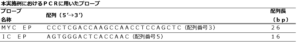
 <Eprobe法におけるマイコプラズマP1遺伝子と内部コントロールの検出>
(Embodiment 3)
<Detection of Mycoplasma P1 Gene and Internal Control in Eprobe Method>
 Eprobe法におけるマイコプラズマP1遺伝子と内部コントロールの検出を行った。 The Mycoplasma P1 gene and internal control were detected by the Eprobe method.
 PCRに用いたプライマーと核酸試料は実施の形態1と同じものを用いた。実施の形態3におけるPCR法に用いるプローブの情報は、(表8)に示すとおりである。 The same primers and nucleic acid sample used in PCR as those in Embodiment 1 were used. Information on probes used for the PCR method in Embodiment 3 is as shown in (Table 8).
Figure JPOXMLDOC01-appb-T000008
 Eprobeは、株式会社ダナフォームが作製したソフトウェアである「Edesign」を用い計算したTm値を参考にし、特定領域を選択した。 Eprobe selected a specific region with reference to the Tm value calculated using “Design”, which is software created by DanaForm Co., Ltd.
 pMYCに特異的にアニールするEprobeであるMYC EPとpICM5に特異的にアニールするEprobeであるIC EPの蛍光色素にはD514を使用し、MYC EPは5’末端から19番目のTを標識し、IC EPは5’末端から9番目のTを標識した。 MYC EP, which is an probe that specifically anneals to pMYC, and D514 are used as the fluorescent dye of IC EP, which is an probe that specifically anneals to pICM5, and MYC EP labels the 19th T from the 5 ′ end, IC EP labeled the 9th T from the 5 'end.
 Eprobeはユーロフィンジェノミクス株式会社に合成を委託して作製した。PCR反応液組成、反応条件を(表9)と(表10)に示す。 Eprobe was produced by consigning synthesis to Eurofin Genomics. The PCR reaction solution composition and reaction conditions are shown in (Table 9) and (Table 10).
Figure JPOXMLDOC01-appb-T000009
Figure JPOXMLDOC01-appb-T000010
 蛍光測定時の励起波長と蛍光波長の組み合わせは530-548nmを選択した。 530-548 nm was selected as the combination of excitation wavelength and fluorescence wavelength during fluorescence measurement.
 実施の形態3におけるFsは次の演算を用い、その他は実施の形態1と同様な演算処理を行った。
Fs=Fe-F40
Fs in the third embodiment uses the following calculation, and otherwise performs the same calculation process as in the first embodiment.
Fs = Fe-F40
 実施の形態3における判定は下記に示す方法で行った。第1標的核酸の有無は、測定試料のF40の値と閾値5を比較して[測定試料のF40>閾値5]を陽性と判定した。 The determination in Embodiment 3 was performed by the following method. The presence or absence of the first target nucleic acid was determined by comparing the F40 value of the measurement sample with the threshold 5 and [F40 of measurement sample> threshold 5] as positive.
 閾値5は、陰性対照のF40の平均値+標準偏差の3倍(以下mean+3SD)の値を用いた。 As the threshold 5, a value of the average value of F40 of the negative control + three times the standard deviation (hereinafter mean + 3SD) was used.
 第2標的核酸の有無は、第1標的核酸が陽性、陰性に関わらず、Fsの値と閾値6を比較して[Fs>閾値6]を陽性と判定した。 The presence or absence of the second target nucleic acid was determined as [Fs> threshold 6] positive by comparing the Fs value with the threshold 6 regardless of whether the first target nucleic acid was positive or negative.
 閾値6は、pMYCだけを添加した試料のFsのmean+3SDの値を用いた。判定方法のフローチャートは、図13に示すとおりである。 As the threshold 6, the value of mean + 3SD of Fs of the sample to which only pMYC was added was used. The flowchart of the determination method is as shown in FIG.
 MYC EPの配列はTm値を76.4℃に、IC EPの配列はTm値を59.8℃に設定した。そのため、PCR中の温度範囲(70℃から95℃)では、プローブはMYC EPだけがアニールすると推測された。 The MYC EP array had a Tm value of 76.4 ° C, and the IC EP array had a Tm value of 59.8 ° C. Therefore, in the temperature range during PCR (70 ° C. to 95 ° C.), it was speculated that only the MYC EP probe annealed.
 又、第2標的検出ステップの蛍光測定は、95℃とIC EPのTm値よりも低い55℃で行うため、第2標的検出ステップの測定値はMYC EPとIC EPの発光を同時に検出すると推測された。 In addition, since the fluorescence measurement in the second target detection step is carried out at 95 ° C. and 55 ° C., which is lower than the Tm value of IC EP, it is assumed that the measurement value in the second target detection step simultaneously detects luminescence of MYC EP and IC EP It was done.
 陰性対照の増幅曲線は、PCRが終了する40サイクル目まで発光が見られず(F40の値1.007が閾値5の値1.013よりも高くないため)、この時のmean+3SDは1.013となり、第1標的核酸検出のための閾値5とし、(表11)に示す。増幅曲線を図14に示す。 In the amplification curve of the negative control, no luminescence was observed until the 40th cycle when PCR was completed (because F40 value 1.007 is not higher than threshold value 1.013), and mean + 3SD at this time was 1.013. Thus, the threshold value 5 for detecting the first target nucleic acid is set as shown in (Table 11). The amplification curve is shown in FIG.
 第1標的核酸であるpMYCの増幅曲線は、29サイクル付近からMYC EPのアニーリングに伴う発光が観察され、F40の値2.314が閾値5より高く、pMYCの目的の領域が増幅したことが示された。 The amplification curve of pMYC, which is the first target nucleic acid, shows that light emission accompanying MYC EP annealing was observed from around 29 cycles, F40 value 2.314 was higher than threshold 5, and the target region of pMYC was amplified. It was done.
 また、この時のFsのmean+3SDは3.695であり、第2標的検出のための閾値6とし(表11)に示す。増幅曲線を図15に示す。 Further, mean + 3SD of Fs at this time is 3.695, which is shown in Table 11 as a threshold 6 for detecting the second target. The amplification curve is shown in FIG.
 第2標的核酸であるpICM5の増幅曲線は、増幅反応中は全く発光が見られず(F40の値1.012が閾値5の値1.013よりも高くないため)、Fsの値4.012は、閾値6より高かった。 In the amplification curve of the second target nucleic acid pICM5, no luminescence was observed during the amplification reaction (because the F40 value of 1.012 is not higher than the threshold 5 value of 1.013), and the Fs value of 4.012. Was higher than threshold 6.
 この結果より、pMYCの目的の領域が増幅しなかったことと、pICM5の目的の領域が増幅したことが示された。Fsの値を(表11)に示す。増幅曲線を図16に示す。 From this result, it was shown that the target region of pMYC was not amplified and the target region of pICM5 was amplified. The value of Fs is shown in (Table 11). The amplification curve is shown in FIG.
Figure JPOXMLDOC01-appb-T000011
 第1標的核酸であるpMYCと第2標的核酸であるpICM5の両方を添加した際の増幅曲線は、pMYCの増幅曲線と同様に、29サイクル付近からMYC EPのアニーリングに伴う発光が観察され、F40の値2.355は閾値5より高く、pMYCの目的の領域が増幅したことが示された。 The amplification curve when adding both the first target nucleic acid pMYC and the second target nucleic acid pICM5 is similar to the amplification curve of pMYC, and light emission accompanying annealing of MYC EP is observed from around 29 cycles. The value of 2.355 was higher than the threshold value 5, indicating that the target region of pMYC was amplified.
 Fsの値4.254は、閾値6より高く、pICM5の目的の領域が増幅したことが示された。Fsの値を(表11)に示す。増幅曲線を図17に示す。 Fs value 4.254 was higher than threshold 6, indicating that the target region of pICM5 was amplified. The value of Fs is shown in (Table 11). The amplification curve is shown in FIG.
 以上の結果より、Eprobe法においてもプローブの設計方法と温度変化プロファイルを組み合わせ、1種類の反応液が入った1つの反応容器で単一蛍光標識を用い、マイコプラズマP1遺伝子と内部コントロールを検出できることが示された。 From the above results, it is possible to detect the mycoplasma P1 gene and the internal control using a single fluorescent label in one reaction vessel containing one kind of reaction solution by combining the probe design method and the temperature change profile also in the probe method. Indicated.
 (実施の形態4)
 <TaqManプローブ法におけるマイコプラズマP1遺伝子と内部コントロールの検出>
(Embodiment 4)
<Detection of Mycoplasma P1 Gene and Internal Control in TaqMan Probe Method>
 TaqManプローブ法におけるマイコプラズマP1遺伝子と内部コントロールの検出を行った。PCRに用いたプライマーと核酸試料は実施の形態1と同じものを用いた。 The mycoplasma P1 gene and internal control were detected in the TaqMan probe method. The same primers and nucleic acid samples used for PCR as in Embodiment 1 were used.
 実施の形態4におけるPCR法に用いるプローブの情報は、(表12)に示すとおりである。 Information on probes used in the PCR method in Embodiment 4 is as shown in (Table 12).
Figure JPOXMLDOC01-appb-T000012
 TaqManプローブは、nearest neighbor法を用い計算したTm値を参考にし、特定領域を選択した。 For the TaqMan probe, a specific region was selected with reference to the Tm value calculated using the nearest neighbor method.
 pMYCに特異的にアニールするTaqManプローブであるMYC TaqとpICM5に特異的にアニールするTaqManプローブであるIC Taqの蛍光色素には、それぞれ5’末端にFAM(登録商標)、3’末端にTAMRA(登録商標)を標識した。 The fluorescent dyes of MYC Taq, which is a TaqMan probe that specifically anneals to pMYC, and IC Taq, which is a TaqMan probe that specifically anneals to pICM5, have FAM (registered trademark) at the 5 ′ end and TAMRA ( (Registered trademark).
 TaqManプローブはタカラバイオ株式会社に合成を委託して作製した。PCR反応液組成、反応条件を(表13)と(表14)に示す。 The TaqMan probe was produced by consigning synthesis to Takara Bio Inc. The PCR reaction solution composition and reaction conditions are shown in (Table 13) and (Table 14).
Figure JPOXMLDOC01-appb-T000013
Figure JPOXMLDOC01-appb-T000014
 蛍光測定時の励起波長と蛍光波長の組み合わせは530-548nmを選択した。 530-548 nm was selected as the combination of excitation wavelength and fluorescence wavelength during fluorescence measurement.
 増幅反応後の第2標的を検出するためのステップでは95℃と50℃において蛍光測定を行った。 In the step for detecting the second target after the amplification reaction, fluorescence measurement was performed at 95 ° C. and 50 ° C.
 実施の形態4ではfhyb.e:第2標的検出ステップ50℃の蛍光強度値とし、その他は実施の形態1と同様な演算ステップを行った。 In the fourth embodiment, fhyb. e: Second target detection step The fluorescence intensity value at 50 ° C. was used, and the other calculation steps were the same as in Embodiment 1.
 実施の形態4における判定方法は下記に示す方法で行った。第1標的核酸の有無は、測定試料のF44の値と閾値7を比較して[測定試料のF44>閾値7]を陽性と判定した。 The determination method in Embodiment 4 was performed by the following method. The presence or absence of the first target nucleic acid was determined by comparing the F44 value of the measurement sample with the threshold 7 and [F44 of measurement sample> threshold 7] as positive.
 閾値7は、陰性対照のF44の平均値+標準偏差の3倍(以下mean+3SD)の値を用いた。 As the threshold 7, a value of the average value of F44 of the negative control + three times the standard deviation (hereinafter mean + 3SD) was used.
 第2標的核酸の有無は、第1標的核酸が陽性、陰性に関わらず、Fsの値と閾値8を比較して[Fs>閾値8]を陽性と判定した。 The presence or absence of the second target nucleic acid was determined as [Fs> threshold 8] positive by comparing the Fs value with the threshold 8 regardless of whether the first target nucleic acid was positive or negative.
 閾値8は、pMYCだけを添加した試料のFsのmean+3SDの値を用いた。 As the threshold value 8, the value of mean + 3SD of Fs of the sample to which only pMYC was added was used.
 MYC Taqの配列はTm値を75.8℃に、IC Taqの配列はTm値を53.7℃に設定した。そのため、PCR中の温度範囲(70℃から95℃)では、プローブはMYC Taqだけがアニールすると推測された。 The MYC Taq array had a Tm value of 75.8 ° C, and the IC Taq array had a Tm value of 53.7 ° C. Therefore, in the temperature range during PCR (70 ° C. to 95 ° C.), it was speculated that only MYC Taq annealed as a probe.
 又、第2標的検出ステップの蛍光測定は、95℃とIC TaqのTm値よりも低い50℃で行うため、第2標的検出ステップの測定値はMYC TaqとIC Taqの発光を同時に検出すると推測された。 In addition, since the fluorescence measurement in the second target detection step is performed at 95 ° C. and 50 ° C., which is lower than the Tm value of IC Taq, the measurement value in the second target detection step is estimated to detect luminescence of MYC Taq and IC Taq simultaneously. It was done.
 陰性対照の増幅曲線は、PCRが終了する44サイクル目まで発光が見られず(F44の値1.028が閾値7の値1.028よりも高くないため)、この時のmean+3SDは1.028となり、第1標的核酸検出のための閾値7とし、(表15)に示す。増幅曲線を図18に示す。 In the amplification curve of the negative control, no luminescence was observed until the 44th cycle when PCR was completed (because the F44 value of 1.028 is not higher than the threshold 7 value of 1.028), and the mean + 3SD at this time was 1.028. Thus, the threshold value 7 for detecting the first target nucleic acid is set as shown in (Table 15). The amplification curve is shown in FIG.
 第1標的核酸であるpMYCの増幅曲線は、28サイクル付近からMYC Taqのアニーリングに伴う発光が観察され、F44の値1.427が閾値7より高く、pMYCの目的の領域が増幅したことが示された。 The amplification curve of pMYC, which is the first target nucleic acid, shows that luminescence accompanying MYC Taq annealing was observed from around 28 cycles, F44 value 1.427 was higher than threshold 7, and the target region of pMYC was amplified. It was done.
 また、この時のFsのmean+3SDは0.110であり、第2標的検出のための閾値8とし(表15)に示す。増幅曲線を図19に示す。 Further, the mean + 3SD of Fs at this time is 0.110, which is shown as a threshold value 8 for detecting the second target (Table 15). The amplification curve is shown in FIG.
Figure JPOXMLDOC01-appb-T000015
 第2標的核酸であるpICM5の増幅曲線は、増幅反応中は全く発光が見られず(F44の値1.028が閾値7の値1.028よりも高くないため)、Fsの値0.130は、閾値8より高かった。 The amplification curve of the second target nucleic acid pICM5 shows no luminescence during the amplification reaction (because the F44 value of 1.028 is not higher than the threshold 7 value of 1.028) and the Fs value of 0.130. Was higher than threshold 8.
 この結果より、pMYCの目的の領域が増幅しなかったことと、pICM5の目的の領域が増幅したことが示された。 From this result, it was shown that the target region of pMYC was not amplified and the target region of pICM5 was amplified.
 Fsの値を(表15)に示す。増幅曲線を図20に示す。 The values of Fs are shown in (Table 15). The amplification curve is shown in FIG.
 第1標的核酸であるpMYCと第2標的核酸であるpICM5の両方を添加した際の増幅曲線は、pMYCの増幅曲線と同様に、29サイクル付近からMYC EPのアニーリングに伴う発光が観察され、F40の値1.348は閾値7より高く、pMYCの目的の領域が増幅したことが示された。 The amplification curve when adding both the first target nucleic acid pMYC and the second target nucleic acid pICM5 is similar to the amplification curve of pMYC, and light emission accompanying annealing of MYC EP is observed from around 29 cycles. A value of 1.348 was higher than threshold 7, indicating that the target region of pMYC was amplified.
 Fsの値0.177は、閾値8より高く、pICM5の目的の領域が増幅したことが示された。Fsの値を(表15)に示す。増幅曲線を図21に示す。 Fs value 0.177 was higher than threshold 8, indicating that the target region of pICM5 was amplified. The value of Fs is shown in (Table 15). The amplification curve is shown in FIG.
 以上の結果より、TaqManプローブ法においてもプローブの設計方法と温度変化プロファイルを組み合わせ、1種類の反応液が入った1つの反応容器で単一蛍光標識を用い、マイコプラズマP1遺伝子と内部コントロールを検出できることが示された。 Based on the above results, the TaqMan probe method can detect the mycoplasma P1 gene and internal control using a single fluorescent label in one reaction vessel containing one type of reaction solution by combining the probe design method and temperature change profile. It has been shown.
 (実施の形態5)
 <実検体を用いたQProbe法におけるマイコプラズマP1遺伝子と内部コントロールの検出>
(Embodiment 5)
<Detection of Mycoplasma P1 Gene and Internal Control in QProbe Method Using Real Specimens>
 実検体を用いた、QProbe法におけるマイコプラズマP1遺伝子と内部コントロールの検出を行った。 The mycoplasma P1 gene and internal control were detected in the QProbe method using real samples.
 マイコプラズマP1遺伝子は、LAMP法(日本国特許第3313358号)によって、マイコプラズマ・ニューモニエが陽性と判定された(検査は株式会社ビー・エム・エルに委託)咽頭拭い液からQIAamp(登録商標)DNA mini Kit(QIAGEN社)を用いて抽出したトータルDNAを用いた。 The mycoplasma P1 gene was determined to be positive for Mycoplasma pneumoniae by the LAMP method (Japanese Patent No. 3313358) (examination was entrusted to BML Inc.) QIAamp (registered trademark) DNA mini from the throat swab Total DNA extracted using Kit (QIAGEN) was used.
 また、陰性試料としてLAMP法によって、マイコプラズマ・ニューモニエが陰性と判定された咽頭拭い液からQIAamp DNA mini Kitを用いて抽出したトータルDNAを用いた。その他条件は、実施の形態1と同様にPCRを実施した。 In addition, as a negative sample, total DNA extracted using a QIAamp DNA mini Kit from the pharyngeal wiping solution determined to be negative for Mycoplasma pneumoniae by the LAMP method was used. For other conditions, PCR was performed in the same manner as in the first embodiment.
 得られた生データに対して実施の形態1と同様の演算処理を行った。 The same arithmetic processing as that in the first embodiment was performed on the obtained raw data.
 以下に、実施の形態5の判定方法を示す。第1標的核酸の有無は、測定試料のF44の値と閾値9を比較して[測定試料のF44<閾値9]を陽性と判定した。 The determination method of Embodiment 5 is shown below. The presence or absence of the first target nucleic acid was determined by comparing the F44 value of the measurement sample with the threshold 9 and [F44 of the measurement sample <threshold 9] being positive.
 閾値9は、陰性試料のF44の平均値-標準偏差の3倍(以下mean-3SD)の値を用いた。 As the threshold value 9, a value of F44 average value of negative samples−three times the standard deviation (hereinafter mean-3SD) was used.
 第2標的核酸の有無は、第1標的核酸が陽性、陰性に関わらず、Fsの値と閾値10を比較して[Fs<閾値10]を陽性と判定した。閾値10は、陽性試料のFsのmean-3SDの値を用いた。 The presence or absence of the second target nucleic acid was determined as [Fs <threshold 10] positive by comparing the Fs value with the threshold 10 regardless of whether the first target nucleic acid was positive or negative. As the threshold 10, the value of mean-3SD of Fs of a positive sample was used.
 陰性試料の増幅曲線は、PCRが終了する44サイクル目まで消光が見られず(F44の値0.997が閾値9の値0.997よりも低くないため)、この時のmean-3SDは0.997となり、本例の第1標的検出のための閾値9とし、(表16)に示す。増幅曲線を図22に示す。 In the amplification curve of the negative sample, no quenching was observed until the 44th cycle when the PCR was completed (because the F44 value of 0.997 is not lower than the threshold 9 value of 0.997). It is set to the threshold value 9 for the first target detection of this example, and is shown in (Table 16). The amplification curve is shown in FIG.
 第1標的核酸である陽性試料の増幅曲線は、35サイクル付近からMYC QPのアニーリングに伴う消光が観察され、F44の値0.951は閾値9より低く、pMYCの目的の領域が増幅したことが示された。 As for the amplification curve of the positive sample as the first target nucleic acid, quenching accompanying annealing of MYC QP was observed from around 35 cycles, F44 value 0.951 was lower than threshold 9, and the target region of pMYC was amplified. Indicated.
 また、この時のFsのmean-3SDは-0.132であり、第2標的検出のための閾値10とし、(表16)に示す。増幅曲線を図23に示す。 Further, the mean-3SD of Fs at this time is −0.132, and is set as a threshold 10 for detecting the second target, as shown in (Table 16). The amplification curve is shown in FIG.
 第2標的核酸であるpICM5の増幅曲線は、増幅反応中は全く消光が見られず(F44の値0.998が閾値9の値0.997よりも低くないため)、Fsの値-0.227は、閾値10より低かった。 The amplification curve of pICM5, which is the second target nucleic acid, shows no quenching during the amplification reaction (because the F44 value of 0.998 is not lower than the threshold 9 value of 0.997), and the Fs value is −0. 227 was below threshold 10.
 この結果より、pMYCの目的の領域が増幅しなかったことと、pICM5の目的の領域が増幅したことが示された。Fsの値を(表16)に示す。増幅曲線を図24に示す。 From this result, it was shown that the target region of pMYC was not amplified and the target region of pICM5 was amplified. The value of Fs is shown in (Table 16). The amplification curve is shown in FIG.
 第1標的核酸である陽性試料に第2標的核酸であるpICM5を添加した際の増幅曲線は、陽性試料の増幅曲線と同様に、35サイクル付近からMYC QPのアニーリングに伴う消光が観察され、F44の値0.953は閾値9より低く、pMYCの目的の領域が増幅したことが示された。 As for the amplification curve when pICM5 as the second target nucleic acid was added to the positive sample as the first target nucleic acid, quenching due to annealing of MYC QP was observed from around 35 cycles, similar to the amplification curve of the positive sample, and F44 The value of 0.953 was lower than the threshold value 9, indicating that the target region of pMYC was amplified.
 Fsの値-0.235は、閾値10より低く、pICM5の目的の領域が増幅したことが示された。Fsの値を(表16)に示す。増幅曲線を図25に示す。 The value of Fs−0.235 was lower than the threshold value 10, indicating that the target region of pICM5 was amplified. The value of Fs is shown in (Table 16). The amplification curve is shown in FIG.
Figure JPOXMLDOC01-appb-T000016
 以上の結果より、実際の臨床検体でも、プローブの設計方法と温度変化プロファイルを組み合わせ、1種類の反応液が入った1つの反応容器で単一蛍光色素を用い、マイコプラズマP1遺伝子と内部コントロールを検出できることが示された。 Based on the above results, the mycoplasma P1 gene and internal control can be detected using a single fluorescent dye in a single reaction vessel containing one type of reaction solution by combining the probe design method and temperature change profile in an actual clinical sample. It was shown that it can be done.
 本発明は、感染症の病原体の亜型識別や一塩基多型の識別にも応用可能である。 The present invention can also be applied to subtype identification of pathogens of infectious diseases and single nucleotide polymorphism identification.
 (実施の形態6)
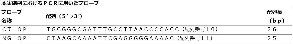
 <QProbe法におけるクラミジア内在性プラスミド遺伝子と淋菌CMT遺伝子の検出>
(Embodiment 6)
<Detection of Chlamydia endogenous plasmid gene and Neisseria gonorrhoeae CMT gene in QProbe method>
 QProbe法におけるクラミジア内在性プラスミド遺伝子と淋菌CMT遺伝子の検出を行った。 Chlamydia endogenous plasmid gene and Neisseria gonorrhoeae CMT gene were detected by the QProbe method.
 (pCT)
 性器クラミジア感染症の病原体であるクラミジア・トラコマチス共通の内在性プラスミド(pLGV440)遺伝子断片を人工合成後、pUC57ベクターに組み込んだプラスミドDNAは、北海道システムサイエンス株式会社に合成を委託して作製した。
(PCT)
After artificial synthesis of an endogenous plasmid (pLGV440) gene fragment common to Chlamydia trachomatis, a pathogen of genital chlamydia infection, plasmid DNA incorporated into the pUC57 vector was prepared by consigning synthesis to Hokkaido System Science Co., Ltd.
 (pNG)
 淋病の病原体であるナイセリア・ゴノレアのシトシンDNAメチルトランスフェラーゼ(CMT)遺伝子断片を人工合成後、pUC57ベクターに組み込んだプラスミドDNAは北海道システムサイエンス株式会社に合成を委託して作製した。
(PNG)
After artificially synthesizing the cytosine DNA methyltransferase (CMT) gene fragment of Neisseria gonorrhoeae that is a pathogen of gonorrhea, plasmid DNA incorporated into the pUC57 vector was prepared by consigning synthesis to Hokkaido System Science Co., Ltd.
 上記のプラスミドの長さは、pCTは3064bp、pNGは3059bpである。 The length of the above plasmid is 3064 bp for pCT and 3059 bp for pNG.
 各プラスミドの長さとプラスミド溶液の濃度(μg/μl)から1μLあたりのコピー数を計算した後、TE緩衝溶液(10mM Tris-HCl、1.0mM EDTA pH8.0)を用いて、pCT、pNG共に1×10^5copies/μlとなるように希釈した。 After calculating the number of copies per 1 μL from the length of each plasmid and the concentration of the plasmid solution (μg / μl), using a TE buffer solution (10 mM Tris-HCl, 1.0 mM EDTA pH 8.0), both pCT and pNG Dilute to 1 × 10 ^ 5 copies / μl.
 実施の形態6におけるPCR法に用いたプライマー対は、(表17)に示すとおりである。 Primer pairs used in the PCR method in Embodiment 6 are as shown in (Table 17).
Figure JPOXMLDOC01-appb-T000017
 実施の形態6におけるPCR法に用いるプローブの情報は、(表18)に示すとおりである。 Information on probes used in the PCR method in Embodiment 6 is as shown in (Table 18).
Figure JPOXMLDOC01-appb-T000018
 CT QPは配列番号6と配列番号7のプライマー対で増幅し得るPCR産物の塩基配列から日鉄住金環境株式会社J-Bio21センターのホームページ上のQProbe設計支援ツールを用い計算したTm値を参考にし、特定領域を選択した。 CT QP refers to the Tm value calculated using the QProbe design support tool on the website of J-Bio21 Center of Nippon Steel & Sumikin Environment Co., Ltd. from the base sequence of the PCR product that can be amplified with the primer pair of SEQ ID NO: 6 and SEQ ID NO: 7. Selected a specific area.
 NG QPは配列番号8と配列番号9のプライマー対で増幅し得るPCR産物の塩基配列から、J-Bio21センターのホームページ上のQProbe設計支援ツールを用い計算したTm値を参考にし、特定領域を選択した。 NG QP selects a specific region from the base sequence of the PCR product that can be amplified with the primer pair of SEQ ID NO: 8 and SEQ ID NO: 9, referring to the Tm value calculated using the QProbe design support tool on the J-Bio21 Center website. did.
 pCTに特異的にアニールするQProbeであるCT QPとpNGに特異的にアニールするQProbeであるNG QPの蛍光色素にはそれぞれBODIPY(登録商標)FLを使用し、それぞれ3’末端のCを標識した。 BODIPY (registered trademark) FL was used as a fluorescent dye for CT QP, a QProbe that specifically anneals to pCT, and NG QP, a QProbe that specifically anneals to pNG. .
 QProbeは日鉄住金環境株式会社に合成を委託して作製した。PCR反応液組成、反応条件を(表19)と(表20)に示す。 QProbe was produced by consigning synthesis to Nippon Steel & Sumikin Environment Co., Ltd. The PCR reaction solution composition and reaction conditions are shown in (Table 19) and (Table 20).
Figure JPOXMLDOC01-appb-T000019
Figure JPOXMLDOC01-appb-T000020
 蛍光測定時の励起波長と蛍光波長の組み合わせは510-528nmを選択した。 The combination of excitation wavelength and fluorescence wavelength at the time of fluorescence measurement was selected to be 510-528 nm.
 各サイクルの変性ステップ(95℃)と伸長ステップ(68℃)で蛍光値を測定し、更に、増幅反応後に第2標的を検出するためのステップ(95℃と55℃)において蛍光測定を行った。 The fluorescence value was measured at the denaturation step (95 ° C.) and the extension step (68 ° C.) of each cycle, and further the fluorescence measurement was performed at the steps (95 ° C. and 55 ° C.) for detecting the second target after the amplification reaction. .
 得られた生データに対して実施の形態1と同様の演算処理を行った。 The same arithmetic processing as that in the first embodiment was performed on the obtained raw data.
 以下に、本実施の形態の判定方法を示す。第1標的核酸の有無は、測定試料のF44の値と閾値11を比較して[測定試料のF44<閾値11]を陽性と判定した。 The determination method of this embodiment is shown below. The presence or absence of the first target nucleic acid was determined by comparing the value of F44 of the measurement sample with the threshold 11 and [F44 of the measurement sample <threshold 11] being positive.
 閾値11は、陰性対照のF44の平均値-標準偏差の3倍(以下mean-3SD)の値を用いた。 As the threshold value 11, a value of the average value of F44 of the negative control—three times the standard deviation (hereinafter mean-3SD) was used.
 第2標的核酸の有無は、第1標的核酸が陽性、陰性に関わらず、Fsの値と閾値12を比較して[Fs<閾値12]を陽性と判定した。 The presence or absence of the second target nucleic acid was determined as [Fs <threshold 12] positive by comparing the Fs value with the threshold 12 regardless of whether the first target nucleic acid was positive or negative.
 閾値12は、pNGだけを添加した試料のFsのmean-3SDの値を用いた。 As the threshold 12, the value of mean-3SD of Fs of a sample to which only pNG was added was used.
 CT QPの配列はTm値を73.4℃に、NG QPの配列はTm値を62.5℃に設定した。そのため、PCR中の温度範囲(68℃から95℃)では、プローブはCT QPだけがアニールすると推測された。 The CT QP array had a Tm value of 73.4 ° C, and the NG QP array had a Tm value of 62.5 ° C. Therefore, in the temperature range during PCR (68 ° C. to 95 ° C.), it was speculated that only CT QP annealed for the probe.
 又、第2標的検出ステップの蛍光測定は、95℃とNG QPのTm値よりも低い55℃で行うため、第2標的検出ステップの測定値はCT QPとNG QPの発光を同時に検出すると推測された。 In addition, since the fluorescence measurement in the second target detection step is performed at 55 ° C. which is lower than the Tm value of 95 ° C. and NG QP, it is assumed that the measurement value in the second target detection step simultaneously detects the emission of CT QP and NG QP. It was done.
 陰性対照の増幅曲線は、PCRが終了する44サイクル目まで消光が見られず(F44の値0.998が閾値11の値0.996よりも低くないため)、この時のmean-3SDは0.996となり、本例の第1標的検出のための閾値11とし、(表21)に示す。増幅曲線を図26に示す。 The amplification curve of the negative control shows no quenching until the 44th cycle when PCR ends (because the F44 value of 0.998 is not lower than the threshold 11 value of 0.996) and the mean-3SD at this time is 0 996, which is the threshold 11 for detecting the first target in this example, and is shown in (Table 21). The amplification curve is shown in FIG.
 第1標的核酸であるpCTの増幅曲線は、28サイクル付近からCT QPのアニーリングに伴う消光が観察され、F44の値0.720は閾値11より低く、pCTの目的の領域が増幅したことが示された。 The amplification curve of pCT, which is the first target nucleic acid, shows that quenching accompanying annealing of CT QP was observed from around 28 cycles, and F44 value 0.720 was lower than threshold 11, indicating that the target region of pCT was amplified. It was done.
 また、この時のFsのmean-3SDは-0.039であり、第2標的検出のための閾値12とし、(表21)に示す。増幅曲線を図27に示す。 Further, the mean-3SD of Fs at this time is −0.039, which is set as a threshold 12 for detecting the second target, and is shown in (Table 21). The amplification curve is shown in FIG.
 第2標的核酸であるpNGの増幅曲線は、増幅反応中は全く消光が見られず(F44の値0.998が閾値11の値0.996よりも低くないため)、Fsの値-0.065は、閾値12より低かった。 The amplification curve of pNG, the second target nucleic acid, shows no quenching during the amplification reaction (because the F44 value of 0.998 is not lower than the threshold 11 value of 0.996), and the Fs value is −0. 065 was lower than threshold 12.
 この結果より、pCTの目的の領域が増幅しなかったことと、pNGの目的の領域が増幅したことが示された。Fsの値を(表21)に示す。増幅曲線を図28に示す。 From this result, it was shown that the target region of pCT was not amplified and the target region of pNG was amplified. The value of Fs is shown in (Table 21). The amplification curve is shown in FIG.
Figure JPOXMLDOC01-appb-T000021
 第1標的核酸であるpCTと第2標的核酸であるpNGの両方を添加した際の増幅曲線は、pCTの増幅曲線と同様に、28サイクル付近からCT QPのアニーリングに伴う消光が観察され、F44の値0.729は閾値11より低く、pCTの目的の領域が増幅したことが示された。 In the amplification curve when both the first target nucleic acid pCT and the second target nucleic acid pNG were added, as with the pCT amplification curve, quenching accompanying annealing of CT QP was observed from around 28 cycles, and F44 A value of 0.729 was lower than threshold 11, indicating that the target region of pCT was amplified.
 Fsの値-0.111は、閾値12より低く、pNGの目的の領域が増幅したことが示された。Fsの値を(表21)に示す。増幅曲線を図29に示す。 The value of Fs−0.111 was lower than the threshold value 12, indicating that the target region of pNG was amplified. The value of Fs is shown in (Table 21). The amplification curve is shown in FIG.
 以上の結果より、2項目の遺伝子においてもプローブの設計方法と温度変化プロファイルを組み合わせ、1種類の反応液が入った1つの反応容器で単一蛍光色素を用い、マイコプラズマP1遺伝子と内部コントロールを検出できることが示された。 Based on the above results, the probe design method and temperature change profile were combined for two genes, and a single fluorescent dye was used in one reaction vessel containing one type of reaction solution to detect the Mycoplasma P1 gene and internal control. It was shown that it can be done.
 (実施の形態7)
 <増幅反応中に複数回第2標的を検出するQProbe法におけるマイコプラズマP1遺伝子と内部コントロールの検出>
(Embodiment 7)
<Detection of Mycoplasma P1 Gene and Internal Control in QProbe Method that Detects Second Target Multiple Times During Amplification Reaction>
 第2標的検出ステップを最終サイクルの後だけではなく、増幅反応中に折り込んだ方法でQProbe法におけるマイコプラズマP1遺伝子と内部コントロールの検出を行った。 The detection of the mycoplasma P1 gene and internal control in the QProbe method was performed not only after the final cycle but also by a method in which the second target detection step was folded during the amplification reaction.
 PCRに用いたプライマー、プローブ、反応液組成と核酸試料は実施の形態1と同じものを用いた。反応条件は、(表22)に示すとおりである。 The same primer, probe, reaction solution composition and nucleic acid sample used in PCR as those in Embodiment 1 were used. The reaction conditions are as shown in (Table 22).
Figure JPOXMLDOC01-appb-T000022
 また増幅反応の温度変化をグラフ化したものを図30に示す。 FIG. 30 shows a graph of the temperature change of the amplification reaction.
 実施の形態7の解析においては次の演算を行い、その他は実施の形態1同様のステップを行った。
fen=fhyb.en/fden.en (数4)
fen:(数4)により算出されたn回目の第2標的検出ステップにおける蛍光強度値
fhyb.en:n回目の第2標的検出ステップ55℃の蛍光強度値
fden.en:n回目の第2標的検出ステップ95℃の蛍光強度値
Fen=fen/f10 (数5)
Fen:10サイクル目の(数4)により得られた蛍光強度値を1とした時のn回目の第2標的検出ステップの相対値
Fs1=Fe1-F20 (数6)
Fs2=Fe2-F27 (数6’)
Fs3=Fe3-F34 (数6’’)
Fs4=Fe4-F41 (数6’’’)
Fs1:1回目の第2標的検出ステップの測定値
Fs2:2回目の第2標的検出ステップの測定値
Fs3:3回目の第2標的検出ステップの測定値
Fs4:4回目の第2標的検出ステップの測定値
In the analysis of the seventh embodiment, the following calculation was performed, and the other steps were the same as in the first embodiment.
fen = fhyb. en / fden. en (Equation 4)
fen: The fluorescence intensity value fhyb. in the n-th second target detection step calculated by (Expression 4). en: n-th second target detection step 55 ° C. fluorescence intensity value fden. en: nth second target detection step Fluorescence intensity value at 95 ° C. Fen = fen / f10 (Equation 5)
Fen: relative value Fs1 = Fe1-F20 in the n-th second target detection step when the fluorescence intensity value obtained by (Expression 4) in the 10th cycle is set to 1 (Expression 6)
Fs2 = Fe2-F27 (Equation 6 ′)
Fs3 = Fe3-F34 (Equation 6 '')
Fs4 = Fe4-F41 (Formula 6 ''')
Fs1: First measurement value of second target detection step Fs2: Second measurement value of second target detection step Fs3: Third measurement value of second target detection step Fs4: Fourth measurement value of second target detection step measured value
 判定は下記に示す方法で行った。第1標的核酸の有無は、測定試料のF41の値と閾値13を比較して[測定試料のF41<閾値13]を陽性と判定した。
 閾値13は、陰性対照のF41の平均値-標準偏差の3倍(以下mean-3SD)の値を用いた。
Determination was performed by the method shown below. The presence or absence of the first target nucleic acid was determined by comparing the F41 value of the measurement sample with the threshold 13 and [F41 of the measurement sample <threshold 13] as positive.
As the threshold 13, a value of the average value of F41 of the negative control minus 3 times the standard deviation (hereinafter mean-3SD) was used.
 第2標的核酸の有無は、1回目の第2標的検出ステップでは、[Fs1<閾値14]を陽性と判定した。 In the first second target detection step, [Fs1 <threshold 14] was determined as positive for the presence or absence of the second target nucleic acid.
 閾値14は、pMYCだけを添加した試料のFs1のmean-3SDの値を用いた。 As the threshold value 14, the value of mean-3SD of Fs1 of the sample added with only pMYC was used.
 2回目の第2標的検出ステップでは、[Fs2<閾値15]を陽性と判定した。 [Fs2 <threshold 15] was determined to be positive in the second second target detection step.
 閾値15は、pMYCだけを添加した試料のFs2のmean-3SDの値を用いた。 As the threshold value 15, the value of mean-3SD of Fs2 of the sample to which only pMYC was added was used.
 3回目の第2標的検出ステップでは、[Fs3<閾値16]を陽性と判定した。 In the third second target detection step, [Fs3 <threshold 16] was determined to be positive.
 閾値16は、pMYCだけを添加した試料のFs3のmean-3SDの値を用いた。 As the threshold 16, the value of mean-3SD of Fs3 of the sample to which only pMYC was added was used.
 4回目の第2標的検出ステップでは、[Fs4<閾値17]を陽性と判定した。 [Fs4 <threshold 17] was determined to be positive in the fourth second target detection step.
 閾値17は、pMYCだけを添加した試料のFs4のmean-3SDの値を用いた。 As the threshold value 17, the value of mean-3SD of Fs4 of the sample to which only pMYC was added was used.
 陰性対照の増幅曲線は、各第2標的検出ステップ以外ではPCRが終了する41サイクル目まで消光が見られず(F41の値0.999が閾値13の値0.998よりも低くないため)、この時のmean-3SDは0.998となり、本例の第1標的検出のための閾値13とし、(表23)に示す。増幅曲線を図31に示す。 The amplification curve of the negative control shows no quenching until the 41st cycle when PCR is completed except for each second target detection step (because the F41 value of 0.999 is not lower than the threshold 13 value of 0.998), The mean-3SD at this time is 0.998, which is the threshold 13 for detecting the first target in this example, and is shown in (Table 23). The amplification curve is shown in FIG.
 第1標的核酸であるpMYCの増幅曲線は、30サイクル付近からMYC QPのアニーリングに伴う消光が観察され、F41の値0.892は閾値13より低く、pMYCの目的の領域が増幅したことが示された。 The amplification curve of pMYC, which is the first target nucleic acid, showed that quenching accompanying annealing of MYC QP was observed from around 30 cycles, F41 value 0.892 was lower than threshold 13, indicating that the target region of pMYC was amplified. It was done.
 また、この時のFs1のmean-3SDは-0.058、Fs2のmean-3SDは-0.070、Fs3のmean-3SDは-0.080、Fs4のmean-3SDは-0.087でそれぞれ第2標的検出のための閾値14、15,16,17とし、(表23)に示す。増幅曲線を図32に示す。 At this time, mean-3SD of Fs1 is -0.058, mean-3SD of Fs2 is -0.070, mean-3SD of Fs3 is -0.080, and mean-3SD of Fs4 is -0.087. The thresholds 14, 15, 16, and 17 for detecting the second target are shown in (Table 23). The amplification curve is shown in FIG.
Figure JPOXMLDOC01-appb-T000023
 第2標的核酸であるpICM5の増幅曲線は、各第2標的検出ステップ以外では増幅反応中は全く消光が見られず(F41の値0.999が閾値13の値0.998よりも低くないため)、Fs1の値-0.058は、閾値14より高く、pICM5の目的の領域が1回目の第2標的検出ステップまでに検出限界以上に増幅していないことが示された。 In the amplification curve of pICM5, which is the second target nucleic acid, no quenching was observed during the amplification reaction except for each second target detection step (because the F41 value 0.999 is not lower than the threshold 13 value 0.998). ), The value of Fs1 −0.058 is higher than the threshold 14, indicating that the target region of pICM5 has not been amplified beyond the detection limit by the first second target detection step.
 Fs2の値-0.057も閾値15より高く、pICM5の目的の領域が2回目の第2標的検出ステップまでに検出限界以上に増幅していないことが示された。 Fs2 value -0.057 was also higher than threshold value 15, indicating that the target region of pICM5 was not amplified beyond the detection limit by the second second target detection step.
 Fs3の値-0.084は閾値16より低く、pICM5の目的の領域が3回目の第2標的検出ステップまでに検出限界以上に増幅していることが示された。 Fs3 value -0.084 was lower than threshold 16, indicating that the target region of pICM5 was amplified beyond the detection limit by the third second target detection step.
 Fs4の値-0.192も閾値17より低かった。Fs1、Fs2、Fs3、Fs4の値を(表23)に示す。増幅曲線を図33に示す。 The value of Fs4 -0.192 was also lower than the threshold value 17. The values of Fs1, Fs2, Fs3, and Fs4 are shown in (Table 23). The amplification curve is shown in FIG.
 第1標的核酸であるpMYCと第2標的核酸であるpICM5の両方を添加した際の増幅曲線は、pMYCの増幅曲線と同様に、30サイクル付近からMYC QPのアニーリングに伴う消光が観察され、F41の値0.897は閾値13より低く、pMYCの目的の領域が増幅したことが示された。 As with the amplification curve of pMYC, the amplification curve when both pMYC as the first target nucleic acid and pICM5 as the second target nucleic acid were added was observed to be quenched due to annealing of MYC QP from around 30 cycles. A value of 0.897 was lower than threshold 13, indicating that the target region of pMYC was amplified.
 Fs1の値-0.058は、閾値14よりも高く、pICM5の目的の領域が1回目の第2標的検出ステップまでに検出限界以上に増幅していないことが示された。 Fs1 value −0.058 is higher than threshold 14, indicating that the target region of pICM5 has not been amplified beyond the detection limit by the first second target detection step.
 Fs2の値-0.068も閾値15よりも高く、pICM5の目的の領域が2回目の第2標的検出ステップまでに検出限界以上に増幅していないことが示された。 Fs2 value -0.068 is also higher than threshold value 15, indicating that the target region of pICM5 has not been amplified beyond the detection limit by the second second target detection step.
 Fs3の値-0.109は閾値16より低く、pICM5の目的の領域が3回目の第2標的検出ステップまでに検出限界以上に増幅していることが示された。 Fs3 value -0.109 was lower than threshold 16, indicating that the target region of pICM5 had been amplified beyond the detection limit by the third second target detection step.
 Fs4の値―0.154も閾値17よりも低かった。Fs1、Fs2、Fs3、Fs4の値は、(表23)に示すとおりである。増幅曲線を図34に示す。 The value of Fs4—0.154 was also lower than the threshold value 17. The values of Fs1, Fs2, Fs3, and Fs4 are as shown in (Table 23). The amplification curve is shown in FIG.
 以上の結果より、第2標的検出ステップを最終サイクルの後だけではなく、増幅反応中に折り込んだ方法でもマイコプラズマP1遺伝子と内部コントロールを検出できることが示された。 From the above results, it was shown that the mycoplasma P1 gene and the internal control can be detected not only after the final cycle but also by a method in which the second target detection step is folded during the amplification reaction.
本発明の実施の形態1における混合液の温度変化を示すグラフThe graph which shows the temperature change of the liquid mixture in Embodiment 1 of this invention 本発明の実施の形態1における蛍光測定のタイミング説明図Explanatory drawing of timing of fluorescence measurement in Embodiment 1 of the present invention 本発明の実施の形態1における判定方法を示すフローチャートThe flowchart which shows the determination method in Embodiment 1 of this invention. 本発明の実施の形態1における陰性対照の蛍光強度変化を示すグラフThe graph which shows the fluorescence intensity change of the negative control in Embodiment 1 of this invention 本発明の実施の形態1における第1標的核酸の蛍光強度変化を示すグラフThe graph which shows the fluorescence intensity change of the 1st target nucleic acid in Embodiment 1 of this invention 本発明の実施の形態1における第2標的核酸の蛍光強度変化を示すグラフThe graph which shows the fluorescence intensity change of the 2nd target nucleic acid in Embodiment 1 of this invention 本発明の実施の形態1における第1、第2標的核酸の蛍光強度変化を示すグラフThe graph which shows the fluorescence intensity change of the 1st, 2nd target nucleic acid in Embodiment 1 of this invention 本発明の実施の形態2における蛍光測定のタイミング説明図Timing explanatory diagram of fluorescence measurement in Embodiment 2 of the present invention 本発明の実施の形態2における陰性対照の蛍光強度変化を示すグラフThe graph which shows the fluorescence intensity change of the negative control in Embodiment 2 of this invention 本発明の実施の形態2における第1標的核酸の蛍光強度変化を示すグラフThe graph which shows the fluorescence intensity change of the 1st target nucleic acid in Embodiment 2 of this invention 本発明の実施の形態2における第2標的核酸の蛍光強度変化を示すグラフThe graph which shows the fluorescence intensity change of the 2nd target nucleic acid in Embodiment 2 of this invention 本発明の実施の形態2における第1、第2標的核酸の蛍光強度変化を示すグラフThe graph which shows the fluorescence intensity change of the 1st, 2nd target nucleic acid in Embodiment 2 of this invention 本発明の実施の形態3における判定方法を示すフローチャートThe flowchart which shows the determination method in Embodiment 3 of this invention. 本発明の実施の形態3における陰性対照の蛍光強度変化を示すグラフThe graph which shows the fluorescence intensity change of the negative control in Embodiment 3 of this invention 本発明の実施の形態3における第1標的核酸の蛍光強度変化を示すグラフThe graph which shows the fluorescence intensity change of the 1st target nucleic acid in Embodiment 3 of this invention 本発明の実施の形態3における第2標的核酸の蛍光強度変化を示すグラフThe graph which shows the fluorescence intensity change of the 2nd target nucleic acid in Embodiment 3 of this invention 本発明の実施の形態3における第1、第2標的核酸の蛍光強度変化を示すグラフGraph showing changes in fluorescence intensity of the first and second target nucleic acids in Embodiment 3 of the present invention 本発明の実施の形態4における陰性対照の蛍光強度変化を示すグラフThe graph which shows the fluorescence intensity change of the negative control in Embodiment 4 of this invention 本発明の実施の形態4における第1標的核酸の蛍光強度変化を示すグラフThe graph which shows the fluorescence intensity change of the 1st target nucleic acid in Embodiment 4 of this invention 本発明の実施の形態4における第2標的核酸の蛍光強度変化を示すグラフThe graph which shows the fluorescence intensity change of the 2nd target nucleic acid in Embodiment 4 of this invention 本発明の実施の形態4における第1、第2標的核酸の蛍光強度変化を示すグラフThe graph which shows the fluorescence intensity change of the 1st, 2nd target nucleic acid in Embodiment 4 of this invention 本発明の実施の形態5における陰性対照の蛍光強度変化を示すグラフThe graph which shows the fluorescence intensity change of the negative control in Embodiment 5 of this invention 本発明の実施の形態5における第1標的核酸の蛍光強度変化を示すグラフThe graph which shows the fluorescence intensity change of the 1st target nucleic acid in Embodiment 5 of this invention 本発明の実施の形態5における第2標的核酸の蛍光強度変化を示すグラフThe graph which shows the fluorescence intensity change of the 2nd target nucleic acid in Embodiment 5 of this invention 本発明の実施の形態5における第1、第2標的核酸の蛍光強度変化を示すグラフThe graph which shows the fluorescence intensity change of the 1st, 2nd target nucleic acid in Embodiment 5 of this invention 本発明の実施の形態6における陰性対照の蛍光強度変化を示すグラフThe graph which shows the fluorescence intensity change of the negative control in Embodiment 6 of this invention 本発明の実施の形態6における第1標的核酸の蛍光強度変化を示すグラフThe graph which shows the fluorescence intensity change of the 1st target nucleic acid in Embodiment 6 of this invention 本発明の実施の形態6における第2標的核酸の蛍光強度変化を示すグラフThe graph which shows the fluorescence intensity change of the 2nd target nucleic acid in Embodiment 6 of this invention 本発明の実施の形態6における第1、第2標的核酸の蛍光強度変化を示すグラフGraph showing changes in fluorescence intensity of the first and second target nucleic acids in Embodiment 6 of the present invention 本発明の実施の形態7における混合液の温度変化を示すグラフThe graph which shows the temperature change of the liquid mixture in Embodiment 7 of this invention. 本発明の実施の形態7における陰性対照の蛍光強度変化を示すグラフThe graph which shows the fluorescence intensity change of the negative control in Embodiment 7 of this invention 本発明の実施の形態7における第1標的核酸の蛍光強度変化を示すグラフThe graph which shows the fluorescence intensity change of the 1st target nucleic acid in Embodiment 7 of this invention 本発明の実施の形態7における第2標的核酸の蛍光強度変化を示すグラフThe graph which shows the fluorescence intensity change of the 2nd target nucleic acid in Embodiment 7 of this invention 本発明の実施の形態7における第1、第2標的核酸の蛍光強度変化を示すグラフGraph showing changes in fluorescence intensity of the first and second target nucleic acids in Embodiment 7 of the present invention 本発明の実施の形態1における温度変化の拡大図Enlarged view of temperature change in Embodiment 1 of the present invention 本発明の実施の形態1における混合液成分を示す説明図Explanatory drawing which shows the liquid mixture component in Embodiment 1 of this invention (a)本発明の実施の形態1における変性ステップの説明図 (b)本発明の実施の形態1におけるアニーリングステップの説明図 (c)本発明の実施の形態1における伸長ステップの説明図 (d)本発明の実施の形態1における伸長完了ステップの説明図(A) Explanatory diagram of the denaturation step in the first embodiment of the present invention (b) Explanatory diagram of the annealing step in the first embodiment of the present invention (c) Explanatory diagram of the extension step in the first embodiment of the present invention (d ) Explanatory drawing of the expansion completion step in Embodiment 1 of the present invention (a)本発明の実施の形態1における変性ステップの説明図 (b)本発明の実施の形態1におけるアニーリングステップの説明図(A) Explanatory diagram of the denaturation step in the first embodiment of the present invention (b) Explanatory diagram of the annealing step in the first embodiment of the present invention
1 容器
2 溶液
10 第1標的核酸
13 第1標的用Fプライマー
14 第1標的用Rプライマー
15 第1標的用プローブ
16 第1標識物
20 第2標的核酸
23 第2標的用Fプライマー
24 第2標的用Rプライマー
25 第2標的用プローブ
26 第2標識物
30 DNAポリメラーゼ
31 デオキシリボヌクレオチド3リン酸
T0 変性温度
T1 アニーリング温度
T2 伸長温度
T3 第2標的検出温度
1 Container 2 Solution 10 First Target Nucleic Acid 13 First Target F Primer 14 First Target R Primer 15 First Target Probe 16 First Label 20 Second Target Nucleic Acid 23 Second Target F Primer 24 Second Target R primer 25 second target probe 26 second label 30 DNA polymerase 31 deoxyribonucleotide 3 phosphate T0 denaturation temperature T1 annealing temperature T2 extension temperature T3 second target detection temperature

Claims (7)

  1. 変性温度をT0、アニーリング温度をT1、伸長温度をT2、第2標的検出温度をT3とすると、T0>T2≧T1>T3を満たすように温度設定される複数の標的核酸の検出キットであって、
     前記変性温度T0において、2本鎖の水素結合が切断され、それぞれ2本の1本鎖に解離する、第1標的核酸及び第2標的核酸を含み得る溶液に、
     前記アニーリング温度T1において、前記第1標的核酸が解離した2本の1本鎖のいずれかに特異的に結合する第1標的用プライマーと、
     前記アニーリング温度T1において、前記第2標的核酸が解離した2本の1本鎖のいずれかに特異的に結合する第2標的用プライマーと、
     前記アニーリング温度T1において、前記第1標的核酸が解離した2本の1本鎖のいずれかに特異的に結合する第1標的用プローブであって、結合により蛍光シグナルが変化する第1標識物を有するものと、
     DNAポリメラーゼと、
     前記伸長温度T2において、前記DNAポリメラーゼの作用により、前記第1標的核酸が解離した2本の1本鎖及び前記第2標的核酸が解離した2本の1本鎖に結合するデオキシリボヌクレオチド3リン酸と、
     前記アニーリング温度T1においては、前記第1標的核酸が解離した2本の1本鎖及び前記第2標的核酸が解離した2本の1本鎖のいずれにも結合せず、前記第2標的検出温度T3において、前記第2標的核酸が解離した2本の1本鎖のいずれかに特異的に結合する第2標的用プローブであって、結合により蛍光シグナルが変化する第2標識物を有するものとを含有することを特徴とする複数の標的核酸の検出キット。
    A detection kit for a plurality of target nucleic acids whose temperature is set to satisfy T0> T2 ≧ T1> T3, where T0 is a denaturation temperature, T1 is an annealing temperature, T2 is an extension temperature, and T3 is a second target detection temperature. ,
    In the denaturation temperature T0, in a solution containing a first target nucleic acid and a second target nucleic acid, each of which breaks a double-strand hydrogen bond and dissociates into two single-strands,
    A first target primer that specifically binds to one of the two single strands from which the first target nucleic acid is dissociated at the annealing temperature T1;
    A second target primer that specifically binds to one of the two single strands from which the second target nucleic acid is dissociated at the annealing temperature T1;
    A first target probe that specifically binds to one of the two single strands from which the first target nucleic acid has been dissociated at the annealing temperature T1, wherein a first labeled product that changes in fluorescence signal upon binding is obtained. What you have,
    DNA polymerase;
    Deoxyribonucleotide triphosphate that binds to the two single strands from which the first target nucleic acid has been dissociated and the two single strands from which the second target nucleic acid has been dissociated by the action of the DNA polymerase at the extension temperature T2. When,
    At the annealing temperature T1, the second target detection temperature does not bind to either the two single strands from which the first target nucleic acid has been dissociated or the two single strands from which the second target nucleic acid has dissociated. A second target probe that specifically binds to one of the two single strands from which the second target nucleic acid is dissociated at T3, and has a second label that changes the fluorescence signal upon binding; A kit for detecting a plurality of target nucleic acids, comprising:
  2. 前記第1標識物及び前記第2標識物は、QProbe(登録商標)プローブ、Eprobe(登録商標)プローブ及びTaqMan(登録商標)プローブからなる群から選択される請求の範囲第1項記載の複数の標的核酸の検出キット。 The plurality of the first label and the second label are selected from the group consisting of a QProbe (registered trademark) probe, an Eprobe (registered trademark) probe, and a TaqMan (registered trademark) probe. Target nucleic acid detection kit.
  3. 前記蛍光シグナルは、アニールすると消光するものである請求の範囲第1項または第2項記載の複数の標的核酸の検出キット。 The detection kit for a plurality of target nucleic acids according to claim 1 or 2, wherein the fluorescence signal is quenched upon annealing.
  4. 前記蛍光シグナルは、アニールすると発光するものである請求の範囲第1項または第2項記載の複数の標的核酸の検出キット。 The detection kit for a plurality of target nucleic acids according to claim 1 or 2, wherein the fluorescent signal emits light when annealed.
  5. 前記第1標的核酸はマイコプラズマP1遺伝子であり、前記第2標的核酸は内部コントロールである請求の範囲第1項から第4項のいずれかに記載の複数の標的核酸の検出キット。 The detection kit for a plurality of target nucleic acids according to any one of claims 1 to 4, wherein the first target nucleic acid is a mycoplasma P1 gene, and the second target nucleic acid is an internal control.
  6. 前記第1標的核酸はクラミジア内在性プラスミド遺伝子であり、前記第2標的核酸は淋菌CMT遺伝子である請求の範囲第1項から第4項のいずれかに記載の複数の標的核酸の検出キット。 The detection kit for a plurality of target nucleic acids according to any one of claims 1 to 4, wherein the first target nucleic acid is a Chlamydia endogenous plasmid gene, and the second target nucleic acid is a Neisseria gonorrhoeae CMT gene.
  7. 請求の範囲第1項から第6項のいずれかに記載の複数の標的核酸を測定する検出キットを用いる検出方法であって、
     前記溶液の温度を前記変性温度T0に上げて、含有が疑われる前記第1標的核酸及び前記第2標的核酸のそれぞれを、2本の1本鎖に解離させる解離ステップと、
     前記溶液の温度を前記アニーリング温度T1まで下げて、前記第1標的用プローブによる蛍光シグナルに基づき、第1標的核酸の存否を検出する第1標的核酸検出ステップと、
     前記溶液の温度を前記伸長温度T2とし、含有が疑われる前記第1標的核酸及び前記第2標的核酸が解離した、それぞれの2本の1本鎖を2本鎖の第1標的核酸及び第2標的核酸に増幅する増幅ステップと、
     前記溶液の温度を前記第2標的検出温度T3まで下げて、前記第2標的用プローブによる蛍光シグナルに基づき、第2標的核酸の存否を検出する第2標的核酸検出ステップとを含む検出方法。
    A detection method using a detection kit for measuring a plurality of target nucleic acids according to any one of claims 1 to 6,
    A dissociation step of raising the temperature of the solution to the denaturation temperature T0 and dissociating each of the first target nucleic acid and the second target nucleic acid suspected of being contained into two single strands;
    A first target nucleic acid detection step of lowering the temperature of the solution to the annealing temperature T1 and detecting the presence or absence of the first target nucleic acid based on a fluorescence signal from the first target probe;
    The temperature of the solution is the extension temperature T2, and the first target nucleic acid and the second target nucleic acid, which are suspected of being contained, are dissociated into two single strands, a double-stranded first target nucleic acid and a second target nucleic acid. An amplification step that amplifies to the target nucleic acid;
    And a second target nucleic acid detection step of detecting the presence or absence of the second target nucleic acid based on a fluorescence signal from the second target probe by lowering the temperature of the solution to the second target detection temperature T3.
PCT/JP2016/063831 2015-06-04 2016-05-10 Detection kit for multiple target nucleic acids and detection method using same WO2016194552A1 (en)

Priority Applications (15)

Application Number Priority Date Filing Date Title
JP2017521758A JP6720161B2 (en) 2015-06-04 2016-05-10 Detection kit for a plurality of target nucleic acids and detection method using the same
DK16802989.0T DK3305912T3 (en) 2015-06-04 2016-05-10 KIT FOR THE SAME DETECTION OF MULTIPLE TARGET NUCLEIC ACIDS, WHICH ARE DIFFERENT FROM EACH OTHER, AND DETECTION METHOD USING IT
MYPI2017704387A MY182821A (en) 2015-06-04 2016-05-10 Kit for together detecting multiple target nucleic acids differing from each other and detection method using the same
US15/576,923 US20180155764A1 (en) 2015-06-04 2016-05-10 Kit for together detecting multiple target nucleic acids differing from each other and detection method using the same
BR112017026001-8A BR112017026001A2 (en) 2015-06-04 2016-05-10 A detection kit of a plurality of target nucleic acid, and a detecting method using it
MX2017015673A MX2017015673A (en) 2015-06-04 2016-05-10 Detection kit for multiple target nucleic acids and detection method using same.
EP16802989.0A EP3305912B1 (en) 2015-06-04 2016-05-10 Kit for together detecting multiple target nucleic acids differing from each other and detection method using the same
CN201680031912.2A CN107849617B (en) 2015-06-04 2016-05-10 Detection kit for a plurality of target nucleic acids and detection method using the same
CA2983428A CA2983428C (en) 2015-06-04 2016-05-10 Kit for together detecting multiple target nucleic acids differing from each other and detection method using the same
ES16802989T ES2899826T3 (en) 2015-06-04 2016-05-10 Kit for co-detecting multiple target nucleic acids that differ from each other and detection method using the same
KR1020177033935A KR102077577B1 (en) 2015-06-04 2016-05-10 Detection kit of a plurality of target nucleic acids and a detection method using the same
SG11201708920PA SG11201708920PA (en) 2015-06-04 2016-05-10 Kit for together detecting multiple target nucleic acids differing from each other and detection method using the same
AU2016269875A AU2016269875B2 (en) 2015-06-04 2016-05-10 Kit for together detecting multiple target acids differing from each other and detection method using the same
PH12017502015A PH12017502015A1 (en) 2015-06-04 2017-11-06 Kit for together detecting multiple target nucleic acids differing from each other and detection method using the same
US17/205,364 US20210238653A1 (en) 2015-06-04 2021-03-18 Kit for together detecting multiple target nucleic acids differing from each other and detection method using the same

Applications Claiming Priority (2)

Application Number Priority Date Filing Date Title
JP2015113979 2015-06-04
JP2015-113979 2015-06-04

Related Child Applications (2)

Application Number Title Priority Date Filing Date
US15/576,923 A-371-Of-International US20180155764A1 (en) 2015-06-04 2016-05-10 Kit for together detecting multiple target nucleic acids differing from each other and detection method using the same
US17/205,364 Division US20210238653A1 (en) 2015-06-04 2021-03-18 Kit for together detecting multiple target nucleic acids differing from each other and detection method using the same

Publications (1)

Publication Number Publication Date
WO2016194552A1 true WO2016194552A1 (en) 2016-12-08

Family

ID=57439990

Family Applications (1)

Application Number Title Priority Date Filing Date
PCT/JP2016/063831 WO2016194552A1 (en) 2015-06-04 2016-05-10 Detection kit for multiple target nucleic acids and detection method using same

Country Status (16)

Country Link
US (2) US20180155764A1 (en)
EP (1) EP3305912B1 (en)
JP (1) JP6720161B2 (en)
KR (1) KR102077577B1 (en)
CN (1) CN107849617B (en)
AU (1) AU2016269875B2 (en)
BR (1) BR112017026001A2 (en)
CA (1) CA2983428C (en)
DK (1) DK3305912T3 (en)
ES (1) ES2899826T3 (en)
MX (1) MX2017015673A (en)
MY (1) MY182821A (en)
PH (1) PH12017502015A1 (en)
SG (1) SG11201708920PA (en)
TW (1) TWI686481B (en)
WO (1) WO2016194552A1 (en)

Cited By (2)

* Cited by examiner, † Cited by third party
Publication number Priority date Publication date Assignee Title
WO2020059458A1 (en) 2018-09-18 2020-03-26 株式会社ミズホメディー Detection method using kit for detecting plurality of target nucleic acids
JP2020520664A (en) * 2017-05-25 2020-07-16 エフ.ホフマン−ラ ロシュ アーゲーF. Hoffmann−La Roche Aktiengesellschaft Multiplex nucleic acid amplification assay

Families Citing this family (1)

* Cited by examiner, † Cited by third party
Publication number Priority date Publication date Assignee Title
US20210040542A1 (en) * 2018-04-20 2021-02-11 Seegene, Inc. Method And Apparatus For Detecting A Plurality Of Target Nucleic Acid Sequences In Sample

Citations (7)

* Cited by examiner, † Cited by third party
Publication number Priority date Publication date Assignee Title
US20050053950A1 (en) * 2003-09-08 2005-03-10 Enrique Zudaire Ubani Protocol and software for multiplex real-time PCR quantification based on the different melting temperatures of amplicons
JP2010207220A (en) * 2009-03-10 2010-09-24 F Hoffmann La Roche Ag Multiplex quantitative nucleic acid amplification and melting assay
JP2012511328A (en) * 2008-12-10 2012-05-24 スミスズ ディテクション インコーポレイティド Identification and differentiation of nucleic acid sequences using temperature-dependent hybridization
JP2012513215A (en) * 2008-12-22 2012-06-14 ユニバーシティ オブ ユタ リサーチ ファウンデーション Monochromatic multiplex quantitative PCR
JP2013000060A (en) * 2011-06-17 2013-01-07 Hiroshima Prefecture Method of detecting and identifying target nucleic acid
WO2013065574A1 (en) * 2011-10-31 2013-05-10 栄研化学株式会社 Method for detecting target nucleic acid
JP2014501533A (en) * 2011-01-06 2014-01-23 エピスタム リミテッド Mutation analysis

Family Cites Families (9)

* Cited by examiner, † Cited by third party
Publication number Priority date Publication date Assignee Title
JPS4724380Y1 (en) 1969-11-20 1972-08-01
ATE428801T1 (en) 1996-06-04 2009-05-15 Univ Utah Res Found MONITORING HYBRIDIZATION DURING PCR
DE60140804D1 (en) * 2000-06-27 2010-01-28 Nat Inst Of Advanced Ind Scien NEW NUCLEIC ACID EASTS AND METHOD FOR TESTING NUCLEIC ACIDS UNDER USE
JP2002136300A (en) 2000-08-25 2002-05-14 Otsuka Pharmaceut Co Ltd Method for detecting leukemia chimera gene
JP4460228B2 (en) 2002-04-22 2010-05-12 日鉄環境エンジニアリング株式会社 New method for measuring nucleic acids
US20040022764A1 (en) * 2002-07-31 2004-02-05 Hanan Polansky Inhibition of microcompetition with a foreign polynucleotide as treatment of chronic disease
EP1411133B1 (en) * 2002-09-24 2009-01-07 Qiagen GmbH Enhanced coamplification of nucleic acids
EP2116614A1 (en) * 2008-05-06 2009-11-11 Qiagen GmbH Simultaneous detection of multiple nucleic acid sequences in a reaction
CN101624629B (en) * 2009-07-24 2012-09-05 上海浩源生物科技有限公司 PCR detection method of multiple-target nucleic acid in single pipe and kit thereof

Patent Citations (7)

* Cited by examiner, † Cited by third party
Publication number Priority date Publication date Assignee Title
US20050053950A1 (en) * 2003-09-08 2005-03-10 Enrique Zudaire Ubani Protocol and software for multiplex real-time PCR quantification based on the different melting temperatures of amplicons
JP2012511328A (en) * 2008-12-10 2012-05-24 スミスズ ディテクション インコーポレイティド Identification and differentiation of nucleic acid sequences using temperature-dependent hybridization
JP2012513215A (en) * 2008-12-22 2012-06-14 ユニバーシティ オブ ユタ リサーチ ファウンデーション Monochromatic multiplex quantitative PCR
JP2010207220A (en) * 2009-03-10 2010-09-24 F Hoffmann La Roche Ag Multiplex quantitative nucleic acid amplification and melting assay
JP2014501533A (en) * 2011-01-06 2014-01-23 エピスタム リミテッド Mutation analysis
JP2013000060A (en) * 2011-06-17 2013-01-07 Hiroshima Prefecture Method of detecting and identifying target nucleic acid
WO2013065574A1 (en) * 2011-10-31 2013-05-10 栄研化学株式会社 Method for detecting target nucleic acid

Non-Patent Citations (1)

* Cited by examiner, † Cited by third party
Title
See also references of EP3305912A4 *

Cited By (5)

* Cited by examiner, † Cited by third party
Publication number Priority date Publication date Assignee Title
JP2020520664A (en) * 2017-05-25 2020-07-16 エフ.ホフマン−ラ ロシュ アーゲーF. Hoffmann−La Roche Aktiengesellschaft Multiplex nucleic acid amplification assay
JP7198225B2 (en) 2017-05-25 2022-12-28 エフ.ホフマン-ラ ロシュ アーゲー Multiplex nucleic acid amplification assay
WO2020059458A1 (en) 2018-09-18 2020-03-26 株式会社ミズホメディー Detection method using kit for detecting plurality of target nucleic acids
JP2020043789A (en) * 2018-09-18 2020-03-26 株式会社ミズホメディー Detection method using kit for detecting plurality of target nucleic acid
JP7221491B2 (en) 2018-09-18 2023-02-14 株式会社ミズホメディー Detection method using a kit for detecting multiple target nucleic acids

Also Published As

Publication number Publication date
BR112017026001A2 (en) 2018-11-06
EP3305912B1 (en) 2021-10-06
AU2016269875B2 (en) 2019-05-09
SG11201708920PA (en) 2017-11-29
ES2899826T3 (en) 2022-03-14
CN107849617B (en) 2021-11-02
CA2983428C (en) 2021-12-07
KR102077577B1 (en) 2020-02-17
TWI686481B (en) 2020-03-01
EP3305912A4 (en) 2018-04-11
MY182821A (en) 2021-02-05
TW201708545A (en) 2017-03-01
CA2983428A1 (en) 2016-12-08
DK3305912T3 (en) 2021-12-06
AU2016269875A2 (en) 2017-11-16
EP3305912A1 (en) 2018-04-11
JPWO2016194552A1 (en) 2018-03-22
US20180155764A1 (en) 2018-06-07
KR20170139150A (en) 2017-12-18
PH12017502015A1 (en) 2018-04-02
US20210238653A1 (en) 2021-08-05
MX2017015673A (en) 2018-06-19
AU2016269875A1 (en) 2017-11-02
JP6720161B2 (en) 2020-07-08
CN107849617A (en) 2018-03-27

Similar Documents

Publication Publication Date Title
JP2012235778A (en) Method for simultaneously detecting gene mutations of acetaldehyde dehydrogenase 2 and alcohol dehydrogenase 2
US20210238653A1 (en) Kit for together detecting multiple target nucleic acids differing from each other and detection method using the same
JP6126381B2 (en) Target nucleic acid detection method and kit
JP2011083286A (en) Method for quick detection of nucleic acid
US11209368B2 (en) Method for detecting specific nucleic acid sequences
JP2008306935A (en) Method for quickly detecting nucleic acid
JP5096007B2 (en) Real-time PCR method using nucleic acid probe set
US9040242B2 (en) Method to amplify nucleic acids to generate fluorescence labeled fragments of conserved and arbitrary products
AU2013204483B2 (en) Probe, and polymorphism detection method using the same
JP5670396B2 (en) Nucleic acid probe set and method of using the same
JP6205216B2 (en) Mutation detection probe, mutation detection method, efficacy determination method, and mutation detection kit
JP2017175953A (en) SYT-SSX fusion gene detection probe, SYT-SSX fusion gene detection probe set, SYT-SSX fusion gene detection method and SYT-SSX fusion gene detection kit
JP5930825B2 (en) Reagent kit for EGFR exon 19 polymorphism detection test and use thereof
JP2020178681A (en) Nucleic acid detection method, and reagent kit
JP7221491B2 (en) Detection method using a kit for detecting multiple target nucleic acids
JP5860667B2 (en) Primer set for detecting EGFR exon 21L858R gene polymorphism and use thereof
KR101513276B1 (en) Method for multiplex determination of copy number variation using modified MLPA
JP2007215413A (en) Method for detecting deletion
JP2010172248A (en) New method for determining nucleic acid and new reagent kit usable therefor
US20110117546A1 (en) Increase of signal sensitivity using dual probes in pcr reactions
JP2008161121A (en) Method for discriminating bacterium helicobacter pylori and discrimination kit

Legal Events

Date Code Title Description
121 Ep: the epo has been informed by wipo that ep was designated in this application

Ref document number: 16802989

Country of ref document: EP

Kind code of ref document: A1

ENP Entry into the national phase

Ref document number: 2983428

Country of ref document: CA

REEP Request for entry into the european phase

Ref document number: 2016802989

Country of ref document: EP

WWE Wipo information: entry into national phase

Ref document number: 11201708920P

Country of ref document: SG

ENP Entry into the national phase

Ref document number: 2016269875

Country of ref document: AU

Date of ref document: 20160510

Kind code of ref document: A

WWE Wipo information: entry into national phase

Ref document number: 12017502015

Country of ref document: PH

ENP Entry into the national phase

Ref document number: 2017521758

Country of ref document: JP

Kind code of ref document: A

ENP Entry into the national phase

Ref document number: 20177033935

Country of ref document: KR

Kind code of ref document: A

WWE Wipo information: entry into national phase

Ref document number: 15576923

Country of ref document: US

WWE Wipo information: entry into national phase

Ref document number: MX/A/2017/015673

Country of ref document: MX

NENP Non-entry into the national phase

Ref country code: DE

REG Reference to national code

Ref country code: BR

Ref legal event code: B01A

Ref document number: 112017026001

Country of ref document: BR

REG Reference to national code

Ref country code: BR

Ref legal event code: B01E

Ref document number: 112017026001

Country of ref document: BR

ENP Entry into the national phase

Ref document number: 112017026001

Country of ref document: BR

Kind code of ref document: A2

Effective date: 20171201