JP5096007B2 - Real-time PCR method using nucleic acid probe set - Google Patents
Real-time PCR method using nucleic acid probe set Download PDFInfo
- Publication number
- JP5096007B2 JP5096007B2 JP2007020722A JP2007020722A JP5096007B2 JP 5096007 B2 JP5096007 B2 JP 5096007B2 JP 2007020722 A JP2007020722 A JP 2007020722A JP 2007020722 A JP2007020722 A JP 2007020722A JP 5096007 B2 JP5096007 B2 JP 5096007B2
- Authority
- JP
- Japan
- Prior art keywords
- nucleic acid
- target nucleic
- probe
- fluorescent
- nucleotide
- Prior art date
- Legal status (The legal status is an assumption and is not a legal conclusion. Google has not performed a legal analysis and makes no representation as to the accuracy of the status listed.)
- Expired - Fee Related
Links
Images


Abstract
Description
本発明は、遺伝子工学の分野に関し、より詳しくは核酸の分析のために用いる核酸プローブセットおよびその使用方法に関する。 The present invention relates to the field of genetic engineering, and more particularly to a nucleic acid probe set used for nucleic acid analysis and a method for using the same.
標的核酸の検出方法においては、標的核酸と特異的にハイブリダイズする一本鎖核酸プローブが広く用いられている。一本鎖核酸プローブを用いる標的核酸の検出方法の一つに、当該核酸プローブを蛍光物質で標識し、この核酸プローブが標的核酸に結合したときに、標的核酸中のグアニンの作用で蛍光が消光し、標的核酸から解離するといったん消光した蛍光が発蛍光する現象を利用するQ−プローブ法(Quenching Probe)がある。Q−プローブ法は、プローブの構造がシンプルであること、プローブ設計にトライ・アンド・エラーが不要であることおよび精度の高い結果が得られるという優れた利点がある(例えば、特許文献1および特許文献2を参照のこと)。
In the method for detecting a target nucleic acid, a single-stranded nucleic acid probe that specifically hybridizes with the target nucleic acid is widely used. One method of detecting a target nucleic acid using a single-stranded nucleic acid probe is to label the nucleic acid probe with a fluorescent substance, and when this nucleic acid probe binds to the target nucleic acid, the fluorescence is quenched by the action of guanine in the target nucleic acid. In addition, there is a Q-probe method (Quenching Probe) that utilizes the phenomenon that fluorescence that has been quenched once emitted from the target nucleic acid. The Q-probe method has excellent advantages that the structure of the probe is simple, that trial and error are not required for the probe design, and that a highly accurate result is obtained (for example,
しかし、このような従来の一本鎖核酸プローブは、対象とする標的核酸ごとに、異なる塩基配列をもつ蛍光標識核酸プローブを用意しなければならない。このような蛍光標識した核酸プローブは、比較的高価であり、また、その合成に長時間を要するという問題点があるため、それを用いた実験もコストがかかり、準備に時間を要するという問題点がある。 However, in such a conventional single-stranded nucleic acid probe, a fluorescently labeled nucleic acid probe having a different base sequence must be prepared for each target nucleic acid. Such a fluorescently labeled nucleic acid probe is relatively expensive and has a problem that it takes a long time to synthesize. Therefore, an experiment using the nucleic acid probe is also costly and requires time for preparation. There is.
従って、本発明の目的は、上述した従来技術の問題点を解決し、短時間で、安価に調製できる核酸プローブおよびその使用方法を提供することである。 Accordingly, an object of the present invention is to provide a nucleic acid probe that can solve the above-described problems of the prior art and can be prepared in a short time and at a low cost, and a method for using the same.
上記目的は、以下の本発明によって達成される。すなわち、本発明は、グアニンと相互作用したときに蛍光強度が減衰する蛍光消光物質(d)で標識されたヌクレオチド(a)を含むオリゴヌクレオチドからなる1個または2個の蛍光プローブ(A)と、該蛍光プローブ(A)がハイブリダイズする1個または2個の蛍光プローブ結合領域(b1)と、標的核酸(C)とハイブリダイズする標的核酸結合領域(b2)とを有するオリゴヌクレオチドからなるバインディングプローブ(B)とからなり、上記蛍光プローブ(A)が、上記蛍光プローブ結合領域(b1)にハイブリダイズし、かつ上記標的核酸(C)が上記標的核酸結合領域(b2)にハイブリダイズしたときに、上記標的核酸(C)を含む核酸中のグアニンと、上記ヌクレオチド(a)に標識された蛍光消光物質(d)とが相互作用して該蛍光消光物質(d)の蛍光強度が減衰するように、上記蛍光プローブ(A)と上記バインディングプローブ(B)が設計されている核酸プローブセットを用いることを特徴とするリアルタイムPCR方法である。 The above object is achieved by the present invention described below. That is, the present invention relates to one or two fluorescent probes (A) comprising an oligonucleotide containing a nucleotide (a) labeled with a fluorescence quencher (d) whose fluorescence intensity is attenuated when interacting with guanine ; Binding comprising an oligonucleotide having one or two fluorescent probe binding regions (b1) to which the fluorescent probe (A) hybridizes and a target nucleic acid binding region (b2) to hybridize with the target nucleic acid (C) When the fluorescent probe (A) hybridizes to the fluorescent probe binding region (b1) and the target nucleic acid (C) hybridizes to the target nucleic acid binding region (b2) to a guanine in nucleic acids comprising the target nucleic acid (C), the nucleotide (a) the labeled fluorescent quencher (d) Togaai Acts as the fluorescence intensity of the fluorescence quencher (d) is attenuated, real-time PCR, which comprises using a nucleic acid probe sets that have been designed the fluorescent probe (A) and the binding probe (B) is Is the method .
上記本発明の核酸プローブセットにおいては、前記蛍光消光物質(d)が、テトラブロモスルホンフルオレセイン(TBSF)、2-オキソ-6,8-ジフルオロ-7-ヒドロキシ-2H-1-ベンゾピラン-3-カルボン酸(Pacific Blue(商標))、ローダミン6G、カルボキシローダミン6G、カルボキシテトラメチルローダミンおよび4,4-ジフルオロ-5,7-ジメチル-4-ボラ-3a,4a-ジアザ-s-インダセン-3-プロピオン酸(BODIPY(商標)-FL)からなる群から選択されるいずれかであること;前記1個または2個の蛍光プローブ(A)が、5’末端ヌクレオチドまたは3’末端ヌクレオチドを蛍光消光物質(d)で標識したオリゴヌクレオチドであること;前記1個または2個の蛍光プローブ(A)が、5〜50塩基長であること;前記標的核酸結合領域(b2)が5〜60塩基長であることが好ましい。 In the nucleic acid probe set of the present invention, the fluorescence quencher (d) is tetrabromosulfonefluorescein (TBSF), 2-oxo-6,8-difluoro-7-hydroxy-2H-1-benzopyran-3-carboxylic acid. acid (P acific Blue (TM)), rhodamine 6G, carboxy rhodamine 6G, carboxymethyl tetramethylrhodamine and 4,4-difluoro-5,7-dimethyl-4-bora -3a, 4a-diaza -s- indacene - Any one selected from the group consisting of 3-propionic acid ( BODIPY ™ -FL ) ; the one or two fluorescent probes (A) fluoresce the 5 ′ terminal nucleotide or the 3 ′ terminal nucleotide The oligonucleotide is labeled with a quencher (d); the one or two fluorescent probes (A) are 5 to 50 bases long; the target nucleic acid binding region (b2) is 5 to 60 bases In length Preferably there is.
従来、核酸の種々の分析方法に使用される核酸プローブは、高価な蛍光物質で標識したものを分析対象の標的核酸ごとに調製する必要があったが、本発明によれば、高価な蛍光物質で標識した核酸プローブを標的核酸ごとに調製せずにすむため、従来の核酸プローブに比べ、安価にかつ短時間で核酸プローブを提供することができる。 Conventionally, nucleic acid probes used for various nucleic acid analysis methods have to be prepared for each target nucleic acid to be analyzed, which is labeled with an expensive fluorescent material. According to the present invention, an expensive fluorescent material is used. Therefore, it is not necessary to prepare the nucleic acid probe labeled with the target nucleic acid for each target nucleic acid, so that the nucleic acid probe can be provided at a lower cost and in a shorter time than the conventional nucleic acid probe.
次に、本発明を実施するための最良の形態を、図面を参照しながら説明する。なお、本発明において、上記蛍光プローブ(A)と上記バインディングプローブ(B)とがハイブリダイズした複合体を「核酸プローブセット複合体」と呼ぶことがある。 Next, the best mode for carrying out the present invention will be described with reference to the drawings. In the present invention, a complex obtained by hybridizing the fluorescent probe (A) and the binding probe (B) may be referred to as a “nucleic acid probe set complex”.
また、本発明において「ヌクレオチド」というときは、デオキシリボヌクレオチドまたはリボヌクレオチドのモノマーを意味し、「オリゴヌクレオチド」というときは、ヌクレオチドから構成されるオリゴマーのことをいい、このオリゴマーは、デオキシリボヌクレオチドまたはリボヌクレオチドのみから構成されていてもよいし、両者を含むものであってもよい。 In the present invention, the term “nucleotide” means a deoxyribonucleotide or ribonucleotide monomer, and the term “oligonucleotide” means an oligomer composed of nucleotides. It may be composed only of nucleotides or may contain both.
本発明でいう「標的核酸」とは、本発明の核酸プローブセットを構成するバインディングプローブ(B)の標的核酸結合領域(b2)と特異的にハイブリダイズする塩基配列の領域を意味し、種々の核酸または遺伝子の一部である場合も含むものとする。また、標的核酸は、その種類はいかなるものでもよく、DNA、RNA、LNA、PNAおよび人工的に修飾された核酸などを挙げることができる。 The “target nucleic acid” in the present invention means a region of a base sequence that specifically hybridizes with the target nucleic acid binding region (b2) of the binding probe (B) constituting the nucleic acid probe set of the present invention. The case where it is part of a nucleic acid or gene is also included. The target nucleic acid may be of any type, and examples thereof include DNA, RNA, LNA, PNA, and artificially modified nucleic acid.
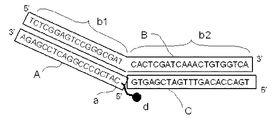
本発明の核酸プローブセットの一例を図1、図2および図8に示す。これらの図中、(A)は蛍光プローブ、(B)はバインディングプローブであり、本発明の核酸プローブセットは、上記蛍光プローブ(A)とバインディングプローブ(B)とからなる。(C)は標的核酸の一例である。 An example of the nucleic acid probe set of the present invention is shown in FIG. 1, FIG. 2, and FIG. In these figures, (A) is a fluorescent probe, (B) is a binding probe, and the nucleic acid probe set of the present invention comprises the fluorescent probe (A) and the binding probe (B). (C) is an example of the target nucleic acid.
図1は、蛍光プローブ(A)と蛍光プローブ結合領域(b1)と標的核酸結合領域(b2)とがそれぞれ1個であり、かつ標的核酸(C)とハイブリダイズする前の本発明の核酸プローブセット複合体を示す模式図である。図2は、図1に示す標的核酸(C)と本発明の核酸プローブセット複合体とがハイブリダイズした状態を示す模式図である。図8は、蛍光プローブ(A)と蛍光プローブ結合領域(b1)がそれぞれ2個であり、かつ標的核酸(C)とハイブリダイズする前の本発明の核酸プローブセット複合体を示す模式図である。 FIG. 1 shows that the nucleic acid probe of the present invention has one fluorescent probe (A), one fluorescent probe binding region (b1), and one target nucleic acid binding region (b2), and before hybridization with the target nucleic acid (C). It is a schematic diagram which shows a set composite_body | complex. FIG. 2 is a schematic diagram showing a state in which the target nucleic acid (C) shown in FIG. 1 and the nucleic acid probe set complex of the present invention are hybridized. FIG. 8 is a schematic diagram showing the nucleic acid probe set complex of the present invention before hybridization with the target nucleic acid (C), which has two fluorescent probes (A) and two fluorescent probe binding regions (b1). .
本発明の核酸プローブセットを構成する1個または2個の蛍光プローブ(A)(以下単に「蛍光プローブ(A)」と云う場合がある)は、蛍光物質(d)で標識されたヌクレオチド(a)を含むオリゴヌクレオチドである。当該蛍光プローブ(A)の塩基配列は、バインディングプローブ(B)の蛍光プローブ結合領域(b1)とハイブリダイズするものであれば特に制限されず、検出または分析対象の標的核酸の種類にかかわらず常に同じものを用いることもできる。このように、本発明の核酸プローブセットを構成する蛍光プローブ(A)は、特定の標的核酸に対応する塩基配列を持つ必要がないため、本発明の核酸プローブセットを用いて、標的核酸の分析を行う場合、高価な蛍光物質を有する蛍光プローブを検出または分析対象の標的核酸ごとに調製せずにすむという利点がある。 One or two fluorescent probes (A) constituting the nucleic acid probe set of the present invention (hereinafter sometimes simply referred to as “fluorescent probe (A)”) are nucleotides (a) labeled with a fluorescent substance (d) ). The base sequence of the fluorescent probe (A) is not particularly limited as long as it hybridizes with the fluorescent probe binding region (b1) of the binding probe (B), and is always irrespective of the type of target nucleic acid to be detected or analyzed. The same can be used. Thus, since the fluorescent probe (A) constituting the nucleic acid probe set of the present invention does not need to have a base sequence corresponding to a specific target nucleic acid, analysis of the target nucleic acid using the nucleic acid probe set of the present invention is performed. Is advantageous in that it is not necessary to prepare a fluorescent probe having an expensive fluorescent substance for each target nucleic acid to be detected or analyzed.
一方、本発明の核酸プローブセットを構成するバインディングプローブ(B)は、上記蛍光プローブ(A)とハイブリダイズする1個または2個の蛍光プローブ結合領域(b1)(以下単に「蛍光プローブ結合領域(b1)」と云う場合がある)および標的核酸(C)とハイブリダイズする標的核酸結合領域(b2)とを有するオリゴヌクレオチドである。 On the other hand, the binding probe (B) constituting the nucleic acid probe set of the present invention has one or two fluorescent probe binding regions (b1) (hereinafter simply referred to as “fluorescent probe binding regions”) that hybridize with the fluorescent probe (A). b1) ”and a target nucleic acid binding region (b2) that hybridizes with the target nucleic acid (C).
図1に示す本発明の核酸プローブセットは、図2に示すように、本発明の核酸プローブセット複合体と標的核酸(C)とがハイブリダイズしたときに、蛍光プローブ(A)中の蛍光物質(d)と標的核酸(C)を含む核酸中のグアニンとが相互作用して該蛍光物質(d)の蛍光キャラクターが変化するように設計されている。 As shown in FIG. 2, the nucleic acid probe set of the present invention shown in FIG. 1 has a fluorescent substance in the fluorescent probe (A) when the nucleic acid probe set complex of the present invention and the target nucleic acid (C) are hybridized. It is designed so that the fluorescent character of the fluorescent substance (d) is changed by interaction between (d) and guanine in the nucleic acid containing the target nucleic acid (C).
上記グアニンは、標的核酸を含む核酸中にあれば、標的核酸の塩基配列領域中にあっても、標的核酸の塩基配列領域の外側にあってもよい。上記グアニンが、標的核酸の塩基配列中にあり、ハイブリダイズしたバインディングプローブ(B)のシトシンと塩基対を形成している場合、蛍光物質(d)とグアニンとの相互作用が幾分低下するが特に問題はなく、さらには上記グアニンが、標的核酸の塩基配列領域の外側にあるなどの理由で塩基対を形成していない場合、蛍光物質(d)とグアニンとの相互作用が容易となるのでより好ましい。 As long as the guanine is in the nucleic acid containing the target nucleic acid, it may be in the base sequence region of the target nucleic acid or outside the base sequence region of the target nucleic acid. When the guanine is present in the base sequence of the target nucleic acid and forms a base pair with the cytosine of the hybridized binding probe (B), the interaction between the fluorescent substance (d) and guanine is somewhat reduced. There is no particular problem, and if the guanine does not form a base pair because it is outside the base sequence region of the target nucleic acid, the interaction between the fluorescent substance (d) and guanine becomes easy. More preferred.
次に、図3を用いて、本発明の核酸プローブセット複合体と標的核酸(C)とがハイブリダイズしている場合に、蛍光物質(d)と標的核酸(C)を含む核酸中のグアニン塩基を持つ任意のヌクレオチド(以下、「ヌクレオチドε」と略す。)とが相互作用できる条件について説明する。以下の説明中、標的核酸(C)の3’末端のグアニン塩基を持つヌクレオチドをヌクレオチドεとする。図3は、図2の蛍光物質(d)の周辺部分を拡大して表示したものである。蛍光物質(d)とヌクレオチドεとが相互作用できる条件は、後述する蛍光物質(d)と蛍光物質(d)で標識されたヌクレオチド(a)とを結ぶスペーサーの長さにも依存し、一定しないが、一般化すると以下のように表現できる。 Next, referring to FIG. 3, when the nucleic acid probe set complex of the present invention and the target nucleic acid (C) are hybridized, guanine in the nucleic acid containing the fluorescent substance (d) and the target nucleic acid (C) The conditions under which any nucleotide having a base (hereinafter abbreviated as “nucleotide ε”) can interact will be described. In the following description, a nucleotide having a guanine base at the 3 'end of the target nucleic acid (C) is referred to as nucleotide ε. FIG. 3 is an enlarged view of the peripheral portion of the fluorescent material (d) in FIG. The conditions under which the fluorescent substance (d) and nucleotide ε can interact depend on the length of the spacer connecting the fluorescent substance (d) and the nucleotide (a) labeled with the fluorescent substance (d), which will be described later, and are constant. Although not generalized, it can be expressed as follows.
蛍光プローブ(A)とバインディングプローブ(B)とがハイブリダイズしている場合において、蛍光プローブ(A)と塩基対を形成している蛍光プローブ結合領域(b1)中のヌクレオチドであって、標的核酸結合領域(b2)に最も近いヌクレオチド(以下、「ヌクレオチドα」と略す。)と塩基対を形成している蛍光プローブ(A)中のヌクレオチドをβとする。このヌクレオチドβと蛍光物質で標識されたヌクレオチド(a)との距離(β−a)を塩基数で表した数をXとする。図3において、ヌクレオチドαは、蛍光プローブ結合領域(b1)中3’末端にあるチミン塩基を持つヌクレオチドであり、ヌクレオチドβは、蛍光プローブ(A)中蛍光物質(d)で標識されたヌクレオチド(a)に隣接するアデニン塩基を持つヌクレオチドであり、この場合のXは1である。なお、隣接しているヌクレオチドを「X=1」とカウントし、1個おいて隣接しているヌクレオチドを「X=2」とカウントする。 When the fluorescent probe (A) and the binding probe (B) are hybridized, the nucleotide in the fluorescent probe binding region (b1) that forms a base pair with the fluorescent probe (A), the target nucleic acid Let β be the nucleotide in the fluorescent probe (A) that forms a base pair with the nucleotide closest to the binding region (b2) (hereinafter abbreviated as “nucleotide α”). Let X denote the number (base) of the distance (β-a) between this nucleotide β and the nucleotide (a) labeled with a fluorescent substance. In FIG. 3, nucleotide α is a nucleotide having a thymine base at the 3 ′ end in the fluorescent probe binding region (b1), and nucleotide β is a nucleotide labeled with a fluorescent substance (d) in the fluorescent probe (A) ( It is a nucleotide having an adenine base adjacent to a), and X in this case is 1. Note that adjacent nucleotides are counted as “X = 1”, and one adjacent nucleotide is counted as “X = 2”.
次に、標的核酸(C)と塩基対を形成している標的核酸結合領域(b2)中のヌクレオチドであって、ヌクレオチドαに最も近いヌクレオチド(以下「ヌクレオチドγ」と略す。)とヌクレオチドαとの距離(γ−α)を塩基数で表した数をYとする。図3において、ヌクレオチドγは、標的核酸結合領域(b2)中、5’末端にあるシトシン塩基を持つヌクレオチドであり、この場合のYは1である。なお、YのカウントはXの場合と同じである。 Next, nucleotides in the target nucleic acid binding region (b2) forming a base pair with the target nucleic acid (C) and closest to the nucleotide α (hereinafter abbreviated as “nucleotide γ”) and the nucleotide α. Y is the number of bases (γ−α) expressed by the number of bases. In FIG. 3, nucleotide γ is a nucleotide having a cytosine base at the 5 ′ end in the target nucleic acid binding region (b2), and Y in this case is 1. The count of Y is the same as that of X.
また、バインディングプローブ(B)と標的核酸(C)とがハイブリダイズしている場合において、ヌクレオチドγと塩基対を形成している標的核酸(C)中のヌクレオチドをδとする。このヌクレオチドδと上記ヌクレオチドεとの距離(δ−ε)を塩基数で表した数をZとする。図3において、ヌクレオチドδは標的核酸(C)中3’末端のグアニン塩基を持つヌクレオチドであり、このヌクレオチドはヌクレオチドεであるので、Zは0である。なお、ZのカウントはXの場合と同じである。 In the case where the binding probe (B) and the target nucleic acid (C) are hybridized, the nucleotide in the target nucleic acid (C) that forms a base pair with the nucleotide γ is defined as δ. Let Z be the number that represents the distance (δ−ε) between the nucleotide δ and the nucleotide ε in terms of the number of bases. In FIG. 3, nucleotide δ is a nucleotide having a 3 ′ terminal guanine base in the target nucleic acid (C), and since this nucleotide is nucleotide ε, Z is 0. The count of Z is the same as that of X.
本発明の核酸プローブセット複合体と標的核酸(C)とがハイブリダイズしている場合に、蛍光物質(d)とヌクレオチドεのグアニンとが相互作用できる条件は、X、YおよびZの和が6以下となる場合が好ましい。蛍光物質(d)と蛍光物質(d)で標識されたヌクレオチド(a)とを結ぶスペーサーの長さにも依存するが、X、YおよびZの和は4以下がより好ましく、最も好ましくは2である。 When the nucleic acid probe set complex of the present invention and the target nucleic acid (C) are hybridized, the condition that the fluorescent substance (d) and the guanine of the nucleotide ε can interact is that the sum of X, Y and Z is The case where it becomes 6 or less is preferable. Although depending on the length of the spacer connecting the fluorescent substance (d) and the nucleotide (a) labeled with the fluorescent substance (d), the sum of X, Y and Z is more preferably 4 or less, most preferably 2 It is.
本発明においては、蛍光プローブ(A)に標識する蛍光物質(d)は、グアニンと相互作用したときに、蛍光物質(d)の蛍光キャラクターが変化するものを使用する。なお、本発明において、「蛍光キャラクター」とは、蛍光強度のことを意味し、「グアニンと蛍光物質とが相互作用して蛍光物質の蛍光キャラクターが変化する」とは、グアニンと蛍光物質とが相互作用していない状態での蛍光物質の蛍光強度と、相互作用している状態での蛍光強度とが異なることを意味し、その程度は限定しないものとする。また、蛍光の「消光」とは、蛍光物質がグアニンと相互作用しない場合と比べて、グアニンと相互作用したときに蛍光強度が減衰することを意味し、その程度は限定しないものとする。 In the present invention, the fluorescent substance (d) labeled on the fluorescent probe (A) is one that changes the fluorescent character of the fluorescent substance (d) when interacting with guanine. In the present invention, “fluorescent character” means fluorescence intensity, and “guanine and fluorescent substance interact to change the fluorescent character of fluorescent substance” means that guanine and fluorescent substance change. It means that the fluorescence intensity of the fluorescent substance in a state where it does not interact is different from the fluorescence intensity in a state where it interacts, and the degree thereof is not limited. Further, “quenching” of fluorescence means that the fluorescence intensity is attenuated when interacting with guanine as compared with the case where the fluorescent substance does not interact with guanine, and the degree thereof is not limited.
本発明の核酸プローブセットに好適に使用できる蛍光物質の例としては、フルオレセインおよびその誘導体(例えば、フルオレセイン−4−イソチオシアネート(FITC)、テトラクロロフルオレセイン、ヘキサクロロフルオレセイン、テトラブロモスルホンフルオレセイン(TBSF)およびそれらの誘導体)、EDANS(5−(2−アミノエチル)アミノ−1−ナフタレンスルホン酸)、6-JOE(6-カルボキシ-4',5'-ジクロロ-2',7'-ジメトキシフルオレセイン)、3,6-ジアミノ-9-[2,4-ビス(リチオオキシカルボニル)フェニル]-4-(リチオオキシスルホニル)-5-スルホナトキサンチリウム/3,6-ジアミノ-9-[2,5-ビス(リチオオキシカルボニル)フェニル]-4-(リチオオキシスルホニル)-5-スルホナトキサンチリウム(Alexa Fluor(登録商標) 488(モレキュラー・プローブ社))、2,3,3,7,7,8-ヘキサメチル-5-[4-[5-(2,5-ジオキソ-3-ピロリン-1-イル)ペンチルカルバモイル]フェニル]-2,3,7,8-テトラヒドロ-9-アゾニア-1H-ピラノ[3,2-f:5,6-f']ジインドール-10,12-ジスルホン酸12-ナトリウム(Alexa Fluor(登録商標) 532(モレキュラー・プローブ社))、1-[6-[(2,5-ジオキソ-1-ピロリジニル)オキシ]-6-オキソヘキシル]-2-[3-[1-[6-[(2,5-ジオキソ-1-ピロリジニル)オキシ]-6-オキソヘキシル]-1,3-ジヒドロ-3,3-ジメチル-5-スルホ-2H-インドール-2-イリデン]-1-プロペニル]-3,3-ジメチル-5-スルホ-3H-インドリウム(Cy3(商標;アマシャム・バイオサイエンス社))、6-[[2-[5-[1-[6-(スクシンイミジルオキシ)-6-オキソヘキシル]-3,3-ジメチル-5-スルホナト-1H-インドール-2(3H)-イリデン]-1,3-ペンタジエニル]-3,3-ジメチル-5-スルホナト-3H-インドール-1-イウム]-1-イル]ヘキサン酸スクシンイミジル・カリウム(Cy5(商標;アマシャム・バイオサイエンス社))、2-オキソ-6,8-ジフルオロ-7-ヒドロキシ-2H-1-ベンゾピラン-3-カルボン酸(Pacific Blue(商標;モレキュラー・プローブ社))、ローダミン6G(R6G)およびその誘導体(例えば、カルボキシローダミン6G(CR6G)、テトラメチルローダミン(TMR)、テトラメチルローダミンイソチオシアネート(TMRITC)、x−ローダミン、カルボキシテトラメチルローダミン(TAMRA))、9-[2-(クロロスルホニル)-4-スルホナトフェニル]-2,3,6,7,12,13,16,17-オクタヒドロ-1H,5H,11H,15H-キサンテノ[2,3,4-ij:5,6,7-i'j']ジキノリジン-18-イウム/9-[4-(クロロスルホニル)-2-スルホナトフェニル]-2,3,6,7,12,13,16,17-オクタヒドロ-1H,5H,11H,15H-キサンテノ[2,3,4-ij:5,6,7-i'j']ジキノリジン-18-イウム(テキサスレッド(モレキュラー・プローブ社))、4,4-ジフルオロ-5,7-ジメチル-4-ボラ-3a,4a-ジアザ-s-インダセン-3-プロピオン酸(BODIPY(商標)-FL(モレキュラー・プローブ社))、4,4-ジフルオロ-5-フェニル-4-ボラ-3a,4a-ジアザ-s-インダセン-3-プロピオン酸(BODIPY(商標)-R6G(モレキュラー・プローブ社))、4,4-ジフルオロ-5-スチリル-4-ボラ-3a,4a-ジアザ-s-インダセン-3-プロピオン酸(BODIPY(商標)564(モレキュラー・プローブ社)並びに4,4-ジフルオロ-5-(4-フェニル-1,3-ブタジエニル)-4-ボラ-3a,4a-ジアザ-s-インダセン-3-プロピオン酸(BODIPY(商標)581(モレキュラー・プローブ社))を挙げることができる。 Examples of fluorescent substances that can be suitably used in the nucleic acid probe set of the present invention include fluorescein and its derivatives (eg, fluorescein-4-isothiocyanate (FITC), tetrachlorofluorescein, hexachlorofluorescein, tetrabromosulfone fluorescein (TBSF) and Derivatives thereof), EDANS (5- (2-aminoethyl) amino-1-naphthalenesulfonic acid), 6-JOE (6-carboxy-4 ′, 5′-dichloro-2 ′, 7′-dimethoxyfluorescein) , 3,6-Diamino-9- [2,4-bis (lithiooxycarbonyl) phenyl] -4- (lithiooxysulfonyl) -5-sulfonatoxanthylium / 3,6-diamino-9- [2,5- Bis (lithiooxycarbonyl) phenyl] -4- (lithiooxysulfonyl) -5-sulfonatoxanthylium ( Alexa Fluor (registered trademark) 488 (Molecular Probes) ) 2,3,3,7,7,8-hexamethyl-5- [4- [5- (2,5-dioxo-3-pyrrolin-1-yl) pentylcarbamoyl] phenyl] -2,3,7, 8-Tetrahydro-9-azonia-1H-pyrano [3,2-f: 5,6-f '] diindole-10,12-sodium disulfonate ( Alexa Fluor® 532 (Molecular Probes ) ) ) 1- [6-[(2,5-Dioxo-1-pyrrolidinyl) oxy] -6-oxohexyl] -2- [3- [1- [6-[(2,5-dioxo-1- Pyrrolidinyl) oxy] -6-oxohexyl] -1,3-dihydro-3,3-dimethyl-5-sulfo-2H-indole-2-ylidene] -1-propenyl] -3,3-dimethyl-5-sulfo -3H-Indolium ( Cy3 ( trademark; Amersham Biosciences)), 6-[[2- [5- [1- [6- (succinimidyloxy) -6-oxohexyl] -3,3 -Dimethyl-5-sulfonato-1H-indole-2 (3H) -ylidene] -1,3-pentadienyl] -3,3-dimethyl-5-sulfonato-3H-indole-1-ium] -1-yl] hex Phosphate succinimidyl potassium (Cy5 (TM; Amersham Biosciences)), 2-oxo-6,8-difluoro-7-hydroxy-2H-1-benzopyran-3-carboxylic acid (Pacific Blue (TM; Molecular Probes) ) , rhodamine 6G (R6G) and derivatives thereof (eg, carboxyrhodamine 6G (CR6G), tetramethylrhodamine (TMR), tetramethylrhodamine isothiocyanate (TMRITC), x-rhodamine, carboxytetramethylrhodamine (TAMRA) ), 9- [2- (chlorosulfonyl) -4-sulfonatophenyl] -2,3,6,7,12,13,16,17-octahydro-1H, 5H, 11H, 15H-xantheno [2,3 , 4-ij: 5,6,7-i'j '] diquinolizine-18-ium / 9- [4- (chlorosulfonyl) -2-sulfonatophenyl] -2,3,6,7,12,13 , 16,17-octahydro -1H, 5H, 11H, 15H- Kisanteno [2,3,4-ij: 5,6,7-i'j '] Jikinorijin 18- ium (Texas Head (Molecular Probes)), 4,4-difluoro-5,7-dimethyl-4-bora -3a, 4a-diaza -s- indacene-3-propionic acid (BODIPY (TM) -FL (Molecular Probe Corporation) ), 4,4-Difluoro-5-phenyl-4-bora-3a, 4a-diaza-s-indacene-3-propionic acid ( BODIPY ™ -R6G (Molecular Probes) ) , 4, 4-difluoro-5-styryl-4-bora-3a, 4a-diaza-s-indacene-3-propionic acid ( BODIPY ™ 564 (Molecular Probes) and 4,4-difluoro-5- (4- Phenyl-1,3-butadienyl) -4-bora-3a, 4a-diaza-s-indacene-3-propionic acid ( BODIPY ™ 581 (Molecular Probes) ) .
これらの中でもテトラブロモスルホンフルオレセイン、2-オキソ-6,8-ジフルオロ-7-ヒドロキシ-2H-1-ベンゾピラン-3-カルボン酸(Pacific Blue(商標))、ローダミン6G、カルボキシローダミン6G、カルボキシテトラメチルローダミンおよび4,4-ジフルオロ-5,7-ジメチル-4-ボラ-3a,4a-ジアザ-s-インダセン-3-プロピオン酸(BODIPY(商標)-FL)を使用することがより好ましく、4,4-ジフルオロ-5,7-ジメチル-4-ボラ-3a,4a-ジアザ-s-インダセン-3-プロピオン酸(BODIPY(商標)-FL)を使用することがもっとも好ましい。 Te tiger bromosulfonic fluorescein Among these, 2-oxo-6,8-difluoro-7-hydroxy-2H-1-benzopyran-3-carboxylic acid (Pacific Blue (TM)), rhodamine 6G, carboxy rhodamine 6G, carboxymethyl tetramethylrhodamine and 4,4-difluoro-5,7-dimethyl-4-bora -3a, it is used 4a- diaza -s- indacene-3-propionic acid (BODIPY (TM) -FL) More preferably, it is most preferred to use 4,4-difluoro-5,7-dimethyl-4-bora-3a, 4a-diaza-s-indacene-3-propionic acid ( BODIPY ™ -FL ) .
本発明の核酸プローブセットに使用する蛍光プローブ(A)は、オリゴヌクレオチドの製造受託会社(例えば、つくばオリゴサービス株式会社、茨城県)などに製造を委託して使用することができる。オリゴヌクレオチドに蛍光物質を標識する方法は、特に限定されず、従来公知の標識方法を利用することができる。(Nature Biotechnology、14巻、303〜308頁、1996年;Applied and Environmental Microbiology、63巻、1143〜1147頁、1997年;Nucleic acids Research、24巻、4532〜4535頁、1996年)。 The fluorescent probe (A) used in the nucleic acid probe set of the present invention can be used by consigning production to an oligonucleotide manufacturing contract company (for example, Tsukuba Oligo Service Co., Ltd., Ibaraki Prefecture). The method for labeling the oligonucleotide with the fluorescent substance is not particularly limited, and a conventionally known labeling method can be used. (Nature Biotechnology, 14, 303-308, 1996; Applied and Environmental Microbiology, 63, 1143-1147, 1997; Nucleic acids Research, 24, 4532-4535, 1996).
例えば、5’末端ヌクレオチドに蛍光物質を結合させる場合は、まず、常法に従って5’末端のリン酸基にスペーサーとして、例えば、−(CH2)n−SHを導入する。このようなスペーサーは市販のものを使用することができる(例えば、Midland Certified Reagent Company社、米国)。この場合、nは3〜8、好ましくは6である。このスペーサーにSH基反応性を有する蛍光物質またはその誘導体を結合させることにより蛍光標識されたオリゴヌクレオチドを得ることができる。当該蛍光標識されたオリゴヌクレオチドは、逆相クロマトグラフィーなどで精製して、本発明の蛍光プローブ(A)とすることができる。 For example, when a fluorescent substance is bound to the 5 ′ terminal nucleotide, first, for example, — (CH 2 ) n —SH is introduced as a spacer into the 5 ′ terminal phosphate group according to a conventional method. A commercially available spacer can be used (for example, Midland Certified Reagent Company, USA). In this case, n is 3-8, preferably 6. A fluorescently labeled oligonucleotide can be obtained by binding a fluorescent substance having SH group reactivity or a derivative thereof to this spacer. The fluorescently labeled oligonucleotide can be purified by reverse phase chromatography or the like to obtain the fluorescent probe (A) of the present invention.
また、オリゴヌクレオチドの3’末端ヌクレオチドに蛍光物質を結合させることもできる。この場合は、リボースまたはデオキシリボースの3’位CのOH基にスペーサーとして、例えば、−(CH2)n−NH2を導入する。このようなスペーサーも市販のものを使用することができる(例えば、Midland Certified Reagent Company社、米国)。別の方法として、リボースまたはデオキシリボースの3’位CのOH基にリン酸基を導入して、リン酸基のOH基にスペーサーとして、例えば、−(CH2)n−SHを導入する。この場合、nは3〜8、好ましくは4〜7である。 In addition, a fluorescent substance can be bound to the 3 ′ terminal nucleotide of the oligonucleotide. In this case, for example, — (CH 2 ) n —NH 2 is introduced as a spacer into the OH group at the 3′-position C of ribose or deoxyribose. A commercially available spacer can also be used (for example, Midland Certified Reagent Company, USA). As another method, a phosphate group is introduced into the OH group at the 3′-position C of ribose or deoxyribose, and, for example, — (CH 2 ) n —SH is introduced as a spacer into the OH group of the phosphate group. In this case, n is 3 to 8, preferably 4 to 7.
上記のスペーサーにアミノ基またはSH基に反応性を有する蛍光物質またはその誘導体を結合させることにより蛍光物質で標識されたオリゴヌクレオチドを合成することができる。当該オリゴヌクレオチドを逆相クロマトグラフィーなどで精製して、本発明の蛍光プローブ(A)とすることができる。スペーサーとして−(CH2)n−NH2を導入する場合、キット試薬(例えば、Uni-link aminomodifier、クロンテック社)を用いるのが便利である。そして、常法に従って当該オリゴヌクレオチドに蛍光物質を結合させることができる。 An oligonucleotide labeled with a fluorescent substance can be synthesized by binding a fluorescent substance or a derivative thereof reactive to an amino group or an SH group to the spacer. The oligonucleotide can be purified by reverse phase chromatography or the like to obtain the fluorescent probe (A) of the present invention. As spacers - When introducing (CH 2) n -NH 2, kit reagents (e.g., Uni-link aminomodifier, Clontech) to use is convenient. Then, a fluorescent substance can be bound to the oligonucleotide according to a conventional method.
蛍光プローブ(A)中の蛍光物質で標識されたヌクレオチド(a)は、オリゴヌクレオチドの両末端ヌクレオチドに限定されるものではなく、両末端ヌクレオチド以外のヌクレオチドを蛍光物質で標識することもできる(ANALYTICAL BIOCHEMISTRY、225、32−38頁、1998年)。 The nucleotide (a) labeled with the fluorescent substance in the fluorescent probe (A) is not limited to the both terminal nucleotides of the oligonucleotide, and nucleotides other than the both terminal nucleotides can be labeled with the fluorescent substance (ANALYTICAL). BIOCHEMISTRY, 225, 32-38, 1998).
以上のようにして本発明の核酸プローブセットを構成する蛍光プローブ(A)を調製できるが、好ましい蛍光プローブ(A)の形態は、5’末端または3’末端ヌクレオチドを蛍光物質(d)で標識したものであり、より好ましくは、5’末端ヌクレオチドをBODIPY(商標)-FLで標識したものである。また、蛍光物質(d)で標識されたヌクレオチドの塩基としては、グアニンまたはシトシンであることが好ましい。 Although the fluorescent probe (A) constituting the nucleic acid probe set of the present invention can be prepared as described above, the preferred form of the fluorescent probe (A) is that the 5 ′ terminal or 3 ′ terminal nucleotide is labeled with the fluorescent substance (d). More preferably, the 5 ′ terminal nucleotide is labeled with BODIPY ™ -FL. The nucleotide base labeled with the fluorescent substance (d) is preferably guanine or cytosine.
本発明の核酸プローブセットを構成する蛍光プローブ(A)は、バインディングプローブ(B)の蛍光プローブ結合領域(b1)とハイブリダイズする塩基配列であればよく、塩基長に特に制限はないが、4塩基以下であるとハイブリダイズ効率が低いという点で好ましくなく、51塩基以上であると非特異的なハイブリッドが生じやすいという点で好ましくないことがある。従って、蛍光プローブ(A)は、5〜50塩基であることが好ましく、より好ましくは10〜35塩基、特に好ましくは12〜30塩基である。 The fluorescent probe (A) constituting the nucleic acid probe set of the present invention may be any base sequence that hybridizes with the fluorescent probe binding region (b1) of the binding probe (B), and the base length is not particularly limited. If it is less than the base, it is not preferable in terms of low hybridization efficiency, and if it is more than 51 bases, it may not be preferable in that non-specific hybrids are likely to occur. Therefore, the fluorescent probe (A) preferably has 5 to 50 bases, more preferably 10 to 35 bases, and particularly preferably 12 to 30 bases.
蛍光プローブ(A)の塩基配列は、バインディングプローブ(B)の蛍光プローブ結合領域(b1)とハイブリダイズする限り、当該蛍光プローブ結合領域(b1)と相補的でないヌクレオチドを有してもよい。例えば、図1の蛍光プローブ(A)のように、5’末端にバインディングプローブ(B)とは相補的でないヌクレオチドを有するものでもよい。一方、上記バインディングプローブ(B)中の上記蛍光プローブ結合領域(b1)の塩基配列は、蛍光プローブ(A)とハイブリダイズするものであればよく、その塩基長は蛍光プローブ(A)の塩基長に依存する。 The base sequence of the fluorescent probe (A) may have nucleotides that are not complementary to the fluorescent probe binding region (b1) as long as it hybridizes with the fluorescent probe binding region (b1) of the binding probe (B). For example, like the fluorescent probe (A) of FIG. 1, it may have a nucleotide that is not complementary to the binding probe (B) at the 5 'end. On the other hand, the base sequence of the fluorescent probe binding region (b1) in the binding probe (B) only needs to be hybridized with the fluorescent probe (A), and the base length is the base length of the fluorescent probe (A). Depends on.
バインディングプローブ(B)中の標的核酸結合領域(b2)は、標的核酸(C)とハイブリダイズする塩基配列であればよく、その塩基長は、標的核酸の塩基長に依存するが、4塩基以下であると標的核酸(C)とのハイブリダイズ効率が低いという点で好ましくなく、61塩基以上であると非特異的なハイブリッドが生じやすいという点で好ましくないことがある。従って、標的核酸結合領域(b2)は、5〜60塩基であることが好ましく、より好ましくは15〜30塩基である。標的核酸結合領域(b2)の塩基配列は、標的核酸(C)とハイブリダイズする限り標的核酸(C)と塩基対を形成しない塩基配列を有してもよい。 The target nucleic acid binding region (b2) in the binding probe (B) may be a base sequence that hybridizes with the target nucleic acid (C), and its base length depends on the base length of the target nucleic acid, but it is 4 bases or less. If it is, it is not preferable in that the hybridization efficiency with the target nucleic acid (C) is low, and if it is 61 bases or more, it may not be preferable in that a non-specific hybrid is likely to be generated. Therefore, the target nucleic acid binding region (b2) preferably has 5 to 60 bases, more preferably 15 to 30 bases. The base sequence of the target nucleic acid binding region (b2) may have a base sequence that does not form a base pair with the target nucleic acid (C) as long as it hybridizes with the target nucleic acid (C).
本発明の核酸プローブセットは、核酸の種々の分析方法に用いることができる。本発明の核酸プローブセットを用いて、溶液中に標的核酸が存在するか否かを判定する標的核酸の検出方法の例を以下に示す。 The nucleic acid probe set of the present invention can be used in various nucleic acid analysis methods. An example of a target nucleic acid detection method for determining whether or not a target nucleic acid is present in a solution using the nucleic acid probe set of the present invention is shown below.
まず、標的核酸の検出をする溶液(以下、「検出サンプル」と略す。)を段階希釈して、数種の溶液を調製する。これらの段階希釈した検出サンプルに本発明の核酸プローブセット、すなわち蛍光プローブ(A)とバインディングプローブ(B)をそれぞれ一定量加え、次いで、添加した本発明の核酸プローブセット複合体と標的核酸とがハイブリダイズするように溶液の温度を調整した後、蛍光強度を測定する。上記本発明のプローブセット複合体と標的核酸とをハイブリダイズさせる温度は、本発明の核酸プローブセット複合体と標的核酸との融解温度(以下「Tm1」と呼ぶ)やその他の溶液条件により変動するが、当該核酸プローブセット複合体と標的核酸との配列特異的なハイブリダイゼーションが起こり、かつ非特異的なハイブリダイゼーションが生じない温度範囲であることが好ましく、好ましくはTm1〜(Tm1−40)℃、より好ましくはTm1〜(Tm1−20)℃、さらに好ましくはTm1〜(Tm1−10)℃である。このような好ましい温度の一例として、約60℃が挙げられる。 First, a solution for detecting a target nucleic acid (hereinafter abbreviated as “detection sample”) is serially diluted to prepare several types of solutions. A certain amount of each of the nucleic acid probe set of the present invention, that is, the fluorescent probe (A) and the binding probe (B) is added to these serially diluted detection samples, and then the added nucleic acid probe set complex of the present invention and the target nucleic acid are added. After adjusting the temperature of the solution to hybridize, the fluorescence intensity is measured. The temperature at which the probe set complex of the present invention is hybridized with the target nucleic acid varies depending on the melting temperature of the nucleic acid probe set complex of the present invention and the target nucleic acid (hereinafter referred to as “Tm1”) and other solution conditions. Is preferably in a temperature range in which sequence-specific hybridization between the nucleic acid probe set complex and the target nucleic acid occurs, and non-specific hybridization does not occur, preferably Tm1 to (Tm1-40) ° C. More preferably, it is Tm1- (Tm1-20) degreeC, More preferably, it is Tm1- (Tm1-10) degreeC. An example of such a preferred temperature is about 60 ° C.
また、本発明の核酸プローブセットを構成する蛍光プローブとバインディングプローブとの融解温度(以下「Tm2」と呼ぶ)は、蛍光強度を確実に測定するために、Tm1より高いことが好ましく、Tm2−Tm1が5℃以上であることがより好ましい。 The melting temperature (hereinafter referred to as “Tm2”) of the fluorescent probe and the binding probe constituting the nucleic acid probe set of the present invention is preferably higher than Tm1 in order to reliably measure the fluorescence intensity. Is more preferably 5 ° C. or higher.
上記検出サンプル中に標的核酸が存在しない場合、段階希釈した検出サンプルのいずれにおいても同程度の蛍光強度が観察される。一方、上記検出サンプル中に標的核酸が存在する場合、本発明の核酸プローブセットの蛍光物質による蛍光は標的核酸を含む核酸中のグアニンにより消光される。この消光の程度は、上記溶液中の核酸プローブセットと標的核酸との比率を変えることによって変化する。このため、上述したように段階希釈した検出サンプル中に本発明の核酸プローブセットを添加して、それらの蛍光強度を測定することにより、蛍光消光の発生の有無から標的核酸の存在を判定することができ、蛍光消光の大きさから標的核酸の存在量を定量することができる。 When the target nucleic acid is not present in the detection sample, the same fluorescence intensity is observed in any of the detection samples diluted serially. On the other hand, when the target nucleic acid is present in the detection sample, fluorescence from the fluorescent substance of the nucleic acid probe set of the present invention is quenched by guanine in the nucleic acid containing the target nucleic acid. The degree of this quenching is changed by changing the ratio between the nucleic acid probe set and the target nucleic acid in the solution. Therefore, the presence of the target nucleic acid can be determined from the occurrence of fluorescence quenching by adding the nucleic acid probe set of the present invention to the detection sample serially diluted as described above and measuring their fluorescence intensity. Thus, the abundance of the target nucleic acid can be quantified from the magnitude of fluorescence quenching.
また、本発明の核酸プローブセットは、リアルタイムPCR方法に用いることもできる。リアルタイムPCR方法において、本発明の核酸プローブセットを用いて増幅産物を定量する場合、PCRで増幅する塩基配列またはその一部を標的核酸として、当該標的核酸とハイブリダイズするようにバインディングプローブ(B)中の標的核酸結合領域(b2)の塩基配列を決定する。 The nucleic acid probe set of the present invention can also be used in a real-time PCR method. In the real-time PCR method, when the amplification product is quantified using the nucleic acid probe set of the present invention, the binding probe (B) is hybridized with the target nucleic acid using the base sequence amplified by PCR or a part thereof as the target nucleic acid. The base sequence of the target nucleic acid binding region (b2) is determined.
このようにして作製した本発明の核酸プローブセットをPCR反応溶液に添加して、PCR反応を行い、PCRの各サイクルにおいて蛍光強度を測定する。PCR反応によって、反応溶液中の標的核酸が増幅すると、当該標的核酸を含む核酸中のグアニンにより本発明の核酸プローブセットの蛍光物質による蛍光が消光されるため、蛍光強度および蛍光消光の大きさからPCRによる増幅産物を定量することができる。 The nucleic acid probe set of the present invention thus prepared is added to a PCR reaction solution, a PCR reaction is performed, and the fluorescence intensity is measured in each cycle of PCR. When the target nucleic acid in the reaction solution is amplified by the PCR reaction, the fluorescence due to the fluorescent substance of the nucleic acid probe set of the present invention is quenched by guanine in the nucleic acid containing the target nucleic acid. PCR amplification products can be quantified.
さらに、本発明の核酸プローブセットは、核酸の塩基配列多型の分析に用いることもできる。分析できる塩基配列多型の例としては、基準となる塩基配列からの1塩基多型、塩基置換、塩基欠失および塩基挿入などを挙げることができる。このような分析方法の一例を以下に示す。 Furthermore, the nucleic acid probe set of the present invention can also be used for analysis of nucleotide sequence polymorphisms of nucleic acids. Examples of base sequence polymorphisms that can be analyzed include single base polymorphisms from base nucleotide sequences, base substitutions, base deletions, and base insertions. An example of such an analysis method is shown below.
本分析方法では、基準となる塩基配列を標的核酸(C)として、標的核酸(C)を含む溶液と、分析対象の核酸を含む溶液とを調製する。それぞれの溶液に本発明の核酸プローブセット、すなわち上記標的核酸(C)とハイブリダイズするように設計した標的核酸結合領域(b2)を有するバインディングプローブ(B)と蛍光プローブ(A)とを加えた後、各溶液において、添加した本発明の核酸プローブセット複合体と標的核酸(C)または分析対象の核酸とをハイブリダイズさせ、次いで蛍光強度の温度依存性を測定する。具体的には、溶液の温度を低温から高温に変化させながら、各温度について蛍光強度を測定する。 In this analysis method, a solution containing the target nucleic acid (C) and a solution containing the nucleic acid to be analyzed are prepared using the base sequence serving as a reference as the target nucleic acid (C). The nucleic acid probe set of the present invention, that is, the binding probe (B) having the target nucleic acid binding region (b2) designed to hybridize with the target nucleic acid (C) and the fluorescent probe (A) were added to each solution. Thereafter, in each solution, the added nucleic acid probe set complex of the present invention and the target nucleic acid (C) or the nucleic acid to be analyzed are hybridized, and then the temperature dependence of the fluorescence intensity is measured. Specifically, the fluorescence intensity is measured for each temperature while changing the temperature of the solution from a low temperature to a high temperature.
この測定結果を温度に対してプロットしたものを、「融解曲線」と呼ぶ。標的核酸を含む溶液について融解曲線を温度で微分することにより、本発明の核酸プローブセット複合体と標的核酸(C)とのTm1を、極値を示す温度として容易に求めることができる。このような融解曲線分析は、当業者に周知の市販のプログラムを用いることによって行うことができる。 A plot of the measurement results against temperature is called a “melting curve”. By differentiating the melting curve of the solution containing the target nucleic acid with respect to the temperature, Tm1 between the nucleic acid probe set complex of the present invention and the target nucleic acid (C) can be easily determined as the temperature showing the extreme value. Such melting curve analysis can be performed by using a commercial program well known to those skilled in the art.
上記標的核酸(C)を含む溶液の蛍光強度は、低温では標的核酸(C)を含む核酸中のグアニンによる蛍光消光現象により抑制される。しかし、溶液温度をTm1付近まで上げると本発明の核酸プローブセット複合体から標的核酸が解離し、蛍光消光の程度が弱くなるため、蛍光強度は急激に増加する。分析対象の核酸の塩基配列中に塩基配列多型、例えば、標的核酸の塩基配列からの1塩基多型、塩基置換、塩基欠失および塩基挿入などがあると、当該分析対象の核酸と本発明の核酸プローブセット複合体とのTm1は、標的核酸(C)と本発明の核酸プローブセット複合体とのTm1より低い値を示す。このため、標的核酸と本発明の核酸プローブセット複合体の蛍光強度の温度依存性と、分析対象の核酸と本発明の核酸プローブセット複合体の蛍光強度の温度依存性とを比較することにより、標的核酸の塩基配列に対する分析対象の核酸の塩基配列多型を分析することができる。このような分析手順としては、融解曲線どうしを比較することもできるが、それぞれの融解曲線を温度で微分し、極値を与える温度としてTm1を求め、それを比較することによって、容易に変異の有無を判断することができる。 The fluorescence intensity of the solution containing the target nucleic acid (C) is suppressed by a fluorescence quenching phenomenon due to guanine in the nucleic acid containing the target nucleic acid (C) at a low temperature. However, when the solution temperature is increased to around Tm1, the target nucleic acid is dissociated from the nucleic acid probe set complex of the present invention, and the degree of fluorescence quenching becomes weak, so that the fluorescence intensity increases rapidly. If there is a base sequence polymorphism in the base sequence of the nucleic acid to be analyzed, for example, a single base polymorphism from the base sequence of the target nucleic acid, base substitution, base deletion, or base insertion, the nucleic acid to be analyzed and the present invention Tm1 with the nucleic acid probe set complex of 1 shows a lower value than Tm1 between the target nucleic acid (C) and the nucleic acid probe set complex of the present invention. For this reason, by comparing the temperature dependence of the fluorescence intensity of the target nucleic acid and the nucleic acid probe set complex of the present invention and the temperature dependence of the fluorescence intensity of the nucleic acid to be analyzed and the nucleic acid probe set complex of the present invention, The base sequence polymorphism of the nucleic acid to be analyzed with respect to the base sequence of the target nucleic acid can be analyzed. As such an analysis procedure, melting curves can be compared with each other, but each melting curve is differentiated with respect to temperature, Tm1 is obtained as a temperature giving an extreme value, and comparison is made so that mutation can be easily performed. The presence or absence can be determined.
また、分析対象の核酸の塩基配列のうち、蛍光プローブの蛍光物質に対して消光作用をするグアニン塩基を持つヌクレオチドが変異している場合、いずれの温度においても蛍光消光現象による蛍光強度の低下が生じないため、その融解曲線から変異を特定することができる。 In addition, in the nucleotide sequence of the nucleic acid to be analyzed, when a nucleotide having a guanine base that quenches the fluorescent substance of the fluorescent probe is mutated, the fluorescence intensity decreases due to the fluorescence quenching phenomenon at any temperature. Since it does not occur, the mutation can be identified from the melting curve.
従来、融解曲線分析においては、標的核酸ごとに高価な蛍光標識をした塩基配列の異なる核酸プローブを用意する必要があり、その合成に長時間を要していたが、本発明の核酸プローブセットを融解曲線分析に使用することにより、標的核酸ごとに高価な蛍光標識をした核酸プローブを用意する必要がないため、融解曲線分析の準備時間を短縮でき、かつ融解曲線分析をより安価に実施することができる。 Conventionally, in melting curve analysis, it was necessary to prepare nucleic acid probes having different base sequences with expensive fluorescent labels for each target nucleic acid, and it took a long time to synthesize the nucleic acid probes. By using for melting curve analysis, it is not necessary to prepare expensive fluorescently labeled nucleic acid probes for each target nucleic acid, so preparation time for melting curve analysis can be shortened and melting curve analysis can be performed at a lower cost Can do.
前記で説明した本発明の核酸プローブセットは、1個の蛍光プローブ結合領域(b1)を持つバインディングプローブ(B)と1個の蛍光プローブ(A)とから構成されているが、本発明の核酸プローブセットは、図8に示すように、蛍光プローブ結合領域(b1)を2個有するものであってもよい。この蛍光プローブ結合領域(b1)は同じ塩基配列でも異なる塩基配列でもよいが、異なる塩基配列であることが好ましい。この場合には蛍光プローブは1個でもよいが、塩基配列の異なる2個の蛍光プローブ(A)を有することが好ましい。図8に示す本発明の核酸プローブセットにおける蛍光プローブ結合領域(b1)と蛍光プローブ(A)との構成は前記と同様である。 The nucleic acid probe set of the present invention described above is composed of a binding probe (B) having one fluorescent probe binding region (b1) and one fluorescent probe (A). As shown in FIG. 8, the probe set may have two fluorescent probe binding regions (b1). The fluorescent probe binding regions (b1) may have the same base sequence or different base sequences, but preferably have different base sequences. In this case, the number of fluorescent probes may be one, but it is preferable to have two fluorescent probes (A) having different base sequences. The structures of the fluorescent probe binding region (b1) and the fluorescent probe (A) in the nucleic acid probe set of the present invention shown in FIG. 8 are the same as described above.
上記バインディングプローブ(B)が蛍光プローブ結合領域を2個有し、それらの塩基配列が異なる本発明の核酸プローブセットは、ABC−PCR(Alternately Binding probe Competitive PCR;Taniら、Analytical Chemistry、印刷準備中を参照のこと)方法において従来使用されているABプローブの代わりとして好適に使用することができる。 The above-described binding probe (B) has two fluorescent probe binding regions and the nucleic acid probe sets of the present invention having different base sequences are prepared by ABC-PCR (Alternately Binding probe Competitive PCR; Tani et al., Analytical Chemistry, printing preparations). It can be suitably used as an alternative to the AB probe conventionally used in the method.
Taniらが開示するABC−PCR方法は、競合的PCR方法の一種であり、PCR反応溶液中に増幅対象の塩基配列(以下、「増幅配列」と略す。)を含む核酸の他に、増幅配列の一部をグアニンに置換した核酸(以下、「内部標準核酸」と略す。)並びに上記増幅配列の一部と相補的な塩基配列を持ち、その両端をそれぞれの励起波長および蛍光波長が異なる別の蛍光物質で標識した核酸プローブ(以下、「ABプローブ」と略す。)を加えてPCR反応を行う方法である。上記ABプローブは、増幅配列を含む核酸とハイブリダイズしたときに、片方の蛍光物質のみが当該核酸中のグアニンにより消光され、かつ、内部標準核酸とハイブリダイズしたときには、両方の蛍光物質が当該内部標準核酸中のグアニンにより消光されるように設計されているため、2種の蛍光物質の蛍光強度から、PCR反応による増幅産物を定量することができる。 The ABC-PCR method disclosed by Tani et al. Is a kind of competitive PCR method. In addition to a nucleic acid containing a base sequence to be amplified (hereinafter abbreviated as “amplified sequence”) in a PCR reaction solution, an amplified sequence is used. Has a base sequence complementary to a part of the above amplified sequence, and both ends thereof are different in excitation wavelength and fluorescence wavelength. In this method, a nucleic acid probe (hereinafter abbreviated as “AB probe”) labeled with a fluorescent substance is added to perform a PCR reaction. When the AB probe is hybridized with a nucleic acid containing an amplification sequence, only one fluorescent substance is quenched by guanine in the nucleic acid, and when hybridized with an internal standard nucleic acid, both fluorescent substances are Since it is designed to be quenched by guanine in the standard nucleic acid, the amplification product by the PCR reaction can be quantified from the fluorescence intensities of the two fluorescent substances.
上記従来のABC−PCR方法において使用する蛍光標識されたABプローブは、増幅対象の塩基配列ごとに異なるプローブを使用しなければならないが、本発明の核酸プローブセットを上記ABプローブの代わりに用いることにより、高価な蛍光標識をした蛍光プローブを、増幅対象の塩基配列ごとに用意する必要がないので、より安価にABC−PCR方法を実施することができる。 The fluorescently labeled AB probe used in the above conventional ABC-PCR method must use a different probe for each base sequence to be amplified, but the nucleic acid probe set of the present invention is used in place of the AB probe. Thus, it is not necessary to prepare an expensive fluorescently labeled fluorescent probe for each base sequence to be amplified, so that the ABC-PCR method can be carried out at a lower cost.
このようなABC−PCR方法での使用に適する本発明の核酸プローブセットの一例を図8に示す。この場合、PCRで増幅する塩基配列の一部を標的核酸(C)として、当該標的核酸とハイブリダイズするようにバインディングプローブ(B)中の標的核酸結合領域(b2)の塩基配列を決定する。ABC−PCR方法においてABプローブの代わりとして本発明の核酸プローブセットを使用する場合、当該核酸プローブセットを構成する2本の蛍光プローブに標識するそれぞれの励起波長および蛍光波長が異なる蛍光物質の好ましい組み合わせの例としては、BODIPY(商標)-FLとTAMRAの組み合わせを挙げることができる。 An example of the nucleic acid probe set of the present invention suitable for use in such an ABC-PCR method is shown in FIG. In this case, the base sequence of the target nucleic acid binding region (b2) in the binding probe (B) is determined so as to hybridize with the target nucleic acid, with a part of the base sequence amplified by PCR as the target nucleic acid (C). When the nucleic acid probe set of the present invention is used in place of the AB probe in the ABC-PCR method, preferred combinations of fluorescent substances having different excitation wavelengths and different fluorescence wavelengths to be labeled on the two fluorescent probes constituting the nucleic acid probe set As an example, a combination of BODIPY (trademark) -FL and TAMRA can be mentioned.
次に実施例を挙げて本発明をさらに具体的に説明するが、これらの実施例は本発明の単なる例示であって、本発明の限定を意図するものではない。 EXAMPLES Next, although an Example is given and this invention is demonstrated further more concretely, these Examples are only the illustrations of this invention, and do not intend limiting of this invention.
[実施例1]<標的核酸検出試験>
図1に示すように、ヒトUCP1遺伝子の一部と同じ塩基配列を持つオリゴヌクレオチドを標的核酸(配列番号:1)とし、5’末端側に蛍光プローブと相補的な塩基配列の蛍光プローブ結合領域(b1)を、3’末端側に標的核酸と相補的な塩基配列の標的核酸結合領域(b2)を持つバインディングプローブ(配列番号:2)および配列番号3の塩基配列を持ち、5’末端ヌクレオチドをBODIPY(商標)-FL(モレキュラー・プローブ社)で標識した蛍光プローブからなる核酸プローブセットを用いて、標的核酸検出試験を行った。
配列番号1:tgaccacagtttgatcgagtg
配列番号2:tctcggagtccgggcgatcactcgatcaaactgtggtca−リン酸
配列番号3:BODIPY(商標)-FL-catcgcccggactccgaga−リン酸
[Example 1] <Target nucleic acid detection test>
As shown in FIG. 1, an oligonucleotide having the same base sequence as a part of the human UCP1 gene is a target nucleic acid (SEQ ID NO: 1), and a fluorescent probe binding region having a base sequence complementary to the fluorescent probe on the 5 ′ end side (B1) has a binding probe (SEQ ID NO: 2) having a target nucleic acid binding region (b2) of a base sequence complementary to the target nucleic acid on the 3 ′ end side and a base sequence of SEQ ID NO: 3 and a 5 ′ terminal nucleotide A target nucleic acid detection test was performed using a nucleic acid probe set consisting of a fluorescent probe labeled with BODIPY (trademark) -FL (Molecular Probes).
Sequence number 1: tgaccacagtttgatcgagtg
SEQ ID NO: 2: tctcggagtccgggcgatcactcgatcaaactgtggtca-phosphate SEQ ID NO: 3: BODIPY (trademark) -FL-catcgcccggactccgaga-phosphate
なお、上記バインディングプローブと上記蛍光プローブは、3’末端にリン酸基を持つ。上記標的核酸、バインディングプローブおよび蛍光プローブの合成は、つくばオリゴサービス株式会社(つくば市)に委託した。 The binding probe and the fluorescent probe have a phosphate group at the 3 'end. The synthesis of the target nucleic acid, binding probe and fluorescent probe was outsourced to Tsukuba Oligo Service Co., Ltd. (Tsukuba City).
PCR緩衝液(50mM KCl、10mM トリス塩酸、1.5mM MgCl2を含む。濃度はいずれも最終濃度。pH8.7)中に標的核酸の最終濃度が0、0.1、0.2、0.3、0.4および0.5μMとなるように段階希釈した6種の標的核酸溶液を調製し、それぞれに蛍光プローブ(最終濃度0.05μM)およびバインディングプローブ(最終濃度0.2μM)を加え、各溶液について60℃にて蛍光強度の測定を行った。蛍光強度の測定には、LightCycler(登録商標)480(Roche社)を用いた。測定結果を図4に示す。 The final concentration of the target nucleic acid is 0, 0.1, 0.2, 0.00 in PCR buffer (containing 50 mM KCl, 10 mM Tris-HCl, 1.5 mM MgCl 2 , all at the final concentration, pH 8.7). Prepare 6 target nucleic acid solutions serially diluted to 3, 0.4 and 0.5 μM, and add fluorescent probe (final concentration 0.05 μM) and binding probe (final concentration 0.2 μM) to each, The fluorescence intensity of each solution was measured at 60 ° C. LightCycler (registered trademark) 480 (Roche) was used for measurement of fluorescence intensity. The measurement results are shown in FIG.
図4より、溶液中に標的核酸が存在しない場合と比べ、溶液中に標的核酸が存在する場合には、蛍光強度が減少することおよび溶液中の標的核酸濃度が高いほど蛍光強度の減少が大きいことを確認した。これらの結果は、標的核酸溶液に本発明の核酸プローブセットを添加してその蛍光強度を測定することにより、特定の塩基配列を持つ標的核酸の存在を判定できることを示しており、かつこの測定が溶液中の標的核酸の定量に応用できることを示唆するものである。 As shown in FIG. 4, when the target nucleic acid is present in the solution, the fluorescence intensity decreases and the decrease in the fluorescence intensity is larger as the target nucleic acid concentration in the solution is higher than when the target nucleic acid is not present in the solution. It was confirmed. These results indicate that the presence of a target nucleic acid having a specific base sequence can be determined by adding the nucleic acid probe set of the present invention to the target nucleic acid solution and measuring the fluorescence intensity thereof. This suggests that it can be applied to quantification of target nucleic acids in solution.
[実施例2]<定量リアルタイムPCR実験>
PCRの鋳型DNAとしてヒトゲノムDNA試料(プロメガ株式会社)を500ng、50ng、5ng、0.5ngまたは0.05ng含む5種のリアルタイムPCR用の反応溶液を調製した。各反応溶液は、いずれも、DNA合成酵素としてTITANIUM Taq DNAポリメラーゼ(クロンテック社)、4種のdNTP(それぞれ最終濃度0.2mM)、フォワードプライマー(配列番号:4、最終濃度0.5μM)、リバースプライマー(配列番号:5、最終濃度0.15μM)、所定量のTITANUM Taq PCR緩衝液(クロンテック社)および本発明の核酸プローブセットを含む。
[Example 2] <Quantitative real-time PCR experiment>
Five types of reaction solutions for real-time PCR containing 500 ng, 50 ng, 5 ng, 0.5 ng or 0.05 ng of human genomic DNA samples (Promega Corporation) as PCR template DNA were prepared. In each reaction solution, TITANIUM Taq DNA polymerase (Clontech), 4 types of dNTP (each final concentration 0.2 mM), forward primer (SEQ ID NO: 4, final concentration 0.5 μM), reverse A primer (SEQ ID NO: 5, final concentration 0.15 μM), a predetermined amount of TITANUM Taq PCR buffer (Clontech) and the nucleic acid probe set of the present invention are included.
当該本発明の核酸プローブセットは、ヒトゲノム中のUCP1遺伝子の一部(配列番号:1)を標的核酸とし、5’末端側に蛍光プローブと相補的な塩基配列の蛍光プローブ結合領域(b1)を、3’末端側に標的核酸と相補的な塩基配列の標的核酸結合領域(b2)を持つバインディングプローブ(配列番号:2、最終濃度0.2μM)と、配列番号3の塩基配列を持ち、5’末端ヌクレオチドをBODIPY(商標)−FL(モレキュラー・プローブ社)で標識した蛍光プローブ(最終濃度0.05μM)とから構成される。各PCR反応溶液は、滅菌水で20μlにメスアップして調製した。本PCR反応により上記標的核酸を含む核酸が増幅される。
配列番号4:agtggtggctaatgagagaa
配列番号5:aaggagtggcagcaagt
In the nucleic acid probe set of the present invention, a part of UCP1 gene (SEQ ID NO: 1) in the human genome is a target nucleic acid, and a fluorescent probe binding region (b1) having a base sequence complementary to the fluorescent probe is provided on the 5 ′ end side. It has a binding probe (SEQ ID NO: 2, final concentration 0.2 μM) having a target nucleic acid binding region (b2) having a base sequence complementary to the target nucleic acid on the 3 ′ end side, and has a base sequence of SEQ ID NO: 3 'The terminal nucleotide is composed of a fluorescent probe (final concentration 0.05 μM) labeled with BODIPY ™ -FL (Molecular Probes). Each PCR reaction solution was prepared by making up to 20 μl with sterile water. By this PCR reaction, the nucleic acid containing the target nucleic acid is amplified.
Sequence number 4: agtggtggctaatgagagaa
Sequence number 5: aaggagtggcagcaagt
なお、バインディングプローブと蛍光プローブは、3’末端にリン酸基を持つ。上記標的核酸および核酸プローブは、つくばオリゴサービス株式会社(つくば市)に、フォワードプライマーおよびリバースプライマーは、株式会社日本遺伝子研究所(仙台市)に合成を委託した。 The binding probe and the fluorescent probe have a phosphate group at the 3 'end. The target nucleic acid and nucleic acid probe were commissioned to Tsukuba Oligo Service Co., Ltd. (Tsukuba City), and the forward primer and reverse primer were commissioned to Japan Genetic Research Institute Co., Ltd. (Sendai City).
上記反応溶液をリアルタイムPCR装置(LightCycler(登録商標)480(Roche社))を用いて以下のPCR反応に供した。
(1)熱変性工程:95℃で120秒間
(2)熱変性工程:95℃、10秒間
(3)アニーリング工程:60℃、20秒間
(4)伸長工程:72℃、10秒間
(1)の熱反応工程の後、工程(2)〜(4)を繰り返した。(3)の各アニーリング工程において蛍光強度を測定し、蛍光強度が、5サイクル目の蛍光強度の95%以下の値となった最初のサイクル数(以下、Ct(サイクルスレッシュホールド)値と略す)を求めた。なお、Ct値は、各PCRサイクルでの蛍光強度を直線で結び、この直線から求めた値である。
The reaction solution was subjected to the following PCR reaction using a real-time PCR apparatus (LightCycler (registered trademark) 480 (Roche)).
(1) Thermal denaturation step: 120 seconds at 95 ° C (2) Thermal denaturation step: 95 ° C, 10 seconds (3) Annealing step: 60 ° C, 20 seconds (4) Extension step: 72 ° C, 10 seconds Steps (2) to (4) were repeated after the thermal reaction step. The fluorescence intensity was measured in each annealing step of (3), and the first cycle number at which the fluorescence intensity became 95% or less of the fluorescence intensity at the fifth cycle (hereinafter abbreviated as Ct (cycle threshold) value). Asked. The Ct value is a value obtained by connecting the fluorescence intensity in each PCR cycle with a straight line.
各初期添加鋳型DNA量に対するCt値(2回の測定の平均値)をプロットした図を図5に示す。図5から、初期添加鋳型DNA量と本発明の核酸プローブセットを用いて測定したCt値との間には良好な相関関係があることが明らかとなった。すなわち、本実験結果は、リアルタイムPCR方法において本発明の核酸プローブセットが、PCR反応前の反応溶液中に含まれる標的核酸を含む鋳型DNAの定量およびPCR反応後の反応溶液中に含まれる標的核酸を含む増幅産物の定量のためのプローブとして非常に有用であることを示している。 FIG. 5 shows a plot of the Ct value (average value of two measurements) versus the amount of each initially added template DNA. FIG. 5 reveals that there is a good correlation between the amount of template DNA added initially and the Ct value measured using the nucleic acid probe set of the present invention. That is, the results of this experiment show that in the real-time PCR method, the nucleic acid probe set of the present invention quantifies the template DNA containing the target nucleic acid contained in the reaction solution before the PCR reaction and the target nucleic acid contained in the reaction solution after the PCR reaction. It is very useful as a probe for quantification of amplification products containing
[実施例3]<本発明の核酸プローブセットと標的核酸との複合体の融解曲線分析>
ヒトUCP1遺伝子の一部である21塩基長の核酸(配列番号:1)を標的核酸とし、5’末端側に蛍光プローブと相補的な塩基配列領域を持ち、3’末端側に配列番号1の標的核酸と相補的な塩基配列領域を持つバインディングプローブ(配列番号:2)と配列番号3の塩基配列を持ち、5’末端のヌクレオチドをBODIPY(商標)−FL(モレキュラー・プローブ社)で標識した蛍光プローブとからなる本発明の核酸プローブセットを用いて、本発明の核酸プローブセットと標的核酸との複合体の融解曲線分析を行った。また、上記標的核酸の5’末端側から17番目の塩基をアデニンに置換した核酸(配列番号:6)を上記標的核酸の代わりに用いて、同様に融解曲線分析を行った。なお、以下の実施例3において標的核酸および配列番号6の核酸を総称して「標的核酸類」と略す。
配列番号6:tgaccacagtttgatcaagtg
[Example 3] <Melting curve analysis of complex of nucleic acid probe set of the present invention and target nucleic acid>
A 21-base long nucleic acid (SEQ ID NO: 1), which is a part of the human UCP1 gene, is used as a target nucleic acid, and has a base sequence region complementary to the fluorescent probe on the 5 ′ end side and SEQ ID NO: 1 on the 3 ′ end side. A binding probe (SEQ ID NO: 2) having a base sequence region complementary to the target nucleic acid and the nucleotide sequence of SEQ ID NO: 3 and the 5 ′ terminal nucleotide was labeled with BODIPY (trademark) -FL (Molecular Probes) A melting curve analysis of a complex of the nucleic acid probe set of the present invention and a target nucleic acid was performed using the nucleic acid probe set of the present invention comprising a fluorescent probe. Further, a melting curve analysis was similarly performed using a nucleic acid (SEQ ID NO: 6) in which the 17th base from the 5 ′ end side of the target nucleic acid was substituted with adenine instead of the target nucleic acid. In Example 3 below, the target nucleic acid and the nucleic acid of SEQ ID NO: 6 are collectively referred to as “target nucleic acids”.
Sequence number 6: tgaccacagtttgatcaagtg
なお、上記バインディングプローブと上記蛍光プローブは、3’末端にリン酸基を持つ。上記標的核酸類および核酸プローブの合成は、つくばオリゴサービス株式会社(つくば市)に委託した。 The binding probe and the fluorescent probe have a phosphate group at the 3 'end. The synthesis of the target nucleic acids and nucleic acid probes was outsourced to Tsukuba Oligo Service Co., Ltd. (Tsukuba City).
本発明の核酸プローブセット(蛍光プローブ(最終濃度0.05μM)およびバインディングプローブ(最終濃度0.2μM))を含むPCR緩衝液(50mM KCl、10mM トリス塩酸、1.5mM MgCl2を含む。濃度はいずれも最終濃度。pH8.7)に、標的核酸または配列番号6の核酸(いずれも最終濃度0.1μM)を加えた溶液および標的核酸類を加えない溶液の3種類の溶液を用意し、それぞれの溶液を40℃にて60秒間保ち、本発明の核酸プローブセットと上記標的核酸類とをハイブリダイズさせた後、溶液温度を40℃から80℃まで上昇させながら蛍光強度を測定し(測定は1℃につき5回)、図6に示す融解曲線を得た。図中、配列番号1の標的核酸を含む溶液の融解曲線をD、配列番号6の核酸を含む溶液の融解曲線をE、標的核酸類を含まない溶液の融解曲線をFで示す。また、これらの融解曲線の負の一次微分曲線を図7に示す。なお、蛍光強度の測定にはLightCycler(登録商標)480(Roche社)を用いた。 A PCR buffer solution (50 mM KCl, 10 mM Tris-HCl, 1.5 mM MgCl 2 ) containing the nucleic acid probe set of the present invention (fluorescent probe (final concentration 0.05 μM) and binding probe (final concentration 0.2 μM)) is contained. All prepared 3 types of solutions, a solution in which the target nucleic acid or the nucleic acid of SEQ ID NO: 6 (both final concentration 0.1 μM) was added to the final concentration (pH 8.7) and a solution in which the target nucleic acids were not added. Was kept at 40 ° C. for 60 seconds, the nucleic acid probe set of the present invention and the target nucleic acid were hybridized, and then the fluorescence intensity was measured while raising the solution temperature from 40 ° C. to 80 ° C. The melting curve shown in FIG. 6 was obtained. In the figure, the melting curve of the solution containing the target nucleic acid of SEQ ID NO: 1 is shown as D, the melting curve of the solution containing the nucleic acid of SEQ ID NO: 6 is shown as E, and the melting curve of the solution containing no target nucleic acids is shown as F. Moreover, the negative first derivative curve of these melting curves is shown in FIG. For measurement of fluorescence intensity, LightCycler (registered trademark) 480 (Roche) was used.
実験の結果、本発明の核酸プローブセットを用いて融解曲線分析を行うことにより、図7のDおよびEで示される融解曲線の負の一次微分曲線から、上記本発明の核酸プローブセットと標的核酸類との複合体のTm1を極小を示す温度として明確に求めることができた。上記標的核酸結合領域と完全に相補的な配列番号1の標的核酸と上記核酸プローブセットとの複合体のTm1は約64℃であったが、上記標的核酸結合領域とミスマッチ塩基対を持つ配列番号6の核酸と上記核酸プローブセットとの複合体のTm1は、より低い約56℃であった。これらの結果は、本発明の核酸プローブセットが、融解曲線分析において従来使用されている核酸プローブの代わりとして、好適に使用できることを示すものである。 As a result of the experiment, by performing the melting curve analysis using the nucleic acid probe set of the present invention, the nucleic acid probe set of the present invention and the target nucleic acid are obtained from the negative first derivative curve of the melting curve shown by D and E in FIG. It was possible to clearly determine Tm1 of the complex with the class as a temperature showing the minimum. The Tm1 of the complex of the target nucleic acid of SEQ ID NO: 1 and the nucleic acid probe set, which is completely complementary to the target nucleic acid binding region, was about 64 ° C, but the SEQ ID NO: having a mismatched base pair with the target nucleic acid binding region The Tm1 of the complex of 6 nucleic acids and the nucleic acid probe set was lower, about 56 ° C. These results indicate that the nucleic acid probe set of the present invention can be suitably used as an alternative to the nucleic acid probes conventionally used in melting curve analysis.
[実施例4]<ABC−PCR実験>
アンモニア酸化細菌(Nitrosomonas europaea(NBRC 14298))のアンモニア酸化酵素をコードするamoA遺伝子の一部(配列番号:7)を標的核酸とする本発明の核酸プローブセットを用いて、ABC−PCR実験を行い、本発明の核酸プローブセットの有効性を評価した。
配列番号7:gtcaccagccagttgcgtgtcagat
[Example 4] <ABC-PCR experiment>
An ABC-PCR experiment was performed using the nucleic acid probe set of the present invention in which a part of the amoA gene (SEQ ID NO: 7) encoding ammonia oxidase of ammonia-oxidizing bacteria (Nitrosomonas europaea (NBRC 14298)) is the target nucleic acid. The effectiveness of the nucleic acid probe set of the present invention was evaluated.
Sequence number 7: gtcaccagccagttgcgtgtcagat
本ABC−PCR実験に用いる核酸プローブセットは、図8に示すように1本のバインディングプローブと、2本の蛍光プローブとから構成される。上記バインディングプローブ(配列番号:8)は、5’末端側と3’末端側に塩基配列の異なる2つの蛍光プローブ結合領域を持ち、中央部に標的核酸結合領域を持つ55塩基長のオリゴヌクレオチドである。上記バインディングプローブの5’末端側とハイブリダイズする蛍光プローブ(A1−amoA)は、配列番号9の塩基配列を持ち、5’末端ヌクレオチドをBODIPY(商標)-FL(モレキュラー・プローブ社)で標識し、3’末端にリン酸基を持つ15塩基長のオリゴヌクレオチドである。 The nucleic acid probe set used in the ABC-PCR experiment is composed of one binding probe and two fluorescent probes as shown in FIG. The binding probe (SEQ ID NO: 8) is a 55-base long oligonucleotide having two fluorescent probe binding regions with different base sequences on the 5 ′ end side and 3 ′ end side and a target nucleic acid binding region in the center. is there. The fluorescent probe (A1-amoA) that hybridizes with the 5 ′ end side of the binding probe has the base sequence of SEQ ID NO: 9, and the 5 ′ terminal nucleotide is labeled with BODIPY (trademark) -FL (Molecular Probes). It is a 15-base long oligonucleotide having a phosphate group at the 3 ′ end.
一方、上記バインディングプローブの3’末端側とハイブリダイズする蛍光プローブ(A2−amoA)は、配列番号10の塩基配列を持ち、3’末端ヌクレオチドをカルボキシテトラメチルローダミン(TAMRA)で標識した15塩基長のオリゴヌクレオチドである。
配列番号8:attcgggcgggcttaatctgacacgcaactggctggtgactttgggggggggttt
配列番号9:BODIPY(商標)-FL-taagcccgcccgaat−リン酸
配列番号10:aaacccccccccaaa-TAMRA
On the other hand, the fluorescent probe (A2-amoA) that hybridizes with the 3 ′ terminal side of the binding probe has a base sequence of SEQ ID NO: 10 and has a 15 base length in which the 3 ′ terminal nucleotide is labeled with carboxytetramethylrhodamine (TAMRA). Oligonucleotide.
Sequence number 8: attcgggcgggcttaatctgacacgcaactggctggtgactttgggggggggttt
SEQ ID NO: 9: BODIPY (trademark) -FL-taagcccgcccgaat-phosphate SEQ ID NO: 10: aaaaccccccccccaaa-TAMRA
また本実施例では、内部標準核酸として、上記標的核酸を有するアンモニア酸化細菌のamoA遺伝子中、標的核酸の3’末端に隣接する3’末端側(外側)の2塩基を公知の方法(例えば、Higuchiら、Nucleic Acids Res.、1988年、第16巻、7351〜7367頁を参照のこと)によりグアニンに置換したamoA遺伝子を使用した。前記蛍光プローブ(A1−amoA)は、核酸プローブセット複合体として標的核酸とハイブリダイズした場合には消光されないが、内部標準核酸とハイブリダイズした場合、amoA遺伝子中の上記置換されたグアニンにより消光される。 In this example, as the internal standard nucleic acid, 2 bases on the 3 ′ end side (outside) adjacent to the 3 ′ end of the target nucleic acid in the amoA gene of the ammonia-oxidizing bacterium having the target nucleic acid are known methods (for example, (See Higuchi et al., Nucleic Acids Res., 1988, Vol. 16, pp. 7351-7367), and the amoA gene substituted with guanine was used. The fluorescent probe (A1-amoA) is not quenched when hybridized with a target nucleic acid as a nucleic acid probe set complex, but is quenched with the substituted guanine in the amoA gene when hybridized with an internal standard nucleic acid. The
ABC−PCR実験に用いたPCR反応溶液は、DNA合成酵素として所定量のTITANIUM Taq DNAポリメラーゼ(クロンテック社)、所定量のTITANUM Taq PCR緩衝液(クロンテック社)、最終濃度200μMのdATP、dCTPおよびdGTP、最終濃度600μMのdUTP(ロシュ・ダイアグノスティックス社)、フォワードプライマー(配列番号:11、最終濃度0.1μM)、リバースプライマー(配列番号:12、最終濃度1μM)、上記バインディングプローブ(最終濃度0.1μM)および上記2種の蛍光プローブ(いずれも最終濃度0.15μM)、増幅産物によるコンタミネーションを防ぐためのウラシルDNAグリコシダーゼ(0.25U、ロシュ・ダイアグノスティックス社)並びに内部標準核酸を103コピー含む。上記標的核酸を含まないPCR反応溶液および上記標的核酸を10、100、300、1,000、3,000、10,000または100,000コピー含むPCR反応溶液を調製し、それぞれ滅菌水で25μlにメスアップした。
配列番号11:gghgactgggayttctgg
配列番号12:cctckgsaaagccttcttc
The PCR reaction solution used for the ABC-PCR experiment was a predetermined amount of TITANIUM Taq DNA polymerase (Clontech) as a DNA synthase, a predetermined amount of TITANUM Taq PCR buffer (Clontech), a final concentration of 200 μM dATP, dCTP and dGTP. DUTP (Roche Diagnostics) at a final concentration of 600 μM, forward primer (SEQ ID NO: 11, final concentration 0.1 μM), reverse primer (SEQ ID NO: 12,
Sequence number 11: gghgactgggayttctgg
Sequence number 12: cctckgsaaagccttcttc
上記配列中、HはA、CまたはTを意味し、YはCまたはTを意味し、KはGまたはTを意味し、SはGまたはCを意味する。
なお、上記標的核酸は、NBRC液体培地中で培養したアンモニア酸化細菌(Nitrosomonas europaea(NBRC 14298)、独立行政法人 製品評価技術基盤機構 生物遺伝資源部門より購入)0.5g(湿重量)からBio101 FastDNAスピンキット(Qbiogene社)を用いてDNAを抽出し、これをMicrocon(登録商標) YM-50(ミリポア社)を用いて精製したゲノムDNAとして使用した。
In the above sequences, H means A, C or T, Y means C or T, K means G or T, and S means G or C.
The above target nucleic acid is obtained from 0.5 g (wet weight) of ammonia-oxidizing bacteria (Nitrosomonas europaea (NBRC 14298), purchased from the National Institute of Technology and Evaluation, Biological Genetic Resources Division) cultured in NBRC liquid medium. DNA was extracted using a spin kit (Qbiogene), and this was used as genomic DNA purified using Microcon (registered trademark) YM-50 (Millipore).
コンタミネーションを防ぐために、上記PCR反応溶液を調製する間、該溶液を室温に保ちウラシルDNAグリコシダーゼによるグリコシダーゼ反応をさせ、溶液調製後、以下のPCR反応に供した。
(1)熱変性工程:95℃で120秒間
(2)熱変性工程:95℃、45秒間
(3)アニーリング工程:55℃、60秒間
(4)伸長工程:72℃、45秒間
(5)伸長工程:72℃、45秒間
(1)の熱反応工程の後、工程(2)〜(4)を60サイクル行ったのち、(5)の伸長工程を行った。PCR反応のサーマルサイクラーは、iCycler(バイオ・ラッド ラボラトリーズ株式会社)を使用した。
In order to prevent contamination, during the preparation of the PCR reaction solution, the solution was kept at room temperature to cause a glycosidase reaction with uracil DNA glycosidase. After the solution was prepared, it was subjected to the following PCR reaction.
(1) Thermal denaturation step: 95 ° C for 120 seconds (2) Thermal denaturation step: 95 ° C, 45 seconds (3) Annealing step: 55 ° C, 60 seconds (4) Extension step: 72 ° C, 45 seconds (5) Extension Process: 72 degreeC, 45 second After the thermal reaction process of (1), after performing 60 cycles of process (2)-(4), the expansion | extension process of (5) was performed. ICycler (Bio-Rad Laboratories, Inc.) was used as the thermal cycler for the PCR reaction.
PCR反応終了後、SmartCycler(登録商標、セフェイド社)を用いて95℃および55℃にて反応溶液の蛍光強度を測定した。蛍光プローブA1−amoAの蛍光強度(以下、蛍光強度A1と略す。)は、励起波長450〜495nm、検出波長505〜537nmで測定し、蛍光プローブA2−amoAの蛍光強度(以下、蛍光強度A2と略す。)は、励起波長527〜555nm、検出波長565〜605nmで測定した。 After completion of the PCR reaction, the fluorescence intensity of the reaction solution was measured at 95 ° C. and 55 ° C. using SmartCycler (registered trademark, Sephaide). The fluorescence intensity of the fluorescent probe A1-amoA (hereinafter abbreviated as fluorescence intensity A1) was measured at an excitation wavelength of 450 to 495 nm and a detection wavelength of 505 to 537 nm, and the fluorescence intensity of the fluorescent probe A2-amoA (hereinafter referred to as fluorescence intensity A2). (Abbreviated) was measured at an excitation wavelength of 527 to 555 nm and a detection wavelength of 565 to 605 nm.
得られた蛍光強度を以下の式1に代入し、標的核酸を含む7種の反応溶液について補正蛍光強度値を求めた。
[式1]
蛍光強度補正値=[(GU,55/GU,95)-(G55/G95)]/[(RU,55/RU,95)-(R55/R95)]
ここで、
GU,55:標的核酸を含まない反応溶液の55℃における蛍光強度A1
GU,95:標的核酸を含まない反応溶液の95℃における蛍光強度A1
G55:標的核酸を含む反応溶液の55℃における蛍光強度A1
G95:標的核酸を含む反応溶液の95℃における蛍光強度A1
RU,55:標的核酸を含む反応溶液の55℃における蛍光強度A2
RU,95:標的核酸を含む反応溶液の95℃における蛍光強度A2
R55:標的核酸を含まない反応溶液の55℃における蛍光強度A2
R95:標的核酸を含まない反応溶液の95℃における蛍光強度A2
である。
The obtained fluorescence intensity was substituted into the following
[Formula 1]
Fluorescence intensity correction value = [(G U, 55 / G U, 95 )-(G 55 / G 95 )] / [(R U, 55 / R U, 95 )-(R 55 / R 95 )]
here,
GU , 55 : fluorescence intensity at 55 ° C. of reaction solution not containing the target nucleic acid A1
GU , 95 : fluorescence intensity A1 of the reaction solution containing no target nucleic acid at 95 ° C.
G 55 : fluorescence intensity A1 at 55 ° C. of the reaction solution containing the target nucleic acid
G 95 : fluorescence intensity A1 of the reaction solution containing the target nucleic acid at 95 ° C.
R U, 55 : fluorescence intensity A2 of the reaction solution containing the target nucleic acid at 55 ° C.
R U, 95 : fluorescence intensity A2 of the reaction solution containing the target nucleic acid at 95 ° C.
R 55 : fluorescence intensity at 55 ° C. of reaction solution not containing the target nucleic acid A2
R 95 : fluorescence intensity A2 of the reaction solution not containing the target nucleic acid at 95 ° C.
It is.
得られた補正蛍光強度値を、PCR反応前の標的核酸数を横軸にとった片対数グラフにプロットした図を図9に示す。この図から、本発明の核酸プローブセットを用いてABC−PCR実験を行っても、従来使用されるABプローブを用いる場合と同様に直角双曲線に回帰するグラフ(関係式)が得られた。 FIG. 9 is a graph in which the obtained corrected fluorescence intensity values are plotted on a semilogarithmic graph with the number of target nucleic acids before the PCR reaction taken on the horizontal axis. From this figure, even when an ABC-PCR experiment was performed using the nucleic acid probe set of the present invention, a graph (relational expression) that reverted to a right-angled hyperbola was obtained as in the case of using a conventionally used AB probe.
次に、この関係式を検量線として用い、ABC−PCR反応後の反応溶液の蛍光強度から、PCR反応前の反応溶液中に含まれる標的核酸数を算出する実験を行った。
標的核酸のコピー数以外は前記ABC−PCR反応溶液と同様の3種類のPCR反応溶液を調製した。該反応溶液として、標的核酸を0、2,000または6,000コピー含む溶液をそれぞれ5サンプルずつ調製した。上記3種類のPCR反応溶液を、上記ABC−PCR実験と同様に60サイクルのPCR反応に供し、反応後の蛍光強度を上記式1に代入して補正蛍光強度値を求め、この値を上記の検量線にあてはめることにより、PCR反応前の標的核酸数を算出した。結果を表1に示す。
Next, using this relational expression as a calibration curve, an experiment was performed to calculate the number of target nucleic acids contained in the reaction solution before the PCR reaction from the fluorescence intensity of the reaction solution after the ABC-PCR reaction.
Three types of PCR reaction solutions similar to the ABC-PCR reaction solution were prepared except for the copy number of the target nucleic acid. As the reaction solution, 5 samples each of a solution containing 0, 2,000 or 6,000 copies of the target nucleic acid were prepared. The three kinds of PCR reaction solutions were subjected to 60 cycles of PCR reaction in the same manner as the ABC-PCR experiment, and the corrected fluorescence intensity value was obtained by substituting the fluorescence intensity after the reaction into the above-mentioned
[表1]

表1から明らかなように、PCR反応後の反応溶液の蛍光強度から算出したPCR反応前の標的核酸数の算出値は、実際に使用した標的核酸のコピー数とよく一致していた。この結果は、本発明の核酸プローブセットが、従来使用されてきたABプローブと同様にABC−PCR実験に利用でき、高い信頼性を持つことを示すものである。 As is apparent from Table 1, the calculated value of the target nucleic acid number before the PCR reaction calculated from the fluorescence intensity of the reaction solution after the PCR reaction was in good agreement with the copy number of the target nucleic acid actually used. This result shows that the nucleic acid probe set of the present invention can be used for ABC-PCR experiments in the same manner as the conventionally used AB probe, and has high reliability.
本発明により、標的核酸を特異的に分析できる核酸プローブセットが提供される。本発明の核酸プローブセットは、高価な蛍光標識した核酸プローブを分析対象の標的核酸ごとに調製する必要がないため、従来の核酸プローブに比べ安価にかつ短時間で標的核酸に対する核酸プローブを調製することができるという利点を有する。本発明の核酸プローブセットは、医学、分子生物学および農学などの分野において、核酸の検出、定量、多型分析、突然変異の検出などに応用することができる。 According to the present invention, a nucleic acid probe set capable of specifically analyzing a target nucleic acid is provided. In the nucleic acid probe set of the present invention, since it is not necessary to prepare an expensive fluorescently labeled nucleic acid probe for each target nucleic acid to be analyzed, a nucleic acid probe for a target nucleic acid is prepared at a lower cost and in a shorter time than conventional nucleic acid probes. Has the advantage of being able to. The nucleic acid probe set of the present invention can be applied to nucleic acid detection, quantification, polymorphism analysis, mutation detection and the like in the fields of medicine, molecular biology and agriculture.
A 蛍光プローブ
B バインディングプローブ
C 標的核酸
a 蛍光物質で標識されたヌクレオチド
b1 蛍光プローブ結合領域
b2 標的核酸結合領域
d 蛍光物質
α ヌクレオチドα
β ヌクレオチドβ
γ ヌクレオチドγ
δ ヌクレオチドδ
ε ヌクレオチドε
A fluorescent probe B binding probe C target nucleic acid a nucleotide labeled with fluorescent substance b1 fluorescent probe binding region b2 target nucleic acid binding region d fluorescent substance α nucleotide α
β nucleotide β
γ nucleotide γ
δ nucleotide δ
ε nucleotide ε
Claims (5)
Priority Applications (1)
| Application Number | Priority Date | Filing Date | Title |
|---|---|---|---|
| JP2007020722A JP5096007B2 (en) | 2007-01-31 | 2007-01-31 | Real-time PCR method using nucleic acid probe set |
Applications Claiming Priority (1)
| Application Number | Priority Date | Filing Date | Title |
|---|---|---|---|
| JP2007020722A JP5096007B2 (en) | 2007-01-31 | 2007-01-31 | Real-time PCR method using nucleic acid probe set |
Related Child Applications (1)
| Application Number | Title | Priority Date | Filing Date |
|---|---|---|---|
| JP2012179144A Division JP5670396B2 (en) | 2012-08-13 | 2012-08-13 | Nucleic acid probe set and method of using the same |
Publications (2)
| Publication Number | Publication Date |
|---|---|
| JP2008182974A JP2008182974A (en) | 2008-08-14 |
| JP5096007B2 true JP5096007B2 (en) | 2012-12-12 |
Family
ID=39726423
Family Applications (1)
| Application Number | Title | Priority Date | Filing Date |
|---|---|---|---|
| JP2007020722A Expired - Fee Related JP5096007B2 (en) | 2007-01-31 | 2007-01-31 | Real-time PCR method using nucleic acid probe set |
Country Status (1)
| Country | Link |
|---|---|
| JP (1) | JP5096007B2 (en) |
Cited By (2)
| Publication number | Priority date | Publication date | Assignee | Title |
|---|---|---|---|---|
| US8844530B2 (en) | 2008-11-17 | 2014-09-30 | Hill-Rom Services Pte. Ltd. | Combination lung ventilation and mucus clearance apparatus and method |
| US9795752B2 (en) | 2012-12-03 | 2017-10-24 | Mhs Care-Innovation, Llc | Combination respiratory therapy device, system, and method |
Families Citing this family (3)
| Publication number | Priority date | Publication date | Assignee | Title |
|---|---|---|---|---|
| US20110212442A1 (en) * | 2008-07-30 | 2011-09-01 | Nippon Steel Kankyo Engineering Co., Ltd. | Universal nucleic acid probe set and method for utilization thereof |
| US20110189666A1 (en) * | 2008-07-30 | 2011-08-04 | Nippon Steel Kankyo Engineering Co., Ltd | Nucleic acid probe set and method of using the same |
| JP6144623B2 (en) * | 2011-06-16 | 2017-06-07 | 日鉄住金環境株式会社 | Nucleic acid probe for nucleic acid measurement |
Family Cites Families (9)
| Publication number | Priority date | Publication date | Assignee | Title |
|---|---|---|---|---|
| JP3437816B2 (en) * | 1999-04-20 | 2003-08-18 | 環境エンジニアリング株式会社 | Methods for measuring nucleic acids |
| JP3985959B2 (en) * | 1999-04-20 | 2007-10-03 | 独立行政法人産業技術総合研究所 | Nucleic acid probe used in nucleic acid measurement method and data analysis method |
| JP3965872B2 (en) * | 2000-06-27 | 2007-08-29 | 日鉄環境エンジニアリング株式会社 | New quantitative polymorphism analysis method |
| JP3963422B2 (en) * | 2000-08-03 | 2007-08-22 | 日鉄環境エンジニアリング株式会社 | Nucleic acid measurement method |
| JP2002191372A (en) * | 2000-09-26 | 2002-07-09 | National Institute Of Advanced Industrial & Technology | New nucleic acid probe, method for assaying nucleic acid and method for analyzing data obtained by the method |
| US20020081589A1 (en) * | 2000-10-12 | 2002-06-27 | Jing-Shan Hu | Gene expression monitoring using universal arrays |
| JP2002355084A (en) * | 2001-03-27 | 2002-12-10 | National Institute Of Advanced Industrial & Technology | New nucleic acid probe and new method for assaying nucleic acid using the same |
| JP4460228B2 (en) * | 2002-04-22 | 2010-05-12 | 日鉄環境エンジニアリング株式会社 | New method for measuring nucleic acids |
| JP4276897B2 (en) * | 2002-05-31 | 2009-06-10 | 日鉄環境エンジニアリング株式会社 | Novel measurement method of nucleic acid by using labeled nucleotide |
-
2007
- 2007-01-31 JP JP2007020722A patent/JP5096007B2/en not_active Expired - Fee Related
Cited By (3)
| Publication number | Priority date | Publication date | Assignee | Title |
|---|---|---|---|---|
| US8844530B2 (en) | 2008-11-17 | 2014-09-30 | Hill-Rom Services Pte. Ltd. | Combination lung ventilation and mucus clearance apparatus and method |
| US9795752B2 (en) | 2012-12-03 | 2017-10-24 | Mhs Care-Innovation, Llc | Combination respiratory therapy device, system, and method |
| US10814082B2 (en) | 2012-12-03 | 2020-10-27 | Mhs Care-Innovation, Llc | Combination respiratory therapy device, system and method |
Also Published As
| Publication number | Publication date |
|---|---|
| JP2008182974A (en) | 2008-08-14 |
Similar Documents
| Publication | Publication Date | Title |
|---|---|---|
| PT1805199E (en) | Methods for nucleic acid amplification | |
| EP2518164B1 (en) | Probe for detecting mutation in exon 12 of NPM1 gene and use thereof | |
| JP6144623B2 (en) | Nucleic acid probe for nucleic acid measurement | |
| JP5096007B2 (en) | Real-time PCR method using nucleic acid probe set | |
| JP2013090622A (en) | Probe for polymorphism detection, polymorphism detection method, drug efficacy determination method, and kit for polymorphism detection | |
| JP5593582B2 (en) | Rapid detection method of nucleic acid | |
| JP5272230B2 (en) | Universal nucleic acid probe set and method of use thereof | |
| CN112823212A (en) | Multiplex detection of nucleic acids | |
| WO2010013317A1 (en) | Nucleic acid probe set and method of using the same | |
| JP5670396B2 (en) | Nucleic acid probe set and method of using the same | |
| WO2016194552A1 (en) | Detection kit for multiple target nucleic acids and detection method using same | |
| KR101569127B1 (en) | Probe for detecting polymorphism, method of detecting polymorphism, method of evaluating drug efficacy, disease prediction method and reagent kit for detecting polymorphism | |
| US20120107815A1 (en) | Polymorphism Detection Probe, Polymorphism Detection Method, Evaluation of Drug Efficacy, and Polymorphism Detection Kit | |
| EP2520666B1 (en) | Oligonucleotide for detection test of polymorphism of EGFR exon 19 and use thereof | |
| US9315871B2 (en) | Mutation detection probe, mutation detection method, method of evaluating drug efficacy, and mutation detection kit | |
| KR102438915B1 (en) | Methods for detecting target nucleotide sequences Methods and kits for designing and manufacturing probes | |
| US7625723B2 (en) | Method of detecting or quantitatively determining mitochondrial DNA 3243 variation, and kit therefor | |
| JP5720564B2 (en) | Genotype identification method | |
| EP3387146B1 (en) | Methods and kits for joining fragmented nucleic acids together | |
| Sawamura et al. | A fluorescence quenching assay based on molecular beacon formation through a ligase detection reaction for facile and rapid detection of point mutations | |
| US20160273036A1 (en) | Photoinduced electron transfer (pet) primer for nucleic acid amplification | |
| EP3339447B1 (en) | Method for analyzing template nucleic acid, method for analyzing target substance, analysis kit for template nucleic acid or target substance, and analyzer for template nucleic acid or target substance | |
| US20070269803A1 (en) | Fluorogenic Nucleic Acid Probes Including Lna for Methods to Detect and/or Quantify Nucleic Acid Analytes | |
| JP4310675B2 (en) | Nucleotide polymorphism identification method |
Legal Events
| Date | Code | Title | Description |
|---|---|---|---|
| A621 | Written request for application examination |
Free format text: JAPANESE INTERMEDIATE CODE: A621 Effective date: 20100114 |
|
| A711 | Notification of change in applicant |
Free format text: JAPANESE INTERMEDIATE CODE: A712 Effective date: 20110720 |
|
| A521 | Request for written amendment filed |
Free format text: JAPANESE INTERMEDIATE CODE: A821 Effective date: 20110720 |
|
| A131 | Notification of reasons for refusal |
Free format text: JAPANESE INTERMEDIATE CODE: A131 Effective date: 20120612 |
|
| A521 | Request for written amendment filed |
Free format text: JAPANESE INTERMEDIATE CODE: A523 Effective date: 20120813 |
|
| TRDD | Decision of grant or rejection written | ||
| A01 | Written decision to grant a patent or to grant a registration (utility model) |
Free format text: JAPANESE INTERMEDIATE CODE: A01 Effective date: 20120904 |
|
| A01 | Written decision to grant a patent or to grant a registration (utility model) |
Free format text: JAPANESE INTERMEDIATE CODE: A01 |
|
| A61 | First payment of annual fees (during grant procedure) |
Free format text: JAPANESE INTERMEDIATE CODE: A61 Effective date: 20120920 |
|
| R150 | Certificate of patent or registration of utility model |
Ref document number: 5096007 Country of ref document: JP Free format text: JAPANESE INTERMEDIATE CODE: R150 Free format text: JAPANESE INTERMEDIATE CODE: R150 |
|
| FPAY | Renewal fee payment (event date is renewal date of database) |
Free format text: PAYMENT UNTIL: 20150928 Year of fee payment: 3 |
|
| FPAY | Renewal fee payment (event date is renewal date of database) |
Free format text: PAYMENT UNTIL: 20150928 Year of fee payment: 3 |
|
| S533 | Written request for registration of change of name |
Free format text: JAPANESE INTERMEDIATE CODE: R313533 |
|
| FPAY | Renewal fee payment (event date is renewal date of database) |
Free format text: PAYMENT UNTIL: 20150928 Year of fee payment: 3 |
|
| R350 | Written notification of registration of transfer |
Free format text: JAPANESE INTERMEDIATE CODE: R350 |
|
| R250 | Receipt of annual fees |
Free format text: JAPANESE INTERMEDIATE CODE: R250 |
|
| R250 | Receipt of annual fees |
Free format text: JAPANESE INTERMEDIATE CODE: R250 |
|
| R250 | Receipt of annual fees |
Free format text: JAPANESE INTERMEDIATE CODE: R250 |
|
| R250 | Receipt of annual fees |
Free format text: JAPANESE INTERMEDIATE CODE: R250 |
|
| S531 | Written request for registration of change of domicile |
Free format text: JAPANESE INTERMEDIATE CODE: R313531 |
|
| S533 | Written request for registration of change of name |
Free format text: JAPANESE INTERMEDIATE CODE: R313533 |
|
| R350 | Written notification of registration of transfer |
Free format text: JAPANESE INTERMEDIATE CODE: R350 |
|
| R250 | Receipt of annual fees |
Free format text: JAPANESE INTERMEDIATE CODE: R250 |
|
| R250 | Receipt of annual fees |
Free format text: JAPANESE INTERMEDIATE CODE: R250 |
|
| LAPS | Cancellation because of no payment of annual fees |