WO2016175618A1 - 특정 전사인자를 이용한 중간엽 줄기세포를 내피세포로 전환시키는 방법 - Google Patents
특정 전사인자를 이용한 중간엽 줄기세포를 내피세포로 전환시키는 방법 Download PDFInfo
- Publication number
- WO2016175618A1 WO2016175618A1 PCT/KR2016/004557 KR2016004557W WO2016175618A1 WO 2016175618 A1 WO2016175618 A1 WO 2016175618A1 KR 2016004557 W KR2016004557 W KR 2016004557W WO 2016175618 A1 WO2016175618 A1 WO 2016175618A1
- Authority
- WO
- WIPO (PCT)
- Prior art keywords
- cells
- stem cells
- lmo2
- endothelial
- tal1
- Prior art date
- Legal status (The legal status is an assumption and is not a legal conclusion. Google has not performed a legal analysis and makes no representation as to the accuracy of the status listed.)
- Ceased
Links
Images


Classifications
-
- A—HUMAN NECESSITIES
- A61—MEDICAL OR VETERINARY SCIENCE; HYGIENE
- A61K—PREPARATIONS FOR MEDICAL, DENTAL OR TOILETRY PURPOSES
- A61K35/00—Medicinal preparations containing materials or reaction products thereof with undetermined constitution
- A61K35/12—Materials from mammals; Compositions comprising non-specified tissues or cells; Compositions comprising non-embryonic stem cells; Genetically modified cells
- A61K35/44—Vessels; Vascular smooth muscle cells; Endothelial cells; Endothelial progenitor cells
-
- C—CHEMISTRY; METALLURGY
- C07—ORGANIC CHEMISTRY
- C07K—PEPTIDES
- C07K14/00—Peptides having more than 20 amino acids; Gastrins; Somatostatins; Melanotropins; Derivatives thereof
- C07K14/435—Peptides having more than 20 amino acids; Gastrins; Somatostatins; Melanotropins; Derivatives thereof from animals; from humans
- C07K14/46—Peptides having more than 20 amino acids; Gastrins; Somatostatins; Melanotropins; Derivatives thereof from animals; from humans from vertebrates
- C07K14/47—Peptides having more than 20 amino acids; Gastrins; Somatostatins; Melanotropins; Derivatives thereof from animals; from humans from vertebrates from mammals
-
- C—CHEMISTRY; METALLURGY
- C12—BIOCHEMISTRY; BEER; SPIRITS; WINE; VINEGAR; MICROBIOLOGY; ENZYMOLOGY; MUTATION OR GENETIC ENGINEERING
- C12N—MICROORGANISMS OR ENZYMES; COMPOSITIONS THEREOF; PROPAGATING, PRESERVING, OR MAINTAINING MICROORGANISMS; MUTATION OR GENETIC ENGINEERING; CULTURE MEDIA
- C12N15/00—Mutation or genetic engineering; DNA or RNA concerning genetic engineering, vectors, e.g. plasmids, or their isolation, preparation or purification; Use of hosts therefor
- C12N15/09—Recombinant DNA-technology
- C12N15/63—Introduction of foreign genetic material using vectors; Vectors; Use of hosts therefor; Regulation of expression
- C12N15/79—Vectors or expression systems specially adapted for eukaryotic hosts
- C12N15/85—Vectors or expression systems specially adapted for eukaryotic hosts for animal cells
- C12N15/86—Viral vectors
- C12N15/867—Retroviral vectors
Definitions
- the present invention relates to a method for converting mesenchymal stem cells into endothelial cells using specific transcription factors, and more particularly, to convert mesenchymal stem cells into endothelial cells using specific transcription factors Oct4, Nanog, Tal1 and LMO2. It is about how to let.
- endothelial cells from pluripotent stem cells, such as embryonic stem cells and induced pluripotent stem cells, is important in the treatment of ischemic diseases that require angiogenesis. Endothelial cells are also essential in regenerative tissue fields such as liver tissues because they can provide an important vascular network. Many studies have led to the differentiation of embryonic stem cells or induced pluripotent stem cells into endothelial cells, but cells made from these cells are concerned about the possibility of cancer and the incomplete differentiation. Alternative methods of transdifferentiation into endothelial cells by partial reprogramming of cells may be an alternative [Graf, T. et al., Nature., 2009].
- MyoD gene has been successfully converted into muscle cells by treatment with MyoD gene and 5-azacytidine in mouse embryonic fibroblast [Davis, RL, et al Cell, 1987], and introduced Ngn3, Pdx1 and Mafa genes into pancreatic exocrine cells. Has been converted to beta cell-like cells that secrete insulin (Zhou, Q., et al., Nature, 2008).
- TLR3 toll-like receptor 3
- innate immunity Sayed, N., et al., Circulation, 2015
- Oct4 and Klf4 genes. was introduced to convert into endothelial cell characteristics (Li, J., et al., Arterioscler Thromb Vasc Biol, 2013).
- Another object of the present invention is to provide a novel composition for converting mesenchymal stem cells into endothelial cells.
- Another object of the present invention provides a composition for tissue regeneration comprising the endothelial cells obtained by the method of the present invention as an active ingredient.
- Another object of the present invention provides a cell therapy composition for the treatment of ischemic diseases comprising the endothelial cells obtained by the method of the present invention as an active ingredient.
- the present invention provides a method for converting mesenchymal stem cells into endothelial cells, comprising the step of introducing Oct4, Nanog, Tal1 and LMO2 genes or proteins encoded by the genes into mesenchymal stem cells. to provide.
- the Oct4, Nanog, Tal1 and LMO2 genes may be transduced into mesenchymal stem cells using a lentiviral vector.
- the Oct4 gene is represented by SEQ ID NO: 1
- Nanog gene is represented by SEQ ID NO: 2
- Tal1 gene is represented by SEQ ID NO: 3
- LMO2 gene may be represented by SEQ ID NO: 4 have.
- the present invention also provides a composition for inducing differentiation of mesenchymal stem cells into endothelial cells comprising Oct4, Nanog, Tal1 and LMO2 genes or proteins encoded by the genes.
- the present invention provides a composition for tissue regeneration comprising the endothelial cells obtained by the method of the present invention as an active ingredient.
- the present invention provides a cell therapeutic composition for the treatment of ischemic diseases comprising the endothelial cells obtained by the method of the present invention as an active ingredient.
- two genes that are directly related to cancer induction among the reprogramming factors of cells and transcription factors that are not expressed or low in mesenchymal stem cells (MSCs) among transcription factor factors related to blood vessel development By selecting two species and combining these four, we developed a method of converting adult cells or adult stem cells, mesenchymal stem cells, into endothelial cells, which are endothelial cells for constructing regenerative tissue in tissue engineering and treatment of ischemic diseases. There is an effect that can be applied to the generation of.
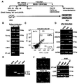
- Figure 1 relates to the conversion of endothelial cells (EC) by Oct4, Nanog, Tal1, LMO2 gene introduction
- A is a schematic diagram of the induction of MSCs into endothelial cells
- B of the umbilical cord-derived MSCs Gene expression was confirmed by RT-PCR after Oct4, Nanog, Tal1, and LMO2 genes were transformed into lentiviral for induction into endothelial cells
- C is an endothelial cell marker after induction into endothelial cells.
- Cells positive for CD31 / CD144 (iEC) were analyzed by FACS, and
- D RT-PCR was used to analyze endothelial marker expression after passage of amplified iECs into endothelial cells.
- E shows the results of RT4-PCR analysis of Oct4, Nanog, Tal1, LMO2 gene expression after expansion of iEC. When cultured in SB431542), it is a result indicating that it is not converted to endothelial cells.
- Figure 2 is a repeat experiment by changing the incubation time in the conversion of MSC to EC by Oct4, Nanog, Tal1, LMO2, confirming the production reproducibility and conversion rate of CD31 / CD144 positive cells.
- FIG. 3 shows the results of investigating the expression of VE-cadherin by immunofluorescence after conversion of expanded iEC, and the absorption and angiogenesis of ac-LDL.
- Figure 4 confirms the optimal combination of gene combinations required for conversion from WJ-MSC to endothelial cells (4A), 4B shows the results of repeated experiments.
- ONTL is a group using a gene combination of Oct4, Nanog, Tal1 4, LMO2, OTL is a gene combination of Oct4, Tal1 4, LMO2, NTL is a combination of Nanog, Tal1 4, LMO2, ONL is Oct4, Nanog, LMO2 gene combinations are shown.
- Tal1 and LMO2 are known as major regulators of hematopoietic and endothelial cell transcription.
- Human umbilical cord-derived mesenchymal stem cells were transduced using recombinant lentiviruses capable of carrying cDNAs of Oct4, Nanog, Tal1 and LMO2 (FIG. 1A), blasticidine (350 ng / ml), puromycin ( Only transduced cells were selected by culturing for 10 days in a medium containing 350 ng / ml). RT-PCR was performed using total RNA to confirm that these four genes were expressed in selected cells (FIG. 1B).
- FIG. 1D As a result of analyzing Oct 4, Nanog, Tal1 and LMO2 gene expression in cultured iEC, the expression of Nanog, Tal1 and LMO2 except for Oct 4 was observed (FIG. 1E). In contrast, culture of MSCs without transduction of these four transcription factors in the same induction medium did not induce conversion into EC cells (FIG. 1F). Direct conversion of MSCs to ECs by these four factors was confirmed by repeated experiments, with conversion to EC cells even induction on day 11 (FIG. 2), with CD31 and CD144 positive cells ranging from 1.4-7%. It was confirmed.
- VE-Cadherin is expressed around the cells as in HUVEC, and these cells were Dil-labeled in contrast to MSC. It was confirmed that it has the ability to absorb ac-LDL, indicating the characteristics of endothelial cells.
- the inventors confirmed that reprogramming of MSCs and adult cells into endothelial cells can be achieved by Oct4, nanog, Tal1 and LMO2.
- the gene of Oct4, Nanog, LMO2, Tal1 4 introduced for endothelial cell conversion in the present invention is preferably Oct4 gene represented by SEQ ID NO: 1, Nanog gene represented by SEQ ID NO: 2, LMO2 gene represented by SEQ ID NO: 3, SEQ ID NO: You can use Tal1 4 as indicated by 4.
- the introduction of the genes into cells can be used as long as the gene introduction techniques known in the art, one embodiment of the present invention used a lentiviral vector.
- Endothelial cells obtained by the above method was found to be applicable to the production of endothelial cells (EC) for constituting regenerative tissue in tissue engineering and treatment of ischemic diseases.
- the present invention can provide a composition for tissue regeneration comprising the endothelial cells obtained by the method of the present invention as an active ingredient, and the present invention includes the endothelial cells obtained by the method of the present invention as an active ingredient. It can provide a cell therapy composition for the treatment of ischemic disease.
- HUVEC Human umbilical vein endothelial cells
- Lentiviral vectors carrying Nanog, Tal1 and LMO2 were purchased from GeneCopoeia (USA), and Oct4 vectors were obtained from Prof. Won-hee Seo from Ajou University. 293FT cells were dispensed into 10 cm tissue culture plates the day before transfection and allowed to be 90-95% density of the plates on the day of transduction.
- the desired vector plasmids psi-EF1-Bls-hOct4, psi-EF1-Puro-hNanog, psi-EF1-Puro-hTaL1 and psi-EF1-Puro-hLMO2 (Genecopoeia; 3 for each plasmid ) was mixed with 9 ⁇ g ViraPowerPackaging Mix and 12 ⁇ g Lipofectamine2000 complex and added to 293FT cells. After overnight incubation at 37 ° C., 5% CO 2 conditions, the medium was replaced with 293FT medium without antibiotics. Culture supernatants were collected 48 hours after medium replacement.
- Virus supernatants were filtered by 0.45 ⁇ m PVDF filter to remove cells and virus stocks were stored at -80 ° C.
- MSCs (5 ⁇ 10 4 / well) were plated for 24 hours and infected with 5 MOI in 10% FBS-DMEM medium containing 8 ⁇ g / ml polybrene (Sigma-Aldrich). After 24 hours, the virus-containing medium was replaced with fresh DMEM medium containing 20% FBS and penicillin / streptomycin and cultured for 2 days. Cells were then incubated for 10 days in medium containing blasticidine (350 ng / ml), puromycin (350 ng / ml) and transduced cells were selected (see FIG. 1A).
- Lentivirus-transduced MSC cells were cultured in gelatin coated dishes containing endothelial cell growth medium EGM2 and 10 ⁇ M SB431542 to induce transformation into endothelial cells.
- the medium was changed every 2 days and split 1: 2 after 3-5 days when cells became 90% density. After culturing the cells for 11-35 days, iEC was isolated by FACS.
- RT-PCR By using the RT-PCR as described above, when transfected with a lentiviral, and then examined whether Oct4, Nanog, Tal1, LMO2 is expressed, it was confirmed that these genes are expressed (Fig. 1B). On the other hand, these cells were induced by differentiation into endothelial cells, and then separated by FACS sorting. Subsequent amplification of the isolated iEC was followed by amplification of endothelial cell marker expression by RT-PCR as described above. It was confirmed that the expression of cadherin, VEGFR2, CD31, eNOS (Fig. 1D), the expression of Nanog, Tal1, LMO2 was confirmed by RT-PCR (Fig. 1E).
- CD31 + / CD144 + cells were harvested for 11-35 days in endothelial cell differentiation induction medium after viral transduction and cells were collected and analyzed by FACS Beckman coulter cell sorter (BD Biosciences). Data was analyzed by FACS summit software (version 6.1.3). CD31 + / CD144 + iECs were incubated in EGM2 containing 10 ⁇ M SB431542 using 1% gelatin coated plates after separation into FACS.
- VE-cadherin expression was examined by immunofluorescence, and ac-LDL uptake and angiogenesis were examined.
- ac-LDL The uptake of ac-LDL by adherent cells known as one of the main features of endothelial cell lineage was measured (Murohara et al., J. Clin. Invest., 2000). Cells incubated in gelatin-coated chamber slides for 24 hours were treated at 37 ° C., 4 in medium containing 15 ⁇ g / ml Dil-labeled acetyl-low density lipoprotein (Dil-ac-LDL, Molecular Probes, Eugene, OR). The reaction was carried out for a time. After washing, the samples were observed by fluorescence microscopy and reversed phase contrast microscopy, and three fields were taken at random.
- Dil-ac-LDL Dil-labeled acetyl-low density lipoprotein
- Chilled Matrigel (150 ⁇ l, BD Bioscience) was placed in pre-cooled 48-well plates and incubated at 37 ° C. for 30 minutes. HUVECs, MSC and iEC were aliquoted onto solidified Matrigel (2 ⁇ 10 4 cells / well) and tube formation was taken 20 hours after incubation in EGM.
- VE-cadherin was expressed at the cell interface, absorbed Dil-labeled ac-LDL, and vasculature, similar to HUVEC cells, although completely different from MSC in expanded iEC. It also confirmed that it has the ability.
- the present inventors carried out the following experiment to identify the optimal gene required for the conversion of mesenchymal stem cells to endothelial cells confirmed as a result of the above example.
- endothelial cell differentiation ability was investigated in the group which introduced three genes of Oct4, Nanog, LMO2 (ONL).
Landscapes
- Health & Medical Sciences (AREA)
- Life Sciences & Earth Sciences (AREA)
- Chemical & Material Sciences (AREA)
- Genetics & Genomics (AREA)
- Zoology (AREA)
- Organic Chemistry (AREA)
- Engineering & Computer Science (AREA)
- General Health & Medical Sciences (AREA)
- Biomedical Technology (AREA)
- Biotechnology (AREA)
- General Engineering & Computer Science (AREA)
- Virology (AREA)
- Bioinformatics & Cheminformatics (AREA)
- Cell Biology (AREA)
- Wood Science & Technology (AREA)
- Molecular Biology (AREA)
- Biophysics (AREA)
- Biochemistry (AREA)
- Medicinal Chemistry (AREA)
- Proteomics, Peptides & Aminoacids (AREA)
- Pharmacology & Pharmacy (AREA)
- Gastroenterology & Hepatology (AREA)
- Physics & Mathematics (AREA)
- Plant Pathology (AREA)
- Microbiology (AREA)
- Vascular Medicine (AREA)
- Developmental Biology & Embryology (AREA)
- Immunology (AREA)
- Toxicology (AREA)
- Epidemiology (AREA)
- Animal Behavior & Ethology (AREA)
- Public Health (AREA)
- Veterinary Medicine (AREA)
- Medicines Containing Material From Animals Or Micro-Organisms (AREA)
- Micro-Organisms Or Cultivation Processes Thereof (AREA)
- Materials For Medical Uses (AREA)
Abstract
본 발명은 특정 전사인자를 이용한 중간엽 줄기세포를 내피세포로 전환시키는 방법에 관한 것으로, 더 상세하게는 특정 전사인자인 Oct4, Nanog, Tal1 및 LMO2를 이용하여 중간엽 줄기세포를 내피세포로 전환시키는 방법에 관한 것이다. 본 발명에 따르면, 세포의 리프로그래밍 인자 중 암 유발과 직접적인 관련이 적은 유전자 2종과 혈관 발달에 관련한 전사인자 인자 중에서 중간엽 줄기세포에서 발현이 되지 않거나 낮은 수준을 발현되는 전사인자 2 종을 선별하여, 이들 4개를 조합함으로써 성체세포나 성체줄기세포인 중간엽 줄기세포를 내피세포로 전환시키는 방법을 개발하였고, 이는 조직 공학과 허혈성 질환의 치료에서 재생 조직을 구성하기 위한 내피세포의 생성에 적용될 수 있는 효과가 있다.
Description
본 발명은 특정 전사인자를 이용한 중간엽 줄기세포를 내피세포로 전환시키는 방법에 관한 것으로, 더 상세하게는 특정 전사인자인 Oct4, Nanog, Tal1 및 LMO2를 이용하여 중간엽 줄기세포를 내피세포로 전환시키는 방법에 관한 것이다.
배아줄기세포와 유도만능 줄기세포 등 pluripotent stem cell로부터 내피세포의 생성은 혈관 생성을 필요로 하는 허혈질환 치료에 있어 적용할 수 있어 중요하다. 또한 간 조직과 같은 재생조직 분야에서도 내피세포는 중요한 혈관 망을 제공할 수 있어 필수적이다. 배아줄기세포나 유도만능 줄기세포로부터 내피세포로 분화유도하는 연구들이 많이 진행되었지만, 이들 세포로부터 만든 세포의 경우 암 유발 가능성과 분화가 완전하지 않을 수 있는 점이 우려되므로, 섬유아세포에서와 같은 성숙한 somatic 세포의 부분적 reprogramming에 의해 내피세포로 transdifferentiation시키는 방법이 대안이 될 수 있다 [Graf, T. et al., Nature., 2009].
실제로, MyoD 유전자를 mouse embryonic fibroblast에 MyoD 유전자와 5-azacytidine 처리로 근육세포로 성공적으로 전환시킨 바가 있고 [Davis, R.L., et al Cell, 1987], pancreatic exocrine cell에 Ngn3, Pdx1 and Mafa 유전자를 도입시켜 인슐린을 분비하는 beta cell-like cell로 전화시킨 바가 있다 [Zhou, Q., et al., Nature, 2008].
내피세포의 경우에도 2012년에 Ginsberg 등이 처음으로 human midgestation c-Kit- linease-committed amniotic cells에 ETS transcription factors ETV2, FLI1, ERG1를 도입시키고, TGF beta 저해제 존재 하에 EGM2 배지에서 유도 배양하여 내피세포 특성을 나타내는 세포로 전환시킬 수 있음을 보고하였다 [Ginsberg, M., et al., Cell, 2012]. 이들 세포는 Matrigel plug과 재생 간 조직에서 안정된 혈관을 형성하는 것으로 관찰되었다. 이 후, 인간 피부 fibroblast에 ETV2 유전자 하나만 도입하여 정맥내피세포 특성을 나타내는 세포로 전환시킬 수 있음이 보고되었다 [Morita, R., et al., Proc Natl Acad Sci U S A, 2015].
이외에도 toll-like receptor 3 (TLR3) agonist를 사용하여 innate immunity를 자극함으로서 fibroblast를 내피세포 특성을 나타내는 세포로 전화시키거나 [Sayed, N., et al., Circulation, 2015], Oct4 와 Klf4 두 유전자를 도입하여 내피세포 특성을 나타내는 세포로 전환시키기도 하였다 [Li, J., et al., Arterioscler Thromb Vasc Biol, 2013].
이외에 Yamanaka factor 4개 Oct4, Sox2, Klf4, c-Myc을 도입하되 iPS 생성단계까지 유도하지 않고 부분 reprogramming을 시켜 내피세포로 전환시키는 시도도 이루어졌다 [Margariti, A., et al., Proc Natl Acad Sci 2012]. 본 발명에서는 reprogramming 인자 중에 암 유발과 직접적으로 관련이 적은 유전자 2종과 혈관 development에 관련한 전사인자 인자 중에서 제대 유래 중간엽 줄기세포에서 발현이 되지 않거나 낮은 수준을 발현되는 전사인자 2 종을 선별 하여, 이들 4개를 조합하여 adult somatic cell이나 성체줄기세포인 중간엽 줄기세포 (MSC)를 내피세포로 전환시키는 방법을 개발하고자 하였다.
본 발명의 목적은 중간엽 줄기세포를 내피세포로 전환시키는 신규한 방법을 제공하는 것이다.
본 발명의 다른 목적은 중간엽 줄기세포를 내피세포로 전환시키기 위한 신규한 조성물을 제공하는 것이다.
본 발명의 다른 목적은 본 발명의 방법으로 수득한 내피세포를 유효성분으로 포함하는 조직재생용 조성물을 제공한다.
본 발명의 다른 목적은 본 발명의 방법으로 수득한 내피세포를 유효성분으로 포함하는 허혈성 질환의 치료를 위한 세포치료제 조성물을 제공한다.
상기 목적을 달성하기 위하여, 본 발명은 Oct4, Nanog, Tal1 및 LMO2 유전자 또는 상기 유전자들이 코딩하는 단백질을 중간엽 줄기세포에 도입하는 단계를 포함하는, 중간엽 줄기세포를 내피세포로 전환시키는 방법을 제공한다.
본 발명의 일실시예에 있어서, 상기 Oct4, Nanog, Tal1 및 LMO2 유전자는 렌티바이러스 벡터를 이용하여 중간엽 줄기세포에 형질도입하는 것일 수 있다.
본 발명의 일실시예에 있어서, 상기 Oct4 유전자는 서열번호 1로 표시된 것이고, Nanog 유전자는 서열번호 2로 표시된 것이고, Tal1 유전자는 서열번호 3으로 표시된 것이고, LMO2 유전자는 서열번호 4로 표시된 것일 수 있다.
또한 본 발명은 Oct4, Nanog, Tal1 및 LMO2 유전자 또는 상기 유전자들이 코딩하는 단백질을 포함하는 중간엽 줄기세포의 내피세포로의 분화유도용 조성물을 제공한다.
또한 본 발명은 상기 본 발명의 방법으로 수득한 내피세포를 유효성분으로 포함하는 조직재생용 조성물을 제공한다.
또한 본 발명은 상기 본 발명의 방법으로 수득한 내피세포를 유효성분으로 포함하는 허혈성 질환의 치료를 위한 세포치료제 조성물을 제공한다.
본 발명에 따르면, 세포의 리프로그래밍 인자 중 암 유발과 직접적으로 관련이 적은 유전자 2종과 혈관 발달에 관련한 전사인자 인자 중에서 중간엽 줄기세포 (MSC)에서 발현이 되지 않거나 낮은 수준을 발현되는 전사인자 2 종을 선별하여, 이들 4개를 조합함으로써 성체세포나 성체줄기세포인 중간엽 줄기세포를 내피세포로 전환시키는 방법을 개발하였고, 이는 조직 공학과 허혈성 질환의 치료에서 재생 조직을 구성하기 위한 내피세포의 생성에 적용될 수 있는 효과가 있다.
도 1은 Oct4, Nanog, Tal1, LMO2 유전자 도입에 의한 내피세포로(EC)의 전환에 관한 것으로, (A)는 MSC의 내피세포로의 유도에 관한 모식도이고, (B)는 제대 유래 MSC의 내피세포로의 유도를 위해 렌티바이러스로 형질전환하기 위한 Oct4, Nanog, Tal1, LMO2 유전자 도입을 한 후 유전자 발현을 RT-PCR로 확인한 결과이며, (C)는 내피세포로의 유도 후 내피세포 표지자인 CD31/CD144에 대해 양성인 세포 (iEC)를 FACS 분석한 결과이고, (D)는 내피세포로의 유도된 iEC를 계대 배양하여 증폭한(expansion) 후 내피세포 마커 발현 양상을 RT-PCR로 분석한 결과이고, (E)는 iEC를 expansion한 후에 Oct4, Nanog, Tal1, LMO2 유전자 발현 양상을 RT-PCR로 조사한 결과이고, (F)는 MSC를 유전자 도입 없이 EGM2와 내피세포 유도 배지 (EGM2+ 10uM SB431542)에서 배양하였을 때, 내피세포로 전환되지 않음을 나타내는 결과이다.
도 2는 Oct4, Nanog, Tal1, LMO2에 의한 MSC의 EC로의 전환에 있어서 배양시간을 변화시켜 반복실험하며, CD31/CD144 양성 세포의 생성 재현성과 전환율을 확인한 결과이다.
도 3은 Expansion된 iEC의 전환 후, VE-cadherin 발현을 면역형광법으로 조사하고, ac-LDL의 흡수 및 맥관형성을 조사한 결과이다.
도 4는 WJ-MSC로부터 내피세포로의 전환에 필요한 유전자 조합의 최적 조합을 확인한 것이며(4A), 4B는 반복실험 결과를 나타낸 것이다. 도면에서 ONTL은 Oct4, Nanog, Tal1 4, LMO2의 유전자 조합을 사용한 군이고, OTL은 Oct4, Tal1 4, LMO2의 유전자 조합을, NTL은 Nanog, Tal1 4, LMO2의 유전자 조합을, ONL은 Oct4, Nanog, LMO2의 유전자 조합을 나타낸 것이다.
Tal1과 LMO2는 조혈 및 내피 세포의 전사에 주요 조절자로 알려져 있다. 인간 제대유래 중간엽줄기세포에 Oct4, Nanog, Tal1 및 LMO2의 cDNA를 운반할 수 있는 재조합 렌티바이러스를 이용하여 형질도입하고(도 1A), 블라스티시딘 (350 ng/ml),퓨로마이신(350 ng/ml)을 포함하는 배지에서 10일간 배양함으로써 형질도입된 세포만을 선별하였다. 총 RNA를 이용하여 RT-PCR을 실시하여, 이들 4 가지 유전자가 선별된 세포에서 발현되고 있음을 확인할 수 있었다(도 1B). 이 후, 이들 세포를 35일간 TGF 저해제인 SB431542 (10 uM)를 포함하는 EGM2에서 배양하여 내피세포로의 전환을 유도하였다. 이들 세포를 FACS 분석하였을 때, 4.96%의 CD31+VE-cadherin+ 세포를 포함하는 유도된 세포 집단을 확인할 수 있었다(도 1C). 이들 CD31+VE-cadherin+ 세포를 FACS로 sorting하고 같은 배지에서 배양하였다. 이들 배양된 세포(iEC)를 PCR 분석을 통해 VE-cadherin, VEGFR2, CD31, 및 eNOS의 내피 유전자 분석을 진행해 본 결과, 배양된 iEC 세포에서 VE-caderin, VEGFR2, CD31 및 eNOS가 HUVEC에서와 같이 발현되었다(도 1D). 배양된 iEC에서 Oct 4, Nanog, Tal1 및 LMO2 유전자 발현을 분석해 본 결과, Oct 4를 제외한 Nanog, Tal1 및 LMO2의 발현이 관찰되었다(도 1E). 이와 대조적으로, 같은 유도 배지에서 이들 4개의 전사 인자의 형질도입 없는 MSC의 배양은 EC 세포로의 전환을 유도하지 못하는 것으로 나타났다(도 1F). 이러한 4개 인자에 의한 MSC의 EC로의 직접적 전환을 반복 실험으로 확인할 수 있었는데, 11일째 유도에서 조차 EC 세포로의 전환이 일어났으며(도 2), CD31과 CD144 양성 세포는 1.4~7% 범위로 확인되었다.
iEC의 특성을 좀 더 자세히 분석해 보고자, 이들 세포를 면역형광 분석해 보았는데, 그 결과, HUVEC에서와 같이 세포 주변에 VE-Cadherin이 발현되는 것을 알 수 있었고, 이들 세포는 MSC와는 대조적으로 Dil-표지된 ac-LDL을 흡수할 수 있는 능력을 가지고 있는 것으로 확인되어 내피세포의 특성을 나타내는 것을 알 수 있었다.
마지막으로, 본 발명자들은 이들 유도된 세포들이 튜브-유사 구조를 형성할 수 있는지 확인해 보았는데, 도 3에서 볼 수 있듯이 이들 세포는 MSC와는 대조적으로 HUVEC에서와 같이 20시간에서 마트리겔 상에서 튜브-유사 구조를 형성하는 것을 알 수 있었다.
상기와 같은 결과를 통해 본 발명자들은 MSC와 성체세포가 내피 세포로 리프로그래밍되는 것이 Oct4, nanog, Tal1 및 LMO2에 의해 이루어질 수 있음을 확인하였다.
또한 본 발명자들은 상기와 같이, Oct4, Nanog, Tal1, LMO2 4가지 유전자 모두를 사용할 경우만이 MSC와 성체세포가 내피세포로 효율적으로 전환될 수 있음을 확인하였는데, 본 발명의 일실시예에 의하면 Oct4, Tal1, LMO2, Nanog, Tal1, LMO2(NTL) 6가지 유전자를 도입한 군과, Oct4, Nanog, LMO2 (ONL) 3가지 유전자를 도입한 군 및 Oct4, Nanog, Tal1, LMO2 4가지 유전자를 도입한 군을 대상으로 유전자 조합에 따른 내피세포 전환 정도를 분석하였다.
그 결과, 도 4에 나타낸 바와 같이 Oct4, Nanog, LMO2, Tal1 4 가지 유전자를 도입한 군의 경우에는 CD31/CD144 양성 세포로의 전환이 일어난 것을 확인할 수 있었으나, Oct4, Tal1, LMO2 (OTL), Nanog, Tal1, LMO2 (NTL) 또는 Oct4, Nanog, LMO2 (ONL)의 3개 유전자로 이루어진 조합으로 도입한 경우에는 CD31/CD144 양성 세포로의 전환이 일어나지 않음을 확인하였다.
따라서 이러한 결과를 통해 본 발명에서는 MSC와 성체세포가 내피세포로 완벽하게 전환하기 위해서는 Oct4, Nanog, LMO2, Tal1 4 가지의 특별한 유전자를 모두 도입해야 함을 알 수 있었다.
본 발명에서 내피세포 전환을 위해 도입한 상기 Oct4, Nanog, LMO2, Tal1 4 의 유전자는 바람직하게 서열번호 1로 표시된 Oct4 유전자, 서열번호 2로 표시된 Nanog 유전자, 서열번호 3으로 표시된 LMO2 유전자, 서열번호 4로 표시된 Tal1 4을 사용할 수 있다.
또한 상기 유전자들의 세포내로의 도입은 당업계에 공지된 유전자 도입 기술이라면 모두 사용할 수 있으며, 본 발명의 일실시예에서는 렌티바이러스 벡터를 이용하였다.
상기와 같은 방법으로 수득된 내피세포는 조직 공학과 허혈성 질환의 치료에서 재생 조직을 구성하기 위한 내피세포(EC)의 생성에 적용될 수 있음을 알 수 있었다.
따라서 본 발명은 상기 본 발명의 방법으로 수득한 내피세포를 유효성분으로 포함하는 조직재생용 조성물을 제공할 수 있고, 또한, 본 발명은 상기 본 발명의 방법으로 수득한 내피세포를 유효성분으로 포함하는 허혈성 질환의 치료를 위한 세포치료제 조성물을 제공할 수 있다.
본 발명에 의해 줄기세포에서부터 전환된 내피세포는 조직재생 또는 허혈성 질환의 치료를 위한 세포치료제로서 사용 가능한데, 조직재생의 경우, 조직 구조 측면에서 내피세포에 해당하는 부분을 본 발명에서 전환된 내피세포를 사용할 수 있다.
이하, 실시예를 통하여 본 발명을 보다 상세히 설명하고자 한다. 이들 실시예는 본 발명을 보다 구체적으로 설명하기 위한 것으로, 본 발명의 범위가 이들 실시예에 한정되는 것은 아니다.
<
실시예
1>
Oct4
,
Nanog
,
Tal1
,
LMO2
유전자 도입에 의한 내피세포로의 전환
1-1. 세포배양 및 시약 준비
HUVEC(Human umbilical vein endothelial cells ) 세포는 Jaffe, E.A의 논문에 개시된 방법에 따라 인간 혈관에서 분리하였다. HUVEC 세포는 20% FBS(Invitrogen), 30㎍/ml의 내피 세포 성장 보조제(ECGS, BD Biosciences), 90㎍/ml의 헤파린, 1% 항생제를 포함하는 M199 배지에서 배양하였다. iEC는 10 μM SB431542(Sigma)가 첨가된 EGM2 배지(Lonza)에서 배양하였고, 제대 유래 중간엽줄기세포(MSC)는 20% FBS와 1% 항생제가 포함된 DMEM(Invitrogen)배지에서 배양하였다. 인간 탯줄 샘플은 가톨릭대학교 임상 시험 심사위원회의 승인된 절차에 따라 분리되었다(과제번호 MC13TISI0078, MC12TISI0094).
1-2.
렌티바이러스
벡터 및 형질도입
Nanog, Tal1 및 LMO2를 운반하는 렌티바이러스 벡터는 GeneCopoeia(USA)에서 구입하였고, Oct4 벡터는 아주대학교 서원희 교수로부터 수득하였다. 트랜스펙션 전날 10cm 티슈 배양 플레이트에 293FT 세포를 분주하고 트랜스덕션 당일 플레이트의 90~95% 밀도가 되도록 하였다. 렌티바이러스를 생산하기 위하여, 목적하는 벡터 플라스미드인 psi-EF1-Bls-hOct4, psi-EF1-Puro-hNanog, psi-EF1-Puro-hTaL1 및 psi-EF1-Puro-hLMO2 (Genecopoeia; 3 for each plasmid) 를 9㎍의 ViraPowerPackaging Mix와 12㎍의 Lipofectamine2000 복합체와 혼합한 후, 293FT 세포에 첨가하였다. 37℃, 5% CO2 조건에서 밤새 배양한 후, 배지를 항생제를 포함하지 않는 293FT 배지로 교체하였다. 배지 교체 후 48시간이 지나 배양 상청액을 수집하였다. 바이러스 상청액은 0.45㎛ PVDF 필터로 필터링하여 세포를 제거하였고, 바이러스 stock은 -80℃에 보관하였다. MSC(5×10⁴/well)를 24시간 동안 플레이팅하고 8㎍/ml 폴리브린(polybrene, Sigma-Aldrich)를 포함하는 10% FBS-DMEM 배지에서 5 MOI로 감염시켰다. 24시간 후, 바이러스를 포함하는 배지를 20% FBS와 페니실린/스트렙토마이신을 첨가한 신선한 DMEM 배지로 교환하여 2일간 배양하였다. 이후 세포를 블라스티시딘 (350 ng/ml), 퓨로마이신(350ng/ml)이 있는 배지에서 10일간 배양하고 형질도입된 세포를 선별하였다(도 1A 참조).
1-3.
렌티바이러스가
형질도입된
MSC의
내피세포로의 유도
렌티바이러스가 형질도입된 MSC 세포를 내피 세포 성장 배지 EGM2와 10μM SB431542를 포함하는 젤라틴 코팅된 접시에서 배양하여 내피세포로의 변환을 유도하였다. 배지는 2일마다 교체하였고, 3~5일 후에 세포가 90% 밀도가 되면 1:2로 스플릿하였다. 세포를 11~35일간 배양한 후, iEC를 FACS로 분리하였다.
1-4.
RNA
추출 및 RT-
PCR
총 RNA를 Trizol 시약(Invitrogen)을 이용하여 배양된 세포에서 추출하였다. RT-PCR을 위한 cDNA 합성은 제조자의 지시에 따라 SuperScript 합성 시스템(Enzynomics)을 이용하여 수행하였다. 각 유전자는 표 1에 개시된 프라이머 세트를 사용하여 PCR로 증폭하였다. 합성된 cDNA를 이용하여 반응한 PCR 조건은 하기와 같다: 95℃ 7분(최초 변성), 95℃ 1 분(변성) - 54℃~60℃ 1분(어닐링) - 72℃ 1분 연장 과정을 총 30 사이클, 72℃ 10분(최후 연장). PCR 산물은 1.0% 아가로오즈-겔 전기영동으로 분석하였고, DNA 밴드는 SYBR safe DNA 겔 염색(Invitrogen)으로 가시화하였다.
| Name | Forward | Reserve | Accession no. |
| Oct4 | AGGAGATATGCAAAGCAGAA | AGAGTGGTGACGGAGACAG | NM_002701 |
| Nanog | ATCCAGCTTGTCCCCAAAG | ATTTCATTCTCTGGTTCTGG | NM_024865.2 |
| Tal1 | TCACCACCAACAATCGAGTGAAGAGG | CTCCTCCTGGTCATTGAGCAGCTTGG | NM_003189.2 |
| LMO2 | TCCCCAATGTCCTCGGCCAT | ATCCGCTTGTCACAGGAT | NM_005574.3 |
| CD31 | ATGATGCCCAGTTTGAGGTC | GACGTCTTCAGTGGGGTTGT | NM_000442.4 |
| VE-ead | CCCTACCAGCCCAAAGTGTG | CGACTTGGCATCCCATTGTC | NM_001795.3 |
| VEGFR2 | GCGATGGCCTCTTCTGTAAG | ACACGACTCCATGTTGGTCA | EU826563.1 |
| eNOS | TGATGGCGAAGCGAGTGAA | ACTCATCCATACACAGGACCCG | NM_000603.4 |
상기와 같은 RT-PCR을 이용하여 우선, 렌티바이러스로 형질도입한 후 Oct4, Nanog, Tal1, LMO2 가 발현되는지 조사하였을 때, 이들 유전자들이 발현되고 있음을 확인할 수 있었다(도 1B). 한편, 이들 세포를 내피세포로 분화 유도시킨 후 FACS sorting하여 분리하고, 분리한 iEC를 계대 배양하여 증폭시킨 후 내피세포 마커 발현 양상을 상기와 같은 RT-PCR로 살펴본 결과, HUVEC과 유사하게 VE-cadherin, VEGFR2, CD31, eNOS가 발현됨을 확인할 수 있었고(도 1D), Nanog, Tal1, LMO2가 발현되는 것을 RT-PCR로 확인할 수 있었다(도 1E).
1-5.
FACS
분석
내피세포로 분화 유도한 후 FACS 분석을 통해 내피세포 표지자인 CD31/CD144 양성 세포를 확인해 보고자 하였다.
이를 위해 세포를 수집하고 PE-접합된 VE-cadherin 항체(1:400; BD Biosciences) 또는 FITC-접합된 CD31 항체(1:400; BD Biosciences)로 아이스에서 30분간 염색하였다. 샘플을 FACSAriaIII cell analyzer(BD Biosciences)로 분석하였다. 데이터는 BD CellQuest Pro software(version 5.2.1)로 분석하였다. CD31+/CD144+ 세포를 확인하기 위하여, 바이러스 형질도입 후 내피세포 분화 유도 배지에서 35일간 배양한 후 세포를 수집하고 FACS Beckman coulter cell sorter(BD Biosciences)로 분석하였다. 데이터는 FACS summit software(version 6.1.3)로 분석하였다. CD31+/CD144+ iECs는 FACS로 분리 후 1% 젤라틴이 코팅된 플레이트를 이용하여 10μM SB431542를 포함하는 EGM2에서 계속해서 배양하였다.
그 결과, 도 1C에서 볼 수 있듯이 내피세포 표지자인 CD31/CD144에 대해 양성인 세포(iEC)가 4.96% 존재함을 확인할 수 있었다.
<
실시예
2>
Oct4
,
Nanog
,
Tal1
,
LMO2에
의한
MSC의
EC로의
전환에 있어서
재현성 및 전환율 검토
실시예 1에서와 같은 실험방법으로 4 가지 유전자를 도입시키고, 내피세포를 분화 유도시키되, 내피세포 유도배지에서의 배양시간을 변화시켜 가며 반복실험하여 CD31/CD144 양성세포가 재현성 있게 생성되는지 여부와 전환율을 확인해 보았다.
CD31+/CD144+ 세포를 확인하기 위하여, 바이러스 형질도입 후 내피세포 분화 유도 배지에서 11~35일간 배양한 후 세포를 수집하고 FACS Beckman coulter cell sorter(BD Biosciences)로 분석하였다. 데이터는 FACS summit software(version 6.1.3)로 분석하였다. CD31+/CD144+ iECs는 FACS로 분리 후 1% 젤라틴이 코팅된 플레이트를 이용하여 10μM SB431542를 포함하는 EGM2에서 배양하였다.
그 결과, 도 2에서 볼 수 있듯이, CD31/CD144 양성세포가 재현성 있게 생성되었고, 약 1.4~7% 비율로 생성되었음을 확인할 수 있었다.
<
실시예
3>
계대
배양을 통해 증폭(
expansion
)된
iEC의
특성 조사
FACS를 통해 sorting된 CD31/CD144 양성 세포 (iEC)를 expansion한 후에, VE-cadherin 발현을 면역형광법으로 조사하였고, ac-LDL 흡수 및 맥관형성을 조사하였다.
3-1.
면역형광염색법을
통한
VE
-
cadherin
발현 조사
세포를 100% 메탄올로 5분간 고정하고 0.1% Triton X-100으로 3분간 투과화하였다. 비특이적 단백질 결합 부위를 5% BSA로 1시간동안 블락킹하였고, 세포를 VE-cadherin (1:100, cell signaling) 1차 항체로 반응시킨후 PBS로 3번 세척하고, Cy 3 (Millipore)가 접합된 2차 항체로 2시간 동안 반응하였다. 마지막으로, 모든 슬라이드를 1㎍/ml DAPI (Sigma)로 대조염색하였다. 이미지는 공초점 현미경(Zeiss LSM 510 Meta with LSM image examiner software, Germany)으로 얻을 수 있었다.
3-2. 시험관 내
(
In
vitro
)
내피세포 기능 분석
내피세포 계열의 주요한 특징 중 하나로 알려진 부착 세포에 의한 ac-LDL의 흡수를 측정해 보았다(Murohara et al., J. Clin. Invest., 2000). 24시간 동안 젤라틴 코팅된 챔버 슬라이드에서 배양한 세포를, 15㎍/ml Dil-라벨된 아세틸-저밀도 리포프로테인(Dil-ac-LDL, Molecular Probes, Eugene, OR)을 포함하는 배지에서 37℃, 4시간 동안 반응시켰다. 세척 후, 샘플을 형광 현미경 및 역 위상차 현미경으로 관찰하고, 세 개의 필드를 임의로 선정하여 촬영하였다.
3-3. 튜브(맥관) 형성 분석
냉장된 마트리겔(150㎕, BD Bioscience)을 예냉된 48-웰 플레이트에 넣고 37℃에서 30분간 인큐베이션하였다. HUVECs, MSC 및 iEC를 고형화된 마트리겔 위에 분주하고(2×10⁴ cells/well), EGM에서 배양 20시간 후에 튜브 형성을 촬영하였다.
그 결과, 도 3에서 볼 수 있듯이, expansion된 iEC에서 MSC와 전혀 다르지만 HUVEC 세포와는 유사하게, VE-cadherin이 세포 경계면에서 발현되고, Dil-표지된 ac-LDL을 흡수할 수 있었으며, 맥관 형성 능력도 보유하고 있음을 확인할 수 있었다.
<
실시예
4>
Wharton's
jelly
mesenchymal
stem
cells(WJ-MSC)로부터
내피세포로의 전환에 필요한 최적 유전자 조합 조사
본 발명자들은 상기 실시예의 결과로 확인된 중간엽 줄기세포의 내피세포로의 전환을 위해 필요한 최적 유전자를 확인하기 위해 다음과 같은 실험을 수행하였다. 상기 실시예에서 사용한 4가지 유전자인 Oct4, Nanog, Tal1, LMO2을 도입한 군; Oct4, Tal1, LMO2 (OTL)의 3가지 유전자를 도입한 군; Nanog, Tal1, LMO2 (NTL)의 3가지 유전자를 도입한 군; 및 Oct4, Nanog, LMO2 (ONL)의 3가지 유전자를 도입한 군을 대상으로 내피세포 분화능을 조사하였다. 구체적으로 상기 실시예1에서와 동일한 실험 방법으로 Oct4, Tal1, LMO2 (OTL), Nanog, Tal1, LMO2 (NTL) 혹은 Oct4, Nanog, LMO2 (ONL)의 3가지 유전자로 이루어진 조합으로 유전자를 세포내에 도입시키고 24 시간 후 페니실린/스트렙토마이신을 첨가한 신선한 DMEM 배지로 교환하여 2일간 배양한 다음, 10일간 세포선별(selection) 하여 형질 도입된 세포를 선별하였다. 이후 내피세포 성장 배지 EGM2에 10 uM SB431542를 포함하여 젤라틴 코팅된 접시에서 배양하였고 2일 마다 배지를 교체하면서 21일간 배양한 다음 FACS 분석을 실시하였다.
분석결과, 도 4에 나타낸 바와 같이, Oct4, Nanog, Tal1, LMO2 4 가지 유전자를 도입시킨 것은 CD31/CD144 양성 세포로의 전환이 일어났지만 3가지 유전자로 이루어진 조합들을 도입한 경우에는 CD31/CD144 양성인 세포를 얻을 수 없었다(도 4 A 참조). 추가적인 반복실험을 실시하여 조사한 경우에도 4 가지 유전자를 동시에 도입한 경우 CD31/CD144 양성세포가 높은 효율(~5%)로 얻어진 반면, Nanog, Tal1, LMO2 3가지 유전자로 이루어진 조합을 도입한 경우에는 CD31/CD144 양성세포가 거의 나타나지 않음을 확인하였다(도 4B 참조).
이러한 결과를 통해 본 발명자들은 본 실험의 분화 배양 조건에서는 MSC가 내피세포로 효율적으로 전환되기 위해서는 4개의 전사인자인 Oct4, Nanog, Tal1, LMO2가 모두 필요함을 확인 할 수 있었고, 이 중 1개의 유전자가 없는 경우 내피세포로의 전환이 이루어지지 않음을 알 수 있었다.
이제까지 본 발명에 대하여 그 바람직한 실시예들을 중심으로 살펴보았다. 본 발명이 속하는 기술 분야에서 통상의 지식을 가진 자는 본 발명이 본 발명의 본질적인 특성에서 벗어나지 않는 범위에서 변형된 형태로 구현될 수 있음을 이해할 수 있을 것이다. 그러므로 개시된 실시예들은 한정적인 관점이 아니라 설명적인 관점에서 고려되어야 한다. 본 발명의 범위는 전술한 설명이 아니라 특허청구범위에 나타나 있으며, 그와 동등한 범위 내에 있는 모든 차이점은 본 발명에 포함된 것으로 해석되어야 할 것이다.
Claims (7)
- Oct4, Nanog, Tal1 및 LMO2 유전자 또는 상기 유전자들이 코딩하는 단백질을 중간엽 줄기세포에 도입하는 단계를 포함하는, 중간엽 줄기세포를 내피세포로 전환시키는 방법.
- 제1항에 있어서,상기 Oct4, Nanog, Tal1 및 LMO2 유전자는 렌티바이러스 벡터를 이용하여 중간엽 줄기세포에 형질도입하는 것을 특징으로 하는, 중간엽 줄기세포를 내피세포로 전환시키는 방법.
- 제2항에 있어서,상기 Oct4 유전자는 서열번호 1로 표시된 것이고, Nanog 유전자는 서열번호 2로 표시된 것이고, Tal1 유전자는 서열번호 3으로 표시된 것이고, LMO2 유전자는 서열번호 4로 표시된 것을 특징으로 하는, 중간엽 줄기세포를 내피세포로 전환시키는 방법.
- 제1항에 있어서,Oct4 단백질은 서열번호 5로 표시된 것이고, Nanog 단백질은 서열번호 6으로 표시된 것이고, Tal1 단백질은 서열번호 7로 표시된 것이고, LMO2 단백질은 서열번호 8로 표시된 것을 특징으로 하는, 중간엽 줄기세포를 내피세포로 전환시키는 방법.
- Oct4, Nanog, Tal1 및 LMO2 유전자 또는 상기 유전자들이 코딩하는 단백질을 포함하는 중간엽 줄기세포의 내피세포로의 분화유도용 조성물.
- 제1항의 방법으로 수득한 내피세포를 유효성분으로 포함하는 조직재생용 조성물.
- 제1항의 방법으로 수득한 내피세포를 유효성분으로 포함하는 허혈성 질환의 치료를 위한 세포치료제 조성물.
Priority Applications (1)
| Application Number | Priority Date | Filing Date | Title |
|---|---|---|---|
| US15/570,619 US10428307B2 (en) | 2015-04-29 | 2016-04-29 | Method for converting mesenchymal stem cells into endothelial cells by using specific transcription factors |
Applications Claiming Priority (4)
| Application Number | Priority Date | Filing Date | Title |
|---|---|---|---|
| KR10-2015-0060259 | 2015-04-29 | ||
| KR20150060259 | 2015-04-29 | ||
| KR1020160053144A KR101789417B1 (ko) | 2015-04-29 | 2016-04-29 | 특정 전사인자를 이용한 중간엽 줄기세포를 내피세포로 전환시키는 방법 |
| KR10-2016-0053144 | 2016-04-29 |
Publications (2)
| Publication Number | Publication Date |
|---|---|
| WO2016175618A1 true WO2016175618A1 (ko) | 2016-11-03 |
| WO2016175618A8 WO2016175618A8 (ko) | 2017-11-30 |
Family
ID=57198874
Family Applications (1)
| Application Number | Title | Priority Date | Filing Date |
|---|---|---|---|
| PCT/KR2016/004557 Ceased WO2016175618A1 (ko) | 2015-04-29 | 2016-04-29 | 특정 전사인자를 이용한 중간엽 줄기세포를 내피세포로 전환시키는 방법 |
Country Status (1)
| Country | Link |
|---|---|
| WO (1) | WO2016175618A1 (ko) |
Citations (3)
| Publication number | Priority date | Publication date | Assignee | Title |
|---|---|---|---|---|
| CN1546656A (zh) * | 2003-12-16 | 2004-11-17 | 滨 冯 | 成人骨髓间充质干细体外定向诱导分化为血管内皮细胞的一种方法 |
| CN102776150A (zh) * | 2011-05-13 | 2012-11-14 | 上海交通大学医学院附属第九人民医院 | 一种诱导脐带间充质干细胞分化为内皮细胞的方法 |
| US20140349398A1 (en) * | 2010-07-07 | 2014-11-27 | Cellular Dynamics International, Inc. | Endothelial cell production by programming |
-
2016
- 2016-04-29 WO PCT/KR2016/004557 patent/WO2016175618A1/ko not_active Ceased
Patent Citations (3)
| Publication number | Priority date | Publication date | Assignee | Title |
|---|---|---|---|---|
| CN1546656A (zh) * | 2003-12-16 | 2004-11-17 | 滨 冯 | 成人骨髓间充质干细体外定向诱导分化为血管内皮细胞的一种方法 |
| US20140349398A1 (en) * | 2010-07-07 | 2014-11-27 | Cellular Dynamics International, Inc. | Endothelial cell production by programming |
| CN102776150A (zh) * | 2011-05-13 | 2012-11-14 | 上海交通大学医学院附属第九人民医院 | 一种诱导脐带间充质干细胞分化为内皮细胞的方法 |
Non-Patent Citations (2)
| Title |
|---|
| HAN, JUNG - KYU ET AL.: "Direct Conversion of Adult Skin Fibroblasts to Endothelial Cells by Defined Factors", CIRCULATION, vol. 130, no. 14, 2014, pages 1168 - 1178, XP055326271 * |
| OSWALD, JOACHIM ET AL.: "Mesenchymal Stem Cells can be differentiated into Endothelial Cells in Vitro", STEM CELLS, vol. 22, no. 3, 2004, pages 377 - 384, XP002505417 * |
Also Published As
| Publication number | Publication date |
|---|---|
| WO2016175618A8 (ko) | 2017-11-30 |
Similar Documents
| Publication | Publication Date | Title |
|---|---|---|
| JP6708617B2 (ja) | 再プログラム化多能性細胞の作製 | |
| US20130157365A1 (en) | Induced pluripotent stem cells from human umbilical cord tissue-derived cells | |
| WO2015088288A1 (ko) | 트롬빈을 이용한 줄기세포 유래 엑소좀의 생성 촉진 방법 | |
| US10428307B2 (en) | Method for converting mesenchymal stem cells into endothelial cells by using specific transcription factors | |
| Tian et al. | Direct conversion of dermal fibroblasts into neural progenitor cells by a novel cocktail of defined factors | |
| WO2013015644A1 (ko) | 태반-유래 줄기세포의 증식방법 | |
| WO2016117816A1 (ko) | 인간 중간엽 줄기세포의 줄기세포능을 증가시키는 방법 | |
| WO2017135795A1 (ko) | 간세포성장인자를 발현하는 중간엽줄기세포 및 이의 용도 | |
| US20140162366A1 (en) | Generation of vascular progenitor cells | |
| WO2012047037A2 (ko) | 배아줄기세포 유래 심근세포 및 이를 유효성분으로 포함하는 세포치료제 | |
| WO2011102680A9 (ko) | Pi3k/akt/gsk3 경로를 통해 성체줄기세포의 증식, 다분화능 및 재프로그래밍을 촉진하는 cd49f | |
| US20140073049A1 (en) | Induced pluripotent stem cells prepared from human kidney-derived cells | |
| WO2020209636A1 (ko) | 소변세포로부터 신장전구세포로의 직접 역분화를 유도하는 방법 및 이의 방법으로 역분화된 신장전구세포를 포함하는 신장세포 손상 질환 예방 또는 치료용 약학 조성물 | |
| US20210340495A1 (en) | Method for inducing and differentiating pluripotent stem cells and uses thereof | |
| WO2015133792A1 (ko) | 체세포로부터 혈관 전구 세포로의 직접교차분화 유도용 조성물 및 이의 용도 | |
| López-Muneta et al. | Generation of NKX2. 5GFP reporter human iPSCs and differentiation into functional cardiac fibroblasts | |
| WO2023167575A1 (ko) | 저면역원성 줄기세포, 줄기세포로부터 분화되거나 유래된 저면역원성 세포 및 이의 제조방법 | |
| WO2022010220A1 (ko) | 면역원성이 감소된 신규한 이식용 세포 | |
| Mohammad et al. | Mesenchymal and neural stem cell differentiation into Purkinje-like cells: Morphological study | |
| WO2025173930A1 (ko) | 배아 줄기세포 유래 중간엽 줄기세포로부터 혈관내피세포를 제조하는 방법 및 제조된 혈관내피세포를 포함하는 심뇌 및 혈관 질환 예방 또는 치료용 조성물 | |
| Pruller et al. | Overview of Cell Types Capable of Contributing to Skeletal Muscle Repair and Regeneration | |
| JP2011083216A (ja) | コアネットワーク構成転写因子の同定方法 | |
| WO2009120043A2 (ko) | 중배엽 줄기세포의 증식을 촉진하기 위한 cd45+ 세포 또는 cd45+ 세포 배양액의 용도 | |
| WO2021118325A1 (ko) | 항암 바이러스가 도입을 통한 생존능이 향상된 중간엽 줄기세포의 제조방법 | |
| Wei-xin et al. | A microenvironment, rather than chemical, initiates the cardiomyogenic differentiation of marrow stromal cells |
Legal Events
| Date | Code | Title | Description |
|---|---|---|---|
| 121 | Ep: the epo has been informed by wipo that ep was designated in this application |
Ref document number: 16786804 Country of ref document: EP Kind code of ref document: A1 |
|
| NENP | Non-entry into the national phase |
Ref country code: DE |
|
| 122 | Ep: pct application non-entry in european phase |
Ref document number: 16786804 Country of ref document: EP Kind code of ref document: A1 |