WO2016136718A1 - 超解像蛍光イメージング用プローブ - Google Patents
超解像蛍光イメージング用プローブ Download PDFInfo
- Publication number
- WO2016136718A1 WO2016136718A1 PCT/JP2016/055199 JP2016055199W WO2016136718A1 WO 2016136718 A1 WO2016136718 A1 WO 2016136718A1 JP 2016055199 W JP2016055199 W JP 2016055199W WO 2016136718 A1 WO2016136718 A1 WO 2016136718A1
- Authority
- WO
- WIPO (PCT)
- Prior art keywords
- group
- super
- compound
- probe
- resolution
- Prior art date
- Legal status (The legal status is an assumption and is not a legal conclusion. Google has not performed a legal analysis and makes no representation as to the accuracy of the status listed.)
- Ceased
Links
- 0 CC(C1)C(*(*)*)*=C(*2)C1C(*)C(C=*)C2=CC([C@@](*)C*)=C** Chemical compound CC(C1)C(*(*)*)*=C(*2)C1C(*)C(C=*)C2=CC([C@@](*)C*)=C** 0.000 description 1
Images




Classifications
-
- G—PHYSICS
- G01—MEASURING; TESTING
- G01N—INVESTIGATING OR ANALYSING MATERIALS BY DETERMINING THEIR CHEMICAL OR PHYSICAL PROPERTIES
- G01N21/00—Investigating or analysing materials by the use of optical means, i.e. using sub-millimetre waves, infrared, visible or ultraviolet light
- G01N21/62—Systems in which the material investigated is excited whereby it emits light or causes a change in wavelength of the incident light
- G01N21/63—Systems in which the material investigated is excited whereby it emits light or causes a change in wavelength of the incident light optically excited
- G01N21/64—Fluorescence; Phosphorescence
- G01N21/645—Specially adapted constructive features of fluorimeters
- G01N21/6456—Spatial resolved fluorescence measurements; Imaging
- G01N21/6458—Fluorescence microscopy
-
- A—HUMAN NECESSITIES
- A61—MEDICAL OR VETERINARY SCIENCE; HYGIENE
- A61K—PREPARATIONS FOR MEDICAL, DENTAL OR TOILETRY PURPOSES
- A61K49/00—Preparations for testing in vivo
- A61K49/001—Preparation for luminescence or biological staining
- A61K49/0013—Luminescence
- A61K49/0017—Fluorescence in vivo
- A61K49/0019—Fluorescence in vivo characterised by the fluorescent group, e.g. oligomeric, polymeric or dendritic molecules
-
- A—HUMAN NECESSITIES
- A61—MEDICAL OR VETERINARY SCIENCE; HYGIENE
- A61K—PREPARATIONS FOR MEDICAL, DENTAL OR TOILETRY PURPOSES
- A61K49/00—Preparations for testing in vivo
- A61K49/001—Preparation for luminescence or biological staining
- A61K49/0013—Luminescence
- A61K49/0017—Fluorescence in vivo
- A61K49/005—Fluorescence in vivo characterised by the carrier molecule carrying the fluorescent agent
-
- C—CHEMISTRY; METALLURGY
- C09—DYES; PAINTS; POLISHES; NATURAL RESINS; ADHESIVES; COMPOSITIONS NOT OTHERWISE PROVIDED FOR; APPLICATIONS OF MATERIALS NOT OTHERWISE PROVIDED FOR
- C09K—MATERIALS FOR MISCELLANEOUS APPLICATIONS, NOT PROVIDED FOR ELSEWHERE
- C09K11/00—Luminescent materials, e.g. electroluminescent or chemiluminescent
- C09K11/06—Luminescent materials, e.g. electroluminescent or chemiluminescent containing organic luminescent materials
-
- G—PHYSICS
- G01—MEASURING; TESTING
- G01N—INVESTIGATING OR ANALYSING MATERIALS BY DETERMINING THEIR CHEMICAL OR PHYSICAL PROPERTIES
- G01N21/00—Investigating or analysing materials by the use of optical means, i.e. using sub-millimetre waves, infrared, visible or ultraviolet light
- G01N21/62—Systems in which the material investigated is excited whereby it emits light or causes a change in wavelength of the incident light
- G01N21/63—Systems in which the material investigated is excited whereby it emits light or causes a change in wavelength of the incident light optically excited
- G01N21/64—Fluorescence; Phosphorescence
- G01N21/6428—Measuring fluorescence of fluorescent products of reactions or of fluorochrome labelled reactive substances, e.g. measuring quenching effects, using measuring "optrodes"
-
- G—PHYSICS
- G01—MEASURING; TESTING
- G01N—INVESTIGATING OR ANALYSING MATERIALS BY DETERMINING THEIR CHEMICAL OR PHYSICAL PROPERTIES
- G01N33/00—Investigating or analysing materials by specific methods not covered by groups G01N1/00 - G01N31/00
- G01N33/48—Biological material, e.g. blood, urine; Haemocytometers
- G01N33/50—Chemical analysis of biological material, e.g. blood, urine; Testing involving biospecific ligand binding methods; Immunological testing
- G01N33/53—Immunoassay; Biospecific binding assay; Materials therefor
- G01N33/531—Production of immunochemical test materials
- G01N33/532—Production of labelled immunochemicals
- G01N33/533—Production of labelled immunochemicals with fluorescent label
-
- G—PHYSICS
- G01—MEASURING; TESTING
- G01N—INVESTIGATING OR ANALYSING MATERIALS BY DETERMINING THEIR CHEMICAL OR PHYSICAL PROPERTIES
- G01N33/00—Investigating or analysing materials by specific methods not covered by groups G01N1/00 - G01N31/00
- G01N33/48—Biological material, e.g. blood, urine; Haemocytometers
- G01N33/50—Chemical analysis of biological material, e.g. blood, urine; Testing involving biospecific ligand binding methods; Immunological testing
- G01N33/68—Chemical analysis of biological material, e.g. blood, urine; Testing involving biospecific ligand binding methods; Immunological testing involving proteins, peptides or amino acids
-
- A—HUMAN NECESSITIES
- A61—MEDICAL OR VETERINARY SCIENCE; HYGIENE
- A61K—PREPARATIONS FOR MEDICAL, DENTAL OR TOILETRY PURPOSES
- A61K49/00—Preparations for testing in vivo
- A61K49/001—Preparation for luminescence or biological staining
- A61K49/0013—Luminescence
- A61K49/0017—Fluorescence in vivo
- A61K49/0019—Fluorescence in vivo characterised by the fluorescent group, e.g. oligomeric, polymeric or dendritic molecules
- A61K49/0021—Fluorescence in vivo characterised by the fluorescent group, e.g. oligomeric, polymeric or dendritic molecules the fluorescent group being a small organic molecule
- A61K49/0041—Xanthene dyes, used in vivo, e.g. administered to a mice, e.g. rhodamines, rose Bengal
-
- C—CHEMISTRY; METALLURGY
- C09—DYES; PAINTS; POLISHES; NATURAL RESINS; ADHESIVES; COMPOSITIONS NOT OTHERWISE PROVIDED FOR; APPLICATIONS OF MATERIALS NOT OTHERWISE PROVIDED FOR
- C09K—MATERIALS FOR MISCELLANEOUS APPLICATIONS, NOT PROVIDED FOR ELSEWHERE
- C09K2211/00—Chemical nature of organic luminescent or tenebrescent compounds
- C09K2211/10—Non-macromolecular compounds
- C09K2211/1003—Carbocyclic compounds
- C09K2211/1007—Non-condensed systems
-
- C—CHEMISTRY; METALLURGY
- C09—DYES; PAINTS; POLISHES; NATURAL RESINS; ADHESIVES; COMPOSITIONS NOT OTHERWISE PROVIDED FOR; APPLICATIONS OF MATERIALS NOT OTHERWISE PROVIDED FOR
- C09K—MATERIALS FOR MISCELLANEOUS APPLICATIONS, NOT PROVIDED FOR ELSEWHERE
- C09K2211/00—Chemical nature of organic luminescent or tenebrescent compounds
- C09K2211/10—Non-macromolecular compounds
- C09K2211/1003—Carbocyclic compounds
- C09K2211/1011—Condensed systems
-
- C—CHEMISTRY; METALLURGY
- C09—DYES; PAINTS; POLISHES; NATURAL RESINS; ADHESIVES; COMPOSITIONS NOT OTHERWISE PROVIDED FOR; APPLICATIONS OF MATERIALS NOT OTHERWISE PROVIDED FOR
- C09K—MATERIALS FOR MISCELLANEOUS APPLICATIONS, NOT PROVIDED FOR ELSEWHERE
- C09K2211/00—Chemical nature of organic luminescent or tenebrescent compounds
- C09K2211/10—Non-macromolecular compounds
- C09K2211/1018—Heterocyclic compounds
- C09K2211/1025—Heterocyclic compounds characterised by ligands
- C09K2211/1029—Heterocyclic compounds characterised by ligands containing one nitrogen atom as the heteroatom
-
- C—CHEMISTRY; METALLURGY
- C09—DYES; PAINTS; POLISHES; NATURAL RESINS; ADHESIVES; COMPOSITIONS NOT OTHERWISE PROVIDED FOR; APPLICATIONS OF MATERIALS NOT OTHERWISE PROVIDED FOR
- C09K—MATERIALS FOR MISCELLANEOUS APPLICATIONS, NOT PROVIDED FOR ELSEWHERE
- C09K2211/00—Chemical nature of organic luminescent or tenebrescent compounds
- C09K2211/10—Non-macromolecular compounds
- C09K2211/1018—Heterocyclic compounds
- C09K2211/1025—Heterocyclic compounds characterised by ligands
- C09K2211/1074—Heterocyclic compounds characterised by ligands containing more than three nitrogen atoms as heteroatoms
-
- C—CHEMISTRY; METALLURGY
- C09—DYES; PAINTS; POLISHES; NATURAL RESINS; ADHESIVES; COMPOSITIONS NOT OTHERWISE PROVIDED FOR; APPLICATIONS OF MATERIALS NOT OTHERWISE PROVIDED FOR
- C09K—MATERIALS FOR MISCELLANEOUS APPLICATIONS, NOT PROVIDED FOR ELSEWHERE
- C09K2211/00—Chemical nature of organic luminescent or tenebrescent compounds
- C09K2211/10—Non-macromolecular compounds
- C09K2211/1018—Heterocyclic compounds
- C09K2211/1025—Heterocyclic compounds characterised by ligands
- C09K2211/1096—Heterocyclic compounds characterised by ligands containing other heteroatoms
-
- G—PHYSICS
- G01—MEASURING; TESTING
- G01N—INVESTIGATING OR ANALYSING MATERIALS BY DETERMINING THEIR CHEMICAL OR PHYSICAL PROPERTIES
- G01N21/00—Investigating or analysing materials by the use of optical means, i.e. using sub-millimetre waves, infrared, visible or ultraviolet light
- G01N21/62—Systems in which the material investigated is excited whereby it emits light or causes a change in wavelength of the incident light
- G01N21/63—Systems in which the material investigated is excited whereby it emits light or causes a change in wavelength of the incident light optically excited
- G01N21/64—Fluorescence; Phosphorescence
- G01N21/6428—Measuring fluorescence of fluorescent products of reactions or of fluorochrome labelled reactive substances, e.g. measuring quenching effects, using measuring "optrodes"
- G01N2021/6439—Measuring fluorescence of fluorescent products of reactions or of fluorochrome labelled reactive substances, e.g. measuring quenching effects, using measuring "optrodes" with indicators, stains, dyes, tags, labels, marks
-
- G—PHYSICS
- G02—OPTICS
- G02B—OPTICAL ELEMENTS, SYSTEMS OR APPARATUS
- G02B21/00—Microscopes
- G02B21/0004—Microscopes specially adapted for specific applications
- G02B21/002—Scanning microscopes
- G02B21/0024—Confocal scanning microscopes (CSOMs) or confocal "macroscopes"; Accessories which are not restricted to use with CSOMs, e.g. sample holders
- G02B21/0052—Optical details of the image generation
- G02B21/0076—Optical details of the image generation arrangements using fluorescence or luminescence
-
- G—PHYSICS
- G02—OPTICS
- G02B—OPTICAL ELEMENTS, SYSTEMS OR APPARATUS
- G02B27/00—Optical systems or apparatus not provided for by any of the groups G02B1/00 - G02B26/00, G02B30/00
- G02B27/58—Optics for apodization or superresolution; Optical synthetic aperture systems
Definitions
- the present invention relates to a novel super-resolution fluorescence imaging probe utilizing fluorescence emission characteristics by intermolecular nucleophilic addition-dissociation equilibrium reaction, and a super-resolution fluorescence imaging method using the probe.
- the point image spreads to about half of the wavelength due to light diffraction, so the spatial resolution of the optical microscope is limited to the Abbe diffraction limit, and the two points located closer to the point image spread are separated. It has been considered impossible to image.
- several super-resolution imaging techniques with a resolution exceeding the diffraction limit have been reported since the 1990s (for example, Non-Patent Document 1), and organisms that could not be observed by conventional methods when applied to living cells. The possibility of capturing scientific phenomena is expanding.
- SMLM single-chain fluorescent dyes and fluorescent proteins are mainly used.
- organic fluorescent dye in order to cause such a commercially available organic fluorescent dye to emit light stochastically, it is generally excited by a high-intensity laser of about 0.8 kW / cm 2 in the presence of thiol of about 10 to 100 mM. It is essential to create a long-lived non-fluorescent state such as a term, and such conditions are not suitable for imaging in living cells.
- the present invention does not require an additive other than the probe compound, can omit the laser irradiation that was necessary to make the fluorescent probe non-fluorescent before data acquisition, and It is an object of the present invention to develop a fluorescent probe compound suitable for super-resolution fluorescence imaging that functions even with low-power laser irradiation, and to provide a super-resolution fluorescence imaging technique that can be applied to living cells. It is another object of the present invention to provide a multi-color super-resolution imaging technique that can be observed in the same visual field using such a fluorescent probe compound.
- a -SH group-containing compound such as glutathione, which is a nucleophilic compound inherently present in living cells, pyronin and a pyronin-like skeleton ( Focusing on the intermolecular nucleophilic addition-dissociation equilibrium reaction with a fluorophore having a “pyronine skeleton”), and using light blinking as a principle of operation, it can be applied to living cells.
- fluorescent probe molecules that enable super-resolution fluorescence imaging under conditions.
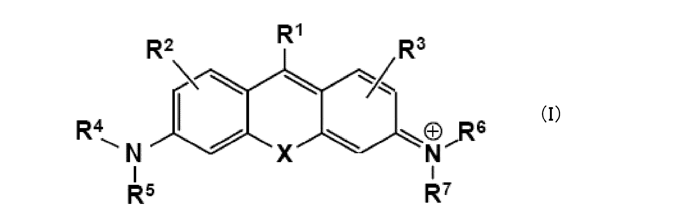
- X represents an oxygen atom or C (R a ) (R b ) or Si (R a ) (R b ) (where R a and R b independently represent a hydrogen atom or an alkyl group)
- R 1 represents a hydrogen atom or an optionally substituted aryl (provided that when R 1 is a phenyl group, it does not have a substituent at the 2-position or 6-position in the benzene ring of the phenyl group)
- R 2 and R 3 are each independently a hydrogen atom, a hydroxyl group, a halogen atom, an alkyl group that may be substituted, a sulfo group, a carboxyl group, an ester group, an amide group, and an azide group.
- R 4 and R 5 each independently represent a hydrogen atom or an optionally substituted alkyl group, or N (R 4 ) (R 5 ) forms an amide group or a carbamate group (where R 4 Or when R 5 is an alkyl group, each together with R 2 may form a ring structure containing the nitrogen atom to which they are attached);
- R 6 and R 7 each independently represent a hydrogen atom or an optionally substituted alkyl group, or N (R 6 ) (R 7 ) forms an amide group or a carbamate group (where R 6 Or when R 7 is an alkyl group, each may be taken together with R 3 to form a ring structure containing the nitrogen atom to which they are attached.
- a probe for super-resolution fluorescence imaging containing a salt thereof The compound represented by the formula (I) or a salt thereof performs a nucleophilic addition-dissociation equilibrium reaction with a nucleophilic compound containing an —SH group, The super-resolution fluorescent imaging probe; (2) The super-resolution fluorescence imaging probe according to (1), wherein the nucleophilic addition-dissociation equilibrium reaction is performed on a carbon atom to which R 1 is bonded; (3) The super-resolution fluorescent imaging probe according to (1) or (2) above, wherein a dissociation constant in the nucleophilic addition-dissociation equilibrium reaction is in the range of 0.1 ⁇ M to 10 mM.
- a nucleophilic addition reaction rate constant in the nucleophilic addition-dissociation equilibrium reaction is 1 to 1.0 ⁇ 10 6 s ⁇ 1 in an aqueous solution under neutral conditions,
- R 1 is a hydrogen atom or an optionally substituted phenyl group (provided that the phenyl group has no substituent at the 2- or 6-position in the benzene ring),
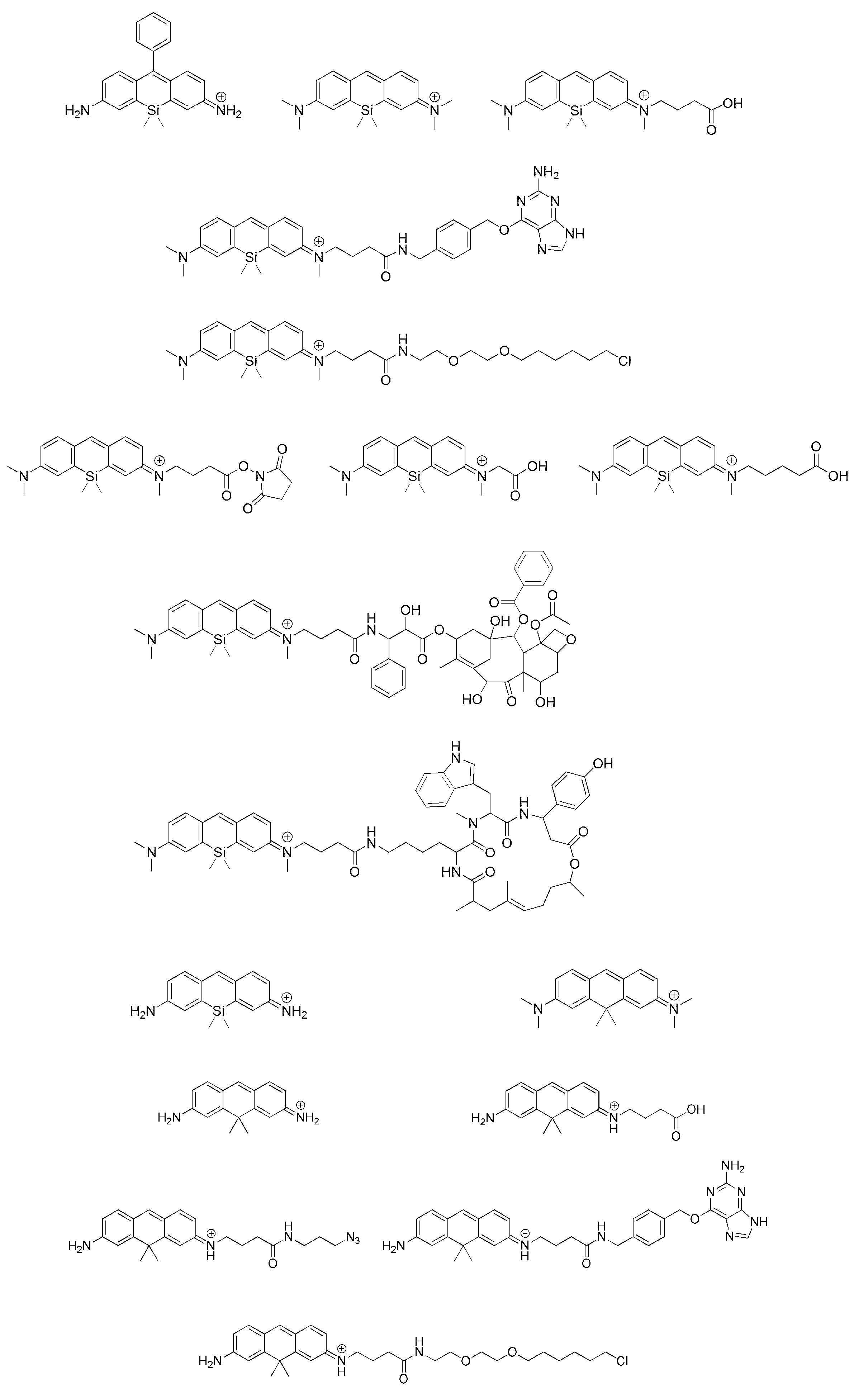
- a probe for super-resolution fluorescence imaging according to claim 1; (11) The super-resolution fluorescent imaging probe according to any one of (1) to (10), wherein the compound containing the —SH group is a compound having a cysteine residue; (12) The super-resolution fluorescent imaging probe according to any one of (1) to (10) above, wherein the compound containing the —SH group is glutathione; and (13) represented by formula (I): The super-resolution fluorescence imaging probe according to (1), wherein the compound to be selected is a compound selected from the following group: Is to provide.
- the invention provides: (14) A super-resolution fluorescence imaging method using the super-resolution fluorescence imaging probe according to any one of (1) to (13) above, A probe molecule is bound to a biomolecule, and laser light is irradiated in the presence of a compound containing an —SH group to acquire image data obtained by photographing fluorescence emission from the probe molecule, and this is repeated at regular time intervals.
- the method includes providing an ultrahigh resolution image of the biomolecule structure by analyzing and superimposing a plurality of the obtained image data.
- the present invention uses a stochastic fluorescence emission principle based on an intermolecular equilibrium reaction between an intracellular substance such as glutathione and a probe molecule, so that it is not necessary to separately add a high concentration of thiol as in the conventional method.
- Toxicity can be reduced.
- by combining the fluorescent probe compound of the present invention it is possible to provide a multi-color super-resolution imaging technique that can be observed in the same visual field.
- the probe for super-resolution fluorescence imaging of the present invention has a binding site (carboxyl group or the like) to a protein or the like in the molecule, it is suitable for labeling to a target protein or the like, It can be expected that it can be used for a wide range of research in living cell systems.
- the super-resolution fluorescence imaging method based on the probe is considered to be a highly versatile technique because a commercially available microscope can be used as an optical system. In this way, it can be said that the basic utility of the developed probe has extremely high industrial utility value and economic effect.
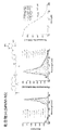
- FIG. 1 shows plots of absorption spectrum change (left), fluorescence spectrum change (center), and absorbance dependence of glutathione concentration (right figure) at each glutathione concentration of the compound 2 which is a probe for super-resolution fluorescence imaging of the present invention. It is shown.
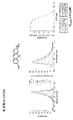
- FIG. 2 is a plot of absorption spectrum change (left), fluorescence spectrum change (center), and absorbance dependence of glutathione concentration (right figure) at each glutathione concentration of compound 5, which is a probe for super-resolution fluorescence imaging of the present invention. It is shown.
- FIG. 1 shows plots of absorption spectrum change (left), fluorescence spectrum change (center), and absorbance dependence of glutathione concentration (right figure) at each glutathione concentration of compound 5, which is a probe for super-resolution fluorescence imaging of the present invention. It is shown.
- FIG. 1 shows plots of absorption spectrum change (left), fluorescence spectrum change (center), and absorbance dependence of glutathione concentration (right figure) at each glut
- FIG. 3 is a plot (right diagram) of the absorption spectrum change (left), fluorescence spectrum change (center), and absorbance dependence of glutathione concentration at each glutathione concentration of compound 12, which is the super-resolution fluorescence imaging probe of the present invention. It is shown.
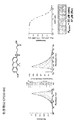
- FIG. 4 shows plots of absorption spectrum change (left), fluorescence spectrum change (middle), and absorbance dependence of glutathione concentration (right figure) at each glutathione concentration of compound 14, which is a probe for super-resolution fluorescence imaging of the present invention. It is shown.
- FIG. 4 shows plots of absorption spectrum change (left), fluorescence spectrum change (middle), and absorbance dependence of glutathione concentration (right figure) at each glutathione concentration of compound 14, which is a probe for super-resolution fluorescence imaging of the present invention. It is shown.
- FIG. 5 is a plot (right diagram) of the absorption spectrum change (left), fluorescence spectrum change (center), and absorbance dependence of glutathione concentration at each glutathione concentration of compound 16, which is the probe for super-resolution fluorescence imaging of the present invention. It is shown.
- FIG. 6 is a plot of the glutathione concentration dependence of the absorption spectrum change (left), fluorescence spectrum change (center), absorbance, and fluorescence intensity at each glutathione concentration of compound 28, which is a super-resolution fluorescent imaging probe of the present invention (right). Figure).
- FIG. 7 is a plot of the dependence of the absorption spectra and fluorescence intensity on the glutathione concentration at each glutathione concentration (left), fluorescence spectrum change (center), absorbance and fluorescence intensity of the compound 32 which is the probe for super-resolution fluorescence imaging of the present invention (right).
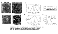
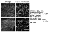
- FIG. 8 shows the results of SMLM imaging of microtubules in fixed Vero cells using an antibody labeled with compound 17, which is a probe for super-resolution fluorescence imaging of the present invention.
- the right figure in FIG. 8a is a super-resolution image by SMLM, and the left figure shows an averaged image of fluorescence intensity as a comparison.
- FIG. 8a is a super-resolution image by SMLM
- FIG. 8b is a cross-sectional profile of normalized fluorescence intensity in the strip region in the top view (scale bar: 3 ⁇ m) of the averaged image of FIG. 8a and the strip region in the top view (scale bar: 3 ⁇ m) of the super-resolution image. Shows a cross-sectional profile of the number of recognized bright spots in.
- FIG. 8c is a cross-sectional profile of normalized fluorescence intensity in the in-frame region in the lower diagram (scale bar: 1 ⁇ m) of the averaged image of FIG. 8a and the in-frame region in the lower diagram of the super-resolution image (scale bar: 1 ⁇ m) Shows a cross-sectional profile of the number of recognized bright spots in.
- FIG. 8c is a cross-sectional profile of normalized fluorescence intensity in the in-frame region in the lower diagram (scale bar: 1 ⁇ m) of the averaged image of FIG. 8a and the in-frame region in the lower diagram of the super-resolution image (scale bar:
- FIG. 8d shows the frequency distribution of the position determination accuracy with respect to the bright spots constituting the upper diagram of the super-resolution image of FIG. 8a.
- FIG. 9 shows the results of SMLM imaging of microtubules in fixed Vero cells using an antibody labeled with compound 33, which is a probe for super-resolution fluorescence imaging of the present invention.
- the right figure in FIG. 9 is a super-resolution image by SMLM, and the left figure shows an averaged image of fluorescence intensity as a comparison.
- FIG. 10 shows the results of SMLM imaging in two colors of microtubules and mitochondria in fixed Vero cells, obtained by using a combination of an antibody labeled with compound 17 and an antibody labeled with compound 33. Is.
- the right figure in FIG. 10 is a super-resolution image by SMLM, and the left figure shows an averaged image of fluorescence intensity as a comparison.
- halogen atom means a fluorine atom, a chlorine atom, a bromine atom, or an iodine atom.
- alkyl may be any of an aliphatic hydrocarbon group composed of linear, branched, cyclic, or a combination thereof.
- the number of carbon atoms of the alkyl group is not particularly limited, for example, 1 to 20 carbon atoms (C 1-20), having 3 to 15 carbon (C 3-15), having 5 to 10 carbon atoms (C 5-10 ). When the number of carbons is specified, it means “alkyl” having the number of carbons within the range.
- C 1-8 alkyl includes methyl, ethyl, n-propyl, isopropyl, n-butyl, isobutyl, sec-butyl, tert-butyl, n-pentyl, isopentyl, neo-pentyl, n-hexyl, isohexyl, n-heptyl, n-octyl and the like are included.
- the alkyl group may have one or more arbitrary substituents.
- substituents examples include, but are not limited to, an alkoxy group, a halogen atom, an amino group, a mono- or di-substituted amino group, a substituted silyl group, and acyl.
- alkyl group has two or more substituents, they may be the same or different.
- alkyl part of other substituents containing an alkyl part for example, an alkoxy group, an arylalkyl group, etc.
- a functional group when a functional group is defined as “may be substituted”, the type of substituent, the substitution position, and the number of substituents are not particularly limited, and two or more substitutions are made. If they have groups, they may be the same or different.
- the substituent group include, but are not limited to, an alkyl group, an alkoxy group, a hydroxyl group, a carboxyl group, a halogen atom, a sulfo group, an amino group, an alkoxycarbonyl group, and an oxo group. These substituents may further have a substituent. Examples of such include, but are not limited to, a halogenated alkyl group, a dialkylamino group, and the like.
- aryl may be either a monocyclic or condensed polycyclic aromatic hydrocarbon group, and a hetero atom (for example, an oxygen atom, a nitrogen atom, or a sulfur atom) as a ring constituent atom Etc.) may be an aromatic heterocyclic ring. In this case, it may be referred to as “heteroaryl” or “heteroaromatic”. Whether aryl is a single ring or a fused ring, it can be attached at all possible positions.
- Non-limiting examples of monocyclic aryl include phenyl group (Ph), thienyl group (2- or 3-thienyl group), pyridyl group, furyl group, thiazolyl group, oxazolyl group, pyrazolyl group, 2-pyrazinyl Group, pyrimidinyl group, pyrrolyl group, imidazolyl group, pyridazinyl group, 3-isothiazolyl group, 3-isoxazolyl group, 1,2,4-oxadiazol-5-yl group or 1,2,4-oxadiazole-3 -Yl group and the like.
- Non-limiting examples of fused polycyclic aryl include 1-naphthyl group, 2-naphthyl group, 1-indenyl group, 2-indenyl group, 2,3-dihydroinden-1-yl group, 2,3 -Dihydroinden-2-yl group, 2-anthryl group, indazolyl group, quinolyl group, isoquinolyl group, 1,2-dihydroisoquinolyl group, 1,2,3,4-tetrahydroisoquinolyl group, indolyl group, Isoindolyl group, phthalazinyl group, quinoxalinyl group, benzofuranyl group, 2,3-dihydrobenzofuran-1-yl group, 2,3-dihydrobenzofuran-2-yl group, 2,3-dihydrobenzothiophen-1-yl group, 2 , 3-dihydrobenzothiophen-2-yl group, benzothiazolyl group,
- an aryl group may have one or more arbitrary substituents on the ring.
- substituents include, but are not limited to, an alkoxy group, a halogen atom, an amino group, a mono- or di-substituted amino group, a substituted silyl group, and acyl.
- the aryl group has two or more substituents, they may be the same or different. The same applies to the aryl moiety of other substituents containing the aryl moiety (for example, an aryloxy group and an arylalkyl group).
- the “alkoxy group” is a structure in which the alkyl group is bonded to an oxygen atom, and examples thereof include a saturated alkoxy group that is linear, branched, cyclic, or a combination thereof.
- methoxy group, ethoxy group, n-propoxy group, isopropoxy group, cyclopropoxy group, n-butoxy group, isobutoxy group, s-butoxy group, t-butoxy group, cyclobutoxy group, cyclopropylmethoxy group, n- Pentyloxy group, cyclopentyloxy group, cyclopropylethyloxy group, cyclobutylmethyloxy group, n-hexyloxy group, cyclohexyloxy group, cyclopropylpropyloxy group, cyclobutylethyloxy group, cyclopentylmethyloxy group, etc. are preferable Take as an example.
- ring structure when formed by a combination of two substituents, means a heterocyclic or carbocyclic group, such group being saturated, unsaturated, or aromatic.
- it includes cycloalkyl, cycloalkenyl, aryl, and heteroaryl as defined above. Examples include cycloalkyl, phenyl, naphthyl, morpholinyl, piperidinyl, imidazolyl, pyrrolidinyl, pyridyl and the like.
- a substituent can form a ring structure with another substituent, and when such substituents are bonded to each other, those skilled in the art will recognize a specific substitution, such as bonding to hydrogen.
- the probe for super-resolution fluorescence imaging of the present invention includes a compound having a structure represented by the following general formula (I).
- X represents an oxygen atom, C (R a ) (R b ), or Si (R a ) (R b ). Since the excitation wavelength is shifted by the substitution of X, it is possible to provide a multicolor imaging probe that can be excited by a general laser.
- R a and R b each independently represent a hydrogen atom or an alkyl group.
- R a and R b are alkyl groups, they can have one or more substituents, and examples of such substituents include alkyl groups, alkoxy groups, halogen atoms, hydroxyl groups, carboxyl groups, You may have 1 or 2 or more amino groups, sulfo groups, etc.
- R a and R b are preferably both methyl groups. In some cases, R a and R b may be bonded to each other to form a ring structure. For example, when R a and R b are both alkyl groups, R a and R b can be bonded to each other to form a spiro ring.
- the ring formed is preferably about 5 to 8 membered ring, for example.
- R 1 represents a hydrogen atom or an optionally substituted aryl.
- substituent in the aryl which may be substituted include, but are not limited to, an alkyl group, an alkoxy group, a halogen atom, a hydroxyl group, a carboxyl group, an amino group, and a sulfo group.
- R 1 is a hydrogen atom or an optionally substituted phenyl group.
- R 1 is a phenyl group
- the 2-position or 6-position in the benzene ring of the phenyl group that is, the position where R 1 is ortho to the xanthene ring moiety as a substituent on the benzene ring
- R 1 is a hydrogen atom
- the compound of general formula (I) has a structure similar to pyronin
- R 1 is phenyl
- the compound of general formula (I) has a structure similar to rosamine. .
- R 2 and R 3 are each independently a hydrogen atom, a hydroxyl group, a halogen atom, an alkyl group that may be substituted, a sulfo group, a carboxyl group, an ester group, an amide group, and an azide group. Represents 1 to 3 identical or different substituents selected. Preferably, R 2 and R 3 are both hydrogen atoms. In some cases, either R 2 or R 3 can include a labeled substituent that can be covalently bound to a biomolecule such as a protein described below. In that case, they may themselves be labeled substituents, or may be substituted with substituents including labeled substituents.
- R 4 and R 5 each independently represent a hydrogen atom or an alkyl group, or N (R 4 ) (R 5 ) may form an amide group or a carbamate group.
- R 4 and R 5 both represent an alkyl group, they may be the same or different.
- R 4 and R 5 are each independently preferably a hydrogen atom, a methyl group or an ethyl group from the viewpoint of the nucleophilic addition reaction rate with a nucleophilic compound containing a —SH group, and R 4 More preferably, R 5 and R 5 are both a hydrogen atom or a methyl group. It is also preferred that one of R 4 or R 5 is a hydrogen atom and the other is a carbonyl group.
- N (R 4 ) (R 5 ) forms an amide bond.
- bonded with a carbonyl group is not specifically limited, An alkyl group is preferable and a methyl group is more preferable.
- N (R 4 ) (R 5 ) preferably contains a substituent (labeled substituent) that can bind to a biomolecule such as a protein by a covalent bond or a non-covalent bond. In that case, they may themselves be labeled substituents, or may be substituted with substituents including labeled substituents.
- R 4 and R 5 are substituted with a substituent including a leaving group such as N-hydroxysuccinimide, a benzylguanine derivative that is a substrate of SNAP-tag, or a ligand used in HaloTag®. It may be. Alternatively, it may be substituted with a substituent containing a taxane such as paclitaxel or docetaxel that binds to microtubules or a cyclic peptide such as phalloidin that binds to actin filaments. Alternatively, it may be substituted with N-hydroxysuccinimide that forms a bond with an amino group such as a lysine residue of a protein present in the cell membrane.
- a substituent including a leaving group such as N-hydroxysuccinimide, a benzylguanine derivative that is a substrate of SNAP-tag, or a ligand used in HaloTag®. It may be. Alternatively, it may be substituted with a substituent
- R 4 or R 5 when R 4 or R 5 is an alkyl group, it may be combined with R 2 to form a ring structure containing a nitrogen atom to which they are bonded. In that case, only one of R 4 and R 2 or a combination of R 5 and R 2 may form a ring structure, or both may form a ring structure.
- R 6 and R 7 each independently represent a hydrogen atom or an alkyl group, or N (R 6 ) (R 7 ) may form an amide group or a carbamate group.
- R 6 and R 7 both represent an alkyl group, they may be the same or different.
- R 6 and R 7 are each independently preferably a hydrogen atom, a methyl group, or an ethyl group, and R 6 and R 7 are both hydrogen atoms, or both are methyl groups. preferable. It is also preferred that one of R 6 or R 7 is a hydrogen atom and the other is a carbonyl group. In this case, N (R 6 ) (R 7 ) forms an amide bond.
- N (R 6 ) (R 7 ) contains the above-described labeled substituent. In that case, they may themselves be labeled substituents, or may be substituted with substituents including labeled substituents. As described above, such a labeled substituent is not particularly limited, and any known substituent in the art can be used.
- R 6 or R 7 when R 6 or R 7 is an alkyl group, each may be taken together with R 3 to form a ring structure containing a nitrogen atom to which they are bonded. In that case, only one of R 6 and R 3 , or a combination of R 7 and R 3 may form a ring structure, or both may form a ring structure.
- X is C (R a ) (R b ) or Si (R a ) (R b ), and R a and R b are both methyl groups.
- R 1 is a hydrogen atom or a phenyl group
- R 2 and R 3 are all hydrogen atoms
- R 4 , R 5 , R 6 and R 7 are all hydrogen atoms or all are methyl groups. There are cases.
- X is an oxygen atom
- R 1 is a hydrogen atom or a phenyl group
- R 2 and R 3 are both hydrogen atoms
- R 4 , R 5 , R 6 and R 7 are either Also preferred is a hydrogen atom or a methyl group.
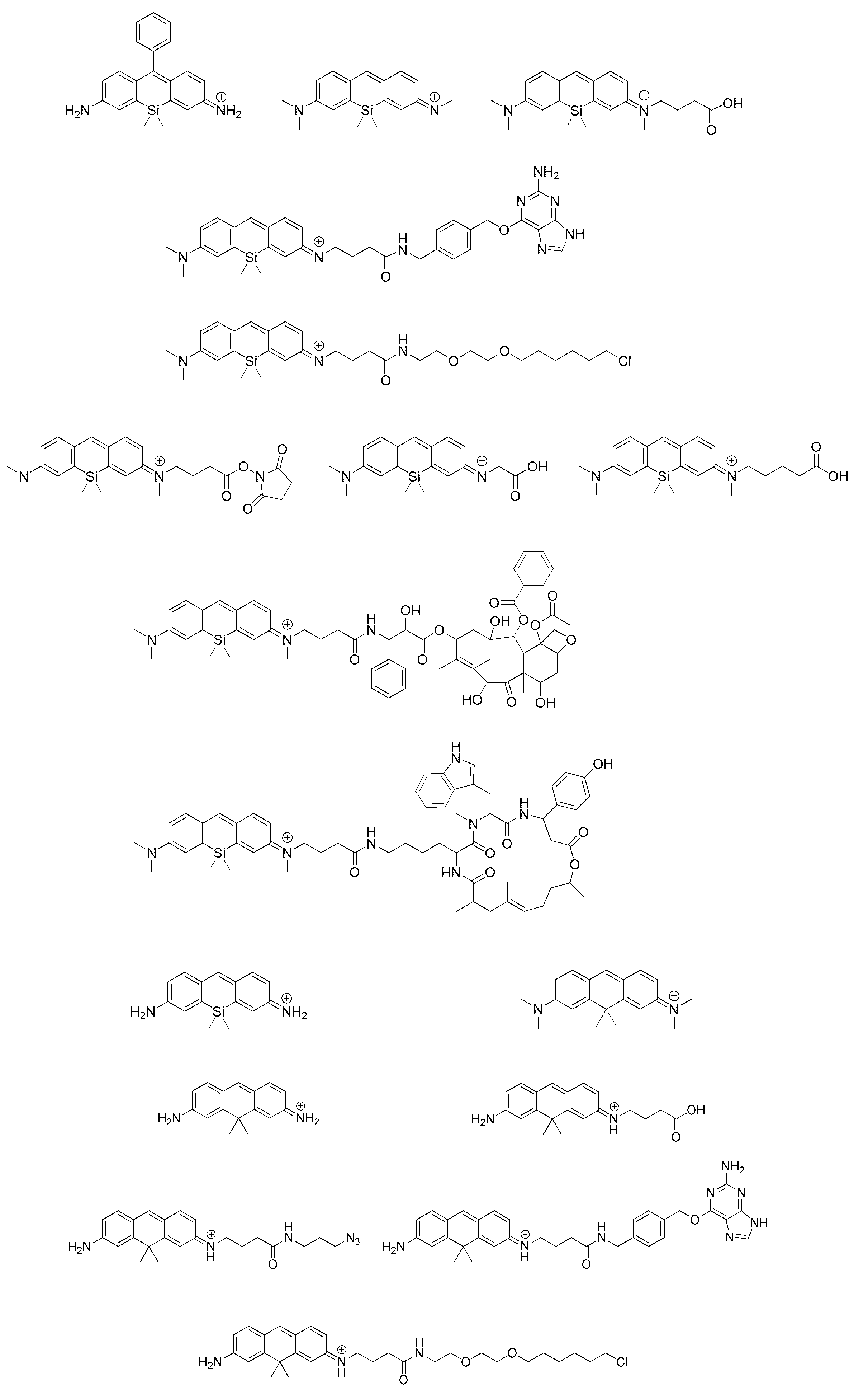
- the compound of the formula (I) which is representative as a probe for super-resolution fluorescence imaging of the present invention, Can be mentioned. However, it is not limited to these. Moreover, the said compound can have a labeled substituent in arbitrary positions.
- the compound represented by the above formula (I) has a monovalent positive charge at the N atom to which R 6 and R 7 are linked, it usually exists as a salt.
- salts include base addition salts, acid addition salts, amino acid salts and the like.
- base addition salt include metal salts such as sodium salt, potassium salt, calcium salt, magnesium salt, ammonium salt, or organic amine salts such as triethylamine salt, piperidine salt, morpholine salt, and acid addition salt.
- Examples include mineral acids such as hydrochlorides, sulfates and nitrates, carboxylates such as trifluoroacetates, organic acids such as methanesulfonate, paratoluenesulfonate, citrate and oxalate. Mention may be made of salts. Examples of amino acid salts include glycine salts. However, it is not limited to these salts.
- the compound represented by the formula (I) may have one or more asymmetric carbons depending on the type of substituent, and there are stereoisomers such as optical isomers or diastereoisomers. There is a case. Pure forms of stereoisomers, any mixture of stereoisomers, racemates, and the like are all within the scope of the present invention.
- the compound represented by the formula (I) or a salt thereof may exist as a hydrate or a solvate, and any of these substances is included in the scope of the present invention.
- solvents such as ethanol, acetone, isopropanol, can be illustrated.
- the above-described fluorescent probe may be used as a composition by blending an additive for use in a physiological environment, if necessary.
- an additive for example, additives such as a solubilizing agent, a pH adjusting agent, a buffering agent, and an isotonic agent can be used, and the amount of these can be appropriately selected by those skilled in the art.
- These compositions can be provided as a composition in an appropriate form such as a powder-form mixture, a lyophilized product, a granule, a tablet, or a liquid.
- the super-resolution fluorescence imaging method of the present invention comprises contacting a probe molecule of formula (I) described above with a biomolecule, and In the presence, image data is acquired by a CCD camera or the like that has been irradiated with laser light to photograph fluorescence emission from the probe molecules, and a plurality of the image data obtained by repeating this at a predetermined time interval are obtained. This includes superimposing an image with respect to the structure of the biomolecule by superimposing it after analysis. Examples of biomolecules to be observed include cell membranes (lipids), proteins, DNA, RNA, and the like.
- SMLM single-molecule-localization-microscopy
- the basic procedure in the super-resolution fluorescence imaging method of the present invention can refer to the super-resolution fluorescence imaging method described in the document except that the probe of formula (I) is used.
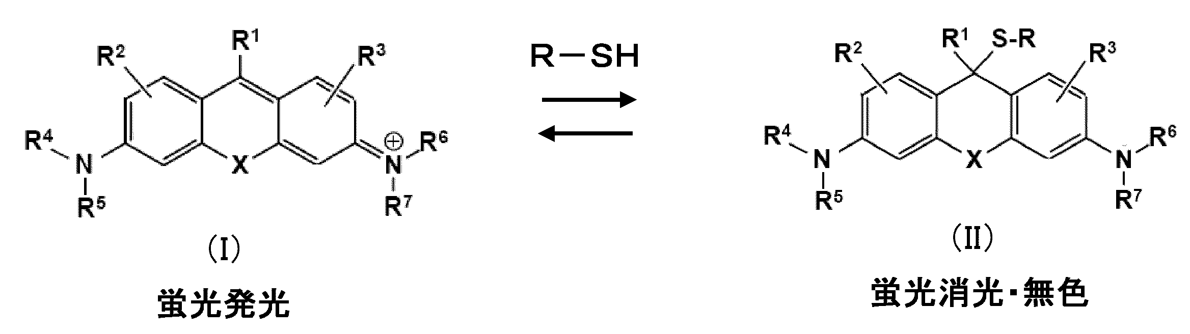
- the compound (I) before the reaction (left side) absorbs excitation light of about 400 to 700 nm and emits strong fluorescence, whereas a nucleophilic compound (here, a compound containing an —SH group such as glutathione).
- a nucleophilic compound here, a compound containing an —SH group such as glutathione.
- R—SH nucleophilic attacked by “R—SH”.
- nucleophilic compound that causes such a nucleophilic addition-dissociation equilibrium reaction include, in addition to the —SH group-containing compounds shown here, for example, compounds containing —OH groups such as water and hydroxide ions, Alternatively, a —NH group-containing compound such as a lysine residue in a protein can be used.
- a CCD that is a detector in which preferred fluorescence flashing due to the above-described equilibrium reaction is generally used in the presence of glutathione in such a concentration range.
- the present invention has an advantage that cytotoxicity can be reduced because the intensity of the laser used can be reduced to about 1/10 of the conventional one.
- the intensity of the laser is preferably about 0.0001 to 10 kW / cm 2 , and from the viewpoint of signal-to-noise ratio (S / N ratio), cytotoxicity, etc., about 0.01 to 0.5 kW / cm 2. It is more preferable that
- the fluorescent probe molecule In super-resolution fluorescence imaging with SMLM, it is not preferable that the fluorescent probe molecule emits light at a location closer than the diffraction limit of light. Therefore, the number of probe molecules that emit fluorescence in one measurement needs to be less than a certain percentage. There is. Therefore, the abundance ratio of compound (I) to compound (II) in the above equilibrium is preferably 1: 10000 to 1:10 at room temperature in an aqueous solution under neutral conditions, and 1: 1000 to 1: 100 is more preferable.
- the dissociation constant K d of the nuclear addition-dissociation equilibrium is preferably in the range of 0.1 ⁇ M to 10 mM, and more preferably in the range of 0.1 to 100 ⁇ M from the viewpoint of improving the labeling density.
- the lifetime of the compound of formula (I) in the fluorescence state is preferably in the range of 1 microsecond to 1 second, and the signal-to-noise ratio From the viewpoint of improving the (S / N ratio), it is more preferably in the range of 1 millisecond to 1 second.
- the nucleophilic addition reaction rate constant in the above equilibrium should be in the range of 1 to 1.0 ⁇ 10 6 s ⁇ 1 at room temperature in an aqueous solution under neutral conditions. Preferably, it is in the range of 1 to 1000 s ⁇ 1 .
- the reaction rate is calculated by exciting a fluorescent probe with a nanosecond laser and measuring transient absorption on the order of nanoseconds to milliseconds using a system such as laser flash photolysis known in the art. Can do.
- the compound that functions as a nucleophilic molecule in the nucleophilic addition-dissociation equilibrium reaction is a compound containing an —SH group, and preferably glutathione that is a nucleophilic molecule present in a cell.
- glutathione listed here, and any compound having a —SH group in the molecule can include compounds and peptides having a wide cysteine residue, and these compounds can be used as long as the above equilibrium reaction is obtained.
- Etc. can also be used in the present invention.
- a compound having a nucleophilic functional group such as an —OH group or —NH group in the molecule, such as a compound having water, a lysine residue, etc.
- Any peptide can be used as the nucleophilic compound in the present invention as long as the above equilibrium reaction can be obtained.
- these dissociation constants and nucleophilic addition reaction rate constants can also obtain more optimal dissociation constants and reaction rates by molecular design that selects an appropriate combination as a substituent or the like in the compound of formula (I). .
- the obtained compound was dissolved in methanol (2 ml), and sodium borohydride was added until the color of the solution became pale yellow. Water was added to stop the reaction, and the mixture was extracted twice with dichloromethane. The obtained organic layer was washed with saturated brine, dried over anhydrous sodium sulfate, and the solvent was removed under reduced pressure. The obtained compound was dissolved in deoxygenated dichloromethane (5 ml), and 1,3-dimethylbarbituric acid (185 mg, 1.17 mmol, 21 eq.) And tetrakis (triphenylphosphine) palladium (11 mg, 9 ⁇ mol, 0.2 eq.) was added and stirred at room temperature overnight.
- reaction solution was extracted with ethyl acetate, washed twice with a saturated aqueous sodium hydrogen carbonate solution, saturated brine, dried over anhydrous sodium sulfate, filtered, and then the solvent was removed under reduced pressure.
- Chloranil 23 mg, 0.094 mmol, 1.7 eq. was added to the obtained compound, and the mixture was stirred at room temperature for 5 minutes.
- dichlorodimethylsilane (700 ⁇ l, 5.75 mmol, 1.3 eq.) was diluted with THF (5 ml), gradually returned to room temperature, and stirred for 2 hours.
- 1 N Hydrochloric acid was added to acidify, saturated aqueous sodium hydrogen carbonate solution was added, and THF was removed under reduced pressure.
- the obtained aqueous layer was extracted with ethyl acetate, and then the organic phase was washed with water and saturated brine, dried over anhydrous sodium sulfate, and the solvent was removed under reduced pressure.
- the organic phase was washed with water and saturated brine, dried over anhydrous sodium sulfate, and the solvent was removed under reduced pressure.
- the organic layer was washed with water and saturated brine, dried over anhydrous sodium sulfate, filtered, and the solvent was removed under reduced pressure.
- the reaction mixture was filtered through celite, the filtrate and celite were washed with ethyl acetate, and the solvent was removed under reduced pressure from the mixture of the filtrate and the washing solution.
- the obtained aqueous layer was extracted with dichloromethane, and then the organic layer was washed with saturated brine, dried over anhydrous sodium sulfate, and the solvent was removed under reduced pressure.
- the obtained residue was dissolved in methanol, sodium borohydride (376.5 mg, 9.95 mmol, 1.6 eq.) Was added at 0 ° C, and the mixture was stirred for 10 min. Water was added and extracted with dichloromethane, and then the organic layer was washed with saturated brine, dried over anhydrous sodium sulfate, and the solvent was removed under reduced pressure.
- the reaction mixture was filtered through celite, the filtrate and celite were washed with ethyl acetate, and the solvent was removed under reduced pressure from the mixture of the filtrate and the washing solution.
- the organic layer was washed with saturated brine, dried over anhydrous sodium sulfate, and the solvent was removed under reduced pressure.
- dehydrated acetone (210 ⁇ l, 2.85 mmol, 6.0 eq.) was added little by little, and the mixture was stirred at ⁇ 78 ° C. for 15 minutes, then returned to room temperature, and further stirred for 5 hours.
- tetrahydrofuran was removed under reduced pressure.
- the obtained aqueous phase was extracted with dichloromethane, and then the organic layer was washed with saturated brine, dried over anhydrous sodium sulfate, and the solvent was removed under reduced pressure.
- the organic layer was washed with saturated brine, dried over anhydrous sodium sulfate, and the solvent was removed under reduced pressure.
- N-diisopropylamine (8.8 ⁇ l, 0.0504 mmol, 20 eq.)
- O- (7-azabenzotriazol-1-yl) -N, N, N ', N'-tetramethyluronium hexafluorophosphate Salt (5.9 mg, 0.0155 mmol, 6.1 eq.) was added, and the mixture was stirred for 15 minutes, then returned to room temperature and further stirred for 13 hours.
- FIGS. 1 to 5 show absorption spectrum changes (left figure) and fluorescence spectrum changes (center figure; excitation wavelength: compound 2 is 570 nm, compounds 5, 14 and 16 are 620 nm, and compounds are compounds 2, 5, 12, 14 and 16. 12 is 600 nm), and a plot (right figure) of the absorbance dependence of glutathione concentration is shown.
- the measurement was performed with a 1% DMSO aqueous solution containing 1 ⁇ M of the probe compound.
- the obtained dissociation constant values are shown in Table 1-1 below.
- FIGS. 6 and 7 show changes in the absorption spectra of compounds 28 and 32 (left figure), fluorescence spectrum changes (middle figure; excitation wavelength 530 nm), and glutathione concentration dependence of absorbance and fluorescence intensity (right figure). Respectively.
- the measurement was performed with a 1% DMSO aqueous solution containing 1 ⁇ M of the probe compound.
- the obtained dissociation constant values are shown in Table 1-2 below.
- the dissociation constants of these compounds are all in the range of 1 to 100 ⁇ M, and it was found that most of them exist as non-fluorescent nucleophilic adducts at a concentration of several mM of glutathione, which is similar to that in cells. . This indicates that the abundance ratio of the unreacted probe compound that emits fluorescence and the compound that has been quenched by reacting with glutathione is within the range suitable for super-resolution fluorescence imaging by SMLM.
- the probe compound of the present invention shows that nucleophilic addition with glutathione proceeds with a time constant on the order of milliseconds, and that under conventional laser irradiation of 1/10 or less. It became clear that the fluorescence blinking suitable for super-resolution fluorescence imaging was demonstrated.
- the fluorescence quantum yield of the probe compound of the present invention was measured.
- the solution conditions are 1 ⁇ M probe compound, 1% DMSO aqueous solution, pH 7.4 (200 mM NaPi buffer).
- the results obtained for compounds 5, 12, 14, 16, 17, 28, and 32 are shown in Table 7. It has been clarified that the probe compound of the present invention exhibits a high fluorescence quantum yield suitable for super-resolution fluorescence imaging by SMLM.
- the antibody labeled with the probe compound was purified by PD MiniTrap TM G-25 (GE Healthcare) using phosphate buffered saline (PBS) (pH 7.4, GIBCO) as the eluent.
- PBS phosphate buffered saline
- the ratio of probe compound [mol] / antibody molecule [mol] was calculated as the degree of labeling (DOL) of the probe compound relative to the antibody.
- DOL degree of labeling
- the concentration of the probe compound was calculated from the absorbance measurement value derived from the probe compound, and the concentration of the antibody molecule was assumed to have no loss of the antibody molecule in the purification process, and the concentration of the antibody molecule used for the preparation was Calculated from the total amount.
- SMLM super-resolution imaging
- a labeled anti-mouse antibody with a labeling degree of 2.1 and a labeled anti-rabbit antibody with a labeling degree of 1.3 were used.
- Compound 33 which is a probe compound having an azido group introduced at the end of compound 28, was similarly labeled to an antibody.
- Compound 33 and dibenzocyclooctyne-N-hydroxysuccinimidyl ester were each dissolved in DMSO to prepare a 10 mM stock solution.
- a goat-derived anti-mouse secondary antibody IgG, Sigma-Aldrich
- IgG immunostaining of ⁇ -tubulin was mixed with DBCO-NHS in 0.2 M sodium phosphate buffer (pH 8.5) ( Room temperature for 30 minutes).
- An antibody (DBCO-IgG) labeled with DBCO-NHS was purified by PD MiniTrap TM G-25 (GE Healthcare) using PBS (pH 7.4, GIBCO) as an eluent.
- the stock solution of Compound 33 was added to the obtained eluent containing DBCO-IgG and mixed in PBS (pH 7.4, GIBCO) (room temperature, 30 minutes).
- the antibody (DBCO-IgG) labeled with the probe compound was purified by PD MiniTrap TM G-25 (GE Healthcare) using PBS (pH 7.4, GIBCO) as an eluent.
- the ratio of probe compound [mol] / antibody molecule [mol] was calculated as the degree of labeling (DOL) of the probe compound relative to the antibody.
- DOL degree of labeling
- the concentration of the probe compound was calculated from the absorbance measurement value derived from the probe compound, and the concentration of the antibody molecule was assumed to have no loss of the antibody molecule in the purification process, and the concentration of the antibody molecule used for the preparation was Calculated from the total amount.
- SMLM super-resolution imaging
- Vero cells derived from the kidney of African green monkeys were transformed into Dulbecco's modified Eagle medium (high glucose) (GIBCO) without phenol red and L-glutamine with fetal calf serum (Invitrogen) and 1% penicillin. - streptomycin solution (manufactured by Wako Pure Chemical Industries, Ltd.) were cultured in the added medium in (37 °C, 5% CO 2 ). The day before the imaging, the cells were seeded on a Lab-Tek II 8-well chamber cover glass (Thermo Fisher Scientific). Fixing and immunostaining were performed as follows with reference to the following documents. ⁇ M. Bates, G. T. Dempsey, K. H.
- Methanol fixation and ⁇ -tubulin immunostaining were performed as follows. Cells were washed with PBS pre-warmed to 37 ° C. and fixed with ⁇ 20 ° C. methanol containing 5 mM glycol ether diamine tetraacetic acid (EGTA) for 5-10 minutes. Wash 2 to 3 times with BRB80 (80 mM potassium PIPES buffer containing 1 mM magnesium chloride and 1 mM EGTA, pH 6.8), block with blocking buffer (PBS containing 1% bovine serum albumin) for 20 minutes, then blocking buffer Mouse anti- ⁇ -tubulin antibody (TUB2.1, Sigma-Aldrich, T4026, diluted 1/100) diluted with 1 was added and shaken for 1 hour.
- BRB80 80 mM potassium PIPES buffer containing 1 mM magnesium chloride and 1 mM EGTA, pH 6.8
- blocking buffer PBS containing 1% bovine serum albumin
- the fixation with paraformaldehyde and glutaraldehyde, and immunostaining of ⁇ -tubulin and Tom20 were performed as follows. Cells were washed with PBS pre-warmed to 37 ° C. and fixed with PBS containing 3% paraformaldehyde and 0.1% glutaraldehyde for 10 minutes at room temperature. The unreacted aldehyde group and the fluorescent product generated during the fixing operation were reduced with the PBS solution of 0.1% sodium borohydride prepared immediately before. After washing with PBS, blocking and membrane permeabilization were performed for 20 minutes with blocking buffer (3% BSA and 0.5% Triton X-100 in PBS).
- mouse anti- ⁇ -tubulin antibody (TUB2.1, Sigma-Aldrich, T4026, / 100 dilution) and / or rabbit anti-Tom20 antibody (Santa Cruz, sc11415, 1/50 dilution) diluted with blocking buffer was added. And shaken for 30 minutes. Thereafter, the sample was washed with a washing buffer (0.2% BSA and 0.1% Triton X-100 in PBS) three times for 10 minutes each. Staining was carried out for 30 minutes with a labeled secondary antibody (10-20 ⁇ g / mL for ⁇ -tubulin; 10 ⁇ g / mL for Tom20) diluted in blocking buffer.
- a labeled secondary antibody (10-20 ⁇ g / mL for ⁇ -tubulin; 10 ⁇ g / mL for Tom20
- the plate was washed three times with a washing buffer for 10 minutes, further washed with PBS for 10 minutes, and post-fixed with PBS containing 3% paraformaldehyde and 0.1% glutaraldehyde at room temperature for 10 minutes. Thereafter, it was washed 3 times with PBS. Prior to SMLM imaging, the buffer was replaced with 0.2 M sodium phosphate buffer containing 1-10 mM GSH (Wako Pure Chemical Industries).
- SMLM imaging was performed at room temperature using an SMLM imaging N-STORM device (Nikon). Excitation of the compound 17 and the compound 33, which are probe compounds, is performed by total reflection illumination using laser light of 647 nm (40 to 500 W / cm 2 ) and 561 nm (40 to 500 W / cm 2 ), or light-shielding illumination close to total reflection illumination, respectively. It went by. Image data was recorded at 15 ms / frame or 30 ms / frame, and a super-resolution image was constructed by analyzing with image integration software (NIS-Elements Advanced Research, Nikon). In displaying the super-resolution image, only bright spots where 150 photons or more or 200 photons or more were detected were displayed.
- image integration software NIS-Elements Advanced Research, Nikon
- FIG. 8 shows the results of SMLM imaging of microtubules in fixed Vero cells using an antibody labeled with Compound 17.
- the right figure in FIG. 8a is a super-resolution image by SMLM, and the left figure shows an averaged image of fluorescence intensity as a comparison.
- FIG. 8b is a cross-sectional profile of normalized fluorescence intensity in the strip region in the top view (scale bar: 3 ⁇ m) of the averaged image of FIG. 8a and the strip region in the top view (scale bar: 3 ⁇ m) of the super-resolution image. Shows a cross-sectional profile of the number of recognized bright spots in.
- FIG. 8 shows the results of SMLM imaging of microtubules in fixed Vero cells using an antibody labeled with Compound 17.
- the right figure in FIG. 8a is a super-resolution image by SMLM, and the left figure shows an averaged image of fluorescence intensity as a comparison.
- FIG. 8b is a cross-sectional
- FIG. 8c is a cross-sectional profile of normalized fluorescence intensity in the in-frame region in the lower diagram (scale bar: 1 ⁇ m) of the averaged image of FIG. 8a and the in-frame region in the lower diagram of the super-resolution image (scale bar: 1 ⁇ m). Shows a cross-sectional profile of the number of recognized bright spots in.
- FIG. 8d shows the frequency distribution of the position determination accuracy with respect to the bright spots constituting the upper diagram of the super-resolution image of FIG. 8a.
- the results shown in FIG. 8 demonstrate super-resolution imaging of microtubules in fixed Vero cells using an antibody labeled with compound 17.
- the super-resolution image showed higher spatial resolution than the averaged image in microtubule line width and separation of adjacent microtubules.
- FIG. 9 shows the results of SMLM imaging of microtubules in fixed Vero cells using an antibody labeled with Compound 33.
- the right figure in FIG. 9 is a super-resolution image by SMLM, and the left figure shows an averaged image of fluorescence intensity as a comparison.
- the results shown in FIG. 9 demonstrate super-resolution imaging of microtubules in fixed Vero cells using an antibody labeled with compound 33.
- the super-resolution image showed higher spatial resolution than the averaged image in microtubule line width and separation of adjacent microtubules.
- FIG. 10 shows the results of SMLM imaging in two colors of microtubules and mitochondria in fixed Vero cells using a combination of an antibody labeled with compound 17 and an antibody labeled with compound 33.
- the right figure in FIG. 10 is a super-resolution image by SMLM, and the left figure shows an averaged image of fluorescence intensity as a comparison.
- the antibody labeled with compound 33 stains microtubules shown in green, while the antibody labeled with compound 17 stains mitochondria shown in red. By merging these images, a super-resolution image in two colors indicated by red and green was obtained.
- FIG. 10 demonstrate that the present invention can be applied to two-color super-resolution imaging by using two types of probe compounds of the present invention in which the X atom in formula (I) is changed.
Landscapes
- Health & Medical Sciences (AREA)
- Life Sciences & Earth Sciences (AREA)
- Immunology (AREA)
- Chemical & Material Sciences (AREA)
- Engineering & Computer Science (AREA)
- Biomedical Technology (AREA)
- General Health & Medical Sciences (AREA)
- Physics & Mathematics (AREA)
- Molecular Biology (AREA)
- Hematology (AREA)
- Urology & Nephrology (AREA)
- Analytical Chemistry (AREA)
- Biochemistry (AREA)
- General Physics & Mathematics (AREA)
- Pathology (AREA)
- Biotechnology (AREA)
- Cell Biology (AREA)
- Microbiology (AREA)
- Food Science & Technology (AREA)
- Medicinal Chemistry (AREA)
- Nuclear Medicine, Radiotherapy & Molecular Imaging (AREA)
- Epidemiology (AREA)
- Veterinary Medicine (AREA)
- Public Health (AREA)
- Animal Behavior & Ethology (AREA)
- Materials Engineering (AREA)
- Organic Chemistry (AREA)
- Proteomics, Peptides & Aminoacids (AREA)
- Optics & Photonics (AREA)
- Chemical Kinetics & Catalysis (AREA)
- Investigating, Analyzing Materials By Fluorescence Or Luminescence (AREA)
- Measuring Or Testing Involving Enzymes Or Micro-Organisms (AREA)
- Investigating Or Analysing Biological Materials (AREA)
- Investigating Or Analysing Materials By The Use Of Chemical Reactions (AREA)
Abstract
Description
(1)以下の式(I)で表される化合物
〔式中、
Xは、酸素原子又はC(Ra)(Rb)又はSi(Ra)(Rb)を表し(ここで、Ra及びRbは、それぞれ独立に水素原子、又はアルキル基を表す);
R1は、水素原子又は置換されていてもよいアリールを表し(ただし、R1がフェニル基の場合、当該フェニル基のベンゼン環における2位又は6位の位置に置換基を有しない);
R2及びR3は、それぞれ独立に、水素原子、ヒドロキシル基、ハロゲン原子、それぞれ置換されていてもよいアルキル基、スルホ基、カルボキシル基、エステル基、アミド基及びアジド基よりなる群から独立に選択される1~3個の同一又は異なる置換基を表し;
R4及びR5は、それぞれ独立に水素原子又は置換されていてもよいアルキル基を表し、又は、N(R4)(R5)がアミド基もしくはカルバメート基を形成する(ここで、R4又はR5がアルキル基である場合、それぞれR2と一緒になって、それらが結合する窒素原子を含む環構造を形成してもよい);及び、
R6及びR7は、それぞれ独立に水素原子もしくは置換されていてもよいアルキル基を表し、又は、N(R6)(R7)がアミド基もしくはカルバメート基を形成する(ここで、R6又はR7がアルキル基である場合、それぞれR3と一緒になって、それらが結合する窒素原子を含む環構造を形成してもよい。)
又はその塩を含む超解像蛍光イメージング用プローブであって、
式(I)で表される化合物又はその塩が、-SH基を含有する求核性化合物との間で求核付加-解離平衡反応を行うことを特徴とする、
該超解像蛍光イメージング用プローブ;
(2)前記求核付加-解離平衡反応が、R1が結合する炭素原子において行われる、上記(1)に記載の超解像蛍光イメージング用プローブ;
(3)前記求核付加-解離平衡反応における解離定数が、0.1μM~10mMの範囲内であることを特徴とする、上記(1)又は(2)に記載の超解像蛍光イメージング用プローブ;
(4)前記求核付加-解離平衡反応における求核付加反応速度定数が、中性条件の水系溶液中において、1~1.0x10⁶s-1であることを特徴とする、上記(1)~(3)のいずれか1に記載の超解像蛍光イメージング用プローブ;
(5)Xが、C(Ra)(Rb)又はSi(Ra)(Rb)である、上記(1)~(4)のいずれか1に記載の超解像蛍光イメージング用プローブ;
(6)Ra及びRbが、いずれもメチル基である、上記(5)に記載の超解像蛍光イメージング用プローブ;
(7)R1は、水素原子又は置換されていてもよいフェニル基である(ただし、当該フェニル基のベンゼン環における2位又は6位の位置に置換基を有しない)、上記(1)~(6)のいずれか1に記載の超解像蛍光イメージング用プローブ;
(8)R4、R5、R6及びR7が、いずれも水素原子である、上記(1)~(7)のいずれか1に記載の超解像蛍光イメージング用プローブ;
(9)R4、R5、R6及びR7が、いずれもメチル基である、上記(1)~(7)のいずれか1に記載の超解像蛍光イメージング用プローブ;
(10)R4、R5、R6及びR7のうち少なくとも1つが、生体分子と共有結合又は非共有結合によって結合し得るラベル化置換基を含む、上記(1)~(7)のいずれか1に記載の超解像蛍光イメージング用プローブ;
(11)前記-SH基を含有する化合物が、システイン残基を有する化合物である、上記(1)~(10)のいずれか1に記載の超解像蛍光イメージング用プローブ;
(12)前記-SH基を含有する化合物が、グルタチオンである、上記(1)~(10)のいずれか1に記載の超解像蛍光イメージング用プローブ;及び
(13)式(I)で表される化合物が以下の群から選択される化合物である、上記(1)に記載の超解像蛍光イメージング用プローブ
を提供するものである。
(14)上記(1)~(13)のいずれか1に記載の超解像蛍光イメージング用プローブを用いる超解像蛍光イメージング方法であって、
生体分子にプローブ分子を結合させ、-SH基を含有する化合物の存在下でレーザー光を照射して前記プローブ分子からの蛍光発光を撮影した画像データを取得し、一定の時間間隔でこれを繰り返して得られた複数の前記画像データを解析したうえで重ね合わせることによって、前記生体分子の構造に対する超高解像のイメージ画像を得ることを含む、該方法
を提供するものである。
本明細書中において、「ハロゲン原子」とは、フッ素原子、塩素原子、臭素原子、又はヨウ素原子を意味する。
本発明の超解像蛍光イメージング用プローブは、一態様において、以下の一般式(I)で表される構造を有する化合物を含むものである。

を挙げることができる。ただし、これらに限定されるものではない。また、当該化合物は、任意の位置にラベル化置換基を有することができる。
本発明の超解像蛍光イメージング方法は、生体分子に上記で説明した式(I)のプローブ分子を接触させ、-SH基を含有する化合物の存在下で、これにレーザー光を照射して前記プローブ分子からの蛍光発光を撮影したCCDカメラ等によって画像データを取得し、一定の時間間隔でこれを繰り返して得られた複数の前記画像データを解析したうえで重ね合わせることによって、前記生体分子の構造に対する超高解像のイメージ画像を得ることを含むものである。観測対象となる生体分子としては、細胞膜(脂質)、タンパク質、DNA、RNA等が挙げられる。
以下に示すとおり、本発明の超解像蛍光イメージング用プローブである化合物2、5、12、14、16、17、28、32、33、34、及び35をそれぞれ合成した。
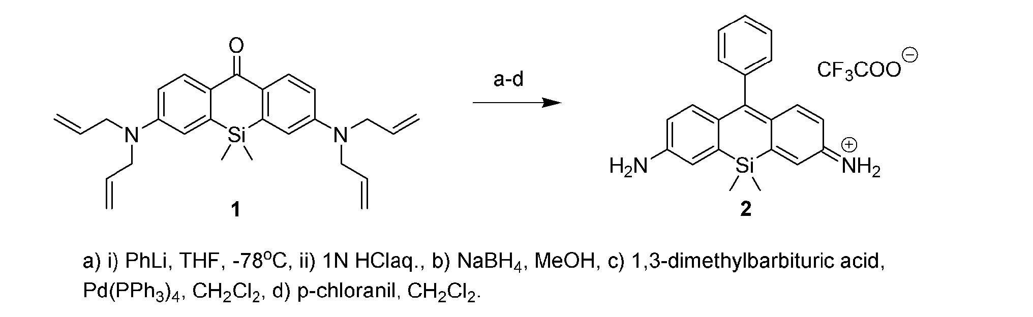
以下のスキームに従って化合物2を合成した。なお、化合物1はKushida K et al. Bioorg. Med. Chem. Lett. 22 (2012) 3908-3911に開示されており、これに基づいて合成を行なった。
以下のスキームに従って化合物4を合成した。なお、化合物3及び化合物4の合成については、Y. Koide et al., J. Am. Chem. Soc. 2012, 134, 5029-5031.に開示されており、これに基づいて合成を行なった。
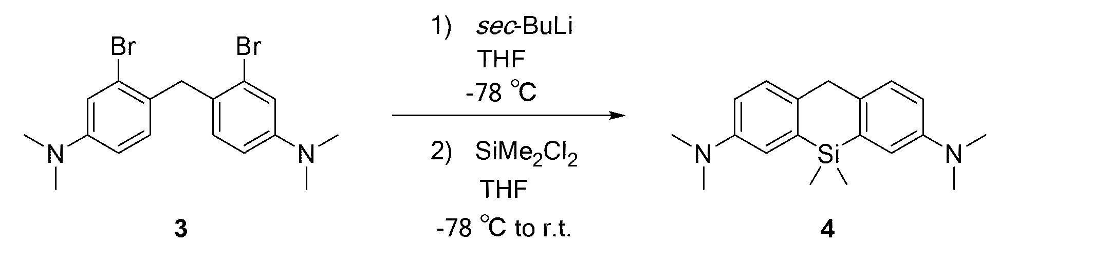
化合物3(1.80 g, 4.37 mmol, 1 eq.)をTHF(100 ml)に溶かし、アルゴン雰囲気下、-78 °Cで15分間撹拌した。1.1 M sec-ブチルリチウム シクロヘキサン/n-ヘキサン溶液(9 ml, 9.9 mmol, 2.3 eq.)を15分間かけてゆっくり加えた。次いでジクロロジメチルシラン(700 μl, 5.75 mmol, 1.3 eq.)をTHF(5 ml)に希釈して加えて徐々に室温に戻し、2時間撹拌した。1 N 塩酸を加えて酸性にし、次いで飽和炭酸水素ナトリウム水溶液を加え、THFを減圧除去した。得られた水層を酢酸エチルで抽出し、次いで有機相を水及び飽和食塩水で洗浄し、無水硫酸ナトリウムで乾燥し、溶媒を減圧除去した。得られた残渣を中圧シリカゲルクロマトグラフィーで精製し(溶離液:ノルマルヘキサン/酢酸エチル = 95/5 to 80/20)、目的化合物4 (852 mg, 63%)を薄黄色固体として得た。
1H NMR (400 MHz, CDCl3): δ 0.28 (s, 3H) , 2.96 (s, 12H), 3.98 (s, 2H), 6.76 (dd, J = 8.4, 2.8 Hz, 2H), 7.02 (d, J = 2.8 Hz, 2H), 7.21 (d, J = 8.4 Hz, 2H); 13C NMR (100 MHz, CDCl3): δ -2.6, 39.3, 41.3, 114.2, 117.7, 128.6, 135.3, 136.2, 148.8.
以下のスキームに従って化合物5を合成した。

化合物4(36.5 mg, 0.115 mmol, 1 eq.)をクロラニル(27 mg, 0.115 mmol, 1 eq.)を加えて室温にて10分撹拌し、濃青色の反応液をシリカゲルカラムクロマトグラフィー(溶離液:ジクロロメタン/メタノール = 100/0 to 80/20)にて分離し、青色のフラクションを回収して溶媒を減圧留去し、さらにHPLCで精製し(溶離液A(H2O, 1 % アセトニトリル, 0.1 % トリフルオロ酢酸)、溶離液B(アセトニトリル, 1 % H2O)(A/B = 90/10 to 10/90, 25 min)、目的化合物5 (53.6 mg, 96%)を青色固体として得た。
1H NMR (400 MHz, CD3CN): δ 0.50 (s, 6H), 3.30 (s, 12H), 6.89 (dd, J = 9.2, 2.7 Hz, 2H), 7.25 (d, J = 2.7 Hz, 2H), 7.64 (d, J = 9.2 Hz, 2H), 7.80 (s, 1H); HRMS (ESI+): Calcd for [M]+, 309.17815, Found, 309.17801 (-0.1 mmu).
以下のスキームに従って化合物6を合成した。
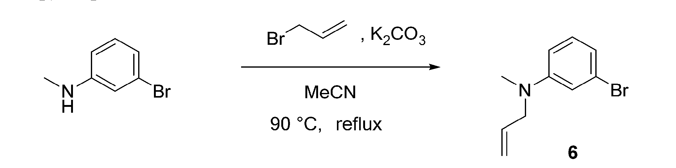
炭酸カリウム(7.46 g, 54.0 mmol, 2.0 eq.)をアセトニトリル(20 ml)に懸濁させ、3-ブロモ-N-メチルアニリン(4.96 g, 26.7 mmol, 1 eq.)及び臭化アリル(3.87 g, 31.3 mmol, 1.2 eq.)を加え、撹拌しながら90 °Cで終夜加熱還流した。反応混合物をセライトで濾過した後、濾物及びセライトを酢酸エチルで洗浄し、濾液及び洗浄液の混合液から溶媒を減圧除去した。得られた残渣を中圧シリカゲルクロマトグラフィーで精製し(溶離液:ノルマルヘキサン/酢酸エチル = 100/0 to 98/2)、目的化合物6 (5.98 g, 99 %)を得た。
1H NMR (400 MHz, CDCl3): δ 2.95 (s, 3H), 3.91-3.93 (m, 2H), 5.15-5.21 (m, 2H), 5.79-5.89 (m, 1H), 6.63-6.65 (m, 1H), 6.82-6.86 (m, 2H), 7.06-7.10 (m, 1H); 13C NMR (100 MHz, CDCl3): δ 38.1, 55.0, 110.9, 115.0, 116.4, 119.1, 123.5, 130.4, 133.1, 150.7; HRMS (ESI+): Calcd for [M+H]+, 226.02259, Found, 226.02210 (-0.5 mmu).
以下のスキームに従って化合物7を合成した。
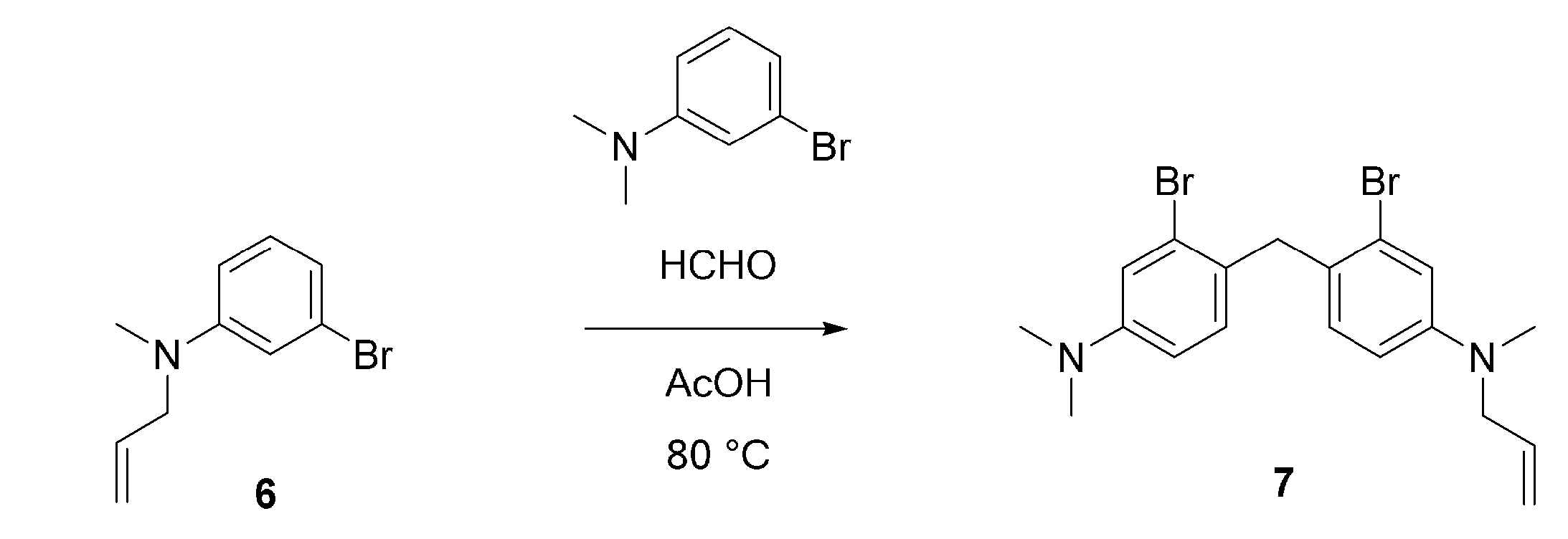
化合物6(4.60 g, 20.4 mmol, 1 eq.)及び3-ブロモ-N,N-ジメチルアニリン(5.00 g, 25.0 mmol, 1.2 eq.)を酢酸(10 ml)に溶かし、37 % ホルムアルデヒド水溶液(6.30 ml, 227 mmol, 11 eq.)を加え、80 °Cまで徐々に昇温させた後、3時間加熱した。反応混合液から酢酸を減圧除去し、飽和炭酸水素ナトリウム水溶液を加え、ジクロロメタンで抽出し、有機相を水及び飽和食塩水で洗浄し、無水硫酸ナトリウムで乾燥し、溶媒を減圧除去した。得られた残渣を中圧シリカゲルクロマトグラフィーで精製し(溶離液:ノルマルヘキサン/ジクロロメタン = 80/20 to 70/30 to 60/40 to 50/50)、目的化合物7を得た。
1H NMR (400 MHz, CDCl3): δ 2.92 (s, 3H), 2.93 (s, 6H), 3.88-3.90 (m, 2H), 4.02 (s, 2H), 5.15-5.20 (m, 2H), 5.83 (ddt, 1H, J = 14.8, 12.6, 4.9 Hz), 6.57-6.62 (m, 2H), 6.85 (d, 1H, J = 8.6 Hz), 6.88 (d, 1H, J = 8.6 Hz), 6.95 (d, 1H, J = 2.7 Hz), 6.96 (d, 1H, J = 2.6 Hz); 13C NMR (100 MHz, THF-d8): δ 38.2, 40.5, 40.6, 55.7, 112.6, 112.7, 116.3, 116.7, 116.8, 126.16, 126.17, 127.6, 127.7, 131.5, 131.6, 134.7, 150.1, 151.2; HRMS (ESI+): Calcd for [M+H]+, 437.02225, Found, 437.02225 (±0.0 mmu).
以下のスキームに従って化合物8を合成した。

化合物7(900 mg, 2.05mmol, 1 eq.)をTHF(60 ml)に溶かし、アルゴン雰囲気下、-78 °Cで15分間撹拌した。1 M sec-ブチルリチウム シクロヘキサン/n-ヘキサン溶液(6.50 ml, 6.50 mmol, 3.2 eq.)を8分間かけてゆっくり加え、22分間撹拌した。次いでジクロロジメチルシラン(500 μl, 4.18 mmol, 2.0 eq.)のTHF(13 ml)溶液を加えて徐々に室温に戻し、3時間撹拌した。2 N 塩酸を加えて酸性にし、次いで飽和炭酸水素ナトリウム水溶液を加え、THFを減圧除去した。得られた水層をジクロロメタンで抽出し、次いで有機相を水及び飽和食塩水で洗浄し、無水硫酸ナトリウムで乾燥し、溶媒を減圧除去した。得られた残渣を中圧シリカゲルクロマトグラフィーで精製し(溶離液:ノルマルヘキサン/酢酸エチル = 100/0 to 94/6)、目的化合物8 (382 mg, 55 %)を得た。
1H NMR (400 MHz, CDCl3): δ 0.44 (s, 6H), 2.927-2.93 (m, 9H), 3.90-3.91 (m, 2H), 3.95 (s, 2H), 5.12-5.20 (m, 2H), 5.85 (ddt, 1H, J = 17.2, 10.2, 5.2 Hz), 6.69-6.74 (m, 2H), 6.97 (d, 1H, J = 2.8 Hz), 6.99 (d, 1H, J = 2.8 Hz), 7.15-7.19 (m, 2H); 13C NMR (100 MHz, CDCl3): δ -2.7, 38.3, 39.2, 41.2, 55.9, 114.0, 114.2, 116.4, 117.4, 117.6, 128.56, 128.58, 134.3, 134.9, 135.3, 136.09, 136.13, 147.5, 148.8; HRMS (ESI+): Calcd for [M+H]+, 337.20945, Found, 337.20957 (+0.1 mmu).
以下のスキームに従って化合物9を合成した。

化合物8 (382 mg, 1.13 mmol, 1 eq.)を脱酸素したジクロロメタン(14 ml)に溶かし、1,3-ジメチルバルビツール酸(1.48 g, 9.48 mmol, 8.4 eq.)及びテトラキス(トリフェニルホスフィン)パラジウム(274 mg, 237 μmol, 0.2 eq.)を加え、室温にて終夜撹拌した。飽和炭酸水素ナトリウム水溶液を加え、ジクロロメタンで抽出し、次いで有機層を水及び飽和食塩水で洗浄し、無水硫酸ナトリウムで乾燥し、濾過後、溶媒を減圧除去した。得られた残渣を中圧シリカゲルクロマトグラフィーで精製し(溶離液:ノルマルヘキサン/酢酸エチル = 91/9 to 70/30)、目的化合物9 (289 mg, 86 %)を得た。
1H NMR (400 MHz, CDCl3): δ 0.44 (s, 6H), 2.85 (s, 3H), 2.93 (s, 6H), 3.94 (s, 2H), 6.58 (dd, 1H, J = 8.1, 2.6 Hz), 6.73 (dd, 1H, J = 8.4, 2.8 Hz), 6.85 (d, 1H, J = 2.6 Hz), 6.99 (d, 1H, J = 2.8 Hz), 7.14 (d, 1H, J = 8.1 Hz), 7.18 (d, 1H, J = 8.4 Hz); 13C NMR (100 MHz, CDCl3): δ -2.7, 31.2, 39.4, 41.3, 113.3, 114.3, 117.4, 117.7, 128.6, 128.7, 135.4, 135.7, 136.1, 136.4, 147.1, 148.8; HRMS (ESI+): Calcd for [M+H]+, 297.17815, Found, 297.17853 (+0.4 mmu).
以下のスキームに従って化合物11を合成した。なお、化合物10の合成については、J. Le Notre et al., Adv. Synth. Catal. 349, 432 (2007).に開示されており、これに基づいて合成を行なった。
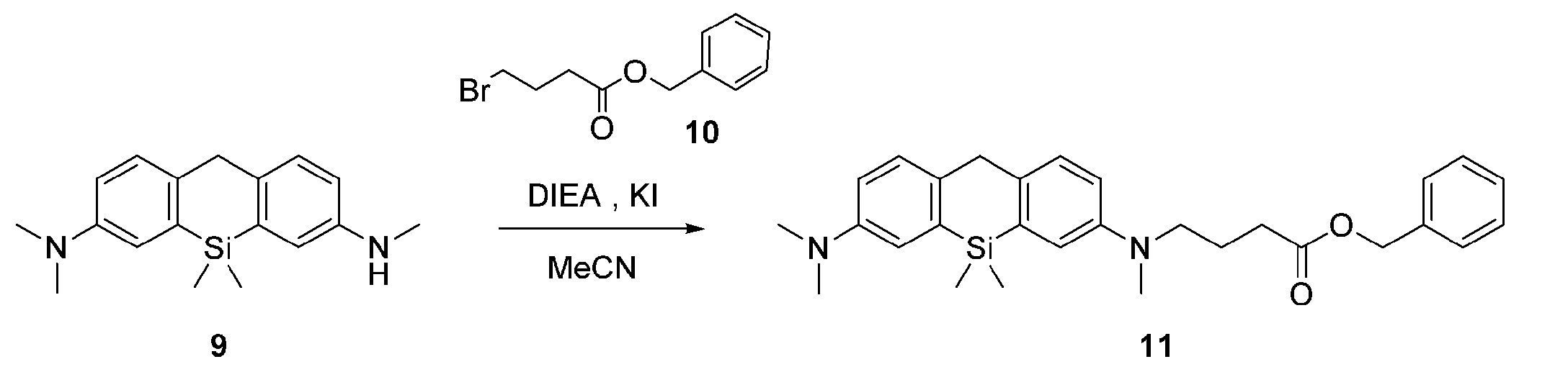
化合物9(102.8 mg, 0.347 mmol, 1 eq.)及び化合物10(756.9 mg, 2.94 mmol, 8.5 eq.)をアセトニトリル(4 ml)に溶かし、N,N-ジイソプロピルエチルアミン(512 μl, 2.94 mmol, 8.5 eq.)及び少量のヨウ化カリウムを加え、80 °Cで5.5時間加熱還流した。水を加え、ジクロロメタンで抽出し、有機相を水及び飽和食塩水で洗浄し、無水硫酸ナトリウムで乾燥し、溶媒を減圧除去した。得られた残渣を中圧シリカゲルクロマトグラフィーで精製し(溶離液:ノルマルヘキサン/酢酸エチル = 86/14 to 65/35)、目的化合物11 (91.7 mg, 56 %)を得た。
1H NMR (400 MHz, CDCl3): δ 0.45 (s, 6H), 1.93 (quin, 2H, J = 7.2 Hz), 2.41 (t, 2H, J = 7.2 Hz), 2.90 (s, 3H), 2.94 (s, 6H), 3.34 (t, 2H, J = 7.2 Hz), 3.95 (s, 2H), 5.10 (s, 2H), 6.68 (dd, 1H, J = 8.4, 2.8 Hz), 6.74 (dd, 1H, J = 8.4, 2.8 Hz), 6.95 (d, 1H, J = 2.8 Hz), 6.99 (d, 1H, J = 2.8 Hz), 7.15 (d, 1H, J = 8.4 Hz), 7.18 (d, 1H, J = 8.4 Hz), 7.31-7.36 (m, 5H); 13C NMR (100 MHz, CDCl3): δ -2.7, 22.4, 31.8, 38.6, 39.2, 41.2, 52.4, 66.4, 113.8, 114.2, 117.2, 117.3, 117.7, 128.3, 128.4, 128.5, 128.65, 128.67, 134.8, 136.0, 136.1, 136.2, 147.3, 148.8, 173.2; HRMS (ESI+): Calcd for [M+H]+, 473.26188, Found, 473.26185 (-0.0 mmu).
以下のスキームに従って化合物12を合成した。

化合物11(171 mg, 0.361 mmol, 1 eq.)をメタノールに溶かし、パラジウム炭素を加え、水素雰囲気下、室温で2日間撹拌した。反応混合物を濾過し、濾液から溶媒を減圧除去した。得られた残渣をジクロロメタンに溶かし、2,3-ジクロロ-5,6-ジシアノ-p-ベンゾキノン(180.3 mg, 0.794 mmol, 2.2 eq.)を加え、室温で終夜撹拌した。水を加え、ジクロロメタンで抽出し、有機相を水及び飽和食塩水で洗浄し、無水硫酸ナトリウムで乾燥し、溶媒を減圧除去した。得られた残渣をHPLCで精製し(溶離液A(H2O, 1 % アセトニトリル, 0.1 % トリフルオロ酢酸)、溶離液B(アセトニトリル, 1 % H2O)(A/B = 90/10 to 0/100 40 min))、目的化合物12 (110 mg, 61 %)を得た。
1H NMR (400 MHz, CD3OD): δ 0.54 (s, 6H), 1.95-2.02 (m, 2H), 2.45 (t, 2H, J = 6.5 Hz), 3.34 (s, 3H), 3.37 (s, 6H), 3.73-3.77 (m, 2H), 6.98 (dd, 1H, J = 9.2, 2.7 Hz), 7.02 (dd, 1H, J = 9.1, 2.6 Hz), 7.33 (d, 1H, J = 2.2 Hz), 7.51 (m, 1H), 7.71-7.73 (m, 2H, g), 7.87 (s, 1H); 13C NMR (100 MHz, CD3CN): δ -1.3, 23.0, 31.1, 39.8, 41.4, 53.1, 115.06, 115.12, 122.3, 122.4, 128.25, 128.31, 144.0, 144.1, 148.42, 148.44, 155.68, 156.17, 160.6, 174.8; HRMS (ESI+): Calcd for [M]+, 381.19928, Found, 381.19929 (+0.0 mmu).
以下のスキームに従って化合物14を合成した。なお、化合物13の合成については、Nat.Biotech.,2003, 21,86-89に開示されており、これに基づいて合成を行なった。
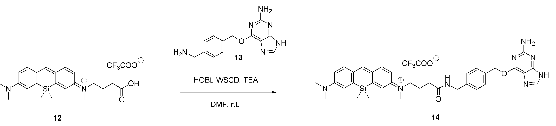
化合物12(9.40 mg, 0.0190 mmol, 1 eq.)を脱水N,N-ジメチルホルムアミドに溶かし、化合物13(7.5 mg, 0.0277 mmol, 1.5 eq.)、1-ヒドロキシベンゾトリアゾール(9.1 mg, 0.0594 mmol, 3.1 eq.)、1-エチル-3-(3-ジメチルアミノプロピル)カルボジイミド塩酸塩(6.6 mg. 0.0344 mmol, 1.8 eq.)、及びトリエチルアミン(10 μl, 0.0717 mmol, 3.8 eq.)を加え、アルゴン雰囲気下、室温で21時間撹拌した後、化合物13(2.3 mg, 0.00850 mmol, 0.45 eq.)を加え、さらに24時間撹拌した。溶媒を減圧除去し、得られた残渣をHPLCで精製し(溶離液A(H2O, 1 % アセトニトリル, 0.1 % トリフルオロ酢酸)、溶離液B(アセトニトリル, 1 % H2O)(A/B = 90/10 to 0/100 40 min))、目的化合物14 (7.5 mg, 53 %)を得た。
1H NMR (400 MHz, CD3OD): δ 0.52 (s, 6H), 1.99-2.06 (m, 2H), 2.38 (t, 2H, J = 6.8 Hz), 3.33 (s, 3H), 3.37 (s, 6H), 3.70-3.74 (m, 2H), 4.41 (s, 2H), 5.63 (s, 2H), 6.96-7.00 (m, 2H), 7.31 (d, 1H, J = 2.3 Hz), 7.35 (d, 2H, J = 8.1 Hz), 7.41 (br s, 1H), 7.52 (d, 2H, J = 8.1 Hz), 7.68-7.72 (m, 2H), 7.86 (s, 1H), 8.33 (s, 1H); 13C NMR (100 MHz, CD3OD): δ -1.4, 24.0, 33.1, 39.5, 41.0, 43.8, 53.4, 70.8, 108.2, 115.3, 122.3, 122.4, 128.86, 128.95, 128.98, 130.2, 135.4, 141.0, 143.5, 144.5, 144.6, 149.0, 149.1, 153.7, 156.2, 156.8, 158.3, 161.09, 161.11, 161.7, 174.6; HRMS (ESI+): Calcd for [M]+, 633.31163, Found, 633.31169 (+0.1 mmu).
以下のスキームに従って化合物16を合成した。なお、化合物15の合成については、J.Am.Chem.Soc.2013,135,6184-6191に開示されており、これに基づいて合成を行なった。

化合物12(11.3 mg, 0.0228 mmol, 1 eq.)を脱水N,N-ジメチルホルムアミド(1.5 ml)に溶かし、化合物15(9.1 mg, 0.0407 mmol, 1.8 eq.)、1-ヒドロキシベンゾトリアゾール(6.1 mg, 0.0451 mmol, 2.0 eq.)、1-エチル-3-(3-ジメチルアミノプロピル)カルボジイミド塩酸塩(7.2 mg. 0.0375 mmol, 1.6 eq.)、及びトリエチルアミン(14.3 μl, 0.103 mmol, 4.5 eq.)を0 °Cで加え、アルゴン雰囲気下で徐々に室温に戻し、終夜撹拌した。溶媒を減圧除去し、得られた残渣をHPLCで精製し(溶離液A(H2O, 1 % アセトニトリル, 0.1 % トリフルオロ酢酸)、溶離液B(アセトニトリル, 1 % H2O)(A/B = 90/10 to 0/100 40 min))、目的化合物16 (9.21 mg, 58 %)を得た。
1H NMR (400 MHz, CD3OD): δ 0.55 (s, 6H), 1.32-1.44 (m, 4H), 1.55 (quin, 2H, J = 7.0 Hz), 1.72 (quin, 2H, J = 7.0 Hz), 1.98-2.05 (m, 2H), 2.33 (t, 2H, J = 6.8 Hz), 3.34 (s, 3H), 3.38 (s, 6H), 3.44 (t, 2H, J = 6.6 Hz), 3.50-3.60 (m, 10H), 3.73 (t, 2H, J = 7.7 Hz), 6.98-7.03 (m, 2H), 7.33 (d, 1H, J = 2.5 Hz), 7.41 (br s, 1H), 7.71-7.73 (m, 2H), 7.88 (s, 1H); 13C NMR (100 MHz, CD3OD): δ -1.4, 24.0, 26.5, 27.7, 30.5, 33.2, 33.7, 39.6, 40.4, 41.0, 45.7, 53.4, 70.5, 71.21, 71.24, 72.2, 115.26, 115.29, 122.3, 122.4, 128.97, 129.00, 144.57, 144.60, 149.0, 149.1, 156.2, 156.8, 161.1, 174.8; HRMS (ESI+): Calcd for [M]+, 586.32262, Found, 586.32267 (+0.0 mmu).
以下のスキームに従って化合物17を合成した。

化合物12(7.0 mg, 0.0142 mmol, 1 eq.)を脱水N,N-ジメチルホルムアミド(5 ml)に溶かし、N-ヒドロキシコハク酸イミド(8.7 mg, 0.0756 mmol, 5.3 eq.)及び1-エチル-3-(3-ジメチルアミノプロピル)カルボジイミド塩酸塩(6.1 mg. 0.0318 mmol, 2.2 eq.)を加え、アルゴン雰囲気下、室温で33時間撹拌した後、N-ヒドロキシコハク酸イミド(9.8 mg, 0.0852 mmol, 6.0 eq.)及び1-エチル-3-(3-ジメチルアミノプロピル)カルボジイミド塩酸塩(11.1 mg. 0.0579 mmol, 4.1 eq.)を加え、さらに43時間撹拌した。溶媒を減圧除去し、得られた残渣をHPLCで精製し(溶離液A(H2O, 1 % アセトニトリル, 0.1 % トリフルオロ酢酸)、溶離液B(アセトニトリル, 1 % H2O)(A/B = 90/10 to 0/100 40 min))、目的化合物17 (1.7 mg, 20 %)を得た。
1H NMR (400 MHz, CD3OD): δ 0.53 (s, 6H), 2.10-2.17 (m, 2H), 2.82 (t, 2H, J = 6.6 Hz), 2.87 (s, 4H), 3.36 (s, 3H), 3.38 (s, 6H), 3.80-3.84 (m, 2H), 6.98-7.04 (m, 2H), 7.34 (d, 1H, J = 2.6 Hz), 7.38 (d, 1H, J = 2.4 Hz), 7.71-7.74 (m, 2H), 7.89 (s, 1H); 13C NMR (100 MHz, CD3OD): δ -1.5, 23.2, 26.5, 28.7, 39.5, 41.1, 52.6, 115.3, 115.4, 122.1, 122.4, 129.0, 129.1, 144.6, 144.8, 148.9, 149.4, 156.1, 156.9, 161.4, 170.2, 171.8; HRMS (ESI+): Calcd for [M]+, 478.21566, Found, 478.21559 (-0.1 mmu).
以下のスキームに従って化合物19を合成した。

炭酸カリウム(11.96 g, 86.54 mmol, 2.5 eq.)をアセトニトリル(40 ml)に懸濁させ、3-ブロモアニリン(5.96 g, 34.65 mmol, 1 eq.)及び臭化アリル(21.89 g, 180.9 mmol, 5.2 eq.)を加え、撹拌しながら80 °Cで26時間加熱還流した。室温に戻した後、反応混合物をセライトで濾過し、濾物及びセライトを酢酸エチルで洗浄し、濾液及び洗浄液の混合液から溶媒を減圧除去した。得られた残渣を中圧シリカゲルクロマトグラフィーで精製し(溶離液:ノルマルヘキサン/酢酸エチル = 100/0 to 89/11)、目的化合物19(5.68 g, 22.5 mmol, 65 %)を得た。
1H NMR (400 MHz, CDCl3): δ 3.93-3.95 (m, 4H), 5.20-5.25 (m, 4H), 5.83-5.93 (m, 2H), 6.64-6.67 (m, 1H), 6.84-6.89 (m, 2H), 7.06-7.10 (m, 1H); 13C NMR (100 MHz, CDCl3): δ 52.7, 110.9, 115.0, 116.3, 119.1, 123.4, 130.3, 133.3, 149.9; HRMS (ESI+): Calcd for [M+H]+, 252.03824, Found, 252.03819 (-0.1 mmu).
以下のスキームに従って化合物21を合成した。

アルゴンガスを封入したフラスコ内で化合物20(1.57 g, 6.22 mmol, 1 eq.)をトルエン(10 ml)に溶かし、N,N-ジメチルホルムアミド(700 μl, 9.04 mmol, 1.5 eq.)及び塩化ホスホリル(700 μl, 7.51 mmol, 1.2 eq.)を加え、80 °Cで3.5時間撹拌した。室温に戻した後、2N水酸化ナトリウム水溶液を加え、混合溶液からトルエンを減圧除去した。得られた水層をジクロロメタンで抽出し、次いで有機層を飽和食塩水で洗浄し、無水硫酸ナトリウムで乾燥し、溶媒を減圧除去した。得られた残渣をメタノールに溶かし、0 °C下で水素化ホウ素ナトリウム(376.5 mg, 9.95 mmol, 1.6 eq.)を加え、10分間撹拌した。水を加え、ジクロロメタンで抽出し、次いで有機層を飽和食塩水で洗浄し、無水硫酸ナトリウムで乾燥し、溶媒を減圧除去した。得られた残渣を中圧シリカゲルクロマトグラフィーで精製し(溶離液:ノルマルヘキサン/酢酸エチル = 83/17 to 62/38)、目的化合物21(740 mg, 2.62 mmol, 42 %)を得た。
1H NMR (400 MHz, CDCl3): δ 3.89-3.90 (m, 4H), 4.59 (s, 2H), 5.13-5.19 (m, 4H), 5.82 (ddt, 2H, J = 17.0, 10.5, 4.8 Hz), 6.60 (dd, 1H, J = 8.5, 2.6 Hz), 6.86 (d, 1H, J = 2.6 Hz), 7.20 (d, 1H, J = 8.5 Hz); 13C NMR (100 MHz, CDCl3): δ 52.7, 64.8, 111.4, 115.9, 116.4, 124.3, 127.1, 130.3, 133.2, 149.3; HRMS (ESI+): Calcd for [M+Na]+, 304.03075, Found, 304.03081 (+0.1 mmu).
以下のスキームに従って化合物23を合成した。

炭酸カリウム(9.53 g, 68.97 mmol, 2.1 eq.)をアセトニトリル(30 ml)に懸濁させ、アニリン(3.07 g, 32.96 mmol, 1 eq.)及び臭化アリル(10.28 g, 85.01 mmol, 2.6 eq.)を加え、撹拌しながら80 °Cで5時間加熱還流した。臭化アリル(2.58 g, 21.29 mmol, 0.6 eq.)を追加した後、80 °Cでさらに13時間撹拌及び加熱還流した。室温に戻した後、反応混合物をセライトで濾過し、濾物及びセライトを酢酸エチルで洗浄し、濾液及び洗浄液の混合液から溶媒を減圧除去した。得られた残渣を中圧シリカゲルクロマトグラフィーで精製し(溶離液:ノルマルヘキサン/酢酸エチル = 100/0 to 97/3)、目的化合物23(5.35 g, 30.9 mmol, 94 %)を得た。
1H NMR (400 MHz, CDCl3): δ 3.86-3.88 (m, 4H), 5.10-5.18 (m, 4H), 5.82 (ddt, 2H, J = 17.2, 10.3, 4.9 Hz), 6.64-6.68 (m, 3H), 7.14-7.19 (m, 2H); 13C NMR (100 MHz, CDCl3): δ 52.8, 112.4, 116.0, 116.4, 129.1, 134.1, 148.7; HRMS (ESI+): Calcd for [M+H]+, 174.12773, Found, 174.12867 (+0.9 mmu).
以下のスキームに従って化合物25を合成した。
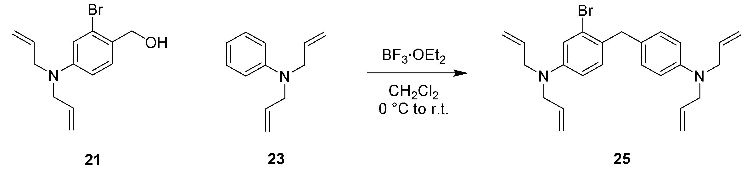
化合物21(386.0 mg, 1.368 mmol, 1.1 eq.)及び化合物23(218.8 mg, 1.263 mmol, 1 eq.)を脱水ジクロロメタン(10 ml)に溶かし、0 °C下で三フッ化ホウ素ジエチルエーテル錯体(500 μl, 4.051 mmol, 3.2 eq.)を少しずつ加え、10分間撹拌した後、室温に戻してさらに3時間撹拌した。飽和炭酸水素ナトリウム水溶液を加え、ジクロロメタンで抽出し、次いで有機層を飽和食塩水で洗浄し、無水硫酸ナトリウムで乾燥し、溶媒を減圧除去した。得られた残渣を中圧シリカゲルクロマトグラフィーで精製し(溶離液:ノルマルヘキサン/酢酸エチル = 100/0 to 88/12)、目的化合物25(395.2 mg, 0.903 mmol, 72 %)を得た。
1H NMR (400 MHz, CDCl3): δ 3.90-3.94 (m, 10H), 5.17-5.25 (m, 8H), 5.82-5.94 (m, 4H), 6.60 (dd, 1H, J = 8.6, 2.7 Hz), 6.67-6.70 (m, 2H), 6.93 (d, 1H, J = 2.7 Hz), 6.98 (d, 1H, J = 8.6 Hz), 7.08 (d, 2H, J = 8.6 Hz); 13C NMR (100 MHz, CDCl3): δ 39.7, 52.9, 53.0, 111.9, 112.6, 116.06, 116.12, 116.3, 125.5, 128.4, 128.6, 129.6, 131.1, 133.7, 134.4, 147.2, 148.1; HRMS (ESI+): Calcd for [M+H]+, 437.15869, Found, 437.15873 (+0.0 mmu).
以下のスキームに従って化合物26を合成した。

アルゴンを封入したフラスコ内で化合物25(208.5 mg, 0.477 mmol, 1 eq.)を脱水テトラヒドロフラン(15 ml)に溶かし、撹拌しながら-78 °Cに冷却した。1 M sec-ブチルリチウム シクロヘキサン/ノルマルヘキサン溶液(1.0 ml, 1.0 mmol, 2.1 eq.)を少しずつ加え、10分間撹拌した。次いで脱水アセトン(210 μl, 2.85 mmol, 6.0 eq.)を少しずつ加え、-78 °Cのまま15分間撹拌した後、室温に戻し、さらに5時間撹拌した。飽和塩化アンモニウム水溶液を加えた後、テトラヒドロフランを減圧除去した。得られた水相をジクロロメタンで抽出し、次いで有機層を飽和食塩水で洗浄し、無水硫酸ナトリウムで乾燥し、溶媒を減圧除去した。得られた残渣を中圧シリカゲルクロマトグラフィーで精製し(溶離液:ノルマルヘキサン/酢酸エチル = 89/11 to 68/32)、目的化合物26(125.8 mg, 0.302 mmol, 63 %)を得た。
1H NMR (400 MHz, CDCl3): δ 1.61 (s, 6H), 1.77 (s, 1H), 3.87-3.89 (m, 4H), 3.91-3.92 (m, 4H), 4.17 (s, 2H), 5.12-5.23 (m, 8H), 5.80-5.92 (m, 4H), 6.57 (dd, 1H, J = 8.5, 2.7 Hz), 6.62 (d, 2H, J = 8.7 Hz), 6.83 (d, 1H, J = 2.7 Hz), 6.94-6.98 (m, 3H); 13C NMR (100 MHz, CDCl3): δ 31.9, 38.0, 53.0, 53.2, 74.3, 110.3, 111.4, 112.7, 116.1, 116.2, 126.5, 129.4, 130.9, 134.0, 134.5, 134.6, 146.6, 146.9, 147.0; HRMS (ESI+): Calcd for [M+H]+, 417.29004, Found, 417.29000 (-0.0 mmu).
以下のスキームに従って化合物27を合成した。
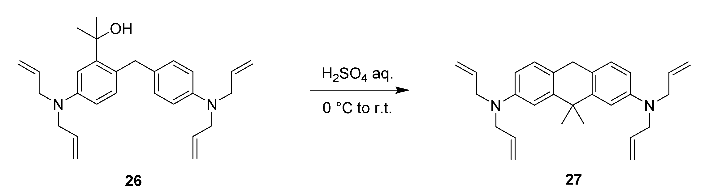
0 °C下で化合物26(188.5 mg, 0.452 mmol, 1 eq.)に80 %硫酸を加え、30分間撹拌した後、室温に戻し、さらに30分間激しく撹拌した。水を加え、ジクロロメタンで抽出し、次いで有機層を飽和水酸化ナトリウム水溶液で洗浄し、無水硫酸ナトリウムで乾燥し、溶媒を減圧除去し、目的化合物(177.2 mg, 0.445 mmol, 98 %)を得た。
1H NMR (400 MHz, CD3OD): δ 1.49 (s, 6H), 3.80 (s, 2H), 3.91-3.93 (m, 8H), 5.12-5.21 (m, 8H), 5.89 (ddt, 4H, J = 17.2, 10.3, 5.0 Hz), 6.57 (dd, 2H, J = 8.3, 2.6 Hz), 6.90 (d, 2H, J = 2.6 Hz), 7.02 (d, 2H, J = 8.3 Hz); 13C NMR (100 MHz, CD3OD): δ 29.4, 34.6, 40.6, 54.6, 110.7, 112.2, 116.3, 126.0, 129.1, 136.1, 146.7, 148.8; HRMS (ESI+): Calcd for [M+H]+, 399.27948, Found, 399.27955 (+0.1 mmu).
以下のスキームに従って化合物28を合成した。
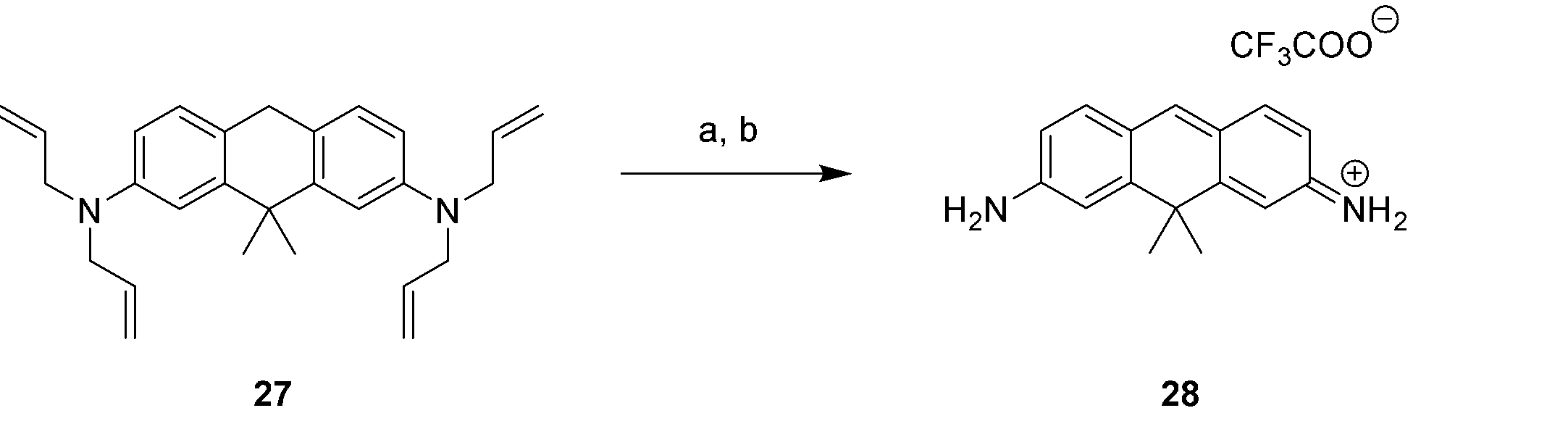
1,3-ジメチルバルビツール酸(60.55 mg, 0.388 mmol, 27 eq.)及びテトラキス(トリフェニルホスフィン)パラジウム(7.8 mg, 0.00675 mmol, 0.47 eq.)を入れたフラスコにアルゴンを封入し、脱酸素したジクロロメタン(7 ml)に溶かした化合物27(5.7 mg, 0.0143 mmol, 1 eq.)を加え、室温で終夜撹拌した。飽和炭酸水素ナトリウム水溶液を加え、ジクロロメタンで抽出し、次いで有機層を飽和食塩水で洗浄し、無水硫酸ナトリウムで乾燥し、濾過後、溶媒を減圧除去した。得られた残渣をジクロロメタンに溶かし、p-クロラニル(20.0 mg, 0.0813 mmol, 5.7 eq.)を加え、室温で1時間撹拌した。水を加え、ジクロロメタンで抽出し、次いで有機層を飽和食塩水で洗浄し、無水硫酸ナトリウムで乾燥し、濾過後、溶媒を減圧除去した。得られた残渣をHPLCで精製し(溶離液A(H2O, 1 % アセトニトリル, 0.1 % トリフルオロ酢酸)、溶離液B(アセトニトリル, 1 % H2O)(A/B = 90/10 to 0/100 40 min))、目的化合物28(2.1 mg, 0.00599 mmol, 42 %)を得た。
1H NMR (400 MHz, CD3OD): δ 1.63 (s, 6H), 6.77 (dd, 2H, J = 8.7, 2.0 Hz), 7.08 (dd, 2H, J = 2.0, 0.8 Hz), 7.63 (d, 2H, J = 8.7 Hz), 8.06 (s, 1H); 13C NMR (100 MHz, CDCl3): δ 33.5, 42.6, 114.1, 116.0, 122.1, 141.6, 156.2, 159.5, 161.4; HRMS (ESI+): Calcd for [M]+, 237.13862, Found, 237.13854 (-0.1 mmu).
以下のスキームに従って化合物29を合成した。

1,3-ジメチルバルビツール酸(2.99 g, 19.14 mmol, 25 eq.)及びテトラキス(トリフェニルホスフィン)パラジウム(181.0 mg, 0.157 mmol, 0.2 eq.)を入れたフラスコにアルゴンを封入し、脱酸素したジクロロメタン(15 ml)に溶かした化合物27(307.0 mg, 0.770 mmol, 1 eq.)を加え、室温で4時間撹拌した。飽和炭酸水素ナトリウム水溶液を加え、ジクロロメタンで抽出し、次いで有機層を飽和食塩水で洗浄し、無水硫酸ナトリウムで乾燥し、濾過後、溶媒を減圧除去した。得られた残渣をHPLCで精製し(溶離液A(H2O, 1 % アセトニトリル, 0.1 % トリフルオロ酢酸)、溶離液B(アセトニトリル, 1 % H2O)(A/B = 90/10 to 0/100 40 min))、目的化合物29(157.0 mg, 0.65 mmol, 86 %)を得た。
以下のスキームに従って化合物31を合成した。
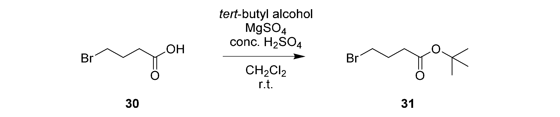
4-ブロモ酪酸(2.57 g, 15.39 mmol, 1 eq.)をジクロロメタン(20 ml)に溶かし、硫酸マグネシウム(7.74 g, 64.30 mmol, 4.2 eq.)、tert-ブチルアルコール(5.70 g, 76.95 mmol, 5 eq.)、及び濃硫酸(0.25 ml)を加え、室温で2日間撹拌した。飽和炭酸水素ナトリウム水溶液を加え、ジクロロメタンで抽出し、次いで有機層を飽和食塩水で洗浄し、無水硫酸ナトリウムで乾燥し、溶媒を減圧除去した。得られた残渣を中圧シリカゲルクロマトグラフィーで精製し(溶離液:ノルマルヘキサン/酢酸エチル = 50/50)、目的化合物31(1.17 g, 5.24 mmol, 47 %)を得た。
1H NMR (400 MHz, CDCl3): δ 1.27 (s, 9H), 1.91-1.98 (m, 2H), 2.22 (t, 2H, J = 7.2 Hz), 3.27 (t, 2H, J = 6.5 Hz); 13C NMR (100 MHz, CDCl3): δ 27.9, 28.0, 32.8, 33.7, 80.5, 171.7.
以下のスキームに従って化合物32を合成した。

化合物29(31.0 mg, 0.130 mmol, 1 eq.)をアセトニトリル(2 ml)に溶かし、化合物31(22.3 mg, 0.100 mmol, 0.8 eq.)、N,N-ジイソプロピルエチルアミン (113.3μl, 0.650 mmol, 5 eq.)、及び少量のヨウ化カリウムを加え、60 °Cで21時間撹拌した後、80 °Cでさらに7時間撹拌した。室温に戻した後、水を加え、ジクロロメタンで抽出し、次いで有機層を飽和食塩水で洗浄し、無水硫酸ナトリウムで乾燥し、溶媒を減圧除去した。得られた残渣をジクロロメタン(10 ml)に溶かし、トリフルオロ酢酸(2 ml)を加え、室温で2時間撹拌した後、溶媒を減圧除去した。得られた残渣をジクロロメタン(5 ml)及びメタノール(2 ml)に溶かし、p-クロラニル(34.1 mg, 0.139 mmol, 1.1 eq.)を加え、室温で30分間撹拌した後、溶媒を減圧除去した。得られた残渣をHPLCで精製し(溶離液A(H2O, 1 % アセトニトリル, 0.1 % トリフルオロ酢酸)、溶離液B(アセトニトリル, 1 % H2O)(A/B = 90/10 to 0/100 40 min))、目的化合物29(11.7 mg, 0.0268 mmol, 21 %)を得た。
1H NMR (400 MHz, CD3OD): δ 1.65 (s, 6H), 1.94-2.01 (m, 2H), 2.46 (t, 2H, J = 7.0 Hz), 3.50 (t, 2H, J = 7.2 Hz), 6.77 (dd, 1H, J = 2.0, 8.7 Hz), 6.84 (d, 1H, J = 8.2 Hz), 7.09 (d, 1H, J = 2.0 Hz), 7.15 (br s, 1H), 7.61-7.66 (m, 2H), 8.05 (s, 1H); 13C NMR (100 MHz, CD3OD): δ 25.2, 31.7,33.6, 43.5, 114.1, 116.0, 122.2, 122.4, 141.4, 155.9, 159.6, 161.2, 176.6; HRMS (ESI+): Calcd for [M]+, 323.17540, Found, 323.17538 (-0.0 mmu).
以下のスキームに従って化合物33を合成した。
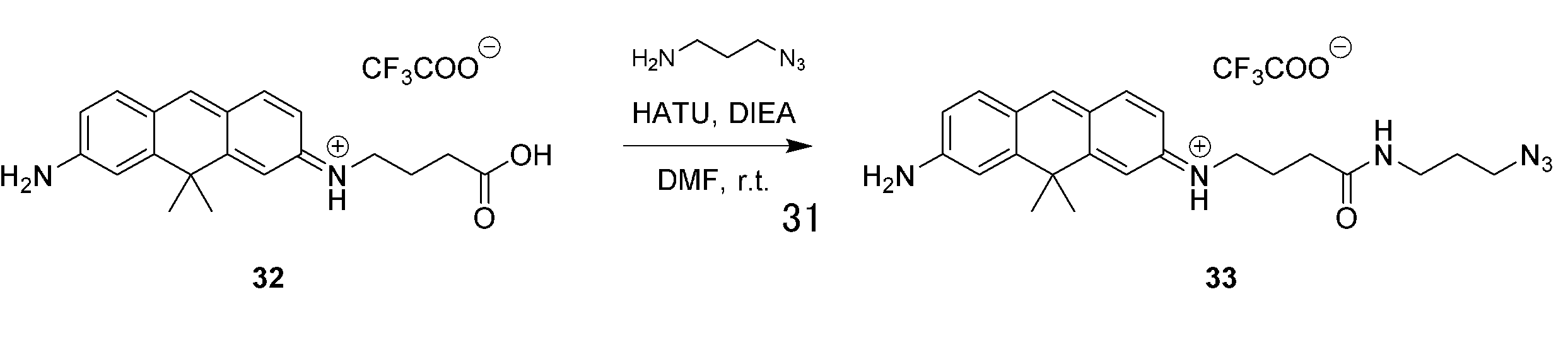
化合物32(1.1 mg, 0.00252 mmol, 1 eq.)をN,N-ジメチルホルムアミド(2 ml)に溶かし、0 °C下で3-アジドプロピルアミン(0.74 μl, 0.00756 mmol, 3 eq.)、 N,N-ジイソプロピルアミン(8.8 μl, 0.0504 mmol, 20 eq.)、及び O-(7-アザベンゾトリアゾール-1-イル)-N,N,N',N'-テトラメチルウロニウムヘキサフルオロリン酸塩(5.9 mg, 0.0155 mmol, 6.1 eq.)を加え、15分間撹拌した後、室温に戻してさらに13時間撹拌した。溶媒を減圧除去し、得られた残渣をHPLCで精製し(溶離液A(H2O, 1 % アセトニトリル, 0.1 % トリフルオロ酢酸)、溶離液B(アセトニトリル, 1 % H2O)(A/B = 90/10 to 0/100 40 min))、目的化合物33(1.0 mg, 0.00193 mmol, 77 %)を得た。
1H NMR (400 MHz, CD3OD): δ 1.66 (s, 6H), 1.76 (qui, 2H, J = 6.7 Hz), 1.99 (qui, 2H, J = 7.2 Hz), 2.35 (t, 2H, J = 7.2 Hz), 3.27 (t, 2H, J = 6.7 Hz), 3.35 (t, 2H, J = 6.7 Hz), 3.48 (t, 2H, J = 7.2 Hz), 6.77 (dd, 1H, J = 2.1, 8.7 Hz), 6.83 (d, 1H, J = 8.0 Hz), 7.09-7.11 (m, 2H), 7.11 (brs, 1H), 7.62-7.67 (m, 2H), 8.06 (s, 1H); 13C NMR (100 MHz, CD3OD): δ 25.9, 29.7, 33.6, 33.8, 37.8, 40.4, 43.7, 43.7, 50.1, 114.1, 116.0, 122.2, 122.4, 141.5, 155.9, 159.5, 161.3, 175.1; HRMS (ESI+): Calcd for [M]+, 405.24028, Found, 405.23991 (-0.4 mmu).
以下のスキームに従って化合物34を合成した。
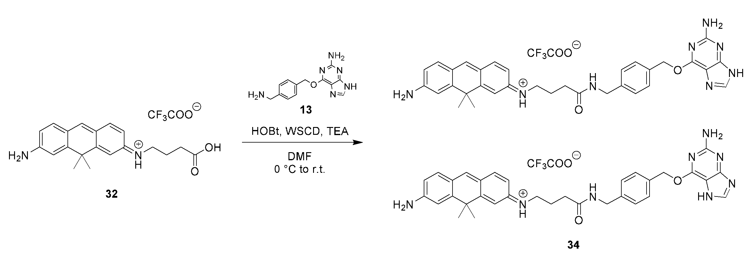
化合物32(1.15 mg, 0.00356 mmol, 1 eq.)及び化合物13(3.6 mg, 0.0133 mmol, 3.3 eq.)をN,N-ジメチルホルムアミド(1.5 ml)に溶かし、0 °C下で1-エチル-3-(3-ジメチルアミノプロピル)カルボジイミド塩酸塩(4.5 mg, 0.0235 mmol, 6.6 eq.)、1-ヒドロキシベンゾトリアゾール(2.0 mg, 0.0148 mmol, 4.2 eq.)、及びトリエチルアミン(4.96 μl, 0.0356, 10 eq.)を加え、10分間撹拌した後、室温に戻してさらに22時間撹拌した。溶媒を減圧除去し、得られた残渣をHPLCで精製し(溶離液A(H2O, 1 % アセトニトリル, 0.1 % トリフルオロ酢酸)、溶離液B(アセトニトリル, 1 % H2O)(A/B = 90/10 to 0/100 40 min))、目的化合物34(1.3 mg, 0.00189 mmol, 72 %)を得た。
1H NMR (400 MHz, CD3OD): δ 1.62 (s, 6H), 1.98-2.05 (m, 2H), 2.40 (t, 2H, J = 7.0 Hz), 3.44-3.48 (m, 2H), 4.40 (s, 2H), 5.59 (s, 2H), 6.75-6.81 (m, 2H), 7.06-7.08 (m, 2H), 7.33 (d, 2H, J = 8.4 Hz), 7.49 (d, 2H, J = 8.4 Hz), 7.60-7.65 (m, 2H), 8.03 (s, 1H), 8.16 (s, 1H); HRMS (ESI+): Calcd for [M]+, 575.28775, Found, 575.28769 (-0.1 mmu).
1H NMR (400 MHz, CD3OD): δ 1.62 (s, 6H, f), 1.98-2.05 (m, 2H, l), 2.40 (t, 2H, J = 7.0 Hz, m), 3.44-3.48 (m, 2H, k), 4.39 (s, 2H, o), 5.59 (s, 2H, r), 6.74-6.79 (m, 2H, c, h), 7.06-7.08 (m, 2H, b, i), 7.32 (d, 2H, J = 8.0 Hz, q), 7.48 (d, 2H, J = 8.0 Hz, p), 7.59-7.64 (m, 2H, d, g), 8.00 (s, 1H, t), 8.02 (s, 1H, e),; HRMS (ESI+): Calcd for [M]+, 575.28775, Found, 575.28769 (-0.1 mmu).
以下のスキームに従って化合物35を合成した。

化合物32(2.3 mg, 0.00711 mmol, 1 eq.)及び化合物15(5.3 mg, 0.0237 mmol, 3.3 eq.)をN,N-ジメチルホルムアミド(2 ml)に溶かし、0 °C下で1-エチル-3-(3-ジメチルアミノプロピル)カルボジイミド塩酸塩(3.2 mg, 0.0167 mmol, 2.3 eq.)、1-ヒドロキシベンゾトリアゾール(3.0 mg, 0.0222 mmol, 3.1 eq.)、及びトリエチルアミン(9.9 μl, 0.0711, 10 eq.)を加え、15分間撹拌した後、室温に戻してさらに2日間撹拌した。溶媒を減圧除去し、得られた残渣をHPLCで精製し(溶離液A(H2O, 1 % アセトニトリル, 0.1 % トリフルオロ酢酸)、溶離液B(アセトニトリル, 1 % H2O)(A/B = 90/10 to 0/100 40 min))、目的化合物35(1.2 mg, 0.00187 mmol, 36 % yield)を得た。
1H NMR (400 MHz, CD3OD): δ 1.29-1.43 (m, 4H), 1.563 (qui, 2H, J = 7.2 Hz), 1.66 (s, 6H), 1.69-1.77 (m, 2H), 2.00 (qui, 2H, J = 7.2 Hz), 2.36 (t, 2H, J = 7.2 Hz), 3.37-3.61 (m, 14H), 6.77 (dd, 1H, J = 2.0, 8.8 Hz), 6.84 (d, 1H, J = 7.6 Hz), 7.09-7.11 (m, 2H), 7.62-7.64 (m, 2H), 8.06 (s, 1H); HRMS (ESI+): Calcd for [M]+, 528.29875, Found, 528.29876 (+0.0 mmu).
本発明のプローブ化合物について、グルタチオン添加に伴う吸収スペクトル変化及び蛍光スペクトル変化を測定し、本発明のプローブ化合物とグルタチオンとの間の求核付加-解離平衡反応における解離定数を算出した。
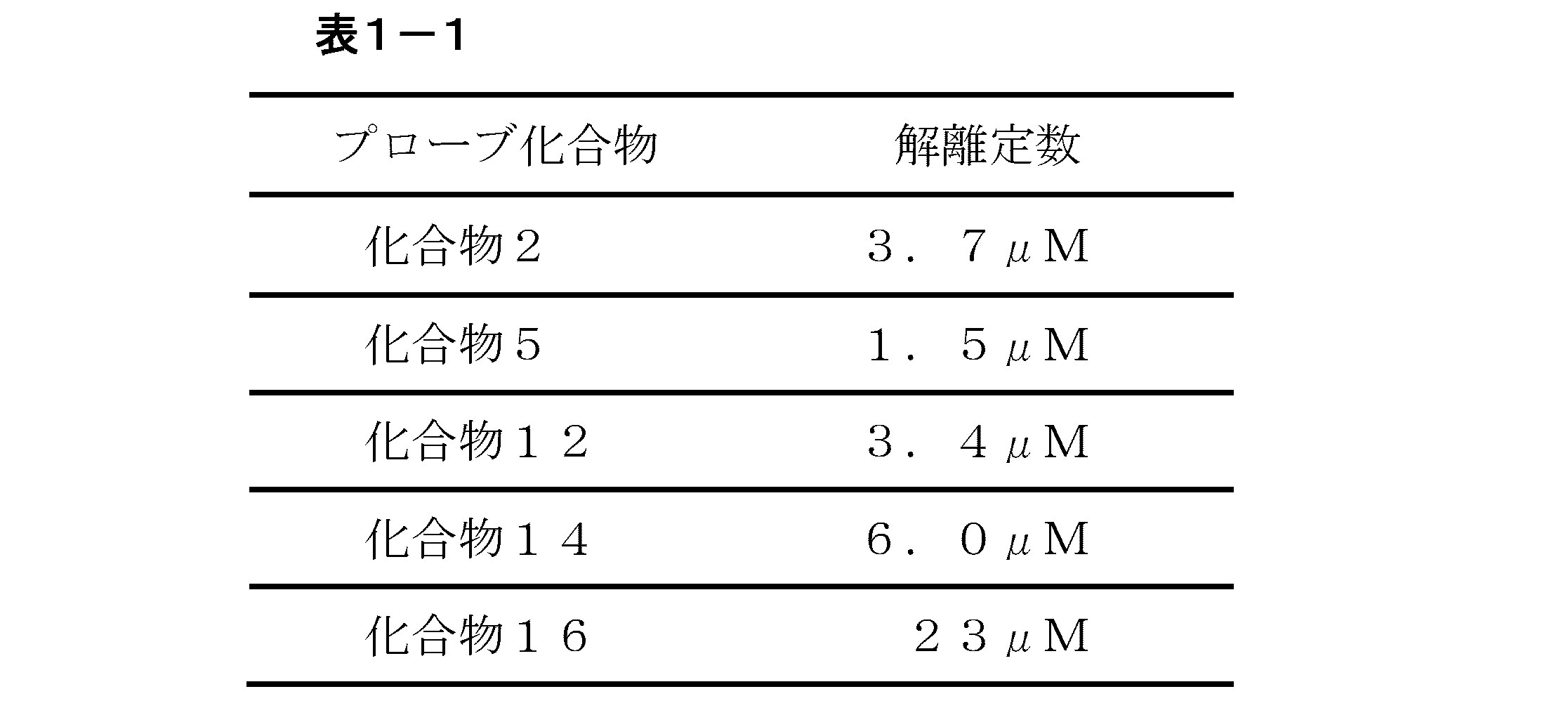

レーザーフラッシュフォトリシスにより、種々のグルタチオン濃度における本発明のプローブ化合物の過渡吸収の経時変化を測定することによって、本発明のプローブ化合物とグルタチオンとの間の求核付加-解離平衡反応における求核付加反応速度定数k、求核付加反応速度時定数τを算出した。溶液条件は、1%DMSO水溶液、pH7.4(10mM NaPiバッファー)である。測定条件は295Kで、光源は10mJ/pulseのXeClエキシマーレーザー(308nm)である。化合物2、5、12、28、及び32について得られた結果をそれぞれ表2~6に示す。
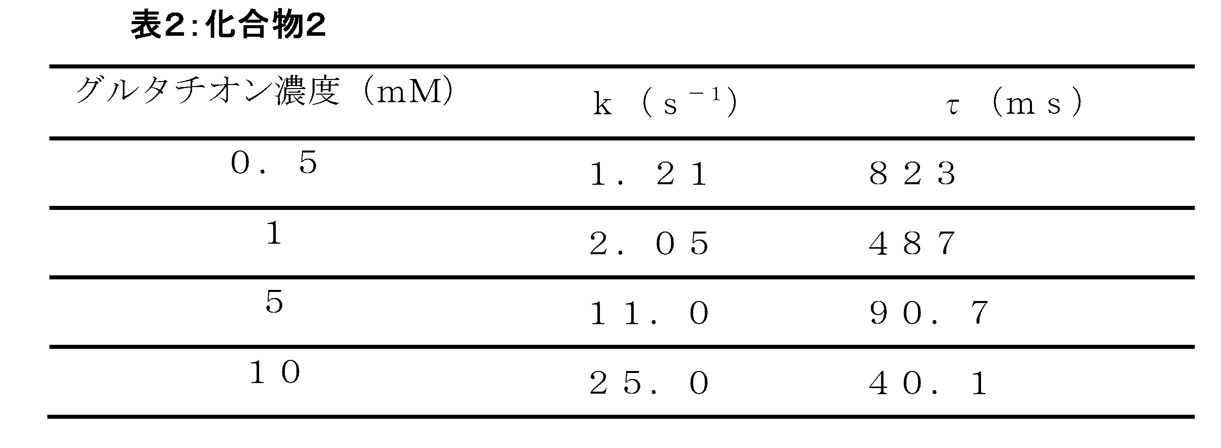


本発明のプローブ化合物について、蛍光量子収率を測定した。溶液条件は、プローブ化合物1μM、1%DMSO水溶液、pH7.4(200mM NaPiバッファー)である。化合物5、12、14、16、17、28、及び32について得られた結果を表7に示す。本発明のプローブ化合物は、SMLMによる超解像蛍光イメージングに適した高い蛍光量子収率を示すことが明らかとなった。

本発明のプローブ化合物でラベル化した抗体を用いて、細胞の超解像イメージングを行った。
化合物5の末端にスクシンイミジルエステルを導入したプローブ化合物である化合物17をDMSOに溶解し、1mM及び10mMのストック溶液を調製した。β-チューブリンの間接免疫染色のためのヤギ由来・抗マウス二次抗体(IgG、Sigma-Aldrich社)及びTom20の間接免疫染色のためのヤギ由来・抗ウサギ二次抗体(IgG、Jackson ImmunoResearch社)をそれぞれ、0.2Mのリン酸ナトリウムバッファー(pH8.5)中で化合物17と混和した(室温、30分間)。プローブ化合物でラベル化した抗体を、溶離液としてリン酸緩衝生理食塩水(PBS)(pH7.4、GIBCO社)を用いてPD MiniTrap TM G-25(GEヘルスケア社)によって精製した。抗体に対するプローブ化合物の標識度(DOL)として、プローブ化合物[mol]/抗体分子[mol]の比を算出した。ここで、プローブ化合物の濃度はプローブ化合物に由来する吸光度の測定値から算出し、抗体分子の濃度は、精製処理における抗体分子の損失はなかったものと仮定して、調製に使用した抗体分子の全量から算出した。超解像イメージング(SMLM)においては、標識度が2.1であるラベル化抗マウス抗体及び標識度が1.3であるラベル化抗ウサギ抗体を使用した。
抗体に対するプローブ化合物の標識度(DOL)として、プローブ化合物[mol]/抗体分子[mol]の比を算出した。ここで、プローブ化合物の濃度はプローブ化合物に由来する吸光度の測定値から算出し、抗体分子の濃度は、精製処理における抗体分子の損失はなかったものと仮定して、調製に使用した抗体分子の全量から算出した。超解像イメージング(SMLM)においては、標識度が1.4であるラベル化抗体を使用した。
アフリカミドリザルの腎臓に由来するベロ細胞を、フェノールレッド不含かつL-グルタミン含有のダルベッコ改変イーグル培地(高グルコース)(GIBCO社)にウシ胎仔血清(Invitrogen社)及び1%ペニシリン-ストレプトマイシン溶液(和光純薬工業社)を添加した培地中で培養した(37℃、5%CO2)。イメージングを行う前日に、当該細胞をLab-Tek II 8ウェルチャンバーのカバーガラス(Thermo Fischer Scientific社)に播種した。以下に挙げる文献を参考に、固定と免疫染色を以下のとおり行った。
・M. Bates, G. T. Dempsey, K. H. Chen, X. Zhuang, Multicolor Super-Resolution Fluorescence Imaging via Multi-Parameter Fluorophore Detection. Chemphyschem : a European journal of chemical physics and physical chemistry 13, 99-107 (2012)
・B. Huang, S. A. Jones, B. Brandenburg, X. Zhuang, Whole-cell 3D STORM reveals interactions between cellular structures with nanometer-scale resolution. Nature methods 5, 1047-1052 (2008)
・C. Dellagiacoma et al., Targeted photoswitchable probe for nanoscopy of biological structures. Chembiochem 11, 1361-1363 (2010)
・D. R. Whelan, T. D. Bell, Image artifacts in single molecule localization microscopy: why optimization of sample preparation protocols matters. Scientific reports 5, 7924 (2015)
N-STORM装置(ニコン社)を用いて、室温でSMLMイメージングを行った。プローブ化合物である化合物17及び化合物33の励起は、それぞれ647nm(40~500W/cm2)及び561nm(40~500W/cm2)のレーザー光を用いた全反射照明または全反射照明に近い遮光照明により行った。画像データは、15ms/フレームまたは30ms/フレームで記録し、画像統合ソフトウェア(NIS-Elements Advanced Research、ニコン社)により解析することで超解像度画像を構築した。超解像度画像の表示にあたっては、150フォトン以上又は200フォトン以上が検出された輝点のみを表示した。
Claims (14)
- 以下の式(I)で表される化合物

〔式中、
Xは、酸素原子又はC(Ra)(Rb)又はSi(Ra)(Rb)を表し(ここで、Ra及びRbは、それぞれ独立に水素原子、又はアルキル基を表す);
R1は、水素原子又は置換されていてもよいアリールを表し(ただし、R1がフェニル基の場合、当該フェニル基のベンゼン環における2位又は6位の位置に置換基を有しない);
R2及びR3は、それぞれ独立に、水素原子、ヒドロキシル基、ハロゲン原子、それぞれ置換されていてもよいアルキル基、スルホ基、カルボキシル基、エステル基、アミド基及びアジド基よりなる群から独立に選択される1~3個の同一又は異なる置換基を表し;
R4及びR5は、それぞれ独立に水素原子又は置換されていてもよいアルキル基を表し、又は、N(R4)(R5)がアミド基もしくはカルバメート基を形成する(ここで、R4又はR5がアルキル基である場合、それぞれR2と一緒になって、それらが結合する窒素原子を含む環構造を形成してもよい);及び、
R6及びR7は、それぞれ独立に水素原子もしくは置換されていてもよいアルキル基を表し、又は、N(R6)(R7)がアミド基もしくはカルバメート基を形成する(ここで、R6又はR7がアルキル基である場合、それぞれR3と一緒になって、それらが結合する窒素原子を含む環構造を形成してもよい。)〕
又はその塩を含む超解像蛍光イメージング用プローブであって、
式(I)で表される化合物又はその塩が、-SH基を含有する求核性化合物との間で求核付加-解離平衡反応を行うことを特徴とする、
該超解像蛍光イメージング用プローブ。 - 前記求核付加-解離平衡反応が、R1が結合する炭素原子において行われる、請求項1に記載の超解像蛍光イメージング用プローブ。
- 前記求核付加-解離平衡反応における解離定数が、0.1μM~10mMの範囲内であることを特徴とする、請求項1又は2に記載の超解像蛍光イメージング用プローブ。
- 前記求核付加-解離平衡反応における求核付加反応速度定数が、中性条件の水系溶液中において、1~1.0x10⁶s-1であることを特徴とする、請求項1~3のいずれか1項に記載の超解像蛍光イメージング用プローブ。
- Xが、C(Ra)(Rb)又はSi(Ra)(Rb)である、請求項1~4のいずれか1項に記載の超解像蛍光イメージング用プローブ。
- Ra及びRbが、いずれもメチル基である、請求項5に記載の超解像蛍光イメージング用プローブ。
- R1は、水素原子又は置換されていてもよいフェニル基である(ただし、当該フェニル基のベンゼン環における2位又は6位の位置に置換基を有しない)、請求項1~6のいずれか1項に記載の超解像蛍光イメージング用プローブ。
- R4、R5、R6及びR7が、いずれも水素原子である、請求項1~7のいずれか1項に記載の超解像蛍光イメージング用プローブ。
- R4、R5、R6及びR7が、いずれもメチル基である、請求項1~7のいずれか1項に記載の超解像蛍光イメージング用プローブ。
- R4、R5、R6及びR7のうち少なくとも1つが、生体分子と共有結合又は非共有結合によって結合し得るラベル化置換基を含む、請求項1~7のいずれか1項に記載の超解像蛍光イメージング用プローブ。
- 前記-SH基を含有する化合物が、システイン残基を有する化合物である、請求項1~10のいずれか1項に記載の超解像蛍光イメージング用プローブ。
- 前記-SH基を含有する化合物が、グルタチオンである、請求項1~10のいずれか1項に記載の超解像蛍光イメージング用プローブ。
- 式(I)で表される化合物が以下の群から選択される化合物である、請求項1に記載の超解像蛍光イメージング用プローブ。
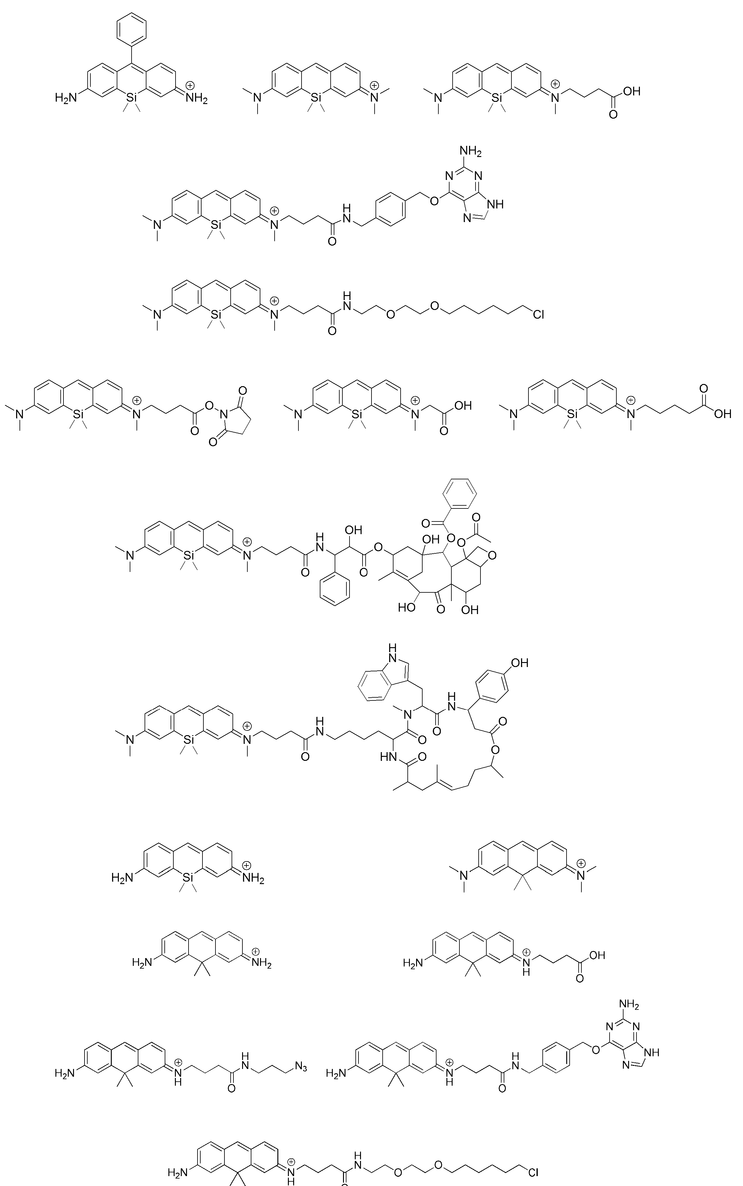
- 請求項1~13のいずれか1項に記載の超解像蛍光イメージング用プローブを用いる超解像蛍光イメージング方法であって、
生体分子にプローブ分子を結合させ、-SH基を含有する化合物の存在下でレーザー光を照射して前記プローブ分子からの蛍光発光を撮影した画像データを取得し、一定の時間間隔でこれを繰り返して得られた複数の前記画像データを解析したうえで重ね合わせることによって、前記生体分子の構造に対する超高解像のイメージ画像を得ることを含む、該方法。
Priority Applications (2)
| Application Number | Priority Date | Filing Date | Title |
|---|---|---|---|
| US15/551,149 US10837909B2 (en) | 2015-02-27 | 2016-02-23 | Super-resolution fluorescent imaging probe |
| JP2017502373A JP6754970B2 (ja) | 2015-02-27 | 2016-02-23 | 超解像蛍光イメージング用プローブ |
Applications Claiming Priority (4)
| Application Number | Priority Date | Filing Date | Title |
|---|---|---|---|
| JP2015-037519 | 2015-02-27 | ||
| JP2015037519 | 2015-02-27 | ||
| JPPCT/JP2016/051314 | 2016-01-18 | ||
| PCT/JP2016/051314 WO2016136328A1 (ja) | 2015-02-27 | 2016-01-18 | 超解像蛍光イメージング用プローブ |
Publications (1)
| Publication Number | Publication Date |
|---|---|
| WO2016136718A1 true WO2016136718A1 (ja) | 2016-09-01 |
Family
ID=56788294
Family Applications (2)
| Application Number | Title | Priority Date | Filing Date |
|---|---|---|---|
| PCT/JP2016/051314 Ceased WO2016136328A1 (ja) | 2015-02-27 | 2016-01-18 | 超解像蛍光イメージング用プローブ |
| PCT/JP2016/055199 Ceased WO2016136718A1 (ja) | 2015-02-27 | 2016-02-23 | 超解像蛍光イメージング用プローブ |
Family Applications Before (1)
| Application Number | Title | Priority Date | Filing Date |
|---|---|---|---|
| PCT/JP2016/051314 Ceased WO2016136328A1 (ja) | 2015-02-27 | 2016-01-18 | 超解像蛍光イメージング用プローブ |
Country Status (3)
| Country | Link |
|---|---|
| US (1) | US10837909B2 (ja) |
| JP (1) | JP6754970B2 (ja) |
| WO (2) | WO2016136328A1 (ja) |
Cited By (3)
| Publication number | Priority date | Publication date | Assignee | Title |
|---|---|---|---|---|
| CN108362671A (zh) * | 2018-02-23 | 2018-08-03 | 银川高新区广煜科技有限公司 | 检测半胱氨酸的方法 |
| CN112028874A (zh) * | 2020-09-10 | 2020-12-04 | 苏州富德兆丰生化科技有限公司 | 艾立替尼的合成方法 |
| CN114728908A (zh) * | 2019-11-08 | 2022-07-08 | 静冈县公立大学法人 | 化合物或其盐 |
Families Citing this family (7)
| Publication number | Priority date | Publication date | Assignee | Title |
|---|---|---|---|---|
| KR101776551B1 (ko) | 2016-11-30 | 2017-09-11 | 한국생명공학연구원 | Alp 검출용 근적외선 형광 프로브 및 이의 제조방법 |
| WO2018125913A1 (en) * | 2016-12-27 | 2018-07-05 | Profusa, Inc. | Near-ir glucose sensors |
| CN107033879B (zh) * | 2017-04-26 | 2019-03-29 | 许昌学院 | 一种用于检测谷胱甘肽的荧光探针及其制备方法和应用 |
| JP7561037B2 (ja) | 2018-06-27 | 2024-10-03 | プロフサ,インコーポレイテッド | 近赤外線グルコースセンサー |
| EP3633009A1 (en) * | 2018-10-03 | 2020-04-08 | Max-Planck-Gesellschaft zur Förderung der Wissenschaften e.V. | Use of a substituted or unsubstituted polycyclic aromatic hydrocarbon compound for high-resolution microscopy |
| CN114106031B (zh) * | 2021-10-29 | 2024-02-23 | 四川大学 | 一种检测牙髓状态的次氯酸荧光探针 |
| CN119350307A (zh) * | 2024-11-07 | 2025-01-24 | 上海泓博智源医药股份有限公司 | 选择性的细胞核atp小分子探针及其制备方法与应用 |
Citations (5)
| Publication number | Priority date | Publication date | Assignee | Title |
|---|---|---|---|---|
| WO2012111818A1 (ja) * | 2011-02-18 | 2012-08-23 | 国立大学法人 東京大学 | 蛍光プローブ |
| WO2013126816A1 (en) * | 2012-02-23 | 2013-08-29 | Oregon State Board Of Higher Education On Behalf Of Portland State University | Selective detection of thiols |
| WO2014106957A1 (ja) * | 2013-01-07 | 2014-07-10 | 国立大学法人 東京大学 | 非対称Siローダミン及びロドールの合成 |
| JP2014157150A (ja) * | 2013-01-18 | 2014-08-28 | Univ Of Tokyo | 超解像蛍光イメージング用プローブ |
| WO2015129705A1 (ja) * | 2014-02-28 | 2015-09-03 | 国立大学法人 東京大学 | グルタチオン検出用蛍光プローブ |
Family Cites Families (4)
| Publication number | Priority date | Publication date | Assignee | Title |
|---|---|---|---|---|
| US9156986B2 (en) * | 2003-04-03 | 2015-10-13 | Enzo Life Sciences, Inc. | Multisignal labeling reagents and processes and uses therefor |
| US7776613B2 (en) | 2006-08-07 | 2010-08-17 | President And Fellows Of Harvard College | Sub-diffraction image resolution and other imaging techniques |
| WO2013029650A1 (en) * | 2011-08-26 | 2013-03-07 | Ecole Polytechnique Federale De Lausanne (Epfl) | Cell permeable, fluorescent dye |
| EP3502693A1 (en) * | 2014-05-16 | 2019-06-26 | Life Technologies Corporation | Cell tracking reagents and their methods of use |
-
2016
- 2016-01-18 WO PCT/JP2016/051314 patent/WO2016136328A1/ja not_active Ceased
- 2016-02-23 US US15/551,149 patent/US10837909B2/en not_active Expired - Fee Related
- 2016-02-23 WO PCT/JP2016/055199 patent/WO2016136718A1/ja not_active Ceased
- 2016-02-23 JP JP2017502373A patent/JP6754970B2/ja not_active Expired - Fee Related
Patent Citations (5)
| Publication number | Priority date | Publication date | Assignee | Title |
|---|---|---|---|---|
| WO2012111818A1 (ja) * | 2011-02-18 | 2012-08-23 | 国立大学法人 東京大学 | 蛍光プローブ |
| WO2013126816A1 (en) * | 2012-02-23 | 2013-08-29 | Oregon State Board Of Higher Education On Behalf Of Portland State University | Selective detection of thiols |
| WO2014106957A1 (ja) * | 2013-01-07 | 2014-07-10 | 国立大学法人 東京大学 | 非対称Siローダミン及びロドールの合成 |
| JP2014157150A (ja) * | 2013-01-18 | 2014-08-28 | Univ Of Tokyo | 超解像蛍光イメージング用プローブ |
| WO2015129705A1 (ja) * | 2014-02-28 | 2015-09-03 | 国立大学法人 東京大学 | グルタチオン検出用蛍光プローブ |
Cited By (5)
| Publication number | Priority date | Publication date | Assignee | Title |
|---|---|---|---|---|
| CN108362671A (zh) * | 2018-02-23 | 2018-08-03 | 银川高新区广煜科技有限公司 | 检测半胱氨酸的方法 |
| CN114728908A (zh) * | 2019-11-08 | 2022-07-08 | 静冈县公立大学法人 | 化合物或其盐 |
| US12551578B2 (en) | 2019-11-08 | 2026-02-17 | Shizuoka Prefectural University Corporation | Compound or salt thereof |
| CN112028874A (zh) * | 2020-09-10 | 2020-12-04 | 苏州富德兆丰生化科技有限公司 | 艾立替尼的合成方法 |
| CN112028874B (zh) * | 2020-09-10 | 2021-12-24 | 苏州富德兆丰生化科技有限公司 | 艾立替尼的合成方法 |
Also Published As
| Publication number | Publication date |
|---|---|
| JP6754970B2 (ja) | 2020-09-16 |
| US20180052109A1 (en) | 2018-02-22 |
| WO2016136328A1 (ja) | 2016-09-01 |
| US10837909B2 (en) | 2020-11-17 |
| JPWO2016136718A1 (ja) | 2017-12-07 |
Similar Documents
| Publication | Publication Date | Title |
|---|---|---|
| JP6754970B2 (ja) | 超解像蛍光イメージング用プローブ | |
| JP6583793B2 (ja) | グルタチオン検出用蛍光プローブ | |
| JP6090934B2 (ja) | 酸性環境検出蛍光プローブ | |
| Poole et al. | Synthesis and characterisation of highly emissive and kinetically stable lanthanide complexes suitable for usage ‘in cellulo’ | |
| CN103087545B (zh) | 一类以荧光素为母体的荧光染料、其制备方法及应用 | |
| JP6349091B2 (ja) | 超解像蛍光イメージング用プローブ | |
| JP5636080B2 (ja) | pH感受性蛍光プローブ | |
| JP2019530636A (ja) | 光活性フルオロフォア及びインビボにおける標識方法 | |
| US20180118943A1 (en) | Ph sensitive fluorescent probe | |
| Huang et al. | A series of iridophosphors with tunable excited states for hypoxia monitoring via time-resolved luminescence microscopy | |
| JP6485799B2 (ja) | 三光子励起により発光する発光性組成物 | |
| JPWO2018043579A1 (ja) | ホスファローダミン化合物若しくはその塩、並びにそれを用いた蛍光色素 | |
| JP2018025399A (ja) | ハイドロポリスルフィド検出用蛍光プローブ | |
| JP2012154693A (ja) | カリウム蛍光プローブ | |
| JP6877769B2 (ja) | ホスホール化合物 | |
| EP4377311A1 (en) | 2-diazo-3-oxo-2,3-dihydrospiro[indene-1,9'-xanthene] derivatives and similar compounds as photoactive fluorescent compounds for protein labeling | |
| EP4519384B1 (en) | Photoactivatable compounds and uses thereof | |
| JP2024126464A (ja) | -sh基含有化合物検出型ラマンプローブ | |
| Yu | The synthesis of N-hydroxyoxindole-based photoactivatable fluorophores and its application for bioimaging |
Legal Events
| Date | Code | Title | Description |
|---|---|---|---|
| 121 | Ep: the epo has been informed by wipo that ep was designated in this application |
Ref document number: 16755462 Country of ref document: EP Kind code of ref document: A1 |
|
| ENP | Entry into the national phase |
Ref document number: 2017502373 Country of ref document: JP Kind code of ref document: A |
|
| WWE | Wipo information: entry into national phase |
Ref document number: 15551149 Country of ref document: US |
|
| NENP | Non-entry into the national phase |
Ref country code: DE |
|
| 122 | Ep: pct application non-entry in european phase |
Ref document number: 16755462 Country of ref document: EP Kind code of ref document: A1 |