WO2014156444A1 - 画像表示装置および方法 - Google Patents
画像表示装置および方法 Download PDFInfo
- Publication number
- WO2014156444A1 WO2014156444A1 PCT/JP2014/054665 JP2014054665W WO2014156444A1 WO 2014156444 A1 WO2014156444 A1 WO 2014156444A1 JP 2014054665 W JP2014054665 W JP 2014054665W WO 2014156444 A1 WO2014156444 A1 WO 2014156444A1
- Authority
- WO
- WIPO (PCT)
- Prior art keywords
- image
- correction amount
- radiation
- images
- breast
- Prior art date
- Legal status (The legal status is an assumption and is not a legal conclusion. Google has not performed a legal analysis and makes no representation as to the accuracy of the status listed.)
- Ceased
Links
Images



Classifications
-
- A—HUMAN NECESSITIES
- A61—MEDICAL OR VETERINARY SCIENCE; HYGIENE
- A61B—DIAGNOSIS; SURGERY; IDENTIFICATION
- A61B6/00—Apparatus or devices for radiation diagnosis; Apparatus or devices for radiation diagnosis combined with radiation therapy equipment
- A61B6/48—Diagnostic techniques
- A61B6/486—Diagnostic techniques involving generating temporal series of image data
-
- A—HUMAN NECESSITIES
- A61—MEDICAL OR VETERINARY SCIENCE; HYGIENE
- A61B—DIAGNOSIS; SURGERY; IDENTIFICATION
- A61B6/00—Apparatus or devices for radiation diagnosis; Apparatus or devices for radiation diagnosis combined with radiation therapy equipment
- A61B6/52—Devices using data or image processing specially adapted for radiation diagnosis
- A61B6/5211—Devices using data or image processing specially adapted for radiation diagnosis involving processing of medical diagnostic data
- A61B6/5229—Devices using data or image processing specially adapted for radiation diagnosis involving processing of medical diagnostic data combining image data of a patient, e.g. combining a functional image with an anatomical image
-
- G—PHYSICS
- G06—COMPUTING OR CALCULATING; COUNTING
- G06T—IMAGE DATA PROCESSING OR GENERATION, IN GENERAL
- G06T3/00—Geometric image transformations in the plane of the image
- G06T3/02—Affine transformations
-
- G—PHYSICS
- G06—COMPUTING OR CALCULATING; COUNTING
- G06T—IMAGE DATA PROCESSING OR GENERATION, IN GENERAL
- G06T7/00—Image analysis
- G06T7/0002—Inspection of images, e.g. flaw detection
- G06T7/0012—Biomedical image inspection
- G06T7/0014—Biomedical image inspection using an image reference approach
- G06T7/0016—Biomedical image inspection using an image reference approach involving temporal comparison
-
- G—PHYSICS
- G06—COMPUTING OR CALCULATING; COUNTING
- G06T—IMAGE DATA PROCESSING OR GENERATION, IN GENERAL
- G06T7/00—Image analysis
- G06T7/30—Determination of transform parameters for the alignment of images, i.e. image registration
- G06T7/33—Determination of transform parameters for the alignment of images, i.e. image registration using feature-based methods
- G06T7/337—Determination of transform parameters for the alignment of images, i.e. image registration using feature-based methods involving reference images or patches
-
- A—HUMAN NECESSITIES
- A61—MEDICAL OR VETERINARY SCIENCE; HYGIENE
- A61B—DIAGNOSIS; SURGERY; IDENTIFICATION
- A61B6/00—Apparatus or devices for radiation diagnosis; Apparatus or devices for radiation diagnosis combined with radiation therapy equipment
- A61B6/46—Arrangements for interfacing with the operator or the patient
- A61B6/461—Displaying means of special interest
- A61B6/463—Displaying means of special interest characterised by displaying multiple images or images and diagnostic data on one display
-
- A—HUMAN NECESSITIES
- A61—MEDICAL OR VETERINARY SCIENCE; HYGIENE
- A61B—DIAGNOSIS; SURGERY; IDENTIFICATION
- A61B6/00—Apparatus or devices for radiation diagnosis; Apparatus or devices for radiation diagnosis combined with radiation therapy equipment
- A61B6/50—Apparatus or devices for radiation diagnosis; Apparatus or devices for radiation diagnosis combined with radiation therapy equipment specially adapted for specific body parts; specially adapted for specific clinical applications
- A61B6/502—Apparatus or devices for radiation diagnosis; Apparatus or devices for radiation diagnosis combined with radiation therapy equipment specially adapted for specific body parts; specially adapted for specific clinical applications for diagnosis of breast, i.e. mammography
-
- A—HUMAN NECESSITIES
- A61—MEDICAL OR VETERINARY SCIENCE; HYGIENE
- A61B—DIAGNOSIS; SURGERY; IDENTIFICATION
- A61B6/00—Apparatus or devices for radiation diagnosis; Apparatus or devices for radiation diagnosis combined with radiation therapy equipment
- A61B6/52—Devices using data or image processing specially adapted for radiation diagnosis
- A61B6/5205—Devices using data or image processing specially adapted for radiation diagnosis involving processing of raw data to produce diagnostic data
-
- A—HUMAN NECESSITIES
- A61—MEDICAL OR VETERINARY SCIENCE; HYGIENE
- A61B—DIAGNOSIS; SURGERY; IDENTIFICATION
- A61B6/00—Apparatus or devices for radiation diagnosis; Apparatus or devices for radiation diagnosis combined with radiation therapy equipment
- A61B6/52—Devices using data or image processing specially adapted for radiation diagnosis
- A61B6/5211—Devices using data or image processing specially adapted for radiation diagnosis involving processing of medical diagnostic data
- A61B6/5229—Devices using data or image processing specially adapted for radiation diagnosis involving processing of medical diagnostic data combining image data of a patient, e.g. combining a functional image with an anatomical image
- A61B6/5235—Devices using data or image processing specially adapted for radiation diagnosis involving processing of medical diagnostic data combining image data of a patient, e.g. combining a functional image with an anatomical image combining images from the same or different ionising radiation imaging techniques, e.g. PET and CT
-
- G—PHYSICS
- G06—COMPUTING OR CALCULATING; COUNTING
- G06T—IMAGE DATA PROCESSING OR GENERATION, IN GENERAL
- G06T2207/00—Indexing scheme for image analysis or image enhancement
- G06T2207/10—Image acquisition modality
- G06T2207/10004—Still image; Photographic image
-
- G—PHYSICS
- G06—COMPUTING OR CALCULATING; COUNTING
- G06T—IMAGE DATA PROCESSING OR GENERATION, IN GENERAL
- G06T2207/00—Indexing scheme for image analysis or image enhancement
- G06T2207/10—Image acquisition modality
- G06T2207/10116—X-ray image
-
- G—PHYSICS
- G06—COMPUTING OR CALCULATING; COUNTING
- G06T—IMAGE DATA PROCESSING OR GENERATION, IN GENERAL
- G06T2207/00—Indexing scheme for image analysis or image enhancement
- G06T2207/30—Subject of image; Context of image processing
- G06T2207/30004—Biomedical image processing
- G06T2207/30068—Mammography; Breast
Definitions
- the present invention relates to an image display apparatus and method for displaying a radiation image of a breast or the like.
- comparative interpretation is performed in which a plurality of images of a subject are displayed on a display device such as a CRT or a liquid crystal display, and interpretation is performed while comparing the images.
- a display device such as a CRT or a liquid crystal display
- a technique for displaying a radiation image on a display device so as to facilitate such comparative interpretation is known.
- Patent Documents 1 and 2 propose a method of switching and displaying a plurality of radiation images for comparative interpretation on one screen.
- a method for calculating a difference between a plurality of radiographic images is also used as a comparative interpretation method. By calculating the difference in this way, it is easy to confirm a portion having a change, so that comparative interpretation can be easily performed.
- a plurality of radiographic images to be subjected to comparative interpretation have different positions of the subject or image quality included in the radiographic image because the positions of the imaging parts are different, the imaging conditions at the time of imaging are different, and the imaging devices are different. May be different in the radiation image.
- an image that performs alignment processing of a subject included in the radiographic image or matches the image quality of the plurality of radiographic images It is necessary to perform processing. For this reason, in the method described in Patent Document 1, a plurality of radiation images are aligned by affine transformation or the like, and the luminances of the plurality of radiation images are matched.
- the comparative interpretation described above is intended to compare the latest radiograph taken most recently with a past radiographic image. For this reason, the alignment process or the process of making the image quality constant is performed based on the latest radiation image. Therefore, alignment processing and / or image quality correction processing is performed so that the radiographic images acquired in the past match the latest radiographic image.
- the comparative interpretation described above is performed every time a new radiation image is acquired. Performing registration processing and image quality correction processing on all past radiographic images with a new radiographic image as a reference every time a new radiographic image is acquired requires a large amount of computation, and processing Takes a long time. In this case, if all the radiographic images subjected to the alignment process and the image quality correction process are saved, the position of the latest radiographic image and any one radiographic image and the correction amount of the image quality can be calculated. Since the position and image quality of the radiation image can be corrected, the calculation time can be shortened.
- the present invention has been made in view of the above circumstances, and reduces the amount of calculation and the cost for storage when performing alignment processing and / or image quality correction processing of a plurality of radiographic images for comparative interpretation. Objective.
- An image display device is an image display device that displays a plurality of radiation images including the same subject for comparison, Of each of the radiographic images, a correction amount is calculated for each of the other radiographic images to match the position and / or image quality of the radiographic image other than the reference radiographic image with the position and / or image quality of the reference radiographic image.
- Correction amount calculating means to perform The image processing apparatus includes a storage processing unit that performs processing for storing the correction amount for each of the other radiation images in association with the reference radiation image.
- the correction amount calculation means uses the rotation for matching the position of the subject included in the other radiographic image with the position of the subject included in the reference radiographic image, as the position correction amount. Calculate at least one of the correction amounts for scaling and translation, and correct luminance, contrast, sharpness, and granularity to match the image quality of other radiographic images with the image quality of the reference radiographic image as the image quality correction amount At least one of the quantities may be calculated.
- the reference radiation image may be the latest radiation image among a plurality of radiation images.
- the image display device further includes image processing means for performing image processing for matching the position and / or image quality of another radiation image with the position and / or image quality of the reference radiation image based on the correction amount. May be.
- the image display device may further include a display control means for switching and displaying the reference radiation image and another radiation image subjected to the correction process on the display means.
- the correction amount calculation unit sets the new radiation image as a new reference radiation image, and newly sets the position and / or image quality of the reference radiation image before the new reference radiation image.
- a correction amount for each of the other radiation images calculated before calculating a new correction amount using the reference correction amount by calculating a reference correction amount for matching the position and / or image quality of the correct reference radiation image
- the storage processing unit may store a new correction amount for each of other new radiographic images other than the new reference radiographic image in association with the new reference radiographic image.
- An image display method is an image display method for displaying a plurality of radiation images including the same subject for comparison, Of each of the radiographic images, a correction amount is calculated for each of the other radiographic images to match the position and / or image quality of the radiographic image other than the reference radiographic image with the position and / or image quality of the reference radiographic image. And steps to And a step of storing a correction amount for each of the other radiation images in association with the reference radiation image.
- the image display method according to the present invention may be provided as a program for causing a computer to execute.
- the amount of correction for matching the position and / or image quality of a radiation image other than the reference radiation image with the position and / or image quality of the reference radiation image among the plurality of radiation images is stored in association with the reference radiation image. For this reason, when a new radiation image is acquired, the following processing becomes possible.
- a new radiation image is set as a new reference radiation image, and a reference correction amount is calculated to match the position and / or image quality of the reference radiation image before the new reference radiation image with the new reference radiation image. To do. Then, by correcting the correction amount for each of the other radiation images calculated before calculating the new correction amount by the reference correction amount, a new correction amount including the reference correction amount and the corrected correction amount is obtained. calculate. Further, a new correction amount for each of other new radiographic images other than the new reference radiographic image is stored in association with the new reference radiographic image. For this reason, according to the present invention, it is not necessary to calculate a correction amount for all of the new reference radiation image and all the other radiation images. Can be reduced.
- FIG. 1 is a schematic block diagram showing a configuration of a medical image support system including an image display device according to an embodiment of the present invention.
- Schematic block diagram showing the configuration of the image display device in the present embodiment A flowchart showing processing performed in the present embodiment Flow chart showing processing performed when new imaging of breast is performed Diagram for explaining calculation of new correction amount
- FIG. 1 is a schematic block diagram showing a configuration of a medical image support system to which an image display device according to an embodiment of the present invention is applied.
- the system 10 is installed in a medical facility or the like, and an imaging device 12 that captures a subject and acquires a radiographic image of the subject, and an image database that stores a radiographic image captured by the imaging device 12.
- Image DB image database
- an image display device 16 having a high-definition monitor (not shown) for radiographic image interpretation.
- the image display device 16 is a doctor's interpretation terminal.
- the imaging device 12 is a mammography imaging device that images a patient's breast as a subject.
- the mammography imaging apparatus images a breast of a patient and acquires a breast image that is a radiation image of the breast.
- the acquired image data of the breast image is stored in the image database 14.
- incidental information such as a patient name and an imaging date is stored in the image database 14 in association with the image data of the breast image.
- FIG. 2 is a schematic block diagram showing the configuration of the image display apparatus.
- the image display device 16 includes a display unit 20 such as a CRT or a liquid crystal display that displays an image, and a plurality of images representing breast images that have been input and have different imaging dates and times Based on the data, calculating a correction amount 22 for substantially matching the positions of the anatomical structures in the plurality of breast images and substantially matching the image quality of the plurality of breast images, An image processing unit 24 that performs image processing including alignment processing and image quality correction processing for matching position and image quality to at least one of the plurality of image data, and the plurality of image data subjected to image processing Based on this, a display control unit 26 that controls the plurality of image-processed breast images to be switched and displayed on the screen of the display unit 20, and the correction amount calculation unit 22 calculates.
- a storage processing unit 28 performs a process for storing the amount in the image database 14, a mouse, an input unit 30 such as a keyboard.
- the display unit 20, the correction amount calculation unit 22, the image processing unit 24, the display control unit 26, the storage processing unit 28, and the input unit 30 described above can be configured from a computer system such as a general personal computer. .
- the plurality of breast images having different imaging dates and times used in the present embodiment are the latest breast image M0 at the present time and n breast images M1 to Mn acquired in the past for the same patient.
- the breast to be imaged may be either left or right.
- the correction amount calculation unit 22 calculates a correction amount for matching the position and image quality of the past breast images M1 to Mn with the position and image quality of the latest breast image M0.
- the latest breast image M0 corresponds to the reference radiation image
- the past breast images M1 to Mn correspond to other radiation images.
- the position correction amount is set so that the positions of the past breast images M1 to Mn are the latest breasts so that the anatomical structures of the past breast images M1 to Mn coincide with the anatomical structures of the latest breast image M0. This is a correction amount for the alignment process for matching the position of the image M0.
- Affine transformation obtains a plurality of corresponding points corresponding to each other between images, calculates parameters for rotational movement, enlargement / reduction, and parallel movement for matching corresponding points between images, and based on this parameter, calculates one image. This is a method of converting the image and aligning with the other image.
- the correction amount calculation unit 22 uses affine transformation parameters for aligning the position of the latest breast image M0 with respect to each of the past breast images M1 to Mn, that is, rotational movement, enlargement / reduction, and parallel movement. Are calculated as position correction amounts P1 to Pn.
- the breast gland is extracted from the breast image with reference to the luminance of the breast image, and the past breast image M1 is matched so that the center of gravity position and shape of the breast gland coincide
- parameters for rotational movement, enlargement / reduction, and parallel movement for matching the position with the latest breast image M0 may be calculated as position correction amounts.
- the method proposed in Japanese Patent Laid-Open No. 2002-65613 may be used. Specifically, the position of the breast on the image is recognized by a known recognition technique, and the position of the image side on the pectoral muscle side of the breast and the position of the nipple are matched between the two images. Alternatively, a technique for performing a warping process may be used. In this case, a parameter for the warping process may be calculated as a position correction amount.
- the correction amount of the image quality is a correction for performing image processing for correcting the image quality on the past breast images M1 to Mn so that the image quality of the past breast images M1 to Mn matches the image quality of the latest breast image M0. Amount. Specifically, in the present embodiment, correction amounts for luminance, contrast, sharpness, and granularity are calculated as image quality correction amounts G1 to Gn.
- the correction amount calculation unit 22 sets a correction amount for matching the maximum value and the minimum value of the pixel values of the past breast images M1 to Mn with the maximum value and the minimum value of the pixel values of the latest breast image M0. Calculated as a luminance correction amount. Further, a correction amount for making the contrast of the past breast images M1 to Mn coincide with the contrast of the pixel value of the latest breast image M0 is calculated as a contrast correction amount. As the contrast, the difference between the maximum value and the minimum value of the pixel values can be used. In this case, the contrast correction amount is the gradient of the gamma curve that changes the gradation of the breast image.
- the correction amount calculation unit 22 performs frequency analysis processing on the latest breast image M0 and the past breast images M1 to Mn, obtains the frequency band response of each breast image as a sharpness characteristic, and determines the past breast image M1. A correction amount for matching the sharpness of .about.Mn with the sharpness of the latest breast image M0 is calculated as the sharpness correction amount.
- the correction amount calculation unit 22 obtains the response of the frequency band included in the latest breast image M0 and the past breast images M1 to Mn as granularity characteristics by frequency analysis, and the granularity of the past breast images M1 to Mn is obtained.
- a correction amount for matching the response in the frequency band of the above to the response in the granular frequency band of the latest breast image M0 is calculated as the correction amount of the granularity.
- the correction amount calculation unit 22 uses the position correction amounts P1 to Pn and the image quality correction amounts G1 to Gn calculated for each of the past breast images M1 to Mn as the position and image quality correction amounts H1 to Hn. 24 and the storage processing unit 28.
- the image processing unit 24 performs image processing on the past breast images M1 to Mn to match the position and image quality of the past breast images M1 to Mn with the position and image quality of the latest breast image M0. That is, of the correction amounts H1 to Hn calculated by the correction amount calculation unit 22, the position of the past breast images M1 to Mn is matched with the position of the latest breast image M0 using the position correction amounts P1 to Pn. Perform the alignment process. Further, using the image quality correction amounts G1 to Gn, the density, contrast, sharpness and granularity of the past breast images M1 to Mn are matched with the density, contrast, sharpness and granularity of the latest breast image M0. Perform image quality correction processing.
- the display control unit 26 switches and displays a plurality of image-processed breast images. Is to control. At the time of switching, for example, a display speed of 1 top to 5 tops per second can be set.
- the storage processing unit 28 performs processing for storing the correction amounts M1 to Mn calculated by the correction amount calculation unit 22 in the image database 14 in association with a reference breast image serving as a reference for matching the position and image quality.
- the reference breast image is the latest breast image M0.
- FIG. 3 is a flowchart showing processing performed in the present embodiment.
- a plurality of breast images including the latest breast image M0 and past breast images M1 to Mn of the same patient are read from the image database 14 and input to the image display device 16 (step ST1).
- the correction amount calculation unit 22 corrects the correction amount H1 for matching the position and image quality of the past breast images M1 to Mn with the position and image quality of the latest breast image M0. To Hn are calculated (step ST2). The correction amounts H1 to Hn are supplied to the image processing unit 24 and the storage processing unit 28. Based on the correction amounts H1 to Hn, the image processing unit 24 performs image processing for matching the position and image quality of the past breast images M1 to Mn with the position and image quality of the latest breast image M0 (step ST3).
- the display control unit 26 displays any one of the breast images M0, M1 to Mn on the screen of the display unit 20 (step ST4).
- the latest breast image M0 is first displayed.
- the display control unit 26 starts switching display of the breast images M0, M1 to Mn (step ST5). That is, the breast images M0, M1 to Mn are repeatedly switched on the screen of the display unit 20 at a predetermined display speed (for example, about 1 to 5 frames per second) in chronological order (for example, the order of the latest photographing date and time). Make it visible. As a result, the latest breast image M0 and the past breast images M1 to Mn are switched and displayed.
- a predetermined display speed for example, about 1 to 5 frames per second
- chronological order for example, the order of the latest photographing date and time
- the display speed may be changed based on, for example, information input from the input unit 30 before and during the switching display. Thereby, the operator can adjust to a desired display speed that he can easily observe.
- step ST6 the display control unit 26 starts monitoring whether or not an instruction to stop image display is input from the input unit 30 (step ST6), and ends the display when step ST6 is affirmed.
- the storage processing unit 28 stores the correction amounts H1 to Hn corresponding to the past breast images M1 to Mn calculated by the correction amount calculation unit 22 in the image database 14 in association with the latest breast image M0. Is performed (step ST7), and the process is terminated.
- the saving process is performed so that the latest breast image M0 and the correction amounts H1 to Hn corresponding to each of the past breast images M1 to Mn are inseparably integrated.
- This is a process of storing Hn in the image database 14.
- the correction amounts H1 to Hn are described in the header of the latest breast image M0, and the file names of the correction amounts H1 to Hn are associated with the file name of the latest breast image M0. Is stored in the same folder as the file of the latest breast image M0.
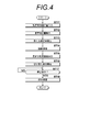
- FIG. 4 is a flowchart showing processing performed by the image display device 16 when a new image of a breast is taken for the same patient.
- Mnew latest breast image
- M0, M1 to Mn for the same patient are read from the image database 14 and input to the image display device 16 (step ST11).
- correction amounts M1 to Mn associated with the breast image M0 are also input to the image display device 16.
- the correction amount calculation unit 22 uses the latest breast image Mnew and the previous latest breast image M0 to match the position and image quality of the breast image M0 with the position and image quality of the latest breast image Mnew.
- the amount of correction is calculated.
- This correction amount serves as a reference for calculating a correction amount for matching the position and image quality of the latest breast image Mnew and breast images M1 to Mn, which will be described later (hereinafter referred to as “reference correction amount H0”).
- FIG. 5 is a diagram for explaining calculation of new correction amounts H1 ′ to Hn ′.
- the breast image M0 is associated with the respective correction amounts H1 to Hn of the breast images M1 to Mn whose photographing date and time is earlier than that.
- the correction amount H1 is for matching the position and image quality of the breast image H1 with the position and image quality of the breast image M0
- the correction amount H2 is the position and image quality of the breast image H2 and the position and image quality of the breast image M0. It is for matching.
- the reference correction amount H0 is a correction amount for matching the position and image quality of the breast image M0 with the position and image quality of the latest breast image Mnew. Therefore, by correcting the correction amounts H1 to Hn using the reference correction amount H0, a new correction for matching the position and image quality of the past breast images M1 to Mn with the position and image quality of the latest breast image Mnew.
- the quantities H1 ′ to Hn ′ can be calculated. Specifically, the new correction amounts H1 ′ to Hn ′ can be calculated by performing a predetermined calculation using (in combination with) the correction amounts H1 to Hn and the reference correction amount H0. For example, as shown in FIG.
- new correction amounts H1 ′ to Hn ′ are calculated by adding the reference correction amount H0 to the correction amounts H1 to Hn.
- the correction amount calculation unit 22 calculates new correction amounts H1 ′ to Hn ′ by correcting the correction amounts H1 to Hn using the reference correction amount H0.
- the image processing unit 24 matches the position and image quality of the breast images M0, M1 to Mn with the position and image quality of the latest breast image Mnew based on the reference correction amount H0 and the new correction amounts H1 ′ to Hn ′.
- Image processing is performed (step ST14).
- the display control unit 26 displays the latest breast image Mnew on the screen of the display unit 20 (step ST15).
- the display control unit 26 starts switching display of breast images Mnew, M0, M1 to Mn (step ST16).
- the display control unit 26 starts monitoring whether or not an instruction to stop image switching display is input from the input unit 30 (step ST17), and ends the switching display when step ST17 is affirmed.
- the storage processing unit 28 stores the reference correction amount H0 and the new correction amounts H1 ′ to Hn ′ calculated by the correction amount calculation unit 22 in the image database 14 in association with the latest breast image Mnew. (Step ST18), and the process ends.
- the correction amounts H1 to Hn associated with the breast image M0 may be stored in the image database 14 in association with the breast image M0 as they are, or may be deleted in order to reduce the data capacity. Also good.
- correction amounts H1 to Hn for matching the position and image quality of the past breast images M1 to Mn with the position and image quality of the latest breast image M0 are used as the past breast images M1 to Mn.
- Each of Mn is calculated, and correction amounts H1 to Hn are stored in association with the latest breast image M0. For this reason, when a new breast image Mnew is acquired, the reference for matching the position and image quality of the breast image M0 whose imaging date and time is next to the new breast image Mnew to the position and image quality of the latest breast image Mnew.
- the correction amount H0 is calculated, the correction amounts H1 to Hn for the past breast images M1 to Mn are corrected by the reference correction amount G0, and new correction amounts H1 ′ to Hn ′ are calculated.
- the reference correction amount H0 and the new correction amount H0 are calculated. It is possible to store the correction amounts H1 ′ to Hn ′ in association with the new breast image Mnew. For this reason, when a new breast image Mnew is input, it is not necessary to calculate a correction amount for the new breast image Mnew and all of the other breast images M0 and M1 to Mn. The amount of calculation for the matching process can be reduced.
- the position and image quality can be matched using the correction amount, it is not necessary to store the breast image whose position and image quality are corrected (storing the correction amount is sufficient). Accordingly, the amount of data to be stored can be reduced, and thus the cost for storing data in the image database 14 can be reduced.
- parameters for rotation, enlargement / reduction, and translation are calculated as the position correction amounts P1 to Pn.
- a parameter for at least one of rotation, enlargement / reduction, and translation is used.
- the position correction amounts P1 to Pn may be calculated.
- the correction amounts for luminance, contrast, sharpness and granularity are calculated as the image quality correction amounts G1 to Gn.
- at least one of luminance, contrast, sharpness and granularity is calculated. May be calculated as image quality correction amounts G1 to Gn.
- the correction amounts H1 to Hn for matching the position and image quality with the breast image M0 are calculated, but at least one of the position and image quality (position and / or image quality) is calculated.
- Correction amounts H1 to Hn for matching may be calculated. That is, any one of the position correction amounts P1 to Pn and the image quality correction amounts G1 to Gn may be calculated.
- the correction amount may be calculated in advance before switching display of a plurality of breast images having different imaging dates and times. Accordingly, it is not necessary to calculate the correction amount when performing the switching display, and thus it is possible to shorten the time until the switching display is started.
- the subject is the breast.
- the subject is not limited to this, and any part of the human body such as the chest, abdomen, head, and extremities can be the subject.
Landscapes
- Engineering & Computer Science (AREA)
- Health & Medical Sciences (AREA)
- Life Sciences & Earth Sciences (AREA)
- Medical Informatics (AREA)
- Physics & Mathematics (AREA)
- Radiology & Medical Imaging (AREA)
- Nuclear Medicine, Radiotherapy & Molecular Imaging (AREA)
- General Health & Medical Sciences (AREA)
- Computer Vision & Pattern Recognition (AREA)
- Theoretical Computer Science (AREA)
- General Physics & Mathematics (AREA)
- Veterinary Medicine (AREA)
- Biomedical Technology (AREA)
- Molecular Biology (AREA)
- Surgery (AREA)
- Animal Behavior & Ethology (AREA)
- Optics & Photonics (AREA)
- Public Health (AREA)
- Pathology (AREA)
- Heart & Thoracic Surgery (AREA)
- Biophysics (AREA)
- High Energy & Nuclear Physics (AREA)
- Quality & Reliability (AREA)
- Apparatus For Radiation Diagnosis (AREA)
- Image Processing (AREA)
- Image Analysis (AREA)
- Dentistry (AREA)
- Oral & Maxillofacial Surgery (AREA)
Abstract
比較読影のために複数の放射線画像の位置合わせ処理および/または画質の補正処理を行う際の演算量および保存のためのコストを低減させる。補正量算出部22が、同一被写体を含む複数の放射線画像のうち、基準放射線画像以外の他の放射線画像の位置および/または画質を、基準放射線画像の位置および/または画質に一致させるための補正量を、他の放射線画像のそれぞれについて算出する。保存処理部28が、他の放射線画像のそれぞれについての補正量を、基準放射線画像と対応づけて保存する処理を行う。
Description
本発明は、乳房等の放射線画像を表示する画像表示装置および方法に関するものである。
被写体の複数の画像をCRTあるいは液晶ディスプレイ等の表示装置に表示させて、画像を比較しながら読影をする比較読影が従来から行われている。例えば、医療診断を目的として、被写体である患者の同一部位についての現在および過去の放射線画像を表示させて比較読影することにより、病変の進行度合いの確認、あるいは異常を早期に発見することができる。また、このような比較読影を行いやすいように、放射線画像を表示装置に表示させる技術が知られている。例えば、特許文献1,2には、比較読影するための複数の放射線画像を、1画面上において切り替えて表示する手法が提案されている。このように比較の対象となる放射線画像を切り替えて表示することにより、変化がある部分の確認が容易となるため、比較読影を容易に行うことができる。また、比較読影の手法として複数の放射線画像の差分を算出する手法も用いられている。このように差分を算出することによっても、変化がある部分の確認が容易となるため、比較読影を容易に行うことができる。
ところで、比較読影を行う複数の放射線画像は、撮影部位のポジショニングが異なったり、撮影時の撮影条件が異なったり、撮影装置が異なったりするため、放射線画像に含まれる被写体の位置あるいは画質が、複数の放射線画像において異なるものとなる場合がある。このような場合、複数の放射線画像を切り替えて表示することによる比較読影を効率よく行うためには、放射線画像に含まれる被写体の位置合わせ処理を行ったり、複数の放射線画像の画質を一致させる画像処理を行ったりする必要がある。このため、特許文献1に記載された手法では、アフィン変換等により複数の放射線画像を位置合わせし、かつ複数の放射線画像の輝度を一致させることが行われている。
上述した比較読影は、最も新しく撮影された最新の放射線画像を過去の放射線画像と比較することを目的とするものである。このため、位置合わせ処理あるいは画質を一定にする処理は、最新の放射線画像を基準として行われる。したがって、過去に取得された放射線画像は、最新の放射線画像と一致するように位置合わせ処理および/または画質の補正処理が行われる。
上述した比較読影は、新たな放射線画像が取得される度に行われる。新たな放射線画像が取得される度に、新たな放射線画像を基準として、すべての過去の放射線画像に対して位置合わせ処理および画質の補正処理を行うことは、演算量が非常に多くなり、処理に長時間を要する。この場合、位置合わせ処理および画質の補正処理を行った放射線画像をすべて保存しておくことにより、最新の放射線画像といずれか1つの放射線画像との位置および画質の補正量を算出すれば、他の放射線画像の位置および画質を補正することができるため、演算時間を短縮することができる。
しかしながら、過去の比較読影にて位置および画質が補正されたすべての放射線画像を保存しておくことは、データ量が膨大なものとなるため、放射線画像を保存しておくためのコストが増大する。
本発明は上記事情に鑑みなされたものであり、比較読影のために複数の放射線画像の位置合わせ処理および/または画質の補正処理を行う際の演算量および保存のためのコストを低減させることを目的とする。
本発明による画像表示装置は、同一被写体を含む複数の放射線画像を比較のために表示する画像表示装置であって、
複数の放射線画像のうち、基準放射線画像以外の他の放射線画像の位置および/または画質を、基準放射線画像の位置および/または画質に一致させるための補正量を、他の放射線画像のそれぞれについて算出する補正量算出手段と、
他の放射線画像のそれぞれについての補正量を、基準放射線画像と対応づけて保存する処理を行う保存処理手段とを備えたことを特徴とする。
複数の放射線画像のうち、基準放射線画像以外の他の放射線画像の位置および/または画質を、基準放射線画像の位置および/または画質に一致させるための補正量を、他の放射線画像のそれぞれについて算出する補正量算出手段と、
他の放射線画像のそれぞれについての補正量を、基準放射線画像と対応づけて保存する処理を行う保存処理手段とを備えたことを特徴とする。
なお、本発明による画像表示装置においては、補正量算出手段を、位置の補正量として、他の放射線画像に含まれる被写体の位置を基準放射線画像に含まれる被写体の位置に一致させるための回転、拡大縮小および平行移動の補正量の少なくとも1つを算出し、画質の補正量として、他の放射線画像の画質を基準放射線画像の画質に一致させるための輝度、コントラスト、鮮鋭度および粒状度の補正量の少なくとも1つを算出してもよい。
また、本発明による画像表示装置においては、基準放射線画像を、複数の放射線画像のうち、最新の放射線画像としてもよい。
また、本発明による画像表示装置においては、補正量に基づいて、他の放射線画像の位置および/または画質を基準放射線画像の位置および/または画質に一致させる画像処理を行う画像処理手段をさらに備えてもよい。
また、本発明による画像表示装置においては、基準放射線画像および補正処理が行われた他の放射線画像を表示手段に切り替え表示する表示制御手段をさらに備えてもよい。
また、本発明による画像表示装置においては、
補正量算出手段を、新たな放射線画像が取得された場合、新たな放射線画像を新たな基準放射線画像に設定し、新たな基準放射線画像の前の基準放射線画像の位置および/または画質を、新たな基準放射線画像の位置および/または画質に一致させるための基準補正量を算出し、基準補正量を用いて、新たな補正量を算出する前に算出した他の放射線画像のそれぞれについての補正量を修正することにより、基準補正量および修正された補正量を含む新たな補正量を算出し、
保存処理手段を、新たな基準放射線画像以外の新たな他の放射線画像のそれぞれについての新たな補正量を、新たな基準放射線画像と対応づけて保存してもよい。
補正量算出手段を、新たな放射線画像が取得された場合、新たな放射線画像を新たな基準放射線画像に設定し、新たな基準放射線画像の前の基準放射線画像の位置および/または画質を、新たな基準放射線画像の位置および/または画質に一致させるための基準補正量を算出し、基準補正量を用いて、新たな補正量を算出する前に算出した他の放射線画像のそれぞれについての補正量を修正することにより、基準補正量および修正された補正量を含む新たな補正量を算出し、
保存処理手段を、新たな基準放射線画像以外の新たな他の放射線画像のそれぞれについての新たな補正量を、新たな基準放射線画像と対応づけて保存してもよい。
本発明による画像表示方法は、同一被写体を含む複数の放射線画像を比較のために表示する画像表示方法であって、
複数の放射線画像のうち、基準放射線画像以外の他の放射線画像の位置および/または画質を、基準放射線画像の位置および/または画質に一致させるための補正量を、他の放射線画像のそれぞれについて算出するステップと、
他の放射線画像のそれぞれについての補正量を、基準放射線画像と対応づけて保存する処理を行うステップとを有することを特徴とする。
複数の放射線画像のうち、基準放射線画像以外の他の放射線画像の位置および/または画質を、基準放射線画像の位置および/または画質に一致させるための補正量を、他の放射線画像のそれぞれについて算出するステップと、
他の放射線画像のそれぞれについての補正量を、基準放射線画像と対応づけて保存する処理を行うステップとを有することを特徴とする。
また、本発明による画像表示方法を、コンピュータに実行させるためのプログラムとして提供してもよい。
本発明によれば、複数の放射線画像のうち、基準放射線画像以外の他の放射線画像の位置および/または画質を、基準放射線画像の位置および/または画質に一致させるための補正量が、他の放射線画像のそれぞれについて算出され、他の放射線画像のそれぞれについての補正量が、基準放射線画像と対応づけられて保存される。このため、新たな放射線画像が取得された場合、以下のような処理が可能となる。
すなわち、新たな放射線画像を新たな基準放射線画像に設定し、新たな基準放射線画像の前の基準放射線画像の位置および/または画質を、新たな基準放射線画像に一致させるための基準補正量を算出する。そして、基準補正量により、新たな補正量を算出する前に算出した他の放射線画像のそれぞれについての補正量を修正することにより、基準補正量および修正された補正量を含む新たな補正量を算出する。さらに、新たな基準放射線画像以外の新たな他の放射線画像のそれぞれについての新たな補正量を、新たな基準放射線画像と対応づけて保存する。このため、本発明によれば、新たな基準放射線画像と他の放射線画像のすべてについての補正量を算出する必要が無くなり、その結果、位置および/または画質を一致させる処理のための演算量を低減することができる。また、補正量のみを保存しておくことにより、位置および/または画質を一致させる処理を行うことができるため、位置および/または画質が補正された放射線画像を保存しておく必要が無くなる。したがって、保存するデータ量を低減することができ、よって、データ保存のためのコストを低減することができる。
以下、図面を参照して本発明の実施形態について説明する。図1は本発明の実施形態による画像表示装置を適用した医療画像支援システムの構成を示す概略ブロック図である。図1に示すように、このシステム10は、医用施設等に設置され、被写体を撮影して被写体の放射線画像を取得する撮影装置12と、撮影装置12で撮影された放射線画像を保存する画像データベース(画像DB)14と、高精細なモニタ(不図示)を有する、放射線画像の読影を行うための画像表示装置16とから構成されている。これらの装置は、ネットワーク18により相互に接続されている。なお、画像表示装置16は医師の読影端末となる。
なお、本実施形態においては、撮影装置12は、被写体である患者の乳房を撮影するマンモグラフィ撮影装置とする。マンモグラフィ撮影装置は、患者の乳房を撮影し、乳房の放射線画像である乳房画像を取得する。取得された乳房画像の画像データは、画像データベース14に保存される。この際、患者名、撮影日等の付帯情報が乳房画像の画像データと対応づけられて画像データベース14に保存される。
図2は画像表示装置の構成を示す概略ブロック図である。図2に示すように、本実施形態による画像表示装置16は、画像を表示するCRTあるいは液晶ディスプレイ等の表示部20と、入力された、同一患者の撮影日時が異なる乳房画像を表す複数の画像データに基づいて、複数の乳房画像における解剖学的構造物の位置を互いに略一致させ、かつ複数の乳房画像の画質を略一致させるための補正量を算出する22と、補正量に基づいて、上記複数の画像データのうち少なくともいずれかに、位置および画質を一致させるための位置合わせ処理および画質補正処理を含む画像処理を施す画像処理部24と、画像処理が施された複数の画像データに基づいて、画像処理済みの複数の乳房画像が表示部20の画面に切り替え表示されるように制御する表示制御部26と、補正量算出部22が算出した補正量を画像データベース14に保存するための処理を行う保存処理部28と、マウス、キーボード等の入力部30とを備える。
以上述べた表示部20、補正量算出部22、画像処理部24、表示制御部26、保存処理部28、および入力部30は、例えば一般的なパーソナルコンピュータ等のコンピュータシステムから構成することができる。
なお、本実施形態において使用する撮影日時が異なる複数の乳房画像は、現時点での最新の乳房画像M0および同一患者について過去に撮影したn枚の乳房画像M1~Mnとする。また、撮影対象の乳房は左右のいずれであってもよい。
補正量算出部22は、過去の乳房画像M1~Mnの位置および画質を、最新の乳房画像M0の位置および画質に一致させるための補正量を算出する。なお、最新の乳房画像M0が基準放射線画像に相当し、過去の乳房画像M1~Mnが他の放射線画像に相当する。まず、位置の補正量の算出について説明する。位置の補正量は、過去の乳房画像M1~Mnの解剖学的構造物が最新の乳房画像M0の解剖学的構造物に一致するように、過去の乳房画像M1~Mnの位置を最新の乳房画像M0の位置に一致させる位置合わせ処理のための補正量である。
ここで、位置合わせの手法としては、例えば、特許文献1、特開平7-262346号公報、特開2010-188003号公報に記載されたアフィン変換を用いる手法が挙げられる。アフィン変換は、画像間において互いに対応する対応点を複数求め、画像間の対応点を一致させるための回転移動、拡大縮小および平行移動のためのパラメータを算出し、このパラメータに基づいて一方の画像を変換して他方の画像と位置合わせする手法である。本実施形態においては、補正量算出部22は、過去の乳房画像M1~Mnのそれぞれについて、最新の乳房画像M0と位置を一致させるためのアフィン変換のパラメータ、すなわち回転移動、拡大縮小および平行移動のためのパラメータを、位置の補正量P1~Pnとして算出する。
また、特開2002-125961号公報に記載された手法を用いて、乳房画像の輝度を基準として乳房画像から乳腺を抽出し、乳腺の重心位置および形状を一致させるように、過去の乳房画像M1~Mnのそれぞれについて、最新の乳房画像M0と位置を一致させるための回転移動、拡大縮小および平行移動のためのパラメータを、位置の補正量として算出してもよい。
また、特開2001-325584号公報、特開2002-324238号公報に記載された手法を用いてもよい。具体的には、特定の構造物(例えば乳房画像におけるニップル)を一致させるように、過去の乳房画像M1~Mnのそれぞれについて、最新の乳房画像M0と位置を一致させるための回転移動、拡大縮小および平行移動のためのパラメータを、位置の補正量として算出してもよい。
また、特開2002-65613公報にて提案されている手法を用いてもよい。具体的には、画像上における乳房の位置を公知の認識技術により認識し、乳房の胸筋側の画像辺の位置と乳頭の位置が2つの画像間でそれぞれ一致するように、一方の画像に対してワーピング処理を施す手法を用いてもよい。この場合、ワーピング処理のパラメータを位置の補正量として算出すればよい。
次いで、画質の補正量について説明する。ここで、画質を補正する理由は、各画像間で画質にばらつきがあると、後述するように画像を切り替え表示する際に、表示された画像を見る際に観察者が見辛くなるからである。画質の補正量は、過去の乳房画像M1~Mnの画質が最新の乳房画像M0の画質に一致するように、過去の乳房画像M1~Mnに対して画質を補正する画像処理を行うための補正量である。本実施形態においては、具体的には、輝度、コントラスト、鮮鋭度および粒状度の補正量を画質の補正量G1~Gnとして算出する。
例えば、補正量算出部22は、過去の乳房画像M1~Mnの画素値の最大値および最小値を、最新の乳房画像M0の画素値の最大値および最小値に一致させるための補正量を、輝度の補正量として算出する。また、過去の乳房画像M1~Mnのコントラストを、最新の乳房画像M0の画素値のコントラストに一致させるための補正量を、コントラストの補正量として算出する。なお、コントラストとしては、画素値の最大値と最小値との差を用いることができる。この場合、コントラスト補正量は、乳房画像の階調を変更するガンマカーブの傾きとなる。
補正量算出部22は、最新の乳房画像M0および過去の乳房画像M1~Mnに対して周波数解析処理を行い、各乳房画像の周波数帯域のレスポンスを鮮鋭度の特性として求め、過去の乳房画像M1~Mnの鮮鋭度を最新の乳房画像M0の鮮鋭度に一致させるための補正量を、鮮鋭度の補正量として算出する。
また、補正量算出部22は、周波数解析により、最新の乳房画像M0および過去の乳房画像M1~Mnに含まれる周波数帯域のレスポンスを粒状度の特性として求め、過去の乳房画像M1~Mnの粒状の周波数帯域のレスポンスを最新の乳房画像M0の粒状の周波数帯域のレスポンスに一致させるための補正量を、粒状度の補正量として算出する。
補正量算出部22は、過去の乳房画像M1~Mnのそれぞれについて算出した、位置の補正量P1~Pnおよび画質の補正量G1~Gnを、位置および画質の補正量H1~Hnとして画像処理部24および保存処理部28に提供する。
画像処理部24は、過去の乳房画像M1~Mnの位置および画質を、最新の乳房画像M0の位置および画質と一致させるための画像処理を、過去の乳房画像M1~Mnに対して行う。すなわち、補正量算出部22が算出した補正量H1~Hnのうち、位置の補正量P1~Pnを用いて、過去の乳房画像M1~Mnの位置を最新の乳房画像M0の位置と一致させる位置合わせ処理を行う。また、画質の補正量G1~Gnを用いて、過去の乳房画像M1~Mnの濃度、コントラスト、鮮鋭度および粒状度を、最新の乳房画像M0の濃度、コントラスト、鮮鋭度および粒状度と一致させる画質補正処理を行う。
表示制御部26は、操作者により入力部30から入力された、画面に表示する乳房画像の切り替えの指示に応答して、画像処理済みの複数の乳房画像を切り替えて表示するように表示部20を制御するものである。なお、切り替えの際には、例えば毎秒1こまから毎秒5こまの間の表示速度とすることができる。
保存処理部28は、補正量算出部22が算出した補正量M1~Mnを、位置および画質を一致させる基準となる基準乳房画像と対応づけて、画像データベース14に保存する処理を行う。なお、本実施形態においては、基準乳房画像は最新の乳房画像M0である。
次いで、本実施形態において行われる処理について説明する。図3は本実施形態において行われる処理を示すフローチャートである。まず、同一患者の最新の乳房画像M0および過去の乳房画像M1~Mnを含む複数の乳房画像が、画像データベース14から読み出されて画像表示装置16に入力される(ステップST1)。
乳房画像M0,M1~Mnが入力されると、補正量算出部22は、過去の乳房画像M1~Mnの位置および画質を、最新の乳房画像M0の位置および画質に一致させるための補正量H1~Hnを算出する(ステップST2)。補正量H1~Hnは画像処理部24および保存処理部28に供給される。画像処理部24は、補正量H1~Hnに基づいて、過去の乳房画像M1~Mnの位置および画質を、最新の乳房画像M0の位置および画質に一致させる画像処理を行う(ステップST3)。
画像処理後、表示制御部26は、乳房画像M0,M1~Mnのうちのいずれか1つを表示部20の画面に表示する(ステップST4)。本実施形態においては、まず最新の乳房画像M0が表示されるものとする。
そして、操作者が、入力部30により画像切り替え表示開始の指示を行うと、表示制御部26は、乳房画像M0,M1~Mnの切り替え表示を開始する(ステップST5)。すなわち、乳房画像M0,M1~Mnを、表示部20の画面上において、所定の表示速度(例えば毎秒1~5こま程度の)で、時系列順(例えば撮影日時が新しい順)に繰返し切り替えて表示させるようにする。これにより、最新の乳房画像M0と過去の乳房画像M1~Mnとが切り替え表示される。
なお、切り替え表示前および切り替え表示中において、例えば、入力部30から入力される情報に基づいて表示速度を変更するようにしてもよい。これにより、操作者は、自身が観察しやすい所望の表示速度に調整することができる。
そして、表示制御部26は、画像の切り替え表示の停止指示が入力部30から入力されたか否かの監視を開始し(ステップST6)、ステップST6が肯定されると切り替え表示を終了する。
次いで、保存処理部28が、補正量算出部22が算出した、過去の乳房画像M1~Mnのそれぞれに対応する補正量H1~Hnを、最新の乳房画像M0と対応づけて画像データベース14に保存する処理を行い(ステップST7)、処理を終了する。なお、保存する処理は、最新の乳房画像M0と、過去の乳房画像M1~Mnのそれぞれに対応する補正量H1~Hnとが一体不可分となるように、最新の乳房画像M0および補正量H1~Hnを画像データベース14に保存する処理である。保存する処理としては、例えば、補正量H1~Hnを最新の乳房画像M0のヘッダに記述する、補正量H1~Hnのファイル名を最新の乳房画像M0のファイル名と関連づける、補正量H1~Hnのファイルを最新の乳房画像M0のファイルと同一フォルダに保存する等が挙げられる。
次いで、同一患者に対して乳房のさらに新たな撮影が行われた場合の処理について説明する。図4は、同一患者に対して乳房のさらに新たな撮影が行われた場合に画像表示装置16が行う処理を示すフローチャートである。乳房の新たな撮影により最新の乳房画像(「Mnew」とする)が撮影装置12により取得されると、最新の乳房画像Mnewは画像データベース14に保存される。そして、画像の切り替え表示時には、同一患者についての最新の乳房画像Mnewおよび乳房画像M0,M1~Mnが画像データベース14から読み出されて、画像表示装置16に入力される(ステップST11)。この際、上述した乳房画像M0に対応づけられた補正量M1~Mnも併せて画像表示装置16に入力される。
補正量算出部22は、まず、最新の乳房画像Mnewと1つ前の最新の乳房画像M0とを用いて、乳房画像M0の位置および画質を最新の乳房画像Mnewの位置および画質と一致させるための補正量を算出する。なお、この補正量は後述する最新の乳房画像Mnewと乳房画像M1~Mnの位置および画質とを一致させるための補正量の算出の基準となる(以降、「基準補正量H0」と称する)(基準補正量算出、ステップST12)。
次いで補正量算出部22は、基準補正量H0を用いて、乳房画像M0と対応づけられた補正量H1~Hnを修正して、新たな補正量H1′~Hn′を算出する(ステップST13)。図5は新たな補正量H1′~Hn′の算出を説明するための図である。図5に示すように、乳房画像M0には、それよりも撮影日時が前の乳房画像M1~Mnのそれぞれの補正量H1~Hnが対応づけられている。ここで、補正量H1は乳房画像H1の位置および画質を乳房画像M0の位置および画質と一致させるためのものであり、補正量H2は乳房画像H2の位置および画質を乳房画像M0の位置および画質と一致させるためのものである。
基準補正量H0は、乳房画像M0の位置および画質を、最新の乳房画像Mnewの位置および画質と一致させるための補正量である。このため、補正量H1~Hnを基準補正量H0を用いて補正することにより、過去の乳房画像M1~Mnの位置および画質を最新の乳房画像Mnewの位置および画質と一致させるための新たな補正量H1′~Hn′を算出することができる。具体的には、新たな補正量H1′~Hn′は、補正量H1~Hnと基準補正量H0とを用いて(組み合わせて)所定の演算を行うことにより算出することができる。例えば、図5に示すように、新たな補正量H1′~Hn′は、補正量H1~Hnに基準補正量H0を加算することで算出される。このように、補正量算出部22は、基準補正量H0を用いて補正量H1~Hnを修正することにより、新たな補正量H1′~Hn′を算出する。
次いで、画像処理部24が、基準補正量H0および新たな補正量H1′~Hn′に基づいて、乳房画像M0,M1~Mnの位置および画質を、最新の乳房画像Mnewの位置および画質に一致させる画像処理を行う(ステップST14)。画像処理後、表示制御部26は、最新の乳房画像Mnewを表示部20の画面に表示する(ステップST15)。そして、操作者が、入力部30により画像切り替え表示開始の指示を行うと、表示制御部26は、乳房画像Mnew,M0,M1~Mnの切り替え表示を開始する(ステップST16)。そして、表示制御部26は、画像の切り替え表示の停止指示が入力部30から入力されたか否かの監視を開始し(ステップST17)、ステップST17が肯定されると切り替え表示を終了する。
次いで、保存処理部28が、補正量算出部22が算出した、基準補正量H0および新たな補正量H1′~Hn′を、最新の乳房画像Mnewと対応づけて画像データベース14に保存する処理を行い(ステップST18)、処理を終了する。なお、乳房画像M0に対応づけられた補正量H1~Hnは、そのまま乳房画像M0と対応づけて画像データベース14に保存しておいてもよいし、データ容量を削減するために削除するようにしてもよい。
このように、本実施形態においては、過去の乳房画像M1~Mnの位置および画質を、最新の乳房画像M0の位置および画質に一致させるための補正量H1~Hnを、過去の乳房画像M1~Mnのそれぞれについて算出し、補正量H1~Hnを最新の乳房画像M0と対応づけて保存するようにしたものである。このため、さらに新たな乳房画像Mnewが取得された場合、新たな乳房画像Mnewの次に撮影日時が新しい乳房画像M0の位置および画質を最新の乳房画像Mnewの位置および画質に一致させるための基準補正量H0を算出し、基準補正量G0により、過去の乳房画像M1~Mnについての補正量H1~Hnを修正して新たな補正量H1′~Hn′を算出し、基準補正量H0および新たな補正量H1′~Hn′を、新たな乳房画像Mnewと対応づけて保存するようにすることができる。このため、新たな乳房画像Mnewが入力された場合に、新たな乳房画像Mnewと他の乳房画像M0,M1~Mnのすべてについての補正量を算出する必要が無くなり、その結果、位置および画質を一致させる処理のための演算量を低減することができる。また、補正量を用いて位置および画質を一致させる処理を行うことができるため、位置および画質が補正された乳房画像を保存しておく必要が無くなる(補正量を保存することで足りる)。したがって、保存するデータ量を低減することができ、よって、画像データベース14においてデータ保存のためのコストを低減することができる。
なお、上記実施形態においては、位置の補正量P1~Pnとして、回転、拡大縮小および平行移動のためのパラメータを算出しているが、回転、拡大縮小および平行移動の少なくとも1つについてのパラメータを、位置の補正量P1~Pnとして算出するようにしてもよい。
また、上記実施形態においては、画質の補正量G1~Gnとして、輝度、コントラスト、鮮鋭度および粒状度の補正量を算出しているが、輝度、コントラスト、鮮鋭度および粒状度の少なくとも1つについての補正量を、画質の補正量G1~Gnとして算出するようにしてもよい。
また、上記実施形態においては、乳房画像M0との位置および画質を一致させるための補正量H1~Hnを算出しているが、位置および画質のうち少なくともいずれか一方(位置および/または画質)を一致させるための補正量H1~Hnを算出するようにしてもよい。すなわち、位置の補正量P1~Pnおよび画質の補正量G1~Gnのいずれかを算出するようにしてもよい。
また、上記実施形態においては、撮影日時が異なる複数の乳房画像の切り替え表示を行う前に、あらかじめ補正量を算出しておくようにしてもよい。これにより、切り替え表示を行う際に、補正量を算出する必要が無くなるため、切り替え表示を開始するまでの時間を短縮できる。
また、上記実施形態においては、乳房を被写体としているが、これに限定されるものではなく、人体の胸部、腹部、頭部および四肢等の任意の部位を被写体とすることが可能である。
また、本発明は、以上の例には限定されず、本発明の要旨を逸脱しない範囲において、各種の改良や変形を行ってもよいことはいうまでもない。
10 医療画像支援システム
12 撮影装置
14 画像データベース
16 画像表示装置
20 表示部
22 補正量算出部
24 画像処理部
26 表示制御部
28 保存処理部
30 入力部
12 撮影装置
14 画像データベース
16 画像表示装置
20 表示部
22 補正量算出部
24 画像処理部
26 表示制御部
28 保存処理部
30 入力部
Claims (7)
- 同一被写体を含む複数の放射線画像を比較のために表示する画像表示装置であって、
前記複数の放射線画像のうち、基準放射線画像以外の他の放射線画像の位置および/または画質を、前記基準放射線画像の位置および/または画質に一致させるための補正量を、前記他の放射線画像のそれぞれについて算出する補正量算出手段と、
前記他の放射線画像のそれぞれについての補正量を、前記基準放射線画像と対応づけて保存する処理を行う保存処理手段と、
を備えたことを特徴とする画像表示装置。
- 前記補正量算出手段は、
前記位置の補正量として、前記他の放射線画像に含まれる被写体の位置を前記基準放射線画像に含まれる被写体の位置に一致させるための回転、拡大縮小および平行移動の補正量の少なくとも1つを算出し、
前記画質の補正量として、前記他の放射線画像の画質を前記基準放射線画像の画質に一致させるための輝度、コントラスト、鮮鋭度および粒状度の補正量の少なくとも1つを算出する請求項1記載の画像表示装置。
- 前記基準放射線画像は、前記複数の放射線画像のうち、最新の放射線画像である請求項1または2記載の画像表示装置。
- 前記補正量に基づいて、前記他の放射線画像の位置および/または画質を前記基準放射線画像の位置および/または画質に一致させる画像処理を行う画像処理手段、
をさらに備えた請求項1から3のいずれか1項記載の画像表示装置。
- 前記基準放射線画像および前記補正処理が行われた前記他の放射線画像を表示手段に切り替え表示する表示制御手段、
をさらに備えた請求項4記載の画像表示装置。
- 前記補正量算出手段は、
新たな放射線画像が取得された場合、該新たな放射線画像を新たな基準放射線画像に設定し、
該新たな基準放射線画像の前の基準放射線画像の位置および/または画質を、該新たな基準放射線画像の位置および/または画質に一致させるための基準補正量を算出し、
該基準補正量を用いて、該新たな補正量を算出する前に算出した前記他の放射線画像のそれぞれについての補正量を修正することにより、前記基準補正量および前記修正された補正量を含む新たな補正量を算出し、
前記保存処理手段は、
前記新たな基準放射線画像以外の新たな他の放射線画像のそれぞれについての新たな補正量を、前記新たな基準放射線画像と対応づけて保存する請求項1から5のいずれか1項記載の画像表示装置。
- 同一被写体を含む複数の放射線画像を比較のために表示する画像表示方法であって、
前記複数の放射線画像のうち、基準放射線画像以外の他の放射線画像の位置および/または画質を、前記基準放射線画像の位置および/または画質に一致させるための補正量を、前記他の放射線画像のそれぞれについて算出するステップと、
前記他の放射線画像のそれぞれについての補正量を、前記基準放射線画像と対応づけて保存する処理を行うステップと、
を有することを特徴とする画像表示方法。
Priority Applications (1)
| Application Number | Priority Date | Filing Date | Title |
|---|---|---|---|
| US14/846,082 US9911064B2 (en) | 2013-03-28 | 2015-09-04 | Image display device and method |
Applications Claiming Priority (2)
| Application Number | Priority Date | Filing Date | Title |
|---|---|---|---|
| JP2013-067793 | 2013-03-28 | ||
| JP2013067793A JP6053012B2 (ja) | 2013-03-28 | 2013-03-28 | 画像表示装置および方法 |
Related Child Applications (1)
| Application Number | Title | Priority Date | Filing Date |
|---|---|---|---|
| US14/846,082 Continuation US9911064B2 (en) | 2013-03-28 | 2015-09-04 | Image display device and method |
Publications (1)
| Publication Number | Publication Date |
|---|---|
| WO2014156444A1 true WO2014156444A1 (ja) | 2014-10-02 |
Family
ID=51623439
Family Applications (1)
| Application Number | Title | Priority Date | Filing Date |
|---|---|---|---|
| PCT/JP2014/054665 Ceased WO2014156444A1 (ja) | 2013-03-28 | 2014-02-26 | 画像表示装置および方法 |
Country Status (3)
| Country | Link |
|---|---|
| US (1) | US9911064B2 (ja) |
| JP (1) | JP6053012B2 (ja) |
| WO (1) | WO2014156444A1 (ja) |
Families Citing this family (6)
| Publication number | Priority date | Publication date | Assignee | Title |
|---|---|---|---|---|
| JP6491011B2 (ja) * | 2015-03-30 | 2019-03-27 | キヤノンメディカルシステムズ株式会社 | 医用画像処理装置および医用画像処理方法 |
| JP6525903B2 (ja) * | 2016-03-09 | 2019-06-05 | 富士フイルム株式会社 | 画像表示装置、方法およびプログラム |
| JP6962025B2 (ja) * | 2017-06-22 | 2021-11-05 | コニカミノルタ株式会社 | 解析装置及び解析システム |
| US10949987B2 (en) * | 2019-05-31 | 2021-03-16 | Mitsubishi Electric Research Laboratories, Inc. | Robust image registration for multiple rigid transformed images |
| JP7209599B2 (ja) | 2019-07-29 | 2023-01-20 | 富士フイルム株式会社 | 画像処理装置、方法およびプログラム |
| JP7203705B2 (ja) | 2019-09-17 | 2023-01-13 | 富士フイルム株式会社 | 画像処理装置、方法およびプログラム、並びに画像表示装置、方法およびプログラム |
Citations (5)
| Publication number | Priority date | Publication date | Assignee | Title |
|---|---|---|---|---|
| JP2003510730A (ja) * | 1999-09-30 | 2003-03-18 | コーニンクレッカ フィリップス エレクトロニクス エヌ ヴィ | 画像処理方法及び画像列における移動対象を追跡するシステム |
| JP2005020719A (ja) * | 2003-06-06 | 2005-01-20 | Fuji Photo Film Co Ltd | 画像読影支援方法及び装置並びにプログラム |
| WO2009104459A1 (ja) * | 2008-02-19 | 2009-08-27 | コニカミノルタエムジー株式会社 | 小規模施設用診断支援装置及びプログラム |
| JP2011125355A (ja) * | 2008-04-10 | 2011-06-30 | Konica Minolta Medical & Graphic Inc | 小規模施設用診断支援装置 |
| JP2011125358A (ja) * | 2009-12-15 | 2011-06-30 | Fujifilm Corp | 医用画像表示装置および方法、並びにプログラム |
Family Cites Families (2)
| Publication number | Priority date | Publication date | Assignee | Title |
|---|---|---|---|---|
| JP2006006435A (ja) | 2004-06-23 | 2006-01-12 | Fuji Photo Film Co Ltd | 画像表示方法および装置並びにプログラム |
| JP5698068B2 (ja) | 2011-05-09 | 2015-04-08 | 富士フイルム株式会社 | 画像処理装置、画像表示システム、放射線画像撮影システム、画像処理プログラム、及び画像処理方法 |
-
2013
- 2013-03-28 JP JP2013067793A patent/JP6053012B2/ja not_active Expired - Fee Related
-
2014
- 2014-02-26 WO PCT/JP2014/054665 patent/WO2014156444A1/ja not_active Ceased
-
2015
- 2015-09-04 US US14/846,082 patent/US9911064B2/en not_active Expired - Fee Related
Patent Citations (5)
| Publication number | Priority date | Publication date | Assignee | Title |
|---|---|---|---|---|
| JP2003510730A (ja) * | 1999-09-30 | 2003-03-18 | コーニンクレッカ フィリップス エレクトロニクス エヌ ヴィ | 画像処理方法及び画像列における移動対象を追跡するシステム |
| JP2005020719A (ja) * | 2003-06-06 | 2005-01-20 | Fuji Photo Film Co Ltd | 画像読影支援方法及び装置並びにプログラム |
| WO2009104459A1 (ja) * | 2008-02-19 | 2009-08-27 | コニカミノルタエムジー株式会社 | 小規模施設用診断支援装置及びプログラム |
| JP2011125355A (ja) * | 2008-04-10 | 2011-06-30 | Konica Minolta Medical & Graphic Inc | 小規模施設用診断支援装置 |
| JP2011125358A (ja) * | 2009-12-15 | 2011-06-30 | Fujifilm Corp | 医用画像表示装置および方法、並びにプログラム |
Also Published As
| Publication number | Publication date |
|---|---|
| US9911064B2 (en) | 2018-03-06 |
| US20150379374A1 (en) | 2015-12-31 |
| JP6053012B2 (ja) | 2016-12-27 |
| JP2014188250A (ja) | 2014-10-06 |
Similar Documents
| Publication | Publication Date | Title |
|---|---|---|
| JP6053012B2 (ja) | 画像表示装置および方法 | |
| JP5314614B2 (ja) | 医用画像表示装置及び医用画像表示方法並びにプログラム | |
| US11676277B2 (en) | Medical image processing apparatus and method | |
| US10692198B2 (en) | Image processing apparatus, image processing method, image processing system, and non-transitory computer-readable storage medium for presenting three-dimensional images | |
| US9704242B2 (en) | Dynamic image processing apparatus and computer-readable recording medium for providing diagnosis support | |
| US20130038629A1 (en) | Method and device for visualizing the registration quality of medical image datasets | |
| JP5698068B2 (ja) | 画像処理装置、画像表示システム、放射線画像撮影システム、画像処理プログラム、及び画像処理方法 | |
| CN105246405A (zh) | 医用图像处理装置以及x射线诊断装置 | |
| JP2018148964A (ja) | 動態解析システム | |
| CN111902071A (zh) | 诊断辅助装置、学习装置、诊断辅助方法、学习方法及程序 | |
| US20060239585A1 (en) | System and method for reducing artifacts in motion corrected dynamic image sequences | |
| US20110122146A1 (en) | Systems and methods for matching medical images | |
| JP2006006435A (ja) | 画像表示方法および装置並びにプログラム | |
| JP2014183876A (ja) | 画像表示装置および方法 | |
| JP4960468B2 (ja) | 磁気共鳴イメージング装置 | |
| JP2001218110A (ja) | 画像間演算方法および装置並びに画像表示方法および装置 | |
| JP2006014931A (ja) | 画像読影支援システム、および、画像位置合わせ装置、画像出力装置、ならびにプログラム | |
| CN101208042A (zh) | 异常阴影候选检测方法、异常阴影候选检测装置 | |
| US10980518B2 (en) | Medical image capturing control device, method, and program | |
| JP5402576B2 (ja) | X線撮影装置およびx線撮影方法 | |
| CN112120789A (zh) | 断骨复位模拟方法和断骨复位模拟装置 | |
| US12033366B2 (en) | Matching apparatus, matching method, and matching program | |
| JP2005136594A (ja) | 画像処理装置及びその制御方法 | |
| JP2021094219A (ja) | 画像処理方法、画像処理装置及びプログラム | |
| US20070286525A1 (en) | Generation of imaging filters based on image analysis |
Legal Events
| Date | Code | Title | Description |
|---|---|---|---|
| 121 | Ep: the epo has been informed by wipo that ep was designated in this application |
Ref document number: 14773619 Country of ref document: EP Kind code of ref document: A1 |
|
| NENP | Non-entry into the national phase |
Ref country code: DE |
|
| 122 | Ep: pct application non-entry in european phase |
Ref document number: 14773619 Country of ref document: EP Kind code of ref document: A1 |