WO2014126198A1 - 抗cdh3ヒト化抗体、その薬剤コンジュゲート、及びそれらの使用 - Google Patents
抗cdh3ヒト化抗体、その薬剤コンジュゲート、及びそれらの使用 Download PDFInfo
- Publication number
- WO2014126198A1 WO2014126198A1 PCT/JP2014/053473 JP2014053473W WO2014126198A1 WO 2014126198 A1 WO2014126198 A1 WO 2014126198A1 JP 2014053473 W JP2014053473 W JP 2014053473W WO 2014126198 A1 WO2014126198 A1 WO 2014126198A1
- Authority
- WO
- WIPO (PCT)
- Prior art keywords
- cdh3
- variable region
- chain variable
- antibody
- humanized antibody
- Prior art date
Links
- 239000003814 drug Substances 0.000 title claims description 67
- 229940079593 drug Drugs 0.000 title description 60
- 101000762242 Homo sapiens Cadherin-15 Proteins 0.000 claims abstract description 110
- 210000004027 cell Anatomy 0.000 claims abstract description 109
- 102100024153 Cadherin-15 Human genes 0.000 claims abstract description 106
- 101000714553 Homo sapiens Cadherin-3 Proteins 0.000 claims abstract description 106
- 206010028980 Neoplasm Diseases 0.000 claims abstract description 71
- 201000011510 cancer Diseases 0.000 claims abstract description 48
- 108010047041 Complementarity Determining Regions Proteins 0.000 claims abstract description 40
- 229940127089 cytotoxic agent Drugs 0.000 claims abstract description 19
- 210000004602 germ cell Anatomy 0.000 claims abstract description 15
- 125000003275 alpha amino acid group Chemical group 0.000 claims description 35
- 235000001014 amino acid Nutrition 0.000 claims description 34
- 230000027455 binding Effects 0.000 claims description 32
- 150000001413 amino acids Chemical class 0.000 claims description 24
- 125000000539 amino acid group Chemical class 0.000 claims description 23
- -1 α-maleimidoacetoxy Chemical group 0.000 claims description 23
- NFGXHKASABOEEW-UHFFFAOYSA-N 1-methylethyl 11-methoxy-3,7,11-trimethyl-2,4-dodecadienoate Chemical compound COC(C)(C)CCCC(C)CC=CC(C)=CC(=O)OC(C)C NFGXHKASABOEEW-UHFFFAOYSA-N 0.000 claims description 21
- 239000002246 antineoplastic agent Substances 0.000 claims description 21
- 230000036961 partial effect Effects 0.000 claims description 19
- 238000006467 substitution reaction Methods 0.000 claims description 19
- 230000001472 cytotoxic effect Effects 0.000 claims description 16
- 206010006187 Breast cancer Diseases 0.000 claims description 15
- 208000026310 Breast neoplasm Diseases 0.000 claims description 15
- ALYNCZNDIQEVRV-UHFFFAOYSA-N 4-aminobenzoic acid Chemical compound NC1=CC=C(C(O)=O)C=C1 ALYNCZNDIQEVRV-UHFFFAOYSA-N 0.000 claims description 13
- 206010058467 Lung neoplasm malignant Diseases 0.000 claims description 13
- 231100000433 cytotoxic Toxicity 0.000 claims description 13
- 239000012634 fragment Substances 0.000 claims description 13
- 201000005202 lung cancer Diseases 0.000 claims description 13
- 208000020816 lung neoplasm Diseases 0.000 claims description 13
- 239000000126 substance Substances 0.000 claims description 12
- 201000010099 disease Diseases 0.000 claims description 10
- 208000037265 diseases, disorders, signs and symptoms Diseases 0.000 claims description 10
- 229940127121 immunoconjugate Drugs 0.000 claims description 10
- 235000004279 alanine Nutrition 0.000 claims description 8
- FPKVOQKZMBDBKP-UHFFFAOYSA-N 1-[4-[(2,5-dioxopyrrol-1-yl)methyl]cyclohexanecarbonyl]oxy-2,5-dioxopyrrolidine-3-sulfonic acid Chemical compound O=C1C(S(=O)(=O)O)CC(=O)N1OC(=O)C1CCC(CN2C(C=CC2=O)=O)CC1 FPKVOQKZMBDBKP-UHFFFAOYSA-N 0.000 claims description 7
- 206010061902 Pancreatic neoplasm Diseases 0.000 claims description 7
- 108010044540 auristatin Proteins 0.000 claims description 7
- 150000002148 esters Chemical class 0.000 claims description 7
- 208000015486 malignant pancreatic neoplasm Diseases 0.000 claims description 7
- 201000002528 pancreatic cancer Diseases 0.000 claims description 7
- 208000008443 pancreatic carcinoma Diseases 0.000 claims description 7
- LLXVXPPXELIDGQ-UHFFFAOYSA-N (2,5-dioxopyrrolidin-1-yl) 3-(2,5-dioxopyrrol-1-yl)benzoate Chemical compound C=1C=CC(N2C(C=CC2=O)=O)=CC=1C(=O)ON1C(=O)CCC1=O LLXVXPPXELIDGQ-UHFFFAOYSA-N 0.000 claims description 6
- BQWBEDSJTMWJAE-UHFFFAOYSA-N (2,5-dioxopyrrolidin-1-yl) 4-[(2-iodoacetyl)amino]benzoate Chemical compound C1=CC(NC(=O)CI)=CC=C1C(=O)ON1C(=O)CCC1=O BQWBEDSJTMWJAE-UHFFFAOYSA-N 0.000 claims description 6
- PMJWDPGOWBRILU-UHFFFAOYSA-N (2,5-dioxopyrrolidin-1-yl) 4-[4-(2,5-dioxopyrrol-1-yl)phenyl]butanoate Chemical compound O=C1CCC(=O)N1OC(=O)CCCC(C=C1)=CC=C1N1C(=O)C=CC1=O PMJWDPGOWBRILU-UHFFFAOYSA-N 0.000 claims description 6
- WCMOHMXWOOBVMZ-UHFFFAOYSA-N (2,5-dioxopyrrolidin-1-yl) 6-[3-(2,5-dioxopyrrol-1-yl)propanoylamino]hexanoate Chemical compound O=C1CCC(=O)N1OC(=O)CCCCCNC(=O)CCN1C(=O)C=CC1=O WCMOHMXWOOBVMZ-UHFFFAOYSA-N 0.000 claims description 6
- 206010009944 Colon cancer Diseases 0.000 claims description 6
- 230000002018 overexpression Effects 0.000 claims description 6
- 239000000941 radioactive substance Substances 0.000 claims description 6
- PVGATNRYUYNBHO-UHFFFAOYSA-N (2,5-dioxopyrrolidin-1-yl) 4-(2,5-dioxopyrrol-1-yl)butanoate Chemical compound O=C1CCC(=O)N1OC(=O)CCCN1C(=O)C=CC1=O PVGATNRYUYNBHO-UHFFFAOYSA-N 0.000 claims description 5
- 239000004472 Lysine Substances 0.000 claims description 5
- KDXKERNSBIXSRK-UHFFFAOYSA-N Lysine Natural products NCCCCC(N)C(O)=O KDXKERNSBIXSRK-UHFFFAOYSA-N 0.000 claims description 5
- KZSNJWFQEVHDMF-UHFFFAOYSA-N Valine Natural products CC(C)C(N)C(O)=O KZSNJWFQEVHDMF-UHFFFAOYSA-N 0.000 claims description 5
- 239000004474 valine Substances 0.000 claims description 5
- MFRNYXJJRJQHNW-DEMKXPNLSA-N (2s)-2-[[(2r,3r)-3-methoxy-3-[(2s)-1-[(3r,4s,5s)-3-methoxy-5-methyl-4-[methyl-[(2s)-3-methyl-2-[[(2s)-3-methyl-2-(methylamino)butanoyl]amino]butanoyl]amino]heptanoyl]pyrrolidin-2-yl]-2-methylpropanoyl]amino]-3-phenylpropanoic acid Chemical compound CN[C@@H](C(C)C)C(=O)N[C@@H](C(C)C)C(=O)N(C)[C@@H]([C@@H](C)CC)[C@H](OC)CC(=O)N1CCC[C@H]1[C@H](OC)[C@@H](C)C(=O)N[C@H](C(O)=O)CC1=CC=CC=C1 MFRNYXJJRJQHNW-DEMKXPNLSA-N 0.000 claims description 4
- FWMNVWWHGCHHJJ-SKKKGAJSSA-N 4-amino-1-[(2r)-6-amino-2-[[(2r)-2-[[(2r)-2-[[(2r)-2-amino-3-phenylpropanoyl]amino]-3-phenylpropanoyl]amino]-4-methylpentanoyl]amino]hexanoyl]piperidine-4-carboxylic acid Chemical compound C([C@H](C(=O)N[C@H](CC(C)C)C(=O)N[C@H](CCCCN)C(=O)N1CCC(N)(CC1)C(O)=O)NC(=O)[C@H](N)CC=1C=CC=CC=1)C1=CC=CC=C1 FWMNVWWHGCHHJJ-SKKKGAJSSA-N 0.000 claims description 4
- 108010021625 Immunoglobulin Fragments Proteins 0.000 claims description 4
- 102000008394 Immunoglobulin Fragments Human genes 0.000 claims description 4
- QNAYBMKLOCPYGJ-REOHCLBHSA-N L-alanine Chemical compound C[C@H](N)C(O)=O QNAYBMKLOCPYGJ-REOHCLBHSA-N 0.000 claims description 4
- 125000003295 alanine group Chemical group N[C@@H](C)C(=O)* 0.000 claims description 4
- 102000048587 human CDH3 Human genes 0.000 claims description 4
- TYKASZBHFXBROF-UHFFFAOYSA-N (2,5-dioxopyrrolidin-1-yl) 2-(2,5-dioxopyrrol-1-yl)acetate Chemical compound O=C1CCC(=O)N1OC(=O)CN1C(=O)C=CC1=O TYKASZBHFXBROF-UHFFFAOYSA-N 0.000 claims description 3
- IHVODYOQUSEYJJ-UHFFFAOYSA-N (2,5-dioxopyrrolidin-1-yl) 6-[[4-[(2,5-dioxopyrrol-1-yl)methyl]cyclohexanecarbonyl]amino]hexanoate Chemical compound O=C1CCC(=O)N1OC(=O)CCCCCNC(=O)C(CC1)CCC1CN1C(=O)C=CC1=O IHVODYOQUSEYJJ-UHFFFAOYSA-N 0.000 claims description 3
- OJQSISYVGFJJBY-UHFFFAOYSA-N 1-(4-isocyanatophenyl)pyrrole-2,5-dione Chemical compound C1=CC(N=C=O)=CC=C1N1C(=O)C=CC1=O OJQSISYVGFJJBY-UHFFFAOYSA-N 0.000 claims description 3
- HBEDKBRARKFPIC-UHFFFAOYSA-N 6-(2,5-dioxopyrrol-1-yl)hexanoic acid;1-hydroxypyrrolidine-2,5-dione Chemical compound ON1C(=O)CCC1=O.OC(=O)CCCCCN1C(=O)C=CC1=O HBEDKBRARKFPIC-UHFFFAOYSA-N 0.000 claims description 3
- 208000010507 Adenocarcinoma of Lung Diseases 0.000 claims description 3
- OMNVYXHOSHNURL-WPRPVWTQSA-N Ala-Phe Chemical compound C[C@H](N)C(=O)N[C@H](C(O)=O)CC1=CC=CC=C1 OMNVYXHOSHNURL-WPRPVWTQSA-N 0.000 claims description 3
- 206010005003 Bladder cancer Diseases 0.000 claims description 3
- 208000001333 Colorectal Neoplasms Diseases 0.000 claims description 3
- 101001099381 Homo sapiens Peroxisomal biogenesis factor 19 Proteins 0.000 claims description 3
- KZSNJWFQEVHDMF-BYPYZUCNSA-N L-valine Chemical compound CC(C)[C@H](N)C(O)=O KZSNJWFQEVHDMF-BYPYZUCNSA-N 0.000 claims description 3
- 206010033128 Ovarian cancer Diseases 0.000 claims description 3
- 206010061535 Ovarian neoplasm Diseases 0.000 claims description 3
- 102100038883 Peroxisomal biogenesis factor 19 Human genes 0.000 claims description 3
- 206010060862 Prostate cancer Diseases 0.000 claims description 3
- 208000000236 Prostatic Neoplasms Diseases 0.000 claims description 3
- 206010039491 Sarcoma Diseases 0.000 claims description 3
- 208000000102 Squamous Cell Carcinoma of Head and Neck Diseases 0.000 claims description 3
- 208000013128 Squamous cell carcinoma of pancreas Diseases 0.000 claims description 3
- 208000034254 Squamous cell carcinoma of the cervix uteri Diseases 0.000 claims description 3
- 208000034331 Squamous cell carcinoma of the stomach Diseases 0.000 claims description 3
- 208000024770 Thyroid neoplasm Diseases 0.000 claims description 3
- IEDXPSOJFSVCKU-HOKPPMCLSA-N [4-[[(2S)-5-(carbamoylamino)-2-[[(2S)-2-[6-(2,5-dioxopyrrolidin-1-yl)hexanoylamino]-3-methylbutanoyl]amino]pentanoyl]amino]phenyl]methyl N-[(2S)-1-[[(2S)-1-[[(3R,4S,5S)-1-[(2S)-2-[(1R,2R)-3-[[(1S,2R)-1-hydroxy-1-phenylpropan-2-yl]amino]-1-methoxy-2-methyl-3-oxopropyl]pyrrolidin-1-yl]-3-methoxy-5-methyl-1-oxoheptan-4-yl]-methylamino]-3-methyl-1-oxobutan-2-yl]amino]-3-methyl-1-oxobutan-2-yl]-N-methylcarbamate Chemical compound CC[C@H](C)[C@@H]([C@@H](CC(=O)N1CCC[C@H]1[C@H](OC)[C@@H](C)C(=O)N[C@H](C)[C@@H](O)c1ccccc1)OC)N(C)C(=O)[C@@H](NC(=O)[C@H](C(C)C)N(C)C(=O)OCc1ccc(NC(=O)[C@H](CCCNC(N)=O)NC(=O)[C@@H](NC(=O)CCCCCN2C(=O)CCC2=O)C(C)C)cc1)C(C)C IEDXPSOJFSVCKU-HOKPPMCLSA-N 0.000 claims description 3
- 108010011559 alanylphenylalanine Proteins 0.000 claims description 3
- 210000004556 brain Anatomy 0.000 claims description 3
- 201000006612 cervical squamous cell carcinoma Diseases 0.000 claims description 3
- 229960002173 citrulline Drugs 0.000 claims description 3
- 201000000493 colon squamous cell carcinoma Diseases 0.000 claims description 3
- 239000003431 cross linking reagent Substances 0.000 claims description 3
- 208000007276 esophageal squamous cell carcinoma Diseases 0.000 claims description 3
- 201000000496 gastric squamous cell carcinoma Diseases 0.000 claims description 3
- 201000010536 head and neck cancer Diseases 0.000 claims description 3
- 208000014829 head and neck neoplasm Diseases 0.000 claims description 3
- 201000000459 head and neck squamous cell carcinoma Diseases 0.000 claims description 3
- 201000005249 lung adenocarcinoma Diseases 0.000 claims description 3
- 201000005243 lung squamous cell carcinoma Diseases 0.000 claims description 3
- 201000001441 melanoma Diseases 0.000 claims description 3
- 208000037819 metastatic cancer Diseases 0.000 claims description 3
- 208000011575 metastatic malignant neoplasm Diseases 0.000 claims description 3
- 208000002154 non-small cell lung carcinoma Diseases 0.000 claims description 3
- 201000008968 osteosarcoma Diseases 0.000 claims description 3
- 201000006691 pancreatic squamous cell carcinoma Diseases 0.000 claims description 3
- 229920000889 poly(m-phenylene isophthalamide) Polymers 0.000 claims description 3
- SLUMFVNAWKPKOT-UHFFFAOYSA-N pyridin-2-ylsulfanyl butanoate Chemical compound C(CCC)(=O)OSC1=NC=CC=C1 SLUMFVNAWKPKOT-UHFFFAOYSA-N 0.000 claims description 3
- NHKZQYJGWDGXMY-UHFFFAOYSA-N pyridin-2-ylsulfanyl pentanoate Chemical compound CCCCC(=O)OSC1=CC=CC=N1 NHKZQYJGWDGXMY-UHFFFAOYSA-N 0.000 claims description 3
- 208000012972 squamous cell carcinoma of colon Diseases 0.000 claims description 3
- 201000002510 thyroid cancer Diseases 0.000 claims description 3
- 208000029729 tumor suppressor gene on chromosome 11 Diseases 0.000 claims description 3
- 206010061534 Oesophageal squamous cell carcinoma Diseases 0.000 claims description 2
- 108091005804 Peptidases Proteins 0.000 claims description 2
- 239000004365 Protease Substances 0.000 claims description 2
- 102100037486 Reverse transcriptase/ribonuclease H Human genes 0.000 claims description 2
- 208000021712 Soft tissue sarcoma Diseases 0.000 claims description 2
- 208000036765 Squamous cell carcinoma of the esophagus Diseases 0.000 claims description 2
- 125000003588 lysine group Chemical group [H]N([H])C([H])([H])C([H])([H])C([H])([H])C([H])([H])C([H])(N([H])[H])C(*)=O 0.000 claims description 2
- 206010041823 squamous cell carcinoma Diseases 0.000 claims description 2
- 125000002987 valine group Chemical group [H]N([H])C([H])(C(*)=O)C([H])(C([H])([H])[H])C([H])([H])[H] 0.000 claims description 2
- NCPQROHLJFARLL-UHFFFAOYSA-N 4-(2,5-dioxopyrrol-1-yl)butanoic acid Chemical compound OC(=O)CCCN1C(=O)C=CC1=O NCPQROHLJFARLL-UHFFFAOYSA-N 0.000 claims 1
- 239000000611 antibody drug conjugate Substances 0.000 abstract description 45
- 229940049595 antibody-drug conjugate Drugs 0.000 abstract description 45
- 230000005847 immunogenicity Effects 0.000 abstract description 7
- 239000002254 cytotoxic agent Substances 0.000 abstract 1
- 230000008021 deposition Effects 0.000 abstract 1
- 241000699666 Mus <mouse, genus> Species 0.000 description 55
- 238000000034 method Methods 0.000 description 39
- 230000014509 gene expression Effects 0.000 description 38
- 108090000623 proteins and genes Proteins 0.000 description 37
- 210000004978 chinese hamster ovary cell Anatomy 0.000 description 27
- 238000012360 testing method Methods 0.000 description 25
- 210000004408 hybridoma Anatomy 0.000 description 24
- 239000000243 solution Substances 0.000 description 23
- 210000001519 tissue Anatomy 0.000 description 23
- 239000013604 expression vector Substances 0.000 description 21
- 239000000427 antigen Substances 0.000 description 19
- 108091007433 antigens Proteins 0.000 description 19
- 102000036639 antigens Human genes 0.000 description 19
- 239000003153 chemical reaction reagent Substances 0.000 description 17
- 125000005647 linker group Chemical group 0.000 description 17
- 238000006243 chemical reaction Methods 0.000 description 16
- 229910052751 metal Inorganic materials 0.000 description 16
- 239000002184 metal Substances 0.000 description 15
- 235000018102 proteins Nutrition 0.000 description 15
- 102000004169 proteins and genes Human genes 0.000 description 15
- 241001465754 Metazoa Species 0.000 description 14
- FAPWRFPIFSIZLT-UHFFFAOYSA-M Sodium chloride Chemical compound [Na+].[Cl-] FAPWRFPIFSIZLT-UHFFFAOYSA-M 0.000 description 14
- 239000002299 complementary DNA Substances 0.000 description 11
- 239000000562 conjugate Substances 0.000 description 11
- 238000001943 fluorescence-activated cell sorting Methods 0.000 description 11
- 239000003053 toxin Substances 0.000 description 11
- 231100000765 toxin Toxicity 0.000 description 11
- 108700012359 toxins Proteins 0.000 description 11
- 239000012228 culture supernatant Substances 0.000 description 10
- 238000000684 flow cytometry Methods 0.000 description 10
- 238000003752 polymerase chain reaction Methods 0.000 description 10
- 230000002285 radioactive effect Effects 0.000 description 10
- 108020004414 DNA Proteins 0.000 description 9
- 230000000692 anti-sense effect Effects 0.000 description 9
- 229960003330 pentetic acid Drugs 0.000 description 9
- KCXVZYZYPLLWCC-UHFFFAOYSA-N EDTA Chemical compound OC(=O)CN(CC(O)=O)CCN(CC(O)=O)CC(O)=O KCXVZYZYPLLWCC-UHFFFAOYSA-N 0.000 description 8
- 238000010367 cloning Methods 0.000 description 8
- 230000003053 immunization Effects 0.000 description 8
- 238000002649 immunization Methods 0.000 description 8
- 239000011780 sodium chloride Substances 0.000 description 8
- 238000001890 transfection Methods 0.000 description 8
- 238000005406 washing Methods 0.000 description 8
- 108091032973 (ribonucleotides)n+m Proteins 0.000 description 7
- 206010035226 Plasma cell myeloma Diseases 0.000 description 7
- 239000000872 buffer Substances 0.000 description 7
- 230000000694 effects Effects 0.000 description 7
- 239000000203 mixture Substances 0.000 description 7
- 201000000050 myeloid neoplasm Diseases 0.000 description 7
- 238000002360 preparation method Methods 0.000 description 7
- 230000009467 reduction Effects 0.000 description 7
- 241000283707 Capra Species 0.000 description 6
- LFQSCWFLJHTTHZ-UHFFFAOYSA-N Ethanol Chemical compound CCO LFQSCWFLJHTTHZ-UHFFFAOYSA-N 0.000 description 6
- 239000012980 RPMI-1640 medium Substances 0.000 description 6
- 239000002671 adjuvant Substances 0.000 description 6
- 230000000259 anti-tumor effect Effects 0.000 description 6
- 210000000628 antibody-producing cell Anatomy 0.000 description 6
- 230000007910 cell fusion Effects 0.000 description 6
- 238000005516 engineering process Methods 0.000 description 6
- 238000002372 labelling Methods 0.000 description 6
- 230000010474 transient expression Effects 0.000 description 6
- QTCHATUWDXOORW-UHFFFAOYSA-N CCCCCCCCCCN.O=C(C=C1)NC1=O Chemical compound CCCCCCCCCCN.O=C(C=C1)NC1=O QTCHATUWDXOORW-UHFFFAOYSA-N 0.000 description 5
- AOJJSUZBOXZQNB-TZSSRYMLSA-N Doxorubicin Chemical compound O([C@H]1C[C@@](O)(CC=2C(O)=C3C(=O)C=4C=CC=C(C=4C(=O)C3=C(O)C=21)OC)C(=O)CO)[C@H]1C[C@H](N)[C@H](O)[C@H](C)O1 AOJJSUZBOXZQNB-TZSSRYMLSA-N 0.000 description 5
- 238000002835 absorbance Methods 0.000 description 5
- 238000011161 development Methods 0.000 description 5
- 239000012091 fetal bovine serum Substances 0.000 description 5
- 238000004519 manufacturing process Methods 0.000 description 5
- 108091008146 restriction endonucleases Proteins 0.000 description 5
- 239000013598 vector Substances 0.000 description 5
- DTRVZAALOQTDSF-UHFFFAOYSA-N 2-[[2-[bis(carboxymethyl)amino]-3-phenylpropyl]-(carboxymethyl)amino]acetic acid Chemical compound OC(=O)CN(CC(O)=O)CC(N(CC(O)=O)CC(O)=O)CC1=CC=CC=C1 DTRVZAALOQTDSF-UHFFFAOYSA-N 0.000 description 4
- QKNYBSVHEMOAJP-UHFFFAOYSA-N 2-amino-2-(hydroxymethyl)propane-1,3-diol;hydron;chloride Chemical compound Cl.OCC(N)(CO)CO QKNYBSVHEMOAJP-UHFFFAOYSA-N 0.000 description 4
- 101100370002 Mus musculus Tnfsf14 gene Proteins 0.000 description 4
- 241000699670 Mus sp. Species 0.000 description 4
- VWQVUPCCIRVNHF-OUBTZVSYSA-N Yttrium-90 Chemical compound [90Y] VWQVUPCCIRVNHF-OUBTZVSYSA-N 0.000 description 4
- 239000006285 cell suspension Substances 0.000 description 4
- 238000010790 dilution Methods 0.000 description 4
- 239000012895 dilution Substances 0.000 description 4
- VQNATVDKACXKTF-XELLLNAOSA-N duocarmycin Chemical compound COC1=C(OC)C(OC)=C2NC(C(=O)N3C4=CC(=O)C5=C([C@@]64C[C@@H]6C3)C=C(N5)C(=O)OC)=CC2=C1 VQNATVDKACXKTF-XELLLNAOSA-N 0.000 description 4
- 230000002163 immunogen Effects 0.000 description 4
- 238000005259 measurement Methods 0.000 description 4
- 239000002609 medium Substances 0.000 description 4
- 108020004999 messenger RNA Proteins 0.000 description 4
- 230000035772 mutation Effects 0.000 description 4
- 239000013642 negative control Substances 0.000 description 4
- 239000000047 product Substances 0.000 description 4
- 238000000746 purification Methods 0.000 description 4
- 230000002829 reductive effect Effects 0.000 description 4
- 239000006152 selective media Substances 0.000 description 4
- LWIHDJKSTIGBAC-UHFFFAOYSA-K tripotassium phosphate Chemical compound [K+].[K+].[K+].[O-]P([O-])([O-])=O LWIHDJKSTIGBAC-UHFFFAOYSA-K 0.000 description 4
- 241000283690 Bos taurus Species 0.000 description 3
- 102000000905 Cadherin Human genes 0.000 description 3
- 108050007957 Cadherin Proteins 0.000 description 3
- 241000725101 Clea Species 0.000 description 3
- OKKJLVBELUTLKV-UHFFFAOYSA-N Methanol Chemical compound OC OKKJLVBELUTLKV-UHFFFAOYSA-N 0.000 description 3
- QPCDCPDFJACHGM-UHFFFAOYSA-N N,N-bis{2-[bis(carboxymethyl)amino]ethyl}glycine Chemical compound OC(=O)CN(CC(O)=O)CCN(CC(=O)O)CCN(CC(O)=O)CC(O)=O QPCDCPDFJACHGM-UHFFFAOYSA-N 0.000 description 3
- 241000283973 Oryctolagus cuniculus Species 0.000 description 3
- 238000011579 SCID mouse model Methods 0.000 description 3
- XSQUKJJJFZCRTK-UHFFFAOYSA-N Urea Natural products NC(N)=O XSQUKJJJFZCRTK-UHFFFAOYSA-N 0.000 description 3
- 230000001093 anti-cancer Effects 0.000 description 3
- 229940125644 antibody drug Drugs 0.000 description 3
- 230000008033 biological extinction Effects 0.000 description 3
- 230000015572 biosynthetic process Effects 0.000 description 3
- 230000000903 blocking effect Effects 0.000 description 3
- 239000006143 cell culture medium Substances 0.000 description 3
- 210000000170 cell membrane Anatomy 0.000 description 3
- 208000029742 colonic neoplasm Diseases 0.000 description 3
- 238000007796 conventional method Methods 0.000 description 3
- 108091092330 cytoplasmic RNA Proteins 0.000 description 3
- 230000004069 differentiation Effects 0.000 description 3
- 239000012153 distilled water Substances 0.000 description 3
- 108020001507 fusion proteins Proteins 0.000 description 3
- 102000037865 fusion proteins Human genes 0.000 description 3
- 238000011534 incubation Methods 0.000 description 3
- APFVFJFRJDLVQX-AHCXROLUSA-N indium-111 Chemical compound [111In] APFVFJFRJDLVQX-AHCXROLUSA-N 0.000 description 3
- 230000000670 limiting effect Effects 0.000 description 3
- OHSVLFRHMCKCQY-NJFSPNSNSA-N lutetium-177 Chemical compound [177Lu] OHSVLFRHMCKCQY-NJFSPNSNSA-N 0.000 description 3
- 239000000463 material Substances 0.000 description 3
- 150000002739 metals Chemical class 0.000 description 3
- 238000012986 modification Methods 0.000 description 3
- 230000004048 modification Effects 0.000 description 3
- 108010059074 monomethylauristatin F Proteins 0.000 description 3
- 239000013612 plasmid Substances 0.000 description 3
- 108090000765 processed proteins & peptides Proteins 0.000 description 3
- 238000011002 quantification Methods 0.000 description 3
- KZUNJOHGWZRPMI-AKLPVKDBSA-N samarium-153 Chemical compound [153Sm] KZUNJOHGWZRPMI-AKLPVKDBSA-N 0.000 description 3
- 210000004989 spleen cell Anatomy 0.000 description 3
- 238000010186 staining Methods 0.000 description 3
- 238000003786 synthesis reaction Methods 0.000 description 3
- 230000008685 targeting Effects 0.000 description 3
- 230000001988 toxicity Effects 0.000 description 3
- 231100000419 toxicity Toxicity 0.000 description 3
- 239000012096 transfection reagent Substances 0.000 description 3
- 210000003462 vein Anatomy 0.000 description 3
- XLYOFNOQVPJJNP-UHFFFAOYSA-N water Chemical compound O XLYOFNOQVPJJNP-UHFFFAOYSA-N 0.000 description 3
- 238000001262 western blot Methods 0.000 description 3
- IAKHMKGGTNLKSZ-INIZCTEOSA-N (S)-colchicine Chemical compound C1([C@@H](NC(C)=O)CC2)=CC(=O)C(OC)=CC=C1C1=C2C=C(OC)C(OC)=C1OC IAKHMKGGTNLKSZ-INIZCTEOSA-N 0.000 description 2
- YXIWHUQXZSMYRE-UHFFFAOYSA-N 1,3-benzothiazole-2-thiol Chemical compound C1=CC=C2SC(S)=NC2=C1 YXIWHUQXZSMYRE-UHFFFAOYSA-N 0.000 description 2
- WUAPFZMCVAUBPE-NJFSPNSNSA-N 188Re Chemical compound [188Re] WUAPFZMCVAUBPE-NJFSPNSNSA-N 0.000 description 2
- JHALWMSZGCVVEM-UHFFFAOYSA-N 2-[4,7-bis(carboxymethyl)-1,4,7-triazonan-1-yl]acetic acid Chemical compound OC(=O)CN1CCN(CC(O)=O)CCN(CC(O)=O)CC1 JHALWMSZGCVVEM-UHFFFAOYSA-N 0.000 description 2
- STQGQHZAVUOBTE-UHFFFAOYSA-N 7-Cyan-hept-2t-en-4,6-diinsaeure Natural products C1=2C(O)=C3C(=O)C=4C(OC)=CC=CC=4C(=O)C3=C(O)C=2CC(O)(C(C)=O)CC1OC1CC(N)C(O)C(C)O1 STQGQHZAVUOBTE-UHFFFAOYSA-N 0.000 description 2
- 239000012103 Alexa Fluor 488 Substances 0.000 description 2
- FERIUCNNQQJTOY-UHFFFAOYSA-N Butyric acid Chemical compound CCCC(O)=O FERIUCNNQQJTOY-UHFFFAOYSA-N 0.000 description 2
- AGBRGZJNYKTYJJ-UHFFFAOYSA-N CCCCCN.O=C(C=C1)NC1=O Chemical compound CCCCCN.O=C(C=C1)NC1=O AGBRGZJNYKTYJJ-UHFFFAOYSA-N 0.000 description 2
- HEDRZPFGACZZDS-UHFFFAOYSA-N Chloroform Chemical compound ClC(Cl)Cl HEDRZPFGACZZDS-UHFFFAOYSA-N 0.000 description 2
- VYZAMTAEIAYCRO-BJUDXGSMSA-N Chromium-51 Chemical compound [51Cr] VYZAMTAEIAYCRO-BJUDXGSMSA-N 0.000 description 2
- GUTLYIVDDKVIGB-OUBTZVSYSA-N Cobalt-60 Chemical compound [60Co] GUTLYIVDDKVIGB-OUBTZVSYSA-N 0.000 description 2
- 108091035707 Consensus sequence Proteins 0.000 description 2
- 235000005956 Cosmos caudatus Nutrition 0.000 description 2
- 208000037845 Cutaneous squamous cell carcinoma Diseases 0.000 description 2
- 102000012410 DNA Ligases Human genes 0.000 description 2
- 108010061982 DNA Ligases Proteins 0.000 description 2
- 230000004544 DNA amplification Effects 0.000 description 2
- 102000004190 Enzymes Human genes 0.000 description 2
- 108090000790 Enzymes Proteins 0.000 description 2
- GYHNNYVSQQEPJS-OIOBTWANSA-N Gallium-67 Chemical compound [67Ga] GYHNNYVSQQEPJS-OIOBTWANSA-N 0.000 description 2
- GYHNNYVSQQEPJS-YPZZEJLDSA-N Gallium-68 Chemical compound [68Ga] GYHNNYVSQQEPJS-YPZZEJLDSA-N 0.000 description 2
- 101000935587 Homo sapiens Flavin reductase (NADPH) Proteins 0.000 description 2
- 108060003951 Immunoglobulin Proteins 0.000 description 2
- XEEYBQQBJWHFJM-AKLPVKDBSA-N Iron-59 Chemical compound [59Fe] XEEYBQQBJWHFJM-AKLPVKDBSA-N 0.000 description 2
- PEEHTFAAVSWFBL-UHFFFAOYSA-N Maleimide Chemical compound O=C1NC(=O)C=C1 PEEHTFAAVSWFBL-UHFFFAOYSA-N 0.000 description 2
- NWIBSHFKIJFRCO-WUDYKRTCSA-N Mytomycin Chemical compound C1N2C(C(C(C)=C(N)C3=O)=O)=C3[C@@H](COC(N)=O)[C@@]2(OC)[C@@H]2[C@H]1N2 NWIBSHFKIJFRCO-WUDYKRTCSA-N 0.000 description 2
- ZDZOTLJHXYCWBA-VCVYQWHSSA-N N-debenzoyl-N-(tert-butoxycarbonyl)-10-deacetyltaxol Chemical compound O([C@H]1[C@H]2[C@@](C([C@H](O)C3=C(C)[C@@H](OC(=O)[C@H](O)[C@@H](NC(=O)OC(C)(C)C)C=4C=CC=CC=4)C[C@]1(O)C3(C)C)=O)(C)[C@@H](O)C[C@H]1OC[C@]12OC(=O)C)C(=O)C1=CC=CC=C1 ZDZOTLJHXYCWBA-VCVYQWHSSA-N 0.000 description 2
- 108091028043 Nucleic acid sequence Proteins 0.000 description 2
- 229930012538 Paclitaxel Natural products 0.000 description 2
- 102000003992 Peroxidases Human genes 0.000 description 2
- ISWSIDIOOBJBQZ-UHFFFAOYSA-N Phenol Chemical compound OC1=CC=CC=C1 ISWSIDIOOBJBQZ-UHFFFAOYSA-N 0.000 description 2
- 239000002202 Polyethylene glycol Substances 0.000 description 2
- 108010076504 Protein Sorting Signals Proteins 0.000 description 2
- 108010039491 Ricin Proteins 0.000 description 2
- UIIMBOGNXHQVGW-UHFFFAOYSA-M Sodium bicarbonate Chemical compound [Na+].OC([O-])=O UIIMBOGNXHQVGW-UHFFFAOYSA-M 0.000 description 2
- JVHROZDXPAUZFK-UHFFFAOYSA-N TETA Chemical compound OC(=O)CN1CCCN(CC(O)=O)CCN(CC(O)=O)CCCN(CC(O)=O)CC1 JVHROZDXPAUZFK-UHFFFAOYSA-N 0.000 description 2
- 241000700605 Viruses Species 0.000 description 2
- 108010084455 Zeocin Proteins 0.000 description 2
- 208000009956 adenocarcinoma Diseases 0.000 description 2
- 230000010056 antibody-dependent cellular cytotoxicity Effects 0.000 description 2
- 230000009286 beneficial effect Effects 0.000 description 2
- JCXGWMGPZLAOME-AKLPVKDBSA-N bismuth-212 Chemical compound [212Bi] JCXGWMGPZLAOME-AKLPVKDBSA-N 0.000 description 2
- 210000004369 blood Anatomy 0.000 description 2
- 239000008280 blood Substances 0.000 description 2
- 230000037396 body weight Effects 0.000 description 2
- 229930195731 calicheamicin Natural products 0.000 description 2
- HXCHCVDVKSCDHU-LULTVBGHSA-N calicheamicin Chemical compound C1[C@H](OC)[C@@H](NCC)CO[C@H]1O[C@H]1[C@H](O[C@@H]2C\3=C(NC(=O)OC)C(=O)C[C@](C/3=C/CSSSC)(O)C#C\C=C/C#C2)O[C@H](C)[C@@H](NO[C@@H]2O[C@H](C)[C@@H](SC(=O)C=3C(=C(OC)C(O[C@H]4[C@@H]([C@H](OC)[C@@H](O)[C@H](C)O4)O)=C(I)C=3C)OC)[C@@H](O)C2)[C@@H]1O HXCHCVDVKSCDHU-LULTVBGHSA-N 0.000 description 2
- 229910002091 carbon monoxide Inorganic materials 0.000 description 2
- 239000013522 chelant Substances 0.000 description 2
- MYSWGUAQZAJSOK-UHFFFAOYSA-N ciprofloxacin Chemical compound C12=CC(N3CCNCC3)=C(F)C=C2C(=O)C(C(=O)O)=CN1C1CC1 MYSWGUAQZAJSOK-UHFFFAOYSA-N 0.000 description 2
- 230000002860 competitive effect Effects 0.000 description 2
- RYGMFSIKBFXOCR-IGMARMGPSA-N copper-64 Chemical compound [64Cu] RYGMFSIKBFXOCR-IGMARMGPSA-N 0.000 description 2
- 238000012258 culturing Methods 0.000 description 2
- 230000003013 cytotoxicity Effects 0.000 description 2
- 231100000135 cytotoxicity Toxicity 0.000 description 2
- 231100000263 cytotoxicity test Toxicity 0.000 description 2
- 229960000975 daunorubicin Drugs 0.000 description 2
- STQGQHZAVUOBTE-VGBVRHCVSA-N daunorubicin Chemical compound O([C@H]1C[C@@](O)(CC=2C(O)=C3C(=O)C=4C=CC=C(C=4C(=O)C3=C(O)C=21)OC)C(C)=O)[C@H]1C[C@H](N)[C@H](O)[C@H](C)O1 STQGQHZAVUOBTE-VGBVRHCVSA-N 0.000 description 2
- 230000002950 deficient Effects 0.000 description 2
- 230000001419 dependent effect Effects 0.000 description 2
- 238000011033 desalting Methods 0.000 description 2
- 238000013461 design Methods 0.000 description 2
- 229940039227 diagnostic agent Drugs 0.000 description 2
- 239000000032 diagnostic agent Substances 0.000 description 2
- 238000003113 dilution method Methods 0.000 description 2
- 229960004679 doxorubicin Drugs 0.000 description 2
- 229960005501 duocarmycin Drugs 0.000 description 2
- 229930184221 duocarmycin Natural products 0.000 description 2
- 238000004520 electroporation Methods 0.000 description 2
- 125000000524 functional group Chemical group 0.000 description 2
- 230000004927 fusion Effects 0.000 description 2
- LNTHITQWFMADLM-UHFFFAOYSA-N gallic acid Chemical compound OC(=O)C1=CC(O)=C(O)C(O)=C1 LNTHITQWFMADLM-UHFFFAOYSA-N 0.000 description 2
- 239000000499 gel Substances 0.000 description 2
- 238000002523 gelfiltration Methods 0.000 description 2
- 238000010353 genetic engineering Methods 0.000 description 2
- 230000012010 growth Effects 0.000 description 2
- 230000036541 health Effects 0.000 description 2
- 230000001900 immune effect Effects 0.000 description 2
- 238000003018 immunoassay Methods 0.000 description 2
- 102000018358 immunoglobulin Human genes 0.000 description 2
- 238000012744 immunostaining Methods 0.000 description 2
- 230000002779 inactivation Effects 0.000 description 2
- 229910052738 indium Inorganic materials 0.000 description 2
- APFVFJFRJDLVQX-UHFFFAOYSA-N indium atom Chemical group [In] APFVFJFRJDLVQX-UHFFFAOYSA-N 0.000 description 2
- 210000003734 kidney Anatomy 0.000 description 2
- WABPQHHGFIMREM-BKFZFHPZSA-N lead-212 Chemical compound [212Pb] WABPQHHGFIMREM-BKFZFHPZSA-N 0.000 description 2
- 210000001165 lymph node Anatomy 0.000 description 2
- 125000005439 maleimidyl group Chemical group C1(C=CC(N1*)=O)=O 0.000 description 2
- 239000003550 marker Substances 0.000 description 2
- WKPWGQKGSOKKOO-RSFHAFMBSA-N maytansine Chemical compound CO[C@@H]([C@@]1(O)C[C@](OC(=O)N1)([C@H]([C@@H]1O[C@@]1(C)[C@@H](OC(=O)[C@H](C)N(C)C(C)=O)CC(=O)N1C)C)[H])\C=C\C=C(C)\CC2=CC(OC)=C(Cl)C1=C2 WKPWGQKGSOKKOO-RSFHAFMBSA-N 0.000 description 2
- 230000007721 medicinal effect Effects 0.000 description 2
- GLVAUDGFNGKCSF-UHFFFAOYSA-N mercaptopurine Chemical compound S=C1NC=NC2=C1NC=N2 GLVAUDGFNGKCSF-UHFFFAOYSA-N 0.000 description 2
- 231100000065 noncytotoxic Toxicity 0.000 description 2
- 230000002020 noncytotoxic effect Effects 0.000 description 2
- 238000012758 nuclear staining Methods 0.000 description 2
- 239000002773 nucleotide Substances 0.000 description 2
- 125000003729 nucleotide group Chemical group 0.000 description 2
- 229960001592 paclitaxel Drugs 0.000 description 2
- 108040007629 peroxidase activity proteins Proteins 0.000 description 2
- CWCMIVBLVUHDHK-ZSNHEYEWSA-N phleomycin D1 Chemical compound N([C@H](C(=O)N[C@H](C)[C@@H](O)[C@H](C)C(=O)N[C@@H]([C@H](O)C)C(=O)NCCC=1SC[C@@H](N=1)C=1SC=C(N=1)C(=O)NCCCCNC(N)=N)[C@@H](O[C@H]1[C@H]([C@@H](O)[C@H](O)[C@H](CO)O1)O[C@@H]1[C@H]([C@@H](OC(N)=O)[C@H](O)[C@@H](CO)O1)O)C=1N=CNC=1)C(=O)C1=NC([C@H](CC(N)=O)NC[C@H](N)C(N)=O)=NC(N)=C1C CWCMIVBLVUHDHK-ZSNHEYEWSA-N 0.000 description 2
- 229920001223 polyethylene glycol Polymers 0.000 description 2
- 229910000160 potassium phosphate Inorganic materials 0.000 description 2
- 235000011009 potassium phosphates Nutrition 0.000 description 2
- 231100000654 protein toxin Toxicity 0.000 description 2
- 230000005180 public health Effects 0.000 description 2
- WQGWDDDVZFFDIG-UHFFFAOYSA-N pyrogallol Chemical compound OC1=CC=CC(O)=C1O WQGWDDDVZFFDIG-UHFFFAOYSA-N 0.000 description 2
- 230000009257 reactivity Effects 0.000 description 2
- 230000002441 reversible effect Effects 0.000 description 2
- WUAPFZMCVAUBPE-IGMARMGPSA-N rhenium-186 Chemical compound [186Re] WUAPFZMCVAUBPE-IGMARMGPSA-N 0.000 description 2
- 102200090720 rs137852501 Human genes 0.000 description 2
- 102200067034 rs80356913 Human genes 0.000 description 2
- KJTLSVCANCCWHF-NJFSPNSNSA-N ruthenium-103 Chemical compound [103Ru] KJTLSVCANCCWHF-NJFSPNSNSA-N 0.000 description 2
- 238000012216 screening Methods 0.000 description 2
- 201000010106 skin squamous cell carcinoma Diseases 0.000 description 2
- 239000007790 solid phase Substances 0.000 description 2
- 239000002904 solvent Substances 0.000 description 2
- RCINICONZNJXQF-MZXODVADSA-N taxol Chemical compound O([C@@H]1[C@@]2(C[C@@H](C(C)=C(C2(C)C)[C@H](C([C@]2(C)[C@@H](O)C[C@H]3OC[C@]3([C@H]21)OC(C)=O)=O)OC(=O)C)OC(=O)[C@H](O)[C@@H](NC(=O)C=1C=CC=CC=1)C=1C=CC=CC=1)O)C(=O)C1=CC=CC=C1 RCINICONZNJXQF-MZXODVADSA-N 0.000 description 2
- 230000001225 therapeutic effect Effects 0.000 description 2
- UMGDCJDMYOKAJW-UHFFFAOYSA-N thiourea Chemical compound NC(N)=S UMGDCJDMYOKAJW-UHFFFAOYSA-N 0.000 description 2
- 238000012546 transfer Methods 0.000 description 2
- WOWDZACBATWTAU-FEFUEGSOSA-N (2s)-2-[[(2s)-2-(dimethylamino)-3-methylbutanoyl]amino]-n-[(3r,4s,5s)-1-[(2s)-2-[(1r,2r)-3-[[(1s,2r)-1-hydroxy-1-phenylpropan-2-yl]amino]-1-methoxy-2-methyl-3-oxopropyl]pyrrolidin-1-yl]-3-methoxy-5-methyl-1-oxoheptan-4-yl]-n,3-dimethylbutanamide Chemical compound CC(C)[C@H](N(C)C)C(=O)N[C@@H](C(C)C)C(=O)N(C)[C@@H]([C@@H](C)CC)[C@H](OC)CC(=O)N1CCC[C@H]1[C@H](OC)[C@@H](C)C(=O)N[C@H](C)[C@@H](O)C1=CC=CC=C1 WOWDZACBATWTAU-FEFUEGSOSA-N 0.000 description 1
- MCEHFIXEKNKSRW-LBPRGKRZSA-N (2s)-2-[[3,5-dichloro-4-[(2,4-diaminopteridin-6-yl)methyl-methylamino]benzoyl]amino]pentanedioic acid Chemical compound C=1N=C2N=C(N)N=C(N)C2=NC=1CN(C)C1=C(Cl)C=C(C(=O)N[C@@H](CCC(O)=O)C(O)=O)C=C1Cl MCEHFIXEKNKSRW-LBPRGKRZSA-N 0.000 description 1
- OOIBFPKQHULHSQ-UHFFFAOYSA-N (3-hydroxy-1-adamantyl) 2-methylprop-2-enoate Chemical compound C1C(C2)CC3CC2(O)CC1(OC(=O)C(=C)C)C3 OOIBFPKQHULHSQ-UHFFFAOYSA-N 0.000 description 1
- YZBBUYKPTHDZHF-KNVGNIICSA-N (3R)-7,2'-dihydroxy-4'-methoxyisoflavanol Chemical compound OC1=CC(OC)=CC=C1[C@H]1C(O)C2=CC=C(O)C=C2OC1 YZBBUYKPTHDZHF-KNVGNIICSA-N 0.000 description 1
- INAUWOVKEZHHDM-PEDBPRJASA-N (7s,9s)-6,9,11-trihydroxy-9-(2-hydroxyacetyl)-7-[(2r,4s,5s,6s)-5-hydroxy-6-methyl-4-morpholin-4-yloxan-2-yl]oxy-4-methoxy-8,10-dihydro-7h-tetracene-5,12-dione;hydrochloride Chemical compound Cl.N1([C@H]2C[C@@H](O[C@@H](C)[C@H]2O)O[C@H]2C[C@@](O)(CC=3C(O)=C4C(=O)C=5C=CC=C(C=5C(=O)C4=C(O)C=32)OC)C(=O)CO)CCOCC1 INAUWOVKEZHHDM-PEDBPRJASA-N 0.000 description 1
- QBPPRVHXOZRESW-UHFFFAOYSA-N 1,4,7,10-tetraazacyclododecane Chemical compound C1CNCCNCCNCCN1 QBPPRVHXOZRESW-UHFFFAOYSA-N 0.000 description 1
- MDAXKAUIABOHTD-UHFFFAOYSA-N 1,4,8,11-tetraazacyclotetradecane Chemical compound C1CNCCNCCCNCCNC1 MDAXKAUIABOHTD-UHFFFAOYSA-N 0.000 description 1
- NBYLBWHHTUWMER-UHFFFAOYSA-N 2-Methylquinolin-8-ol Chemical compound C1=CC=C(O)C2=NC(C)=CC=C21 NBYLBWHHTUWMER-UHFFFAOYSA-N 0.000 description 1
- URDCARMUOSMFFI-UHFFFAOYSA-N 2-[2-[bis(carboxymethyl)amino]ethyl-(2-hydroxyethyl)amino]acetic acid Chemical compound OCCN(CC(O)=O)CCN(CC(O)=O)CC(O)=O URDCARMUOSMFFI-UHFFFAOYSA-N 0.000 description 1
- YGDVXSDNEFDTGV-UHFFFAOYSA-N 2-[6-[bis(carboxymethyl)amino]hexyl-(carboxymethyl)amino]acetic acid Chemical compound OC(=O)CN(CC(O)=O)CCCCCCN(CC(O)=O)CC(O)=O YGDVXSDNEFDTGV-UHFFFAOYSA-N 0.000 description 1
- RTQWWZBSTRGEAV-PKHIMPSTSA-N 2-[[(2s)-2-[bis(carboxymethyl)amino]-3-[4-(methylcarbamoylamino)phenyl]propyl]-[2-[bis(carboxymethyl)amino]propyl]amino]acetic acid Chemical compound CNC(=O)NC1=CC=C(C[C@@H](CN(CC(C)N(CC(O)=O)CC(O)=O)CC(O)=O)N(CC(O)=O)CC(O)=O)C=C1 RTQWWZBSTRGEAV-PKHIMPSTSA-N 0.000 description 1
- VHMWJVAFPCGUTJ-UHFFFAOYSA-N 2-[[2-[bis(carboxymethyl)amino]-3-(4-isothiocyanatophenyl)propyl]-(carboxymethyl)amino]acetic acid Chemical compound OC(=O)CN(CC(O)=O)CC(N(CC(O)=O)CC(O)=O)CC1=CC=C(N=C=S)C=C1 VHMWJVAFPCGUTJ-UHFFFAOYSA-N 0.000 description 1
- RPBJIOJIMOUOIN-UHFFFAOYSA-N 2-[[3-(4-aminophenyl)-2-[bis(carboxymethyl)amino]propyl]-(carboxymethyl)amino]acetic acid Chemical compound NC1=CC=C(CC(CN(CC(O)=O)CC(O)=O)N(CC(O)=O)CC(O)=O)C=C1 RPBJIOJIMOUOIN-UHFFFAOYSA-N 0.000 description 1
- WYMDDFRYORANCC-UHFFFAOYSA-N 2-[[3-[bis(carboxymethyl)amino]-2-hydroxypropyl]-(carboxymethyl)amino]acetic acid Chemical compound OC(=O)CN(CC(O)=O)CC(O)CN(CC(O)=O)CC(O)=O WYMDDFRYORANCC-UHFFFAOYSA-N 0.000 description 1
- BFSVOASYOCHEOV-UHFFFAOYSA-N 2-diethylaminoethanol Chemical compound CCN(CC)CCO BFSVOASYOCHEOV-UHFFFAOYSA-N 0.000 description 1
- YIMDLWDNDGKDTJ-QLKYHASDSA-N 3'-deamino-3'-(3-cyanomorpholin-4-yl)doxorubicin Chemical compound N1([C@H]2C[C@@H](O[C@@H](C)[C@H]2O)O[C@H]2C[C@@](O)(CC=3C(O)=C4C(=O)C=5C=CC=C(C=5C(=O)C4=C(O)C=32)OC)C(=O)CO)CCOCC1C#N YIMDLWDNDGKDTJ-QLKYHASDSA-N 0.000 description 1
- NDMPLJNOPCLANR-UHFFFAOYSA-N 3,4-dihydroxy-15-(4-hydroxy-18-methoxycarbonyl-5,18-seco-ibogamin-18-yl)-16-methoxy-1-methyl-6,7-didehydro-aspidospermidine-3-carboxylic acid methyl ester Natural products C1C(CC)(O)CC(CC2(C(=O)OC)C=3C(=CC4=C(C56C(C(C(O)C7(CC)C=CCN(C67)CC5)(O)C(=O)OC)N4C)C=3)OC)CN1CCC1=C2NC2=CC=CC=C12 NDMPLJNOPCLANR-UHFFFAOYSA-N 0.000 description 1
- IWTIBPIVCKUAHK-UHFFFAOYSA-N 3-[bis(2-carboxyethyl)amino]propanoic acid Chemical compound OC(=O)CCN(CCC(O)=O)CCC(O)=O IWTIBPIVCKUAHK-UHFFFAOYSA-N 0.000 description 1
- TVZGACDUOSZQKY-LBPRGKRZSA-N 4-aminofolic acid Chemical compound C1=NC2=NC(N)=NC(N)=C2N=C1CNC1=CC=C(C(=O)N[C@@H](CCC(O)=O)C(O)=O)C=C1 TVZGACDUOSZQKY-LBPRGKRZSA-N 0.000 description 1
- LGZKGOGODCLQHG-CYBMUJFWSA-N 5-[(2r)-2-hydroxy-2-(3,4,5-trimethoxyphenyl)ethyl]-2-methoxyphenol Chemical compound C1=C(O)C(OC)=CC=C1C[C@@H](O)C1=CC(OC)=C(OC)C(OC)=C1 LGZKGOGODCLQHG-CYBMUJFWSA-N 0.000 description 1
- AIEMSTCGCMIJTI-UHFFFAOYSA-N 6-oxo-6-phenylhexanoic acid Chemical compound OC(=O)CCCCC(=O)C1=CC=CC=C1 AIEMSTCGCMIJTI-UHFFFAOYSA-N 0.000 description 1
- WUWFMDMBOJLQIV-UHFFFAOYSA-N 7-(3-aminopyrrolidin-1-yl)-1-(2,4-difluorophenyl)-6-fluoro-4-oxo-1,4-dihydro-1,8-naphthyridine-3-carboxylic acid Chemical compound C1C(N)CCN1C(C(=C1)F)=NC2=C1C(=O)C(C(O)=O)=CN2C1=CC=C(F)C=C1F WUWFMDMBOJLQIV-UHFFFAOYSA-N 0.000 description 1
- FJHBVJOVLFPMQE-QFIPXVFZSA-N 7-Ethyl-10-Hydroxy-Camptothecin Chemical compound C1=C(O)C=C2C(CC)=C(CN3C(C4=C([C@@](C(=O)OC4)(O)CC)C=C33)=O)C3=NC2=C1 FJHBVJOVLFPMQE-QFIPXVFZSA-N 0.000 description 1
- 239000005725 8-Hydroxyquinoline Substances 0.000 description 1
- GSDSWSVVBLHKDQ-UHFFFAOYSA-N 9-fluoro-3-methyl-10-(4-methylpiperazin-1-yl)-7-oxo-2,3-dihydro-7H-[1,4]oxazino[2,3,4-ij]quinoline-6-carboxylic acid Chemical compound FC1=CC(C(C(C(O)=O)=C2)=O)=C3N2C(C)COC3=C1N1CCN(C)CC1 GSDSWSVVBLHKDQ-UHFFFAOYSA-N 0.000 description 1
- 102220554159 APC membrane recruitment protein 1_E55A_mutation Human genes 0.000 description 1
- 108010066676 Abrin Proteins 0.000 description 1
- 108010077805 Bacterial Proteins Proteins 0.000 description 1
- 108091003079 Bovine Serum Albumin Proteins 0.000 description 1
- 208000003170 Bronchiolo-Alveolar Adenocarcinoma Diseases 0.000 description 1
- 206010058354 Bronchioloalveolar carcinoma Diseases 0.000 description 1
- NJYLEDQOPAAPQD-UHFFFAOYSA-N C(CC)N.C1(C=CC(N1)=O)=O Chemical compound C(CC)N.C1(C=CC(N1)=O)=O NJYLEDQOPAAPQD-UHFFFAOYSA-N 0.000 description 1
- AUJXLBOHYWTPFV-BLWRDSOESA-N CS[C@H]1SC[C@H]2N(C)C(=O)[C@@H](C)NC(=O)[C@H](COC(=O)[C@@H](C(C)C)N(C)C(=O)[C@@H]1N(C)C(=O)[C@@H](C)NC(=O)[C@H](COC(=O)[C@@H](C(C)C)N(C)C2=O)NC(=O)c1cnc2ccccc2n1)NC(=O)c1cnc2ccccc2n1 Chemical compound CS[C@H]1SC[C@H]2N(C)C(=O)[C@@H](C)NC(=O)[C@H](COC(=O)[C@@H](C(C)C)N(C)C(=O)[C@@H]1N(C)C(=O)[C@@H](C)NC(=O)[C@H](COC(=O)[C@@H](C(C)C)N(C)C2=O)NC(=O)c1cnc2ccccc2n1)NC(=O)c1cnc2ccccc2n1 AUJXLBOHYWTPFV-BLWRDSOESA-N 0.000 description 1
- OYPRJOBELJOOCE-UHFFFAOYSA-N Calcium Chemical compound [Ca] OYPRJOBELJOOCE-UHFFFAOYSA-N 0.000 description 1
- KLWPJMFMVPTNCC-UHFFFAOYSA-N Camptothecin Natural products CCC1(O)C(=O)OCC2=C1C=C3C4Nc5ccccc5C=C4CN3C2=O KLWPJMFMVPTNCC-UHFFFAOYSA-N 0.000 description 1
- 208000005623 Carcinogenesis Diseases 0.000 description 1
- 201000009030 Carcinoma Diseases 0.000 description 1
- 241000282693 Cercopithecidae Species 0.000 description 1
- UHDGCWIWMRVCDJ-CCXZUQQUSA-N Cytarabine Chemical compound O=C1N=C(N)C=CN1[C@H]1[C@@H](O)[C@H](O)[C@@H](CO)O1 UHDGCWIWMRVCDJ-CCXZUQQUSA-N 0.000 description 1
- 108010092160 Dactinomycin Proteins 0.000 description 1
- 102100024746 Dihydrofolate reductase Human genes 0.000 description 1
- 108010053187 Diphtheria Toxin Proteins 0.000 description 1
- 102000016607 Diphtheria Toxin Human genes 0.000 description 1
- 101800000585 Diphtheria toxin fragment A Proteins 0.000 description 1
- 102220552085 Docking protein 1_Q55A_mutation Human genes 0.000 description 1
- 239000006144 Dulbecco’s modified Eagle's medium Substances 0.000 description 1
- 108010009858 Echinomycin Proteins 0.000 description 1
- 241000282326 Felis catus Species 0.000 description 1
- GHASVSINZRGABV-UHFFFAOYSA-N Fluorouracil Chemical compound FC1=CNC(=O)NC1=O GHASVSINZRGABV-UHFFFAOYSA-N 0.000 description 1
- 108700004714 Gelonium multiflorum GEL Proteins 0.000 description 1
- 229920002527 Glycogen Polymers 0.000 description 1
- 101000840258 Homo sapiens Immunoglobulin J chain Proteins 0.000 description 1
- 101000998953 Homo sapiens Immunoglobulin heavy variable 1-2 Proteins 0.000 description 1
- 101001008255 Homo sapiens Immunoglobulin kappa variable 1D-8 Proteins 0.000 description 1
- 101001047628 Homo sapiens Immunoglobulin kappa variable 2-29 Proteins 0.000 description 1
- 101001008321 Homo sapiens Immunoglobulin kappa variable 2D-26 Proteins 0.000 description 1
- 101001047619 Homo sapiens Immunoglobulin kappa variable 3-20 Proteins 0.000 description 1
- 101001008263 Homo sapiens Immunoglobulin kappa variable 3D-15 Proteins 0.000 description 1
- VEXZGXHMUGYJMC-UHFFFAOYSA-N Hydrochloric acid Chemical compound Cl VEXZGXHMUGYJMC-UHFFFAOYSA-N 0.000 description 1
- 102000006496 Immunoglobulin Heavy Chains Human genes 0.000 description 1
- 108010019476 Immunoglobulin Heavy Chains Proteins 0.000 description 1
- 102000013463 Immunoglobulin Light Chains Human genes 0.000 description 1
- 108010065825 Immunoglobulin Light Chains Proteins 0.000 description 1
- 102100036887 Immunoglobulin heavy variable 1-2 Human genes 0.000 description 1
- 102100022964 Immunoglobulin kappa variable 3-20 Human genes 0.000 description 1
- 102100034343 Integrase Human genes 0.000 description 1
- FBOZXECLQNJBKD-ZDUSSCGKSA-N L-methotrexate Chemical compound C=1N=C2N=C(N)N=C(N)C2=NC=1CN(C)C1=CC=C(C(=O)N[C@@H](CCC(O)=O)C(O)=O)C=C1 FBOZXECLQNJBKD-ZDUSSCGKSA-N 0.000 description 1
- 229930126263 Maytansine Natural products 0.000 description 1
- 102000018697 Membrane Proteins Human genes 0.000 description 1
- 108010052285 Membrane Proteins Proteins 0.000 description 1
- HYFMSAFINFJTFH-UHFFFAOYSA-N Mitomycin-A Natural products O=C1C(OC)=C(C)C(=O)C2=C1C(COC(N)=O)C1(OC)N2CC2NC21 HYFMSAFINFJTFH-UHFFFAOYSA-N 0.000 description 1
- 241000711408 Murine respirovirus Species 0.000 description 1
- 101100278853 Mus musculus Dhfr gene Proteins 0.000 description 1
- FONIWJIDLJEJTL-UHFFFAOYSA-N N(8)-acetylspermidine Chemical compound CC(=O)NCCCCNCCCN FONIWJIDLJEJTL-UHFFFAOYSA-N 0.000 description 1
- FXHOOIRPVKKKFG-UHFFFAOYSA-N N,N-Dimethylacetamide Chemical compound CN(C)C(C)=O FXHOOIRPVKKKFG-UHFFFAOYSA-N 0.000 description 1
- FSVCELGFZIQNCK-UHFFFAOYSA-N N,N-bis(2-hydroxyethyl)glycine Chemical compound OCCN(CCO)CC(O)=O FSVCELGFZIQNCK-UHFFFAOYSA-N 0.000 description 1
- JYXGIOKAKDAARW-UHFFFAOYSA-N N-(2-hydroxyethyl)iminodiacetic acid Chemical compound OCCN(CC(O)=O)CC(O)=O JYXGIOKAKDAARW-UHFFFAOYSA-N 0.000 description 1
- 102000001068 Neural Cell Adhesion Molecules Human genes 0.000 description 1
- 108010069196 Neural Cell Adhesion Molecules Proteins 0.000 description 1
- 108091034117 Oligonucleotide Proteins 0.000 description 1
- YYVFXSYQSOZCOQ-UHFFFAOYSA-N Oxyquinoline sulfate Chemical compound [O-]S([O-])(=O)=O.C1=C[NH+]=C2C(O)=CC=CC2=C1.C1=C[NH+]=C2C(O)=CC=CC2=C1 YYVFXSYQSOZCOQ-UHFFFAOYSA-N 0.000 description 1
- 241000609499 Palicourea Species 0.000 description 1
- ABLZXFCXXLZCGV-UHFFFAOYSA-N Phosphorous acid Chemical compound OP(O)=O ABLZXFCXXLZCGV-UHFFFAOYSA-N 0.000 description 1
- 108010064851 Plant Proteins Proteins 0.000 description 1
- 229920001213 Polysorbate 20 Polymers 0.000 description 1
- 108010029485 Protein Isoforms Proteins 0.000 description 1
- 102000001708 Protein Isoforms Human genes 0.000 description 1
- 241000589516 Pseudomonas Species 0.000 description 1
- 108010092799 RNA-directed DNA polymerase Proteins 0.000 description 1
- 108010084592 Saporins Proteins 0.000 description 1
- 239000012506 Sephacryl® Substances 0.000 description 1
- 229920002684 Sepharose Polymers 0.000 description 1
- 239000012505 Superdex™ Substances 0.000 description 1
- PZBFGYYEXUXCOF-UHFFFAOYSA-N TCEP Chemical compound OC(=O)CCP(CCC(O)=O)CCC(O)=O PZBFGYYEXUXCOF-UHFFFAOYSA-N 0.000 description 1
- WDLRUFUQRNWCPK-UHFFFAOYSA-N Tetraxetan Chemical compound OC(=O)CN1CCN(CC(O)=O)CCN(CC(O)=O)CCN(CC(O)=O)CC1 WDLRUFUQRNWCPK-UHFFFAOYSA-N 0.000 description 1
- 239000007983 Tris buffer Substances 0.000 description 1
- 102000004243 Tubulin Human genes 0.000 description 1
- 108090000704 Tubulin Proteins 0.000 description 1
- JXLYSJRDGCGARV-WWYNWVTFSA-N Vinblastine Natural products O=C(O[C@H]1[C@](O)(C(=O)OC)[C@@H]2N(C)c3c(cc(c(OC)c3)[C@]3(C(=O)OC)c4[nH]c5c(c4CCN4C[C@](O)(CC)C[C@H](C3)C4)cccc5)[C@@]32[C@H]2[C@@]1(CC)C=CCN2CC3)C JXLYSJRDGCGARV-WWYNWVTFSA-N 0.000 description 1
- LJFFDOBFKICLHN-IXWHRVGISA-N [(1S,2R,3S,5S,6S,16E,18E,20R,21S)-11-chloro-21-hydroxy-12,20-dimethoxy-2,5,9,16-tetramethyl-8,23-dioxo-4,24-dioxa-9,22-diazatetracyclo[19.3.1.110,14.03,5]hexacosa-10,12,14(26),16,18-pentaen-6-yl] (2S)-2-[methyl(4-sulfanylpentanoyl)amino]propanoate Chemical compound CO[C@@H]([C@@]1(O)C[C@H](OC(=O)N1)[C@@H](C)[C@@H]1O[C@@]1(C)[C@@H](OC(=O)[C@H](C)N(C)C(=O)CCC(C)S)CC(=O)N1C)\C=C\C=C(C)\CC2=CC(OC)=C(Cl)C1=C2 LJFFDOBFKICLHN-IXWHRVGISA-N 0.000 description 1
- JLCPHMBAVCMARE-UHFFFAOYSA-N [3-[[3-[[3-[[3-[[3-[[3-[[3-[[3-[[3-[[3-[[3-[[5-(2-amino-6-oxo-1H-purin-9-yl)-3-[[3-[[3-[[3-[[3-[[3-[[5-(2-amino-6-oxo-1H-purin-9-yl)-3-[[5-(2-amino-6-oxo-1H-purin-9-yl)-3-hydroxyoxolan-2-yl]methoxy-hydroxyphosphoryl]oxyoxolan-2-yl]methoxy-hydroxyphosphoryl]oxy-5-(5-methyl-2,4-dioxopyrimidin-1-yl)oxolan-2-yl]methoxy-hydroxyphosphoryl]oxy-5-(6-aminopurin-9-yl)oxolan-2-yl]methoxy-hydroxyphosphoryl]oxy-5-(6-aminopurin-9-yl)oxolan-2-yl]methoxy-hydroxyphosphoryl]oxy-5-(6-aminopurin-9-yl)oxolan-2-yl]methoxy-hydroxyphosphoryl]oxy-5-(6-aminopurin-9-yl)oxolan-2-yl]methoxy-hydroxyphosphoryl]oxyoxolan-2-yl]methoxy-hydroxyphosphoryl]oxy-5-(5-methyl-2,4-dioxopyrimidin-1-yl)oxolan-2-yl]methoxy-hydroxyphosphoryl]oxy-5-(4-amino-2-oxopyrimidin-1-yl)oxolan-2-yl]methoxy-hydroxyphosphoryl]oxy-5-(5-methyl-2,4-dioxopyrimidin-1-yl)oxolan-2-yl]methoxy-hydroxyphosphoryl]oxy-5-(5-methyl-2,4-dioxopyrimidin-1-yl)oxolan-2-yl]methoxy-hydroxyphosphoryl]oxy-5-(6-aminopurin-9-yl)oxolan-2-yl]methoxy-hydroxyphosphoryl]oxy-5-(6-aminopurin-9-yl)oxolan-2-yl]methoxy-hydroxyphosphoryl]oxy-5-(4-amino-2-oxopyrimidin-1-yl)oxolan-2-yl]methoxy-hydroxyphosphoryl]oxy-5-(4-amino-2-oxopyrimidin-1-yl)oxolan-2-yl]methoxy-hydroxyphosphoryl]oxy-5-(4-amino-2-oxopyrimidin-1-yl)oxolan-2-yl]methoxy-hydroxyphosphoryl]oxy-5-(6-aminopurin-9-yl)oxolan-2-yl]methoxy-hydroxyphosphoryl]oxy-5-(4-amino-2-oxopyrimidin-1-yl)oxolan-2-yl]methyl [5-(6-aminopurin-9-yl)-2-(hydroxymethyl)oxolan-3-yl] hydrogen phosphate Polymers Cc1cn(C2CC(OP(O)(=O)OCC3OC(CC3OP(O)(=O)OCC3OC(CC3O)n3cnc4c3nc(N)[nH]c4=O)n3cnc4c3nc(N)[nH]c4=O)C(COP(O)(=O)OC3CC(OC3COP(O)(=O)OC3CC(OC3COP(O)(=O)OC3CC(OC3COP(O)(=O)OC3CC(OC3COP(O)(=O)OC3CC(OC3COP(O)(=O)OC3CC(OC3COP(O)(=O)OC3CC(OC3COP(O)(=O)OC3CC(OC3COP(O)(=O)OC3CC(OC3COP(O)(=O)OC3CC(OC3COP(O)(=O)OC3CC(OC3COP(O)(=O)OC3CC(OC3COP(O)(=O)OC3CC(OC3COP(O)(=O)OC3CC(OC3COP(O)(=O)OC3CC(OC3COP(O)(=O)OC3CC(OC3COP(O)(=O)OC3CC(OC3CO)n3cnc4c(N)ncnc34)n3ccc(N)nc3=O)n3cnc4c(N)ncnc34)n3ccc(N)nc3=O)n3ccc(N)nc3=O)n3ccc(N)nc3=O)n3cnc4c(N)ncnc34)n3cnc4c(N)ncnc34)n3cc(C)c(=O)[nH]c3=O)n3cc(C)c(=O)[nH]c3=O)n3ccc(N)nc3=O)n3cc(C)c(=O)[nH]c3=O)n3cnc4c3nc(N)[nH]c4=O)n3cnc4c(N)ncnc34)n3cnc4c(N)ncnc34)n3cnc4c(N)ncnc34)n3cnc4c(N)ncnc34)O2)c(=O)[nH]c1=O JLCPHMBAVCMARE-UHFFFAOYSA-N 0.000 description 1
- MKBUQYWFFBCMFG-UHFFFAOYSA-N acetic acid propane-1,1-diamine Chemical compound CC(O)=O.CC(O)=O.CC(O)=O.CC(O)=O.CCC(N)N MKBUQYWFFBCMFG-UHFFFAOYSA-N 0.000 description 1
- RUSUZAGBORAKPY-UHFFFAOYSA-N acetic acid;n'-[2-(2-aminoethylamino)ethyl]ethane-1,2-diamine Chemical compound CC(O)=O.CC(O)=O.CC(O)=O.CC(O)=O.CC(O)=O.CC(O)=O.NCCNCCNCCN RUSUZAGBORAKPY-UHFFFAOYSA-N 0.000 description 1
- 239000002253 acid Substances 0.000 description 1
- 229930183665 actinomycin Natural products 0.000 description 1
- 230000009471 action Effects 0.000 description 1
- 239000004480 active ingredient Substances 0.000 description 1
- 230000002411 adverse Effects 0.000 description 1
- 238000001042 affinity chromatography Methods 0.000 description 1
- 238000000246 agarose gel electrophoresis Methods 0.000 description 1
- SHGAZHPCJJPHSC-YCNIQYBTSA-N all-trans-retinoic acid Chemical compound OC(=O)\C=C(/C)\C=C\C=C(/C)\C=C\C1=C(C)CCCC1(C)C SHGAZHPCJJPHSC-YCNIQYBTSA-N 0.000 description 1
- WNROFYMDJYEPJX-UHFFFAOYSA-K aluminium hydroxide Chemical compound [OH-].[OH-].[OH-].[Al+3] WNROFYMDJYEPJX-UHFFFAOYSA-K 0.000 description 1
- 125000003277 amino group Chemical group 0.000 description 1
- 229960003896 aminopterin Drugs 0.000 description 1
- 230000003321 amplification Effects 0.000 description 1
- 210000004102 animal cell Anatomy 0.000 description 1
- 229940027991 antiseptic and disinfectant quinoline derivative Drugs 0.000 description 1
- 238000003556 assay Methods 0.000 description 1
- LFYJSSARVMHQJB-QIXNEVBVSA-N bakuchiol Chemical compound CC(C)=CCC[C@@](C)(C=C)\C=C\C1=CC=C(O)C=C1 LFYJSSARVMHQJB-QIXNEVBVSA-N 0.000 description 1
- 230000008901 benefit Effects 0.000 description 1
- 125000001797 benzyl group Chemical group [H]C1=C([H])C([H])=C(C([H])=C1[H])C([H])([H])* 0.000 description 1
- 239000011230 binding agent Substances 0.000 description 1
- JCXGWMGPZLAOME-RNFDNDRNSA-N bismuth-213 Chemical compound [213Bi] JCXGWMGPZLAOME-RNFDNDRNSA-N 0.000 description 1
- 210000004899 c-terminal region Anatomy 0.000 description 1
- 229910052791 calcium Inorganic materials 0.000 description 1
- 239000011575 calcium Substances 0.000 description 1
- 239000001506 calcium phosphate Substances 0.000 description 1
- 229910000389 calcium phosphate Inorganic materials 0.000 description 1
- 235000011010 calcium phosphates Nutrition 0.000 description 1
- 229940127093 camptothecin Drugs 0.000 description 1
- VSJKWCGYPAHWDS-FQEVSTJZSA-N camptothecin Chemical compound C1=CC=C2C=C(CN3C4=CC5=C(C3=O)COC(=O)[C@]5(O)CC)C4=NC2=C1 VSJKWCGYPAHWDS-FQEVSTJZSA-N 0.000 description 1
- 230000036952 cancer formation Effects 0.000 description 1
- 239000012830 cancer therapeutic Substances 0.000 description 1
- 239000004202 carbamide Substances 0.000 description 1
- 125000003178 carboxy group Chemical group [H]OC(*)=O 0.000 description 1
- 231100000504 carcinogenesis Toxicity 0.000 description 1
- 229930188550 carminomycin Natural products 0.000 description 1
- XREUEWVEMYWFFA-CSKJXFQVSA-N carminomycin Chemical compound C1[C@H](N)[C@H](O)[C@H](C)O[C@H]1O[C@@H]1C2=C(O)C(C(=O)C3=C(O)C=CC=C3C3=O)=C3C(O)=C2C[C@@](O)(C(C)=O)C1 XREUEWVEMYWFFA-CSKJXFQVSA-N 0.000 description 1
- XREUEWVEMYWFFA-UHFFFAOYSA-N carminomycin I Natural products C1C(N)C(O)C(C)OC1OC1C2=C(O)C(C(=O)C3=C(O)C=CC=C3C3=O)=C3C(O)=C2CC(O)(C(C)=O)C1 XREUEWVEMYWFFA-UHFFFAOYSA-N 0.000 description 1
- 229950001725 carubicin Drugs 0.000 description 1
- 101150059702 cdh-3 gene Proteins 0.000 description 1
- 230000021164 cell adhesion Effects 0.000 description 1
- 238000004113 cell culture Methods 0.000 description 1
- 230000010261 cell growth Effects 0.000 description 1
- 210000003855 cell nucleus Anatomy 0.000 description 1
- 230000004663 cell proliferation Effects 0.000 description 1
- 238000001516 cell proliferation assay Methods 0.000 description 1
- 238000005119 centrifugation Methods 0.000 description 1
- 239000003638 chemical reducing agent Substances 0.000 description 1
- 239000003795 chemical substances by application Substances 0.000 description 1
- IPPWILKGXFOXHO-UHFFFAOYSA-N chloranilic acid Chemical compound OC1=C(Cl)C(=O)C(O)=C(Cl)C1=O IPPWILKGXFOXHO-UHFFFAOYSA-N 0.000 description 1
- 239000003593 chromogenic compound Substances 0.000 description 1
- 229960003405 ciprofloxacin Drugs 0.000 description 1
- 230000007012 clinical effect Effects 0.000 description 1
- GUTLYIVDDKVIGB-YPZZEJLDSA-N cobalt-57 Chemical compound [57Co] GUTLYIVDDKVIGB-YPZZEJLDSA-N 0.000 description 1
- GUTLYIVDDKVIGB-BJUDXGSMSA-N cobalt-58 Chemical compound [58Co] GUTLYIVDDKVIGB-BJUDXGSMSA-N 0.000 description 1
- 229960001338 colchicine Drugs 0.000 description 1
- 238000004040 coloring Methods 0.000 description 1
- LGZKGOGODCLQHG-UHFFFAOYSA-N combretastatin Natural products C1=C(O)C(OC)=CC=C1CC(O)C1=CC(OC)=C(OC)C(OC)=C1 LGZKGOGODCLQHG-UHFFFAOYSA-N 0.000 description 1
- 230000009918 complex formation Effects 0.000 description 1
- 239000000306 component Substances 0.000 description 1
- 238000012790 confirmation Methods 0.000 description 1
- 238000011254 conventional chemotherapy Methods 0.000 description 1
- RYGMFSIKBFXOCR-AKLPVKDBSA-N copper-67 Chemical compound [67Cu] RYGMFSIKBFXOCR-AKLPVKDBSA-N 0.000 description 1
- XUJNEKJLAYXESH-UHFFFAOYSA-N cysteine Natural products SCC(N)C(O)=O XUJNEKJLAYXESH-UHFFFAOYSA-N 0.000 description 1
- 235000018417 cysteine Nutrition 0.000 description 1
- 210000000805 cytoplasm Anatomy 0.000 description 1
- 230000001086 cytosolic effect Effects 0.000 description 1
- 230000006378 damage Effects 0.000 description 1
- 230000034994 death Effects 0.000 description 1
- 238000010586 diagram Methods 0.000 description 1
- 108020001096 dihydrofolate reductase Proteins 0.000 description 1
- 238000007865 diluting Methods 0.000 description 1
- 239000003085 diluting agent Substances 0.000 description 1
- 229930187817 disorazole Natural products 0.000 description 1
- VSJKWCGYPAHWDS-UHFFFAOYSA-N dl-camptothecin Natural products C1=CC=C2C=C(CN3C4=CC5=C(C3=O)COC(=O)C5(O)CC)C4=NC2=C1 VSJKWCGYPAHWDS-UHFFFAOYSA-N 0.000 description 1
- 229960003668 docetaxel Drugs 0.000 description 1
- AMRJKAQTDDKMCE-UHFFFAOYSA-N dolastatin Chemical compound CC(C)C(N(C)C)C(=O)NC(C(C)C)C(=O)N(C)C(C(C)C)C(OC)CC(=O)N1CCCC1C(OC)C(C)C(=O)NC(C=1SC=CN=1)CC1=CC=CC=C1 AMRJKAQTDDKMCE-UHFFFAOYSA-N 0.000 description 1
- 229930188854 dolastatin Natural products 0.000 description 1
- 231100000673 dose–response relationship Toxicity 0.000 description 1
- 239000003937 drug carrier Substances 0.000 description 1
- KBQHZAAAGSGFKK-NJFSPNSNSA-N dysprosium-165 Chemical compound [165Dy] KBQHZAAAGSGFKK-NJFSPNSNSA-N 0.000 description 1
- 239000002158 endotoxin Substances 0.000 description 1
- 230000002708 enhancing effect Effects 0.000 description 1
- 229960002549 enoxacin Drugs 0.000 description 1
- IDYZIJYBMGIQMJ-UHFFFAOYSA-N enoxacin Chemical compound N1=C2N(CC)C=C(C(O)=O)C(=O)C2=CC(F)=C1N1CCNCC1 IDYZIJYBMGIQMJ-UHFFFAOYSA-N 0.000 description 1
- YJGVMLPVUAXIQN-UHFFFAOYSA-N epipodophyllotoxin Natural products COC1=C(OC)C(OC)=CC(C2C3=CC=4OCOC=4C=C3C(O)C3C2C(OC3)=O)=C1 YJGVMLPVUAXIQN-UHFFFAOYSA-N 0.000 description 1
- 229930013356 epothilone Natural products 0.000 description 1
- 150000003883 epothilone derivatives Chemical class 0.000 description 1
- 201000006608 esophagus squamous cell carcinoma Diseases 0.000 description 1
- FRPJXPJMRWBBIH-RBRWEJTLSA-N estramustine Chemical compound ClCCN(CCCl)C(=O)OC1=CC=C2[C@H]3CC[C@](C)([C@H](CC4)O)[C@@H]4[C@@H]3CCC2=C1 FRPJXPJMRWBBIH-RBRWEJTLSA-N 0.000 description 1
- 229960001842 estramustine Drugs 0.000 description 1
- DEFVIWRASFVYLL-UHFFFAOYSA-N ethylene glycol bis(2-aminoethyl)tetraacetic acid Chemical compound OC(=O)CN(CC(O)=O)CCOCCOCCN(CC(O)=O)CC(O)=O DEFVIWRASFVYLL-UHFFFAOYSA-N 0.000 description 1
- IFQUWYZCAGRUJN-UHFFFAOYSA-N ethylenediaminediacetic acid Chemical compound OC(=O)CNCCNCC(O)=O IFQUWYZCAGRUJN-UHFFFAOYSA-N 0.000 description 1
- VJJPUSNTGOMMGY-MRVIYFEKSA-N etoposide Chemical compound COC1=C(O)C(OC)=CC([C@@H]2C3=CC=4OCOC=4C=C3[C@@H](O[C@H]3[C@@H]([C@@H](O)[C@@H]4O[C@H](C)OC[C@H]4O3)O)[C@@H]3[C@@H]2C(OC3)=O)=C1 VJJPUSNTGOMMGY-MRVIYFEKSA-N 0.000 description 1
- 229960005420 etoposide Drugs 0.000 description 1
- 229960000752 etoposide phosphate Drugs 0.000 description 1
- LIQODXNTTZAGID-OCBXBXKTSA-N etoposide phosphate Chemical compound COC1=C(OP(O)(O)=O)C(OC)=CC([C@@H]2C3=CC=4OCOC=4C=C3[C@@H](O[C@H]3[C@@H]([C@@H](O)[C@@H]4O[C@H](C)OC[C@H]4O3)O)[C@@H]3[C@@H]2C(OC3)=O)=C1 LIQODXNTTZAGID-OCBXBXKTSA-N 0.000 description 1
- 238000011156 evaluation Methods 0.000 description 1
- 238000010195 expression analysis Methods 0.000 description 1
- 238000000605 extraction Methods 0.000 description 1
- 239000011536 extraction buffer Substances 0.000 description 1
- 229960003306 fleroxacin Drugs 0.000 description 1
- XBJBPGROQZJDOJ-UHFFFAOYSA-N fleroxacin Chemical compound C1CN(C)CCN1C1=C(F)C=C2C(=O)C(C(O)=O)=CN(CCF)C2=C1F XBJBPGROQZJDOJ-UHFFFAOYSA-N 0.000 description 1
- 229960002949 fluorouracil Drugs 0.000 description 1
- 229940074391 gallic acid Drugs 0.000 description 1
- 235000004515 gallic acid Nutrition 0.000 description 1
- 229940006110 gallium-67 Drugs 0.000 description 1
- 238000001641 gel filtration chromatography Methods 0.000 description 1
- 210000004907 gland Anatomy 0.000 description 1
- 230000000762 glandular Effects 0.000 description 1
- 229940096919 glycogen Drugs 0.000 description 1
- PCHJSUWPFVWCPO-OUBTZVSYSA-N gold-198 Chemical compound [198Au] PCHJSUWPFVWCPO-OUBTZVSYSA-N 0.000 description 1
- 238000004128 high performance liquid chromatography Methods 0.000 description 1
- KJZYNXUDTRRSPN-OUBTZVSYSA-N holmium-166 Chemical compound [166Ho] KJZYNXUDTRRSPN-OUBTZVSYSA-N 0.000 description 1
- 210000005260 human cell Anatomy 0.000 description 1
- 229910052739 hydrogen Inorganic materials 0.000 description 1
- 229960001001 ibritumomab tiuxetan Drugs 0.000 description 1
- NBZBKCUXIYYUSX-UHFFFAOYSA-N iminodiacetic acid Chemical compound OC(=O)CNCC(O)=O NBZBKCUXIYYUSX-UHFFFAOYSA-N 0.000 description 1
- 238000011532 immunohistochemical staining Methods 0.000 description 1
- 230000002637 immunotoxin Effects 0.000 description 1
- 229940051026 immunotoxin Drugs 0.000 description 1
- 239000002596 immunotoxin Substances 0.000 description 1
- 231100000608 immunotoxin Toxicity 0.000 description 1
- 229940055742 indium-111 Drugs 0.000 description 1
- 239000000411 inducer Substances 0.000 description 1
- 208000015181 infectious disease Diseases 0.000 description 1
- 230000005764 inhibitory process Effects 0.000 description 1
- 239000007924 injection Substances 0.000 description 1
- 238000002347 injection Methods 0.000 description 1
- 238000003780 insertion Methods 0.000 description 1
- 230000037431 insertion Effects 0.000 description 1
- 238000011835 investigation Methods 0.000 description 1
- 238000001001 laser micro-dissection Methods 0.000 description 1
- 231100001231 less toxic Toxicity 0.000 description 1
- 238000001638 lipofection Methods 0.000 description 1
- 229960002422 lomefloxacin Drugs 0.000 description 1
- ZEKZLJVOYLTDKK-UHFFFAOYSA-N lomefloxacin Chemical compound FC1=C2N(CC)C=C(C(O)=O)C(=O)C2=CC(F)=C1N1CCNC(C)C1 ZEKZLJVOYLTDKK-UHFFFAOYSA-N 0.000 description 1
- 208000016992 lung adenocarcinoma in situ Diseases 0.000 description 1
- 239000012139 lysis buffer Substances 0.000 description 1
- 210000003712 lysosome Anatomy 0.000 description 1
- 230000001868 lysosomic effect Effects 0.000 description 1
- 210000004962 mammalian cell Anatomy 0.000 description 1
- SGDBTWWWUNNDEQ-LBPRGKRZSA-N melphalan Chemical compound OC(=O)[C@@H](N)CC1=CC=C(N(CCCl)CCCl)C=C1 SGDBTWWWUNNDEQ-LBPRGKRZSA-N 0.000 description 1
- 229960001924 melphalan Drugs 0.000 description 1
- 229960001428 mercaptopurine Drugs 0.000 description 1
- QSHDDOUJBYECFT-AHCXROLUSA-N mercury-197 Chemical compound [197Hg] QSHDDOUJBYECFT-AHCXROLUSA-N 0.000 description 1
- QSHDDOUJBYECFT-NJFSPNSNSA-N mercury-203 Chemical compound [203Hg] QSHDDOUJBYECFT-NJFSPNSNSA-N 0.000 description 1
- ANZJBCHSOXCCRQ-FKUXLPTCSA-N mertansine Chemical compound CO[C@@H]([C@@]1(O)C[C@H](OC(=O)N1)[C@@H](C)[C@@H]1O[C@@]1(C)[C@@H](OC(=O)[C@H](C)N(C)C(=O)CCS)CC(=O)N1C)\C=C\C=C(C)\CC2=CC(OC)=C(Cl)C1=C2 ANZJBCHSOXCCRQ-FKUXLPTCSA-N 0.000 description 1
- 229960000485 methotrexate Drugs 0.000 description 1
- 238000000520 microinjection Methods 0.000 description 1
- 208000024191 minimally invasive lung adenocarcinoma Diseases 0.000 description 1
- 229960004857 mitomycin Drugs 0.000 description 1
- HYFMSAFINFJTFH-NGSRAFSJSA-N mitomycin A Chemical compound O=C1C(OC)=C(C)C(=O)C2=C1[C@@H](COC(N)=O)[C@]1(OC)N2C[C@@H]2N[C@@H]21 HYFMSAFINFJTFH-NGSRAFSJSA-N 0.000 description 1
- 238000002156 mixing Methods 0.000 description 1
- 108010093470 monomethyl auristatin E Proteins 0.000 description 1
- 210000004897 n-terminal region Anatomy 0.000 description 1
- MGFYIUFZLHCRTH-UHFFFAOYSA-N nitrilotriacetic acid Chemical compound OC(=O)CN(CC(O)=O)CC(O)=O MGFYIUFZLHCRTH-UHFFFAOYSA-N 0.000 description 1
- 239000012299 nitrogen atmosphere Substances 0.000 description 1
- 229960001180 norfloxacin Drugs 0.000 description 1
- OGJPXUAPXNRGGI-UHFFFAOYSA-N norfloxacin Chemical compound C1=C2N(CC)C=C(C(O)=O)C(=O)C2=CC(F)=C1N1CCNCC1 OGJPXUAPXNRGGI-UHFFFAOYSA-N 0.000 description 1
- 238000003199 nucleic acid amplification method Methods 0.000 description 1
- 229960001699 ofloxacin Drugs 0.000 description 1
- 230000008520 organization Effects 0.000 description 1
- 229960003540 oxyquinoline Drugs 0.000 description 1
- 229960001257 oxyquinoline sulfate Drugs 0.000 description 1
- 210000004976 peripheral blood cell Anatomy 0.000 description 1
- 239000005426 pharmaceutical component Substances 0.000 description 1
- 239000000546 pharmaceutical excipient Substances 0.000 description 1
- 239000002504 physiological saline solution Substances 0.000 description 1
- 235000021118 plant-derived protein Nutrition 0.000 description 1
- 229960001237 podophyllotoxin Drugs 0.000 description 1
- YJGVMLPVUAXIQN-XVVDYKMHSA-N podophyllotoxin Chemical compound COC1=C(OC)C(OC)=CC([C@@H]2C3=CC=4OCOC=4C=C3[C@H](O)[C@@H]3[C@@H]2C(OC3)=O)=C1 YJGVMLPVUAXIQN-XVVDYKMHSA-N 0.000 description 1
- YVCVYCSAAZQOJI-UHFFFAOYSA-N podophyllotoxin Natural products COC1=C(O)C(OC)=CC(C2C3=CC=4OCOC=4C=C3C(O)C3C2C(OC3)=O)=C1 YVCVYCSAAZQOJI-UHFFFAOYSA-N 0.000 description 1
- 229920000642 polymer Polymers 0.000 description 1
- 239000000256 polyoxyethylene sorbitan monolaurate Substances 0.000 description 1
- 235000010486 polyoxyethylene sorbitan monolaurate Nutrition 0.000 description 1
- 239000008057 potassium phosphate buffer Substances 0.000 description 1
- QSKDCVGFAPHSHX-UHFFFAOYSA-M potassium;3-phenyl-1-thia-3-aza-4-azanidacyclopentane-2,5-dithione Chemical compound [K+].S=C1SC([S-])=NN1C1=CC=CC=C1 QSKDCVGFAPHSHX-UHFFFAOYSA-M 0.000 description 1
- 239000002244 precipitate Substances 0.000 description 1
- 238000001556 precipitation Methods 0.000 description 1
- 239000002243 precursor Substances 0.000 description 1
- 230000008569 process Effects 0.000 description 1
- 125000004076 pyridyl group Chemical group 0.000 description 1
- 229940079877 pyrogallol Drugs 0.000 description 1
- MCJGNVYPOGVAJF-UHFFFAOYSA-N quinolin-8-ol Chemical compound C1=CN=C2C(O)=CC=CC2=C1 MCJGNVYPOGVAJF-UHFFFAOYSA-N 0.000 description 1
- MZPCTRNDYNHZQE-UHFFFAOYSA-N quinolin-8-yl acetate Chemical compound C1=CN=C2C(OC(=O)C)=CC=CC2=C1 MZPCTRNDYNHZQE-UHFFFAOYSA-N 0.000 description 1
- 150000003248 quinolines Chemical class 0.000 description 1
- 125000002943 quinolinyl group Chemical group N1=C(C=CC2=CC=CC=C12)* 0.000 description 1
- AUJXLBOHYWTPFV-UHFFFAOYSA-N quinomycin A Natural products CN1C(=O)C(C)NC(=O)C(NC(=O)C=2N=C3C=CC=CC3=NC=2)COC(=O)C(C(C)C)N(C)C(=O)C2N(C)C(=O)C(C)NC(=O)C(NC(=O)C=3N=C4C=CC=CC4=NC=3)COC(=O)C(C(C)C)N(C)C(=O)C1CSC2SC AUJXLBOHYWTPFV-UHFFFAOYSA-N 0.000 description 1
- 230000005855 radiation Effects 0.000 description 1
- 238000003127 radioimmunoassay Methods 0.000 description 1
- 238000011363 radioimmunotherapy Methods 0.000 description 1
- 229930002330 retinoic acid Natural products 0.000 description 1
- 102200087445 rs566961335 Human genes 0.000 description 1
- 230000028327 secretion Effects 0.000 description 1
- 238000012163 sequencing technique Methods 0.000 description 1
- 210000002966 serum Anatomy 0.000 description 1
- 239000012679 serum free medium Substances 0.000 description 1
- 235000017557 sodium bicarbonate Nutrition 0.000 description 1
- 229910000030 sodium bicarbonate Inorganic materials 0.000 description 1
- VSIVTUIKYVGDCX-UHFFFAOYSA-M sodium;4-[2-(2-methoxy-4-nitrophenyl)-3-(4-nitrophenyl)tetrazol-2-ium-5-yl]benzene-1,3-disulfonate Chemical compound [Na+].COC1=CC([N+]([O-])=O)=CC=C1[N+]1=NC(C=2C(=CC(=CC=2)S([O-])(=O)=O)S([O-])(=O)=O)=NN1C1=CC=C([N+]([O-])=O)C=C1 VSIVTUIKYVGDCX-UHFFFAOYSA-M 0.000 description 1
- 125000006850 spacer group Chemical group 0.000 description 1
- 229960004954 sparfloxacin Drugs 0.000 description 1
- DZZWHBIBMUVIIW-DTORHVGOSA-N sparfloxacin Chemical compound C1[C@@H](C)N[C@@H](C)CN1C1=C(F)C(N)=C2C(=O)C(C(O)=O)=CN(C3CC3)C2=C1F DZZWHBIBMUVIIW-DTORHVGOSA-N 0.000 description 1
- 241000894007 species Species 0.000 description 1
- 230000010473 stable expression Effects 0.000 description 1
- 238000010561 standard procedure Methods 0.000 description 1
- 230000000638 stimulation Effects 0.000 description 1
- 239000012089 stop solution Substances 0.000 description 1
- CIOAGBVUUVVLOB-OIOBTWANSA-N strontium-85 Chemical compound [85Sr] CIOAGBVUUVVLOB-OIOBTWANSA-N 0.000 description 1
- CIOAGBVUUVVLOB-OUBTZVSYSA-N strontium-89 Chemical compound [89Sr] CIOAGBVUUVVLOB-OUBTZVSYSA-N 0.000 description 1
- 229940006509 strontium-89 Drugs 0.000 description 1
- 239000000758 substrate Substances 0.000 description 1
- 239000006228 supernatant Substances 0.000 description 1
- 230000004083 survival effect Effects 0.000 description 1
- 208000024891 symptom Diseases 0.000 description 1
- 229940063683 taxotere Drugs 0.000 description 1
- NRZGVGVFPHPXEO-UHFFFAOYSA-M tetraphenylarsanium;chloride Chemical compound [Cl-].C1=CC=CC=C1[As+](C=1C=CC=CC=1)(C=1C=CC=CC=1)C1=CC=CC=C1 NRZGVGVFPHPXEO-UHFFFAOYSA-M 0.000 description 1
- BKVIYDNLLOSFOA-OIOBTWANSA-N thallium-201 Chemical compound [201Tl] BKVIYDNLLOSFOA-OIOBTWANSA-N 0.000 description 1
- 229940124597 therapeutic agent Drugs 0.000 description 1
- 125000003396 thiol group Chemical group [H]S* 0.000 description 1
- 229960000303 topotecan Drugs 0.000 description 1
- UCFGDBYHRUNTLO-QHCPKHFHSA-N topotecan Chemical compound C1=C(O)C(CN(C)C)=C2C=C(CN3C4=CC5=C(C3=O)COC(=O)[C@]5(O)CC)C4=NC2=C1 UCFGDBYHRUNTLO-QHCPKHFHSA-N 0.000 description 1
- 229950008187 tosufloxacin Drugs 0.000 description 1
- 238000013519 translation Methods 0.000 description 1
- 230000014616 translation Effects 0.000 description 1
- 229960000575 trastuzumab Drugs 0.000 description 1
- 229960001612 trastuzumab emtansine Drugs 0.000 description 1
- 229960001727 tretinoin Drugs 0.000 description 1
- QORWJWZARLRLPR-UHFFFAOYSA-H tricalcium bis(phosphate) Chemical compound [Ca+2].[Ca+2].[Ca+2].[O-]P([O-])([O-])=O.[O-]P([O-])([O-])=O QORWJWZARLRLPR-UHFFFAOYSA-H 0.000 description 1
- LENZDBCJOHFCAS-UHFFFAOYSA-N tris Chemical compound OCC(N)(CO)CO LENZDBCJOHFCAS-UHFFFAOYSA-N 0.000 description 1
- HRXKRNGNAMMEHJ-UHFFFAOYSA-K trisodium citrate Chemical compound [Na+].[Na+].[Na+].[O-]C(=O)CC(O)(CC([O-])=O)C([O-])=O HRXKRNGNAMMEHJ-UHFFFAOYSA-K 0.000 description 1
- 230000004614 tumor growth Effects 0.000 description 1
- 241001430294 unidentified retrovirus Species 0.000 description 1
- 229960003048 vinblastine Drugs 0.000 description 1
- JXLYSJRDGCGARV-XQKSVPLYSA-N vincaleukoblastine Chemical compound C([C@@H](C[C@]1(C(=O)OC)C=2C(=CC3=C([C@]45[C@H]([C@@]([C@H](OC(C)=O)[C@]6(CC)C=CCN([C@H]56)CC4)(O)C(=O)OC)N3C)C=2)OC)C[C@@](C2)(O)CC)N2CCC2=C1NC1=CC=CC=C21 JXLYSJRDGCGARV-XQKSVPLYSA-N 0.000 description 1
- 229960004528 vincristine Drugs 0.000 description 1
- OGWKCGZFUXNPDA-XQKSVPLYSA-N vincristine Chemical compound C([N@]1C[C@@H](C[C@]2(C(=O)OC)C=3C(=CC4=C([C@]56[C@H]([C@@]([C@H](OC(C)=O)[C@]7(CC)C=CCN([C@H]67)CC5)(O)C(=O)OC)N4C=O)C=3)OC)C[C@@](C1)(O)CC)CC1=C2NC2=CC=CC=C12 OGWKCGZFUXNPDA-XQKSVPLYSA-N 0.000 description 1
- OGWKCGZFUXNPDA-UHFFFAOYSA-N vincristine Natural products C1C(CC)(O)CC(CC2(C(=O)OC)C=3C(=CC4=C(C56C(C(C(OC(C)=O)C7(CC)C=CCN(C67)CC5)(O)C(=O)OC)N4C=O)C=3)OC)CN1CCC1=C2NC2=CC=CC=C12 OGWKCGZFUXNPDA-UHFFFAOYSA-N 0.000 description 1
- 229960004355 vindesine Drugs 0.000 description 1
- UGGWPQSBPIFKDZ-KOTLKJBCSA-N vindesine Chemical compound C([C@@H](C[C@]1(C(=O)OC)C=2C(=CC3=C([C@]45[C@H]([C@@]([C@H](O)[C@]6(CC)C=CCN([C@H]56)CC4)(O)C(N)=O)N3C)C=2)OC)C[C@@](C2)(O)CC)N2CCC2=C1N=C1[C]2C=CC=C1 UGGWPQSBPIFKDZ-KOTLKJBCSA-N 0.000 description 1
- JFCFGYGEYRIEBE-YVLHJLIDSA-N wob38vs2ni Chemical compound CO[C@@H]([C@@]1(O)C[C@H](OC(=O)N1)[C@@H](C)[C@@H]1O[C@@]1(C)[C@@H](OC(=O)[C@H](C)N(C)C(=O)CCC(C)(C)S)CC(=O)N1C)\C=C\C=C(C)\CC2=CC(OC)=C(Cl)C1=C2 JFCFGYGEYRIEBE-YVLHJLIDSA-N 0.000 description 1
- 229910052727 yttrium Inorganic materials 0.000 description 1
- VWQVUPCCIRVNHF-UHFFFAOYSA-N yttrium atom Chemical compound [Y] VWQVUPCCIRVNHF-UHFFFAOYSA-N 0.000 description 1
Images


Classifications
-
- C—CHEMISTRY; METALLURGY
- C07—ORGANIC CHEMISTRY
- C07K—PEPTIDES
- C07K16/00—Immunoglobulins [IGs], e.g. monoclonal or polyclonal antibodies
- C07K16/18—Immunoglobulins [IGs], e.g. monoclonal or polyclonal antibodies against material from animals or humans
- C07K16/28—Immunoglobulins [IGs], e.g. monoclonal or polyclonal antibodies against material from animals or humans against receptors, cell surface antigens or cell surface determinants
-
- A—HUMAN NECESSITIES
- A61—MEDICAL OR VETERINARY SCIENCE; HYGIENE
- A61K—PREPARATIONS FOR MEDICAL, DENTAL OR TOILETRY PURPOSES
- A61K47/00—Medicinal preparations characterised by the non-active ingredients used, e.g. carriers or inert additives; Targeting or modifying agents chemically bound to the active ingredient
- A61K47/50—Medicinal preparations characterised by the non-active ingredients used, e.g. carriers or inert additives; Targeting or modifying agents chemically bound to the active ingredient the non-active ingredient being chemically bound to the active ingredient, e.g. polymer-drug conjugates
- A61K47/51—Medicinal preparations characterised by the non-active ingredients used, e.g. carriers or inert additives; Targeting or modifying agents chemically bound to the active ingredient the non-active ingredient being chemically bound to the active ingredient, e.g. polymer-drug conjugates the non-active ingredient being a modifying agent
- A61K47/68—Medicinal preparations characterised by the non-active ingredients used, e.g. carriers or inert additives; Targeting or modifying agents chemically bound to the active ingredient the non-active ingredient being chemically bound to the active ingredient, e.g. polymer-drug conjugates the non-active ingredient being a modifying agent the modifying agent being an antibody, an immunoglobulin or a fragment thereof, e.g. an Fc-fragment
- A61K47/6801—Drug-antibody or immunoglobulin conjugates defined by the pharmacologically or therapeutically active agent
- A61K47/6803—Drugs conjugated to an antibody or immunoglobulin, e.g. cisplatin-antibody conjugates
- A61K47/68033—Drugs conjugated to an antibody or immunoglobulin, e.g. cisplatin-antibody conjugates the drug being a maytansine
-
- A—HUMAN NECESSITIES
- A61—MEDICAL OR VETERINARY SCIENCE; HYGIENE
- A61K—PREPARATIONS FOR MEDICAL, DENTAL OR TOILETRY PURPOSES
- A61K47/00—Medicinal preparations characterised by the non-active ingredients used, e.g. carriers or inert additives; Targeting or modifying agents chemically bound to the active ingredient
- A61K47/50—Medicinal preparations characterised by the non-active ingredients used, e.g. carriers or inert additives; Targeting or modifying agents chemically bound to the active ingredient the non-active ingredient being chemically bound to the active ingredient, e.g. polymer-drug conjugates
- A61K47/51—Medicinal preparations characterised by the non-active ingredients used, e.g. carriers or inert additives; Targeting or modifying agents chemically bound to the active ingredient the non-active ingredient being chemically bound to the active ingredient, e.g. polymer-drug conjugates the non-active ingredient being a modifying agent
- A61K47/68—Medicinal preparations characterised by the non-active ingredients used, e.g. carriers or inert additives; Targeting or modifying agents chemically bound to the active ingredient the non-active ingredient being chemically bound to the active ingredient, e.g. polymer-drug conjugates the non-active ingredient being a modifying agent the modifying agent being an antibody, an immunoglobulin or a fragment thereof, e.g. an Fc-fragment
- A61K47/6835—Medicinal preparations characterised by the non-active ingredients used, e.g. carriers or inert additives; Targeting or modifying agents chemically bound to the active ingredient the non-active ingredient being chemically bound to the active ingredient, e.g. polymer-drug conjugates the non-active ingredient being a modifying agent the modifying agent being an antibody, an immunoglobulin or a fragment thereof, e.g. an Fc-fragment the modifying agent being an antibody or an immunoglobulin bearing at least one antigen-binding site
- A61K47/6849—Medicinal preparations characterised by the non-active ingredients used, e.g. carriers or inert additives; Targeting or modifying agents chemically bound to the active ingredient the non-active ingredient being chemically bound to the active ingredient, e.g. polymer-drug conjugates the non-active ingredient being a modifying agent the modifying agent being an antibody, an immunoglobulin or a fragment thereof, e.g. an Fc-fragment the modifying agent being an antibody or an immunoglobulin bearing at least one antigen-binding site the antibody targeting a receptor, a cell surface antigen or a cell surface determinant
-
- A—HUMAN NECESSITIES
- A61—MEDICAL OR VETERINARY SCIENCE; HYGIENE
- A61K—PREPARATIONS FOR MEDICAL, DENTAL OR TOILETRY PURPOSES
- A61K47/00—Medicinal preparations characterised by the non-active ingredients used, e.g. carriers or inert additives; Targeting or modifying agents chemically bound to the active ingredient
- A61K47/50—Medicinal preparations characterised by the non-active ingredients used, e.g. carriers or inert additives; Targeting or modifying agents chemically bound to the active ingredient the non-active ingredient being chemically bound to the active ingredient, e.g. polymer-drug conjugates
- A61K47/51—Medicinal preparations characterised by the non-active ingredients used, e.g. carriers or inert additives; Targeting or modifying agents chemically bound to the active ingredient the non-active ingredient being chemically bound to the active ingredient, e.g. polymer-drug conjugates the non-active ingredient being a modifying agent
- A61K47/68—Medicinal preparations characterised by the non-active ingredients used, e.g. carriers or inert additives; Targeting or modifying agents chemically bound to the active ingredient the non-active ingredient being chemically bound to the active ingredient, e.g. polymer-drug conjugates the non-active ingredient being a modifying agent the modifying agent being an antibody, an immunoglobulin or a fragment thereof, e.g. an Fc-fragment
- A61K47/6889—Conjugates wherein the antibody being the modifying agent and wherein the linker, binder or spacer confers particular properties to the conjugates, e.g. peptidic enzyme-labile linkers or acid-labile linkers, providing for an acid-labile immuno conjugate wherein the drug may be released from its antibody conjugated part in an acidic, e.g. tumoural or environment
-
- A—HUMAN NECESSITIES
- A61—MEDICAL OR VETERINARY SCIENCE; HYGIENE
- A61P—SPECIFIC THERAPEUTIC ACTIVITY OF CHEMICAL COMPOUNDS OR MEDICINAL PREPARATIONS
- A61P35/00—Antineoplastic agents
-
- A—HUMAN NECESSITIES
- A61—MEDICAL OR VETERINARY SCIENCE; HYGIENE
- A61P—SPECIFIC THERAPEUTIC ACTIVITY OF CHEMICAL COMPOUNDS OR MEDICINAL PREPARATIONS
- A61P43/00—Drugs for specific purposes, not provided for in groups A61P1/00-A61P41/00
-
- C—CHEMISTRY; METALLURGY
- C07—ORGANIC CHEMISTRY
- C07K—PEPTIDES
- C07K2317/00—Immunoglobulins specific features
- C07K2317/20—Immunoglobulins specific features characterized by taxonomic origin
- C07K2317/24—Immunoglobulins specific features characterized by taxonomic origin containing regions, domains or residues from different species, e.g. chimeric, humanized or veneered
-
- C—CHEMISTRY; METALLURGY
- C07—ORGANIC CHEMISTRY
- C07K—PEPTIDES
- C07K2317/00—Immunoglobulins specific features
- C07K2317/50—Immunoglobulins specific features characterized by immunoglobulin fragments
- C07K2317/56—Immunoglobulins specific features characterized by immunoglobulin fragments variable (Fv) region, i.e. VH and/or VL
- C07K2317/565—Complementarity determining region [CDR]
Definitions
- the present invention relates to an anti-CDH3 humanized antibody and an immune complex thereof, particularly a drug conjugate thereof.
- the present invention further relates to methods of using anti-CDH3 humanized antibodies and immune complexes thereof.
- Cancer is an important disease that occupies the top causes of death, but its therapeutic needs have not yet been met.
- cancer treatment with molecular targeted drugs that design and treat drugs targeting specific molecules specifically expressed in cancer cells Has been actively studied.
- CDH3 P-cadherin
- CDH3 is a membrane protein discovered as a molecule that is involved in cell adhesion with homophilicity in a calcium-dependent manner (Yoshida and Takeichi, Cell 28: 217-224, 1982 (Non-patent Document 1)).
- a protein having a cadherin repeat consisting of about 110 amino acid residues having high homology to each other is called a cadherin superfamily, and CDH3 belongs to its main member.
- ADCC Antibody Dependent Cellular Cytotoxicity
- ADC Antibody Drug Conjugate
- compositions used in ADC include bacterial protein toxins such as diphtheria toxin, plant protein toxins such as ricin, low molecular weight toxins such as auristatin, maytansinoid, calicheamicin, and derivatives thereof.
- the drug bound to the antibody circulates in the blood and accumulates in the target tumor, and then has a medicinal effect. Release of the drug outside the tumor site (withdrawal from the antibody) is not always preferable because there is a risk of causing side effects. That is, it is preferable that the drug bound to the antibody is designed to be released from the antibody after being taken into the cell.
- a radioimmunotherapy drug has been developed in which a radioactive substance is bound to an antibody for treatment, and Zevalin (generic name) is used as a drug in which a radioactive substance 90Y (yttrium) or 111In (indium) is bound to a chimerized anti-CD20 antibody.
- Zevalin generic name
- 90Y yttrium
- 111In indium
- Ibritumomab Chiuxetane is on the market.
- an administration antibody that may generate an antibody against a heterologous immunoglobulin (eg, human anti-mouse antibody, HAMA), etc. It is desirable that its own immunogenicity be minimal or not at all, and it would be beneficial to make drug conjugates utilizing such antibodies.
- a heterologous immunoglobulin eg, human anti-mouse antibody, HAMA
- CDR complementarity determining region
- FR Framework region
- the humanized antibody CDR sequence derived from a heterologous organism such as a mouse and the FR sequence derived from a human are preferably 100% identical to the original amino acid sequence, Amino acid residue substitution aimed at maintaining the binding to the antigen during the chimerization process is routinely attempted. It is also preferable to make genetic engineering modifications for the purpose of maintaining affinity within the range that maintains the binding to CDH3 and does not extremely increase its immunogenicity, and consists of CDR and FR combined by humanization.
- the degree of sequence homology to the original sequence is at least 90%, 91%, 92%, 93%, 94%, 95%, 96%, 97%, 98%, 99% or higher, or 100% identical.
- Such a partially modified antibody is considered to be an antibody that maintains the properties of the CDR derived from the original hybridoma in the sense that it specifically binds to a specific epitope of CDH3.
- the use of the humanized antibody thus obtained minimizes its immunogenicity, and is further used for treatment of diseases as an immunocomplex having a strong cytotoxicity such as ADC. It can be a clear benefit for the patient receiving the dose.
- additional drugs to treat various cancers such as lung cancer, colon cancer, and breast cancer.
- Agents that are particularly useful for this purpose include anti-CDH3 humanized antibody drug conjugates that are significantly less toxic but have beneficial therapeutic efficacy.
- the present invention solves the problem of providing an anti-CDH3 humanized antibody drug conjugate that creates a less immunogenic anti-CDH3 humanized antibody and uses it to more efficiently kill CDH3-expressing cancer cells. It was an issue that should be done.
- the present inventors have improved affinity by combining a CDR sequence defined from an antibody that specifically recognizes CDH3 and various human-derived FR sequences.
- Anti-CDH3 humanized antibody drug that produces anti-CDH3 humanized antibody with less immunogenicity by introducing an appropriate amino acid mutation to kill cancer cells expressing CDH3 more efficiently Successful creation of the conjugate has led to the completion of the present invention.
- the complementarity determining region sequence derived from the heavy chain variable region of an antibody produced by a cell having the accession number NITE BP-1536 (hereinafter, this mouse antibody is designated as antibody number: PPAT-076-44M) ( Hereinafter, CDR-H1, H2, H3) and a light chain variable region-derived complementarity determining region sequence (hereinafter, CDR-L1, L2, L3), and the framework region sequence is heavy chain variable region is heavy chain human
- An anti-CDH3 humanized antibody is provided that is a subgroup III consensus framework sequence, wherein the light chain variable region comprises a light chain human kappa subgroup I consensus framework sequence.
- CDR-H1, H2, and H3 are SEQ ID NO: 56, SEQ ID NO: 57, and SEQ ID NO: 58, respectively, and CDR-L1, L2, and L3 are SEQ ID NO: 59, SEQ ID NO: 60, and Comprising the SEQ ID NO: 61, the framework region sequence is a heavy chain variable region is a heavy chain human subgroup III consensus framework sequence, and the light chain variable region is a light chain human kappa subgroup I consensus framework sequence.
- An anti-CDH3 humanized antibody is provided.
- the complementarity determining region sequence (CDR-H1, H2, H3) derived from the heavy chain variable region of the antibody produced by the cell having the deposit number NITE BP-1536 and the complementarity derived from the light chain variable region
- An anti-CDH3 humanized antibody comprising a sequence derived from a human germline comprising a decision region sequence (CDR-L1, L2, L3) and a framework region sequence selected under optimal alignment Provided.
- CDR-H1, H2, and H3 are SEQ ID NO: 56, SEQ ID NO: 57, and SEQ ID NO: 58, respectively, and CDR-L1, L2, and L3 are SEQ ID NO: 59, SEQ ID NO: 60, and SEQ ID NO:
- An anti-CDH3 humanized antibody comprising a sequence derived from the human germline comprising number 61 and the framework region sequence selected under optimal alignment is provided.
- the present invention further provides an anti-CDH3 humanized antibody having a sequence homology of at least 90% with the above-mentioned anti-CDH3 humanized antibody and capable of recognizing CDH3.
- an anti-CDH3 humanized antibody in which one to several amino acids in the above-described antibody framework region are substituted with other amino acids and can recognize CDH3.
- anti-CDH3 humanization in which one to several amino acids at the boundary with the framework region in the antibody complementarity-determining region sequences described above are substituted with other amino acids and can recognize CDH3.
- An antibody is provided.
- the amino acid to be substituted is the 55th amino acid by Kabat numbering in the light chain variable region.
- the amino acid to be substituted is an amino acid at least one selected from positions 49, 71, or 78 by Kabat numbering in the heavy chain variable region.
- the amino acid at position 55 by Kabat numbering in the light chain variable region is substituted with alanine.
- the amino acid at position 71 by Kabat numbering in the heavy chain variable region is substituted with lysine.
- the amino acid at position 78 by Kabat numbering in the heavy chain variable region is substituted with valine.
- the amino acid at position 49 by Kabat numbering in the heavy chain variable region is substituted with alanine.
- the antibody has a substitution of an amino acid residue at position 49 with alanine, an amino acid residue at position 71 with lysine, and an amino acid residue at position 78 with valine by Kabat numbering in the heavy chain variable region. And one or more substitutions selected from substitution of the amino acid residue at position 55 with alanine by Kabat numbering in the light chain variable region.
- any of the following antibodies is provided.
- an anti-CDH3 humanized antibody having the amino acid sequence of SEQ ID NO: 52 in the heavy chain variable region and the amino acid sequence of SEQ ID NO: 53 in the light chain variable region;
- the antibody of the present invention has the ability to bind to CDH3.
- the antibody of the present invention is Fab, F (ab ′) 2 or scFv.
- CDH3 is human CDH3.
- CDH3 is the extracellular region of SEQ ID NO: 2 (corresponding to 1 to 654 amino acids of SEQ ID NO: 2).
- an immune complex in which the above-mentioned anti-CDH3 humanized antibody, a fragment thereof or a partial sequence thereof, and a chemotherapeutic agent or a radioactive substance are linked.
- the chemotherapeutic agent is a cytotoxic substance.
- the cytotoxic substance is maytansinoid or a derivative thereof, or auristatin or a derivative thereof.
- the cytotoxic substance is maytansinoid or a derivative thereof selected from DM1, DM3 or DM4, or auristatin or a derivative selected from MMAE or MMAF.
- an average of 1 to 7 DM1s are bound per molecule of an anti-CDH3 humanized antibody, a fragment thereof or a partial sequence thereof. This average drug binding number does not affect antibody affinity.
- the anti-CDH3 humanized antibody, a fragment or a partial sequence thereof and a chemotherapeutic agent are linked via a linker.
- the anti-CDH3 humanized antibody, a fragment or a partial sequence thereof and a chemotherapeutic agent are linked via an intramolecular disulfide bond of the Fc region of the antibody, or the Fc region of the antibody is genetically engineered. Modified and linked.
- the linker is a divalent reactive crosslinking reagent.
- the linker is N-succinimidyl 4- (maleimidomethyl) cyclohexanecarboxylate (SMCC), sulfosuccinimidyl-4- (N-maleimidomethyl) -cyclohexane-1-carboxylate (Sulfo-SMCC), N -Succinimidyl-4- (N-maleimidomethyl) -cyclohexane-1-carboxy- (6-amidocaproate) (LC-SMCC), ⁇ -maleimidoundecanoic acid N-succinimidyl ester (KMUA), ⁇ -maleimidobutyric acid N-succinimidyl ester (GMBS), ⁇ -maleimidocaproic acid N-hydroxysuccinimide ester (EMCS), m-maleimidobenzoyl-N-hydroxysuccinimide ester (MBS), N- ( ⁇ -maleimidoacetoxy) Succini
- the linker is cleaved by a protease.
- the linker comprises at least one or more of valine-citrulline (Val-Cit), alanine-phenylalanine (ala-phe), and paraaminobenzoic acid (PABA).
- the cytotoxic performance is enhanced by humanization of the framework region sequence of the antibody variable region.
- the present invention further provides a medicament for treating a disease characterized by overexpression of CDH3, comprising the above-described immune complex.
- the disease characterized by CDH3 overexpression is cancer.
- the cancer is colorectal cancer, non-small cell lung cancer, breast cancer, head and neck cancer, ovarian cancer, lung cancer, invasive bladder cancer, pancreatic cancer, brain metastatic cancer, thyroid cancer, head and neck squamous cell carcinoma, esophagus Squamous cell carcinoma, lung squamous cell carcinoma, cutaneous squamous cell carcinoma, melanoma, breast cancer, lung adenocarcinoma, cervical squamous cell carcinoma, pancreatic squamous cell carcinoma, colon squamous cell carcinoma, or gastric squamous cell carcinoma, prostate cancer, osteosarcoma Or selected from soft tissue sarcomas.
- the medicament of the present invention is used as an antitumor agent.
- the present invention further provides a method for treating a disease characterized by overexpression of CDH3, comprising administering the above-described immune complex to a patient.
- the present invention further provides the use of an immune complex as described above for the manufacture of a medicament for treating a disease characterized by overexpression of CDH3.
- Anti-CDH3 humanized antibody is expected to have reduced immunogenicity as compared to its original antibody.
- a humanized antibody is composed of an appropriate combination of the CDR sequence of the original antibody and a human-derived FR sequence. If this does not show affinity with the antigen, an attempt to restore affinity can also be made by introducing amino acid mutations into the antibody variable region.
- an anti-CDH3 humanized antibody that specifically binds to CDH3 can be obtained.
- the thus obtained immune complex comprising the anti-CDH3 humanized antibody of the present invention and a chemotherapeutic agent linked to a cancer cell expressing CDH3 as compared with an antibody that does not bind a chemotherapeutic agent.
- the immune complex formed by linking the anti-CDH3 humanized antibody of the present invention and a chemotherapeutic agent also has an affinity compared to the anti-CDH3 chimerized antibody described in WO2013 / 150623 (Patent Document 6). It exhibits enhanced cytotoxic activity compared to an immunoconjugate comprising an anti-CDH3 chimerized antibody and a chemotherapeutic agent linked. Therefore, by administering the immunoconjugate of the present invention to a patient having cancer cells that express CDH3, a high anticancer effect can be exerted, and a reduction in its own immunogenicity can also be achieved.
- the immune complex of the present invention is useful as an anticancer agent.
- FIG. 1 shows the results of flow cytometry in which a human CDH3 forced expression cell line was reacted with a commercially available anti-human CDH3 antibody.
- A CHO cells
- B CDH3 forced expression CHO cells.
- C Lung cancer-derived cell line NCI-H358.
- a anti-CDH3 antibody 0.01 ug / mL
- b anti-CDH3 antibody 0.1 ug / mL
- c anti-CDH3 antibody 1 ug / mL
- FIG. 2 shows the flow cytometry results of the obtained antibody. Three examples of typical flow cytometry results of the obtained antibody group are shown in AC.
- C Lung cancer-derived cell line NCI-H358.
- c Anti-CDH3 antibody 1 ug / mL.
- D shows the results of flow cytometry of a mouse antibody (PPAT-076-44M) purified from a hybridoma derived from the accession number NITE BP-1536. The right-side peak in each figure of D shows a negative control using an isotype-matched antibody, and the left-side peak shows the result of measuring PPAT-076-44M at 10 ug / mL.
- FIG. 3 shows the expression results of CDH3 mRNA in various tumor tissues.
- A Normal tissue
- B Various cancer tissues
- C Degree of pancreatic cancer differentiation
- FIG. 4 shows the expression results of CDH3 in various human tumor tissues.
- FIG. 5 shows the results of flow cytometry in which each CDH3 antibody was reacted.
- the cell lines used are A: lung cancer-derived cell line NCI-H358, B: CHO cells, C: CDH3 forced expression CHO cells. The peak on the left side of each figure represents the negative control.
- FIG. 6 shows the structure of DM1SMe.
- FIG. 7 shows the cytotoxicity test results of the CDH3 antibody drug conjugate.
- FIG. 8 shows animal test results (HCC1954 breast cancer model) using CDH3 antibody drug conjugates (drug binding to PPAT-076-44Hb and PPAT-076-44Hd).
- FIG. 9 shows dose-dependence of animal test results (HCC70 breast cancer model) using CDH3 humanized antibody drug conjugate (drug binding to PPAT-076-44Hb) and chimerized antibody drug conjugate (PPAT-076-44C). Comparison results with drug binding) are shown.
- FIG. 10 shows the dose-dependence of animal test results (HCC70 breast cancer model) with CDH3 humanized antibody drug conjugate (drug binding to PPAT-076-44Hd) and chimerized antibody drug conjugate (PPAT-076-44C). Comparison results with drug binding) are shown.
- FIG. 11 shows animal test results (OKa-C-1 lung cancer model) using a CDH3 humanized antibody drug conjugate (drug binding to PPAT-076-44Hd).
- FIG. 12 shows the expression result of CDH3N terminal partial length protein.
- A CBB staining (right lane is an expression product, left lane is a molecular weight marker).
- B Western blot (M: size marker, 1: commercial CDH3 antibody (BD BIOSCIENCE), 2: commercial CDH3 antibody (R & D Systems), 3: no antibody).
- FIG. 13 shows the measurement results of ELISA using the CDH3N terminal partial length protein as a solid phase.
- FIG. 14 shows the affinity comparison results of PPAT-076-44Hd (humanized antibody) and its parent antibodies PPAT-076-44M (mouse antibody) and PPAT-076-44C (chimerized antibody).
- FIG. 15 shows the relative affinities of CDH3 antibody drug conjugates with different mean drug binding numbers (DAR) and non-drug bound antibodies.
- FIG. 16 shows CDH3 expression in a tumor-bearing tumor tissue part of a mouse tumor-bearing model used in animal tests.
- the present invention relates to anti-CDH3 humanized antibodies and methods of use thereof.
- the anti-CDH3 humanized antibody of the present invention is provided by combining a CDR sequence defined from an antibody that specifically recognizes CDH3 and various appropriate human-derived FR sequences, and further suitable for improving the affinity. It is provided by introducing an amino acid mutation.
- the antibodies of the invention bind to CDH3 expressed on the cell surface.
- the antibodies of the invention bind to an epitope within the CDH3 region.
- the antibody of the present invention binds to CDH3 expressed on the surface of human cells, particularly preferably binds to CDH3 expressed on the surface of cancer cells.
- an antibody of the invention may be a humanized antibody fragment selected from Fab, Fab′-SH, Fv, scFv or (Fab ′) 2 fragments.
- Such antibodies can be used as antibody drug conjugates by, for example, efficiently binding to chemotherapeutic agents via various linkers.
- the antibody of the present invention can also bind to a toxin via any spacer sequence. That is, according to the present invention, there is provided an anti-CDH3 humanized antibody drug conjugate that efficiently kills cancer cells.
- CDH3 or a partial peptide thereof can be used as an antigen for producing the antibody of the present invention.
- soluble CDH3 protein corresponding to the CDH3 extracellular region corresponding to amino acids 1 to 654 of SEQ ID NO: 2 can be used, but is not limited thereto.
- the antibody of the present invention is a humanized monoclonal antibody.
- a hybridoma is obtained through immunization of a mouse as a material for obtaining a humanized monoclonal antibody.
- Such materials can be obtained by various methods well known in the art. For example, it can be obtained by the method described below, but is not limited thereto.
- CDH3 or a partial peptide thereof is first administered to a mouse as an antigen.
- the dose per animal is 0.1 to 100 mg when no adjuvant is used, and 1 to 100 ⁇ g when an adjuvant is used.
- adjuvants include Freund's complete adjuvant (FCA), Freund's incomplete adjuvant (FIA), and aluminum hydroxide adjuvant.
- FCA Freund's complete adjuvant
- FIA Freund's incomplete adjuvant
- Immunization is performed mainly by injecting intravenously, subcutaneously or intraperitoneally.
- the immunization interval is not particularly limited, and immunization is performed 1 to 10 times, preferably 2 to 5 times at intervals of several days to several weeks, preferably 2 to 5 weeks.
- antibody-producing cells are collected 1 to 60 days, preferably 1 to 14 days after the final immunization day.
- Examples of antibody-producing cells include spleen cells, lymph node cells, peripheral blood cells, etc., but spleen cells or local lymph node cells are preferred.
- a hybridoma cell fusion between antibody-producing cells and myeloma cells is performed.
- myeloma cells generally available mouse-derived cell lines that have drug selectivity to HAT medium and the like can be used. For example, P3X63-Ag. 8).
- Cell fusion is performed by using 1 ⁇ 10 6 to 1 ⁇ 10 7 antibody-producing cells and 2 ⁇ 10 5 to 2 ⁇ 10 6 in animal cell culture media such as serum-free DMEM and RPMI-1640 medium.
- animal cell culture media such as serum-free DMEM and RPMI-1640 medium.
- Individual / mL myeloma cells can be mixed and a fusion reaction can be performed in the presence of a cell fusion promoter.
- a cell fusion promoter polyethylene glycol having an average molecular weight of 1000 to 6000 daltons can be used.
- antibody-producing cells and myeloma cells can be fused using a commercially available cell fusion device utilizing electrical stimulation.
- the hybridoma can be obtained by culturing a selective medium. After appropriately diluting the cell suspension with, for example, fetal bovine serum-containing RPMI-1640 medium, seed the cells on a microtiter plate at about 3 ⁇ 10 5 cells / well, add selective medium to each well, and then add appropriate selective medium. Replace and culture. As a result, cells that grow from about 14 days after the start of culture in the selective medium can be obtained as hybridomas.
- Hybridoma screening is not particularly limited, and may be performed according to ordinary methods. For example, a part of the culture supernatant contained in a well grown as a hybridoma can be collected, and a hybridoma producing an antibody that binds to CDH3 can be screened by enzyme immunoassay, radioimmunoassay or the like. Cloning of the fused cells is performed by limiting dilution or the like, and finally, a hybridoma that is a monoclonal antibody-producing cell can be established.
- an established hybridoma is used as a material
- humanization of an antibody derived therefrom can be achieved by a known method. Specifically, a DNA sequence designed to link CDRs of a mouse antibody and a framework region (FR) of a human antibody was PCR-prepared from several oligonucleotides prepared so as to have a portion overlapping the terminal portion. Synthesize by the method. The obtained DNA is obtained by ligating with DNA encoding a human antibody constant region, then incorporating it into an expression vector, introducing it into a host and producing it (EP239400 (Patent Document 7), International Publication WO96 / 02576). Gazette (patent document 8) etc.).
- Complementarity determining region are particularly distinct in variable regions between antibodies and represent sequence regions that play a vital role in determining the specificity of the antibody.
- the amino acid residues belonging to this region are thought to contain many residues that are directly related to the binding and specificity to the antigen, and there are 3 regions each in the light chain and heavy chain variable regions.
- CDRs are described by Kabat et al. (Sequences of proteins of immunological interest, 5th Ed., Public Health Service, National Institutes of Health, et al., 19). Mol. Biol .; 196, p901 (1987) (Non-patent Document 16)).
- the CDR defined by Kabat generally has the light chain variable region located near residues 24-34, residues 50-56, residues 89-97, and heavy chain variable regions near residues 31-35 , Located in the vicinity of residues 50-65 and 95-102, but not all residues in this region are directly involved in antigen binding. It is not exactly the same.
- the numbering system when amino acid residue numbers are indicated in the present specification follows Kabat numbering.
- the appropriate human FR sequence is selected in a timely manner.
- the selected human FR sequence is a sequence containing a light chain variable region or a heavy chain variable region obtained from a human consensus framework sequence.
- the human consensus framework sequence is an FR sequence representing the most commonly occurring amino acid residue in the human immunoglobulin light chain or heavy chain variable region.
- the human immunoglobulin light or heavy chain variable region sequence is selected from a subgroup of variable region sequences. According to Kabat et al., The light chain variable region is light chain human kappa subgroup I and the heavy chain variable region is heavy chain human subgroup III.
- the light chain human kappa subgroup I consensus sequence includes at least a part or all of the following sequences, and a part of each sequence of FR-L1, FR-L2, FR-L3, FR-L4: Or it is comprised in the form where CDR sequence was inserted in all.
- DIQMTQSPSSLSASVGDRVTITCRASQ SEQ ID NO: 62 (FR-L1)
- WYQQKPGKAPK SEQ ID NO: 63 (FR-L2)
- LQSGVPSRFSGGSGTDFLTTISSLQPEDFATYYC SEQ ID NO: 64 (FR-L3)
- FGQGTKVEIK SEQ ID NO: 65 (FR-L4)
- the heavy chain human subgroup III consensus sequence comprises at least part or all of the following sequences, part of each of FR-H1, FR-H2, FR-H3, FR-H4 sequences or The entire structure is composed of CDR sequences.
- EVQLVESGGGLVQPGGSLRLSCAASGF SEQ ID NO: 66 (FR-H1)
- WVRQAPGKGLEWV SEQ ID NO: 67 (FR-H2)
- YADSVKGRFTISRDNSKNTLYLQMNSLRAEDTAVYYC SEQ ID NO: 68 (FR-H3)
- WGQGTLVTVSS SEQ ID NO: 69 (FR-H4)
- the above human consensus framework sequences are composed of amino acid residues that appear most frequently in each subgroup.
- framework sequences derived from the human germline selected under optimal alignment can also be used. This corresponds to the fact that human consensus framework sequences may not always be appropriate for antibody humanization and is known as the best-fit method.
- a mouse antibody variable region sequence is screened against a known published human variable region sequence library.
- a human variable region sequence that is closest to the sequence can be used as a human framework sequence derived from the germline of a humanized antibody (Sims et al., J. Immunol .; 151, p2296 (1993) (non-patent document). Reference 17), Chothia et al., J. Mol. Biol .; 196, p901 (1987) (Non-patent document 18), Tempest et al., Biotechnology; 9, p266 (1991) (Non-patent document 19)).
- the light chain germline sequence includes at least a part or all of the following sequences, and a part or all of each sequence of FR-L1, FR-L2, FR-L3, FR-L4 is a CDR sequence: It is composed in the form of being sandwiched.
- DIQLTQSPSSLSASVGDRVTITCRASQ SEQ ID NO: 72 (FR-L1)
- WYQQKPGKAPK SEQ ID NO: 73 (FR-L2)
- LESGVPSRFSGSGSGTDFLTTISSLQPEDFATYYC SEQ ID NO: 74 (FR-L3)
- FGQGTKVEIK SEQ ID NO: 75 (FR-L4)
- the heavy chain germline sequence includes at least part or all of the following sequences, and part or all of each of the FR-H1, FR-H2, FR-H3, and FR-H4 sequences is a CDR sequence: It is composed in the form of being sandwiched.
- QVQLVESGGGGVQPGRSLRLSCCAASGF SEQ ID NO: 76 (FR-H1)
- WVRQAPGKGLEWV SEQ ID NO: 77 (FR-H2)
- YADSVKGRFTISRDNSKNTLYLQMNSLRAEDTAVYYC SEQ ID NO: 78 (FR-H3)
- WGQGTLVTVSS SEQ ID NO: 79 (FR-H4)
- FR sequence selected by any method is suitable for humanization depends on the combination with each antibody clone CDR sequence and the appropriate three-dimensional structure for the antigen to be bound. Judgment can be made based on whether or not it has been maintained.
- the position of the amino acid residue to be substituted differs depending on the target antibody, and in many cases, it is not specified until actual expression.
- 55 of the light chain variable region in which substitution is performed in a relatively large number of documents eg, Proc. Natl. Acad. Sci. USA; 89, p4285 (1992)). It was intended to improve the reduction in affinity by substituting amino acid residues at one or more positions selected from positions 49, 71, and 78 of the heavy chain variable region.
- the position, combination, and type of amino acid residue after substitution are arbitrary, and are not limited thereto.
- Many hosts for producing antibodies are of mammalian origin, but those skilled in the art can appropriately select a specific host cell system most suitable for the gene product to be expressed.
- Common host cell lines include CHO-derived cell lines (Chinese hamster ovary cell line), CV1 (monkey kidney line), COS (derivative of CV1 that carries SV40T antigen), SP2 / 0 (mouse myeloma), P3x63-Ag3 .653 (mouse myeloma), 293 (human kidney), and 293T (a derivative of 293 that carries the SV40T antigen), and the like.
- the host cell system can be obtained from various manufacturers, the American Tissue Culture Collection (ATCC), or a paper publication organization described in the literature.
- ATCC American Tissue Culture Collection
- a CHO-derived cell line or SP2 / 0, which is preferably dgfr gene deficient can be used (Urland, G. et al., Somat. Cell. Mol. Genet .; 12, p5555 (1986). (Non-patent document 4), and Schulman, M. et al., Nature; 276, p269 (1978) (non-patent document 5)).
- the host cell line is DHFR deficient CHO.
- Transfection of the plasmid into the host cell can be performed using any technique. Specific methods include transfection (including calcium phosphate method, DEAE method, lipofection, and electroporation), a method of introducing DNA using an envelope such as Sendai virus, microinjection, retrovirus virus, adeno Examples include, but are not limited to, infection using virus vectors such as viruses (Current Protocols in Molecular Biology, Chapter 9 Introduction of DNA into Mammalian Cells, John Wis.). Most preferred is the introduction of the plasmid into the host by electroporation.
- these antibodies recognize CDH3, they may be monovalent antibodies, bivalent antibodies, and multivalent antibodies, and may be low molecular weight antibodies such as antibody fragments (fragments), modified antibodies, and the like.
- an Fc part may be fused to an antibody fragment or a low molecular weight antibody, for example, Fab, Fab ′, F (ab ′) 2 , Fv, ScFv (single chain Fv), Diabody, and the like.
- a gene encoding these antibodies may be constructed, introduced into an expression vector, and then expressed in an appropriate host cell.
- an immune complex in which a chemotherapeutic agent such as a cytotoxic substance is bound to the antibody, that is, an antibody drug conjugate (ADC).
- ADC antibody drug conjugate
- the immune complex of the present invention can injure cancer cells, for example, by contacting with a cancer cell expressing CDH3.
- chemotherapeutic agents used in the present invention include duocarmycin, duocarmycin analogs and inducers, CC-1065, duocarmycin analogs based on CBI, and duocarmycins based on MCBI.
- the immune complex in the present invention can be prepared by binding the chemotherapeutic agent and the antibody by a known method.
- the antibody and the chemotherapeutic agent may be directly bonded via a linking group or the like possessed by themselves, or may be indirectly bonded via a linker or other substance.
- linking group when the drug is directly bonded examples include a disulfide bond using an SH group and a bond via maleimide.
- the intramolecular disulfide bond in the Fc region of the antibody and the disulfide bond of the drug are reduced, and both are bonded by a disulfide bond.
- Another method is to introduce cysteine into the antibody by genetic engineering.
- the linker preferably has one or more functional groups that react with the antibody or drug or both.
- functional groups include amino groups, carboxyl groups, mercapto groups, maleimide groups, pyridinyl groups, and the like.
- linkers include N-succinimidyl 4- (maleimidomethyl) cyclohexanecarboxylate (SMCC), sulfosuccinimidyl-4- (N-maleimidomethyl) -cyclohexane-1-carboxylate (Sulfo-SMCC), N Succinimidyl-4- (N-maleimidomethyl) -cyclohexane-1-carboxy- (6-amidocaproate) (LC-SMCC), ⁇ -maleimidoundecanoic acid N-succinimidyl ester (KMUA), ⁇ -maleimidobutyric acid N-succinimidyl ester (GMBS), ⁇ -maleimidocaproic acid N-hydroxysuccinimide ester (EMCS), m-maleimidobenzoyl-N-hydroxysuccinimide ester (MBS), N- ( ⁇ -maleimidoacetoxy)- Cuccinimide ester (SM
- a peptide linker such as valine-citrulline (Val-Cit) or alanine-phenylalanine (ala-phe) may be combined with paraaminobenzoic acid (PABA). You may use it combining each timely.
- a peptide linker such as valine-citrulline (Val-Cit) or alanine-phenylalanine (ala-phe) may be combined with paraaminobenzoic acid (PABA). You may use it combining each timely.
- PABA paraaminobenzoic acid
- a so-called immunotoxin in which a toxin is chemically or genetically bound to an antibody can be mentioned.
- the toxin used include, but are not limited to, diphtheria toxin A chain, Pseudomonas endotoxin, ricin A chain, abrin A chain, modexin A chain, gelonin, and saporin.
- a radioactive substance can be bound to the antibody.
- the radioactive substance is preferably a cytotoxic radioactive metal when used as a cancer therapeutic agent, and is preferably a non-cytotoxic radioactive metal when used as a cancer diagnostic agent.
- cytotoxic radioactive metals examples include yttrium 90 (90Y), rhenium 186 (186Re), rhenium 188 (188Re), copper 67 (67Cu), iron 59 (59Fe), strontium 89 (89Sr), and gold 198. (198Au), mercury 203 (203Hg), lead 212 (212Pb), dysprosium 165 (165Dy), ruthenium 103 (103Ru), bismuth 212 (212Bi), bismuth 213 (213Bi), holmium 166 (166Ho), samarium 153 (153Sm) ), Lutetium 177 (177Lu) and the like.
- 90Y, 153Sm, and 177Lu are preferable from the viewpoints of half-life, radiation energy, easy labeling reaction, labeling rate, and complex stability, but are not limited thereto.
- non-cytotoxic radioactive metals used for diagnostic agents include technesium 99m (99mTc), indium 111 (111In), indium 113m (113mIn), gallium 67 (67Ga), gallium 68 (68Ga), and thallium 201 (201Tl).
- Chromium 51 (51Cr), cobalt 57 (57Co), cobalt 58 (58Co), cobalt 60 (60Co), strontium 85 (85Sr), mercury 197 (197Hg), and copper 64 (64Cu) are preferably used. It is not limited to.
- the antibody is reacted with a metal chelate reagent and then reacted with the radioactive metal element to form a complex.
- a radioactive metal element is bound via a metal chelating reagent.
- metal chelating reagents used for such complex formation include (1) 8-hydroxyquinoline, 8-acetoxyquinoline, 8-hydroxyquinaldine, oxyquinoline sulfate, O-acetyloxin, O-benzoyloxin, Op-nitrobenzoyloxins, quinoline derivatives having a quinoline skeleton such as norfloxacin, ofloxacin, enoxacin, ciprofloxacin, lomefloxacin, tosufloxacin, fleroxacin, sparfloxacin and the like; (2) chloranilic acid, aluminone , Thiourea, pyrogallol, cuperone, bismuthiol (II), galloyl gallic acid, thiolide, 2-mercaptobenzothiazole, tetraphenylarsonium chloride, etc .; (3) ethylenediamine Tetraacetic acid (EDTA), diethylenetriaminepentaacetic acid (DTPA) and
- isothiocyanobenzyl DOTA, methylisothiocyanobenzyl DTPA, and cyclohexylisothiocyanobenzyl DTPA are easy to introduce metal chelates into antibodies, labeling rate, complex stability, etc. preferable.
- the binding of the radioactive metal element to the antibody can be performed according to a conventional method.
- the reaction can be performed by reacting an antibody with a metal chelating reagent, preparing a labeling precursor in advance, and then reacting with a radioactive metal element.
- the immune complex provided by the present invention can appropriately contain a pharmaceutically acceptable carrier, excipient, diluent, etc. in order to cope with maintaining a stable state of the drug.
- the immune complex of the present invention can be formulated, for example, as an injection.
- the dose of the immunoconjugate of the present invention depends on the patient's symptom level, age and body weight, administration method and the like, and the weight of the antibody as the active ingredient is usually in the range of about 10 ng to about 100 mg / kg body weight. It is.
- the disease that can be treated with the immune complex of the present invention is not particularly limited as long as CDH3 is expressed in the cell.
- Example 1 Establishment of CDH3-expressing CHO cell line In order to obtain a cell line for screening an anti-CDH3 antibody, a CHO cell expressing full-length CDH3 was established.
- (1) Preparation of CDH3 gene expression vector In order to insert the full-length human CDH3 DNA shown in SEQ ID NO: 1 into the mammalian expression vector pEF4 / myc-HisB (Invitrogen), two types of restriction enzymes KpnI (Takara Bio) and XbaI (Takara) Biotechnology) at 37 ° C.
- CDH3 full-length expression CHO clones were selected by Western blotting using an anti-c-Myc monoclonal antibody (SANTA CRUZ BIOTECHNOLOGY). As a result, the CDH3 full-length expression CHO cell line with high expression and good growth ( EXZ1501) was obtained.
- FIG. 1 shows the results of measurement of this cell line, parental CHO cell, and NCI-H358 lung cancer cell line in which CDH3 expression has been confirmed and a commercially available anti-CDH3 antibody (R & D SYSTEMS) using a flow cytometer.
- Soluble CDH3 (sCDH3) protein lacking the C-terminal transmembrane region and the subsequent region was produced for use as an immunogen for producing anti-CDH3 antibody.
- sCDH3 cDNA a portion corresponding to CDH3 extracellular region (corresponding to 1-2010 of SEQ ID NO: 1, hereinafter referred to as sCDH3 cDNA) using CDH3 full-length cDNA as a template PCR reaction was performed using a primer (CGCGGGTACCATGGGGCTCCCTCGT: SEQ ID NO: 3) and a reverse primer (CCGTCTAGATAACCCTCCCTTCAGGGTCC: SEQ ID NO: 4). The reaction was carried out using KOD-Plus (Toyobo Co., Ltd.) at 94 ° C. for 15 seconds, 55 ° C. for 30 seconds and 68 ° C. for 90 seconds under 30 cycles of reaction conditions.
- KOD-Plus Toyobo Co., Ltd.
- this sCDH3 cDNA was treated with two types of restriction enzymes KpnI and XbaI and then treated with KpnI and XbaI using p4 / myc-HisB using T4 DNA ligase.
- the insertion was performed according to the method to obtain the expression vector pEF4-sCDH3-myc-His.
- Soluble CDH3-expressing CHO cells were selected by Western blotting using an anti-c-Myc monoclonal antibody (SANTA CRUZ BIOTECHNOLOGY). As a result of selecting a cell line with a large amount of secretion into the culture supernatant and good growth, a soluble CDH3-expressing CHO cell line (EXZ1702) was obtained.
- the selected soluble CDH3-expressing CHO cell line (EXZ1702) uses 3 roller bottles with a culture area of 1,500 cm 2 , and 333 mL of serum-free medium CHO-S-SFM-II (Invitrogen) per roller bottle. The culture supernatant was collected for 72 hours.
- the obtained culture supernatant is soluble by affinity chromatography using a HisTrap (registered trademark) HP column (GE Healthcare Bioscience) and gel filtration chromatography using a Superdex (registered trademark) 200 pg column (GE Healthcare Bioscience).
- Type CDH3 protein was obtained.
- Example 3 Production of Anti-CDH3 Mouse Antibody (1) Production of Monoclonal Antibody Using Soluble CDH3 Protein as Immunogen 50 ⁇ g of soluble CDH3 protein and Titer-MAX Gold (registered trademark) dissolved in physiological saline ( Titermax Co., Ltd.) was mixed in an equal volume, and MRL / lpr mice (Japan SLC Co., Ltd.) were injected intraperitoneally and subcutaneously to perform initial immunization. The second and subsequent immunizations were carried out by mixing the similarly prepared soluble CDH3 protein equivalent to the amount of 25 ⁇ g protein and Titer-MAX Gold and injecting them intraperitoneally and subcutaneously.
- Soluble CDH3 Protein As Immunogen 50 ⁇ g of soluble CDH3 protein and Titer-MAX Gold (registered trademark) dissolved in physiological saline ( Titermax Co., Ltd.) was mixed in an equal volume, and MRL / lpr mice (Japan SLC Co
- mice Three days after the final immunization, spleen cells were aseptically prepared from mice, and cell fusion with mouse myeloma cells SP2 / O-Ag14 or P3-X63-Ag8.653 was carried out by the polyethylene glycol method according to a conventional method.
- FIGS. 2A-C show the results of flow cytometry of a mouse antibody (antibody number: PPAT-076-44M) purified from a hybridoma derived from the accession number NITE BP-1536.
- Example 4 Expression of CDH3 mRNA in normal tissues and cancer tissues
- Total RNA was extracted from samples collected from normal human tissues and various cancer tissues by laser microdissection method using ISOGEN (Nippon Gene) according to a standard method. Prepared. Gene expression of each RNA of 10 ng was analyzed using GeneChip U-133B (Affymetrix) according to Expression Analysis Technical Manual (Affymetrix). When an average expression score of all genes was set to 100 and a gene whose expression is enhanced in cancer cells was searched for, CDH3 expression was limited in normal human tissues, and expression was high in lung cancer, colon cancer and pancreatic cancer (Fig. 3A, B). Further, when the expression of CDH3 mRNA in pancreatic cancer tissues having different degrees of differentiation was examined, tissues with high expression were recognized regardless of the degree of differentiation (FIG. 3C).
- Example 5 Expression of CDH3 protein in cancer tissue by immunohistochemical staining
- immunostaining was performed on a cancer specimen tissue array.
- the cancer cell tissue array is manufactured by Shanghai Outdo Biotech Co., Ltd., pancreatic cancer (adenocarcinoma), lung cancer (adenocarcinoma), lung cancer (squamous cell carcinoma) and colon cancer (glandular gland). Cancer).
- Each tissue array slide was deparaffinized and activated with 10 mM Tris, 1 mM EDTA (pH 9.0) at 95 ° C. for 40 minutes. After inactivation of endogenous peroxidase with a blocking reagent attached to ENVISION + Kit (Dako), anti-CDH3 antibody 610227 (BD BIOSSCENCE) and anti-HBs antibody Hyb-3423 at a concentration of 5 ⁇ g / mL as a negative control The reaction was allowed to proceed overnight at 4 ° C. After washing off the antibody solution, it was reacted with the polymer secondary antibody reagent attached to ENVISION + Kit for 30 minutes at room temperature.
- RNA present in the cytoplasm was extracted from Gough, Rapid and quantitative of cytoplasmic RNA, and 95% of biosmale cells. 1988) (Non-Patent Document 10), but instead of the lysis buffer described in this paper, another TNE buffer 25 mM Tris-HCl, pH 7.5; 1% NP-40; 150 mM NaCl; 1 mM EDTA, pH 8.0 was used).
- TNE buffer 25 mM Tris-HCl, pH 7.5; 1% NP-40; 150 mM NaCl; 1 mM EDTA, pH 8.0 was used.
- 5 ⁇ 10 6 hybridoma cells were suspended in 0.2 mL of TNE buffer to dissolve the cell membrane, and then cell nuclei were removed by centrifugation.
- RNA precipitate was dissolved by adding 10-50 ⁇ L of sterile distilled water so that the cytoplasmic RNA concentration was 0.5-2 ⁇ g / ⁇ L.
- Example 7 Preparation of cDNA library from RNA prepared from hybridoma
- cytoplasmic RNA prepared as described above was added to 50 mM Tris-HCl, pH 8.3 (room temperature ); 75 mM KCl; 3 mM MgCl 2 ; 10 mM DTT, 100 ng random primer, 0.5 mM dNTP, 200 units of Superscript II (reverse transcriptase, Invitrogen) was prepared and incubated at 42 ° C. for 50 minutes did.
- the cDNA library thus synthesized was directly used as a template for the polymerase chain reaction (PCR) method.
- PCR polymerase chain reaction
- Example 8 Amplification of gene encoding anti-CDH3 mouse antibody variable region by PCR method To determine the sequence of the anti-CDH3 mouse antibody variable region, the cDNA library obtained in Example 7 was used as a template to perform anti-CDH Gene amplification of the CDH3 mouse antibody variable region was performed. All primers were synthesized by Hokkaido System Science Co., Ltd., and the combinations were as described below.
- PCR primer for mouse light chain variable region coding gene PCR primer having homology with the FR1 portion at the 5 ′ end and 4 set primers (1) having homology with the J chain gene in the mouse light chain at the 3 ′ end, or 5 By polymerase chain reaction using two types of primer sets, a primer set (2) having homology to the light chain signal portion (7 set primers) at the 'end and the KC portion (KVL antisense primer) at the 3' end, Mouse immunoglobulin light chain variable region DNA was isolated from the cDNA.
- the primer sequences were as follows: In the nucleotide sequence, M is A or C, R is A or G, W is A or T, S is C or G, Y is C or T, K is G or T, V is A or C or G, H represents A or C or T, D represents A or G or T, B represents C or G or T, and N represents A or C or G or T, respectively.
- Mouse light chain variable region cloning 4 set sense primer “Page Display-A Laboratory Manual-, Barbas Burton Scott Silverman” PROTOCOL 9.5 (Non-patent Document 11) with reference to 17 types of Prime Primer and Reverse Pr species Synthesized.
- VK sense primer (FR1 part, mixture of the following 17 primers) 5′-GAYATCCAGCTGACTCAGCC-3 ′ (degeneracy 2): SEQ ID NO: 5 5′-GAYATTGTTCTCWCCCAGTC-3 ′ (degeneracy 4): SEQ ID NO: 6 5′-GAYATTGTGMTMACTCAGTC-3 ′ (degree of degeneracy 8): SEQ ID NO: 7 5′-GAYATTTGTGYTRACACAGTC-3 ′ (degeneracy 8): SEQ ID NO: 8 5′-GAYATTGTRATGACMCAGTC-3 ′ (degree of degeneracy 8): SEQ ID NO: 9 5′-GAYATTTMAGATRAMCCAGTC-3 ′ (degree of degeneracy 16): SEQ ID NO: 10 5′-GAYATTCAGATGAYDCAGTC-3 ′ (degeneracy 12): SEQ ID NO: 11 5′-GAYATYCAGATGACACAGAC-3 ′ (degree of degen
- J antisense (4 sets primer) J1 / J2 antisense primer (1) 5′-GGSACCAARCTGGAAATMAA-3 ′ (degeneracy: 8): SEQ ID NO: 22 J4 antisense primer (2) 5′-GGGACAAAGTTGGGAAATAAA-3 ′: SEQ ID NO: 23 J5 antisense primer (3) 5′-GGGACCAAGCTGGAGCTGAAA-3 ′: SEQ ID NO: 24 J1 / J2, J4, J5 antisense primer mixture (4)
- PCR primer for mouse heavy chain variable region coding gene Primer having homology with mouse heavy chain signal part (4 set primers) at 5 'end and primer having homology with KC part at 3' end, or FR1 at 5 'end
- Variable region DNA was isolated.
- the primer sequences were as follows:
- 5′-ATGGRATGSAGCTGKGTMATSCTCTT-3 ′ (degeneracy: 32): SEQ ID NO: 42 5′-ATGRACTTCGGGGYTGAGCTKGGTTTTT-3 ′ (degeneracy: 8): SEQ ID NO: 43 5′-ATGGCTGTCTTGGGGCTGCTCTTTCT-3 ′: SEQ ID NO: 44 5′-ATGGRCACGRCCTTACWTYY-3 ′ (degeneracy: 32): SEQ ID NO: 45
- VH antisense primer (designed by degenerate base sequence so that it can be annealed with the antisense primer common to (3) and (4), all isoforms of mouse IgG).
- 5′-CASCCCCCATCDGTCTATCC-3 ′ (degeneracy: 6): SEQ ID NO: 47
- Example 9 Sequencing of variable region of anti-CDH3 mouse antibody
- the variable regions of the anti-CDH3 mouse antibody light chain and heavy chain were respectively expressed by PCR using DNA Engine (Bio-Rad) using the primers shown in Example 8. Amplified. The amplified DNA fragment was incorporated into a subcloning vector pGEM (Promega), and the nucleotide sequence was determined using T7 and SP6 universal primers of this vector.
- pGEM Promega
- the amino acid sequence corresponding to the CDR is shown below.
- SLTSYGVH SEQ ID NO: 56 (CDR-H1)
- GVIWSGGSTD SEQ ID NO: 57 (CDR-H2)
- ARNSNNGFAY SEQ ID NO: 58 (CDR-H3)
- NIYSNLA SEQ ID NO: 59 (CDR-L1)
- LLVYAAKN SEQ ID NO: 60 (CDR-L2)
- QHFYDTPWT SEQ ID NO: 61 (CDR-L3)
- CDR-H1, H2, and H3 represent the heavy chain of each antibody
- CDR-L1, L2, and L3 represent the CDR sequence of each antibody light chain.
- Example 10 Generation of Transient Expression Vector for Anti-CDH3 Antibody
- the gene encoding the light chain and heavy chain V regions of the cloned anti-CDH3 mouse antibody encodes the human Ck region for the chimeric light chain expression vector. Genes were designed by connecting genes encoding the human Cg1 region to the chimeric heavy chain expression vector, and these light chain and heavy chain chimeric antibody genes were artificially synthesized in full length by GenScript. Restriction enzyme sites (NheI on the 5 ′ side and EcoRI on the 3 ′ side) were added to both ends.
- an anti-CDH3 chimerized antibody derived from a cell having the accession number NITE BP-1536 (hereinafter referred to as antibody number PPAT-076-44C.
- Heavy chain and light chain variable region sequences are provided for use in a chimerized antibody expression vector.
- the same as PPAT-076-44M was synthesized.
- the cell having the deposit number NITE BP-1536 was founded on February 13, 2013 at the Center for Patent Microbiology Deposits of the National Institute of Technology and Evaluation (Postal Code 292-0818, 2-5-8 Kazusa Kamashichi, Kisarazu City, Chiba Prefecture, Japan). (Request for transfer to international deposit is January 24, 2014: receipt number NITE ABP-1536).
- the region corresponding to FR was replaced with a human-derived FR sequence, and similarly, full-length artificial synthesis was performed.
- a human consensus frame sequence SEQ ID NO: 62 to 69
- a germline frame sequence SEQ ID NO: 72 to 79
- a high sequence was chosen and designed.
- amino acid sequence replacement corresponding to the decrease in affinity was also performed.
- Antibody numbers PPAT-076-44Ha, PPAT-076-44Hb, PPAT-076-44Hc, and PPAT-076-44Hd have the same CDR sequences as antibody number PPAT-076-44M.
- Antibody numbers PPAT-076-44Ha and PPAT-076-44Hb use human consensus framework sequences for FR, and antibody numbers PPAT-076-44Hc and PPAT-076-44Hd are most similar to mouse antibodies derived from FR Human germline sequences are used. PPAT-076-44Hb and PPAT-076-44Hd introduce amino acid sequence substitutions as described above.
- Antibody number PPAT-076-44Ha (SEQ ID NO: 48 is the heavy chain variable region, SEQ ID NO: 49 is the light chain variable region) EVQLVESGGGLVQPGGSLRLSCCAASGFSLTSSYGVHWVRQAPGKGLEWVGVIWSGGSTDYADSVKGRFTISRDNSKNTLYLQMNSLRAEDTAVYYCARNSNNGFAYWGQGTLVTVSS: array number 48 (sequence number 48) DIQMTQSPSSLSASVGDRVTITCRASQNIYSNLAWYQQKPGKAPKLLVYAAAKNLQSGVPSRFSGGSGSDFLTTISSLQPEDFATYYQQHFYDTPWTFGQGTKVEIK: Sequence number 49 (light chain variable region)
- Antibody number PPAT-076-44Hb (having SEQ ID NO: 50 in the heavy chain variable region and SEQ ID NO: 51 in the light chain variable region) EVQLVESGGGLVQPGGSLRLSCCAASGFSLTSSYGVHWVRQAPGKGLEWVAVIWSGGSTDYADSVKGRFTISKDNSKNTVYLQMNSLRAEDTAVYYCARNSNNGFAYWGQGTLVTVSS: array number 50 (G49A, R71K, and L78V are substituted for SEQ ID NO: 48) DIQMTQSPSSLSASVGDRVTITCRASQNIYSNLAWYQQKPGKAPKLLVYAAKNLASGVPPSGSGSGSDTFTTISSLQPEDFATYYCQHFYDTPWTFGQGTKVEIK: Sequence number 51 (light chain variable region) (Q55A is substituted for SEQ ID NO: 49)
- Antibody number PPAT-076-44Hc (having SEQ ID NO: 52 in the heavy chain variable region and SEQ ID NO: 53 in the light chain variable region)
- DIQLTQSPSSLSASVGDRVTITCRASQNIYSNLAWYQQKPGKAPKLLVYAAKNLESGVPSRFSGGSGTDFLTTISSLQPEDFATYYQQHFYDTPWTFGQGTKVEIK Sequence number 53 (light chain variable region)
- Antibody number PPAT-076-44Hd (having SEQ ID NO: 54 in the heavy chain variable region and SEQ ID NO: 55 in the light chain variable region)
- DIQLTQSPSSLSASVGDRVTITCRASQNIYSNLAWYQQKPGKAPKLLVYAAKNLASGVPSRFSGGSGSDFLTTISSLQPEDFATYYQQHFYDTPWTFGQGTKVEIK light chain variable region (E55A is substituted for SEQ ID NO: 53)
- An artificially synthesized gene designed to have these sequences after amino acid sequence conversion a heavy chain variable region gene into a pCXN3 vector in which a constant region gene derived from human IgG1 is incorporated, and a constant region gene from human ⁇ chain
- An expression vector in which the light chain variable region gene was inserted into the pCXN3 vector into which was incorporated was constructed, and an anti-CDH3 humanized antibody (or chimerized antibody) light chain expression vector and heavy chain expression vector were obtained.
- Example 11 Transient expression and purification of anti-CDH3 antibody
- Transient expression of anti-CDH3 antibody FreeStyle (Life Technologies) was used for the transient expression of anti-CDH3 antibody.
- 293-F (Life Technologies), a floating cell for gene transfer, was passaged the day before.
- 400 mL of cell suspension prepared to a cell concentration of 1 ⁇ 10 6 cells / mL was prepared for the expression of one kind of antibody.
- solution (II) 200 ⁇ L of MAX reagent was added to 8 mL of OptiPRO SFM to form a solution (II).
- Solution (I) and solution (II) were mixed and allowed to stand at room temperature for 10 to 20 minutes.
- a total of 16 mL of the reaction solution was added to 400 mL of 293 expression medium in which 293-F cells were suspended, and cultured on a cell culture shaker TAITEC BioShaker BR-43FL at 37 ° C. and 8% CO 2 for 6 to 7 days. After 6 to 7 days, the culture supernatant containing each recombinant antibody was collected and purified using this.
- the IgG antibody protein contained in the culture supernatant was purified with an Ab-Capcher ExTra (Protenova) affinity column using AKTAprime (GE Healthcare). The obtained peak fraction was further purified by gel filtration through a Sephacryl S-300 column equilibrated with Dulbecco's PBS as a solvent. The quantification of the purified IgG antibody protein was calculated using the extinction coefficient. The extinction coefficient of IgG antibody was calculated by using EXPASY's ProtParam (http://web.expasy.org/protparam/) using the total amino acid sequence of each antibody.
- Example 12 Quantification of antibody by enzyme immunoassay (ELISA) The culture supernatant of the transfected CHO cells was measured by ELISA to confirm that a chimeric antibody was produced. To detect the chimeric antibody, the plates were coated with goat anti-human IgG (H + L) (absorbed against mouse, rabbit, bovine, mouse IgG) (Cosmo Bio: AQI, Cat A-110UD). After blocking, the culture supernatant from anti-CDH3 chimeric antibody-producing CHO cells was serially diluted and added to each well.
- H + L goat anti-human IgG
- Example 13 Binding activity of antibody The binding activity of the antibody having the sequence shown in Example 10 was evaluated by flow cytometry.
- a cell line to be reacted (NCI-H358 cell line in which high expression of CDH3 has been confirmed) is treated with 2 mM EDTA-PBS, and detached from the culture plate, so that the FACS is 1 ⁇ 10 6 cells / mL. Suspended in solution. This cell suspension was seeded in a 96-well plate at 50 ⁇ L / well, and the purified chimeric antibody was added at 10 ⁇ g / mL and reacted at 4 ° C. for 60 minutes.
- the humanized antibodies (PPAT-076-44Ha, PPAT-076-44Hc) showed a weak reaction in the CDH3-expressing cancer cell line (NCI-H358). Furthermore, strong reactivity was observed with NCI-H358 in the antibodies subjected to Reshape (PPAT-076-44Hb, PPAT-076-44Hd) (FIG. 5A). PPAT-076-44Hb and PPAT-076-44Hd did not react with CHO cells, but CDH3-forced expression CHO cells showed the same reaction as NCI-H358 cell line (FIGS. 5B and 5C).
- Example 14 Drug Synthesis DM1SMe was prepared as described in US Pat. No. 5,208,020 and US Pat. No. 6,333,410B1. The synthesis was outsourced to Sinstar Japan. Its structural formula is shown in FIG.
- Example 15 Preparation of drug-bound antibody Reduction treatment of binding agent 0.78 mg of DM1SMe dissolved in 300 ⁇ L of ethanol, 180 ⁇ L of 50 mM potassium phosphate buffer (pH 7.5) and 20 ⁇ L of TCEP Solution (Bond Breaker, Thermo Fisher Scientific) were mixed, and under nitrogen atmosphere The reaction was carried out at room temperature for 30 minutes or more to reduce the drug.
- the reducing agent was purified using HPLC, and then the solvent was distilled off and dissolved in dimethylacetamide so as to be 10 mg / mL.
- Example 17 Cytotoxicity test The cytotoxicity and specificity of drug-bound antibodies were evaluated using a cell proliferation assay reagent (Dojindo Laboratories, Cell counting assay kit-8) using WST-8 as a chromogenic substrate. . That is, various cancer cell lines and humanized antibody drug conjugates were allowed to coexist in any amount and incubated at 37 ° C. for 3 days in a 5% CO 2 environment. The medium was determined according to each cell line to which FBS was added. Then, after adding the cell proliferation measurement reagent, the absorbance of A450 / 620 was measured, and the relative value when the absorbance value obtained from the well in which only the cancer cell line was not added with the antibody was taken as 100% was the cell. Expressed as survival rate.
- FIG. 7A NCI-H358 was used as the cell line, and PPAT-076-44Hb and PPAT-076-44Hd were combined with the drug as antibody drug conjugates. Both antibody drug conjugates were cytotoxic.
- FIG. 7B HCC1954 is used as a cell line and a drug is bound to PPAT-076-44Hd as an antibody drug conjugate.
- FIG. 7C a drug is bound to HCC70 as a cell line and PPAT-076-44Hd is used as an antibody drug conjugate. What was done was used. Since both cell lines express CDH3, the drug conjugate of PPAT-076-44Hd is cytotoxic.
- FIG. 7A NCI-H358 was used as the cell line, and PPAT-076-44Hb and PPAT-076-44Hd were combined with the drug as antibody drug conjugates. Both antibody drug conjugates were cytotoxic.
- FIG. 7B HCC1954 is used as a cell line and a drug is bound to
- the cell line described in Table 1 was used, and the antibody drug conjugate used was a drug bound to PPAT-076-44Hd.
- Each drug was bound by the method described in Example 15.
- the mRNA signal values of the cell lines described in Table 1 are obtained from the public database (https://cabig-stage.nci.nih.gov/community/caArray_GSKdata/), and the average value is small (NCIH1930, SW962).
- CDH3 expression negative controls Cell growth in which CDH3 was expressed was inhibited regardless of carcinoma. All drug conjugates used had a DAR of 3-4.
- Example 18 HCC1954 tumor-bearing animal test using humanized antibody Tumor reduction effect of antibody drug conjugates of humanized antibodies (PPAT-076-44Hb and PPAT-076-44Hd) was transplanted with breast cancer cell line HCC1954 Confirmed with a xenograft model.
- the drug binding number (DAR) of both antibody drug conjugates determined by the method of Example 16 was PPAT-076-44Hb (DAR3.69) and PPAT-076-44Hd (DAR3.51).
- an anti-asialo GM1 antibody (WAKO 014-09801) was dissolved in 1 mL of Otsuka distilled water, and 4 mL of Otsuka saline was added to make a total volume of 5 mL. Administered.
- HCC1954 was cultured using 10% FBS-containing RPMI1640 medium, and transplanted to 5 ⁇ 10 6 mice / mouse subcutaneously on the right flank of SCID mice (female, CLEA Japan). The test consisted of 5 animals in each group, and was administered at 5 mg / kg once a week for a total of 2 times via the tail vein. The results of measuring the tumor volume are shown in FIG. As shown in FIG. 8, the antibody drug conjugate with the humanized antibody showed high antitumor effect.
- Example 19 HCC70 cancer-bearing animal test using humanized antibody (1)
- the tumor reduction effect of the antibody drug conjugates of the humanized antibody (PPAT-076-44Hb) and the chimerized antibody (PPAT-076-44C) was confirmed in a xenograft model in which the breast cancer cell line HCC70 was transplanted.
- the DAR of both antibody drug conjugates was PPAT-076-44Hb (DAR 2.90), PPAT-076-44C (DAR 3.07).
- an anti-asialo GM1 antibody (WAKO 014-09801) was dissolved in 1 mL of Otsuka distilled water, and 4 mL of Otsuka saline was added to make a total volume of 5 mL, and then this solution was administered intraperitoneally to 100 uL per mouse. did.
- HCC70 was cultured using 10% FBS-containing RPMI640 medium, and transplanted to 5 ⁇ 10 6 mice / mouse subcutaneously on the right ventral part of SCID mice (female, CLEA Japan).
- the test consists of 5 animals in each group, humanized antibody conjugate is 0.6, 3.0 or 15 mg / kg, chimerized antibody conjugate is 3.0 mg / kg, drug-free humanized antibody (Naked) is 15 mg Administration was performed twice a week at each dose of / kg. The results of measuring the tumor volume are shown in FIG. As shown in FIG. 9, the antibody drug conjugate with the humanized antibody repeatedly showed a high antitumor effect as compared with the antibody drug conjugate with the chimerized antibody.
- Example 20 HCC70 cancer-bearing animal test using humanized antibody (2)
- the tumor reduction effect of antibody drug conjugates of humanized antibody (PPAT-076-44Hd) and chimerized antibody (PPAT-076-44C) was confirmed in a xenograft model in which the breast cancer cell line HCC70 was transplanted.
- Carcinogenesis was carried out in the same manner as in Example 19, humanized antibody conjugates were 0.6, 3.0 or 15 mg / kg, chimerized antibody conjugates were 3.0 mg / kg, drug-free humanized antibodies (Naked) ) was administered from the tail vein twice a week at a dose of 15 mg / kg once a week.
- the test was 5 animals in each group.
- the DAR of both antibody drug conjugates was PPAT-076-44C (DAR 3.07), PPAT-076-44Hd (DAR 2.98).
- the results of measuring the tumor volume are shown in FIG.
- the humanized antibody showed a high antitumor effect by binding the drug, and a dose-dependent antitumor effect was confirmed. Further, at the same dose (3.0 mg / kg), a high antitumor effect was shown again as compared with the antibody drug conjugate of the chimerized antibody as in Example 19.
- Example 21 Oka-C-1 tumor-bearing animal test using humanized antibody
- the ability of the antibody-drug conjugate of the present invention to inhibit tumor growth was measured using a lung cancer cell line OKa-C-1 (Incorporated Administrative Agency, Institute of Pharmaceutical Sciences, This was confirmed with a xenograft model transplanted with JCRB 1343).
- OKa-C-1 was cultured in 10% FBS-containing RPMI1640 medium and transplanted to 6.5 ⁇ 10 6 cells / mouse subcutaneously on the right flank of SCID mice (female, CLEA Japan).
- the humanized antibody conjugate was administered from the tail vein twice a week at a dose of 15 mg / kg. The test was 3 animals in each group.
- PPAT-076-44Hd was used as a humanized antibody, and a drug was bound to each by the method described in Example 15.
- DAR drug binding number
- Example 22 Expression of CDH3N-terminal partial length protein (1) Preparation of expression vector for CDH3N-terminal partial length protein In order to confirm the reactivity of the obtained CDH3 antibody, the N-terminal region of CDH3 antigen was linked to the Fc portion of mouse IgG2a. A fusion protein was prepared. The cDNA sequence of the fusion protein is shown in SEQ ID NO: 70, and the amino acid sequence is shown in SEQ ID NO: 71. The signal peptide used was that of the antibody ⁇ chain, and for the subcloning, a restriction enzyme site was added to the 5 prime side with NheI, and an EcoRI site was added to the 3 prime side (synthesized by GenScript, USA). This was incorporated into pCAGGS, a mammalian expression vector digested with NheI and EcoRI, or pCAGGS-DHFR into which the mouse DHFR gene was incorporated for gene amplification.
- pCAGGS a mammalian expression vector digest
- FIG. 12A shows a CBB staining diagram of an antigen expressed and purified by transient expression
- FIG. 12B shows a staining image using a commercially available CDH3 antibody (BD BIOSCIENCE and R & D Systems) as a primary antibody.
- CDH3 antibody BD BIOSCIENCE and R & D Systems
- Anti Mouse IgG F (ab ′) 2-HRP (goat IgG) was used as the secondary labeled antibody.
- Example 23 CDH3N terminal partial protein solid phase ELISA
- the CDH3N terminal partial length protein was adjusted to 2.5 ⁇ g / mL with PBS, dispensed at 100 ⁇ L / well in a 96-well plate, and allowed to stand at 4 ° C. overnight. On the next day, the solution in the well was discarded and washed with Buffer A: 50 mM Tris-HCl / 150 mM NaCl / 1 mM CaCl 2 /0.05% Tween 20 (pH 7.5). Next, a test substance (anti-CDH3 antibody) was prepared as a dilution series, dispensed at 100 ⁇ L / well, and shaken at room temperature for 1 hour.
- HRP-labeled antibody HRP-goat anti human IgG (H + L) (absorbed with mouse, rabbit, bovine IgG) (American Qualex International, cat) A10 PD-A110)
- HRP-goat anti human IgG H + L
- bovine IgG American Qualex International, cat
- A10 PD-A110 A 1,000-fold dilution was prepared, dispensed at 100 ⁇ L / well, and shaken at room temperature for 1 hour.
- TMB color developing solution was added at 100 ⁇ L / well, and color development was performed by allowing to stand for 15 minutes in the dark. Stop solution was added at 100 ⁇ L / well, and the absorbance at 450 nm was measured with a plate reader. The results are shown in FIG. PPAT-076-44Hb and PPAT-076-44Hd were used as test substances.
- Example 24 Preparation of Alexa488-labeled antibody
- the antibody used for labeling is replaced with a labeling buffer (50 mM NaHCO3, 0.5 M NaCl pH 8.5).
- a labeling buffer 50 mM NaHCO3, 0.5 M NaCl pH 8.5.
- To 1 mg of antibody 0.5 ⁇ l of 25 mM Alexa 488 (1 mg dissolved in DMF 62.1 ⁇ l, Life Technology) is added and left at room temperature for 1 hour in the dark. Thereafter, the buffer was replaced with PBS.
- Example 25 Antibody affinity comparison test (1) The effect of humanization on affinity was confirmed by a competition test using a flow cytometer (FACS). Measurements were made by coexisting with a test antibody that made a dilution series, a cell line expressing CDH3, and a certain amount of Amaxa488-labeled antibody that competed with the test antibody, reacted at room temperature for 1 hour, washed with FACS solution, and then measured by FACS Was done. From the GEO Mean value of each antibody concentration, the inhibition rate when the GEO Mean value of the competitive antibody alone was taken as 100% was calculated to obtain IC50, which was used as an affinity index.
- FACS flow cytometer
- a mouse antibody purified from the NITE BP-989 cell product was labeled by the method of Example 24, and PPAT-076-44Hd, PPAT-076-44C, and PPAT-076- were used as test antibodies. 44M was used. The result is shown in FIG. All test antibodies compete with Alexa 488-labeled antibody, but in different degrees, indicating that the affinity of the humanized antibody of the present application (PPAT-076-44Hd) is improved compared to mouse and chimerized antibody. It was done.
- Example 26 Antibody affinity comparison test (2) The effect of the average drug binding number (DAR) bound to the humanized antibody of the present invention on the affinity was confirmed by a competition test using FACS as in Example 25.
- Antibody drug conjugates were prepared in the range of average DAR 0-8 by the method described in Example 15. DAR was quantified by the method described in Example 16.
- NCI-H358 was used, and when PPAT-076-44Hb and PPAT-076-44Hd were measured, a mouse purified from NITE BP-989 cell product labeled with Alexa488 by the procedure of Example 24 as a competitive antibody. Using the antibody, each IC50 value was calculated as an affinity index by the same FACS competition test as shown in Example 25.
- the comparison shown in FIG. 15 (A: PPAT-076-44Hb, B: PPAT-076-44Hd) represents a relative value with an IC50 value of 1 for each antibody not bound to a drug. All humanized antibodies show that the affinity does not decrease when the drug binds.
- Example 27 Confirmation of CDH3 expression of cancer-bearing model strain
- immunostaining of a tumor-bearing tissue section was performed.
- a tumor tissue obtained from a tumor-bearing mouse that had been transplanted with a cell line subcutaneously for a predetermined number of days was deparaffinized, and activated by autoclaving at 121 ° C. for 15 minutes.
- Inactivation of endogenous peroxidase with methanol containing 0.3% H 2 O 2 was followed by blocking with 10% goat serum, followed by reaction with anti-CDH3 antibody 610227 (BD BIOSSCENCE) overnight at 4 ° C.
- BD BIOSSCENCE anti-CDH3 antibody 610227
Landscapes
- Health & Medical Sciences (AREA)
- Chemical & Material Sciences (AREA)
- Engineering & Computer Science (AREA)
- Bioinformatics & Cheminformatics (AREA)
- Life Sciences & Earth Sciences (AREA)
- Immunology (AREA)
- Medicinal Chemistry (AREA)
- General Health & Medical Sciences (AREA)
- Public Health (AREA)
- Veterinary Medicine (AREA)
- Pharmacology & Pharmacy (AREA)
- Animal Behavior & Ethology (AREA)
- Organic Chemistry (AREA)
- Epidemiology (AREA)
- Genetics & Genomics (AREA)
- Molecular Biology (AREA)
- Proteomics, Peptides & Aminoacids (AREA)
- Biophysics (AREA)
- Biochemistry (AREA)
- Chemical Kinetics & Catalysis (AREA)
- Nuclear Medicine, Radiotherapy & Molecular Imaging (AREA)
- Cell Biology (AREA)
- General Chemical & Material Sciences (AREA)
- Peptides Or Proteins (AREA)
- Medicines Containing Antibodies Or Antigens For Use As Internal Diagnostic Agents (AREA)
- Medicinal Preparation (AREA)
- Pharmaceuticals Containing Other Organic And Inorganic Compounds (AREA)
- Medicines That Contain Protein Lipid Enzymes And Other Medicines (AREA)
- Preparation Of Compounds By Using Micro-Organisms (AREA)
Abstract
Description
好ましくは、置換されるアミノ酸が重鎖可変領域におけるKabatナンバリングによる49位、71位、あるいは78位から1つ以上が選ばれるアミノ酸である。
好ましくは、重鎖可変領域におけるKabatナンバリングによる71位のアミノ酸がリジンに置換している。
好ましくは、重鎖可変領域におけるKabatナンバリングによる78位のアミノ酸がバリンに置換している。
好ましくは、重鎖可変領域におけるKabatナンバリングによる49位のアミノ酸がアラニンに置換している。
(1)重鎖可変領域に配列番号48に記載のアミノ酸配列を有し、軽鎖可変領域に配列番号49に記載のアミノ酸配列を有する、抗CDH3ヒト化抗体;(抗体番号:PPAT-076-44Ha)
(2)重鎖可変領域に配列番号50に記載のアミノ酸配列を有し、軽鎖可変領域に配列番号51に記載のアミノ酸配列を有する、抗CDH3ヒト化抗体;(抗体番号:PPAT-076-44Hb)
(3)重鎖可変領域に配列番号52に記載のアミノ酸配列を有し、軽鎖可変領域に配列番号53に記載のアミノ酸配列を有する、抗CDH3ヒト化抗体;(抗体番号:PPAT-076-44Hc)
(4)重鎖可変領域に配列番号54に記載のアミノ酸配列を有し、軽鎖可変領域に配列番号55に記載のアミノ酸配列を有する、抗CDH3ヒト化抗体;(抗体番号:PPAT-076-44Hd)
好ましくは、本発明の抗体は、Fab,F(ab′)2、又はscFvである。
好ましくは、CDH3がヒトCDH3である。
好ましくは、CDH3が配列番号2の細胞外領域(配列番号2の1から654アミノ酸に相当する)である。
好ましくは、細胞傷害性物質が、メイタンシノイド又はその誘導体、あるいはアウリスタチン又はその誘導体である。
好ましくは、細胞傷害性物質が、DM1、DM3又はDM4から選択されるメイタンシノイド又はその誘導体、あるいはMMAEあるいはMMAFから選択されるアウリスタチン又はその誘導体である。
好ましくは、抗CDH3ヒト化抗体、その断片又はその部分配列1分子あたり平均1~7個のDM1が結合している。この平均薬剤結合個数は抗体親和性に影響を及ぼさない。
好ましくは、抗CDH3ヒト化抗体、その断片又はその部分配列と、化学療法剤とが、抗体のFc領域の分子内ジスルフィド結合を介して連結しているか、又は抗体のFc領域を遺伝子工学的に改変して連結している。
好ましくは、リンカーが、2価反応性架橋試薬である。
好ましくは、リンカーが、バリン-シトルリン(Val-Cit)、アラニン-フェニルアラニン(ala-phe)、及びパラアミノベンゾイック酸(PABA)の少なくとも1以上を含む。
好ましくは、細胞傷害性能が抗体可変領域のフレームワーク領域配列のヒト化によって増強されている。
好ましくは、CDH3の過剰発現によって特徴づけられる疾患が癌である。
好ましくは、癌は、結腸直腸癌、非小細胞肺癌、乳癌、頭頚部癌、卵巣癌、肺癌、浸潤性膀胱癌、膵臓癌、脳の転移性癌、甲状腺癌、頭頚部扁平上皮癌、食道扁平上皮癌、肺扁平上皮癌、皮膚扁平上皮癌、メラノーマ、乳腺癌、肺腺癌、子宮頚部扁平上皮癌、膵臓扁平上皮癌、結腸扁平上皮癌、又は胃扁平上皮癌、前立腺癌、骨肉腫又は軟組織肉腫から選択される。
好ましくは、本発明の医薬は、抗腫瘍剤として使用する。
本発明によればさらに、CDH3の過剰発現によって特徴づけられる疾患を治療するための医薬の製造のための、上記した免疫複合体の使用が提供される。
本発明は、抗CDH3ヒト化抗体及びその使用方法に関する。本発明の抗CDH3ヒト化抗体は、CDH3を特異的に認識する抗体から規定されるCDR配列と種々の適切なヒト由来FR配列を組み合わせることにより提供され、更には親和性を改善するために適切なアミノ酸変異を導入することにより提供される。一態様では、本発明の抗体は細胞表面に発現されるCDH3に結合する。一態様では、本発明の抗体はCDH3領域内のエピトープに結合する。好ましくは、本発明の抗体は、ヒト細胞表面に発現されるCDH3に結合し、特に好ましくは癌細胞表面に発現されるCDH3に結合する。一態様では、本発明の抗体は、Fab、Fab’-SH、Fv、scFv又は(Fab’)2断片から選択されるヒト化抗体断片でもよい。このような抗体は、例えば各種リンカーを介して効率的に化学療法剤と結合することによって、抗体薬剤コンジュゲートとして用いることができる。また本発明の抗体は、任意のスペーサー配列を介して毒素と結合することもできる。即ち、本発明によれば、癌細胞を効率的に殺傷する抗CDH3ヒト化抗体薬剤コンジュゲートが提供される。
CDRはKabatら(Sequences of proteins of immunological interest, 5th Ed., Public Health Service, National Institutes of Health, Bethesda, MD (1991)(非特許文献15))によって配列比較により規定され、Chothiaら(J.Mol.Biol.;196,p901(1987)(非特許文献16))により三次元構造によっても規定される。
DIQMTQSPSSLSASVGDRVTITCRASQ:配列番号62(FR-L1)
WYQQKPGKAPK:配列番号63(FR-L2)
LQSGVPSRFSGSGSGTDFTLTISSLQPEDFATYYC:配列番号64(FR-L3)
FGQGTKVEIK:配列番号65(FR-L4)
EVQLVESGGGLVQPGGSLRLSCAASGF:配列番号66(FR-H1)
WVRQAPGKGLEWV:配列番号67(FR-H2)
YADSVKGRFTISRDNSKNTLYLQMNSLRAEDTAVYYC:配列番号68(FR-H3)
WGQGTLVTVSS:配列番号69(FR-H4)
DIQLTQSPSSLSASVGDRVTITCRASQ:配列番号72(FR-L1)
WYQQKPGKAPK:配列番号73(FR-L2)
LESGVPSRFSGSGSGTDFTLTISSLQPEDFATYYC:配列番号74(FR-L3)
FGQGTKVEIK:配列番号75(FR-L4)
QVQLVESGGGVVQPGRSLRLSCAASGF:配列番号76(FR-H1)
WVRQAPGKGLEWV:配列番号77(FR-H2)
YADSVKGRFTISRDNSKNTLYLQMNSLRAEDTAVYYC:配列番号78(FR-H3)
WGQGTLVTVSS:配列番号79(FR-H4)
抗CDH3抗体スクリーニング用細胞株を得るため、全長CDH3を発現するCHO細胞を樹立した。
(1)CDH3遺伝子発現ベクターの作製
配列番号1に示す全長ヒトCDH3DNAを哺乳類発現ベクターpEF4/myc-HisB(インビトロジェン社)へ挿入するため、2種類の制限酵素KpnI(タカラバイオ社)及びXbaI(タカラバイオ社)で37℃、1時間処理した後、同じくKpnI及びXbaIで処理したpEF4/myc-HisBへT4 DNAリガーゼ(プロメガ社)により常法に従って挿入し、発現ベクターpEF4-CDH3-myc-Hisを得た。
FuGENE(登録商標)6トランスフェクション試薬(ロシュ・ダイアグノスティックス社)のプロトコールに準じ、トランスフェクション前日に径10cmディッシュに8×10⁵細胞のCHO細胞を播種し一晩培養後、8μgの発現ベクターpEF4-CDH3-myc-Hisと16μLのFuGENE6試薬を400μLの無血清RPMI1640培地(SIGMA-ALDRICH社)に混合し15分間室温放置後、細胞培養液に加えトランスフェクションを行った。トランスフェクション翌々日に選択試薬(Zeocin(登録商標))を用いて限界希釈法にてクローニングを行った。
抗CDH3抗体作製の免疫原とするため、C末端膜貫通領域以降を欠損させた可溶型CDH3(sCDH3)タンパク質を作製した。
(1)可溶型CDH3抗原発現ベクターの作製
CDH3全長cDNAをテンプレートとして、CDH3細胞外領域に相当する部分(配列番号1の1-2010に相当、以下sCDH3cDNA)を増幅するように設計されたフォワードプライマー(CGCGGTACCATGGGGCTCCCTCGT:配列番号3)とリバースプライマー(CCGTCTAGATAACCTCCCTTCCAGGGTCC:配列番号4)を用いてPCR反応を行った。反応にはKOD-Plus(東洋紡社)を用い、94℃で15秒、55℃で30秒、68℃で90秒を30サイクルの反応条件で行った。
FuGENE6トランスフェクション試薬のプロトコールに準じ、トランスフェクション前日に径10cmディッシュに8×10⁵個のCHO細胞を播種し一晩培養後、8μgの発現ベクターpEF4-sCDH3-myc-Hisと16μLのFuGENE6試薬を400μLの無血清RPMI1640培地に混合、15分間室温放置後、細胞培養液に加えトランスフェクションを行った。トランスフェクション翌々日に選択試薬(Zeocin)を用いて限界希釈法にてクローニングを行った。
(1)可溶型CDH3タンパク質を免疫原としたモノクローナル抗体の作製
生理食塩水に溶解した50μgの可溶型CDH3タンパク質とTiter-MAX Gold(登録商標)(タイターマックス社)を等量混合し、MRL/lprマウス(日本エスエルシー株式会社)の腹腔内および皮下に注射することにより初回免疫を行った。2回目以降の免疫は同様に調製した25μgタンパク質量相当の可溶型CDH3タンパク質とTiter-MAX Goldを混合して腹腔内および皮下に注射することにより実施した。最終免疫から3日後にマウスから脾臓細胞を無菌的に調製し、常法に従って、ポリエチレングリコール法によりマウスミエローマ細胞SP2/O-Ag14あるいはP3-X63-Ag8.653との細胞融合を行った。
抗CDH3マウス抗体の選抜は、全長CDH3を発現するCHO細胞株(EXZ1501)を用いたフローサイトメトリで行った。すなわち、全長CDH3を発現するCHO細胞株(EXZ1501)を2mM EDTA-PBSで処理することで培養プレートから剥離後、1×10⁶個/mLとなるようにFACS溶液(1%BSA,2mM EDTA,0.1%NaN3入りPBS)に懸濁した。この細胞懸濁液を50μL/ウェルとなるように96ウェルプレートに播種し、ハイブリドーマ培養上清を加えて4℃で60分間反応させ、FACS溶液(200μL/ウェル)で2回洗浄した後、AlexaFluor488標識抗マウスIgG・ヤギF(ab‘)2(インビトロジェン社)を加えて、4℃で30分間反応させた。その後FACS溶液で2回洗浄した後、フローサイトメトリを実施し、CDH3発現CHO細胞との反応が認められるハイブリドーマを選抜した。
正常ヒト組織および各種癌組織より、レーザーマイクロダイセクション法(Laser Capture Microdissection)で回収したサンプルよりISOGEN(ニッポンジーン社)を用い定法に従って全RNAを調製した。RNA各10ngをGeneChipU-133B(Affymetrix社)を用い、Expression Analysis Technical Manual(Affymetrix社)に準じて遺伝子発現を解析した。全遺伝子の発現スコアの平均値を100とし、癌細胞において発現が亢進する遺伝子を探索したところ、CDH3は正常ヒト組織では発現が限られ、肺癌、大腸癌、膵臓癌で発現が高かった(図3A、B)。また、分化度の異なる膵臓癌組織におけるCDH3mRNAの発現を検討したところ、分化度に関わらず発現が高い組織が認められた(図3C)。
癌臨床検体でのCDH3タンパク質の発現を確認するため、癌検体組織アレイで免疫染色を行った。癌細胞組織アレイは、上海芯超生物科技有限公司社(Shanghai Outdo Biotech Co.,Ltd.)製の、膵癌(腺癌)、肺癌(腺癌)、肺癌(扁平上皮癌)および大腸癌(腺癌)を使用した。
図4に結果を示す。癌細胞は抗CDH3抗体で染色され、正常細胞は染色されなかった。
CDH3抗体を産生するマウスハイブリドーマ細胞から、細胞質に存在するRNAをGough, Rapid and quantitative preparation of cytoplasmic RNA from small numbers of cells, Analytical Biochemisty, 173, p93-95 (1988)(非特許文献10)により記載されている方法(ただし、この論文に記されている溶解緩衝液のかわりに別のTNE緩衝液 25mM Tris-HCl,pH7.5;1%NP-40;150mM NaCl;1mM EDTA,pH8.0を用いた)に従って単離した。具体的な操作方法としては、5×10⁶個のハイブリドーマ細胞を0.2mLのTNE緩衝液に懸濁して細胞膜を溶解後、遠心により細胞核を除去した。得られた約0.2mL細胞質上清に0.2mLの抽出緩衝液(10mM Tris-HCl,pH7.5;0.35M NaCl;1%(w/v)SDS;10mM EDTA,pH8.0;7M尿素)を加えた。この混合物をフェノールおよびクロロホルムで抽出し、得られたRNA溶液にキャリアとしてグリコーゲン(ロッシュ、Cat No.901393)を加えてから、エタノールで沈澱させた。次にRNA沈殿物を、細胞質RNA濃度が0.5~2μg/μLになるように10~50μLの滅菌蒸留水を加えて溶解した。
一本鎖cDNAを合成するため、前記のように調製した細胞質RNAの0.5~3μgを50mM Tris-HCl,pH8.3(室温);75mM KCl;3mM MgCl2;10mM DTT、100ngのランダムプライマー、0.5mM dNTP、200ユニットのSuperscriptII(逆転写酵素、インビトロジェン社)を含む20μL反応混合液を調製し、42℃で50分間インキュベートした。このように合成したcDNAライブラリーをポリメラーゼ連鎖反応(PCR)法の鋳型として直接使用した。
抗CDH3マウス抗体可変領域の配列を決定するため、実施例7で得られたcDNAライブラリーをテンプレートとして、PCR法により抗CDH3マウス抗体可変領域の遺伝子増幅を実施した。プライマーは全て北海道システムサイエンス株式会社に合成依頼し、以下に記載するような組み合わせで実施した。
5’末端においてFR1部分と相同性を有するPCRプライマーと3’末端においてマウス軽鎖内のJ鎖遺伝子と相同性を有する4セットプライマー(1)、あるいは5’末端において軽鎖シグナル部分(7セットプライマー)と3’末端においてKC部分(KVLアンチセンスプライマー)と相同性を有するプライマーセット(2)の2種類のプライマーセットを用いてポリメラーゼ連鎖反応により、該cDNAからマウス免疫グロブリン軽鎖可変域DNAを単離した。プライマー配列は次のとおりであった。
なお、塩基配列において、MはAまたはC、RはAまたはG、WはAまたはT、SはCまたはG、YはCまたはT、KはGまたはT、VはAまたはCまたはG、HはAまたはCまたはT、DはAまたはGまたはT、BはCまたはGまたはT、NはAまたはCまたはGまたはTをそれぞれ示す。
「Phage Display -A Laboratory Manual-,Barbas Burton Scott Silverman」 PROTOCOL 9.5(非特許文献11)を参考にSense Primer 17種、Reverse Primer 3種を合成した。
VKセンスプライマー(FR1部分、下記17プライマーの混合物)
5'-GAYATCCAGCTGACTCAGCC-3'(縮重度2):配列番号5
5'-GAYATTGTTCTCWCCCAGTC-3'(縮重度4):配列番号6
5'-GAYATTGTGMTMACTCAGTC-3'(縮重度8):配列番号7
5'-GAYATTGTGYTRACACAGTC-3'(縮重度8):配列番号8
5'-GAYATTGTRATGACMCAGTC-3'(縮重度8):配列番号9
5'-GAYATTMAGATRAMCCAGTC-3'(縮重度16):配列番号10
5'-GAYATTCAGATGAYDCAGTC-3'(縮重度12):配列番号11
5'-GAYATYCAGATGACACAGAC-3'(縮重度4):配列番号12
5'-GAYATTGTTCTCAWCCAGTC-3'(縮重度4):配列番号13
5'-GAYATTGWGCTSACCCAATC-3'(縮重度8):配列番号14
5'-GAYATTSTRATGACCCARTC-3'(縮重度16):配列番号15
5'-GAYRTTKTGATGACCCARAC-3'(縮重度16):配列番号16
5'-GAYATTGTGATGACBCAGKC-3'(縮重度12):配列番号17
5'-GAYATTGTGATAACYCAGGA-3'(縮重度4):配列番号18
5'-GAYATTGTGATGACCCAGWT-3'(縮重度4):配列番号19
5'-GAYATTGTGATGACACAACC-3'(縮重度2):配列番号20
5'-GAYATTTTGCTGACTCAGTC-3'(縮重度2):配列番号21
J1/J2アンチセンスプライマー(1)
5'-GGSACCAARCTGGAAATMAAA-3'(縮重度:8):配列番号22
J4アンチセンスプライマー(2)
5'-GGGACAAAGTTGGAAATAAAA-3':配列番号23
J5アンチセンスプライマー(3)
5'-GGGACCAAGCTGGAGCTGAAA-3':配列番号24
J1/J2,J4,J5アンチセンスプライマー混合物(4)
VKセンスプライマー(シグナルペプチド部分、ノバジェン社のマウスIg-プライマーセット(Novagen;Merck,Cat.No.69831-3)を元に制限酵素部位を除去するように塩基配列を改変)
Aセットセンスプライマー
5'-ATGRAGWCACAKWCYCAGGTCTTT-3':配列番号25
Bセットセンスプライマー
5'-ATGGAGACAGACACACTCCTGCTAT-3':配列番号26
Cセットセンスプライマー
5'-ATGGAGWCAGACACACTSCTGYTATGGGT-3':配列番号27
Dセットセンスプライマー(下記2プライマーの混合物)
5'-ATGAGGRCCCCTGCTCAGWTTYTTGGIWTCTT-3':配列番号28
5'-ATGGGCWTCAAGATGRAGTCACAKWYYCWGG-3':配列番号29
Eセットセンスプライマー(下記3プライマーの混合物)
5'-ATGAGTGTGCYCACTCAGGTCCTGGSGTT-3':配列番号30
5'-ATGTGGGGAYCGKTTTYAMMCTTTTCAATTG-3':配列番号31
5'-ATGGAAGCCCCAGCTCAGCTTCTCTTCC-3':配列番号32
Fセットセンスプライマー(下記4プライマーの混合物)
5'-ATGAGIMMKTCIMTTCAITTCYTGGG-3':配列番号33
5'-ATGAKGTHCYCIGCTCAGYTYCTIRG-3':配列番号34
5'-ATGGTRTCCWCASCTCAGTTCCTTG-3':配列番号35
5'-ATGTATATATGTTTGTTGTCTATTTCT-3':配列番号36
Gセットセンスプライマー(下記4プライマーの混合物)
5'-ATGAAGTTGCCTGTTAGGCTGTTGGTGCT-3':配列番号37
5'-ATGGATTTWCARGTGCAGATTWTCAGCTT-3':配列番号38
5'-ATGGTYCTYATVTCCTTGCTGTTCTGG-3':配列番号39
5'-ATGGTYCTYATVTTRCTGCTGCTATGG-3':配列番号40
KVLアンチセンスプライマー
5'-ACTGGATGGTGGGAAGATGGA-3':配列番号41
5’末端においてマウス重鎖シグナル部分(4セットプライマー)と相同性を有するプライマーと3’末端においてKC部分と相同性を有するプライマー、あるいは5’末端においてFR1部分と相同性を有する1セットのプライマーと3’末端においてマウス重鎖の定常領域(IGHC)と相同性を有する2種類のプライマーセットを用いてポリメラーゼ連鎖反応により、該cDNAからマウス免疫グロブリン重鎖可変域DNAを単離した。プライマー配列は次のとおりであった。
VHセンスプライマー(シグナル部分:4セットプライマー、Current Protocols in Immunology(John Wiley and Sons, Inc.), Unit 2.12 Cloning, Expression, and Modification of Antibody V RegionsのTable 2.12.2を参考にした)。
5'-ATGGRATGSAGCTGKGTMATSCTCTT-3'(縮重度:32):配列番号42
5'-ATGRACTTCGGGYTGAGCTKGGTTTT-3'(縮重度:8):配列番号43
5'-ATGGCTGTCTTGGGGCTGCTCTTCT-3':配列番号44
5'-ATGGRCAGRCTTACWTYY-3'(縮重度:32):配列番号45
VHセンスプライマー(FR1部分、Tanら、Journal of Immunology;169,p1119(2002)(非特許文献14)のセンスプライマーの塩基配列を改変してデザインした)。
5'-SAGGTSMARCTKSAGSAGTCWGG-3'(縮重度:256):配列番号46
5'-CASCCCCATCDGTCTATCC-3'(縮重度:6):配列番号47
DNA Engine(Bio-Rad社)を用いたPCR法により抗CDH3マウス抗体軽鎖、重鎖それぞれの可変領域を実施例8に示したプライマーを用いて増幅した。増幅したDNAフラグメントはサブクローニングベクターpGEM(プロメガ社)に組み込んで、このベクターのT7,SP6ユニバーサルプライマーで塩基配列を決定した。
これにより配列決定された受託番号NITE BP-1536であるマウスハイブリドーマに由来する抗CDH3マウス抗体(抗体番号:PPAT-076-44M)の可変領域のうち、CDRに相当するアミノ酸配列を以下に示す。
GVIWSGGSTD:配列番号57(CDR-H2)
ARNSNNGFAY:配列番号58(CDR-H3)
NIYSNLA:配列番号59(CDR-L1)
LLVYAAKN:配列番号60(CDR-L2)
QHFYDTPWT:配列番号61(CDR-L3)
両抗体の軽鎖、および重鎖可変域の塩基配列はIMGT/V-QUEST(http://www.imgt.org/IMGT_vquest/vquest?livret=0&Option=mouseIg)で検索して、確かに抗体遺伝子がクローニングできていることを確認した。
クローニングされた抗CDH3マウス抗体の軽鎖及び重鎖のV領域をコードする遺伝子は、キメラ軽鎖発現ベクターにはヒトCk領域をコードする遺伝子を、キメラ重鎖発現ベクターにはヒトCg1領域をコードする遺伝子をそれぞれ接続した遺伝子を設計し、これら軽鎖、重鎖キメラ抗体遺伝子をGenScript社によって全長人工合成した。両端には制限酵素部位(5’側にNheI,3’側にEcoRI)を付加した。
受託番号NITE BP-1536を有する細胞は、2013年2月13日に独立行政法人 製品評価技術基盤機構特許微生物寄託センター(郵便番号292-0818 日本国千葉県木更津市かずさ鎌足2-5-8)に寄託されている(国際寄託への移管請求は、2014年1月24日:受領番号NITE ABP-1536)。
抗体番号PPAT-076-44Ha、PPAT-076-44Hb、PPAT-076-44Hc、およびPPAT-076-44Hdは、抗体番号PPAT-076-44Mと同じCDR配列を持つ。
EVQLVESGGGLVQPGGSLRLSCAASGFSLTSYGVHWVRQAPGKGLEWVGVIWSGGSTDYADSVKGRFTISRDNSKNTLYLQMNSLRAEDTAVYYCARNSNNGFAYWGQGTLVTVSS:配列番号48(重鎖可変領域)
DIQMTQSPSSLSASVGDRVTITCRASQNIYSNLAWYQQKPGKAPKLLVYAAKNLQSGVPSRFSGSGSGTDFTLTISSLQPEDFATYYCQHFYDTPWTFGQGTKVEIK:配列番号49(軽鎖可変領域)
EVQLVESGGGLVQPGGSLRLSCAASGFSLTSYGVHWVRQAPGKGLEWVAVIWSGGSTDYADSVKGRFTISKDNSKNTVYLQMNSLRAEDTAVYYCARNSNNGFAYWGQGTLVTVSS:配列番号50(重鎖可変領域)
(配列番号48に対してG49A、R71K、L78Vが置換されている)
DIQMTQSPSSLSASVGDRVTITCRASQNIYSNLAWYQQKPGKAPKLLVYAAKNLASGVPSRFSGSGSGTDFTLTISSLQPEDFATYYCQHFYDTPWTFGQGTKVEIK:配列番号51(軽鎖可変領域)
(配列番号49に対してQ55Aが置換されている)
QVQLVESGGGVVQPGRSLRLSCAASGFSLTSYGVHWVRQAPGKGLEWVGVIWSGGSTDYADSVKGRFTISRDNSKNTLYLQMNSLRAEDTAVYYCARNSNNGFAYWGQGTLVTVSS:配列番号52(重鎖可変領域)
DIQLTQSPSSLSASVGDRVTITCRASQNIYSNLAWYQQKPGKAPKLLVYAAKNLESGVPSRFSGSGSGTDFTLTISSLQPEDFATYYCQHFYDTPWTFGQGTKVEIK:配列番号53(軽鎖可変領域)
QVQLVESGGGVVQPGRSLRLSCAASGFSLTSYGVHWVRQAPGKGLEWVGVIWSGGSTDYADSVKGRFTISKDNSKNTVYLQMNSLRAEDTAVYYCARNSNNGFAYWGQGTLVTVSS:配列番号54(重鎖可変領域)
(配列番号52に対してR71K、L78Vが置換されている)
DIQLTQSPSSLSASVGDRVTITCRASQNIYSNLAWYQQKPGKAPKLLVYAAKNLASGVPSRFSGSGSGTDFTLTISSLQPEDFATYYCQHFYDTPWTFGQGTKVEIK:配列番号55(軽鎖可変領域)
(配列番号53に対してE55Aが置換されている)
(1)抗CDH3抗体の一過性発現
抗CDH3抗体の一過性発現にはFreeStyle(ライフテクノロジーズ社)を用いた。遺伝子導入用浮遊細胞である293-F(ライフテクノロジーズ社)は前日に継代した。トランスフェクション当日、一種類の抗体発現には、1x10⁶細胞/mLの細胞濃度に調製した400mLの細胞懸濁液を準備した。これに抗体重鎖発現ベクター100μg及び軽鎖発現ベクター100μgの合計200μgのプラスミドをOptiProSFMに懸濁した溶液(I)を調製した。次に200μLのMAX reagentを8mLのOptiPRO SFMに加えて溶液(II)とした。溶液(I)と溶液(II)を混合して室温で10分から20分静置した。この合計16mLの反応液を293-F細胞を懸濁した400mLの293発現培地に加え、6日から7日間37℃、8%CO2で細胞培養震盪機TAITEC BioShaker BR-43FLで培養した。6日から7日間後、それぞれの組換え抗体を含む培養上清を回収し、これを用いて精製をおこなった。
培養上清に含まれるIgG抗体タンパク質は、AKTAprime(GEヘルスケア社)を用いたAb-Capcher ExTra(プロテノバ)アフィニティーカラムで精製した。得られたピークフラクションは、溶媒としてダルベッコのPBSで平衡化したセファクリルS-300カラムによるゲルろ過をして、さらに精製した。精製したIgG抗体タンパク質の定量は、吸光係数を用いて算出した。IgG抗体の吸光係数はEXPASYのProtParam(http://web.expasy.org/protparam/)に各抗体の全アミノ酸配列を用いて計算して求めた。
遺伝子導入したCHO細胞の培養上清をELISAにより測定して、キメラ抗体が生産されていることを確認した。キメラ抗体を検出するため、プレートをヤギ抗ヒトIgG(H+L)(マウス、ウサギ、ウシ、マウスIgGに対して吸収済み)(コスモバイオ:AQI, Cat A-110UD)でコートした。ブロックした後、抗CDH3キメラ抗体産生CHO細胞からの培養上清を段階希釈し、各ウエルに加えた。プレートをインキュベーション、および洗浄後、ヤギ抗ヒトIgG(H+L)(マウス、ウサギ、ウシ、マウスIgGに対して吸収済み)-HRP(コスモバイオ:AQI,Cat.A-110PD)を加えた。インキュベーション及び洗浄の後、TMB発色液(エア・ブラウン社、Cat.TM4999)を加えた。さらにインキュベーションした後、反応を停止しそして450nmにおける吸光度を測定した。標準として精製ヒトIgGを用いた。
実施例10に示した配列を持つ抗体は、その結合活性をフローサイトメトリで評価した。
反応対象となる細胞株(CDH3の高発現が確認されているNCI-H358細胞株)を2mM EDTA-PBSで処理することにより培養プレートから剥離後、1×10⁶個/mLとなるようにFACS溶液に懸濁した。この細胞懸濁液を50μL/ウェルとなるように96ウェルプレートに播種し、精製したキメラ抗体を10μg/mLになるように添加し、4℃で60分間反応させた。FACS溶液(150μL/ウェル)で2回洗浄した後、AlexaFluor488標識抗ヒトIgG・ヤギF(ab‘)2(インビトロジェン社)4μg/mlを加えて、4℃で30分間反応させた。その後FACS溶液で2回洗浄した後、フローサイトメトリを実施した。
DM1SMeは、米国特許第5,208,020号(特許文献10)および米国特許6,333,410B1号(特許文献11)に記載されたように調製した。合成は株式会社シンスタージャパンに委託した。その構造式を図6に示す。
1.結合薬剤の還元処理
エタノール300μLで溶解した0.78mgのDM1SMeと50mMリン酸カリウム緩衝液(pH7.5)180μL及びTCEP Solution(Bond Breaker、サーモフィッシャーサイエンティフィック社)20μLを混合し、窒素雰囲気下、室温で30分以上反応させ、薬剤を還元した。
還元薬剤はHPLCを使用して精製した後に溶媒を留去し、10mg/mLになるようにジメチルアセトアミドに溶解した。
1mg/mL抗体にモル比30倍過剰となる量のsulfo-SMCC(PIERCE社)を加え、30℃、1時間反応させた。
過剰な架橋剤を除くため、50mMリン酸カリウム、50mM NaCl、2mM EDTA(pH6.5)で平衡化した脱塩カラムで脱塩処理(ZebaSpinColumn、 サーモフィッシャーサイエンティフィック社)した。
1mg/mLのマレイミド化した抗体と、結合したマレイミド基数の1.7倍に相当する還元薬剤とを50mMリン酸カリウム、50mM NaCl、2mM EDTA(pH6.5)中で、室温にて一晩反応させた。その後、過剰な薬剤をゲルろ過操作により除いた。
抗体当たりの薬剤結合数は、252nm及び280nmの吸光度を測定することで決定した。決定方法は(J.Med.Chem.,49,4392-4408(2006))(非特許文献12)、及びMethods.Mol.Biol.525、p445-67(2009)(非特許文献21))に記載の方法を参考に、吸光係数も記載の値(εAb280=223,000M-1cm-1, εAb252=82,510M-1cm-1, εDM1280=5,180M-1cm-1, εDM1252=26,160M-1cm-1)を用いて行った。
薬剤結合抗体の細胞傷害性と特異性はWST-8を発色基質とした細胞増殖測定試薬(同仁化学研究所社,Cell counting assay kit-8)を使用して評価した。
即ち、各種癌細胞株とヒト化抗体薬剤コンジュゲートを任意の量共存させ、37℃で3日間、5%CO2環境下でインキュベートした。培地はFBSを添加した各細胞株所定のものとした。その後、細胞増殖測定試薬を添加放置後、A450/620の吸光度を測定し、癌細胞株のみで抗体を加えないウェルより得られた吸光度の値を100%としたときの相対的な値を細胞生存率として表記した。
図7Bでは、細胞株としてHCC1954、抗体薬剤コンジュゲートとしてPPAT-076-44Hdに薬剤を結合したものを用い、図7Cでは細胞株としてHCC70、抗体薬剤コンジュゲートとしてPPAT-076-44Hdに薬剤を結合したものを用いた。両細胞株ともにCDH3が発現するため、PPAT-076-44Hdの薬剤コンジュゲートは細胞傷害性を示す。
図7Dでは、細胞株は表1に記したものを用い、抗体薬剤コンジュゲートとして、PPAT-076-44Hdに薬剤を結合したものを用いた。薬剤は、それぞれ実施例15に記載した方法で結合した。表1に記した細胞株のmRNAシグナル値は公開データベース(https://cabig-stage.nci.nih.gov/community/caArray_GSKdata/)から平均値を取得し、シグナルの小さなもの(NCIH1930, SW962)はCDH3発現陰性対照として試験した。CDH3が発現する細胞株は癌腫に関わらず、細胞増殖が阻害された。
いずれの薬剤コンジュゲートもDARは3~4のものを用いた。
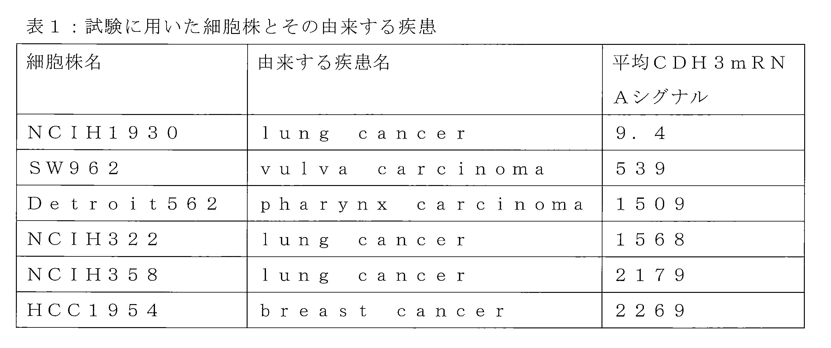
ヒト化抗体(PPAT-076-44Hb、及びPPAT-076-44Hd)の抗体薬剤コンジュゲートによる腫瘍縮小効果を、乳がん細胞株HCC1954を移植したゼノグラフトモデルで確認した。
実施例16の方法で定量した両抗体薬剤コンジュゲートの薬剤結合数(DAR)は、PPAT-076-44Hb(DAR3.69)、PPAT-076-44Hd(DAR3.51)であった。
担癌は、まず抗アシアロGM1抗体(WAKO 014-09801)を、大塚蒸留水1mLで溶解後、大塚生理食塩水4mLを加えて全量5mLとした後、この溶液をマウス1匹あたり100uL腹腔内に投与した。次にHCC1954は10%FBS含有RPMI1640培地を用いて培養し、SCIDマウス(メス、日本クレア)の右腹側部皮下に5x10⁶個/マウスになるように移植した 。
試験は各群5匹とし、5mg/kgで週に1度、計2回尾静脈より投与を行なった。
腫瘍体積を測定した結果を図8に示す。図8に示す通りヒト化抗体による抗体薬剤コンジュゲートは高い抗腫瘍効果を示した。
ヒト化抗体(PPAT-076-44Hb)とキメラ化抗体(PPAT-076-44C)の抗体薬剤コンジュゲートによる腫瘍縮小効果を、乳がん細胞株HCC70を移植したゼノグラフトモデルにて確認した。
両抗体薬剤コンジュゲートのDARは、PPAT-076-44Hb(DAR2.90)、PPAT-076-44C(DAR3.07)であった。
担癌は、抗アシアロGM1抗体(WAKO 014-09801)を、大塚蒸留水1mLで溶解後、大塚生理食塩水4mLを加えて全量5mLとした後、この溶液をマウス1匹あたり100uL腹腔内に投与した。次にHCC70は10%FBS含有RPMI640培地を用いて培養し、SCIDマウス(メス、日本クレア)の右腹側部皮下に5x10⁶個/マウスになるように移植した。
試験は各群5匹とし、ヒト化抗体コンジュゲートは0.6、3.0あるいは15mg/kg、キメラ化抗体コンジュゲートは3.0mg/kg、薬剤非結合のヒト化抗体(Naked)は15mg/kgの各用量で週に1度、計2回投与を行なった。腫瘍体積を測定した結果を図9に示す。図9に示す通り、ヒト化抗体による抗体薬剤コンジュゲートは、キメラ化抗体による抗体薬剤コンジュゲートに比して高い抗腫瘍効果が繰り返し示された。
ヒト化抗体(PPAT-076-44Hd)とキメラ化抗体(PPAT-076-44C)の抗体薬剤コンジュゲートによる腫瘍縮小効果を、乳がん細胞株HCC70を移植したゼノグラフトモデルにて確認した。
担癌は実施例19と同様に行ない、ヒト化抗体コンジュゲートは0.6、3.0あるいは15mg/kg、キメラ化抗体コンジュゲートは3.0mg/kg、薬剤非結合のヒト化抗体(Naked)は15mg/kgの各用量で週に1度、計2回尾静脈より投与を行なった。試験は各群5匹とした。両抗体薬剤コンジュゲートのDARは、PPAT-076-44C(DAR3.07)、PPAT-076-44Hd(DAR2.98)だった。
腫瘍体積を測定した結果を図10に示す。図10に示した通り、ヒト化抗体は薬剤を結合する事で高い抗腫瘍効果を示し、用量に依存した抗腫瘍効果が確認された。また同じ投与量(3.0mg/kg)においては、実施例19と同様にキメラ化抗体の抗体薬剤コンジュゲートに比して高い抗腫瘍効果が再度示された。
本願発明の抗体薬剤コンジュゲートの腫瘍増殖抑制能を、肺がん細胞株OKa-C-1(独立行政法人医薬基盤研究所、JCRB1343)を移植したゼノグラフトモデルで確認した。担癌は、OKa-C-1を10%FBS含有RPMI1640培地を用いて培養し、SCIDマウス(メス、日本クレア)の右腹側部皮下に6.5x10⁶個/マウスになるように移植した。ヒト化抗体コンジュゲートは15mg/kgの用量で週に1度、計2回尾静脈より投与を行なった。試験は各群3匹とした。ここではヒト化抗体としてPPAT-076-44Hdを使用し、それぞれに実施例15に記載した方法で薬剤を結合した。各抗体分子当たりの平均薬剤結合数(DAR)を実施例16に記載した方法で定量したところ、DARは3.04だった。腫瘍体積を測定した結果を図11に示す。図11に示した通り、ヒト化抗体は薬剤を結合する事で、乳癌細胞株に対するものと同様に高い抗腫瘍効果が確認された。
(1)CDH3N末部分長タンパク質の発現ベクター作製
取得したCDH3抗体の反応性を確認するため、CDH3抗原のN末端領域をマウスIgG2aのFc部分と連結した融合蛋白質を調製した。融合タンパク質のcDNA配列は配列番号70、アミノ酸配列は配列番号71に記載した。シグナルペプチドは抗体κ鎖のものを使用し、サブクローニングのために制限酵素部位を5プライム側にNheIを、3プライム側にEcoRI部位を付加した(米GenScript社で合成)。これをNheIとEcoRIで消化した哺乳類用発現ベクターのpCAGGS、あるいは遺伝子増幅用にマウスDHFR遺伝子を組み込んだpCAGGS-DHFRに組み込んだ。
一過性発現はライフテクノロジー社のフリースタイルを用いた。生産細胞にはライフテクノロジー社の293Fを使用し、遺伝子導入試薬はフリースタイル・マックス・トランスフェクション試薬(ライフテクノロジー社)を用いた。Fc融合可溶性抗原の遺伝子を導入した293Fは、CO2濃度を制御できるタイテック製の震盪機で4~7日間培養して生産した。生産した融合蛋白質はプロテインAセファロース(プロテノバ社)カラムで精製した。一過性発現で発現・精製した抗原のCBB染色図を図12Aに示し、図12Bに市販CDH3抗体(BD BIOSCIENCE社、及びR&D Systems社)を1次抗体として用いた染色像を示す。2次標識抗体にはAnti Mouse IgG F(ab’)2-HRP(goat IgG)(CAPPEL #55553)を用いた。
CDH3N末部分長タンパク質をPBSで2.5μg/mLとし、96ウェルプレートに100μL/ウェルで分注して、4℃で一晩静置した。翌日、ウェル内の溶液を捨て、Buffer A: 50mM Tris-HCl/150mM NaCl/1mM CaCl2/0.05% Tween20 (pH7.5)で洗浄した。次に被検物質(抗CDH3抗体)を希釈系列を作って100μL/ウェルで分注し、室温1時間震盪した。溶液を捨て、Buffer Aで洗浄した後、HRP標識抗体(HRP-goat anti human IgG (H+L) (absorbed with mouse, rabbit,bovine IgG) (American Qualex International, cat. A-110PD))をbufferAで10,000倍希釈したものを調製し、100μL/ウェルで分注し、室温1時間震盪した。Buffer Aで洗浄した後、TMB発色液を100μL/ウェルで加え、暗所で15分静置して発色を行った。停止液を100μL/ウェルで加えた後、プレートリーダーで450nmの吸光度を測定した。結果を図13に示す。披検物質としてPPAT-076-44Hb、及びPPAT-076-44Hdを用いた。
標識に用いる抗体を標識用緩衝液(50mM NaHCO3, 0.5M NaCl pH8.5)に置換する。抗体1mgに対し、25mM Alexa488(1mgをDMF62.1μlに溶解, ライフテクノロジー社)0.5μlを添加して、遮光下室温で1時間静置する。その後、PBSに緩衝液を交換した。
ヒト化が親和性に及ぼす影響をフローサイトメーター(FACS)を用いた競合試験で確かめた。測定は希釈系列を作った被験抗体、CDH3が発現している細胞株、及び被験抗体と競合する一定量のAmaxa488標識抗体を共存させて、室温1時間反応、FACS用液で洗浄後、FACS測定を行なった。各抗体濃度のGEO Mean値から、競合抗体のみのGEO Mean値を100%とした場合の阻害率を算出してIC50を求め、これを親和性の指標とした。
被験抗体と競合する抗体として、NITE BP-989細胞産生物から精製したマウス抗体を実施例24の方法で標識し、被験抗体としてPPAT-076-44Hd、PPAT-076-44C、及びPPAT-076-44Mを用いた。その結果を図14に示す。被験抗体は全てAlexa488標識抗体と競合するがその度合いが異なり、マウス及びキメラ化抗体に比して、本願のヒト化抗体(PPAT-076-44Hd)の親和性度合いが向上している事が示された。
本願発明のヒト化抗体に結合した平均薬剤結合数(DAR)が親和性に及ぼす影響を実施例25と同じくFACSを用いた競合試験で確かめた。抗体薬剤コンジュゲートは、実施例15に記載した方法で、平均DAR0~8の範囲で調製した。DARは実施例16に記載した方法で定量した。
細胞株はNCI-H358を用い、PPAT-076-44Hb及びPPAT-076-44Hdを測定する時は、競合抗体として実施例24の手順でAlexa488を標識したNITE BP-989細胞産生物から精製したマウス抗体を用い、実施例25で示したのと同じFACS競合試験により、親和性の指標として各IC50値を算出した。
図15(A:PPAT-076-44Hb、B:PPAT-076-44Hd)に示した比較は、薬剤を結合していない各抗体のIC50値を1とした相対値を表している。いずれのヒト化抗体も薬剤が結合する事で親和性は低下しない事を示している。
担癌モデル株のCDH3タンパク質の発現を確認するため、担癌組織切片の免疫染色を行った。マウス皮下に細胞株を移植して所定日数経過させた担癌マウスから得た腫瘍組織を脱パラフィン処理し、オートクレーブで121℃、15分賦活化を行った。0.3% H2O2を含むメタノールにて内在性ペルオキシダーゼの不活性化、10%ヤギ血清でブロッキング処理後、抗CDH3抗体610227(BD BIOSCIENCE社)に4℃一晩反応させた。抗体溶液を洗い流した後に、ヒストファインシンプルステインMAX-PO二次抗体試薬と室温1時間反応させた。ヒストファインDAB基質キットを添付プロトコールのとおり使ってDAB発色を行い、ヘマトキシリンエオジン溶液にて核染色を行った。図16(A:HCC1954、B:HCC70、C:OKa-C-1)に結果を示す。担癌腫瘍は細胞膜部分が抗CDH3抗体で染色された。
Claims (38)
- 受託番号NITE BP-1536を有する細胞が産生する抗体の重鎖可変領域由来の相補性決定領域配列(CDR-H1、H2、H3)及び軽鎖可変領域由来の相補性決定領域配列(CDR-L1、L2、L3)を含み、かつフレームワーク領域配列が重鎖可変領域が重鎖ヒトサブグループIIIコンセンサスフレームワーク配列であり、軽鎖可変領域が軽鎖ヒトκサブグループIコンセンサスフレームワーク配列を含有してなる抗CDH3ヒト化抗体。
- 重鎖可変領域の相補性決定領域配列が配列番号56、配列番号57、及び配列番号58であり、軽鎖可変領域の相補性決定領域配列が配列番号59、配列番号60、及び配列番号61を含み、かつフレームワーク領域配列が重鎖可変領域が重鎖ヒトサブグループIIIコンセンサスフレームワーク配列であり、軽鎖可変領域が軽鎖ヒトκサブグループIコンセンサスフレームワーク配列を含有してなる抗CDH3ヒト化抗体。
- 受託番号NITE BP-1536を有する細胞が産生する抗体の重鎖可変領域由来の相補性決定領域配列(CDR-H1、H2、H3)及び軽鎖可変領域由来の相補性決定領域配列(CDR-L1、L2、L3)を含み、かつフレームワーク領域配列が最適なアライメントのもとで選択されたヒトの生殖系列に由来する配列を含有してなる抗CDH3ヒト化抗体。
- 重鎖可変領域の相補性決定領域配列が配列番号56、配列番号57、及び配列番号58であり、軽鎖可変領域の相補性決定領域配列が配列番号59、配列番号60、及び配列番号61を含み、かつフレームワーク領域配列が最適なアライメントのもとで選択されたヒトの生殖系列に由来する配列を含有してなる抗CDH3ヒト化抗体。
- 請求項1から4のいずれか1項に記載した抗CDH3ヒト化抗体との配列相同性が少なくとも90%以上であり、かつCDH3を認識できる抗CDH3ヒト化抗体。
- 請求項1から4のいずれか1項に記載した抗体のフレームワーク領域部分における1から数個のアミノ酸が他のアミノ酸に置換しており、かつCDH3を認識できる、抗CDH3ヒト化抗体。
- 請求項1から4のいずれか1項に記載した抗体の相補性決定領域配列のうちフレームワーク領域との境界における1から数個のアミノ酸が他のアミノ酸に置換しており、かつCDH3を認識できる抗CDH3ヒト化抗体。
- 置換されるアミノ酸が軽鎖可変領域におけるKabatナンバリングによる55位のアミノ酸である、請求項6に記載の抗CDH3ヒト化抗体。
- 置換されるアミノ酸が重鎖可変領域におけるKabatナンバリングによる71位、及び78位のアミノ酸から選ばれる1つ以上である、請求項6に記載の抗CDH3ヒト化抗体。
- 置換されるアミノ酸が重鎖可変領域におけるKabatナンバリングによる49位のアミノ酸である、請求項7に記載の抗CDH3ヒト化抗体。
- 軽鎖可変領域におけるKabatナンバリングによる55位のアミノ酸がアラニンに置換している、請求項6に記載の抗CDH3ヒト化抗体。
- 重鎖可変領域におけるKabatナンバリングによる71位のアミノ酸がリジンに置換している、請求項6に記載の抗CDH3ヒト化抗体。
- 重鎖可変領域におけるKabatナンバリングによる78位のアミノ酸がバリンに置換している、請求項6に記載の抗CDH3ヒト化抗体。
- 重鎖可変領域におけるKabatナンバリングによる49位のアミノ酸がアラニンに置換している、請求項7に記載の抗CDH3ヒト化抗体。
- 重鎖可変領域におけるKabatナンバリングによる49位のアミノ酸残基のアラニンへの置換、71位のアミノ酸残基のリジンへの置換、78位のアミノ酸残基のバリンへの置換、及び軽鎖可変領域におけるKabatナンバリングによる55位のアミノ酸残基のアラニンへの置換から選択される1以上の置換を有する、請求項1又は2に記載の抗CDH3ヒト化抗体。
- 重鎖可変領域におけるKabatナンバリングによる71位のアミノ酸残基のリジンへの置換、78位のアミノ酸残基のバリンへの置換、及び軽鎖可変領域におけるKabatナンバリングによる55位のアミノ酸残基のアラニンへの置換から選択される1以上の置換を有する、請求項3又は4に記載の抗CDH3ヒト化抗体。
- 以下の何れかの抗体。
(1)重鎖可変領域に配列番号48に記載のアミノ酸配列を有し、軽鎖可変領域に配列番号49に記載のアミノ酸配列を有する、抗CDH3ヒト化抗体;
(2)重鎖可変領域に配列番号50に記載のアミノ酸配列を有し、軽鎖可変領域に配列番号51に記載のアミノ酸配列を有する、抗CDH3ヒト化抗体;
(3)重鎖可変領域に配列番号52に記載のアミノ酸配列を有し、軽鎖可変領域に配列番号53に記載のアミノ酸配列を有する、抗CDH3ヒト化抗体;及び
(4)重鎖可変領域に配列番号54に記載のアミノ酸配列を有し、軽鎖可変領域に配列番号55に記載のアミノ酸配列を有する、抗CDH3ヒト化抗体: - CDH3との結合能を有する、請求項1から17のいずれか1項に記載の抗CDH3ヒト化抗体の断片。
- Fab,F(ab′)2、又はscFvである、請求項18に記載の抗CDH3ヒト化抗体の断片。
- CDH3との結合能を有する、請求項1から19のいずれか1項に記載した抗体の部分配列。
- CDH3がヒトCDH3である、請求項1から17のいずれか1項に記載の抗CDH3ヒト化抗体。
- CDH3が配列番号2の細胞外領域である、請求項1から17のいずれか1項に記載の抗CDH3ヒト化抗体。
- 請求項1から22のいずれか1項に記載の抗CDH3ヒト化抗体、その断片又はその部分配列と、化学療法剤又は放射性物質とが連結している、免疫複合体。
- 化学療法剤が、細胞障害性物質である、請求項23に記載の免疫複合体。
- 細胞傷害性物質が、メイタンシノイド又はその誘導体、あるいはアウリスタチン又はその誘導体である、請求項24に記載の免疫複合体。
- 細胞傷害性物質が、DM1、DM3又はDM4から選択されるメイタンシノイド又はその誘導体、あるいはMMAEあるいはMMAFから選択されるアウリスタチン又はその誘導体である、請求項24に記載の免疫複合体。
- 抗CDH3ヒト化抗体、その断片又はその部分配列1分子あたり平均1~7個のDM1が結合している、請求項24に記載の免疫複合体。
- 抗CDH3ヒト化抗体、その断片又はその部分配列と、化学療法剤とがリンカーを介して連結している、請求項23から27の何れか1項に記載の免疫複合体。
- 抗CDH3ヒト化抗体、その断片又はその部分配列と、化学療法剤とが、抗体のFc領域の分子内ジスルフィド結合を介して連結しているか、又は抗体のFc領域を遺伝子工学的に改変して連結しいる、請求項23から27の何れか1項に記載の免疫複合体。
- リンカーが、2価反応性架橋試薬である、請求項28に記載の免疫複合体。
- リンカーが、N-スクシンイミジル4-(マレイミドメチル)シクロヘキサンカルボキシレート(SMCC)、スルホスクシイミジル-4-(N-マレイミドメチル)-シクロヘキサン-1-カルボキシレート(Sulfo-SMCC)、N-スクシンイミジル-4-(N-マレイミドメチル)-シクロヘキサン-1-カルボキシ-(6-アミドカプロエート) (LC-SMCC)、κ-マレイミドウンデカン酸N-スクシンイミジルエステル(KMUA)、γ-マレイミド酪酸N-スクシンイミジルエステル(GMBS)、ε-マレイミドカプロン酸N-ヒドロキシスクシンイミドエステル(EMCS)、m-マレイミドベンゾイル-N-ヒドロキシスクシンイミドエステル(MBS)、N-(α-マレイミドアセトキシ)-スクシンイミドエステル(AMAS)、スクシンイミジル-6-(β-マレイミドプロピオンアミド)ヘキサノエート(SMPH)、N-スクシンイミジル4-(p-マレイミドフェニル)-ブチレート(SMPB)、N-(p-マレイミドフェニル)イソシアネート(PMPI)、N-スクシンイミジル4(2-ピリジルチオ)ペンタノエート(SPP)、N-スクシンイミジル(4-イオド-アセチル)アミノ安息香酸エステル(SIAB)、6-マレイミドカプロイル(MC)、マレイミドプロパノイル(MP)、p-アミノベンジルオキシカルボンイル(PAB)、及びN-スクシンイミジル4(2-ピリジルチオ)ブタノエート(SPDB)から成る群より選択される請求項28に記載の免疫複合体。
- リンカーがプロテアーゼによって切断される、請求項28に記載の免疫複合体。
- リンカーが、バリン-シトルリン(Val-Cit)、アラニン-フェニルアラニン(ala-phe)、及びパラアミノベンゾイック酸(PABA)の少なくとも1以上を含む、請求項28に記載の免疫複合体。
- 細胞傷害性能が抗体可変領域のフレームワーク領域配列のヒト化によって増強されている、請求項23に記載の免疫複合体。
- 請求項23から34の何れか1項に記載の免疫複合体を含む、CDH3の過剰発現によって特徴づけられる疾患を治療するための医薬。
- CDH3の過剰発現によって特徴づけられる疾患が癌である、請求項35に記載の医薬。
- 癌が、結腸直腸癌、非小細胞肺癌、乳癌、頭頚部癌、卵巣癌、肺癌、浸潤性膀胱癌、膵臓癌、脳の転移性癌、甲状腺癌、頭頚部扁平上皮癌、食道扁平上皮癌、肺扁平上皮癌、皮膚扁平上皮癌、メラノーマ、乳腺癌、肺腺癌、子宮頚部扁平上皮癌、膵臓扁平上皮癌、結腸扁平上皮癌、又は胃扁平上皮癌、前立腺癌、骨肉腫又は軟組織肉腫から選択される、請求項36に記載の医薬。
- 抗腫瘍剤として使用する、請求項35から37の何れか1項に記載の医薬。
Priority Applications (12)
| Application Number | Priority Date | Filing Date | Title |
|---|---|---|---|
| EP14751339.4A EP2957632B1 (en) | 2013-02-15 | 2014-02-14 | Anti-cdh3 humanized antibody, drug conjugate thereof, and utilization of same |
| BR112015019328A BR112015019328A2 (pt) | 2013-02-15 | 2014-02-14 | Anticorpo anti-cdh3 humanizado, conjugado do fármaco do anticorpo anti-cdh3 humanizado, e seu uso |
| SG11201506358PA SG11201506358PA (en) | 2013-02-15 | 2014-02-14 | Anti-cdh3 humanized antibody, drug conjugate thereof, and use thereof |
| JP2015500306A JP6377601B2 (ja) | 2013-02-15 | 2014-02-14 | 抗cdh3ヒト化抗体、その薬剤コンジュゲート、及びそれらの使用 |
| CN201480009141.8A CN105073988A (zh) | 2013-02-15 | 2014-02-14 | 抗cdh3人源化抗体、其药剂偶联物以及它们的用途 |
| AU2014216959A AU2014216959A1 (en) | 2013-02-15 | 2014-02-14 | Anti-CDH3 humanized antibody, drug conjugate thereof, and use thereof |
| CA2901214A CA2901214A1 (en) | 2013-02-15 | 2014-02-14 | Anti-cdh3 humanized antibody, drug conjugate thereof, and use thereof |
| RU2015139147A RU2015139147A (ru) | 2013-02-15 | 2014-02-14 | Гуманизированное антитело к cdh3, его конъюгат с лекарственным средством и их применение |
| US14/768,151 US9644028B2 (en) | 2013-02-15 | 2014-02-14 | Anti-CDH3 humanized antibody, drug conjugate thereof, and use thereof |
| KR1020157024911A KR20150119206A (ko) | 2013-02-15 | 2014-02-14 | 항 cdh3 인간화 항체, 그의 약물 콘쥬게이트, 이들의 용도 |
| ES14751339T ES2829597T3 (es) | 2013-02-15 | 2014-02-14 | Anticuerpo humanizado anti-CDH3, conjugado de fármaco del mismo y utilización del mismo |
| HK16103909.6A HK1215959A1 (zh) | 2013-02-15 | 2016-04-06 | 人源化抗體、其藥劑偶聯物以及它們的用途 |
Applications Claiming Priority (4)
| Application Number | Priority Date | Filing Date | Title |
|---|---|---|---|
| JP2013-027386 | 2013-02-15 | ||
| JP2013027386 | 2013-02-15 | ||
| JP2013091163 | 2013-04-24 | ||
| JP2013-091163 | 2013-04-24 |
Publications (1)
| Publication Number | Publication Date |
|---|---|
| WO2014126198A1 true WO2014126198A1 (ja) | 2014-08-21 |
Family
ID=51354197
Family Applications (1)
| Application Number | Title | Priority Date | Filing Date |
|---|---|---|---|
| PCT/JP2014/053473 WO2014126198A1 (ja) | 2013-02-15 | 2014-02-14 | 抗cdh3ヒト化抗体、その薬剤コンジュゲート、及びそれらの使用 |
Country Status (13)
| Country | Link |
|---|---|
| US (1) | US9644028B2 (ja) |
| EP (1) | EP2957632B1 (ja) |
| JP (1) | JP6377601B2 (ja) |
| KR (1) | KR20150119206A (ja) |
| CN (1) | CN105073988A (ja) |
| AU (1) | AU2014216959A1 (ja) |
| BR (1) | BR112015019328A2 (ja) |
| CA (1) | CA2901214A1 (ja) |
| ES (1) | ES2829597T3 (ja) |
| HK (1) | HK1215959A1 (ja) |
| RU (1) | RU2015139147A (ja) |
| SG (1) | SG11201506358PA (ja) |
| WO (1) | WO2014126198A1 (ja) |
Cited By (4)
| Publication number | Priority date | Publication date | Assignee | Title |
|---|---|---|---|---|
| WO2015095953A1 (en) | 2013-12-27 | 2015-07-02 | The Centre For Drug Research And Development | Sulfonamide-containing linkage systems for drug conjugates |
| WO2016075670A1 (en) * | 2014-11-14 | 2016-05-19 | Novartis Ag | Antibody drug conjugates |
| JP2018500275A (ja) * | 2013-10-02 | 2018-01-11 | スリ テクノロジーズ エルティーディー. | 不均一な腫瘍を処置するための患者特異的免疫療法 |
| JP2021000130A (ja) * | 2015-06-12 | 2021-01-07 | シアトル ジェネティクス インコーポレーテッド | Cd123抗体及びその複合体 |
Families Citing this family (1)
| Publication number | Priority date | Publication date | Assignee | Title |
|---|---|---|---|---|
| EP4337702A1 (en) * | 2021-05-13 | 2024-03-20 | Wuxi Biologics Ireland Limited | Antibody conjugate comprising anti-p-cadherin antibody and uses thereof |
Citations (17)
| Publication number | Priority date | Publication date | Assignee | Title |
|---|---|---|---|---|
| EP0239400A2 (en) | 1986-03-27 | 1987-09-30 | Medical Research Council | Recombinant antibodies and methods for their production |
| US5208020A (en) | 1989-10-25 | 1993-05-04 | Immunogen Inc. | Cytotoxic agents comprising maytansinoids and their therapeutic use |
| WO1996002576A1 (fr) | 1994-07-13 | 1996-02-01 | Chugai Seiyaku Kabushiki Kaisha | Anticorps humain reconstitue contre l'interleukine-8 humaine |
| US6180370B1 (en) | 1988-12-28 | 2001-01-30 | Protein Design Labs, Inc. | Humanized immunoglobulins and methods of making the same |
| US6333410B1 (en) | 2000-08-18 | 2001-12-25 | Immunogen, Inc. | Process for the preparation and purification of thiol-containing maytansinoids |
| WO2002097395A2 (en) | 2001-05-31 | 2002-12-05 | Chiron Corporation | P-cadherin as a target for anti-cancer therapy |
| JP2005000169A (ja) | 1991-06-14 | 2005-01-06 | Genentech Inc | 免疫グロブリン変異体 |
| WO2007102525A1 (en) | 2006-02-28 | 2007-09-13 | Oncotherapy Science, Inc. | Methods for damaging cells using effector functions of anti-cdh3 antibodies |
| JP2008516896A (ja) | 2004-09-23 | 2008-05-22 | ジェネンテック・インコーポレーテッド | システイン操作抗体および結合体 |
| WO2010126137A1 (ja) * | 2009-05-01 | 2010-11-04 | 国立大学法人 東京大学 | 抗カドヘリン抗体 |
| WO2011080796A1 (en) | 2009-12-28 | 2011-07-07 | Oncotherapy Science, Inc. | Anti-cdh3 antibodies and uses thereof |
| WO2011099524A1 (ja) * | 2010-02-10 | 2011-08-18 | 富士フイルムRiファーマ株式会社 | 放射性金属標識抗カドヘリン抗体 |
| JP2011526583A (ja) | 2008-06-30 | 2011-10-13 | オンコセラピー・サイエンス株式会社 | 放射性同位体標識で標識された抗cdh3抗体およびその使用 |
| WO2012057315A1 (ja) * | 2010-10-29 | 2012-05-03 | 株式会社ペルセウスプロテオミクス | 高い親和性を有する抗cdh3抗体 |
| WO2012057328A1 (ja) * | 2010-10-29 | 2012-05-03 | 株式会社ペルセウスプロテオミクス | 高い内在化能力を有する抗cdh3抗体 |
| WO2012176765A1 (ja) * | 2011-06-24 | 2012-12-27 | 株式会社ペルセウスプロテオミクス | 抗ヒトp-カドへリン(cdh3)遺伝子組み換え抗体 |
| WO2013150623A1 (ja) | 2012-04-04 | 2013-10-10 | 株式会社ペルセウスプロテオミクス | 抗cdh3(p-カドヘリン)抗体の薬剤コンジュゲート |
Family Cites Families (5)
| Publication number | Priority date | Publication date | Assignee | Title |
|---|---|---|---|---|
| AU2005267720B2 (en) * | 2004-08-05 | 2012-02-23 | Genentech, Inc. | Humanized anti-cmet antagonists |
| SI2247620T1 (sl) * | 2008-01-31 | 2016-09-30 | Genentech, Inc. | Protitelesa proti CD79b in imunokonjugati in postopki za uporabo |
| AU2009246071B2 (en) | 2008-05-16 | 2013-10-03 | Genentech, Inc. | Use of biomarkers for assessing treatment of gastrointestinal inflammatory disorders with beta7integrin antagonists |
| TWI428142B (zh) * | 2009-05-08 | 2014-03-01 | Genentech Inc | 人類化之抗-egfl7抗體及其使用方法 |
| US7866675B2 (en) | 2010-04-05 | 2011-01-11 | Hauser Ray L | Composite ice blade |
-
2014
- 2014-02-14 AU AU2014216959A patent/AU2014216959A1/en not_active Abandoned
- 2014-02-14 JP JP2015500306A patent/JP6377601B2/ja active Active
- 2014-02-14 CN CN201480009141.8A patent/CN105073988A/zh active Pending
- 2014-02-14 CA CA2901214A patent/CA2901214A1/en not_active Abandoned
- 2014-02-14 SG SG11201506358PA patent/SG11201506358PA/en unknown
- 2014-02-14 KR KR1020157024911A patent/KR20150119206A/ko not_active Application Discontinuation
- 2014-02-14 RU RU2015139147A patent/RU2015139147A/ru not_active Application Discontinuation
- 2014-02-14 BR BR112015019328A patent/BR112015019328A2/pt not_active IP Right Cessation
- 2014-02-14 US US14/768,151 patent/US9644028B2/en active Active
- 2014-02-14 WO PCT/JP2014/053473 patent/WO2014126198A1/ja active Application Filing
- 2014-02-14 ES ES14751339T patent/ES2829597T3/es active Active
- 2014-02-14 EP EP14751339.4A patent/EP2957632B1/en active Active
-
2016
- 2016-04-06 HK HK16103909.6A patent/HK1215959A1/zh unknown
Patent Citations (18)
| Publication number | Priority date | Publication date | Assignee | Title |
|---|---|---|---|---|
| EP0239400A2 (en) | 1986-03-27 | 1987-09-30 | Medical Research Council | Recombinant antibodies and methods for their production |
| US6180370B1 (en) | 1988-12-28 | 2001-01-30 | Protein Design Labs, Inc. | Humanized immunoglobulins and methods of making the same |
| US5208020A (en) | 1989-10-25 | 1993-05-04 | Immunogen Inc. | Cytotoxic agents comprising maytansinoids and their therapeutic use |
| JP2005000169A (ja) | 1991-06-14 | 2005-01-06 | Genentech Inc | 免疫グロブリン変異体 |
| JP4836147B2 (ja) | 1991-06-14 | 2011-12-14 | ジェネンテック, インコーポレイテッド | 免疫グロブリン変異体 |
| WO1996002576A1 (fr) | 1994-07-13 | 1996-02-01 | Chugai Seiyaku Kabushiki Kaisha | Anticorps humain reconstitue contre l'interleukine-8 humaine |
| US6333410B1 (en) | 2000-08-18 | 2001-12-25 | Immunogen, Inc. | Process for the preparation and purification of thiol-containing maytansinoids |
| WO2002097395A2 (en) | 2001-05-31 | 2002-12-05 | Chiron Corporation | P-cadherin as a target for anti-cancer therapy |
| JP2008516896A (ja) | 2004-09-23 | 2008-05-22 | ジェネンテック・インコーポレーテッド | システイン操作抗体および結合体 |
| WO2007102525A1 (en) | 2006-02-28 | 2007-09-13 | Oncotherapy Science, Inc. | Methods for damaging cells using effector functions of anti-cdh3 antibodies |
| JP2011526583A (ja) | 2008-06-30 | 2011-10-13 | オンコセラピー・サイエンス株式会社 | 放射性同位体標識で標識された抗cdh3抗体およびその使用 |
| WO2010126137A1 (ja) * | 2009-05-01 | 2010-11-04 | 国立大学法人 東京大学 | 抗カドヘリン抗体 |
| WO2011080796A1 (en) | 2009-12-28 | 2011-07-07 | Oncotherapy Science, Inc. | Anti-cdh3 antibodies and uses thereof |
| WO2011099524A1 (ja) * | 2010-02-10 | 2011-08-18 | 富士フイルムRiファーマ株式会社 | 放射性金属標識抗カドヘリン抗体 |
| WO2012057315A1 (ja) * | 2010-10-29 | 2012-05-03 | 株式会社ペルセウスプロテオミクス | 高い親和性を有する抗cdh3抗体 |
| WO2012057328A1 (ja) * | 2010-10-29 | 2012-05-03 | 株式会社ペルセウスプロテオミクス | 高い内在化能力を有する抗cdh3抗体 |
| WO2012176765A1 (ja) * | 2011-06-24 | 2012-12-27 | 株式会社ペルセウスプロテオミクス | 抗ヒトp-カドへリン(cdh3)遺伝子組み換え抗体 |
| WO2013150623A1 (ja) | 2012-04-04 | 2013-10-10 | 株式会社ペルセウスプロテオミクス | 抗cdh3(p-カドヘリン)抗体の薬剤コンジュゲート |
Non-Patent Citations (37)
| Title |
|---|
| "Current Protocols in Immunology", JOHN WILEY AND SONS, INC., article "Unit 2.12 Cloning, Expression, and Modification of Antibody V Regions" |
| "Current Protocols in Molecular Biology", JOHN WILEY AND SONS, INC., article "Chapter 9 Introduction of DNA into Mammalian Cells" |
| "Phage Display - A Laboratory Manual", ., article "PROTOCOL 9.5" |
| "Sequences of proteins of immunological interest, 5th ed.", 1991, NATIONAL INSTITUTES OF HEALTH |
| ANALYTICAL BIOCHEMISTY, vol. 173, 1988, pages 93 |
| ANALYTICAL BIOCHEMISTY, vol. 173, 1988, pages 93 - 95 |
| BIO CONJUGATE CHEMISTRY, vol. 19, 2008, pages 1673 |
| BIOTECHNOLOGY, vol. 9, 1991, pages 266 |
| CANCER RES., vol. 52, 1992, pages 127 |
| CANCER RES., vol. 68, no. 15, 2008, pages 6300 |
| CANCER RES., vol. 68, no. 22, 2008, pages 9280 |
| CHOTHIA ET AL., J. MOL. BIOL., vol. 196, 1987, pages 901 |
| J. IMMUNOL., vol. 151, 1993, pages 2296 |
| J. MED. CHEM., vol. 49, 2006, pages 4392 |
| J. MED. CHEM., vol. 49, 2006, pages 4392 - 4408 |
| J. MOL BIOL., vol. 196, 1987, pages 901 |
| J. MOL. BIOL., vol. 196, 1987, pages 901 |
| JOURNAL OF IMMUNOLOGY, vol. 169, 2002, pages 1119 |
| KABAT ET AL.: "Sequences of proteins of immunological interest, 5th ed.", 1991, NATIONAL INSTITUTES OF HEALTH |
| METHODS. MOL. BIOL., vol. 525, 2009, pages 445 |
| METHODS. MOL. BIOL., vol. 525, 2009, pages 445 - 67 |
| N. ENGL. J. MED, vol. 367, no. 19, 8 November 2012 (2012-11-08), pages 1783 - 91 |
| N. ENGL. J. MED., vol. 367, no. 19, 8 November 2012 (2012-11-08), pages 1783 - 91 |
| NATURE BIOTECHNOLOGY, vol. 26, no. 8, 2008, pages 925 |
| NATURE, vol. 276, 1978, pages 269 |
| NATURE, vol. 332, 1988, pages 323 |
| PROC. NATL. ACAD. SCI. U.S.A., vol. 89, 1992, pages 4285 |
| PROC. NATL. ACAD. SCI. USA, vol. 89, 1992, pages 4285 |
| SCHULMAN, M. ET AL., NATURE, vol. 276, 1978, pages 269 |
| See also references of EP2957632A4 |
| SIMS ET AL., J. IMMUNOL., vol. 151, 1993, pages 2296 |
| SOMAT. CELL. MOL. GENET, vol. 12, 1986, pages 5555 |
| TAN ET AL., JOURNAL OF IMMUNOLOGY, vol. 169, 2002, pages 1119 |
| TEMPEST ET AL., BIOTECHNOLOGY, vol. 9, 1991, pages 266 |
| URLAND, G. ET AL., SOMAT. CELL. MOL. GENET., vol. 12, 1986, pages 5555 |
| YOSHIDA; TAKEICHI, CELL, vol. 28, 1982, pages 217 - 224 |
| ZHANG C.C. ET AL.: "PF-03732010: A Fully Human Monoclonal Antibody against P-Cadherin with Antitumor and Antimetastatic Activity", CLIN. CAN. RES., vol. 16, pages 5177 - 5188, XP008137607 * |
Cited By (8)
| Publication number | Priority date | Publication date | Assignee | Title |
|---|---|---|---|---|
| JP2018500275A (ja) * | 2013-10-02 | 2018-01-11 | スリ テクノロジーズ エルティーディー. | 不均一な腫瘍を処置するための患者特異的免疫療法 |
| WO2015095953A1 (en) | 2013-12-27 | 2015-07-02 | The Centre For Drug Research And Development | Sulfonamide-containing linkage systems for drug conjugates |
| WO2016075670A1 (en) * | 2014-11-14 | 2016-05-19 | Novartis Ag | Antibody drug conjugates |
| CN108064244A (zh) * | 2014-11-14 | 2018-05-22 | 诺华股份有限公司 | 抗体药物缀合物 |
| US10005836B2 (en) | 2014-11-14 | 2018-06-26 | Novartis Ag | Antibody drug conjugates |
| AU2015347015B2 (en) * | 2014-11-14 | 2019-02-14 | Novartis Ag | Antibody drug conjugates |
| US10626172B2 (en) | 2014-11-14 | 2020-04-21 | Novartis Ag | Antibody drug conjugates |
| JP2021000130A (ja) * | 2015-06-12 | 2021-01-07 | シアトル ジェネティクス インコーポレーテッド | Cd123抗体及びその複合体 |
Also Published As
| Publication number | Publication date |
|---|---|
| US9644028B2 (en) | 2017-05-09 |
| EP2957632B1 (en) | 2020-09-30 |
| AU2014216959A1 (en) | 2015-10-01 |
| EP2957632A4 (en) | 2016-10-05 |
| KR20150119206A (ko) | 2015-10-23 |
| BR112015019328A2 (pt) | 2017-08-22 |
| JP6377601B2 (ja) | 2018-08-22 |
| CN105073988A (zh) | 2015-11-18 |
| HK1215959A1 (zh) | 2016-09-30 |
| ES2829597T3 (es) | 2021-06-01 |
| CA2901214A1 (en) | 2014-08-21 |
| US20160152703A1 (en) | 2016-06-02 |
| JPWO2014126198A1 (ja) | 2017-02-02 |
| SG11201506358PA (en) | 2015-09-29 |
| RU2015139147A (ru) | 2017-03-21 |
| EP2957632A1 (en) | 2015-12-23 |
Similar Documents
| Publication | Publication Date | Title |
|---|---|---|
| JP6006640B2 (ja) | 高い内在化能力を有する抗cdh3抗体 | |
| JP6800883B2 (ja) | クローディン18.2に対する抗体を含む薬物コンジュゲート | |
| JP6010241B2 (ja) | Dll3モジュレーター及び使用方法 | |
| JP6326137B2 (ja) | 抗her2抗体及びその結合体 | |
| JP2021516960A (ja) | Cd73を標的とする抗体および抗体−薬物複合体、その製造方法と使用 | |
| JP6113717B2 (ja) | 抗cdh3(p−カドヘリン)抗体の薬剤コンジュゲート | |
| TW201216987A (en) | Antibody drug conjugates (ADC) that bind to 191P4D12 proteins | |
| CN107847593B (zh) | 治疗抗体及其用途 | |
| JP2021530436A (ja) | Axlを標的とする抗体および抗体−薬物複合体ならびにその製造方法と使用 | |
| JP6377601B2 (ja) | 抗cdh3ヒト化抗体、その薬剤コンジュゲート、及びそれらの使用 | |
| JP2021533732A (ja) | Sema4d抗体及びその製造方法と使用 | |
| WO2012057315A1 (ja) | 高い親和性を有する抗cdh3抗体 |
Legal Events
| Date | Code | Title | Description |
|---|---|---|---|
| WWE | Wipo information: entry into national phase |
Ref document number: 201480009141.8 Country of ref document: CN |
|
| 121 | Ep: the epo has been informed by wipo that ep was designated in this application |
Ref document number: 14751339 Country of ref document: EP Kind code of ref document: A1 |
|
| ENP | Entry into the national phase |
Ref document number: 2901214 Country of ref document: CA |
|
| ENP | Entry into the national phase |
Ref document number: 2015500306 Country of ref document: JP Kind code of ref document: A |
|
| WWE | Wipo information: entry into national phase |
Ref document number: 14768151 Country of ref document: US |
|
| NENP | Non-entry into the national phase |
Ref country code: DE |
|
| REG | Reference to national code |
Ref country code: BR Ref legal event code: B01A Ref document number: 112015019328 Country of ref document: BR |
|
| ENP | Entry into the national phase |
Ref document number: 20157024911 Country of ref document: KR Kind code of ref document: A |
|
| ENP | Entry into the national phase |
Ref document number: 2015139147 Country of ref document: RU Kind code of ref document: A |
|
| WWE | Wipo information: entry into national phase |
Ref document number: 2014751339 Country of ref document: EP |
|
| ENP | Entry into the national phase |
Ref document number: 2014216959 Country of ref document: AU Date of ref document: 20140214 Kind code of ref document: A |
|
| ENP | Entry into the national phase |
Ref document number: 112015019328 Country of ref document: BR Kind code of ref document: A2 Effective date: 20150812 |