WO2014123151A1 - 多系統萎縮症リスクの検査方法、検査キット、及び多系統萎縮症の治療又は予防薬 - Google Patents
多系統萎縮症リスクの検査方法、検査キット、及び多系統萎縮症の治療又は予防薬 Download PDFInfo
- Publication number
- WO2014123151A1 WO2014123151A1 PCT/JP2014/052658 JP2014052658W WO2014123151A1 WO 2014123151 A1 WO2014123151 A1 WO 2014123151A1 JP 2014052658 W JP2014052658 W JP 2014052658W WO 2014123151 A1 WO2014123151 A1 WO 2014123151A1
- Authority
- WO
- WIPO (PCT)
- Prior art keywords
- mutation
- coenzyme
- msa
- coq2
- coq10
- Prior art date
- Legal status (The legal status is an assumption and is not a legal conclusion. Google has not performed a legal analysis and makes no representation as to the accuracy of the status listed.)
- Ceased
Links
Images








Classifications
-
- A—HUMAN NECESSITIES
- A61—MEDICAL OR VETERINARY SCIENCE; HYGIENE
- A61K—PREPARATIONS FOR MEDICAL, DENTAL OR TOILETRY PURPOSES
- A61K31/00—Medicinal preparations containing organic active ingredients
- A61K31/12—Ketones
- A61K31/122—Ketones having the oxygen directly attached to a ring, e.g. quinones, vitamin K1, anthralin
-
- A—HUMAN NECESSITIES
- A61—MEDICAL OR VETERINARY SCIENCE; HYGIENE
- A61P—SPECIFIC THERAPEUTIC ACTIVITY OF CHEMICAL COMPOUNDS OR MEDICINAL PREPARATIONS
- A61P25/00—Drugs for disorders of the nervous system
-
- A—HUMAN NECESSITIES
- A61—MEDICAL OR VETERINARY SCIENCE; HYGIENE
- A61P—SPECIFIC THERAPEUTIC ACTIVITY OF CHEMICAL COMPOUNDS OR MEDICINAL PREPARATIONS
- A61P25/00—Drugs for disorders of the nervous system
- A61P25/14—Drugs for disorders of the nervous system for treating abnormal movements, e.g. chorea, dyskinesia
- A61P25/16—Anti-Parkinson drugs
-
- A—HUMAN NECESSITIES
- A61—MEDICAL OR VETERINARY SCIENCE; HYGIENE
- A61P—SPECIFIC THERAPEUTIC ACTIVITY OF CHEMICAL COMPOUNDS OR MEDICINAL PREPARATIONS
- A61P25/00—Drugs for disorders of the nervous system
- A61P25/28—Drugs for disorders of the nervous system for treating neurodegenerative disorders of the central nervous system, e.g. nootropic agents, cognition enhancers, drugs for treating Alzheimer's disease or other forms of dementia
-
- C—CHEMISTRY; METALLURGY
- C12—BIOCHEMISTRY; BEER; SPIRITS; WINE; VINEGAR; MICROBIOLOGY; ENZYMOLOGY; MUTATION OR GENETIC ENGINEERING
- C12Q—MEASURING OR TESTING PROCESSES INVOLVING ENZYMES, NUCLEIC ACIDS OR MICROORGANISMS; COMPOSITIONS OR TEST PAPERS THEREFOR; PROCESSES OF PREPARING SUCH COMPOSITIONS; CONDITION-RESPONSIVE CONTROL IN MICROBIOLOGICAL OR ENZYMOLOGICAL PROCESSES
- C12Q1/00—Measuring or testing processes involving enzymes, nucleic acids or microorganisms; Compositions therefor; Processes of preparing such compositions
- C12Q1/68—Measuring or testing processes involving enzymes, nucleic acids or microorganisms; Compositions therefor; Processes of preparing such compositions involving nucleic acids
- C12Q1/6876—Nucleic acid products used in the analysis of nucleic acids, e.g. primers or probes
- C12Q1/6883—Nucleic acid products used in the analysis of nucleic acids, e.g. primers or probes for diseases caused by alterations of genetic material
-
- G—PHYSICS
- G01—MEASURING; TESTING
- G01N—INVESTIGATING OR ANALYSING MATERIALS BY DETERMINING THEIR CHEMICAL OR PHYSICAL PROPERTIES
- G01N33/00—Investigating or analysing materials by specific methods not covered by groups G01N1/00 - G01N31/00
- G01N33/48—Biological material, e.g. blood, urine; Haemocytometers
- G01N33/50—Chemical analysis of biological material, e.g. blood, urine; Testing involving biospecific ligand binding methods; Immunological testing
- G01N33/68—Chemical analysis of biological material, e.g. blood, urine; Testing involving biospecific ligand binding methods; Immunological testing involving proteins, peptides or amino acids
- G01N33/6893—Chemical analysis of biological material, e.g. blood, urine; Testing involving biospecific ligand binding methods; Immunological testing involving proteins, peptides or amino acids related to diseases not provided for elsewhere
- G01N33/6896—Neurological disorders, e.g. Alzheimer's disease
-
- C—CHEMISTRY; METALLURGY
- C12—BIOCHEMISTRY; BEER; SPIRITS; WINE; VINEGAR; MICROBIOLOGY; ENZYMOLOGY; MUTATION OR GENETIC ENGINEERING
- C12Q—MEASURING OR TESTING PROCESSES INVOLVING ENZYMES, NUCLEIC ACIDS OR MICROORGANISMS; COMPOSITIONS OR TEST PAPERS THEREFOR; PROCESSES OF PREPARING SUCH COMPOSITIONS; CONDITION-RESPONSIVE CONTROL IN MICROBIOLOGICAL OR ENZYMOLOGICAL PROCESSES
- C12Q2600/00—Oligonucleotides characterized by their use
- C12Q2600/136—Screening for pharmacological compounds
-
- C—CHEMISTRY; METALLURGY
- C12—BIOCHEMISTRY; BEER; SPIRITS; WINE; VINEGAR; MICROBIOLOGY; ENZYMOLOGY; MUTATION OR GENETIC ENGINEERING
- C12Q—MEASURING OR TESTING PROCESSES INVOLVING ENZYMES, NUCLEIC ACIDS OR MICROORGANISMS; COMPOSITIONS OR TEST PAPERS THEREFOR; PROCESSES OF PREPARING SUCH COMPOSITIONS; CONDITION-RESPONSIVE CONTROL IN MICROBIOLOGICAL OR ENZYMOLOGICAL PROCESSES
- C12Q2600/00—Oligonucleotides characterized by their use
- C12Q2600/156—Polymorphic or mutational markers
-
- C—CHEMISTRY; METALLURGY
- C12—BIOCHEMISTRY; BEER; SPIRITS; WINE; VINEGAR; MICROBIOLOGY; ENZYMOLOGY; MUTATION OR GENETIC ENGINEERING
- C12Q—MEASURING OR TESTING PROCESSES INVOLVING ENZYMES, NUCLEIC ACIDS OR MICROORGANISMS; COMPOSITIONS OR TEST PAPERS THEREFOR; PROCESSES OF PREPARING SUCH COMPOSITIONS; CONDITION-RESPONSIVE CONTROL IN MICROBIOLOGICAL OR ENZYMOLOGICAL PROCESSES
- C12Q2600/00—Oligonucleotides characterized by their use
- C12Q2600/158—Expression markers
-
- G—PHYSICS
- G01—MEASURING; TESTING
- G01N—INVESTIGATING OR ANALYSING MATERIALS BY DETERMINING THEIR CHEMICAL OR PHYSICAL PROPERTIES
- G01N2800/00—Detection or diagnosis of diseases
- G01N2800/28—Neurological disorders
- G01N2800/2814—Dementia; Cognitive disorders
Definitions
- the present invention relates to a method for examining the risk of developing multisystem atrophy.
- MSA Multiple system atrophy
- MSA-C which is a major symptom of cerebellar ataxia
- MSA-P a major symptom of parkinsonism
- MSA-C corresponds to Olive Bridge cerebellar atrophy
- -P corresponds to striatal nigra degeneration.
- Shy-Drager syndrome in which autonomic neuropathy is the main symptom.
- MSA-C is more frequent in Japanese than MSA-P, and MSA-P is more frequent in Westerners.
- Pathologically it is characterized by glial cytoplasmic inclusion (GCI), and ⁇ -synuclein is the main component.
- GCI glial cytoplasmic inclusion
- MSA has been positioned as a typical solitary neurodegenerative disease, a number of MSA multiple families have been found, although very rare so far (Non-Patent Documents 1-3). The involvement of factors is suggested. To date, the pathogenesis of MSA is completely unknown, and symptomatic treatment according to symptoms is performed as treatment.
- the object of the present invention is to elucidate the mechanism of onset through identification of the causative gene of MSA and to find a treatment method.
- Para-hydroxybenzoate-polyprenyltransferase (hereinafter referred to as Para-hydroxybenzoate-polyprenyltransferase (EC 2.5.1.39)) has been added to MSA multiple families as “coenzyme Q2” or “ CoQ2 ”, and the gene is sometimes referred to as“ COQ2 ”or“ COQ2 gene. ”)
- the gene is sometimes referred to as“ COQ2 ”or“ COQ2 gene. ”
- the homology of the COQ2 mutation It was confirmed that if there was a conjugative mutation or a complex heterozygous mutation, it developed under sufficient conditions.
- the present invention [1] A method for examining the risk of multiple system atrophy in a subject, the method comprising a step of detecting a mutation that reduces coenzyme Q10 biosynthesis in a sample collected from the subject; [2] The method according to [1] above, wherein the mutation that reduces biosynthesis of coenzyme Q10 is a mutation that suppresses the expression or function of Para-hydroxybenzoate-polyprenyltransferase (coenzyme Q2); [3] The mutation that suppresses the expression or function of coenzyme Q2 is selected from the group consisting of P49H, S57T, R69H, M78V, I97T, P107S, S113F, T267A, S297C, R337Q, R337X, and V343A in SEQ ID NO: 1.
- Multisystem atrophy risk test kit comprising at least one of the following (i) to (iii): (i) 49th, 57th, 69th, 78th, 97th, 107th, 113th, 267th, 297th, 337th, and 343 of the coenzyme Q2 protein (SEQ ID NO: 1) in the coenzyme Q2 gene
- a primer set capable of amplifying a region
- a prophylactic or therapeutic agent for multiple system atrophy including coenzyme Q10 [6] A prophylactic or therapeutic agent for multiple system atrophy including coenzyme Q10; [7] A method for preventing or treating multiple system atrophy, comprising the step of administering coenzyme Q10; [8] A screening method for a prophylactic or therapeutic agent for multiple system atrophy, Incubating the candidate compound and cells in contact; and Selecting a candidate compound that increases the amount of coenzyme Q10 in the cell; as well as, [9] a nucleic acid encoding a coenzyme Q2 protein containing the V343A mutation, About.
- the risk of developing MSA can be evaluated by a simple method of detecting the presence or absence of a predetermined mutation in the COQ2 gene in a subject. If it is judged that the risk of developing MSA is high, supplementation with CoQ10 is expected to suppress the onset, and it is strongly suggested that supplementation with CoQ10 may be effective in treating MSA.
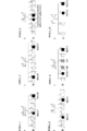
- FIG. 1A shows a family tree for six MSA multiple families.
- FMSA_1's parents were first-degree-cousin. Both FMSA_1 patients (II-4 and II-8) also suffered from retinitis pigmentosa, but neither other sibling MSA nor retinitis pigmentosa.
- the definitive diagnosis of MSA of FMSA_1 II-4 and II-8 and FMSA_8 II-6 was performed by biopsy. The other two brothers of FMSA_8 were PD patients. ⁇ indicates male, ⁇ indicates female, black indicates MSA patient, gray indicates PD patient, white indicates no affected person, and black dot indicates that genomic DNA is available.
- FIG. 1B shows a multipoint parametric linkage analysis of FMSA_1.
- SNPHiTLink Hardy-Weinberg test with p value> 0.05, call rate> 0.95, genotyping confidence score ⁇ 0.1, control minor allele frequency> 0, and marker distance for linkage analysis is 80kb SNP that was 120 kb was selected.
- Multipoint parametric linkage analysis autosomal recessive inheritance with complete penetrance
- haplotype reconstruction were performed in Allegro version 2.
- FIG. 1C is a diagram illustrating a process of narrowing down candidate mutations.
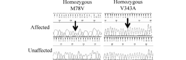
- FIG. 1D shows the results of direct sequencing of FMSA_1 patients (II-4. Upper panel) and unaffected individuals (II-7. Lower panel). The patient had a homozygote for M78V-V343A.
- FIG. 2A shows the result of the yeast complementation assay of the COQ2 mutant.
- the growth panel of yeast coq2 null mutant transformed with pAUR123 vector containing wild type (wt) human COQ2 gene cDNA or mock vector is shown in the left panel.
- the growth curve of the yeast coq2 null mutant transformed with pAUR123 vector containing human COQ2 cDNA with various mutations is shown in the middle panel. I97T, T267A, and S297C showed a significantly reduced growth rate compared to wild-type COQ2, but a higher growth rate than the coq2 null strain (moderately deleterious mutation).
- L16V, P22L, F29L, N336H, and V343A showed the same growth rate as wild-type COQ2.
- Growth curve of coq2 null mutant transformed with pAUR123 vector containing human COQ2 cDNA (P49H, S57T, R69H, M78V, M78V-V343A, P107S, S113F, R337X, or R337Q) with various mutations in the right panel Show. These showed a marked decrease in the respiratory dependent growth rate, as did the coq2 null strain (a very deleterious mutation).
- Each yeast strain was pre-cultured in YPD medium, diluted to an OD600 of 0.1, and incubated for 4 days at 23 ° C.
- FIG. 2B shows the measurement results of the enzyme activity of the CoQ2 mutant. Measure CoQ2 activity in lymphoblastoid cells obtained from MSA patients with any mutation of COQ2 gene (R337Q / V343A, R337X / V343A, V343A / V343A, or V343A / wt) and controls without mutation did.
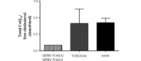
- FIG. 2C shows the results of measuring the CoQ10 concentration in frozen cerebellum samples of MSA patients. CoQ10 concentrations were measured in frozen cerebellar samples of 3 MSA patients (1 M78V-V343A / M78V-V343A and 2 V343A / wt) and 3 controls (wt / wt).
- FIG. 3 is Figure ⁇ 1 of Andrew J. et al., The American Journal of Human Genetics 84, 558-566, May 15, 2009. The outline of biosynthesis of CoQ10 is shown.
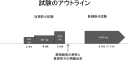
- FIG. 4 shows the outline of a clinical trial of ubiquinol in MSA patients.
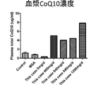
- FIG. 5 shows the measurement results of plasma CoQ10 concentration ( ⁇ g / ml) after ubiquinol was administered at each dose.
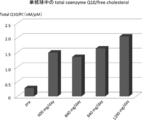
- FIG. 6 shows the measurement results of total CoQ10 / free cholesterol (nM / ⁇ M) in mononuclear cells after ubiquinol was administered at each dose.
- FIG. 7 shows the measurement results of cerebrospinal fluid CoQ10 concentration ( ⁇ g / ml) after ubiquinol was administered at each dose.
- FIG. 8 shows the measurement results of urinary 8-OHdG (ng / mg-Cre) after ubiquinol was administered at each dose.
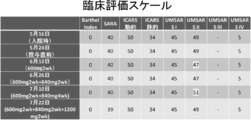
- FIG. 9 shows the clinical evaluation scale of ubiquinol.
- FIG. 10 shows the measurement results of cerebral blood flow and oxygen metabolism before and after ubiquinol administration.
- the test method for MSA risk according to the present invention includes a step of detecting a mutation that decreases biosynthesis of coenzyme Q10 in a sample collected from a subject.
- the MSA risk test method is whether the subject is actually MSA when the possibility of developing MSA is determined or when the subject has MSA-like symptoms.
- the inspection method according to the present invention can be implemented by an inspection company or the like.
- the clinical disease type of MSA is not particularly limited, as described later, a specific embodiment of the method according to the present invention is suitable for specific detection of MSA-C.
- CoQ10 means a benzoquinone derivative also called ubiquinone. Although the oxidized form is sometimes called ubiquinone and the reduced form is called ubiquinol, CoQ10 in this specification may be either oxidized or reduced.
- An outline of the biosynthesis pathway of CoQ10 is shown in FIG. When CoQ2 is catalyzed to prenylate parahydroxybenzoate (PHB), decaprenyl PHB is produced. This decaprenyl PHB undergoes various modifications by more coenzymes to biosynthesize CoQ10.
- PHB parahydroxybenzoate
- mutations in the COQ2 gene related to CoQ10 biosynthesis are involved in MSA risk. Therefore, it is understood that not only COQ2 mutation but also mutation that reduces CoQ10 biosynthesis may increase MSA risk.
- mutations that reduce CoQ10 biosynthesis include, but are not limited to, mutations of various enzymes involved in CoQ10 biosynthesis in addition to COQ2 mutations described below. Those skilled in the art can detect such mutations by appropriately selecting known mutations that reduce the biosynthesis of CoQ10 or newly discovered mutations.
- the sample collected from the subject may be any sample as long as a mutation that reduces the biosynthesis of CoQ10 can be detected.
- a sample for example, blood and other body fluids, skin, tissue, cells, etc. Is mentioned.
- CoQ2 is an enzyme having the following amino acid sequence and is encoded by the COQ2 gene. Also called Para-hydroxybenzoate-polyprenyltransferase, it catalyzes the transfer of decaprenyl group from decaprenyl pyrophosphate to PHB in the biosynthesis of CoQ10.
- Examples of the CoQ2 mutation that reduces the biosynthesis of CoQ10 include P49H, S57T, R69H, M78V, I97T, P107S, S113F, T267A, S297C, R337Q, R337X, and V343A.
- the numbers such as 49 and 57 represent the 49th and 57th positions of the amino acid sequence represented by SEQ ID NO: 1, respectively, and the amino acid represented by one letter to the left of the number represents a wild-type amino acid residue.
- the amino acid represented by one letter to the right of the number indicates a mutant amino acid residue.
- R337X When there is no alphabet to the right of the number, such as R337X, or when the alphabet to the right of the number is X, it means that a stop codon occurs in the amino acid part in the mutant and the subsequent amino acid sequence is deleted To do.
- V343A was unique to Japanese. Since Japanese have more MSA-C than Westerners, the mutation in V343A is considered to be highly related to MSA-C.
- the human COQ2 gene contains four ATG codons in the first exon.
- SEQ ID NO: 1 is Of these four ATG codons, this is based on the UniProt database (Q96H96, http://www.uniprot.org/uniprot/Q96H96) with the fourth as the translation initiation codon.
- the following table shows a comparison when each mutation is annotated using NM_015697.7 and NG_015825.1 of NCBI Reference Sequence using the first ATG codon as a translation initiation codon and BC008804.2 of GenBank.
- the mutations may be detected by detecting mutations on the genomic DNA, or at the RNA level or protein level.
- a cDNA may be prepared from RNA derived from a subject's sample, and a mutation in the cDNA may be detected.
- the method for isolating DNA or RNA is not particularly limited, and chromosomal DNA or RNA can be extracted and isolated by methods known to those skilled in the art.
- detecting nucleic acid mutations those skilled in the art can easily identify what mutations should be detected at the nucleic acid level from the amino acid mutations described above.
- a gene having a specific mutation is a method for detecting a mutation by utilizing the fact that the length of fragments generated by cleavage with a restriction enzyme is different. For example, it can be carried out by the following procedure. For genomic DNA obtained from a subject, a region containing the mutation to be detected is amplified by PCR. A restriction enzyme that recognizes the mutation site is allowed to act on the amplified product, and fragments are separated and identified by electrophoresis or the like. From the length of the generated fragment, it can be confirmed whether or not there is a restriction enzyme, and thus mutation. This method may be performed by extracting RNA from a sample of a subject and preparing cDNA by reverse transcriptase.
- Allele-specific PCR PCR is performed using a primer (allele-specific oligonuceoide: ASO) that hybridizes to the region containing the mutation. If there is a mutation, a mismatch occurs between the primer and the template DNA, so extension occurs when the annealing temperature is high. This is a method utilizing the fact that no reaction occurs. By separating and identifying amplification products by electrophoresis or the like and confirming the presence or absence of amplification, the presence or absence of mutation in the sample DNA can be known.
- ASO allele-specific oligonuceoide
- chromosomal DNA obtained from a subject is amplified by PCR using a primer labeled with 32 P or the like.
- the obtained labeled DNA fragment is denatured by heating to give single-stranded DNA, which is separated by polyacrylamide gel electrophoresis, and the change in the position of the band is detected by autoradiography.
- DGGE Denaturing gradient gel electrophoresis
- the concentration gradient of the denaturant may be applied vertically (vertical gradient method) or in parallel (parallel gradient method).
- temperature gradient gel electrophoresis (TGGE) using a principle similar to DGGE has been developed.
- (v) Method using a microarray This is a method for analyzing the presence or absence of mutation by detecting specific hybridization between a probe DNA immobilized on a microarray and a DNA or RNA sample.
- Etc Various kinds of labels such as fluorescent dyes, radioactive substances, and compounds that can be detected electrochemically can be selected.
- the specific hybridization means specific hybridization under normal hybridization conditions, preferably under highly stringent hybridization conditions.
- highly stringent conditions refer to, for example, hybridization in a 65 ° C. solution of at least about 6 ⁇ SSC and 1% SDS, and an initial wash at 20 ° C. of 20% (v / v) formamide ( (In 0.1 ⁇ SSC) for 10 minutes, and the following washing can be performed under conditions of 0.2 ⁇ SSC and 0.1% SDS at 65 ° C., but is not limited thereto, and those skilled in the art can appropriately select the conditions. .
- each DNA is labeled by various methods, and the base sequence is determined by a cycle sequence reaction using a thermal cycler or the like.
- a method for labeling DNA there are a dye primer method for fluorescently labeling a primer, a dye terminator method for fluorescently labeling ddNTP, and an internal label method for labeling a substrate dNTP.
- a genomic DNA obtained from a subject can be used as a template, and a region containing a mutation to be detected can be amplified by the PCR method to be a target DNA.
- next-generation sequencer a method comprising amplifying a region containing a mutation to be detected by PCR using genomic DNA obtained from a subject as a template, and analyzing the sequence using a next-generation sequencer Can also be used.
- the next-generation sequencer is a term used in contrast to a sequencer using the Sanger method as a “first generation sequencer”.
- massive parallel sequences can be analyzed at a low cost and in a short time by massively parallel processing (for example, Holt RA and Jones SJ: Genome Res., Vol.18 (6 ): 839-864, 2008).
- Other methods for detecting mutations include using a mass spectrometer (MALDI TOF-MS, etc.) to detect polymorphism by mass difference; allele-specific oligos labeled with quenchers and fluorescent dyes And TaqMan PCR method, in which PCR is performed using Taq DNA polymerase; so-called invader method; rolling circle method; method for analyzing the sequence of sample DNA using a sequencer, denatured HPLC method, Melting Temperature analysis, PCR Examples include, but are not limited to, the -SSOP (sequence-specific oligonucleotide nucleotide probe) method, the PCR-PHFA (preferential homoformation assay) method, and the PCR-RSCA (reference strand conformation assay) method.
- MALDI TOF-MS mass spectrometer
- RNA is removed from a sample derived from a subject according to a conventional method, and heated in a solution containing glyoxal, formamide, formalin, methylmercury, etc. Break the next structure to make it on the line. Thereafter, electrophoresis is performed on an agarose gel containing formalin. Transfer the gel to a nylon membrane or nitrocellulose membrane with a high salt solution of 15-20 x SSC. In the case of a nitrocellulose membrane, RNA is fixed by treating in a vacuum oven at 80 ° C for about 2 hours.
- the RNA is immobilized by crosslinking by irradiating with ultraviolet rays.
- a probe is prepared from the cloned cDNA, labeled, and after contacting the probe and membrane under specific conditions, only the mRNA bound to the cDNA probe is detected. it can.
- Hybridization conditions can be appropriately selected by those skilled in the art depending on salt concentration, temperature, base length, composition, and the like.
- RNA sample is subjected to a reverse transcription reaction using a reverse transcriptase and an oligo (dT) primer, the obtained cDNA is amplified by a PCR method, and then the cDNA is detected using a labeled complementary nucleic acid.
- Dot blot method A modified version of the Northern blot method, in which mRNA extracted from a sample derived from a subject is denatured by the methylmercury hydroxide method, etc., and then applied to filters such as nitrocellulose at various concentrations. Spotted as (dot) and hybridized with a probe labeled with a radioactive label or the like to detect the intensity of the signal. Although there is a possibility of detecting another mRNA having a homology to the probe or a mRNA having a different length from the target mRNA, it is simpler than the Northern blot method.
- RNase protection assay This is a method that utilizes the property that RNase does not degrade double-stranded RNA hybridized in perfect agreement with the target RNA, but cleaves when there is a mismatch.
- mRNA labeled with 32 P or the like is used as a probe to hybridize with sample RNA, and then digested with RNase.
- the reaction product is electrophoresed on an agarose gel or polyacrylamide gel, and the size is measured. If the transcript is completely complementary to the probe RNA, a large single band is displayed, but if there is a mismatch, the size of the band will be reduced or two or more bands will be generated. The presence or absence of RNA can be confirmed.
- In situ hybridization A sample obtained from a subject is pretreated with proteolytic enzyme or hydrochloric acid, and then blocked with salmon sperm DNA or albumin to suppress nonspecific binding of the probe. Thereafter, the probe RNA labeled with the labeling substance and the tissue specimen are hybridized over about 24 hours, and then the tissue specimen is washed, and the target site is detected by autoradiography or immunohistochemical method. In addition, in situ RT-PCR method is used when the target mRNA is a very small amount. A reverse transcription reaction is performed using a reverse transcriptase and an oligo (dT) primer, and the obtained cDNA is amplified by a PCR method, and then the cDNA is detected using a labeled complementary nucleic acid.
- dT oligo
- the real-time PCR method is a method for detecting a PCR amplification product by fluorescence.
- cDNA can be obtained from RNA in a sample by reverse transcriptase and used as a template.
- DNA microarray In addition, detection at the RNA level can also be performed by a method using a DNA microarray. By using a DNA microarray in which total RNA is extracted from a sample of a subject and DNA complementary to each of the target RNAs having a plurality of mutations is immobilized, a plurality of mutations can be detected at a time.
- Examples of the method for detecting mutation at the protein level include an immunoassay that confirms the presence or absence of binding to an antibody using an antibody that binds to either CoQ2 having mutation or CoQ2 not having mutation without cross-linking. It is done.
- Such antibodies can be prepared by methods known to those skilled in the art. Detection of binding to the antibody can be performed by labeling the antibody or the secondary antibody by a known method.
- the labeling substance examples include enzymes such as peroxidase and alkaline phosphatase; radioactive substances such as 125 I, 131 I, 35 S, and 3 H; fluorescein isothiocyanate, rhodamine, dansyl chloride, phycoerythrin, tetramethylrhodamine isothiocyanate And fluorescent materials such as near-infrared fluorescent materials; luminescent materials such as luciferase, luciferin, and aequorin; and nanoparticles such as gold colloids and quantum dots.
- enzymes such as peroxidase and alkaline phosphatase
- radioactive substances such as 125 I, 131 I, 35 S, and 3 H
- fluorescent materials such as near-infrared fluorescent materials
- Western blotting can also be mentioned.
- a sample obtained from a subject is treated with SDS or the like to destroy and denature the higher-order structure of the protein, and then the protein is separated according to molecular weight by SDS-PAGE.
- the gel is overlapped with a membrane (nitrocellulose, nylon, PVDF, etc.) and set in a transfer device, and the protein band developed in the gel is electrically transferred (blotted) onto the membrane.
- a secondary antibody that specifically recognizes the primary antibody molecule labeled with a chromogenic enzyme or the like is reacted with the primary antibody, and the target protein is detected through detection of the secondary antibody.
- a decrease in the function of the CoQ2 protein may be detected as a detection of a mutation that decreases the biosynthesis of CoQ10.
- the decrease in the function of CoQ2 can be confirmed, for example, by measuring the decrease in prenylation activity of parahydroxybenzoate.
- CoQ2 activity can be measured by labeling PHB with a radioactive substance and detecting decaprenyl PHB.
- the test kit for MSA risk includes at least one of the following (i) to (iii).
- nucleic acids and antibodies can be used in the MSA risk testing method according to the present invention.
- the nucleic acid of (i) can be specifically bound to a mutation site in the nucleic acid to detect the presence or absence of the mutation.
- the nucleic acid can be, for example, 5 to 100 bases long, 10 to 50 bases long, 15 to 30 bases long, and the like.
- the sequence of the nucleic acid can be appropriately designed by those skilled in the art depending on the detection sequence.
- These nucleic acids may be immobilized on a solid support.
- the solid phase carrier is not particularly limited as long as it is a carrier capable of immobilizing DNA, glass, metal, resin, etc. microtiter plate, substrate, beads, nitrocellulose membrane, nylon membrane, PVDF membrane Etc. DNA can be immobilized on these solid phase carriers by a known method.
- the nucleic acid (ii) described above enables detection of mutation by specifically amplifying a nucleic acid having mutation by PCR.
- Each primer can be, for example, 5 to 100 bases long, 10 to 50 bases long, 15 to 30 bases long, and the like.
- the primer sequence can be appropriately designed by those skilled in the art according to the detection sequence.
- the antibody (iii) above can detect a mutation in the CoQ2 protein by binding to either a protein having a mutation or a wild-type protein without crossover.
- the antibody can be prepared according to a method known to those skilled in the art.
- the antibody may be labeled or may be immobilized on a solid support.
- the kit according to the present invention may contain a secondary antibody as necessary.
- the antibody may be a monoclonal antibody or a polyclonal antibody.
- a monoclonal antibody can be produced by isolating antibody-producing cells from an animal immunized with an antigen and fusing them with myeloma cells to produce a hybridoma and producing the hybridoma.
- Polyclonal antibodies can be obtained from the sera of animals immunized with an antigen.
- the antibody contained in the test kit according to the present invention can be labeled with a fluorescent substance, a radioactive substance or the like.
- a kit may further include, for example, a detection probe, a reverse transcriptase, various reaction / detection reagents, a buffer, an instruction manual, a secondary antibody, and the like.
- the present invention also includes an MSA diagnostic method for detecting a mutation that reduces CoQ10 biosynthesis in a sample collected from a subject. Detection of a mutation that reduces the biosynthesis of CoQ10 can be performed in the same manner as in the test method according to the present invention, and thus description thereof is omitted here.
- the therapeutic agent for MSA according to the present invention contains CoQ10 as an active ingredient.
- the method for preventing or treating MSA according to the present invention includes a step of administering CoQ10.
- the decrease in the amount of CoQ10 due to the mutation of an enzyme involved in CoQ10 biosynthesis is involved in the onset.
- the administration form of MSA is not particularly limited, and may be orally or parenterally administered. Parenteral administration includes, for example, intramuscular injection, intravenous injection, subcutaneous injection, transdermal administration, transmucosal administration (nasal, oral cavity, ocular, pulmonary, vaginal, transrectal) administration. Etc. are raised.
- CoQ10 may be administered as it is, or may be formulated by adding pharmaceutically acceptable carriers, excipients, additives and the like.
- the dosage form include liquids (for example, injections), dispersions, suspensions, tablets, pills, powders, suppositories, powders, fine granules, granules, capsules, syrups, lozenges, Examples include inhalants, ointments, eye drops, nasal drops, ear drops, and poultices.
- Formulation includes, for example, excipients, binders, disintegrants, lubricants, solubilizers, solubilizers, colorants, flavoring agents, stabilizers, emulsifiers, absorption enhancers, surfactants, pH adjustments It can be carried out by a conventional method using an agent, preservative, antioxidant and the like.
- ingredients used for formulation include purified water, saline, phosphate buffer, dextrose, glycerol, ethanol and other pharmaceutically acceptable organic solvents, animal and vegetable oils, lactose, mannitol, glucose, sorbitol, crystalline cellulose , Hydroxypropyl cellulose, starch, corn starch, silicic anhydride, magnesium magnesium silicate, collagen, polyvinyl alcohol, polyvinyl pyrrolidone, carboxyvinyl polymer, sodium carboxymethyl cellulose, sodium polyacrylate, sodium alginate, water-soluble dextran, sodium carboxymethyl starch , Pectin, methylcellulose, ethylcellulose, xanthan gum, gum arabic, tragacanth, casein, agar, polyethylene glycol, diglyse Emissions, glycerine, propylene glycol, vaseline, paraffin, octyldodecyl myristate, isopropyl myristate
- Pills or tablets can also be coated with sugar coatings, gastric and enteric substances.
- the injection can contain distilled water for injection, physiological saline, propylene glycol, polyethylene glycol, vegetable oil, alcohols and the like.
- wetting agents, emulsifiers, dispersants, stabilizers, solubilizers, solubilizers, preservatives and the like can be added.
- CoQ10 may be administered in combination with other drugs and treatments useful for MSA. For example, it can be combined with medicines currently used for symptomatic treatment. CoQ10 is also useful for the prevention of MSA. When it is determined that the MSA risk is high by the test method according to the present invention, it is possible to prevent the onset or delay the onset by supplementing CoQ10 even if it does not develop. CoQ10 is also used in so-called health foods. It is safe and has few side effects, so it can be taken continuously.
- the dosage varies depending on the patient's age, sex, weight, sensitivity difference, administration method, administration interval, active ingredient type, formulation type, and is not particularly limited. For example, 30 mg to 2000 MG 40MG-1500MG, 100MG-1200MG can be administered once or divided into several times.
- the method for screening for a prophylactic or therapeutic agent for MSA is a screening method for a prophylactic or therapeutic agent for multiple system atrophy, comprising a step of contacting a candidate compound with a cell, and increasing the amount of coenzyme Q10 in the cell. Selecting a candidate compound to be made.
- the cells used in the screening method are not particularly limited as long as CoQ10 is biosynthesized, and examples thereof include lymphoblastoid cells.
- the candidate compound is not particularly limited, and may be a low molecular compound, a high molecular compound, a nucleic acid, a protein, or the like.
- the V343A mutation is peculiar to Japanese people, and by studying this mutation, it is considered that the mechanism of onset is further elucidated and contributes to research and development of a method for treating or preventing MSA. Nucleic acids containing the V343A mutation are useful for such research and development.
- the present invention further includes a recombinant vector containing the nucleic acid, a transformant containing the vector, and the like.
- MSA multiple familial MSA diagnosis was based on MSA diagnostic criteria with current consensus (Gilman S, et al. Neurology 2008; 71: 670-6.). The present inventors have reported four Japanese MSA multiple families (FMSA_1 to FMSA_4) so far (Reference 12). Six MSA multiple families were enrolled in this study, including two new Japanese MSA families (FMSA_8 and FMSA_12) with two sets of brothers affected by MSA (FIG. 1A). FMSA_1 had a clan marriage (parents first degree cousins), suggesting the possibility of autosomal recessive inheritance. Table 2 shows the clinical findings of the six MSA multiple families. Biopsy was performed on II-4 and II-8 of FMSA_1 and II-6 of FMSA_8 to make a definitive diagnosis of MSA.
- MSA sporadic MSA and controls MSA were diagnosed according to current consensus criteria (Gilman S, et al. Neurology 2008; 71: 670-6.).
- JAMSAC Japan Multiple System Atrophy Research Consortium
- European and North American cohorts were used as independent MSA cohorts.
- European genomic DNA samples included 138 MSA patients and 281 controls at Pittier-Salpetrière Hospital (France), 34 MSA cases and 34 controls at Federico II University (Italy) Of 46 MSA patients from University of Bonn (Germany).
- the European cohort also included samples of five MSA patients from the University of Sydney.
- North American genomic DNA samples include 172 MSA patients and 294 controls provided by the North American Multiple System Atrophy Study Group (NAMSA-SG) It was. Table 3 shows the statistics of the participants.
- NAMSA-SG North American Multiple System Atrophy Study Group
- JGSCAD Japanese Genetic Study Consortium for Alzheimer Disease
- JaCALS Japanese Consortium for Amyotrophic Lateral Sclerosis research
- ALS amyotrophic lateral sclerosis
- FMSA_1 Association analysis and whole genome sequence Association analysis was performed on FMSA_1 using Affymetrix SNP 6.0 arrays. FMSA_1 II-4 genomic DNA samples were analyzed four times with Illumina Genome Analyzer IIx (100-bp-long paired ends). Burrows Wheeler Aligner (BWA) and Smatools were used for alignment and mutation detection with respect to the human genome reference sequence (NCBI36 / hg18 assembly).
- BWA Burrows Wheeler Aligner
- Smatools Smatools were used for alignment and mutation detection with respect to the human genome reference sequence (NCBI36 / hg18 assembly).
- CoQ2 activity The activity of CoQ2 (EC 2.5.1.39) was assayed by measuring the incorporation of radioactive parahydroxybenzoate (PHB) into decaprenyl PHB.
- PHB radioactive parahydroxybenzoate
- the mitochondrial fraction prepared from lymphoblastoid cells using QProteome Mitochondria Isolation kit (Qiagen) was used as the enzyme source. According to the method of Lopez-Martin et al. (Lopez-Martin JM, et al.
- the four SNVs are c.2120A> G of the SHROOM3 gene (NM_020859, Q8TF72), p.K707R in, c.1178T> C, p.V343A of the COQ2 gene (NM_015697, Q96H96), c.382A> G of the COQ2 gene p.M78V, and c.691A> G, p.R231G of the SCEL gene (NM_144777, O95171).
- FMSA_12 A simultaneous segregation analysis of FMSA_12 revealed that their mother (I-2) had a heterozygote for V343A, that the unaffected sibling (II-1) had a wild-type sequence, Undiscovered brother (II-2) had a heterozygote of R337X, and two patients, brothers (II-3 and II-4), had a composite heterozygote of R337X and V343A It was confirmed that it has. R337X was not found in 180 Japanese controls. In the other four MSA families (FMSA_2, FMSA_3, FMSA_4, and FMSA_8), no mutations in the COQ2 gene were found.
- MSA may develop under sufficient conditions all right.
- the nucleotide sequence analysis of the subcloned mutant allele revealed that R337Q / V343A was a complex heterozygote.
- the phase of I97T / V343A could not be determined because the distance between I97T and V343A was too large to be amplified by PCR and genomic DNA samples from parents were not available. While 29 MSA patients had heterozygotes for V343A and 2 MSA patients had another heterozygous mutation (P107S and S113F), 17 controls also had V343A It was also confirmed that two controls, including the heterozygote, had another heterozygote mutation (P22L and N336H).
- V343A Due to genotyping of other neurodegenerative diseases including AD, PD, and ALS, the allele frequency of V343A was 109 / 5,456 (2.0%) in AD patients (2 of them had V343A homozygotes) in PD patients 33 / 1,318 (2.5%) and 31/1268 (2.4%) in ALS patients. These allele frequencies were not significantly different in the first and second cohorts of controls, confirming the specificity of the V343A mutation of the COQ2 gene for MSA.
- transformants with mutant COQ2 cDNA including I97T, T267A and S297C showed significantly lower growth rates than transformants expressing wild-type CoQ2, but a higher growth rate than the coq2 null strain (moderate) Detrimental mutations).
- Transformants with mutant COQ2 cDNA including L16V, P22L, F29L, N336H and V343A showed a growth rate equivalent to that of transformants expressing wild-type CoQ2.
- CoQ2 activity in lymphoblastoid cells was measured in relation to the V343A mutant for those for which lymphoblastoid cells having a COQ2 mutation were available.
- V343A is a mutation that is well associated with MSA and was selected because this mutant showed normal growth in the yeast complementation assay. Lymphoblastoid cells from MSA patients with any of the CoQ2 mutations (R337Q / V343A, R337X / V343A, V343A / V343A, or V343A / wt) and lymphoblastoid cells from controls without the mutation CoQ2 activity in the cells was measured.
- CoQ2 activity in these patients was significantly lower compared to controls without mutations (FIG. 2B).
- the CoQ2 activity in the V343A mutant was significantly reduced even though the yeast coq2 null strain transformed with the V343A mutant COQ2 cDNA showed a normal growth rate.
- Intracellular CoQ10 concentration in lymphoblastoid cells Lymphoblastoid cells obtained from MSA patients with V343A carrier, lymphoblastoid cells obtained from MSA patients without mutation, and lymph obtained from controls without mutation The intracellular CoQ10 concentration in blast-like cells was measured.
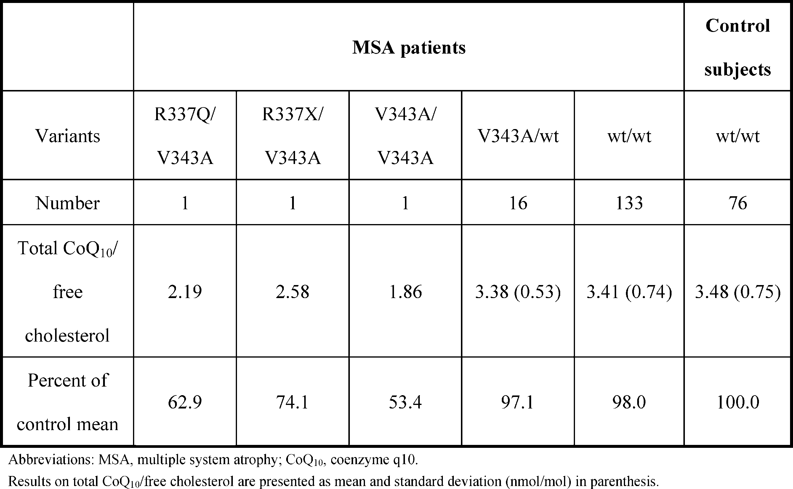
- the subjects were (1) MSA patients who were carriers of two mutant alleles (R337Q / V343A, R337X / V343A, and V343A / V343A), (2) 16 MSA patients who were carriers of heterozygotes for V343A, ( 3) 133 MSA patients without mutation and (4) 76 controls without mutation (Table 9).
- lymphoblastoid intracellular CoQ10 concentration obtained from MSA patients as carriers of the two mutant alleles was significantly lower than that of the control without mutation.
- MSA patients with V343A heterozygotes tended to have lower intracellular CoQ10 concentrations compared to controls without mutations, but this was not a significant difference.
- Lymphoblastoid intracellular CoQ10 concentrations in MSA patients without the COQ2 mutation were comparable to controls without the COQ2 mutation.
- Test Evaluation Items are as follows. (1) Primary endpoint ⁇ Coenzyme Q10 concentration in plasma, leukocytes, and cerebrospinal fluid ⁇ Existence of adverse events (2) Secondary endpoint ⁇ UMSARS Part II (Evaluation of motor function) ⁇ Urine 8-OHdG ⁇ Evaluation of oxygen metabolism by 15O-PET (3) Safety evaluation items and liver function tests (AST, ALT, ⁇ -GTP, ALP, T.Bil) There was a report that AST and ALT were slightly elevated after 300mg / kg coenzyme Q10 was administered to rats. ⁇ Subjective symptoms, physical examinations, objective findings by neurological examinations, abnormalities in various laboratory values
- Test Outline The test outline is shown in FIG.
- FIG. 6 shows the measurement results of total CoQ10 / free cholesterol (nM / ⁇ M) in mononuclear cells. It was confirmed that administration of ubiquinol increased total CoQ10 / free cholesterol in mononuclear cells in a dose-dependent manner.
- Clinical Evaluation Scale The clinical evaluation scale is shown in FIG. Although there were minor fluctuations, there were no changes that could be considered statistically significant. Impressions such as improved response to calls from the attending physician and family (wife), improved upper limb fist, and reduced trembling of the limbs were obtained. However, there were multiple factors such as subjective bias and rehabilitation effects during hospitalization, making judgment difficult.
- CoQ10 can compensate for the decrease in CoQ10. 5.
- No adverse events were observed after 2 weeks of ubiquinol 1200 mg. 6).
- Evaluation of [ 15 O] O 2 PET clearly improved brain oxygen consumption compared to pre-dose.
- CoQ10 supplementation may be a surrogate marker for evaluating improvement of CNS function.
Landscapes
- Health & Medical Sciences (AREA)
- Life Sciences & Earth Sciences (AREA)
- Chemical & Material Sciences (AREA)
- Engineering & Computer Science (AREA)
- Biomedical Technology (AREA)
- General Health & Medical Sciences (AREA)
- Organic Chemistry (AREA)
- Proteomics, Peptides & Aminoacids (AREA)
- Medicinal Chemistry (AREA)
- Neurosurgery (AREA)
- Neurology (AREA)
- Bioinformatics & Cheminformatics (AREA)
- Molecular Biology (AREA)
- Immunology (AREA)
- Analytical Chemistry (AREA)
- Veterinary Medicine (AREA)
- Pharmacology & Pharmacy (AREA)
- Animal Behavior & Ethology (AREA)
- Public Health (AREA)
- Biotechnology (AREA)
- Biochemistry (AREA)
- Zoology (AREA)
- Wood Science & Technology (AREA)
- Genetics & Genomics (AREA)
- Hematology (AREA)
- Pathology (AREA)
- Microbiology (AREA)
- Physics & Mathematics (AREA)
- Urology & Nephrology (AREA)
- Chemical Kinetics & Catalysis (AREA)
- Nuclear Medicine, Radiotherapy & Molecular Imaging (AREA)
- General Chemical & Material Sciences (AREA)
- Cell Biology (AREA)
- General Physics & Mathematics (AREA)
- Food Science & Technology (AREA)
- Biophysics (AREA)
- General Engineering & Computer Science (AREA)
- Epidemiology (AREA)
- Psychology (AREA)
- Hospice & Palliative Care (AREA)
Abstract
Description
病理学的には、glial cytoplasmic inclusion(GCI)が特徴で、α-シヌクレインが主要成分である。
現在までのところMSAの発症機構はまったく不明であり、治療としては症状に応じた対症療法が行われている。
即ち、本発明は、
〔1〕被検者の多系統萎縮症リスクの検査方法であって、被検者から採取した試料におけるコエンザイムQ10の生合成を低下させる変異を検出する工程を含む、方法;
〔2〕前記コエンザイムQ10の生合成を低下させる変異は、Para-hydroxybenzoate-polyprenyltransferase(コエンザイムQ2)の発現又は機能を抑制する変異である、上記〔1〕に記載の方法;
〔3〕前記コエンザイムQ2の発現又は機能を抑制する変異は、配列番号:1におけるP49H、S57T、R69H、M78V、I97T、P107S、S113F、T267A、S297C、R337Q、R337X、及びV343Aからなる群より選択される、上記〔2〕に記載の方法;
〔4〕前記コエンザイムQ2の発現又は機能を抑制する変異は、V343Aである、上記〔2〕に記載の方法;
〔5〕多系統萎縮症リスクの検査キットであって、以下の(i)から(iii)の少なくとも1つを含むキット:
(i) コエンザイムQ2遺伝子における、コエンザイムQ2タンパク質(配列番号:1)の49位、57位、69位、78位、97位、107位、113位、267位、297位、337位、及び343位からなる群より選択される部位のアミノ酸をコードする核酸を含む領域にハイブリダイズする核酸;
(ii) コエンザイムQ2遺伝子における、コエンザイムQ2タンパク質(配列番号:1)の49位、57位、69位、78位、97位、107位、113位、267位、297位、337位、及び343位からなる群より選択される部位のアミノ酸をコードする核酸を含む領域を増幅可能なプライマーセット;及び
(iii) 配列番号:1において、P49H、S57T、R69H、M78V、I97T、P107S、S113F、T267A、S297C、R337Q、R337X、及びV343Aからなる群より選択される少なくとも1つの変異を有するコエンザイムQ2タンパク質と、野生型コエンザイムQ2タンパク質のいずれかのみに交差性なく結合する抗体;
〔6〕コエンザイムQ10を含む多系統萎縮症の予防又は治療剤;
〔7〕コエンザイムQ10を投与する工程を含む、多系統萎縮症の予防又は治療方法;
〔8〕多系統萎縮症の予防又は治療剤のスクリーニング方法であって、
候補化合物と細胞とを接触させてインキュベートする工程と、
細胞内のコエンザイムQ10量を上昇させる候補化合物を選択する工程と、を含む方法;
及び、
〔9〕V343A変異を含むコエンザイムQ2タンパク質をコードする核酸、
に関する。
MSA発症リスクが高いと判断された場合には、CoQ10の補充により発症が抑制されることが期待され、またCoQ10の補充はMSAの治療にも有効である可能性が強く示唆される。
本発明に係るMSAリスクの検査方法は、被検者から採取した試料における、コエンザイムQ10の生合成を低下させる変異を検出する工程を含む。
本明細書において、MSAリスクの検査方法とは、被検者がMSAを発症する可能性の高さを判定したり、被検者にMSA様の症状が見られる場合に実際にMSAであるか否かを判定したりするために必要な情報を収集するために行う検査の方法をいう。本発明に係る検査方法は、検査会社等で実施されうる。
MSAの臨床病型は特に限定されないが、後述するとおり、本発明に係る方法のうち特定の態様は、MSA-Cの特異的な検出に適している。
CoQ10の生合成経路の概略を図3に示す。CoQ2が触媒してパラヒドロキシベンゾエート(PHB)がプレニル化されると、デカプレニルPHBが生じる。このデカプレニルPHBが、さらに多くの補酵素によって様々な修飾を受けてCoQ10が生合成される。
本明細書において、CoQ2は、以下のアミノ酸配列を有する酵素であり、COQ2遺伝子によってコードされる。Para-hydroxybenzoate-polyprenyltransferaseとも呼ばれ、CoQ10の生合成において、デカプレニルピロリン酸からPHBにデカプレニル基を転移する反応を触媒する。
MLGSRAAGFARGLRALALAWLPGWRGRSFALARAAGAPHGGDLQPPACPEPRGRQLSLSAAAVVDSAPRPLQPYLRLMRLDKPIGTWLLYLPCTWSIGLAAEPGCFPDWYMLSLFGTGAILMRGAGCTINDMWDQDYDKKVTRTANRPIAAGDISTFQSFVFLGGQLTLALGVLLCLNYYSIALGAGSLLLVITYPLMKRISYWPQLALGLTFNWGALLGWSAIKGSCDPSVCLPLYFSGVMWTLIYDTIYAHQDKRDDVLIGLKSTALRFGENTKPWLSGFSVAMLGALSLVGVNSGQTAPYYAALGAVGAHLTHQIYTLDIHRPEDCWNKFISNRTLGLIVFLGIVLGNLWKEKKTDKTKKGIENKIEN (配列番号:1)
この4つのATGコドンのうち、4番目を翻訳開始コドンとしたUniProt database(Q96H96, http://www.uniprot.org/uniprot/Q96H96)に従ったものである。
1番目のATGコドンを翻訳開始コドンとするNCBI Reference SequenceのNM_015697.7及びNG_015825.1と、GenBankのBC008804.2のそれぞれを用いて各変異をアノテーションした場合の比較を下表に示す。

核酸の変異を検出する場合、当業者は上述したアミノ酸の変異から、核酸レベルではどのような変異を検出すればよいか容易に特定することができる。
(i) PCR-RFLP(Restriction Fragment Length Polymorphism)
特定の変異を有する遺伝子は制限酵素による切断によって生じる断片の長さが異なることを利用して、変異を検出する方法であり、例えば以下の手順により行うことができる。被検者から得たゲノムDNAについて、検出しようとする変異を含む領域をPCR法により増幅する。増幅産物に変異部位を認識する制限酵素を作用させ、断片を電気泳動法等により分離、同定する。生じた断片の長さから制限酵素による切断の有無、ひいては変異の有無を確認できる。本方法は、被検者の試料からRNAを抽出し、逆転写酵素によりcDNAを調製して行ってもよい。
変異を含む領域にハイブリダイズするプライマー(allele-specific oligonuceoide: ASO)を用いてPCRを行い、変異を有する場合、当該プライマーと鋳型DNAとの間にミスマッチが生じるため、アニール温度が高い場合に伸長反応が起こらないことを利用する方法である。増幅産物を電気泳動法等により分離、同定し、増幅の有無を確認することによって、試料DNAの変異の有無を知ることができる。
DNAをPCRで増幅した後、一本鎖に解離させると、一本鎖DNAは塩基対形成をはじめとする種々の分子内相互作用で塩基配列に依存した固有の高次構造をとる。安定な二重らせん構造をとる二本鎖DNAに比べ、一本鎖DNAの高次構造は、一塩基の違いが存在しても変化することがある。そのため一本鎖DNAをポリアクリルアミド中で電気泳動すると、高次構造の違いに応じて移動度も異なり、この移動度の違いを検出することによって変異の有無を判定することができる。
具体的には、例えば、被検者から得られた染色体DNAを、32P等で標識したプライマーを用いてPCRにより増幅する。得られた標識DNA断片を加熱変性して一本鎖DNAとし、これをポリアクリルアミドゲル電気泳動で分離して、オートラジオグラフィーによりバンドの位置変化を検出する。
変性剤による変性は二本鎖DNAにミスマッチが存在すると起こりやすくなること、二本鎖DNAが部分的に融解した状態では電気泳動の移動度が著しく低下することを利用して、変異の有無を検出する方法である。
具体的には、尿素、ホルムアミド等の変性剤の濃度に勾配をつけたポリアクリルアミドゲル上で、必要に応じて制限酵素等で処理した被検者のDNAサンプルと、正常DNA断片と、これらに相補的なプローブ核酸との混合物の電気泳動を行ってサンプルを分離する。変異に起因するミスマッチが存在すると、より低い濃度の変性剤で一本鎖に解離するため、変性剤濃度が低いゲル領域で泳動速度が遅くなる。従って、正常DNAサンプルのバンドと比較することによって、ミスマッチの存在の有無を検出することが可能となり、結果として変異の有無を検出することができる。
変性剤の濃度勾配は、垂直につけてもよいし(垂直勾配法)と平行につけてもよい(平行勾配法)。また、DGGEと類似の原理を利用した温度勾配ゲル電気泳動法(temperature gradient gel electrophoresis: TGGE)も開発されている。
マイクロアレイ上に固定されたプローブDNAと、DNA又はRNAサンプルとの特異的なハイブリダイゼーションを検出して、変異の有無を解析する方法である。ハイブリダイゼーションの有無を検出する方法や、プローブDNAの3'末端を変異の予想される部位にあわせてハイブリダイゼーションさせ、標識したジデオキシヌクレオチドとDNAポリメラーゼを添加して、伸長反応の有無を検出する方法等がある。標識は、蛍光色素、放射性物質、電気化学的に検出できる化合物等各種選択することができる。
なお、特異的なハイブリダイゼーションとは、通常のハイブリダイゼーション条件で、好ましくは高ストリンジェントなハイブリダイゼーション条件下における特異的なハイブリダイゼーションを意味する。本明細書において高ストリンジェントな条件とは、例えば、少なくとも約6×SSC及び1%SDSの65℃溶液中でハイブリダイゼーションを行い、最初の洗浄を42℃の20%(v/v)ホルムアミド(0.1×SSC中)で10分、次の洗浄を65℃の0.2×SSC及び0.1%SDSで行う条件とすることができるが、これに限定されず、当業者は適宜条件を選択することができる。
シーケンス反応を化学発光で検出する方法である。被検者由来のDNAをPCRで増幅した後、一本鎖DNAを精製し、適当なプライマーをハイブリダイズさせてからデオキシヌクレオチドを一塩基ずつ添加する。伸長反応に伴って発生するピロリン酸がカスケード反応を開始させ、生じたATPによりルシフェリンを基質としてルシフェラーゼが発光する。この発光を検出することにより被検DNAの塩基配列を決定することができ、高精度に変異を検出できる(Alderborn, A. et al.: Genome Res. (2000) 28:1249-1258)。
標的DNAとプライマーを用いてDNAを合成する際、4種のデオキシリボヌクレオチド(dNTP)とともに1種のジデオキシリボヌクレオチド(ddNTP)のいずれかを加えておき、ddNTPが取り込まれたときに合成が止まるようにして、様々な長さのDNAを合成する。この反応を4種のジデオキシリボヌクレオチドのそれぞれについて行った後、様々な長さを、ポリアクリルアミドゲルによる電気泳動にかけて分離する。ポリアクリルアミドゲルによる電気泳動では1塩基の違いも道程できるので、どの位置で合成が止まったかを検出することにより、標的DNAの塩基配列がわかる。具体的には、各DNAは種々の方法で標識し、サーマルサイクラーを用いたサイクルシーケンス反応等で塩基配列を決定する。DNAの標識方法としては、プライマーを蛍光標識するDye Primer法、ddNTPを蛍光標識するDye Terminator法、基質dNTPを標識するInternal-label法がある。
本発明においては、被検者から得たゲノムDNAを鋳型として、検出しようとする変異を含む領域をPCR法により増幅して標的DNAとすることができる。
本発明では、被検者から得たゲノムDNAを鋳型として、検出しようとする変異を含む領域をPCR法により増幅し、次世代シーケンサーを用いて配列を解析する方法も用いることができる。次世代シーケンサーは、Sanger法を用いるシーケンサーを「第1世代シーケンサー」とし、これと対比して用いられる用語である。原理は様々なものがあるが、超並列的な処理によって、低コストかつ短時間で大量の塩基配列を解析することができる(例えば、Holt R.A. and Jones S.J.: Genome Res., Vol.18 (6):839-864, 2008)。
(i) ノーザンブロット法
被検者に由来する試料から常法に従ってmRNAを取り出し、これをグリオキサール、ホルムアミド、ホルマリン、メチル水銀などを含む溶液中で加熱し、分子内の水素結合をはずして、高次構造を壊して線上にする。その後、ホルマリンを含むアガロースゲルで電気泳動する。ゲルを15~20×SSCの高塩溶液でナイロンメンブレンやニトロセルロースメンブレンにトランスファーする。ニトロセルロースメンブレンの場合は、80℃で約2時間真空オーブンで処理し、RNAを固定する。ナイロンメンブレンの場合は、たとえば紫外線を照射して架橋によりRNAを固定する。
次に、メンブレン上の特定のmRNAを同定するために、クローン化したcDNAからプローブを作製して標識し、プローブとメンブレンを特定条件下で接触させた後、cDNAプローブが結合したmRNAのみを検出できる。ハイブリダイゼーションの条件は、塩濃度、温度、塩基の長さ、組成等によって、当業者が適宜選択することができる。
標的mRNAが微量の場合は、RT-PCR法を組み合わせることもできる。RNA試料に対して、逆転写酵素とオリゴ(dT)プライマーを用いて逆転写反応を行い、得られたcDNAをPCR法により増幅した後、標識した相補的核酸を使用してcDNAを検出する。
ノーザンブロット法の変法であり、被検者に由来する試料から取り出したmRNAを水酸化メチル水銀法等で変成させた後、様々な濃度でニトロセルロース等のフィルターに点(ドット)としてスポッティングし、放射性標識等で標識したプローブとハイブリダイゼーションさせ、シグナルの強さを検出する。プローブに相同性を有する別のmRNAや、目的mRNAとは長さの異なるものを検出する可能性もあるが、ノーザンブロット法よりも簡便である。
RNaseが標的RNAと完全に一致してハイブリダイズした二本鎖RNAは分解しないが、ミスマッチがあるとそこで切断するという性質を利用する方法である。まず、32P等で標識したmRNAをプローブとして試料RNAとハイブリダイズさせたのち、RNaseによって消化する。反応産物をアガロースゲル又はポリアクリルアミドゲル等で電気泳動し、サイズを測定する。転写産物がプローブRNAと完全に相補的であれば、大きな一つのバンドを示すが、ミスマッチが存在する場合、バンドのサイズが小さくなったり、2本以上のバンドが生じたりするので、目的とするRNAの有無を確認できる。
被検者から得た試料をタンパク質分解酵素や塩酸などで前処理した後、プローブの非特異的な結合を抑えるためにサケ精子DNAやアルブミンなどでブロッキングする。その後、標識物質でラベルしたプローブRNAと組織標本とを約24時間かけてハイブリダイズさせ、その後組織標本を洗浄して、オートラジオグラフィーや免疫組織化学的方法により標的部位を検出する。また、標的mRNAが微量の場合には、in situ RT-PCR法が用いられる。逆転写酵素とオリゴ(dT)プライマーを用いて逆転写反応を行い、得られたcDNAをPCR法により増幅した後、標識した相補的核酸を使用してcDNAを検出する。
リアルタイムPCR法は、PCR増幅産物を蛍光で検出する方法である。SYBR Greenに代表される二本鎖核酸に特異的に挿入する蛍光標識を用いるインターカレート法と、TaqManプローブに代表される蛍光標識した変異配列特異的なプローブを用いる方法がある。リアルタイムPCR法を用いる場合も、試料中のRNAから逆転写酵素でcDNAを得て、これを鋳型とすることができる。
その他、RNAレベルでの検出は、DNAマイクロアレイを用いた方法によっても行うことができる。被検者の試料から全RNAを抽出し、目的とする複数の変異を有するRNAのそれぞれと相補的なDNAを固定したDNAマイクロアレイを用いれば、複数の変異を一度に検出することができる。
CoQ2の機能の低下は、例えば、パラヒドロキシベンゾエートのプレニル化活性の低下を測定して確認することができる。実施例に記載するとおり、CoQ2活性の測定は、PHBを放射性物質で標識し、デカプレニルPHBを検出して行うことができる。
本発明に係るMSAリスクの検査キットは、以下の(i)から(iii)の少なくとも1つを含む。(i) コエンザイムQ2遺伝子における、コエンザイムQ2タンパク質の49位、57位、69位、78位、97位、107位、113位、267位、297位、337位、及び343位からなる群より選択される部位のアミノ酸をコードする核酸を含む領域にハイブリダイズする核酸;
(ii) コエンザイムQ2遺伝子における、コエンザイムQ2タンパク質の49位、57位、69位、78位、97位、107位、113位、267位、297位、337位、及び343位からなる群より選択される部位のアミノ酸をコードする核酸を含む領域を増幅可能なプライマーセット;及び
(iii) P49H、S57T、R69H、M78V、I97T、P107S、S113F、T267A、S297C、R337Q、及びR337X、V343Aからなる群より選択される少なくとも1つの変異を有するコエンザイムQ2タンパク質と、野生型コエンザイムQ2タンパク質のいずれかのみに交差性なく結合する抗体。
上記(i)の核酸は、核酸における変異部位に特異的に結合させて、変異の有無を検出することができる。核酸は、例えば5~100塩基長、10~50塩基長、15~30塩基長等とすることができる。核酸の配列は、検出配列に応じて、当業者が適宜設計可能である。
これらの核酸は、固相担体に固定されていてもよい。本明細書において固相担体とは、DNAを固定できる担体であれば特に限定されず、ガラス製、金属製、樹脂製等のマイクロタイタープレート、基板、ビーズ、ニトロセルロースメンブレン、ナイロンメンブレン、PVDFメンブレン等が挙げられる。DNAは、これらの固相担体に公知の方法で固定することが可能である。
なお、当該抗体は、モノクローナル抗体であっても、ポリクローナル抗体であってもよい。モノクローナル抗体は、抗原で免疫した動物から抗体産生細胞を単離し、これを骨髄腫細胞等と融合させてハイブリドーマを作製し、このハイブリドーマに産生させることができる。ポリクローナル抗体は、抗原で免疫した動物の血清等から得ることができる。本発明に係る検査キットに含まれる抗体は、蛍光物質、放射性物質等で標識しておくこともできる。
このようなキットは、例えば、検出用プローブ、逆転写酵素、各種反応・検出試薬、緩衝液、取扱説明書、二次抗体等をさらに備えていてもよい。
本発明は、被検者から採取した試料におけるCoQ10の生合成を低下させる変異を検出するMSAの診断方法も包含する。CoQ10の生合成を低下させる変異の検出は、本発明に係る検査方法と同様に行うことができるので、ここでは説明を省略する。
本発明に係るMSAの治療剤は、有効成分としてCoQ10を含む。また、本発明に係るMSAの予防又は治療方法は、CoQ10を投与する工程を含む。上述のとおり、MSAの一部では、CoQ10の生合成に関与する酵素の変異によりCoQ10量が減少することが発症に関与する。
MSAの投与形態は特に限定されず、経口的投与でも非経口的投与でもよい。非経口投与としては、例えば、筋肉内注射、静脈内注射、皮下注射等の注射投与、経皮投与、経粘膜投与(経鼻、経口腔、経眼、経肺、経膣、経直腸)投与等が上げられる。
CoQ10は、そのまま投与してもよいし、薬学的に許容できる担体、賦形剤、添加剤等を加えて製剤化してもよい。剤形としては、例えば、液剤(例えば注射剤)、分散剤、懸濁剤、錠剤、丸剤、粉末剤、坐剤、散剤、細粒剤、顆粒剤、カプセル剤、シロップ剤、トローチ剤、吸入剤、軟膏剤、点眼剤、点鼻剤、点耳剤、パップ剤等が挙げられる。
製剤化は、例えば、賦形剤、結合剤、崩壊剤、滑沢剤、溶解剤、溶解補助剤、着色剤、矯味矯臭剤、安定化剤、乳化剤、吸収促進剤、界面活性剤、pH調整剤、防腐剤、抗酸化剤などを適宜使用し、常法により行うことができる。
製剤化に用いられる成分の例としては、精製水、食塩水、リン酸緩衝液、デキストロース、グリセロール、エタノール等薬学的に許容される有機溶剤、動植物油、乳糖、マンニトール、ブドウ糖、ソルビトール、結晶セルロース、ヒドロキシプロピルセルロース、デンプン、コーンスターチ、無水ケイ酸、ケイ酸アルミニウムマグネシウム、コラーゲン、ポリビニルアルコール、ポリビニルピロリドン、カルボキシビニルポリマー、カルボキシメチルセルロースナトリウム、ポリアクリル酸ナトリウム、アルギン酸ナトリウム、水溶性デキストラン、カルボキシメチルスターチナトリウム、ぺクチン、メチルセルロース、エチルセルロース、キサンタンガム、アラビアゴム、トラガント、カゼイン、寒天、ポリエチレングリコール、ジグリセリン、グリセリン、プロピレングリコール、ワセリン、パラフィン、ミリスチン酸オクチルドデシル、ミリスチン酸イソプロピル、高級アルコール、ステアリルアルコール、ステアリン酸、ヒト血清アルブミン、等が挙げられるがこれらに限定されない。
注射剤は、注射用蒸留水、生理食塩水、プロピレングリコール、ポリエチレングリコール、植物油、アルコール類等を含むことができる。さらに、湿潤剤、乳化剤、分散剤、安定化剤、溶解剤、溶解補助剤、防腐剤等を加えることができる。
CoQ10は、MSAの予防にも有用である。本発明に係る検査方法でMSAリスクが高いと判断された場合、発症していなくてもCoQ10を補充することによって、発症を防止したり、発症を遅らせたりすることが可能である。CoQ10は、いわゆる健康食品にも用いられており、安全性が高く副作用も少ないので、継続的に服用することが可能である。
本発明に係るMSAの予防又は治療薬のスクリーニング方法は、多系統萎縮症の予防又は治療剤のスクリーニング方法であって、候補化合物と細胞とを接触させる工程と、細胞内のコエンザイムQ10量を上昇させる候補化合物を選択する工程と、を含む。
スクリーニング方法に用いられる細胞は、CoQ10が生合成される細胞であれば特に限定されないが、例えば、リンパ芽球様細胞などが挙げられる。
候補化合物も特に限定されず、低分子化合物、高分子化合物、核酸、タンパク質等でありうる。これらの候補化合物と細胞とを接触させてインキュベートする際の温度や時間を含む実験条件は、当業者が適宜決定することができる。例えば、CoQ10の生合成に関与する物質の機能を亢進させる候補化合物や、CoQ10の生合成に関与する物質の機能を低下させる物質の機能を阻害する候補化合物などが考えられる。
細胞内のCoQ10量が上昇するか否かも、当業者が公知の方法にしたがって行うことができる。CoQ10活性を測定して、量を評価してもよい。
V343A変異は日本人に特有のものであり、これを研究することにより、さらに発症のメカニズムを解明し、MSAの治療又は予防方法の研究開発に資すると考えられる。V343A変異を含む核酸は、かかる研究開発に有用である。本発明は、さらに、かかる核酸を含む組換えベクター、当該ベクターを含む形質転換体等も包含する。
1-1.ヒトを対象とする研究の承認
被検者は、東京大学及び他の研究参加機関の審査委員会に承認された研究計画に登録された。全ての被検者から書面によるインフォームドコンセントを得た。
MSAの診断は、現在コンセンサスの得られているMSAの診断基準に基づいて行った(Gilman S, et al. Neurology 2008;71:670-6.)。
本発明者らは、これまでに4つの日本のMSA多発家系(FMSA_1からFMSA_4)について報告した(文献12)。MSAを罹患する2組の兄弟がいる2つの新たな日本のMSA家系(FMSA_8及びFMSA_12)を含めて6つのMSA多発家系が本研究に登録された(図1A)。FMSA_1には血族婚があり(両親がfirst degree cousins)、常染色体劣性遺伝の可能性が示唆された。6つのMSA多発家系の臨床所見を表2に示す。FMSA_1のII-4及びII-8とFMSA_8のII-6に生検を実施し、MSAの確定診断とした。

MSAの診断は、現在コンセンサスが得られている基準に従って行った(Gilman S, et al. Neurology 2008;71:670-6.)。日本人コホートは、Japan Multiple System Atrophy Research Consortium (JAMSAC)から試料提供された195人のMSA患者と113人の健常者を含んだ。さらに、東京大学、高齢者ブレインバンク、東京都健康長寿医療センター、新潟大学脳研究所、北海道大学大学院医学研究科、及び鹿児島大学大学院医歯学総合研究科における168人のMSA患者と407人の対照者を、対象とした。

レプリケーションスタディのための対照者の独立コホート(n=2,383)は、Japanese Genetic Study Consortium for Alzheimer Disease(JGSCAD)及びJapanese Consortium for Amyotrophic Lateral Sclerosis research (JaCALS)から提供された。CoQ2変異体とMSAの関連の特異性を調べるために、他の神経変性疾患(アルツハイマー病(AD)、パーキンソン病(PD)、及び筋萎縮性側索硬化症(ALS)患者;JGSCADから2,728人のAD患者、東京大学及びJapanese Parkinson Disease Susceptibility Gene Consortiumから659人のPD患者、東京大学及びJaCALSから634人のALS患者)も調査対象とした。
関連解析は、Affymetrix SNP 6.0 arraysを用いて、FMSA_1に対して行った。FMSA_1のII-4のゲノムDNAサンプルを、Illumina Genome Analyzer IIx (100-bp-long paired ends)で4回分析した。ヒトゲノム参照配列(NCBI36/hg18 assembly)に対するアライメント及び変異の検出には、Burrows Wheeler Aligner (BWA)及びSmatoolsを用いた。
COQ2遺伝子の各エクソンを増幅するプライマーペア表4を用いてPCRを行い、続いてヌクレオチド配列解析を行った。

Exon 1 Reverse primer sequence 配列番号:3
Exon 2 Forward primer sequence 配列番号:4
Exon 2 Reverse primer sequence 配列番号:5
Exon 3 Forward primer sequence 配列番号:6
Exon 3 Reverse primer sequence 配列番号:7
Exon 4 Forward primer sequence 配列番号:8
Exon 4 Reverse primer sequence 配列番号:9
Exon 5 Forward primer sequence 配列番号:10
Exon 5 Reverse primer sequence 配列番号:11
Exon 6 Forward primer sequence 配列番号:12
Exon 6 Reverse primer sequence 配列番号:13
Exon 7 Forward primer sequence 配列番号:14
Exon 7 Reverse primer sequence 配列番号:15
野生型ヒトCOQ2遺伝子に対し、表5のプライマー(上から順に、配列番号:16~47)を用いてPCRによる部位特異的変異導入を行った。続いて野生型と変異型のヒトCOQ2遺伝子cDNAを酵母発現型pAUR123ベクター(Takara社)に挿入した。酵母coq2遺伝子ヌル変異体であるBY4741Δcoq2株を、野生型又は変異型ヒトCOQ2遺伝子cDNAを含むpAUR123ベクターで、Yeastmaker Yeast Transformation System 2(Clontech社)を使って形質転換した。非発酵性炭素源を含む培地(酵母エキス・ペプトン・グリセロール培地)中での増殖を、OD monitor(Titech社)で培地の600nmの吸光度(OD600)をモニターすることにより測定した。

CoQ2(EC 2.5.1.39)の活性は、放射性パラヒドロキシベンゾエート(PHB)のデカプレニルPHBへの取り込みを測定することによりアッセイした。具体的には、QProteome Mitochondria Isolation kit(Qiagen社)を用いてリンパ芽球様細胞から調製したミトコンドリア画分を酵素源とした。Lopez-Martinらの方法(Lopez-Martin JM, et al. Hum Mol Genet 2007;16:1091-7.)に従い、500μgのミトコンドリアリッチ画分、1000μM [14C]PHB (1.85 MBq/μmol)、及び5μMデカプレニルピロリン酸を含む100μLのアッセイ用バッファー(0.05% 3-[(3-クロルアミドプロピル)ジメチルアンモニオ]-1-プロパンスルホネート(CHAPS)を含む)からなる反応液を調製した。反応は37℃で60分行い、1000μLのヘキサンで抽出した。ヘキサン相の放射性物質を液体シンチレーションカウンターTri-Carb 2000CA(PerkinElmer社)で測定した。結果は、9つの独立した実験の平均値で示した。
152人のMSA患者と76人の対照者から樹立したEBウイルス不死化リンパ芽球様細胞(JAMSACより提供を受けた)を、10%ウシ胎児血清を含むRPMI-1640培地で培養した。遊離コレステロールとCoQ10を、約107から108個のリンパ芽球様細胞から4倍量の2プロパノールで抽出するか、3人のMSA患者及び3人の対照者の生検によって調製した小脳の凍結サンプルから9倍量の2プロパノールで抽出した。
抽出物中の遊離コレステロールと総CoQ10濃度(ユビキノン10及びユビキノール10)は、高速液体クロマトグラフィーで測定した。
すべての結果は、平均±標準偏差で示した。キャリアと非キャリアの発症年齢の平均値の有意差は、student t検定で;アレル頻度と分割表の有意差はYates補正カイ2乗検定又はFisherの直接確率検定で、オッズ比と対応する95%信頼区間(CI)はFisherの直接確率検定でそれぞれ評価した。群比較は、Kruskal-Wallis検定とSteel法を用いた。すべての統計は両側検定とし、有意水準はp=0.05とした。
2-1.家族性MSAの原因遺伝子の同定
常染色体劣性遺伝様式の遺伝モデルを使ったFMSA_1のパラメトリックな多点連鎖解析によれば、染色体4、5、6、7、9及び13を含む約80Mbにわたる領域におけるLODスコアの最高値は1.93であった(図1B)。
FMSA_1における患者(II-4)について、ホールゲノムシーケンスを行ったところ、合計で187.5Gbのshort readが得られた。参照ゲノムの平均カバレッジは58倍であった。候補領域に位置する47,830の一塩基変異(SNVs)又は挿入/欠失(indels)から始めて、この家族の変異候補を4つの新規な非同義SNVに絞り込んだ(図1C)。
4つのSNVは、SHROOM3遺伝子(NM_020859, Q8TF72)のc.2120A>G、p.K707R in、COQ2遺伝子(NM_015697, Q96H96)のc.1178T>C, p.V343A、COQ2遺伝子のc.382A>G, p.M78V、及びSCEL遺伝子(NM_144777, O95171)のc.691A>G, p.R231Gであった。
以上の結果から、FMSA_1及びFMSA_12の2家系の家族性MSAでは、M78VとV343Aのホモ接合体、又はR337XとV343Aの複合ヘテロ接合体を有すると、これが十分条件となってMSAが発症することがわかった。
孤発性MSAに対するCOQ2遺伝子変異の関与を調べるため、より大きな日本人コホート(363人のMSA患者と520人の対照者)について、COQ2遺伝子のリシーケンスを行った。dbSNP130に登録された非同義COQ2遺伝子変異体は一つだけであった(L16V、rs6818847)が、本発明者らは、L16Vのアレル頻度が日本人のMSA患者においては0.90、日本人の対照者においては0.88であることを確認したため、この変異体をその後の解析に含めなかった。
COQ2遺伝子のリシーケンスにより、4人のMSA患者が2つの変異を同時に有していたが(1人はI97T/V343A、1人はR337Q/V343A、2人はV343A/V343A)、対照者には2つの変異をCOQ2遺伝子に有する者は存在しないことが明らかとなった(表6)。


AD、PD及びALSを含む他の神経変性疾患のジェノタイピングにより、V343Aのアレル頻度はAD患者において109/5,456 (2.0%)(うち2人はV343Aのホモ接合体を有した)、PD患者において33/1,318 (2.5%)、ALS患者において31/1268 (2.4 %)であった。これらのアレル頻度は、対照者の第一及び第二コホートにおいては有意差が見られず、COQ2遺伝子のV343A変異のMSAに対する特異性が確認できた。
CoQ10の生合成と好気的エネルギー産生に対する、各変異の機能への影響を調べるために、酵母coq2遺伝子ヌル変異体を、野生型又は変異型ヒトCOQ2遺伝子cDNAで形質転換して、機能相補性解析を行った(図2A)。BY4741Δcoq2酵母株の変異COQ2遺伝子(P49H、S57T、R69H、M78V、M78V-V343A、P107S、S113F、R337Q及びR337Xを含む)による形質転換体においては、coq2ヌル株に見られるのと同様に、呼吸依存性増殖が著しく低下した(非常に有害な変異)。さらに、変異型COQ2 cDNA(I97T、T267A及びS297Cを含む)による形質転換体は、野生型CoQ2を発現する形質転換体に比べてかなり低いが、coq2ヌル株よりは高い増殖率を示した(中等度に有害な変異)。変異型COQ2 cDNA(L16V、P22L、F29L、N336H及びV343Aを含む)による形質転換体は、野生型CoQ2を発現する形質転換体と同等の増殖率を示した。
次に、COQ2変異を有するリンパ芽球様細胞が入手できるものについて、V343A変異体との関連でCoQ2活性を測定した。V343Aは、MSAによく関連する変異であり、この変異体がyeast complementation assayにおいて通常の増殖を示したので選択した。CoQ2変異(R337Q/V343A、R337X/V343A、V343A/V343A、またはV343A/wt)のいずれかを有するMSA患者から得たリンパ芽球様細胞と、変異を有しない対照者から得たリンパ芽球様細胞におけるCoQ2活性を測定した。これらの患者におけるCoQ2活性は、変異を有しない対照者と比較して著しく低かった(図2B)。V343A変異体におけるCoQ2活性は、yeast complementation analysisにおいて、V343A変異COQ2 cDNAで形質転換した酵母coq2ヌル株が通常の増殖速度を示したにもかかわらず、著しく減少していた。
COQ2遺伝子変異(yeast complementation assay及びCoQ2活性測定において確認された機能損失したCoQ2)のキャリアである孤発性MSA患者と、非キャリアの孤発性MSA患者の臨床的特徴を表8に示す。
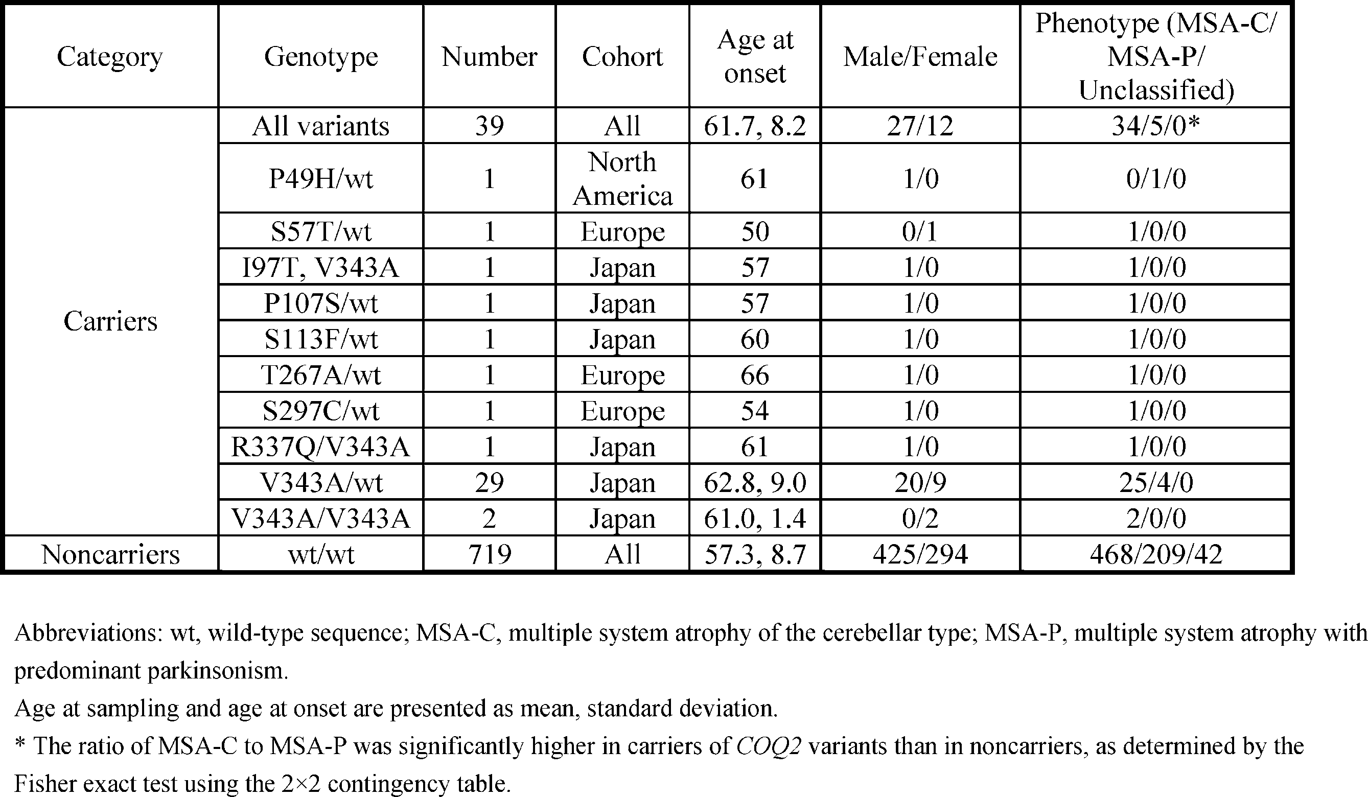
キャリア群のMSA発症年齢の平均は非キャリア群より高く、その差は有意であった。(p=0.0021)。臨床病型は、34人のキャリアがMSA-C、5人のキャリアがMSA-P、1人のキャリアがいずれにも分類されない型であり、468人の非キャリアがMSA-C、209人の非キャリアがMSA-P、42人の非キャリアがいずれにも分類されない型であった。MSA-Pに対するMSA-Cの比は、2×2分割表を用いたFisherの直接確率検定で求めたところ、CoQ2変異のキャリアにおいて非キャリアより有意に高かった(p=0.018)(表8)。
V343AキャリアのMSA患者から得たリンパ芽球様細胞、変異を有しないMSA患者から得たリンパ芽球様細胞、及び変異を有しない対照者から得たリンパ芽球様細胞における細胞内CoQ10濃度を測定した。対象者は(1)2つの変異アレル(R337Q/V343A、R337X/V343A、及びV343A/V343A)のキャリアであるMSA患者、(2)V343Aのヘテロ接合体のキャリアである16人のMSA患者、(3)変異を有しない133人のMSA患者、及び(4)変異を有しない76人の対照者に分けた(表9)。
COQ2変異を有するMSA患者から得られる脳組織の数は限られているが、COQ2変異を有する3人の患者(1人はM78V-V343A/M78V-V343A、2人はV343A/wt)の凍結脳組織と、変異を有しない対照者の凍結脳組織におけるCoQ10濃度を測定した(図2C)。M78V-V343Aのホモ接合体を有するMSA患者におけるCoQ10濃度は、変異を有しない対照者と比較して有意に低かった。
3-1.試験薬と投与方法
家族性MSAの発症者で,COQ2遺伝子のR337X/V343A複合ヘテロ接合性変異を有する患者1例(図1に示すFMSA_12のII-4)を対象として、ユビキノールの臨床試験を行った。
カネカから提供を受けたユビキノール(2-[(2E,6E,10E,14E,18E,22E,26E,30E,34E)-3,7,11,15,19,23,27,31,35,39-decamethyltetraconta-2,6,10,14,18,22,26,30,34,38-decaenyl]-5,6-dimethoxy-3-methylcyclohexa-2,5-diene-1,4-diol)として、120mg含有する安定粉末を、規定の用量(600mg、840mg、1200mg)に従い、胃瘻チューブから朝一回投与した。
対象1例で対照を設定しない非盲検の探索的臨床試験であり、以下の事項の検討を目的として行った。
1.高用量投与の安全性を確認する
2.薬物動態を調べる
3.薬物動態から長期投与の用量を判断する
4.臨床指標を検討する
試験の評価項目は以下のとおりである。
(1) 主要評価項目(Primary endpoint)
・血漿および白血球中,髄液中コエンザイムQ10濃度
・有害事象の有無
(2) 副次的評価項目(Secondary endpoint)
・UMSARS Part II(運動機能の評価)
・尿中8-OHdG
・15O-PETによる酸素代謝能の評価
(3) 安全性評価項目
・肝機能検査(AST,ALT,γ-GTP,ALP,T.Bil)
ラットにコエンザイムQ10を300mg/kg投与したところ,AST,ALTが軽度上昇したという報告があるため。
・自覚症状,身体診察・神経学的診察による他覚所見
・施行される各種検査値の異常
試験のアウトラインを図4に示す。
・有害事象について
試験期間中,自覚症状・他覚症状の有無,臨床検査(血算,生化学的検査:AST,ALT,γ-GTP,ALP,T.Bil, BUN, Cre, Na, K, Cl, Glu, CK)について経時的な観察をした。
試験薬に関連すると判断される有害事象は生じなかった.
血漿CoQ10濃度の測定結果を図5に示す。
ベースの血漿CoQ10濃度は低いレベルであった。1200mg投与で、既報告におけるプラトーに達した。ユビキノンの高用量投与の既報告では、ユビキノンとして2400mg以上の投与によって、7.5~8.0μg/mlでプラトーに達している。本試験では、ユビキノール1200mgで7.9μg/mlに達しており、既報告のデータに照らしてプラトーと判断された。
単核球中の全CoQ10/遊離コレステロール(nM/μM)の測定結果を図6に示す。ユビキノール投与により、用量依存的に、単核球中の全CoQ10/遊離コレステロールが上昇していることが確認された。
髄液CoQ10濃度(μg/ml)を測定した結果を図7に示す。ユビキノン・ユビキノール投与時の髄液CoQ10濃度の変化は報告がない。ベースの髄液CoQ10濃度は低いレベルであったが、840mg,1200mg投与群でプラトーに達しているように見えた。
尿中8-OHdG(ng/mg-Cre)の測定結果を図8に示す。ベースの尿中8-OHdGは高いレベル(平均8.4, n=500)であったが、ユビキノール投与により低下した。用量依存性は明確には見られなかった。
臨床評価スケールを図9に示す。軽微な変動はあったが、統計的に有意と判断できる変化は認められなかった。担当医、家族(妻)からの呼びかけに対する反応の改善、上肢の拳上の改善、四肢の震えの軽減との感想が得られた。但し、主観的なバイアス、入院中のリハビリ効果など複合的な要素があり、判断は困難であった。
脳血流量と酵素代謝量の測定結果を図10に示す。いずれも投与後に上昇する傾向が見られた。
今回の試験で以下の点が初めて明らかになった。
1.高用量投与においても、ユビキノールはユビキノンに比べ、bioavailabilityが高い。
2.ユビキノール1200mgで、血漿CoQ10はプラトーに達すること。先行研究で観察されている血漿CoQ10のプラトーに一致する結果が観察された。このことは、投与されたCoQ10が血漿へ移行することを示す。
3.ユビキノール投与により、末梢血単核球のCoQ10含量が上昇することが確認できた。このことは、投与されたCoQ10が細胞内に移行することを示す。
4.ユビキノール投与により、髄液CoQ10濃度、正常コントロールの髄液CoQ10濃度を超える値まで上昇することが確認された。このことは、投与されたCoQ10が髄液へ移行することを示す。
5.2~4.より、投与されたCoQ10が、CoQ10の低下を補うことができることが示された。
5.ユビキノール1200mgの2週間投与で、有害事象は観察されなかった。
6.[15O]O2 PETの評価によって、脳酸素消費量が投与前に比べて明らかに改善した。CoQ10補充により、中枢神経系の機能改善を評価するサロゲートマーカーになる可能性が示唆された。
Claims (9)
- 被検者の多系統萎縮症リスクの検査方法であって、被検者から採取した試料におけるコエンザイムQ10の生合成を低下させる変異を検出する工程を含む、方法。
- 前記コエンザイムQ10の生合成を低下させる変異は、Para-hydroxybenzoate-polyprenyltransferase(コエンザイムQ2)の発現又は機能を抑制する変異である、請求項1に記載の方法。
- 前記コエンザイムQ2の発現又は機能を抑制する変異は、配列番号:1におけるP49H、S57T、R69H、M78V、I97T、P107S、S113F、T267A、S297C、R337Q、R337X、及びV343Aからなる群より選択される、請求項2に記載の方法。
- 前記コエンザイムQ2の発現又は機能を抑制する変異は、V343Aである、請求項2に記載の方法。
- 多系統萎縮症リスクの検査キットであって、以下の(i)から(iii)の少なくとも1つを含むキット:
(i) コエンザイムQ2遺伝子における、コエンザイムQ2タンパク質(配列番号:1)の49位、57位、69位、78位、97位、107位、113位、267位、297位、337位、及び343位からなる群より選択される部位のアミノ酸をコードする核酸を含む領域にハイブリダイズする核酸;
(ii) コエンザイムQ2遺伝子における、コエンザイムQ2タンパク質(配列番号:1)の49位、57位、69位、78位、97位、107位、113位、267位、297位、337位、及び343位からなる群より選択される部位のアミノ酸をコードする核酸を含む領域を増幅可能なプライマーセット;及び
(iii) 配列番号:1において、P49H、S57T、R69H、M78V、I97T、P107S、S113F、T267A、S297C、R337Q、R337X、及びV343Aからなる群より選択される少なくとも1つの変異を有するコエンザイムQ2タンパク質と、野生型コエンザイムQ2タンパク質のいずれかのみに交差性なく結合する抗体。 - コエンザイムQ10を含む多系統萎縮症の予防又は治療剤。
- コエンザイムQ10を投与する工程を含む、多系統萎縮症の予防又は治療方法;
- 多系統萎縮症の予防又は治療剤のスクリーニング方法であって、
候補化合物と細胞とを接触させてインキュベートする工程と、
細胞内のコエンザイムQ10量を上昇させる候補化合物を選択する工程と、を含む方法。 - V343A変異を含むコエンザイムQ2タンパク質をコードする核酸。
Priority Applications (3)
| Application Number | Priority Date | Filing Date | Title |
|---|---|---|---|
| US14/763,794 US20160067195A1 (en) | 2013-02-05 | 2014-02-05 | Method for testing risk of multiple system atrophy, test kit, and drug for the treatment or prevention of multiple system atrophy |
| JP2014560782A JP6358962B2 (ja) | 2013-02-05 | 2014-02-05 | 多系統萎縮症リスクの検査方法、検査キット、及び多系統萎縮症の治療又は予防薬 |
| CN201480019831.1A CN105102621A (zh) | 2013-02-05 | 2014-02-05 | 多系统萎缩症风险的检测方法、检测试剂盒以及多系统萎缩症的治疗或预防药物 |
Applications Claiming Priority (2)
| Application Number | Priority Date | Filing Date | Title |
|---|---|---|---|
| JP2013-020763 | 2013-02-05 | ||
| JP2013020763 | 2013-02-05 |
Publications (1)
| Publication Number | Publication Date |
|---|---|
| WO2014123151A1 true WO2014123151A1 (ja) | 2014-08-14 |
Family
ID=51299743
Family Applications (1)
| Application Number | Title | Priority Date | Filing Date |
|---|---|---|---|
| PCT/JP2014/052658 Ceased WO2014123151A1 (ja) | 2013-02-05 | 2014-02-05 | 多系統萎縮症リスクの検査方法、検査キット、及び多系統萎縮症の治療又は予防薬 |
Country Status (4)
| Country | Link |
|---|---|
| US (1) | US20160067195A1 (ja) |
| JP (1) | JP6358962B2 (ja) |
| CN (1) | CN105102621A (ja) |
| WO (1) | WO2014123151A1 (ja) |
Families Citing this family (4)
| Publication number | Priority date | Publication date | Assignee | Title |
|---|---|---|---|---|
| JP3196153B2 (ja) | 1995-02-20 | 2001-08-06 | 日本鋪道株式会社 | 斜面用施工装置の支持台車 |
| CN110208231B (zh) * | 2019-06-05 | 2021-12-17 | 宁海县浙工大科学技术研究院 | 基于纳米孔膜/Au@ZIF检测8-羟基脱氧鸟苷的荧光生物传感器的制备方法 |
| CN111341452B (zh) * | 2020-04-01 | 2023-07-07 | 四川大学华西医院 | 多系统萎缩失能预测方法、模型建立方法、装置及设备 |
| JP2024147841A (ja) * | 2021-08-31 | 2024-10-17 | 国立大学法人 東京大学 | 多系統萎縮症治療用の医薬組成物 |
Citations (1)
| Publication number | Priority date | Publication date | Assignee | Title |
|---|---|---|---|---|
| JP2007528367A (ja) * | 2003-06-09 | 2007-10-11 | アルナイラム ファーマシューティカルズ インコーポレイテッド | 神経変性疾患を治療する方法 |
Family Cites Families (2)
| Publication number | Priority date | Publication date | Assignee | Title |
|---|---|---|---|---|
| CN1753662A (zh) * | 2002-12-24 | 2006-03-29 | 神经化学(国际)有限公司 | 用于治疗β-淀粉状蛋白相关疾病的治疗制剂 |
| WO2007017168A1 (en) * | 2005-08-10 | 2007-02-15 | Dsm Ip Assets B.V. | Process for the preparation of ubihydroquinones and ubiquinones |
-
2014
- 2014-02-05 CN CN201480019831.1A patent/CN105102621A/zh active Pending
- 2014-02-05 JP JP2014560782A patent/JP6358962B2/ja active Active
- 2014-02-05 WO PCT/JP2014/052658 patent/WO2014123151A1/ja not_active Ceased
- 2014-02-05 US US14/763,794 patent/US20160067195A1/en not_active Abandoned
Patent Citations (1)
| Publication number | Priority date | Publication date | Assignee | Title |
|---|---|---|---|---|
| JP2007528367A (ja) * | 2003-06-09 | 2007-10-11 | アルナイラム ファーマシューティカルズ インコーポレイテッド | 神経変性疾患を治療する方法 |
Non-Patent Citations (6)
| Title |
|---|
| "The Multiple-System Atrophy Research Collaboration., Mutations in COQ2 in familial and sporadic multiple-system atrophy.", N. ENGL. J. MED., vol. 369, no. 3, 18 July 2013 (2013-07-18), pages 233 - 244 * |
| DATABASE DDBJ/EMBL/ GENBANK 28 November 2012 (2012-11-28), "4-hydroxybenzoate polyprenyltransferase, mitochondrial.", accession no. 96H96 * |
| DIOMEDI-CAMASSEI, F. ET AL.: "COQ2 nephropathy: a newly described inherited mitochondriopathy with primary renal involvement.", J. AM. SOC. NEPHROL., vol. 18, 2007, pages 2773 - 2780 * |
| LOPEZ-MARTIN, J.M. ET AL.: "Missense mutation of the COQ2 gene causes defects of bioenergetics and de novo pyrimidine synthesis.", HUM. MOL. GENET., vol. 16, no. 9, 2007, pages 1091 - 1097 * |
| QUINZII, C. ET AL.: "A mutation in para- hydroxybenzoate-polyprenyl transferase (COQ2) causes primary coenzyme Q10 deficiency.", AM. J. HUM. GENET., vol. 78, February 2006 (2006-02-01), pages 345 - 349 * |
| YASUO NAKAHARA ET AL.: "Genome Wide Kanren Kaiseki ni Motozuku Takeito Ishukusho (MSA) no Shikkan Kanjusei Idenshi no Tansaku", CLINICAL NEUROLOGY, vol. 50, no. 12, December 2010 (2010-12-01), pages 1155 * |
Also Published As
| Publication number | Publication date |
|---|---|
| CN105102621A (zh) | 2015-11-25 |
| JP6358962B2 (ja) | 2018-07-18 |
| US20160067195A1 (en) | 2016-03-10 |
| JPWO2014123151A1 (ja) | 2017-02-02 |
Similar Documents
| Publication | Publication Date | Title |
|---|---|---|
| Udar et al. | SOD1: a candidate gene for keratoconus | |
| Burdon et al. | Genome-wide association study identifies susceptibility loci for open angle glaucoma at TMCO1 and CDKN2B-AS1 | |
| US20210095345A1 (en) | Method of identifying disease risk factors | |
| Munesue et al. | Two genetic variants of CD38 in subjects with autism spectrum disorder and controls | |
| Yuan et al. | Cosegregation of the G7444A mutation in the mitochondrial COI/tRNASer (UCN) genes with the 12S rRNA A1555G mutation in a Chinese family with aminoglycoside‐induced and nonsyndromic hearing loss | |
| Choi et al. | Brain-derived neurotrophic factor gene polymorphism (Val66Met) and citalopram response in major depressive disorder | |
| Marin et al. | Leigh syndrome associated with mitochondrial complex I deficiency due to novel mutations In NDUFV1 and NDUFS2 | |
| KR20140089384A (ko) | 알츠하이머병의 치료, 진단 및 모니터링 방법 | |
| US20080286796A1 (en) | Genetic polymorphisms associated with neurodegenerative diseases, methods of detection and uses thereof | |
| US20100196400A1 (en) | Genetic polymorphisms associated with alzheimer's disease, methods of detection and uses thereof | |
| CA2846307A1 (en) | Method for diagnosing a neurodegenerative disease | |
| Campbell et al. | Polymorphisms in KCNE1 or KCNE3 are not associated with Meniere disease in the Caucasian population | |
| Turaca et al. | Novel GAA mutations in patients with Pompe disease | |
| JP2009520460A (ja) | 心筋梗塞に関連する遺伝的多型、その検出方法および使用 | |
| JP6358962B2 (ja) | 多系統萎縮症リスクの検査方法、検査キット、及び多系統萎縮症の治療又は予防薬 | |
| US20240026454A1 (en) | Single nucleotide polymorphism marker for predicting risk of alzheimers disease and use thereof | |
| KR20130043107A (ko) | 루이 소체 치매 진단용 유전자 마커 | |
| Hilal et al. | Screening of CYP1B1 and MYOC in Moroccan families with primary congenital glaucoma: three novel mutations in CYP1B1 | |
| Péquignot et al. | New Splicing-site Mutations in the SURF1Gene in Leigh Syndrome Patients | |
| Rezvani et al. | Fifteen novel mutations in the mitochondrial NADH dehydrogenase subunit 1, 2, 3, 4, 4L, 5 and 6 genes from Iranian patients with Leber’s hereditary optic neuropathy (LHON) | |
| US20160186263A1 (en) | Using plexin-a4 as a biomarker and therapeutic target for alzheimer's disease | |
| Hsieh et al. | Association between retinoid-X receptor-gamma genetic polymorphisms and diabetic retinopathy. | |
| US20060166204A1 (en) | Parkinson's disease susceptibility haptotype as a tool for genetic screening | |
| Meulemans et al. | A new family with the mitochondrial tRNAGLU gene mutation m. 14709T> C presenting with hydrops fetalis | |
| Ding et al. | Association between the rs189037 single nucleotide polymorphism in the ATM gene promoter and cognitive impairment |
Legal Events
| Date | Code | Title | Description |
|---|---|---|---|
| WWE | Wipo information: entry into national phase |
Ref document number: 201480019831.1 Country of ref document: CN |
|
| 121 | Ep: the epo has been informed by wipo that ep was designated in this application |
Ref document number: 14748587 Country of ref document: EP Kind code of ref document: A1 |
|
| ENP | Entry into the national phase |
Ref document number: 2014560782 Country of ref document: JP Kind code of ref document: A |
|
| NENP | Non-entry into the national phase |
Ref country code: DE |
|
| WWE | Wipo information: entry into national phase |
Ref document number: 14763794 Country of ref document: US |
|
| 122 | Ep: pct application non-entry in european phase |
Ref document number: 14748587 Country of ref document: EP Kind code of ref document: A1 |