WO2014106967A1 - 뉴로펩티드 y를 유효성분으로 포함하는 골다공증의 예방 또는 치료용 약학적 조성물 - Google Patents
뉴로펩티드 y를 유효성분으로 포함하는 골다공증의 예방 또는 치료용 약학적 조성물 Download PDFInfo
- Publication number
- WO2014106967A1 WO2014106967A1 PCT/KR2013/001102 KR2013001102W WO2014106967A1 WO 2014106967 A1 WO2014106967 A1 WO 2014106967A1 KR 2013001102 W KR2013001102 W KR 2013001102W WO 2014106967 A1 WO2014106967 A1 WO 2014106967A1
- Authority
- WO
- WIPO (PCT)
- Prior art keywords
- neuropeptide
- bone marrow
- cells
- blood
- mice
- Prior art date
- Legal status (The legal status is an assumption and is not a legal conclusion. Google has not performed a legal analysis and makes no representation as to the accuracy of the status listed.)
- Ceased
Links
Images





Classifications
-
- A—HUMAN NECESSITIES
- A61—MEDICAL OR VETERINARY SCIENCE; HYGIENE
- A61K—PREPARATIONS FOR MEDICAL, DENTAL OR TOILETRY PURPOSES
- A61K38/00—Medicinal preparations containing peptides
- A61K38/16—Peptides having more than 20 amino acids; Gastrins; Somatostatins; Melanotropins; Derivatives thereof
- A61K38/17—Peptides having more than 20 amino acids; Gastrins; Somatostatins; Melanotropins; Derivatives thereof from animals; from humans
-
- A—HUMAN NECESSITIES
- A61—MEDICAL OR VETERINARY SCIENCE; HYGIENE
- A61K—PREPARATIONS FOR MEDICAL, DENTAL OR TOILETRY PURPOSES
- A61K38/00—Medicinal preparations containing peptides
- A61K38/16—Peptides having more than 20 amino acids; Gastrins; Somatostatins; Melanotropins; Derivatives thereof
- A61K38/17—Peptides having more than 20 amino acids; Gastrins; Somatostatins; Melanotropins; Derivatives thereof from animals; from humans
- A61K38/22—Hormones
- A61K38/2271—Neuropeptide Y
-
- A—HUMAN NECESSITIES
- A61—MEDICAL OR VETERINARY SCIENCE; HYGIENE
- A61K—PREPARATIONS FOR MEDICAL, DENTAL OR TOILETRY PURPOSES
- A61K38/00—Medicinal preparations containing peptides
- A61K38/16—Peptides having more than 20 amino acids; Gastrins; Somatostatins; Melanotropins; Derivatives thereof
-
- A—HUMAN NECESSITIES
- A61—MEDICAL OR VETERINARY SCIENCE; HYGIENE
- A61P—SPECIFIC THERAPEUTIC ACTIVITY OF CHEMICAL COMPOUNDS OR MEDICINAL PREPARATIONS
- A61P19/00—Drugs for skeletal disorders
-
- A—HUMAN NECESSITIES
- A61—MEDICAL OR VETERINARY SCIENCE; HYGIENE
- A61P—SPECIFIC THERAPEUTIC ACTIVITY OF CHEMICAL COMPOUNDS OR MEDICINAL PREPARATIONS
- A61P19/00—Drugs for skeletal disorders
- A61P19/08—Drugs for skeletal disorders for bone diseases, e.g. rachitism, Paget's disease
- A61P19/10—Drugs for skeletal disorders for bone diseases, e.g. rachitism, Paget's disease for osteoporosis
Definitions
- the present invention relates to a pharmaceutical composition for preventing or treating osteoporosis comprising neuropeptide Y as an active ingredient.
- Osteoporosis is postmenopausal osteoporosis, which is indicated by increased bone resorption due to the activation of osteoclasts due to rapid hormonal changes following menopause, and senile osteoporosis, in which osteoblasts decrease due to aging and osteoblasts decrease. Can be classified as Since osteoporosis fractures lead to severe activity limitations and are associated with a high mortality rate of about 15-35% in hip fractures, the diagnosis and treatment of osteoporosis is important before osteoporotic fractures occur (osteoporosis diagnosis). And Treatment Guidelines 2007, 2008).
- Known therapeutic agents for osteoporosis include bisphosphonate-based drugs, which are deposited on the mineral component of bone and form an ATP analog that does not hydrolyze when osteoclasts phage on the bone where the bisphosphonates are deposited.
- bisphosphonate-based drugs which are deposited on the mineral component of bone and form an ATP analog that does not hydrolyze when osteoclasts phage on the bone where the bisphosphonates are deposited.
- it is known to be toxic to cells or to cause osteoclast activity and apoptosis in osteoclasts in various ways, thereby reducing bone resorption and thereby increasing bone density.
- These drugs are known to be relatively safe, but in recent years, their long-term use affects bone remodeling or bone regeneration after bone fracture due to normal bone resorption and bone formation. Concerns have been raised that this could have an undesired effect, and in fact it has been reported that this causes fatigue fractures in many patients.
- bone remodeling (remodeling) has been thought to be mainly controlled by the endocrine system, local action factors such as cytokines and growth factors.
- cytokines and growth factors.
- this view is gradually changing as evidence that neurological factors can change the activity of bone cells is revealed, and many recent studies have supported the discovery of nerve fibers and neurological factors in bone tissue.
- Neuropeptide Y a sympathetic neurotransmitter
- NPY a sympathetic neurotransmitter
- NPY a neurotransmitter consisting of 36 amino acids most abundantly present in the mammalian brain, secreted from the Y2 receptors of the hypothalamus, and is characterized by appetite control, memory, and seizures. It is known to be involved in various physiological and pathological conditions such as (Balasubramaniam, 1997; Vezzani et al., 1999; Wettstein et al., 1995).
- the secretion of neuropeptide Y increases from the brain and the sympathetic nervous system, and as a result, the feeding action increases obesity, but neuropeptide Y is still a hematopoietic stem cell, differentiation.
- neuropeptide Y can be used in the prevention or treatment of osteoporosis by inducing the release of hematopoietic stem cells from the bone marrow into the blood and completed the present invention.
- the present invention is to provide a pharmaceutical composition for preventing or treating osteoporosis comprising neuropeptide Y as an active ingredient.
- the present invention provides a pharmaceutical composition for preventing or treating osteoporosis comprising neuropeptide Y as an active ingredient.
- Neuropeptide Y according to the present invention has an excellent effect of reducing the expression of cell adhesion factors of osteoblasts in which the Y1 receptor is present, thereby releasing hematopoietic stem cells from the bone marrow into the blood.
- the number of osteoclasts which differentiate from hematopoietic stem cells and induce osteoporotic lesions, can suppress the progression of bone erosion by osteoclasts. Therefore, neuropeptide Y, an active ingredient of the composition of the present invention, may be usefully used as a therapeutic agent for osteoporosis.
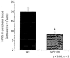
- FIG. 1 is a diagram showing the expression levels of myeloid hematopoietic stem cell adhesion factor in neuropeptide Y knockout (NPY KO) mice and WT (wild type) mice.
- FIG. 2 is a diagram showing the number of blood myeloid hematopoietic progenitor cells in NPY KO mice and WT mice.
- Figure 3 is a diagram showing the outline of the experiment of the present invention carried out to determine the effect of neuropeptide Y of the present invention on the expression level of myeloid hematopoietic stem cell adhesion factor.
- Figure 4 is a diagram showing the results of the concentration of the effect of neuropeptide Y on the expression level of myeloid hematopoietic stem cell adhesion factor of the present invention.
- Figure 5 is a diagram showing the expression change of myeloid hematopoietic stem cell adhesion factor upon administration of neuropeptide Y of the present invention.
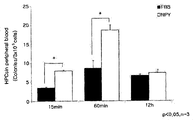
- Figure 6 is a diagram showing the number of myeloid hematopoietic progenitor cells in blood with time upon administration of neuropeptide Y of the present invention.
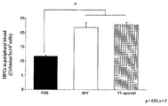
- FIG. 7 is a diagram showing the degree of release of myeloid hematopoietic stem cells into the blood upon administration of neuropeptide Y of the present invention using markers of myeloid hematopoietic stem cells.
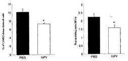
- Figure 8 is a diagram showing the results of the competitive transplantation (competitive transplantation) experiments the release of myeloid hematopoietic stem cells into the blood upon administration of neuropeptide Y of the present invention.
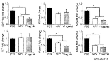
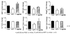
- FIG. 9 is a diagram showing the change in the expression level of myeloid hematopoietic stem cell adhesion factor upon administration of a neuropeptide Y receptor agonist (Y1 agonist).
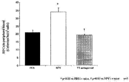
- Fig. 10 shows the number of myeloid hematopoietic progenitor cells in blood upon administration of a neuropeptide Y receptor agonist.
- FIG. 11 is a diagram showing the expression changes of myeloid hematopoietic stem cell adhesion factor upon administration of neuropeptide Y receptor antagonist (Y1 antagonist).
- Fig. 12 shows the number of myeloid hematopoietic progenitor cells in blood upon administration of a neuropeptide Y receptor antagonist.
- FIG. 13 is a diagram showing an outline of experiments performed to investigate the effect of neuropeptide Y of the present invention on the prevention and treatment of osteoporosis.
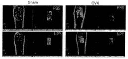
- FIG. 14 is a photograph showing the overall bone density change when the neuropeptide Y administration to the osteoporosis model after taking a micro-CT (micro CT).
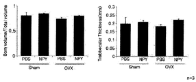
- FIG. 15 is a graph illustrating quantification of bone density change and bone tissue thickness change by micro citi after micropeptide Y administration to osteoporosis model.
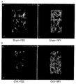
- Figure 16 is a diagram showing a 3D reconstruction of the bone density change when neuropeptide Y administration in the osteoporosis model after microciti (A: Sham, B: OVX).
- the present invention provides a pharmaceutical composition for treating osteoporosis comprising neuropeptide Y as an active ingredient.
- Stem cell niche is a microenvironment where stem cells are found and stem cells interact to control the fate of stem cells.
- the word niche may refer to the microenvironment of stem cells in vivo and in vitro.
- various habitat elements act on embryonic stem cells to affect gene expression and induce cell proliferation or differentiation to develop the fetus.
- the microenvironment of each tissue actively sends signals to the habitat where stem cells exist, and thus self-renewal of stem cells in the habitat promote differentiation for self renewal or formation of new tissue.
- Stem cell characteristics in stem cell habitat are controlled by the interaction between stem cells and surrounding differentiated cells or adhesion molecules, extracellular matrix components, growth factors, physicochemical environments such as cytokines and pH, and calcium ion concentrations.
- Cell-cell interactions between stem cells, as well as ionic strength and metabolites such as ATP, are important, and reversible signals for maintaining stem cells at both developmental and adult stages between stem cells and stem cell habitats It is known to give and receive.
- the human hippocampus is known to exist in the hippocampus of the brain, neural stem cell niche of the lateral ventricle, bone marrow stem cell niche in the bone marrow, and the like.
- the bone marrow stem cell habitat is defined as a molecular microenvironment that regulates the function and fate of cells through the interaction between various cells in the bone marrow, including bone marrow stem cells.
- endosteal niche and “perivascular” Two bone marrow stem cell habitats of niche ”are known.
- endosteal niche is a microenvironment in which the basal cells in the bone marrow exist, and mainly there are basal cells, osteoblasts, and osteoclasts. These cells are adherent factors (CXCL12, Ang-1, VCAM1, stem cell factor (IL-7, etc.), adjacent to other cells, in particular hematopoietic progenitor cells or hematopoietic stem cells (HPCs), or hematopoietic stem cells (HSCs), and respond to neurotransmitter signals from the outside.
- the hematopoietic stem cells attached to the osteoblasts are released into the blood, or the bone marrow stem cell habitat affects the maintenance of bone marrow homeostasis by returning to the bone marrow.
- peripheral niche is a microenvironment composed of macrophage, mesenchymal stem cells (MSC) and CAR cells (CXCL12 abundant reticular cells), similar to endosteal niche. Bone marrow stem cell habitat adjacent to hematopoietic stem cells and involved in releasing hematopoietic stem cells held by macrophages and mesenchymal stem cells into the blood in response to external neurotransmitters such as stress and neurotransmitters to be.
- MSC mesenchymal stem cells
- CAR cells CXCL12 abundant reticular cells
- stem cells in bone marrow are mobilized into the bloodstream or the stem cells in bone marrow released are homing.
- the mobilization of stem cells in the bone marrow into the blood is an important phenomenon in the bone marrow when our body is stressed or damaged and refers to the migration of stem cells, immune-related cells and osteoclasts from the bone marrow to blood vessels.
- the cells recruited into the blood vessels take the bloodstream to the damaged lesions to help heal them.
- stem cells in the bone marrow that healed in the lesions are returned to the bone marrow through the blood flow again. This phenomenon is called regression (homing).
- Neuropeptide Y which is an active ingredient of the present invention, can be obtained by extracting in vivo in a conventional manner, or purchased and commercially available.
- Neuropeptide Y when administered in vivo, has an effect of reducing the expression of adhesion factors related to the maintenance of bone marrow hematopoietic stem cells in bone marrow, or bone marrow hematopoietic stem cells maintained in bone marrow The effect of inducing release of cells into the blood is excellent.
- Myeloid stem cells can differentiate into lymphoid and myeloid cells in the bone marrow. Osteoclasts, one of myeloid cells that are differentiated from myeloid hematopoietic stem cells, maintain bone formation and erosion through balance with osteoblasts in bone marrow. When this balance is broken, osteoporosis occurs by increasing the number of osteoclasts and the speed of bone erosion.
- S1P sphingosine-1-phosphate
- FY720 agonist of S1P receptor
- the neuropeptide Y of the present invention can be usefully used for the prevention or treatment of osteoporosis by inducing the release of hematopoietic stem cells into the blood and reducing the differentiation into osteoblasts in the bone marrow.
- composition of the present invention may include one or more known active ingredients having neuropeptide Y having a prophylactic or therapeutic effect of osteoporosis.
- composition of the present invention may be prepared by including one or more pharmaceutically acceptable carriers in addition to the above-described active ingredients for administration.
- Pharmaceutically acceptable carriers may be used in combination with saline, sterile water, Ringer's solution, buffered saline, dextrose solution, maltodextrin solution, glycerol, ethanol and one or more of these components, if necessary, as an antioxidant, buffer And other conventional additives such as bacteriostatic agents can be added.
- Diluents, dispersants, surfactants, binders and lubricants may also be added in addition to formulate into injectable formulations, pills, capsules, granules or tablets such as aqueous solutions, suspensions, emulsions and the like.
- it may be preferably formulated according to each disease or component by a suitable method in the art or using a method disclosed in Remington's Pharmaceutical Science (Recent Edition), Mack Publishing Company, Easton PA.
- composition of the present invention can be administered orally or parenterally (eg, applied intravenously, subcutaneously, intraperitoneally or topically) according to the desired method, and the dosage is based on the weight, age, sex and health of the patient. The range varies depending on the diet, the time of administration, the method of administration, the rate of excretion and the severity of the disease.
- the daily dose of neuropeptide Y is about 0.01 to 1 mg / kg, preferably about 0.05 to 0.1) mg / kg, preferably administered once to several times a day.
- composition of the present invention can be used alone or in combination with methods for using surgery, hormonal therapy, drug treatment, and biological response modifiers for the prevention, treatment of osteoporosis, or an increase in bone mineral density.
- mice used in the experiment were 6 to 8 week old mice, NPY KO mice and NPY WT (wild type) mice, genetically knocked out neuropeptide Y (NPY), Ly5.1. (BoyJ) mouse and Ly5.2 (C57BL / 6) Mouse, purchased from Jackson Laboratory (Bar Harbor, Maine, USA).
- Neuropeptide Y, Y1 agonist, Y1 antagonist were purchased from Bache and used.
- NPY 1 and 5 nM were diluted and injected into each medium.
- three mice per group a total of 15 mice, were anesthetized with a mixture of 100 mg / kg ketamine and 10 mg / kg xylazine, and neuropeptides respectively.
- 50 ⁇ g / kg of Y, Y1 agonist, Y1 antagonist, and 100 ⁇ l of PBS (Gibco) were injected via tail vein injection in mice.
- the cells were incubated for 1 week and stemXVivo Osteogenic / Adipogenic Base Media (R & D systems) supplemented with StemXVivo Osteogenic supplement (20X) and penicillin-steptomycin (100X) for differentiation into osteoblasts. Incubation was for three weeks, and the cultures were changed every two to three days.
- RNeasy Plus Mini Kit Qiagen, Korea, Ltd
- Clontech Clontech (Mountain View, CA).
- a Corbett research RG-6000 real-time PCR instrument 95 °C, 10 minutes; 95 ° C., 10 seconds; 58 ° C., 15 seconds; Real-time quantitative PCR was performed to repeat 40 cycles at 72 ° C., 20 seconds;
- CFC analysis was performed to determine the number of bone marrow hematopoietic progenitor cells in the blood of mice.
- bone marrow was collected 60 minutes after injecting NPY and PBS into normal mice, respectively.
- Lineage and Sca markers of bone marrow hematopoietic stem cells FACs were analyzed using five antibodies, -1, c-kit, CD48, and CD150.
- bone marrow harvested from tibias and femurs of C57BL / 6 mice aged 4 to 6 weeks were depleted of red blood cells with ammonium chloride solution (Stem Cell Technologies, Inc. 1: 4). After washing with PBS (Gibco) solution containing 10% fetal bovine serum (FBS, Gibco) and 1% sodium azide (Sigma-Aldrich), and centrifuged at 300 xg for 10 minutes.
- PBS Gibco
- FBS fetal bovine serum
- sodium azide Sigma-Aldrich
- Hematopoietic cells contained in bone marrow were removed with MACs beads (Miltenyi Biotec) using a biotinylated lineage antibody (Miltenyi Biotec), and the remaining cells were Sca-1-PECY7, c-kit-APC, CD150-PE, and CD48-FITC (The reaction was carried out at 4 ° C. for 30 minutes using an BD science antibody, and then analyzed by LSRII (BD science) flow cytometry.
- Repopulating unit (RU) (20 x CD45.2%) / (100-CD45.2%)
- the femur was separated from the mouse and refrigerated in 80% ethanol, followed by micro CT scanner (Inveon preclinical CT, Siemens Healthcare, Hoffman Estates, IL) for tissue thickness 40 ⁇ m, exposure time of 600 msec, 70 keV The photon energy of and a current of 400 ⁇ A was measured.
- micro CT scanner Inveon preclinical CT, Siemens Healthcare, Hoffman Estates, IL
- the photon energy of and a current of 400 ⁇ A was measured.
- the volume portion of 2.5 x 0.5 x 0.5 mm 3 of the same area per group was measured using Siemens Inveon Software.
- NPY KO mice hematopoietic stem cell adhesion factors
- NPY KO mice and WT (wild type) mice were purchased from the Jackson Laboratory (Bar Harbor, Maine, USA) and bone marrow of each mouse was collected.
- Example 1-2 In order to examine the effect of neuropeptide Y on the expression level of the adhesion factor involved in the maintenance of bone marrow hematopoietic stem cells in the bone marrow, the following experiment was performed according to the method of Example 1-2 and Example 1-3 .
- bone marrow was harvested from 4 to 6 week old C57BL / 6 mice to harvest bone marrow mesenchymal stem cells (BM-MSC), and then cultured for 3 weeks in osteoblast differentiation induction medium to differentiate into osteoblasts.
- BM-MSC bone marrow mesenchymal stem cells
- neuropeptide Y was treated with 0, 1, and 5 nM for 3 days, and bone cells were collected and examined for expression levels of six adhesion factors by real-time quantitative PCR.
- FIG. 3 A schematic diagram of the experimental procedure is shown in FIG. 3, and the result of measuring the expression level of the adhesion factor is shown in FIG. 4.
- neuropeptide Y may induce the release of myeloid hematopoietic stem cells from bone marrow to blood by reducing the expression level of myeloid hematopoietic stem cell adhesion factor present in osteoblasts.
- neuropeptide Y of the present invention In order to examine the effect of neuropeptide Y of the present invention on the expression level of myeloid hematopoietic stem cell adhesion factor in vivo, 60 minutes after injection of neuropeptide Y through mouse tail vein, Bone marrow was collected from the femur and femur, and the expression levels of adhesion factors were examined by real-time quantitative PCR.
- mice normal mice were divided into two groups of 3 mice each, and 50 ⁇ g / kg of neuropeptide Y and 100 ⁇ l of PBS (Gibco) were respectively administered, and bone marrow was collected 60 minutes later. From the collected bone marrow, five markers of lineage, Sca-1, c-kit, CD48, and CD150, which are markers of bone marrow hematopoietic stem cells, were analyzed by FACs.
- Example 1-6 In order to confirm the release of myeloid hematopoietic stem cells and myeloid hematopoietic progenitor cells into the blood by administration of the neuropeptide Y of the present invention, a Competitive transplantation experiment was performed according to the method of Example 1-6.
- Repopulating unit (RU) (20 x CD45.2%) / (100-CD45.2%)
- the percentage of normal bone marrow CD45.2 + cells injected with neuropeptide Y was reduced compared to CD45.2 + cells in mouse bone marrow injected with control PBS.
- bone marrow hematopoietic stem cells which are CD45.2 +, are released into the blood, and the number of hematopoietic stem cells is present in the bone marrow of mice injected with PBS. Since less bone marrow hematopoietic stem cells are present, it can be seen that there are fewer donor-derived BM-HSCs of CD45.2 + donor-derived BM-HSC in the bone marrow of the bone marrow transplanted mouse.
- Example 5 Effect of Neuropeptide Y Receptor Activity on Bone Marrow Hematopoietic Stem Cell Adhesion Factor and Bone Marrow Hematopoietic Progenitor Cell Release.
- the neuropeptide Y receptor Y1 receptor is present in osteoblasts involved in bone formation in bone marrow. Therefore, to determine whether the neuropeptide Y is expressed through bone marrow hematopoietic stem cell adhesion factor and whether it is made through osteoblast Y1 receptor, The same experiment was performed.
- mice injected with 50 ⁇ g / kg of Y1 agonist reacting with Y1 receptor by acting similar to neuropeptide Y through tail vein injection were used.
- mice injected with the same amount of neuropeptide Y and PBS were used.
- bone marrow of each mouse was harvested, and the expression level of bone marrow hematopoietic stem cell adhesion factors was examined by real-time quantitative PCR method of Experimental Example 1-3.
- activation of the neuropeptide Y receptor generally reduced the expression levels of Sdf-1a, Angpt1, Vcam1, and Spp1, which are adhesion factors of myeloid hematopoietic stem cells, than when neuropeptide Y was administered.
- P ⁇ 0.05, n 3 per group.
- colony numbers of myeloid progenitor cells were counted by the CFC assay of Example 1-4 to determine the effect of neuropeptide Y receptor activation on the release of myeloid progenitor cells into the blood.
- experiment was performed in the same manner as in the above 5-1 to determine the effect of the inhibition of neuropeptide Y receptor on the release of myeloid hematopoietic progenitor cells into the blood.
- FIG. 13 A schematic diagram of the experimental procedure is shown in FIG. 13.
- mice were subjected to ovarian ablation for each osteoporosis model, and one week later, 50 ⁇ g / kg of neuropeptide Y and 100 ⁇ l of PBS (Gibco) were added 2 times a day. Intraperitoneally, three times a week.
- mice subjected to subcutaneous incision only in normal mice and intraperitoneally administered neuropeptide Y of 100 ⁇ l or 50 ⁇ g / kg PBS were used.
- the femurs of the mice were separated and stored in 80% ethanol and stored in 80% ethanol to measure bone density and bone tissue thickness using micro CT.
- FIG. 14 Micro CT observations of the bones of the control and osteoporosis models are shown in FIG. 14, and graphs of the changes in bone density and bone tissue thickness thereof are shown in FIG. 15, and micro-Citi observation results of the same bone tissues of the control and osteoporosis models are shown in FIG. 15.
- the 3D reconstructed photo is shown in FIG. 16.
Landscapes
- Health & Medical Sciences (AREA)
- Life Sciences & Earth Sciences (AREA)
- Engineering & Computer Science (AREA)
- Bioinformatics & Cheminformatics (AREA)
- Chemical & Material Sciences (AREA)
- Veterinary Medicine (AREA)
- Public Health (AREA)
- Medicinal Chemistry (AREA)
- General Health & Medical Sciences (AREA)
- Animal Behavior & Ethology (AREA)
- Pharmacology & Pharmacy (AREA)
- Gastroenterology & Hepatology (AREA)
- Immunology (AREA)
- Epidemiology (AREA)
- Proteomics, Peptides & Aminoacids (AREA)
- Physical Education & Sports Medicine (AREA)
- Zoology (AREA)
- Endocrinology (AREA)
- Organic Chemistry (AREA)
- Chemical Kinetics & Catalysis (AREA)
- General Chemical & Material Sciences (AREA)
- Nuclear Medicine, Radiotherapy & Molecular Imaging (AREA)
- Orthopedic Medicine & Surgery (AREA)
- Rheumatology (AREA)
- Medicines That Contain Protein Lipid Enzymes And Other Medicines (AREA)
- Peptides Or Proteins (AREA)
Abstract
본 발명은 뉴로펩티드 Y를 유효성분으로 포함하는 골다공증 예방 또는 치료용 약학적 조성물에 관한 것이다. 본 발명에 따른 뉴로펩티드 Y는 Y1 수용체가 존재하는 골아세포의 세포부착인자의 발현을 감소시키고, 이에 따라 골수로부터 조혈줄기세포를 혈중으로 방출시키는 효과가 우수하다. 조혈줄기세포가 혈중으로 방출되면 조혈줄기세포로부터 분화되어 골다공증성 병변을 유도하는 파골세포의 수적인 감소가 일어나므로, 파골세포에 의한 골침식 진행을 억제할 수 있다. 따라서 본 발명의 조성물의 유효성분인 뉴로펩티드 Y는 골다공증의 치료제로서 유용하게 사용될 수 있다.
Description
본 발명은 뉴로펩티드 Y를 유효성분으로 포함하는 골다공증의 예방 또는 치료용 약학적 조성물에 관한 것이다.
골다공증은 폐경에 따른 급격한 호르몬의 변화에 의한 파골세포(osteoclast)의 활성화에 따른 골흡수 증가로 나타나는 폐경 후 골다공증과, 노화가 되면서 조골세포(osteoblast)의 기능이 감소하여 골형성이 감소하는 노인성 골다공증으로 분류할 수 있다. 이러한 골다공증으로 인한 골절은 심각한 활동 제한에 이르게 되고, 고관절 골절의 경우 약 15-35%의 높은 사망률과 관련되어 있기 때문에, 골다공증성 골절이 발생하기 이전에 골다공증의 진단과 치료가 중요하다(골다공증 진단 및 치료 지침 2007, 2008).
국내의 골다공증 유병률은 2008년 기준 최근 5년간 약 3배 증가되었고, 골다공증 골절에 의한 연간 사회경제적 손실이 약 1조 500억원에 달할 정도로 심각한 수준에 이르고 있다고 보고된다(골다공증 진단 및 치료 지침 2007, 2008). 또한, 최신의 2009년도 국민건강통계에 따르면, 50세 이상 및 65세 이상 한국인 전체의 골다공증 유병률은 23.1%와 42.0%로 만성질환 중에서도 유병률이 매우 높게 나타나 국민건강에 커다란 문제로 떠오르고 있다(2009년 국민건강통계-국민건강영양조사).
기존에 알려진 골다공증 치료제로는 비스포스포네이트(bisphosphonate)계열의 약물이 있는데, 비스포스포네이트는 골의 무기질 성분에 침착되며, 비스포스포네이트가 침착된 골을 파골세포가 탐식할 경우 가수분해되지 않는 ATP 유사체(analogue)를 형성하여 세포에 독성을 나타내거나 파골세포 내에서 다양한 방식으로 파골세포의 활성 감소 및 아폽토시스(apoptosis)를 초래해 골 흡수를 줄이고 이를 통해 골밀도를 증가시키는 것으로 알려져 있다. 이러한 약물들은 비교적 안전한 것으로 알려져 있으나, 최근 장기간 사용시 정상적인 골 흡수 및 골 형성에 의한 골의 재형성(remodeling)이나 골절 이후 골 재생(healing)과정에 영향을 주어 골의 탄성이 떨어져 오히려 골 강도에 좋지 않은 영향을 줄 수 있다는 우려들이 제기되고 있으며, 실제로 이로 인해 많은 환자에서 피로 골절을 일으킨다는 보고가 있다.
따라서 골다공증 발병과 관련된 새로운 골대사 기전의 발견 및 골다공증 예방 또는 치료제 개발의 필요성이 절실하게 요구되고 있는 실정이다.
전통적으로 뼈의 재생(remodeling, regeneration)은 내분비계와, 사이토카인, 성장인자와 같은 국소작용인자에 의해서 주로 조절되는 것으로 생각되어왔다. 그러나, 이러한 관점은 신경학적 인자가 골세포의 활성을 변화시킬 수 있다는 증거가 속속 밝혀지면서 점차 변하고 있으며, 최근 많은 연구에서 골조직 내에서 신경섬유 및 신경학적 인자가 발견되고 있어 이를 뒷받침하고 있다.
교감신경계 전달물질인 뉴로펩티드 Y(neuropeptide Y, NPY)는 포유류의 뇌에 가장 풍부하게 존재하는 36개의 아미노산으로 이루어진 신경전달물질로서, 시상하부의 Y2 수용체로부터 분비되고 식욕조절, 기억, 및 발작과 같은 다양한 생리 및 병리 조건에 관여하는 것으로 알려져 있다(Balasubramaniam,1997; Vezzani et al., 1999; Wettstein et al., 1995). 이와 관련하여서 우리 몸이 스트레스 환경에 처하면 뇌와 교감신경계로부터 뉴로펩티드 Y의 분비가 증가하고, 그 결과 섭식 작용이 증가하여 비만이 발생한다는 보고는 있었으나, 아직까지 뉴로펩티드 Y가 조혈줄기세포, 분화세포, 및 면역관련세포 등의 골수유래 줄기세포의 기능조절, 방출 및 회귀에 미치는 영향에 대한 연구보고는 알려진 바 없으며, 특히 뉴로펩티드 Y와 골다공증의 관련성에 대한 연구는 전무한 상태이다.
본 발명의 발명자들은 뉴로펩티드 Y가 골수로부터 조혈줄기세포의 혈중으로의 방출을 유도하여 골다공증의 예방 또는 치료에 사용될 수 있음을 확인하고 본 발명을 완성하였다.
본 발명은 뉴로펩티드 Y를 유효성분으로 포함하는 골다공증의 예방 또는 치료용 약학적 조성물을 제공하고자 한다.
본 발명은 뉴로펩티드 Y를 유효성분으로 포함하는 골다공증의 예방 또는 치료용 약학적 조성물을 제공한다.
본 발명에 따른 뉴로펩티드 Y는 Y1 수용체가 존재하는 골아세포의 세포부착인자의 발현을 감소시키고, 이에 따라 골수로부터 조혈줄기세포를 혈중으로 방출시키는 효과가 우수하다. 조혈줄기세포가 혈중으로 방출되면 조혈줄기세포로부터 분화되어 골다공증성 병변을 유도하는 파골세포의 수적인 감소가 일어나므로, 파골세포에 의한 골침식 진행을 억제할 수 있다. 따라서 본 발명의 조성물의 유효성분인 뉴로펩티드 Y는 골다공증의 치료제로서 유용하게 사용될 수 있다.
도 1은 뉴로펩티드 Y 넉아웃(NPY KO) 마우스 및 WT(wild type) 마우스에서의 골수조혈줄기세포 접착인자의 발현량을 나타낸 도이다.
도 2는 NPY KO 마우스 및 WT 마우스에서의 혈중 골수조혈전구세포의 수를 나타낸 도이다.
도 3은 본 발명의 뉴로펩티드 Y가 골수조혈줄기세포 접착인자의 발현량에 미치는 영향을 알아보기 위하여 수행한 본 발명의 실험의 개요를 나타낸 도이다.
도 4는 본 발명의 뉴로펩티드 Y의 골수조혈줄기세포 접착인자의 발현량에 미치는 영향을 농도별로 알아본 결과를 나타낸 도이다.
도 5는 본 발명의 뉴로펩티드 Y의 투여시 골수조혈줄기세포 접착인자의 발현량 변화를 나타낸 도이다.
도 6은 본 발명의 뉴로펩티드 Y의 투여시 시간에 따른 혈중 골수조혈전구세포의 수를 나타낸 도이다.
도 7은 본 발명의 뉴로펩티드 Y의 투여시 골수조혈줄기세포의 혈중으로의 방출 정도를 골수조혈줄기세포의 표지인자를 이용하여 나타낸 도이다.
도 8은 본 발명의 뉴로펩티드 Y의 투여시 골수조혈줄기세포의 혈중으로 방출 정도를 경쟁적 이식(competitive transplantation) 실험결과로 나타낸 도이다.
도 9는 뉴로펩티드 Y 수용체 작용제(Y1 agonist)의 투여시의 골수조혈줄기세포 접착인자의 발현량 변화를 나타낸 도이다.
도 10은 뉴로펩티드 Y 수용체 작용제의 투여시의 혈중 골수조혈전구세포의 수를 나타낸 도이다.
도 11은 뉴로펩티드 Y 수용체 길항제(Y1 antagonist)의 투여시의 골수조혈줄기세포 접착인자의 발현량 변화를 나타낸 도이다.
도 12는 뉴로펩티드 Y 수용체 길항제의 투여시의 혈중 골수조혈전구세포의 수를 나타낸 도이다.
도 13은 본 발명의 뉴로펩티드 Y가 골다공증 예방 및 치료에 미치는 영향을 알아보기 위하여 수행한 실험의 개요를 나타낸 도이다.
도 14은 골다공증 모델에 뉴로펩티드 Y 투여시의 전체적인 골밀도 변화를 마이크로 씨티(micro CT)로 촬영한 후 사진으로 나타낸 도이다.
도 15는 골다공증 모델에 뉴로펩티드 Y 투여시의 골밀도 변화 및 골조직 두께 변화를 마이크로 씨티(micro CT)로 촬영한 후 그래프로 정량화하여 나타낸 도이다.
도 16은 골다공증 모델에 뉴로펩티드 Y 투여시의 골밀도 변화를 마이크로 씨티로 촬영한 후 3D로 재구성한 사진을 나타낸 도이다(A: Sham, B: OVX).
본 발명은 뉴로펩티드 Y를 유효성분으로 포함하는 골다공증 치료용 약학적 조성물을 제공한다.
이하, 본 발명을 상세히 설명한다.
줄기세포 서식지(Stem cell niche)란, 줄기세포들이 발견되며, 줄기세포들이 상호작용하여 줄기세포의 운명을 조절하는 미세환경을 말한다. 상기 서식지(niche) 라는 단어는 인비보(in vivo) 및 인비트로(in vitro)의 줄기세포의 미세환경을 의미할 수 있다. 배아의 발달과정에서는, 다양한 서식지 요소들이 배아줄기세포에 작용하여 유전자의 발현에 영향을 미치고, 세포의 증식 또는 분화를 유도하여 태아를 발달시킨다. 그러나 성체 내에서는 성체줄기세포를 휴지 상태로 유지하다가 조직의 손상이나 세포에 상처가 생기면 주변 각 조직의 미세환경이 줄기세포가 존재하는 서식지로 활발하게 신호를 보내어 서식지 내에 존재하는 줄기세포의 자가재생(self renewal) 또는 새로운 조직의 형성을 위한 분화를 촉진한다.
줄기세포 서식지 내에서 줄기세포 특성을 조절하는 데는 줄기세포와 주변의 분화 세포 또는 부착 분자간의 상호작용, 세포외기질 성분, 성장인자, 사이토카인, pH등의 생리화학적 환경, 칼슘이온 농도 등의 이온강도(ionic strength), 및 ATP등의 대사산물뿐만 아니라, 줄기세포 사이의 세포-세포 상호작용이 중요하며, 줄기세포 및 줄기세포 서식지 간에는 발달단계 및 성체시기 모두에서 줄기세포의 유지를 위한 가역적 신호를 주고받는 것으로 알려져 있다.
인간의 생체 내에는 뇌의 해마, 가쪽 뇌실(lateral ventricle)의 신경줄기세포 서식지(neural stem cell niche), 골수 내의 골수줄기세포 서식지(bone marrow stem cell niche) 등이 존재하는 것으로 알려져 있다. 그 중에서도 골수줄기세포 서식지는 골수줄기세포를 비롯하여 골수 내 존재하는 다양한 세포들 간의 상호작용을 통해 세포들의 기능 및 운명을 조절하는 분자적 미세환경으로 정의되며, 현재까지 크게 “endosteal niche”와 “perivascular niche”의 두 가지의 골수줄기세포 서식지가 알려져 있다.
먼저 “endosteal niche”는 골수 내 기저세포들이 존재하는 미세환경으로 주로 기저세포나 골아세포(osteoblast), 파골세포(osteoclast) 등이 존재하는데, 이들 세포들은 접착인자(CXCL12, Ang-1, VCAM1, stem cell factor, IL-7 등)에 의해 다른 세포들, 특히 조혈전구세포 또는 조혈줄기세포(hematopoietic progenitor cells, HPCs, or hematopoietic stem cell, HSC)와 인접해 있으며, 외부로부터의 신경전달신호에 반응하여 골아세포에 부착되어 있던 조혈줄기세포가 혈중으로 방출되거나, 골수로 회귀하여 골수의 항상성 유지에 영향을 주는 골수줄기세포 서식지이다.
또한, “perivascular niche”는 대식세포(macrophage), 골수중간엽줄기세포 (mesenchymal stem cell, MSC) 및 CAR 세포(CXCL12 abundant reticular cell)에 의해 구성된 미세환경으로, endosteal niche와 유사하게 세포간 접착인자들에 의해 조혈줄기세포와 인접해 있으며, 스트레스나 신경전달물질 등 외부로부터의 신경전달신호에 반응하여 대식세포, 중간엽줄기세포가 잡고 있는 조혈줄기세포를 혈중으로 방출하는데 관여하는 골수줄기세포 서식지이다.
상기와 같이, 골수서식지에서는 골수 내 줄기세포가 혈류로 방출(mobilization)되거나 방출된 골수 내 줄기세포가 회귀(homing)하는 현상이 일어난다. 골수 내 줄기세포의 혈중으로의 방출(mobilization)은 우리 몸이 스트레스를 받거나 손상을 입었을 때 골수 내에서 나타나는 중요한 현상으로 줄기세포, 면역관련세포 및 파골세포들이 골수로부터 혈관으로 이동하는 현상을 말하는데, 혈관으로 동원된 세포들은 혈류를 타고 손상이 발생한 병변부위로 이동하여 이를 치유하는데 도움을 준다. 한편, 이러한 병변부에서 치유작용을 마친 골수 내 줄기세포들은 다시 혈류를 타고 골수 내로 돌아오게 되는데 이러한 현상을 회귀(homing)라고 한다.
본 발명의 유효성분인 뉴로펩티드 Y는 통상적인 방법으로 생체 내에서 추출하여 얻거나, 또는 시판되는 것을 구입하여 사용할 수 있다.
본 발명의 유효성분인 뉴로펩티드 Y는 생체 내 투여될 경우, 골수 내 골수조혈줄기세포의 유지와 관련된 접착인자의 발현을 감소시키는 효과가 있어, 골수 내에 유지된 골수조혈줄기세포, 또는 골수조혈전구세포의 혈중으로의 방출을 유도하는 효과가 우수하다.
골수조혈줄기세포는 골수 내에서 림프구성세포와 골수성세포로 분화될 수 있다. 골수조혈줄기세포로부터 분화되는 골수성세포 중 하나인 파골세포는 골수 내 골아세포와의 균형을 통해 골 형성 및 침식 작용을 유지하고 있다. 이러한 균형이 깨어지면 파골세포의 수적 증가 및 골 침식 작용의 속도가 빠르게 진행되어 골다공증이 발생하게 된다.
골수 내 파골세포의 수를 감소시키기 위해 파골세포의 전구세포(osteoclast precursors)의 혈중 방출을 유도하는 물질인 S1P (sphingosine-1-phosphate)를 투여하거나, S1P 수용체의 작용제(FTY720)를 투여하게 되면 골 형성이 증가하는 것으로 알려져 있으며(nature, 2009; Masaru lshii
et al.), 마찬가지로 파골세포의 원시세포인 골수조혈줄기세포의 혈중으로 방출을 유도하게 되면 파골세포로의 분화가 감소되어 골수 내 파골세포의 수가 감소된다.
상기와 같이 조혈줄기세포의 혈중 방출로 인한 파골세포의 수적 감소는 파골세포에 의한 골 침식 작용을 감소시킴으로써 골다공증 병변의 진행을 억제하게 된다. 따라서, 본 발명의 뉴로펩티드 Y는 조혈줄기세포의 혈중으로의 방출을 유도하여 골수내 파골세포로의 분화를 감소시킴으로써 골다공증의 예방 또는 치료에 유용하게 사용될 수 있다.
본 발명의 조성물은 뉴로펩티드 Y와 함께 골다공증의 예방 또는 치료효과를 갖는 공지의 유효성분을 1종 이상 포함할 수 있다.
본 발명의 조성물은 투여를 위해서 상기 기재한 유효성분 이외에 추가로 약학적으로 허용가능한 담체를 1종 이상 포함하여 제조할 수 있다. 약학적으로 허용 가능한 담체는 식염수, 멸균수, 링거액, 완충 식염수, 덱스트로오스 용액, 말토 덱스트린 용액, 글리세롤, 에탄올 및 이들 성분 중 1 성분 이상을 혼합하여 사용할 수 있으며, 필요에 따라 항산화제, 완충액, 정균제 등 다른 통상의 첨가제를 첨가할 수 있다. 또한 희석제, 분산제, 계면활성제, 결합제 및 윤활제를 부가적으로 첨가하여 수용액, 현탁액, 유탁액 등과 같은 주사용 제형, 환약, 캡슐, 과립 또는 정제로 제제화할 수 있다. 더 나아가 당분야의 적정한 방법으로 또는 Remington's Pharmaceutical Science(최근판), Mack Publishing Company, Easton PA에 개시되어 있는 방법을 이용하여 각 질환에 따라 또는 성분에 따라 바람직하게 제제화할 수 있다.
본 발명의 조성물은 목적하는 방법에 따라 경구 투여하거나 비경구 투여(예를 들어, 정맥 내, 피하, 복강 내 또는 국소에 적용)할 수 있으며, 투여량은 환자의 체중, 연령, 성별, 건강상태, 식이, 투여시간, 투여방법, 배설율 및 질환의 중증도 등에 따라 그 범위가 다양하다. 상기 뉴로펩티드 Y의 일일 투여량은 약 0.01~1 ㎎/㎏, 바람직하게는 약 0.05~0.1) ㎎/㎏이며, 하루 일회 내지 수회에 나누어 투여하는 것이 바람직하다.
본 발명의 조성물은 골다공증의 예방, 치료 또는 골밀도의 증가를 위하여 단독으로, 또는 수술, 호르몬 치료, 약물 치료 및 생물학적 반응 조절제를 사용하는 방법들과 병용하여 사용할 수 있다.
이하, 본 발명의 이해를 돕기 위하여 바람직한 실시예를 제시한다. 그러나 하기의 실시예는 본 발명을 보다 쉽게 이해하기 위하여 제공되는 것일 뿐, 실시예에 의해 본 발명의 내용이 한정되는 것은 아니다.
실시예 1. 실험 재료 및 실험 방법
1-1. 마우스의 준비 및 약물처리 프로토콜
실험에 사용된 모든 마우스는 6주령 내지 8주령의 마우스로, 뉴로펩티드 Y(NPY, neuropeptide Y)를 유전적으로 넉아웃(knock out)시킨 NPY KO 마우스 및 NPY WT(wild type) 마우스, Ly5.1 (BoyJ)마우스 및 Ly5.2 (C57BL/
6)마우스이며 Jackson Laboratory (Bar Harbor, Maine, USA)로부터 구입하여 사용하였다.
뉴로펩티드 Y, Y1 작용제(agonist), Y1 길항제(antagonist)는 Bache으로부터 구입하여 사용하였다. 인 비트로(in vitro) 실험을 위하여는 NPY 1, 5 nM을 각 배지에 희석하여 주입하였다. 또한, 인 비보(in vivo) 실험을 위하여는 한 그룹 당 3마리, 총 15마리의 마우스를 100 ㎎/㎏ 케타민(ketamine) 및 10 ㎎/㎏ 자일라진(xylazine) 혼합액으로 마취시키고, 각각 뉴로펩티드 Y, Y1 작용제, Y1 길항제 50 ㎍/㎏, PBS (Gibco) 100 ㎕를 마우스의 꼬리 정맥 주사를 통해 주입하였다.
또한, 골다공증 모델을 만들기 위해 한 그룹당 3마리, 총 6마리의 12 주령 암컷 마우스를 난소적출 수술을 실시하였고, 일주일 후에 50 ㎍/㎏의 뉴로펩티드 Y와 PBS(Gibco) 100 ㎕를 12시간 간격으로 하루에 2회씩 3주간 복강 투여하였다. 대조군으로는 골다공증의 Sham 모델로 한 그룹당 3마리, 총 6마리의 암컷마우스의 피하절개술만 실시를 하였고, 100 ㎕의 PBS또는 50 ㎍/㎏의 뉴로펩티드 Y를 복강 투여하였다.
1-2. 골수 중간엽 줄기세포의 배양 및 골아세포로의 분화 유도
4주 내지 6주된 C57BL/6 마우스를 마취 후 희생시킨 다음, 경골 (tibias)과 대퇴골 (femurs)을 절개하였다. 경골 및 대퇴골로부터 골수를 수거하고 단일세포 현탁액을 40 ㎛ cell strainer (Becton-Dickinson LAβware, Franklin Lakes, NJ)를 사용하여 얻었다. 대략 107개의 세포를, 항생제가 첨가된 중간엽 줄기세포 Stimulatory Supplements (Stem Cell Technologies, Inc) 및 MesenCultTM MSC Basal 배지를 함유하는 75-㎠ 플라스크에 분주하였다. 상기 세포들을 1주 동안 배양하고, 골아세포(osteoblast)로의 분화를 위해 StemXVivo Osteogenic supplement (20X)와 페니실린-스트렙토마이신(Penicillin-steptomycin, 100X)이 첨가된 StemXVivo Osteogenic/Adipogenic Base Media (R&D systems)로 3주간 배양하였고, 배양액은 매 2일 내지 3일 간격으로 교체하였다.
1-3. 실시간 정량 PCR(Real-time quantitative PCR)
골아세포(osteoblast)에 존재하는 조혈줄기세포 접착인자(Sdf-1a, Kitl, Angpt1, IL7, Vcam1, Spp1)의 발현량을 측정하기 위하여 실시간 정량 PCR법을 사용하였다.
총 RNA를 RNeasy Plus 미니 키트 (Qiagen, Korea, Ltd)를 사용하여 세포 용출액 및 골수 세포로부터 추출하고, Clontech (Mountain View, CA)사의 키트를 사용하여 5 ㎍ 총 RNA로부터 cDNA를 합성하였다. 또한, Corbett research RG-6000 real-time PCR 기기를 사용하여, 95℃, 10분; 95℃, 10초; 58℃, 15초; 72℃, 20초;를 1 사이클로 하여 40 사이클을 반복하는 실시간 정량 PCR을 수행하였다.
상기 실시간 정량 PCR에 사용한 프라이머는 표 1과 같다.
표 1
| SDF-1 α | F | 5'-TTCCTATCAGAGCCCATAGAG-3' |
| R | 5'-CCAGACCATCCTGGATAATG-3' | |
| Kit ligand (stem cell factor; SCF) | F | 5'-CCAAAAGCAAAGCCAATTACAAG-3' |
| R | 5'-AGACTCGGGCCTACAATGGA-3' | |
| Angiopoietin-1 (Angpt1) | F | 5'-ACGGGGGTCAATTCTAAG-3' |
| R | 5'-GCCATTCCTGACTCCACA-3' | |
| Vascular cell adhesion molecule-1 (Vcam1) | F | 5'-AAAAGCGGAGACAGGAGACA-3' |
| R | 5'-AGCACGAGAAGCTCAGGAGA-3' | |
| IL7 | F | 5'-ATTGAACCTGCAGACCAAGC-3' |
| R | 5'-GCAACAGAACAAGGATCAGG-3' | |
| Spp1 (osteopontin) | F | 5'-TGTGGAGTTTTAGAGATATTAGATAGTGGG-3' |
| R | 5'-AACA CACTCTTAACACCACTAAATCACC-3' | |
| GAPDH | F | 5'-TTGCTGTTGAAGTCGCAGGAG-3' |
| R | 5'-TGTGTCCGTCGTGGATCTGA-3' |
1-4. Colony-forming cell(CFC) assays
마우스의 혈중 골수 조혈전구세포의 수를 측정하기 위하여 CFC 분석을 수행하였다.
먼저 마우스를 마취한 후, 심장 채혈로 500 ㎕ 내지 700 ㎕의 혈액을 헤파린 튜브에 모은 다음, 염화암모늄 용액(Stem Cell Technologies, Inc. 1:10)에 넣고 15분간 얼음에 넣어두어 적혈구를 제거하였다. 2분 내지 3분 간격으로 적혈구가 잘 제거되도록 흔들어 준 뒤 7분간 1000 rpm에서 원심 분리시켰다. 상층액을 제거하고 2% 소태아혈청(FBS, Gibco)이 공급된 IMDM(Gibco)으로 세척하였다. 세척한 세포 (3 x 10? per mouse)를 메틸셀룰로오스-기반 배양액(methylcellulose-based media, Methocult, Stem cell)이 포함된 35 ㎜ 접시 3개에 각각 분주 (1 x 10? per dish)하여 상기 세포를 2주 동안 배양한 뒤 플라스크에 생긴 콜로니의 개수를 세었다.
1-5. Flow Cytometry Analysis (FACs)
마우스의 골수 내에 존재하는 골수조혈줄기세포의 수에는 어떠한 변화가 있는지 알아보기 위해, 정상 마우스에 각각 NPY 및 PBS를 주입한 후 60분 뒤에 골수를 채취하여 골수조혈줄기세포의 표지인자인 Lineage, Sca-1, c-kit, CD48, CD150 다섯 가지의 항체를 사용하여 FACs로 분석하였다.
골수조혈줄기세포 분석을 위해, 4주 내지 6주된 C57BL/6 마우스의 경골 (Tibias)과 대퇴골 (femurs)로부터 수거한 골수를 염화암모늄 용액(Stem Cell Technologies, Inc. 1:4)으로 적혈구를 제거한 후, 10% 소태아혈청 (FBS, Gibco)과 1% 아지드 나트륨(sodium azide, Sigma-Aldrich)이 포함된 PBS(Gibco)용액으로 세척한 뒤 10분간 300 xg에서 원심 분리시켰다. 골수에 포함된 조혈세포들은 biotinylated lineage 항체(Miltenyi Biotec)를 이용하여 MACs 비드(Miltenyi Biotec)로 제거하였고, 남은 세포들은 Sca-1-PECY7, c-kit-APC, CD150-PE, CD48-FITC(BD science) 항체를 이용하여 4℃에서 30분간 반응시킨 후 LSRⅡ (BD science) 유세포 분석기(flow cytometry)로 분석하였다.
1-6. Competitive transplantation assay
마우스의 골수 내에 존재하는 골수조혈줄기세포 및 골수조혈전구세포의 혈중으로 동원을 확인할 수 있는 실험 방법으로 Competitive transplantation 실험을 실시하였다.
전신을 10 Gy 조사 (2 x 5 Gy)에 노출시킨 수용체(recipient) 마우스에 Ly5.2 (C57BL/6) 공여체(donor) 마우스의 골수세포 (2 x 10?) 및 Ly5.1 (BoyJ) 마우스의 골수세포 (2 x 10?)를 혼합하여 꼬리 정맥을 통해 주입하였다. 골수세포 이식 후 8주째에, 수용체 마우스의 혈액을 채취하여 CD45.1-PE (BD science), CD45.2-FITC (BD science)항체를 이용하여 LSRⅡ (BD science) 유세포 분석기로 분석하였다. Repopulating units (RU)은 하기 수학식 1의 계산법을 이용하여 나타내었다.
[수학식 1]
Repopulating unit(RU) = (20 x CD45.2%) / (100-CD45.2%)
1-7. 마이크로 씨티(micro CT)
마우스로부터 대퇴골을 분리하여 80% 에탄올에 냉장 보관한 뒤, micro CT촬영을 위해 micro CT scanner(Inveon preclinical CT, Siemens Healthcare, Hoffman Estates, IL)로 조직 두께 40 μm, 600 msec의 노출시간, 70 keV의 광자에너지, 400 μA의 전류를 흘려 측정하였다. 골밀도(bone volume/total volume) 및 골조직 두께(trabecular thickness) 측정을 위해서 그룹당 동일한 부위의 2.5 x 0.5 x 0.5 mm3 의 부피부분을 Siemens Inveon Software를 이용하여 측정하였다.
실시예 2. 뉴로펩티드 Y 발현 유전자의 넉아웃이 조혈줄기세포 접착인자의 발현 및 혈중 조혈줄기세포의 수에 미치는 영향
뉴로펩티드 Y를 발현하는 유전자를 넉아웃(knock out)시킨 마우스(이하 NPY KO 마우스)의 골아세포에 존재하는 조혈줄기세포 접착인자(Sdf-1a, Kitl, Angpt1, IL7, Vcam1, Spp1)의 발현 및 혈중의 골수조혈전구세포의 수에 미치는 영향을 알아보기 위하여 하기와 같은 실험을 수행하였다.
2-1. 조혈줄기세포 접착인자의 발현량 확인
먼저, Jackson Laboratory (Bar Harbor, Maine, USA)로부터 NPY KO 마우스 및 WT(wild type) 마우스를 구입하여 각 마우스의 골수를 채취하였다. 채취한 골수에서 상기 실시예 1-3에 기재된 방법으로 총 RNA의 추출 cDNA를 합성하여 골수 내 골수조혈줄기세포의 유지에 관여하는 주요 접착인자들인 Sdf-1a, Kitl, Angpt1, IL7, Vcam1, Spp1의 발현량을 실시간 정량 PCR로 분석하였다.
결과는 도 1에 나타내었다.
도 1에 나타낸 바와 같이, 상기 6개의 인자들의 발현량이 모두 WT 마우스의 골수에서보다 NPY KO 마우스의 골수에서 더 증가한 것을 확인할 수 있었다(p < 0.05, n=3 per group).
2-2. 혈중 골수조혈전구세포의 수 측정
NPY KO 마우스와 WT 마우스의 혈액에 존재하는 골수조혈전구세포의 수를 측정하기 위해 실시예 1-4에 기재된 방법으로 실험을 수행하였다. 마우스의 심장에서 혈액을 채취하고 CFC 분석법에 따라 세포의 집락을 계수하여 혈액 내에 존재하는 골수조혈전구세포의 수를 조사하였다.
결과는 도 2에 나타내었다.
도 2에 나타낸 바와 같이, WT 마우스에 비해 NPY KO 마우스의 혈액 내 골수조혈전구세포의 수가 상당히 감소한 것을 확인할 수 있었다(p < 0.05, n=3 per group).
상기 결과로부터, NPY KO 마우스의 골수 내에 존재하는 골수조혈줄기세포 접착인자의 발현량이 증가하여 골수조혈줄기세포가 혈류로 방출되기 어렵기 때문에 혈액에서 골수조혈전구세포의 수가 감소된다는 것을 알 수 있다.
실시예 3. 본 발명의 뉴로펩티드 Y가 조혈줄기세포 접착인자의 발현에 미치는 영향
뉴로펩티드 Y가 골수 내에서 골수조혈줄기세포의 유지에 관여하는 접착인자의 발현량에 미치는 영향을 알아보기 위하여 실시예 1-2 및 실시예 1-3의 방법에 따라 하기와 같은 실험을 수행하였다.
먼저 4주 내지 6주된 C57BL/6 마우스로부터 골수를 채취하여 골수 중간엽줄기세포(BM-MSC)를 수거한 뒤, 골세포 분화 유도 배지로 3주간 배양하여 골아세포(osteoblast)로 분화시켰다. 21 일째에 뉴로펩티드 Y를 0, 1, 5 nM로 3일간 처리 한 뒤, 골세포를 수거하여 실시간 정량 PCR로 6가지 접착인자의 발현량을 조사하였다.
실험과정에 관한 모식도는 도 3에 나타내었고, 상기 접착인자의 발현량 측정 결과는 도 4에 나타내었다.
도 4에 나타낸 바와 같이, 뉴로펩티드 Y를 처리하지 않은 골아세포(control)와 비교시 1, 5 nM의 뉴로펩티드 Y를 처리한 골세포의 Sdf-1a, Kitl, Angpt1의 발현량이 농도의존적으로 감소한 것을 확인할 수 있었다(p < 0.05, n=3 per group).
상기 결과로부터, 뉴로펩티드 Y가 골아세포에 존재하는 골수조혈줄기세포 접착인자의 발현량을 감소시킴으로써 골수에서 혈액으로의 골수조혈줄기세포의 방출을 유도할 가능성이 있음을 알 수 있다.
실시예 4. 본 발명의 뉴로펩티드 Y의 골수 내 골수조혈줄기세포의 혈중내 방출 조절에 미치는 영향
4-1. 골수조혈줄기세포 접착인자의 발현량 확인
본 발명의 뉴로펩티드 Y가 생체 내에서 골수조혈줄기세포 접착인자의 발현량에 미치는 영향을 알아보기 위해, 뉴로펩티드 Y 50 ㎍/㎏을 마우스 꼬리 정맥을 통해 주사하고 60분 후에 마우스의 경골(tibia)과 대퇴골(femur)로부터 골수를 채취하여 실시간 정량 PCR법으로 접착인자의 발현량을 조사하였다.
결과는 도 5에 나타내었다.
도 5에 나타낸 바와 같이, Sdf-1a, Kitl, Angpt1, Vcam1의 주요 접착인자들의 발현량이 감소되었다 (p < 0.05, n=3 per group).
4-2. 혈중 골수조혈전구세포의 수 측정(CFC 분석)
본 발명의 뉴로펩티드 Y의 투여에 의한 접착인자의 발현량 감소로 인해 혈액으로 골수조혈전구세포의 방출이 유도되었는지 알아보기 위해, NPY 50 ㎍/㎏을 마우스 꼬리 정맥을 통해 주사하고 15분, 60분, 12시간 간격으로 마우스의 심장에서 혈액을 채취하여 CFC(Colony-forming cell) 분석을 실시하였다.
결과는 도 6에 나타내었다.
도 6에 나타낸 바와 같이, 뉴로펩티드 Y 주입 60분 뒤에 혈중으로 방출된 골수조혈전구세포의 콜로니 수가 가장 많았고, 뉴로펩티드 Y 주입 12시간 뒤에 확인한 콜로니 수가 PBS를 주입한 마우스의 혈중 골수조혈전구세포의 콜로니 수와 비슷한 것으로 보아 주입 12시간 후에는 뉴로펩티드 Y에 의한 골수조혈전구세포의 방출이 정상으로 회복되는 것으로 보인다 (p < 0.05, n=3 per group).
4-3. 혈중 골수조혈줄기세포의 수 측정(FACs 분석)
본 발명의 뉴로펩티드 Y의 투여가 골수 내에 존재하는 골수조혈줄기세포의 수에 미치는 영향을 알아보기 위해, 실시예 1-5의 방법에 따른 하기의 실험을 수행하였다.
먼저 정상 마우스를 각군당 3마리씩 두군으로 나누어 각각 50 ㎍/㎏의 뉴로펩티드 Y와 PBS(Gibco) 100 ㎕를 투여하고 60분 후에 골수를 채취하였다. 채취한 골수로부터 골수조혈줄기세포의 표지인자인 Lineage, Sca-1, c-kit, CD48, CD150 다섯 가지의 항체를 사용하여 FACs로 분석하였다.
결과는 도 7에 나타내었다.
도 7에 나타낸 바와 같이, Lin-, Sca-1+, c-kit+, CD48-, CD150+로 표지되는 골수조혈줄기세포가 PBS를 투여하였을 때 보다 뉴로펩티드 Y를 투여하였을 때 골수 내에서 감소되었다 (p < 0.05, n=3 per group).
4-4. 혈중 골수조혈줄기세포 및 골수조혈전구세포의 방출 확인(Competitive transplantation assay)
본 발명의 뉴로펩티드 Y의 투여에 의한 골수조혈줄기세포 및 골수조혈전구세포의 혈중으로 방출을 확인하기 위하여, 실시예 1-6의 방법에 따라 Competitive transplantation 실험을 수행하였다.
전신을 10 Gy 조사 (2 x 5 Gy)에 노출시킨 수용체(recipient) 마우스에 Ly5.2 (C57BL/6) 공여체(donor) 마우스의 골수세포 (2 x 10?) 및 Ly5.1 (BoyJ) 마우스의 골수세포 (2 x 10?)를 혼합하여 꼬리 정맥을 통해 주입하였다. 골수세포 이식 후 8주째에, 수용체 마우스의 혈액을 채취하여 CD45.1-PE (BD science), CD45.2-FITC (BD science)항체를 이용하여 LSRⅡ (BD science) 유세포 분석기로 분석하였다. Repopulating units (RU)은 하기 수학식 1의 계산법을 이용하여 나타내었다.
[수학식 1]
Repopulating unit(RU) = (20 x CD45.2%) / (100-CD45.2%)
뉴로펩티드 Y를 주입한 마우스의 골수 (CD45.2+)와 Ly5.1 마우스의 골수 (CD45.1+; CD45.2+세포와 구별하기 위함)를 함께 혼합하여, 전신을 10 Gy(2x5 Gy)의 방사선에 조사시킨 정상마우스(recipient)에 주입하였다. 이로부터 8주 후에 골수를 주입시킨 마우스의 혈액을 채취하여 CD45.1+, CD45.2+ 세포의 비율(%)을 FACs 분석법으로 분석하였다.
결과는 도 8에 나타내었다.
도 8에 나타낸 바와 같이, 뉴로펩티드 Y를 주입한 정상마우스의 골수인 CD45.2+ 세포의 비율(%)은 대조군인 PBS를 주입한 마우스 골수의 CD45.2+ 세포보다 감소되었다.
RU(Repopulating unit)값 또한 PBS를 주입한 마우스의 골수를 주입시킨 마우스의 혈액에서보다 감소되었다(p < 0.05, n=3 per group).
상기 결과로부터, 본 발명의 뉴로펩티드 Y를 주입한 마우스의 골수에서는 CD45.2+인 골수조혈줄기세포가 혈중으로 방출되어, 조혈줄기세포의 수가 PBS를 주입한 마우스의 골수에 존재하는 CD45.2+인 골수조혈줄기세포보다 적게 존재하게 되므로, 골수를 이식한 수용체 마우스의 골수 내에 CD45.2+의 공여자유래 골수조혈줄기세포(donor-derived BM-HSC)가 적게 존재함을 알 수 있다.
실시예 5. 본 발명의 뉴로펩티드 Y 수용체의 활성이 골수 내 골수조혈줄기세포 접착인자 발현 및 골수조혈전구세포의 방출에 미치는 영향
뉴로펩티드 Y 수용체인 Y1 수용체는 골수 내에서 뼈 형성에 관여하는 골아세포에 존재하므로, 뉴로펩티드 Y에 의한 골수조혈줄기세포 접착인자의 발현량과 골아세포의 Y1 수용체를 통하여 이루어지는 것인지를 알아보기 위하여 하기와 같은 실험을 수행하였다.
5-1. 뉴로펩티드 Y 수용체 활성화가 골수조혈줄기세포 접착인자의 발현 및 혈중 골수조혈전구세포의 방출에 미치는 영향
먼저 실험군으로는 뉴로펩티드 Y와 유사한 작용을 하여 Y1 수용체와 반응하는 Y1 작용제(agonist) 50 ㎍/㎏을 꼬리정맥 주사를 통해 주입한 마우스를 사용하였다. 대조군으로는 뉴로펩티드 Y 및 PBS를 동량 주입한 마우스를 사용하였다. 60분 후에 각각의 마우스의 골수를 채취하여 실험예 1-3의 실시간 정량 PCR방법으로 골수조혈줄기세포 접착인자들의 발현량을 조사하였다.
결과는 도 9에 나타내었다.
도 9에 나타낸 바와 같이, 뉴로펩티드 Y 수용체의 활성화는 뉴로펩티드 Y를 투여했을 때보다 대체로 골수조혈줄기세포의 접착인자인 Sdf-1a, Angpt1, Vcam1, Spp1 의 발현량을 더욱 감소시키는 것을 알 수 있었다 (p < 0.05, n=3 per group).
또한, 뉴로펩티드 Y 수용체의 활성화가 혈중으로의 골수전구세포 방출에 미치는 영향을 알아보기 위하여 실시예 1-4의 CFC 분석법으로 골수전구세포의 집락수를 계수하였다.
결과는 도 10에 나타내었다.
도 10에 나타낸 바와 같이, 뉴로펩티드 Y수용체의 활성화는 뉴로펩티드 Y를 투여했을 때보다 골수조혈전구세포의 혈중 방출을 증가시킴을 알 수 있었다 (p < 0.05, n=3 per group).
5-2. 뉴로펩티드 Y 수용체 억제가 골수조혈줄기세포 접착인자의 발현 및 혈중 골수조혈전구세포의 방출에 미치는 영향
실험군으로 뉴로펩티드 Y가 Y1 수용체와 반응하는 것을 방해하는 Y1 길항제(antagonist) 50 ㎍/㎏을 꼬리정맥 주사를 통해 주입한 마우스를 사용한 것을 제외하고는 상기 5-1과 동일한 방법으로 실험을 수행하였다.
결과는 도 11에 나타내었다.
도 11에 나타낸 바와 같이, 뉴로펩티드 Y 수용체의 억제는 골수조혈줄기세포의 접착인자인 Sdf-1a, Kitl, Angpt1, Vcam1의 발현량을 감소시키지 않는 반면, 뉴로펩티드 Y의 투여는 골수조혈줄기세포 접착인자의 발현을 대체로 감소시키는 것을 확인하였다 (p < 0.05, n=3 per group).
또한, 뉴로펩티드 Y 수용체의 억제가 혈중으로의 골수조혈전구세포 방출에 미치는 영향을 알아보기 위하여 상기 5-1과 동일한 방법으로 실험을 수행하였다.
결과는 도 12에 나타내었다.
도 12에 나타낸 바와 같이, 뉴로펩티드 Y 수용체의 억제는 뉴로펩티드 Y를 투여했을 때보다 골수조혈전구세포의 혈중 방출을 감소시킴을 알 수 있었다 (p < 0.05, n=3 per group).
상기 결과로부터, 골수조혈전구세포의 혈중 동원이 Y1 작용제에 의해서는 증가되고, Y1 길항제에 의해서는 증가하지 않는 것으로 보아 뉴로펩티드 Y에 의한 골수조혈줄기세포 및 골수조혈전구세포의 혈중 방출은 골아세포에 존재하는 Y1 수용체와 반응하여 골수조혈줄기세포 접착인자의 발현량을 감소시킴으로써 일어나는 것임을 알 수 있다.
실시예 6. 본 발명의 뉴로펩티드 Y가 골다공증 예방 및 치료에 미치는 영향
본 발명의 뉴로펩티드 Y의 투여에 의한 조혈줄기세포의 혈중으로 방출이 골다공증 예방 및 치료에 효과가 있는지 알아보기 위하여 실시예 1-1및 1-7의 방법에 따라 하기와 같은 실험을 수행하였다.
실험과정에 대한 모식도는 도 13에 나타내었다.
먼저 골다공증 모델을 만들기 위해 12주된 C57BL/6 암컷 마우스를 각군당 3마리씩 난소적출 수술을 실시하였고, 일주일 후에 50 ㎍/㎏의 뉴로펩티드 Y와 PBS(Gibco) 100 ㎕를 12시간 간격으로 하루에 2회씩 3주간 복강 투여하였다. 대조군으로는 정상 마우스에 피하절개술만 실시하여PBS 100 ㎕ 또는 를 50 ㎍/㎏의 뉴로펩티드 Y 를 복강 투여한 마우스를 사용하였다. 22일째에 마우스의 대퇴골을 분리하여 80% 에탄올에 냉장 보관한 뒤 마이크로 시티(micro CT) 를 이용하여 골밀도 및 골조직 두께를 측정하였다.
대조군과 골다공증 모델의 골의 마이크로 씨티(micro CT) 관찰 결과는 도 14에, 이들의 골밀도 및 골조직 두께 변화 결과에 대한 그래프는 도 15에 나타내었으며, 대조군과 골다공증 모델의 동일한 골조직의 마이크로 씨티 관찰 결과를 3D로 재구성한 사진은 도 16에 나타내었다.
도 14에 나타낸 바와 같이, 마이크로 씨티(micro CT) 로 촬영한 사진에서 골다공증 마우스에 뉴로펩티드 Y를 주입한 경우, 마우스의 전체적인 골밀도가 PBS를 주입한 골다공증 마우스에서보다 더 높게 관찰되었으며, 대조군 모델에서도 이와 마찬가지로 뉴로펩티드 Y를 주입한 마우스에서, PBS를 주입한 마우스에서보다 골밀도가 더 높게 관찰되었다.
또한, 도 15에 나타낸 바와 같이, 골다공증 마우스에 뉴로펩티드 Y를 주입한 마우스의 골밀도 퍼센트(BV/TV, %) 및 골조직 두께(Trabecular Thickness, mm)가 PBS를 주입한 마우스 보다 증가한 것을 확인하였고, 대조군 모델에서도 유사한 경향을 확인하였다 (n=3 per group).
한편, 도 16에 나타낸 바와 같이, 뉴로펩티드 Y를 투여하자 골다공증 마우스의 감소된 골밀도가 PBS 투여군에 비하여 확연히 증가하는 것을 확인하였고, 대조군 그룹에서도 뉴로펩티드 Y를 주입 하였을 때의 골밀도가 PBS 투여군에 비하여 증가 됨을 알 수 있었다.
상기 결과로부터, 골다공증 모델에서 장기적인 뉴로펩티드 Y의 투여에 의한 골수조혈줄기세포의 혈중 방출은 골수 내에서 조혈줄기세포로부터 파골세포로의 분화 및 파골세포의 수적인 감소를 유도함으로써 골다공증 마우스의 골밀도 및 골조직 두께 감소를 억제시키는 것임을 알 수 있다.
Claims (3)
- 뉴로펩티드 Y를 유효성분으로 포함하는 골다공증의 예방 또는 치료용 약학적 조성물.
- 제1항에 있어서, 상기 뉴로펩티드 Y는 골아세포에 존재하는 Y1 수용체에 작용하여 골아세포상의 골수조혈줄기세포 접착인자 발현을 감소시키는 것을 특징으로 하는, 골다공증의 예방 또는 치료용 약학적 조성물.
- 제1항에 있어서, 상기 뉴로펩티드 Y는 골수 내로부터 혈중으로 골수조혈줄기세포의 방출을 증가시키는 것을 특징으로 하는, 골다공증의 예방 또는 치료용 약학적 조성물.
Priority Applications (1)
| Application Number | Priority Date | Filing Date | Title |
|---|---|---|---|
| US14/773,375 US9724394B2 (en) | 2013-01-07 | 2013-02-13 | Pharmaceutical composition for preventing or treating osteoporosis which comprises neuropeptide Y as active ingredient |
Applications Claiming Priority (2)
| Application Number | Priority Date | Filing Date | Title |
|---|---|---|---|
| KR10-2013-0001681 | 2013-01-07 | ||
| KR1020130001681A KR101503020B1 (ko) | 2013-01-07 | 2013-01-07 | 뉴로펩티드 y를 유효성분으로 포함하는 골다공증의 예방 또는 치료용 약학적 조성물 |
Publications (1)
| Publication Number | Publication Date |
|---|---|
| WO2014106967A1 true WO2014106967A1 (ko) | 2014-07-10 |
Family
ID=51062282
Family Applications (1)
| Application Number | Title | Priority Date | Filing Date |
|---|---|---|---|
| PCT/KR2013/001102 Ceased WO2014106967A1 (ko) | 2013-01-07 | 2013-02-13 | 뉴로펩티드 y를 유효성분으로 포함하는 골다공증의 예방 또는 치료용 약학적 조성물 |
Country Status (3)
| Country | Link |
|---|---|
| US (1) | US9724394B2 (ko) |
| KR (1) | KR101503020B1 (ko) |
| WO (1) | WO2014106967A1 (ko) |
Families Citing this family (1)
| Publication number | Priority date | Publication date | Assignee | Title |
|---|---|---|---|---|
| KR102017311B1 (ko) | 2017-07-05 | 2019-09-02 | 경북대학교 산학협력단 | 조혈줄기세포 혈중 방출효과 및 골다공증 치료효과를 나타내는 신규 펩타이드 및 이의 용도 |
Citations (3)
| Publication number | Priority date | Publication date | Assignee | Title |
|---|---|---|---|---|
| US20040053864A1 (en) * | 2000-01-20 | 2004-03-18 | Gerard Karsenty | Methods and compositions for control of bone formation via modulation of neuropeptide y activity |
| WO2005026342A1 (en) * | 2003-09-18 | 2005-03-24 | Garvan Institute Of Medical Research | Methods of modulating bone growth, bone remodeling and adiposity |
| US20060247156A1 (en) * | 2004-02-24 | 2006-11-02 | Ray Vanderby | Use of neuropeptides for ligament, cartilage, and bone healing |
-
2013
- 2013-01-07 KR KR1020130001681A patent/KR101503020B1/ko active Active
- 2013-02-13 US US14/773,375 patent/US9724394B2/en active Active
- 2013-02-13 WO PCT/KR2013/001102 patent/WO2014106967A1/ko not_active Ceased
Patent Citations (3)
| Publication number | Priority date | Publication date | Assignee | Title |
|---|---|---|---|---|
| US20040053864A1 (en) * | 2000-01-20 | 2004-03-18 | Gerard Karsenty | Methods and compositions for control of bone formation via modulation of neuropeptide y activity |
| WO2005026342A1 (en) * | 2003-09-18 | 2005-03-24 | Garvan Institute Of Medical Research | Methods of modulating bone growth, bone remodeling and adiposity |
| US20060247156A1 (en) * | 2004-02-24 | 2006-11-02 | Ray Vanderby | Use of neuropeptides for ligament, cartilage, and bone healing |
Non-Patent Citations (2)
| Title |
|---|
| LEE ET AL.: "NPY regulation of bone remodelling", NEUROPEPTIDES, vol. 43, no. 6, 11 September 2009 (2009-09-11), pages 457 - 463 * |
| MATIC ET AL.: "Bone-specific overexpression of NPY modulates osteogenesis", JOURNAL OF MUSCULOSKELETAL AND NEURONAL INTERACTIONS, vol. 12, no. 4, December 2012 (2012-12-01), pages 209 - 218 * |
Also Published As
| Publication number | Publication date |
|---|---|
| US9724394B2 (en) | 2017-08-08 |
| KR20140093300A (ko) | 2014-07-28 |
| KR101503020B1 (ko) | 2015-03-18 |
| US20160243198A1 (en) | 2016-08-25 |
Similar Documents
| Publication | Publication Date | Title |
|---|---|---|
| WO2018101708A1 (ko) | 미토콘드리아를 포함하는 약학 조성물 | |
| KR101705412B1 (ko) | Stat3 억제제가 처리된 간엽줄기세포를 유효성분으로 포함하는 면역질환의 예방 또는 치료용 조성물 | |
| WO2014157918A1 (ko) | 조절 t 세포로의 분화 유도 및 증식 촉진을 통한 면역 반응 억제용 약학 조성물 | |
| EP3626728A1 (en) | Peptide for inhibiting bone resorption | |
| EP4261283A1 (en) | Composition for treating or preventing myopathy, obesity or diabetes | |
| Yang et al. | Myrislignan targets extracellular signal-regulated kinase (ERK) and modulates mitochondrial function to dampen osteoclastogenesis and ovariectomy-induced osteoporosis | |
| WO2019004792A9 (ko) | 인간 유래 심장 줄기세포 미세구의 제조 방법 및 용도 | |
| WO2017146538A1 (ko) | 조절 t 세포 매개성 질환의 예방 또는 치료용 약학적 조성물 | |
| WO2021045595A1 (ko) | 치아 주변조직 유래 다분화능 줄기세포를 포함하는 비만 또는 비알코올성 지방간의 예방 또는 치료용 약학적 조성물 | |
| WO2015076430A1 (ko) | 메트포민을 유효성분으로 포함하는 면역질환의 예방 또는 치료용 조성물 | |
| WO2018062876A2 (ko) | 통증이 경감되는 골관절염 치료용 키트 | |
| WO2017086744A1 (ko) | Fstl1 단백질을 유효성분으로 함유하는 암 또는 암전이 예방 및 치료용 약학적 조성물 | |
| KR101432881B1 (ko) | 레티날 또는 레티노산을 유효성분으로 포함하는 자연살해세포의 독성 억제를 위한 세포보호용 조성물 | |
| KR101503020B1 (ko) | 뉴로펩티드 y를 유효성분으로 포함하는 골다공증의 예방 또는 치료용 약학적 조성물 | |
| EP4389124A1 (en) | Method for treating immune diseases by using calcineurin inhibitor and stem cells | |
| WO2023182801A1 (ko) | 자연살해세포를 포함하는 퇴행성 뇌질환 예방 또는 치료용 약학 조성물 | |
| WO2014038823A1 (ko) | Grim19를 유효성분으로 함유하는 비만 또는 지질 관련 대사성 질환의 예방 또는 치료용 조성물 | |
| WO2022158698A1 (ko) | Ptx-3, timp1 및 bdnf를 발현하는 간엽줄기세포를 유효성분으로 포함하는 염증 질환 또는 통증의 예방 또는 치료용 약학 조성물 | |
| WO2015194710A1 (ko) | Stat3 억제제가 처리된 간엽줄기세포를 유효성분으로 포함하는 면역질환의 예방 또는 치료용 조성물 | |
| WO2025121881A1 (ko) | 턱관절 질환의 예방 또는 치료용 줄기세포 치료제 및 이의 제조방법 | |
| WO2022085819A1 (ko) | 골 형성 촉진용 조성물 | |
| CA2986354A1 (en) | Ship inhibition to induce expression of granulocyte colony stimulating factor in a subject | |
| KR100517385B1 (ko) | 파골세포 분화 및 골흡수를 억제하는 탄시논 ⅱα | |
| CZ290671B6 (cs) | Farmaceutické preparáty pro inhibici tumornekrosefaktoru | |
| WO2013125899A1 (ko) | 줄기세포를 유효 성분으로 함유하는 면역 질환 또는 염증 질환의 예방 또는 치료용 조성물 |
Legal Events
| Date | Code | Title | Description |
|---|---|---|---|
| 121 | Ep: the epo has been informed by wipo that ep was designated in this application |
Ref document number: 13870143 Country of ref document: EP Kind code of ref document: A1 |
|
| NENP | Non-entry into the national phase |
Ref country code: DE |
|
| WWE | Wipo information: entry into national phase |
Ref document number: 14773375 Country of ref document: US |
|
| 122 | Ep: pct application non-entry in european phase |
Ref document number: 13870143 Country of ref document: EP Kind code of ref document: A1 |