WO2014077645A1 - 코드화된 고분자 미세입자 - Google Patents
코드화된 고분자 미세입자 Download PDFInfo
- Publication number
- WO2014077645A1 WO2014077645A1 PCT/KR2013/010457 KR2013010457W WO2014077645A1 WO 2014077645 A1 WO2014077645 A1 WO 2014077645A1 KR 2013010457 W KR2013010457 W KR 2013010457W WO 2014077645 A1 WO2014077645 A1 WO 2014077645A1
- Authority
- WO
- WIPO (PCT)
- Prior art keywords
- microparticles
- polymer
- encoded
- silica
- silica shell
- Prior art date
Links
Images


Classifications
-
- C—CHEMISTRY; METALLURGY
- C08—ORGANIC MACROMOLECULAR COMPOUNDS; THEIR PREPARATION OR CHEMICAL WORKING-UP; COMPOSITIONS BASED THEREON
- C08J—WORKING-UP; GENERAL PROCESSES OF COMPOUNDING; AFTER-TREATMENT NOT COVERED BY SUBCLASSES C08B, C08C, C08F, C08G or C08H
- C08J7/00—Chemical treatment or coating of shaped articles made of macromolecular substances
- C08J7/04—Coating
- C08J7/06—Coating with compositions not containing macromolecular substances
-
- G—PHYSICS
- G01—MEASURING; TESTING
- G01N—INVESTIGATING OR ANALYSING MATERIALS BY DETERMINING THEIR CHEMICAL OR PHYSICAL PROPERTIES
- G01N33/00—Investigating or analysing materials by specific methods not covered by groups G01N1/00 - G01N31/00
- G01N33/48—Biological material, e.g. blood, urine; Haemocytometers
- G01N33/50—Chemical analysis of biological material, e.g. blood, urine; Testing involving biospecific ligand binding methods; Immunological testing
- G01N33/53—Immunoassay; Biospecific binding assay; Materials therefor
- G01N33/543—Immunoassay; Biospecific binding assay; Materials therefor with an insoluble carrier for immobilising immunochemicals
- G01N33/54313—Immunoassay; Biospecific binding assay; Materials therefor with an insoluble carrier for immobilising immunochemicals the carrier being characterised by its particulate form
- G01N33/54326—Magnetic particles
- G01N33/5434—Magnetic particles using magnetic particle immunoreagent carriers which constitute new materials per se
-
- C—CHEMISTRY; METALLURGY
- C12—BIOCHEMISTRY; BEER; SPIRITS; WINE; VINEGAR; MICROBIOLOGY; ENZYMOLOGY; MUTATION OR GENETIC ENGINEERING
- C12Q—MEASURING OR TESTING PROCESSES INVOLVING ENZYMES, NUCLEIC ACIDS OR MICROORGANISMS; COMPOSITIONS OR TEST PAPERS THEREFOR; PROCESSES OF PREPARING SUCH COMPOSITIONS; CONDITION-RESPONSIVE CONTROL IN MICROBIOLOGICAL OR ENZYMOLOGICAL PROCESSES
- C12Q1/00—Measuring or testing processes involving enzymes, nucleic acids or microorganisms; Compositions therefor; Processes of preparing such compositions
- C12Q1/68—Measuring or testing processes involving enzymes, nucleic acids or microorganisms; Compositions therefor; Processes of preparing such compositions involving nucleic acids
- C12Q1/6813—Hybridisation assays
- C12Q1/6816—Hybridisation assays characterised by the detection means
- C12Q1/6825—Nucleic acid detection involving sensors
-
- C—CHEMISTRY; METALLURGY
- C12—BIOCHEMISTRY; BEER; SPIRITS; WINE; VINEGAR; MICROBIOLOGY; ENZYMOLOGY; MUTATION OR GENETIC ENGINEERING
- C12Q—MEASURING OR TESTING PROCESSES INVOLVING ENZYMES, NUCLEIC ACIDS OR MICROORGANISMS; COMPOSITIONS OR TEST PAPERS THEREFOR; PROCESSES OF PREPARING SUCH COMPOSITIONS; CONDITION-RESPONSIVE CONTROL IN MICROBIOLOGICAL OR ENZYMOLOGICAL PROCESSES
- C12Q1/00—Measuring or testing processes involving enzymes, nucleic acids or microorganisms; Compositions therefor; Processes of preparing such compositions
- C12Q1/70—Measuring or testing processes involving enzymes, nucleic acids or microorganisms; Compositions therefor; Processes of preparing such compositions involving virus or bacteriophage
- C12Q1/701—Specific hybridization probes
- C12Q1/708—Specific hybridization probes for papilloma
-
- G—PHYSICS
- G01—MEASURING; TESTING
- G01N—INVESTIGATING OR ANALYSING MATERIALS BY DETERMINING THEIR CHEMICAL OR PHYSICAL PROPERTIES
- G01N33/00—Investigating or analysing materials by specific methods not covered by groups G01N1/00 - G01N31/00
- G01N33/48—Biological material, e.g. blood, urine; Haemocytometers
- G01N33/50—Chemical analysis of biological material, e.g. blood, urine; Testing involving biospecific ligand binding methods; Immunological testing
- G01N33/53—Immunoassay; Biospecific binding assay; Materials therefor
- G01N33/531—Production of immunochemical test materials
-
- G—PHYSICS
- G01—MEASURING; TESTING
- G01N—INVESTIGATING OR ANALYSING MATERIALS BY DETERMINING THEIR CHEMICAL OR PHYSICAL PROPERTIES
- G01N33/00—Investigating or analysing materials by specific methods not covered by groups G01N1/00 - G01N31/00
- G01N33/48—Biological material, e.g. blood, urine; Haemocytometers
- G01N33/50—Chemical analysis of biological material, e.g. blood, urine; Testing involving biospecific ligand binding methods; Immunological testing
- G01N33/53—Immunoassay; Biospecific binding assay; Materials therefor
- G01N33/543—Immunoassay; Biospecific binding assay; Materials therefor with an insoluble carrier for immobilising immunochemicals
- G01N33/551—Immunoassay; Biospecific binding assay; Materials therefor with an insoluble carrier for immobilising immunochemicals the carrier being inorganic
- G01N33/552—Glass or silica
Definitions
- the technology disclosed herein relates to encoded polymeric microparticles, and more particularly, to encoded polymeric microparticles having excellent chemical and physical stability and capable of mass production, a preparation method thereof, and a multi-bio analysis method using the same.
- the encoded polymeric microparticle core comprising: And a silica shell surrounding the microparticle core.
- a mixture of a photocurable material and a linker having a functional group and an alkoxysilyl group polymerizable with the photocurable material Applying patterned energy to cure and code the mixture to obtain a coded polymeric microparticle core; And processing a silica precursor on the coded polymer microparticle core to form a silica shell on the coded polymer microparticle core.
- a multi-bio-analysis method using encoded polymer microparticles comprising a encoded polymer microparticle core, a silica shell surrounding the microparticle core, and a biomaterial bonded to the silica shell.
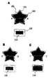
- FIG 1 shows encoded polymeric microparticles according to one embodiment of the invention.
- FIG. 2 is a process flow diagram illustrating a method for preparing encoded polymeric microparticles.
- FIG. 3 shows a process for making encoded polymer microparticles according to one embodiment of the invention.
- SEM scanning electron microscope
- FIG. 6 is a photograph comparing the characteristics of the polymer microparticles (star shape) without the silica shell and the polymer microparticles (circle shape) with the silica shell.
- FIG. 7 shows a process of performing multiple DNA hybridization assays by fixing oligonucleotides on silica coated polymer microparticle surfaces.
- the encoded polymer microparticle 100 includes an encoded polymer microparticle core 110 and a silica shell 120 surrounding the core.
- Core 110 may be coded in a variety of known manners.
- the encoded polymeric microparticle core 110 may have a graphical code, a fluorescent code, or a color code.
- the polymer constituting the polymer microparticle core 110 is preferably a photocurable polymer in that it can be variously patterned by optical lithography.
- the photocurable polymer may mainly contain an acrylic curable material.
- the photocurable polymer may be mixed with an acrylic photocurable material and a linker material having a functional group capable of reacting with the photocurable material and a photocurable functional group and a silica forming agent at the same time.
- the shape of the polymer microparticle core 110 made by photocuring may have various shapes including a disc shape and a spherical shape.
- the size of the core 110 may range from several ⁇ m to several mm.
- the encoded polymer microparticles 100 may further include a magnetic material.
- the microparticle core 110 may further contain magnetic nanoparticles 130 therein (B of FIG. 1).
- a layer of magnetic nanoparticles 130 may be interposed between the microparticle core 110 and the silica shell 120 (FIG. 1C).
- the layers of the magnetic nanoparticles 130 are interposed between the core 110 and the shell 120, the amount of the magnetic nanoparticles 130 is relatively small, and thus a stronger magnetic field is required for controlling the microparticles 100 later.
- the encoded polymer microparticles 100 may include the magnetic nanoparticles to control the microparticles 100 by an external magnetic field. As a result, it can be efficiently used in the solution exchange process of the bioassay later, it is possible to separate the fine particles 100 can increase the accuracy and convenience of the bioassay.
- the silica shell 120 surrounds and protects the microparticle core 110, and prevents an analytical error from being absorbed into the polymer of the microparticle core 110 by the foreign material desired to be detected.
- the silica shell 120 provides chemical and mechanical stability of the encoded polymer microparticles 130 to help the microparticles 130 be used in various environments and solutions.
- the encoded polymer microparticle core 110 and the silica shell 120 may be connected by -Si-O-Si- bonding. Thus, a stable structure may be formed by a solid chemical bond between the core 110 and the shell 120. Due to the presence of the silica shell 120, the surface of the polymer microparticles 100 has low binding property of the unspecified material and the bonding property with the biomaterial is improved.
- a functional group such as a carboxyl group or an amine group may be introduced to the surface of the silica shell 120.
- a functional group such as a carboxyl group or an amine group
- the functional group can be covalently bonded to a variety of biomolecules widely used in the field of biomedical or clinical diagnostics.
- any one biomaterial selected from the group consisting of antigen, antibody, DNA, RNA and oligonucleotide may be introduced to the surface of the silica shell 120.
- Coded polymer microparticles according to an embodiment of the present invention can be prepared by the following method.
- 2 is a process flow diagram illustrating a method for preparing encoded polymeric microparticles.
- step S1 a mixture of a photocurable material and a linker having a functional group and an alkoxysilyl group polymerizable with the photocurable material is provided.
- the photocurable material is a material that makes the basic skeleton structure of the fine particles by curing according to the application of energy.
- the photocurable material is ethoxylated trimethylolpropane triacrylate, 2-hydroxyethyl methacrylate, methyl methacrylate, acrylamide, allylamine, polyethylene oxide, polyethylene glycol diacrylate, polypropylene glycol diacrylate, Polyvinylpyrrolidone, polyvinyl alcohol, polyacrylate, and combinations thereof.
- polyethyleneglycol diacrylate which is a photocurable material, has acrylate functional groups at both ends of polyethyleneglycol, and may be crosslinked with a hydrogel having a three-dimensional structure when free radical polymerization occurs.
- the photocurable material may be any type of material that can be changed from liquid to solid by external light.
- the linker reacts with the photocurable material to form a copolymer, forming a skeleton of the microparticles, and allowing the alkoxysilyl group to be grafted on the surface of the encoded microparticle core.
- the formation of the silica shell through the silica coating is not easy.
- a linker having a photocurable material and a polymerizable functional group and an alkoxysilyl group at the same time is mixed with the photocurable material to form a mixture and hardened to graft the surface of the microparticle core. It is possible to coat the silica shell through the alkoxysilyl group.
- the linker may be, for example, a compound represented by Formula 1 below.
- R 1 may be hydrogen, methyl or ethyl
- R 2 may be C 1 to C 8 linear or branched alkyl
- L may be an alkylene or arylene having C 1 to C 12 or a structure in which the alkylene and the arylene are optionally connected.
- Chemical Formula 1 may be 3- (trimethoxysilyl) propyl acrylate (TMSPA).
- the mixture may further comprise an initiator and may cause free radical polymerization by an external energy source.
- the initiator may be an azo compound or a peroxide.
- the mixture may further include a suitable crosslinking agent, and examples thereof include N, N'-methylenebisacrylamide, methylenebismethacrylamide and ethylene glycol dimethacrylate.
- the method may further include adding magnetic nanoparticles to the mixture as necessary to control the encoded polymer microparticles.
- magnetic nanoparticles may be introduced into the polymer microparticle core.
- the patterned energy is applied in step S2 to cure and code the mixture to obtain the encoded polymeric microparticle core.
- the patterned energy ultraviolet rays, visible rays, infrared rays, and electron beams may be included without limitation.
- the irradiation of the patterned energy may be performed by a physical mask or a digital micromirror device (DMD) using ultraviolet light.
- DMD digital micromirror device
- the encoding can be performed in a variety of ways.
- the method of encoding the microparticle core may be applied to, for example, a method of patterning a graphical code by applying an optical lithography method.
- the shape of the graphical code may be in the shape of the microparticle itself (for example, star shape, circle shape, etc.), or may be in the form of a binary code engraved on the microparticle.
- the encoding of the graphical code may be performed by applying a photocurable polymer to the preparation of the particles as described above and patterning it by an optical lithography method.
- the photolithographic lithography method of Korean Patent No. 1004769, the fluid lithography method and the polymerization method of US Patent No. 7709544 may be applied, but not limited thereto, and various known lithography methods may be applied.
- the method of encoding the microparticle core is, for example, on the photocurable polymer, the code on the particles by patterning the labels meaning '1' and '0' to be distinguished from each other according to the degree of photocuring, respectively Can be formed.
- the above-described optical lithography method for example, when using a digital micromirror device that does not use a mask, may have the advantage of forming as many as one million or more various kinds of codes for particles containing the target material. have.
- the encoding of the microparticle core may be achieved by including in the microparticle core phosphors of various colors that are distinguished from one another.
- Various known techniques can be applied to incorporate various fluorescent materials into the microparticle core.
- the method of encoding the microparticle core may be a method of forming a color code using a magnetic ink.
- a method of using the magnetic ink as disclosed in Korean Patent Application No. 10-2010-0029613 is as follows. First, a photocurable material including magnetic nanoparticles is provided and an external magnetic field is applied to align the magnetic nanoparticles in the photocurable material. And, it may be performed by applying light from the outside to cure the photocurable material. The arrangement of the magnetic nanoparticles may be changed by the intensity of the external magnetic field to express different colors. By applying this technique, the magnetic particle may be color-coded by arranging the magnetic nanoparticles to be discriminated from each other in the microparticle core made of the photocurable polymer. The contents of this patent can be incorporated by reference herein.
- the method may further include attaching the hydrophilic polymer-coated magnetic nanoparticles to the encoded polymer microparticle core to control the encoded polymer microparticles.
- the encoded polymer microparticles are prepared by treating a silica precursor on the encoded polymer microparticle core to form a silica shell on the encoded polymer microparticle core.
- the formation of the silica shell can be carried out by various known methods, for example a modified Stober method can be used.
- the fine particles grafted with an alkoxysilyl group are added to a solution of distilled water, ethanol and NH 4 OH.
- Tetraethylorthosilicate (TEOS) is then injected into the solution as a silica precursor and reacted to form a silica shell.
- TEOS Tetraethylorthosilicate
- -Si-O-Si-bonds may be formed at the core-shell interface by the reaction of the alkoxysilyl group grafted on the surface of the microparticle core with the silica precursor.
- Polymeric microparticles are flexible, soft and easy to fabricate in a variety of structures and forms. At the same time, however, they are easily damaged mechanically and chemically. Also, in the bioassay, the absorption of small molecules into the polymer matrix can lead to detection errors. In contrast, inorganic materials such as titania and silica are generally much harder and more chemically resistant than polymer organics. However, this is also fragile and difficult to mold in various forms. Therefore, by coating the silica shell on the polymer microparticles, the advantages of the two materials can be combined to have a strong, hard, chemically stable, durable and easy to mold in various forms at the same time.
- the silica coated coded polymeric microparticles described above can be used for multiplexed bioanalysis.
- encoded polymer microparticles have been used for analysis of biomolecules such as DNA and proteins. This is a powerful and adaptable method because it can use almost unlimited code and provides high analysis throughput. Therefore, according to one embodiment of the present invention, the surface of the silica shell may be modified to introduce a carboxyl group or an amine group for the application of the multiple bioanalysis. The presence of these functional groups allows covalent bonding with various biomaterials.
- the above-described manufacturing method may further comprise the step of bonding the biomaterial to the silica shell surface.
- FIG. 3 shows a process for making encoded polymer microparticles according to one embodiment of the invention.
- a photocurable material ethoxylated trimethylopropane triacrylate (ETPTA), a linker, TMSPA (3- (trimethoxysilyl) propylacrylate), and a photoinitiator ( 2-hydroxy-2-methylpropiophenone) is mixed in an appropriate ratio (ex. 10: 1: 1) to form a photocurable mixture.
- ETPTA ethoxylated trimethylopropane triacrylate
- TMSPA trimethoxysilyl propylacrylate
- a photoinitiator 2-hydroxy-2-methylpropiophenone
- TMSPA not only has a silane group with silicon capable of forming silica, but also serves as a seed for subsequent silica coating.
- This compound is irradiated with patterned UV light using various known lithographic methods including photofluidless maskless lithography of FIG. 3 to produce alkoxysilane containing copolymer microparticles with a graphical code.
- photofluidic maskless lithography OFML
- free-floating particles can be produced continuously by in-situ photopolymerization by introducing raw materials through microfluidic channels and applying patterned energy, and using digital micro mirror devices ( Coded particles of various shapes can be easily obtained by a maskless method using DMD) as compared to other lithography methods using masks.
- Silica coating is performed on the formed copolymer microparticles by applying a modified Stober method to form silica shells.
- This simple coating process is very fast and is a direct and efficient way to coat silica on silane-containing microparticles. It is also a method that can process millions of silica-coated microparticles at once.
- the thickness of the silica shell can be adjusted from several hundred nanometers to several micrometers by adjusting the reaction rate or the concentration of the silica precursor.
- various functional groups can be introduced by using various conventional silica surface treatment methods on the fine particles having the silica shell.
- FIG. 4 is a scanning electron microscope (SEM) image showing the growth of the silica shell on the actual particle surface.
- SEM scanning electron microscope
- FIG. 5 is a comparative analysis of the spectra of silica coated microparticles with an electron probe micro-analyzer (EPMA) and non-silica coated microparticles.
- EPMA electron probe micro-analyzer
- the polymer microparticles not coated with silica were prepared using only ETPTA and a photoinitiator without TMSPA (seed material).
- EPMA spectra of uncoated microparticles show that only strong C and weak O are pure organic polymers.
- the Pt signal was also slightly detected because it was sputtered with Pt coating. Looking at the bottom of Figure 5 it can be seen that the pure silica shell is well formed on the polymer microparticles.
- the O signal is much larger than C because of silica (SiO 2 ).
- the reason for the C signal is that the beam penetration depth of the EPMA is 1 ⁇ m and the thickness of the silica shell is less.
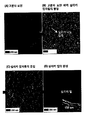
- FIG. 6 is a photograph comparing the characteristics of the polymer microparticles (star shape) without the silica shell and the polymer microparticles (circle shape) with the silica shell.
- aqueous Rhodamine B solution red fluorescence
- 6A shows how the aqueous solution of rhodamine is absorbed into the uncoated polymer microparticles (star shape) and the silica coated polymer microparticles (circle shape), respectively.
- the aqueous solution of rhodamine was well absorbed into the polymer hydrogel material forming the microparticles as clearly seen in the fluorescence image. It is well known that polymeric hydrogel materials absorb liquids well without any chemical or physical treatment.
- silica coated microparticles can provide more stable and accurate results when applied to bioanalyses. Because if antigens or oligonucleotides are absorbed into the polymer matrix, this can be a source of error. In addition, in this result that no fluorescence is emitted from the image of the coated particles, it can be confirmed that due to the low non-specific binding of the silica surface, the absorption of some dye did not occur at the silica surface.
- the microparticles were placed in 1M aqueous acetic acid and stirred. After 24 hours, the surface of the silica coated microparticles remained stable, but the surface of the uncoated microparticles was severely damaged by the acidic solution (FIG. 6C).
- polymer hydrogels are very sensitive to external environments such as pH and temperature.
- silica is generally much more robust and has good chemical stability than organic polymers. As a result, these silica-coated microparticles can withstand better than polymer microparticles in a variety of chemical reactions, including organic solvents and dry environments. Therefore, there are few restrictions in applying various functional groups to the particles.
- the microparticles can have high stability and low nonspecific binding properties.
- silica coated microparticles easily react with 3-aminopropyltriethoxysilane (APTES) and primary amine through condensation of terminal hydroxyl groups. When these amines are subsequently reacted with succinic acid to form carboxylated surfaces, they can react with amino groups in the DNA. 5 'amino terminus DNAs are used to immobilize DNAs on the carboxylated silica surface.
- APTES 3-aminopropyltriethoxysilane
- the encoded polymeric microparticles according to one embodiment of the present invention provide useful advantages for multiplexed bioassays.
- silica-coated microparticles were used to demonstrate 10-plex in vitro human papillomavirus (HPV) genotyping.
- Target HPV genes were prepared by a two step PCR process.
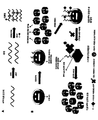
- 8 shows multiple HPV genotyping using silica coated coded polymeric microparticles.
- 8A is a schematic of amplification and labeling PCR. After amplification PCR, labeling PCR for labeling label probe-complementary HPV gene sequences is performed using biotin attached dCTP.
- 8B is a schematic diagram of 10-plexed HPV genotyping using probe attached microparticles.
- Each target HPV gene is complementary to one of the different probes.
- the graphical code consisting of unpolymerized pores in the microparticle structure is intended to identify the probe on the silica surface of the microparticles. Multiplexing capacity can be easily increased by changing the graphical code. After hybridization assay with the target HPV sequence, fluorescent dye labeled streptavidins are introduced to obtain a fluorescent signal.
- FIG. 9 shows the preparation and handling of silica coated coded "magnetic" microparticles.
- 9A illustrates a step of preparing silica coated magnetic microparticles.
- FIG. 9B is a field emission scanning electron microscope (FE-SEM) image of magnetic microparticles before (B1) and after (B2) of a silica coating process.
- the magnetic microparticles are obtained by attaching polyacrylic acid (PAA) coated Fe 3 O 4 nanoparticles on the surface of the silane grafted microparticles.
- PAA coated Fe 3 O 4 nanoparticles can have a size of, for example, 80 ⁇ 10 nm.
- FIG. 9C shows a solution exchange process through magnetic separation of carriers.
- a mixture containing silica coated magnetic particles and biomolecules bound to these silica coated magnetic particles can be selectively separated from the solution mixture.
- 9D are optical microscope images showing magnetic handling of magnetic particles. It can be seen that free floating magnetic microparticles move easily in the direction of an applied external magnetic field. Therefore, the magnetic handling of free-floating magnetic microparticles is useful for bioassays involving solution exchange. Moreover, magnetic separation allows for washing and concentration of the separated targets that were bound to the particles.
- the encoded polymer microparticles are prepared using a mixture of a photocurable material and a linker having a polymerizable and alkoxysilyl group.
- a silica precursor solution By encapsulating the thus prepared encoded polymer microparticles in a silica precursor solution, millions of encoded polymer microparticles can be silica coated at once by a simple process.
- magnetic nanoparticles By introducing the above-mentioned magnetic nanoparticles into the polymer microparticles, handling of the particles in the bioassay becomes easy.
- Encoded microparticles according to one embodiment of the present invention have a chemical and physical stability by having a silica shell and do not absorb external biomolecules, thereby enabling accurate analysis. Therefore, the above-described encoded polymer microparticles can be usefully used in various applications including DNA or protein based diagnosis.
Landscapes
- Health & Medical Sciences (AREA)
- Life Sciences & Earth Sciences (AREA)
- Immunology (AREA)
- Chemical & Material Sciences (AREA)
- Engineering & Computer Science (AREA)
- Molecular Biology (AREA)
- Biomedical Technology (AREA)
- Urology & Nephrology (AREA)
- Hematology (AREA)
- Analytical Chemistry (AREA)
- Microbiology (AREA)
- Biochemistry (AREA)
- General Health & Medical Sciences (AREA)
- Physics & Mathematics (AREA)
- Biotechnology (AREA)
- Organic Chemistry (AREA)
- Medicinal Chemistry (AREA)
- Cell Biology (AREA)
- General Physics & Mathematics (AREA)
- Pathology (AREA)
- Food Science & Technology (AREA)
- Proteomics, Peptides & Aminoacids (AREA)
- Wood Science & Technology (AREA)
- Zoology (AREA)
- Bioinformatics & Cheminformatics (AREA)
- Biophysics (AREA)
- General Engineering & Computer Science (AREA)
- Genetics & Genomics (AREA)
- Inorganic Chemistry (AREA)
- Virology (AREA)
- Chemical Kinetics & Catalysis (AREA)
- Polymers & Plastics (AREA)
- Compositions Of Macromolecular Compounds (AREA)
- Measuring Or Testing Involving Enzymes Or Micro-Organisms (AREA)
- Processes Of Treating Macromolecular Substances (AREA)
Priority Applications (5)
| Application Number | Priority Date | Filing Date | Title |
|---|---|---|---|
| CN201380059253.XA CN104781321B (zh) | 2012-11-16 | 2013-11-18 | 编码的聚合物微粒 |
| US14/129,546 US10557846B2 (en) | 2012-11-16 | 2013-11-18 | Encoded polymeric microparticles |
| JP2015542957A JP6085035B2 (ja) | 2012-11-16 | 2013-11-18 | コード化された高分子微粒子 |
| ES13855838.2T ES2656987T3 (es) | 2012-11-16 | 2013-11-18 | Micropartículas de polímero codificadas |
| EP13855838.2A EP2905304B1 (de) | 2012-11-16 | 2013-11-18 | Codierte polymermikropartikel |
Applications Claiming Priority (2)
| Application Number | Priority Date | Filing Date | Title |
|---|---|---|---|
| KR20120130096 | 2012-11-16 | ||
| KR10-2012-0130096 | 2012-11-16 |
Publications (1)
| Publication Number | Publication Date |
|---|---|
| WO2014077645A1 true WO2014077645A1 (ko) | 2014-05-22 |
Family
ID=50731481
Family Applications (1)
| Application Number | Title | Priority Date | Filing Date |
|---|---|---|---|
| PCT/KR2013/010457 WO2014077645A1 (ko) | 2012-11-16 | 2013-11-18 | 코드화된 고분자 미세입자 |
Country Status (7)
| Country | Link |
|---|---|
| US (1) | US10557846B2 (de) |
| EP (1) | EP2905304B1 (de) |
| JP (1) | JP6085035B2 (de) |
| KR (2) | KR101582384B1 (de) |
| CN (1) | CN104781321B (de) |
| ES (1) | ES2656987T3 (de) |
| WO (1) | WO2014077645A1 (de) |
Families Citing this family (13)
| Publication number | Priority date | Publication date | Assignee | Title |
|---|---|---|---|---|
| JP6182520B2 (ja) * | 2014-12-01 | 2017-08-16 | 三洋化成工業株式会社 | 磁性シリカ粒子、該磁性シリカ粒子を用いた測定対象物質測定方法及び測定対象物質測定用試薬 |
| KR101758145B1 (ko) * | 2015-02-16 | 2017-07-17 | 한국과학기술연구원 | 바이오 어세이를 위한 마이크로 입자의 제조방법 및 이에 의해 제조된 바이오 어세이를 위한 마이크로 입자 |
| US20160322560A1 (en) * | 2015-04-30 | 2016-11-03 | The Regents Of The University Of California | 3d piezoelectric polymer materials and devices |
| KR102435668B1 (ko) * | 2015-10-20 | 2022-08-24 | 주식회사 퀀타매트릭스 | 다중 분석 칩 및 이를 이용한 분석 장치 |
| KR102060306B1 (ko) * | 2016-10-05 | 2019-12-30 | 서울대학교산학협력단 | 저장, 보존 및 색인이 가능한 생화학 물질 운반체 및 이의 제조방법 |
| KR101879585B1 (ko) * | 2016-10-06 | 2018-07-18 | 경희대학교 산학협력단 | 하이드로겔 입자의 자동 코팅 장치 및 방법 |
| KR101850854B1 (ko) * | 2016-10-17 | 2018-04-20 | 연세대학교 원주산학협력단 | 퀀타매트릭스 어세이 플랫폼 기반 그람양성, 그람음성균 및 캔디다의 검출 및 동정과 항생제 내성여부를 동시 확인할 수 있는 진단법 및 그 키트 |
| KR101886278B1 (ko) * | 2016-11-04 | 2018-08-08 | 주식회사 퀀타매트릭스 | 인유두종바이러스 유전자형 검출용 조성물 |
| US20190353649A1 (en) * | 2016-12-01 | 2019-11-21 | University Of Florida Research Foundation, Inc. | Polymer conjugates, methods of making polymer conjugates, and methods of using polymer conjugates |
| KR101905800B1 (ko) | 2017-02-13 | 2018-10-08 | 충남대학교산학협력단 | 고분자-나노실리카 복합체 및 그의 제조방법 |
| KR101941361B1 (ko) * | 2017-02-17 | 2019-01-22 | 경희대학교 산학협력단 | 폴리도파민으로 코팅된 투명 원반형 마이크로입자 |
| JP7091129B2 (ja) * | 2018-04-27 | 2022-06-27 | キヤノン株式会社 | 粒子、及びその製造方法 |
| JP7363327B2 (ja) | 2019-10-09 | 2023-10-18 | 東ソー株式会社 | 抗体結合用磁性粒子およびその製造方法 |
Citations (7)
| Publication number | Priority date | Publication date | Assignee | Title |
|---|---|---|---|---|
| US20040013569A1 (en) * | 2001-02-19 | 2004-01-22 | Balkus Kenneth J. | Encoded molecular sieve particle-based sensors |
| KR20100029613A (ko) | 2008-09-08 | 2010-03-17 | 엘지전자 주식회사 | 차량 네비게이션 방법 및 그 장치 |
| KR20100029962A (ko) * | 2008-09-09 | 2010-03-18 | 서울대학교산학협력단 | 광유체적 리소그래피 시스템, 2층구조 마이크로유체관 제조방법 및 3차원 마이크로구조물 제조방법 |
| US7709544B2 (en) | 2005-10-25 | 2010-05-04 | Massachusetts Institute Of Technology | Microstructure synthesis by flow lithography and polymerization |
| KR20110110046A (ko) * | 2010-03-31 | 2011-10-06 | 서울대학교산학협력단 | 자성 구조물을 자기적으로 제어하는 방법 |
| KR20110113625A (ko) * | 2009-04-14 | 2011-10-17 | 서울대학교산학협력단 | 컬러 코드화된 자성 구조물 |
| KR101101310B1 (ko) * | 2011-05-17 | 2011-12-30 | 서울대학교산학협력단 | 코드화된 입자 기반의 플랫폼을 이용하는 분석방법 |
Family Cites Families (15)
| Publication number | Priority date | Publication date | Assignee | Title |
|---|---|---|---|---|
| IL111186A (en) * | 1994-10-06 | 1999-09-22 | Univ Bar Ilan | Process for the preparation of microspheres and microspheres made thereby |
| JP2000169591A (ja) * | 1998-12-11 | 2000-06-20 | Ube Nitto Kasei Co Ltd | 光硬化性樹脂被覆粒子、その製造方法および液晶表示素子の製造方法 |
| US6548264B1 (en) * | 2000-05-17 | 2003-04-15 | University Of Florida | Coated nanoparticles |
| GB0220063D0 (en) * | 2002-08-29 | 2002-10-09 | Isis Innovation | Magnetic particle and process for preparation |
| US20040101822A1 (en) * | 2002-11-26 | 2004-05-27 | Ulrich Wiesner | Fluorescent silica-based nanoparticles |
| CN1317560C (zh) | 2005-10-19 | 2007-05-23 | 华中科技大学 | 一种提高量子点编码微球的编码稳定性的方法 |
| JP4780710B2 (ja) * | 2006-03-16 | 2011-09-28 | 大阪府 | コア−シェル型高分子ゲル微粒子及びその製造方法 |
| GB0617480D0 (en) * | 2006-09-06 | 2006-10-18 | Univ Sheffield | Novel nanoparticles |
| GB2445580A (en) | 2007-01-11 | 2008-07-16 | Secr Defence | An encoded microsphere |
| GB2445579A (en) | 2007-01-11 | 2008-07-16 | Secr Defence | An encoded microsphere |
| IE20090904A1 (en) * | 2008-11-26 | 2010-07-07 | Univ College Cork Nat Univ Ie | A process for preparing microparticles |
| WO2010120109A2 (ko) * | 2009-04-14 | 2010-10-21 | 서울대학교산학협력단 | 구조색 생성방법 |
| KR101125191B1 (ko) * | 2009-11-09 | 2012-03-19 | 한국과학기술원 | 콜로이드 입자로 안정화된 액적을 이용한 표면구조를 갖는 미세입자의 제조방법 |
| CN101912757B (zh) * | 2010-08-30 | 2012-06-27 | 武汉大学 | 一种荧光-磁性双编码微球的制备方法 |
| CN102120168B (zh) * | 2010-12-07 | 2013-05-29 | 复旦大学 | 多功能核壳结构荧光编码磁性微球及其制备方法 |
-
2013
- 2013-11-18 KR KR1020130140211A patent/KR101582384B1/ko active IP Right Grant
- 2013-11-18 US US14/129,546 patent/US10557846B2/en active Active
- 2013-11-18 WO PCT/KR2013/010457 patent/WO2014077645A1/ko active Application Filing
- 2013-11-18 EP EP13855838.2A patent/EP2905304B1/de active Active
- 2013-11-18 CN CN201380059253.XA patent/CN104781321B/zh active Active
- 2013-11-18 ES ES13855838.2T patent/ES2656987T3/es active Active
- 2013-11-18 JP JP2015542957A patent/JP6085035B2/ja active Active
-
2015
- 2015-11-24 KR KR1020150164665A patent/KR102007715B1/ko active IP Right Grant
Patent Citations (8)
| Publication number | Priority date | Publication date | Assignee | Title |
|---|---|---|---|---|
| US20040013569A1 (en) * | 2001-02-19 | 2004-01-22 | Balkus Kenneth J. | Encoded molecular sieve particle-based sensors |
| US7709544B2 (en) | 2005-10-25 | 2010-05-04 | Massachusetts Institute Of Technology | Microstructure synthesis by flow lithography and polymerization |
| KR20100029613A (ko) | 2008-09-08 | 2010-03-17 | 엘지전자 주식회사 | 차량 네비게이션 방법 및 그 장치 |
| KR20100029962A (ko) * | 2008-09-09 | 2010-03-18 | 서울대학교산학협력단 | 광유체적 리소그래피 시스템, 2층구조 마이크로유체관 제조방법 및 3차원 마이크로구조물 제조방법 |
| KR101004769B1 (ko) | 2008-09-09 | 2011-01-04 | 서울대학교산학협력단 | 광유체적 리소그래피 시스템 및 마이크로구조물 제조방법 |
| KR20110113625A (ko) * | 2009-04-14 | 2011-10-17 | 서울대학교산학협력단 | 컬러 코드화된 자성 구조물 |
| KR20110110046A (ko) * | 2010-03-31 | 2011-10-06 | 서울대학교산학협력단 | 자성 구조물을 자기적으로 제어하는 방법 |
| KR101101310B1 (ko) * | 2011-05-17 | 2011-12-30 | 서울대학교산학협력단 | 코드화된 입자 기반의 플랫폼을 이용하는 분석방법 |
Non-Patent Citations (1)
| Title |
|---|
| See also references of EP2905304A4 |
Also Published As
| Publication number | Publication date |
|---|---|
| EP2905304A4 (de) | 2016-03-02 |
| EP2905304B1 (de) | 2017-09-13 |
| EP2905304A1 (de) | 2015-08-12 |
| JP6085035B2 (ja) | 2017-02-22 |
| CN104781321B (zh) | 2018-01-23 |
| KR102007715B1 (ko) | 2019-08-07 |
| KR20150141165A (ko) | 2015-12-17 |
| CN104781321A (zh) | 2015-07-15 |
| ES2656987T3 (es) | 2018-03-01 |
| JP2016501286A (ja) | 2016-01-18 |
| KR20140063480A (ko) | 2014-05-27 |
| US10557846B2 (en) | 2020-02-11 |
| US20140228252A1 (en) | 2014-08-14 |
| KR101582384B1 (ko) | 2016-01-05 |
Similar Documents
| Publication | Publication Date | Title |
|---|---|---|
| WO2014077645A1 (ko) | 코드화된 고분자 미세입자 | |
| EP1432989B1 (de) | Arrays in zufallsanordnung und herstellungs- und verwendungsverfahren | |
| US11391673B2 (en) | Method for preparing encoded hydrogel particles, and encoded hydrogel particles prepared thereby | |
| Park et al. | Dual functional, polymeric self-assembled monolayers as a facile platform for construction of patterns of biomolecules | |
| CA2596807A1 (en) | Method for the photochemical attachment of biomolecules to a substrate | |
| US11819843B2 (en) | Flow cells with a hydrophobic barrier | |
| Shamansky et al. | Immobilization and detection of DNA on microfluidic chips | |
| US20100190654A1 (en) | Nanoarrays and methods and materials for fabricating same | |
| Torelli et al. | DNA origami nanorobot fiber optic genosensor to TMV | |
| US20220155211A1 (en) | Altering flow cell signals | |
| US11447733B2 (en) | Transparent disc-shaped microparticles coated with polydopamine | |
| EP1202062B1 (de) | Gemusterte Polymeroberflächen geeignet für Biokonjugationen und Verfahren zu ihrer Herstellung | |
| WO2012060620A2 (ko) | 항생물부착성 ssq/peg 네트워크 및 그 제조방법 | |
| KR101507757B1 (ko) | 미세패턴된 친수성 고분자 기판 상에 선택적으로 소수성 코팅층을 형성하는 방법 및 이에 의해 형성된 소수성 코팅층을 가지는 미세패턴된 친수성 고분자 기판 | |
| US20110053799A1 (en) | Method of producing microarray substrate, radiation-sensitive composition, partition of microarray substrate, method of producing biochip, and biochip | |
| Jung et al. | Integrated fabrication-conjugation approaches for biomolecular assembly and protein sensing with hybrid microparticle platforms and biofabrication-A focused minireview | |
| WO2016085053A1 (ko) | 랜덤 모자이크 식별 코드 | |
| Sola et al. | 14 Surface Modifications by Polymers for Biosensing Applications | |
| CN114994322A (zh) | 磁性量子点编码微球及制备方法和高通量检测用装置 | |
| He | Application of encoded microparticles fabricated by standard lithography on multiplexed bioassay | |
| CN113646441A (zh) | 在流通池中的固定 | |
| Kim | Microfluidic processes to create structured microparticle arrangements and their applications |
Legal Events
| Date | Code | Title | Description |
|---|---|---|---|
| WWE | Wipo information: entry into national phase |
Ref document number: 14129546 Country of ref document: US |
|
| 121 | Ep: the epo has been informed by wipo that ep was designated in this application |
Ref document number: 13855838 Country of ref document: EP Kind code of ref document: A1 |
|
| REEP | Request for entry into the european phase |
Ref document number: 2013855838 Country of ref document: EP |
|
| WWE | Wipo information: entry into national phase |
Ref document number: 2013855838 Country of ref document: EP |
|
| ENP | Entry into the national phase |
Ref document number: 2015542957 Country of ref document: JP Kind code of ref document: A |
|
| NENP | Non-entry into the national phase |
Ref country code: DE |