WO2013122072A1 - 血液情報の測定方法及び装置 - Google Patents
血液情報の測定方法及び装置 Download PDFInfo
- Publication number
- WO2013122072A1 WO2013122072A1 PCT/JP2013/053321 JP2013053321W WO2013122072A1 WO 2013122072 A1 WO2013122072 A1 WO 2013122072A1 JP 2013053321 W JP2013053321 W JP 2013053321W WO 2013122072 A1 WO2013122072 A1 WO 2013122072A1
- Authority
- WO
- WIPO (PCT)
- Prior art keywords
- blood
- information
- plasma
- flow cell
- light
- Prior art date
- Legal status (The legal status is an assumption and is not a legal conclusion. Google has not performed a legal analysis and makes no representation as to the accuracy of the status listed.)
- Ceased
Links
Images


Classifications
-
- A—HUMAN NECESSITIES
- A61—MEDICAL OR VETERINARY SCIENCE; HYGIENE
- A61B—DIAGNOSIS; SURGERY; IDENTIFICATION
- A61B5/00—Measuring for diagnostic purposes; Identification of persons
- A61B5/145—Measuring characteristics of blood in vivo, e.g. gas concentration or pH-value ; Measuring characteristics of body fluids or tissues, e.g. interstitial fluid or cerebral tissue
- A61B5/1455—Measuring characteristics of blood in vivo, e.g. gas concentration or pH-value ; Measuring characteristics of body fluids or tissues, e.g. interstitial fluid or cerebral tissue using optical sensors, e.g. spectral photometrical oximeters
-
- G—PHYSICS
- G01—MEASURING; TESTING
- G01N—INVESTIGATING OR ANALYSING MATERIALS BY DETERMINING THEIR CHEMICAL OR PHYSICAL PROPERTIES
- G01N21/00—Investigating or analysing materials by the use of optical means, i.e. using sub-millimetre waves, infrared, visible or ultraviolet light
- G01N21/01—Arrangements or apparatus for facilitating the optical investigation
- G01N21/03—Cuvette constructions
- G01N21/05—Flow-through cuvettes
-
- G—PHYSICS
- G01—MEASURING; TESTING
- G01N—INVESTIGATING OR ANALYSING MATERIALS BY DETERMINING THEIR CHEMICAL OR PHYSICAL PROPERTIES
- G01N21/00—Investigating or analysing materials by the use of optical means, i.e. using sub-millimetre waves, infrared, visible or ultraviolet light
- G01N21/17—Systems in which incident light is modified in accordance with the properties of the material investigated
- G01N21/25—Colour; Spectral properties, i.e. comparison of effect of material on the light at two or more different wavelengths or wavelength bands
- G01N21/31—Investigating relative effect of material at wavelengths characteristic of specific elements or molecules, e.g. atomic absorption spectrometry
-
- G—PHYSICS
- G01—MEASURING; TESTING
- G01N—INVESTIGATING OR ANALYSING MATERIALS BY DETERMINING THEIR CHEMICAL OR PHYSICAL PROPERTIES
- G01N21/00—Investigating or analysing materials by the use of optical means, i.e. using sub-millimetre waves, infrared, visible or ultraviolet light
- G01N21/17—Systems in which incident light is modified in accordance with the properties of the material investigated
- G01N21/55—Specular reflectivity
- G01N21/552—Attenuated total reflection
-
- G—PHYSICS
- G01—MEASURING; TESTING
- G01N—INVESTIGATING OR ANALYSING MATERIALS BY DETERMINING THEIR CHEMICAL OR PHYSICAL PROPERTIES
- G01N33/00—Investigating or analysing materials by specific methods not covered by groups G01N1/00 - G01N31/00
- G01N33/48—Biological material, e.g. blood, urine; Haemocytometers
- G01N33/483—Physical analysis of biological material
- G01N33/487—Physical analysis of biological material of liquid biological material
- G01N33/49—Blood
-
- G—PHYSICS
- G01—MEASURING; TESTING
- G01N—INVESTIGATING OR ANALYSING MATERIALS BY DETERMINING THEIR CHEMICAL OR PHYSICAL PROPERTIES
- G01N21/00—Investigating or analysing materials by the use of optical means, i.e. using sub-millimetre waves, infrared, visible or ultraviolet light
- G01N21/17—Systems in which incident light is modified in accordance with the properties of the material investigated
- G01N2021/1734—Sequential different kinds of measurements; Combining two or more methods
- G01N2021/1736—Sequential different kinds of measurements; Combining two or more methods with two or more light sources
-
- G—PHYSICS
- G01—MEASURING; TESTING
- G01N—INVESTIGATING OR ANALYSING MATERIALS BY DETERMINING THEIR CHEMICAL OR PHYSICAL PROPERTIES
- G01N21/00—Investigating or analysing materials by the use of optical means, i.e. using sub-millimetre waves, infrared, visible or ultraviolet light
- G01N21/84—Systems specially adapted for particular applications
- G01N2021/8405—Application to two-phase or mixed materials, e.g. gas dissolved in liquids
Definitions
- the present invention relates to a blood information measurement method and apparatus, and more particularly, hemolysis (plasma free hemoglobin concentration), which can noninvasively and continuously obtain information on only plasma components without depending on hematocrit,
- hemolysis plasma free hemoglobin concentration
- the present invention relates to a method and an apparatus for measuring blood information such as blood coagulation degree (thrombus).
- hemoglobin monitoring in dialysis is important as an indicator of water removal efficiency, but current continuous hemoglobin monitors are not reliable.
- Patent Document 1 discloses characteristic parameters such as morphological information and absorption information of particles (blood cells, cells, etc.) contained in a sample liquid such as blood and urine from light transmitted through a flow cell.
- a transmitted light sensor and a scattered light sensor are arranged so as to be perpendicular to each other, the transmitted light sensor receives light along a transmission path passing through the cuppet, and the scattered light sensor is A technique for measuring the concentration of total hemoglobin or red blood cells in a blood flow by receiving light scattered at 90 degrees with respect to the transmission path and obtaining the ratio of the scattered signal to the transmitted signal is described.
- Patent Document 3 describes a spectroscopic optical analysis technique for blood in which a transmitted light sensor and a scattered light sensor are arranged in parallel.
- Patent Document 4 discloses a blood coagulation in which a scattered light amount value from a specimen to which a predetermined reagent is added is obtained at predetermined time intervals, and a coagulation end point is detected based on a change over time of the scattered light amount value. An analyzer is described.
- Patent Document 5 scattered light from a blood sample is received, and the coagulation time is calculated by measuring the saturation in the temporal change in the amount of scattered light after the coagulation reagent is added to the blood sample.
- a blood coagulation measuring device is described.
- the optical properties of blood depend on the erythrocyte volume MCV (particle volume), erythrocyte hemoglobin concentration MCHC (particle refractive index), hematocrit HCT (particle density), and plasma refractive index Np (refractive index of extraparticle solvent). Therefore, light propagation in blood can be regarded as a function having these as variables. However, conventionally, information Np of plasma components in blood cannot be measured noninvasively and continuously.
- the present invention has been made to solve the above-mentioned conventional problems, and can noninvasively and continuously measure information on plasma components in blood without separating blood components by mechanical or chemical treatment.
- the challenge is to do so.
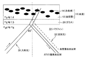
- the inventors have a blood 10 flowing in a flow cell formed of glass 20, which is a transparent material having a refractive index different from that of a plasma layer (also simply referred to as plasma) 12 in blood 10.
- the first measurement light (also referred to as incident light) 30 is incident on the boundary surface between the glass 20 and the blood 20 in an oblique direction of 45 degrees from the paraffin 22 having substantially the same refractive index as the glass 20. It was found that the information of the plasma layer 12 can be obtained by spectroscopically analyzing the light 32 (also referred to as reflected light) that has been specularly reflected (totally reflected here) at the boundary surface.
- Np-i is involved in light absorption, which can be obtained by obtaining an absorption spectrum.
- Np-i varies depending on proteins contained in plasma and blood coagulation state, that is, chemical composition of plasma. The principle of spectrum measurement is that when reflected at the glass / plasma layer boundary, evanescent light is generated by the boundary.
- the light intensity is attenuated by the interaction between the evanescent light and the substance (plasma layer), and information on plasma components can be obtained by measuring the attenuation for each wavelength with a spectrophotometer. Since this measurement method does not penetrate the target blood 10, Np can be measured basically without depending on blood cells.
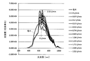
- Fig. 2 shows the spectrum change at each flow rate when the circulation circuit flow rate is changed. It can be seen that when the flow rate is increased, the received light intensity increases, that is, the reflected light intensity increases.
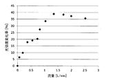
- FIG. 3 shows the rate of change of the spectrum with respect to the flow rate of 0 L / min by integrating the waveform of FIG. From this, it can be seen that the change decreases and becomes almost constant at a flow rate of 1.35 L / min or more. Strictly speaking, although there is a change, the deviation is about 1.47%, and there is no problem in actual measurement.
- the distribution of red blood cells in blood is stable in orientation, the spectrum is stable regardless of the flow rate, and measurement is easy.
- correction can be performed and measurement can be performed at any flow rate.
- the horizontal axis in Fig. 3 is the current flow rate, but it can be converted to an average flow velocity by dividing by the cross-sectional area of the flow cell.
- Re UD / ( ⁇ / ⁇ ) (1)
- U is a characteristic flow velocity [m / sec]
- D is a characteristic length [m]
- ⁇ is a fluid viscosity [Pa ⁇ s]
- ⁇ is a fluid density [kg / m 3 ].
- the Reynolds number Re represents the ratio between the viscous force and the inertial force, and the greater the Re, the stronger the inertial force.
- the viscous force is a frictional resistance caused by the viscosity of the fluid itself when the fluid moves (flows), and this force is dragged by the surrounding fluid elements to similarly move. That is, in a flow field with a certain flow velocity distribution, it represents the force that the fluid moves according to the streamline.
- the lower the Reynolds number Re (the higher the viscous force), the less the flow is disturbed and the laminar flow along the streamline.
- the inertial force represents the opposite, and is the inertia generated by the mass of the fluid as the fluid moves, and represents the force that moves against the surrounding fluid elements. Therefore, the stronger the inertial force, the more freely the fluid behaves without following the viscous force. Therefore, the higher the Reynolds number Re (the higher the inertial force), the more turbulent flow that is not chaotic and chaotic. It is said that Re> 2000 as a measure of transition from laminar flow to turbulent flow.
- the Reynolds number Re is a dimensionless measure that represents how well the fluid behavior is ordered, and is therefore used as a flow similarity law. For example, when a flow in a certain pipe is considered, even if the pipe diameter or the viscosity of the fluid is different in density, if the Reynolds number Re is the same, the flow pattern is the same. Therefore, even if the flow cell size is different (similar in shape), even if the density and viscosity of the blood are different, the same measurement is performed as long as the conditions are satisfied from the Reynolds number Re.
- the measurement conditions themselves can be expressed numerically.
- the Reynolds number Re at 1.35 L / min is obtained.
- the characteristic length D of the formula (1) is a pipe diameter.
- the wavelength of light hitting the boundary surface is 600 nm or less, and more preferably 500 to 600 nm.
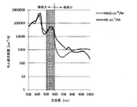
- the HCT differential spectrum ⁇ HCT there is almost no change in the spectrum even when the hematocrit HCT is changed at a wavelength of 500 nm to 600 nm, whereas in FIG. This is because, as shown in the differential spectrum ⁇ fHb of free hemoglobin fHb, there is a characteristic with respect to hemolysis, and a characteristic of the absorption characteristic of hemoglobin Hb depending on plasma free hemoglobin fHb is obtained, and plasma layer boundary reflection spectroscopy can be performed in this wavelength band. .
- the incident angle is not limited to 45 degrees or less, may not be total reflection, and may have a light wavelength of 600 nm or more.
- the present invention has been made on the basis of the above-described findings, and is obliquely inclined at an angle shallower than 90 degrees to the boundary surface between blood and a flow cell formed of a transparent material having a refractive index different from that of plasma.
- the first measurement light is incident from the direction, the light specularly reflected at the interface between the flow cell and the blood is dispersed, and the plasma component information is obtained from the absorption spectrum, thereby solving the above problem. is there.
- the plasma component information can be the refractive index of plasma.
- the reflected light can be totally reflected light from the boundary surface.
- the Reynolds number or flow rate of the blood flowing through the flow cell can be set within a predetermined range (for example, Reynolds number Re is 511 to 2000, and the flow rate is 1.35 L / min to 5.28 L / min).
- the wavelength of the first measurement light applied to the boundary surface can be 600 nm or less.
- the incident angle of the first measurement light with respect to the boundary surface can be set to 45 degrees or less.
- the transmitted light that passes through the blood flow path of the flow cell and exits from the opposite side is transmitted.
- Information on blood cells and plasma components is obtained from the absorption spectrum, and information on blood cells can be obtained by comparing with information on the plasma components obtained by the above method.
- the plasma component is measured by injecting the first measurement light into one slope of the side wall of the trapezoidal flow cell having the blood flow channel side as the bottom surface, and is parallel to the blood flow channel of the same flow cell.
- the blood cells and plasma components can be measured by entering the second measurement light perpendicular to the side wall.
- the measurement of the plasma component and the measurement of the blood cell and the plasma component can be performed alternately.
- the present invention also provides a flow cell in which one side wall of a blood flow path has a pair of slopes on the outside and is formed of a transparent material having a refractive index different from that of plasma, and a first measurement from one slope of the flow cell.
- the first light source for entering light, the reflected light reflected from the blood flow path and blood boundary surface of the flow cell and emitted from the other slant surface of the flow cell are spectrally separated, and the absorption spectrum of the plasma component is determined.
- a blood information measuring device comprising a first spectroscopic means for obtaining information.
- the transparent material may be glass, plastic and / or paraffin.
- the second light source for allowing the second measurement light to enter perpendicular to the side wall parallel to the blood flow path of the flow cell, and the transmitted light transmitted through the blood flow path of the flow cell and emitted from the opposite side thereof are spectrally separated.
- the second spectroscopic means for acquiring information on blood cells and plasma components from the absorption spectrum, and the information on blood cells and plasma components acquired by the second spectroscopic means are acquired by the first spectroscopic means. Comparing with the information on the plasma component thus obtained, a calculation means for obtaining blood cell information can be further provided.
- first and / or second light source can be a white light source.
- one side wall of the flow cell has a trapezoidal shape with a blood flow channel side as a bottom surface, and a flow cell for obtaining information on the plasma component and a flow cell for obtaining information on the blood cell and plasma component are shared. Can do.
- a flow cell for obtaining information on the plasma components and a flow cell for obtaining information on the blood cells and plasma components can be provided independently.
- blood such as hemolysis and blood coagulation degree is obtained by non-invasively and continuously measuring information of only the plasma component independent of hematocrit without separating the blood component by mechanical or chemical treatment. Information can be obtained. Therefore, noninvasive continuous measurement of hemolysis and thrombus is possible, and the drug effect and blood cell damage degree of the anticoagulant can be known.
- FIG. 1 Schematic diagram illustrating the principle of the present invention
- FIG. 1 The figure which similarly shows the example of the relationship between flow and spectrum
- FIG. 1 The figure which similarly shows the rate of change of the spectrum with respect to the flow rate shown in FIG.
- the figure which similarly shows the light absorption characteristic of hemoglobin Hb Sectional drawing which shows the structure of 1st Embodiment of this invention. Sectional drawing which shows the structure of 2nd Embodiment of this invention. Schematic which shows the structure of 3rd Embodiment of this invention.
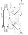
- a glass tube 42 forming a blood flow path formed in a tubular shape having a square cross section, and one side wall (lower side wall in the figure) of the glass tube 42.
- a trapezoidal glass container 44 fixed to the glass container, a flow cell 40 composed of liquid paraffin 46 filled in the glass container 44, a white light source 50, and white light generated by the white light source 50 is converted into the glass container.
- a light receiving fiber 58 for detecting the reflected light 32 that is reflected from the other inclined surface (right inclined surface in the figure) 44B of the glass container 44 through the collimator lens 56, and the light receiving fiber.
- Spectrally reflected light obtained by the server 58 is provided with a first spectrophotometer 60 for obtaining information Np plasma components from the absorption spectrum, the.
- the glass tube 42 has, for example, a glass wall thickness of 1.25 mm, a square tube portion 42A having a cross section of 10 mm ⁇ 10 mm square, a length of 42.5 mm, and a diameter of the circular tube portion 42B on the inlet side and the outlet side.
- the length is 4.5 mm and the length is 15 mm.
- the space filled with the liquid paraffin 46 has a cylindrical shape with an inner diameter of 30 mm and a depth of 15 mm.
- the white light source 50 for example, a halogen white light source having a wavelength of 300 nm to 1100 nm can be used.
- the white light guided by the incident fiber 52 is incident on the side surface of the glass container 44 of the flow cell 40.
- the angle formed between the incident axis and the glass side surface is an angle that transmits through the glass and totally reflects at the boundary between the glass and the plasma layer.
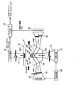
- 44C enters white light through the incident fiber 72, passes through the blood flow path of the flow cell 40, and transmits transmitted light emitted from the opposite side 42C through the light receiving fiber 74.
- blood cells and The second spectrophotometer 76 for obtaining information on the plasma components MCV, MCHC, HCT, and Np, and information on blood cells and plasma components acquired by the second spectrophotometer 76 are the same as those in the first embodiment.
- the computer 78 for obtaining blood cell information MCV, MCHC, and HCT is further provided.
- white light guided by the incident fiber 72 is vertically incident on the trapezoidal top surface 44C of the glass container 44.
- the light passes through the glass, further passes through the blood and is received by the light receiving fiber 74 installed on the flow cell incident opposite surface 42C, and is guided to the second spectrophotometer 76 to measure the absorption spectrum.
- the light since light propagates in blood, the light is mainly absorbed and scattered by red blood cells. Since a typical absorber is hemoglobin, a spectrum having a wavelength band of 600 nm or more with less absorption by hemoglobin is used.
- the received light intensity at the isosbestic wavelength (wavelength whose absorption does not depend on the oxygen saturation level) 805 nm is used as a reference. That is, an absorption spectrum in a range of ⁇ 30 nm (775 nm to 835 nm) having no wavelength dependency with respect to scattering is used centering on 805 nm.
- this measurement state is input to the computer 78, and the inventors propose a Monte Carlo simulation (photon-cell interactive Monte-Carlo simulation: pciMC) of light propagation in blood proposed in Non-Patent Documents 1 and 2.
- the blood input parameters are MCV, MCHC, HCT, and Np
- Np is input as the value obtained in the first embodiment.
- appropriate values are input as initial values.
- the MCV range may be 70 to 110 fL
- the MCHC may be 25 to 40 g / dL
- the HCT range may be 20 to 60%.
- the wavelength is also set in the range of 775 to 835 nm, and an absorption spectrum is obtained by performing pciMC simulation.
- An inverse problem is performed to search for MCV, MCHC, and HCT which are input values of pciMC when the spectrum obtained on the simulation matches the actually measured spectrum (inverse Monte Carlo method).
- MCV, MCHC, and HCT are input values of pciMC when the spectrum obtained on the simulation matches the actually measured spectrum.
- the entire range of the above input parameters is simulated in advance, the database is constructed, and the MCV, MCHC, HCT that matches the measurement result is searched from the database to reduce the calculation cost. Can be minimized.
- a switching device 80 is provided, The light sources 50 and 70 can be turned on and off alternately, so that plasma measurement and blood cell measurement can be incident alternately.
- This switching frequency is about 1 Hz, and blood plasma output calculation is performed during plasma measurement, and plasma output calculation is performed during blood cell measurement, and each measurement value is continuously interrupted at the switching frequency interval. Can be output.
- the plasma measurement flow cell 40 and the separate blood cell measurement flow cell 41 can be arranged in tandem to perform plasma measurement and blood cell measurement at all times.
- a delay circuit 82 that performs a delay according to the blood flow rate can be provided to obtain information on the same blood portion.
- the delay time can be set to a constant delay time by measuring the blood flow rate and changing it accordingly, or by making the blood flow rate constant.
- the blood cell measurement flow cell 41 may have a simple cylindrical shape instead of a trapezoidal shape.
- the cross-sectional area of the glass tube 42 is 1 cm 2 , but it can be reduced when the blood flow rate is small.
- the light source is not limited to a halogen white light source.
- the present invention can be used for measuring blood information such as hemolysis (plasma free hemoglobin concentration) and blood coagulation degree (thrombus), which can obtain information only on plasma components noninvasively and continuously.
- hemolysis plasma free hemoglobin concentration
- thrombus blood coagulation degree
Landscapes
- Health & Medical Sciences (AREA)
- Life Sciences & Earth Sciences (AREA)
- Physics & Mathematics (AREA)
- Chemical & Material Sciences (AREA)
- Engineering & Computer Science (AREA)
- General Health & Medical Sciences (AREA)
- Pathology (AREA)
- Analytical Chemistry (AREA)
- Biochemistry (AREA)
- General Physics & Mathematics (AREA)
- Immunology (AREA)
- Biomedical Technology (AREA)
- Spectroscopy & Molecular Physics (AREA)
- Hematology (AREA)
- Biophysics (AREA)
- Molecular Biology (AREA)
- Ecology (AREA)
- Medicinal Chemistry (AREA)
- Food Science & Technology (AREA)
- Urology & Nephrology (AREA)
- Medical Informatics (AREA)
- Veterinary Medicine (AREA)
- Public Health (AREA)
- Animal Behavior & Ethology (AREA)
- Surgery (AREA)
- Heart & Thoracic Surgery (AREA)
- Optics & Photonics (AREA)
- Investigating Or Analysing Biological Materials (AREA)
- Investigating Or Analysing Materials By Optical Means (AREA)
- Optical Measuring Cells (AREA)
Priority Applications (6)
| Application Number | Priority Date | Filing Date | Title |
|---|---|---|---|
| IN1512MUN2014 IN2014MN01512A (enExample) | 2012-02-13 | 2013-02-13 | |
| KR1020147020029A KR20140119033A (ko) | 2012-02-13 | 2013-02-13 | 혈액 정보의 측정 방법 및 장치 |
| CA2863012A CA2863012A1 (en) | 2012-02-13 | 2013-02-13 | Method and device for measuring blood information |
| CN201380009221.9A CN104136911A (zh) | 2012-02-13 | 2013-02-13 | 血液信息的测定方法及装置 |
| EP13749756.6A EP2793015A4 (en) | 2012-02-13 | 2013-02-13 | METHOD AND DEVICE FOR MEASURING BLOOD DATA |
| US14/378,090 US20150025341A1 (en) | 2012-02-13 | 2013-02-13 | Method and device for measuring blood information |
Applications Claiming Priority (2)
| Application Number | Priority Date | Filing Date | Title |
|---|---|---|---|
| JP2012028231A JP5901012B2 (ja) | 2012-02-13 | 2012-02-13 | 血液情報の測定方法及び装置 |
| JP2012-028231 | 2012-02-13 |
Publications (1)
| Publication Number | Publication Date |
|---|---|
| WO2013122072A1 true WO2013122072A1 (ja) | 2013-08-22 |
Family
ID=48984182
Family Applications (1)
| Application Number | Title | Priority Date | Filing Date |
|---|---|---|---|
| PCT/JP2013/053321 Ceased WO2013122072A1 (ja) | 2012-02-13 | 2013-02-13 | 血液情報の測定方法及び装置 |
Country Status (9)
| Country | Link |
|---|---|
| US (1) | US20150025341A1 (enExample) |
| EP (1) | EP2793015A4 (enExample) |
| JP (1) | JP5901012B2 (enExample) |
| KR (1) | KR20140119033A (enExample) |
| CN (1) | CN104136911A (enExample) |
| CA (1) | CA2863012A1 (enExample) |
| IN (1) | IN2014MN01512A (enExample) |
| TW (1) | TW201341779A (enExample) |
| WO (1) | WO2013122072A1 (enExample) |
Cited By (2)
| Publication number | Priority date | Publication date | Assignee | Title |
|---|---|---|---|---|
| WO2015129615A1 (ja) * | 2014-02-25 | 2015-09-03 | コニカミノルタ株式会社 | 測定方法および測定装置 |
| CN112285064A (zh) * | 2020-10-22 | 2021-01-29 | 中国计量大学 | 一种spr传感器及基于spr的单细胞检测装置及方法 |
Families Citing this family (27)
| Publication number | Priority date | Publication date | Assignee | Title |
|---|---|---|---|---|
| US9759651B2 (en) * | 2014-12-23 | 2017-09-12 | Magellan Diagnostics, Inc. | Combination optical hemoglobin and electrochemical lead assay |
| WO2016158139A1 (ja) * | 2015-03-31 | 2016-10-06 | ソニー株式会社 | 電気的特性測定装置、電気的特性測定方法、血液状態解析システム、及び該方法をコンピューターに実現させるための電気的特性測定用プログラム |
| KR102491852B1 (ko) | 2015-08-28 | 2023-01-26 | 삼성전자주식회사 | 광 센서 및 그 동작 방법 |
| WO2017082089A1 (ja) * | 2015-11-13 | 2017-05-18 | コニカミノルタ株式会社 | 表面プラズモン共鳴蛍光分析方法および表面プラズモン共鳴蛍光分析装置 |
| EP3377878B1 (en) * | 2015-11-18 | 2019-12-25 | Radiometer Medical ApS | Optical sensor for detection of free hemoglobin in a whole blood sample |
| JP6799245B2 (ja) * | 2016-02-25 | 2020-12-16 | 国立研究開発法人産業技術総合研究所 | 血液のフィブリン量変化の計測方法 |
| WO2017200907A1 (en) * | 2016-05-20 | 2017-11-23 | Instrumentation Laboratory Company | Evanescent hemolysis detection |
| CA3090418A1 (en) * | 2016-10-13 | 2018-04-19 | Instrumentation Laboratory Company | Total protein measurement using whole blood refractometry |
| CN108387943B (zh) * | 2017-02-03 | 2019-11-12 | 徐州诚凯知识产权服务有限公司 | 一种液滴光学检测装置 |
| EP3370058A1 (en) * | 2017-03-01 | 2018-09-05 | Danmarks Tekniske Universitet | Planar waveguide device with nano-sized filter |
| MX2019015114A (es) | 2017-06-15 | 2020-08-03 | Siemens Healthcare Diagnostics Inc | Metodo y dispositivo para determinar la concentracion de analitos en una muestra de sangre completa. |
| CN107402212A (zh) * | 2017-08-28 | 2017-11-28 | 深圳小孚医疗科技有限公司 | 一种粪便应用液检测装置 |
| JP6986266B2 (ja) | 2017-11-14 | 2021-12-22 | ジーニアルライト株式会社 | 体液分析装置 |
| US10893829B2 (en) * | 2018-04-13 | 2021-01-19 | Fenwal, Inc. | Optical detection and measurement of hematocrit and free hemoglobin concentration |
| WO2020033194A1 (en) * | 2018-08-06 | 2020-02-13 | Siemens Healthcare Diagnostics Inc. | Method, device and system for determining the concentration of analytes in a sample |
| US11978201B2 (en) * | 2018-08-09 | 2024-05-07 | Siemens Healthcare Diagnostics Inc. | Method and device for determining the concentration of analytes in a sample |
| JP2022511512A (ja) * | 2018-12-07 | 2022-01-31 | シーメンス・ヘルスケア・ダイアグノスティックス・インコーポレイテッド | 評価要素を含む溶液採取装置 |
| WO2020136833A1 (ja) * | 2018-12-27 | 2020-07-02 | パイオニア株式会社 | 総蛋白質量測定装置及び総蛋白質量測定方法 |
| US11484891B2 (en) | 2019-05-23 | 2022-11-01 | Fenwal, Inc. | Adjustment of target interface location between separated fluid components in a centrifuge |
| CN110363719B (zh) * | 2019-07-01 | 2021-07-27 | 湖南开启时代智能科技有限公司 | 一种细胞分层图像处理方法及系统 |
| KR102812067B1 (ko) * | 2019-10-10 | 2025-05-23 | 주식회사 엘지에너지솔루션 | 미 산란을 이용한 가스 발생 감지장치를 포함하는 배터리 팩 및 이를이용한 가스 검출 방법 |
| US20210299339A1 (en) * | 2020-03-31 | 2021-09-30 | Fresenius Medical Care Holdings, Inc. | Blood monitoring system for determining a calibrated hemoglobin concentration value for a patient based on patient-specific mean corpuscular hemoglobin concentration data |
| CN112697655B (zh) * | 2020-12-11 | 2023-08-25 | 南京工业大学 | Od检测仪 |
| CN112697654B (zh) * | 2020-12-11 | 2023-08-25 | 南京工业大学 | Od检测仪传感器 |
| KR102538736B1 (ko) * | 2021-04-09 | 2023-05-31 | 한밭대학교 산학협력단 | 결로 측정 센서 |
| CN116637251A (zh) * | 2023-05-31 | 2023-08-25 | 北京航天长峰股份有限公司 | 一种用于ecmo的血液多参监测装置及方法 |
| WO2025137402A1 (en) * | 2023-12-19 | 2025-06-26 | Enquyst Technologies Inc. | Multichannel optical switch for in-line spectroscopic compound analysis |
Citations (10)
| Publication number | Priority date | Publication date | Assignee | Title |
|---|---|---|---|---|
| JPH01500928A (ja) * | 1986-08-14 | 1989-03-30 | レイディオミーター アクティーゼルスカブ | 全血液の試料中の分析物のレベルを決定する方法および装置 |
| JPH0638947A (ja) | 1992-03-31 | 1994-02-15 | Univ Manitoba | 血液の分光測光分析法及び該装置 |
| JPH06186156A (ja) | 1992-10-21 | 1994-07-08 | Toa Medical Electronics Co Ltd | 粒子分析装置 |
| JPH09311099A (ja) * | 1996-05-24 | 1997-12-02 | Hitachi Ltd | 血液用冷却素子 |
| JPH10123140A (ja) | 1996-10-22 | 1998-05-15 | Toa Medical Electronics Co Ltd | 血液凝固測定装置 |
| JP2002531824A (ja) | 1998-11-30 | 2002-09-24 | ガンブロ アクチボラグ | 血液パラメータを測定する方法および装置 |
| WO2006095615A1 (ja) * | 2005-03-07 | 2006-09-14 | Kuraray Co., Ltd. | マイクロチャネルアレイ及び製造方法、並びにこれを用いた血液測定方法 |
| JP2008224524A (ja) * | 2007-03-14 | 2008-09-25 | Toshiba Corp | 光導波路型抗体チップ |
| JP2010210759A (ja) | 2009-03-09 | 2010-09-24 | Casio Computer Co Ltd | 液晶注入装置 |
| JP2011232137A (ja) * | 2010-04-27 | 2011-11-17 | Nippon Telegr & Teleph Corp <Ntt> | 凝固活性測定装置、測定チップおよび測定方法 |
Family Cites Families (12)
| Publication number | Priority date | Publication date | Assignee | Title |
|---|---|---|---|---|
| DE3481644D1 (de) * | 1984-12-10 | 1990-04-19 | Prutec Ltd | Verfahren zum optischen nachweis von parametern von substanzen in einem fluessigen analyt. |
| US4704029A (en) * | 1985-12-26 | 1987-11-03 | Research Corporation | Blood glucose monitor |
| JPH0228541A (ja) * | 1988-07-19 | 1990-01-30 | Meidensha Corp | 光式濃度検出装置 |
| CA2025330C (en) * | 1989-09-18 | 2002-01-22 | David W. Osten | Characterizing biological matter in a dynamic condition using near infrared spectroscopy |
| US5598842A (en) * | 1993-09-03 | 1997-02-04 | Toa Medical Electronics Co., Ltd. | Non-invasive blood analyzer and method using the same |
| US5601080A (en) * | 1994-12-28 | 1997-02-11 | Coretech Medical Technologies Corporation | Spectrophotometric blood analysis |
| JP2001174405A (ja) * | 1999-12-22 | 2001-06-29 | Shimadzu Corp | グルコースモニタ及びグルコース濃度の測定方法 |
| EP1335665A2 (en) * | 2000-11-15 | 2003-08-20 | Cytometrics, LLC. | Measuring haematocrit in blood vessels |
| WO2003083458A2 (de) * | 2002-04-03 | 2003-10-09 | Johann Wolfgang Goethe-Universität Frankfurt am Main | Infrarotmessvorrichtung, insbesondere für die spektrometrie wässriger systeme, vorzugsweise von mehrkomponentensystemen |
| DE602005027907D1 (de) * | 2004-12-13 | 2011-06-16 | Bayer Healthcare Llc | Transmissionspektroskopiesystem zur verwendung bei der bestimmung von analyten in körperflüssigkeit |
| US7319894B2 (en) * | 2005-09-13 | 2008-01-15 | Edwards Lifesciences Corporation | Continuous spectroscopic measurement of total hemoglobin |
| US8209128B1 (en) * | 2007-02-21 | 2012-06-26 | Paul L. Gourley | Nanolaser spectroscopy and micro-optical resonators for detecting, analyzing, and manipulating bioparticles |
-
2012
- 2012-02-13 JP JP2012028231A patent/JP5901012B2/ja not_active Expired - Fee Related
-
2013
- 2013-02-13 CN CN201380009221.9A patent/CN104136911A/zh active Pending
- 2013-02-13 KR KR1020147020029A patent/KR20140119033A/ko not_active Withdrawn
- 2013-02-13 EP EP13749756.6A patent/EP2793015A4/en not_active Withdrawn
- 2013-02-13 IN IN1512MUN2014 patent/IN2014MN01512A/en unknown
- 2013-02-13 CA CA2863012A patent/CA2863012A1/en not_active Abandoned
- 2013-02-13 US US14/378,090 patent/US20150025341A1/en not_active Abandoned
- 2013-02-13 WO PCT/JP2013/053321 patent/WO2013122072A1/ja not_active Ceased
- 2013-02-18 TW TW102105501A patent/TW201341779A/zh unknown
Patent Citations (10)
| Publication number | Priority date | Publication date | Assignee | Title |
|---|---|---|---|---|
| JPH01500928A (ja) * | 1986-08-14 | 1989-03-30 | レイディオミーター アクティーゼルスカブ | 全血液の試料中の分析物のレベルを決定する方法および装置 |
| JPH0638947A (ja) | 1992-03-31 | 1994-02-15 | Univ Manitoba | 血液の分光測光分析法及び該装置 |
| JPH06186156A (ja) | 1992-10-21 | 1994-07-08 | Toa Medical Electronics Co Ltd | 粒子分析装置 |
| JPH09311099A (ja) * | 1996-05-24 | 1997-12-02 | Hitachi Ltd | 血液用冷却素子 |
| JPH10123140A (ja) | 1996-10-22 | 1998-05-15 | Toa Medical Electronics Co Ltd | 血液凝固測定装置 |
| JP2002531824A (ja) | 1998-11-30 | 2002-09-24 | ガンブロ アクチボラグ | 血液パラメータを測定する方法および装置 |
| WO2006095615A1 (ja) * | 2005-03-07 | 2006-09-14 | Kuraray Co., Ltd. | マイクロチャネルアレイ及び製造方法、並びにこれを用いた血液測定方法 |
| JP2008224524A (ja) * | 2007-03-14 | 2008-09-25 | Toshiba Corp | 光導波路型抗体チップ |
| JP2010210759A (ja) | 2009-03-09 | 2010-09-24 | Casio Computer Co Ltd | 液晶注入装置 |
| JP2011232137A (ja) * | 2010-04-27 | 2011-11-17 | Nippon Telegr & Teleph Corp <Ntt> | 凝固活性測定装置、測定チップおよび測定方法 |
Non-Patent Citations (3)
| Title |
|---|
| D. SAKOTA ET AL., JOURNAL OF BIOMEDICAL OPTICS, vol. 15, no. 6, 2010, pages 14 |
| D. SAKOTA; S. TAKATANI: "Newly developed photon-cell interactive Monte Carlo (pciMC) simulation for non-invasive and continuous diagnosis of blood during extracorporeal circulation support", PROC. SPIE 8092, vol. 80920Y, 2011, pages 1 - 8, XP060015664, DOI: doi:10.1117/12.889327 |
| See also references of EP2793015A4 |
Cited By (5)
| Publication number | Priority date | Publication date | Assignee | Title |
|---|---|---|---|---|
| WO2015129615A1 (ja) * | 2014-02-25 | 2015-09-03 | コニカミノルタ株式会社 | 測定方法および測定装置 |
| JPWO2015129615A1 (ja) * | 2014-02-25 | 2017-03-30 | コニカミノルタ株式会社 | 測定方法および測定装置 |
| US9778184B2 (en) | 2014-02-25 | 2017-10-03 | Konica Minolta, Inc. | Measurement method and measurement device |
| CN112285064A (zh) * | 2020-10-22 | 2021-01-29 | 中国计量大学 | 一种spr传感器及基于spr的单细胞检测装置及方法 |
| CN112285064B (zh) * | 2020-10-22 | 2022-11-25 | 中国计量大学 | 一种spr传感器及基于spr的单细胞检测装置及方法 |
Also Published As
| Publication number | Publication date |
|---|---|
| TW201341779A (zh) | 2013-10-16 |
| KR20140119033A (ko) | 2014-10-08 |
| IN2014MN01512A (enExample) | 2015-05-15 |
| JP5901012B2 (ja) | 2016-04-06 |
| US20150025341A1 (en) | 2015-01-22 |
| CA2863012A1 (en) | 2013-08-22 |
| CN104136911A (zh) | 2014-11-05 |
| EP2793015A4 (en) | 2015-08-26 |
| JP2013164372A (ja) | 2013-08-22 |
| EP2793015A1 (en) | 2014-10-22 |
Similar Documents
| Publication | Publication Date | Title |
|---|---|---|
| JP5901012B2 (ja) | 血液情報の測定方法及び装置 | |
| CA2972848C (en) | Spatial separation of particles in a particle containing solution for biomedical sensing and detection | |
| Yeom et al. | Microfluidic-based speckle analysis for sensitive measurement of erythrocyte aggregation: A comparison of four methods for detection of elevated erythrocyte aggregation in diabetic rat blood | |
| EP1579196B1 (en) | Method and device for measurements in blood | |
| KR102084826B1 (ko) | 자기교정 혈액챔버 | |
| JP2020530560A (ja) | 製造中のバイオポリマーおよび合成ポリマーの特性評価および制御のための装置ならびに方法 | |
| Ren et al. | Cerebral blood flow imaged with ultrahigh-resolution optical coherence angiography and<? A3B2 show [pmg: line-break justify=" yes"/]?> Doppler tomography | |
| Yang et al. | Hematocrit dependence of flow signal in optical coherence tomography angiography | |
| JP6728043B2 (ja) | 溶血を検出するための、又はヘマトクリット値の測定において溶血の影響を補正するための補正因子を決定する方法及び装置 | |
| Sakota et al. | Development of a real-time and quantitative thrombus sensor for an extracorporeal centrifugal blood pump by near-infrared light | |
| Kang et al. | Simple assessment of red blood cell deformability using blood pressure in capillary channels for effective detection of subpopulations in red blood cells | |
| Morita et al. | Real-time, non-invasive thrombus detection in an extracorporeal circuit using micro-optical thrombus sensors | |
| Naeem et al. | Current and emerging microfluidic-based integrated solutions for free hemoglobin and hemolysis detection and measurement | |
| Lin et al. | Optoelectronic online monitoring system for hemodialysis and its data analysis | |
| Reichenwallner et al. | Optical investigation of individual red blood cells for determining cell count and cellular hemoglobin concentration in a microfluidic channel | |
| Larsson et al. | Toward a velocity-resolved microvascular blood flow measure by decomposition of the laser Doppler spectrum | |
| Sakota et al. | Development of an optical detector of thrombus formation on the pivot bearing of a rotary blood pump | |
| Fang et al. | Multiple forward scattering reduces the measured scattering coefficient of whole blood in visible-light optical coherence tomography | |
| CN1529565A (zh) | 测量血管中的血细胞比容 | |
| US10782306B2 (en) | Method and a system for determinations of cell suspensions | |
| Zeidan et al. | In vitro hematocrit measurement using spectrally encoded flow cytometry | |
| Chang et al. | Whole blood reflectance for assessment of hematologic condition and detection of angiographic contrast media | |
| Oshima et al. | Development of optical sensing system for noninvasive and dynamic monitoring of thrombogenic process | |
| Ishida et al. | Measurement of swirling flow in a blood chamber by laser Doppler imaging system | |
| Can et al. | Modeling diffuse reflectance spectra of donated blood with their hematological parameters |
Legal Events
| Date | Code | Title | Description |
|---|---|---|---|
| 121 | Ep: the epo has been informed by wipo that ep was designated in this application |
Ref document number: 13749756 Country of ref document: EP Kind code of ref document: A1 |
|
| WWE | Wipo information: entry into national phase |
Ref document number: 2013749756 Country of ref document: EP |
|
| ENP | Entry into the national phase |
Ref document number: 20147020029 Country of ref document: KR Kind code of ref document: A |
|
| ENP | Entry into the national phase |
Ref document number: 2863012 Country of ref document: CA |
|
| WWE | Wipo information: entry into national phase |
Ref document number: 14378090 Country of ref document: US |
|
| NENP | Non-entry into the national phase |
Ref country code: DE |