WO2011125606A1 - 多価抗原測定用コンジュゲート、これを用いた多価抗原測定用イムノクロマトテストストリップおよびイムノクロマト測定方法 - Google Patents
多価抗原測定用コンジュゲート、これを用いた多価抗原測定用イムノクロマトテストストリップおよびイムノクロマト測定方法 Download PDFInfo
- Publication number
- WO2011125606A1 WO2011125606A1 PCT/JP2011/057621 JP2011057621W WO2011125606A1 WO 2011125606 A1 WO2011125606 A1 WO 2011125606A1 JP 2011057621 W JP2011057621 W JP 2011057621W WO 2011125606 A1 WO2011125606 A1 WO 2011125606A1
- Authority
- WO
- WIPO (PCT)
- Prior art keywords
- antibody
- conjugate
- concentration
- measuring
- solution
- Prior art date
- Legal status (The legal status is an assumption and is not a legal conclusion. Google has not performed a legal analysis and makes no representation as to the accuracy of the status listed.)
- Ceased
Links
Images




Classifications
-
- G—PHYSICS
- G01—MEASURING; TESTING
- G01N—INVESTIGATING OR ANALYSING MATERIALS BY DETERMINING THEIR CHEMICAL OR PHYSICAL PROPERTIES
- G01N33/00—Investigating or analysing materials by specific methods not covered by groups G01N1/00 - G01N31/00
- G01N33/48—Biological material, e.g. blood, urine; Haemocytometers
- G01N33/50—Chemical analysis of biological material, e.g. blood, urine; Testing involving biospecific ligand binding methods; Immunological testing
- G01N33/53—Immunoassay; Biospecific binding assay; Materials therefor
- G01N33/543—Immunoassay; Biospecific binding assay; Materials therefor with an insoluble carrier for immobilising immunochemicals
- G01N33/54366—Apparatus specially adapted for solid-phase testing
- G01N33/54386—Analytical elements
- G01N33/54387—Immunochromatographic test strips
- G01N33/54388—Immunochromatographic test strips based on lateral flow
-
- G—PHYSICS
- G01—MEASURING; TESTING
- G01N—INVESTIGATING OR ANALYSING MATERIALS BY DETERMINING THEIR CHEMICAL OR PHYSICAL PROPERTIES
- G01N33/00—Investigating or analysing materials by specific methods not covered by groups G01N1/00 - G01N31/00
- G01N33/48—Biological material, e.g. blood, urine; Haemocytometers
- G01N33/50—Chemical analysis of biological material, e.g. blood, urine; Testing involving biospecific ligand binding methods; Immunological testing
- G01N33/58—Chemical analysis of biological material, e.g. blood, urine; Testing involving biospecific ligand binding methods; Immunological testing involving labelled substances
- G01N33/585—Chemical analysis of biological material, e.g. blood, urine; Testing involving biospecific ligand binding methods; Immunological testing involving labelled substances with a particulate label, e.g. coloured latex
Definitions
- the present invention relates to an immunochromatographic test strip for measuring a multivalent antigen, and an immunochromatographic measuring method using the same. More specifically, the present invention uses a conjugate produced with a concentration of the antibody in a mixed solution of a solution of an antibody against a multivalent antigen to be measured and a colloidal gold solution in a specific range, The present invention relates to an immunochromatographic test strip and an immunochromatographic measuring method in which a measuring range is adjusted.
- POCT Point of Care Testing by Testing
- colloidal gold is widely used as an insoluble carrier for immobilizing an antibody or antigen to a substance to be measured.
- Various measurement methods using this gold colloid as a label have been reported (Patent Documents 1 to 4).
- a conjugate of a conjugate in which an antibody to a measurement target substance is immobilized on a gold colloid and a measurement target substance in a sample is developed in an insoluble membrane carrier by capillary action, and the membrane carrier
- a typical measurement method is based on a so-called sandwich reaction, which is captured by an antibody against a substance to be measured immobilized on the medium.
- Patent Document 5 discloses a method of adjusting detection sensitivity using an influenza antigen as a substance to be measured by adjusting the amount of antibody bound per unit surface area on colloidal gold particles.
- this method only gives a natural result that detection sensitivity increases as the amount of antibody bound per unit surface area increases, and it is widely used in sandwich immunochromatography measurement methods for measuring multivalent antigens. No mention is made of a method that allows measurement in the concentration range.
- JP-A-2-141665 JP-A-6-94719 : Japanese Patent Laid-Open No. 6-213891 : JP 2001-21564 A : JP 2007-33378 A
- An object of the present invention is to provide an immunochromatographic test strip for measuring a multivalent antigen having a wide measurement range in an immunochromatographic measuring method using a multivalent antigen as a measurement target and a gold colloid as a label.
- the present inventors have a problem that a wide measurement range cannot be secured when measuring a multivalent antigen such as CRP or D-dimer in an immunochromatography measurement method based on a sandwich reaction.
- the present invention was completed by considering the following as a result of intensive studies.
- the amount (number) of aggregates that can be developed in the sample pad, conjugate pad, or insoluble membrane carrier constituting the immunochromatographic test strip decreases, and as a result, the capture antibody immobilization site
- the sandwich complex conjuggate, complex to be measured and capture antibody complex
- the measurement signal is decreased. 4
- a method of reducing the particle size of the aggregate so that more aggregates can develop and reach the measurement line can be considered. 5.
- the signal of the sandwich complex on the measurement line itself becomes smaller and the detection sensitivity is lowered.
- the present invention provides an immunochromatographic test strip using a conjugate for measuring a multivalent antigen in which a monoclonal antibody against a multivalent antigen to be measured is bound to a colloidal gold, wherein the conjugate has a maximum binding antibody concentration of the antibody.
- An immunochromatographic test strip which is a conjugate obtained by mixing the antibody solution and the colloidal gold solution so that the antibody concentration is 1/4 to 1/2 of the above.
- the present invention has the following configuration.
- An immunochromatographic test strip for measuring a multivalent antigen comprising the following (1) and (2).
- (1) The conjugate according to [1] (2) An insoluble membrane carrier on which a monoclonal antibody against a multivalent antigen to be measured is immobilized
- a method for producing a conjugate for measuring a multivalent antigen comprising the following steps 1) and 2), wherein the antibody concentration in the mixed solution of 1) is 1/4 to 1 /
- the conjugate for measuring polyvalent antigen of the present invention the immunochromatographic test strip for measuring multivalent antigen and the immunochromatographic measuring method using the conjugate, measurement in a wide concentration range using the multivalent antigen as a measurement target substance becomes possible. , Complicated operations such as dilution can be avoided. In addition, since the labor and cost at the time of measurement can be reduced, it is possible to meet the social needs of Point of Care Testing (POCT), which is required in clinics and small hospitals.
- POCT Point of Care Testing
- FIG. 3 is a schematic diagram of a D-dimer measurement test strip. It is a graph which shows the result (reflected light intensity) which measured D-dimer of each density
- measuring object refers to a multivalent antigen having a plurality of binding sites with a monoclonal antibody. Examples include, but are not limited to, CRP, D-dimer, hemoglobin and the like.
- the monoclonal antibody used in the present invention is a monoclonal antibody against a multivalent antigen to be measured, and hybridomas producing the antibody are generally produced by the method of Kohler and Milstein [Nature, Vol. 256, p. 495 (1975). According to)], spleen cells of animals immunized with the antigen and myeloma cells (myeloma cells) of the same type can be fused with each other. Moreover, if the antibody with respect to a measuring object is marketed, it is possible to utilize them.
- the “label” means a gold colloid (particle).
- the particle size of the gold colloid is known to greatly affect the sensitivity of the immunochromatographic test strip, but the particle size of the gold colloid of the present invention is preferably 20 to 60 nm, particularly preferably 35 to 45 nm.
- the above gold colloid can be produced by a generally known method, for example, by dropping and stirring a trisodium citrate aqueous solution into a heated tetrachlorogold (III) acid aqueous solution.
- Immobilization of antibody on colloidal gold is usually performed by physical adsorption.
- a substance in which a monoclonal antibody or a control antibody against a multivalent antigen to be measured is immobilized on a label as described above is referred to as a “conjugate”.
- the “maximum binding antibody concentration” of the antibody to the colloidal gold refers to a concentration determined by a known salt titration method described in Examples described later. That is, the antibody solution of each concentration is added to the colloidal gold solution and stirred to react the antibody and the colloidal gold, and then the salt (10% NaCl) is added to change the color change of the colloidal gold and antibody mixture.
- the minimum antibody concentration that gives a state in which no change in color tone is observed even when the antibody concentration is increased is observed as the maximum binding antibody concentration.
- the change in color tone may be detected by visual observation or by other optical means.
- the pH conditions are varied as shown in the Examples, it is preferable that the conjugate is prepared under pH conditions where the determined maximum binding antibody concentration is lower.
- a conjugate in which a monoclonal antibody is bound to a gold colloid is obtained from a mixed solution obtained by mixing an antibody solution and a colloidal gold solution.
- the maximum binding antibody obtained by determining the antibody concentration in the mixed solution by the salt titration method. By setting the antibody concentration to 1/4 to 1/2 of the concentration, the desired conjugate can be produced.
- the antibody concentration of the mixed solution for obtaining the conjugate of the present invention needs to be 1/4 to 1/2 of the maximum binding antibody concentration. However, if the antibody concentration is less than 1/4, the measurement sensitivity is too low. When the concentration is higher than 1/2, aggregates due to the antigen-antibody reaction rapidly increase and the measurement range becomes narrow. Therefore, a concentration of 1/4 to 1/2 is desirable because a suitable sensitivity can be maintained and a wide measurement range can be achieved, and a concentration of 1/3 is even more desirable.
- the buffer and pH for binding the antibody to the colloidal gold are determined at the same time as determining the maximum bound antibody concentration by salt titration, either 2 mmol / L phosphate buffer (pH 6-7) or 2 mmol / L borate buffer.
- the optimum conditions are determined from the solution (pH 8-9).
- the region on the gold colloid where the antibody is not bound is preferably blocked by binding BSA soot or the like. It should be noted that when blocking by binding BSA etc., the antibody concentration in the mixed solution before binding BSA etc. needs to be 1/4 to 1/2 of the maximum binding antibody concentration. Absent.
- the conjugate thus obtained is dispersed and stored in a denaturation inhibitor for preventing denaturation.
- a denaturation inhibitor proteins such as BSA, glycerin, sugars and the like are used.
- the “detection reagent” is specifically a solution containing at least a conjugate.
- the detection reagent keeps the conjugate in a stable state, and when mixed with the measurement sample, it promotes that the antibody immobilized on the conjugate reacts specifically with the measurement target, or makes the conjugate quick and effective.
- one or more stabilizers, solubilizing agents, and the like may be included for the purpose of dissolution and fluidization.
- the stabilizer and solubilizer include bovine serum albumin (BSA), sucrose, casein, and amino acids.
- BSA bovine serum albumin
- the detection reagent is intended to improve detection sensitivity, and may contain a known sensitizer such as polyethylene glycol, polyvinyl pyrrolidone, dextran, 2-methacryloyloxyethyl phosphorylcholine as necessary.
- the detection reagent may include EDTA, EGTA, etc., which are chelating agents for Ca 2+ ions.
- detection or “measurement” must be interpreted in the broadest sense including proof and / or quantification of the existence of the measurement target, and should be interpreted in a limited manner in any sense. must not.
- the “sample pad” is a part that receives a measurement sample, and absorbs the measurement sample of liquid in a state of being molded into the pad, and has any substance and form that can pass through the liquid and the measurement target.
- materials suitable for the sample pad include, but are not limited to, glass fiber (glass fiber), acrylic fiber, hydrophilic polyethylene material, dry paper, paper pulp, and fabric.
- a fiberglass pad is used.
- the sample pad can also have the function of a conjugate pad described later.
- the sample pad may contain a blocking agent or the like that is usually used for the purpose of preventing / suppressing nonspecific reaction (adsorption) in the antibody-immobilized membrane described later.
- blocking agent examples include NEO PROTEIN SAVER (Toyobo Co., Ltd.), ImmunoBlock TM (Dainippon Pharmaceutical Co., Ltd.), Apply Block (Seikagaku Biobusiness Co., Ltd.), SEA BLOCK TM / EIA / WB (East Coast Biologics). company), Blocking One (Nacalai Tesque), BSA, Blocking Peptide Fragment (Toyobo Co., Ltd.), Starting Block TM (PBS) Blocking Buffer (Thermo Fisher Scientific , Inc.), Smart Block TM (CANDOR Bioscience, Inc.), HeteroBlock (OMEGA Biologicals), etc., will not adversely affect the specific reaction itself. It can be appropriately selected things.
- the “conjugate pad” includes a conjugate that specifically reacts with a measurement target, and when the measurement sample passes through the conjugate pad, the conjugate and the measurement target in the sample are combined.
- the conjugate pad may be disposed so as to be in contact with the antibody-immobilized membrane alone, or is disposed in contact with the sample pad, and receives a measurement sample that has passed through the sample pad by capillary flow, Subsequently, the measurement sample may be arranged to be transferred by capillary flow to another pad (3rd Pad described later) that comes into contact with a surface different from the sample pad. Note that the selection of one or more parts of the sample pad and the conjugate pad, and how to arrange the selected part on the antibody-immobilized membrane can be appropriately changed.
- Suitable materials for the conjugate pad include, but are not limited to, paper, cellulose blend, nitrocellulose, polyester, acrylonitrile copolymer, glass fiber (glass fiber) or non-woven fiber such as rayon.
- a fiberglass pad is used.
- the conjugate pad is labeled with a “control antibody” for ensuring the reliability of the assay, for example, a label, and a substance to be measured Including non-reactive antibodies and highly antigenic proteins such as KLH (Scattle hemocyanin) labeled with a label.
- control antibodies are components (substances) that are unlikely to be present in the measurement sample, and can be appropriately selected so as to appropriately correspond to a “control capture antibody” described later.
- the conjugate pad may contain metal ions or metal ion chelating agents such as EDTA and EGTA.
- the 3rd Pad can be arranged for the purpose of removing the unnecessary components for measurement from the reaction components of the measurement sample and the detection reagent, so that the necessary reaction components can smoothly deploy the antibody-immobilized membrane. it can.
- This 3rd Pad also has the additional effect of removing in advance the aggregates that have moved to the antibody-immobilized membrane from the aggregates produced by the antigen-antibody reaction and cannot be expanded smoothly.
- 3rd Pad includes any material and form that allows the liquid and the component to be measured to pass through. Specific examples include, but are not limited to, glass fiber (glass fiber), acrylic fiber, hydrophilic polyethylene material, dry paper, paper pulp, and woven fabric.
- a blood cell separation membrane or a similar membrane is used.
- the capture antibody is physically or chemically supported on an insoluble membrane carrier or the state in which the capture antibody is supported is “immobilized”, “immobilized”, “solid phase”, “sensitized”, “adsorbed”. Sometimes expressed.
- the capture antibody can be immobilized on an insoluble membrane carrier by a generally known method.
- the above-described capture antibody is prepared to a predetermined concentration, and the solution is applied to an insoluble membrane carrier in a certain amount such as a certain symbol such as a dot or +.
- the above-described capture antibody is prepared to a predetermined concentration, and a line having a mechanism capable of moving the liquid in a horizontal direction while discharging the liquid from the nozzle at a constant speed is used. It is carried out by applying to an insoluble membrane carrier in a state.
- the concentration of the capture antibody is preferably 0.1 mg / mL to 5 mg / mL, and more preferably 0.5 mg / mL to 2 mg / mL.
- the amount of the capture antibody immobilized on the insoluble membrane carrier can be optimized by adjusting the amount applied to the insoluble membrane carrier in the case of the flow-through type, and the above-described apparatus in the case of the lateral flow type. It can be optimized by adjusting the discharge speed from the nozzle. In particular, in the case of the lateral flow type, 0.5 ⁇ L / cm to 2 ⁇ L / cm is preferable.
- flow-through membrane assay refers to a method in which a sample or the like is developed so as to pass in a direction perpendicular to the insoluble membrane carrier
- lateral flow membrane assay This refers to a system in which a sample is developed so as to move in a parallel direction with respect to the insoluble membrane carrier.
- the above-described capture antibody can usually be prepared using a predetermined buffer.
- the buffer include normal buffers such as phosphate buffer, Tris buffer, and Good's buffer.
- the pH of the buffer is preferably in the range of 6.0 to 9.5, more preferably 6.5 to 8.5, and even more preferably 7.0 to 8.0.
- the buffer may further contain salts such as NaCl, stabilizers such as sucrose, preservatives, preservatives such as procrine, and the like. Salts include those added for adjusting the ionic strength, such as NaCl, and those added for the purpose of adjusting the pH of the buffer, such as sodium hydroxide.
- blocking can also be carried out by coating a portion other than the capture antibody immobilization site with a commonly used blocking agent in the form of a solution or vapor.
- the insoluble membrane carrier on which the capture antibody is immobilized as described above may be referred to as “antibody-immobilized membrane”.
- control capture antibody can be immobilized on the insoluble membrane carrier.
- the control capture antibody is an antibody for ensuring the reliability of the assay, and captures the corresponding “control antibody” contained in the conjugate pad.
- an anti-KLH antibody or the like corresponds to the control capture antibody.
- the position at which the control capture antibody is immobilized on the insoluble membrane carrier can be appropriately selected to suit the design of the assay system.
- any material can be used as the insoluble membrane carrier (sometimes simply referred to as a membrane).
- examples thereof include, but are not limited to, polyethylene, polyethylene terephthalate, nylons, glass, polysaccharides such as cellulose and cellulose derivatives, and ceramics. Specific examples include glass fiber filter paper and cellulose filter paper sold by Millipore, Toyo Roshi, Whatman and the like.
- the pore size and structure of the insoluble membrane carrier it is possible to control the speed at which the aggregate flows in the membrane. By controlling the flow rate of the aggregate through the membrane, the amount of conjugate bound to the antibody (capture antibody) immobilized on the membrane can be adjusted. Therefore, the pore size and structure of the membrane are determined by the immunochromatography of the present invention. It is desirable to optimize considering the combination of the test strip with other components.
- Millipore, Hi-Flow Plus-HF180, etc. are preferably used.
- the absorption pad is a part having liquid absorbency that controls the development of the measurement sample by absorbing the measurement sample that has moved and passed through the antibody-immobilized insoluble membrane carrier.
- the lateral flow type it may be provided at the most downstream side of the strip configuration, and in the flow through type, for example, it may be provided at the lower part of the membrane on which the capture antibody is immobilized.
- the absorber for example, filter paper can be used, but is not limited thereto.
- Whatman, 740-E is used.
- the “immunochromatographic test strip” is not limited as long as it includes at least an insoluble membrane on which an antibody is immobilized, and further includes a reagent component or other membrane as necessary.
- Other membranes include sample pads, conjugate pads, absorption pads and the like.
- the test strip is usually arranged on a solid support such as a plastic adhesive sheet. It is obvious that the solid support is composed of a substance that does not hinder the capillary flow of the measurement sample, and the adhesive component is a substance that does not hinder the capillary flow of the measurement sample. It is also possible to laminate a polyester film or the like for the purpose of increasing the mechanical strength of the antibody-immobilized membrane and preventing the evaporation (drying) of moisture during the assay.
- test strip is stored and mounted in an appropriate container (housing) that takes into account the size of the test strip, the method and position of adding the sample to be measured, the position of antibody immobilization on the membrane, and the signal detection method.
- the state stored and mounted in this way is called a “device”.
- the immunochromatographic test strip for measuring a multivalent antigen of the present invention has at least a monoclonal antibody-immobilized membrane for a multivalent antigen and a monoclonal antibody for the multivalent antigen prepared by the production method of the present invention immobilized on a label. As long as it contains a conjugate, and may contain other reagents and structures depending on the measurement conditions and measurement sample.
- sample also referred to as “measurement sample” or “antigen solution” mainly refers to a biological fluid derived from a living organism. Specific examples include, but are not limited to, blood, serum, plasma, urine, saliva, sputum, pancreatic extract, tears, ear leakage, or prostate fluid. In general, serum and plasma are preferred.
- sample can be appropriately diluted with a diluent, in this specification, it is expressed as “sample” or “measurement sample” regardless of whether it is diluted or undiluted.
- the diluted solution may have an action of hemolyzing red blood cells.
- the dilution rate can be appropriately adjusted according to the intended measurement range, and is preferably 10 to 500 times.
- the diluted solution having a hemolytic action include purified water and a pH 6.0 to 10.0 buffer.
- the pH 6.0 to 10.0 buffer is preferably 10 to 20 mmol / L, for example, 10 mmol / L to 20 mmol / L phosphate buffer, 10 mmol / L to 20 mmol / L Tris-HCl buffer. Liquid, 10 mmol / L to 20 mmol / L glycine-HCl buffer and the like are preferable.
- EDTA or EGTA which is a chelating agent for Ca 2+ ions, may be contained in the above-mentioned diluted solution.
- the immunochromatographic test strip of the present invention can be produced by appropriately modifying and modifying the methods described in the examples.
- the signal derived from the conjugate may be measured according to a known method, for example, the absorbance or the intensity of reflected light may be measured.
- Example 1 Determination of maximum binding antibody concentration to colloidal gold of anti-CRP monoclonal antibody by salt titration method
- Anti-CRP monoclonal antibody is pH 6.0 , 7.0, 8.0, 9.0, diluted with 4 buffer conditions (i) to iv)), and for each pH, 10, 20, 30, 40, 50, 60, 70, 80, 90, 100 ⁇ g / mL of antibody solution was prepared.
- a 1 OD / mL gold colloid (particle size: 40 nm) solution was adjusted to pH of 4 conditions of pH 6.0, 7.0, 8.0, 9.0 using 0.2 mol / L K 2 CO 3 .
- 50 ⁇ L of each concentration of antibody solution was added to 450 ⁇ L of colloidal gold solution, and the maximum binding antibody concentration was determined based on the antibody concentration in this mixture.
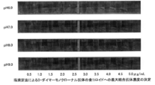
- 100 ⁇ L of 10% NaCl solution to each mixture and react for 5 minutes at room temperature. Then, visually determine the color change of the mixture (photographing) (Fig. 1) and increase the antibody concentration. The minimum antibody concentration that gave a state in which no further change in color tone was observed was taken as the maximum bound antibody concentration.
- the maximum binding antibody concentration to the colloidal gold (particle size 40 nm) of FERM BP-11344 antibody was determined to be 3 ⁇ g / mL, and the optimum sensitization pH was determined to be pH 7.0.
- conjugate (conjugate) of anti-CRP monoclonal antibody and colloidal gold FERM BP-11344 antibody and colloidal gold (particle size 40 nm) Conjugates were made by mixing to a concentration of (3 ⁇ g / mL) and 4/5, 3/5, 1/2, 1/3, 1/4, 1/6 of the maximum binding antibody concentration. That is, to 20 mL of 1 OD / mL colloidal gold solution (pH 7.0), add 1 mL of the antibody solution prepared with 2 mmol / L phosphate buffer (pH 7.0) so as to have the above-mentioned concentrations. For 10 minutes.
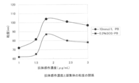
- FIG. 2 shows the relationship between the antibody concentration added to the mixed solution and the particle size of the aggregates 5 minutes after the start of the reaction.
- the aggregate particle diameter increases rapidly and exceeds 100 ⁇ m when the antibody concentration exceeds half the maximum bound antibody concentration (3 ⁇ g / mL).
- the particle size of the aggregates was maintained in a small state of 70 ⁇ m to 80 ⁇ m.
- the aggregate particle size is increased when the antibody concentration is more than half of the maximum binding antibody concentration (3 ⁇ g / mL).
- the aggregate particle size is as small as 60 ⁇ m to 70 ⁇ m. Maintained at.
- sample pad Glass fiber pad obtained by cutting 20 mmol / L Tris-HCl buffer (pH 7.2) containing 24 mmol / L sodium chloride, 0.5% sucrose and 30 mmol / L ethylenediaminetetraacetic acid (EDTA) into a fixed volume. (Lydall) was impregnated with 1.15 times the volume of the pad. The sample pad was dried in a dry oven at 70 ° C. for 45 minutes.
- test strip The above-mentioned anti-CRP antibody-immobilized membrane (b) is affixed to a plastic adhesive sheet (a), the anti-CRP antibody (c) is disposed on the upstream side of the membrane, and 3rd Pad (h ).
- conjugate pad (d) prepared in 1) above was placed and mounted.
- sample pad (e) prepared in 3) above was placed and mounted so as to overlap this conjugate pad, and the absorbent pad (f) (Whatman, 740-E) was placed and mounted on the opposite end.
- a polyester film (g) was placed and laminated on the upper surface so as to cover the antibody-immobilized membrane and the absorption pad.
- test strip was produced by cutting each structural element into a stacked structure.
- the test strip was housed and mounted in a dedicated plastic housing (having a sample addition window and a detection window, not shown in FIG. 3) to form an immunochromatographic test device.
- FIG. 3 shows a schematic configuration diagram of the test strip.
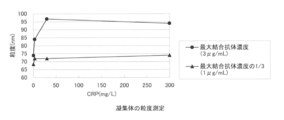
- Example 2 Relationship between measurement range of CRP and aggregate particle size using immunochromatography device using conjugate prepared with mixed solution of maximum binding antibody concentration (3 ⁇ g / mL) or 1/3 of the concentration determined in Example 1 It was investigated.
- CRP calibrator A for Nanopia manufactured by Sekisui Medical Co., Ltd.
- antigen solutions with CRP concentrations of 0, 1.5, 3.0, 6.0, 30.0, 90.0, 180.0, 300.0 mg / L are prepared, and 10 mmol of each concentration of antigen solution is prepared. Dilute 100-fold with / L phosphate buffer (pH 7.2).
- the particle size of the aggregate was about 70 to 100 nm at 0 to 30.0 mg / L, similar to the reflection absorbance, and remained at about 100 nm even at a concentration of 30.0 mg / L or more.
- the reflected absorbance increased in proportion to the CRP concentration from 0 to 300.0 mg / L.
- the particle size of the aggregates was only slightly increased from 69 nm to 74 nm.
- Example 3 Determination of the maximum bound antibody concentration to colloidal gold of anti-D-dimer monoclonal antibody by salt titration method (1) Anti-D-dimer monoclonal antibody Kohler and Milstein's method using batroxobin-treated fibrinogen as an immunogen [Nature, vol. 256, page 495 (See 1975)], a spleen cell of a mouse immunized with the antigen and a myeloma cell (myeloma cell) of the same species are fused to produce a hybridoma that produces the antibody, from which D- Clone # 672101 with high reactivity to dimer was selected.
- # 672101 antibody the antibody produced by clone # 672101 is simply referred to as # 672101 antibody.
- the # 672101 antibody obtained above was diluted with the following four buffer solutions of pH 6.0, 7.0, 8.0 and 9.0 (i) to iv) below, and for each pH, 10, 20, 30, 40, 50, 60, 70, 80, 90, 100 ⁇ g / mL antibody solution was prepared.
- a 1 OD / mL gold colloid (particle size: 40 nm) solution was adjusted to pH of 4 conditions of pH 6.0, 7.0, 8.0, and 9.0 using 0.2 mol / L K 2 CO 3 .
- the maximum binding antibody concentration to the colloidal gold (particle size 40 nm) of antibody # 672101 was determined to be 3 ⁇ g / mL, and the optimum sensitization pH was determined to be pH 8.0.
- conjugate (conjugate) of anti-D-dimer monoclonal antibody and colloidal gold # 672101 antibody has a maximum binding antibody concentration (3 ⁇ g / mL) determined in 1 above and 1/3 of the maximum binding antibody concentration
- a conjugate was prepared by mixing with gold colloid (particle size 40 nm). That is, to 20 mL of 1 OD / mL colloidal gold solution (pH 8.0), add 1 mL of the antibody solution prepared with 2 mmol / L borate buffer (pH 8.0) so as to have the above-mentioned concentrations. Stir for 10 minutes.
- Anti-D-dimer monoclonal antibody (murine MAb against human D-dimer FDP fragment) prepared at 1 mg / mL at one end of the short side of nitrocellulose membrane (Millipore, HF180) , clone DD3B6, manufactured by American Diagnosticainc.) and 10 mmol / L phosphate buffer (pH 7.2) containing 2.5% sucrose to 1 ⁇ L / cm using an immunochromatographic dispenser “XYZ3050” (BIO DOT) It was set and applied in a line. The membrane was dried in a dry oven at 70 ° C. for 45 minutes to obtain an anti-D-dimer antibody-immobilized membrane.
- sample pad Glass fiber pad obtained by cutting 20 mmol / L Tris-HCl buffer (pH 7.2) containing 24 mmol / L sodium chloride, 0.5% sucrose and 30 mmol / L ethylenediaminetetraacetic acid (EDTA) into a fixed volume. (Lydall) was impregnated with 1.15 times the volume of the pad. The sample pad was dried in a dry oven at 70 ° C. for 45 minutes.
- test strip The above-mentioned anti-D-dimer antibody-immobilized membrane (j) is pasted on a plastic adhesive sheet (i), and the anti-D-dimer antibody (k) coating part is placed on the upstream side of the membrane. Furthermore, 3rd Pad (m) was installed. Next, the conjugate pad (l) prepared in 1) above was placed and mounted. Further, the sample pad (n) prepared in 3) above was placed and mounted so as to overlap this conjugate pad, and the absorbent pad (p) (Whatman, 740-E) was placed and mounted on the opposite end. Finally, a polyester film (q) was placed and laminated on the upper surface so as to cover the antibody-immobilized membrane and the absorption pad.
- test strip was produced by cutting each structural element into a stacked structure.
- the test strip was housed and mounted in a dedicated plastic housing (having a sample addition window and a detection window, not shown in FIG. 7), and was in the form of an immunochromatographic test device.
- FIG. 7 shows a schematic configuration diagram of the test strip.
- D-dimer calibrator for Nanopia (Sekisui Medical Co., Ltd., 60 ⁇ g / mL) was diluted with commercially available human pooled plasma.
- the concentration of each antigen solution was 0.4, 1.1, 5.3, 10.0, 15.3, 19.4 ⁇ g / mL.
- the conjugate for measuring a multivalent antigen of the present invention the immunochromatographic test strip for measuring a multivalent antigen and the immunochromatographic measuring method using the conjugate, the measurement having a wider measurement range than that using a multivalent antigen as a measurement target substance is possible. become.
- the labor and cost of dilution operations can be reduced, it is possible to meet the social needs of Point of Care Testing (POCT), which is required by clinics and small hospitals.
- POCT Point of Care Testing
- FERM BP-11344 (# 08202 producing hybridoma) The name and address of the depository that deposited the biological material AIST National Institute of Advanced Industrial Science and Technology Patent Biological Deposit Center 1-chome, East 1-chome, Tsukuba City, Ibaraki Prefecture, Japan 6 (zip code 305-8566) Date on which biological materials were deposited at the depository in Loi No.
Landscapes
- Health & Medical Sciences (AREA)
- Life Sciences & Earth Sciences (AREA)
- Immunology (AREA)
- Engineering & Computer Science (AREA)
- Molecular Biology (AREA)
- Biomedical Technology (AREA)
- Chemical & Material Sciences (AREA)
- Hematology (AREA)
- Urology & Nephrology (AREA)
- Cell Biology (AREA)
- Microbiology (AREA)
- Biotechnology (AREA)
- Food Science & Technology (AREA)
- Medicinal Chemistry (AREA)
- Physics & Mathematics (AREA)
- Analytical Chemistry (AREA)
- Biochemistry (AREA)
- General Health & Medical Sciences (AREA)
- General Physics & Mathematics (AREA)
- Pathology (AREA)
- Peptides Or Proteins (AREA)
Abstract
本発明は、多価抗原を測定対象とし、金コロイドを標識体として用いるイムノクロマト測定方法において、広い測定範囲を有する多価抗原測定用イムノクロマトテストストリップを提供することを課題とする。 測定対象である多価抗原に対するモノクローナル抗体の溶液と金コロイド溶液との混合液から得られる多価抗原測定用コンジュゲートであって、該混合液中の該抗体濃度が、該抗体の金コロイドへの最大結合抗体濃度の1/4~1/2の抗体濃度であることを特徴とする多価抗原測定用コンジュゲートを用いることで、従来に比べて広い測定範囲を可能とすることに成功した。
Description
本発明は、多価抗原を測定対象とするイムノクロマトテストストリップ、及びこれを用いたイムノクロマト測定方法に関する。より具体的には、本発明は、測定対象となる多価抗原に対する抗体の溶液と金コロイド溶液との混合液中の該抗体の濃度を特定の範囲にして製造したコンジュゲートを用いることにより、測定範囲を調節した、イムノクロマトテストストリップ及びイムノクロマト測定方法に関する。
医療分野、医学研究分野などにおいて、血液や体液中の特定成分(血清アルブミン、免疫グロブリン、肝炎ウイルス、リウマチ因子、C反応性タンパク質(CRP)、D-ダイマー、ヘモグロビン等)の濃度を測定することが必要とされ、種々の測定方法が開発されている。なかでも多く採用されている方法は、前記の種々の測定対象物質(または成分)に対する抗体または抗原などを不溶性担体粒子に固定化し、これと対応する抗原又は抗体(すなわち測定対象物質)を反応させて、検体中のこれらの存在や量を検査する免疫学的検査が広く実施されている。定性検査としてはポリクローナル抗体を用いた一元免疫拡散法による方法がある。また、定量検査としては、ラテックス凝集免疫測定法や免疫比濁法などがある。
最近では、診療所や小病院においても、「患者を診察している間に種々の検査を実施したい」、というニーズが増加しており、従来の検査センターなどへの外注検査から、Point of Care Testing(POCT)による検査が行われるようになってきている。このようなPOCT試薬の代表例としては、インフルエンザ感染などを検査するイムノクロマトテストストリップが挙げられる。
イムノクロマト測定方法において、測定対象物質に対する抗体または抗原を固定化する不溶性担体として、金コロイドが広く使われている。また、この金コロイドを標識体とする種々の測定方法が報告されている(特許文献1~4)。このようなイムノクロマト測定方法としては、測定対象物質に対する抗体を金コロイドに固定化したコンジュゲートと検体中の測定対象物質との複合体が、毛細管現象により不溶性メンブレン担体中を展開し、該メンブレン担体に固定化された測定対象物質に対する抗体に捕捉される、いわゆるサンドイッチ反応を原理とする測定方法が代表的である。
従来から、このようなサンドイッチ反応を原理とするイムノクロマト測定方法において、CRPやD-ダイマーのような多価抗原を測定対象物質とすると、溶液系で反応を行うラテックス凝集反応に比べて広い測定範囲を確保できないという課題がある。特許文献5には、金コロイド粒子上への単位表面積当りの抗体の結合量を調整することにより、インフルエンザ抗原を測定対象物質として、検出感度を調整する方法が開示されている。しかしながら、この方法では、単位表面積当りの抗体の結合量が増すにつれて検出感度が高くなるといういわば当然の結果が得られているに過ぎず、多価抗原を測定対象としたサンドイッチイムノクロマト測定方法において広い濃度範囲での測定を可能にする方法については一切言及されていない。
本発明は、多価抗原を測定対象とし、金コロイドを標識体として用いるイムノクロマト測定方法において、広い測定範囲を有する多価抗原測定用イムノクロマトテストストリップを提供することを課題とする。
上記の課題を解決すべく、本発明者らは、サンドイッチ反応を原理とするイムノクロマト測定方法において、CRPやDダイマーのような多価抗原を測定する際、広い測定範囲を確保できないという問題点について次のように考察し、鋭意研究を行い、本発明を完成させた。
(考察)
1.測定対象がCRPやDダイマーのような多価抗原である場合、抗体との結合点が複数あるため、一価抗原(抗原決定基が1つの抗原)に比べて、抗原抗体反応を介して容易にコンジュゲートと測定対象多価抗原との凝集体が生成する。
2.ラテックス凝集反応を利用するような溶液系での測定方法と異なり、イムノクロマト法では、上記の凝集体が毛細管現象により不溶性メンブレン担体中を展開し、測定対象である多価抗原に対するモノクローナル抗体(捕捉抗体ということがある)が固定化された測定ラインに到達する必要がある。
3.そのため、生成する凝集体が大きくなるにつれて、イムノクロマトテストストリップを構成するサンプルパッド、コンジュゲートパッドまたは不溶性メンブレン担体の中を展開できる凝集体量(数)が低下し、その結果、捕捉抗体固定化部位でのサンドイッチ複合体(コンジュゲート、測定対象および捕捉抗体の複合体)が減少し測定シグナルが低下する。
4.上記の測定シグナルの低下を起こりにくくするためには、より多くの凝集体が展開し、測定ラインに到達できるように、凝集体の粒子サイズを小さくする方法が考えられる。
5.しかしながら、凝集体を小さくすると、相反して測定ラインのサンドイッチ複合体のシグナル自体は小さくなり、検出感度が低下するというイムノクロマト測定法に付随する問題点がある。
1.測定対象がCRPやDダイマーのような多価抗原である場合、抗体との結合点が複数あるため、一価抗原(抗原決定基が1つの抗原)に比べて、抗原抗体反応を介して容易にコンジュゲートと測定対象多価抗原との凝集体が生成する。
2.ラテックス凝集反応を利用するような溶液系での測定方法と異なり、イムノクロマト法では、上記の凝集体が毛細管現象により不溶性メンブレン担体中を展開し、測定対象である多価抗原に対するモノクローナル抗体(捕捉抗体ということがある)が固定化された測定ラインに到達する必要がある。
3.そのため、生成する凝集体が大きくなるにつれて、イムノクロマトテストストリップを構成するサンプルパッド、コンジュゲートパッドまたは不溶性メンブレン担体の中を展開できる凝集体量(数)が低下し、その結果、捕捉抗体固定化部位でのサンドイッチ複合体(コンジュゲート、測定対象および捕捉抗体の複合体)が減少し測定シグナルが低下する。
4.上記の測定シグナルの低下を起こりにくくするためには、より多くの凝集体が展開し、測定ラインに到達できるように、凝集体の粒子サイズを小さくする方法が考えられる。
5.しかしながら、凝集体を小さくすると、相反して測定ラインのサンドイッチ複合体のシグナル自体は小さくなり、検出感度が低下するというイムノクロマト測定法に付随する問題点がある。
本発明者らは、上記考察に基づき以下(i)~(v)の方策を検討した結果、(v)の方策の中でも特定の条件で製造されたコンジュゲートを用いたイムノクロマトテストストリップにおいて上記の問題点を解決しうることを見出した。
(i)金コロイドの粒子サイズを調節し、凝集体の粒子サイズを制御する。
(ii)界面活性剤の添加、pH、イオン強度などを調節することにより、抗原抗体反応を制御し、凝集体の粒子サイズを制御する。
(iii)不溶性メンブレン担体の孔径を調節することにより、凝集体の展開を制御する。
(iv)あらかじめ大きな凝集体を除去することにより、不溶性メンブレン担体中での展開を向上させる。
(v)金コロイドに結合する抗体分子数(量)を制御し、凝集体の粒子サイズを制御する。
(i)金コロイドの粒子サイズを調節し、凝集体の粒子サイズを制御する。
(ii)界面活性剤の添加、pH、イオン強度などを調節することにより、抗原抗体反応を制御し、凝集体の粒子サイズを制御する。
(iii)不溶性メンブレン担体の孔径を調節することにより、凝集体の展開を制御する。
(iv)あらかじめ大きな凝集体を除去することにより、不溶性メンブレン担体中での展開を向上させる。
(v)金コロイドに結合する抗体分子数(量)を制御し、凝集体の粒子サイズを制御する。
すなわち本発明は、測定対象である多価抗原に対するモノクローナル抗体が金コロイドと結合した多価抗原測定用コンジュゲートを用いたイムノクロマトテストストリップであって、該コンジュゲートが、該抗体の最大結合抗体濃度の1/4~1/2の抗体濃度となるように該抗体溶液と金コロイド溶液とを混合することにより得られたコンジュゲートである、イムノクロマトテストストリップである。
本発明は以下の構成を有する。
[1]測定対象である多価抗原に対するモノクローナル抗体の溶液と金コロイド溶液との混合液から得られる多価抗原測定用コンジュゲートであって、該混合液の該抗体濃度が、該抗体の金コロイドへの最大結合抗体濃度の1/4~1/2の抗体濃度であることを特徴とする多価抗原測定用コンジュゲート。
[2]以下の(1)および(2)を含む多価抗原測定用イムノクロマトテストストリップ。
(1)前記[1]に記載のコンジュゲート
(2)測定対象である多価抗原に対するモノクローナル抗体が固定化された不溶性メンブレン担体
[3]さらに以下の(3)~(5)を含む前記[2]に記載の多価抗原測定用イムノクロマトテストストリップ。
(3)サンプルパッド
(4)サンプルパッドに接して配置され、前記[2](1)に記載のコンジュゲートを含有するコンジュゲートパッド
(5)吸収パッド
[4]測定対象である多価抗原が、CRPまたはD-ダイマーである前記[2]または[3]に記載の多価抗原測定用イムノクロマトテストストリップ。
[5]前記[2]~[4]のいずれかに記載のイムノクロマトテストストリップを用いて、試料中の測定対象である多価抗原を測定するイムノクロマト測定方法。
[6]以下の1)および2)の工程を含む多価抗原測定用コンジュゲートを製造する方法であって、1)の混合液の抗体濃度が、最大結合抗体濃度の1/4~1/2の抗体濃度であることを特徴とする多価抗原測定用コンジュゲートの製造方法。
1)多価抗原に対するモノクローナル抗体溶液と金コロイド溶液との混合液を得る工程
2)該混合液からコンジュゲートを得る工程
本発明は以下の構成を有する。
[1]測定対象である多価抗原に対するモノクローナル抗体の溶液と金コロイド溶液との混合液から得られる多価抗原測定用コンジュゲートであって、該混合液の該抗体濃度が、該抗体の金コロイドへの最大結合抗体濃度の1/4~1/2の抗体濃度であることを特徴とする多価抗原測定用コンジュゲート。
[2]以下の(1)および(2)を含む多価抗原測定用イムノクロマトテストストリップ。
(1)前記[1]に記載のコンジュゲート
(2)測定対象である多価抗原に対するモノクローナル抗体が固定化された不溶性メンブレン担体
[3]さらに以下の(3)~(5)を含む前記[2]に記載の多価抗原測定用イムノクロマトテストストリップ。
(3)サンプルパッド
(4)サンプルパッドに接して配置され、前記[2](1)に記載のコンジュゲートを含有するコンジュゲートパッド
(5)吸収パッド
[4]測定対象である多価抗原が、CRPまたはD-ダイマーである前記[2]または[3]に記載の多価抗原測定用イムノクロマトテストストリップ。
[5]前記[2]~[4]のいずれかに記載のイムノクロマトテストストリップを用いて、試料中の測定対象である多価抗原を測定するイムノクロマト測定方法。
[6]以下の1)および2)の工程を含む多価抗原測定用コンジュゲートを製造する方法であって、1)の混合液の抗体濃度が、最大結合抗体濃度の1/4~1/2の抗体濃度であることを特徴とする多価抗原測定用コンジュゲートの製造方法。
1)多価抗原に対するモノクローナル抗体溶液と金コロイド溶液との混合液を得る工程
2)該混合液からコンジュゲートを得る工程
本発明の多価抗原測定用コンジュゲート、これを用いた多価抗原測定用イムノクロマトテストストリップおよびイムノクロマト測定方法によれば、多価抗原を測定対象物質とした広い濃度範囲での測定が可能になり、希釈などの煩雑な操作を回避できる。また、測定時の手間とコストが削減できるため、診療所や小病院で求められているPoint of Care Testing(POCT)という社会ニーズにも応えることができる。
本発明における「測定対象」(「測定対象物」と言うこともある)とは、モノクローナル抗体との結合部位を複数有する多価抗原をいう。CRP、D-ダイマー、ヘモグロビンなどが例示されるがこれに制限されるものではない。
本発明で用いられるモノクローナル抗体は、測定対象の多価抗原に対するモノクローナル抗体であり、当該抗体を産生するハイブリドーマは、一般的には、KohlerとMilsteinの方法[Nature、第256巻495頁(1975年)参照]に準じ、前記抗原で免疫した動物の脾臓細胞と同種のミエローマ細胞(骨髄腫細胞)とを細胞融合して作製することができる。また、測定対象に対する抗体が市販されていれば、それらを利用することが可能である。
本発明において「標識体」とは、金コロイド(粒子)のことである。金コロイドの粒径は、イムノクロマトテストストリップの感度に大きく影響することが知られているが、本発明の金コロイドの粒径としては20~60nmが好ましく、特に35~45nmが好ましい。
上記の金コロイドは一般に知られている方法、例えば、加熱したテトラクロロ金(III)酸水溶液にクエン酸三ナトリウム水溶液を滴下攪拌することによって製造することができる。
抗体の金コロイドへの固定化は、通常物理吸着によって行う。本明細書では、上記のような標識体に測定対象の多価抗原に対するモノクローナル抗体あるいはコントロール用抗体が固定化されたものを「コンジュゲート」という。
本発明における抗体の金コロイドへの「最大結合抗体濃度」とは、後述の実施例に記載した公知の塩滴定法によって求められる濃度をいう。すなわち、金コロイド溶液に対して各濃度の抗体溶液を添加して撹拌し抗体と金コロイドを反応させた後、塩(10%NaCl)を添加して金コロイドと抗体の混合液の色調変化の程度を観察し、抗体濃度を上げてもそれ以上色調変化が認められない状態を与える最小の抗体濃度を、最大結合抗体濃度とする。色調の変化は目視観察でも、その他の光学的手段で検出してもよい。実施例に示すようにpH条件を変動させた場合、求められた最大結合抗体濃度がより低いpH条件でコンジュゲートを作成することが好ましい。
抗体溶液と金コロイド溶液とを混合した混合液より金コロイドにモノクローナル抗体が結合したコンジュゲートが得られるが、本発明では、該混合液の抗体濃度を、上記塩滴定法によって求められる最大結合抗体濃度の1/4~1/2の抗体濃度とすることにより、所望のコンジュゲートを製造することができる。
本発明のコンジュゲートを得るための混合液の抗体濃度は上記最大結合抗体濃度の1/4~1/2である必要があるが、抗体濃度が1/4未満では測定感度が低下しすぎるため不適当であり、1/2より濃くなると抗原抗体反応による凝集体が急激に大きくなり、測定範囲が狭くなる。したがって、1/4~1/2の濃度が適当な感度を維持でき、広い測定範囲を達成できるため望ましく、1/3の濃度はよりいっそう望ましい。抗体を金コロイドに結合させる際の緩衝液とpHは、塩滴定法により最大結合抗体濃度を決定する際同時に決定され、2mmol/Lリン酸緩衝液(pH6-7)または2mmol/Lホウ酸緩衝液(pH8-9)等から至適条件が決定される。抗体を金コロイドに結合させた後、金コロイド上の抗体が結合していない領域は、BSA などを結合させブロッキングするのが好適である。なお、BSA などを結合させブロッキングする場合には、BSA などを結合させる前における混合液の抗体濃度を、最大結合抗体濃度の1/4~1/2にしておく必要があることはいうまでもない。
このようにして得られたコンジュゲートは、変性を阻止するための変性阻止剤中に分散され保存される。この変性阻止剤としては、BSA などの蛋白質、グリセリン、糖などが用いられる。
本発明において、「検出試薬」とは具体的には少なくともコンジュゲートを含有する溶液である。
検出試薬は、コンジュゲートを安定な状態に保ち、測定試料と混合されたときにコンジュゲートに固定化された抗体が測定対象と特異的に反応するのを促進する、あるいはコンジュゲートを迅速かつ効果的に溶解、流動化する目的で、例えば1種類以上の安定化剤、溶解補助剤等を含み得る。該安定化剤、溶解補助剤等としては、例えばウシ血清アルブミン(BSA)、スクロース、カゼイン、アミノ酸類などをあげることができる。また、検出試薬は、検出感度の向上を目的とし、必要に応じてポリエチレングリコール、ポリビニルピロリドン、デキストラン、2-メタクリロイルオキシエチルホスホリルコリン等の公知の増感剤を含み得る。更に検出試薬は、Ca2+イオンのキレート剤であるEDTAやEGTAなども含み得る。
なお、本明細書において、「検出」又は「測定」という用語は、測定対象の存在の証明及び/又は定量などを含めて最も広義に解釈する必要があり、いかなる意味においても限定的に解釈してはならない。
本発明において、「サンプルパッド」とは、測定試料を受け入れる部位であり、パッドに成型された状態で液体の測定試料を吸収し、液体と測定対象とが通り抜けることができるどんな物質及び形態をも含む。サンプルパッドに適した材料の具体例として、ガラス繊維(グラスファイバー)、アクリル繊維、親水性ポリエチレン材、乾燥紙、紙パルプ、織物等が含まれるが、これらに限定されない。好適には、グラスファイバー製パッドが用いられる。該サンプルパッドには、後述するコンジュゲートパッドの機能を併せ持たせることも出来る。また、サンプルパッドには、後述する抗体固定化メンブレンにおける非特異的反応(吸着)を防止・抑制する目的で、通常使用されるブロッキング剤等を含ませることができる。該ブロッキング剤としては、例えばNEO PROTEIN SAVER(東洋紡績株式会社)、イムノブロックTM(大日本製薬株式会社)、Applie Block(生化学バイオビジネス株式会社)、SEA BLOCKTM/EIA/WB(East Coast Biologics社)、Blocking One(ナカライテスク社)、BSA、Blocking Peptide Fragment(東洋紡績株式会社)、Starting BlockTM(PBS)Blocking Buffer(Thermo Fisher Scientific社)、Smart BlockTM(CANDOR Bioscience 社)、HeteroBlock(OMEGA Biologicals社)、等から、特異的反応そのものに悪影響を与えないものを適宜選択可能である。
本発明において、「コンジュゲートパッド」とは、測定対象と特異的に反応するコンジュゲートを内在し、測定試料が該コンジュゲートパッドを通過する際、コンジュゲートと試料中の測定対象物とが複合体(凝集体)を形成する機能を有する部位をいう。該コンジュゲートパッドは、それ単独で抗体固定化メンブレンに接するように配置されていてもよいし、あるいは、前記サンプルパッドと接触して配置され、毛細管流によってサンプルパッドを通過した測定試料を受入れ、引き続き該測定試料を毛細管流によって前記サンプルパッドとは異なる面で接触する別のパッド(後述する3rd Pad)へ移送するように配置してもよい。なお、サンプルパッド、コンジュゲートパッドの一種以上の部位の選択や、選択された部位を抗体固定化メンブレンにどのように配置するかは、適宜に変更可能である。
該コンジュゲートパッドに適した材料として、紙、セルロース混合物、ニトロセルロース、ポリエステル、アクリロニトリルコポリマー、ガラス繊維(グラスファイバー)またはレーヨンのような不織繊維が挙げられるが、これらに限定されない。好適には、グラスファイバー製パッドが用いられる。
後述の如く不溶性メンブレン担体に「コントロール捕捉抗体」を固定化する場合には、コンジュゲートパッドに、アッセイの信頼性を担保するための「コントロール抗体」、例えば、標識体で標識され且つ測定対象物質と反応しない抗体や、標識体で標識されたKLH(スカシ貝ヘモシアニン)などの高抗原性タンパク質などを含ませる。これらのコントロール抗体は、測定試料中に存在する可能性が考えられない成分(物質)であり、後述する「コントロール捕捉抗体」と適切に対応するよう適宜に選択可能である。また、抗原抗体反応がイオン感受性を持つ場合には、コンジュゲートパッドに金属イオン類または金属イオンのキレート剤であるEDTAやEGTAなどを含有させておいてもよい。
3rd Padは、測定試料と検出試薬との反応成分のうち、測定に不要な成分を除去し、必要な反応成分が抗体固定化メンブレンをスムーズに展開できるようにすることを目的として配置させることができる。例えば、全血または全血を溶血させた試料を用いる場合には、血球や不溶性の血球破砕物などは測定に不要な成分として除去することが望ましい。また、この3rd Padには、抗原抗体反応により生成する凝集体の内、抗体固定化メンブレンに移動し、スムーズに展開できない位に大きくなった凝集体をあらかじめ除去するという付加的な効果を併せ持たせることも可能である。3rd Padとしては、液体と測定対象の成分とが通り抜けることができるどんな物質及び形態をも含む。具体例として、ガラス繊維(グラスファイバー)、アクリル繊維、親水性ポリエチレン材、乾燥紙、紙パルプ、織物等が含まれるが、これらに限定されない。好適には血球分離膜やそれに類する膜が用いられる。
本明細書において、捕捉抗体を不溶性メンブレン担体に物理的あるいは化学的に担持させることあるいは担持させた状態を「固定」、「固定化」、「固相化」、「感作」、「吸着」と表現することがある。
本発明のイムノクロマトテストストリップにおいて、捕捉抗体の不溶性メンブレン担体への固定化は、一般に周知の方法で実施することができる。例えば、フロースルー式の場合、上記の捕捉抗体を所定の濃度に調製し、その液を一定量、点あるいは+など特定のシンボル状に、不溶性メンブレン担体に塗布する。ラテラルフロー式の場合には、上記の捕捉抗体を所定の濃度に調製し、その液をノズルから一定の速度で吐出しながら水平方向に移動させることのできる機構を有する装置などを用いて、ライン状に不溶性メンブレン担体に塗布することにより行われる。この際、捕捉抗体の濃度は0.1mg/mL~5mg/mLが好ましく、0.5mg/mL~2mg/mLがさらに好適である。また、捕捉抗体の不溶性メンブレン担体への固定化量は、フロースルー式の場合には不溶性メンブレン担体に滴下する塗付量を調節することによって最適化でき、ラテラルフロー式の場合には上記の装置のノズルからの吐出速度を調節することによって最適化できる。特に、ラテラルフロー式の場合、0.5μL/cm~2μL/cmが好適である。
なお、本発明において、「フロースルー式メンブレンアッセイ」という場合は、試料等が不溶性メンブレン担体に対して垂直方向に通過するように展開する方式を指し、「ラテラルフロー式メンブレンアッセイ」という場合は、試料が不溶性メンブレン担体に対して並行方向に移動するように展開する方式を指す。
また、上記の捕捉抗体は、通常所定の緩衝液を用いて調製することができる。該緩衝液の種類としては、リン酸緩衝液、トリス緩衝液、グッド緩衝液など通常使用される緩衝液をあげることができる。緩衝液のpHは6.0~9.5の範囲が好ましく、6.5~8.5がより好ましく、7.0~8.0がさらに好ましい。緩衝液には、さらにNaClなどの塩類、スクロースなどの安定剤や保存剤、プロクリンなどの防腐剤等を含んでもよい。塩類はNaClなどのようにイオン強度の調整のために含ませるもののほか、水酸化ナトリウムなど緩衝液のpHを調整する目的で添加するものも含まれる。
不溶性メンブレン担体に捕捉抗体を固定化した後、さらに、通常使用されるブロッキング剤を溶液あるいは蒸気状にして捕捉抗体固定化部位以外を被覆し、ブロッキングを行うこともできる。
本明細書では、上記のように捕捉抗体が固定化された不溶性メンブレン担体を「抗体固定化メンブレン」ということがある。
なお、不溶性メンブレン担体には、「コントロール捕捉抗体」を固定化することができる。該コントロール捕捉抗体は、アッセイの信頼性を担保するための抗体であって、コンジュゲートパッドに含ませた対応する「コントロール抗体」を捕捉するものである。例えば、コンジュゲートパッドに標識されたKLHをコントロール抗体として含む場合には、抗KLH抗体などがコントロール捕捉抗体に該当する。コントロール捕捉抗体を不溶性メンブレン担体に固定化する位置は、アッセイ系の設計に適合するよう適宜選択することができる。
本発明において、不溶性メンブレン担体(単に、メンブレンと記載することがある)としては、任意の材質が使用できる。例えば、ポリエチレン、ポリエチレンテレフタレート、ナイロン類、ガラス、セルロースやセルロース誘導体などの多糖類あるいはセラミックス等があげられるがこれらに限定されない。具体的には、ミリポア社、東洋濾紙社、ワットマン社などより販売されているガラス繊維ろ紙やセルロースろ紙などをあげることができる。また、この不溶性メンブレン担体の孔径と構造を適宜選択することにより、凝集体がメンブレン中を流れる速度を制御することが可能である。凝集体がメンブレン中を流れる速度を制御することにより、メンブレンに固定化された抗体(捕捉抗体)に結合するコンジュゲート量を調節することができるため、メンブレンの孔径と構造は、本発明のイムノクロマトテストストリップのほかの構成材料との組み合わせを考慮して最適化することが望ましい。メンブレンとして、好適には、ミリポア社、Hi Flow Plus HF180などが用いられる。
本発明において、吸収パッドとは、抗体固定化不溶性メンブレン担体部を移動・通過した測定試料を吸収することにより、測定試料の展開を制御する液体吸収性を有する部位である。ラテラルフロー式においては、ストリップ構成の最下流に設ければよく、フロースルー式においては、例えば捕捉抗体を固定化した膜の下部に設ければよい。該吸収体としては、例えば、ろ紙を用いることができるが、これに限定されない。好適には、Whatman社、740-Eが用いられる。
本発明において、「イムノクロマトテストストリップ」とは、少なくとも抗体を固定化した不溶性メンブレンを含むものであればよく、さらに必要に応じて試薬成分や他のメンブレン等を含むものをいう。他のメンブレンとしては、サンプルパッド、コンジュゲートパッド、吸収パッド等が挙げられる。
該テストストリップは、通常、プラスチック製粘着シートのような固相支持体上に配列させる。該固相支持体を測定試料の毛管流を妨げない物質で構成することはもとより、接着剤の成分を測定試料の毛管流を妨げない物質とすることは明らかである。なお、抗体固定化メンブレンの機械的強度を上げ且つアッセイ中の水分の蒸発(乾燥)を防ぐ目的でポリエステルフィルムなどをラミネートすることも可能である。該テストストリップは、テストストリップの大きさや、測定試料の添加方法・位置、メンブレン上の抗体の固定化位置、シグナルの検出方法などを考慮した適当な容器(ハウジング)に格納・搭載して使用することができ、このように格納・搭載された状態を「デバイス」という。
該テストストリップは、通常、プラスチック製粘着シートのような固相支持体上に配列させる。該固相支持体を測定試料の毛管流を妨げない物質で構成することはもとより、接着剤の成分を測定試料の毛管流を妨げない物質とすることは明らかである。なお、抗体固定化メンブレンの機械的強度を上げ且つアッセイ中の水分の蒸発(乾燥)を防ぐ目的でポリエステルフィルムなどをラミネートすることも可能である。該テストストリップは、テストストリップの大きさや、測定試料の添加方法・位置、メンブレン上の抗体の固定化位置、シグナルの検出方法などを考慮した適当な容器(ハウジング)に格納・搭載して使用することができ、このように格納・搭載された状態を「デバイス」という。
また、本発明の多価抗原測定用イムノクロマトテストストリップは、少なくとも、多価抗原に対するモノクローナル抗体固定化メンブレンおよび、本発明の製造方法で作製された多価抗原に対するモノクローナル抗体が標識体に固定化されているコンジュゲートを含むものであればよく、測定条件、測定試料に応じて他の試薬や構成を含み得る。
本明細書において「試料」(「測定試料」または「抗原液」ともいう)とは、主に生体(生物)由来の体液を言う。具体的には、血液、血清、血漿、尿、唾液、喀痰、膵臓抽出液、涙液、耳漏又は前立腺液などを挙げることができるが、これらに限定されるものではない。一般的には、血清、血漿が好ましい。また、上記の試料は適宜希釈液によって希釈することが可能であるが、本明細書では、希釈・未希釈に係わらず「試料」または「測定試料」と表す。
全血を検体とするときには、該希釈液に赤血球を溶血する作用を併せ持たせてもよい。この際の希釈倍率は適宜目的とする測定範囲に合わせて調節でき、好ましくは10倍から500倍である。上記の溶血作用を持たせた希釈液としては、精製水、pH6.0~10.0の緩衝液等が挙げられる。pH6.0~10.0の緩衝液としては10 mmol/L~20mmol/Lのものが好ましく、例えば、10mmol/L~20mmol/Lのリン酸緩衝液、10mmol/L~20mmol/LのTris-HCl緩衝液、10mmol/L~20mmol/Lのグリシン-HCl緩衝液等が好ましい。
また、溶血作用を増強し、試料のテストストリップでの展開速度を制御する目的で、これらの希釈液に抗原抗体反応に影響しない非イオン性の界面活性剤を添加することも可能である。また、上記の希釈液にCa2+イオンのキレート剤であるEDTAやEGTAなどを含有させてもよい。
本発明のイムノクロマトテストストリップの作製は実施例に記載の方法を適宜、修飾・改変して行うことができる。
コンジュゲートに由来するシグナルを測定する方法としては、公知の方法に従って行えばよく、例えば、吸光度あるいは反射光の強度を測定すればよい。
次に実施例を挙げて本発明を具体的に説明するが、これらは本発明の範囲を限定するものではない。
コンジュゲートに由来するシグナルを測定する方法としては、公知の方法に従って行えばよく、例えば、吸光度あるいは反射光の強度を測定すればよい。
次に実施例を挙げて本発明を具体的に説明するが、これらは本発明の範囲を限定するものではない。
[実施例1]
1.塩滴定法による抗CRPモノクローナル抗体の金コロイドへの最大結合抗体濃度の決定
抗CRPモノクローナル抗体(受託番号FERM BP-11344のハイブリドーマが産生する抗体、以下単にFERM BP-11344抗体という)をpH6.0, 7.0, 8.0, 9.0の4条件の緩衝液(後記i)~iv))にて希釈して、各pHについて、10, 20, 30, 40, 50, 60, 70, 80, 90, 100μg/mLの抗体溶液を作製した。また、1 OD/mLの金コロイド(粒径40nm)溶液を0.2mol/L K2CO3を用いて、pH6.0, 7.0, 8.0, 9.0の4条件のpHに調整した。各pH条件において、金コロイド溶液450μLに対して各濃度の抗体溶液を50μL添加し、この混合液中の抗体濃度をもとに最大結合抗体濃度を求めた。撹拌後、それぞれの混合液に10%NaCl溶液100μLを添加し、室温で5分反応させた後、混合液の色調変化を目視で判定(写真撮影)し(図1)、抗体濃度を上げてもそれ以上色調変化が認められない状態を与える最小の抗体濃度を、最大結合抗体濃度とした。その結果、FERM BP-11344抗体の金コロイド(粒径40nm)への最大結合抗体濃度を3μg/mL、至適感作pHをpH7.0に決定した。
i) 2mmol/Lリン酸緩衝液(pH6.0)
ii) 2mmol/Lリン酸緩衝液(pH7.0)
iii)2mmol/Lほう酸緩衝液(pH8.0)
iv)2mmol/Lほう酸緩衝液(pH9.0)
1.塩滴定法による抗CRPモノクローナル抗体の金コロイドへの最大結合抗体濃度の決定
抗CRPモノクローナル抗体(受託番号FERM BP-11344のハイブリドーマが産生する抗体、以下単にFERM BP-11344抗体という)をpH6.0, 7.0, 8.0, 9.0の4条件の緩衝液(後記i)~iv))にて希釈して、各pHについて、10, 20, 30, 40, 50, 60, 70, 80, 90, 100μg/mLの抗体溶液を作製した。また、1 OD/mLの金コロイド(粒径40nm)溶液を0.2mol/L K2CO3を用いて、pH6.0, 7.0, 8.0, 9.0の4条件のpHに調整した。各pH条件において、金コロイド溶液450μLに対して各濃度の抗体溶液を50μL添加し、この混合液中の抗体濃度をもとに最大結合抗体濃度を求めた。撹拌後、それぞれの混合液に10%NaCl溶液100μLを添加し、室温で5分反応させた後、混合液の色調変化を目視で判定(写真撮影)し(図1)、抗体濃度を上げてもそれ以上色調変化が認められない状態を与える最小の抗体濃度を、最大結合抗体濃度とした。その結果、FERM BP-11344抗体の金コロイド(粒径40nm)への最大結合抗体濃度を3μg/mL、至適感作pHをpH7.0に決定した。
i) 2mmol/Lリン酸緩衝液(pH6.0)
ii) 2mmol/Lリン酸緩衝液(pH7.0)
iii)2mmol/Lほう酸緩衝液(pH8.0)
iv)2mmol/Lほう酸緩衝液(pH9.0)
2.抗CRPモノクローナル抗体と金コロイドとの結合体(コンジュゲート)の作製
FERM BP-11344抗体と金コロイド(粒径40nm)とを、これらの混合液の抗体濃度が上記1で決定した最大結合抗体濃度(3μg/mL)及び最大結合抗体濃度の4/5、3/5、1/2、1/3、1/4、1/6の濃度になるように、混合してコンジュゲートを作製した。すなわち、1 OD/mLの金コロイド溶液(pH7.0)20mLに対し、上記の各濃度になるように2mmol/Lリン酸緩衝液(pH7.0)で調製した抗体溶液を1mL添加し、室温で10分間撹拌した。これらの金コロイドと抗体との混合液それぞれに対し、10%ウシ血清アルブミン(BSA)水溶液を2mL添加し、さらに5分間撹拌後、10℃にて、10,000rpmで45分間遠心し、沈渣(コンジュゲート)を得た。得られた沈渣に、Conjugate Dilution Buffer(Scripps社製)を1.2mL添加しそれぞれのコンジュゲートを懸濁し各コンジュゲートの吸光度を531nm(使用した金コロイドの最大吸収波長)で測定した。(結果は図示せず)。
FERM BP-11344抗体と金コロイド(粒径40nm)とを、これらの混合液の抗体濃度が上記1で決定した最大結合抗体濃度(3μg/mL)及び最大結合抗体濃度の4/5、3/5、1/2、1/3、1/4、1/6の濃度になるように、混合してコンジュゲートを作製した。すなわち、1 OD/mLの金コロイド溶液(pH7.0)20mLに対し、上記の各濃度になるように2mmol/Lリン酸緩衝液(pH7.0)で調製した抗体溶液を1mL添加し、室温で10分間撹拌した。これらの金コロイドと抗体との混合液それぞれに対し、10%ウシ血清アルブミン(BSA)水溶液を2mL添加し、さらに5分間撹拌後、10℃にて、10,000rpmで45分間遠心し、沈渣(コンジュゲート)を得た。得られた沈渣に、Conjugate Dilution Buffer(Scripps社製)を1.2mL添加しそれぞれのコンジュゲートを懸濁し各コンジュゲートの吸光度を531nm(使用した金コロイドの最大吸収波長)で測定した。(結果は図示せず)。
3.CRPと抗CRP抗体コンジュゲートとの反応により生成する凝集体の粒度分析
上記2の最大結合抗体濃度(3μg/mL)及び最大結合抗体濃度の4/5(2.4μg/mL)、3/5(1.8μg/mL)、1/2(1.5μg/mL)、1/3(1.0μg/mL)の濃度において作製した抗CRP抗体コンジュゲートを10mmol/Lリン酸緩衝液(10mmol/L PB、pH7.2)または0.3%オクチル硫酸ナトリウム(0.3%SOS-PB)、で希釈し1 OD/mLに調製した。これらのコンジュゲート溶液100μLに対して、コンジュゲートの希釈液と同一の希釈液300μLを粒度分布測定用ガラスキュベットに入れ、さらに30mg/LのCRP標準液(積水メディカル株式会社製、ナノピア用CRPキャリブレーターA )4μLを添加してボルテックスミキサーにて1~2秒撹拌した後、粒度分布をダイナミック光散乱法により5分間測定した(装置:野崎産業株式会社、NICOMP C370)。混合液に添加した抗体濃度と反応開始5分後の凝集体の粒子径の関係を図2に示す。
希釈液にリン酸緩衝液(PB)を用いたときには、抗体濃度が最大結合抗体濃度(3μg/mL)の1/2の濃度よりも大きくなると凝集体の粒子径が急激に増大し100μm以上になるのに対し、最大結合抗体濃度の1/3~1/2の濃度で調製したコンジュゲートでは、凝集体の粒子径が70μm~80μmと小さな状態で維持された。また、0.3%オクチル硫酸ナトリウム-リン酸緩衝液(0.3%SOS-PB)の場合には、抗体濃度が最大結合抗体濃度(3μg/mL)の1/2より濃くなると凝集体の粒度(粒子径)が急激に増大し80μm以上になるのに対し、最大結合抗体濃度の1/3~1/2の濃度の混合液から調製したコンジュゲートでは、凝集体の粒子径が60μm~70μmと小さな状態で維持された。
上記2の最大結合抗体濃度(3μg/mL)及び最大結合抗体濃度の4/5(2.4μg/mL)、3/5(1.8μg/mL)、1/2(1.5μg/mL)、1/3(1.0μg/mL)の濃度において作製した抗CRP抗体コンジュゲートを10mmol/Lリン酸緩衝液(10mmol/L PB、pH7.2)または0.3%オクチル硫酸ナトリウム(0.3%SOS-PB)、で希釈し1 OD/mLに調製した。これらのコンジュゲート溶液100μLに対して、コンジュゲートの希釈液と同一の希釈液300μLを粒度分布測定用ガラスキュベットに入れ、さらに30mg/LのCRP標準液(積水メディカル株式会社製、ナノピア用CRPキャリブレーターA )4μLを添加してボルテックスミキサーにて1~2秒撹拌した後、粒度分布をダイナミック光散乱法により5分間測定した(装置:野崎産業株式会社、NICOMP C370)。混合液に添加した抗体濃度と反応開始5分後の凝集体の粒子径の関係を図2に示す。
希釈液にリン酸緩衝液(PB)を用いたときには、抗体濃度が最大結合抗体濃度(3μg/mL)の1/2の濃度よりも大きくなると凝集体の粒子径が急激に増大し100μm以上になるのに対し、最大結合抗体濃度の1/3~1/2の濃度で調製したコンジュゲートでは、凝集体の粒子径が70μm~80μmと小さな状態で維持された。また、0.3%オクチル硫酸ナトリウム-リン酸緩衝液(0.3%SOS-PB)の場合には、抗体濃度が最大結合抗体濃度(3μg/mL)の1/2より濃くなると凝集体の粒度(粒子径)が急激に増大し80μm以上になるのに対し、最大結合抗体濃度の1/3~1/2の濃度の混合液から調製したコンジュゲートでは、凝集体の粒子径が60μm~70μmと小さな状態で維持された。
4.抗CRP抗体コンジュゲートを用いたイムノクロマトデバイスの作製
1)コンジュゲートパッドの作製
上記2で調製した抗CRPモノクローナル抗体感作コンジュゲートを20 OD/mLとなるように、1.33%カゼイン、4%スクロース溶液(pH7.5)と混合して検出試薬を作製し、一定体積のグラスファイバー製パッド(日本ポール社、No.8964)に該パッド体積の1.2倍容量滲みこませた。ドライオーブン内で70℃、30分間加温することにより乾燥させ、コンジュゲートパッドとした。
1)コンジュゲートパッドの作製
上記2で調製した抗CRPモノクローナル抗体感作コンジュゲートを20 OD/mLとなるように、1.33%カゼイン、4%スクロース溶液(pH7.5)と混合して検出試薬を作製し、一定体積のグラスファイバー製パッド(日本ポール社、No.8964)に該パッド体積の1.2倍容量滲みこませた。ドライオーブン内で70℃、30分間加温することにより乾燥させ、コンジュゲートパッドとした。
2)抗CRP抗体固定化メンブレンの作製
ニトロセルロースメンブレン(ミリポア社、HF180)の短辺の一端に、1mg/mLに調製した抗CRPモノクローナル抗体(受託番号FERM BP-11345のハイブリドーマが産生する抗体、以下単にFERM BP-11345抗体という)及び2.5%スクロースを含む10mmol/Lリン酸緩衝液(pH7.2)を、イムノクロマト用ディスペンサー「XYZ3050」(BIO DOT社)を用いて1μL/cmとなるよう設定し、ライン状に塗布した。ドライオーブン内で70℃、45分乾燥し、抗CRP抗体固定化メンブレンとした。
ニトロセルロースメンブレン(ミリポア社、HF180)の短辺の一端に、1mg/mLに調製した抗CRPモノクローナル抗体(受託番号FERM BP-11345のハイブリドーマが産生する抗体、以下単にFERM BP-11345抗体という)及び2.5%スクロースを含む10mmol/Lリン酸緩衝液(pH7.2)を、イムノクロマト用ディスペンサー「XYZ3050」(BIO DOT社)を用いて1μL/cmとなるよう設定し、ライン状に塗布した。ドライオーブン内で70℃、45分乾燥し、抗CRP抗体固定化メンブレンとした。
3)サンプルパッドの作製
24mmol/L 塩化ナトリウム、0.5%スクロース及び30mmol/Lエチレンジアミンテトラ酢酸(EDTA)を含む20mmol/Lトリス塩酸緩衝液(pH7.2)を、一定体積に切断したグラスファイバー製パッド(Lydall社)に該パッド体積の1.15倍容量滲みこませた。ドライオーブン内で70℃、45分乾燥し、サンプルパッドとした。
24mmol/L 塩化ナトリウム、0.5%スクロース及び30mmol/Lエチレンジアミンテトラ酢酸(EDTA)を含む20mmol/Lトリス塩酸緩衝液(pH7.2)を、一定体積に切断したグラスファイバー製パッド(Lydall社)に該パッド体積の1.15倍容量滲みこませた。ドライオーブン内で70℃、45分乾燥し、サンプルパッドとした。
4)テストストリップの作製
プラスチック製粘着シート(a)に上記抗CRP抗体固定化メンブレン(b)を貼り、該メンブレンの展開上流部側に抗CRP抗体(c)を配置し、さらに3rd Pad(h)を装着した。次いで上記1)で作成したコンジュゲートパッド(d)を配置装着した。さらにこのコンジュゲートパッドに重なるように上記3)で作製したサンプルパッド(e)を配置装着し、反対側の端には吸収パッド(f)(Whatman社、740-E)を配置装着した。また、最後に抗体固定化メンブレンおよび吸収パッドを被覆するように上面にポリエステルフィルム(g)を配置装着しラミネートした。このように各構成要素を重ね合わせた構造物に切断してテストストリップを作製した。該テストストリップは、アッセイの際、プラスチック製の専用のハウジング(サンプル添加窓部及び検出窓部を有する、図3中図示せず)に格納・搭載し、イムノクロマトテストデバイスの形態にした。図3にテストストリップの模式構成図を示した。
プラスチック製粘着シート(a)に上記抗CRP抗体固定化メンブレン(b)を貼り、該メンブレンの展開上流部側に抗CRP抗体(c)を配置し、さらに3rd Pad(h)を装着した。次いで上記1)で作成したコンジュゲートパッド(d)を配置装着した。さらにこのコンジュゲートパッドに重なるように上記3)で作製したサンプルパッド(e)を配置装着し、反対側の端には吸収パッド(f)(Whatman社、740-E)を配置装着した。また、最後に抗体固定化メンブレンおよび吸収パッドを被覆するように上面にポリエステルフィルム(g)を配置装着しラミネートした。このように各構成要素を重ね合わせた構造物に切断してテストストリップを作製した。該テストストリップは、アッセイの際、プラスチック製の専用のハウジング(サンプル添加窓部及び検出窓部を有する、図3中図示せず)に格納・搭載し、イムノクロマトテストデバイスの形態にした。図3にテストストリップの模式構成図を示した。
5)CRP濃度の測定範囲の検討
上記の種々の抗体感作濃度において作製したイムノクロマトテストデバイスを用いて、CRPの測定範囲を検討した。ナノピア用CRPキャリブレーターA (積水メディカル株式会社製)を用いて、CRP濃度1.5,3.0,6.0,30.0,90.0,180.0,300.0mg/Lの抗原液を作製し、各濃度の抗原液を0.3%オクチル硫酸ナトリウム-10mmol/Lリン酸緩衝液(pH7.2)で30倍に希釈した。希釈した抗原液120μLをテストデバイスのサンプルパッド窓部に添加し、イムノクロマトリーダーICA-1000(浜松ホトニクス社)を用いて5分後のテストデバイスの検出窓部の反射光強度を測定した(図4)。その結果、最大結合抗体濃度の1/2より高い濃度で混合液を調製したコンジュゲートでは、抗原(CRP)濃度30.0mg/L位から測定シグナルがプラトーになったのに対し、最大結合抗体濃度の1/4~1/2の濃度で混合液を調製したコンジュゲートでは、抗原(CRP)濃度300.0mg/Lまで測定シグナルが右肩上がりに増大した。また、最大結合抗体濃度の1/6の濃度で調製した標識体では、検出感度が著しく低下した。
上記の種々の抗体感作濃度において作製したイムノクロマトテストデバイスを用いて、CRPの測定範囲を検討した。ナノピア用CRPキャリブレーターA (積水メディカル株式会社製)を用いて、CRP濃度1.5,3.0,6.0,30.0,90.0,180.0,300.0mg/Lの抗原液を作製し、各濃度の抗原液を0.3%オクチル硫酸ナトリウム-10mmol/Lリン酸緩衝液(pH7.2)で30倍に希釈した。希釈した抗原液120μLをテストデバイスのサンプルパッド窓部に添加し、イムノクロマトリーダーICA-1000(浜松ホトニクス社)を用いて5分後のテストデバイスの検出窓部の反射光強度を測定した(図4)。その結果、最大結合抗体濃度の1/2より高い濃度で混合液を調製したコンジュゲートでは、抗原(CRP)濃度30.0mg/L位から測定シグナルがプラトーになったのに対し、最大結合抗体濃度の1/4~1/2の濃度で混合液を調製したコンジュゲートでは、抗原(CRP)濃度300.0mg/Lまで測定シグナルが右肩上がりに増大した。また、最大結合抗体濃度の1/6の濃度で調製した標識体では、検出感度が著しく低下した。
〔実施例2〕
実施例1で求めた最大結合抗体濃度(3μg/mL)またはその1/3の濃度の混合液で作製したコンジュゲートを用いたイムノクロマトデバイスを用い、CRPの測定範囲と凝集体の粒度との関係を検討した。
ナノピア用CRPキャリブレーターA (積水メディカル株式会社製)を用いて、CRP濃度0,1.5,3.0,6.0,30.0,90.0,180.0,300.0mg/Lの抗原液を作製し、各濃度の抗原液を10mmol/Lリン酸緩衝液(pH7.2)で100倍に希釈した。希釈した抗原液120μLをテストデバイスのサンプルパッド窓部に添加し、イムノクロマトリーダーICA-1000(浜松ホトニクス社)を用いて5分後のテストデバイスの検出窓部の反射光強度を測定した(図5-1)。
また、実施例1-3の方法と同様の方法で、CRP濃度0,3.0,30.0,300.0mg/Lの抗原液について、凝集体の粒度分布を測定した(図5-2)。
その結果、最大結合抗体濃度(3μg/mL)の混合液で調製したコンジュゲートを用いたテストデバイスでは、CRP濃度0~30.0mg/Lにおいて、反射吸光度はCRP濃度に比例して増加し、30.0mg/L以上の濃度でプラトーになった。このとき、凝集体の粒度は反射吸光度と同様、0~30.0mg/Lで約70~100nmとなり、30.0mg/L以上の濃度でも約100nmのままであった。一方、最大結合抗体濃度の1/3の濃度の混合液で調製したコンジュゲートを用いたテストデバイスでは、反射吸光度はCRP濃度0~300.0mg/Lまで濃度に比例して増加した。このとき、凝集体の粒度は69nmから74nmまでわずかに大きくなっただけであった。
実施例1で求めた最大結合抗体濃度(3μg/mL)またはその1/3の濃度の混合液で作製したコンジュゲートを用いたイムノクロマトデバイスを用い、CRPの測定範囲と凝集体の粒度との関係を検討した。
ナノピア用CRPキャリブレーターA (積水メディカル株式会社製)を用いて、CRP濃度0,1.5,3.0,6.0,30.0,90.0,180.0,300.0mg/Lの抗原液を作製し、各濃度の抗原液を10mmol/Lリン酸緩衝液(pH7.2)で100倍に希釈した。希釈した抗原液120μLをテストデバイスのサンプルパッド窓部に添加し、イムノクロマトリーダーICA-1000(浜松ホトニクス社)を用いて5分後のテストデバイスの検出窓部の反射光強度を測定した(図5-1)。
また、実施例1-3の方法と同様の方法で、CRP濃度0,3.0,30.0,300.0mg/Lの抗原液について、凝集体の粒度分布を測定した(図5-2)。
その結果、最大結合抗体濃度(3μg/mL)の混合液で調製したコンジュゲートを用いたテストデバイスでは、CRP濃度0~30.0mg/Lにおいて、反射吸光度はCRP濃度に比例して増加し、30.0mg/L以上の濃度でプラトーになった。このとき、凝集体の粒度は反射吸光度と同様、0~30.0mg/Lで約70~100nmとなり、30.0mg/L以上の濃度でも約100nmのままであった。一方、最大結合抗体濃度の1/3の濃度の混合液で調製したコンジュゲートを用いたテストデバイスでは、反射吸光度はCRP濃度0~300.0mg/Lまで濃度に比例して増加した。このとき、凝集体の粒度は69nmから74nmまでわずかに大きくなっただけであった。
[実施例3]
1.塩滴定法による抗D-ダイマーモノクローナル抗体の金コロイドへの最大結合抗体濃度の決定
(1)抗D-ダイマーモノクローナル抗体
バトロキソビン処理フィブリノゲンを免疫原としてKohlerとMilsteinの方法[Nature、第256巻495頁(1975年)参照]に準じ、前記抗原で免疫したマウスの脾臓細胞と同種のミエローマ細胞(骨髄腫細胞)とを細胞融合して当該抗体を産生するハイブリドーマを作製し、そのなかから、D-ダイマーに反応性の高いクローン#672101を選択した。以下、クローン#672101の産生する抗体を単に#672101抗体という。
(2)上記で得られた#672101抗体を以下pH6.0, 7.0, 8.0, 9.0の4条件の緩衝液(後記i)~iv))にて希釈して、各pHについて、10, 20, 30, 40, 50, 60, 70, 80, 90, 100μg/mLの抗体溶液を作製した。また、1 OD/mLの金コロイド(粒径40nm)溶液を0.2mol/L K2CO3を用いて、pH6.0, 7.0, 8.0, 9.0の4条件のpHに調製した。各pH条件において、金コロイド溶液450μLに対して、各濃度の抗体溶液を50μL添加し、この混合液中の抗体濃度をもとに最大結合抗体濃度を求めた。撹拌後、それぞれの混合溶液に10%NaCl溶液100μLを添加し、室温で5分反応させた後、混合液の色調変化を目視で判定(写真撮影)し(図6)、抗体濃度を上げてもそれ以上色調変化が認められない状態を与える最小の抗体濃度を、最大結合抗体濃度とした。その結果、#672101抗体の金コロイド(粒径40nm)への最大結合抗体濃度を3μg/mL、至適感作pHをpH8.0に決定した。
i) 2mmol/Lリン酸緩衝液(pH6.0)
ii) 2mmol/Lリン酸緩衝液(pH7.0)
iii) 2mmol/Lほう酸緩衝液(pH8.0)
iv) 2mmol/Lほう酸緩衝液(pH9.0)
1.塩滴定法による抗D-ダイマーモノクローナル抗体の金コロイドへの最大結合抗体濃度の決定
(1)抗D-ダイマーモノクローナル抗体
バトロキソビン処理フィブリノゲンを免疫原としてKohlerとMilsteinの方法[Nature、第256巻495頁(1975年)参照]に準じ、前記抗原で免疫したマウスの脾臓細胞と同種のミエローマ細胞(骨髄腫細胞)とを細胞融合して当該抗体を産生するハイブリドーマを作製し、そのなかから、D-ダイマーに反応性の高いクローン#672101を選択した。以下、クローン#672101の産生する抗体を単に#672101抗体という。
(2)上記で得られた#672101抗体を以下pH6.0, 7.0, 8.0, 9.0の4条件の緩衝液(後記i)~iv))にて希釈して、各pHについて、10, 20, 30, 40, 50, 60, 70, 80, 90, 100μg/mLの抗体溶液を作製した。また、1 OD/mLの金コロイド(粒径40nm)溶液を0.2mol/L K2CO3を用いて、pH6.0, 7.0, 8.0, 9.0の4条件のpHに調製した。各pH条件において、金コロイド溶液450μLに対して、各濃度の抗体溶液を50μL添加し、この混合液中の抗体濃度をもとに最大結合抗体濃度を求めた。撹拌後、それぞれの混合溶液に10%NaCl溶液100μLを添加し、室温で5分反応させた後、混合液の色調変化を目視で判定(写真撮影)し(図6)、抗体濃度を上げてもそれ以上色調変化が認められない状態を与える最小の抗体濃度を、最大結合抗体濃度とした。その結果、#672101抗体の金コロイド(粒径40nm)への最大結合抗体濃度を3μg/mL、至適感作pHをpH8.0に決定した。
i) 2mmol/Lリン酸緩衝液(pH6.0)
ii) 2mmol/Lリン酸緩衝液(pH7.0)
iii) 2mmol/Lほう酸緩衝液(pH8.0)
iv) 2mmol/Lほう酸緩衝液(pH9.0)
2.抗D-ダイマーモノクローナル抗体と金コロイドとの結合体(コンジュゲート)の作製
#672101抗体を上記1で決定した最大結合抗体濃度(3μg/mL)及び最大結合抗体濃度の1/3の濃度になるように金コロイド(粒径40nm)と混合して、コンジュゲートを作製した。すなわち、1 OD/mLの金コロイド溶液(pH8.0)20mLに対し、上記の各濃度になるように2mmol/Lほう酸緩衝液(pH8.0)で調製した抗体溶液を1mL添加し、室温で10分間撹拌した。
これらの金コロイドと抗体との混合液それぞれに対し、10%ウシ血清アルブミン(BSA)水溶液を2mL添加し、さらに5分間撹拌後、10℃にて、10,000rpmで45分間遠心し、沈渣(コンジュゲート)を得た。得られた沈渣に、Conjugate Dilution Buffer(Scripps社製)を1.2mL添加しそれぞれのコンジュゲートを懸濁し各コンジュゲートの吸光度を531nm(使用した金コロイドの最大吸収波長)で測定した(結果は図示せず)。
#672101抗体を上記1で決定した最大結合抗体濃度(3μg/mL)及び最大結合抗体濃度の1/3の濃度になるように金コロイド(粒径40nm)と混合して、コンジュゲートを作製した。すなわち、1 OD/mLの金コロイド溶液(pH8.0)20mLに対し、上記の各濃度になるように2mmol/Lほう酸緩衝液(pH8.0)で調製した抗体溶液を1mL添加し、室温で10分間撹拌した。
これらの金コロイドと抗体との混合液それぞれに対し、10%ウシ血清アルブミン(BSA)水溶液を2mL添加し、さらに5分間撹拌後、10℃にて、10,000rpmで45分間遠心し、沈渣(コンジュゲート)を得た。得られた沈渣に、Conjugate Dilution Buffer(Scripps社製)を1.2mL添加しそれぞれのコンジュゲートを懸濁し各コンジュゲートの吸光度を531nm(使用した金コロイドの最大吸収波長)で測定した(結果は図示せず)。
3.抗D-ダイマーモノクローナル抗体コンジュゲートを用いたイムノクロマトデバイスの作製
1)コンジュゲートパッドの作製
上記2で調製した抗D-ダイマーモノクローナル抗体感作コンジュゲートを20 OD/mLとなるように、1.33%カゼイン、4%スクロース溶液(pH7.5)と混合して検出試薬を作製し、一定体積のグラスファイバー製パッド(日本ポール社、No.8964)に該パッド体積の1.2倍容量滲みこませた。ドライオーブン内で70℃、30分間加温することにより乾燥させ、コンジュゲートパッドとした。
1)コンジュゲートパッドの作製
上記2で調製した抗D-ダイマーモノクローナル抗体感作コンジュゲートを20 OD/mLとなるように、1.33%カゼイン、4%スクロース溶液(pH7.5)と混合して検出試薬を作製し、一定体積のグラスファイバー製パッド(日本ポール社、No.8964)に該パッド体積の1.2倍容量滲みこませた。ドライオーブン内で70℃、30分間加温することにより乾燥させ、コンジュゲートパッドとした。
2)抗D-ダイマー抗体固定化メンブレンの作製
ニトロセルロースメンブレン(ミリポア社、HF180)の短辺の一端に、1mg/mLに調製した抗D-ダイマーモノクローナル抗体( murine MAb against human D-dimer FDP fragment, clone DD3B6、American Diagnosticainc.社製)及び2.5%スクロースを含む10mmol/Lリン酸緩衝液(pH7.2)を、イムノクロマト用ディスペンサー「XYZ3050」(BIO DOT社)を用いて1μL/cmとなるよう設定し、ライン状に塗布した。ドライオーブン内で70℃、45分乾燥し、抗D-ダイマー抗体固定化メンブレンとした。
ニトロセルロースメンブレン(ミリポア社、HF180)の短辺の一端に、1mg/mLに調製した抗D-ダイマーモノクローナル抗体( murine MAb against human D-dimer FDP fragment, clone DD3B6、American Diagnosticainc.社製)及び2.5%スクロースを含む10mmol/Lリン酸緩衝液(pH7.2)を、イムノクロマト用ディスペンサー「XYZ3050」(BIO DOT社)を用いて1μL/cmとなるよう設定し、ライン状に塗布した。ドライオーブン内で70℃、45分乾燥し、抗D-ダイマー抗体固定化メンブレンとした。
3)サンプルパッドの作製
24mmol/L 塩化ナトリウム、0.5%スクロース及び30mmol/Lエチレンジアミンテトラ酢酸(EDTA)を含む20mmol/Lトリス塩酸緩衝液(pH7.2)を、一定体積に切断したグラスファイバー製パッド(Lydall社)に該パッド体積の1.15倍容量滲みこませた。ドライオーブン内で70℃、45分乾燥し、サンプルパッドとした。
24mmol/L 塩化ナトリウム、0.5%スクロース及び30mmol/Lエチレンジアミンテトラ酢酸(EDTA)を含む20mmol/Lトリス塩酸緩衝液(pH7.2)を、一定体積に切断したグラスファイバー製パッド(Lydall社)に該パッド体積の1.15倍容量滲みこませた。ドライオーブン内で70℃、45分乾燥し、サンプルパッドとした。
4)テストストリップの作製
プラスチック製粘着シート(i)に上記抗D-ダイマー抗体固定化メンブレン(j)を貼り、該メンブレンの展開上流部側に抗D-ダイマー抗体(k)の塗布部を配置し、さら3rd Pad (m)を装着した。次いで上記1)で作製したコンジュゲートパッド(l)を配置装着した。さらにこのコンジュゲートパッドに重なるように上記3)で作製したサンプルパッド(n)を配置装着し、反対側の端には吸収パッド(p)(Whatman社、740-E)を配置装着した。また、最後に抗体固定化メンブレンおよび吸収パッドを被覆するように上面にポリエステルフィルム(q)を配置装着しラミネートした。このように各構成要素を重ね合わせた構造物に切断してテストストリップを作製した。該テストストリップは、アッセイの際、プラスチック製の専用のハウジング(サンプル添加窓部及び検出窓部を有する、図7中図示せず)に格納・搭載し、イムノクロマトテストデバイスの形態にした。図7にテストストリップの模式構成図を示した。
プラスチック製粘着シート(i)に上記抗D-ダイマー抗体固定化メンブレン(j)を貼り、該メンブレンの展開上流部側に抗D-ダイマー抗体(k)の塗布部を配置し、さら3rd Pad (m)を装着した。次いで上記1)で作製したコンジュゲートパッド(l)を配置装着した。さらにこのコンジュゲートパッドに重なるように上記3)で作製したサンプルパッド(n)を配置装着し、反対側の端には吸収パッド(p)(Whatman社、740-E)を配置装着した。また、最後に抗体固定化メンブレンおよび吸収パッドを被覆するように上面にポリエステルフィルム(q)を配置装着しラミネートした。このように各構成要素を重ね合わせた構造物に切断してテストストリップを作製した。該テストストリップは、アッセイの際、プラスチック製の専用のハウジング(サンプル添加窓部及び検出窓部を有する、図7中図示せず)に格納・搭載し、イムノクロマトテストデバイスの形態にした。図7にテストストリップの模式構成図を示した。
5)D-ダイマー濃度の測定範囲の検討
上記の2種類の抗体感作濃度において作製したイムノクロマトテストデバイスを用いて、D-ダイマーの測定範囲を検討した。ナノピア用D-ダイマーキャリブレーター(積水メディカル株式会社製、60μg/mL)を市販ヒトプール血漿にて希釈した。希釈後のD-ダイマー濃度をナノピアD-ダイマー(積水メディカル株式会社製)を用いて測定したところ、各抗原液の濃度は0.4, 1.1, 5.3, 10.0, 15.3, 19.4μg/mLであった。
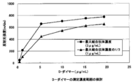
各濃度の抗原液120μLをテストデバイスのサンプルパッド窓部に添加し、イムノクロマトリーダーICA-1000(浜松ホトニクス社)を用いて10分後のテストデバイスの検出窓部の反射光強度を測定した(図8)。その結果、最大結合抗体濃度の混合液で調製したコンジュゲートでは、抗原(D-ダイマー)濃度5.3μg/mL位から測定シグナルがプラトーになったのに対し、最大結合抗体濃度の1/3の濃度の混合液で調製したコンジュゲートでは、15.3μg/mLまで測定シグナルが右肩上がりに増大し、明らかに測定範囲が広がることが判った。
上記の2種類の抗体感作濃度において作製したイムノクロマトテストデバイスを用いて、D-ダイマーの測定範囲を検討した。ナノピア用D-ダイマーキャリブレーター(積水メディカル株式会社製、60μg/mL)を市販ヒトプール血漿にて希釈した。希釈後のD-ダイマー濃度をナノピアD-ダイマー(積水メディカル株式会社製)を用いて測定したところ、各抗原液の濃度は0.4, 1.1, 5.3, 10.0, 15.3, 19.4μg/mLであった。
各濃度の抗原液120μLをテストデバイスのサンプルパッド窓部に添加し、イムノクロマトリーダーICA-1000(浜松ホトニクス社)を用いて10分後のテストデバイスの検出窓部の反射光強度を測定した(図8)。その結果、最大結合抗体濃度の混合液で調製したコンジュゲートでは、抗原(D-ダイマー)濃度5.3μg/mL位から測定シグナルがプラトーになったのに対し、最大結合抗体濃度の1/3の濃度の混合液で調製したコンジュゲートでは、15.3μg/mLまで測定シグナルが右肩上がりに増大し、明らかに測定範囲が広がることが判った。
本発明の多価抗原測定用コンジュゲート、これを用いた多価抗原測定用イムノクロマトテストストリップおよびイムノクロマト測定方法によれば、多価抗原を測定対象物質とした従来よりも測定範囲の広い測定が可能になる。また、希釈操作の手間とコストを削減することができるため、診療所や小病院で求められているPoint of Care Testing(POCT)という社会ニーズにも応えることができる。
(a)プラスチック製粘着シート
(b)抗CRP抗体固定化メンブレン
(c)抗CRP抗体
(d)コンジュゲートパッド
(e)サンプルパッド
(f)吸収パッド
(g)ポリエステルフィルム
(h)3rd Pad
(i)プラスチック製粘着シート
(j)抗D-ダイマー抗体固定化メンブレン
(k)抗D-ダイマー抗体
(l)コンジュゲートパッド
(m)3rd Pad
(n)サンプルパッド
(p)吸収パッド
(q)ポリエステルフィルム
(b)抗CRP抗体固定化メンブレン
(c)抗CRP抗体
(d)コンジュゲートパッド
(e)サンプルパッド
(f)吸収パッド
(g)ポリエステルフィルム
(h)3rd Pad
(i)プラスチック製粘着シート
(j)抗D-ダイマー抗体固定化メンブレン
(k)抗D-ダイマー抗体
(l)コンジュゲートパッド
(m)3rd Pad
(n)サンプルパッド
(p)吸収パッド
(q)ポリエステルフィルム
FERM BP-11344
FERM BP-11345
FERM BP-11345
[寄託生物材料への言及]
(1)FERM BP-11344(#08202産生ハイブリドーマ)
イ 当該生物材料を寄託した寄託機関の名称及び住所
独立行政法人 産業技術総合研究所 特許生物寄託センター
日本国茨城県つくば市東1丁目1番地1中央第6(郵便番号305-8566)
ロ イの寄託機関に生物材料を寄託した日付
平成21年11月26日
ハ イの寄託機関が寄託について付した受託番号
FERM BP-11344
(2)FERM BP-11345(#08203産生ハイブリドーマ)
イ 当該生物材料を寄託した寄託機関の名称及び住所
独立行政法人 産業技術総合研究所 特許生物寄託センター
日本国茨城県つくば市東1丁目1番地1中央第6(郵便番号305-8566)
ロ イの寄託機関に生物材料を寄託した日付
平成21年11月26日
ハ イの寄託機関が寄託について付した受託番号
FERM BP-11345
(1)FERM BP-11344(#08202産生ハイブリドーマ)
イ 当該生物材料を寄託した寄託機関の名称及び住所
独立行政法人 産業技術総合研究所 特許生物寄託センター
日本国茨城県つくば市東1丁目1番地1中央第6(郵便番号305-8566)
ロ イの寄託機関に生物材料を寄託した日付
平成21年11月26日
ハ イの寄託機関が寄託について付した受託番号
FERM BP-11344
(2)FERM BP-11345(#08203産生ハイブリドーマ)
イ 当該生物材料を寄託した寄託機関の名称及び住所
独立行政法人 産業技術総合研究所 特許生物寄託センター
日本国茨城県つくば市東1丁目1番地1中央第6(郵便番号305-8566)
ロ イの寄託機関に生物材料を寄託した日付
平成21年11月26日
ハ イの寄託機関が寄託について付した受託番号
FERM BP-11345
Claims (6)
- 測定対象である多価抗原に対するモノクローナル抗体の溶液と金コロイド溶液との混合液から得られる多価抗原測定用コンジュゲートであって、該混合液の該抗体濃度が、該抗体の金コロイドへの最大結合抗体濃度の1/4~1/2の抗体濃度であることを特徴とする多価抗原測定用コンジュゲート。
- 以下の(1)および(2)を含む多価抗原測定用イムノクロマトテストストリップ。
(1)請求項1に記載のコンジュゲート
(2)測定対象である多価抗原に対するモノクローナル抗体((2)の抗体のエピトープが、(1)の抗体のエピトープと同じ/異なる場合、若しくは(1)の抗体と(2)の抗体が同じ/異なる場合を含む。)が固定化された不溶性メンブレン担体 - さらに以下の(3)~(5)を含む請求項2に記載の多価抗原測定用イムノクロマトテストストリップ。
(3)サンプルパッド
(4)サンプルパッドに接して配置され、請求項2(1)に記載のコンジュゲートを含有するコンジュゲートパッド
(5)吸収パッド - 測定対象である多価抗原が、CRPまたはD-ダイマーである請求項2または3に記載の多価抗原測定用イムノクロマトテストストリップ。
- 試料中の測定対象である多価抗原の量を測定するイムノクロマト測定方法であって、
請求項2~4のいずれかに記載のイムノクロマトテストストリップに試料を供給する工程を含むイムノクロマト測定方法。 - 以下の1)および2)の工程を含む多価抗原測定用コンジュゲートを製造する方法であって、1)の混合液の抗体濃度が、最大結合抗体濃度の1/4~1/2の抗体濃度であることを特徴とする多価抗原測定用コンジュゲートの製造方法。
1)多価抗原に対するモノクローナル抗体溶液と金コロイド溶液との混合液を得る工程
2)該混合液からコンジュゲートを得る工程
Priority Applications (1)
| Application Number | Priority Date | Filing Date | Title |
|---|---|---|---|
| JP2012509454A JP5877150B2 (ja) | 2010-03-31 | 2011-03-28 | 多価抗原測定用コンジュゲートを製造する方法 |
Applications Claiming Priority (2)
| Application Number | Priority Date | Filing Date | Title |
|---|---|---|---|
| JP2010084121 | 2010-03-31 | ||
| JP2010-084121 | 2010-03-31 |
Publications (1)
| Publication Number | Publication Date |
|---|---|
| WO2011125606A1 true WO2011125606A1 (ja) | 2011-10-13 |
Family
ID=44762554
Family Applications (1)
| Application Number | Title | Priority Date | Filing Date |
|---|---|---|---|
| PCT/JP2011/057621 Ceased WO2011125606A1 (ja) | 2010-03-31 | 2011-03-28 | 多価抗原測定用コンジュゲート、これを用いた多価抗原測定用イムノクロマトテストストリップおよびイムノクロマト測定方法 |
Country Status (2)
| Country | Link |
|---|---|
| JP (2) | JP5877150B2 (ja) |
| WO (1) | WO2011125606A1 (ja) |
Cited By (10)
| Publication number | Priority date | Publication date | Assignee | Title |
|---|---|---|---|---|
| CN104165989A (zh) * | 2014-07-10 | 2014-11-26 | 长春理工大学 | 一种检测羊肚菌菌丝的胶体金免疫试纸条 |
| WO2015012384A1 (ja) * | 2013-07-25 | 2015-01-29 | 古河電気工業株式会社 | イムノクロマトグラフィー用試験片、これに用いられる展開液、およびこれを用いたイムノクロマトグラフィー |
| CN104730245A (zh) * | 2014-11-28 | 2015-06-24 | 威海纽普生物技术有限公司 | D二聚体检测试剂盒及检测方法 |
| JPWO2015025968A1 (ja) * | 2013-08-23 | 2017-03-02 | 株式会社ビーエル | マイコプラズマ・ニューモニエの免疫学的検出法およびキット |
| CN108680743A (zh) * | 2018-05-11 | 2018-10-19 | 合肥安为康医学检验有限公司 | 一种快速检测甲型流感病毒的方法 |
| JP2020148580A (ja) * | 2019-03-13 | 2020-09-17 | 東ソー株式会社 | 添加剤によるdダイマー測定での偽高値の抑制 |
| US10852300B2 (en) | 2015-06-01 | 2020-12-01 | Tanaka Kikinzoku Kogyo K.K. | Immunochromatographic analyzer for Mycoplasma pneumoniae detection |
| JPWO2021132470A1 (ja) * | 2019-12-25 | 2021-07-01 | ||
| CN116539876A (zh) * | 2023-04-08 | 2023-08-04 | 南京申基医药科技有限公司 | 一种基于乳胶法检测人类免疫缺陷病毒p24抗原及抗体的试剂盒及其制备方法 |
| US12345705B2 (en) | 2018-11-19 | 2025-07-01 | Sekisui Medical Co., Ltd. | Immunochromatographic test strip and immunochromatographic detection kit |
Families Citing this family (4)
| Publication number | Priority date | Publication date | Assignee | Title |
|---|---|---|---|---|
| US20190064160A1 (en) * | 2016-08-09 | 2019-02-28 | Sekisui Medical Co., Ltd. | Immunochromatographic detection kit |
| JP6736437B2 (ja) * | 2016-09-20 | 2020-08-05 | 積水メディカル株式会社 | イムノクロマトグラフィー検出キット |
| JP6371808B2 (ja) * | 2016-08-09 | 2018-08-08 | 積水メディカル株式会社 | イムノクロマトグラフィー検出キット |
| JP6426872B1 (ja) * | 2018-08-02 | 2018-11-21 | 積水メディカル株式会社 | イムノクロマト用試験片及びイムノクロマト検出キット |
Citations (5)
| Publication number | Priority date | Publication date | Assignee | Title |
|---|---|---|---|---|
| JP2002357599A (ja) * | 2001-03-28 | 2002-12-13 | Gc Corp | 唾液の前処理用キット及びこれを用いた唾液の前処理方法 |
| JP2003344408A (ja) * | 2002-05-27 | 2003-12-03 | Denka Seiken Co Ltd | フロースルーアッセイ方法、キット及び装置 |
| JP2004294157A (ja) * | 2003-03-26 | 2004-10-21 | Denka Seiken Co Ltd | 金コロイドと抗体の結合体を製造する方法 |
| JP2007033378A (ja) * | 2005-07-29 | 2007-02-08 | Denka Seiken Co Ltd | 標識試薬の感度調整方法 |
| JP2010014586A (ja) * | 2008-07-04 | 2010-01-21 | Wako Pure Chem Ind Ltd | 便検体中ヘモグロビンの測定方法及び測定試薬キット |
Family Cites Families (2)
| Publication number | Priority date | Publication date | Assignee | Title |
|---|---|---|---|---|
| JPH06130062A (ja) * | 1992-10-13 | 1994-05-13 | Godo Shiyusei Kk | ブロッキング処理を要しない、金コロイドを用いる免疫学的分析方法、およびこれに用いる分析用ストリップ |
| WO2010001598A1 (ja) * | 2008-06-30 | 2010-01-07 | 積水メディカル株式会社 | 結合アッセイ用多孔性固相及びこれを用いた結合アッセイ法 |
-
2011
- 2011-03-28 JP JP2012509454A patent/JP5877150B2/ja active Active
- 2011-03-28 WO PCT/JP2011/057621 patent/WO2011125606A1/ja not_active Ceased
-
2015
- 2015-09-02 JP JP2015172946A patent/JP6168529B2/ja active Active
Patent Citations (5)
| Publication number | Priority date | Publication date | Assignee | Title |
|---|---|---|---|---|
| JP2002357599A (ja) * | 2001-03-28 | 2002-12-13 | Gc Corp | 唾液の前処理用キット及びこれを用いた唾液の前処理方法 |
| JP2003344408A (ja) * | 2002-05-27 | 2003-12-03 | Denka Seiken Co Ltd | フロースルーアッセイ方法、キット及び装置 |
| JP2004294157A (ja) * | 2003-03-26 | 2004-10-21 | Denka Seiken Co Ltd | 金コロイドと抗体の結合体を製造する方法 |
| JP2007033378A (ja) * | 2005-07-29 | 2007-02-08 | Denka Seiken Co Ltd | 標識試薬の感度調整方法 |
| JP2010014586A (ja) * | 2008-07-04 | 2010-01-21 | Wako Pure Chem Ind Ltd | 便検体中ヘモグロビンの測定方法及び測定試薬キット |
Cited By (18)
| Publication number | Priority date | Publication date | Assignee | Title |
|---|---|---|---|---|
| CN105358981A (zh) * | 2013-07-25 | 2016-02-24 | 古河电气工业株式会社 | 免疫层析用试验片、用于其的展开液及使用其的免疫层析法 |
| WO2015012384A1 (ja) * | 2013-07-25 | 2015-01-29 | 古河電気工業株式会社 | イムノクロマトグラフィー用試験片、これに用いられる展開液、およびこれを用いたイムノクロマトグラフィー |
| JPWO2015012384A1 (ja) * | 2013-07-25 | 2017-03-02 | 古河電気工業株式会社 | イムノクロマトグラフィー用試験片、これに用いられる展開液、およびこれを用いたイムノクロマトグラフィー |
| US10550175B2 (en) | 2013-08-23 | 2020-02-04 | Tauns Co., Ltd. | Mycoplasma pneumoniae immunological detection method and kit |
| JPWO2015025968A1 (ja) * | 2013-08-23 | 2017-03-02 | 株式会社ビーエル | マイコプラズマ・ニューモニエの免疫学的検出法およびキット |
| JP2018173418A (ja) * | 2013-08-23 | 2018-11-08 | 株式会社タウンズ | マイコプラズマ・ニューモニエの免疫学的検出法およびキット |
| JP2020060582A (ja) * | 2013-08-23 | 2020-04-16 | 株式会社タウンズ | マイコプラズマ・ニューモニエの免疫学的検出法およびキット |
| US10995135B2 (en) | 2013-08-23 | 2021-05-04 | Tauns Co., Ltd. | Mycoplasma pneumoniae immunological detection method and kit |
| CN104165989A (zh) * | 2014-07-10 | 2014-11-26 | 长春理工大学 | 一种检测羊肚菌菌丝的胶体金免疫试纸条 |
| CN104730245A (zh) * | 2014-11-28 | 2015-06-24 | 威海纽普生物技术有限公司 | D二聚体检测试剂盒及检测方法 |
| US10852300B2 (en) | 2015-06-01 | 2020-12-01 | Tanaka Kikinzoku Kogyo K.K. | Immunochromatographic analyzer for Mycoplasma pneumoniae detection |
| CN108680743A (zh) * | 2018-05-11 | 2018-10-19 | 合肥安为康医学检验有限公司 | 一种快速检测甲型流感病毒的方法 |
| US12345705B2 (en) | 2018-11-19 | 2025-07-01 | Sekisui Medical Co., Ltd. | Immunochromatographic test strip and immunochromatographic detection kit |
| JP2020148580A (ja) * | 2019-03-13 | 2020-09-17 | 東ソー株式会社 | 添加剤によるdダイマー測定での偽高値の抑制 |
| WO2021132470A1 (ja) * | 2019-12-25 | 2021-07-01 | 富士レビオ株式会社 | イムノクロマト用ストリップ、イムノクロマト用装置、イムノクロマト用キット、及び被検物質検出方法 |
| JP7630445B2 (ja) | 2019-12-25 | 2025-02-17 | 富士レビオ株式会社 | イムノクロマト用ストリップ、イムノクロマト用装置、イムノクロマト用キット、及び被検物質検出方法 |
| JPWO2021132470A1 (ja) * | 2019-12-25 | 2021-07-01 | ||
| CN116539876A (zh) * | 2023-04-08 | 2023-08-04 | 南京申基医药科技有限公司 | 一种基于乳胶法检测人类免疫缺陷病毒p24抗原及抗体的试剂盒及其制备方法 |
Also Published As
| Publication number | Publication date |
|---|---|
| JP2016028241A (ja) | 2016-02-25 |
| JPWO2011125606A1 (ja) | 2013-07-08 |
| JP6168529B2 (ja) | 2017-07-26 |
| JP5877150B2 (ja) | 2016-03-02 |
Similar Documents
| Publication | Publication Date | Title |
|---|---|---|
| JP6168529B2 (ja) | 多価抗原測定用コンジュゲート、これを用いた多価抗原測定用イムノクロマトテストストリップおよびイムノクロマト測定方法 | |
| JP6247741B2 (ja) | 検体不添加サンプルを操作不適サンプルと判定できるイムノクロマトグラフィーに基づく検出方法及びこれに用いるテストストリップ | |
| JP5753942B2 (ja) | 赤血球含有サンプル中の対象物を検出するためのイムノクロマトグラフィー用テストストリップおよびイムノクロマトグラフィーを利用した検出方法 | |
| JP6103776B2 (ja) | ヘマトクリット値の測定方法 | |
| JP6084759B1 (ja) | 免疫学的検出方法及びこれに用いるテストストリップ | |
| JP5955843B2 (ja) | 免疫学的測定方法に用いられるコンジュゲート | |
| JP6464308B1 (ja) | イムノクロマト用試験片 | |
| CN107209179B (zh) | 用于检测含红细胞的样品中的对象物的免疫色谱用测试条、及使用该测试条的免疫色谱 | |
| JP5723484B2 (ja) | 赤血球含有サンプル中の対象物を検出するためのイムノクロマトグラフィー用テストストリップおよびイムノクロマトグラフィーを利用した検出方法 | |
| JP7137168B2 (ja) | イムノクロマト用試験片 | |
| CN113015905B (zh) | 免疫层析测试条和免疫层析检测试剂盒 | |
| WO2020067233A1 (ja) | イムノクロマト用試験片 | |
| JP2020052028A (ja) | イムノクロマト用試験片 | |
| JP2022046827A (ja) | イムノクロマト用試験片及びイムノクロマト検出キット | |
| JP6470147B2 (ja) | 免疫学的検出方法 |
Legal Events
| Date | Code | Title | Description |
|---|---|---|---|
| 121 | Ep: the epo has been informed by wipo that ep was designated in this application |
Ref document number: 11765506 Country of ref document: EP Kind code of ref document: A1 |
|
| WWE | Wipo information: entry into national phase |
Ref document number: 2012509454 Country of ref document: JP |
|
| NENP | Non-entry into the national phase |
Ref country code: DE |
|
| 122 | Ep: pct application non-entry in european phase |
Ref document number: 11765506 Country of ref document: EP Kind code of ref document: A1 |