WO2011065308A1 - 蛍光プローブ - Google Patents
蛍光プローブ Download PDFInfo
- Publication number
- WO2011065308A1 WO2011065308A1 PCT/JP2010/070698 JP2010070698W WO2011065308A1 WO 2011065308 A1 WO2011065308 A1 WO 2011065308A1 JP 2010070698 W JP2010070698 W JP 2010070698W WO 2011065308 A1 WO2011065308 A1 WO 2011065308A1
- Authority
- WO
- WIPO (PCT)
- Prior art keywords
- group
- fluorescent probe
- fluorescent
- general formula
- cyclooctane
- Prior art date
- Legal status (The legal status is an assumption and is not a legal conclusion. Google has not performed a legal analysis and makes no representation as to the accuracy of the status listed.)
- Ceased
Links
- 0 CN(C(CC1S*)=O)C1=O Chemical compound CN(C(CC1S*)=O)C1=O 0.000 description 1
Images






Classifications
-
- C—CHEMISTRY; METALLURGY
- C07—ORGANIC CHEMISTRY
- C07D—HETEROCYCLIC COMPOUNDS
- C07D249/00—Heterocyclic compounds containing five-membered rings having three nitrogen atoms as the only ring hetero atoms
- C07D249/16—Heterocyclic compounds containing five-membered rings having three nitrogen atoms as the only ring hetero atoms condensed with carbocyclic rings or ring systems
-
- C—CHEMISTRY; METALLURGY
- C09—DYES; PAINTS; POLISHES; NATURAL RESINS; ADHESIVES; COMPOSITIONS NOT OTHERWISE PROVIDED FOR; APPLICATIONS OF MATERIALS NOT OTHERWISE PROVIDED FOR
- C09K—MATERIALS FOR MISCELLANEOUS APPLICATIONS, NOT PROVIDED FOR ELSEWHERE
- C09K11/00—Luminescent materials, e.g. electroluminescent or chemiluminescent
- C09K11/06—Luminescent materials, e.g. electroluminescent or chemiluminescent containing organic luminescent materials
-
- G—PHYSICS
- G01—MEASURING; TESTING
- G01N—INVESTIGATING OR ANALYSING MATERIALS BY DETERMINING THEIR CHEMICAL OR PHYSICAL PROPERTIES
- G01N33/00—Investigating or analysing materials by specific methods not covered by groups G01N1/00 - G01N31/00
- G01N33/48—Biological material, e.g. blood, urine; Haemocytometers
- G01N33/50—Chemical analysis of biological material, e.g. blood, urine; Testing involving biospecific ligand binding methods; Immunological testing
- G01N33/58—Chemical analysis of biological material, e.g. blood, urine; Testing involving biospecific ligand binding methods; Immunological testing involving labelled substances
- G01N33/582—Chemical analysis of biological material, e.g. blood, urine; Testing involving biospecific ligand binding methods; Immunological testing involving labelled substances with fluorescent label
-
- C—CHEMISTRY; METALLURGY
- C09—DYES; PAINTS; POLISHES; NATURAL RESINS; ADHESIVES; COMPOSITIONS NOT OTHERWISE PROVIDED FOR; APPLICATIONS OF MATERIALS NOT OTHERWISE PROVIDED FOR
- C09K—MATERIALS FOR MISCELLANEOUS APPLICATIONS, NOT PROVIDED FOR ELSEWHERE
- C09K2211/00—Chemical nature of organic luminescent or tenebrescent compounds
- C09K2211/10—Non-macromolecular compounds
- C09K2211/1003—Carbocyclic compounds
- C09K2211/1007—Non-condensed systems
-
- C—CHEMISTRY; METALLURGY
- C09—DYES; PAINTS; POLISHES; NATURAL RESINS; ADHESIVES; COMPOSITIONS NOT OTHERWISE PROVIDED FOR; APPLICATIONS OF MATERIALS NOT OTHERWISE PROVIDED FOR
- C09K—MATERIALS FOR MISCELLANEOUS APPLICATIONS, NOT PROVIDED FOR ELSEWHERE
- C09K2211/00—Chemical nature of organic luminescent or tenebrescent compounds
- C09K2211/10—Non-macromolecular compounds
- C09K2211/1003—Carbocyclic compounds
- C09K2211/1011—Condensed systems
-
- C—CHEMISTRY; METALLURGY
- C09—DYES; PAINTS; POLISHES; NATURAL RESINS; ADHESIVES; COMPOSITIONS NOT OTHERWISE PROVIDED FOR; APPLICATIONS OF MATERIALS NOT OTHERWISE PROVIDED FOR
- C09K—MATERIALS FOR MISCELLANEOUS APPLICATIONS, NOT PROVIDED FOR ELSEWHERE
- C09K2211/00—Chemical nature of organic luminescent or tenebrescent compounds
- C09K2211/10—Non-macromolecular compounds
- C09K2211/1018—Heterocyclic compounds
- C09K2211/1025—Heterocyclic compounds characterised by ligands
- C09K2211/1044—Heterocyclic compounds characterised by ligands containing two nitrogen atoms as heteroatoms
-
- C—CHEMISTRY; METALLURGY
- C09—DYES; PAINTS; POLISHES; NATURAL RESINS; ADHESIVES; COMPOSITIONS NOT OTHERWISE PROVIDED FOR; APPLICATIONS OF MATERIALS NOT OTHERWISE PROVIDED FOR
- C09K—MATERIALS FOR MISCELLANEOUS APPLICATIONS, NOT PROVIDED FOR ELSEWHERE
- C09K2211/00—Chemical nature of organic luminescent or tenebrescent compounds
- C09K2211/10—Non-macromolecular compounds
- C09K2211/1018—Heterocyclic compounds
- C09K2211/1025—Heterocyclic compounds characterised by ligands
- C09K2211/1059—Heterocyclic compounds characterised by ligands containing three nitrogen atoms as heteroatoms
-
- C—CHEMISTRY; METALLURGY
- C09—DYES; PAINTS; POLISHES; NATURAL RESINS; ADHESIVES; COMPOSITIONS NOT OTHERWISE PROVIDED FOR; APPLICATIONS OF MATERIALS NOT OTHERWISE PROVIDED FOR
- C09K—MATERIALS FOR MISCELLANEOUS APPLICATIONS, NOT PROVIDED FOR ELSEWHERE
- C09K2211/00—Chemical nature of organic luminescent or tenebrescent compounds
- C09K2211/10—Non-macromolecular compounds
- C09K2211/1018—Heterocyclic compounds
- C09K2211/1025—Heterocyclic compounds characterised by ligands
- C09K2211/1092—Heterocyclic compounds characterised by ligands containing sulfur as the only heteroatom
Definitions
- the present invention relates to a fluorescent probe useful for labeling or imaging cells and the like. More specifically, the present invention relates to a fluorescent probe that can be easily synthesized, reduces non-specific binding to a biomolecule such as a cell, and can be labeled and imaged with good fluorescence intensity.
- Fluorescent probes combined with proteins such as antibodies and low molecular weight compounds are useful for imaging biomolecules in living bodies and cells.
- organic fluorescent substances such as fluorescein derivatives, rhodamine derivatives, hydrazide derivatives, and cyanine dyes have been used. Widely used.
- Fluorescent probes used for labeling and imaging need to be able to detect target molecules specifically and have no or reduced influence by non-specific binding to biomolecules other than target molecules.
- a fluorescent probe of a fluorescein derivative when used, there is often a nonspecific binding to a cell or the like.
- the fluorescence intensity may not be sufficient, and the development of a fluorescent probe is not yet sufficient.
- Non-Patent Document 1 S. Nie et al, Engineering Luminescent Quantum Dots for In Vivo Molecular and Cellular Imaging. Annals of Biomedical Engineering, 2006, 34, 3-14.
- a fluorescently labeled object specifically, a physiologically active substance such as an antibody or a protein or a substance such as a low molecular weight compound was directly condensed with the quantum dots.
- a condensation reaction methods such as a thiol exchange reaction, an amidation of an amino group or a carboxylic acid are often used.
- thiol groups, amino groups, and carboxylic acids such as proteins having physiological activity are functional groups that are also important for activity. Therefore, when performing condensation reactions between quantum dots and proteins, such important functional groups are used. Needed to be protected from reacting with the quantum dots. However, if the molecular design in the condensation reaction between quantum dots and physiologically active proteins is insufficient, important functional groups will bind to the quantum dots, and the resulting fluorescent probe will be There was a case where a problem that the function / property was not reflected occurred.
- the condensation of the alkyne derivative and the azide compound is performed by a 1,3-cycloaddition reaction also known as a Huisgen reaction.
- the Huisgen reaction is a reaction in which a triple bond of an alkyne compound and an azide group are condensed under heating conditions.
- An 8-membered ring alkyne derivative is bound to a gel or solid phase carrier, and this is reacted with an azide-modified organic fluorescent dye or enzyme ( ⁇ -Gal). It has been reported that it can bind and immobilize on the gel or solid phase carrier (Non-patent document 2: K. Ebisu et al., N-terminal Specific Point-Immobilization of Active Proteins by the One-Pot NEXT -A Method. ChemBIoChem., 2009, 10,2460-2464).
- the fluorescent substance and the fluorescently labeled object are not bonded via an 8-membered alkyne or an azide compound.
- An object of the present invention is to provide a fluorescent probe that can be easily synthesized, has reduced non-specific binding to a biomolecule such as a cell, and can be labeled and imaged with good fluorescence intensity. And It is another object of the present invention to provide a method for manufacturing such a fluorescent probe.
- the present inventors paid attention to the condensation reaction between an alkyne derivative and an azide compound, and as a result of extensive research, as a result of the quantification of cyclooctane alkyne as an 8-membered ring alkyne derivative to fluorescent nanoparticles such as quantum dots.
- fluorescent probe synthesis intermediate an intermediate obtained by binding a derivative (this conjugate is hereinafter referred to as “fluorescent probe synthesis intermediate”) and a substance capable of binding to a fluorescently labeled object into which an azide group has been introduced.
- this invention consists of the following.
- a fluorescent probe in which a compound represented by the following general formula (I) is bonded to the fluorescent nanoparticle surface at the Y portion.
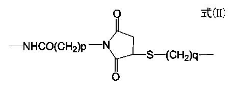
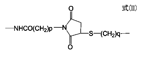
- Y is represented by —NH—CO—W—, —W—CO—NH—, or the following general formula (II), wherein W represents an oxygen atom, an alkylene group, or an alkyleneoxy group.
- W represents an oxygen atom, an alkylene group, or an alkyleneoxy group.
- Ph represents a phenylene group
- B represents an alkylene group or an alkyleneoxy group
- p is an integer of 1 to 6
- q is an integer of 0 to 6.
- Ring A represents a derivative of cyclooctane, and the cyclooctane moiety may be further condensed with an aromatic ring;
- R represents an alkyl group, an alkoxy group or a halogen atom, and n is an integer of 0 to 2;
- Z represents a substance that can bind to the fluorescently labeled object;
- NH and CO in Y may be directly bonded or may be bonded via a linker.
- Y is represented by —NH—CO—W—, —W—CO—NH—, or the following general formula (II), wherein W represents an oxygen atom, an alkylene group, or an alkyleneoxy group.
- W represents an oxygen atom, an alkylene group, or an alkyleneoxy group.
- Z represents a substance that can bind to the fluorescently labeled object; NH and CO in Y may be directly bonded or may be bonded via a linker.
- 3. 3 3.
- the fluorescent probe according to item 2 wherein Y is —NH—CO—O—, wherein the compound represented by formula (III) is bonded to the fluorescent nanoparticle surface at the Y portion. 4). 4.
- the linker is an amino group at both ends of the alkylene group, alkyleneoxy group or polyalkyleneoxy group.
- Y is represented by —NH—CO—W—, —W—CO—NH—, or the following general formula (II), wherein W represents an oxygen atom, an alkylene group, or an alkyleneoxy group.
- W represents an oxygen atom, an alkylene group, or an alkyleneoxy group.
- Ph represents a phenylene group
- B represents an alkylene group or an alkyleneoxy group
- p is an integer of 1 to 6
- q is an integer of 0 to 6.
- Ring A ′ represents an alkyne derivative of cyclooctane, and the cyclooctane moiety may be further condensed with an aromatic ring;
- R represents an alkyl group, an alkoxy group or a halogen atom, and n is an integer of 0 to 2;
- NH and CO in Y may be directly bonded or may be bonded via a linker.
- Y is represented by —NH—CO—W—, —W—CO—NH—, or the following general formula (II), wherein W represents an oxygen atom, an alkylene group, or an alkyleneoxy group.
- W represents an oxygen atom, an alkylene group, or an alkyleneoxy group.
- NH and CO in Y may be directly bonded or may be bonded via a linker.
- the linker is at both ends of the alkylene group, alkyleneoxy group or polyalkyleneoxy group.
- the method for producing a fluorescent probe synthesis intermediate comprising the following steps: 1) A step of producing fluorescent nanoparticles into which a carboxylic acid group, amino group, thiol group, maleimide group, carbonate group, or active ester group is introduced; 2) A functional group capable of reacting with various functional groups introduced into the fluorescent nanoparticles in the step 1) is introduced into the cyclooctane alkyne derivative, and the cyclooctane alkyne derivative into which the functional group is introduced is obtained. Producing step; 3) A step of binding the fluorescent nanoparticles into which various functional groups prepared in the steps 1) and 2) are introduced and an alkyne derivative of cyclooctane. 11.
- the method for producing a fluorescent probe which comprises the following steps: A) a step of introducing an azide group into a substance (Z) that can bind to a fluorescently labeled object; B) A step of binding the alkyne derivative of cyclooctane in the fluorescent probe synthesis intermediate according to any one of 6 to 9 above and the azide group introduced in the step A).
- the method for producing a fluorescent probe according to any one of 1 to 5 above which comprises the following steps: A) a step of introducing an azide group into a substance (Z) that can bind to a fluorescently labeled object; B) a step of binding the alkyne derivative of cyclooctane in the fluorescent probe synthesis intermediate according to any one of 6 to 9 above and the azide group introduced in the step of A); C) A step of hydrophilizing the fluorescent probe obtained in the step B).
- the fluorescent probe of the present invention uses fluorescent nanoparticles such as hydrophilic quantum dots, and is a fluorescent probe that is extremely bright and excellent in light stability as compared with organic fluorescent dyes.
- fluorescent nanoparticles such as hydrophilic quantum dots
- the fluorescent probe of the present invention can be said to be a highly specific fluorescent probe that suppresses non-specific binding.
- the fluorescent probe of the present invention can be used for analysis by flow cytometry and microscopic analysis.
- a fluorescent probe library and a target fluorescent probe can be easily produced.
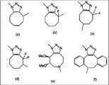
- FIG. 3 is a diagram showing an example of a cyclooctane derivative condensed with a triazole ring in the formula (I).
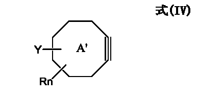
- FIG. 4 is a diagram showing an example of an alkyne derivative of cyclooctane in the formula (IV).
- FIG. 3 is a diagram showing examples of raw material compounds of an alkyne derivative of cyclooctane that can be used for the synthesis of a fluorescent probe synthesis intermediate of formula (IV) and further a fluorescent probe of formula (I). It is a figure which shows the reaction aspect of the fluorescent nanoparticle of this invention, and the alkyne derivative of cyclooctane.
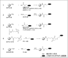
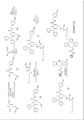
- Example 1 It is a figure which shows the synthetic scheme of the fluorescent probe synthetic intermediate (QD-DIB) of this invention.
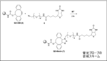
- Example 1 It is a figure which shows the synthetic scheme of the fluorescent probe (QD-Biotin) of this invention.
- Example 2 It is a figure which shows the synthetic scheme of the fluorescent probe (QD-NP) of this invention.
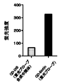
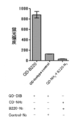
- Example 3 It is a figure which shows the binding effect with the streptavidin of the fluorescent probe (QD-Biotin) of this invention.
- Experimental example 2-1 It is a figure which shows the analysis result of the flow cytometry regarding the coupling
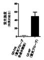
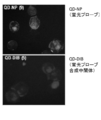
- Example 3-1 It is a figure which shows the cell surface antibody imaging effect of the fluorescent probe (QD-NP) of this invention.
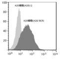
- Example 3-2 Analysis of the fluorescent probe (QD-NP) of the present invention by flow cytometry in A20 cells (A20 / BCR) in which IgG1 is expressed on the cell surface and A20 cells (A20 (-)) in which IgG1 is not expressed It is a figure which shows a result.
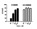
- Example 3-3 It is a figure which shows the analysis result by a flow cytometry when the fluorescent probe (QD-NP) of this invention and its unlabeled antagonist or non-antagonist are added.
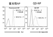
- Example 3-4 It is a figure which shows the analysis result by the flow cytometry when not using the fluorescent probe (fluorescent NP) using the fluorescent probe (QD-NP) or organic fluorescent pigment
- Example 3-5 and Comparative Example It is a figure which shows the synthetic scheme of the fluorescent probe (QD-B220) of this invention.
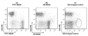
- Example 4 It is a figure which shows the analysis result by the flow cytometry with the A20 cell of the fluorescent probe (QD-B220) of this invention.
- Experimental example 4-1) It is a figure which shows the analysis result by the flow cytometry with the mouse
- Example 5-1 It is a figure which shows the synthetic scheme of the hydrophilization QD-OMe (reference example) which processed the alkyne derivative of cyclooctane to PEG (polyethyleneglycol), and the fluorescent probe hydrophilization QD-BIBP of this invention which processed PEG.
- Example 6 It is a figure which shows that nonspecific adsorption
- Example 6-1 It is a figure which shows the synthetic scheme of the hydrophilization QD-OMe (reference example) which processed the alkyne derivative of cyclooctane to PEG (polyethyleneglycol), and the fluorescent probe hydrophilization QD-BIBP of this invention which processed PEG.
- the present invention relates to a fluorescent probe useful for labeling or imaging cells and the like. More specifically, the present invention relates to a fluorescent probe that can be easily synthesized, reduces non-specific binding to a biomolecule such as a cell, and can be labeled and imaged with good fluorescence intensity.
- the fluorescent probe of the present invention has a structure in which a compound represented by the following general formula (I) is bonded to the surface of the fluorescent nanoparticle at the Y portion, and the cycloprobe condensed with the fluorescent nanoparticle (X) and the triazole ring.
- ring A a derivative of octane
- Z a substance that can bind to the fluorescently labeled object
- X binds to ring A via Y.
- One or more compounds represented by the general formula (I) are bonded to the surface of the fluorescent nanoparticle.
- Y is represented by —NH—CO—W—, —W—CO—NH—, or the following general formula (II), wherein W represents an oxygen atom, an alkylene group, or an alkyleneoxy group.
- W represents an oxygen atom, an alkylene group, or an alkyleneoxy group.
- Ring A represents a cyclooctane derivative, and the cyclooctane moiety may be further condensed with an aromatic ring such as a benzene ring;
- Rn means a substituent substituted on the ring A as an R group, R represents an alkyl group, an alkoxy group or a halogen atom, and n is an integer of 0 to 2;
- Z represents a substance that can bind to the fluorescently labeled object;
- NH and CO in Y may be directly bonded or may be bonded via a linker.
- the present invention also extends to a fluorescent probe synthesis intermediate for producing the above fluorescent probe.
- the fluorescent probe synthesis intermediate of the present invention has a structure in which a compound represented by the following general formula (IV) is bonded to the surface of the fluorescent nanoparticle at the Y portion, and includes the fluorescent nanoparticle (X) and cyclooctane. It is composed of an alkyne derivative (ring A ′), and X is bonded to ring A ′ via Y.
- One or more compounds represented by the general formula (IV) are bonded to the surface of the fluorescent nanoparticle.
- Y is represented by —NH—CO—W—, —W—CO—NH—, or the following general formula (II), wherein W represents an oxygen atom, an alkylene group, or an alkyleneoxy group.
- W represents an oxygen atom, an alkylene group, or an alkyleneoxy group.
- Ring A ′ represents an alkyne derivative of cyclooctane, and the cyclooctane moiety may be further condensed with an aromatic ring such as a benzene ring;
- Rn represents a substituent substituted on the ring A ′ as an R group, R represents an alkyl group, an alkoxy group, or a halogen atom, and n is an integer of 0 to 2;
- NH and CO in Y may be directly bonded or may be bonded via a linker.
- the fluorescent probe Y in which the compound represented by the general formula (I) or (IV) is bonded to the fluorescent nanoparticle surface at the Y portion when NH and CO are bonded via a linker, Is not particularly limited as long as it can bond NH and CO.
- an amino group, an alkylamino group (for example, an alkylamino group having 1 to 4 carbon atoms) or a carboxylic acid group at both ends of the group or a polyalkyleneoxy group having 2 to 20 carbon atoms, preferably 4 to 12 carbon atoms Compound etc. are mentioned. Specific examples include compounds represented by the following formula (VI) or formula (VII).
- W is an alkylene group, an alkyleneoxy group or an alkylenecarbonyl group.
- the alkylene group preferably has 1 to 3 carbon atoms, particularly preferably 1.
- W is —Ph—B—
- the alkylene group or alkyleneoxy group of B preferably has 1 to 3, more preferably 1, carbon atoms.
- W is an oxygen atom.
- R of the fluorescent probe in which the compound represented by the above general formula (I) or (IV) is bonded to the fluorescent nanoparticle surface at the Y portion is an alkyl group or an alkoxy group
- the number of carbon atoms is 1 to 3 In particular, 1 is preferable.
- R is a halogen atom, it is preferably a fluorine atom.
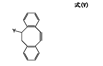
- a preferred fluorescent probe is a compound in which the compound represented by the following general formula (III) is bonded to the fluorescent nanoparticle surface at the Y portion, and a preferred fluorescent probe synthesis intermediate is represented by the general formula (V).
- Y is preferably —NH—CO—O—.
- the present invention also provides a fluorescent probe synthesis intermediate (IV) for producing a fluorescent probe or fluorescent probe library in which the compound represented by the general formula (I) is bound to the fluorescent nanoparticle surface at the Y portion.
- the fluorescent probe library refers to Z in the fluorescent probe of the present invention in which the compound represented by the general formula (I) is bonded to the fluorescent nanoparticle surface at the Y portion, that is, binds to the fluorescently labeled object. It means a probe library in which multiple types of probes with different possible substances are combined.
- the fluorescent nanoparticle (X), the cyclooctane derivative, the cyclooctane alkyne derivative (ring A and ring A ′) and the substance capable of binding to the fluorescent labeling object constituting the structure of the fluorescent probe of the present invention Each of (Z) will be described in detail, and the method for preparing the fluorescent probe synthesis intermediate of the present invention and the method for manufacturing the fluorescent probe will also be described in detail.
- the fluorescent nanoparticles in the fluorescent probe of the present invention usually have a particle size of 40 nm or less, preferably about 10 to 30 nm, and have a short wavelength such as ultraviolet rays or X-rays. Examples thereof include particles that emit fluorescence when irradiated with light.
- the fluorescent nanoparticles of the present invention are preferably water-dispersible.
- the surface of the particles is preferably subjected to a hydrophilic treatment.
- hydrophilic quantum that is a so-called fluorescent nanoparticle having a core-shell structured particle whose surface is covered with a semiconductor material as a core (core) and whose electrons are confined in all three dimensions. Dots are preferred.
- the hydrophilic quantum dot means, for example, the surface of a nanoparticle having a size of about several nanometers of a core-shell structure in which a semiconductor such as cadmium selenide (CdSe) is used as a core and coated with zinc sulfide (ZnS) or the like.
- a semiconductor such as cadmium selenide (CdSe)
- ZnS zinc sulfide
- Those obtained by hydrophilization usually have a particle size of 40 nm or less, preferably about 10 to 30 nm.
- Hydrophilic quantum dots have extremely strong fluorescence compared to organic fluorescent dyes and fluorescent proteins, and emit different fluorescence depending on the particle size of the core-shell structure.
- the particle size of the core-shell structure when the particle size of the core-shell structure is 2 nm, it emits blue fluorescence, 3 mm is green, 4 mm is yellow, and 5 mm is red.
- quantum dots have characteristics such as excellent light stability and difficulty in fading compared to organic fluorescent dyes, a very narrow fluorescence spectrum width, and excitation with various fluorescence wavelengths with one type of light source. Have. Therefore, it is a material that has been vigorously studied for bioimaging applications such as high-sensitivity detection and multicolor imaging.
- the optical properties of hydrophilic quantum dots vary greatly depending on the core semiconductor material.
- the semiconductor material include ZnS, ZnSe, CdS, CdSe, CdTe, PbS, InAs, InP, and the like.
- the core-shell CdSe / ⁇ ⁇ ZnS meaning CdSe particles coated with ZnS, the same applies hereinafter
- CdSe coated with ZnS as the core semiconductor has a high quantum yield and is suitable for flow cytometry. Since it shows fluorescence in the visible light region, it is suitable for the fluorescent probe of the present invention.
- InP / PZnS in which InP is coated with ZnS is also suitable.
- the particles having a core-shell structure usually have a particle size of 10 nm or less, preferably 2 to 8 nm, but the wavelength of the emitted fluorescence varies depending on the particle diameter of the core-shell structure used. For example, if the particle diameter of the core-shell structure is 5 to 8 nm, depending on it, quantum dots that emit fluorescence in the range of 500 to 800 nm can be easily obtained. It is possible to produce a fluorescent probe showing For example, when considering application to flow cytometry, hydrophilic quantum dots that emit fluorescence of 625 nm or 655 nm with strong fluorescence intensity and little overlap of fluorescence wavelength with FITC and PE, which are widely used in multicolor analysis, are suitable. It is. For application to biological imaging, hydrophilic quantum dots that are called biological windows and emit fluorescence in the near-infrared region (650 to 1000 nm) with high biological permeability are suitable.
- hydrophilic quantum dots are characterized by emitting stronger fluorescence as the wavelength becomes shorter. Therefore, when the fluorescent probe of the present invention is used in flow cytometry, it is preferable to excite with a UV laser (355 nm) or a violet laser (405 nm).
- the core-shell structured particles are highly hydrophobic and easily aggregate and precipitate in water, they are hydrophilized by various surface modification methods, and hydrophilic quantum dots subjected to hydrophilized surface treatment are used for the production of fluorescent probes.
- Hydrophilic treatment is performed, for example, by coating the surface of the particle with a compound having a mercapto group and a hydrophilic group such as a carboxyl group and an amino group, specifically, mercaptoacetic acid, mercaptosuccinic acid, cysteine, dihydroriboic acid, and the like.
- a hydrophilic quantum dot can be synthesized by introducing a carboxylic acid by covalently coordinating a thiol group.
- hydrophilic quantum dots it is preferable to modify the surface of the hydrophilic quantum dots with a polyether such as polyethylene glycol (PEG) because nonspecific adsorption can be suppressed.
- a polyether such as polyethylene glycol (PEG)
- Qdot (R) nanocrystals, eFluor TM nanocrystals, and the like are commercially available from Invitrogen and Evident Technologies.
- the particles of the core-shell structure are hydrophilized as described above, and appropriately non-specific adsorption suppression treatment is performed, usually 40 nm or less, preferably about 10 to 30 nm. Fluorescent nanoparticles having a particle size can be suitably used.
- a carboxylic acid group, an amino group, a thiol group, a maleimide group, a carbonate group, or an active ester group is formed on the surface of the fluorescent nanoparticle in order to bind the alkyne derivative of cyclooctane and the fluorescent nanoparticle.
- a carboxylic acid group, an amino group, a thiol group, a maleimide group, a carbonate group, or an active ester group is formed on the surface of the fluorescent nanoparticle in order to bind the alkyne derivative of cyclooctane and the fluorescent nanoparticle.
- Ring A in the fluorescent probe of the present invention of the formula (I) is a derivative of cyclooctane condensed with a triazole ring, for example, as shown in FIGS. 1-1 (a) to (f) Such a thing is mentioned.
- ring A ′ in the intermediate of formula (IV) is an alkyne derivative of cyclooctane, and examples thereof include those shown in FIGS. 1-2 (a-1) to (f-1).
- the “—Y-ring A ′” moiety in the intermediate of formula (IV) is, for example, (a-1-1), (a-1-2), (b-1-1) in FIG. (C-1-1), (d-1-1), (d-1-2), (e-1-1), and (f-1-1) alkyne derivatives of cyclooctane Formed by using. Furthermore, by reacting the alkyne moiety of the “—Y-ring A ′” with a compound having an azide group, the “—Y-ring A-” moiety in the fluorescent probe of formula (I) is formed.
- alkyne is a general term for hydrocarbons that have only one carbon-carbon triple bond in the molecule.
- the raw material compound of the cyclooctane alkyne derivative that can be used in the present invention is not particularly limited. Among them, the 3-hydroxy described in (f-1-1) of FIG. -1,2: 5,6-Dibenzocycloocta-1,5,7-triene (DIB) (X. Ning et al., Angew. Chem. Int. Ed., 2008, 47, 2253) can be preferably used. Also, by introducing two fluorine atoms adjacent to the alkyne portion of cyclooctane and improving the reactivity, (c-1-1) in FIG. The difluoro-cyclooctyne derivatives described in (d-1-1) and (d-1-2) (JA Codelli, et al., JACS 2008, 130, 11486) can also be suitably used.
- Substance (Z) that can bind to fluorescently labeled object includes cells, tissues, proteins, peptides, genes, sugar chains, etc., and is particularly limited is not.
- the substance (Z) that can bind to the fluorescent labeling target substance may be any substance that can bind to the fluorescent labeling target substance, and may be a low-molecular compound, protein, carbohydrate, nucleic acid, or the like. It may be a polymer material and is not particularly limited.
- Z when the fluorescently labeled object is a tissue or a cell, Z may be a substance that can interact with a substance expressed on the surface of the tissue or cell.
- Z may be an antibody, a peptide that can specifically recognize the protein, a receptor for the protein, a low-molecular compound that can bind to the protein, or the like.
- the product is a cell surface receptor such as a 7-transmembrane receptor (GPCR)
- GPCR 7-transmembrane receptor
- Z is a compound that binds to the receptor, such as diphenhydramine or mepyramine that binds to the histamine receptor.
- Agents and the like When the fluorescently labeled object is streptavidin, biotin (Biotin) is exemplified as Z.
- Biotin biotin
- antibodies can be preferably used.
- a step of producing fluorescent nanoparticles into which a carboxylic acid group, amino group, thiol group, maleimide group, carbonate group, or active ester group is introduced; 2) A step of producing an alkyne derivative of cyclooctane into which functional groups capable of reacting with various functional groups introduced into the fluorescent nanoparticles in the step 1) are introduced; 3) A step of bonding the fluorescent nanoparticles into which various functional groups have been introduced in the steps 1) and 2) and an alkyne derivative of cyclooctane by a condensation reaction or a reaction via a linker.
- any of the steps 1) and 2) may be performed first.
- these functional groups are introduced into the alkyne derivative of cyclooctane by introducing a carboxylic acid group, amino group, thiol group, maleimide group, carbonate ester group, or active ester group.
- This is interpreted as a step of producing a fluorescent nanoparticle in which a functional group capable of reacting with various functional groups introduced into the cyclooctane alkyne derivative is introduced in the step. Can be produced.
- the alkyne derivative of cyclooctane In order to bind the fluorescent nanoparticle to the alkyne derivative of cyclooctane, it is possible to bond the alkyne derivative of cyclooctane and the fluorescent nanoparticle by introducing a combination of functional groups capable of reacting with each other and reacting them.
- a functional group a carboxylic acid group, an amino group, a thiol group, a maleimide group, a carbonate ester group, an active ester group, and the like are suitable, and these functional groups can be introduced in combination.
- Such combinations include carboxylic acid groups and amino groups, maleimide groups and thiol groups, amino groups and carbonate groups, amino groups and active ester groups.
- Table 1 the combinations shown in Table 1 below can be considered.
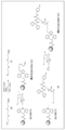
- FIG. 2 schematically shows these reaction examples.
- the method for introducing the functional group is not particularly limited, and a method known per se or any method developed in the future can be applied.
- the carboxylic acid can be introduced by a method in which a thiol group such as mercaptoacetic acid or dihydroriboic acid is covalently coordinated to the surface of the core-shell structured particle.
- a polymer such as polyethyleneimine
- amino groups can be introduced onto the surface of the core-shell structured particles.
- a maleimide group can be introduced by condensation with an amino group on the surface of the core-shell structured particle using a crosslinking reagent such as 4-maleimidobutyric acid succinimidyl ester.
- the fluorescent nanoparticles into which an amino group, a carboxylic acid group or the like has been introduced by the above-described hydrophilization treatment can be used as they are.
- the fluorescent nanoparticles and the alkyne derivative of cyclooctane have a functional group as shown in Table 1 or FIG. 2, they can be directly bonded to each other.
- a carbonate ester group can be introduced by condensation reaction of the alkyne derivative of cyclooctane with N, N′-disuccinimidyl carbonate. it can.
- an amino group is bonded to the carbonate group via a linker having an amino group at both ends, for example, an alkylene group having 1 to 20 carbon atoms, preferably an alkylene group having 1 to 12 carbon atoms or an alkyleneoxy group such as polyethylene glycol (PEG).
- An amino group can be introduce
- the reaction between the fluorescent nanoparticles and the cyclooctane alkyne derivative is performed by reacting an excess amount of the cyclooctane alkyne derivative with respect to 1 ⁇ mol of the fluorescent nanoparticle, usually 20 to 500 ⁇ mol, preferably 50 to 300 ⁇ mol. .
- an excess amount of the cyclooctane alkyne derivative with respect to 1 ⁇ mol of the fluorescent nanoparticle, usually 20 to 500 ⁇ mol, preferably 50 to 300 ⁇ mol. .
- 1 ⁇ mol of the fluorescent nanoparticle usually 20 to 500 ⁇ mol, preferably 50 to 300 ⁇ mol.
- Etc. there are usually a plurality of reaction points on the surface of the fluorescent nanoparticle of the present invention.
- the present invention relates to a fluorescent probe and a fluorescent probe intermediate in which the compound represented by the general formula (I) and the general formula (IV) is bonded to the surface of the fluorescent nanoparticle at the Y portion, respectively.
- the fluorescent probe of the present invention can be prepared by binding the “fluorescent probe synthesis intermediate” of the present invention and “substance (Z) that can bind to a fluorescently labeled object”.
- the fluorescent probe synthesis intermediate of the present invention can be prepared by a method including at least the following steps A) and B).
- the present invention extends to a method for producing a fluorescent probe.
- A) a step of introducing an azide group into a substance (Z) that can bind to a fluorescently labeled object;
- B) A step of reacting the azide group introduced in A) with an alkyne derivative of cyclooctane in the fluorescent probe synthesis intermediate.
- A) Method for introducing an azide group into a substance (Z) that can bind to a fluorescently labeled object is a method known per se, Alternatively, any method developed in the future can be applied and is not particularly limited.
- the high nucleophilicity of an azide ion can be used to introduce by an S N 2 reaction with respect to an alkyl halide or a sulfonate ester.
- the compound having an azide group and an amino group in the same molecule is used, the compound can be introduced by a condensation reaction with a carboxylic acid in a low molecular compound.
- an azide group can be introduced by reaction with an amino group in the low molecular compound.
- an antibody When an antibody is used as the substance Z, an amino group that does not participate in binding can be labeled as shown in the examples described later. Further, since the antibody has a disulfide bond in the molecule, the SH group obtained by reducing this can be used for labeling. Since the SH group thus obtained is unrelated to antigen recognition, there is an advantage that the specificity of the antibody is not impaired even if the SH group is used.
- antibody fragments are treated with proteolytic enzymes pepsin and papain to produce Fab fragments.
- the Fab fragment thus prepared has an SH group and can be used for the next reaction.
- a Fab fragment When a Fab fragment is treated with a crosslinking reagent having an azide group and a maleimide group in the molecule, the reaction between the SH group and the maleimide group in the Fab fragment proceeds selectively, and an azide Fab fragment can be synthesized. .
- an alkyne derivative of cyclooctane By reacting this with an alkyne derivative of cyclooctane, a labeled antibody can be synthesized.
- the SH group can be labeled by reducing the interchain thiol of the antibody according to the method described in JP-T-2008-500961.
- an “azide group-containing substance” a substance that can bind to a fluorescently labeled object into which an azide group has been introduced is referred to as an “azide group-containing substance” for convenience.
- Condensation reaction between the fluorescent probe synthesis intermediate and the azide group-containing substance is performed by synthesizing the azide group and fluorescent probe introduced into the fluorescent labeling object in A) above. It can be carried out by condensation reaction with an alkyne derivative of cyclooctane in the intermediate.
- a method known per se for example, the method described in JACS 2004, 126, 15046 or Non-Patent Document 2, or any method developed in the future can be applied.
- the fluorescent probe synthesis intermediate of the present invention and the azide group-containing substance can be subjected to a condensation reaction only by reacting at room temperature for several hours.
- a fluorescent probe can be easily prepared without using a reagent such as a condensing agent.
- the fluorescent probe of the present invention can be used for fluorescent labeling and imaging of cells, and highly sensitive detection of target molecules such as biosensors. For example, it can be used for high-sensitivity detection using a fluorescence plate reader or flow cytometry, in vitro imaging using a fluorescence microscope or confocal microscope, and in vivo imaging.
- an unreacted cyclooctane alkyne derivative may be adsorbed nonspecifically on the cell membrane surface and the background may be increased.
- One of the reasons is assumed to be due to the molecular structure of the highly hydrophobic DIB group. Therefore, for example, as shown in the examples below, non-specific adsorption is suppressed by hydrophilizing the alkyne derivative portion of unreacted cyclooctane with polyalkylene glycol such as azidated PEG. This is preferable.
- Example 2 Preparation of fluorescent probe (QD-Biotin)
- QD-Biotin QD-Biotin
- a substance (biotin) capable of binding to a fluorescently labeled object is bound to QD-DIB (Compound 5).
- the production of the fluorescent probe (compound 7) will be described.
- Biotin (244 mg, 1.0 mmol) and 14-azido-3,6,9,12-tetraoxatetradecan-1-amine (14-azido-3,6,9,12-tetraoxatetradecan-1-amine) (524 mg, 2.0 mmol) aminoMeOH-MeCN (1: 3) (12 ml) mixed solution was added EDC (N-ethyl-N'-3- (dimethylaminopropyl) carbodiimide) (288 mg, 1.5 mmol) . After stirring at room temperature for 42 hours, the solvent was distilled off under reduced pressure to obtain a crude product. This was purified by silica gel chromatography to obtain Compound 6 in 493 mg mg (yield: 100%%).
- a solution containing the fluorescent probe QD-Biotin (compound 7) was collected from the device for centrifugal filtration, and an appropriate amount of PBS was added to prepare 50 ⁇ l.
- the fluorescent probe QD-Biotin (compound 7) prepared in Example 2 is also simply referred to as “QD-Biotin”.
- a 10 mM DMSO solution (5 ⁇ l, 50 nmol) containing the compound 8 was added to PBS (100 ⁇ l) containing QD-DIB (10 ⁇ l, 0.040 nmol) prepared in Example 1. After standing at room temperature for 2 hours, PBS with (400 [mu] l) was added to the reaction solution was transferred to a centrifuge for filtration devices (10K Nanosep® (R) filter (manufactured by Paul)). Washing operation by centrifugation for 10 minutes at 4 ° C. and 7000 rpm was performed 3 times to remove excess compound 8.
- a solution containing the fluorescent probe QD-NP (compound 9) was collected from the device for centrifugal filtration, and an appropriate amount of PBS was added to prepare 50 ⁇ l.
- the QD-NP (compound 9) of the fluorescent probe prepared in Example 3 is also simply referred to as “QD-NP”.
- Example 3-1 Evaluation of fluorescent probe (QD-NP) by flow cytometry 1
- QD-NP prepared in Example 3 was evaluated using A20 cells (A20 / BCR) that highly express immunoglobulin IgG1 that specifically recognizes NP (nitrophenylacetyl) as an antigen. It was. A20 cells (A20 / BCR) (3 x 10 5 cells) were suspended in 2% FBS / PBS (200 ⁇ l), and the QD-NP solution prepared in Example 3 was diluted 10 times with a fluorescent probe solution (2 ⁇ l). In addition, it was allowed to act on ice for 30 minutes.
- a fluorescent probe synthesis intermediate solution obtained by diluting the QD-DIB solution prepared in Example 1 50 times was treated in the same manner. After washing twice with PBS, it was filtered with a cell strainer (Becton Dickinson (BD)), which is a tube for flow cytometry, and measured by flow cytometry (FACS Canto TM II (BD)).
- BD Cell strainer
- FACS Canto TM II FACS Canto TM II
- the A20 cells were prepared as follows according to a conventional method. That is, RNA was extracted from splenic B cells of mice immunized with NPized chicken ⁇ globulin, and the variable region of the immunoglobulin locus (VH186.2) was cloned by PCR. Further, the full-length ⁇ 1 light chain and the constant region of the ⁇ 1 heavy chain constituting the antibody were similarly cloned by the PCR method. Thereafter, the cloned VH186.2 variable region was connected to the ⁇ 1 constant region by PCR to obtain a full-length ⁇ 1 heavy chain gene that recognizes the NP hapten.
- VH186.2 variable region of the immunoglobulin locus
- the ⁇ 1 light chain and full-length ⁇ 1 heavy chain genes obtained by A20 cells which are mouse B cell tumor cells that constantly express the ⁇ 2b heavy chain, were introduced by the retroviral method. Reconstitution of the antigen receptor that recognizes the NP hapten was confirmed by flow cytometry (Facs ARIA TM (manufactured by BD)) by staining with phycoerythrin bovine serum albumin bound to the NP hapten.
- the fluorescent probe (QD-NP) of the present invention is a fluorescent probe that can be used for cell labeling (FIG. 7).
- Example 3-2 Evaluation of fluorescent probe (QD-NP) by fluorescence microscope imaging A20 cells (A20 / BCR) (1 ⁇ 10 6 cells) used in Experimental example 3-1 were 2% FBS / PBS (800 ⁇ l). ), A fluorescent probe solution (8 ⁇ l) obtained by diluting the QD-NP solution prepared in Example 3 10-fold was added and allowed to act on ice for 30 minutes. After washing twice with PBS, 2% formaldehyde / PBS (200 ⁇ l) was added and fixed for 10 minutes. 2% FBS / PBS (8 ⁇ l) was allowed to act at room temperature for 10 minutes and then washed twice with PBS.
- A20 cells (A20 / BCR) (2 x 10 4 cells) are seeded on a slide glass (manufactured by Matsunami) using a cell collection centrifuge (Cytospin (manufactured by Thermo)), and glycerol is placed under the cover glass. After enclosing, imaging observation was performed with a fluorescence microscope (Carl Zeiss). As a control, a fluorescent probe synthesis intermediate solution obtained by diluting the QD-DIB solution prepared in Example 1 50 times was treated in the same manner.
- A20 cells (A20 / BCR) and A20 cells (A20 (-)) using QD-NP A20 cells (A20 / BCR) that highly express IgG1 by QD-NP ) was confirmed to be selectively labeled (FIG. 9). From the above, it was found that the fluorescent probe (QD-NP) of the present invention can be applied not only to imaging by a fluorescence microscope but also to imaging by flow cytometry.
- A20 cells (A20 / BCR) (3 ⁇ 10 5 cells) used in Experimental Example 3-1 were suspended in 2% FBS / PBS (200 ⁇ l) and diluted to a concentration of 0.16 to 20 ⁇ M (NP) or non-antagonist Treated with antagonist (2,4-dinitrophenol (DNP)) (2 ⁇ l). Subsequently, a fluorescent probe solution (2 ⁇ l) obtained by diluting the QD-NP solution prepared in Example 3 10-fold was added and allowed to act on ice for 30 minutes. After washing twice with PBS, it was filtered with a cell strainer (BD), which is a tube for flow cytometry, and measured and analyzed with flow cytometry (FACS Canto TM II (BD)).
- BD cell strainer
- the fluorescent probe (QD-NP) of the present invention can suppress non-specific adsorption, which is a major obstacle in fluorescent probes using conventional organic fluorescent dyes (for example, fluorescent NP), It has been found useful as a fluorescent probe.
- Example 4 Production of Fluorescent Probe (QD-B220)
- QD-B220 the production of Compound 12 (QD-B220) will be described with reference to the synthesis scheme of FIG.
- a cell surface antigen molecule known as a typical B cell marker because it is a transmembrane protein belonging to receptor tyrosine phosphatase (PTP) and expressed in B cell lineage from early pro-B cells to mature B cells
- a fluorescent probe QD-B220 was prepared using an antibody against CD45R (B220) (anti-B220 monoclonal antibody).
- rat IgG2a K isotype control eBioscience, 16-4321
- B220-N 3 compound 11 is added to the fluorescent nanoparticle (QD-NH 2 ) having no DIB group.
- FITC-labeled B220 antibody (FITC-B220) was used instead of QD-B220, and as a negative control, QD-Isotypecontrol used in Experiment 4-1 was used instead of QD-B220. It was allowed to act on ice for 1 hour. Each was washed twice with PBS, filtered through a cell strainer (BD), which is a flow cytometry tube, and measured by flow cytometry (FACS Canto TM II (BD)).
- BD cell strainer
- FACS Canto TM II FACS Canto TM II
- the fluorescent probe (QD-B220) produced by the method of the present invention can specifically fluorescently label only B cells in mouse spleen cells without losing antibody specificity by fluorescent labeling.
- QD-B220 of the present invention which is a fluorescently labeled antibody
- FITC-B220 the commercially available fluorescently labeled antibody
- G protein-coupled seven-transmembrane receptor is a receptor for neurotransmitters and hormones and is important as a target molecule for many diseases. Therefore, there is a demand for the development of fluorescently labeled GPCR ligands useful for detailed analysis of GPCRs and elucidation of intracellular kinetics and signal transduction.
- fluorescently labeled GPCR ligands useful for detailed analysis of GPCRs and elucidation of intracellular kinetics and signal transduction.
- fluorescently labeled GPCR ligands useful for detailed analysis of GPCRs and elucidation of intracellular kinetics and signal transduction.
- fluorescently labeled GPCR ligands useful for detailed analysis of GPCRs and elucidation of intracellular kinetics and signal transduction.
- fluorescently labeling of small molecule ligands is extremely difficult.
- Neuropeptide Y is a bioactive peptide consisting of 36 amino acids with C-terminal amidation, and is a powerful food intake via neuropeptide Y receptors present in the central and peripheral nervous systems. Involved in promoting action, blood pressure, stress, pain, hormone secretion, etc. Neuropeptide Y receptors are membrane proteins classified as GPCRs, and there are six subsets due to differences in biodistribution and ligand affinity. Therefore, in this example, a fluorescent probe (QD-BIBP), which is a fluorescently labeled GPCR ligand, was prepared using BIBP-3226, which is a specific inhibitor of neuropeptide Y1 receptor (Y1R).
- QD-BIBP fluorescent probe
- Example 5-1 Evaluation of fluorescent probe (QD-BIBP) by flow cytometry 2% of human breast cancer cell line MCF-7 (3 ⁇ 10 5 cells) that highly expresses neuropeptide Y1 receptor (Y1R)
- the suspension was suspended in FBS / PBS (200 ⁇ l), and a 3-fold diluted solution (2 ⁇ l) of the QD-BIBP solution prepared in (5) of Example 5 was added thereto and allowed to act at room temperature for 30 minutes. After washing twice with PBS, the solution was filtered with a cell strainer (BD), which is a flow cytometry tube, and measured by flow cytometry (FACS Canto TM II (BD)).
- BD cell strainer
- FACS Canto TM II FACS Canto TM II
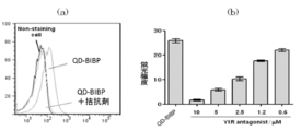
- MCF-7 cells can be labeled with QD-BIBP as shown in FIG.
- the fluorescent label was released by adding the Y1R antagonist BIBP.
- Example 5-2 Competition inhibition experiment by adding BIBP of neuropeptide Y1 receptor (Y1R) antagonist MCF-7 (3 ⁇ 10 5 cells) was suspended in 2% FBS / PBS (200 ⁇ l), A solution (2 ⁇ l) containing BIBP diluted to a concentration of 0.6 ⁇ M was added. Next, a 3-fold diluted solution (2 ⁇ l) of the QD-BIBP solution prepared in (5) of Example 5 was added and allowed to act at room temperature for 30 minutes. After washing twice with PBS, the solution was filtered with a cell strainer (BD), which is a flow cytometry tube, and measured by flow cytometry (FACS Canto TM II (BD)). As a result, as shown in FIG.
- BD cell strainer
- Example 6 Production of hydrophilized fluorescent probe
- hydrophilized fluorescent probes hydrophilized QD-OMe and hydrophilized QD-BIBP
- a partially unreacted cyclooctane alkyne derivative remains on the surface of the fluorescent probe of the present invention, it may be adsorbed non-specifically on the cell membrane surface and the background during fluorescent labeling may increase.
- a treatment for improving hydrophilicity was performed by reacting polyethylene glycol (PEG) having an azide group with an unreacted cyclooctane alkyne, and the nonspecific adsorption suppression effect of the fluorescent probe was examined.
- hydrophilized compound 23 hydrophilized QD-BIBP
- Example 6-1 Evaluation of nonspecific adsorption inhibitory effect of fluorescent probe by hydrophilization treatment
- MCF-7 cells (2 ⁇ 10 5 cells) were suspended in 2% FBS / PBS (200 ⁇ l).
- BD cell strainer
- QD-DIB does not have BIBP which is a ligand of the neuropeptide Y1 receptor (Y1R)
- some nonspecific adsorption is observed, but QD-DIB
- the average fluorescence intensity is reduced to about half compared to QD-DIB. From this, it was found that non-specific adsorption was suppressed by subjecting unreacted cyclooctane alkyne derivative to hydrophilic treatment with PEG (lane 3 and lane 2).
- the hydrophilized QD-BIBP compound 23
- MCF-7 cells could be fluorescently labeled with high sensitivity without any decrease in average fluorescence intensity even after PEG treatment (lane 1). ).
- the fluorescence probe of the present invention can suppress non-specific adsorption by hydrophilization treatment such as PEG and can improve the S / N ratio. Therefore, the usefulness of this method is expected to be further enhanced.
- the fluorescent probe of the present invention can be said to be an excellent fluorescent probe with extremely high specificity, with non-specific binding suppressed.
- the fluorescent probe of the present invention can be used for fluorescent labeling and imaging of cells, and highly sensitive detection of target molecules such as biosensors. For example, it can be used for high-sensitivity detection using a fluorescent plate reader or flow cytometry, in-vitro imaging using a fluorescence microscope or a confocal microscope, and in-vivo in vivo.
- the fluorescent probe of the present invention can be easily and efficiently produced without using a reagent such as a condensing agent.
- a solid phase fluorescent probe such as a bead can also be easily produced.
- the target fluorescent probe can be easily purified.
Landscapes
- Chemical & Material Sciences (AREA)
- Life Sciences & Earth Sciences (AREA)
- Engineering & Computer Science (AREA)
- Organic Chemistry (AREA)
- Health & Medical Sciences (AREA)
- Molecular Biology (AREA)
- Immunology (AREA)
- Biomedical Technology (AREA)
- Urology & Nephrology (AREA)
- Hematology (AREA)
- Food Science & Technology (AREA)
- Analytical Chemistry (AREA)
- Materials Engineering (AREA)
- Cell Biology (AREA)
- Biotechnology (AREA)
- Medicinal Chemistry (AREA)
- Physics & Mathematics (AREA)
- Microbiology (AREA)
- Biochemistry (AREA)
- General Health & Medical Sciences (AREA)
- General Physics & Mathematics (AREA)
- Pathology (AREA)
- Investigating Or Analysing Materials By The Use Of Chemical Reactions (AREA)
- Investigating, Analyzing Materials By Fluorescence Or Luminescence (AREA)
- Luminescent Compositions (AREA)
Abstract
本発明は、簡便に合成することができ、細胞等の生体分子への非特異的結合が低減され、かつ、良好な蛍光強度で標識化やイメージングすることができる蛍光プローブを提供することを課題とする。また、そのような蛍光プローブを作製する方法を提供する。親水性量子ドット等の蛍光性ナノ粒子にシクロオクタンのアルキン誘導体を結合させて得られる中間体と、アジド基が導入された蛍光標識対象物に結合しうる物質を縮合させることで、細胞等の生体分子への非特異的結合が低減され、かつ、良好な蛍光強度で標識化やイメージングすることができる蛍光プローブを提供しうる。
Description
本発明は、細胞等の標識化やイメージングに有用な蛍光プローブに関する。より詳しくは、簡便に合成することができ、細胞等の生体分子への非特異的結合が低減され、かつ、良好な蛍光強度で標識化やイメージングすることができる蛍光プローブに関する。
本出願は、参照によりここに援用されるところの日本出願特願2009-269447号優先権を請求する。
抗体等のタンパク質や低分子化合物を結合させた蛍光プローブは、生体や細胞における生体分子のイメージングに有用であり、従来、フルオレセイン誘導体、ローダミン誘導体、ヒドラジド誘導体、シアニン系色素などの有機系蛍光物質が広く用いられている。
標識化やイメージングに用いる蛍光プローブは、標的分子を特異的に検出でき、標的分子以外の生体分子への非特異的な結合等による影響が無い或いは低減されていることが必要である。しかし、例えば、フルオレセイン誘導体の蛍光プローブを使用した場合、細胞等への非特異的結合があることも少なくない。また蛍光強度としても十分でないこともあり、蛍光プローブの開発は未だ十分とはいえない。
近年、蛍光物質として量子ドットを用いて蛍光プローブを作製することが提案されている(非特許文献1:S. Nie et al, Engineering Luminescent Quantum Dots for In Vivo Molecular and Cellular Imaging. Annals of Biomedical Engineering, 2006, 34, 3-14)。当該蛍光プローブの作製にあたり、蛍光標識対象物、具体的には抗体、タンパク質等の生理活性物質や低分子化合物などの物質を直接量子ドットと縮合反応していた。かかる縮合反応としては、チオール交換反応、アミノ基又はカルボン酸のアミド化等の方法が多く用いられる。しかし、生理活性を有するタンパク質等のチオール基、アミノ基、カルボン酸は、活性にも重要な官能基であるため、量子ドットとタンパク質等の縮合反応を行うに際しては、そのような重要な官能基が量子ドットと反応しないように保護する必要があった。しかしながら、量子ドットと生理活性を有するタンパク質等との縮合反応における分子設計が不十分である場合には、重要な官能基が量子ドットと結合してしまい、得られた蛍光プローブは生理活性物質の機能・性質を反映していないという問題が生じる場合があった。
アルキン誘導体とアジド化合物の縮合は、Huisgen反応としても知られる1,3-環化付加反応により行われる。Huisgen反応は、アルキン化合物の三重結合とアジド基を加熱条件により縮合させる反応である。ゲルや固相担体に8員環アルキン誘導体を結合させておき、これにアジド化した有機系蛍光色素や酵素(β-Gal)を反応させることによって、当該色素や酵素が8員環アルキン誘導体と結合して当該ゲルや固相担体に固定化(immobilization)できることが報告されている(非特許文献2:K. Ebisu et al., N-terminal Specific Point-Immobilization of Active Proteins by the One-Pot NEXT-A Method. ChemBIoChem., 2009, 10,2460-2464)。しかしながら、蛍光物質と蛍光標識対象物を8員環アルキンやアジド化合物を介して結合させるものではない。
簡便に合成することができ、細胞等の生体分子への非特異的結合が無いか、又は低減され、かつ、良好な蛍光強度で標識化やイメージングすることができる蛍光プローブの開発が望まれていた。
Annals of Biomedical Engineering, 2006, 34, 3-14
ChemBioChem., 2009, 10,2460-2464
本発明は、簡便に合成することができ、細胞等の生体分子への非特異的結合が低減され、かつ、良好な蛍光強度で標識化やイメージングすることができる蛍光プローブを提供することを課題とする。また、そのような蛍光プローブを作製する方法を提供することを課題とする。
本発明者らは上記課題を解決するためにアルキン誘導体とアジド化合物の縮合反応に着目し、鋭意研究を重ねた結果、量子ドット等の蛍光性ナノ粒子に8員環アルキン誘導体としてシクロオクタンのアルキン誘導体を結合させて得られる中間体(当該結合体を、以下「蛍光プローブ合成中間体」という。)と、アジド基が導入された蛍光標識対象物に結合しうる物質を縮合させることで、上記課題が解決しうることを見出し、本発明を完成した。
即ち本発明は、以下よりなる。
1.下記の一般式(I)で表される化合物がそのY部分で蛍光性ナノ粒子表面に結合している蛍光プローブ。

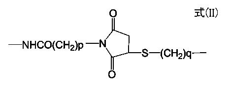
[式中、Yは、-NH-CO-W-、-W-CO-NH-、又は以下の一般式(II)で表され、これらの式中Wは酸素原子、アルキレン基、アルキレンオキシ基、アルキレンカルボニル基、又は-Ph-B-を表し、Phはフェニレン基を表し、Bはアルキレン基若しくはアルキレンオキシ基を表し、pは1~6の整数であり、qは0~6の整数であり;
環Aは、シクロオクタンの誘導体を表し、シクロオクタン部分はさらに芳香族環と縮合していてもよく;
Rは、アルキル基、アルコキシ基又はハロゲン原子を表し、nは0~2の整数であり;
Zは、蛍光標識対象物に結合しうる物質を表し;
YのNHとCOは、直接結合していても良いし、リンカーを介して結合していても良い。]

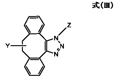
2.下記の一般式(III)で表される化合物がそのY部分で蛍光性ナノ粒子表面に結合している、前項1に記載の蛍光プローブ。
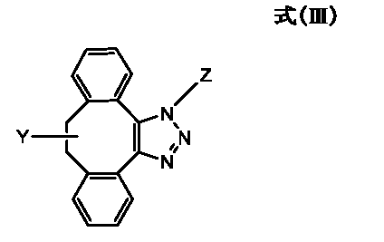
[式中、Yは、-NH-CO-W-、-W-CO-NH-、又は以下の一般式(II)で表され、これらの式中Wは酸素原子、アルキレン基、アルキレンオキシ基、アルキレンカルボニル基、又は-Ph-B-を表し、Phはフェニレン基を表し、Bはアルキレン基若しくはアルキレンオキシ基を表し、pは1~6の整数であり、qは0~6の整数であり;
Zは、蛍光標識対象物に結合しうる物質を表し;
YのNHとCOは、直接結合していても良いし、リンカーを介して結合していても良い。]
3.一般式(III)で表わされる化合物がそのY部分で蛍光性ナノ粒子表面に結合している蛍光プローブにおいて、Yが-NH-CO-O-である前項2に記載の蛍光プローブ。
4.一般式(III)で表される化合物がそのY部分で蛍光性ナノ粒子表面に結合している蛍光プローブにおいて、Zが抗体である前項2又は3に記載の蛍光プローブ。
5.一般式(III)で表される化合物がそのY部分で蛍光性ナノ粒子表面に結合している蛍光プローブにおいて、リンカーが、アルキレン基、アルキレンオキシ基又はポリアルキレンオキシ基の両末端に、アミノ基、アルキルアミノ基又はカルボン酸の基を有する化合物である請求項2~4のいずれか1に記載の蛍光プローブ。
6.下記の一般式(IV)で表される化合物がそのY部分で蛍光性ナノ粒子表面に結合している蛍光プローブ合成中間体。
[式中、Yは、-NH-CO-W-、-W-CO-NH-、又は以下の一般式(II)で表され、これらの式中Wは酸素原子、アルキレン基、アルキレンオキシ基、アルキレンカルボニル基、又は-Ph-B-を表し、Phはフェニレン基を表し、Bはアルキレン基若しくはアルキレンオキシ基を表し、pは1~6の整数であり、qは0~6の整数であり;
環A'は、シクロオクタンのアルキン誘導体を表し、シクロオクタン部分はさらに芳香族環と縮合していてもよく;
Rは、アルキル基、アルコキシ基又はハロゲン原子を表し、nは0~2の整数であり;
YのNHとCOは、直接結合していても良いし、リンカーを介して結合していても良い。]
7.下記の一般式(V)で表される化合物がそのY部分で蛍光性ナノ粒子表面に結合している、前項6に記載の蛍光プローブ合成中間体。
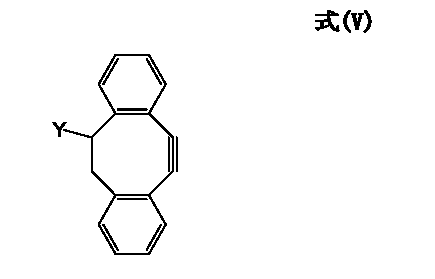
[式中、Yは、-NH-CO-W-、-W-CO-NH-、又は以下の一般式(II)で表され、これらの式中Wは酸素原子、アルキレン基、アルキレンオキシ基、アルキレンカルボニル基、又は-Ph-B-を表し、Phはフェニレン基を表し、Bはアルキレン基若しくはアルキレンオキシ基を表し、pは1~6の整数であり、qは0~6の整数であり;
YのNHとCOは、直接結合していても良いし、リンカーを介して結合していても良い。]

8.一般式(V)で表わされる化合物がそのY部分で蛍光性ナノ粒子表面に結合している蛍光プローブ合成中間体において、Yが-NH-CO-O-である前項7に記載の蛍光プローブ合成中間体。
9.一般式(V)で表される化合物がそのY部分で蛍光性ナノ粒子表面に結合している蛍光プローブ合成中間体において、リンカーが、アルキレン基、アルキレンオキシ基又はポリアルキレンオキシ基の両末端に、アミノ基、アルキルアミノ基又はカルボン酸の基を有する化合物である前項7又は8に記載の蛍光プローブ合成中間体。
10.以下の工程を含む、前項6~9のいずれか1に記載の蛍光プローブ合成中間体の作製方法:
1)カルボン酸基、アミノ基、チオール基、マレイミド基、炭酸エステル基、又は活性エステル基が導入された蛍光性ナノ粒子を作製する工程;
2)シクロオクタンのアルキン誘導体に、上記1)の工程で蛍光性ナノ粒子に導入された各種官能基と反応しうる官能基を導入して、当該官能基が導入されたシクロオクタンのアルキン誘導体を作製する工程;
3)上記1)及び2)の工程で作製された各種官能基が導入された蛍光性ナノ粒子及びシクロオクタンのアルキン誘導体を結合する工程。
11.以下の工程を含む、前項1~5のいずれか1に記載の蛍光プローブの作製方法:
A)蛍光標識対象物に結合しうる物質(Z)にアジド基を導入する工程;
B)前項6~9のいずれか1に記載の蛍光プローブ合成中間体におけるシクロオクタンのアルキン誘導体と、上記A)の工程で導入されたアジド基とを結合する工程。
12.以下の工程を含む、前項1~5のいずれか1に記載の蛍光プローブの作製方法:
A)蛍光標識対象物に結合しうる物質(Z)にアジド基を導入する工程;
B)前項6~9のいずれか1に記載の蛍光プローブ合成中間体におけるシクロオクタンのアルキン誘導体と、上記A)の工程で導入されたアジド基とを結合する工程;次いで、
C)上記B)の工程で得られた蛍光プローブを親水化処理する工程。
1.下記の一般式(I)で表される化合物がそのY部分で蛍光性ナノ粒子表面に結合している蛍光プローブ。
環Aは、シクロオクタンの誘導体を表し、シクロオクタン部分はさらに芳香族環と縮合していてもよく;
Rは、アルキル基、アルコキシ基又はハロゲン原子を表し、nは0~2の整数であり;
Zは、蛍光標識対象物に結合しうる物質を表し;
YのNHとCOは、直接結合していても良いし、リンカーを介して結合していても良い。]
2.下記の一般式(III)で表される化合物がそのY部分で蛍光性ナノ粒子表面に結合している、前項1に記載の蛍光プローブ。
Zは、蛍光標識対象物に結合しうる物質を表し;
YのNHとCOは、直接結合していても良いし、リンカーを介して結合していても良い。]
4.一般式(III)で表される化合物がそのY部分で蛍光性ナノ粒子表面に結合している蛍光プローブにおいて、Zが抗体である前項2又は3に記載の蛍光プローブ。
5.一般式(III)で表される化合物がそのY部分で蛍光性ナノ粒子表面に結合している蛍光プローブにおいて、リンカーが、アルキレン基、アルキレンオキシ基又はポリアルキレンオキシ基の両末端に、アミノ基、アルキルアミノ基又はカルボン酸の基を有する化合物である請求項2~4のいずれか1に記載の蛍光プローブ。
6.下記の一般式(IV)で表される化合物がそのY部分で蛍光性ナノ粒子表面に結合している蛍光プローブ合成中間体。
環A'は、シクロオクタンのアルキン誘導体を表し、シクロオクタン部分はさらに芳香族環と縮合していてもよく;
Rは、アルキル基、アルコキシ基又はハロゲン原子を表し、nは0~2の整数であり;
YのNHとCOは、直接結合していても良いし、リンカーを介して結合していても良い。]
YのNHとCOは、直接結合していても良いし、リンカーを介して結合していても良い。]
9.一般式(V)で表される化合物がそのY部分で蛍光性ナノ粒子表面に結合している蛍光プローブ合成中間体において、リンカーが、アルキレン基、アルキレンオキシ基又はポリアルキレンオキシ基の両末端に、アミノ基、アルキルアミノ基又はカルボン酸の基を有する化合物である前項7又は8に記載の蛍光プローブ合成中間体。
10.以下の工程を含む、前項6~9のいずれか1に記載の蛍光プローブ合成中間体の作製方法:
1)カルボン酸基、アミノ基、チオール基、マレイミド基、炭酸エステル基、又は活性エステル基が導入された蛍光性ナノ粒子を作製する工程;
2)シクロオクタンのアルキン誘導体に、上記1)の工程で蛍光性ナノ粒子に導入された各種官能基と反応しうる官能基を導入して、当該官能基が導入されたシクロオクタンのアルキン誘導体を作製する工程;
3)上記1)及び2)の工程で作製された各種官能基が導入された蛍光性ナノ粒子及びシクロオクタンのアルキン誘導体を結合する工程。
11.以下の工程を含む、前項1~5のいずれか1に記載の蛍光プローブの作製方法:
A)蛍光標識対象物に結合しうる物質(Z)にアジド基を導入する工程;
B)前項6~9のいずれか1に記載の蛍光プローブ合成中間体におけるシクロオクタンのアルキン誘導体と、上記A)の工程で導入されたアジド基とを結合する工程。
12.以下の工程を含む、前項1~5のいずれか1に記載の蛍光プローブの作製方法:
A)蛍光標識対象物に結合しうる物質(Z)にアジド基を導入する工程;
B)前項6~9のいずれか1に記載の蛍光プローブ合成中間体におけるシクロオクタンのアルキン誘導体と、上記A)の工程で導入されたアジド基とを結合する工程;次いで、
C)上記B)の工程で得られた蛍光プローブを親水化処理する工程。
本発明の蛍光プローブは、親水性量子ドット等の蛍光性ナノ粒子を用いており、有機系蛍光色素などと比べて非常に明るく光安定性に優れる蛍光プローブである。また、蛍光性ナノ粒子表面には複数のアミノ基などの反応点が存在するため、蛍光標識対象物と結合しうる物質を本発明の蛍光性ナノ粒子表面に多数導入することができる。そのため、本発明の蛍光プローブは、非特異的結合が抑えられ、極めて特異性の高い蛍光プローブということができる。本発明の蛍光プローブは、フローサイトメトリーによる解析や顕微鏡解析に利用することができる。
さらに、本発明の蛍光プローブの作製方法によれば、反応の選択性が高く、また反応効率もよいので、蛍光プローブライブラリや、目的とする蛍光プローブを容易に作製することができる。
本発明は、細胞等の標識化やイメージングに有用な蛍光プローブに関する。より詳しくは、簡便に合成することができ、細胞等の生体分子への非特異的結合が低減され、かつ、良好な蛍光強度で標識化やイメージングすることができる蛍光プローブに関する。本発明の蛍光プローブは、以下の一般式(I)に示す化合物がそのY部分で蛍光性ナノ粒子表面に結合している構造からなり、蛍光性ナノ粒子(X)、トリアゾール環と縮合したシクロオクタンの誘導体(環A)及び蛍光標識対象物に結合しうる物質(Z)から構成され、XはYを介して環Aと結合する。一般式(I)で表される化合物は蛍光性ナノ粒子表面に1以上結合している。
環Aは、シクロオクタンの誘導体を表し、シクロオクタン部分はさらにベンゼン環などの芳香族環と縮合していてもよく;
Rnは環AにR基としてn個置換されている置換基を意味し、Rは、アルキル基、アルコキシ基又はハロゲン原子を表し、nは0~2の整数であり;
Zは、蛍光標識対象物に結合しうる物質を表し;
YのNHとCOは、直接結合していても良いし、リンカーを介して結合していても良い。]

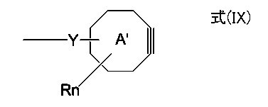
本発明は、上記蛍光プローブを作製するための蛍光プローブ合成中間体にも及ぶ。本発明の蛍光プローブ合成中間体は、以下の一般式(IV)に示す化合物がそのY部分で蛍光性ナノ粒子表面に結合している構造からなり、蛍光性ナノ粒子(X)及びシクロオクタンのアルキン誘導体(環A')から構成され、XはYを介して環A'と結合する。一般式(IV)で表される化合物は蛍光性ナノ粒子表面に1以上結合している。

環A'は、シクロオクタンのアルキン誘導体を表し、シクロオクタン部分はさらにベンゼン環などの芳香族環と縮合していてもよく;
Rnは環A'にR基としてn個置換されている置換基を意味し、Rは、アルキル基、アルコキシ基又はハロゲン原子を表し、nは0~2の整数であり;
YのNHとCOは、直接結合していても良いし、リンカーを介して結合していても良い。]
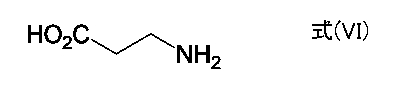
上記一般式(I)又は(IV)に表される化合物がそのY部分で蛍光性ナノ粒子表面に結合している蛍光プローブのYにおいて、NHとCOがリンカーを介して結合する場合、リンカーとはNHとCOを結合しうるものであればよく、特に限定されないが、例えば、炭素数1~20、好ましくは1~12のアルキレン基、炭素数1~6、好ましくは2~4のアルキレンオキシ基、又は炭素数2~20、好ましくは4~12のポリアルキレンオキシ基の両末端に、アミノ基、アルキルアミノ基(例えば、炭素数1~4のアルキルアミノ基)又はカルボン酸の基を有する化合物などが挙げられる。具体的には、以下の式(VI)又は式(VII)に示すような化合物が例示される。

上記一般式(I)又は(IV)に表される化合物がそのY部分で蛍光性ナノ粒子表面に結合している蛍光プローブのYのうち、Wがアルキレン基、アルキレンオキシ基又はアルキレンカルボニル基の場合、それらアルキレン基の炭素数は1~3、特に1が好ましい。また、Wが-Ph-B-の場合は、Bのアルキレン基又はアルキレンオキシ基の炭素数は1~3、特に1が好ましい。本発明においては、Wが酸素原子であるものが特に好ましい。
上記一般式(I)又は(IV)に表される化合物がそのY部分で蛍光性ナノ粒子表面に結合している蛍光プローブのRがアルキル基又はアルコキシ基の場合は、炭素数は1~3、特に1が好ましい。また、Rがハロゲン原子の場合は、フッ素原子であるのが好ましい。
好ましい蛍光プローブは、以下の一般式(III)に示される化合物がそのY部分で蛍光性ナノ粒子表面に結合しているもの、また、好ましい蛍光プローブ合成中間体は一般式(V)に示される化合物がそのY部分で蛍光性ナノ粒子表面に結合しているものが挙げられる。さらにこれらの式中、Yが-NH-CO-O-のものが好ましい。
また、本発明には、一般式(I)で示される化合物がそのY部分で蛍光性ナノ粒子表面に結合している蛍光プローブ又は蛍光プローブライブラリを製造するための蛍光プローブ合成中間体(IV)の使用も含まれる。ここで、蛍光プローブライブラリとは、一般式(I)で示される化合物がそのY部分で蛍光性ナノ粒子表面に結合している本発明の蛍光プローブにおけるZ、すなわち、蛍光標識対象物に結合しうる物質が相違する複数種のプローブを組み合わせたプローブライブラリを意味する。
以下詳細に、本発明の蛍光プローブの構成を構成する蛍光性ナノ粒子(X)、シクロオクタンの誘導体及びシクロオクタンのアルキン誘導体(環A及び環A')及び蛍光標識対象物に結合しうる物質(Z)について、各々詳述し、さらに本発明の蛍光プローブ合成中間体の作製方法、蛍光プローブの作製方法についても詳述する。
1.蛍光性ナノ粒子(X)について
本発明の蛍光プローブにおける蛍光性ナノ粒子としては、通常、40 nm以下、好ましくは10~30 nm程度の粒径を有し、紫外線やX線などの短波長の光を照射することによって蛍光を発する粒子が挙げられる。また、本発明の蛍光性ナノ粒子は、水分散性であることが好ましく、例えば粒子の表面が親水化処理されているのが好適である。例えば、半導体物質をコア(核)とし、その電子を3次元全ての方向から閉じ込めるように外側を被覆した、所謂、コアシェル構造の粒子の表面を親水化処理した蛍光性ナノ粒子である親水性量子ドットが好適である。
本発明の蛍光プローブにおける蛍光性ナノ粒子としては、通常、40 nm以下、好ましくは10~30 nm程度の粒径を有し、紫外線やX線などの短波長の光を照射することによって蛍光を発する粒子が挙げられる。また、本発明の蛍光性ナノ粒子は、水分散性であることが好ましく、例えば粒子の表面が親水化処理されているのが好適である。例えば、半導体物質をコア(核)とし、その電子を3次元全ての方向から閉じ込めるように外側を被覆した、所謂、コアシェル構造の粒子の表面を親水化処理した蛍光性ナノ粒子である親水性量子ドットが好適である。
本発明において親水性量子ドットとは、例えばセレン化カドミウム(CdSe)などの半導体をコアとし、硫化亜鉛(ZnS)などで被覆したコアシェル構造の数 nm程度の大きさのナノ粒子の表面を、さらに親水化処理して得られる、通常、40 nm以下、好ましくは10~30 nm程度の粒径を有するものである。親水性量子ドットは、有機系蛍光色素や蛍光タンパク質に比べて非常に強い蛍光を持ち、そのコアシェル構造の粒子サイズに依存して異なる蛍光を発する。例えば、コアシェル構造の粒子サイズが2 nmでは青色、3 nmでは緑色、4 nmでは黄色の蛍光を発し、また5 nmでは赤色の蛍光を発する。またこのような量子ドットは、有機系蛍光色素に比べて光安定性に優れ退色しにくいこと、蛍光スペクトル幅が非常に狭いこと、一種類の光源で様々な蛍光波長で励起できる等の特徴を有する。そのため、高感度検出やマルチカラーイメージングなどバイオイメージングへの応用研究が精力的になされている材料である。
親水性量子ドットの光学特性は、コアとなる半導体物質により大きく変化する。半導体物質としては、ZnS, ZnSe, CdS, CdSe, CdTe, PbS, InAs, InP等が挙げられる。例えば、このコア半導体としてのCdSeをZnSで被覆したコアシェル構造のCdSe/ ZnS(CdSeの粒子をZnSで被覆した粒子を意味する。以下同様。)は、量子収率が高く、またフローサイトメトリーに利用できる可視光領域の蛍光を示すことから、本発明の蛍光プローブに適している。また、InPをZnSで被覆したInP/ ZnSも適している。
コアシェル構造の粒子は通常10 nm以下、好ましくは、2~8 nmの粒径のものが使用されるが、使用するコアシェル構造の粒子の粒径により、発する蛍光の波長が異なる。例えば、コアシェル構造の粒径が5~8 nmであれば、それに依存して500~800 nmの範囲の蛍光を発する量子ドットを容易にうることができるので、使用目的に応じて各種の蛍光波長を示す蛍光プローブの作製が可能である。例えば、フローサイトメトリーへの応用を考えた場合、蛍光強度が強く、マルチカラー解析で汎用されるFITCやPEと蛍光波長の重なりが少ない625 nmや655 nmの蛍光を発する親水性量子ドットが好適である。また、生体イメージングへ応用する場合には、生体の窓と呼ばれ、生体透過性が高い近赤外領域(650~1000 nm)の蛍光を発する親水性量子ドットが適する。
親水性量子ドットは、従来の有機系蛍光色素とは異なり、短波長になるほど強い蛍光を発することが特徴である。そのため、本発明の蛍光プローブをフローサイトメトリーで使用する場合は、UVレーザー(355 nm)やバイオレットレーザー(405 nm)で励起するのが好適である。
コアシェル構造の粒子は疎水性が高く水中では凝集・沈殿し易いので、種々の表面修飾法により親水化し、蛍光プローブの作製には親水化表面処理の施された親水性量子ドットを使用する。親水化処理は、例えば、メルカプト基と共にカルボキシル基やアミノ基などの親水性基を有する化合物、具体的には、メルカプト酢酸、メルカプトコハク酸、システインやジヒドロリボ酸などで当該粒子の表面を被覆し、チオール基を共有結合的に配位させることでカルボン酸を導入することにより親水性量子ドットを合成することができる。この他にも 、ポリアクリル酸エステル、ポリアクリル酸ブチル、ポリメタクリル酸を構成成分とする両親媒性ポリマーやシリカで被覆する方法、リン脂質などの界面活性剤を用いてミセル化する方法などがある。
さらに、親水性量子ドットの表面をポリエチレングリコール(PEG)などのポリエーテル等で修飾することで、非特異的な吸着を抑制することができるので好適である。このような親水性量子ドットとして、Qdot(R)ナノクリスタルやeFluorTMナノクリスタル等がInvitrogen社やEvident Technologies社で市販されており、入手することができる。
本発明の蛍光プローブには、上述のようにコアシェル構造の粒子を親水化処理し、また適宜、非特異的な吸着の抑制処理をした、通常40 nm以下、好ましくは、10~30 nm程度の粒径の蛍光性ナノ粒子が好適に使用できる。
本発明においては、シクロオクタンのアルキン誘導体と上記蛍光性ナノ粒子を結合するために、蛍光性ナノ粒子の表面にカルボン酸基、アミノ基、チオール基、マレイミド基、炭酸エステル基、又は活性エステル基が導入されたものを使用するが、上記親水化処理によってアミノ基やカルボン酸基等が導入されているものは、それをそのまま使用することができる。
2.環A及び環A'について
式(I)の本発明の蛍光プローブにおける環Aは、トリアゾール環と縮合したシクロオクタンの誘導体であり、例えば、図1-1の(a)~(f)に示すようなものが挙げられる。また、式(IV)の中間体における環A'は、シクロオクタンのアルキン誘導体であり、例えば、図1-2の(a-1)~(f-1)に示すようなものが挙げられる。
式(I)の本発明の蛍光プローブにおける環Aは、トリアゾール環と縮合したシクロオクタンの誘導体であり、例えば、図1-1の(a)~(f)に示すようなものが挙げられる。また、式(IV)の中間体における環A'は、シクロオクタンのアルキン誘導体であり、例えば、図1-2の(a-1)~(f-1)に示すようなものが挙げられる。
式(IV)の中間体における「-Y-環A'」の部分は、例えば、図1-3の(a-1-1)、(a-1-2)、(b-1-1)、(c-1-1)、(d-1-1)、(d-1-2)、(e-1-1)、(f-1-1)に示すようなシクロオクタンのアルキン誘導体を使用することによって形成される。さらに、上記「-Y-環A'」のアルキン部分にアジド基を有する化合物を反応させることにより、式(I)の蛍光プローブにおける「-Y-環A-」の部分が形成される。
ここで、アルキンとは、分子内に炭素間三重結合を1個だけ有する炭化水素の総称である。本発明で使用できるシクロオクタンのアルキン誘導体の原料化合物としては特に限定されないが、中でも、大きな反応速度を示し、安定性に優れる図1-3の(f-1-1)に記載した3-ヒドロキシ-1,2:5,6-ジベンゾシクロオクタ-1,5,7-トリエン(3-hydroxy-1,2:5,6-dibenzocycloocta-1,5,7-triene)(DIB)(X. Ning et al., Angew. Chem. Int. Ed., 2008, 47, 2253)が好適に使用できる。また、シクロオクタンのアルキン部分の隣接位にフッ素原子を2つ導入し、反応性を改善することで、上記化合物と同程度の反応速度を示す図1-3の(c-1-1)、(d-1-1)、(d-1-2)に記載したジフルオロ シクロオクチン(difluorinated cyclooctyne)誘導体 (J.A. Codelli, et al., JACS 2008, 130, 11486.)も好適に使用できる。
3.蛍光標識対象物に結合しうる物質(Z)について
本発明の蛍光プローブにより標識される蛍光標識対象物は、細胞、組織、タンパク質、ペプチド、遺伝子、糖鎖等が挙げられ、特に限定されるものではない。このような蛍光標識対象物に結合しうる物質(Z)としては、蛍光標識対象物質に結合しうる物質であればよく、低分子化合物であってもよいし、タンパク質、糖質、核酸等の高分子物質であってもよく、特に限定されない。例えば、蛍光標識対象物が組織や細胞の場合は、Zは当該組織や細胞の表面に発現している物質と相互作用しうる物質が挙げられる。蛍光標識対象物がタンパク質の場合は、Zは当該タンパク質を特異的に認識しうる抗体、ペプチド、又は当該タンパク質の受容体や当該タンパク質に結合しうる低分子化合物等が挙げられ、例えば蛍光標識対象物が7回膜貫通型受容体(GPCR)などの細胞表面受容体の場合は、Zは当該受容体等に結合する化合物、具体的には、ヒスタミン受容体に結合するジフェンヒドラミンやメピラミン等の拮抗剤等が挙げられる。蛍光標識対象物がストレプトアビジンの場合はZとしてビオチン(Biotin)が挙げられる。特に抗体は好適に使用できる。
本発明の蛍光プローブにより標識される蛍光標識対象物は、細胞、組織、タンパク質、ペプチド、遺伝子、糖鎖等が挙げられ、特に限定されるものではない。このような蛍光標識対象物に結合しうる物質(Z)としては、蛍光標識対象物質に結合しうる物質であればよく、低分子化合物であってもよいし、タンパク質、糖質、核酸等の高分子物質であってもよく、特に限定されない。例えば、蛍光標識対象物が組織や細胞の場合は、Zは当該組織や細胞の表面に発現している物質と相互作用しうる物質が挙げられる。蛍光標識対象物がタンパク質の場合は、Zは当該タンパク質を特異的に認識しうる抗体、ペプチド、又は当該タンパク質の受容体や当該タンパク質に結合しうる低分子化合物等が挙げられ、例えば蛍光標識対象物が7回膜貫通型受容体(GPCR)などの細胞表面受容体の場合は、Zは当該受容体等に結合する化合物、具体的には、ヒスタミン受容体に結合するジフェンヒドラミンやメピラミン等の拮抗剤等が挙げられる。蛍光標識対象物がストレプトアビジンの場合はZとしてビオチン(Biotin)が挙げられる。特に抗体は好適に使用できる。
4.蛍光プローブ合成中間体の作製方法について
本発明の蛍光プローブを作製するために蛍光性ナノ粒子とシクロオクタンのアルキン誘導体の結合体を作製する。この本発明の蛍光プローブ合成中間体は、少なくとも以下の1)~3)の工程を含む方法で作製することができる。本発明は、当該蛍光プローブ合成中間体にも及び、その作製方法にも及ぶ。
本発明の蛍光プローブを作製するために蛍光性ナノ粒子とシクロオクタンのアルキン誘導体の結合体を作製する。この本発明の蛍光プローブ合成中間体は、少なくとも以下の1)~3)の工程を含む方法で作製することができる。本発明は、当該蛍光プローブ合成中間体にも及び、その作製方法にも及ぶ。
1 )カルボン酸基、アミノ基、チオール基、マレイミド基、炭酸エステル基、又は活性エステル基が導入された蛍光性ナノ粒子を作製する工程;
2)上記1)の工程で蛍光性ナノ粒子に導入された各種官能基と反応しうる官能基が導入されたシクロオクタンのアルキン誘導体を作製する工程;
3)上記1)及び2)の工程で各種官能基が導入された蛍光性ナノ粒子とシクロオクタンのアルキン誘導体を、縮合反応又はリンカーを介した反応により結合する工程。
2)上記1)の工程で蛍光性ナノ粒子に導入された各種官能基と反応しうる官能基が導入されたシクロオクタンのアルキン誘導体を作製する工程;
3)上記1)及び2)の工程で各種官能基が導入された蛍光性ナノ粒子とシクロオクタンのアルキン誘導体を、縮合反応又はリンカーを介した反応により結合する工程。
上記において、工程1)と2)の順序は、何れが先であってもよい。例えば2)の工程を先に行なう場合は、シクロオクタンのアルキン誘導体に、カルボン酸基、アミノ基、チオール基、マレイミド基、炭酸エステル基、又は活性エステル基を導入するなどによってこれら官能基が導入されたシクロオクタンのアルキン誘導体を作製する工程とし、当該工程でシクロオクタンのアルキン誘導体に導入された各種官能基と反応しうる官能基が導入された蛍光性ナノ粒子を作製する工程、と解釈して作製することができる。
蛍光性ナノ粒子とシクロオクタンのアルキン誘導体を結合するには、シクロオクタンのアルキン誘導体及び蛍光性ナノ粒子にそれぞれ反応しうる組み合わせの官能基を導入し、それらを反応させることによって結合することができる。当該官能基としては、カルボン酸基、アミノ基、チオール基、マレイミド基、炭酸エステル基、活性エステル基などが好適であり、これらの官能基を組み合わせて導入することができる。かかる組み合わせとしては、カルボン酸基とアミノ基、マレイミド基とチオール基、アミノ基と炭酸エステル基、アミノ基と活性エステル基が挙げられる。例えば、以下の表1に示す組み合わせが考えられる。また、図2にこれらの反応例を模式的に示す。

上記官能基を導入する方法は、特に限定されず、自体公知の方法、又は今後開発されるあらゆる方法を適用することができる。例えば、メルカプト酢酸やジヒドロリボ酸などのチオール基をコアシェル構造の粒子の表面に共有結合的に配位させる方法などによりカルボン酸を導入できる。また、ポリエチレンイミン等のポリマーで被覆することでコアシェル構造の粒子の表面にアミノ基を導入することができる。また、4-マレイミド酪酸サクシニミジルエステルなどの架橋試薬を用いてコアシェル構造の粒子の表面のアミノ基と縮合することでマレイミド基を導入することができる。もとより上述の親水化処理によってアミノ基やカルボン酸基などが導入されている蛍光性ナノ粒子はそれをそのまま使用することができる。
蛍光性ナノ粒子及びシクロオクタンのアルキン誘導体が、表1又は図2のような官能基を有する場合はそれを直接使用して結合させることができる。例えば、前述のDIBのようなヒドロキシ基を有するシクロオクタンのアルキン誘導体の場合には、当該シクロオクタンのアルキン誘導体を炭酸N,N'-ジサクシニミジルと縮合反応することによって炭酸エステル基を導入することができる。さらに、この炭酸エステル基に、両端にアミノ基を有するリンカー、例えば炭素数1~20、好ましくは炭素数1~12のアルキレン基又はポリエチレングリコール(PEG)などのアルキレンオキシ基を介してアミノ基を有するリンカーを反応させることによってアミノ基を導入することができる。また、一端にアミノ基もう一方にカルボン酸基又はチオール基を有するリンカーを反応させるとカルボン酸基又はチオール基を導入することができる(図2参照)。
蛍光性ナノ粒子とシクロオクタンのアルキン誘導体の反応は、蛍光性ナノ粒子1 molに対してシクロオクタンのアルキン誘導体を過剰量、通常、20~500 mol、好ましくは50~300 molの割合で反応させる。なお説明を簡便にするために、本明細書に示す反応式、一般式及び図2~5、12、15、17の反応模式図において、蛍光性ナノ粒子表面の1つの反応点(アミノ基など)における反応等について説明しているが、本発明の蛍光性ナノ粒子表面には、通常、複数の反応点が存在する。例えば市販されている蛍光性ナノ粒子表面、例えば親水性量子ドットでは表面に80~100個程度のアミノ基を有するものを入手することができる。よって本発明は、前記一般式(I)及び一般式(IV) で表される化合物がそのY部分で蛍光性ナノ粒子表面に結合している蛍光プローブ及び蛍光プローブ中間体において、それぞれ下記一般式(VIII)及び一般式(IX)の部分が蛍光性ナノ粒子Xの表面に対して複数個結合している場合を包含する。

5.蛍光プローブの作製方法について
本発明の蛍光プローブは、本発明の「蛍光プローブ合成中間体」と「蛍光標識対象物に結合しうる物質(Z)」を結合することにより作製することができる。本発明の蛍光プローブ合成中間体は、少なくとも以下のA)及びB)の工程を含む方法で作製することができる。本発明は、蛍光プローブの作製方法にも及ぶ。
本発明の蛍光プローブは、本発明の「蛍光プローブ合成中間体」と「蛍光標識対象物に結合しうる物質(Z)」を結合することにより作製することができる。本発明の蛍光プローブ合成中間体は、少なくとも以下のA)及びB)の工程を含む方法で作製することができる。本発明は、蛍光プローブの作製方法にも及ぶ。
A)蛍光標識対象物に結合しうる物質(Z)にアジド基を導入する工程;
B)上記A)で導入されたアジド基と蛍光プローブ合成中間体におけるシクロオクタンのアルキン誘導体とを反応させる工程。
B)上記A)で導入されたアジド基と蛍光プローブ合成中間体におけるシクロオクタンのアルキン誘導体とを反応させる工程。
A)蛍光標識対象物に結合しうる物質(Z)にアジド基を導入する方法
本発明の蛍光標識対象物に結合しうる物質(Z)にアジド基を導入する方法は、自体公知の方法、又は今後開発されるあらゆる方法を適用することができ、特に限定されるものではない。例えば、アジ化物イオンの高い求核性を利用してハロゲン化アルキルやスルホン酸エステルなどに対するSN2反応により導入することができる。また、アジド基とアミノ基を同一分子内に有する化合物を用いれば、低分子化合物中のカルボン酸との縮合反応により導入することができる。アミノ基以外にカルボン酸やアルデヒドなどの官能基を同一分子内に有する化合物を用いれば、低分子化合物中のアミノ基との反応によりアジド基を導入することができる。
本発明の蛍光標識対象物に結合しうる物質(Z)にアジド基を導入する方法は、自体公知の方法、又は今後開発されるあらゆる方法を適用することができ、特に限定されるものではない。例えば、アジ化物イオンの高い求核性を利用してハロゲン化アルキルやスルホン酸エステルなどに対するSN2反応により導入することができる。また、アジド基とアミノ基を同一分子内に有する化合物を用いれば、低分子化合物中のカルボン酸との縮合反応により導入することができる。アミノ基以外にカルボン酸やアルデヒドなどの官能基を同一分子内に有する化合物を用いれば、低分子化合物中のアミノ基との反応によりアジド基を導入することができる。
物質Zとして抗体を使用する場合、後述の実施例に示すように結合に関与しないアミノ基を標識することができる。また、抗体は、分子内にジスルフィド結合を有するので、これを還元して得られるSH基を標識に利用することができる。こうして得られたSH基は抗原認識とは無関係のため、SH基を利用しても抗体の特異性が損なわれない利点がある。まず、抗体分子をタンパク分解酵素ペプシンおよびパパインで処理することにより、Fabフラグメントを作製する。こうして作製したFabフラグメントはSH基を有しており、次の反応に利用できる。アジド基とマレイミド基を分子内に有する架橋試薬を用いてFabフラグメントを処理すると、Fabフラグメント中のSH基とマレイミド基との反応が選択的に進行し、アジド化されたFabフラグメントの合成ができる。これをシクロオクタンのアルキン誘導体と反応することにより、標識化抗体が合成できる。また、例えば、特表2008-500961号公報に記載された方法に従って抗体の鎖間チオールを還元し、SH基を標識することができる。
以下、アジド基が導入された蛍光標識対象物に結合しうる物質を、便宜上「アジド基含有物質」という。
B)蛍光プローブ合成中間体とアジド基含有物質との縮合反応
蛍光プローブ合成中間体とアジド基含有物質との縮合反応は、上記A)で蛍光標識対象物に導入されたアジド基と蛍光プローブ合成中間体におけるシクロオクタンのアルキン誘導体とを縮合反応させることにより行うことができる。当該シクロオクタンのアルキン誘導体とアジド基の反応は、自体公知の方法、例えばJACS 2004, 126, 15046や非特許文献2に記載の方法、又は今後開発されるあらゆる方法を適用することができる。シクロオクタンのアルキン誘導体とアジド基の反応は非常に効率的であるため、本発明の蛍光プローブ合成中間体とアジド基含有物質は、室温で数時間反応させるのみで縮合反応させることができ、特に縮合剤などの試薬を用いなくとも、蛍光プローブを容易に作製することができる。
蛍光プローブ合成中間体とアジド基含有物質との縮合反応は、上記A)で蛍光標識対象物に導入されたアジド基と蛍光プローブ合成中間体におけるシクロオクタンのアルキン誘導体とを縮合反応させることにより行うことができる。当該シクロオクタンのアルキン誘導体とアジド基の反応は、自体公知の方法、例えばJACS 2004, 126, 15046や非特許文献2に記載の方法、又は今後開発されるあらゆる方法を適用することができる。シクロオクタンのアルキン誘導体とアジド基の反応は非常に効率的であるため、本発明の蛍光プローブ合成中間体とアジド基含有物質は、室温で数時間反応させるのみで縮合反応させることができ、特に縮合剤などの試薬を用いなくとも、蛍光プローブを容易に作製することができる。
6.細胞の蛍光標識及びイメージング方法の説明
本発明の蛍光プローブは、細胞の蛍光標識やイメージング、そしてバイオセンサー等の標的分子の高感度検出などに用いることができる。例えば、蛍光プレートリーダーやフローサイトメトリーによる高感度検出、蛍光顕微鏡や共焦点顕微鏡などによるin vitroイメージング、そして生体in vivoイメージングなどに利用することができる。
本発明の蛍光プローブは、細胞の蛍光標識やイメージング、そしてバイオセンサー等の標的分子の高感度検出などに用いることができる。例えば、蛍光プレートリーダーやフローサイトメトリーによる高感度検出、蛍光顕微鏡や共焦点顕微鏡などによるin vitroイメージング、そして生体in vivoイメージングなどに利用することができる。
本発明の蛍光プローブにおいて、未反応のシクロオクタンのアルキン誘導体が残っていると、細胞膜表面に非特異的に吸着してバックグラウンドが高くなることがある。その理由の1つとして、疎水性の高いDIB基の分子構造に起因しているものと推測される。そこで、例えば、後述の実施例に示すように、アジド化したPEG等のポリアルキレングリコールなどで、未反応のシクロオクタンのアルキン誘導体部分を親水化処理することにより、その非特異的吸着を抑制することができるので好ましい。
以下に、本発明の理解を深めるために、実施例により本発明を具体的に説明するが、本発明はこれらに限定されるものではなく、本発明の技術的思想を逸脱しない範囲内で種々の応用が可能である。
(実施例1)蛍光プローブ合成中間体(QD-DIB)の作製
本実施例では、図3の合成スキームを参照し、蛍光プローブ合成中間体の作製について説明する。
本実施例では、図3の合成スキームを参照し、蛍光プローブ合成中間体の作製について説明する。
(1)化合物1~3の合成(図3)
化合物1~3は、既知文献(X. Ning et al., Angew. Chem. Int. Ed. 2008, 47, 2253)に従い、合成した。
化合物1~3は、既知文献(X. Ning et al., Angew. Chem. Int. Ed. 2008, 47, 2253)に従い、合成した。
(2)化合物4の合成(図3)
化合物3 (22 mg, 0.10 mmol)のジメチルフォルムアミド(DMF)溶液(1 ml)に、4-ジメチルアミノピリジン (12 mg, 0.10 mmol)と炭酸N,N'-ジサクシニミジル (77 mg, 0.30 mmol)を加えた。室温で12時間攪拌した後、反応液に1 Nの塩酸溶液を加え、酢酸エチルで抽出した。有機溶剤層を飽和食塩水で洗浄後、無水硫酸マグネシウムで乾燥し、溶剤を減圧下で留去して、粗生成物を得た。これをシリカゲルクロマトグラフィーにより精製し、化合物4を26 mg (収率 73%)で得た。
化合物3 (22 mg, 0.10 mmol)のジメチルフォルムアミド(DMF)溶液(1 ml)に、4-ジメチルアミノピリジン (12 mg, 0.10 mmol)と炭酸N,N'-ジサクシニミジル (77 mg, 0.30 mmol)を加えた。室温で12時間攪拌した後、反応液に1 Nの塩酸溶液を加え、酢酸エチルで抽出した。有機溶剤層を飽和食塩水で洗浄後、無水硫酸マグネシウムで乾燥し、溶剤を減圧下で留去して、粗生成物を得た。これをシリカゲルクロマトグラフィーにより精製し、化合物4を26 mg (収率 73%)で得た。
1H-NMR (CDCl3, 400 MHz) δ: 7.61 (1H, d, J = 8.0 Hz), 5.53 (1H, br), 3.32 (1H, dd, J = 2.0, 15.6 Hz), 3.03 (1H, dd, J = 4.0, 15.6 Hz), 2.81 (4H, s).
(3)化合物5(QD-DIB)の作製(図3)
蛍光プローブ合成中間体(QD-DIB)を構成する蛍光ナノ粒子として、Qdot(R) 655 ITK AMINO (PEG) (表面にアミノ基を有するCdSe/ZnS:Invitrogen社)を用いた。蛍光ナノ粒子(25μl, 0.20 nmol)のPBS(リン酸緩衝生理食塩水)(100μl)に、上記化合物4を含む10 mM DMSO溶液 (5μl, 50 nmol) を加えた。室温で16時間静置した後、前記反応液にPBS (400μl)を加え、遠心ろ過用デバイス(10K Nanosep(R)フィルター (Paul製))に移した。4℃、7000回転で10分間の遠心処理による洗浄操作を3回行い、過剰の化合物4を取り除いた。遠心ろ過用デバイスから蛍光プローブ合成中間体(化合物5)を含む溶液を回収し、PBS を適量加えて50μlに調製した。以降、実施例1で作製した蛍光プローブ合成中間体(化合物5)を、単に「QD-DIB」ともいう。
蛍光プローブ合成中間体(QD-DIB)を構成する蛍光ナノ粒子として、Qdot(R) 655 ITK AMINO (PEG) (表面にアミノ基を有するCdSe/ZnS:Invitrogen社)を用いた。蛍光ナノ粒子(25μl, 0.20 nmol)のPBS(リン酸緩衝生理食塩水)(100μl)に、上記化合物4を含む10 mM DMSO溶液 (5μl, 50 nmol) を加えた。室温で16時間静置した後、前記反応液にPBS (400μl)を加え、遠心ろ過用デバイス(10K Nanosep(R)フィルター (Paul製))に移した。4℃、7000回転で10分間の遠心処理による洗浄操作を3回行い、過剰の化合物4を取り除いた。遠心ろ過用デバイスから蛍光プローブ合成中間体(化合物5)を含む溶液を回収し、PBS を適量加えて50μlに調製した。以降、実施例1で作製した蛍光プローブ合成中間体(化合物5)を、単に「QD-DIB」ともいう。
(実施例2)蛍光プローブ(QD-Biotin)の作製
本実施例では、図4の合成スキームを参照し、QD-DIB(化合物5)に蛍光標識対象物に結合しうる物質(ビオチン)を結合した蛍光プローブ(化合物7)の作製について説明する。
本実施例では、図4の合成スキームを参照し、QD-DIB(化合物5)に蛍光標識対象物に結合しうる物質(ビオチン)を結合した蛍光プローブ(化合物7)の作製について説明する。
ビオチン(biotin) (244 mg, 1.0 mmol)と14-アジド-3,6,9,12-テトラオキサテトラデカン-1-アミン(14-azido-3,6,9,12-tetraoxatetradecan-1-amine) (524 mg, 2.0 mmol)の MeOH-MeCN (1: 3) (12 ml)混合溶液に、EDC (N-ethyl-N'-3- (dimethylaminopropyl) carbodiimide) (288 mg, 1.5 mmol)を加えた。室温で42時間攪拌した後、溶剤を減圧下で留去して、粗生成物を得た。これをシリカゲルクロマトグラフィーにより精製し、化合物6を493 mg (収率 100 %)で得た。
1H-NMR (CDCl3, 400 MHz) δ: 6.63 (1H, brt, J = 4.8 Hz), 5.86 (1H, s), 4.98 (1H, s), 4.50-4.45 (1H, m), 4.32-4.28 (1H, m), 3.70-3.56 (14H, m), 3.54 (2H, t, J = 4.8 Hz), 3.43 (2H, t, J = 4.8 Hz), 3.37 (2H, t, J = 4.8 Hz),3.14-3.10 (1H, m), 2.90 (1H, dd, J = 4.8, 13.2 Hz), 2.71 (1H, d, J =13.2 Hz), 2.21 (2H, dt, J = 2.8, 7.6 Hz), 1.78-1.60 (4H, m), 1.43 (2H, quin, J = 7.6 Hz)
実施例1で調製したQD-DIB(10μl, 0.040 nmol)を含むPBS (100μl)に、前記の化合物6を含む10 mM DMSO溶液 (5μl, 50 nmol)を加えた。室温で2時間静置した後、反応液にPBS (400μl)を加え、遠心ろ過用デバイス(10K Nanosep(R)フィルター (Paul製))に移した。4℃、7000回転で10分間の遠心処理による洗浄操作を3回行い、過剰の化合物6を除去した。遠心ろ過用デバイスから蛍光プローブのQD-Biotin(化合物7)を含む溶液を回収し、PBS を適量加えて50μlに調製した。以降、実施例2で作製した蛍光プローブのQD-Biotin(化合物7)を、単に「QD-Biotin」ともいう。
(実施例3)蛍光プローブ(QD-NP)の作製
本実施例では、図5の合成スキームを参照し、QD-DIB(化合物5)に蛍光標識対象物に結合しうる物質(ニトロフェニルアセチル:NP)を結合した蛍光プローブ(化合物9)の作製について説明する。
本実施例では、図5の合成スキームを参照し、QD-DIB(化合物5)に蛍光標識対象物に結合しうる物質(ニトロフェニルアセチル:NP)を結合した蛍光プローブ(化合物9)の作製について説明する。
4-ヒドロキシ-3-ニトロフェニル酢酸(4-hydroxy-3-nitrophenylacetic acid) (197 mg, 1.0 mmol)と14-アジド-3,6,9,12-テトラオキサテトラデカン-1-アミン(14-azido-3,6,9,12-tetraoxatetradecan-1-amine) (524 mg, 2.0 mmol)のCH2Cl2 (10 ml)溶液に、N,N-ジメチル-4-アミノピリジン (N,N-dimethyl-4-aminopyridine:DMAP) (183 mg, 1.5 mmol)とEDC (288 mg, 1.5 mmol)を加えた。室温で3時間攪拌した後、反応液に1 Nの塩酸溶液を加え、CH2Cl2で抽出した。有機溶剤層を飽和食塩水で洗浄後、無水硫酸マグネシウムで乾燥し、溶剤を減圧下で留去して、粗生成物を得た。これをシリカゲルクロマトグラフィーにより精製し、化合物8を512 mg (収率 100 %)で得た。
1H-NMR (CDCl3, 400 MHz) δ: 10.5 (1H, s), 8.01 (1H, d, J = 2.0 Hz), 7.55 (1H, dd, J = 2.0, 8.8 Hz), 7.11 (1H, d, J = 8.8 Hz), 6.42 (1H, br), 3.68-3.56 (14H, m), 3.53 (2H, t, J = 5.2 Hz), 3.50 (2H, s), 3.43 (2H, t, J = 5.2 Hz), 3.36 (2H, t, J = 5.2 Hz)
実施例1で調製したQD-DIB (10μl, 0.040 nmol)を含むPBS (100μl)に、前記の化合物8を含む10 mM DMSO溶液 (5μl, 50 nmol)を加えた。室温で2時間静置した後、反応液にPBS (400μl)を加え、遠心ろ過用デバイス(10K Nanosep(R)フィルター (Paul製))に移した。4℃、7000回転で10分間の遠心処理による洗浄操作を3回行い、過剰の化合物8を除去した。遠心ろ過用デバイスから蛍光プローブのQD-NP(化合物9)を含む溶液を回収し、PBS を適量加えて50μlに調製した。以降、実施例3で作製した蛍光プローブのQD-NP(化合物9)を、単に「QD-NP」ともいう。
(実験例2-1)ビオチン-ストレプトアビジン相互作用による蛍光プローブ(QD-Biotin)の評価
QD-Biotinを活用した標識法について評価するため、ビオチン-ストレプトアビジン相互作用を利用したアッセイ系を用いて解析を行った。本実験例では、実施例2で作製したQD-Biotinについて、ストレプトアビジンをコートしたプレートへの蛍光プローブの結合効果を確認することにより、QD-Biotinの機能を評価した。ストレプトアビジンをコートした96 wellプレート (Thermo社)をPBSで洗浄した後、2%BSA/ PBS (100μl)を用いてブロッキングした。その後、実施例2で調製したQD-Biotin溶液そのものを2μl加え、室温で60分間作用させた。0.1% Tween-20/ PBSで2回洗浄した後、マルチプレートリーダー(FlexStationTM 、Molecular Devices社)で蛍光測定を行った。対照として、実施例1で調製したQD-DIB溶液を5倍に希釈した蛍光プローブ合成中間体溶液を同様に作用させた。
QD-Biotinを活用した標識法について評価するため、ビオチン-ストレプトアビジン相互作用を利用したアッセイ系を用いて解析を行った。本実験例では、実施例2で作製したQD-Biotinについて、ストレプトアビジンをコートしたプレートへの蛍光プローブの結合効果を確認することにより、QD-Biotinの機能を評価した。ストレプトアビジンをコートした96 wellプレート (Thermo社)をPBSで洗浄した後、2%BSA/ PBS (100μl)を用いてブロッキングした。その後、実施例2で調製したQD-Biotin溶液そのものを2μl加え、室温で60分間作用させた。0.1% Tween-20/ PBSで2回洗浄した後、マルチプレートリーダー(FlexStationTM 、Molecular Devices社)で蛍光測定を行った。対照として、実施例1で調製したQD-DIB溶液を5倍に希釈した蛍光プローブ合成中間体溶液を同様に作用させた。
上記の結果、QD-Biotinでストレプトアビジンをコートしたプレートを処理すると、大きな蛍光強度を示した。一方、対照として蛍光プローブ合成中間体のQD-DIBで処理した場合は、蛍光強度はほとんど示さなかった(図6)。上記により、本発明の蛍光プローブの作製方法により、蛍光ナノ粒子の表面に、蛍光標識対象物に結合しうる物質であるビオチンを導入することに成功したことが分かった。
(実験例3-1)フローサイトメトリーによる蛍光プローブ(QD-NP)の評価1
本実験例では、抗原としてNP(ニトロフェニルアセチル)を特異的に認識する免疫グロブリンIgG1を高発現するA20細胞(A20/ BCR)を用いて、実施例3で作製したQD-NPの評価を行なった。A20細胞(A20/ BCR) (3 x 10⁵ cells)を2% FBS/ PBS (200μl)に懸濁し、実施例3で調製したQD-NP溶液を10倍に希釈した蛍光プローブ溶液(2μl)を加え、氷上で30分間作用させた。対照として、実施例1で調製したQD-DIB溶液を50倍に希釈した蛍光プローブ合成中間体溶液を同様に作用させた。PBS で2回洗浄した後、フローサイトメトリー用チューブであるセルストレーナー(ベクトン・ディッキンソン(BD)製)で濾過し、フローサイトメトリー(FACS CantoTM II (BD製))で測定を行った。非特異的結合検出用に、非標識化NP (100μM)を加えて同様の実験を行った。全結合値から非特異的結合分を差し引くことにより特異的結合を算出した。
本実験例では、抗原としてNP(ニトロフェニルアセチル)を特異的に認識する免疫グロブリンIgG1を高発現するA20細胞(A20/ BCR)を用いて、実施例3で作製したQD-NPの評価を行なった。A20細胞(A20/ BCR) (3 x 10⁵ cells)を2% FBS/ PBS (200μl)に懸濁し、実施例3で調製したQD-NP溶液を10倍に希釈した蛍光プローブ溶液(2μl)を加え、氷上で30分間作用させた。対照として、実施例1で調製したQD-DIB溶液を50倍に希釈した蛍光プローブ合成中間体溶液を同様に作用させた。PBS で2回洗浄した後、フローサイトメトリー用チューブであるセルストレーナー(ベクトン・ディッキンソン(BD)製)で濾過し、フローサイトメトリー(FACS CantoTM II (BD製))で測定を行った。非特異的結合検出用に、非標識化NP (100μM)を加えて同様の実験を行った。全結合値から非特異的結合分を差し引くことにより特異的結合を算出した。
なお、上記A20細胞(A20/ BCR)は定法に準じて次のようにして作製した。すなわち、NP化ニワトリγグロブリンを免疫したマウスの脾臓B細胞からRNAを抽出し、免疫グロブリン遺伝子座(VH186.2)の可変部をPCR法にてクローニングした。また、抗体を構成するλ1軽鎖全長及びγ1重鎖の定常部を同様にPCR法でクローニングした。その後、クローニング済みのVH186.2可変部をPCR法を用いてγ1定常領域に接続することにより、NPハプテンを認識する全長γ1重鎖遺伝子を得た。γ2b重鎖を恒常的に発現しているマウスB細胞腫瘍細胞であるA20細胞に得られたλ1軽鎖及び全長γ1重鎖の遺伝子をレトロウイルス法で導入した。NPハプテンを認識する抗原受容体が再構成されたことは、NPハプテンを結合したフィコエリスリン化牛血清アルブミンによる染色法で、フローサイトメトリー(Facs ARIATM (BD製))により確認した。
QD-NP又は対照のQD-DIBで処理したA20細胞(A20/ BCR)について、フローサイトメトリーによるFACS解析を行った結果、QD-NPで処理した場合は、対照のQD-DIBで処理した場合に比べて蛍光強度が約35倍増大した。上記により、本発明の蛍光プローブ(QD-NP)は、細胞標識に利用可能な蛍光プローブであることが確認された(図7)。
(実験例3-2)蛍光顕微鏡イメージングによる蛍光プローブ(QD-NP)の評価
実験例3-1で使用したA20細胞(A20/ BCR)(1 x 10⁶ cells)を2% FBS/ PBS (800μl)に懸濁し、実施例3で調製したQD-NP溶液を10倍に希釈した蛍光プローブ溶液(8μl)を加え、氷上で30分間作用させた。PBS で2回洗浄した後、2%ホルムアルデヒド/ PBS (200μl)を加え10分固定した。2% FBS/ PBS (8μl)を室温で10分作用させた後に、PBS で2回洗浄した。次に、得られたA20細胞(A20/ BCR)(2 x 10⁴ cells)を集細胞遠心装置(サイトスピン(Thermo製))によりスライドガラス(Matsunami製)に播種し、グリセロールによりカバーグラス下に封入して蛍光顕微鏡(Carl Zeiss製)によるイメージング観察を行った。対照として、実施例1で調製したQD-DIB溶液を50倍に希釈した蛍光プローブ合成中間体溶液を同様に作用させた。
実験例3-1で使用したA20細胞(A20/ BCR)(1 x 10⁶ cells)を2% FBS/ PBS (800μl)に懸濁し、実施例3で調製したQD-NP溶液を10倍に希釈した蛍光プローブ溶液(8μl)を加え、氷上で30分間作用させた。PBS で2回洗浄した後、2%ホルムアルデヒド/ PBS (200μl)を加え10分固定した。2% FBS/ PBS (8μl)を室温で10分作用させた後に、PBS で2回洗浄した。次に、得られたA20細胞(A20/ BCR)(2 x 10⁴ cells)を集細胞遠心装置(サイトスピン(Thermo製))によりスライドガラス(Matsunami製)に播種し、グリセロールによりカバーグラス下に封入して蛍光顕微鏡(Carl Zeiss製)によるイメージング観察を行った。対照として、実施例1で調製したQD-DIB溶液を50倍に希釈した蛍光プローブ合成中間体溶液を同様に作用させた。
QD-NP又は対照のQD-DIBで処理したA20細胞(A20/ BCR)について蛍光顕微鏡によるイメージング観察を行った結果、QD-NPではA20細胞(A20/ BCR)が染色されたが、対照のQD-DIBでは染色されなかった(図8)。上記により、本発明の蛍光プローブ(QD-NP)は、非特異的吸着のようなバックグランドが少なく、蛍光顕微鏡による細胞のイメージングに効果的に活用できることが分かった。
(実験例3-3)フローサイトメトリーによる蛍光プローブ(QD-NP)の評価2
実験例3-1で使用したIgG1を高発現するA20細胞(A20/ BCR)と、IgG1を発現していないA20細胞(A20(-))について、各々3 x 10⁵ cellsを2% FBS/ PBS (200μl)に懸濁し、実施例3で調製したQD-NP溶液を10倍に希釈した蛍光プローブ溶液(2μl)を加え、氷上で30分間作用させた。PBS で2回洗浄した後、フローサイトメトリー用チューブであるセルストレーナー(BD製)で濾過し、フローサイトメトリー(FACS CantoTM II (BD製))で測定し、解析した。
実験例3-1で使用したIgG1を高発現するA20細胞(A20/ BCR)と、IgG1を発現していないA20細胞(A20(-))について、各々3 x 10⁵ cellsを2% FBS/ PBS (200μl)に懸濁し、実施例3で調製したQD-NP溶液を10倍に希釈した蛍光プローブ溶液(2μl)を加え、氷上で30分間作用させた。PBS で2回洗浄した後、フローサイトメトリー用チューブであるセルストレーナー(BD製)で濾過し、フローサイトメトリー(FACS CantoTM II (BD製))で測定し、解析した。
QD-NPを用いて、A20細胞(A20/ BCR)及びA20細胞(A20(-))の各細胞についてイメージング実験を行った結果、QD-NPによって、IgG1を高発現するA20細胞(A20/ BCR)が選択的に標識されることが確認された(図9)。上記により、本発明の蛍光プローブ(QD-NP)は、蛍光顕微鏡によるイメージングだけでなく、フローサイトメトリーによるイメージングにも適応できることが分かった。
(実験例3-4)フローサイトメトリーによる蛍光プローブ(QD-NP)の評価3
蛍光プローブを合成する際に最も懸念されるのは、標識化によって受容体など標的分子への特異性が損なわれることである。そこで本実験例では、本発明の蛍光プローブの特異性を確かめるために、拮抗剤を添加する競合阻害実験を行った。
蛍光プローブを合成する際に最も懸念されるのは、標識化によって受容体など標的分子への特異性が損なわれることである。そこで本実験例では、本発明の蛍光プローブの特異性を確かめるために、拮抗剤を添加する競合阻害実験を行った。
実験例3-1で使用したA20細胞(A20/ BCR)(3 x 10⁵ cells)を2% FBS/ PBS (200μl)に懸濁し、0.16~20μMの濃度に希釈した拮抗剤(NP)又は非拮抗剤(2,4-ジニトロフェノール(DNP))(2μl)で処理した。次いで、実施例3で調製したQD-NP溶液を10倍に希釈した蛍光プローブ溶液(2μl)を加え、氷上で30分間作用させた。PBS で2回洗浄した後、フローサイトメトリー用チューブであるセルストレーナー (BD製)で濾過し、フローサイトメトリー(FACS CantoTM II (BD製))で測定し、解析した。
拮抗剤として非標識のNPを加えると、添加した非標識NPの濃度に依存して蛍光強度が低下した。一方、非拮抗剤のDNPを加えた場合は、その添加濃度に関わらず、蛍光強度に大きな変化は観測されなかった(図10)。つまり、本発明の蛍光プローブ(QD-NP)はIgG1特異的であることが確認された。
(実験例3-5及び比較例)フルオレセイン誘導体の蛍光プローブを使用した場合の評価
本発明の蛍光プローブ(QD-NP)の有用性を確認するために、有機系蛍光色素を用いた蛍光プローブ(蛍光性NP)と本発明の蛍光プローブ(QD-NP)について比較実験を行った。有機系蛍光色素の代表であるフルオレセイン誘導体にリンカーを介してNPを導入し、蛍光性NPを合成した。次いで、実験例3-3で示したフローサイトメトリーによる解析方法に従い、A20細胞(A20/ BCR)を用いたFACS解析に付した。その結果、蛍光性NPで処理した場合は、拮抗剤(非標識NP)100μMの添加の有無によっても、その蛍光強度は低下することはなく、非常に高い非特異的結合が認められた。これに対し、本発明のQD-NPは、拮抗剤の有無によって明確な蛍光強度の差が見られた(図11)。上記により、本発明の蛍光プローブ(QD-NP)は、従来の有機系蛍光色素などを用いた蛍光プローブ(例えば、蛍光性NP)で大きな障害となる非特異的吸着を抑制することができ、蛍光プローブとして有用であることが分かった。
本発明の蛍光プローブ(QD-NP)の有用性を確認するために、有機系蛍光色素を用いた蛍光プローブ(蛍光性NP)と本発明の蛍光プローブ(QD-NP)について比較実験を行った。有機系蛍光色素の代表であるフルオレセイン誘導体にリンカーを介してNPを導入し、蛍光性NPを合成した。次いで、実験例3-3で示したフローサイトメトリーによる解析方法に従い、A20細胞(A20/ BCR)を用いたFACS解析に付した。その結果、蛍光性NPで処理した場合は、拮抗剤(非標識NP)100μMの添加の有無によっても、その蛍光強度は低下することはなく、非常に高い非特異的結合が認められた。これに対し、本発明のQD-NPは、拮抗剤の有無によって明確な蛍光強度の差が見られた(図11)。上記により、本発明の蛍光プローブ(QD-NP)は、従来の有機系蛍光色素などを用いた蛍光プローブ(例えば、蛍光性NP)で大きな障害となる非特異的吸着を抑制することができ、蛍光プローブとして有用であることが分かった。
(実施例4)蛍光プローブ(QD-B220)の作製
本実施例では、図12の合成スキームを参照し、化合物12(QD-B220)の作製について説明する。
受容体型チロシンホスファターゼ(PTP)に属する膜貫通型タンパク質であり、初期pro-B細胞から成熟B細胞までのB細胞lineageに発現するため、典型的なB細胞マーカーとして知られている細胞表面抗原分子であるCD45R (B220)に対する抗体(抗B220モノクローナル抗体)を用いて、蛍光プローブQD-B220を作製した。
本実施例では、図12の合成スキームを参照し、化合物12(QD-B220)の作製について説明する。
受容体型チロシンホスファターゼ(PTP)に属する膜貫通型タンパク質であり、初期pro-B細胞から成熟B細胞までのB細胞lineageに発現するため、典型的なB細胞マーカーとして知られている細胞表面抗原分子であるCD45R (B220)に対する抗体(抗B220モノクローナル抗体)を用いて、蛍光プローブQD-B220を作製した。
(1)化合物10の合成(図12)
6-アジドヘキサン酸(6-azidohexanoic acid)(1.5 g, 9.4 mmol)とN-ヒドロキシスクシンイミド(N-hydroxysuccinimide) (1.3 g, 11.2 mmol)のジクロロメタン(CH2Cl2)(50 ml)溶液に、1-(3-ジメチルアミノプロピル)-3-エチルカルボジイミド 塩酸塩(EDC-HCl) (2.2 g, 11.2 mmol)を加え、室温で2時間撹拌した。反応溶液に1 M 塩酸を加え、塩化メチレンで抽出した後、1 M 水酸化ナトリウム水溶液、飽和食塩水で洗浄、無水硫酸マグネシウムで乾燥し、溶剤を減圧下で留去して、粗精製物を得た。これをシリカゲルクロマトグラフィーで精製し、化合物10を1.4 g (収率56 %)で得た。
6-アジドヘキサン酸(6-azidohexanoic acid)(1.5 g, 9.4 mmol)とN-ヒドロキシスクシンイミド(N-hydroxysuccinimide) (1.3 g, 11.2 mmol)のジクロロメタン(CH2Cl2)(50 ml)溶液に、1-(3-ジメチルアミノプロピル)-3-エチルカルボジイミド 塩酸塩(EDC-HCl) (2.2 g, 11.2 mmol)を加え、室温で2時間撹拌した。反応溶液に1 M 塩酸を加え、塩化メチレンで抽出した後、1 M 水酸化ナトリウム水溶液、飽和食塩水で洗浄、無水硫酸マグネシウムで乾燥し、溶剤を減圧下で留去して、粗精製物を得た。これをシリカゲルクロマトグラフィーで精製し、化合物10を1.4 g (収率56 %)で得た。
1H-NMR (500 MHz, CDCl3) δ: 3.30 (2H, t, J = 7.0 Hz ), 2.84 (4H, s), 2.63 (2H, t, J = 7.5 Hz), 1.79 (2H, quin, J = 7.5 Hz), 1.64 (2H, quin, J = 7.5 Hz), 1.55-1.47 (2H, m).
(2)化合物11(アジド化B220抗体:B220-N3)の合成(図12)
抗B220モノクローナル抗体(eBioscience社製、16-0452-85)を、還元型抗体作製キット(同仁化学研究所社製、Biotin Labeling Kit-SH、製品番号LK10)を使用し、同キットのマニュアル記載の還元処理方法に従って抗B220モノクローナル抗体分子のヒンジ部にあるジスルフィド結合を切断し、還元型抗体を作製した。次に、化合物10を加え、室温で30分反応を行い、遠心濾過デバイスを用いて過剰の未反応化合物10を除去することにより、化合物11(B220-N3)を得た。
抗B220モノクローナル抗体(eBioscience社製、16-0452-85)を、還元型抗体作製キット(同仁化学研究所社製、Biotin Labeling Kit-SH、製品番号LK10)を使用し、同キットのマニュアル記載の還元処理方法に従って抗B220モノクローナル抗体分子のヒンジ部にあるジスルフィド結合を切断し、還元型抗体を作製した。次に、化合物10を加え、室温で30分反応を行い、遠心濾過デバイスを用いて過剰の未反応化合物10を除去することにより、化合物11(B220-N3)を得た。
(3)蛍光プローブ(QD-B220)の作製(図12)
実施例1で調製したQD-DIB (5μl)を含むPBS (50μl)溶液に、図12の化合物11(12.5μl)を加え、室温で3時間静置した。反応液にPBS (200μl)を加え、100K Amicon Ultra遠心デバイス(Millipore製)に移した。4℃、7000回転で10分間遠心する洗浄操作を3回行い、過剰の未反応化合物11を取り除き、蛍光標識化抗B220抗体である蛍光プローブQD-B220(化合物12)を作製した。遠心デバイスからQD-B220を含む溶液を回収し、PBSを加えて25μlに調製した。以降、実施例4で作製した蛍光プローブを、単に「QD-B220」ともいう。
実施例1で調製したQD-DIB (5μl)を含むPBS (50μl)溶液に、図12の化合物11(12.5μl)を加え、室温で3時間静置した。反応液にPBS (200μl)を加え、100K Amicon Ultra遠心デバイス(Millipore製)に移した。4℃、7000回転で10分間遠心する洗浄操作を3回行い、過剰の未反応化合物11を取り除き、蛍光標識化抗B220抗体である蛍光プローブQD-B220(化合物12)を作製した。遠心デバイスからQD-B220を含む溶液を回収し、PBSを加えて25μlに調製した。以降、実施例4で作製した蛍光プローブを、単に「QD-B220」ともいう。
(実験例4-1)フローサイトメトリーによる蛍光プローブ(QD-B220)の評価1
マウスB細胞リンパ腫であるA20細胞 (3 x 10⁵ cells)を2% FBS/ PBS (200μl)に懸濁し、それに実施例4の(3)で調製したQD-B220溶液を10倍に希釈した溶液(2μl)を加え、氷上で0.5時間作用させた。PBS で2回洗浄した後、フローサイトメトリー用チューブであるセルストレーナー (BD製)で濾過し、フローサイトメトリー(FACS CantoTM II (BD製))で測定を行った。
マウスB細胞リンパ腫であるA20細胞 (3 x 10⁵ cells)を2% FBS/ PBS (200μl)に懸濁し、それに実施例4の(3)で調製したQD-B220溶液を10倍に希釈した溶液(2μl)を加え、氷上で0.5時間作用させた。PBS で2回洗浄した後、フローサイトメトリー用チューブであるセルストレーナー (BD製)で濾過し、フローサイトメトリー(FACS CantoTM II (BD製))で測定を行った。
その結果、図13に示すように、上記実施例4で作製したQD-B220を用いた場合のみ、A20細胞は強く蛍光標識化された(lane 1)。一方、対照として、抗B220モノクローナル抗体(eBioscience社製、16-0452-85)の代わりにラットIgG2a Kアイソタイプコントロール(eBioscience社製、16-4321)を使用する以外は実施例4の(2)及び(3)と同様にして調製した蛍光標識アイソタイプコントロール(QD-Isotypecontrol)を作用させた場合(lane 2)、DIB基を持たない蛍光ナノ粒子 (QD-NH2)に B220-N3(化合物11)を反応させたものを作用させた場合(lane 3)では、A20細胞はほとんど蛍光標識されなかった。以上の結果から、蛍光ナノ粒子表面のDIB基と抗体表面のアジド基との環化付加反応は効率よく進行し、また抗体の特異性を損なうことなくA20細胞を蛍光標識できることが分かった。
(実験例4-2)フローサイトメトリーによる蛍光プローブ(QD-B220)の評価2
マウス脾臓細胞 (1.8 x 10⁶ cells)を2% FBS/ PBS (200μl)に懸濁し、実験例4-1と同様に希釈したQD-B220を含む溶液(2μl)及びPE標識CD3抗体(eBioscience社製、12-0031-82)(0.5μl)を加え、氷上で1時間作用させた。また、ポジティブコントロールとして、QD-B220の代わりにFITC標識B220抗体(FITC-B220)及びネガティブコントロールとして、QD-B220の代わりに実験4-1で使用したQD-Isotypecontrolを使用する他は同様にして氷上で1時間作用させた。それぞれPBS で2回洗浄した後、フローサイトメトリー用チューブであるセルストレーナー (BD製)で濾過し、フローサイトメトリー(FACS CantoTM II (BD製))で測定を行った。
マウス脾臓細胞 (1.8 x 10⁶ cells)を2% FBS/ PBS (200μl)に懸濁し、実験例4-1と同様に希釈したQD-B220を含む溶液(2μl)及びPE標識CD3抗体(eBioscience社製、12-0031-82)(0.5μl)を加え、氷上で1時間作用させた。また、ポジティブコントロールとして、QD-B220の代わりにFITC標識B220抗体(FITC-B220)及びネガティブコントロールとして、QD-B220の代わりに実験4-1で使用したQD-Isotypecontrolを使用する他は同様にして氷上で1時間作用させた。それぞれPBS で2回洗浄した後、フローサイトメトリー用チューブであるセルストレーナー (BD製)で濾過し、フローサイトメトリー(FACS CantoTM II (BD製))で測定を行った。
その結果、QD-B220を用いた場合は、図14(b)に示すように、T細胞(CD3陽性B220陰性)及びB細胞(CD3陰性B220陽性)がそれぞれ標識された。また、ポジティブコントロールのFITC-B220を用いた場合も図14(a)に示すようにQD-B220の場合と同様の実験結果が得られた。一方、ネガティブコントロールのQD-Isotypecontrolを用いた場合は、図14(c)に示すように、マウス脾臓細胞中のT細胞は標識されたが、B細胞(図中、点線で囲った部分)は標識されることはなかった。
つまり、本発明の方法で作製した蛍光プローブ(QD-B220)は、蛍光標識化により抗体特異性を損なうことなく、マウス脾臓細胞中のB細胞のみを特異的に蛍光標識できることが分かった。以上のように、蛍光標識化された抗体である本発明のQD-B220は、市販の蛍光標識化抗体(FITC-B220)と遜色なく、マウス脾臓細胞の解析に利用することができる。また本発明の作製方法によれば、ただ遠心濾過するだけで、蛍光標識体と未標識体を分離できるため、非常に簡便で迅速な抗体の蛍光プローブの作製方法ということができる。
(実施例5)蛍光プローブ化合物19(QD-BIBP)の作製
本実施例では、図15の合成スキームを参照し、蛍光プローブ(QD-BIBP)の作製について説明する。
本実施例では、図15の合成スキームを参照し、蛍光プローブ(QD-BIBP)の作製について説明する。
Gタンパク質共役7回膜貫通型受容体(GPCR)は、神経伝達物質やホルモンなどの受容体であり、多くの疾患の標的分子として重要である。そのため、GPCRの詳細な解析や細胞内動態・シグナル伝達の解明に有用な蛍光標識GPCRリガンドの開発が求められている。しかし、GPCRの立体構造には不明な部分が多く、蛍光標識GPCRリガンドを論理的に設計し合成することは困難であり、一般的な方法論は皆無である。さらにペプチド性リガンドと異なり、低分子リガンドの蛍光標識化は極めて難しい。
ニューロペプチドY (NPY)は、C末端がアミド化された36個のアミノ酸からなる生理活性ペプチドであり、中枢神経系や末梢神経系に存在するニューロペプチドY受容体を介して、強力な摂食促進作用や血圧、ストレス、痛み、ホルモン分泌などに関与している。ニューロペプチドY受容体は、GPCRに分類される膜タンパク質であり、体内分布やリガンド親和性の違いにより6つのサブセットが存在する。そこで、本実施例では、ニューロペプチドY1受容体(Y1R)の特異的阻害剤であるBIBP-3226を用いて、蛍光標識GPCRリガンドである蛍光プローブ(QD-BIBP)を作製した。
(1)化合物13~15の合成(図15)
化合物13~15は、文献記載の方法(Max Keller et al., J. Med. Chem. 2008, 51, 8168-8172)に従い、合成した。
化合物13~15は、文献記載の方法(Max Keller et al., J. Med. Chem. 2008, 51, 8168-8172)に従い、合成した。
(2)化合物16の合成(図15)
6-azidohexanoic acid (0.78 g, 5.0 mmol)とN-第3ブチルオキシカルボニル基-S-メチルイソチオウレア(N-Boc-S-methylisothiourea) (0.95 g, 5.0 mmol)のCH2Cl2 (20 ml)溶液に、EDC-HCl (0.96 g, 5.0 mmol)、続いて4-(ジメチルアミノ)ピリジン(DMAP) (0.61 g, 5.0 mmol)を加え、室温で13時間撹拌した。反応溶液を5 %硫酸水素カリウム水溶液、5 %炭酸水素カリウム水溶液、飽和食塩水で洗浄した後、無水硫酸マグネシウムで乾燥し、溶剤を減圧下で留去して、粗精製物を得た。これをシリカゲルクロマトグラフィーで精製し、化合物16を1.6 g (収率98 %)で得た。
6-azidohexanoic acid (0.78 g, 5.0 mmol)とN-第3ブチルオキシカルボニル基-S-メチルイソチオウレア(N-Boc-S-methylisothiourea) (0.95 g, 5.0 mmol)のCH2Cl2 (20 ml)溶液に、EDC-HCl (0.96 g, 5.0 mmol)、続いて4-(ジメチルアミノ)ピリジン(DMAP) (0.61 g, 5.0 mmol)を加え、室温で13時間撹拌した。反応溶液を5 %硫酸水素カリウム水溶液、5 %炭酸水素カリウム水溶液、飽和食塩水で洗浄した後、無水硫酸マグネシウムで乾燥し、溶剤を減圧下で留去して、粗精製物を得た。これをシリカゲルクロマトグラフィーで精製し、化合物16を1.6 g (収率98 %)で得た。
1H-NMR (500 MHz, CDCl3) δ: 3.28 (1H, t, J = 7.0 Hz ), 2.46 (1H, t, J = 7.0 Hz), 2.40 (3H, s), 1.71 (2H, quin, J = 7.5 Hz), 1.63 (1H, quin, J = 7.5 Hz), 1.52 (9H, s), 1.50-1.39 (2H, m).
(3)化合物17の合成(図15)
化合物15(238 mg, 0.49 mmol)と化合物16(160 mg, 0.49 mmol)を含むDMF (6 ml)溶液に、Et3N (0.14 ml, 0.98 mmol)、続いてHgCl2 (132 mg, 0.49 mmol)を加え、室温で18時間撹拌した。反応溶液を濾過し、溶剤を減圧下で留去して、粗精製物を得た。これをシリカゲルクロマトグラフィーで精製し、化合物17を327 mg (収率87 %)で得た。
化合物15(238 mg, 0.49 mmol)と化合物16(160 mg, 0.49 mmol)を含むDMF (6 ml)溶液に、Et3N (0.14 ml, 0.98 mmol)、続いてHgCl2 (132 mg, 0.49 mmol)を加え、室温で18時間撹拌した。反応溶液を濾過し、溶剤を減圧下で留去して、粗精製物を得た。これをシリカゲルクロマトグラフィーで精製し、化合物17を327 mg (収率87 %)で得た。
1H-NMR (500 MHz, CD3OD) δ: 7.32-7.20 (10H, m ), 7.15 (2H, d, J = 8.5 Hz), 6.91 (2H, d, J = 8.5 Hz), 5.08 (1H, s), 4.41 (1H, dd, J = 5.5, 8.5 Hz), 4.34 (1H, d, J = 15 Hz), 4.29 (1H, d, J = 15 Hz), 3.35-3.25 (4H, m), 2.46 (2H, t, J = 7.5 Hz), 1.88-1.78 (1H, m), 1.75-1.40 (9H, m), 1.47 (9H, s), 1.31 (9H, s).
(4)化合物18の合成(図15)
化合物17(25 mg, 0.032 mmol)のCH2Cl2 (2 ml)溶液に、TFA (1 ml)を加え、室温で2時間撹拌した。溶剤を減圧下で留去して、化合物18を25 mgで得た。
化合物17(25 mg, 0.032 mmol)のCH2Cl2 (2 ml)溶液に、TFA (1 ml)を加え、室温で2時間撹拌した。溶剤を減圧下で留去して、化合物18を25 mgで得た。
1H-NMR (500 MHz, CD3OD) δ: 7.32-7.20 (10H, m), 7.05 (1H, d, J = 8.5 Hz), 6.70 (2H, d, J = 8.5 Hz), 5.06 (1H, s), 4.41 (1H, dd, J = 5.5, 8.5 Hz), 4.26 (1H, d, J =15 Hz), 4.21 (1H, d, J = 15 Hz), 3.32-3.20 (4H, m), 2.47 (2H, t, J = 7.0 Hz), 1.90-1.80 (1H, m), 1.75-1.50 (7H, m), 1.50-1.40 (2H, m).
(5)化合物19 (QD-BIBP)の作製(図15)
実施例1で調製したQD-DIB (10μl, 0.040 nmol)を含むPBS (100μl)に、化合物18を含む10 mM DMSO溶液 (5μl, 50 nmol)を加えた。室温で3時間静置した後、反応液にPBS (300μl)を加え、10K Nanosepフィルター (Paul)に移した。4℃、7000回転で10分間遠心する洗浄操作を3回行い、過剰の未反応化合物18を取り除いた。フィルターから化合物19 (QD-BIBP)を含む溶液を回収し、PBS を加えて50μlに調製した。以降、実施例5で作製した蛍光プローブを、単に「QD-BIBP」ともいう。
実施例1で調製したQD-DIB (10μl, 0.040 nmol)を含むPBS (100μl)に、化合物18を含む10 mM DMSO溶液 (5μl, 50 nmol)を加えた。室温で3時間静置した後、反応液にPBS (300μl)を加え、10K Nanosepフィルター (Paul)に移した。4℃、7000回転で10分間遠心する洗浄操作を3回行い、過剰の未反応化合物18を取り除いた。フィルターから化合物19 (QD-BIBP)を含む溶液を回収し、PBS を加えて50μlに調製した。以降、実施例5で作製した蛍光プローブを、単に「QD-BIBP」ともいう。
(実験例5-1)フローサイトメトリーによる蛍光プローブ(QD-BIBP)の評価
ニューロペプチドY1受容体(Y1R)を高発現するヒト乳がん由来細胞株MCF-7 (3 x 10⁵ cells)を2% FBS/ PBS (200μl)に懸濁し、それに実施例5の(5)で調製したQD-BIBP溶液の3倍希釈溶液(2μl)を加え、室温で30分間作用させた。PBS で2回洗浄した後、フローサイトメトリー用チューブであるセルストレーナー (BD製)で濾過し、フローサイトメトリー(FACS CantoTM II (BD製))で測定を行った。非特異的結合を検出するため、Y1R拮抗剤としてBIBP (20μM)を加えて同様に実験を行った。
ニューロペプチドY1受容体(Y1R)を高発現するヒト乳がん由来細胞株MCF-7 (3 x 10⁵ cells)を2% FBS/ PBS (200μl)に懸濁し、それに実施例5の(5)で調製したQD-BIBP溶液の3倍希釈溶液(2μl)を加え、室温で30分間作用させた。PBS で2回洗浄した後、フローサイトメトリー用チューブであるセルストレーナー (BD製)で濾過し、フローサイトメトリー(FACS CantoTM II (BD製))で測定を行った。非特異的結合を検出するため、Y1R拮抗剤としてBIBP (20μM)を加えて同様に実験を行った。
その結果、図16(a)に示すように、QD-BIBPによりMCF-7細胞を標識できることが分かった。また、その蛍光標識はY1R拮抗剤のBIBPを加えることにより解除された。
(実験例5-2)ニューロペプチドY1受容体(Y1R)拮抗剤のBIBP添加による競合阻害実験
MCF-7 (3 x 10⁵ cells)を2% FBS/ PBS (200μl)に懸濁し、それぞれ10μM~0.6μMの濃度に希釈したBIBPを含む溶液(2μl)を加えた。次いで実施例5の(5)で調製したQD-BIBP溶液の3倍希釈溶液(2μl)を加え、室温で30分間作用させた。PBS で2回洗浄した後、フローサイトメトリー用チューブであるセルストレーナー (BD製)で濾過し、フローサイトメトリー(FACS CantoTM II (BD製))で測定を行った。その結果、図16(b)に示すように、BIBP濃度に依存してQD-BIBP による平均蛍光標識強度は漸減し、BIBP10μMの添加によりほぼ完璧に抑制された。このことから、本発明のQD-BIBPにより、細胞膜表面にあるY1Rを特異的に蛍光標識できることが分かった。
MCF-7 (3 x 10⁵ cells)を2% FBS/ PBS (200μl)に懸濁し、それぞれ10μM~0.6μMの濃度に希釈したBIBPを含む溶液(2μl)を加えた。次いで実施例5の(5)で調製したQD-BIBP溶液の3倍希釈溶液(2μl)を加え、室温で30分間作用させた。PBS で2回洗浄した後、フローサイトメトリー用チューブであるセルストレーナー (BD製)で濾過し、フローサイトメトリー(FACS CantoTM II (BD製))で測定を行った。その結果、図16(b)に示すように、BIBP濃度に依存してQD-BIBP による平均蛍光標識強度は漸減し、BIBP10μMの添加によりほぼ完璧に抑制された。このことから、本発明のQD-BIBPにより、細胞膜表面にあるY1Rを特異的に蛍光標識できることが分かった。
(実施例6)親水化処理した蛍光プローブの作製
本実施例では、図17の合成スキームを参照し、親水化処理した蛍光プローブ(親水化QD-OMe及び親水化QD-BIBP)の作製について説明する。
本実施例では、図17の合成スキームを参照し、親水化処理した蛍光プローブ(親水化QD-OMe及び親水化QD-BIBP)の作製について説明する。
本発明の蛍光プローブの表面に、一部未反応のシクロオクタンのアルキン誘導体が残っていると、細胞膜表面に非特異的に吸着して蛍光標識時のバックグラウンドが高くなることがある。その理由の1つとして、疎水性の高いDIB基の分子構造に起因しているものと推測される。そこで、アジド基を有するポリエチレングリコール(PEG)を未反応のシクロオクタンのアルキンと反応させることによって親水性を向上させる処理を行い、蛍光プローブの非特異的吸着抑制効果を検討した。
(1)化合物21の合成(図17)
図17記載の化合物20(3.2 g, 20 mmol)を含むテトラヒドロフラン(THF) 溶液(40 ml)を0℃に冷却し、メシルクロライド(MsCl) (1.8 ml, 24 mmol)、続いてEt3N (3.3 ml, 24 mmol)を加え、室温で2時間撹拌した。反応溶液に蒸留水(10 ml)、炭酸水素ナトリウム(1 g)、アジ化ナトリウム(1.6 g, 24 mmol)を加え、還流下10時間撹拌した。酢酸エチルで抽出し、飽和食塩水で洗浄した後、無水硫酸マグネシウムで乾燥し、溶剤を減圧下で留去して、粗精製物を得た。これをシリカゲルクロマトグラフィーで精製し、化合物21を2.7 g (収率72 %)で得た。
図17記載の化合物20(3.2 g, 20 mmol)を含むテトラヒドロフラン(THF) 溶液(40 ml)を0℃に冷却し、メシルクロライド(MsCl) (1.8 ml, 24 mmol)、続いてEt3N (3.3 ml, 24 mmol)を加え、室温で2時間撹拌した。反応溶液に蒸留水(10 ml)、炭酸水素ナトリウム(1 g)、アジ化ナトリウム(1.6 g, 24 mmol)を加え、還流下10時間撹拌した。酢酸エチルで抽出し、飽和食塩水で洗浄した後、無水硫酸マグネシウムで乾燥し、溶剤を減圧下で留去して、粗精製物を得た。これをシリカゲルクロマトグラフィーで精製し、化合物21を2.7 g (収率72 %)で得た。
1H-NMR (500 MHz, CDCl3) δ: 3.70-3.64 (8H, m), 3.57-3.53 (m, 2H), 3.41-3.37 (2H, m), 3.39 (s, 3H).
(2)化合物22(QD-OMe)の作製(図17)
実施例1で調製したQD-DIB (10μl, 0.040 nmol)を含むPBS (100μl)溶液に、化合物21を含む10 mM DMSO溶液 (5μl, 50 nmol)を加えた。室温で1時間静置した後、反応液にPBS (300μl)を加え、10K Nanosepフィルター (Paul)に移した。4℃、7000回転で10分間遠心する洗浄操作を3回行い、過剰の未反応化合物21を取り除いた。フィルターから化合物22を含む溶液を回収し、PBS を加えて50μlに調製した。これにより、親水化処理した化合物22(親水化QD-OMe)を作製した。
実施例1で調製したQD-DIB (10μl, 0.040 nmol)を含むPBS (100μl)溶液に、化合物21を含む10 mM DMSO溶液 (5μl, 50 nmol)を加えた。室温で1時間静置した後、反応液にPBS (300μl)を加え、10K Nanosepフィルター (Paul)に移した。4℃、7000回転で10分間遠心する洗浄操作を3回行い、過剰の未反応化合物21を取り除いた。フィルターから化合物22を含む溶液を回収し、PBS を加えて50μlに調製した。これにより、親水化処理した化合物22(親水化QD-OMe)を作製した。
(3)化合物23(QD-BIBP) の作製(図17)
実施例1で調製したQD-DIB (10μl, 0.040 nmol)を含むPBS (100μl)溶液に、実施例5の(4)で得た化合物18を含む10 mM DMSO溶液 (5μl, 50 nmol)を加えた。室温で15時間静置した後、化合物21を含む10mM DMSO溶液(5μl, 50 nmol)を加えた。室温で1時間静置した後、反応液にPBS (300μl)を加え、10K Nanosepフィルター (Paul)に移した。4℃、7000回転で10分間遠心する洗浄操作を3回行い、過剰の未反応化合物18及び化合物21を取り除いた。フィルターから親水化処理された化合物23を含む溶液を回収し、PBS を加えて50μlに調製した。これにより、親水化処理した化合物23(親水化QD-BIBP)を作製した。
実施例1で調製したQD-DIB (10μl, 0.040 nmol)を含むPBS (100μl)溶液に、実施例5の(4)で得た化合物18を含む10 mM DMSO溶液 (5μl, 50 nmol)を加えた。室温で15時間静置した後、化合物21を含む10mM DMSO溶液(5μl, 50 nmol)を加えた。室温で1時間静置した後、反応液にPBS (300μl)を加え、10K Nanosepフィルター (Paul)に移した。4℃、7000回転で10分間遠心する洗浄操作を3回行い、過剰の未反応化合物18及び化合物21を取り除いた。フィルターから親水化処理された化合物23を含む溶液を回収し、PBS を加えて50μlに調製した。これにより、親水化処理した化合物23(親水化QD-BIBP)を作製した。
(実験例6-1)親水化処理による蛍光プローブの非特異的吸着抑制効果の評価
MCF-7細胞(2 x 10⁵ cells)を2% FBS/ PBS (200μl)に懸濁し、実施例6の(3)で調製した蛍光プローブの親水化QD-BIBP溶液、実施例6の(2)で調製した化合物の親水化QD-OMe溶液、又は実施例1の(5)で調製した蛍光プローブ合成中間体のQD-BIB溶液の3倍希釈溶液を2μl加え、室温で30分間作用させた。PBS で2回洗浄した後、フローサイトメトリー用チューブであるセルストレーナー (BD製)で濾過し、フローサイトメトリー(FACS CantoTM II (BD製))で測定を行った。
MCF-7細胞(2 x 10⁵ cells)を2% FBS/ PBS (200μl)に懸濁し、実施例6の(3)で調製した蛍光プローブの親水化QD-BIBP溶液、実施例6の(2)で調製した化合物の親水化QD-OMe溶液、又は実施例1の(5)で調製した蛍光プローブ合成中間体のQD-BIB溶液の3倍希釈溶液を2μl加え、室温で30分間作用させた。PBS で2回洗浄した後、フローサイトメトリー用チューブであるセルストレーナー (BD製)で濾過し、フローサイトメトリー(FACS CantoTM II (BD製))で測定を行った。
その結果、図18に示すように、QD-DIBではニューロペプチドY1受容体(Y1R)のリガンドであるBIBPを有していないにもかかわらず若干の非特異的吸着が見られるが、QD-DIBをPEG で親水化処理した親水化QD-OMe(化合物22)では、QD-DIBの場合と比較して平均蛍光強度は約半分程度にまで低減している。このことから、未反応のシクロオクタンのアルキン誘導体をPEGにより親水化処理することにより、非特異的吸着が抑制されることが分かった(lane 3及びlane 2)。一方、PEGにより親水化処理した親水化QD-BIBP(化合物23)では、PEG処理によっても何ら平均蛍光強度が減少することなく高感度にMCF-7細胞を蛍光標識することができた(lane 1)。
以上により、蛍光プローブQD-DIBをPEGで親水化処理することにより、蛍光強度を低下させることなく、非特異的吸着のみを抑制できることが分かった。よって、本発明の蛍光プローブはPEG等の親水化処理により非特異的吸着が抑制できS/N比の向上が図れることから、本法の有用性はさらに高まるものと考えられる。
以上詳述したように、本発明の蛍光プローブは、非特異的結合が抑えられ、極めて特異性の高い優れた蛍光プローブということができる。本発明の蛍光プローブは、細胞の蛍光標識やイメージング、そしてバイオセンサー等の標的分子の高感度検出に用いることができる。例えば、蛍光プレートリーダーやフローサイトメトリーによる高感度検出、蛍光顕微鏡や共焦点顕微鏡などによるin vitroイメージング、そして生体in vivo等に利用することができる。さらに、本発明の蛍光プローブの作製方法によれば、縮合剤などの試薬を特に用いることなく、簡便かつ効率的に本発明の蛍光プローブを作製することができるので、蛍光プローブライブラリの構築や蛍光ビーズ等、固相の蛍光プローブの作製も容易に行うことができる。また、反応の選択性が高くまた反応効率もよいので、目的とする蛍光プローブを容易に精製することができる。
Claims (12)
- 下記の一般式(I)で表される化合物がそのY部分で蛍光性ナノ粒子表面に結合している蛍光プローブ。

環Aは、シクロオクタンの誘導体を表し、シクロオクタン部分はさらに芳香族環と縮合していてもよく;
Rは、アルキル基、アルコキシ基又はハロゲン原子を表し、nは0~2の整数であり;
Zは、蛍光標識対象物に結合しうる物質を表し;
YのNHとCOは、直接結合していても良いし、リンカーを介して結合していても良い。]
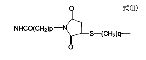
- 下記の一般式(III)で表される化合物がそのY部分で蛍光性ナノ粒子表面に結合している、請求項1に記載の蛍光プローブ。
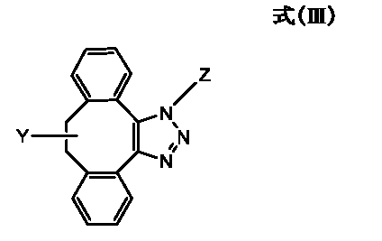
Zは、蛍光標識対象物に結合しうる物質を表し;
YのNHとCOは、直接結合していても良いし、リンカーを介して結合していても良い。]
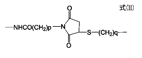
- 一般式(III)で表わされる化合物がそのY部分で蛍光性ナノ粒子表面に結合している蛍光プローブにおいて、Yが-NH-CO-O-である請求項2に記載の蛍光プローブ。
- 一般式(III)で表される化合物がそのY部分で蛍光性ナノ粒子表面に結合している蛍光プローブにおいて、Zが抗体である請求項2又は3に記載の蛍光プローブ。
- 一般式(III)で表される化合物がそのY部分で蛍光性ナノ粒子表面に結合している蛍光プローブにおいて、リンカーが、アルキレン基、アルキレンオキシ基又はポリアルキレンオキシ基の両末端に、アミノ基、アルキルアミノ基又はカルボン酸の基を有する化合物である請求項2~4のいずれか1に記載の蛍光プローブ。
- 下記の一般式(IV)で表される化合物がそのY部分で蛍光性ナノ粒子表面に結合している蛍光プローブ合成中間体。
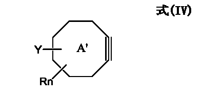
環A'は、シクロオクタンのアルキン誘導体を表し、シクロオクタン部分はさらに芳香族環と縮合していてもよく;
Rは、アルキル基、アルコキシ基又はハロゲン原子を表し、nは0~2の整数であり;
YのNHとCOは、直接結合していても良いし、リンカーを介して結合していても良い。]

- 下記の一般式(V)で表される化合物がそのY部分で蛍光性ナノ粒子表面に結合している、請求項6に記載の蛍光プローブ合成中間体。
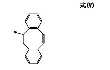
YのNHとCOは、直接結合していても良いし、リンカーを介して結合していても良い。]

- 一般式(V)で表わされる化合物がそのY部分で蛍光性ナノ粒子表面に結合している蛍光プローブ合成中間体において、Yが-NH-CO-O-である請求項7に記載の蛍光プローブ合成中間体。
- 一般式(V)で表される化合物がそのY部分で蛍光性ナノ粒子表面に結合している蛍光プローブ合成中間体において、リンカーが、アルキレン基、アルキレンオキシ基又はポリアルキレンオキシ基の両末端に、アミノ基、アルキルアミノ基又はカルボン酸の基を有する化合物である請求項7又は8に記載の蛍光プローブ合成中間体。
- 以下の工程を含む、請求項6~9のいずれか1に記載の蛍光プローブ合成中間体の作製方法:
1)カルボン酸基、アミノ基、チオール基、マレイミド基、炭酸エステル基、又は活性エステル基が導入された蛍光性ナノ粒子を調製する工程;
2)シクロオクタンのアルキン誘導体に、上記1)の工程で蛍光性ナノ粒子に導入された各種官能基と反応しうる官能基を導入して、当該官能基が導入されたシクロオクタンのアルキン誘導体を調製する工程;
3)上記1)及び2)の工程で調製された各種官能基が導入された蛍光性ナノ粒子及びシクロオクタンのアルキン誘導体を結合する工程。 - 以下の工程を含む、請求項1~5のいずれか1に記載の蛍光プローブの作製方法:
A)蛍光標識対象物に結合しうる物質(Z)にアジド基を導入する工程;
B)請求項6~9のいずれか1に記載の蛍光プローブ合成中間体におけるシクロオクタンのアルキン誘導体と、上記A)の工程で導入されたアジド基とを結合する工程。 - 以下の工程を含む、請求項1~5のいずれか1に記載の蛍光プローブの作製方法:
A)蛍光標識対象物に結合しうる物質(Z)にアジド基を導入する工程;
B)請求項6~9のいずれか1に記載の蛍光プローブ合成中間体におけるシクロオクタンのアルキン誘導体と、上記A)の工程で導入されたアジド基とを結合する工程;次いで、
C)上記B)の工程で得られた蛍光プローブを親水化処理する工程。
Applications Claiming Priority (2)
| Application Number | Priority Date | Filing Date | Title |
|---|---|---|---|
| JP2009269447A JP2013032291A (ja) | 2009-11-27 | 2009-11-27 | 蛍光プローブ |
| JP2009-269447 | 2009-11-27 |
Publications (1)
| Publication Number | Publication Date |
|---|---|
| WO2011065308A1 true WO2011065308A1 (ja) | 2011-06-03 |
Family
ID=44066413
Family Applications (1)
| Application Number | Title | Priority Date | Filing Date |
|---|---|---|---|
| PCT/JP2010/070698 Ceased WO2011065308A1 (ja) | 2009-11-27 | 2010-11-19 | 蛍光プローブ |
Country Status (2)
| Country | Link |
|---|---|
| JP (1) | JP2013032291A (ja) |
| WO (1) | WO2011065308A1 (ja) |
Cited By (1)
| Publication number | Priority date | Publication date | Assignee | Title |
|---|---|---|---|---|
| CN110483548A (zh) * | 2019-08-23 | 2019-11-22 | 宁夏医科大学 | 荜茇酰胺衍生物及其制备方法和应用 |
Families Citing this family (3)
| Publication number | Priority date | Publication date | Assignee | Title |
|---|---|---|---|---|
| JP6327547B2 (ja) * | 2013-08-02 | 2018-05-23 | 国立研究開発法人理化学研究所 | 新規化合物及びその利用 |
| JP6743703B2 (ja) * | 2014-11-06 | 2020-08-19 | コニカミノルタ株式会社 | 免疫染色法、およびこれに用いられる免疫染色試薬キット |
| WO2023190099A1 (ja) * | 2022-03-29 | 2023-10-05 | 株式会社ビー・エム・エル | 対象物質の測定方法、捕捉担体、測定キット、および測定試薬の製造方法 |
Citations (4)
| Publication number | Priority date | Publication date | Assignee | Title |
|---|---|---|---|---|
| JP2007155395A (ja) * | 2005-12-01 | 2007-06-21 | National Institute Of Advanced Industrial & Technology | 免疫化学的検出方法、及びその検出用試薬キット |
| JP2007178239A (ja) * | 2005-12-27 | 2007-07-12 | Kenji Yamamoto | 親水性量子ドット |
| WO2009113375A1 (ja) * | 2008-03-14 | 2009-09-17 | コニカミノルタエムジー株式会社 | シリコンナノ粒子蛍光体含有酸化ケイ素膜、シリコンナノ粒子蛍光体及び単一分子観察方法 |
| EP2161239A1 (fr) * | 2008-09-08 | 2010-03-10 | Commissariat à l'Energie Atomique | Nanocristaux semi-conducteurs |
-
2009
- 2009-11-27 JP JP2009269447A patent/JP2013032291A/ja not_active Withdrawn
-
2010
- 2010-11-19 WO PCT/JP2010/070698 patent/WO2011065308A1/ja not_active Ceased
Patent Citations (4)
| Publication number | Priority date | Publication date | Assignee | Title |
|---|---|---|---|---|
| JP2007155395A (ja) * | 2005-12-01 | 2007-06-21 | National Institute Of Advanced Industrial & Technology | 免疫化学的検出方法、及びその検出用試薬キット |
| JP2007178239A (ja) * | 2005-12-27 | 2007-07-12 | Kenji Yamamoto | 親水性量子ドット |
| WO2009113375A1 (ja) * | 2008-03-14 | 2009-09-17 | コニカミノルタエムジー株式会社 | シリコンナノ粒子蛍光体含有酸化ケイ素膜、シリコンナノ粒子蛍光体及び単一分子観察方法 |
| EP2161239A1 (fr) * | 2008-09-08 | 2010-03-10 | Commissariat à l'Energie Atomique | Nanocristaux semi-conducteurs |
Non-Patent Citations (6)
| Title |
|---|
| AGARD, N. J. ET AL.: "A Comparative Study of Bioorthogonal Reactions with Azides", ACS CHEM. BIO., vol. 1, no. 10, 2006, pages 644 - 648, XP002522847, DOI: doi:10.1021/CB6003228 * |
| KELE, P. ET AL.: "Clickable fluorophores for biological labeling-with or without copper", ORG. BIOMOL. CHEM., vol. 7, no. 17, 7 July 2009 (2009-07-07), pages 3486 - 3490, XP055014209, DOI: doi:10.1039/b907741c * |
| NING, X. ET AL.: "Visualizing Metabolically Labeled Glycoconjugates of Living Cells by Copper-Free and Fast Huisgen Cycloadditions", ANGEW. CHEM. INT. ED., vol. 47, no. 12, 2008, pages 2253 - 2255, XP002659161, DOI: doi:10.1002/anie.200705456 * |
| SLETTEN, E. M. ET AL.: "A Hydrophilic Azacyclooctyne for Cu-Free Click Chemistry", ORG. LETT., vol. 10, no. 14, 2008, pages 3097 - 3099, XP055031301, DOI: doi:10.1021/ol801141k * |
| SMITH, A. M. ET AL.: "Engineering Luminescent Quantum Dots for In Vivo Molecular and Cellular Imaging", ANNALS OF BIOMEDICAL ENGINEERING, vol. 34, no. 1, 2006, pages 3 - 14, XP019273023 * |
| ZOU, Y. ET AL.: "Cu-free cycloaddition for identifying catalytic active adenylation domains of nonribosomal peptide synthetases by phage display", BIOORG. MED. CHEM. LETT., vol. 18, no. ISS. 2, 2008, pages 5664 - 5667, XP025562131, DOI: doi:10.1016/j.bmcl.2008.08.085 * |
Cited By (1)
| Publication number | Priority date | Publication date | Assignee | Title |
|---|---|---|---|---|
| CN110483548A (zh) * | 2019-08-23 | 2019-11-22 | 宁夏医科大学 | 荜茇酰胺衍生物及其制备方法和应用 |
Also Published As
| Publication number | Publication date |
|---|---|
| JP2013032291A (ja) | 2013-02-14 |
Similar Documents
| Publication | Publication Date | Title |
|---|---|---|
| JP2026048733A (ja) | ペンダントクロモフォアを有する水溶性ポリマー色素 | |
| AU2017375751B2 (en) | Water-soluble polymeric dyes | |
| JP5777169B2 (ja) | 新規な蛍光色素およびその使用 | |
| JP6928742B2 (ja) | 分枝鎖状ポリエーテル骨格上の蛍光色素の多量体化に基づく明るい蛍光色素 | |
| CN112955508A (zh) | 具有侧基窄发射受体的聚合物串联染料 | |
| EP2398860B1 (en) | Quenched dendrimeric dyes for fluorescence detection | |
| CN105073761A (zh) | 用于体外和体内成像和检测的取代的硅杂蒽阳离子红至近红外荧光染料 | |
| US20200255668A1 (en) | Labeling dye and kit including same | |
| CN111094462A (zh) | 深紫外线可激发的水溶剂化聚合物染料 | |
| CN104788433A (zh) | 生物相容的含n,n-二取代磺酰胺的荧光染料标记 | |
| WO2011065308A1 (ja) | 蛍光プローブ | |
| Mahmoud et al. | Advanced procedures for labeling of antibodies with quantum dots | |
| JP2025138758A (ja) | 増強された蛍光バイオアッセイのための超高輝度蛍光ナノコンポジット構造体 | |
| Lee et al. | Immuno-nanoparticles for multiplex protein imaging in cells and tissues | |
| WO2020222894A1 (en) | Water-soluble polynorbornene dyes having pendant chromophores | |
| JP7692912B2 (ja) | 発光性の双性イオンポリマーナノ粒子 | |
| US12553899B2 (en) | Luminescent compounds | |
| JP7385850B2 (ja) | 細胞滞留性蛍光化合物並びにそれを用いた細胞の染色方法及び高感度検出方法 | |
| KR100979463B1 (ko) | 니켈 결합 양자점을 이용한 히스티딘 표지 단백질의검출방법 | |
| WO2021044381A1 (en) | Conjugates composed of membrane-targeting peptides for extracellular vesicles isolation, analysis and their integration thereof | |
| JP7736259B2 (ja) | 化合物、その製造方法、複合体および短波赤外蛍光剤 | |
| US20230295440A1 (en) | Violet excitable tandem dyes, and methods for making and using the same | |
| JP2010169666A (ja) | 複合体、それを用いる診断用マーカー及び診断薬、それを用いる検出方法、検出キット | |
| CN117460789A (zh) | 化合物及使用了该化合物的标记生物物质 | |
| HK40079008A (en) | Luminescent zwitterionic polymeric nanoparticles |
Legal Events
| Date | Code | Title | Description |
|---|---|---|---|
| 121 | Ep: the epo has been informed by wipo that ep was designated in this application |
Ref document number: 10833154 Country of ref document: EP Kind code of ref document: A1 |
|
| NENP | Non-entry into the national phase |
Ref country code: DE |
|
| 122 | Ep: pct application non-entry in european phase |
Ref document number: 10833154 Country of ref document: EP Kind code of ref document: A1 |
|
| NENP | Non-entry into the national phase |
Ref country code: JP |