WO2010134226A1 - タンパク質療法や細胞の分化/未分化制御、抗体療法に応用可能な細胞内でのタンパク質の安定化を可能にする酸性アミノ酸からなるモチーフの確立 - Google Patents
タンパク質療法や細胞の分化/未分化制御、抗体療法に応用可能な細胞内でのタンパク質の安定化を可能にする酸性アミノ酸からなるモチーフの確立 Download PDFInfo
- Publication number
- WO2010134226A1 WO2010134226A1 PCT/JP2009/070081 JP2009070081W WO2010134226A1 WO 2010134226 A1 WO2010134226 A1 WO 2010134226A1 JP 2009070081 W JP2009070081 W JP 2009070081W WO 2010134226 A1 WO2010134226 A1 WO 2010134226A1
- Authority
- WO
- WIPO (PCT)
- Prior art keywords
- protein
- amino acid
- motif
- cells
- fused
- Prior art date
- Legal status (The legal status is an assumption and is not a legal conclusion. Google has not performed a legal analysis and makes no representation as to the accuracy of the status listed.)
- Ceased
Links
Images



Classifications
-
- C—CHEMISTRY; METALLURGY
- C07—ORGANIC CHEMISTRY
- C07K—PEPTIDES
- C07K14/00—Peptides having more than 20 amino acids; Gastrins; Somatostatins; Melanotropins; Derivatives thereof
- C07K14/435—Peptides having more than 20 amino acids; Gastrins; Somatostatins; Melanotropins; Derivatives thereof from animals; from humans
- C07K14/46—Peptides having more than 20 amino acids; Gastrins; Somatostatins; Melanotropins; Derivatives thereof from animals; from humans from vertebrates
- C07K14/47—Peptides having more than 20 amino acids; Gastrins; Somatostatins; Melanotropins; Derivatives thereof from animals; from humans from vertebrates from mammals
- C07K14/4701—Peptides having more than 20 amino acids; Gastrins; Somatostatins; Melanotropins; Derivatives thereof from animals; from humans from vertebrates from mammals not used
- C07K14/4702—Regulators; Modulating activity
- C07K14/4703—Inhibitors; Suppressors
-
- C—CHEMISTRY; METALLURGY
- C07—ORGANIC CHEMISTRY
- C07K—PEPTIDES
- C07K14/00—Peptides having more than 20 amino acids; Gastrins; Somatostatins; Melanotropins; Derivatives thereof
- C07K14/435—Peptides having more than 20 amino acids; Gastrins; Somatostatins; Melanotropins; Derivatives thereof from animals; from humans
- C07K14/46—Peptides having more than 20 amino acids; Gastrins; Somatostatins; Melanotropins; Derivatives thereof from animals; from humans from vertebrates
- C07K14/47—Peptides having more than 20 amino acids; Gastrins; Somatostatins; Melanotropins; Derivatives thereof from animals; from humans from vertebrates from mammals
- C07K14/4701—Peptides having more than 20 amino acids; Gastrins; Somatostatins; Melanotropins; Derivatives thereof from animals; from humans from vertebrates from mammals not used
- C07K14/4702—Regulators; Modulating activity
-
- C—CHEMISTRY; METALLURGY
- C07—ORGANIC CHEMISTRY
- C07K—PEPTIDES
- C07K14/00—Peptides having more than 20 amino acids; Gastrins; Somatostatins; Melanotropins; Derivatives thereof
- C07K14/435—Peptides having more than 20 amino acids; Gastrins; Somatostatins; Melanotropins; Derivatives thereof from animals; from humans
- C07K14/46—Peptides having more than 20 amino acids; Gastrins; Somatostatins; Melanotropins; Derivatives thereof from animals; from humans from vertebrates
- C07K14/47—Peptides having more than 20 amino acids; Gastrins; Somatostatins; Melanotropins; Derivatives thereof from animals; from humans from vertebrates from mammals
- C07K14/4701—Peptides having more than 20 amino acids; Gastrins; Somatostatins; Melanotropins; Derivatives thereof from animals; from humans from vertebrates from mammals not used
- C07K14/4702—Regulators; Modulating activity
- C07K14/4705—Regulators; Modulating activity stimulating, promoting or activating activity
Definitions
- the present invention relates to a motif composed of acidic amino acids that can prevent the administered protein from being decomposed by fusing to a protein or antibody used in protein therapy or antibody therapy.
- iPS cells have been established by introducing genes of 4 factors (Oct3 / 4, Sox2, Klf4, c-Myc) into somatic cells using viral vectors such as retroviruses and lentiviruses.
- viral vectors such as retroviruses and lentiviruses.
- virus vectors need to be created in a strictly controlled environment, which has been an obstacle to the spread of iPS cell technology.
- arginine 11 basic amino acids, arginine, were linked to 4 factors, and this was remodeled so that it could easily permeate the cell membrane, and put into mouse fetal cells without using viruses. It was announced that it could be generated.
- the protein is degraded by proteasome, phagosome, autophagy, proteolytic enzyme, etc. In many cases, it is necessary to administer frequently (see, for example, Non-Patent Document 1). So far, a poly-arginine tag has been known as a motif capable of suppressing such cell membrane-permeable protein from proteolysis in cells.
- the present invention provides a protein design / production method that is resistant to proteolysis by searching for motifs that prevent the protein administered to cells and individuals from being degraded by proteasomes, autophagy, and other proteolytic enzymes.
- the purpose is to do.
- the present invention is a proteolysis inhibition motif having a sequence mainly composed of acidic amino acids, and preferably consists of all or part of the amino acid sequence of the acidic amino acid region on the C-terminal side of DP-1 protein, or this Is a proteolytic inhibition motif.
- the form of the degradation inhibitory motif according to the present invention is a degradation inhibitory motif consisting of the amino acid region of the C-terminal side 395 to 410 of the DP-1 protein, and is a proteolytic inhibition motif consisting of the following amino acid sequence.
- (1) EDDEDDDDFNENDEDD (2) EDDEE E: Glutamic acid
- D Aspartic acid
- N Asparagine
- F Phenylalanine
- the other form of this invention is a proteolysis inhibition motif in which the said acidic amino acid mainly consists of glutamic acid or aspartic acid.
- the present invention is a fusion protein in which the above-described motif is fused to at least one of the N-terminal, C-terminal, and midway of any protein, and has acquired proteolytic resistance.
- iPS cells induced pluripotent stem cells
- the present invention is an amino acid sequence in which the motif is fused to the C-terminus of Sox2, an amino acid sequence in which the motif is fused to the N-terminus of Oct4, and an amino acid sequence in which the motif is fused to the N-terminus of Klf4.
- the present invention is a nucleic acid encoding the amino acid sequence of the motif, an expression vector having the nucleic acid, and a transformant containing this expression vector.
- the present invention is a pharmaceutical composition comprising the fusion protein, the cell membrane permeable protein further fused with a cell membrane permeable tag, and further, the fusion protein or the cell membrane permeable protein. It is an antibody protein containing.
- the previously described acidic amino acid motif prevents fused proteins from proteolysis. Therefore, it is possible to provide proteolytic resistant proteins by fusing the motifs to cell membrane permeable proteins and antibodies used in protein therapy and antibody therapy. Useful effects such as (reduction in the number of administrations, increase in effect) are exhibited.
- the motif consisting of acidic amino acids according to the present invention prevents fused proteins from proteolysis, so if an oligonucleotide encoding the same is introduced into an expression plasmid or the like to create a fusion protein expression system with the target protein. Even proteins that have been severely proteolytically impossible to perform functional analysis so far have acquired resistance to degradation and are capable of functional analysis, and are extremely effective as molecular biology tools.
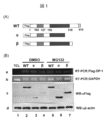
- FIG. 1 is a diagram showing that the C-terminal side of DP-1 that triggered the present invention has a function of preventing proteolysis.
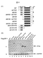
- FIG. 2 is a diagram showing that the C-terminal 396 to 410 amino acid region of DP-1 is a region that prevents DP-1 proteolysis.
- FIG. 3 shows that the 396-410 amino acid region on the C-terminal side of DP-1 prevents DP-1 (•) from proteolysis.
- FIG. 4 is a diagram showing that a DP-1 degradation promoting region exists in the 128-192 amino acid region of DP-1.
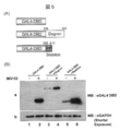
- FIG. 5 shows that the C-terminal 396-410 amino acid region of DP-1 can prevent proteolysis even in other molecules (GAL4DBD; GAL4 DNA binding region).
- FIG. 1 is a diagram showing that the C-terminal side of DP-1 that triggered the present invention has a function of preventing proteolysis.
- FIG. 2 is a diagram showing that the C-terminal 396 to
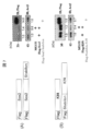
- FIG. 6 is a first diagram illustrating the results of an embodiment in which Stabilon was introduced into an iPS cell inducer.
- FIG. 7 is a second diagram illustrating the results of an embodiment in which Stabilon was introduced into an iPS cell inducer.
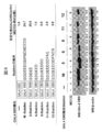
- FIG. 8 is a diagram for explaining the influence of changes in the sequence of Stabilon on the protein degradation inhibitory property.
- DP-1 protein is composed of 410 amino acids, and its sequence is as follows.
- the underlined portion is an acidic amino acid region on the C-terminal side of the DP-1 protein that constitutes a proteolytic inhibition motif.
- the above sequences are registered in the National Center for Biotechnology Information (NC_007111) of NCBI (National Center for Biotechnology Information).
- the motif mainly composed of acidic amino acids according to the present invention is a motif mainly composed of glutamic acid (Glu; E) and aspartic acid (Asp; D), and the composition ratio and length thereof are not particularly defined, and the other In some cases. It may also contain glutamine (Q; Gln), asparagine (Asn; N), and cysteine (Cys; C), which are slightly acidic.
- phosphorylated amino acids are also acidic
- these phosphorylated amino acids may be included.
- an example of the motif according to the present invention consists of 1 to 50 amino acids, of which the number of acidic amino acids is preferably 20% or more.
- the motif of the present invention may contain a non-acidic amino acid such as phenylalanine [Phe: F] as long as it has degradation resistance to the proteasome.
- amino acid is used in its broadest sense and includes not only natural amino acids but also non-natural amino acids such as amino acid variants and derivatives.
- the amino acid contained in the motif consisting of the acidic amino acids of the present invention consists only of natural amino acids.
- the number of substituted amino acids is particularly limited as long as the resulting protein retains the activity of the fused protein. Although not, it is 1 to 9, preferably 1 to 5, more preferably about 1 to 3, or within 20%, preferably within 10% of the total length.
- the amino acid to be substituted or added can be a natural amino acid, a non-natural amino acid or an amino acid analog, and is preferably a natural amino acid.
- the acidic amino acid of the present invention or an acidic amino acid motif (sometimes referred to as “stabilization promoting region”, “Stabilon”, “CTAD; C-terminal acidic domain”, “acidic amino acid region”) is fused.
- stabilization promoting region “Stabilon”, “CTAD; C-terminal acidic domain”, “acidic amino acid region”
- C-terminal acidic domain “acidic amino acid region”
- the motif consisting of the acidic amino acid of the present invention may be located anywhere in the fusion protein, and may be linked to any of the N-terminal side, C-terminal side or inside of the fusion protein.
- a protein to which the degradation inhibitory motif according to the present invention is fused or added is a protein that exhibits resistance to proteolysis by addition of a degradation inhibitory motif and has a predetermined physiological activity or can be expected to be a protein.
- a protein that exhibits resistance to proteolysis by addition of a degradation inhibitory motif and has a predetermined physiological activity or can be expected to be a protein.
- the aforementioned Oct3 / 4, Sox2, Klf4, and c-Myc are preferable examples of the protein.
- the “nucleic acid encoding the fusion protein according to the present invention” according to the present invention may be any nucleic acid as long as it contains the base sequence encoding the fusion protein according to the present invention, and is preferably DNA.
- Such DNA can be obtained from, for example, genomic DNA, genomic DNA library, cDNA derived from a predetermined cell or tissue, and cDNA library, and may be synthesized DNA. It can also be obtained by adjusting the total RNA or mRNA fraction from a predetermined cell / tissue and amplifying it by RT-PCR.
- the nucleic acid encoding the motif consisting of the acidic amino acid of the present invention consists of DNA determined based on the amino acid sequence and synthesized based on the sequence.
- the nucleic acid encoding a protein or peptide fused to the motif consisting of the acidic amino acid of the present invention can be obtained from a cDNA or cDNA library obtained by RT-PCR using, for example, RNA extracted from a cell, It may be synthesized DNA.
- Each nucleic acid can be ligated and then inserted into the expression vector, or each nucleic acid can be ligated so that the fusion protein is expressed by inserting it into the cloning site of the expression vector. It is included in the “nucleic acid encoding a fusion protein” of the present invention.
- the term “linked so that the fusion protein is expressed” refers to the target protein, the motif consisting of the acidic amino acid of the present invention, a purification tag, a nuclear localization signal (NLS), and sometimes referred to as NLS hereinafter. )) And the like, which are linked together (in frame) with a frame of a nucleic acid encoding a motif or the like capable of inducing intracellular localization.
- the “expression vector” according to the present invention is a vector into which a nucleic acid encoding the motif consisting of the above-mentioned acidic amino acid according to the present invention is inserted.
- An expression vector has a condition that it has a self-replicating ability in a host, has a gene of an expression system that can be easily distinguished from a host cell, has at least one restriction enzyme cleavage site, and cannot survive outside the host cell. Any material can be used as long as it is satisfied, and can be selected in combination with the host.
- plasmids such as pBR vectors, pUC vectors, pET vectors, and pQE vectors are preferably used.
- animal cell expression plasmids and E. coli cell expression plasmids are used.
- Plasmids yeast cell expression plasmids, insect cell expression viruses (such as baculovirus), virus vectors, phage vectors, cosmid vectors, yeast-derived plasmids, yeast artificial chromosomes (YAC), and the like can also be used.
- the expression vector incorporates a promoter that functions in the host, and the nucleic acid of the present invention is inserted under the control of the promoter.
- a replication marker, a terminator region, a selection marker gene for selecting a transformant, and the like may be inserted into the expression vector.
- the selectable marker gene for example, antibiotic resistance genes such as tetracycline, ampicillin, kanamycin, neomycin, geneticin and the like are used.
- the “transformant” according to the present invention refers to a host transformed with an expression vector into which a nucleic acid encoding the motif consisting of the above-mentioned acidic amino acid of the present invention has been inserted.
- the expression vector when the expression vector is an expression vector for Escherichia coli, for example, Escherichia coli and other bacteria can be used, but Escherichia coli suitable for mass production is preferred.
- the expression vector is an expression vector for animal cells, for example, cultured cells such as HEK293 cells, HeLa cells, MCF-7 cells, and Cos cells can be used. Next, a method for producing a fusion protein according to the present invention will be described.
- the protein fused with the motif of the acidic amino acid of the present invention is transformed with the expression vector containing the nucleic acid of the present invention described above, the host is cultured, the fusion protein is expressed, and the fusion protein is purified. Can be obtained.
- expression vectors can be transiently examined by introducing an expression vector into animal cultured cells using a transfection reagent, electroporation or the like.
- a method for transforming a host with a protein expression vector fused with an acidic amino acid motif of the present invention a known method can be used. For example, when the host is Escherichia coli, the host is treated with manganese chloride or calcium chloride.
- Examples thereof include a method of preparing competent cells, mixing an expression vector into a suspension and incorporating it by heat shock, an electroporation method, and a method of infecting a host with a phage in the case of a phage vector.
- the host is an animal cultured cell, it can be incorporated by a commercially available transfection reagent, electroporation method, calcium chloride precipitation method, or the like.
- the target fusion protein can be expressed by culturing the thus obtained transformant in an appropriate medium. Conditions such as medium composition, culture temperature, time, addition of inducer, etc. can be determined by those skilled in the art according to known methods so that the transformant grows and the fusion protein is efficiently produced.
- a transformant when an antibiotic resistance gene is incorporated into an expression vector as a selection marker, a transformant can be selected by adding an antibiotic to the medium.
- the produced fusion protein can be purified according to a known method. For example, the host is suspended in a buffer solution and destroyed by a method such as ultrasonic disruption. Subsequently, when the purification tag is fused to the fusion protein, first, the fusion protein adsorbed on the column is eluted with an elution buffer through an affinity column having specific affinity for the purification tag. Can be purified. Further, the purity can be further increased by repeating the step of passing the obtained fusion protein solution through the affinity column a plurality of times.
- a protein fused with a motif comprising the acidic amino acid of the present invention when used as a pharmaceutical, a protein purified to at least 90%, preferably 95% or more, more preferably 98% or more, and further preferably 99% or more
- a purification tag fused with the motif consisting of the acidic amino acid of the present invention when included, such purity can be easily achieved.
- polyhistidine His6
- the expressed protein is expressed as inclusion bodies, and before passing through an affinity column, a protein denaturing agent such as urea or guanidine hydrochloride, or a surfactant is used. It is also preferable to process.
- a protein that has been denatured and purified can be made to function properly by refolding.
- Refolding can be performed, for example, by diluting with a buffer containing an excessive amount of arginine or by dialysis with the same buffer.
- the protein fused with the motif consisting of the acidic amino acid of the present invention becomes a pharmaceutical composition depending on the function of the fused protein.
- Examples of the protein that fuses the motif composed of acidic amino acids of the present invention include cell membrane permeable proteins, antibodies, peptides, proteins that are proteolyzed in cells, and the like.
- the pharmaceutical composition comprising a protein fused with an acidic amino acid motif of the present invention is a diluent such as a commonly used filler, extender, binder, dampener, disintegrant, surfactant, lubricant, etc. Alternatively, it is formulated in the form of a normal pharmaceutical composition using an excipient. Examples of such a pharmaceutical composition include tablets, pills, powders, solutions, suspensions, emulsions, granules, capsules, suppositories, injections and the like.
- the amount of the protein fused with the motif consisting of the acidic amino acid of the present invention contained in the pharmaceutical composition is not particularly limited and can be appropriately selected from a wide range.
- the pharmaceutical composition containing, as an active ingredient, a protein fused with a motif composed of an acidic amino acid of the present invention can further contain other active ingredients, or can be used in combination with a pharmaceutical composition containing other active ingredients. You can also.
- the administration method of the pharmaceutical composition according to the present invention is not particularly limited, and it is administered by a method according to various preparation forms, patient age, sex, disease state, and other conditions. Examples of the administration method in the case of tablets, pills, liquids, suspensions, emulsions, granules and capsules include oral administration.
- an injection it can be administered intravenously, intramuscularly, intradermally, subcutaneously or intraperitoneally alone or mixed with a normal fluid such as glucose or amino acid.
- a normal fluid such as glucose or amino acid.
- a suppository it is administered intrarectally.
- the dosage of the above pharmaceutical composition may be appropriately selected according to usage, patient age, sex, degree of disease, and other conditions. When administered orally, it is usually for an adult (60 kg) per dose.
- the protein fused with the motif comprising the acidic amino acid of the present invention is administered at about 0.1 mg to 100 mg, preferably about 1.0 to 50 mg, more preferably about 1.0 to 20 mg.
- the present invention When administered as an injection, for adults (60 kg), about 0.1 mg to 30 mg, preferably about 0.1 to 20 mg of protein fused with the acidic amino acid motif of the present invention per dose Preferably, about 0.1 to 10 mg is administered.
- the number of administrations of the pharmaceutical composition may be appropriately selected according to the usage, patient age, sex, degree of disease, and other conditions. For example, once every two weeks, once a month, every two months It can be administered as frequently as once.
- the protein fused with the acidic amino acid motif according to the present invention has been proteolytically decomposed quickly in cultured cells by conventional expression in animal cells. It is considered effective against proteins.
- the present invention will be specifically described based on examples, but the present invention is not limited thereto. Referring to FIG.
- the Flag tag is an amino acid sequence consisting of Asp-Tyr-Lys-Asp-Asp-Asp-Asp-Lys. By fusing this sequence, detection by Western blotting becomes extremely easy (HRP fusion used for Western blotting).
- the Flag antibody [SIGMA] was used with a very high sensitivity and capable of detecting a Flag tag fusion protein). Single cloned E. coli containing each plasmid was cultured using LB medium containing ampicillin. After collection by centrifugation, the expression plasmid was purified using Plasmid Purification MAXI Kit (QIAGEN).
- HEK 293F cells were cultured in DMEM medium containing 10% FBS (containing penicillin and streptomycin as antibiotics). For transfection, HEK 293F cells were seeded in a 6 cm dish the day before so as to be 50% confluent at the time of transfection.
- the plasmids were transfected into HEK 293F cells using the combinations shown in the lanes 1 to 7 in FIG. Use 200 ng of Flag-DP-1 / pcDNA3 as the expression plasmid, add cDNA3 expression plasmid for correction to a total of 2.6 mg, and make 100 ml with TE buffer (10 mM Tris-HCl [pH 8.0], 1 mM EDTA).
- a transfection reagent Polyfect transfection reagent was used. 30 ml Polyfect transfection reagent (QIAGEN) was added to the expression plasmid solution, vortexed, and stirred and turbid. The mixture was allowed to stand for 5 minutes in order to form a DNA, Polyfect transfection reagent complex. Meanwhile, the cell culture medium was changed. DNA, Polyfect transfection reagent complex was added to the medium and mixed slowly. CO 2 5% CO in the incubator 2 The culture was continued under conditions of a concentration of 37 ° C. 1.4 Addition of MG132 MG132 used was dissolved in DMSO. In lanes 5 to 7, MG132 was added at a final concentration of 10 mM 6 hours before cell recovery.
- RNA extraction The medium was discarded from the cells that had been cultured for 24 hours after transfection. 1 ml of PBS was added, the cells were detached by light pipetting, and transferred to a 1.5 ml Eppendorf tube. Cells were collected by centrifugation at 1,500 rpm, and the supernatant was discarded. 700 ml of PBS was added to the precipitated cells, and 700 ml of Isogen (Nippon Gene) was added. Total RNA was prepared according to the Isogen protocol.
- RT-PCR method An attempt was made to quantify the amount of Flag-DP-1 mRNA derived from Flag-DP-1 / pcDNA3 to be forcibly expressed by RT-PCR.
- the primer for RT-PCR was GAPDH, which is a housekeeping gene, for Flag-DP-1 mRNA and for correction, and was designed as follows.
- Flag-DP-1 GAPDH RT reaction was performed as follows using AccessQuick RT-PCR system (Promega). 1.5.2. a.
- RT reaction composition RNA (1mg / ml) 1ml Master Mix (2x) 10ml 3'primer (50 pmol / l) 0.4 ml AMV Reverse Transscriptase 0.4ml Nuclease-free water 13ml Total Total 20ml 1.5.2.
- RT-PCR program 45 ° C 45 minutes 94 ° C 2 minutes 4 ° C ⁇ PCR reaction was performed as follows by ExTaq (Takara Bio Inc.). 1.5.2. c.
- the HRP (Horsaradish peroxidase) fusion Flag antibody as the primary antibody and the uniformity of the sample extract are corrected.
- b-actin which is a housekeeping gene, was used.
- HRP fusion anti-mouse IgG antibody was used as the secondary antibody. The detection was performed using Chemilmiwan as a substrate for HRP, and color development was detected by Hyperfilm (GE Healthcare).
- Hyperfilm GE Healthcare
- Flag-DP-1 (•, •) protein was significantly increased by the addition of MG132 (panel c, comparison between lanes 3 and 4 and lanes 6 and 7). Therefore, it was found that the Flag-DP-1 (•, •) protein is a protein that is strongly degraded by the proteasome. In addition, it was considered that a domain or motif that is important for suppressing the degradation of DP-1 is present in the C-terminal region lacking Flag-DP-1 (•, •). Next, FIG. 2 will be described.
- Flag tag fusion DP-1 (WT,7), The same one as described in 1.1 above was used.
- a Flag tag fusion DP-1 ( ⁇ C364, ⁇ C395) expression plasmid a DP-1 deletion mutant fragment was amplified by PCR using the following primers.
- the region that suppresses the degradation of DP-1 by the proteasome is 395-410 amino acids (EDDEEDDDDFNENDED) on the most C-terminal side of DP-1.
- This region is an acidic region rich in glutamic acid (E) and aspartic acid (D).
- FIG. 4 will be described. The degree of degradation of DP-1 when DP-1 (•, + CTAD, ⁇ C364, ⁇ C364 + CTAD) was forcibly expressed in HEK293F cells using an expression plasmid was examined.
- Flag tag fusion DP-1 (• + CTAD, ⁇ C364 + CTAD)
- DP-1 (•) and DP-1 ( ⁇ C364) were ligated with fragments encoding CTAD, respectively. Insertion was confirmed by restriction after miniprep. The sequence was confirmed by sequencing.
- the plasmid was purified using the Plasmid Purification MAXI Kit (QIAGEN) shown in 1.1 above.
- 3.2 Cell culture is the same as in 1.2 above.
- 3.3 The transfection is the same as in 1.3 above, 3.4 Western blotting analysis is the same as in 1.6 above. 3 All experiments in FIG. 3 are performed under the condition where MG132 is not present.
- Flag-DP-1 (•) was hardly observed (FIG. 3-B panel a, lane 1). In addition, Flag-DP-1 (• + CTAD) was sufficiently expressed (FIG. 3-B panel a, lane 2). Flag-DP-1 ( ⁇ C364) was weakly expressed (FIG. 3-B panel a, lane 3). Flag-DP-1 ( ⁇ C364 + CTAD) was sufficiently expressed (FIG. 3-B panel a, lane 4). Therefore, the region that suppresses the degradation of DP-1 by the proteasome determined in FIG. 2 (the 395-410 amino acid at the most C-terminal side of DP-1, EDDEEDDDFNENDED; CTAD) is a DP present in the DP-1 (•) region.
- FIG. 4 Degree of DP-1 degradation when DP-1 (WT, ⁇ CTAD, ⁇ N102, ⁇ N102 + CTAD, ⁇ N126, ⁇ N126 + CTAD, ⁇ N191, ⁇ N191 + CTAD,..., ⁇ C191-410, ⁇ C127-410) is forcibly expressed in HEK293F cells using an expression plasmid Examined.
- Flag tag fusion DP-1 ( ⁇ CTAD, ⁇ N102, ⁇ N102 + ⁇ CTAD, ⁇ N126, ⁇ N126 + ⁇ CTAD, ⁇ N191, ⁇ N191 + ⁇ CTAD,..., ⁇ C191-410, ⁇ C127-410), use the following primers for DP-1 deletion mutant fragments Amplified by PCR.
- GAL4DBD fusion Stabilon CTAD was inserted into GAL4DBD-pcDNA3 described above with EcoRI-XbaI.
- the GAL4DBD fusion Demon was inserted into the aforementioned GAL4DBD-pcDNA3 with EcoRI-XbaI.
- the expression plasmid was purified using the Plasmid Purification MAXI Kit (QIAGEN) shown in 1.1 above. 5.2 Regarding cell culture, 1.2 and 5.3 For transfection, 1.3 and Regarding the addition of 5.4 MG132, 5.5 Western blotting analysis was performed in the same manner as 1.61 and 1.62.
- GAL4DBD antibody (Santa cruz; RK5C1; HRP fusion; sc-501) was used for GAL4 detection.
- GAL4DBD was very weakly expressed (FIG. 5-B panel a, lane 1).
- GAL4DBD + Degron was weakly expressed (FIG. 5-B panel a, lane 3).
- CAD Stabilon
- HEK293 cells cultured in D-MEM medium containing 10% FBS were transfected with Flag or HA tag fusion Oct4, Sox2, Klf4 incorporated into pcDNA3 vector by polyfect.
- the proteasome inhibitor MG132 was added to the dish in each experimental lane 2 at a final concentration of 10 ⁇ M.
- the cells were washed with PBS buffer, and then the protein was extracted with TNE-N + buffer. The extract was developed by SDS-PAGE electrophoresis in equal amounts. After electrophoresis, the separated protein was transferred to a PVDF membrane, and Western blot analysis using HRP-fused Flag antibody and ⁇ -actin antibody was performed.
- Chemil Miwan L (Nacalai) was used as a substrate for detection, and the film was exposed to hyper film (GE Health) for detection. The detected band was quantified with ImageJ (NIH). Stabilon was fused to the N-terminus of Oct4 and klf4 and to the C-terminus of Sox2. The results are shown in FIGS. result HA-Oct4 (WT) was stabilized about 1.7 times by MG132, but stabilized about 2.2 times by Stabilon tag fusion. Flag-Oct4 (pseudogene) was stabilized about 10 times by MG132, but was stabilized about 2.7 times by Stabilon tag fusion.
- Flag-Sox2 was stabilized about 2.3 times by MG132, but was stabilized about 15 times by Stabilon tag fusion.
- Flag-Klf4 was stabilized about 3 times by MG132, but was stabilized about 2.7 times by Stabilon tag fusion. From the above results, it was considered that the Stabilon tag had the same or higher decomposition resistance as MG132. Therefore, it is possible that piPS cells can be produced in about one week by fusing Stabilon to a piPS cell inducer. Next, the relationship between Stabilon's sequence and intracellular protein stabilization was examined.
- HEK293 cells cultured in D-MEM medium containing 10% FBS were transfected with Flag or HA tag fusion Oct4, Sox2, Klf4 incorporated into pcDNA3 vector by polyfect.
- the proteasome inhibitor MG132 was added to the dish in each experimental lane 2 at a final concentration of 10 ⁇ M.
- the cells were washed with PBS buffer, and then the protein was extracted with TNE-N + buffer. The extract was developed by SDS-PAGE electrophoresis in equal amounts. After electrophoresis, the separated protein was transferred to a PVDF membrane, and Western blot analysis using HRP-fused Flag antibody and ⁇ -actin antibody was performed.
- Chemil Miwan L (Nacalai) was used as a substrate for detection, and the film was exposed to hyper film (GE Health) for detection. The detected band was quantified with ImageJ (NIH).
- HA-Oct4 (WT) was stabilized about 1.7 times by MG132, but stabilized about 2.2 times by Stabilon tag fusion.
- Flag-Oct4 (pseudogene) was stabilized about 10 times by MG132, but was stabilized about 2.7 times by Stabilon tag fusion.
- Flag-Sox2 was stabilized about 2.3 times by MG132, but was stabilized about 15 times by Stabilon tag fusion.
- Flag-Klf4 is about 3 times by MG132 Stabilized, but stabilized about 2.7 times by Stabilon tag fusion. From the above results, it is considered that the Stabilon tag has a decomposition resistance equal to or higher than that of MG132.
Landscapes
- Chemical & Material Sciences (AREA)
- Health & Medical Sciences (AREA)
- Life Sciences & Earth Sciences (AREA)
- Organic Chemistry (AREA)
- General Health & Medical Sciences (AREA)
- Molecular Biology (AREA)
- Biochemistry (AREA)
- Biophysics (AREA)
- Zoology (AREA)
- Genetics & Genomics (AREA)
- Medicinal Chemistry (AREA)
- Gastroenterology & Hepatology (AREA)
- Proteomics, Peptides & Aminoacids (AREA)
- Toxicology (AREA)
- Peptides Or Proteins (AREA)
- Medicines That Contain Protein Lipid Enzymes And Other Medicines (AREA)
- Micro-Organisms Or Cultivation Processes Thereof (AREA)
- Medicinal Preparation (AREA)
Abstract
Description
また、再生医療では未分化細胞から特定の細胞を分化させる方法の確立が必要であるが、これにも細胞膜透過性タンパク質が応用される可能性が高い。また、特定タンパク質の機能を、抗体を用いて抑制する抗体療法の開発も急速に進んでいる。
従来、iPS細胞は、体細胞に4因子(Oct3/4、Sox2、Klf4、c−Myc)の遺伝子を、それぞれレトロウイルスやレンチウイルスなどのウイルスベクターで体細胞に導入することによって確立されてきたが、ゲノムへのウイルスベクター挿入に起因する腫瘍形成が危惧されていた。また、ウイルスベクターは厳密に管理された環境で作成する必要があり、iPS細胞技術の普及の障害となっていた。
そこで、4因子に塩基性アミノ酸であるアルギニン11個を繋いで、これを、細胞膜を透過し易い性質を持つように改造し、ウイルスを使わずにマウスの胎児の細胞に入れることによってpiPS細胞を生成できることが発表された。
しかし、これらのタンパク質を用いた療法では、細胞や個体に投与後、タンパク質がプロテアソームやファゴソーム、オートファジーやタンパク質分解酵素等によって分解され、その結果、投与タンパク質があまり効力を発揮しなかったり、極めて頻繁に投与する必要を生じる場合が多い(例えば、非特許文献1を参照)。
これまでに、このような細胞膜透過性タンパク質の細胞内でのタンパク質分解からの抑制を行なえるモチーフとしては、ポリアルギニンタグ(poly−arginine)が知られている。これは8個のアルギニンから成る8Rや主にアルギニンからなるモチーフがプロテアソーム等によるタンパク質分解を防ぐというものである(例えば、非特許文献2、3を参照)。
また、酸性アミノ酸から成るモチーフがタンパク質の安定性に重要である報告はあるものの(例えば、非特許文献4、5を参照)、酸性アミノ酸から成るモチーフがタンパク質分解からの抑制を行っているという報告はない。
piPSにアルギニンが繋がれた誘導因子を供給しても、誘導因子はタンパク質分解に晒さらされるために、誘導因子を長期間大量に細胞に供給しなければならずpiPSの作成には数か月の期間を要するという課題がある。
Zhou H.et.al.,Cell Stem Cell.2009;4(5):381−384 Kloss A.et al.,Eur J Pharm Biopharm.2009;72(1):219−225. Anbanandam A.et.al.,J Mol Biol.2008;384(1):219−227. Yi P.et.al.,Mol Cell.2008;29(4):465−476. Yen HC.et.al.,Science.2008;322(5903):918−923.
即ち、本発明は、酸性アミノ酸から主としてなる配列を有するタンパク質分解阻害モチーフであり、好ましくは、DP−1タンパク質のC末端側の酸性アミノ酸領域の全部又は一部のアミノ酸配列からなるか、又はこれを含む、タンパク質分解阻害モチーフである。
本発明に係る分解阻害モチーフの形態は、前記DP−1タンパク質のC末端側395~410のアミノ酸領域から成る分解阻害モチーフであり、また、下記アミノ酸配列からなるタンパク質分解阻害モチーフである。
(1)EDDEEDDDFNENDEDD
(2)EDDEE
E:グルタミン酸
D:アスパラギン酸
N:アスパラギン
F:フェニルアラニン
さらに、本発明の他の形態は、前記酸性アミノ酸がグルタミン酸またはアスパラギン酸から主として成るタンパク質分解阻害モチーフである。
また、本発明は、既述のモチーフが任意のタンパク質のN末端に、C末端に、その途中に、の少なくともいずれかに融合され、タンパク質分解耐性を得た融合タンパク質である
このタンパク質としては、体細胞を誘導多能性幹細胞(iPS細胞)に誘導するための誘導因子がある。詳しくは、Sox2のC末端に前記モチーフが融合されたアミノ酸配列、Oct4のN末端に前記モチーフが融合されたアミノ酸配列、及び、Klf4のN末端に前記モチーフが融合されたアミノ酸配列である。
さらに本発明は前記モチーフのアミノ酸配列をコードする核酸であり、その核酸を有する発現ベクターであり、そして、この発現ベクターを含む形質転換体である。
さらにまた、本発明は、前記融合タンパク質を含む医薬組成物であり、そして、細胞膜透過性タグがさらに融合された前記細胞膜透過性タンパク質であり、またさらに、前記融合タンパク質、又は、細胞膜透過性タンパク質を含む抗体タンパク質である。
また、本発明に係る、酸性アミノ酸から成るモチーフは融合したタンパク質をタンパク質分解から防ぐことから、発現プラスミド等にこれをコードするオリゴヌクレオチドを導入し、目的タンパク質との融合タンパク質発現系を作製すれば、これまでタンパク質分解が激しく機能解析が不可能であったタンパク質も、分解耐性を獲得し機能解析が可能とになり、分子生物学のツールとしても極めて有効である。
図2は、DP−1のC末端側396~410アミノ酸領域がDP−1のタンパク質分解を防ぐ領域であることを示した図である。
図3は、DP−1のC末端側396~410アミノ酸領域はDP−1(・)のタンパク質分解促進を防ぐことを示した図である。
図4は、DP−1の128~192アミノ酸領域にDP−1の分解促進領域が存在することを示した図である。
図5は、DP−1のC末端側396~410アミノ酸領域は他分子(GAL4DBD;GAL4 DNA結合領域)においてもタンパク質分解を防ぐことが可能であることを示した図である。
図6は、StabilonをiPS細胞誘導因子に導入した実施形態の結果を説明する第1の図である。
図7は、StabilonをiPS細胞誘導因子に導入した実施形態の結果を説明する第2の図である。
図8は、Stabilonの配列の変化がタンパク質分解阻害性に与える影響を説明する図である。


本発明に係る酸性アミノ酸から主として成るモチーフは、主にグルタミン酸(Glu;E)、およびアスパラギン酸(Asp;D)から成るモチーフであり、その構成比や長さは特に規定されず、全体として他のアミノ酸を含む場合もある。また、若干酸性であるグルタミン(Q;Gln)、アスパラギン(Asn;N)、システイン(Cys;C)も含んでもよい。また、リン酸化アミノ酸(セリン[Ser;S]、スレオニン[Thr:T]、チロシン[Tyr;Y])も酸性となることからこれらのリン酸化アミノ酸を含んでもよい。さらに、本発明に係わるモチーフの一例は、1~50個のアミノ酸からなり、そのうち、酸性アミノ酸数が20%以上であることが望ましい。本発明のモチーフには、プロテアソームに対する分解抵抗性を有する限り、フェニルアラニン[Phe:F]などの非酸性アミノ酸が含まれていてもよい。
本明細書において、「アミノ酸」とは、その最も広い意味で用いられ、天然のアミノ酸のみならずアミノ酸変異体及び誘導体といったような非天然アミノ酸を含む。好ましい様態において、本発明の酸性アミノ酸から成るモチーフに含まれるアミノ酸は、天然のアミノ酸のみから成る。
本明細書において、「1若しくは数個のアミノ酸が欠失、置換若しくは付加された」という場合、置換されるアミノ酸の個数は、結果として得られるタンパク質が融合したタンパク質の活性を保持する限り特に限定されないが、1~9個、好ましくは1~5個、より好ましくは1~3個程度であるか、あるいは全体の長さの20%以内、好ましくは10%以内である。置換又は付加されるアミノ酸は、天然のアミノ酸、非天然のアミノ酸又はアミノ酸アナログであり得、好ましくは天然のアミノ酸である。
本発明の酸性アミノ酸から成るか、酸性アミノ酸をからモチーフ(「安定化促進領域」「Stabilon」「CTAD;C−terminal acidic domain」「酸性アミノ酸領域」と表記することもある。)は、融合したタンパク質をタンパク質分解から防ぐことができる限り、構成アミノ酸やその長さ、融合の位置や方式等において特に限定されない。
本発明の酸性アミノ酸から成るモチーフは融合タンパク質のどこに位置してもよく、融合タンパク質のN末端側、C末端側もしくは内部のいずれに連結してもよい。
本発明に係る分解阻害性モチーフが融合或いは付加されるタンパク質は、分解阻害モチーフの追加によって、タンパク質分解に抵抗性が発揮され、さらに、所定の生理活性を有するかこれを期待できるタンパク質であれば特に限定されない。例えば、既述のOct3/4、Sox2、Klf4、c−Mycは、当該タンパク質の好適な例である。
本発明に係る「本発明に係る融合タンパク質をコードする核酸」は本発明に係る融合タンパク質をコードする塩基配列を含むものであればいかなるものであってもよく、好ましくはDNAである。かかるDNAは、例えば、ゲノムDNA、ゲノムDNAライブラリー、所定の細胞、組織に由来するcDNA及びcDNAライブラリーから得ることができ、合成したDNAであってもよい。所定の細胞・組織から全RNAまたはmRNA画分を調整し、RT−PCR法によって増幅して得ることもできる。
本発明の酸性アミノ酸から成るモチーフをコードする核酸は、アミノ酸配列に基づいて塩基配列を決定し、当該配列に基づいて合成したDNAからなる。また、本発明の酸性アミノ酸から成るモチーフに融合するタンパク質やペプチド等をコードする核酸は、例えば細胞から抽出したRNAを用い、RT−PCR法によって得たcDNAやcDNAライブラリーから得ることができ、合成したDNAであってもよい。それぞれの核酸は連結してから発現ベクターに挿入することもできるし、それぞれの核酸を発現ベクターのクローニングサイトに挿入することによって融合タンパク質が発現されるように連結してもよく、いずれの場合も本発明の「融合タンパク質をコードする核酸」に含まれる。
「融合タンパク質が発現されるように」連結するとは、目的タンパク質、本発明の酸性アミノ酸から成るモチーフの他、精製用タグ、核移行シグナル(NLS;nuclear localization signal;以下NLSと表記することもある。)等の細胞内局在を誘導することが可能なモチーフ等をコードする核酸のフレームを合わせて(in frameで)連結することを意味する。
本発明に関わる「発現ベクター」は、上述の本発明に関わる酸性アミノ酸から成るモチーフをコードする核酸が挿入されているベクターである。発現ベクターは、宿主中で自己複製能を有し、宿主細胞と容易に区別しうる表現系の遺伝子を有し、制限酵素切断部位を少なくとも一つ有し、宿主細胞外では生存できないという条件を満たす限り、どのようなものを用いてもよく、宿主との組み合わせで選択することができる。例えば、宿主を大腸菌とする場合には、pBR系ベクター、pUC系ベクター、pET系ベクター、pQE系ベクター等のプラスミドが好ましく用いられ、その他、動物細胞発現用プラスミド、大腸菌細胞発現用
プラスミド、酵母細胞発現用プラスミド、昆虫細胞発現用ウイルス(バキュロウイルス等)、ウイルスベクター、ファージベクター、コスミドベクター、酵母由来プラスミド、酵母人工染色体(YAC)等を用いることもできる。
発現ベクターには、宿主で機能するプロモーターが組み込まれ、プロモーターの制御下に本発明の核酸が挿入される。発現ベクターには、他えば、複製起点、ターミネーター領域、形質転換体を選択するための選択マーカー遺伝子等も挿入される場合がある。選択マーカー遺伝子としては、例えば、テトラサイクリン、アンピシリン、カナマイシン、ネオマイシン、ジェネティシン等の抗生物質耐性遺伝子が用いられる。
本発明に関わる「形質転換体」は、上述した本発明の酸性アミノ酸から成るモチーフをコードする核酸が挿入されている発現ベクターを用いて形質転換した宿主をいう。宿主としては発現ベクターが大腸菌用の発現ベクターである場合、例えば、大腸菌、その他細菌類等を用いることができるが、大量生産にも適する大腸菌が好ましい。発現ベクターが動物細胞用の発現ベクターである場合、例えば、HEK293細胞、HeLa細胞、MCF−7細胞、Cos細胞等の培養細胞を用いることができる。
次に、本発明に係る融合タンパク質の製造方法について説明する。本発明の酸性アミノ酸から成るモチーフを融合したタンパク質は上述した本発明の核酸を含む発現ベクターを用いて、宿主を形質転換し、当該宿主を培養して融合タンパク質を発現させ、当該融合タンパク質を精製して得ることができる。その他、動物培養細胞にトランスフェクション試薬やエレクトロポレーション等を用い発現ベクターを導入し、一過的に発現を調べたりすることも可能である。
本発明の酸性アミノ酸から成るモチーフを融合したタンパク質の発現ベクターで宿主を形質転換させる方法には、公知の方法を用いることができ、例えば、宿主が大腸菌の場合、塩化マンガンや塩化カルシウムで処理してコンピテント細胞を作製し、懸濁液に発現ベクターを混ぜてヒートショックにより取り込ませる方法、エレクトロポレーション法、ファージベクターの場合には宿主にファージを感染させる方法等を挙げることができる。また、宿主が動物培養細胞の場合、市販のトランスフェクション試薬やエレクトロポレーション法、塩化カルシウム沈殿法等により取り込ませることができる。
こうして得られた形質転換体を適当な培地で培養することにより、目的とする融合タンパク質を発現させることができる。培地の組成、培養の温度、時間、誘導物質の添加等の条件は、形質転換体が生育し、融合タンパク質が効率よく産生されるよう、公知の方法に従って当業者が決定することができる。また、例えば、選択マーカーとして抗生物質抵抗性遺伝子を発現ベクターに組み込んだ場合、培地に抗生物質を加えることにより、形質転換体を選択することができる。
産生された融合タンパク質は、公知の方法に従って精製することができる。例えば、宿主を緩衝液に懸濁して超音波破砕等の方法で破壊する。続いて融合タンパク質に精製用タグが融合されている場合は、まず当該精製用タグに特異的な親和性を有するアフィニティーカラムを通し、カラムに吸着された融合タンパク質を溶出用の緩衝液で溶出することによって精製することができる。また、得られた融合タンパク質溶液を、再度アフィニティーカラムを通す工程を複数回繰り返すことによって、さらに純度を高めることが可能である。
特に、本発明の酸性アミノ酸から成るモチーフを融合したタンパク質を医薬として用いる場合には、少なくとも90%、好ましくは95%以上、より好ましくは98%以上、さらに好ましくは99%以上に精製されたものを使用するのが好ましいところ、本発明の酸性アミノ酸から成るモチーフを融合した精製用タグを含む場合、このような純度を容易に達成することができる。
大腸菌における発現系で、精製用タグにポリヒスチジン(His6)を用いる場合、発現タンパク質を封入体として発現させ、アフィニティーカラムに通す前に、尿素やグアニジン塩酸等のタンパク質変性剤や、界面活性剤で処理することも好ましい。変性して精製したタンパク質は、リフォールディングすることによって、適切に機能を発揮させることができる。リフォールディングは、例えば、過剰量のアルギニンを含む緩衝液で希釈することや同緩衝液での透析によって行なうことができる。
本発明の酸性アミノ酸から成るモチーフを融合したタンパク質は、融合したタンパク質の機能によっては医薬組成物となる。
本発明の酸性アミノ酸から成るモチーフを融合するタンパク質としては、細胞膜透過性タンパク質、抗体、ペプチド、細胞内でタンパク質分解されるタンパク質等が挙げられる。
これらのタンパク質が、細胞内や個体内でタンパク質分解される様な状況の場合、本発明の酸性アミノ酸から成るモチーフを融合していないタンパク質の分解は早いが、本発明の酸性アミノ酸から成るモチーフを融合したタンパク質は、タンパク質分解耐性を発揮し難分解性であることが予想される。従って、タンパク質を長期間大量に投与しなくてはならない治療法や細胞分化法に対し、本発明の酸性アミノ酸から成るモチーフを融合するタンパク質を使用した場合、短期間に少量の投与で極めて高い効果が得られると予想される。
本発明の酸性アミノ酸から成るモチーフを融合したタンパク質からなる医薬組成物は、通常使用される充填剤、増量剤、結合剤、不湿剤、崩壊剤、表面活性剤、滑沢剤等の希釈剤あるいは賦形剤を用いて、通常の医薬組成物の形態に製剤化される。このような医薬組成物としては、例えば、錠剤、丸剤、散剤、液剤、懸濁剤、乳剤、顆粒剤、カプセル剤、坐剤、注射等が挙げられる。
医薬組成物中に含有される本発明の酸性アミノ酸から成るモチーフを融合したタンパク質の量は、特に限定されず広い範囲内から適宜選択することができるが、通常、医薬組成物に本発明の酸性アミノ酸から成るモチーフを融合したタンパク質を1~70%重量%含有させるのが好ましい。本発明の酸性アミノ酸から成るモチーフを融合したタンパク質を有効成分として含有する医薬組成物は、さらに他の有効成分を含有することもできるし、他の有効成分を含有する医薬組成物と組み合わせて用いることもできる。
本発明に係る医薬組成物の投与方法としては特に制限はなく、各種製剤形態、患者の年齢、性別、疾患の状態、その他の条件に応じた方法で投与される。錠剤、丸剤、液剤、懸濁剤、乳剤、顆粒剤及びカプセル剤の場合の投与方法としては、例えば、経口投与が挙げられる。また、注射剤の場合には、単独で、又はブドウ糖、アミノ酸等の通常の補液と混合して、静脈内、筋肉内、皮内、皮下又は腹腔内に投与することができる。坐剤の場合には直腸内に投与される。
上記医薬組成物の投与量は、用法、患者の年齢、性別、疾患の程度、その他の条件に応じて適宜選択すればよく、経口投与する場合、通常、成人(60kg)に対し、1回あたり、本発明の酸性アミノ酸から成るモチーフを融合したタンパク質を約0.1mg~100mg、好ましくは約1.0~50mg、より好ましくは約1.0~20mg投与する。また、注射剤として投与する場合、成人(60kg)に対し、1回あたり、本発明の酸性アミノ酸から成るモチーフを融合したタンパク質を約0.1mg~30mg、好ましくは約0.1~20mg、より好ましくは約0.1~10mg投与する。
上記医薬組成物の投与回数は、用法、患者の年齢、性別、疾患の程度、その他の条件に応じて適宜選択すればよく、例えば、2週間に1回、1ヶ月に1回、2ヶ月に1回等の頻度で投与することができる。
本発明の酸性アミノ酸から成るモチーフを融合したタンパク質は、動物培養細胞内で発現させることで従来細胞内でタンパク質分解が早いために、発現が十分に見られず、機能解析が不可能であったタンパク質に対し有効であると考えられる。
以下、本発明を実施例に基づいて具体的に説明するが、本発明は何らこれらに限定されるものではない。
図1について説明する。DP−1(WT,・,・)を発現プラスミドによりHEK 293F細胞に強制発現させた際のDP−1の分解度について、プロテアソーム阻害剤であるMG132の有無の条件下で調べた。
1.1 発現プラスミドの作製および大量調整
Flagタグ融合DP−1(WT,・,・)をほ乳動物培養細胞で発現させるため、pcDNA3発現プラスミド(Invitrogen社)にFlagタグとDP−1をコードするcDNAをLigationにより挿入した(図1−A、本発現プラスミドの構築についてはIshida H.ら、2005 J Biol Chem.2005 vol.280 No.26:24642−8.に報告)。
FlagタグはAsp−Tyr−Lys−Asp−Asp−Asp−Asp−Lysからなるアミノ酸配列であり、この配列を融合することで、ウエスタンブロッティングによる検出が極めて容易になること(ウエスタンブロッティングに用いるHRP融合Flag抗体[SIGMA社]は極めて高感度でFlagタグ融合タンパク質を検出可能である)から使用した。
各プラスミドを含む単一クローン化した大腸菌を、各々アンピシリン入りLB培地を用いて培養した。遠心分離により集菌後、Plasmid Purification MAXI Kit(QIAGEN社)を用い発現プラスミドを精製した。
1.2 細胞培養
HEK 293F細胞を10%FBS入りDMEM培地(抗生物質としてペニシリンとストレプトマイシンを含む)により培養した。トランスフェクション用には、トランスフェクション時に50%コンフレントになるよう、前日6cmディッシュにHEK 293F細胞を撒いた。
1.3 トランスフェクション
図1の1~7レーンの並びの通りの組み合わせで、HEK 293F細胞にプラスミドをトランスフェクションした。発現プラスミドはFlag−DP−1/pcDNA3 200ng用い、補正用にcDNA3発現プラスミドを計2.6mgとなるように加えTEバッファー(10mM Tris−HCl[pH8.0],1mM EDTA)で100mlになるように調整した。トランスフェクション試薬にはPolyfect transfection reagentを用いた。発現プラスミド溶液に30ml Polyfect transfection reagent(QIAGEN社)を加えボルテックスし、撹拌混濁した。DNA、Polyfect transfection reagent複合体を形成するため5分間静置した。その間、細胞の培地を交換した。DNA、Polyfect transfection reagent複合体を培地に添加し、ゆっくり混ぜた。CO2インキュベーターに置き、5% CO2濃度、37℃の条件化で培養を続けた。
1.4 MG132の添加
MG132はDMSOに溶解したものを用いた。レーン5~7にはMG132が最終濃度10mMになるよう、細胞回収の6時間前に添加した。レーン2~6にはDMSO濃度がレーン5~7と同じ濃度になるようDMSOのみを加えた。
RT−PCR法によるmRNAの発現解析(図1、パネルa,b)
1.5.1 Total RNAの抽出
トランスフェクション後24時間培養を続けた細胞から培地を捨てた。1mlのPBSを加え、軽いピペッティングで細胞を剥がし、1.5mlのエッペンチューブに移した。1,500rpmの遠心分離で細胞を回収し、上清を捨てた。沈殿した細胞に700mlのPBSを加え、更に700mlのIsogen(ニッポンジーン社)を加えた。Isogenのプロトコールに従い、total RNAを調整した。
1.5.2 RT−PCR法
RT−PCRによって強制発現させるFlag−DP−1/pcDNA3由来のFlag−DP−1 mRNAの量の定量化を試みた。RT−PCR用のプライマーはFlag−DP−1 mRNA用と補正用にハウスキーピング遺伝子であるGAPDHを用いることとし、以下のように設計した。
Flag−DP−1


1.5.2.a. RT反応組成
RNA(1mg/ml) 1ml
Master Mix(2x) 10ml
3’primer(50pmol/l) 0.4ml
AMV Reverse Transcriptase 0.4ml
Nuclease−free water 13ml
Total 20ml
1.5.2.b. RT−PCRプログラム
45℃ 45分
94℃ 2分
4℃ ∞
PCR反応はExTaq(タカラバイオ社)により以下のように行った。
1.5.2.c. PCR反応組成
10X Ex Taq Buffer 4ml
dNTP Mixture 3.2ml
5’primer 0.8ml
3’primer 0.8ml
TaKaRa Ex Taq(5unit/μl) 0.4ml
H 2 O 30.8ml
Total 40.0ml
1.5.2.d. PCR反応プログラム
Flag−pcDNA3−DP−1の増幅
96℃ 1分
96℃ 20秒
71℃ 45秒
4℃ ∞
(96℃;20秒、71℃;45秒の反応は35サイクル繰り返す。)
GAPDHの増幅
96℃ 1分
96℃ 20秒
71℃ 45秒
4℃ ∞
(この96℃;20秒、71℃;45秒の反応は30サイクル繰り返す。)
1.5.2.e. 電気泳動法による増幅の確認
PCR産物は2%アガロースゲルを用いて電気泳動を行い展開し、エチジウムブロマイドにより染色した後、Transilluminator(株式会社アドバンス)で確認しデジタルカメラFinePix F11(FUJIFILM)によって記録した。
Western blotting解析によるタンパク質発現量解析(図1、パネルc,d)
1.6.1 Total細胞抽出液
トランスフェクション後24時間培養を続けた細胞から培地を捨てた。1mlのPBSを加え、軽いピペッティングで細胞を剥がし、1.5mlのエッペンチューブに移した。1,500rpmの遠心分離で細胞を回収し、上清を捨てた。沈殿した細胞に300mlのNET−N+バッファー(20mM Tris−HCl[pH7.9],1mM EDTA[pH7.9],150mM NaCl,1% NP−40,プロテアーゼインヒビターカクテルを使用直前に添加)を加え、数度のピペッティングにより抽出し、13,500rpm、4℃の遠心分離をした。上清を別の新しい1.5mlのエッペンチューブに移し、300mlの2xSDS−PAGE sampleバッファーを加え撹拌した。98℃で1分間煮沸し、サンプル化した。
1.6.2 Western blotting法
12% SDS−PAGEゲルにより、各レーンの細胞抽出液サンプル10mlをロードし泳動した。泳動後、ゲル板からゲルを外し、トランスファーバッファーに浸した。ゲルをトランスファーブロット(BioRAD社)にセットし、分離したサンプルタンパク質をPVDFメンブレン(ミリポア社)に移した。サンプルタンパク質を移し取ったPVDFメンブレンをブロッキングバッファー(ナカライ社)に浸した。TBSTバッファー(20mM Tris−HCl[pH7.5],150mM NaCl,0.1% Tween20)で4回洗浄後、一次抗体としてHRP(Horseradish peroxidase)融合Flag抗体、またサンプルの抽出液の均一性を補正するためにハウスキーピング遺伝子であるb−アクチンを用いた。b−アクチンを使用した場合は、TBSTバッファーで4回洗浄後、二次抗体としてHRP融合anti−mouse IgG抗体を用いた。検出はケミルミワンをHRPの基質として用い、発色をハイパーフィルム(GEヘルスケア社)によって検出した。
図1、パネルa,bのレーン2~7において、Flag−DP−1(WT,・・,・)mRNAはほぼ等量の良好な発現を示した。MG132添加によるmRNA量の変化はなかった。図1、パネルc,d、DMSOのみの添加群において(レーン2~4)、Flag−DP−1(WT)タンパク質は通常の良好な発現を示したが(パネルc、レーン2)、Flag−DP−1(・,・・)タンパク質は非常に低レベルの発現量であった(パネルc、レーン3,4)。MG132添加群において(レーン5~7)、Flag−DP−1(WT,・・,・)タンパク質はほぼ等量の発現であった(パネルc、レーン5~7)。よって、Flag−DP−1(・,・)タンパク質はMG132添加により、大幅に発現量が増加した(パネルc、レーン3,4とレーン6,7の比較)。よって、Flag−DP−1(・,・)タンパク質はプロテアソームにより強力に分解されるタンパク質であることが判明した。また、Flag−DP−1(・,・)が欠いているC末端側領域にDP−1の分解抑制に重要であるドメインかモチーフが存在することが考えられた。
次に、図2について説明する。DP−1(WT,・,・,ΔC364,ΔC395)を発現プラスミドによりHEK293F細胞に強制発現させた際のDP−1の分解度について、プロテアソーム阻害剤であるMG132の有無の条件下で調べた。
2.1 発現プラスミドの作製および大量調整
Flagタグ融合DP−1(WT,・,・,ΔC364,ΔC395)をほ乳動物培養細胞で発現させるため、pcDNA3発現プラスミド(Invitrogen社)にFlagタグとDP−1をコードするcDNAをLigationにより挿入した(図2−A)。Flagタグ融合DP−1(WT,・・,・)に関しては、前記1.1に記載のものと同じものを使用した。Flagタグ融合DP−1(ΔC364,ΔC395)発現プラスミドの構築については、DP−1欠失変異体フラグメントを以下のプライマーを用いたPCRにより増幅した。
FlagDP−1(ΔC364)


50pmol 5’primer 1μl
50pmol 3’primer 1μl
templete DNA(10ng/μl) 1μl
5×PS Buffer 10μl
dNTP 4μl
H2O 32.5μl
PrimeStar HS DNA polymerase 0.5μl
Total 50μl
以上の混合液を以下のサイクルで反応させた。
98℃ 1分
98℃ 10秒
71℃ 1分
4℃ ∞
(98℃;10秒、71℃;1分は28cycle繰り返す。)
増幅した産物を制限酵素処理し、同制限酵素で処理したpcDNA3−FlagベクターにLigationし挿入した。挿入はミニプレップ後制限処理し確認した。また全配列が正しいことをシークエンスにより確認した。
発現プラスミドの大量調整は〔0039〕で示したPlasmid Purification MAXI Kit(QIAGEN社)を用い発現プラスミドを精製した。
2.2 細胞培養については、前記1.2と同様である、
2.3 トランスフェクションについては、前記1.3と同様である、
2.4 MG132の添加については、前記1.4と同様である、
2.5 Western blotting解析については、前記1.5と同様である。
MG132がない条件において、DP−1(・,・)はほとんど発現が見られなかった(図2−Bパネルa,レーン3,5)。MG132がない条件において、DP−1(・,・)と同様にDP−1(ΔC364,ΔC395)も発現がみられなかった(図2−Bパネルa,レーン7,9)。また、DP−1(ΔC364,ΔC395)の分解はMG132の添加により抑制された(図2−Bパネルa,レーン8,10)。よって、DP−1のプロテアソームによる分解を抑制する領域は、DP−1の最もC末端側の395−410アミノ酸(EDDEEDDDFNENDEDD)であることが判明した。この領域はグルタミン酸(E)とアスパラギン酸(D)に富む酸性領域である。
次に、図4について説明する。DP−1(・,・+CTAD,ΔC364,ΔC364+CTAD)を発現プラスミドによりHEK293F細胞に強制発現させた際のDP−1の分解度について調べた。
3.1 発現プラスミドの作製および大量調整
Flagタグ融合DP−1(・,・+CTAD,ΔC364,ΔC364+CTAD)をほ乳動物培養細胞で発現させるため、pcDNA3発現プラスミド(Invitrogen社)にFlagタグとDP−1をコードするcDNAをLigationにより挿入した(図3−A)。Flagタグ融合DP−1(・)の構築については、前記1.1に記載のものと同じものを使用した。Flagタグ融合DP−1(ΔC364)の構築については、前記2.1に記載のものと同じものを使用した。Flagタグ融合DP−1(・+CTAD,ΔC364+CTAD)の構築については発現プラスミドの構築については、DP−1(・)、DP−1(ΔC364)にそれぞれCTADをコードするフラグメントをLigationした。挿入はミニプレップ後制限処理し確認した。また全配列が正しいことをシークエンスにより確認した。
発現プラスミドの大量調整は前記1.1で示したPlasmid Purification MAXI Kit(QIAGEN社)を用い発現プラスミドを精製した。
3.2 細胞培養については、前記1.2と同様であり、
3.3 トランスフェクションについては、前記1.3と同様であり、
3.4 Western blotting解析については、前記1.6同様である。
図3の実験は全てMG132がない条件において行われている。Flag−DP−1(・)はほとんど発現が見られなかった(図3−Bパネルa,レーン1)。また、Flag−DP−1(・+CTAD)は十分な発現が見られた(図3−Bパネルa,レーン2)。Flag−DP−1(ΔC364)は弱い発現が見られた(図3−Bパネルa,レーン3)。Flag−DP−1(ΔC364+CTAD)は十分な発現が見られた(図3−Bパネルa,レーン4)。よって、図2で決定したDP−1のプロテアソームによる分解を抑制する領域(DP−1の最もC末端側の395−410アミノ酸、EDDEEDDDFNENDEDD;CTAD)はDP−1(・)領域内に存在するDP−1のタンパク質分解に係る領域の抑制を行うことが示された。
次に、図4について説明する。DP−1(WT,ΔCTAD,ΔN102,ΔN102+CTAD,ΔN126,ΔN126+CTAD,ΔN191,ΔN191+CTAD,・,ΔC191−410,ΔC127−410)を発現プラスミドによりHEK293F細胞に強制発現させた際のDP−1の分解度について調べた。
4.1 発現プラスミドの作製および大量調整
Flagタグ融合DP−1(WT,ΔCTAD,ΔN102,ΔN102+ΔCTAD,ΔN126,ΔN126+ΔCTAD,ΔN191,ΔN191+ΔCTAD,・,ΔC191−410,ΔC127−410)をほ乳動物培養細胞で発現させるため、pcDNA3発現プラスミド(Invitrogen社)にFlagタグとDP−1をコードするcDNAをLigationにより挿入した(図4−A)。Flagタグ融合DP−1(WT,・)の構築については〔0039〕に記載のものと同じものを使用した。Flagタグ融合DP−1(ΔCTAD,ΔN102,ΔN102+ΔCTAD,ΔN126,ΔN126+ΔCTAD,ΔN191,ΔN191+ΔCTAD,・,ΔC191−410,ΔC127−410)の構築については、DP−1欠失変異体フラグメントを以下のプライマーを用いたPCRにより増幅した。
FlagDP−1(ΔCTAD)







50pmol 5’primer 1μl
50pmol 3’primer 1μl
templete DNA(10ng/μl) 1μl
5×PS Buffer 10μl
dNTP 4μl
H2O 32.5μl
PrimeStar HS DNA polymerase 0.5μl
Total 50μl
以上の混合液を以下のサイクルで反応させた。
98℃ 1分
98℃ 10秒
71℃ 1分
4℃ ∞
(98℃;10秒、71℃;1分は28cycle繰り返す。)
増幅した産物を制限酵素処理し、同制限酵素で処理したpcDNA3−FlagベクターにLigationし挿入した。挿入はミニプレップ後制限処理し確認した。また全配列が正しいことをシークエンスにより確認した。
発現プラスミドの大量調整は、前記1.1で示したPlasmid Purification MAXI Kit(QIAGEN社)を用い発現プラスミドを精製した。
4.2 細胞培養については、前記1.2と、
4.3 トランスフェクションについては、前記1.3と、
4.4 Western blotting解析については、前記1.61,1.62と、同様に行なった。
図4の実験は全てMG132がない条件において行われている。Flag−DP−1(各種欠失変異体;前記4.1に記載)を発現させたところ、Flag−DP−1(ΔN191,ΔN191+ΔCTAD)に十分な発現が見られた(図4−Bパネルa,レーン7,8)。また、Flag−DP−1(ΔC191−410)はほとんど発現が見られなかったが(図4−Bパネルa,レーン10),Flag−DP−1(ΔC127−410)は発現が見られた(図4−Bパネルa,レーン11)。よって、DP−1のタンパク質分解を促進する領域は128−192アミノ酸の領域であることが示された。
次に、図5について説明する。DP−1(CTAD)が、タンパク質分解される他分子においてもタンパク質分解を阻害するように機能するかを確認するため、DP−1(CTAD)を細胞内で分解されやすいタンパク質であるGAL4のDNA結合領域(DNA binding domain;DBDと略す場合もある)に融合し、HEK293F細胞に強制発現させた際の分解度について、プロテアソーム阻害剤であるMG132の有無の条件下で調べた。
5.1 発現プラスミドの作製および大量調整
GAL4DBD単体、GAL4DBD融合DP−1(Stabilon;CTAD,Degron)をほ乳動物培養細胞で発現させるため、pcDNA3発現プラスミド(Invitrogen社)にGAL4DBDとDP−1をコードするcDNAをLigationにより挿入した(図5−A)。本発現プラスミドの構築については、GAL4DBDはコードするDNAをpMプラスミド(クロンテック社)よりBglII−EcoRIで切り出し、BamHI−EcoRIで制限酵素消化したpcDNA3に挿入した。GAL4DBD融合Stabilon;CTADはEcoRI−XbaIで前述のGAL4DBD−pcDNA3に挿入した。GAL4DBD融合DegronはEcoRI−XbaIで前述のGAL4DBD−pcDNA3に挿入した。
発現プラスミドの大量調整は、前記1.1で示したPlasmid Purification MAXI Kit(QIAGEN社)を用い発現プラスミドを精製した。
5.2 細胞培養については、前記1.2と、
5.3 トランスフェクションについては、前記1.3と、
5.4 MG132の添加については、前記1.4と、
5.5 Western blotting解析については、前記1.61,1.62と、同様に行なった。
ただし、Western blottingの抗体についてはGAL4検出にはGAL4DBD抗体(Santa cruz社;RK5C1;HRP融合;sc−501)を使用した。
MG132がない条件において、GAL4DBDは極めて弱い発現が見られた(図5−Bパネルa,レーン1)。MG132がない条件において、GAL4DBD+Degronは弱い発現が見られた(図5−Bパネルa,レーン3)。MG132がない条件において、GAL4DBD+Stabilon(CTAD)は十分な発現が見られた(図5−Bパネルa,レーン5)。また、GAL4DBD、GAL4DBD+Degron、GAL4DBD+Stabilon(CTAD)ともに、MG132の添加で発現は極めて改善された(図5−Bパネルa,レーン2,4,6)。よって、DP−1のCTAD(DP−1のC末端側の395−410アミノ酸;EDDEEDDDFNENDEDD)はGAL4DBDのプロテアソームによる分解を阻害した。よって、DP−1のCTADはタンパク質分解されるタンパク質の一般的な阻害モチーフとなる可能性が高い。
次に、StabilonをiPS細胞誘導因子に導入した実施形態について説明する。
実験方法
10%FBS入りD−MEM培地で培養したHEK293細胞にpcDNA3ベクターに組み込んだFlagもしくはHAタグ融合Oct4,Sox2,Klf4をポリフェクトによりトランスフェクションした。トランスフェクション24時間後、各実験レーン2のディッシュにプロテアソーム阻害剤MG132を最終濃度10μMで添加した。トランスフェクション36時間培養後、PBSバッファーで細胞を洗浄後、TNE−N+バッファーでタンパク質を抽出した。抽出液を等量ずつSDS−PAGE電気泳動で展開した。泳動後、分離したタンパク質をPVDFメンブレンに移し、HRP融合Flag抗体とβ−actin抗体を用いたウエスタンブロット解析を行なった。検出の基質にはケミルミワンL(ナカライ)を用い、ハイパーフィルム(GEヘルス)に感光させ検出した。検出したバンドはImageJ(NIH)で定量化を行った。StabilonをOct4とklf4のN末端に融合し、Sox2のC末端に融合した。結果を図6、図7に示す。
結果
HA−Oct4(WT)はMG132により約1.7倍安定化したが、Stabilonタグ融合により約2.2倍安定化した。Flag−Oct4(偽遺伝子)はMG132により約10倍安定化したが、Stabilonタグ融合により約2.7倍安定化した。Flag−Sox2はMG132により約2.3倍安定化したが、Stabilonタグ融合により約15倍安定化した。Flag−Klf4はMG132により約3倍安定化したが、Stabilonタグ融合により約2.7倍安定化した。以上の結果から、StabilonタグはMG132と同等もしくはそれ以上の分解耐性能を持つと考えられた。したがって、StabilonをpiPS細胞誘導因子に融合することによって、piPS細胞を1週間程度で作成できる可能性がある。
次に、Stabilonの配列と細胞内でのタンパク質安定化の関係について検討した。
実験方法
10%FBS入りD−MEM培地で培養したHEK293細胞にpcDNA3ベクターに組み込んだFlagもしくはHAタグ融合Oct4,Sox2,Klf4をポリフェクトによりトランスフェクションした。トランスフェクション24時間後、各実験レーン2のディッシュにプロテアソーム阻害剤MG132を最終濃度10μMで添加した。トランスフェクション36時間培養後、PBSバッファーで細胞を洗浄後、TNE−N+バッファーでタンパク質を抽出した。抽出液を等量ずつSDS−PAGE電気泳動で展開した。泳動後、分離したタンパク質をPVDFメンブレンに移し、HRP融合Flag抗体とβ−actin抗体を用いたウエスタンブロット解析を行なった。検出の基質にはケミルミワンL(ナカライ)を用い、ハイパーフィルム(GEヘルス)に感光させ検出した。検出したバンドはImageJ(NIH)で定量化を行った。結果を図8に示す。
結果
HA−Oct4(WT)はMG132により約1.7倍安定化したが、Stabilonタグ融合により約2.2倍安定化した。Flag−Oct4(偽遺伝子)はMG132により約10倍安定化したが、Stabilonタグ融合により約2.7倍安定化した。Flag−Sox2はMG132により約2.3倍安定化したが、Stabilonタグ融合により約15倍安定化した。Flag−Klf4はMG132により約3倍
安定化したが、Stabilonタグ融合により約2.7倍安定化した。以上の結果から、StabilonタグはMG132と同等もしくはそれ以上の分解耐性能を持つと考えられる。
Claims (16)
- 酸性アミノ酸から主としてなる配列を有するタンパク質分解阻害モチーフ。
- DP−1タンパク質のC末端側の酸性アミノ酸領域の全部又は一部のアミノ酸配列からなるか、又はこれを含む、請求項1記載のタンパク質分解阻害モチーフ。
- 前記DP−1タンパク質のC末端側395~410のアミノ酸領域から成るタンパク質請求項2分解阻害モチーフ。
- 前記アミノ酸領域が下記アミノ酸配列からなる、請求項3記載のタンパク質分解阻害モチーフ。
EDDEEDDDFNENDEDD
E:グルタミン酸
D:アスパラギン酸
N:アスパラギン
F:フェニルアラニン - 下記アミノ酸配列からなる、請求項2記載のタンパク質分解阻害モチーフ。
EDDEE
E:グルタミン酸
D:アスパラギン酸 - 前記酸性アミノ酸がグルタミン酸またはアスパラギン酸から主として成る、請求項1記載のプロテアソームによるタンパク質分解阻害モチーフ。
- 請求項1~6の何れか1項に記載のモチーフが任意のタンパク質のN末端、C末端、そして、その途中の少なくともいずれかに融合され、タンパク質分解耐性を得た融合タンパク質。
- 前記タンパク質が体細胞を誘導多能性幹細胞(iPS細胞)に誘導するための誘導因子である、請求項7記載の融合タンパク質。
- Sox2のC末端に請求項1~6のいずれか1項記載のモチーフが融合されたアミノ酸配列、Oct4のN末端に請求項1~6のいずれか1項記載のモチーフが融合されたアミノ酸配列、及び、Klf4のN末端に請求項1~6のいずれか1項記載のモチーフが融合されたアミノ酸配列の少なくとも何れかである請求項6記載の融合タンパク質。
- 請求項1~6のいずれか1項に記載のタンパク質分解阻害モチーフのアミノ酸配列をコードする核酸。
- 請求項10に記載の核酸を有する発現ベクター。
- 請求項11に記載の発現ベクターを含む形質転換体。
- 請求項7~9のいずれか1項に記載の融合タンパク質を含む医薬組成物。
- 細胞膜透過性タグがさらに融合された前記請求項7~9の何れか1項に記載の細胞膜透過性タンパク質。
- 請求項7~9の何れか1項記載の融合タンパク質、又は、請求項14記載の細胞膜透過性タンパク質を含む抗体タンパク質。
- 請求項7~9の何れか1項記載の融合タンパク質、又は、請求項14記載の細胞膜透過性タンパク質より短いペプチド。
Priority Applications (2)
| Application Number | Priority Date | Filing Date | Title |
|---|---|---|---|
| US13/321,381 US9079968B2 (en) | 2009-05-20 | 2009-11-20 | Establishment of motif comprising acidic amino acid, capable of stabilizing protein in cells, and applicable to protein therapy, control of differentiation/undifferentiation of cell and antibody therapy |
| JP2011514288A JP5961380B2 (ja) | 2009-05-20 | 2009-11-20 | ペプチド、融合タンパク質、核酸、発現ベクター、形質転換体、医薬組成物、抗体タンパク質 |
Applications Claiming Priority (2)
| Application Number | Priority Date | Filing Date | Title |
|---|---|---|---|
| JP2009-122552 | 2009-05-20 | ||
| JP2009122552 | 2009-05-20 |
Publications (1)
| Publication Number | Publication Date |
|---|---|
| WO2010134226A1 true WO2010134226A1 (ja) | 2010-11-25 |
Family
ID=43125920
Family Applications (1)
| Application Number | Title | Priority Date | Filing Date |
|---|---|---|---|
| PCT/JP2009/070081 Ceased WO2010134226A1 (ja) | 2009-05-20 | 2009-11-20 | タンパク質療法や細胞の分化/未分化制御、抗体療法に応用可能な細胞内でのタンパク質の安定化を可能にする酸性アミノ酸からなるモチーフの確立 |
Country Status (3)
| Country | Link |
|---|---|
| US (1) | US9079968B2 (ja) |
| JP (1) | JP5961380B2 (ja) |
| WO (1) | WO2010134226A1 (ja) |
Cited By (2)
| Publication number | Priority date | Publication date | Assignee | Title |
|---|---|---|---|---|
| WO2016181836A1 (ja) * | 2015-05-08 | 2016-11-17 | 学校法人日本大学 | 標識ペプチドおよびその使用 |
| WO2022050423A1 (ja) * | 2020-09-07 | 2022-03-10 | 学校法人日本大学 | スタビロン改変体に結合する抗体又はその抗原結合フラグメント |
Citations (3)
| Publication number | Priority date | Publication date | Assignee | Title |
|---|---|---|---|---|
| WO2001021644A2 (en) * | 1999-09-24 | 2001-03-29 | Consejo Superior De Investigaciones Cientificas | Wheat dp proteins and uses thereof |
| WO2007042010A2 (de) * | 2005-10-12 | 2007-04-19 | Trojanon Gmbh & Co. Kg | Synergistische pharmazeutische zusammensetzung enthaltend ein peptid mit 2 bis 5 aminosäuren |
| WO2008083678A2 (de) * | 2007-01-10 | 2008-07-17 | Trojanon Gmbh & Co. Kg | Isoflavon-peptid konjugate und deren verwendung |
Family Cites Families (1)
| Publication number | Priority date | Publication date | Assignee | Title |
|---|---|---|---|---|
| DE10227238A1 (de) * | 2002-06-19 | 2004-01-15 | Wella Ag | Hochaffine kosmetische Mittel |
-
2009
- 2009-11-20 JP JP2011514288A patent/JP5961380B2/ja not_active Expired - Fee Related
- 2009-11-20 WO PCT/JP2009/070081 patent/WO2010134226A1/ja not_active Ceased
- 2009-11-20 US US13/321,381 patent/US9079968B2/en not_active Expired - Fee Related
Patent Citations (3)
| Publication number | Priority date | Publication date | Assignee | Title |
|---|---|---|---|---|
| WO2001021644A2 (en) * | 1999-09-24 | 2001-03-29 | Consejo Superior De Investigaciones Cientificas | Wheat dp proteins and uses thereof |
| WO2007042010A2 (de) * | 2005-10-12 | 2007-04-19 | Trojanon Gmbh & Co. Kg | Synergistische pharmazeutische zusammensetzung enthaltend ein peptid mit 2 bis 5 aminosäuren |
| WO2008083678A2 (de) * | 2007-01-10 | 2008-07-17 | Trojanon Gmbh & Co. Kg | Isoflavon-peptid konjugate und deren verwendung |
Non-Patent Citations (3)
| Title |
|---|
| ARAKAWA T. ET AL.: "Saibo Shuki Chosetsusei Tensha Inshi DP-1 no Proteasome o Kaisuru Bunkai Kiko; sono Bunkai Seigyo Ryoiki Stabilon Oyobi Degron no Dotei", NIPPON NOGEI KAGAKUKAI TAIKAI KOEN YOSHISHU, vol. 2009, 5 March 2009 (2009-03-05), pages 301 - 3P1086B * |
| KAMIYA Y. ET AL.: "Mechanisms of autoproteolysis of cell-cycle regulatory transcription factor DP-1 through proteasome.", JOURNAL OF JAPANESE BIOCHEMICAL SOCIETY, 2008, pages 4P.0404 * |
| YEN H-C. S. ET AL.: "Global Protein Stability Profiling in Mammalian Cells.", SCIENCE, vol. 322, no. 5903, 2008, pages 918 - 923 * |
Cited By (3)
| Publication number | Priority date | Publication date | Assignee | Title |
|---|---|---|---|---|
| WO2016181836A1 (ja) * | 2015-05-08 | 2016-11-17 | 学校法人日本大学 | 標識ペプチドおよびその使用 |
| JP2016210724A (ja) * | 2015-05-08 | 2016-12-15 | 学校法人日本大学 | 標識ペプチドおよびその使用 |
| WO2022050423A1 (ja) * | 2020-09-07 | 2022-03-10 | 学校法人日本大学 | スタビロン改変体に結合する抗体又はその抗原結合フラグメント |
Also Published As
| Publication number | Publication date |
|---|---|
| JP5961380B2 (ja) | 2016-08-02 |
| US9079968B2 (en) | 2015-07-14 |
| JPWO2010134226A1 (ja) | 2012-11-08 |
| US20130288948A1 (en) | 2013-10-31 |
Similar Documents
| Publication | Publication Date | Title |
|---|---|---|
| AU2018345919B2 (en) | Compositions and methods for editing RNA | |
| Zarghampoor et al. | Improved translation efficiency of therapeutic mRNA | |
| US11214779B2 (en) | Activatable CRISPR/CAS9 for spatial and temporal control of genome editing | |
| US20060182736A1 (en) | Transducible dna-binding proteins | |
| WO2010058819A1 (ja) | ヒト癌タンパク質MDM2とヒト癌抑制タンパク質p53との相互作用阻害ペプチド及びその使用 | |
| CN1886500A (zh) | 磷酸激酶及其应用 | |
| US20230192794A1 (en) | Engineered interleukin-22 polypeptides and uses thereof | |
| JP2009527498A (ja) | 癌におけるcaPCNA相互作用のペプチドによる抑制 | |
| Joshi et al. | Crystallographic analysis of polypyrimidine tract-binding protein-Raver1 interactions involved in regulation of alternative splicing | |
| AU2024201112A1 (en) | Composition comprising VGLL1 peptide for treatment of cancer | |
| JP5961380B2 (ja) | ペプチド、融合タンパク質、核酸、発現ベクター、形質転換体、医薬組成物、抗体タンパク質 | |
| WO2009095500A1 (en) | Inhibitors of lentiviral replication | |
| EP2004680B1 (en) | N-terminal vdac variants and uses thereof | |
| US9296994B2 (en) | Archease as RNA ligaes complex member | |
| US10246488B2 (en) | Use of inhibitory peptides for the treatment of inflammatory diseases | |
| AU8072194A (en) | Receptor recognition factors, protein sequences and methods of use thereof | |
| CN109306003B (zh) | 骨保护素的突变体蛋白及其相关产品与应用 | |
| JP6646310B2 (ja) | 標識ペプチドおよびその使用 | |
| WO2014189335A1 (ko) | 아넥신 A1 결합단백질, p53 단백질, 및 p18 또는 p16 단백질을 포함하는 융합 단백질 및 이를 포함하는 암의 예방 또는 치료용 조성물 | |
| CN110156877B (zh) | 抑制肿瘤的多肽类似物UBE2Z1-330aa及其编码核酸、引物对、表达载体和应用 | |
| WO2026084768A1 (en) | Helper proteins for increased secretion | |
| WO2024147344A1 (ja) | ヒト細胞におけるインスリン分泌能の定量方法 | |
| CN120365438A (zh) | 一种多肽TAT-FSHR-pep及其应用 | |
| EP2392585A2 (en) | Scaffold proteins for recombinant peptide aptamers | |
| Lombardo | Treatment of SK-MEL-28 cells with r-Moj-DM upregulates pro-apoptotic genes p53 and TNF-β while downregulating oncogene c-Myc |
Legal Events
| Date | Code | Title | Description |
|---|---|---|---|
| 121 | Ep: the epo has been informed by wipo that ep was designated in this application |
Ref document number: 09844955 Country of ref document: EP Kind code of ref document: A1 |
|
| DPE1 | Request for preliminary examination filed after expiration of 19th month from priority date (pct application filed from 20040101) | ||
| ENP | Entry into the national phase |
Ref document number: 2011514288 Country of ref document: JP Kind code of ref document: A |
|
| NENP | Non-entry into the national phase |
Ref country code: DE |
|
| WWE | Wipo information: entry into national phase |
Ref document number: 13321381 Country of ref document: US |
|
| 122 | Ep: pct application non-entry in european phase |
Ref document number: 09844955 Country of ref document: EP Kind code of ref document: A1 |