WO2010107113A1 - レトロエレメント多型の検出方法 - Google Patents
レトロエレメント多型の検出方法 Download PDFInfo
- Publication number
- WO2010107113A1 WO2010107113A1 PCT/JP2010/054797 JP2010054797W WO2010107113A1 WO 2010107113 A1 WO2010107113 A1 WO 2010107113A1 JP 2010054797 W JP2010054797 W JP 2010054797W WO 2010107113 A1 WO2010107113 A1 WO 2010107113A1
- Authority
- WO
- WIPO (PCT)
- Prior art keywords
- retroelement
- primer
- restriction enzyme
- sequence
- dna
- Prior art date
- Legal status (The legal status is an assumption and is not a legal conclusion. Google has not performed a legal analysis and makes no representation as to the accuracy of the status listed.)
- Ceased
Links
Images


Classifications
-
- C—CHEMISTRY; METALLURGY
- C12—BIOCHEMISTRY; BEER; SPIRITS; WINE; VINEGAR; MICROBIOLOGY; ENZYMOLOGY; MUTATION OR GENETIC ENGINEERING
- C12Q—MEASURING OR TESTING PROCESSES INVOLVING ENZYMES, NUCLEIC ACIDS OR MICROORGANISMS; COMPOSITIONS OR TEST PAPERS THEREFOR; PROCESSES OF PREPARING SUCH COMPOSITIONS; CONDITION-RESPONSIVE CONTROL IN MICROBIOLOGICAL OR ENZYMOLOGICAL PROCESSES
- C12Q1/00—Measuring or testing processes involving enzymes, nucleic acids or microorganisms; Compositions therefor; Processes of preparing such compositions
- C12Q1/68—Measuring or testing processes involving enzymes, nucleic acids or microorganisms; Compositions therefor; Processes of preparing such compositions involving nucleic acids
- C12Q1/6844—Nucleic acid amplification reactions
- C12Q1/686—Polymerase chain reaction [PCR]
-
- C—CHEMISTRY; METALLURGY
- C12—BIOCHEMISTRY; BEER; SPIRITS; WINE; VINEGAR; MICROBIOLOGY; ENZYMOLOGY; MUTATION OR GENETIC ENGINEERING
- C12Q—MEASURING OR TESTING PROCESSES INVOLVING ENZYMES, NUCLEIC ACIDS OR MICROORGANISMS; COMPOSITIONS OR TEST PAPERS THEREFOR; PROCESSES OF PREPARING SUCH COMPOSITIONS; CONDITION-RESPONSIVE CONTROL IN MICROBIOLOGICAL OR ENZYMOLOGICAL PROCESSES
- C12Q2600/00—Oligonucleotides characterized by their use
- C12Q2600/156—Polymorphic or mutational markers
Definitions
- the present invention relates to a method for producing a DNA fragment containing a peripheral sequence of a retroelement, a method for determining the position of a retroelement on genomic DNA, and a method for detecting a polymorphism caused by the retroelement.
- genomic polymorphisms Differences in genome sequences among individuals (hereinafter referred to as “genomic polymorphisms”) are considered to be the main factors that cause differences in biochemical individual constitutions such as susceptibility to diseases, susceptibility to and resistance to drugs. ing. Therefore, at present, researches aiming at improvement of diagnostic technology and medical individualization by making a database of all genome polymorphisms existing in the human genome and collating them with individual genome sequence information are in progress. In order to realize next-generation medical treatment based on this genomic science, technology for efficiently detecting genomic polymorphism is essential.
- Genomic polymorphisms are roughly classified into three types (single nucleotide polymorphisms, copy number polymorphisms, and retroelement (hereinafter referred to as “RE”) polymorphisms) based on the mechanism of their occurrence.
- technologies for efficiently detecting the former two polymorphisms have already been established and are already on the market, while development of technologies for exhaustively detecting RE polymorphism has not progressed.
- the main reason is that RE has long been called “crap DNA” and its biological importance has been neglected, but in the past decade or so, RE has been involved in the function and control of many genes. It has gradually become clear that they are deeply involved.
- the quality of life (Quality of ⁇ Life) can be improved by constructing the RE polymorphism detection method as soon as possible and developing the genome analysis technology necessary for personalized medicine. Should contribute to the improvement.
- Non-patent Document 1 a common method is to prepare an RE polymorphism candidate genomic region separately from the candidate genomic region and detect it by agarose electrophoresis after PCR amplification. Since it is necessary to prepare a huge number of primers, it is not practical.
- the present invention has been made under the technical background as described above, and an object thereof is to provide an RE polymorphism detection means having high completeness and quickness.
- the present inventor has obtained the idea of determining the peripheral sequence of RE using inverse PCR and thereby detecting the RE polymorphism.
- fragments amplified by normal inverse PCR have the RE internal sequence at the end, followed by the peripheral sequence, so when determining the sequence from the end, not only the RE peripheral sequence but also the RE internal sequence.
- the arrangement of the array must also be determined. This problem is particularly acute when the length of bases that can be sequenced is limited.
- the GenomeGenAnalyzer manufactured by Illumina is a device capable of high-speed sequencing, but the length of the readable sequence is about 35-50 base pairs, so if the internal sequence of the RE is long, Peripheral sequence information of RE long enough to determine the position on the genome may not be obtained. Furthermore, since the flanking sequence of polymorphic RE is greatly altered and modified during retrotransposon insertion that generates the RE polymorphism, it is necessary to obtain peripheral sequence information slightly away from RE.
- the present inventor has recognized the above-mentioned problems for the first time, and this problem includes (1) treatment of RE with a restriction enzyme and (2) the end of RE from the resulting DNA fragment. Inverse PCR was performed using the DNA fragment as a template, and (3) it was found that the amplification product could be eliminated by treating with the above-mentioned restriction enzyme.
- the present inventors amplified the peripheral sequence of RE on genomic DNA by inverse PCR, depending on the primer, the amplification product may be biased, and the peripheral sequence of RE may not be comprehensively amplified. I found it. It was also found that this problem can be solved by synthesizing several candidate primers in advance, electrophoresing PCR amplification products obtained from them, and selecting a primer whose electrophoretic image is smeared.
- the present invention has been completed based on the above findings.
- a method for producing a DNA fragment comprising a peripheral sequence of a retroelement contained in genomic DNA comprising the following steps (1) to (3): 1) a step of treating genomic DNA to be prepared with a DNA fragment with a restriction enzyme having at least one cleavage site inside the retroelement; (2) a step of self-cyclizing the DNA fragment cleaved with the restriction enzyme; 3) A step of performing polymerase chain reaction using the following first and second primers and a self-circulated DNA fragment as a template, and the first and second primers are closest to the end of the retroelement and its end. A primer that hybridizes to the region between the restriction enzyme cleavage sites.
- the first primer is located on the restriction enzyme cleavage site side and is designed to extend the DNA in the direction of the restriction enzyme cleavage site. Ri, the second primer is located distally of the retro elements, it is designed to extend the DNA in the distal direction of the retroelement.
- the retroelement is L1 or SVA, and the first and second primers used in step (3) are between the 3 ′ end of the retroelement and the restriction enzyme cleavage site closest to the 3 ′ end.
- the amplification product of the polymerase chain reaction is electrophoresed, and the primer whose smear is the electrophoretic image is used as the first and second primers used in step (3).
- the target retroelement is a retroelement that is likely to cause polymorphism between genomic DNAs, and is the same or complementary to a sequence that is included in the retroelement and not included in the retroelement belonging to the same family
- a method for determining the position of a retroelement contained in genomic DNA on the genomic DNA comprising the following steps (1) to (5): 1) a step of treating genomic DNA targeted for retroelement positioning with a restriction enzyme having at least one cleavage site inside the retroelement, and (2) a step of self-cyclizing a DNA fragment cleaved by the restriction enzyme. (3) a step of performing a polymerase chain reaction using the following first and second primers and using a self-cyclized DNA fragment as a template, the first and second primers at the end of the retroelement and its ends, A primer that hybridizes to the region between the nearest restriction enzyme cleavage site.
- the first primer is located on the restriction enzyme cleavage site side, and DNA is oriented in the direction of the restriction enzyme cleavage site.
- the second primer is located at the end of the retroelement and is designed to extend the DNA in the direction of the end of the retroelement.
- the retroelement is L1 or SVA, and the first and second primers are primers that hybridize to the region between the 3 ′ end of the retroelement and the restriction enzyme cleavage site closest to the 3 ′ end.
- a sequence for identifying genomic DNA is added to the second primer used in step (3), and the sequence for identifying genomic DNA together with the peripheral elements of the retroelement in step (4).
- the amplification product of the polymerase chain reaction is electrophoresed, and the primer whose smear is the electrophoretic image is used as the first and second primers used in step (3).
- the target retroelement is a retroelement that is likely to cause polymorphism between genomic DNAs, and is the same or complementary to a sequence that is included in the retroelement and not included in the retroelement belonging to the same family
- the method for determining the position of a retroelement according to any one of [8] to [13], wherein a primer containing a specific sequence is used as the first and second primers used in step (3).
- a method for detecting a polymorphism caused by a retroelement comprising the following steps (1) to (5): (1) a polymorphism detection target A plurality of genomic DNAs, each genomic DNA is treated with a restriction enzyme having at least one cleavage site inside the retroelement, (2) a DNA fragment cleaved by the restriction enzyme is self-cyclized, ( 3) A step of performing polymerase chain reaction using the following first and second primers and a self-circulated DNA fragment as a template, and the first and second primers are closest to the end of the retroelement and its end. Primer that hybridizes to the region between the restriction enzyme cleavage sites.
- the first primer is located on the restriction enzyme cleavage site side and is designed to extend the DNA in the direction of the restriction enzyme cleavage site.
- the second primer is located at the end of the retroelement and is designed to extend the DNA in the direction of the end of the retroelement.
- step (4) After mixing DNA fragments derived from each genomic DNA in step (4), The method for detecting a polymorphism according to any one of [15] to [18], wherein a base sequence of a sequence for identifying genomic DNA is determined together with a peripheral sequence. [20] The amplification product of the polymerase chain reaction is electrophoresed, and the primer whose smeared electrophoretic image is used as the first and second primers used in the step (3) [15] to [19] ] The detection method of the polymorphism in any one of.
- the target retroelement is a retroelement that is likely to cause polymorphism between genomic DNAs, and is the same or complementary to a sequence that is included in the retroelement and not included in the retroelement belonging to the same family
- the polymorphism detection method according to any one of [15] to [20], wherein a primer containing a specific sequence is used as the first and second primers used in step (3).
- the method of the present invention makes it possible to comprehensively and rapidly detect RE polymorphisms that greatly affect differences between individuals in genomic DNA sequences.
- differences between individuals in the genomic DNA sequence around the RE sequence can be comprehensively and rapidly detected.
- FIG. 3A uses RE101 and RE102 as primers
- FIG. 3B uses primers and RE19 and RE20.
- FIG. 3A a band near 120 base pairs is seen, while in FIG. 3B, there is no specific conspicuous band, and smear is confirmed.
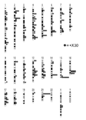
- One circle in the figure corresponds to one clone (total 185 clones). In a place where a plurality of circles are overlapped in the horizontal direction, it means that the number of circles is obtained from the same AluYa5 site.
- a large circle near the long arm telomere of chromosome 7 indicates that 30 clones derived from the AluYa5 site at that location were obtained.
- This local amplification of chromosome 7 with AluYa5 is the origin of the 120 base pair long band (arrow) in FIG. 3A.
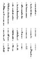
- the figure which shows the position in the genome of the amplification product at the time of using RE19 and RE20 as a primer.
- One circle corresponds to one clone (79 clones in total).
- FIG. 1 shows one embodiment of the present invention and does not limit the scope of the present invention.
- genomic DNA to be detected as a polymorphism is treated with a restriction enzyme having a cleavage site inside RE (FIG. 1A).
- the genomic DNA is cleaved into a plurality of DNA fragments (FIG. 1B).
- Each DNA fragment is self-circulated by ligase (FIG. 1C).
- ligase FIG. 1C
- FIG. 1C Although many circularized DNA fragments are generated, attention is paid to those containing the 3 'end of the RE, and primers for inverse PCR corresponding to the RE contained in this fragment are designed.
- a sequence for identifying genomic DNA is added to the primer on the 3 ′ end side of RE (FIG. 1C).
- a DNA fragment having an internal sequence of RE at one end and a sequence for identifying genomic DNA at the other end is amplified (FIG. 1D).
- This DNA fragment is treated with the same restriction enzyme as originally used.
- a DNA fragment having a peripheral sequence of RE at one end and a sequence for identifying genomic DNA at the other end is generated (FIG. 1E).
- Such a DNA fragment is prepared for each genomic DNA to be detected for polymorphism, and after mixing them, the base sequence of the sequence for distinguishing the peripheral sequence of RE from the genomic DNA is determined. Since it is possible to determine from which genomic DNA the sequence around each RE originates from the sequence for identifying the genomic DNA, the sequence around the RE is compared between the genomic DNAs based on that information. Thereby, the polymorphism of RE can be detected.
- step (1) a plurality of genomic DNAs to be detected as polymorphisms are treated for each genomic DNA with a restriction enzyme having at least one cleavage site inside the RE.
- the genomic DNA is preferably human genomic DNA. However, it may be genomic DNA of non-human organisms such as rats, mice, and monkeys.
- the polymorphism detection method of the present invention is mainly intended for detection of polymorphisms in human genomic DNA, and therefore it is preferably an RE that has transposition activity in human genomic DNA.
- RE family L1, Alu, SVA, HERV and the like are known (Trends in Genetics, Mills, R.E., et al., 2005). These RE families include a number of subfamilies.
- L1 includes subfamilies such as L1HS, L1PA2, L1PA3, and Alu is AluSc, AluSg, AluSp, AluSq, AluSx, AluSz, AluY, AluYa, AlyYb, AluYc, AluYd, AluYe, AluYf, AluYf, AluYf, AluYf, It includes subfamilies such as AluYh, AluYi, AluYj, AluYa5, AluYb8, and AluYc1, and SVA includes subfamilies such as SVA-B, SVA-C, SVA-D, SVA-E, and SVA-F.
- HERV contains a subfamily such as HERV-K.
- L1HS, L1PA2, L1PA3, etc. can be exemplified as the subfamily that is likely to cause polymorphism in L1, and AluYa5, AluYb8, AluYc1, AluY, etc. can be exemplified as the subfamily that is likely to cause polymorphism in Alu.
- Examples of easy subfamilies include SVA-E and SVA-F, and examples of subfamilies where polymorphisms in HERV are likely to occur include HERV-K.
- the restriction enzyme is not particularly limited as long as it has at least one cleavage site inside the RE, but (1) the recognition site and the cleavage site are class II, and (2) the recognition sequence is a palindromic sequence. (3) It is preferable that the cut end is a protruding end.
- the length of the recognition sequence is not particularly limited, but since the length of the resulting DNA fragment is preferably 500 base pairs or less, the length of the recognition sequence is preferably 4 to 6 bases.
- the restriction enzyme actually used may be determined according to the type of RE to be detected as a polymorphism. For example, when the polymorphism detection target is L1, BfaI, MseI, SimI, etc.
- polymorphism detection target when the polymorphism detection target is Alu, Tsp509I, TaqI, BstYI, etc. may be used, When the polymorphism detection target is SVA, Csp6I or the like may be used. When the polymorphism detection target is HERV, XhoI or the like may be used.
- step (2) the DNA fragment cleaved by the restriction enzyme is self-cyclized.
- Self-cyclization of the DNA fragment can be performed using ligase or the like according to a conventional method.
- step (3) polymerase chain reaction is performed using the first and second primers and the self-cyclized DNA fragment as a template.
- the first and second primers are primers that hybridize to the region between the end of RE and the restriction enzyme cleavage site closest to that end, the first primer is located on the restriction enzyme cleavage site side, and the restriction enzyme
- the DNA is designed to extend in the direction of the cleavage site, and the second primer is located on the terminal side of the RE and is designed to extend the DNA in the direction of the terminal of the RE.
- Such primers can be appropriately designed based on the sequence information of RE and the like.
- the first and second primers may be primers as described above, but more preferably primers such as the following (A) and / or (B).
- “smear” is a molecular organism that describes an electrophoretic image observed when a mixture of DNA fragments having different lengths and different lengths from 1 to 20 base pairs is electrophoresed.
- a scientific term As derived from the English word “smear”, “smear” is an electrophoretic image in which DNA fragments are smeared on the electrophoresis lane along the direction of electrophoresis. The width of “smear” corresponds to the full length range of the DNA fragment that is a PCR amplification product.
- the retroelement to be amplified is preferably a retroelement that is likely to cause polymorphism between genomic DNAs.
- the first and second primers should be primers that contain sequences that are the same or complementary to sequences that are included in the retroelements to be amplified and that are not included in other retroelements belonging to the same family. Is preferred.
- the 5 'end of the first primer is chemically modified with biotin in advance, so that only the desired PCR product can be concentrated and purified with streptavidin. As a result, DNA fragments containing the peripheral sequence of RE can be separated and purified from genomic DNA and PCR products fragmented during the treatment.
- a sequence for identifying genomic DNA is added to the second primer.
- the length of this sequence is not particularly limited, but preferably 3 to 5 bases.
- a sequence for identifying genomic DNA may be added to the first primer.
- primers are designed based on either of these regions. May be.
- L1 and SVA it is possible to design a primer based on the region on the 5 'end side, but it is preferable to design the primer based on the region on the 3' end side. This is because many of these retrotransposons that can generate RE polymorphism often migrate in a form that lacks the 5'-terminal region, so primers designed based on the 3'-terminal region However, the RE polymorphism can be detected comprehensively.
- the conditions for the polymerase chain reaction are not particularly limited, and may be the same as those for a general inverse PCR method.
- the restriction enzyme used is preferably the same restriction enzyme as that used in step (1). By using this restriction enzyme, the internal sequence of RE can be completely removed.
- a restriction enzyme that cleaves near the cleavage site of this restriction enzyme may be used. Such restriction enzymes can be appropriately selected based on the internal sequence of RE.
- the vicinity of the cleavage site usually means within the range of 1-1000 bases from the cleavage site, preferably within the range of 1-100 bases, and more preferably within the range of 1-20 bases. Particularly preferably, it means within the range of 1 to 10 bases.
- step (4) the base sequence of the peripheral sequence of RE is determined.
- the method for determining the base sequence is not particularly limited, and can be performed according to a conventional method, but it is preferably performed using a “next-generation sequencer” capable of rapid sequencing.
- a “next-generation sequencer” capable of rapid sequencing.
- a method of identifying the base sequence of the immobilized DNA by distinguishing the type by fluorescence or chemiluminescence is preferred.
- the length of the base sequence to be determined is not particularly limited as long as the position on the genome of RE can be specified, but it is preferably 50 to 100 base pairs, and more preferably 25 to 35 base pairs. .
- the base sequence of this sequence is determined in addition to the peripheral sequence of RE.
- base sequence information can be sorted for each genomic DNA, so it is possible to mix DNA fragments derived from each genomic DNA and determine the base sequence of the peripheral sequence of the RE at once. Become.
- the above-mentioned Genome Analyzer manufactured by Illumina can decipher the base sequences at both ends of the DNA fragment, and can simultaneously determine both the peripheral sequence of RE and the sequence for identification. It is suitable as a sequencer used in.
- the determined peripheral sequence of RE is compared between genomic DNAs to be detected as polymorphisms or compared with genomic DNA in a database, and the peripheral sequence of RE included in a specific genomic DNA Is detected.
- a database related to human genome sequences can be used. Any database related to the human genome sequence to be used may be used. For example, UCSC Genome Browser, Ensembl genome Browser, etc. can be used.
- the present invention also includes “a method for producing a DNA fragment containing a peripheral sequence of RE” and “a method for determining the position of RE on genomic DNA”.
- the “method for producing a DNA fragment containing a peripheral sequence of RE” may be performed by performing steps (1) to (3) of the polymorphism detection method, and “the method for determining the position of RE on genomic DNA” Steps (1) to (4) of the polymorphism detection method are performed, the base sequence of the determined peripheral sequence of RE is compared with the genome database, and the position of RE on the genome may be determined.
- Browser etc. can be used, for example.
- Consensus sequences for the L1 subfamily (L1HS and L1PA2) that can generate L1 polymorphisms are obtained from Repbase (http://www.girinst.org/repbase/index.html), a retroelement database Based on this sequence, BfaI was selected as a restriction enzyme.
- HeLa cell-derived genomic DNA (1 mg) was reacted with BfaI restriction enzyme at 37 ° C for 2 hours, inactivated at 80 ° C, and eluted with 50 ⁇ l of buffer when purified by Qiagen PCR-Purification-Kit. Among them, 5 ⁇ l of genomic DNA solution was mixed with 5 ⁇ l of Ligation Mix (Wako), and self-cyclization reaction was performed at 16 ° C. for 5 minutes.
- a PCR reaction was performed with 1 ⁇ l of this genomic DNA solution in a total volume of 50 ⁇ l.
- a plurality of primers were designed by the public primer design website Primer3 (http://frodo.wi.mit.edu/cgi-bin/primer3/primer3www.cgi).
- Reverse primer REp002: GTGCGCTGCACCCACTAAC (SEQ ID NO: 2)
- the reaction solution contains REp001 (0.3 ⁇ M), REp002 (0.3 ⁇ M), 1 ⁇ GXL Buffer (Takara), dNTPs (each 0.2 mM), and Primerstar GXL (Takara; 1.25 U).
- the PCR reaction was performed at 98 ° C for 10 seconds and 68 ° C for 2 minutes after 5 cycles, followed by 98 cycles of 98 ° C for 10 seconds, 65 ° C for 15 seconds and 68 ° C for 2 minutes for 35 cycles. Thereafter, DNA of 1 kb or less was separated by 1% agarose electrophoresis, and then purified by Qiagen Gel Extraction Kit. The purified DNA fragment was cloned by TA TOPO Cloning of Invitrogen, extended by M13 Forward primer using BigDye terminator cycle sequencing kit (ABI), and the base sequence was determined by ABI Sequencer 3100.
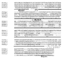
- the base sequence of 4 clones from which sequences were obtained is shown in FIG. Of these 4 clones, 3 clones contained the 3 'end region of L1HS and the remaining 1 clone contained the 3' end region of L1PA2.
- the L1 peripheral sequence was completely different for every 4 clones. From this, it is considered that these four L1s are present at completely different positions on the genomic DNA.
- BfaI is also present in the clone sequence, by treating the PCR reaction product with BfaI, a DNA fragment having an L1 peripheral sequence at the end that can be easily decoded with a high-speed sequencer can be produced. Conceivable.
- Example 2 Electrophoretic image of amplified product and position in genome Using two sets of primers, the peripheral sequence of retroelement AluYa5 was amplified by PCR, and an electrophoretic image of the amplified product was obtained. Moreover, the base sequence of each amplification product was determined, and the position in the genome was determined.
- PCR A PCR reaction was performed in a total volume of 50 ⁇ l using 20 ⁇ l of the above 25 ⁇ l post-ligation solution. Two sets of primers, RE101 and RE102, and RE19 and RE20, were used. Primer sequences are shown below.
- RE19 and RE20 amplify only AluYa5, which is prone to polymorphism, and belong to the same Alu family, so as not to amplify AluS and AluJ where polymorphism cannot occur. It is designed based on each base sequence from 224th to 241st. These sequences include sequences specific to AluYa5 at the 148th, 155th, 165th, 236th, and 241st positions.
- the reaction solution contains Forward primer (0.2 ⁇ M), Reverse primer (0.2 ⁇ M), 1 ⁇ GXL buffer (Takara), dNTP (each 0.25 ⁇ M), and PrimeSTAR GXL (Takara; 0.75 ⁇ U).
- the PCR reaction was performed at 98 ° C. for 10 seconds, 50 ° C. for 15 seconds, and 68 ° C. for 30 seconds for 30 cycles. Thereafter, DNA of 600 bp or less was separated by 4% agarose electrophoresis (FIGS. 3A and 3B) and purified by QIAGEN-Gel-Extraction-Kit.
- AluYa5 is amplified more comprehensively when RE19 and RE20 with smeared electrophoretic images are used than when RE101 and RE102 with specific bands detected are used. Can do.
- the present invention makes it possible to easily acquire information on the RE polymorphism.
- Such information on the RE polymorphism is useful for the development of new genetic diagnostic agents and the quality control of cells used in regenerative medicine.
Landscapes
- Chemical & Material Sciences (AREA)
- Organic Chemistry (AREA)
- Life Sciences & Earth Sciences (AREA)
- Chemical Kinetics & Catalysis (AREA)
- Zoology (AREA)
- Wood Science & Technology (AREA)
- Proteomics, Peptides & Aminoacids (AREA)
- Health & Medical Sciences (AREA)
- Engineering & Computer Science (AREA)
- Biophysics (AREA)
- Immunology (AREA)
- Microbiology (AREA)
- Molecular Biology (AREA)
- Biotechnology (AREA)
- Analytical Chemistry (AREA)
- Physics & Mathematics (AREA)
- Biochemistry (AREA)
- Bioinformatics & Cheminformatics (AREA)
- General Engineering & Computer Science (AREA)
- General Health & Medical Sciences (AREA)
- Genetics & Genomics (AREA)
- Measuring Or Testing Involving Enzymes Or Micro-Organisms (AREA)
Abstract
網羅性及び迅速性の高いレトロエレメント多型検出手段を提供することを目的として、レトロエレメントによって生じた多型を検出する方法であって、以下の(1)~(5)の工程を含むことを特徴とする多型の検出方法を提供する、(1)多型の検出対象とする複数のゲノムDNAを、ゲノムDNAごとに、レトロエレメント内部に少なくとも一つの切断部位を持つ制限酵素で処理する工程、(2)制限酵素によって切断されたDNA断片を自己環化させる工程、(3)自己環化させたDNA断片を鋳型としてインバースPCRを行う工程、(4)増幅されたDNA断片のレトロエレメントの周辺配列の塩基配列を決定する工程、(5)決定したレトロエレメントの周辺配列を、多型の検出対象とするゲノムDNA間で比較、又はデータベース上のゲノムDNAと比較し、特定のゲノムDNAに含まれていたレトロエレメントの周辺配列を検出する工程。
Description
本発明は、レトロエレメントの周辺配列を含むDNA断片の作製方法、レトロエレメントのゲノムDNA上の位置の決定方法、及びレトロエレメントによって生じた多型の検出方法に関する。
DNA配列解読技術が進み、ヒトゲノム配列には個人差があることが明らかになっている。個人間のゲノム配列の相違(以下、「ゲノム多型」という)が、病気の罹りやすさや、薬剤に対する感受性や耐性などの、生化学的な個人体質の違いを生み出す主要因であると考えられている。そこで現在では、ヒトゲノムに存在する全てのゲノム多型をデータベース化し、個人のゲノム配列情報と照合させることで、診断技術の向上および医療の個別化を目指す研究が進んでいる。このゲノム科学をベースとする次世代医療を実現するためには、ゲノム多型を効率よく検出する技術が必須である。
ゲノム多型は、その発生メカニズムから大きく3種類(一塩基多型、コピー数多型、レトロエレメント(以下、「RE」という)多型)に分類される。これらのうち前者2つの多型を効率よく検出する技術はすでに確立され、すでに市場に出回っているのに対して、RE多型を網羅的に検出する技術の開発は進んでいない。その主要因は、REが長い間「がらくたDNA」と呼ばれ、その生物学的重要性が軽視されていたためであるが、過去10年ほどの間に、REが多くの遺伝子の機能や制御に深く関わっていることが徐々に明らかになってきた。さらにRE多型が疾患の原因であった症例も年々増加している。RE多型の重要性が明確になった今こそ、RE多型検出法を1日でも早く構築し、個別化医療に必要なゲノム解析技術を整備することで、生活の質(Quality of Life)の向上に貢献すべきである。
REの多型検出法としては、多型を生み出す可能性のある各REファミリーに対するプローブを用いるサザンブロティング法が容易に考えられるが、その低い解像度のため、検出できるRE多型の数は少なく、その網羅性はかなり低い。さらにこの方法では、多型の有無を確認できるだけであり、そのゲノム内位置情報は得られないという致命的な欠点がある。
また、RE多型候補ゲノム領域を、候補ゲノム領域に対して、個別にプライマーを用意してPCR増幅後にアガロース電気泳動より検出する方法が一般的であるが(非特許文献1)、この方法では膨大な数のプライマーを用意する必要があるため、実用的ではない。
J.S. Myers et al., Am. J. Hum. Genet., Volume 71, (2002), Pages312-326.
本発明は、以上のような技術的背景の下になされたものであり、網羅性及び迅速性の高いRE多型検出手段を提供することを目的とする。
本発明者は、上記課題を解決するため鋭意検討を重ねた結果、インバースPCRを利用してREの周辺配列を決定し、それによりRE多型を検出するという発想を得た。しかし、通常のインバースPCRによって増幅される断片は、RE内部配列が末端に存在し、その後に周辺配列が続くので、末端から配列を決定していく場合、REの周辺配列だけでなくREの内部配列の配列も決定しなければならないという問題がある。この問題は、配列決定可能な塩基の長さが限られている場合に特に深刻化する。例えば、Illumina社製のGenome Analyzerは、高速シークエンスが可能な装置であるが、読み取り可能な配列の長さは35~50塩基対程度であるため、REの内部配列が長い場合には、REのゲノム上の位置を決定できるほどの長さのREの周辺配列情報を得られない可能性がある。さらに、多型REの隣接配列は、RE多型を生み出すレトロトランスポゾン挿入の際に大きく改変及び修飾されているため、REから少し離れた周辺配列情報を得る必要がある。
本発明者は、以上のような問題点を初めて認識し、そして、この問題点は、(1)REを制限酵素で処理し、(2)それによって生じるDNA断片の中からREの末端を含むDNA断片を鋳型としてインバースPCRを行い、(3)その増幅産物を前記した制限酵素で処理することにより解消できることを見出した。
また、本発明者は、ゲノムDNA上のREの周辺配列をインバースPCRによって増幅させた場合、プライマーによっては、増幅産物に偏りが生じ、REの周辺配列を網羅的に増幅できないことがあることを見出した。そして、この問題は、予め候補とするプライマーを幾つか合成しておき、それらによるPCR増幅産物を電気泳動し、その電気泳動像がスメアになるプライマーを選択することによって解決できることも見出した。
本発明は、以上の知見に基づき完成されたものである。
即ち、本発明は、以下の〔1〕~〔21〕を提供する。
〔1〕ゲノムDNAに含まれるレトロエレメントの周辺配列を含むDNA断片を作製する方法であって、以下の(1)~(3)の工程を含むことを特徴とするDNA断片の作製方法、(1)DNA断片の作製対象とするゲノムDNAを、レトロエレメント内部に少なくとも一つの切断部位を持つ制限酵素で処理する工程、(2)制限酵素によって切断されたDNA断片を自己環化させる工程、(3)下記の第一及び第二のプライマーを用い、自己環化させたDNA断片を鋳型としてポリメラーゼ連鎖反応を行う工程、第一及び第二のプライマーは、レトロエレメントの末端とその末端に最も近い制限酵素切断部位との間の領域にハイブリダイズするプライマーで、第一のプライマーは制限酵素切断部位側に位置し、制限酵素切断部位方向にDNAを伸長させるように設計されており、第二のプライマーはレトロエレメントの末端側に位置し、レトロエレメントの末端方向にDNAを伸長させるように設計されている。
〔2〕工程(3)において増幅されたDNA断片を制限酵素で処理し、第一のプライマー側の末端を切除することを特徴とする〔1〕に記載のDNA断片の作製方法。
〔3〕制限酵素が、工程(1)で使用した制限酵素と同一の制限酵素であることを特徴とする〔2〕に記載のDNA断片の作製方法。
〔4〕レトロエレメントが、L1又はSVAであり、工程(3)において使用する第一及び第二のプライマーが、レトロエレメントの3’末端と3’末端に最も近い制限酵素切断部位との間の領域にハイブリダイズするプライマーであることを特徴とする〔1〕乃至〔3〕のいずれかに記載のDNA断片の作製方法。
〔5〕工程(3)において使用する第二のプライマーに、ゲノムDNAを識別するための配列が付加されていることを特徴とする〔1〕乃至〔4〕のいずれかに記載のDNA断片の作製方法。
〔6〕ポリメラーゼ連鎖反応の増幅産物を電気泳動し、その電気泳動像がスメアになるプライマーを工程(3)において使用する第一及び第二プライマーとすることを特徴とする〔1〕乃至〔5〕のいずれかに記載のDNA断片の作製方法。
〔7〕対象とするレトロエレメントがゲノムDNA間で多型を生じ易いレトロエレメントであり、そのレトロエレメントに含まれ、そのレトロエレメントと同一のファミリーに属するレトロエレメントに含まれない配列と同一又は相補的な配列を含むプライマーを工程(3)において使用する第一及び第二プライマーとすることを特徴とする〔1〕乃至〔6〕のいずれかに記載のDNA断片の作製方法。
〔8〕ゲノムDNAに含まれるレトロエレメントのゲノムDNA上の位置を決定する方法であって、以下の(1)~(5)の工程を含むことを特徴とするレトロエレメントの位置決定方法、(1)レトロエレメントの位置決定の対象とするゲノムDNAを、レトロエレメント内部に少なくとも一つの切断部位を持つ制限酵素で処理する工程、(2)制限酵素によって切断されたDNA断片を自己環化させる工程、(3)下記の第一及び第二のプライマーを用い、自己環化させたDNA断片を鋳型としてポリメラーゼ連鎖反応を行う工程、第一及び第二のプライマーは、レトロエレメントの末端とその末端に最も近い制限酵素切断部位との間の領域にハイブリダイズするプライマーで、第一のプライマーは制限酵素切断部位側に位置し、制限酵素切断部位方向にDNAを伸長させるように設計されており、第二のプライマーはレトロエレメントの末端側に位置し、レトロエレメントの末端方向にDNAを伸長させるように設計されている、(4)増幅されたDNA断片のレトロエレメントの周辺配列の塩基配列を決定する工程、(5)決定したレトロエレメントの周辺配列の塩基配列とゲノムデータベースとを比較し、レトロエレメントのゲノム上の位置を決定する工程。
〔9〕工程(3)において増幅されたDNA断片を制限酵素で処理し、第一のプライマー側の末端を切除することを特徴とする〔8〕に記載のレトロエレメントの位置決定方法。
〔10〕制限酵素が、工程(1)で使用した制限酵素と同一の制限酵素であることを特徴とする〔9〕に記載のレトロエレメントの位置決定方法。
〔11〕レトロエレメントが、L1又はSVAであり、第一及び第二のプライマーが、レトロエレメントの3’末端と3’末端に最も近い制限酵素切断部位との間の領域にハイブリダイズするプライマーであることを特徴とする〔8〕乃至〔10〕のいずれかに記載のレトロエレメントの位置決定方法。
〔12〕工程(3)において使用する第二のプライマーに、ゲノムDNAを識別するための配列が付加されており、工程(4)においてレトロエレメントの周辺配列と共にゲノムDNAを識別するための配列の塩基配列を決定することを特徴とする〔8〕乃至〔11〕のいずれか一項に記載のレトロエレメントの位置決定方法。
〔13〕ポリメラーゼ連鎖反応の増幅産物を電気泳動し、その電気泳動像がスメアになるプライマーを工程(3)において使用する第一及び第二プライマーとすることを特徴とする〔8〕乃至〔12〕のいずれかに記載のレトロエレメントの位置決定方法。
〔14〕対象とするレトロエレメントがゲノムDNA間で多型を生じ易いレトロエレメントであり、そのレトロエレメントに含まれ、そのレトロエレメントと同一のファミリーに属するレトロエレメントに含まれない配列と同一又は相補的な配列を含むプライマーを工程(3)において使用する第一及び第二プライマーとすることを特徴とする〔8〕乃至〔13〕のいずれかに記載のレトロエレメントの位置決定方法。
〔15〕レトロエレメントによって生じた多型を検出する方法であって、以下の(1)~(5)の工程を含むことを特徴とする多型の検出方法、(1)多型の検出対象とする複数のゲノムDNAを、ゲノムDNAごとに、レトロエレメント内部に少なくとも一つの切断部位を持つ制限酵素で処理する工程、(2)制限酵素によって切断されたDNA断片を自己環化させる工程、(3)下記の第一及び第二のプライマーを用い、自己環化させたDNA断片を鋳型としてポリメラーゼ連鎖反応を行う工程、第一及び第二のプライマーは、レトロエレメントの末端とその末端に最も近い制限酵素切断部位との間の領域にハイブリダイズするプライマーで、第一のプライマーは制限酵素切断部位側に位置し、制限酵素切断部位方向にDNAを伸長させるように設計されており、第二のプライマーはレトロエレメントの末端側に位置し、レトロエレメントの末端方向にDNAを伸長させるように設計されている、(4)増幅されたDNA断片のレトロエレメントの周辺配列の塩基配列を決定する工程、(5)決定したレトロエレメントの周辺配列を、多型の検出対象とするゲノムDNA間で比較、又はデータベース上のゲノムDNAと比較し、特定のゲノムDNAに含まれていたレトロエレメントの周辺配列を検出する工程。
〔16〕工程(3)において増幅されたDNA断片を制限酵素で処理し、第一のプライマー側の末端を切除することを特徴とする〔15〕に記載の多型の検出方法。
〔17〕制限酵素が、工程(1)で使用した制限酵素と同一の制限酵素であることを特徴とする〔16〕に記載の多型の検出方法。
〔18〕レトロエレメントが、L1又はSVAであり、工程(3)において使用する第一及び第二のプライマーが、レトロエレメントの3’末端と3’末端に最も近い制限酵素切断部位との間の領域にハイブリダイズするプライマーであることを特徴とする〔15〕乃至〔17〕のいずれか一項に記載の多型の検出方法。
〔19〕工程(3)において使用する第二のプライマーに、ゲノムDNAを識別するための配列が付加されており、工程(4)において各ゲノムDNA由来のDNA断片を混合した後、レトロエレメントの周辺配列と共にゲノムDNAを識別するための配列の塩基配列を決定することを特徴とする〔15〕乃至〔18〕のいずれかに記載の多型の検出方法。
〔20〕ポリメラーゼ連鎖反応の増幅産物を電気泳動し、その電気泳動像がスメアになるプライマーを工程(3)において使用する第一及び第二プライマーとすることを特徴とする〔15〕乃至〔19〕のいずれかに記載の多型の検出方法。
〔21〕対象とするレトロエレメントがゲノムDNA間で多型を生じ易いレトロエレメントであり、そのレトロエレメントに含まれ、そのレトロエレメントと同一のファミリーに属するレトロエレメントに含まれない配列と同一又は相補的な配列を含むプライマーを工程(3)において使用する第一及び第二プライマーとすることを特徴とする〔15〕乃至〔20〕のいずれか一項に記載の多型の検出方法。
〔1〕ゲノムDNAに含まれるレトロエレメントの周辺配列を含むDNA断片を作製する方法であって、以下の(1)~(3)の工程を含むことを特徴とするDNA断片の作製方法、(1)DNA断片の作製対象とするゲノムDNAを、レトロエレメント内部に少なくとも一つの切断部位を持つ制限酵素で処理する工程、(2)制限酵素によって切断されたDNA断片を自己環化させる工程、(3)下記の第一及び第二のプライマーを用い、自己環化させたDNA断片を鋳型としてポリメラーゼ連鎖反応を行う工程、第一及び第二のプライマーは、レトロエレメントの末端とその末端に最も近い制限酵素切断部位との間の領域にハイブリダイズするプライマーで、第一のプライマーは制限酵素切断部位側に位置し、制限酵素切断部位方向にDNAを伸長させるように設計されており、第二のプライマーはレトロエレメントの末端側に位置し、レトロエレメントの末端方向にDNAを伸長させるように設計されている。
〔2〕工程(3)において増幅されたDNA断片を制限酵素で処理し、第一のプライマー側の末端を切除することを特徴とする〔1〕に記載のDNA断片の作製方法。
〔3〕制限酵素が、工程(1)で使用した制限酵素と同一の制限酵素であることを特徴とする〔2〕に記載のDNA断片の作製方法。
〔4〕レトロエレメントが、L1又はSVAであり、工程(3)において使用する第一及び第二のプライマーが、レトロエレメントの3’末端と3’末端に最も近い制限酵素切断部位との間の領域にハイブリダイズするプライマーであることを特徴とする〔1〕乃至〔3〕のいずれかに記載のDNA断片の作製方法。
〔5〕工程(3)において使用する第二のプライマーに、ゲノムDNAを識別するための配列が付加されていることを特徴とする〔1〕乃至〔4〕のいずれかに記載のDNA断片の作製方法。
〔6〕ポリメラーゼ連鎖反応の増幅産物を電気泳動し、その電気泳動像がスメアになるプライマーを工程(3)において使用する第一及び第二プライマーとすることを特徴とする〔1〕乃至〔5〕のいずれかに記載のDNA断片の作製方法。
〔7〕対象とするレトロエレメントがゲノムDNA間で多型を生じ易いレトロエレメントであり、そのレトロエレメントに含まれ、そのレトロエレメントと同一のファミリーに属するレトロエレメントに含まれない配列と同一又は相補的な配列を含むプライマーを工程(3)において使用する第一及び第二プライマーとすることを特徴とする〔1〕乃至〔6〕のいずれかに記載のDNA断片の作製方法。
〔8〕ゲノムDNAに含まれるレトロエレメントのゲノムDNA上の位置を決定する方法であって、以下の(1)~(5)の工程を含むことを特徴とするレトロエレメントの位置決定方法、(1)レトロエレメントの位置決定の対象とするゲノムDNAを、レトロエレメント内部に少なくとも一つの切断部位を持つ制限酵素で処理する工程、(2)制限酵素によって切断されたDNA断片を自己環化させる工程、(3)下記の第一及び第二のプライマーを用い、自己環化させたDNA断片を鋳型としてポリメラーゼ連鎖反応を行う工程、第一及び第二のプライマーは、レトロエレメントの末端とその末端に最も近い制限酵素切断部位との間の領域にハイブリダイズするプライマーで、第一のプライマーは制限酵素切断部位側に位置し、制限酵素切断部位方向にDNAを伸長させるように設計されており、第二のプライマーはレトロエレメントの末端側に位置し、レトロエレメントの末端方向にDNAを伸長させるように設計されている、(4)増幅されたDNA断片のレトロエレメントの周辺配列の塩基配列を決定する工程、(5)決定したレトロエレメントの周辺配列の塩基配列とゲノムデータベースとを比較し、レトロエレメントのゲノム上の位置を決定する工程。
〔9〕工程(3)において増幅されたDNA断片を制限酵素で処理し、第一のプライマー側の末端を切除することを特徴とする〔8〕に記載のレトロエレメントの位置決定方法。
〔10〕制限酵素が、工程(1)で使用した制限酵素と同一の制限酵素であることを特徴とする〔9〕に記載のレトロエレメントの位置決定方法。
〔11〕レトロエレメントが、L1又はSVAであり、第一及び第二のプライマーが、レトロエレメントの3’末端と3’末端に最も近い制限酵素切断部位との間の領域にハイブリダイズするプライマーであることを特徴とする〔8〕乃至〔10〕のいずれかに記載のレトロエレメントの位置決定方法。
〔12〕工程(3)において使用する第二のプライマーに、ゲノムDNAを識別するための配列が付加されており、工程(4)においてレトロエレメントの周辺配列と共にゲノムDNAを識別するための配列の塩基配列を決定することを特徴とする〔8〕乃至〔11〕のいずれか一項に記載のレトロエレメントの位置決定方法。
〔13〕ポリメラーゼ連鎖反応の増幅産物を電気泳動し、その電気泳動像がスメアになるプライマーを工程(3)において使用する第一及び第二プライマーとすることを特徴とする〔8〕乃至〔12〕のいずれかに記載のレトロエレメントの位置決定方法。
〔14〕対象とするレトロエレメントがゲノムDNA間で多型を生じ易いレトロエレメントであり、そのレトロエレメントに含まれ、そのレトロエレメントと同一のファミリーに属するレトロエレメントに含まれない配列と同一又は相補的な配列を含むプライマーを工程(3)において使用する第一及び第二プライマーとすることを特徴とする〔8〕乃至〔13〕のいずれかに記載のレトロエレメントの位置決定方法。
〔15〕レトロエレメントによって生じた多型を検出する方法であって、以下の(1)~(5)の工程を含むことを特徴とする多型の検出方法、(1)多型の検出対象とする複数のゲノムDNAを、ゲノムDNAごとに、レトロエレメント内部に少なくとも一つの切断部位を持つ制限酵素で処理する工程、(2)制限酵素によって切断されたDNA断片を自己環化させる工程、(3)下記の第一及び第二のプライマーを用い、自己環化させたDNA断片を鋳型としてポリメラーゼ連鎖反応を行う工程、第一及び第二のプライマーは、レトロエレメントの末端とその末端に最も近い制限酵素切断部位との間の領域にハイブリダイズするプライマーで、第一のプライマーは制限酵素切断部位側に位置し、制限酵素切断部位方向にDNAを伸長させるように設計されており、第二のプライマーはレトロエレメントの末端側に位置し、レトロエレメントの末端方向にDNAを伸長させるように設計されている、(4)増幅されたDNA断片のレトロエレメントの周辺配列の塩基配列を決定する工程、(5)決定したレトロエレメントの周辺配列を、多型の検出対象とするゲノムDNA間で比較、又はデータベース上のゲノムDNAと比較し、特定のゲノムDNAに含まれていたレトロエレメントの周辺配列を検出する工程。
〔16〕工程(3)において増幅されたDNA断片を制限酵素で処理し、第一のプライマー側の末端を切除することを特徴とする〔15〕に記載の多型の検出方法。
〔17〕制限酵素が、工程(1)で使用した制限酵素と同一の制限酵素であることを特徴とする〔16〕に記載の多型の検出方法。
〔18〕レトロエレメントが、L1又はSVAであり、工程(3)において使用する第一及び第二のプライマーが、レトロエレメントの3’末端と3’末端に最も近い制限酵素切断部位との間の領域にハイブリダイズするプライマーであることを特徴とする〔15〕乃至〔17〕のいずれか一項に記載の多型の検出方法。
〔19〕工程(3)において使用する第二のプライマーに、ゲノムDNAを識別するための配列が付加されており、工程(4)において各ゲノムDNA由来のDNA断片を混合した後、レトロエレメントの周辺配列と共にゲノムDNAを識別するための配列の塩基配列を決定することを特徴とする〔15〕乃至〔18〕のいずれかに記載の多型の検出方法。
〔20〕ポリメラーゼ連鎖反応の増幅産物を電気泳動し、その電気泳動像がスメアになるプライマーを工程(3)において使用する第一及び第二プライマーとすることを特徴とする〔15〕乃至〔19〕のいずれかに記載の多型の検出方法。
〔21〕対象とするレトロエレメントがゲノムDNA間で多型を生じ易いレトロエレメントであり、そのレトロエレメントに含まれ、そのレトロエレメントと同一のファミリーに属するレトロエレメントに含まれない配列と同一又は相補的な配列を含むプライマーを工程(3)において使用する第一及び第二プライマーとすることを特徴とする〔15〕乃至〔20〕のいずれか一項に記載の多型の検出方法。
本発明の方法により、ゲノムDNA配列の個人間の違いに大きな影響を与えるRE多型を網羅的かつ迅速に検出することが可能になる。RE多型以外にも、RE配列周辺のゲノムDNA配列の個人間の違いを網羅的にかつ迅速に検出することも可能となる。
以下、本発明を詳細に説明する。
最初に本発明の多型検出方法の原理を図1によって説明する。なお、図1は本発明の一態様を示すものであり、本発明の範囲を限定するものではない。
まず、多型の検出対象とするゲノムDNAをRE内部に切断部位を持つ制限酵素で処理する(図1A)。これにより、ゲノムDNAは、複数のDNA断片に切断される(図1B)。各DNA断片は、リガーゼにより自己環化させる(図1C)。環化したDNA断片は多数生じるが、その中のREの3’末端を含むものに着目し、この断片に含まれるREに対応したインバースPCR用のプライマーを設計する(図1C)。この際、REの3’末端側のプライマーには、ゲノムDNAを識別するための配列を付加しておく(図1C)。以上のようなプライマーを用いてインバースPCRを行うことにより、一方の末端にREの内部配列を持ち、他方の末端にゲノムDNAを識別するための配列を持つDNA断片が増幅される(図1D)。このDNA断片を最初に使用したものと同一の制限酵素で処理する。これにより、一方の末端にREの周辺配列を持ち、他方の末端にゲノムDNAを識別するための配列を持つDNA断片が生じる(図1E)。このようなDNA断片を、多型の検出対象とするゲノムDNAごとに作製し、それらを混合した後、REの周辺配列とゲノムDNAを識別するための配列の塩基配列を決定する。ゲノムDNAを識別するための配列により、各々のREの周辺配列がどのゲノムDNAに由来するものか判別することが可能なので、その情報に基づき、REの周辺配列をゲノムDNA間で比較する。これにより、REの多型を検出することができる。
次に、本発明の多型検出方法を工程ごとに説明する。
工程(1)では、多型の検出対象とする複数のゲノムDNAを、ゲノムDNAごとに、RE内部に少なくとも一つの切断部位を持つ制限酵素で処理する。
本発明の多型検出方法は、主としてヒトゲノムDNAの多型の検出を目的とするものであるため、ゲノムDNAは、ヒトゲノムDNAであることが好ましい。但し、ヒト以外の生物、例えば、ラット、マウス、サルのゲノムDNAであってもよい。
REの種類は特に限定されないが、前述のように、本発明の多型検出方法は主としてヒトゲノムDNAの多型の検出を目的とするので、ヒトゲノムDNA内で転移活性のあるREであることが好ましい。このようなREのファミリーとしては、L1、Alu、SVA、HERVなどが知られている(Trends in Genetics, Mills, R.E., et al., 2005)。これらのREファミリーは、多数のサブファミリーを含んでいる。例えば、L1は、L1HS、L1PA2、L1PA3などのサブファミリーを含んでおり、Aluは、AluSc、AluSg、AluSp、AluSq、AluSx、AluSz、AluY、AluYa、AlyYb、AluYc、AluYd、AluYe、AluYf、AluYg、AluYh、AluYi、AluYj、AluYa5、AluYb8、AluYc1などのサブファミリーを含んでおり、SVAは、SVA-B、SVA-C、SVA-D、SVA-E、SVA-Fなどのサブファミリーを含んでおり、HERVは、HERV-Kなどのサブファミリーを含んでいる。L1における多型の生じ易いサブファミリーとしてはL1HS、L1PA2、L1PA3などを例示でき、Alu における多型の生じ易いサブファミリーとしてはAluYa5、AluYb8、AluYc1、AluYなどを例示でき、SVAにおける多型の生じ易いサブファミリーとしてはSVA-E、SVA-Fなどを例示でき、HERVにおける多型の生じ易いサブファミリーとしてはHERV-Kなどを例示できる。
制限酵素は、RE内部に少なくとも一つの切断部位を持つものであれば特に制限されないが、(1)認識部位と切断部位が一致しているクラスIIであり、(2)認識配列が回文配列であり、(3)切断末端が突出末端であることが好ましい。認識配列の長さは特に限定されないが、生じるDNA断片の長さは500塩基対以下であることが好ましいので、認識配列の長さは4~6塩基であることが好ましい。実際に使用する制限酵素は、多型の検出対象とするREの種類に応じて決めればよい。例えば、多型の検出対象をL1とした場合は、BfaI、MseI、SimIなどを使用すればよく、多型の検出対象をAluとした場合は、Tsp509I、TaqI、BstYIなどを使用すればよく、多型の検出対象をSVA とした場合は、Csp6Iなどを使用すればよく、多型の検出対象をHERVとした場合は、XhoIなどを使用すればよい。
工程(2)では、制限酵素によって切断されたDNA断片を自己環化させる。DNA断片の自己環化は、常法に従い、リガーゼなどを用いて行うことができる。
工程(3)では、第一及び第二のプライマーを用い、自己環化させたDNA断片を鋳型としてポリメラーゼ連鎖反応を行う。
第一及び第二のプライマーは、REの末端とその末端に最も近い制限酵素切断部位との間の領域にハイブリダイズするプライマーで、第一のプライマーは制限酵素切断部位側に位置し、制限酵素切断部位方向にDNAを伸長させるように設計されており、第二のプライマーはREの末端側に位置し、REの末端方向にDNAを伸長させるように設計されている。このようなプライマーは、REの配列情報などに基づき、適宜設計可能である。
第一及び第二のプライマーは、上記のようなプライマーであればよいが、下記の(A)及び/又は(B)のようなプライマーであることがより好ましい。
(A)ゲノムDNA上のREの周辺配列に対しインバースPCRを行ったときに、ゲノム内の特定のRE配列だけが優先的に増幅され、REの周辺配列を網羅的に増幅できない場合がある。このような場合、PCRの増幅産物を電気泳動すると、その電気泳動像に特定の長さのバンドが検出される。一方、増幅に偏りがなく、ゲノム内に散在する数多くのREの周辺配列を網羅的に増幅できる場合は、増幅産物の電気泳動像は「スメア」になる。従って、予め候補とするプライマーを幾つか合成しておき、それらによるPCR増幅産物を電気泳動し、その電気泳動像が「スメア」になるプライマーを第一及び第二のプライマーとすることにより、REの周辺配列を網羅的に増幅できる。ここで、「スメア」とは、全長が異なり、かつそれぞれの長さの違いが1から20塩基対までのDNA断片の混合物が電気泳動される場合に観察される電気泳動像を形容する分子生物学用語をいう。英語の「smear((油などを)塗り付ける)」に由来するように、電気泳動方向に沿って泳動レーンの上にDNA断片が塗り付けられたような泳動像が「スメア」である。また「スメア」の幅は、PCR増幅産物であるDNA断片の全長の範囲に相当する。
(B)本発明は、ゲノム多型の検出を主な目的とするため、増幅の対象とするレトロエレメントは、ゲノムDNA間で多型を生じ易いレトロエレメントであることが好ましい。この多型を生じ易いレトロエレメントを増幅させる場合、同時にこのレトロエレメントと同一のファミリーに属する他のレトロエレメントも増幅されてしまうことがある。これを避けるため、増幅の対象とするレトロエレメントに含まれ、同一のファミリーに属する他のレトロエレメントに含まれない配列と同一又は相補的な配列を含むプライマーを第一及び第二プライマーとすることが好ましい。
第一のプライマーの5'末端には、ビオチンなどをあらかじめ化学修飾することで、目的とするPCR産物だけをストレプトアビジンにより濃縮精製が可能になる。これにより、ゲノムDNAや処理途中で分断したPCR産物から、REの周辺配列を含むDNA断片を分離精製できる。
第二のプライマーには、ゲノムDNAを識別するための配列が付加されていることが好ましい。この配列の長さは特に限定されないが、3~5塩基であることが好ましい。ゲノムDNAを識別するための配列は、第一のプライマーに付加してもよい。
「REの末端とその末端に最も近い制限酵素切断部位との間の領域」は、REの5’末端側と3’末端側に二つ存在し、これらいずれの領域に基づいてプライマーを設計してもよい。但し、L1とSVAについては、5’末端側の領域に基づいてプライマーを設計することも可能であるが、3’末端側の領域に基づいてプライマーを設計することが好ましい。これは、RE多型を生み出す可能性のあるこれらレトロトランスポゾンの多くが、5’末端領域を欠失した形で転移することが多いため、3’末端側の領域に基づいてプライマーを設計した方が、網羅的にRE多型を検出できるからである。
ポリメラーゼ連鎖反応の条件は特に限定されず、一般的なインバースPCR法と同様の条件でよい。
増幅されたDNA断片の第一のプライマー側の末端には、REの内部配列が存在するので、これを切断、除去するため、増幅されたDNA断片を制限酵素で処理することが好ましい。使用する制限酵素は、工程(1)で使用した制限酵素と同一の制限酵素が好ましい。この制限酵素を用いることにより、REの内部配列を完全に除去できる。工程(1)で使用した制限酵素と同一の制限酵素の代わりに、この制限酵素の切断部位付近を切断する制限酵素を用いてもよい。このような制限酵素はREの内部配列を基に適宜選択可能である。なお、切断部位付近とは、切断部位から、通常、1~1000塩基範囲内を意味し、好適には、1~100塩基範囲内を意味し、更に好適には1~20塩基範囲内を意味し、特に好適には1~10塩基範囲内を意味する。
工程(4)では、REの周辺配列の塩基配列を決定する。
塩基配列を決定する方法は特に限定されず、常法に従って行うことができるが、迅速な配列決定が可能な「次世代シークエンサー」を用いて行うことが好ましい。「次世代シークエンサー」を用いた方法の中でも、ビーズまたはガラス基盤上に固定化された1分子DNAを鋳型に相補鎖を1塩基あるいは2塩基ずつ伸長させる際に、相補鎖に取り込まれた塩基の種類を蛍光あるいは化学発光により識別することで、固定化されたDNAの塩基配列を特定する方法(Illumina社製のGenome Analyzerを用いた方法)が好ましい。
決定する塩基配列の長さは、REのゲノム上の位置を特定できる長さであれば特に限定されないが、50~100塩基対とするのが好ましく、25~35塩基対とするのが更に好ましい。
工程(3)で第二のプライマーに、ゲノムDNAを識別するための配列を付加した場合には、REの周辺配列のほかに、この配列の塩基配列も決定する。この配列を付加した場合には、ゲノムDNAごとに塩基配列情報を振り分けることができるので、各ゲノムDNA由来のDNA断片を混合し、一度にREの周辺配列の塩基配列を決定することも可能になる。前述したIllumina社製のGenome Analyzerは、DNA断片の両端の塩基配列の解読が可能であり、REの周辺配列と識別のための配列の両方の塩基配列を同時に決定することができるため、この工程において使用するシークエンサーとして好適である。
工程(5)では、決定したREの周辺配列を、多型の検出対象とするゲノムDNA間で比較、又はデータベース上のゲノムDNAと比較し、特定のゲノムDNAに含まれていたREの周辺配列を検出する。データベースとしては、ヒトゲノム配列に関わるデータベースを使用することができる。使用するヒトゲノム配列に関わるデータベースはどのようなものでもよく、例えば、UCSC Genome Browser、Ensembl genome Browserなどを使用できる。
本発明には、以上の多型検出方法のほか、「REの周辺配列を含むDNA断片を作製する方法」や「REのゲノムDNA上の位置を決定する方法」も含まれる。「REの周辺配列を含むDNA断片を作製する方法」は、多型検出方法の工程(1)~(3)を実施すればよく、「REのゲノムDNA上の位置を決定する方法」は、多型検出方法の工程(1)~(4)を実施し、決定したREの周辺配列の塩基配列とゲノムデータベースとを比較し、REのゲノム上の位置を決定すればよい。ゲノムデータベースとしては、例えば、UCSC Genome Browser、Ensembl genome Browserなどを使用できる。
以下、実施例により本発明を更に詳細に説明する。
〔実施例1〕 L1の周辺配列の決定
ヒトゲノム中に含まれるREであるL1の周辺配列の塩基配列を以下のようにして決定した。
ヒトゲノム中に含まれるREであるL1の周辺配列の塩基配列を以下のようにして決定した。
L1多型を生み出す可能性のあるL1サブファミリー(L1HSおよびL1PA2)のコンセンサス配列をレトロエレメント・データベースであるRepbase(http://www.girinst.org/repbase/index.html)から取得し、それらの配列に基づき、制限酵素としてBfaIを選択した。
HeLa細胞由来のゲノムDNA(1 mg)をBfaI制限酵素により、37℃で2時間反応させたのち、80℃で不活性化し、Qiagen社PCR Purification Kitにより精製した際に50μlのバッファーで溶出した。そのうち5μlのゲノムDNA溶液を、5μlのLigation Mix(Wako)と混合し、16℃で5分間の自己環化反応を行った。
このゲノムDNA溶液1μlを、全量50μlのPCR反応を行った。プライマーは、公共のプライマー設計ウェブサイトPrimer3(http://frodo.wi.mit.edu/cgi-bin/primer3/primer3www.cgi)により複数設計した。それらの配列を以下に示す:
Forward primer: REp001: GTGCACATGTACCCTAAAACTTAGA(配列番号1)
Reverse primer: REp002: GTGCGCTGCACCCACTAAC(配列番号2)
反応液には、ゲノムDNA以外にREp001(0.3μM)、REp002(0.3μM)、1xGXL Buffer(Takara)、dNTPs(各0.2mM)、Primerstar GXL(Takara;1.25U)が含まれている。PCR反応は、98℃で10秒、68℃で2分を5サイクル後に、98℃で10秒、65℃で15秒、68℃で2分を35サイクル行った。その後、1kb以下のDNAを1%アガロース電気泳動で分離後、Qiagen社のGel Extraction Kitにより精製した。精製したDNA断片を、Invitrogen社のTA TOPO Cloningによりクローニング後、BigDye terminator cycle sequencing kit (ABI)を用いてM13 Forward primerにより伸長反応後、ABI Sequencer 3100により塩基配列を決定した。
Forward primer: REp001: GTGCACATGTACCCTAAAACTTAGA(配列番号1)
Reverse primer: REp002: GTGCGCTGCACCCACTAAC(配列番号2)
反応液には、ゲノムDNA以外にREp001(0.3μM)、REp002(0.3μM)、1xGXL Buffer(Takara)、dNTPs(各0.2mM)、Primerstar GXL(Takara;1.25U)が含まれている。PCR反応は、98℃で10秒、68℃で2分を5サイクル後に、98℃で10秒、65℃で15秒、68℃で2分を35サイクル行った。その後、1kb以下のDNAを1%アガロース電気泳動で分離後、Qiagen社のGel Extraction Kitにより精製した。精製したDNA断片を、Invitrogen社のTA TOPO Cloningによりクローニング後、BigDye terminator cycle sequencing kit (ABI)を用いてM13 Forward primerにより伸長反応後、ABI Sequencer 3100により塩基配列を決定した。
配列が得られた4クローンの塩基配列を図2に示す。これらの4クローンのうち3クローンはL1HSの3’末端領域を含んでおり、残りの1クローンはL1PA2の3’末端領域を含んでいた。また、L1周辺配列は4クローンごとに全く異なっていた。このことから、これら4つのL1はゲノムDNA上の全く異なる位置に存在していると考えられる。また、クローンの配列中には、BfaIも存在していることから、PCR反応産物をBfaIで処理することにより、高速シークエンサーで容易に解読可能な末端にL1周辺配列を持つDNA断片を作製できると考えられる。
〔実施例2〕 増幅産物の電気泳動像とゲノム内位置
二組のプライマーを用いて、レトロエレメントAluYa5の周辺配列をPCRで増幅し、その増幅産物の電気泳動像を得た。また、各増幅産物の塩基配列を決定し、ゲノム内の位置を決定した。
二組のプライマーを用いて、レトロエレメントAluYa5の周辺配列をPCRで増幅し、その増幅産物の電気泳動像を得た。また、各増幅産物の塩基配列を決定し、ゲノム内の位置を決定した。
(1)DNAの制限酵素処理
ヒトゲノム(200 ng)をTsp509I制限酵素により全量50 μlの反応系で65℃、2時間反応させたのち、2%のアガロースゲルにて電気泳動を行い、600 bp以下のフラグメントを切り出した。QIAGEN Gel Extraction Kitにより精製した際に50 μlのTris-HCl(pH8.0)にて溶出した。
ヒトゲノム(200 ng)をTsp509I制限酵素により全量50 μlの反応系で65℃、2時間反応させたのち、2%のアガロースゲルにて電気泳動を行い、600 bp以下のフラグメントを切り出した。QIAGEN Gel Extraction Kitにより精製した際に50 μlのTris-HCl(pH8.0)にて溶出した。
(2)ライゲーション反応
上記の制限酵素処理済みDNA溶液1 μlを使用した。反応は 1xT4 DNA ligation Buffer(Takara) Takara T4 DNA ligase (200 U)を含む溶液中で行い、全量100 μlの反応系で16℃ 18時間の反応を行った。QIAGEN PCR purification kitを使用して精製した際に、25 μlのTris-HCl(pH8.0)にて溶出した。
上記の制限酵素処理済みDNA溶液1 μlを使用した。反応は 1xT4 DNA ligation Buffer(Takara) Takara T4 DNA ligase (200 U)を含む溶液中で行い、全量100 μlの反応系で16℃ 18時間の反応を行った。QIAGEN PCR purification kitを使用して精製した際に、25 μlのTris-HCl(pH8.0)にて溶出した。
(3)PCR
上記のライゲーション後溶液25 μl中20 μlを使用して、全量50 μlのPCR反応を行った。プライマーは、RE101とRE102、及びRE19とRE20の二組を用いた。プライマー配列を以下に示す。
上記のライゲーション後溶液25 μl中20 μlを使用して、全量50 μlのPCR反応を行った。プライマーは、RE101とRE102、及びRE19とRE20の二組を用いた。プライマー配列を以下に示す。
Forward primer(RE101):GATCTCCTGACCTCGT(配列番号3)
Reverse primer(RE102):ACCATCCCGGCTAAAAC(配列番号4)
Forward primer(RE19):CTACAGGCGCCCGCCACT(配列番号5)
Reverse primer(RE20):TGCAGTGAGCCGAGATCC(配列番号6)
RE19とRE20は、多型の生じ易いAluYa5のみを増幅し、同じAluファミリーに属し、多型の生じ得ないAluSやAluJを増幅しないようにするため、AluYa5コンセンサス配列中の148から165番目までと224から241番目までのそれぞれの塩基配列に基づいて設計されている。これらの配列中には、148番目、155番目、165番目、236番目、241番目にAluYa5に特異的な配列が含まれる。
Reverse primer(RE102):ACCATCCCGGCTAAAAC(配列番号4)
Forward primer(RE19):CTACAGGCGCCCGCCACT(配列番号5)
Reverse primer(RE20):TGCAGTGAGCCGAGATCC(配列番号6)
RE19とRE20は、多型の生じ易いAluYa5のみを増幅し、同じAluファミリーに属し、多型の生じ得ないAluSやAluJを増幅しないようにするため、AluYa5コンセンサス配列中の148から165番目までと224から241番目までのそれぞれの塩基配列に基づいて設計されている。これらの配列中には、148番目、155番目、165番目、236番目、241番目にAluYa5に特異的な配列が含まれる。
反応液には、ゲノムDNA以外にForward primer(0.2 μM)、Reverse primer(0.2 μM)、1xGXL Buffer(Takara)、dNTP(各0.25 mM)、PrimeSTAR GXL(Takara;0.75 U)が含まれている。PCR反応は、98℃で10秒、50℃で15秒、68℃で30秒を30サイクル行った。その後、600 bp以下のDNAを4%アガロース電気泳動で分離後(図3A及びB)、QIAGEN Gel Extraction Kitにより精製した。
精製したDNA断片を、Invitrogen社のTA TOPO Cloningによりクローニング後、BigDye terminator cyclesequencing kit(ABI)を用いてM13 Forward primerにより伸長反応後、Sequencer 3100(ABI)により塩基配列を決定し、ゲノム内の位置を決定した(図4及び図5)。
図に示すように、電気泳動像がスメアになったRE19とRE20を用いた場合の方が、特定のバンドが検出されたRE101とRE102を用いた場合よりも、網羅的にAluYa5を増幅することができる。
本発明により、RE多型に関する情報を容易に取得することが可能になる。このようなRE多型に関する情報は、新規な遺伝子診断薬の開発、及び再生医療などに用いられる細胞の品質管理などに有用である。
Claims (21)
- ゲノムDNAに含まれるレトロエレメントの周辺配列を含むDNA断片を作製する方法であって、以下の(1)~(3)の工程を含むことを特徴とするDNA断片の作製方法、(1)DNA断片の作製対象とするゲノムDNAを、レトロエレメント内部に少なくとも一つの切断部位を持つ制限酵素で処理する工程、(2)制限酵素によって切断されたDNA断片を自己環化させる工程、(3)下記の第一及び第二のプライマーを用い、自己環化させたDNA断片を鋳型としてポリメラーゼ連鎖反応を行う工程、第一及び第二のプライマーは、レトロエレメントの末端とその末端に最も近い制限酵素切断部位との間の領域にハイブリダイズするプライマーで、第一のプライマーは制限酵素切断部位側に位置し、制限酵素切断部位方向にDNAを伸長させるように設計されており、第二のプライマーはレトロエレメントの末端側に位置し、レトロエレメントの末端方向にDNAを伸長させるように設計されている。
- 工程(3)において増幅されたDNA断片を制限酵素で処理し、第一のプライマー側の末端を切除することを特徴とする請求項1に記載のDNA断片の作製方法。
- 制限酵素が、工程(1)で使用した制限酵素と同一の制限酵素であることを特徴とする請求項2に記載のDNA断片の作製方法。
- レトロエレメントが、L1又はSVAであり、工程(3)において使用する第一及び第二のプライマーが、レトロエレメントの3’末端と3’末端に最も近い制限酵素切断部位との間の領域にハイブリダイズするプライマーであることを特徴とする請求項1乃至3のいずれか一項に記載のDNA断片の作製方法。
- 工程(3)において使用する第二のプライマーに、ゲノムDNAを識別するための配列が付加されていることを特徴とする請求項1乃至4のいずれか一項に記載のDNA断片の作製方法。
- ポリメラーゼ連鎖反応の増幅産物を電気泳動し、その電気泳動像がスメアになるプライマーを工程(3)において使用する第一及び第二プライマーとすることを特徴とする請求項1乃至5のいずれか一項に記載のDNA断片の作製方法。
- 対象とするレトロエレメントがゲノムDNA間で多型を生じ易いレトロエレメントであり、そのレトロエレメントに含まれ、そのレトロエレメントと同一のファミリーに属するレトロエレメントに含まれない配列と同一又は相補的な配列を含むプライマーを工程(3)において使用する第一及び第二プライマーとすることを特徴とする請求項1乃至6のいずれか一項に記載のDNA断片の作製方法。
- ゲノムDNAに含まれるレトロエレメントのゲノムDNA上の位置を決定する方法であって、以下の(1)~(5)の工程を含むことを特徴とするレトロエレメントの位置決定方法、(1)レトロエレメントの位置決定の対象とするゲノムDNAを、レトロエレメント内部に少なくとも一つの切断部位を持つ制限酵素で処理する工程、(2)制限酵素によって切断されたDNA断片を自己環化させる工程、(3)下記の第一及び第二のプライマーを用い、自己環化させたDNA断片を鋳型としてポリメラーゼ連鎖反応を行う工程、第一及び第二のプライマーは、レトロエレメントの末端とその末端に最も近い制限酵素切断部位との間の領域にハイブリダイズするプライマーで、第一のプライマーは制限酵素切断部位側に位置し、制限酵素切断部位方向にDNAを伸長させるように設計されており、第二のプライマーはレトロエレメントの末端側に位置し、レトロエレメントの末端方向にDNAを伸長させるように設計されている、(4)増幅されたDNA断片のレトロエレメントの周辺配列の塩基配列を決定する工程、(5)決定したレトロエレメントの周辺配列の塩基配列とゲノムデータベースとを比較し、レトロエレメントのゲノム上の位置を決定する工程。
- 工程(3)において増幅されたDNA断片を制限酵素で処理し、第一のプライマー側の末端を切除することを特徴とする請求項8に記載のレトロエレメントの位置決定方法。
- 制限酵素が、工程(1)で使用した制限酵素と同一の制限酵素であることを特徴とする請求項9に記載のレトロエレメントの位置決定方法。
- レトロエレメントが、L1又はSVAであり、第一及び第二のプライマーが、レトロエレメントの3’末端と3’末端に最も近い制限酵素切断部位との間の領域にハイブリダイズするプライマーであることを特徴とする請求項8乃至10のいずれか一項に記載のレトロエレメントの位置決定方法。
- 工程(3)において使用する第二のプライマーに、ゲノムDNAを識別するための配列が付加されており、工程(4)においてレトロエレメントの周辺配列と共にゲノムDNAを識別するための配列の塩基配列を決定することを特徴とする請求項8乃至11のいずれか一項に記載のレトロエレメントの位置決定方法。
- ポリメラーゼ連鎖反応の増幅産物を電気泳動し、その電気泳動像がスメアになるプライマーを工程(3)において使用する第一及び第二プライマーとすることを特徴とする請求項8乃至12のいずれか一項に記載のレトロエレメントの位置決定方法。
- 対象とするレトロエレメントがゲノムDNA間で多型を生じ易いレトロエレメントであり、そのレトロエレメントに含まれ、そのレトロエレメントと同一のファミリーに属するレトロエレメントに含まれない配列と同一又は相補的な配列を含むプライマーを工程(3)において使用する第一及び第二プライマーとすることを特徴とする請求項8乃至13のいずれか一項に記載のレトロエレメントの位置決定方法。
- レトロエレメントによって生じた多型を検出する方法であって、以下の(1)~(5)の工程を含むことを特徴とする多型の検出方法、(1)多型の検出対象とする複数のゲノムDNAを、ゲノムDNAごとに、レトロエレメント内部に少なくとも一つの切断部位を持つ制限酵素で処理する工程、(2)制限酵素によって切断されたDNA断片を自己環化させる工程、(3)下記の第一及び第二のプライマーを用い、自己環化させたDNA断片を鋳型としてポリメラーゼ連鎖反応を行う工程、第一及び第二のプライマーは、レトロエレメントの末端とその末端に最も近い制限酵素切断部位との間の領域にハイブリダイズするプライマーで、第一のプライマーは制限酵素切断部位側に位置し、制限酵素切断部位方向にDNAを伸長させるように設計されており、第二のプライマーはレトロエレメントの末端側に位置し、レトロエレメントの末端方向にDNAを伸長させるように設計されている、(4)増幅されたDNA断片のレトロエレメントの周辺配列の塩基配列を決定する工程、(5)決定したレトロエレメントの周辺配列を、多型の検出対象とするゲノムDNA間で比較、又はデータベース上のゲノムDNAと比較し、特定のゲノムDNAに含まれていたレトロエレメントの周辺配列を検出する工程。
- 工程(3)において増幅されたDNA断片を制限酵素で処理し、第一のプライマー側の末端を切除することを特徴とする請求項15に記載の多型の検出方法。
- 制限酵素が、工程(1)で使用した制限酵素と同一の制限酵素であることを特徴とする請求項16に記載の多型の検出方法。
- レトロエレメントが、L1又はSVAであり、工程(3)において使用する第一及び第二のプライマーが、レトロエレメントの3’末端と3’末端に最も近い制限酵素切断部位との間の領域にハイブリダイズするプライマーであることを特徴とする請求項15乃至17のいずれか一項に記載の多型の検出方法。
- 工程(3)において使用する第二のプライマーに、ゲノムDNAを識別するための配列が付加されており、工程(4)において各ゲノムDNA由来のDNA断片を混合した後、レトロエレメントの周辺配列と共にゲノムDNAを識別するための配列の塩基配列を決定することを特徴とする請求項15乃至18のいずれか一項に記載の多型の検出方法。
- ポリメラーゼ連鎖反応の増幅産物を電気泳動し、その電気泳動像がスメアになるプライマーを工程(3)において使用する第一及び第二プライマーとすることを特徴とする請求項15乃至19のいずれか一項に記載の多型の検出方法。
- 対象とするレトロエレメントがゲノムDNA間で多型を生じ易いレトロエレメントであり、そのレトロエレメントに含まれ、そのレトロエレメントと同一のファミリーに属するレトロエレメントに含まれない配列と同一又は相補的な配列を含むプライマーを工程(3)において使用する第一及び第二プライマーとすることを特徴とする請求項15乃至20のいずれか一項に記載の多型の検出方法。
Priority Applications (1)
| Application Number | Priority Date | Filing Date | Title |
|---|---|---|---|
| JP2011504895A JPWO2010107113A1 (ja) | 2009-03-19 | 2010-03-19 | レトロエレメント多型の検出方法 |
Applications Claiming Priority (2)
| Application Number | Priority Date | Filing Date | Title |
|---|---|---|---|
| JP2009-067708 | 2009-03-19 | ||
| JP2009067708 | 2009-03-19 |
Publications (1)
| Publication Number | Publication Date |
|---|---|
| WO2010107113A1 true WO2010107113A1 (ja) | 2010-09-23 |
Family
ID=42739769
Family Applications (1)
| Application Number | Title | Priority Date | Filing Date |
|---|---|---|---|
| PCT/JP2010/054797 Ceased WO2010107113A1 (ja) | 2009-03-19 | 2010-03-19 | レトロエレメント多型の検出方法 |
Country Status (2)
| Country | Link |
|---|---|
| JP (1) | JPWO2010107113A1 (ja) |
| WO (1) | WO2010107113A1 (ja) |
-
2010
- 2010-03-19 JP JP2011504895A patent/JPWO2010107113A1/ja active Pending
- 2010-03-19 WO PCT/JP2010/054797 patent/WO2010107113A1/ja not_active Ceased
Non-Patent Citations (3)
| Title |
|---|
| AN W. ET AL.: "Active retrotransposition by a synthetic L1 element in mice", PROC. NATL. ACAD. SCI. USA, vol. 103, no. 49, 5 December 2006 (2006-12-05), pages 18662 - 18667 * |
| KALMYKOVA A.I. ET AL.: "Argonaute protein PIWI controls mobilization of retrotransposons in the Drosophila male germline", NUCLEIC ACIDS RES., vol. 33, no. 6, 7 April 2005 (2005-04-07), pages 2052 - 2059 * |
| MORRISH T.A. ET AL.: "DNA repair mediated by endonuclease-independent LINE-1 retrotransposition", NAT. GENET., vol. 31, no. 2, June 2002 (2002-06-01), pages 159 - 165 * |
Also Published As
| Publication number | Publication date |
|---|---|
| JPWO2010107113A1 (ja) | 2012-09-20 |
Similar Documents
| Publication | Publication Date | Title |
|---|---|---|
| JP5262230B2 (ja) | 新規多型検出法 | |
| CN103898199B (zh) | 一种高通量核酸分析方法及其应用 | |
| EP2885427B1 (en) | Colorectal cancer methylation marker | |
| CN121653234A (zh) | 新引物及其用途 | |
| KR100973049B1 (ko) | 사이토크롬 피450 2디6 유전자의 유전형 분석을 위한해플로타입 마커 단일염기변이 및 이를 이용한 유전자분석칩 | |
| WO2016046635A1 (en) | Methods for characterizing human papillomavirus associated cervical lesions | |
| Tanaka et al. | An alternative fast and convenient genotyping method for the screening of angiotensin converting enzyme gene polymorphisms | |
| KR101788989B1 (ko) | 벼멸구 저항성 벼 품종 판별용 마커 및 이의 용도 | |
| CN102439167A (zh) | 通过竞争pcr测定dna拷贝数目的方法 | |
| CN118240929A (zh) | 一种检测tgfbi基因突变的试剂盒 | |
| US20020037507A1 (en) | Compositions, methods and kits for allele discrimination | |
| CN110029162B (zh) | 一种用于检测系统性红斑狼疮易感性位于非编码基因区的snp标志物及其应用 | |
| CN107937493A (zh) | 一种用于等位基因pcr的发夹修饰引物 | |
| Qiu et al. | Mitochondrial single nucleotide polymorphism genotyping by matrix-assisted laser desorption/ionization time-of-flight mass spectrometry using cleavable biotinylated dideoxynucleotides | |
| CN116162693B (zh) | 一种检测阿尔茨海默病致病基因突变和apoe基因型的引物组合及试剂盒 | |
| JP5043985B2 (ja) | 正常眼圧緑内障疾患感受性遺伝子及びその利用 | |
| KR101249635B1 (ko) | 양극성 장애 진단용 egr2 유전자 snp, 그를 포함하는 마이크로어레이 및 키트 | |
| JP6728556B2 (ja) | 一塩基多型検出用キット及び方法 | |
| WO2010107113A1 (ja) | レトロエレメント多型の検出方法 | |
| CN114182003A (zh) | 用于cyp3a5多态性位点基因分型检测的试剂盒及其检测方法 | |
| WO2016180725A1 (en) | Diagnosis of dementia with lewy bodies | |
| RU2600874C2 (ru) | Набор олигонуклеотидных праймеров и зондов для генотипирования полиморфных локусов днк, ассоциированных с риском развития спорадической формы болезни альцгеймера в российских популяциях | |
| KR20170049768A (ko) | 피부 색상 및 흑화 민감도 진단용 단일염기다형성 마커 및 이의 용도 | |
| Van Cauwenbergh et al. | Genetic testing techniques | |
| JPWO2006070666A1 (ja) | 遺伝子多型の同時検出方法 |
Legal Events
| Date | Code | Title | Description |
|---|---|---|---|
| 121 | Ep: the epo has been informed by wipo that ep was designated in this application |
Ref document number: 10753604 Country of ref document: EP Kind code of ref document: A1 |
|
| WWE | Wipo information: entry into national phase |
Ref document number: 2011504895 Country of ref document: JP |
|
| NENP | Non-entry into the national phase |
Ref country code: DE |
|
| 122 | Ep: pct application non-entry in european phase |
Ref document number: 10753604 Country of ref document: EP Kind code of ref document: A1 |