WO2010053101A1 - 眼内移行性の高い点眼剤および蛍光イメージング剤ならびにそれらの製造方法 - Google Patents
眼内移行性の高い点眼剤および蛍光イメージング剤ならびにそれらの製造方法 Download PDFInfo
- Publication number
- WO2010053101A1 WO2010053101A1 PCT/JP2009/068851 JP2009068851W WO2010053101A1 WO 2010053101 A1 WO2010053101 A1 WO 2010053101A1 JP 2009068851 W JP2009068851 W JP 2009068851W WO 2010053101 A1 WO2010053101 A1 WO 2010053101A1
- Authority
- WO
- WIPO (PCT)
- Prior art keywords
- eye
- eye drop
- prodrug
- fluorescence
- solvent
- Prior art date
- Legal status (The legal status is an assumption and is not a legal conclusion. Google has not performed a legal analysis and makes no representation as to the accuracy of the status listed.)
- Ceased
Links
Images


Classifications
-
- A—HUMAN NECESSITIES
- A61—MEDICAL OR VETERINARY SCIENCE; HYGIENE
- A61K—PREPARATIONS FOR MEDICAL, DENTAL OR TOILETRY PURPOSES
- A61K49/00—Preparations for testing in vivo
- A61K49/001—Preparation for luminescence or biological staining
- A61K49/006—Biological staining of tissues in vivo, e.g. methylene blue or toluidine blue O administered in the buccal area to detect epithelial cancer cells, dyes used for delineating tissues during surgery
-
- A—HUMAN NECESSITIES
- A61—MEDICAL OR VETERINARY SCIENCE; HYGIENE
- A61K—PREPARATIONS FOR MEDICAL, DENTAL OR TOILETRY PURPOSES
- A61K49/00—Preparations for testing in vivo
- A61K49/001—Preparation for luminescence or biological staining
- A61K49/0013—Luminescence
- A61K49/0017—Fluorescence in vivo
- A61K49/0019—Fluorescence in vivo characterised by the fluorescent group, e.g. oligomeric, polymeric or dendritic molecules
- A61K49/0021—Fluorescence in vivo characterised by the fluorescent group, e.g. oligomeric, polymeric or dendritic molecules the fluorescent group being a small organic molecule
- A61K49/0041—Xanthene dyes, used in vivo, e.g. administered to a mice, e.g. rhodamines, rose Bengal
- A61K49/0043—Fluorescein, used in vivo
-
- A—HUMAN NECESSITIES
- A61—MEDICAL OR VETERINARY SCIENCE; HYGIENE
- A61K—PREPARATIONS FOR MEDICAL, DENTAL OR TOILETRY PURPOSES
- A61K49/00—Preparations for testing in vivo
- A61K49/001—Preparation for luminescence or biological staining
- A61K49/0063—Preparation for luminescence or biological staining characterised by a special physical or galenical form, e.g. emulsions, microspheres
-
- A—HUMAN NECESSITIES
- A61—MEDICAL OR VETERINARY SCIENCE; HYGIENE
- A61K—PREPARATIONS FOR MEDICAL, DENTAL OR TOILETRY PURPOSES
- A61K49/00—Preparations for testing in vivo
- A61K49/001—Preparation for luminescence or biological staining
- A61K49/0063—Preparation for luminescence or biological staining characterised by a special physical or galenical form, e.g. emulsions, microspheres
- A61K49/0069—Preparation for luminescence or biological staining characterised by a special physical or galenical form, e.g. emulsions, microspheres the agent being in a particular physical galenical form
- A61K49/0089—Particulate, powder, adsorbate, bead, sphere
- A61K49/0091—Microparticle, microcapsule, microbubble, microsphere, microbead, i.e. having a size or diameter higher or equal to 1 micrometer
- A61K49/0093—Nanoparticle, nanocapsule, nanobubble, nanosphere, nanobead, i.e. having a size or diameter smaller than 1 micrometer, e.g. polymeric nanoparticle
-
- A—HUMAN NECESSITIES
- A61—MEDICAL OR VETERINARY SCIENCE; HYGIENE
- A61K—PREPARATIONS FOR MEDICAL, DENTAL OR TOILETRY PURPOSES
- A61K9/00—Medicinal preparations characterised by special physical form
- A61K9/0012—Galenical forms characterised by the site of application
- A61K9/0048—Eye, e.g. artificial tears
-
- A—HUMAN NECESSITIES
- A61—MEDICAL OR VETERINARY SCIENCE; HYGIENE
- A61P—SPECIFIC THERAPEUTIC ACTIVITY OF CHEMICAL COMPOUNDS OR MEDICINAL PREPARATIONS
- A61P27/00—Drugs for disorders of the senses
- A61P27/02—Ophthalmic agents
-
- A—HUMAN NECESSITIES
- A61—MEDICAL OR VETERINARY SCIENCE; HYGIENE
- A61P—SPECIFIC THERAPEUTIC ACTIVITY OF CHEMICAL COMPOUNDS OR MEDICINAL PREPARATIONS
- A61P43/00—Drugs for specific purposes, not provided for in groups A61P1/00-A61P41/00
Definitions
- the present invention relates to an eye drop having high intraocular transferability and a method for producing the same.
- Eye diseases include diseases that occur on the surface side of the eye, such as conjunctivitis, and diseases that occur in the posterior segment, such as age-related macular degeneration and (diabetic) retinopathy.
- FIG. Fig. 1 is a partially modified horizontal cross-sectional view of the right eyeball by Kenji Honse, ophthalmic solution (Nanzandou), p169, Fig. 10-1.
- the instilled drug is first mixed with tear fluid and stored in the tear fluid of the conjunctival sac.
- the amount of tear fluid in the conjunctival sac in a normal person is 7-8 ml, and the maximum volume of the corneal sac is 30 ml. Therefore, if the amount of normal eye drop drops, that is, 40-50 ml, which is washed away from the punctum or eyelid margin is removed, it is partially transferred into the eyeball effectively.
- the concentration of drugs in the conjunctival sac is rapidly decreasing within one hour immediately after instillation. Become.
- the actual rate of decrease depends on drug properties such as fat solubility, water solubility, protein binding, and pH, but a significant concentration decrease occurs within one hour.
- the molecular weight and polarity are important factors in drug penetration into the cornea. Drugs with a molecular weight of 100 or less have high permeability to the corneal epithelium. On the other hand, when the molecular weight of the drug is 500 or more, the permeability is extremely lowered.
- the more a drug is fat-soluble the easier it is to penetrate the corneal epithelium.
- the lipid solubility of a drug becomes high, the permeability of the hydrophilic corneal substance mainly composed of water decreases.
- the more water-soluble the drug the lower the permeability of the corneal epithelium and the easier it is to pass through the corneal stroma with water as the main component. That is, the corneal permeability of the drug is contradictory between the corneal epithelium and the substance.
- Lipid solubility is an important factor for improving the corneal epithelial permeability of drugs, but for ordinary eye drops, it is necessary to dissolve the drug in an eye drop base (which is a hydrophilic liquid such as water). For this reason, the lipophilicity of drugs is set low. Therefore, by chemically modifying hydrophilic drugs in advance, the liposolubility is increased, drug distribution to the corneal epithelium is increased, and then converted to the original hydrophilic drug mainly by enzymatic reactions that occur in the corneal epithelium. There has been reported a technique for improving the corneal permeability of a drug by facilitating permeation through the corneal stroma.
- DPE dipivalial epinephrine
- epinephrine a prodrug of epinephrine
- Non-Patent Document 1 This DPE was obtained by esterifying the phenolic hydroxyl group of epinephrine, which has a strong hydrophobicity, increased migration to the epithelium, and at the same time is less susceptible to oxidative degradation. Stabilization is also planned. It has been reported that this DPE undergoes hydrolysis in the corneal epithelium to become epinephrine, appears in the anterior chamber, and exhibits the same efficacy as epinephrine at doses of 1/5 or less of conventional epinephrine.
- a compound having a property of enhancing lipophilicity by chemically modifying a hydrophilic drug in advance and being converted into the original drug by an enzymatic reaction in aqueous humor after permeation of the corneal epithelium When the hydrophobicity of the compound is strong, aggregates are easily formed in water and become particles. Since the particle size is usually on the order of micrometers, when used as an eye drop, penetration of the drug into the corneal epithelial layer is difficult due to the size factor of the drug particle.
- the drug that has passed through the cornea and reached the anterior chamber after instillation diffuses in the anterior chamber water and reaches the lens and iris.
- the drug penetration into the iris parenchyma is very easy and the drug has an immediate effect on the iris.
- the transuveal scleral flow loss system which is one of the anterior aqueous humor flow pathways, plays an important role in reaching the ciliary body of drugs in the anterior chamber.
- the maximum drug concentration in each tissue is, for example, the concentration of eye drops in the cornea (generally about 0.1%, 1,000 mg / L). 1/100, about 1 / 5,000 in aqueous humor. Drug concentrations in iris tissue are often considered the same as aqueous humor concentrations.
- the transfer of the drug in the anterior chamber to the posterior chamber is limited to some extent. Therefore, as described above, the eye drops move from the cornea to the anterior chamber, iris, ciliary body, and crystalline lens, and from the conjunctiva to the sclera, and partly to the retina and the surrounding tissues.
- the eye drops move from the cornea to the anterior chamber, iris, ciliary body, and crystalline lens, and from the conjunctiva to the sclera, and partly to the retina and the surrounding tissues.
- the main action site of the eye drops must be concentrated on the ocular surface and the anterior eye segment.
- oral administration of drugs intravenous injection, subcutaneous injection, intramuscular injection, or local administration such as vitreous injection is selected.
- Examples of methods for applying eye drops to the vitreous body and retina of deep intraocular tissues include local vitreous injection, retinal injection (see Patent Document 1), intra-Tenon sac injection, and vitreous implantable preparation. .
- the drug is limited to a gene DNA, but an eye drop containing a specific liposome encapsulating an expression vector incorporating the gene DNA has been developed (patent) Reference 2). Furthermore, eye drops including liposomes that can deliver various drugs to the posterior eye segment as well as gene DNA have been developed (see Non-Patent Document 2).
- JP 2006-507368 A Japanese Patent No. 3963506
- an object of the present invention is to provide an eye drop that does not contain a special carrier such as a liposome and has a high intraocular drug transfer property and a method for producing the same.
- the present inventors have previously made a hydrophilic or water-soluble drug (compound) into a prodrug to make it hydrophobic or fat-soluble, and this is performed by a method called a reprecipitation method. It was found that high intraocular transferability of the drug can be achieved by dispersing in the eye drop as particles having a particle size of the order of nanometer, and the present invention has been completed.
- the gist of the present invention is as follows.
- An eye drop comprising particles comprising a prodrug that changes from hydrophobic or fat-soluble to hydrophilic or water-soluble upon hydrolysis, wherein the particle size of the particles is 10 nm or more and less than 1 ⁇ m. Eye drops.
- the particle size of the particles is preferably 10 nm or more and 500 nm or less.
- the prodrug is dissolved in the solvent 1 in which the prodrug dissolves, and the resulting solution A is mixed with the solvent 2 in which the prodrug does not dissolve, so that the prodrug exists as nanoparticles. And can be produced by mixing with an eye drop base.
- a fluorescent imaging agent for observing the eye at the cellular level is provided.
- the fluorescent imaging agent of the present invention comprises particles comprising a prodrug that undergoes a hydrolysis reaction to change from hydrophobic or fat-soluble to hydrophilic or water-soluble and emits fluorescence, and the particle size of the particles is It is characterized by being 10 nm or more and less than 1 ⁇ m.
- the particle size of the particles is preferably 10 nm or more and 500 nm or less.
- the fluorescent imaging agent of the present invention is administered to the eyes of animals other than humans, the eyes are removed, and the extracted eyes are separated into respective parts, There is a method of observing fluorescence emitted from a site by optical means.
- the present invention it is possible to effectively transport the compound to the intraocular tissue in the form of eye drops.
- effective transfer of compounds to the cornea, conjunctiva, anterior aqueous humor, ciliary body, lens, sclera, choroid, and retina is possible.
- FIG. 1 is a schematic diagram showing the structure of an eye.
- aa ′ indicates the eyeball axis
- bb ′ indicates the line of sight
- cc ′ indicates the equator.
- FIG. 2 is a diagram showing the results of fluorescence spectrum evaluation in Example 1 and Comparative Examples 1 to 3.
- the curve with the whole marker is the fluorescence spectrum of fluorescein diacetate nanoparticles
- the dotted curve with the triangular marker is the fluorescence spectrum of fluorescein diacetate microparticles
- the dotted line without the marker The curve is the fluorescence spectrum of sodium fluorescein
- the curve consisting of the solid line without the marker is the fluorescence spectrum of fluorescein.
- FIG. 3 is a diagram showing the results of time-dependent evaluation of eyeball fluorescence spectra in Examples 2 to 5 and Comparative Example 4.
- the curve with a round marker is a fluorescence spectrum using a rat eye as a sample 0.5 hours after administration of the eye drop, and is a dotted line with a triangular marker pointing upwards.
- the curve is a fluorescence spectrum in which the eye of a rat 1 hour after administration of eye drops is used as a sample, and the dotted curve with a square marker is a curve of a rat 2 hours after administration of eye drops.
- Fluorescence spectrum of eye sample, dotted curve with downward marker marker is a fluorescence spectrum of rat eye sample 3 hours after administration of eye drops.
- FIG. 4 is a graph showing the results of evaluation over time of fluorescence spectra of fluorescein diacetate transferred to an aqueous humor in Examples 6 to 8 and Comparative Example 5.
- the curve with a round marker is a fluorescence spectrum using a rat eye 10 minutes after administration of the eye drops
- the dotted curve with a triangular marker is the eye drop.
- It is a fluorescence spectrum using a rat eye 30 minutes after administration as a sample
- a dotted curve with a square marker is a fluorescence spectrum using a rat eye 60 minutes after administration of eye drops as a sample.
- the dotted curve without the marker, which is the spectrum is the control.
- FIG. 5 is a diagram showing the results of evaluation of transferability of eye drops to the anterior segment / retina in Example 9 and Comparative Example 6.
- the curve with a round marker is the fluorescence spectrum of the anterior segment after administration of eye drops
- the dotted curve with a triangular marker is the retinal tissue after administration of eye drops.
- the dotted curve with a square marker is the fluorescence spectrum of the anterior segment of the control
- the dotted curve without the marker is the fluorescence spectrum of the control retinal tissue.
- FIG. 6 is a confocal laser microscope image of retinal tissue in Example 9 and Comparative Example 6 (the left side is Example 9 and the right side is Comparative Example 6).
- FIG. 7 is a graph showing the results of comparison of the fluorescence spectrum and fluorescence intensity in each tissue in Example 10.
- the curve with a round marker is the fluorescence spectrum of the cornea
- the dotted curve with a triangular marker is the fluorescence spectrum of the choroid and retina of the posterior eye.
- FIG. 8 is a graph showing the results of evaluation of changes in fluorescence intensity over time in the cornea (right side) and the retina and choroid (left side) in Examples 11 to 14.
- FIG. 9 is a graph showing the calibration curve used to determine the migration rate of fluorescein diacetate in Examples 11-14.
- FIG. 10 is a diagram showing the results of fluorescence observation of corneal tissue using a confocal laser microscope in Examples 15 to 18. In FIG.
- FIG. 11 shows an electron micrograph of the dexamethasone derivative nanoparticles produced in Example 19.
- FIG. 12 shows the results of HPLC of the sample solution in Example 19 (right side) and the results of analyzing dexamethasone standards by HPLC (left side).
- FIG. 13 shows the evaluation results by HPLC of the eye drop of Example 19 (nanoparticles, left side) and the eye drop of Comparative Example 7 (micro particles, right side).
- FIG. 14 is a diagram showing the results of HPLC evaluation of intraocular transfer properties of eye drops of Example 19 and Comparative Examples 7 and 8, together with the results of controls.
- the leftmost bar graph (a) is the result of the eye drop of Example 19
- the second bar graph (b) from the left is the result of the eye drop of Comparative Example 7
- the third bar graph from the left (C) is the result of the eye drop of Comparative Example 8
- the rightmost bar graph (d) is the result of the control.
- FIG. 15 shows a calibration curve used in Example 19 and Comparative Example 8 to calculate the amount of dexamethasone transferred into the eyeball by administration of eye drops.
- the eye drop of the present invention is an eye drop containing particles comprising a prodrug that undergoes a hydrolysis reaction and changes from hydrophobic or fat-soluble to hydrophilic or water-soluble, and the particle size of the particles is 10 nm or more and 1 ⁇ m. It is characterized by being less than.
- the prodrug contained in the eye drop and the fluorescent imaging agent of the present invention is excellent in intraocular transferability because the particle size of the particle composed of the prodrug is on the order of nanometers. First, a method for producing nanoparticles having such a particle size on the order of nanometers will be described.
- Nanoparticles composed of prodrugs can be produced by a method called a reprecipitation method.
- a prodrug is a compound that undergoes a hydrolysis reaction to change from hydrophobic or fat-soluble to hydrophilic or water-soluble as described above. That is, the prodrug is hydrophobic or fat-soluble. Specific examples of the prodrug will be described later.
- hydrophobic or fat-soluble refers to a property that the solubility in water at 20 ° C. is less than 0.01 g / L.
- Hydrophobic or water-soluble refers to the property that the solubility in water at 20 ° C. is 0.01 g / L or more.
- the prodrug is dissolved in the solvent 1 in which the prodrug dissolves, and the obtained solution A is mixed with the solvent 2 in which the prodrug does not dissolve.
- a dispersion liquid in which nanoparticles composed of the prodrug and the solvent 1 are dispersed in the solvent 2 is obtained.
- the manufacturing method of the eye drop of the present invention is characterized by including a step of obtaining such a dispersion.
- solvent means that 1 mg or more of prodrug dissolved in 100 g of solvent 1 at 20 ° C.
- does not dissolve means prodrug that dissolves in 100 g of solvent 2 at 20 ° C. Say less than 1 mg.
- the dispersion is mixed with the eye drop base. Therefore, the solvent 2 needs to be not harmful to the living body at the same time that the prodrug is not dissolved. Therefore, the most preferable as the solvent 2 is water which is the liquid most contained in the living body.
- the solvent 2 include hydrophilic liquids such as an ethanol aqueous solution (ethanol concentration is 0.08 mL / mL or less). The amount of the solvent 2 used is usually 50 to 1,000,000 parts by weight, preferably 1000 to 100,000 parts by weight per 100 parts by weight of the solution A.
- solvent 1 examples include alcohols such as ethanol, and organic solvents such as acetone, dimethyl sulfoxide (DMSO), 1-methyl-2-pyrrolidone, and tetrahydrofuran.
- the amount of solvent 1 used is usually 100 to 100,000 parts by weight, preferably 1000 to 50000 parts by weight, per 100 parts by weight of the prodrug.
- the prodrug is dissolved in the solvent 1, and the obtained solution A is mixed with the solvent 2.
- the solution A is utilized using a tubular object such as a syringe needle. Is preferably supplied as droplets in the solvent 2 and the solution A and the solvent 2 are mixed.
- the solvent 1 and the solvent 2 can be diluted infinitely, so that the droplet of the solution A quickly diffuses and disappears in the solvent 2.
- the solubility rapidly decreases, and the prodrug is precipitated.
- nanoparticles having a particle size of the order of nanometers can be easily produced in a state of being uniformly dispersed in the solvent 2.
- the particle size of the prodrug-containing particles is 10 nm or more and less than 1 ⁇ m, preferably 10 nm or more and 500 nm or less, more preferably 10 nm or more and 250 nm or less. If the particle size is not in the range of such a small value, the prodrug cannot be transferred to the posterior eye part, and high intraocular transferability cannot be achieved for the eye drop.
- the particle size refers to the particle diameter of a particle measured by a scanning electron microscope and a dynamic light scattering method regardless of the shape of the particle.
- the particle size is adjusted by the diameter of the tubular object such as the syringe needle, the stirring method when mixing the solution A and the solvent 2, and the temperature of the solution A and the solvent 2 when mixing the solution A and the solvent 2 can do.
- grains (solution A) can also be raised by using surfactant, a suspending agent, etc.
- An example of a surfactant is polysorbate 80, and an example of a suspending agent is polyvinylpyrrolidone K30.
- the particle size varies depending on the types of the solvent 1 and the solvent 2 and the concentration of the prodrug in the solution A.
- the droplet size of the solution A supplied into the solvent 2 is reduced by reducing the diameter of the tube, so that the particle size of the obtained nanoparticles is reduced.
- the particle size decreases as the stirring speed is increased, and the particle size increases as the stirring speed is decreased.
- the influence of the temperature of the solution A and the solvent 2 on the particle size depends on the prodrug used in the production of the eye drop of the present invention.
- a prodrug that dissolves in a concentration range of 0.01 ⁇ g or more and less than 1 mg in 100 g of solvent 2 at 20 ° C. increases in particle size with increasing temperature and dissolves in solvent 2 at 20 ° C. and 100 g.
- the particle size decreases as the temperature of Solution A and Solvent 2 increases.
- the temperature range for forming the nanoparticles is generally 0 to 100 ° C. at 1 atm in the solution A and the solvent 2.
- the dispersion of nanoparticles obtained by mixing the solution A and the solvent 2 is composed of a prodrug, a solvent 1 such as an organic solvent, and a solvent 2 such as water.
- Organic solvents are harmful to living organisms and should be removed by dialysis. This removal may be performed after the dispersion is in the form of eye drops described later.
- the eye drop of the present invention is obtained by mixing a dispersion liquid in which nanoparticles composed of the above prodrug (including solvent 1 if not removed by dialysis or the like) in solvent 2 are mixed with an eye drop base. Can be obtained. This operation is performed aseptically. Alternatively, the eye drops may be sterilized using a filter sterilization filter having a pore size of 0.22 ⁇ m and 0.45 ⁇ m.
- the eye drop base is a liquid containing water and various additives. Further, when the nanoparticles contain the solvent 1, the solvent 1 is removed by dialysis or the like.
- the said dispersion liquid can be freeze-dried, and this can also be mixed and used for an eye drop base at the time of use.
- the additive examples include isotonic agents such as sodium chloride, concentrated glycerin, sorbitol, glucose; Buffering agents such as sodium hydrogen phosphate, sodium acetate, boric acid, citric acid; Surfactants such as polysorbate 80, polyoxyl 40 stearate, polyoxyethylene hydrogenated castor oil 60; Stabilizers such as sodium citrate, sodium edetate; Preservatives such as benzalkonium chloride, benzethonium chloride, methyl paraoxybenzoate, chlorhexidine gluconate; Thickeners such as polyvinylpyrrolidone K30, hydroxyethylcellulose, propylene glycol, methylcellulose, macrogol 4000; Suspending agents such as carboxyvinyl polymer, macrogol 6000, polyvinylpyrrolidone K25, polyvinylpyrrolidone K30; PH adjusters such as hydrochloric acid, glacial acetic acid, sodium hydroxide; Solubilizing agents such as
- the concentration of the prodrug in the eye drop of the present invention containing the above-described additives as appropriate is appropriately determined depending on the type of the prodrug, but is usually in the range of 0.001 wt% to 10 wt%. .
- Prodrugs are compounds that change from hydrophobic or lipophilic to hydrophilic or water-soluble upon hydrolysis, and usually become active as active ingredients in eye drops upon hydrolysis. However, there is no problem even if it is active before undergoing the hydrolysis reaction.
- prodrugs examples include esterification, acetalization, amidation, thioesterification, or ketalization of a hydrophilic drug, which undergoes a hydrolysis reaction by a hydrolase in the corneal epithelium, and the ester group. And prodrugs in which the substituent is removed.
- a prodrug obtained by esterifying indomethacin is shown below.
- the solubility of indomethacin in water at 20 ° C. is changed from 0.01 g / L or more (hydrophilic or water-soluble) to less than 0.01 g / L (hydrophobic or fat-soluble).
- the pH of the eye drop of the present invention is usually 5-9.
- the pH of the eye drop can be adjusted with the above pH adjuster.
- the osmotic pressure ratio (ratio to the osmotic pressure of 0.9% by weight physiological saline) of the eye drop of the present invention is usually 0.6 to 1.6.
- the osmotic pressure ratio of the eye drop can be adjusted with the above-mentioned tonicity agent.
- Such an eye drop of the present invention is an eye drop containing particles composed of a prodrug that undergoes a hydrolysis reaction and changes from hydrophobic or fat-soluble to hydrophilic or water-soluble, and the prodrug is as described above. Since it has properties and the particle size of the particles is in a specific range, it is excellent in intraocular transferability.
- prodrugs are effectively transferred to the cornea, conjunctiva, anterior aqueous humor, ciliary body, lens, choroid, sclera, and retina.
- the prodrug constituting the particle is hydrophobic or lipophilic, it has excellent affinity with the corneal epithelium, and when the eye drop of the present invention is instilled, the prodrug penetrates into the corneal epithelium.
- a hydrolase such as an esterase enzyme group exists in the corneal epithelium, and the prodrug is hydrolyzed and changed to hydrophilic or water-soluble by the action of the hydrolase.
- the next site to break through the corneal epithelium for the prodrug to enter the eye is the corneal stroma, which is hydrophilic.
- prodrug B hydrolyzed prodrug
- the prodrug B penetrates into the corneal stroma. After breaking through the corneal stroma, prodrug B moves into the eyeball and moves not only to the anterior segment but also to the posterior segment, ie, the anterior aqueous humor, ciliary body, crystalline lens, choroid, and retina. it is conceivable that.
- the prodrug is considered to be delivered to the posterior segment by the route from the conjunctiva to the sclera after instillation.
- the usage and dosage of the eye drop of the present invention are appropriately determined depending on the type of prodrug.
- the eye drop of the present invention is excellent in intraocular transferability, neovascular macular disease (age-related macular degeneration, etc.), diabetic retinopathy, retinal artery occlusion, retinal vein occlusion, hypertensive retinopathy, central serous choroid Retinopathy, retinitis pigmentosa, acute retinitis pigmentosa, multiple disappearing white spot syndrome, retinitis pigmentosa, proliferative vitreoretinopathy, secondary retinal detachment, cancer-related retinopathy, glaucoma, metastatic choroidal tumor It can be suitably applied to diseases in which a drug needs to be administered to the posterior eye segment such as the retina for treatment.
- neovascular macular disease age-related macular degeneration, etc.
- diabetic retinopathy diabetic retinopathy
- retinal artery occlusion retinal vein occlusion
- hypertensive retinopathy central serous choroid Re
- drugs that are hydrolyzed to treat these diseases eg, synthetic steroids, fibrinolytics, anticoagulants, platelet aggregation inhibitors, prostaglandin-related drugs, sympathetic blockers, carbonic acid Dehydrase inhibitor, sympathomimetic, parasympathomimetic, hyperosmotic, neuroprotective, vasodilator, anti-inflammatory enzyme, vitamin preparation, diuretic, calcium antagonist, anti-inflammatory, cytostatic It is preferable to select a compound obtained by esterifying, acetalizing, amidating, thioesterifying or ketalizing a drug such as a non-steroidal anti-inflammatory agent.
- these diseases eg, synthetic steroids, fibrinolytics, anticoagulants, platelet aggregation inhibitors, prostaglandin-related drugs, sympathetic blockers, carbonic acid Dehydrase inhibitor, sympathomimetic, parasympathomimetic, hyperosmotic, neuroprotective, vasodilator, anti-inflammatory enzyme, vitamin preparation
- the eye drop of the present invention is applied to diseases in which a drug needs to be administered to the anterior segment such as keratitis and conjunctivitis, a sufficient therapeutic effect can be obtained.
- the eye drop of the present invention can be used not only for humans but also for the treatment of eye diseases such as monkeys, cows, horses, dogs, cats and other mammals.
- the prodrug migrates to the posterior eye region and penetrates the entire eye.
- the prodrug is hydrolyzed to generate fluorescence
- the fluorescence from the eye or each part of the eye cornea, conjunctiva, anterior chamber, ciliary body, lens, vitreous body, choroid, retina, Can be observed (observed).
- the fluorescent imaging agent of the present invention that makes it possible to observe such particles is a particle formed of a prodrug that undergoes a hydrolysis reaction to change from hydrophobic or fat-soluble to hydrophilic or water-soluble and emits fluorescence.
- the particle size of the particles is from 10 nm to less than 1 ⁇ m, preferably from 10 nm to 500 nm, and more preferably from 10 nm to 250 nm.
- Prodrugs satisfying the above conditions include diacetate fluorescein, 3′-O-acetyl-2 ′, 7′-bis (carboxyethyl) -4 or 5-carboxyfluorescein, diacetoxymethyl ester (BCECF-AM) and 5- or 6- (N-succinimidyloxycarbonyl) -fluorescein 3 ', 6' -diacetate (CFSE) and the like.
- the fluorescent imaging agent is administered to the eyes of animals other than humans, the eyes are removed, the extracted eyes are separated into each part, and the part And a method of observing fluorescence emitted from the optical means.
- the extracted eye may be observed without being separated into each part.
- Information obtained by observation is digitized, for example, and the digitized information is output by the output means.
- optical means examples include a spectrofluorometer.
- the output means means for displaying the digitized information on a display two-dimensionally or three-dimensionally, means for electrically outputting the digitized information, means for printing the digitized information, Means for recording digitized information on a recording medium can be mentioned.
- Observing may be one, plural or all of the separated parts.
- the fluorescent imaging agent of the present invention may be administered to the eyes of animals other than humans, the eyes may be removed, the removed eyes may be separated into each part, and the part may be observed with a confocal laser microscope. Of course, one, a plurality or all of the separated parts may be observed. Further, the eyes may be observed directly without being separated into each part. At that time, the eyes may be observed without being removed or removed.
- the fluorescent imaging agent of the present invention dissolves the prodrug by dialysis or the like from a dispersion in which the above-mentioned prodrug nanoparticles are dispersed in the solvent 2 in which the prodrug is not dissolved. Is obtained by removing the solvent 1 Alternatively, the dispersion may be obtained by mixing with an eye drop base. Even in this case, the solvent 1 is removed.
- this nanoparticle dispersion was dialyzed using a dialysis tube to remove acetone.
- the individual particle size (particle diameter) was 10 nm or more and 500 nm or less.
- Example 1 One eye of a 4-week-old male rat was instilled with 10 ⁇ l of the eye drop obtained above. After 0.5 hour, the rats were killed by anesthesia, and the eyes instilled with eye drops were sufficiently washed with physiological saline and then removed and further washed with physiological saline.
- the washed eyes were mixed with 5 ml of a mixed solution (volume ratio 1: 1) of a sodium hydroxide aqueous solution (100 mM) and a DMSO aqueous solution.
- a mixed solution volume ratio 1: 1 of a sodium hydroxide aqueous solution (100 mM) and a DMSO aqueous solution.
- fluorescein diacetate was present in the state of fluorescein after hydrolysis, and the eyeball was dissolved.
- the fluorescence spectrum of the obtained sample solution was evaluated. Specifically, the fluorescence intensity when the sample solution was excited with light having a wavelength of 488 nm was measured. The results are shown in FIG.
- the fluorescence spectrum was evaluated twice (conditions 1 and 2) while changing the measurement conditions.
- the measurement result in the case of the condition 1 is a diagram on the left side of FIG. 2, and the measurement result in the case of the condition 2 is a diagram on the right side of FIG.
- Condition 2 differs from condition 1 in the slit width of the excitation side slit and the fluorescence side slit of the spectrofluorophotometer. Specifically, in condition 1, the excitation side slit and the fluorescence side slit are each 5.0 nm, and in condition 2, the excitation side slit and the fluorescence side slit are each 2.5 nm.
- Fluorescein diacetate was recrystallized from an acetone solution in which fluorescein diacetate was dissolved to obtain a powder of fluorescein diacetate. This powder was refined in an agate bowl to obtain microparticles. The microparticles were mixed with water containing sodium chloride and polyvinylpyrrolidone K30 (PVP) to obtain an eye drop. In this eye drop, the final concentration of each component was 0.05% by weight of fluorescein diacetate, 0.9% by weight of sodium chloride, and 2% by weight of PVP. The pH of the eye drop was 7.
- the fluorescence spectrum was evaluated in the same manner as in Condition 1 of Example 1. The results are shown in FIG.
- this dispersion was subjected to dialysis using a dialysis tube to remove dimethyl sulfoxide.
- the resulting dispersion is mixed with water containing salt so that the final concentration of each component is 0.05% by weight for fluorescein, 0.9% by weight for salt and 2% by weight for PVP.
- the pH of the eye drop was 7.
- the obtained eye drop was evaluated for the fluorescence spectrum in the same manner as in Condition 2 of Example 1. The results are shown in FIG.
- the obtained dispersion is mixed with water containing salt so that the final concentration of each component is 0.05% by weight for sodium fluorescein, 0.9% by weight for salt and 2% by weight for PVP.
- an eye drop was obtained.
- the pH of the eye drop was 7.
- the obtained eye drop was evaluated for the fluorescence spectrum in the same manner as in Condition 1 of Example 1. The results are shown in FIG.
- fluorescein diacetate nanoparticles refers to the eye drop of Example 1
- fluorescein diacetate microparticles refers to the eye drop of Comparative Example 1
- fluorescein refers to the eye drop of Comparative Example 2.
- Fluorescein sodium indicates the eye drop of Comparative Example 3.
- the washed eyes were mixed with 5 ml of a mixed solution (volume ratio 1: 1) of a sodium hydroxide aqueous solution (100 mM) and a DMSO aqueous solution.
- a mixed solution volume ratio 1: 1 of a sodium hydroxide aqueous solution (100 mM) and a DMSO aqueous solution.
- fluorescein diacetate was present in the state of fluorescein after hydrolysis, and the eyeball was dissolved.
- the fluorescence spectrum of the obtained sample solution was evaluated. Specifically, the fluorescence intensity when all sample solutions were excited with light having a wavelength of 488 nm was measured. The results are shown in FIG. 3 (the left side of FIG. 3 is the evaluation result of the fluorescence spectrum, and the right side of FIG. 3 is the measurement result of the fluorescence intensity at a wavelength of 525 nm when excited with light having a wavelength of 488 nm).
- Fluorescence spectrum evaluation was performed on the obtained sample solution. Specifically, the fluorescence intensity when all sample solutions were excited with light having a wavelength of 488 nm was measured. The results are shown in FIG.
- fluorescein diacetate was transferred to the anterior aqueous humor, and the transferred fluorescein diacetate (which was hydrolyzed into fluorescein when transferred) contained at least the eye drops. 30 minutes after instillation, it moves to the outside of the anterior aqueous humor over time, and in the eye drop of Comparative Example 5, the eye drop does not contain fluorescein diacetate. It turns out that the fluorescence by fluorescence was detected slightly. Therefore, it can be seen that the fluorescence detected by using the eye drops of Examples 6 to 8 is not autofluorescence such as anterior aqueous humor.
- the washed eye is cut and separated into the anterior segment and the posterior segment, the retinal tissue is removed from the posterior segment, and each sample of the anterior segment and retinal tissue is washed with an aqueous solution of sodium hydroxide (100 mM) and DMSO.
- the mixture was mixed with 5 ml of a mixture (volume ratio 1: 1).
- fluorescein diacetate was present in the state of fluorescein after hydrolysis, and each sample was dissolved.
- Fluorescence spectrum evaluation was performed on the obtained sample solution. Specifically, the fluorescence intensity when all sample solutions were excited with light having a wavelength of 488 nm was measured. The results are shown in FIG. The retinal tissue was also observed with a confocal laser microscope. The result is shown in FIG.
- the washed eye is quickly frozen and solidified, and then the eye is cut and the cornea, anterior aqueous humor, conjunctiva, sclera (posterior eye part), ciliary body, lens, vitreous body (anterior eye part), vitreous body (Rear eye part) and retina and choroid (rear eye part) were separated, and each was mixed with 5 ml of a mixed solution of sodium hydroxide aqueous solution (100 mM) and DMSO aqueous solution (volume ratio 1: 1). In this mixed solution, fluorescein diacetate was present in the state of fluorescein after hydrolysis, and each sample was dissolved.
- the obtained sample solution cornea, the retina of the posterior eye part and the choroid were evaluated for fluorescence spectrum. The results are shown in FIG. 7 (left side). Moreover, the fluorescence intensity in wavelength 525nm was measured about the sample solution of each structure
- FIG. 7 shows that in the eye drop of Example 10, fluorescein diacetate migrates to both the cornea and the retina and choroid of the posterior eye (when it migrates, it is hydrolyzed into fluorescein). It can be seen that the amount is greater in the cornea.
- FIG. 7 shows that when the eye drop of the present invention is instilled, fluorescein diacetate migrates to the entire eye including the posterior segment, although there is a difference in the migration amount.
- the washed eye is quickly frozen and solidified, and then the eye is cut to separate the cornea and the posterior eye retina and choroid, each of which is mixed with a sodium hydroxide aqueous solution (100 mM) and a DMSO aqueous solution (volume ratio). 1: 1) Mixed with 5 ml. In this mixed solution, fluorescein diacetate was present in the state of fluorescein after hydrolysis, and each sample was dissolved.
- FIG. 10 shows that by using the fluorescent imaging agent of the present invention, the cornea structure can be observed in a tomographic fluorescence while maintaining the shape of the eyeball of the eye.
- Dexamethasone (the solubility is 0.035 g / L, calculated by ACD / Solubility DB (manufactured by Advanced chemistry development)) is generated after hydrolysis from the dexamethasone sorbate derivative.
- each particle size (particle diameter) was 10 nm or more and 500 nm or less.
- An electron micrograph of the dexamethasone derivative nanoparticles is shown in FIG.
- ⁇ Preparation of eye drops> A 2 wt% PVP aqueous solution (1 g) was added to the obtained nanoparticle dispersion (10 g), and then freeze-dried to remove ethanol to obtain a nanoparticle powder. Subsequently, the nanoparticle powder obtained by freeze-drying was mixed with 1 g of water containing sodium chloride, and the nanoparticle powder was redispersed in water. An eye drop was obtained by adjusting the final concentration of each component to 0.2% by weight of the dexamethasone derivative, 0.9% by weight of sodium chloride, and 2% by weight of PVP.
- the washed eyes were mixed with 500 ⁇ l of DMSO, and this solution was sonicated (60 minutes). Subsequently, this solution was passed through a syringe filter having a pore size of 0.2 ⁇ m.
- FIG. 12 shows a chromatogram derived from a dexamethasone standard product.
- the result (chromatogram) of Example 19 is shown in FIG. 12 (right). Also in the sample solution containing the eye instilled with the eye drop of Example 19, a peak derived from dexamethasone similar to that of the dexamethasone standard product was detected, indicating that dexamethasone was transferred into the eyeball.
- FIG. 13 and FIG. 14 show the results of comparing nanoparticle eye drops (Example 19) and microparticle eye drops (this comparative example) with respect to dexamethasone content in the eyeball after instillation.
- the leftmost bar graph “a” shows the result of Example 19
- the second bar graph “b” from the left shows the result of this comparative example.
- the dexamethasone content was expressed as the area of the spectrum obtained by HPLC measurement.
- nanoparticle eye drops have higher intraocular mobility than microparticle eye drops. From these examples and comparative examples, it was shown that the eye drop of the present invention having enhanced intraocular transferability was effective.
- Example 8 The dexamethasone derivative was mixed with an aqueous solution containing sodium chloride and containing 2% by weight of PVP, and an aqueous microparticle dispersion was obtained by ultrasonic treatment. In this dispersion, the final concentration of each component was 0.2% by weight of the dexamethasone derivative, 0.9% by weight of sodium chloride, and 2% by weight of PVP to obtain an eye drop.
- the obtained eye drop (the particle size (particle diameter) of each particle in the eye drop is 1 ⁇ m or more and 100 ⁇ m or less) was instilled into a rat in the same manner as in Example 19 and evaluated by HPLC.
- the dexamethasone content in the eyeball after instillation was estimated from the area of the spectrum obtained by HPLC measurement using a calibration curve (see FIG. 15).
- a sufficient amount of dexamethasone that can be calibrated was not transferred into the eye.
- intraocular transferability was observed, and the amount was 1.06 ⁇ 0.34 ⁇ g / g tissue. .
- cornea 11 cornea 12 iris 13 ciliary body 14 sclera 15 choroid 16 retina 17 fovea 18 optic nerve 19 disc recess 20 vitreous 21 lens 22 eyeball conjunctiva 23 posterior chamber 24 anterior chamber
Landscapes
- Health & Medical Sciences (AREA)
- Life Sciences & Earth Sciences (AREA)
- Engineering & Computer Science (AREA)
- Veterinary Medicine (AREA)
- Public Health (AREA)
- General Health & Medical Sciences (AREA)
- Animal Behavior & Ethology (AREA)
- Epidemiology (AREA)
- Biomedical Technology (AREA)
- Chemical & Material Sciences (AREA)
- Medicinal Chemistry (AREA)
- Nuclear Medicine, Radiotherapy & Molecular Imaging (AREA)
- Pharmacology & Pharmacy (AREA)
- Bioinformatics & Cheminformatics (AREA)
- General Chemical & Material Sciences (AREA)
- Ophthalmology & Optometry (AREA)
- Chemical Kinetics & Catalysis (AREA)
- Organic Chemistry (AREA)
- Nanotechnology (AREA)
- Physics & Mathematics (AREA)
- Biodiversity & Conservation Biology (AREA)
- Oncology (AREA)
- Optics & Photonics (AREA)
- Medicinal Preparation (AREA)
- Pharmaceuticals Containing Other Organic And Inorganic Compounds (AREA)
- Medicines Containing Antibodies Or Antigens For Use As Internal Diagnostic Agents (AREA)
Abstract
本発明は、リポソームなどの特別なキャリアを含有しない、薬物の眼内移行性の高い点眼剤およびその製造方法を提供することを目的とする。 本発明の点眼剤は、加水分解反応を受けて疎水性もしくは脂溶性から親水性もしくは水溶性に変化するプロドラッグからなる粒子を含む点眼剤であって、前記粒子の粒子サイズが、10nm以上1μm未満であることを特徴としている。
Description
本発明は、眼内移行性の高い点眼剤およびその製造方法に関する。
眼疾患には、結膜炎などの眼の表面側に起こる疾患と、加齢黄斑変性症や(糖尿病性)網膜症など、後眼部に起こる疾患とがある。眼の構造の一般的な模式図を図1に示す。図1は、本瀬賢治 著、点眼剤(南山堂)、p169、図10-1 右眼球の水平断面図を一部改変したものである。
点眼薬物の眼内移行には、角膜を透過して眼球内に移行する系と、結膜-強膜を透過し眼球内に移行する系の主な2系が存在している。
点眼された薬物は、まず、涙液と混合されて結膜嚢の涙液内に貯留する。正常人における結膜嚢内の涙液量は7~8 mlであり、角膜嚢の最大容積が30 mlである。したがって、通常の点眼液一滴、すなわち40~50mlのうち、涙点や眼瞼縁から流失してしまう量を除くと、有効に眼球内に移行するのは一部である。
さらに、涙液の生理的ターンオーバー率が毎分8~15%であることを考慮すると、薬物の結膜嚢涙液内の濃度は、点眼直後から一時間以内に急速に低下していくことになる。実際の低下速度は、脂溶性、水溶性、蛋白結合性、pHなどの薬物の特性に依存するが、一時間以内にかなりの濃度低下が生じる。
また、結膜嚢内から涙液の排泄を上昇させる瞬目により薬物の85%が除去されることもめずらしくない。眼内移行しなかった薬物は、結膜や鼻粘膜から血行系に吸収され、全身作用を示すことが起こりうる。例えば、β遮断薬のチモロールは、家兔においてその80%が全身吸収されてしまうという報告もある。
薬物の角膜透過においては、その分子量と極性が重要な因子である。分子量が100以下の薬物は角膜上皮の透過性が高い。一方薬物の分子量が500以上になると、その透過性はきわめて低下する。
また、薬物が脂溶性であればあるほど角膜上皮を透過しやすい。しかし、薬物の脂溶性が高くなると、その薬物の、水を主成分とする親水性の角膜実質の透過性は低下する。逆に、薬物が水溶性であればあるほど、角膜上皮の透過性は低下し、水を主成分とする角膜実質を透過しやすくなる。つまり、薬物の角膜透過性は角膜の上皮と実質とで相反している。
薬物の角膜上皮透過性の向上にはその脂溶性が重要な因子であるが、通常の点眼薬では、点眼基剤(これは水など、親水性の液体である)に薬物を溶解する必要があるために、薬物の脂溶性が低く設定されている。そこで、親水性の薬物をあらかじめ化学修飾することによって脂溶性を高め、角膜上皮への薬物の分配を多くし、つづいて主に角膜上皮内で起こる酵素反応によって元の親水性の薬物に変換し、角膜実質を透過し易くすることで薬物の角膜透過性を向上させるという手法が報告されている。
その例として、エピネフリンのプロドラッグであるジピバリエルエピネフリン(DPE)があげられる(非特許文献1参照)。このDPEはエピネフリンのフェノール性水酸基をエステル化して得られたものであり、これにより強い疎水性を持ち、上皮への移行性が増大しており、同時に酸化分解も受けにくくなっており、製剤の安定化も計られている。このDPEは角膜上皮中で加水分解を受けてエピネフリンとなり、前房に現れ、従来のエピネフリンの1/5以下の投与量で、エピネフリンと同等の薬効を発現すると報告されている。
しかしながら、DPEの例のような、親水性の薬物をあらかじめ化学修飾することによって脂溶性を高め、角膜上皮透過後に房水内での酵素反応によって元の薬物に変換される性質を有する化合物は、その化合物の疎水性が強い場合、水中で容易に凝集体が形成され粒子状となる。通常その粒子サイズはマイクロメートルオーダーであるため、点眼剤として使用する場合、薬物粒子のサイズ的要因から、薬物の角膜上皮層への浸透は困難となる。
また一般的に、点眼後に角膜を透過して前眼房に到達した薬物は、前房水中を拡散して水晶体や虹彩に至る。虹彩実質への薬物浸透はきわめて容易であり、虹彩に対して薬物は即効性を有する。また、前房水流失経路の一つである経ぶどう膜強膜流失系は、前眼房中の薬物の毛様体への到達に重要な役割を果たす。
点眼薬物の角膜と前房水中への推移における、薬物の各組織内での最高濃度は、一例として、角膜内では点眼剤の薬物濃度(一般的にはおよそ0.1%, 1,000 mg/L)の1/100であり、房水中では約1/5,000である。虹彩組織内の薬物濃度は房水中濃度と同一とみなされることが多い。
一方、Schlemm管への房水流失経路を考慮すると、前眼房中の薬物の後眼房への移行はある程度制限されると考えられている。そのため、点眼薬は前述のように角膜からは前眼房、虹彩、毛様体、水晶体へ移行し、結膜からは、強膜、さらに一部は網脈絡膜、眼周囲組織へと移行する。しかし、後眼部の眼内深部組織に位置する硝子体や眼内膜である網膜への点眼薬の浸透には限界がある。よって点眼薬の主作用部位は眼表面および前眼部に集中せざるを得ない。
点眼による局所投与が治療に無効な場合、薬物の内服や静脈内注射、皮下注射、筋肉内注射等の全身投与や硝子体注射等の局所投与が選択される。
全身投与において、点眼薬が眼球内に移行するためには、角膜、強膜、虹彩毛様体、および網膜色素上皮細胞層のバリアを通過しなければならない。特に、虹彩毛様体組織に存在する血液房水関門や網膜色素上皮細胞層等に存在する血液網膜関門等の血液眼関門があるため、点眼薬の硝子体や網膜への到達は特に難しい。
眼内深部組織の硝子体や網膜に点眼薬を作用させる方法として、局所的な硝子体注射、網膜注射(特許文献1参照)、Tenon嚢内注射、および硝子体埋め込み型製剤等の実例が挙げられる。
しかしこれらの方法は、物理的に眼球にわずかながらも損傷を与えることや、投与法の難しさ、また患者や医師への負担も考慮した場合には、最適の手段とは言いがたい。
薬物の眼内移行性を高める目的において、たとえば上記のような硝子体や網膜への直接注射など、様々な工夫がなされている。しかし、投与形態の簡便さ、および投与に関する患者や医師への負担の軽減の観点からは、薬物等の化合物を点眼液の形態で投与することが理想的である。
そこで、薬物を後眼部へ送達できる点眼剤として、薬物は遺伝子DNAに限定されるが、遺伝子DNAを組み込んだ発現用ベクターを内包した、特定のリポソームを含有する点眼剤が開発された(特許文献2参照)。さらに、遺伝子DNAに限らず、様々な薬物を後眼部へ送達できるリポソームを含む点眼剤も開発されている(非特許文献2参照)。
しかしながら、このようなリポソーム(キャリア)を使用した点眼剤では、リポソーム自身の安全性が問題となる。そのため上記のリポソームを使用した点眼剤では、安全性はまだ十分であるとはいえない。
東条角治: 眼科の最先端, 先端医療シリーズ3, 先端医療技術研究所, 東京, 64-69, 1999
2008年5月13日発行の日経産業新聞の第9面の記事
以上の点を鑑み、本発明は、リポソームなどの特別なキャリアを含有しない、薬物の眼内移行性の高い点眼剤およびその製造方法を提供することを目的とする。
本発明者らは上記課題を解決するため鋭意検討した結果、親水性もしくは水溶性の薬物(化合物)を予めプロドラッグ化して疎水性もしくは脂溶性にしておき、これを再沈法と呼ばれる方法によりナノメートルオーダーの粒子サイズの粒子として点眼剤中に分散させることで、薬物の高い眼内移行性を達成できることを見出し、本発明を完成するにいたった。
すなわち、本発明の要旨は以下のとおりである。
加水分解反応を受けて疎水性もしくは脂溶性から親水性もしくは水溶性に変化するプロドラッグからなる粒子を含む点眼剤であって、前記粒子の粒子サイズが、10nm以上1μm未満であることを特徴とする点眼剤。
前記粒子の粒子サイズは、10nm以上500nm以下であることが好ましい。
本発明の点眼剤は、前記プロドラッグを、プロドラッグが溶解する溶媒1に溶解し、得られた溶液Aを、プロドラッグが溶解しない溶媒2と混合し、プロドラッグがナノ粒子として存在する溶液を得て、これを点眼基剤と混合することにより製造することができる。
前記溶液Aを前記溶媒2と混合する際には、前記溶液を液滴として前記溶媒2中に供給することが好ましい。
また本発明によれば、眼を細胞レベルで観測するための蛍光イメージング剤が提供される。
本発明の蛍光イメージング剤は、加水分解反応を受けて疎水性もしくは脂溶性から親水性もしくは水溶性に変化し、かつ蛍光を発するようになるプロドラッグからなる粒子を含み、前記粒子の粒子サイズが10nm以上1μm未満であることを特徴としている。
前記粒子の粒子サイズは、10nm以上500nm以下であることが好ましい。
また本発明の蛍光イメージング剤を使用した観測法として、本発明の蛍光イメージング剤を、ヒト以外の動物の眼に投与し、該眼を摘出し、摘出された眼を各部位に分離し、該部位が発する蛍光を光学的手段により観測する方法が挙げられる。
さらに、前記の摘出された眼を各部位に分離せず、眼が発する蛍光を光学的手段により観測する方法も挙げられる。
本発明によれば、点眼の形態において化合物を眼内組織に効果的に輸送することが可能である。特に角膜、結膜、前房水、毛様体、水晶体、強膜、脈絡膜、および網膜に対する化合物の効果的な移行が可能である。
本発明のこのような作用及び利得は、次に説明する発明を実施するための形態から明らかにされる。
[本発明の点眼剤]
本発明の点眼剤は、加水分解反応を受けて疎水性もしくは脂溶性から親水性もしくは水溶性に変化するプロドラッグからなる粒子を含む点眼剤であって、前記粒子の粒子サイズが、10nm以上1μm未満であることを特徴とする。後述するが、プロドラッグからなる粒子の粒子サイズがこのようなナノメートルオーダーであることにより、本発明の点眼剤および蛍光イメージング剤に含まれるプロドラッグは、眼内移行性に優れている。まず、このような粒子サイズがナノメートルオーダーにあるナノ粒子の製造方法を説明する。
本発明の点眼剤は、加水分解反応を受けて疎水性もしくは脂溶性から親水性もしくは水溶性に変化するプロドラッグからなる粒子を含む点眼剤であって、前記粒子の粒子サイズが、10nm以上1μm未満であることを特徴とする。後述するが、プロドラッグからなる粒子の粒子サイズがこのようなナノメートルオーダーであることにより、本発明の点眼剤および蛍光イメージング剤に含まれるプロドラッグは、眼内移行性に優れている。まず、このような粒子サイズがナノメートルオーダーにあるナノ粒子の製造方法を説明する。
<ナノ粒子の製造方法>
プロドラッグからなるナノ粒子は、再沈法と呼ばれる方法により、製造することができる。なお、プロドラッグは前述のように加水分解反応を受けて疎水性もしくは脂溶性から親水性もしくは水溶性に変化する化合物である。すなわちプロドラッグは疎水性もしくは脂溶性である。プロドラッグの具体例については後述する。
プロドラッグからなるナノ粒子は、再沈法と呼ばれる方法により、製造することができる。なお、プロドラッグは前述のように加水分解反応を受けて疎水性もしくは脂溶性から親水性もしくは水溶性に変化する化合物である。すなわちプロドラッグは疎水性もしくは脂溶性である。プロドラッグの具体例については後述する。
なお、本明細書において「疎水性もしくは脂溶性」とは、20℃の水への溶解度が0.01g/L未満である性質をいう。「親水性もしくは水溶性」とは、20℃の水への溶解度が0.01g/L以上である性質をいう。
再沈法では、プロドラッグを、プロドラッグが溶解する溶媒1に溶解し、得られた溶液Aを、プロドラッグが溶解しない溶媒2と混合する。これにより、プロドラッグおよび溶媒1からなるナノ粒子が溶媒2に分散した分散液が得られる。本発明の点眼剤の製造方法は、このような分散液を得る工程を含むことを特徴としている。
上記の「溶解する」とは、20 ℃、100gの溶媒1に溶解するプロドラッグが1mg以上であることを言い、「溶解しない」とは、20 ℃、100gの溶媒2に溶解するプロドラッグが1mg未満であることを言う。
後述するが、本発明の点眼剤を製造するに当たっては、前記分散液を点眼基剤に混合する。そのため溶媒2は、プロドラッグが溶解しないことと同時に、生体に有害でないことが必要である。したがって溶媒2として最も好ましいのは、生体に最も多く含まれる液体である水である。その他、溶媒2としては、エタノール水溶液(エタノール濃度は0.08mL/mL以下)などの親水性液体が挙げられる。溶媒2の使用量は、通常溶液A 100重量部に対して50~1000000重量部であり、好ましくは1000~100000重量部である。
前記溶媒1としては、エタノール等のアルコール類、アセトン、ジメチルスルホキシド(DMSO)、1-メチル-2-ピロリドン、テトラヒドロフランなどの有機溶媒が挙げられる。溶媒1の使用量は、通常プロドラッグ100重量部に対して100~100000重量部であり、好ましくは1000~50000重量部である。
前述のように、再沈法では、プロドラッグを溶媒1に溶解し、得られた溶液Aを溶媒2と混合するが、この際にはシリンジ針などの管状物を利用して、前記溶液Aを液滴として溶媒2中に供給し、溶液Aと溶媒2とを混合することが好ましい。このように液滴として溶液Aを供給すると、前記溶媒1と溶媒2とは互いに無限希釈可能なため、溶液Aの液滴は溶媒2中で速やかに拡散し消失する。この際、溶液Aに溶解していたプロドラッグは溶媒2に対して溶解しないため、急激な溶解度の低下が生じ、プロドラッグが析出する。このようにして、粒子サイズがナノメートルオーダーのナノ粒子を、溶媒2中に均一に分散した状態で容易に作製することができる。
本発明の点眼剤において、プロドラッグを含有する粒子の粒子サイズは10nm以上1μm未満であり、好ましくは10nm以上500nm以下であり、より好ましくは10nm以上250nm以下である。粒子サイズがこのような小さい値の範囲にないと、プロドラッグが後眼部まで移行することができず、点眼剤について高い眼内移行性を達成することができない。
なお、本明細書において粒子サイズとは、粒子の形状によらず、走査型電子顕微鏡および動的光散乱法で測定した粒子の粒子径を指す。
粒子サイズは、前記シリンジ針などの管状物の口径、溶液Aと溶媒2を混合する際の撹拌方法や、溶液Aと溶媒2との混合を行う際の溶液Aおよび溶媒2の温度などにより調整することができる。また、界面活性剤および懸濁化剤等を使用することにより、粒子(溶液A)の溶媒2中での微分散度を高めることもできる。界面活性剤の例としては、ポリソルベート80、懸濁化剤の例としてはポリビニルピロリドンK30が挙げられる。さらに、溶媒1と溶媒2の種類や、溶液A中のプロドラッグの濃度によっても粒子サイズは変化する。
前記シリンジ針などの管状物については、一般的に、その口径を小さくすることにより溶媒2中に供給される溶液Aの液滴が小さくなるので、得られるナノ粒子の粒子サイズは小さくなる。前記撹拌方法については、攪拌速度を増加させると粒子サイズは小さくなり、攪拌速度を減少させると粒子サイズは大きくなる。前記溶液Aおよび溶媒2の温度が粒子サイズに与える影響は、本発明の点眼剤の製造に使用するプロドラッグに依存する。経験的に、20℃、100gの溶媒2に0.01μg以上1mg未満の濃度範囲で溶解する性質を有するプロドラッグは、温度の上昇とともに粒子サイズは大きくなり、20 ℃、100gの溶媒2に溶解する量が0.01μg未満であるプロドラッグにおいては、溶液Aおよび溶媒2の温度の上昇とともに、その粒子サイズは小さくなる。
ナノ粒子を形成する際の温度範囲は、前記溶液Aおよび溶媒2において、一般的には、一気圧において0 ℃から100 ℃である。
前記溶液Aと溶媒2とを混合して得られたナノ粒子の分散液は、プロドラッグと、有機溶媒などである溶媒1と、水などである溶媒2とから構成されている。有機溶媒は生体に有害なので、透析などにより除去する。この除去は、前記分散液を後述する点眼剤の形態にしてから行ってもよい。
<点眼剤>
本発明の点眼剤は、上記のプロドラッグからなるナノ粒子(前記透析などによる除去を行っていない場合には、溶媒1も含む)が溶媒2中に分散した分散液を、点眼基剤と混合することにより得られる。この操作は、無菌的に行われる。また孔径0.22μmおよび0.45μmの濾過滅菌フィルター等を併用して点眼剤を無菌化してもよい。前記点眼基剤は、水及び種々の添加剤を含有する液体である。また、ナノ粒子が溶媒1を含んでいる場合には、透析などにより溶媒1を除去する。
本発明の点眼剤は、上記のプロドラッグからなるナノ粒子(前記透析などによる除去を行っていない場合には、溶媒1も含む)が溶媒2中に分散した分散液を、点眼基剤と混合することにより得られる。この操作は、無菌的に行われる。また孔径0.22μmおよび0.45μmの濾過滅菌フィルター等を併用して点眼剤を無菌化してもよい。前記点眼基剤は、水及び種々の添加剤を含有する液体である。また、ナノ粒子が溶媒1を含んでいる場合には、透析などにより溶媒1を除去する。
なお、前記分散液は凍結乾燥しておき、これを用時に点眼基剤に混合して使用することもできる。
前記添加剤としては、塩化ナトリウム、濃グリセリン、ソルビトール、ブドウ糖などの等張化剤;
リン酸水素ナトリウム、酢酸ナトリウム、ホウ酸、クエン酸などの緩衝化剤;
ポリソルベート80、ステアリン酸ポリオキシル40、ポリオキシエチレン硬化ヒマシ油60などの界面活性剤;
クエン酸ナトリウム、エデト酸ナトリウムなどの安定化剤;
塩化ベンザルコニウム、塩化ベンゼトニウム、パラオキシ安息香酸メチル、グルコン酸クロルヘキシジン液などの防腐剤;
ポリビニルピロリドンK30、ヒドロキシエチルセルロース、プロピレングリコール、メチルセルロース、マクロゴール4000などの粘稠剤;
カルボキシビニルポリマー、マクロゴール6000、ポリビニルピロリドンK25、ポリビニルピロリドンK30などの懸濁化剤;
塩酸、氷酢酸、水酸化ナトリウムなどのpH調整剤;
塩化マグネシウム、エタノール、プロピレングリコールなどの溶解補助剤;
キレート剤などを挙げることができる。
リン酸水素ナトリウム、酢酸ナトリウム、ホウ酸、クエン酸などの緩衝化剤;
ポリソルベート80、ステアリン酸ポリオキシル40、ポリオキシエチレン硬化ヒマシ油60などの界面活性剤;
クエン酸ナトリウム、エデト酸ナトリウムなどの安定化剤;
塩化ベンザルコニウム、塩化ベンゼトニウム、パラオキシ安息香酸メチル、グルコン酸クロルヘキシジン液などの防腐剤;
ポリビニルピロリドンK30、ヒドロキシエチルセルロース、プロピレングリコール、メチルセルロース、マクロゴール4000などの粘稠剤;
カルボキシビニルポリマー、マクロゴール6000、ポリビニルピロリドンK25、ポリビニルピロリドンK30などの懸濁化剤;
塩酸、氷酢酸、水酸化ナトリウムなどのpH調整剤;
塩化マグネシウム、エタノール、プロピレングリコールなどの溶解補助剤;
キレート剤などを挙げることができる。
必要に応じて上記のような添加剤を含む本発明の点眼剤中のプロドラッグの濃度は、プロドラッグの種類によって適切に決定されるが、通常0.001wt%~10wt%の範囲内である。
プロドラッグは、加水分解反応を受けて疎水性もしくは脂溶性から親水性もしくは水溶性に変化する化合物であって、通常は加水分解反応を受けて点眼剤中の有効成分として活性になる。ただし、加水分解反応を受ける前から活性であっても何ら問題はない。
このようなプロドラッグの例としては、親水性薬物を、エステル化、アセタール化、アミド化、チオエステル化またはケタール化等した、角膜上皮中の加水分解酵素によって加水分解反応を受けて前記エステル基などの置換基が外れるプロドラッグが挙げられる。
その例として、インドメタシンにエステル化を施したプロドラッグを下記に示す。このエステル化によって、インドメタシンの20℃の水への溶解度が0.01g/L以上(親水性もしくは水溶性)から0.01g/L未満(疎水性もしくは脂溶性)に変化している。

本発明の点眼剤のpHは、通常5~9である。点眼剤のpHは、上記のpH調整剤により調整することができる。
本発明の点眼剤の浸透圧比(0.9重量%生理食塩水の浸透圧に対する比)は、通常0.6~1.6である。点眼剤の浸透圧比は、上記の等張化剤により調整することができる。
このような本発明の点眼剤は、加水分解反応を受けて疎水性もしくは脂溶性から親水性もしくは水溶性に変化するプロドラッグからなる粒子を含む点眼剤であって、プロドラッグが前記のような性質を有し、かつ、前記粒子の粒子サイズが特定の範囲にあるため、眼内移行性に優れる。特に角膜、結膜、前房水、毛様体、水晶体、脈絡膜、強膜、および網膜に対してプロドラッグは効果的に移行する。
具体的には、前記粒子を構成するプロドラッグは疎水性もしくは脂溶性であるため、角膜上皮との親和性に優れ、本発明の点眼剤を点眼すると、プロドラッグは角膜上皮内へと浸透する。そして、角膜上皮内にはエステラーゼ酵素群などの加水分解酵素が存在し、この加水分解酵素の作用により、プロドラッグは加水分解されて親水性もしくは水溶性に変化する。プロドラッグが眼内に移行するために角膜上皮の次に突破すべき部位は角膜実質であるが、角膜実質は親水性である。したがって、加水分解されたプロドラッグ(以下プロドラッグBという)は角膜実質との親和性に優れるため、プロドラッグBは角膜実質内へと浸透する。角膜実質を突破した後は、プロドラッグBは眼球内へ移行し、前眼部のみならず後眼部、すなわち前房水、毛様体、水晶体、脈絡膜、および網膜などへと移行していくと考えられる。また点眼後、結膜から強膜へ移行するルートによっても、プロドラッグは後眼部へと送達されると考えられる。
(点眼剤の用法・用量および適用疾患)
本発明の点眼剤の用法および用量は、プロドラッグの種類によって適宜定められる。
本発明の点眼剤の用法および用量は、プロドラッグの種類によって適宜定められる。
本発明の点眼剤は眼内移行性に優れるため、新生血管黄斑症(加齢黄斑変性症等)、糖尿病網膜症、網膜動脈閉塞症、網膜静脈閉塞症、高血圧網膜症、中心性漿液性脈絡網膜症、網膜色素上皮症、急性網膜色素上皮炎、多発性消失性白点症候群、網膜色素線条、増殖性硝子体網膜症、続発性網膜剥離、癌関連網膜症、緑内障、転移性脈絡膜腫瘍などの、治療のために網膜などの後眼部に薬物を投与する必要がある疾患に好適に適用することができる。
すなわちプロドラッグとして、加水分解されてこれらの疾患に対する治療薬となる薬物(たとえば、合成ステロイド類、線溶薬、抗凝固薬、血小板凝集抑制薬、プロスタグランジン関連薬、交感神経遮断薬、炭酸脱水酵素阻害薬、交感神経刺激薬、副交感神経作動薬、高浸透圧薬、神経保護薬、血管拡張薬、消炎酵素薬、ビタミン製剤、利尿薬、カルシウム拮抗薬、抗炎症薬、細胞増殖抑制薬、非ステロイド消炎薬等の薬剤をエステル化、アセタール化、アミド化、チオエステル化またはケタール化等して得られた化合物)を選択することが好適である。
なお、角膜炎および結膜炎など、前眼部に薬物を投与する必要がある疾患に本発明の点眼剤を適用しても、十分な治療効果が得られる。
また本発明の点眼剤は、ヒトに限らず、サル、ウシ、ウマ、イヌ、ネコ等の哺乳類などの眼疾患の治療にも用いることができる。
[蛍光イメージング剤]
本発明の点眼剤は、眼内移行性に優れるため、プロドラッグは後眼部まで移行し、眼の全体に浸透する。
本発明の点眼剤は、眼内移行性に優れるため、プロドラッグは後眼部まで移行し、眼の全体に浸透する。
そこで、プロドラッグが加水分解されて蛍光を発生するようにしておけば、その蛍光から眼、または眼の各部位(角膜、結膜、前房、毛様体、水晶体、硝子体、脈絡膜、網膜、強膜など)を観測(観察)することができる。
このような観測を可能とする本発明の蛍光イメージング剤は、加水分解反応を受けて疎水性もしくは脂溶性から親水性もしくは水溶性に変化し、かつ蛍光を発するようになるプロドラッグからなる粒子を含み、前記粒子の粒子サイズが10nm以上1μm未満、好ましくは10nm以上500nm以下、より好ましくは10nm以上250nm以下であることを特徴としている。
上記の条件を満たすプロドラッグとしては、二酢酸フルオレセイン、3'-O-アセチル-2',7'-ビス(カルボキシエチル)-4 or 5-カルボキシフルオレセイン,ジアセトキシメチルエステル(BCECF-AM)および5- or 6-(N-スクシンイミジルオキシカルボニル)-フルオレスセイン 3' , 6' -ジアセテート(CFSE)などが挙げられる。
本発明の蛍光イメージング剤を使用した観測法として具体的には、蛍光イメージング剤をヒト以外の動物の眼に投与し、該眼を摘出し、摘出された眼を各部位に分離し、該部位が発する蛍光を光学的手段により観測する方法が挙げられる。前記の摘出された眼は、各部位に分離せずに観測してもよい。観測して得られた情報は、たとえば電子化され、電子化された情報は出力手段により出力される。
前記光学的手段としては、たとえば分光蛍光光度計が挙げられる。
また前記出力手段としては、電子化された情報を二次元または三次元的にディスプレイ上に表示する手段、電子化された情報を電気的に出力する手段、電子化された情報を印刷する手段、電子化された情報を記録媒体に記録する手段などが挙げられる。
観測するのは、分離された各部位のうち、一つでも複数でも全部でもよい。
また本発明の蛍光イメージング剤をヒト以外の動物の眼に投与し、該眼を摘出し、摘出された眼を各部位に分離し、該部位を共焦点レーザー顕微鏡で観察してもよい。当然のことながら観察するのは、分離された各部位のうち、一つでも複数でも全部でもよい。また該眼を各部位に分離せず直接観察してもよい。その際、該眼を摘出または摘出せずに観察してもよい。
本発明の蛍光イメージング剤は、本発明の点眼剤と同様に、上記のプロドラッグからなるナノ粒子が、プロドラッグが溶解しない溶媒2中に分散した分散液から、透析などにより、プロドラッグが溶解する溶媒1を除去することにより得られる。また前記分散液を点眼基剤に混合することにより得てもよい。この場合においても、溶媒1の除去は行う。
以下本発明を実施例により説明するが、本発明はこれらの実施例により何ら限定されるものではない。
<ナノ粒子の作成>
二酢酸フルオレセイン(溶解度は2.9X10⁻³g/L、ACD/Solubility DB(Advanced chemistry development社製)により計算)25mgをアセトン1gに溶解し、得られた溶液200mgを、口径0.41mmのシリンジ針で、マグネチックスターラーで攪拌中(攪拌速度:1200rpm)の、ポリビニルピロリドンK30(PVP)を含有する水10g中に注入してナノ粒子分散液を得た。
二酢酸フルオレセイン(溶解度は2.9X10⁻³g/L、ACD/Solubility DB(Advanced chemistry development社製)により計算)25mgをアセトン1gに溶解し、得られた溶液200mgを、口径0.41mmのシリンジ針で、マグネチックスターラーで攪拌中(攪拌速度:1200rpm)の、ポリビニルピロリドンK30(PVP)を含有する水10g中に注入してナノ粒子分散液を得た。
そして、透析チューブを使用してこのナノ粒子分散液を透析にかけ、アセトンを除去した。
ナノ粒子の粒子サイズを走査型電子顕微鏡および動的光散乱法により測定したところ、個々の粒子サイズ(粒子径)は10nm以上500nm以下だった。
<点眼剤の調製>
得られたナノ粒子分散液を、食塩を含有する水と混合し、最終的な各成分の濃度が、二酢酸フルオレセインが0.05重量%、食塩が0.9重量%、PVPが2重量%になるようにして点眼剤を得た。点眼剤のpHは7だった。
得られたナノ粒子分散液を、食塩を含有する水と混合し、最終的な各成分の濃度が、二酢酸フルオレセインが0.05重量%、食塩が0.9重量%、PVPが2重量%になるようにして点眼剤を得た。点眼剤のpHは7だった。
<眼球蛍光スペクトル評価>
[実施例1]
4週齢のオスのラットの片眼に、上記で得られた点眼剤10μlを点眼した。0.5時間後、ラットを麻酔死させ、点眼剤を点眼した眼を生理食塩水で十分に洗浄した後に摘出し、さらに生理食塩水で洗浄した。
[実施例1]
4週齢のオスのラットの片眼に、上記で得られた点眼剤10μlを点眼した。0.5時間後、ラットを麻酔死させ、点眼剤を点眼した眼を生理食塩水で十分に洗浄した後に摘出し、さらに生理食塩水で洗浄した。
洗浄した眼を、水酸化ナトリウム水溶液(100mM)とDMSO水溶液の混合液(体積比1:1)5mlと混合した。この混合液中で、二酢酸フルオレセインは加水分解後のフルオレセインの状態で存在し、また眼球は溶解した。
得られた試料溶液の蛍光スペクトルを評価した。具体的には、試料溶液について波長488nmの光で励起したときの蛍光強度を測定した。結果を図2に示す。なお、測定条件を変えて2度(条件1および条件2)蛍光スペクトルを評価した。条件1の場合の測定結果は図2の左側の図であり、条件2の場合の測定結果は図2の右側の図である。また条件2は、条件1と分光蛍光光度計の励起側スリットおよび蛍光側スリットのスリット幅が異なる。具体的には条件1では励起側スリットおよび蛍光側スリットがそれぞれ5.0nmであり、条件2では励起側スリットおよび蛍光側スリットがそれぞれ2.5nmである。
[比較例1]
二酢酸フルオレセインを溶解したアセトン溶液から二酢酸フルオレセインを再結晶し、二酢酸フルオレセインの粉体を得た。この粉体をメノウ鉢で微細化してマイクロ粒子を得た。このマイクロ粒子を、食塩、ポリビニルピロリドンK30(PVP)を含有する水と混合して点眼剤を得た。この点眼剤において、最終的な各成分の濃度は、二酢酸フルオレセインが0.05重量%、食塩が0.9重量%、PVPが2重量%だった。また点眼剤のpHは7だった。
二酢酸フルオレセインを溶解したアセトン溶液から二酢酸フルオレセインを再結晶し、二酢酸フルオレセインの粉体を得た。この粉体をメノウ鉢で微細化してマイクロ粒子を得た。このマイクロ粒子を、食塩、ポリビニルピロリドンK30(PVP)を含有する水と混合して点眼剤を得た。この点眼剤において、最終的な各成分の濃度は、二酢酸フルオレセインが0.05重量%、食塩が0.9重量%、PVPが2重量%だった。また点眼剤のpHは7だった。
得られた点眼剤(点眼剤中の個々の粒子の粒子サイズ(粒子径)は1μm以上100μm以下)について、実施例1の条件1と同様に蛍光スペクトルの評価を行った。結果を図2に示す。
[比較例2]
フルオレセイン25mgをジメチルスルホキシド1gに溶解し、得られた溶液200mgを、口径0.41mmのシリンジ針で、マグネチックスターラーで攪拌中(攪拌速度:1200rpm)のポリビニルピロリドンK30(PVP)を含有する水10g中に注入して分散液を得た。
フルオレセイン25mgをジメチルスルホキシド1gに溶解し、得られた溶液200mgを、口径0.41mmのシリンジ針で、マグネチックスターラーで攪拌中(攪拌速度:1200rpm)のポリビニルピロリドンK30(PVP)を含有する水10g中に注入して分散液を得た。
そして、透析チューブを使用してこの分散液を透析にかけ、ジメチルスルホキシドを除去した。得られた分散液を、食塩を含有する水と混合し、最終的な各成分の濃度が、フルオレセインが0.05重量%、食塩が0.9重量%、PVPが2重量%になるようにして点眼剤を得た。点眼剤のpHは7だった。得られた点眼剤について、実施例1の条件2と同様に蛍光スペクトルの評価を行った。結果を図2に示す。
[比較例3]
フルオレセインナトリウム5mgをポリビニルピロリドンK30(PVP)を含有する水10gに溶解して分散液を得た。
フルオレセインナトリウム5mgをポリビニルピロリドンK30(PVP)を含有する水10gに溶解して分散液を得た。
得られた分散液を、食塩を含有する水と混合し、最終的な各成分の濃度が、フルオレセインナトリウムが0.05重量%、食塩が0.9重量%、PVPが2重量%になるようにして点眼剤を得た。点眼剤のpHは7だった。得られた点眼剤について、実施例1の条件1と同様に蛍光スペクトルの評価を行った。結果を図2に示す。
図2において、「二酢酸フルオレセインナノ粒子」とは実施例1の点眼剤を示し、「二酢酸フルオレセインマイクロ粒子」とは比較例1の点眼剤を示し、「フルオレセイン」とは比較例2の点眼剤を示し、「フルオレセインナトリウム」とは比較例3の点眼剤を示す。
図2より、本発明の点眼剤は、眼内に移行する量が比較例の点眼剤に比べて多いことが分かる。
<眼球蛍光スペクトルの時間依存性評価>
[実施例2~5]
4週齢のオスのラットの片眼に、上記<点眼剤の調製>で得られた点眼剤10μlを点眼した。0.5(実施例2),1(実施例3),2(実施例4),3(実施例5)時間後、ラットを麻酔死させ、点眼剤を点眼した眼を生理食塩水で十分に洗浄した後に摘出し、さらに生理食塩水で洗浄した。
[実施例2~5]
4週齢のオスのラットの片眼に、上記<点眼剤の調製>で得られた点眼剤10μlを点眼した。0.5(実施例2),1(実施例3),2(実施例4),3(実施例5)時間後、ラットを麻酔死させ、点眼剤を点眼した眼を生理食塩水で十分に洗浄した後に摘出し、さらに生理食塩水で洗浄した。
洗浄した眼を、水酸化ナトリウム水溶液(100mM)とDMSO水溶液の混合液(体積比1:1)5mlと混合した。この混合液中で、二酢酸フルオレセインは加水分解後のフルオレセインの状態で存在し、また眼球は溶解した。
得られた試料溶液の蛍光スペクトルを評価した。具体的には、すべての試料溶液について波長488nmの光で励起したときの蛍光強度を測定した。結果を図3に示す(図3左側が蛍光スペクトルの評価結果であり、図3右側が波長488nmの光で励起したときの波長525nmにおける蛍光強度の測定結果である)。
[比較例4]
二酢酸フルオレセインを含有せず、食塩が0.9重量%、PVPが2重量%になるようにした点眼剤をコントロールとした。点眼剤のpHは7だった。
二酢酸フルオレセインを含有せず、食塩が0.9重量%、PVPが2重量%になるようにした点眼剤をコントロールとした。点眼剤のpHは7だった。
得られた点眼剤について、実施例2と同様に蛍光スペクトルの評価を行った。結果を図3に示す。結果は、「コントロール」として示されている。
図3より、実施例2~5の点眼剤では、二酢酸フルオレセインが眼内に移行し、移行した二酢酸フルオレセイン(移行したときには加水分解されてフルオレセインになっている)は、少なくとも点眼剤を点眼して0.5時間経過すると、時間の経過とともに眼外へ移行していくこと、また比較例4の点眼剤では、点眼剤が二酢酸フルオレセインを含有しないため、細胞等の自家蛍光による蛍光がわずかに検出されたことが分かる。そのため実施例2~5の点眼剤を使用することで検出された蛍光は、細胞等の自家蛍光でないことが分かる。
<前房水に移行した二酢酸フルオレセインの蛍光スペクトルの経時的評価>
[実施例6~8]
4週齢のオスのラットの片眼に、上記<点眼剤の調製>で得られた点眼剤10μlを点眼した。10(実施例6),30(実施例7),60(実施例8)分後、ラットを麻酔死させ、点眼剤を点眼した眼を生理食塩水で十分に洗浄した後に摘出し、さらに生理食塩水で洗浄した。
[実施例6~8]
4週齢のオスのラットの片眼に、上記<点眼剤の調製>で得られた点眼剤10μlを点眼した。10(実施例6),30(実施例7),60(実施例8)分後、ラットを麻酔死させ、点眼剤を点眼した眼を生理食塩水で十分に洗浄した後に摘出し、さらに生理食塩水で洗浄した。
眼の前房水を2μl採取し、水酸化ナトリウム水溶液(100mM)とDMSO水溶液の混合液(体積比1:1)3mlと混合した。この混合液中で、二酢酸フルオレセインは加水分解後のフルオレセインの状態で存在した。
得られた試料溶液について、蛍光スペクトル評価を行った。具体的には、すべての試料溶液について波長488nmの光で励起したときの蛍光強度を測定した。結果を図4に示す。
[比較例5]
二酢酸フルオレセインを含有せず、食塩が0.9重量%、PVPが2重量%になるようにした点眼剤をコントロールとした。点眼剤のpHは7だった。得られた点眼剤について、実施例8と同様に蛍光スペクトルの評価を行った。結果を図4に示す。結果は、「コントロール」として示されている。
二酢酸フルオレセインを含有せず、食塩が0.9重量%、PVPが2重量%になるようにした点眼剤をコントロールとした。点眼剤のpHは7だった。得られた点眼剤について、実施例8と同様に蛍光スペクトルの評価を行った。結果を図4に示す。結果は、「コントロール」として示されている。
図4より、実施例6~8の点眼剤では、二酢酸フルオレセインが前房水中へ移行し、移行した二酢酸フルオレセイン(移行したときには加水分解されてフルオレセインになっている)は、少なくとも点眼剤を点眼して30分経過すると、時間の経過とともに前房水の外へ移行していくこと、また比較例5の点眼剤では、点眼剤が二酢酸フルオレセインを含有しないため、前房水等の自家蛍光による蛍光がわずかに検出されたことが分かる。そのため実施例6~8の点眼剤を使用することで検出された蛍光は、前房水等の自家蛍光でないことが分かる。
<前眼部・網膜への点眼剤の移行性評価>
[実施例9]
4週齢のオスのラットの片眼に、上記<点眼剤の調製>で得られた点眼剤10μlを点眼した。1時間後、ラットを麻酔死させ、点眼剤を点眼した眼を生理食塩水で十分に洗浄した後に摘出し、さらに生理食塩水で洗浄した。
[実施例9]
4週齢のオスのラットの片眼に、上記<点眼剤の調製>で得られた点眼剤10μlを点眼した。1時間後、ラットを麻酔死させ、点眼剤を点眼した眼を生理食塩水で十分に洗浄した後に摘出し、さらに生理食塩水で洗浄した。
洗浄した眼を切断して前眼部と後眼部に分離し、後眼部からは網膜組織を摘出し、前眼部と網膜組織の各試料を水酸化ナトリウム水溶液(100mM)とDMSO水溶液の混合液(体積比1:1)5mlと混合した。この混合液中で、二酢酸フルオレセインは加水分解後のフルオレセインの状態で存在し、また各試料は溶解した。
得られた試料溶液について、蛍光スペクトル評価を行った。具体的には、すべての試料溶液について波長488nmの光で励起したときの蛍光強度を測定した。結果を図5に示す。また、網膜組織については、共焦点レーザー顕微鏡による観察も行った。その結果を図6に示す。
[比較例6]
二酢酸フルオレセインを含有せず、食塩が0.9重量%、PVPが2重量%になるようにした点眼剤をコントロールとした。点眼剤のpHは7だった。得られた点眼剤について、実施例9と同様に蛍光スペクトルの評価を行った。結果を図5に示す。結果は、「コントロール」として示されている。また、網膜組織については、共焦点レーザー顕微鏡による観察も行った。その結果を図6に示す。
二酢酸フルオレセインを含有せず、食塩が0.9重量%、PVPが2重量%になるようにした点眼剤をコントロールとした。点眼剤のpHは7だった。得られた点眼剤について、実施例9と同様に蛍光スペクトルの評価を行った。結果を図5に示す。結果は、「コントロール」として示されている。また、網膜組織については、共焦点レーザー顕微鏡による観察も行った。その結果を図6に示す。
図5および6より、実施例9の点眼剤では、二酢酸フルオレセインは前眼部および網膜組織の両方に移行し(移行したときには加水分解されてフルオレセインになっている)、前眼部への移行量が多いこと、また比較例6の点眼剤では、点眼剤が二酢酸フルオレセインを含有しないため、細胞等の自家蛍光による蛍光がわずかに検出されたことが分かる。そのため実施例9の点眼剤を使用することで検出された蛍光は、細胞等の自家蛍光でないことが分かる。
<各組織における蛍光スペクトルおよび蛍光強度の比較>
[実施例10]
4週齢のオスのラットの片眼に、上記<点眼剤の調製>で得られた点眼剤10μlを点眼した。5分後、ラットを麻酔死させ、点眼剤を点眼した眼を生理食塩水で十分に洗浄した後に摘出し、さらに生理食塩水で洗浄した。
[実施例10]
4週齢のオスのラットの片眼に、上記<点眼剤の調製>で得られた点眼剤10μlを点眼した。5分後、ラットを麻酔死させ、点眼剤を点眼した眼を生理食塩水で十分に洗浄した後に摘出し、さらに生理食塩水で洗浄した。
洗浄した眼を速やかに凍結固化させ、つづいて該眼を切断して角膜、前房水、結膜、強膜(後眼部)、毛様体、水晶体、硝子体(前眼部)、硝子体(後眼部)および網膜と脈絡膜(後眼部)に分離し、そのそれぞれを水酸化ナトリウム水溶液(100mM)およびDMSO水溶液の混合液(体積比1:1)5mlと混合した。この混合液中で、二酢酸フルオレセインは加水分解後のフルオレセインの状態で存在し、また各試料は溶解した。
得られた試料溶液角膜、後眼部の網膜と脈絡膜について、蛍光スペクトル評価を行った。結果を図7(左側)に示す。また、各組織の試料溶液について、波長525nmにおける蛍光強度を測定した。結果を図7(右側)に示す。
図7(左側)より、実施例10の点眼剤では、二酢酸フルオレセインは角膜、および後眼部の網膜と脈絡膜の両方に移行し(移行したときには加水分解されてフルオレセインになっている)、移行量は角膜の方が多いことが分かる。また、図7(右側)より、本発明の点眼剤を点眼すると、二酢酸フルオレセインは、移行量に差はあるものの、後眼部を含めて眼の全体へ移行することがわかる。
<角膜および後眼部の網膜と脈絡膜における蛍光強度の経時変化および移行率の評価>
[実施例11~14]
4週齢のオスのラットの片眼に、上記<点眼剤の調製>で得られた点眼剤10μlを点眼した。5(実施例11),30(実施例12),60(実施例13),120(実施例14)分後、ラットを麻酔死させ、点眼剤を点眼した眼を生理食塩水で十分に洗浄した後に摘出し、さらに生理食塩水で洗浄した。
[実施例11~14]
4週齢のオスのラットの片眼に、上記<点眼剤の調製>で得られた点眼剤10μlを点眼した。5(実施例11),30(実施例12),60(実施例13),120(実施例14)分後、ラットを麻酔死させ、点眼剤を点眼した眼を生理食塩水で十分に洗浄した後に摘出し、さらに生理食塩水で洗浄した。
洗浄した眼を速やかに凍結固化させ、つづいて該眼を切断して角膜および後眼部の網膜と脈絡膜を分離し、それぞれを、水酸化ナトリウム水溶液(100mM)とDMSO水溶液の混合液(体積比1:1)5mlと混合した。この混合液中で、二酢酸フルオレセインは加水分解後のフルオレセインの状態で存在し、また各試料は溶解した。
得られた試料溶液について波長525nmにおける蛍光強度を測定した。結果を図8に示す。
図8より、点眼剤を投与すると、5分で二酢酸フルオレセインは角膜(図8左)および後眼部の網膜と脈絡膜(図8右)に移行することが分かる(移行したときには加水分解されてフルオレセインになっている)。
次に、二酢酸フルオレセインの濃度と蛍光強度の関係を表す検量線から(図9)、実施例1、10、および比較例1~3における眼球全体、角膜、後眼部の網膜と脈絡膜における二酢酸フルオレセインの濃度を求めた。その濃度と点眼剤中の二酢酸フルオレセインの濃度から、点眼剤点眼後30分における眼球、および点眼剤点眼後5分における角膜、後眼部の網膜および脈絡膜への二酢酸フルオレセインの移行率を求めた。結果を下記表1に示す。なお、比較例1および3の移行率が表示されておらず、また比較例2の角膜移行率および後眼部の網膜と脈絡膜への移行率が表示されていないのは、移行率を算出するに十分な蛍光強度が得られなかったからである。

<共焦点レーザー顕微鏡を用いた角膜組織の蛍光観察>
[実施例15~18]
4週齢のオスのラットの片眼に、上記<点眼剤の調製>で得られた点眼剤10μlを点眼した。0.5時間後、ラットを麻酔死させ、点眼剤を点眼した眼を生理食塩水で十分に洗浄した後に摘出し、さらに生理食塩水で洗浄した。該眼の角膜部位(角膜上皮表層細胞(実施例15)、角膜上皮基底細胞(実施例16)、角膜実質細胞(実施例17)、および角膜内皮細胞(実施例18))について、共焦点レーザー顕微鏡で蛍光観察を行った。励起光源として波長488nmのアルゴンレーザーを使用した。結果を図10に示す。
[実施例15~18]
4週齢のオスのラットの片眼に、上記<点眼剤の調製>で得られた点眼剤10μlを点眼した。0.5時間後、ラットを麻酔死させ、点眼剤を点眼した眼を生理食塩水で十分に洗浄した後に摘出し、さらに生理食塩水で洗浄した。該眼の角膜部位(角膜上皮表層細胞(実施例15)、角膜上皮基底細胞(実施例16)、角膜実質細胞(実施例17)、および角膜内皮細胞(実施例18))について、共焦点レーザー顕微鏡で蛍光観察を行った。励起光源として波長488nmのアルゴンレーザーを使用した。結果を図10に示す。
図10より、本発明の蛍光イメージング剤を使用することで、該眼の眼球の形状を維持したまま、角膜の構造を断層的に蛍光観察できることが分かる。
<デキサメタゾンのプロドラッグを含む点眼剤の眼内移行性の定量評価>
[実施例19]
<薬剤ナノ粒子の作成>
デキサメタゾン・ソルビン酸エステル(下記式参照、以下「デキサメタゾン誘導体」とも言う。溶解度は1.2X10⁻³g/L、ACD/Solubility DB(Advanced chemistry development社製)により計算)20mgをエタノール1gに溶解し、得られた溶液100mgを、口径0.41mmのシリンジ針で、マグネチックスターラーで攪拌中(攪拌速度:1200rpm)の水10g中に注入してナノ粒子分散液を得た。デキサメタゾン・ソルビン酸エステル誘導体からは加水分解後、デキサメタゾン(溶解度は0.035g/L、ACD/Solubility DB(Advanced chemistry development社製)により計算)が生じる。
[実施例19]
<薬剤ナノ粒子の作成>
デキサメタゾン・ソルビン酸エステル(下記式参照、以下「デキサメタゾン誘導体」とも言う。溶解度は1.2X10⁻³g/L、ACD/Solubility DB(Advanced chemistry development社製)により計算)20mgをエタノール1gに溶解し、得られた溶液100mgを、口径0.41mmのシリンジ針で、マグネチックスターラーで攪拌中(攪拌速度:1200rpm)の水10g中に注入してナノ粒子分散液を得た。デキサメタゾン・ソルビン酸エステル誘導体からは加水分解後、デキサメタゾン(溶解度は0.035g/L、ACD/Solubility DB(Advanced chemistry development社製)により計算)が生じる。
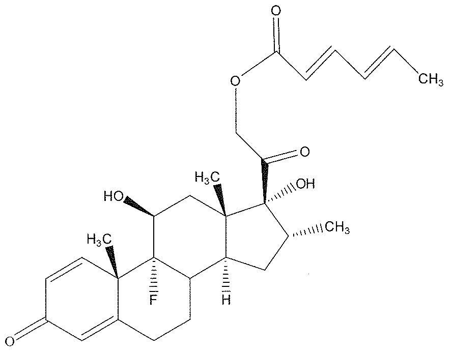
ナノ粒子の粒子サイズを走査型電子顕微鏡および動的光散乱法により測定したところ、個々の粒子サイズ(粒子径)は10nm以上500nm以下だった。
デキサメタゾン誘導体ナノ粒子の電子顕微鏡写真を図11に示す。
デキサメタゾン誘導体ナノ粒子の電子顕微鏡写真を図11に示す。
<点眼剤の調製>
得られたナノ粒子分散液(10g)に2重量%PVP水溶液(1g)を加えたのち、凍結乾燥しエタノールを除去しナノ粒子の紛体を得た。つづいて凍結乾燥して得られたナノ粒子の粉体を、食塩を含有する1gの水と混合しナノ粒子粉体を水中に再分散させた。最終的な各成分の濃度が、デキサメタゾン誘導体が0.2重量%、食塩が0.9重量%、PVPが2重量%になるようにして点眼剤を得た。
得られたナノ粒子分散液(10g)に2重量%PVP水溶液(1g)を加えたのち、凍結乾燥しエタノールを除去しナノ粒子の紛体を得た。つづいて凍結乾燥して得られたナノ粒子の粉体を、食塩を含有する1gの水と混合しナノ粒子粉体を水中に再分散させた。最終的な各成分の濃度が、デキサメタゾン誘導体が0.2重量%、食塩が0.9重量%、PVPが2重量%になるようにして点眼剤を得た。
<デキサメタゾン誘導体ナノ粒子点眼剤の眼内移行評価>
4週齢のオスのラットの片眼に、前記<点眼剤の調製>で得られた点眼剤10μlを点眼した。5分後、ラットを麻酔死させ、点眼剤を点眼した眼を生理食塩水で十分に洗浄した後に摘出し、さらに生理食塩水で洗浄した。
4週齢のオスのラットの片眼に、前記<点眼剤の調製>で得られた点眼剤10μlを点眼した。5分後、ラットを麻酔死させ、点眼剤を点眼した眼を生理食塩水で十分に洗浄した後に摘出し、さらに生理食塩水で洗浄した。
洗浄した眼を500μlのDMSOと混合し、この溶液に超音波処理を施した(60分)。つづいてこの溶液を孔径0.2μmのシリンジフィルターに通した。
得られた試料溶液について、高速液体クロマトグラフィー(High performance liquid chromatography: HPLC)評価を行った。図12(左)にデキサメタゾン標準品由来のクロマトグラムを示す。実施例19の結果(クロマトグラム)を図12(右)に示す。実施例19の点眼剤を点眼した眼を含む試料溶液においても、デキサメタゾン標準品と同様のデキサメタゾン由来のピークが検出されたことから、デキサメタゾンが眼球内に移行していることがわかった。
コントロールとして、デキサメタゾンを含まない点眼剤をラットに点眼し、同様にHPLC評価を行ったところ、わずかなピークが検出された(図14参照)。これは、デキサメタゾンと同じリテンションタイムを有する眼球由来の何らかの化合物が微量に検出されたものと推測される。
[比較例7]
デキサメタゾンの粉体を、食塩を含有し、PVPを2重量%の濃度で含有する水溶液と混合し、超音波処理によりマイクロ粒子水分散液を得た。この分散液において、最終的な各成分の濃度を、デキサメタゾンが0.2重量%、食塩が0.9重量%、PVPが2重量%になるようにして点眼剤を得た。得られた点眼剤(点眼剤中の個々の粒子の粒子サイズ(粒子径)は1μm以上100μm以下)について、実施例19と同様にラットに点眼を行い、HPLCの評価を行った。
デキサメタゾンの粉体を、食塩を含有し、PVPを2重量%の濃度で含有する水溶液と混合し、超音波処理によりマイクロ粒子水分散液を得た。この分散液において、最終的な各成分の濃度を、デキサメタゾンが0.2重量%、食塩が0.9重量%、PVPが2重量%になるようにして点眼剤を得た。得られた点眼剤(点眼剤中の個々の粒子の粒子サイズ(粒子径)は1μm以上100μm以下)について、実施例19と同様にラットに点眼を行い、HPLCの評価を行った。
点眼後の眼球内におけるデキサメタゾン含有量について、ナノ粒子点眼剤(実施例19)とマイクロ粒子点眼剤(本比較例)を比較した結果を図13及び図14に示す。図14において、いちばん左のaの棒グラフが実施例19の結果を、左から2番目のbの棒グラフが本比較例の結果を示す。なお、デキサメタゾン含有量はHPLC測定で得られたスペクトルの面積で表示した。
図13および14より、ナノ粒子点眼剤の場合、マイクロ粒子点眼剤より眼内移行性が高いことが分かる。これらの実施例および比較例により、眼内移行性を高めた本発明の点眼剤が有効であることが示された。
[比較例8]
デキサメタゾン誘導体を、食塩を含有し、PVPを2重量%の濃度で含有する水溶液と混合し、超音波処理によりマイクロ粒子水分散液を得た。この分散液において、最終的な各成分の濃度を、デキサメタゾン誘導体が0.2重量%、食塩が0.9重量%、PVPが2重量%になるようにして点眼剤を得た。得られた点眼剤(点眼剤中の個々の粒子の粒子サイズ(粒子径)は1μm以上100μm以下)について、実施例19と同様にラットに点眼を行いHPLCの評価を行った。
デキサメタゾン誘導体を、食塩を含有し、PVPを2重量%の濃度で含有する水溶液と混合し、超音波処理によりマイクロ粒子水分散液を得た。この分散液において、最終的な各成分の濃度を、デキサメタゾン誘導体が0.2重量%、食塩が0.9重量%、PVPが2重量%になるようにして点眼剤を得た。得られた点眼剤(点眼剤中の個々の粒子の粒子サイズ(粒子径)は1μm以上100μm以下)について、実施例19と同様にラットに点眼を行いHPLCの評価を行った。
点眼後の眼球内におけるデキサメタゾン含有量については、HPLC測定で得られたスペクトルの面積から検量線(図15参照)を用いて試算した。しかしながら、デキサメタゾン誘導体マイクロ粒子点眼剤(本比較例)の場合、検量できる十分な量のデキサメタゾンの眼内移行は認められなかった。一方実施例19の点眼剤(デキサメタゾン誘導体ナノ粒子点眼剤)の結果についても同様の試算を行ったところ、眼内移行性を認め、その量は1.06±0.34μg/g tissueであった。
以上の結果を下記表2にまとめる。

デキサメタゾン誘導体マイクロ粒子点眼剤で眼内移行が認められないことは、デキサメタゾン誘導体マイクロ粒子点眼剤から得られるHPLCピーク面積(図14の左から3番目のcの棒グラフ)が、デキサメタゾンを含まない点眼剤(コントロール:食塩0.9重量%、PVP2重量%のみ含有;図14のいちばん右のd、)から得られるピーク面積と、ほぼ同じくらいの面積値を有することからも確認できる。
さらに図14のa(一番左の棒グラフ、実施例19の点眼剤のHPLC評価結果である)とc(左から3番目の棒グラフ、比較例8の点眼剤のHPLC評価結果である)との比較から、デキサメタゾンの眼内移行性を向上させるためには、薬剤のプロドラッグ化だけでなく、薬剤のナノ粒子化が重要であることがわかる。
11 角膜
12 虹彩
13 毛様体
14 強膜
15 脈絡膜
16 網膜
17 中心窩
18 視神経
19 円板陥凹
20 硝子体
21 水晶体
22 眼球結膜
23 後眼房
24 前眼房
12 虹彩
13 毛様体
14 強膜
15 脈絡膜
16 網膜
17 中心窩
18 視神経
19 円板陥凹
20 硝子体
21 水晶体
22 眼球結膜
23 後眼房
24 前眼房
Claims (8)
- 加水分解反応を受けて疎水性もしくは脂溶性から親水性もしくは水溶性に変化するプロドラッグからなる粒子を含む点眼剤であって、
前記粒子の粒子サイズが、10nm以上1μm未満であることを特徴とする点眼剤。 - 前記粒子の粒子サイズが、10nm以上500nm以下であることを特徴とする請求項1に記載の点眼剤。
- 前記プロドラッグを、プロドラッグが溶解する溶媒1に溶解し、
得られた溶液Aを、プロドラッグが溶解しない溶媒2と混合する工程を含むことを特徴とする請求項1または2に記載の点眼剤の製造方法。 - 前記溶液Aを、液滴として前記溶媒2中に供給し、混合することを特徴とする請求項3に記載の点眼剤の製造方法。
- 加水分解反応を受けて疎水性もしくは脂溶性から親水性もしくは水溶性に変化し、かつ蛍光を発するようになるプロドラッグからなる粒子を含む蛍光イメージング剤であって、
前記粒子の粒子サイズが、10nm以上1μm未満であることを特徴とする蛍光イメージング剤。 - 前記粒子の粒子サイズが、10nm以上500nm以下であることを特徴とする請求項5に記載の蛍光イメージング剤。
- 請求項5または6に記載の蛍光イメージング剤をヒト以外の動物の眼に投与し、該眼を摘出し、摘出された眼を各部位に分離し、該部位が発する蛍光を光学的手段により観測することを特徴とする蛍光観測法。
- 請求項5または6に記載の蛍光イメージング剤をヒト以外の動物の眼に投与し、該眼を摘出し、摘出された眼が発する蛍光を光学的手段により観測することを特徴とする蛍光観測法。
Priority Applications (1)
| Application Number | Priority Date | Filing Date | Title |
|---|---|---|---|
| JP2010536777A JP5709523B2 (ja) | 2008-11-06 | 2009-11-04 | 眼内移行性の高い点眼剤および蛍光イメージング剤ならびにそれらの製造方法 |
Applications Claiming Priority (2)
| Application Number | Priority Date | Filing Date | Title |
|---|---|---|---|
| JP2008285718 | 2008-11-06 | ||
| JP2008-285718 | 2008-11-06 |
Publications (1)
| Publication Number | Publication Date |
|---|---|
| WO2010053101A1 true WO2010053101A1 (ja) | 2010-05-14 |
Family
ID=42152909
Family Applications (1)
| Application Number | Title | Priority Date | Filing Date |
|---|---|---|---|
| PCT/JP2009/068851 Ceased WO2010053101A1 (ja) | 2008-11-06 | 2009-11-04 | 眼内移行性の高い点眼剤および蛍光イメージング剤ならびにそれらの製造方法 |
Country Status (2)
| Country | Link |
|---|---|
| JP (1) | JP5709523B2 (ja) |
| WO (1) | WO2010053101A1 (ja) |
Cited By (5)
| Publication number | Priority date | Publication date | Assignee | Title |
|---|---|---|---|---|
| WO2011155501A1 (ja) * | 2010-06-11 | 2011-12-15 | 独立行政法人科学技術振興機構 | 薬剤多量体微粒子及びその製造方法 |
| WO2013161778A1 (ja) | 2012-04-24 | 2013-10-31 | 国立大学法人大阪大学 | 薬剤ナノ粒子を分散した水分散液の製造法およびその利用 |
| JP2016132616A (ja) * | 2015-01-15 | 2016-07-25 | 大内新興化学工業株式会社 | 眼疾患治療用ナノ粒子製剤 |
| CN110461311A (zh) * | 2016-08-26 | 2019-11-15 | 奥野哲治 | 微细纳米化药剂及其应用 |
| CN118027128A (zh) * | 2024-02-07 | 2024-05-14 | 沈阳药科大学 | 一种用于炎症协同治疗的地塞米松-亚麻酸前药自组装纳米粒及其应用 |
Citations (7)
| Publication number | Priority date | Publication date | Assignee | Title |
|---|---|---|---|---|
| JPH10215894A (ja) * | 1997-02-06 | 1998-08-18 | Mitsubishi Chem Corp | 生細胞の検出方法 |
| JP2004528267A (ja) * | 2000-07-13 | 2004-09-16 | ファルマシア・アンド・アップジョン・カンパニー | 選択的シクロオキシゲナーゼ−2阻害性薬物の眼処方 |
| JP2004530458A (ja) * | 2001-01-24 | 2004-10-07 | ヴィクター ジェイ. ドハーティ, | 携帯型眼用イルミネーター |
| JP2005238342A (ja) * | 2004-02-24 | 2005-09-08 | Japan Science & Technology Agency | 有機化合物の超微粒子の製造方法 |
| JP2007119456A (ja) * | 2005-09-30 | 2007-05-17 | Toyama Chem Co Ltd | 難溶性薬物のナノ微粒子を含有する水性懸濁液剤 |
| JP2007518804A (ja) * | 2004-01-20 | 2007-07-12 | アラーガン、インコーポレイテッド | トリアムシノロンアセトニドおよびヒアルロン酸を好ましく含有する眼局所治療用組成物 |
| JP2008526352A (ja) * | 2005-01-06 | 2008-07-24 | ジーイー・ヘルスケア・アクスイェ・セルスカプ | 光学イメージング |
Family Cites Families (3)
| Publication number | Priority date | Publication date | Assignee | Title |
|---|---|---|---|---|
| JP2000247871A (ja) * | 1999-02-25 | 2000-09-12 | Santen Pharmaceut Co Ltd | 網膜または硝子体への薬物放出制御システム |
| JP2002138036A (ja) * | 2000-08-24 | 2002-05-14 | Santen Pharmaceut Co Ltd | 薬物放出制御システム |
| JP4921750B2 (ja) * | 2005-09-14 | 2012-04-25 | 株式会社日本点眼薬研究所 | ピレノキシンまたは薬理学的に許容される塩を含有する懸濁性医薬組成物 |
-
2009
- 2009-11-04 JP JP2010536777A patent/JP5709523B2/ja not_active Expired - Fee Related
- 2009-11-04 WO PCT/JP2009/068851 patent/WO2010053101A1/ja not_active Ceased
Patent Citations (7)
| Publication number | Priority date | Publication date | Assignee | Title |
|---|---|---|---|---|
| JPH10215894A (ja) * | 1997-02-06 | 1998-08-18 | Mitsubishi Chem Corp | 生細胞の検出方法 |
| JP2004528267A (ja) * | 2000-07-13 | 2004-09-16 | ファルマシア・アンド・アップジョン・カンパニー | 選択的シクロオキシゲナーゼ−2阻害性薬物の眼処方 |
| JP2004530458A (ja) * | 2001-01-24 | 2004-10-07 | ヴィクター ジェイ. ドハーティ, | 携帯型眼用イルミネーター |
| JP2007518804A (ja) * | 2004-01-20 | 2007-07-12 | アラーガン、インコーポレイテッド | トリアムシノロンアセトニドおよびヒアルロン酸を好ましく含有する眼局所治療用組成物 |
| JP2005238342A (ja) * | 2004-02-24 | 2005-09-08 | Japan Science & Technology Agency | 有機化合物の超微粒子の製造方法 |
| JP2008526352A (ja) * | 2005-01-06 | 2008-07-24 | ジーイー・ヘルスケア・アクスイェ・セルスカプ | 光学イメージング |
| JP2007119456A (ja) * | 2005-09-30 | 2007-05-17 | Toyama Chem Co Ltd | 難溶性薬物のナノ微粒子を含有する水性懸濁液剤 |
Non-Patent Citations (1)
| Title |
|---|
| KASSEM, M.A. ET AL.: "Nanosuspension as an ophthalmic delivery system for certain glucocorticoid drugs", INTERNATIONAL JOURNAL OF PHARMACEUTICS, vol. 340, no. 1-2, 12 March 2007 (2007-03-12), pages 126 - 133 * |
Cited By (10)
| Publication number | Priority date | Publication date | Assignee | Title |
|---|---|---|---|---|
| WO2011155501A1 (ja) * | 2010-06-11 | 2011-12-15 | 独立行政法人科学技術振興機構 | 薬剤多量体微粒子及びその製造方法 |
| JP5113958B2 (ja) * | 2010-06-11 | 2013-01-09 | 独立行政法人科学技術振興機構 | 薬剤多量体微粒子及びその製造方法 |
| WO2013161778A1 (ja) | 2012-04-24 | 2013-10-31 | 国立大学法人大阪大学 | 薬剤ナノ粒子を分散した水分散液の製造法およびその利用 |
| EP2842545A4 (en) * | 2012-04-24 | 2015-12-16 | Univ Osaka | PROCESS FOR PREPARING AN AQUEOUS DISPERSION FROM MEDICAMENTAL APPLICATIONS AND USE THEREOF |
| JP2016132616A (ja) * | 2015-01-15 | 2016-07-25 | 大内新興化学工業株式会社 | 眼疾患治療用ナノ粒子製剤 |
| CN110461311A (zh) * | 2016-08-26 | 2019-11-15 | 奥野哲治 | 微细纳米化药剂及其应用 |
| EP3505165A4 (en) * | 2016-08-26 | 2020-05-20 | Tetsuji Okuno | SMALL MEDICINAL PRODUCT IN NANO SIZE AND USE THEREOF |
| US11406684B2 (en) | 2016-08-26 | 2022-08-09 | Tetsuji Okuno | Fine nano-sized medicinal agent and use thereof |
| CN118027128A (zh) * | 2024-02-07 | 2024-05-14 | 沈阳药科大学 | 一种用于炎症协同治疗的地塞米松-亚麻酸前药自组装纳米粒及其应用 |
| CN118027128B (zh) * | 2024-02-07 | 2025-03-18 | 沈阳药科大学 | 一种用于炎症协同治疗的地塞米松-亚麻酸前药自组装纳米粒及其应用 |
Also Published As
| Publication number | Publication date |
|---|---|
| JPWO2010053101A1 (ja) | 2012-04-05 |
| JP5709523B2 (ja) | 2015-04-30 |
Similar Documents
| Publication | Publication Date | Title |
|---|---|---|
| ES2369554T3 (es) | Carboxi-amido-triazoles para el tratamiento local de enfermedades oculares. | |
| JP2007535563A (ja) | レチノイド含有徐放性薬剤送達システム、ならびに関連する製造法 | |
| JP2007535563A5 (ja) | レチノイド含有徐放性眼内薬剤送達システム、ならびに関連する製造法 | |
| JP2008308489A (ja) | ロピニロール又はその塩を有効成分として含有する後眼部疾患の予防又は治療剤 | |
| Mietz et al. | Suramin inhibits wound healing following filtering procedures for glaucoma | |
| SG187770A1 (en) | A liposomal formulation for ocular drug delivery | |
| KR20100072333A (ko) | 약물 전달을 위한 비히클로서의 비-수성 수혼화성 물질 | |
| CN104379129A (zh) | 含有帕唑帕尼的局部眼科药物组合物 | |
| JP5709523B2 (ja) | 眼内移行性の高い点眼剤および蛍光イメージング剤ならびにそれらの製造方法 | |
| Sharma et al. | Recent overviews on the drug delivery aspects and applications of brinzolamide for the management of glaucoma | |
| CN106999427A (zh) | 用于治疗眼部疾病的用于递送水飞蓟宾和其他活性成分的纳米结构化制剂 | |
| US20080166417A1 (en) | Method of Relieving or Avoiding Side Effect of Steroid | |
| RU2339369C2 (ru) | Лечение офтальмологических нарушений с использованием мочевины и ее производных | |
| WO2018011705A1 (en) | Liposome-based eye drops and use thereof for in vivo evaluation of the drug efficacy of medical and surgical anti-glaucoma therapy | |
| Kim et al. | Ocular permeation enhancers | |
| Kanugo et al. | Recent Advances in Artificial Intelligence and Nanotechnology-Driven Strategies for Diagnosis and Therapy of Ocular Diseases | |
| JP4150846B2 (ja) | ステロイドを有効成分とする網脈絡膜疾患治療剤 | |
| US12564572B2 (en) | Topical ocular delivery of cromakalim | |
| Kupersmith et al. | The blood-ocular barrier | |
| Dissanayaka | Advancements in Nanotechnology for Age-Related Macular Degeneration: Innovations in Nanodiagnostics and Nanotherapeutics | |
| WO2026078034A1 (en) | Sitagliptin for use in retinal diseases with neovascularization | |
| Virmani et al. | An overview of ocular drug delivery systemsdconventional and novel drug delivery systems | |
| Regnier | Barriers to Ocular Drug Delivery | |
| WO2025128712A1 (en) | Treatment of ocular diseases using 1-(4-{[4-(dimethylamino)piperidin-1-yl]carbonyl)phenyl)-3-[4-(4,6- dimorpholin-4- yl-1,3,5-triazin-2-yl)phenyl]urea | |
| Genov et al. | Innovative drug delivery in ophthalmology |
Legal Events
| Date | Code | Title | Description |
|---|---|---|---|
| 121 | Ep: the epo has been informed by wipo that ep was designated in this application |
Ref document number: 09824800 Country of ref document: EP Kind code of ref document: A1 |
|
| ENP | Entry into the national phase |
Ref document number: 2010536777 Country of ref document: JP Kind code of ref document: A |
|
| NENP | Non-entry into the national phase |
Ref country code: DE |
|
| 122 | Ep: pct application non-entry in european phase |
Ref document number: 09824800 Country of ref document: EP Kind code of ref document: A1 |