US9713629B2 - Compositions and methods for treating and preventing tissue injury and disease - Google Patents
Compositions and methods for treating and preventing tissue injury and disease Download PDFInfo
- Publication number
- US9713629B2 US9713629B2 US14/429,511 US201314429511A US9713629B2 US 9713629 B2 US9713629 B2 US 9713629B2 US 201314429511 A US201314429511 A US 201314429511A US 9713629 B2 US9713629 B2 US 9713629B2
- Authority
- US
- United States
- Prior art keywords
- tissue
- cells
- composition
- microvascular
- bone
- Prior art date
- Legal status (The legal status is an assumption and is not a legal conclusion. Google has not performed a legal analysis and makes no representation as to the accuracy of the status listed.)
- Active, expires
Links
Images


Classifications
-
- A—HUMAN NECESSITIES
- A61—MEDICAL OR VETERINARY SCIENCE; HYGIENE
- A61K—PREPARATIONS FOR MEDICAL, DENTAL OR TOILETRY PURPOSES
- A61K35/00—Medicinal preparations containing materials or reaction products thereof with undetermined constitution
- A61K35/12—Materials from mammals; Compositions comprising non-specified tissues or cells; Compositions comprising non-embryonic stem cells; Genetically modified cells
- A61K35/44—Vessels; Vascular smooth muscle cells; Endothelial cells; Endothelial progenitor cells
-
- A—HUMAN NECESSITIES
- A61—MEDICAL OR VETERINARY SCIENCE; HYGIENE
- A61K—PREPARATIONS FOR MEDICAL, DENTAL OR TOILETRY PURPOSES
- A61K35/00—Medicinal preparations containing materials or reaction products thereof with undetermined constitution
- A61K35/12—Materials from mammals; Compositions comprising non-specified tissues or cells; Compositions comprising non-embryonic stem cells; Genetically modified cells
-
- A—HUMAN NECESSITIES
- A61—MEDICAL OR VETERINARY SCIENCE; HYGIENE
- A61L—METHODS OR APPARATUS FOR STERILISING MATERIALS OR OBJECTS IN GENERAL; DISINFECTION, STERILISATION OR DEODORISATION OF AIR; CHEMICAL ASPECTS OF BANDAGES, DRESSINGS, ABSORBENT PADS OR SURGICAL ARTICLES; MATERIALS FOR BANDAGES, DRESSINGS, ABSORBENT PADS OR SURGICAL ARTICLES
- A61L27/00—Materials for grafts or prostheses or for coating grafts or prostheses
- A61L27/36—Materials for grafts or prostheses or for coating grafts or prostheses containing ingredients of undetermined constitution or reaction products thereof, e.g. transplant tissue, natural bone, extracellular matrix
- A61L27/3604—Materials for grafts or prostheses or for coating grafts or prostheses containing ingredients of undetermined constitution or reaction products thereof, e.g. transplant tissue, natural bone, extracellular matrix characterised by the human or animal origin of the biological material, e.g. hair, fascia, fish scales, silk, shellac, pericardium, pleura, renal tissue, amniotic membrane, parenchymal tissue, fetal tissue, muscle tissue, fat tissue, enamel
- A61L27/3625—Vascular tissue, e.g. heart valves
-
- A—HUMAN NECESSITIES
- A61—MEDICAL OR VETERINARY SCIENCE; HYGIENE
- A61L—METHODS OR APPARATUS FOR STERILISING MATERIALS OR OBJECTS IN GENERAL; DISINFECTION, STERILISATION OR DEODORISATION OF AIR; CHEMICAL ASPECTS OF BANDAGES, DRESSINGS, ABSORBENT PADS OR SURGICAL ARTICLES; MATERIALS FOR BANDAGES, DRESSINGS, ABSORBENT PADS OR SURGICAL ARTICLES
- A61L27/00—Materials for grafts or prostheses or for coating grafts or prostheses
- A61L27/36—Materials for grafts or prostheses or for coating grafts or prostheses containing ingredients of undetermined constitution or reaction products thereof, e.g. transplant tissue, natural bone, extracellular matrix
- A61L27/3683—Materials for grafts or prostheses or for coating grafts or prostheses containing ingredients of undetermined constitution or reaction products thereof, e.g. transplant tissue, natural bone, extracellular matrix subjected to a specific treatment prior to implantation, e.g. decellularising, demineralising, grinding, cellular disruption/non-collagenous protein removal, anti-calcification, crosslinking, supercritical fluid extraction, enzyme treatment
- A61L27/3687—Materials for grafts or prostheses or for coating grafts or prostheses containing ingredients of undetermined constitution or reaction products thereof, e.g. transplant tissue, natural bone, extracellular matrix subjected to a specific treatment prior to implantation, e.g. decellularising, demineralising, grinding, cellular disruption/non-collagenous protein removal, anti-calcification, crosslinking, supercritical fluid extraction, enzyme treatment characterised by the use of chemical agents in the treatment, e.g. specific enzymes, detergents, capping agents, crosslinkers, anticalcification agents
-
- A—HUMAN NECESSITIES
- A61—MEDICAL OR VETERINARY SCIENCE; HYGIENE
- A61L—METHODS OR APPARATUS FOR STERILISING MATERIALS OR OBJECTS IN GENERAL; DISINFECTION, STERILISATION OR DEODORISATION OF AIR; CHEMICAL ASPECTS OF BANDAGES, DRESSINGS, ABSORBENT PADS OR SURGICAL ARTICLES; MATERIALS FOR BANDAGES, DRESSINGS, ABSORBENT PADS OR SURGICAL ARTICLES
- A61L27/00—Materials for grafts or prostheses or for coating grafts or prostheses
- A61L27/36—Materials for grafts or prostheses or for coating grafts or prostheses containing ingredients of undetermined constitution or reaction products thereof, e.g. transplant tissue, natural bone, extracellular matrix
- A61L27/3683—Materials for grafts or prostheses or for coating grafts or prostheses containing ingredients of undetermined constitution or reaction products thereof, e.g. transplant tissue, natural bone, extracellular matrix subjected to a specific treatment prior to implantation, e.g. decellularising, demineralising, grinding, cellular disruption/non-collagenous protein removal, anti-calcification, crosslinking, supercritical fluid extraction, enzyme treatment
- A61L27/3691—Materials for grafts or prostheses or for coating grafts or prostheses containing ingredients of undetermined constitution or reaction products thereof, e.g. transplant tissue, natural bone, extracellular matrix subjected to a specific treatment prior to implantation, e.g. decellularising, demineralising, grinding, cellular disruption/non-collagenous protein removal, anti-calcification, crosslinking, supercritical fluid extraction, enzyme treatment characterised by physical conditions of the treatment, e.g. applying a compressive force to the composition, pressure cycles, ultrasonic/sonication or microwave treatment, lyophilisation
-
- A—HUMAN NECESSITIES
- A61—MEDICAL OR VETERINARY SCIENCE; HYGIENE
- A61L—METHODS OR APPARATUS FOR STERILISING MATERIALS OR OBJECTS IN GENERAL; DISINFECTION, STERILISATION OR DEODORISATION OF AIR; CHEMICAL ASPECTS OF BANDAGES, DRESSINGS, ABSORBENT PADS OR SURGICAL ARTICLES; MATERIALS FOR BANDAGES, DRESSINGS, ABSORBENT PADS OR SURGICAL ARTICLES
- A61L27/00—Materials for grafts or prostheses or for coating grafts or prostheses
- A61L27/36—Materials for grafts or prostheses or for coating grafts or prostheses containing ingredients of undetermined constitution or reaction products thereof, e.g. transplant tissue, natural bone, extracellular matrix
- A61L27/38—Materials for grafts or prostheses or for coating grafts or prostheses containing ingredients of undetermined constitution or reaction products thereof, e.g. transplant tissue, natural bone, extracellular matrix containing added animal cells
- A61L27/3804—Materials for grafts or prostheses or for coating grafts or prostheses containing ingredients of undetermined constitution or reaction products thereof, e.g. transplant tissue, natural bone, extracellular matrix containing added animal cells characterised by specific cells or progenitors thereof, e.g. fibroblasts, connective tissue cells, kidney cells
- A61L27/3834—Cells able to produce different cell types, e.g. hematopoietic stem cells, mesenchymal stem cells, marrow stromal cells, embryonic stem cells
-
- A—HUMAN NECESSITIES
- A61—MEDICAL OR VETERINARY SCIENCE; HYGIENE
- A61L—METHODS OR APPARATUS FOR STERILISING MATERIALS OR OBJECTS IN GENERAL; DISINFECTION, STERILISATION OR DEODORISATION OF AIR; CHEMICAL ASPECTS OF BANDAGES, DRESSINGS, ABSORBENT PADS OR SURGICAL ARTICLES; MATERIALS FOR BANDAGES, DRESSINGS, ABSORBENT PADS OR SURGICAL ARTICLES
- A61L27/00—Materials for grafts or prostheses or for coating grafts or prostheses
- A61L27/50—Materials characterised by their function or physical properties, e.g. injectable or lubricating compositions, shape-memory materials, surface modified materials
- A61L27/54—Biologically active materials, e.g. therapeutic substances
-
- A—HUMAN NECESSITIES
- A61—MEDICAL OR VETERINARY SCIENCE; HYGIENE
- A61P—SPECIFIC THERAPEUTIC ACTIVITY OF CHEMICAL COMPOUNDS OR MEDICINAL PREPARATIONS
- A61P17/00—Drugs for dermatological disorders
-
- A—HUMAN NECESSITIES
- A61—MEDICAL OR VETERINARY SCIENCE; HYGIENE
- A61P—SPECIFIC THERAPEUTIC ACTIVITY OF CHEMICAL COMPOUNDS OR MEDICINAL PREPARATIONS
- A61P17/00—Drugs for dermatological disorders
- A61P17/02—Drugs for dermatological disorders for treating wounds, ulcers, burns, scars, keloids, or the like
-
- A—HUMAN NECESSITIES
- A61—MEDICAL OR VETERINARY SCIENCE; HYGIENE
- A61P—SPECIFIC THERAPEUTIC ACTIVITY OF CHEMICAL COMPOUNDS OR MEDICINAL PREPARATIONS
- A61P19/00—Drugs for skeletal disorders
-
- A—HUMAN NECESSITIES
- A61—MEDICAL OR VETERINARY SCIENCE; HYGIENE
- A61P—SPECIFIC THERAPEUTIC ACTIVITY OF CHEMICAL COMPOUNDS OR MEDICINAL PREPARATIONS
- A61P19/00—Drugs for skeletal disorders
- A61P19/02—Drugs for skeletal disorders for joint disorders, e.g. arthritis, arthrosis
-
- A—HUMAN NECESSITIES
- A61—MEDICAL OR VETERINARY SCIENCE; HYGIENE
- A61P—SPECIFIC THERAPEUTIC ACTIVITY OF CHEMICAL COMPOUNDS OR MEDICINAL PREPARATIONS
- A61P19/00—Drugs for skeletal disorders
- A61P19/08—Drugs for skeletal disorders for bone diseases, e.g. rachitism, Paget's disease
-
- A—HUMAN NECESSITIES
- A61—MEDICAL OR VETERINARY SCIENCE; HYGIENE
- A61P—SPECIFIC THERAPEUTIC ACTIVITY OF CHEMICAL COMPOUNDS OR MEDICINAL PREPARATIONS
- A61P29/00—Non-central analgesic, antipyretic or antiinflammatory agents, e.g. antirheumatic agents; Non-steroidal antiinflammatory drugs [NSAID]
-
- A—HUMAN NECESSITIES
- A61—MEDICAL OR VETERINARY SCIENCE; HYGIENE
- A61P—SPECIFIC THERAPEUTIC ACTIVITY OF CHEMICAL COMPOUNDS OR MEDICINAL PREPARATIONS
- A61P43/00—Drugs for specific purposes, not provided for in groups A61P1/00-A61P41/00
-
- A—HUMAN NECESSITIES
- A61—MEDICAL OR VETERINARY SCIENCE; HYGIENE
- A61P—SPECIFIC THERAPEUTIC ACTIVITY OF CHEMICAL COMPOUNDS OR MEDICINAL PREPARATIONS
- A61P9/00—Drugs for disorders of the cardiovascular system
-
- A—HUMAN NECESSITIES
- A61—MEDICAL OR VETERINARY SCIENCE; HYGIENE
- A61P—SPECIFIC THERAPEUTIC ACTIVITY OF CHEMICAL COMPOUNDS OR MEDICINAL PREPARATIONS
- A61P9/00—Drugs for disorders of the cardiovascular system
- A61P9/10—Drugs for disorders of the cardiovascular system for treating ischaemic or atherosclerotic diseases, e.g. antianginal drugs, coronary vasodilators, drugs for myocardial infarction, retinopathy, cerebrovascula insufficiency, renal arteriosclerosis
-
- A—HUMAN NECESSITIES
- A61—MEDICAL OR VETERINARY SCIENCE; HYGIENE
- A61P—SPECIFIC THERAPEUTIC ACTIVITY OF CHEMICAL COMPOUNDS OR MEDICINAL PREPARATIONS
- A61P9/00—Drugs for disorders of the cardiovascular system
- A61P9/14—Vasoprotectives; Antihaemorrhoidals; Drugs for varicose therapy; Capillary stabilisers
-
- C—CHEMISTRY; METALLURGY
- C12—BIOCHEMISTRY; BEER; SPIRITS; WINE; VINEGAR; MICROBIOLOGY; ENZYMOLOGY; MUTATION OR GENETIC ENGINEERING
- C12N—MICROORGANISMS OR ENZYMES; COMPOSITIONS THEREOF; PROPAGATING, PRESERVING, OR MAINTAINING MICROORGANISMS; MUTATION OR GENETIC ENGINEERING; CULTURE MEDIA
- C12N5/00—Undifferentiated human, animal or plant cells, e.g. cell lines; Tissues; Cultivation or maintenance thereof; Culture media therefor
- C12N5/06—Animal cells or tissues; Human cells or tissues
- C12N5/0602—Vertebrate cells
- C12N5/0652—Cells of skeletal and connective tissues; Mesenchyme
- C12N5/0653—Adipocytes; Adipose tissue
-
- A—HUMAN NECESSITIES
- A61—MEDICAL OR VETERINARY SCIENCE; HYGIENE
- A61L—METHODS OR APPARATUS FOR STERILISING MATERIALS OR OBJECTS IN GENERAL; DISINFECTION, STERILISATION OR DEODORISATION OF AIR; CHEMICAL ASPECTS OF BANDAGES, DRESSINGS, ABSORBENT PADS OR SURGICAL ARTICLES; MATERIALS FOR BANDAGES, DRESSINGS, ABSORBENT PADS OR SURGICAL ARTICLES
- A61L2300/00—Biologically active materials used in bandages, wound dressings, absorbent pads or medical devices
- A61L2300/40—Biologically active materials used in bandages, wound dressings, absorbent pads or medical devices characterised by a specific therapeutic activity or mode of action
- A61L2300/41—Anti-inflammatory agents, e.g. NSAIDs
-
- A—HUMAN NECESSITIES
- A61—MEDICAL OR VETERINARY SCIENCE; HYGIENE
- A61L—METHODS OR APPARATUS FOR STERILISING MATERIALS OR OBJECTS IN GENERAL; DISINFECTION, STERILISATION OR DEODORISATION OF AIR; CHEMICAL ASPECTS OF BANDAGES, DRESSINGS, ABSORBENT PADS OR SURGICAL ARTICLES; MATERIALS FOR BANDAGES, DRESSINGS, ABSORBENT PADS OR SURGICAL ARTICLES
- A61L2300/00—Biologically active materials used in bandages, wound dressings, absorbent pads or medical devices
- A61L2300/40—Biologically active materials used in bandages, wound dressings, absorbent pads or medical devices characterised by a specific therapeutic activity or mode of action
- A61L2300/412—Tissue-regenerating or healing or proliferative agents
-
- A—HUMAN NECESSITIES
- A61—MEDICAL OR VETERINARY SCIENCE; HYGIENE
- A61L—METHODS OR APPARATUS FOR STERILISING MATERIALS OR OBJECTS IN GENERAL; DISINFECTION, STERILISATION OR DEODORISATION OF AIR; CHEMICAL ASPECTS OF BANDAGES, DRESSINGS, ABSORBENT PADS OR SURGICAL ARTICLES; MATERIALS FOR BANDAGES, DRESSINGS, ABSORBENT PADS OR SURGICAL ARTICLES
- A61L2430/00—Materials or treatment for tissue regeneration
- A61L2430/02—Materials or treatment for tissue regeneration for reconstruction of bones; weight-bearing implants
-
- A—HUMAN NECESSITIES
- A61—MEDICAL OR VETERINARY SCIENCE; HYGIENE
- A61L—METHODS OR APPARATUS FOR STERILISING MATERIALS OR OBJECTS IN GENERAL; DISINFECTION, STERILISATION OR DEODORISATION OF AIR; CHEMICAL ASPECTS OF BANDAGES, DRESSINGS, ABSORBENT PADS OR SURGICAL ARTICLES; MATERIALS FOR BANDAGES, DRESSINGS, ABSORBENT PADS OR SURGICAL ARTICLES
- A61L2430/00—Materials or treatment for tissue regeneration
- A61L2430/06—Materials or treatment for tissue regeneration for cartilage reconstruction, e.g. meniscus
-
- A—HUMAN NECESSITIES
- A61—MEDICAL OR VETERINARY SCIENCE; HYGIENE
- A61L—METHODS OR APPARATUS FOR STERILISING MATERIALS OR OBJECTS IN GENERAL; DISINFECTION, STERILISATION OR DEODORISATION OF AIR; CHEMICAL ASPECTS OF BANDAGES, DRESSINGS, ABSORBENT PADS OR SURGICAL ARTICLES; MATERIALS FOR BANDAGES, DRESSINGS, ABSORBENT PADS OR SURGICAL ARTICLES
- A61L2430/00—Materials or treatment for tissue regeneration
- A61L2430/10—Materials or treatment for tissue regeneration for reconstruction of tendons or ligaments
-
- A—HUMAN NECESSITIES
- A61—MEDICAL OR VETERINARY SCIENCE; HYGIENE
- A61L—METHODS OR APPARATUS FOR STERILISING MATERIALS OR OBJECTS IN GENERAL; DISINFECTION, STERILISATION OR DEODORISATION OF AIR; CHEMICAL ASPECTS OF BANDAGES, DRESSINGS, ABSORBENT PADS OR SURGICAL ARTICLES; MATERIALS FOR BANDAGES, DRESSINGS, ABSORBENT PADS OR SURGICAL ARTICLES
- A61L2430/00—Materials or treatment for tissue regeneration
- A61L2430/34—Materials or treatment for tissue regeneration for soft tissue reconstruction
-
- A—HUMAN NECESSITIES
- A61—MEDICAL OR VETERINARY SCIENCE; HYGIENE
- A61L—METHODS OR APPARATUS FOR STERILISING MATERIALS OR OBJECTS IN GENERAL; DISINFECTION, STERILISATION OR DEODORISATION OF AIR; CHEMICAL ASPECTS OF BANDAGES, DRESSINGS, ABSORBENT PADS OR SURGICAL ARTICLES; MATERIALS FOR BANDAGES, DRESSINGS, ABSORBENT PADS OR SURGICAL ARTICLES
- A61L2430/00—Materials or treatment for tissue regeneration
- A61L2430/38—Materials or treatment for tissue regeneration for reconstruction of the spine, vertebrae or intervertebral discs
Definitions
- compositions comprising multipotent cells and/or microvascular tissue, which has been sterilized and/or treated to inactivate any viruses, and methods for their preparation and allogeneic or xenogeneic use in treating or preventing tissue injury and diseases, such as, e.g., arthritis.
- Tissue repair generally includes several phases, including an initial inflammatory response followed by cellular proliferation and tissue remodeling.
- Fundamental processes of tissue repair include both fibroplasia and angiogenesis.
- Fibroblasts activated by inflammatory mediators migrate into the wound, proliferate, and lay down collagen-rich extracellular matrix, while capillaries in the damaged tissue grow towards the repair zone to reestablish blood flow.
- scar tissue is reabsorbed and replaced with denser, oriented collagen, to produce tissue with some of the characteristics of the original tissue.
- a variety of different therapeutic methods to aid in tissue repair have been developed. These include physical structures, such as better sutures, bone anchors, and patches or implants to provide scaffolding for tissue ingrowth.
- a variety of growth factors have been used to improve tissue growth and migration to the wound site, as well as to promote angiogenesis. For example, there are reports of improved tendon healing using growth factors such as BMP-2, BMP-12, PDGF-BB, and bFGF in preclinical models.
- stem cells are believed to mediate would healing by any of a variety of different mechanisms, including: modulating the inflammatory process; migrating to damaged tissue and recruiting other cells, such as endothelial progenitor cells, necessary for tissue growth; stimulating the proliferation of repair cells; supporting tissue remodeling over scar formation; inhibiting apoptosis; and differentiating into bone, cartilage, tendon, or ligament tissue.
- modulating the inflammatory process migrating to damaged tissue and recruiting other cells, such as endothelial progenitor cells, necessary for tissue growth
- stimulating the proliferation of repair cells supporting tissue remodeling over scar formation; inhibiting apoptosis; and differentiating into bone, cartilage, tendon, or ligament tissue.
- Much of this work has centered on the use of adipose-derived stem cells and other multipotent cells, because they are easily obtained in large numbers.
- compositions, methods, kits, and cell populations that are useful, such as in the repair and/or regeneration of tissue.
- compositions comprising isolated multipotent cells or processed microvascular tissue, or a cell membrane obtained from or derived from said cells or tissue, wherein said composition has angiogenic or anti-inflammatory activity, and wherein said composition is sterilized and/or viruses within said composition are inactivated.
- the cells or composition have not been cultured.
- less than or equal to 50% or less than or equal to 10% of the cells present in the composition are viable.
- substantially none of the cells present in said composition are viable.
- at least 1% of said cells exclude trypan blue.
- the composition is dried, lyophilized or cryopreserved.
- the composition including the sterilized, dried, lyophilized, or cryopreserved composition, retains measurable angiogenic or anti-inflammatory activity when stored at approximately room temperature for at least one month.
- the composition comprises an excipient.
- compositions disclosed herein further comprise an implantable scaffold or matrix, which may be, e.g., a bone-derived implant, a biofiber scaffold, a porous resorbable polymer, a hydrogel, a putty comprising tissue product, or a suture.
- an implantable scaffold or matrix which may be, e.g., a bone-derived implant, a biofiber scaffold, a porous resorbable polymer, a hydrogel, a putty comprising tissue product, or a suture.
- cells, tissue or cell membrane are present on a bone, tendon, or dermal facing surface of said implantable scaffold or matrix.
- a composition of the present invention is formulated for intravenous administration.
- routes of administration including direct administration (either to a tissue surface or by direct injection), intraarterial administration, systemic administration and the like.
- the cells or tissue were obtained from a mammalian donor, optionally a human.
- the donor was a healthy mammal at the time the cells or tissue were obtained.
- the cells comprise stem cells or progenitor cells.
- methods of preparing a composition comprising isolated multipotent cells, wherein said composition has angiogenic or anti-inflammatory activity comprising: dissociating a tissue sample obtained from a donor mammal to release a plurality of multipotent cells therein; separating a plurality of the released multipotent cells from one or more other tissue components to produce a composition comprising isolated multipotent cells; optionally drying, lyophilizing, or cryopreserving the composition before or after sterilization; sterilizing the composition and/or inactivating virus present in the composition, before or after the optional drying, lyophilizing, or cryopreserving of the composition, wherein the sterilized composition retains measurable angiogenic or anti-inflammatory activity.
- the cells or composition are not cultured.
- the method optionally further comprises filtering the released cells or composition, e.g., which may be prior to sterilization or drying, lyophilizing, or cryopreserving.
- the dissociation comprises contacting the tissue sample with one or more proteases.
- the one or more proteases does not comprise collagenase.
- the one or more proteases comprises or consists of: collagenase type 1 and either dispase or thermolysin; or MMP2, MMP 14 and either dispase or thermolysin. Combinations of these proteases (or other functional equivalents) can be used.
- the dissociating or separating comprises ultrasonic agitation, filtration, or use of a density gradient.
- the tissue is adipose-derived tissue, and the ultrasonic agitation, filtration or use of a density gradient separates said released multipotent cells from adipocytes.
- methods of preparing a composition comprising processed microvascular tissue, wherein said composition has angiogenic or anti-inflammatory activity comprising: dissociating a microvascular tissue sample obtained from a donor mammal to produce a composition comprising dissociated microvascular tissue; removing one or more tissue components from the composition comprising dissociated microvascular tissue; optionally drying, lyophilizing, or cryopreserving the composition before or after sterilization; and sterilizing the composition and/or inactivating virus present in the composition before or after the optional drying, lyophilizing, or cryopreserving, wherein the sterilized composition retains measurable angiogenic or anti-inflammatory activity.
- the cells or composition are not cultured.
- the method further comprises filtering the composition.
- the dissociation comprises contacting the tissue sample with one or more proteases.
- the one or more proteases does not comprise collagenase.
- the one or more proteases comprises or consists of: collagenase type 1 and either dispase or thermolysin; or MMP2, MMP 14 and either dispase or thermolysin.
- the dissociation or removing comprises ultrasonic agitation, filtration, or use of a density gradient.
- the microvascular tissue is adipose-derived tissue, and said ultrasonic agitation, filtration or use of a density gradient removes adipocytes from the composition.
- the present invention provides a moisture impermeable container comprising a sterile, dry composition, wherein said composition comprises isolated multipotent cells or processed microvascular tissue, or a cell membrane comprising said cells or tissue or obtained from said cells or tissue, said composition has angiogenic or anti-inflammatory activity, said composition is sterilized and/or viruses within said composition are inactivated, and said composition retains measurable angiogenic or anti-inflammatory activity when stored at approximately room temperature for at least one month.
- the cells or composition have not been cultured.
- less than or equal to 50% or less than or equal to 10% of the cells present in said composition are viable.
- substantially none of the cells present in said composition are viable.
- the composition comprises an excipient.
- the composition further comprises an implantable scaffold or matrix.
- the implantable scaffold or matrix is a bone-derived implant, a biofiber scaffold, a porous resorbable polymer, a hydrogel, a putty comprising tissue product, or a suture.
- the cells, tissue or cell membrane are present on a bone, tendon or dermal facing surface of said implantable scaffold or matrix.
- the composition is formulated for intravenous administration.
- the cells or tissue were obtained from a mammalian donor, optionally a human.
- the donor was a healthy mammal at the time the cells or tissue were obtained.
- the cells comprise stem cells or progenitor cells.
- the container is a vial comprising a hermetic seal. In various embodiments, the container is present within a sealed package comprising a sterile interior.
- the present invention provides a method of treating or preventing an injury or disease, or promoting tissue regeneration, in a mammal, comprising providing to said mammal a composition of the present invention or a composition prepared according to a method of the present invention.
- the composition is surgically implanted into the mammal.
- the composition is implanted within or adjacent to a site of injury or disease in said mammal.
- the composition is provided to said mammal intravenously.
- the injury is present in a soft tissue.
- the injury is present in a tendon, a ligament, skin, a bone, cartilage, a disc, or microvascular tissue.
- the injury or disease is an ischemic injury, a reperfusion injury, a microvascular injury, or inflammation.
- the disease is arthritis, such as e.g., osteoarthritis or rheumatoid arthritis.
- compositions comprising multipotent cells and one or more vessel wall and/or extracellular matrix components.
- the present invention includes a sterilized composition comprising multipotent cells.
- the present invention includes a composition comprising multipotent cells that exclude trypan blue but will not proliferate.
- the present invention includes a composition comprising multipotent cells and one or more vessel wall extracellular matrix components.
- the present invention includes a sterilized composition comprising multipotent cells.
- the present invention includes a composition comprising multipotent cells that exclude trypan blue but will not proliferate.
- the present invention includes a composition comprising sterilized multipotent cells that exclude trypan blue but will not proliferate.
- at least 50% or at least 90% of the cells present in the composition exclude trypan blue but will not proliferate.
- the composition comprises multipotent cells.
- at least 50% or at least 90% of the multipotent cells present in the composition exclude trypan blue but will not proliferate.
- less than or equal to 50% or less than or equal to 10% of the total cells present in the composition are viable.
- substantially none of the cells present in said composition are viable.
- at least 1% or at least 5%, or at least 10%, or at least 20%, at least 50%, or at least 90% of said cells exclude trypan blue.
- the present invention includes a sterilized composition comprising two or more components of multipotent cells.
- the composition comprises five or more or ten or more components of multipotent cells.
- the present invention includes a composition comprising cell membrane and proteins from multipotent cells. In particular embodiments, the composition does not comprise any viable cells or does not comprise any intact cells.
- compositions of the invention may be used in treating or preventing an injury or disease in a subject, including any of the injuries or conditions described herein, wherein said multipotent cells are not autologous to said subject.
- actions taken by a practitioner; however, it should be understood that they can also include the instruction of those actions by another party.
- actions such as “administering microvascular tissue” include “instructing the administration of microvascular tissue.”
- FIG. 1 provides a schematic diagram of the studies described in Example 1.
- FIG. 2 is a graph showing the estimated cell counts per 100 ⁇ magnification of cells in 6 fields.
- BMA and BMB cells in Buffers 1&2 after and before irradiation were used to attract HUVEC cells that were CM-DiI labeled.
- the cell count was estimated by a visual count of 6 images (fields) at 100 ⁇ magnification. The counts were averaged and plotted a shown.
- the dotted lines represent the average for the negative media controls. Cell numbers above that line represent increased migration due to the BMA or BMB material samples.
- the labels shown top to bottom correspond to the bars shown left to right for each time point.
- FIG. 3 shows fluorescence microscopy images of labeled human endothelial cells in the presence of BMA in Buffer 1 over a 12, 24 and 48 hour time course of transmigration.
- FIG. 4 shows fluorescence microscopy images of labeled human endothelial cells in the presence of BMB in Buffer 1 over a 12, 24 and 48 hour time course of transmigration.
- FIG. 5 shows fluorescence microscopy images of labeled human endothelial cells in the presence of BMA in Buffer 2 over a 12, 24 and 48 hour time course of transmigration.
- FIG. 6 shows fluorescence microscopy images of labeled human endothelial cells in the presence of BMB in Buffer 2 over a 12, 24 and 48 hour time course of transmigration.
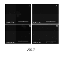
- FIG. 7 shows fluorescence microscopy images of labeled human endothelial cells.
- the sample on the left is undiluted EZ-CPZTM, which is representative of buffers 1 & 2 in which the cells were preserved.
- EZ-CPZTM is a cryopreservation media (Incell Corp., San Antonio, Tex.).
- EZ-CPZTM is described by Incell Corp. as a ready-to-use, serum-free, and protein-free cryopreservation medium that is gently mixed 1:1 (v:v) with a cell suspension.
- EZ-CPZTM supports high viability and re-animation of a variety of cell types including: primary cell cultures, lymphocytes, hybridoma, CHO, colon, IBHK and cancer cell lines.
- EZ-CPZTM contains a proprietary formulation of clinical grade components, vitrification and cryopreservation agents, and a final concentration of 5% DMSO. EZ-CPZTM provides cryoprotection to human and other mammalian cells. On the right is a 50:50 mixture of EZ-CPZ and M3DTM defined media (Incell Corp., San Antonio, Tex.), which is representative of the buffers in which the processed microvascular tissue composition material will be preserved in. M3DTM is described by Incell Corp. as a defined medium that contains salts, amino acids, and sugars, but no growth factors or undefined components such as serum or extracts. The time course of transmigration was imaged at the 12 and 48 hour time points.
- FIG. 8 shows fluorescence microscopy images.
- SVF cells were plated in a 48 well plate and grown to approximately 50% confluence.
- BMA material was stained with CMDiI and rinsed. 50 ⁇ l of the BMA material was put onto a single well of growing SVF cells. Fluorescence is observed where the SVF cells have taken up the CMDiI stained material and incorporated it into their own membranes.
- BMA material in Buffer 1 is depicted in the top row, and BMA material in Buffer 2 is depicted in the bottom row. The material on the left side was irradiated and the material on the right side (Cont) was not irradiated.
- FIG. 9 shows fluorescence microscopy images.
- SVF cells were plated in a 48 well plate and grown to approximately 50% confluence.
- BMB material was stained with CMDiI and rinsed. 50 ⁇ l of the BMA material was put onto a single well of growing SVF cells. Fluorescence is observed where the SVF cells have taken up the CMDiI stained material and incorporated it into their own membranes.
- BMB material in Buffer 1 is depicted in the top row, and BMB material in Buffer 2 is depicted in the bottom row. The material on the left side was irradiated and the material on the right side (Cont) was not irradiated.
- FIG. 10 depicts data related to the migration of human umbilical vein endothelial cells (HUVEC) cells in response to exposure to microvascular tissue.
- the number of HUVEC's crossing the membrane of a Transwell plate was counted at 48 hrs and compared to culture media+EGF controls.
- FIG. 11 depicts data related to the restoration of blood flow to the hindlimbs of mice after femoral artery transection at day 0, and days 7 and 14 after administration of either lyophilized or sterilized microvascular tissue.
- FIG. 12 depicts data related to the generation of new blood vessels in SCID mice after injections with matrigel alone, or in combination with either lyophilized or sterilized microvascular tissue.
- FIGS. 13A-13C relate to the regeneration of bone after implantation of microvascular tissue.
- FIG. 13A depicts data related to the strength, elastic modulus, and toughness (as compared to control contralateral bone) after administration of a scaffold with or without microvascular tissue.
- FIG. 13B depicts histologic data regarding bone regeneration after administration of scaffold alone.
- FIG. 13C depicts histologic data regarding bone regeneration after administration of scaffold in combination with microvascular tissue.
- FIGS. 14A-14H relate to the regeneration of cartilage after implantation of microvascular tissue.
- FIG. 14A shows a macroscopic image of cartilage treated with scaffold alone
- FIGS. 14B, 14C, and 14D show images related to the fill in of the induced cartilage defects, the proteoglycan retention and matrix staining (respectively) in cartilage treated with scaffold alone.
- FIG. 14E shows a macroscopic image of cartilage treated with scaffold supplemented with microvascular tissue
- FIGS. 14F, 14G, and 14H show images related to the fill in of the induced cartilage defects, the proteoglycan retention and matrix staining (respectively) in cartilage treated with scaffold supplemented with microvascular tissue.
- FIGS. 15A-15F related to repair of abraded tendon using microvascular tissue.
- FIGS. 15A and 15B depict, respectively, Masson's Trichrome staining or Tenascin immunohistochemistry of abraded and untreated tendon.
- FIGS. 15C and 15D depict, respectively, Masson's Trichrome staining or Tenascin immunohistochemistry of abraded tendon treated with scaffold alone.
- FIGS. 15E and 15F depict, respectively, Masson's Trichrome staining or Tenascin immunohistochemistry of abraded tendon treated with scaffold supplemented with microvascular tissue.
- the present invention is based, in part, on the development of novel methods for processing microvascular tissue to produce a composition comprising isolated multipotent cells or processed microvascular tissue, or a cell membrane comprised of said cells or tissue.
- the cells or tissue are not cultured during these procedures.
- the composition has angiogenic or anti-inflammatory activity.
- the composition is sterilized and/or viruses within said composition are inactivated during the procedures, yet the composition still displays unexpected therapeutic efficacy.
- compositions produced by the methods of the present invention offer advantages over prior processed microvascular tissue and multipotent cell compositions, including advantages associated with treating or preventing an injury, e.g., a soft tissue injury, in a subject. These advantages include (but are not limited to): (1) the ability to use the compositions of the present invention for the allogeneic or xenogeneic treatment of subjects; (2) compositions of the present invention produce a reduced immune response and reduced likelihood of rejection; (3) compositions of the present invention have anti-inflammatory activity; (4) compositions of the present invention have angiogenic activity; (5) compositions of the present invention are sterile and/or are not contaminated by harmful viruses; and (6) compositions of the present invention may be stably stored prior to use and/or are ready for immediate use.
- compositions conveniently provide all the mechanisms of action inherent in traditional, viable stem or multipotent cell preparations except for differentiation into tissues.
- certain specific details are set forth in order to provide a thorough understanding of various embodiments of the invention. However, one skilled in the art will understand that the invention may be practiced without these details.
- “about” is meant a quantity, level, value, number, frequency, percentage, dimension, size, amount, weight or length that varies by as much as 30, 25, 20, 15, 10, 9, 8, 7, 6, 5, 4, 3, 2 or 1% to a reference quantity, level, value, number, frequency, percentage, dimension, size, amount, weight or length. In any embodiment discussed in the context of a numerical value used in conjunction with the term “about,” it is specifically contemplated that the term about can be omitted.
- the terms “function” and “functional”, and the like, refer to a biological, enzymatic, or therapeutic function.
- An “increased” or “enhanced” amount is typically a “statistically significant” amount, and may include an increase that is 1.1, 1.2, 1.3, 1.4, 1.5, 1.6, 1.7, 1.8, 1.9, 2, 2.5, 3, 3.5, 4, 4.5, 5, 6, 7, 8, 9, 10, 15, 20, 30, 40, or 50 or more times (e.g., 100, 500, 1000 times) (including all integers and decimal points in between and above 1, e.g., 2.1, 2.2, 2.3, 2.4, etc.) an amount or level described herein.
- a “decreased” or “reduced” or “lesser” amount is typically a “statistically significant” amount, and may include a decrease that is about 1.1, 1.2, 1.3, 1.4, 1.5, 1.6 1.7, 1.8, 1.9, 2, 2.5, 3, 3.5, 4, 4.5, 5, 6, 7, 8, 9, 10, 15, 20, 30, 40, or 50 or more times (e.g., 100, 500, 1000 times) (including all integers and decimal points in between and above 1, e.g., 1.5, 1.6, 1.7. 1.8, etc.) an amount or level described herein.
- sample such as, for example, a cell or tissue
- a particular source such as a desired organism or a specific tissue within a desired organism.
- isolated e.g., with respect to a multipotent cell, means removed from its natural environment.
- a cell is isolated if it is separated from some or all of the coexisting materials in its natural environment.
- processed microvascular tissue refers to microvascular tissue that is dissociated as described herein.
- cryopreserved refers to multipotent cell or processed microvascular tissue compositions that are frozen, e.g., at low temperature. Processed microvascular tissue and cryopreserved multipotent cell and microvascular tissue compositions have a variety of biological properties, including anti-inflammatory activity and angiogenic activity.
- Multipotent cells refers to cells that maintain the capacity to differentiate into two or more different specialized cell types. “Multipotent cells” include stem cells and multipotent progenitor cells. As used herein, the term “multipotent cell” refers to a cell's original capacity to differentiate into two or more different specialized cell types prior to it being sterilized or preserved according to a method described herein. Examples of multipotent cells include, but are not limited to, mesenchymal stem cells, embryonic stem cells, neural stem cells, endothelial progenitor cells, adipose-derived stem cells, and umbilical cord stem cells. It is understood that following sterilization or preservation according to the methods described herein, a multipotent cell may lose its capacity to grow or differentiate.
- “Pharmaceutically acceptable carrier, diluent or excipient” includes without limitation any adjuvant, carrier, excipient, glidant, sweetening agent, diluent, preservative, dye/colorant, flavor enhancer, surfactant, wetting agent, dispersing agent, suspending agent, stabilizer, isotonic agent, solvent or emulsifier which has been approved by the United States Food and Drug Administration as being acceptable for use in humans or domestic animals.
- a “pharmaceutical composition” refers to a formulation of a composition of the invention and a medium generally accepted in the art for the delivery of a therapeutic agent to mammals, e.g., humans.
- a medium includes any pharmaceutically acceptable carriers, diluents or excipients therefore.
- treatment indicates an approach for obtaining beneficial or desired results, including clinical results.
- Treatment can involve optionally either the reduction or amelioration of symptoms of an injury, disease or condition, or the delaying of the progression of the injury, disease or condition.
- Administration of a composition described herein may, in some embodiments, treat one or more of such symptoms.
- prevention indicates an approach for preventing, inhibiting or reducing the likelihood of the onset or recurrence of an injury, disease or condition. It also refers to preventing, inhibiting or reducing the likelihood of the occurrence or recurrence of one or more symptoms of an injury, disease or condition, or optionally an approach for delaying the onset or recurrence of an injury, disease or condition or delaying the occurrence or recurrence of one or more symptoms of an injury disease or condition. As used herein, “prevention” and similar words also includes reducing the intensity, effect, symptoms and/or burden of an injury, disease or condition.
- an “effective amount” or a “therapeutically effective amount” of a composition is that amount sufficient to affect a desired biological effect, such as, e.g., beneficial clinical results.
- autologous transfer refers to treatments wherein the tissue donor is also the recipient of the composition produced from the tissue.
- allogeneic transfer refers to treatments wherein the tissue donor is of the same species as the recipient of the composition produced from the tissue, but is not the same individual.
- xenogeneic transfer refers to treatments wherein the tissue donor is of a different species than the recipient of the composition produced from the tissue.
- aspects of the present invention relate to novel methods of processing vascular, e.g., microvascular, tissue to produce a composition comprising multipotent cells or fragments thereof.
- the composition further comprise one or more additional tissue components.
- the term “processed microvascular tissue composition” refers to compositions of the present invention, which may or may not comprise intact multipotent cells.
- a microvascular tissue composition of the present invention does not comprise any intact multipotent cells or does not comprise any live multipotent cells or does not comprise any live cells.
- a microvascular tissue composition of the present invention comprises fragments or cell membranes of multipotent cells.
- a “composition comprising multipotent cells” or “multipotent cell composition” of the present invention may comprise live and/or dead multipotent cells.
- the methods of the present invention include sterilizing the isolated multipotent cells or microvascular tissue composition and/or inactivating viruses within said cells or tissue. It is a surprising and unexpected finding that such sterilized multipotent cell and microvascular tissue compositions retain desirable biological properties, including properties useful in treating or preventing injury, disease and other pathological conditions.
- the multipotent cell and microvascular tissue compositions of the present invention may be prepared from any mammalian tissue, e.g., tissue obtained from a mammal, such as a human, a non-human primate, a dog, a cat, or a horse.
- tissue may be obtained from the subject to be treated, or from a different donor animal, which may be the same or a different species as the subject to be treated.
- the tissue is obtained from an allogeneic donor of the same species as the subject to be treated, e.g., a human or non-human mammalian donor.
- a donor animal is a healthy donor.
- multipotent cell or microvascular tissue compositions are prepared from any of a number of different tissues.
- the tissue is non-embryonic tissue.
- the tissue used to prepare the compositions of the present invention is a vascular tissue or a microvascular tissue, such as, e.g., adipose tissue, skin, bone, tendon tissue, post-partum tissue (e.g., umbilical cord tissue or placental tissue), bone marrow, or muscle tissue.
- compositions of the present invention may be prepared by a method comprising: dissociating a tissue sample to release cells and/or other tissue components; separating at least a portion of the released cells and/or tissue components from one or more other tissue components; and sterilizing the separated cells and/or tissue components and/or treating the separated cells and/or tissue components to inactivate viruses therein.
- the separated cells and/or tissue components are dried, lyophilized, frozen, or cryopreserved before, during or after being sterilized or treated to inactivate viruses.
- the separated cells and/or tissue components are sterilized or treated to inactivate viruses after being dried, lyophilized, frozen, or cryopreserved.
- the separated cells and/or tissue components are sterilized or treated to inactivate viruses after being contacted with a cryoprotectant, e.g., a cryoprotectant that protects cells from sterilizing radiation.
- a cryoprotectant e.g., a cryoprotectant that protects cells from sterilizing radiation.
- Cryoprotectants can protect cell components by stabilizing proteins, quenching free radicals, and resisting oxidation. Damage can be further minimized by cooling the composition during radiation, removing oxygen from the composition (e.g., drying the composition and/or irradiating in a vacuum or inert atmosphere).
- methods of the present invention comprise: dissociating a tissue sample obtained from a donor mammal to release a plurality of multipotent cells therein; separating a plurality of the released multipotent cells from one or more other tissue components to produce a composition comprising isolated multipotent cells; and sterilizing the composition comprising isolated multipotent cells and/or inactivating virus present in the composition comprising isolated multipotent cells.
- the composition comprising multipotent cells is dried, lyophilized, frozen, or cryopreserved before, during or after being sterilized or treated to inactivate viruses.
- the separated cells and/or tissue components are sterilized or treated to inactivate viruses after being dried, lyophilized, frozen, or cryopreserved.
- the separated cells and/or tissue components are sterilized or treated to inactivate viruses after being contacted with a cryoprotectant, e.g., a cryoprotectant that protects cells from sterilizing radiation.
- methods of the present invention comprise: dissociating a tissue sample obtained from a donor mammal to release a plurality of tissue components therein; separating a plurality of released tissue components to produce a composition comprising one or more tissue components; and sterilizing the composition and/or inactivating virus present in the composition.
- the composition is dried, lyophilized, frozen, or cryopreserved before, during or after being sterilized or treated to inactivate viruses.
- the separated cells and/or tissue components are sterilized or treated to inactivate viruses after being dried, lyophilized, frozen, or cryopreserved.
- the separated cells and/or tissue components are sterilized or treated to inactivate viruses after being contacted with a cryoprotectant, e.g., a cryoprotectant that protects cells from sterilizing radiation.
- tissue components comprise one or more multipotent cells, differentiated cells, components of the extracellular matrix, growth factors, angiogenic agents, anti-inflammatory agents, cytokines, chemokines, and/or differentiation agents.
- Extracellular matrix components include but are not limited to extracellular matrix proteins, such as various collagens, fibronectin, vitronectin, and thrombospondin, and others described herein.
- Tissue samples may be obtained from a subject or donor by a variety of different methods, including surgery, lipoaspiration, biopsy or needle biopsy.
- a donor may be alive or dead, e.g., recently deceased.
- Tissue may be dissociated by various methods, including both mechanical and/or enzymatic processing.
- tissue may be dissociated by mechanical force (mincing or shear forces), enzymatic digestion with single or combinatorial proteolytic enzymes, such as a matrix metalloprotease and/or neutral protease, for example, collagenase, trypsin, dispase, LIBERASE (Boehringer Mannheim Corp., Indianapolis, Ind.), hyaluronidase, and/or pepsin, or a combination of mechanical and enzymatic methods.
- methods of the present invention do not employ the use of a collagenase.
- enzymatic digestion methods employ a combination of enzymes, such as a combination of a matrix metalloprotease and a neutral protease.
- the matrix metalloprotease may be a collagenase
- the neutral protease may be thermolysin or dispase.
- Collagenase may be type 1, 2, 3, or 4 (MMP 1, 8, 13, 18).
- enzymatic digestion methods employ a combination of a matrix metalloprotease, a neutral protease, and a mucolytic enzyme for digestion of hyaluronic acid, such as a combination of collagenase, dispase, and hyaluronidase or a combination of LIBERASE (Boehringer Mannheim Corp., Indianapolis, Ind.) and hyaluronidase.
- a matrix metalloprotease such as a combination of collagenase, dispase, and hyaluronidase or a combination of LIBERASE (Boehringer Mannheim Corp., Indianapolis, Ind.) and hyaluronidase.
- a combination of enzymes comprises or consists of Type 1 collagenase with either dispase or thermolysin, Liberase, and/or Vitacyte.
- a combination of enzymes comprises or consists of Type 1 collagenase with either dispase or thermolysin, Liberase, and/or Cizyme.
- a collagenase is not used, either alone or in combination with one or more additional enzymes.
- MMP 2 and/or 14 are used instead of MMP 1 (alone or in any of the combinations described herein.
- the temperature and period of time tissues or cells are in contact with proteases to achieve dissociation is known and may be readily determined by one of skill in the art.
- the enzymatic digestion process can be adjusted to increase or decrease cell dissociation. For example, if more complete cell dissociation is desired, more than one enzyme can be included or digestion time can be increased. While cell viability need not be maintained, in some embodiments it is generally desired that cellular membranes remain generally intact to preserve membranes containing attachment and signaling molecules even if some cell lysis occurs during enzymatic digestion. Thus, the use of enzymes such as lipidases may not be useful in such a process, according to one embodiment of the present invention.
- tissue can be dissociated using a non-enzymatic method.
- tissue can be dissociated using physical or chemical means, including the use of chelators, ultrasonic agitation, ultrasound (e.g., to lyse or remove adipocytes), or mechanical cell dissociation.
- the bone is demineralized prior to enzymatic (or other) processing to free cells from the collagen matrix.
- the bone is demineralized using EDTA (as opposed to a solvent to defat the tissue followed by acid to remove bone minerals).
- methods of processing tissue, e.g., bone do not include one or more of the following: solvent extraction of fats and/or cells, cryo-milling to reduce particle size, and/or acid demineralization.
- the dissociated tissue can be further treated to isolate or separate desired tissue components, e.g., multipotent cells (and/or other desired cellular or non-cellular tissue components), from undesired tissue components. These methods may be used, e.g., to remove undesired cells or molecules, such as red blood cells, adipocytes, other differentiated cells, or lipids.
- a variety of methods may be used to separate multipotent cells and other desired tissue components from undesired cells or tissue components, such as, e.g., filtration (e.g., a 20 micron pore size filter would pass multipotent cells but retain many adipocytes or muscle cells), centrifugation (adipocytes and lipids float, while multipotent cells are pelleted), or density gradients (gradients may be used to pellet red cells and to suspend multipotent cells at different levels than unwanted cells).
- filtration e.g., a 20 micron pore size filter would pass multipotent cells but retain many adipocytes or muscle cells
- centrifugation adipocytes and lipids float, while multipotent cells are pelleted
- density gradients gradients may be used to pellet red cells and to suspend multipotent cells at different levels than unwanted cells.
- the particular method used may depend, in part, upon the source of tissue being processed.
- the dissociated tissue is optionally centrifuged at relatively low force to separate lipids, adipocytes, and some pre-adipocytes from other components of the microvascular tissue, while a density gradient is optionally used when isolating multipotent cells from bone marrow.
- muscle cell isolation protocols such as the use of density gradient centrifugation, may be used to further treat muscle tissue following enzymatic digestion to remove muscle cells and enrich for desired cells.
- a composition of the present invention is also filtered, e.g., to remove clumps.
- filtration e.g., with a 20 to 50 micron pore size filter, to remove large clumps may be performed during preparation of a composition for intravenous injection, to avoid clogging of capillaries.
- filtration is performed after dissociation and prior to use, e.g., after dissociation and prior to lyophilization or sterilization.
- the multipotent cell composition and microvascular tissue compositions are optionally frozen or dried for storage or preservation, e.g., dried (e.g., freeze-dried or spray-dried), cryopreserved, or frozen.
- Any appropriate excipient can be used when preserving compositions of the invention, including sugars (e.g., trehalose, mannitol, sucrose), polyalcohols (e.g., polyethylene glycol), aldehydes, proteins (e.g., albumin), amino acids (e.g., glycine), surfactants (e.g., Tween 20), DMSO, and/or permanganates.
- no excipient is used.
- Cells and their active components can be protected in part from damage by ionizing radiation by several methods.
- Antioxidants or free radical scavengers can be very effective.
- Agents that immobilize macromolecules such as the sugars used in lyophilization and the drying process itself increase the odds that disrupted chains will recombine in their original chemical structure. Removal of water, air and other sources of oxygen will reduce the oxidation of proteins or other biologically active species. Freezing the composition during radiation also reduces the odds of cleaved molecules recombining inappropriately.
- the much greater concentration of excipients than cells or active molecules in the cells means that there will be fewer cleavages of active compounds simply due to mass action effects.
- Freeze drying typically involves four steps: pretreatment, freezing, primary drying, and secondary drying.
- Pretreatment can include concentration adjustment or the addition of one or more excipients.
- the multipotent cell or microvascular tissue composition is frozen.
- the freezing step is typically done in a carefully controlled manner (e.g., at a rate of cooling of between about ⁇ 0.5° C. per minute to about ⁇ 50° C. per minute) to preserve cell structure, however cell viability need not be preserved.
- the multipotent cell or microvascular tissue composition is frozen at a rate of cooling of about ⁇ 10° C. per minute. The rate of cooling can be adjusted based on the particular cells or tissue and excipients used.
- the multipotent cell or microvascular tissue composition can be frozen using any appropriate means, including using mechanical refrigeration and/or exposing a container containing the composition to dry ice or liquid nitrogen until it reaches a temperature suitable for freeze drying.
- the temperature and pressure are adjusted to provide conditions suitable to cause sublimation of water from the multipotent cells or microvascular tissue.
- the specific temperature and pressure can be adjusted to accommodate the excipient used and/or the concentration of the cells or microvascular tissue.
- the temperature and pressure can be further adjusted to facilitate the removal of unfrozen water from the multipotent cells or microvascular tissue.
- the final water content following the secondary drying step is preferably between about 1% and about 4% by weight (including about 1% to about 2%, about 2% to about 3%, about 3% to about 4%, about 4% to about 5%, and overlapping ranges thereof), but can be adjusted in order to maximize shelf life or biological activity.
- the multipotent cell or microvascular tissue composition is spray dried. Prior to spray drying, the multipotent cell or microvascular tissue composition can be pretreated similarly to multipotent cell or microvascular tissue composition that is to be freeze dried, with the excipients being chosen as appropriate for spray drying rather than freeze drying. During spray drying, the multipotent cells or microvascular tissue is atomized into droplets and exposed to heated air in a drying chamber.
- an excipient is a sugar that does not melt under the temperatures utilized.
- an excipient is an antioxidant, such as BHA, BHT, and propyl gallate, for example.
- multipotent cell and microvascular tissue compositions are not processed by drying, but instead are cryopreserved.
- Methods for cryopreserving cells and tissue are known.
- cells or microvascular tissue compositions may be mixed with one or more excipients or cryoprotectants (e.g., DMSO, PEG, albumin, or sugar) and cooled in a carefully controlled manner.
- cryoprotectants e.g., DMSO, PEG, albumin, or sugar
- cooling is done in two or more stages in which the first stage is done in a controlled manner (e.g., reducing the temperature by 1° C. per minute) to an intermediate temperature (e.g., ⁇ 30° C.), with the second stage transferring cells or tissue at the intermediate temperature to a colder storage temperature (e.g., ⁇ 196° C.).
- Cryopreserved multipotent cell and vascular tissue compositions may be stored at a temperature suitable for maintaining the cryopreserved state (e.g., from about ⁇ 30° C. to ⁇ 196° C.). Freeze-dried or spray-dried processed cells or microvascular tissue can be stored in a wider variety of conditions than cryopreserved cells, live cells, or fresh tissue. Suitable temperatures for the storage for processed cells or microvascular tissue include temperatures from about ⁇ 100° C. to about 45° C. In some embodiments, freeze-dried or spray-dried processed cells or microvascular tissue can be stored at room temperature.
- the shelf life of the provided processed cells or microvascular tissue is at least about one week, at least about one month, at least about two months, at least about six months, or greater while maintaining one or more biological activities.
- the composition retains measurable angiogenic or anti-inflammatory activity when stored at approximately 4° C. for at least one about month, at least about two months, at least about four months, at least about six months, or at least one year.
- the composition retains measurable angiogenic or anti-inflammatory activity when stored at approximately ⁇ 20° C. for at least one about month, at least about two months, at least about four months, at least about six months, or at least about one year.
- the measurable angiogenic or anti-inflammatory activity is at least about 10%, at least about 20%, at least about 30%, at least about 40%, at least about 50%, at least about 60%, at least about 70%, at least about 80%, or at least about 90% of the activity prior to storage, when measured in an in vivo or in vitro assay, including any of those described herein.
- Dissociated tissue, or cells and other tissue components isolated therefrom, including the resulting compositions are optionally sterilized, e.g., to reduce or eliminate contamination by microorganisms, such as, e.g., bacterial, viruses, and fungi, or prions.
- compositions comprising multipotent cells and/or other tissue components are sterilized using irradiation.
- Methods of sterilization exist using radiation such as electron beams, X-rays, gamma rays, or ultraviolet radiation.
- sterilization is performed by exposing dissociated tissue, or cells and other tissue components isolated therefrom, to gamma radiation at a dosage in the range of about 0.5 to about 5.0 Mrad, or about 1.0 to about 3.0 Mrad, or about 1.0 Mrad, or about 1.5 Mrad, or about 2.0 Mrad, or about 2.5 Mrad, or about 3.0 Mrad, or about 3.5 Mrad, or about 4.0 Mrad, or about 4.5 Mrad, or about 5.0 Mrad (or any amount of gamma radiation between those values).
- sterilization is performed by exposing dissociated tissue, or cells and other tissue components isolated therefrom, to electron beam radiation at a dosage in the range of about 0.5 to about 5.0 Mrad, or about 1.0 to about 3.0 Mrad, or about 1.0 Mrad, or about 1.5 Mrad, or about 2.0 Mrad, or about 2.5 Mrad, or about 3.0 Mrad, or about 3.5 Mrad, or about 4.0 Mrad, or about 4.5 Mrad, or about 5.0 Mrad (or any amount of gamma radiation between those values). It is often easier to measure the amount of radiation to which the compositions are exposed.
- E-beam or gamma radiation levels for sterilization are about 9 to about 30 kGy, or about 20 to about 30 kGy, or about 9 to about 17 kGy (or any amount of radiation between those values).
- dissociated tissue, or cells and other tissue components isolated therefrom may be treated to inactivate viruses.
- Methods of inactivating viruses are known in the art, including the use of irradiation, as described above for sterilization. Other methods of inactivating viruses may be used, including acid or base treatments, bleach, aldehyde or ethylene oxide solutions, or heat.
- cryprotectants and other excipients used for lyophilizing or freezing the composition may also protect against radiation.
- sugars and albumin (or other stabilizing proteins) along with the low temperature protect against radiation damage to cells. Accordingly, in particular embodiments, sterilization or viral inactivation is performed after lyophilization.
- the preservation process and storage need not be adjusted to maintain viability.
- the percentage of viable cells in the provided multipotent cell or microvascular tissue compositions before processing, sterilization, or cryopreservation can be up to 100%. After processing, sterilization, or cryopreservation, it may be less than about 50%, e.g., less than about 40%, less than about 30%, less than about 20%, less than about 10%, or less than about 1%.
- the provided processed multipotent cell or microvascular tissue composition contains no viable cells after processing, sterilization, or cryopreservation.
- the processed or cryopreserved multipotent cell or microvascular tissue compositions are used in the therapeutic repair and/or regeneration of, for example, soft tissue. In additional embodiments, the processed or cryopreserved multipotent cell or microvascular tissue compositions are used in the therapeutic repair and/or regeneration of hard tissue.
- donors can be screened for a predetermined list of microbial organisms (e.g., HIV, HPV, EBV, TB, etc.) prior to tissue procurement or processing. Screening can be done using known techniques, such as detecting the presence of a microbial nucleic acid using polymerase chain reaction, or by detecting the presence of a molecule associated with a particular microbe by ELISA. Microbially contaminated microvascular tissue can be excluded from use, according to some embodiments of the present invention.
- processed or cryopreserved tissue can be produced using aseptic or sterile techniques.
- the methods of the present invention do not include culturing the dissociated cells or microvascular tissue.
- the methods of the present invention produce unique multipotent cell and microvascular tissue compositions.
- the multipotent cell and microvascular tissue compositions provided herein comprise, in several embodiments, minimally processed, uncultured cells or uncultured microvascular tissue (or components thereof) that may include a mixture of stem and/or progenitor cells produced from the dissociation (e.g., by enzymatic digestion) of a microvascular tissue (e.g., adipose, tendon, or muscle tissue).
- Processed multipotent cell and microvascular tissue compositions can include additional molecules (e.g., whole or fragmented extracellular matrix molecules or growth factors).
- processed multipotent cell and microvascular tissue compositions may, depending on the embodiment, comprise fragments or membranes of multipotent cells.
- processed microvascular tissue may or may not comprise intact multipotent cells.
- the methods of the present invention may be used to prepare a composition comprising multipotent cells or processed microvascular tissue, alone or in combination with one or more additional cell types and/or other components.
- the additional cell type is a stromal, epithelial, or blood-derived cell, including, but not limited to, fibroblasts, keratinocytes including follicular outer root sheath cells, endothelial cells, pericytes, red blood cells, monocytes, lymphocytes including plasma cells, neutrophils, thrombocytes, mast cells, adipocytes, muscle cells, hepatocytes, nerve and neuroglia cells, osteocytes, and osteoblasts.
- the additional tissue component is a component of the extracellular matrix.
- the extracellular matrix comprises diverse constituents such as glycoproteins, proteoglycans, complex carbohydrates, and other molecules.
- the extracellular matrix may comprise any of a number of different proteins, including various collagens, elastin, fibronectin, laminin, proteoglycans, vitronectin, thrombospondin, tenascin (cytoactin), entactin (nidogen), osteonectin (SPARC), anchorin CII, chondronectin, link protein, osteocalcin, bone sialoprotein, osteopontin, epinectin, hyaluronectin, amyloid P component, fibrillin, merosin, s-laminin, undulin, epilligrin, kalinin, fibrin, fibrinogen, and HSP.
- the additional tissue component comprises a growth factor, an angiogenic agent, an anti-inflammatory agent, a cytokine, or a differentiation agent.
- a growth factor or angiogenic agent may be selected from basic fibroblast growth factor, other fibroblast growth factors, bone morphogenetic proteins, hepatocyte growth factor, keratinocyte growth factor, granulocyte macrophage colony stimulating factor, platelet-derived growth factor, transforming growth factor ⁇ 1 and/or ⁇ 3, vascular endothelial cell growth factor.
- Additional growth factors and classes or families of growth factors that may be used include any of those listed in Table 15, which also includes representative biological activities for certain growth factors.
- compositions of the present invention contain no or substantially no adipose tissue, bone mineral, muscle cells, and/or blood cells, e.g., one or more of these cell types or bone mineral was removed from the composition during processing. This can increase the concentration of cells associated with the microvasculature in the composition.
- compositions of the present invention comprise DNA or a substantial amount of DNA, e.g., DNA was not removed from the composition during processing, for example, the tissue was not decellularized and nor was DNA washed out.
- a composition of the present invention is a water-soluble suspension of cells and/or tissue components.
- a composition of the present invention does not comprise a structural scaffold or matrix, such as, e.g., a dermal or tendon graft.
- a composition of the present invention may be produced using microvascular tissues such as skin, umbilical cord, or bone, which are treated to free cells from the matrix.
- a composition of the present invention has one or more biological activities.
- a composition has anti-inflammatory or angiogenic activity.
- a composition promotes blood vessel formation or tissue healing. Combinations of these effects are achieved in several embodiments.
- a composition of the present invention has anti-inflammatory activity.
- an injured or diseased tissue e.g., an injured or diseased tissue undergoing an inflammatory response
- exposed to or contacted with a composition of the present invention exhibits reduced inflammation as compared to when the injured or diseased tissue is similarly treated but not exposed to or contacted with the composition of the present invention.
- the amount of inflammation in the tissue exposed to or contacted with the composition of the present invention is reduced by at least about 10%, at least about 20%, at least about 30%, at least about 40%, at least about 50%, at least about 60%, at least about 70%, at least about 80%, or at least about 90%, as compared to the amount of inflammation when the injured or diseased tissue is not exposed to or contacted with the composition of the present invention.
- Inflammation may be measured by means available in the art, including, e.g., the number of lymphocytes observed in the affected tissue when observed histologically.
- a composition of the present invention has anti-inflammatory activity that may be measured in an in vitro assay.
- the amount of inflammation measured in an in vitro assay in the presence of a composition of the present invention is at least about 10%, at least about 20%, at least about 30%, at least about 40%, at least about 50%, at least about 60%, at least about 70%, at least about 80%, or at least about 90% less than the amount of inflammation measured in the same assay in the absence the composition of the present invention or in the presence of a control composition.
- the in vitro assay is a mixed lymphocyte reaction.
- a composition of the present invention has angiogenic activity.
- an injured or diseased tissue e.g., an injured or diseased tissue undergoing an inflammatory response
- a composition of the present invention exhibits increased angiogenesis as compared to when the injured or diseased tissue is similarly treated but not exposed to or contacted with the composition of the present invention.
- the amount of angiogenesis in the tissue exposed to or contacted with the composition of the present invention is increased by at least about 10%, at least about 20%, at least about 30%, at least about 40%, at least about 50%, at least about 60%, at least about 70%, at least about 80%, at least about 90%, at least about 100%, at least about 150%, at least about 200%, at least about 300%, at least about 400%, or at least about 500%, as compared to the amount of angiogenesis when the injured or diseased tissue is not exposed to or contacted with the composition of the present invention.
- Angiogenesis may be measured by means available in the art, including, e.g., the hindlimb ischemia model described herein.
- a composition of the present invention has angiogenic activity that may be measured in an in vivo or in vitro assay.
- the amount of activity measured in an in vitro angiogenesis assay in the presence of a composition of the present invention is at least about 10%, at least about 20%, at least about 30%, at least about 40%, at least about 50%, at least about 60%, at least about 70%, at least about 80%, at least 90 about %, at least about 100%, at least about 150%, at least about 200%, at least about 300%, at least about 400%, or at least about 500% greater than the amount of activity measured in the same assay in the absence the composition of the present invention or in the presence of a control composition.
- the in vivo assay is a matrigel assay, as described in Example 4.
- the in vitro assay is the endothelial cell migration assay described herein.
- tissue healing activity of a composition is the ability of the composition to facilitate improved healing (e.g., repair or regeneration) of an injured or diseased tissue (e.g., a hard or soft tissue) exposed to the composition as compared to an analogous tissue similarly treated but without exposure to the composition. Improved healing is measured using any appropriate means, such as time to complete healing, amount of new tissue generated, strength of the resulting healed tissue, or functionality of the resulting healed tissue.
- Sterilized or virus-inactivated allogeneic and xenogeneic multipotent cell and processed microvascular tissue compositions have not previously been used to facilitate repair of soft tissues such as ligaments and tendons, because of the difficulty of producing new soft tissue with autologous stem cells, the perception that allogeneic and xenogeneic stem cells will be rejected, and the prior belief that sterilized or virus-inactivated cells will have reduced viability and, thus, reduced biological or therapeutic activity.
- the process and compositions described herein do not necessarily rely on purified stem cells or cell viability.
- the provided process is used to produce a composition containing a mixture of cells, including nonviable cells, mesenchymal stem and progenitor cells, and other molecules secreted by such cells (e.g., cytokines, growth factors, chemotactic molecules, and the like).
- the composition contains a mixture of viable and nonviable cells.
- less than about 50%, less than about 40%, less than about 30%, less than about 20%, less than about 10%, or less than about 5% of the cells present in a composition of the present invention are viable.
- substantially all of the cells are non-viable.
- the term “viable” shall be given its ordinary meaning and shall also refer to a cell that is capable of proliferating when cultured under appropriate conditions, e.g., conditions under which the same cell or type of cell would be expected to proliferate, e.g., if not processed as described herein.
- less than about 2% or less than about 1% of the cells present in said composition are viable.
- none or substantially none of the cells present in the composition are viable.
- non-viable means that the cell is not capable of proliferating when cultured under appropriate conditions, e.g., conditions under which the same cell would be expected to proliferate, e.g., if not processed as described herein.
- At least some of the cells within a composition of the present invention exclude trypan blue.
- at least about 1%, at least about 2%, at least about 3%, at least about 5%, at least about 10%, at least about 20%, at least about 30%, at least about 40%, at least about 50%, at least about 60%, at least about 70%, at least about 80%, or at least about 90% of the cells present in a composition exclude trypan blue.
- a composition of the present invention comprises cells that exclude trypan blue but are not viable. In certain embodiments, at least about 1%, at least about 2%, at least about 5%, at least about 10%, at least about 20%, or at least about 50% of cells present within a composition exclude trypan blue but are not viable.
- compositions described herein may not be viable and may not persist long after being transplanted into a subject, the compositions trigger a cascade of responses in the subject that lead to improved healing, reduced inflammation, or increased angiogenesis.
- the multipotent cell and processed microvascular tissue compositions described herein need not include viable or whole stem cells to promote or induce healing of injured or diseased tissue, such as, e.g., soft tissue.
- the compositions of the present invention may comprise processed tissue and various components thereof, including dissociated tissue, cells, such as multipotent cells (e.g., stem cells), cell membranes, extracellular matrix components, and various growth factors, angiogenic factors, anti-inflammatory agents, cytokines, differentiation agents, etc. present within or associated with a tissue sample used to prepare the compositions.
- compositions may be formulated as pharmaceutical compositions.
- Pharmaceutical compositions of the present invention comprise a composition of the present invention and a pharmaceutically acceptable excipient, carrier and/or diluent.
- the composition of the invention is present in the pharmaceutical composition in an amount sufficient to effect treatment or prevention of an injury, disease or disorder in a subject in need thereof, i.e., in a therapeutically effective amount.
- compositions formulated as liquid solutions include saline and sterile water, and may optionally include antioxidants, buffers, bacteriostats and other common additives.
- the pharmaceutical compositions of the invention can be prepared by combining a composition of the invention with an appropriate pharmaceutically acceptable carrier, diluent or excipient, and may be formulated into preparations in solid, semi-solid, liquid or gaseous forms, such as powders, granules, solutions, injections, inhalants, microspheres and aerosols. These compositions may also contain dispersing and surface active agents, binders and lubricants.
- One skilled in this art may further formulate a composition of the invention in an appropriate manner, and in accordance with accepted practices, such as those disclosed in Remington's Pharmaceutical Sciences , Gennaro, Ed., Mack Publishing Co., Easton, Pa. 1990.
- compositions of the invention include, without limitation, topically, intramuscular, intravenous, intraarterial, intraperitoneal, subcutaneous, oral, nasal, transplantation, implantation, injection, delivery via a catheter, topical, transdermal, inhalation, parenteral, and intranasal.
- parenteral as used herein includes subcutaneous injections, intravenous, intramuscular, intrasternal injection or infusion techniques.
- the compositions of the invention may be surgically implanted, injected, delivered (e.g., by way of a catheter or syringe), or otherwise administered directly or indirectly to the site in need of repair or augmentation.
- compositions of the present invention may be surgically introduced into or adjacent to a site of injury or disease in a subject.
- administration is intravenous.
- Pharmaceutical compositions may be formulated for a particular route of administration.
- the method is surgically for tissue repair, intravenously for treatment of ischemia, injection into joint spaces for treatment of pain and inflammation, injection into wounds, and injection into muscle for treatment of peripheral vascular disease.
- a composition of the present invention is formulated for intravenous administration.
- a composition formulated for intravenous administration in certain embodiments, is filtered to reduce large particles or clumps that could potentially clog capillaries or other blood vessels.
- a composition formulated for intravenous administration is optionally isotonic, has a pH in the range of about 5.0 to about 9.0 (e.g., about 7.3), an osmolarity between about 50 and about 600, and/or an osmolality less than or equal to about 600 mOsm/L, e.g., about 290 mOsm/L.
- compositions of the present invention are combined with an implant, matrix or scaffold.
- Matrices may include biocompatible scaffolds, lattices, self-assembling structures and the like. Such matrices are known in the arts of cell-based therapy, surgical repair, tissue engineering, and wound healing.
- the matrices may be pretreated (e.g., seeded, inoculated, contacted with) with a composition of the invention.
- cells and/or tissue components present within the composition adhere to the matrix.
- the cells are contained within or bridge interstitial spaces of the matrix.
- the cells and/or other tissue components are in close association with the matrix and, when used therapeutically, induce or support ingrowth of the subject's cells and/or angiogenesis.
- Matrices associated with or comprising compositions of the present invention can be introduced into a subject's body in any way known in the art, including but not limited to implantation, injection, surgical attachment, transplantation with other tissue, and the like.
- a composition of the present invention may be combined with an implant, matrix or scaffold before being provided to or implanted within a subject, or a composition of the present invention may be combined with an implant, matrix or scaffold already present within a subject.
- Implants, matrices or scaffolds can provide a physical structure that retains the composition within a desired location within a subject or tissue therein, protects the composition within the subject, and/or allows release or the composition at a desired rate or over a desired time period.
- the matrices used in several embodiments may be configured to the shape and/or size of a tissue or organ in vivo.
- the scaffolds of the invention may be flat or tubular or may comprise sections thereof, as described herein.
- the scaffolds of the invention may be multilayered.
- the implant, matrix or scaffold is a biocompatible implant, matrix, or scaffold.
- the implant, matrix or scaffold may comprise a solid or liquid.
- the implant, matrix, or scaffold may be biodegradable.
- the implant, matrix or scaffold in particular embodiments, comprises microbeads or particles, a bone-derived implant, a biofiber scaffold (e.g., BioFiberTM Scaffold), a porous resorbable polymer, a hydrogel, a putty comprising tissue product, or a suture or an implantable medical device.
- the implant, matrix or scaffold may be, e.g.: a collagen matrix or biocompatible fabric; an orthopedic implant; a porous, flexible implantable scaffold; a surgical implant; a porous coated implant; a polymer solution; solvents such as DMSO, N-methylpyrrolidone (NMP), and alcohols; a hydrogel; hyaluronic acid or other glycosaminoglycans or proteoglycans; collagen; fibrin; thrombin; blood clot; platelets; platelet rich plasma; demineralized bone matrix; autologous cells; and/or cancellous bone.
- solvents such as DMSO, N-methylpyrrolidone (NMP), and alcohols
- a hydrogel hyaluronic acid or other glycosaminoglycans or proteoglycans
- collagen fibrin
- thrombin N-methylpyrrolidone
- a hydrogel hyaluronic acid or other glycosaminoglycans or
- compositions of the invention may be suspended in a hydrogel solution, e.g., for injection.
- suitable hydrogels include self-assembling peptides, such as RAD16.
- the hydrogel solution containing the cells may be allowed to harden to form a matrix having cells dispersed therein prior to implantation.
- the hydrogel may be an organic polymer (natural or synthetic) that is cross-linked via covalent, ionic, or hydrogen bonds to create a three-dimensional open-lattice structure that entraps water molecules to form a gel.
- Examples of materials that can be used to form a hydrogel include polysaccharides such as alginate and salts thereof, peptides, polyphosphazines, and polyacrylates, which are crosslinked ionically, or block polymers such as polyethylene oxide-polypropylene glycol block copolymers which are crosslinked by temperature or pH, respectively.
- a composition of the present invention is associated with, contained within, applied to, or coating a biologically compatible implant, matrix or scaffold.
- a composition of the present invention is used to coat a material, such as, e.g., a flexible biocompatible scaffold (e.g., woven or nonwoven fabric sheets or thread).
- Spray dried processed compositions are particularly well-suited to coating a material comprising microbeads or particles without requiring reconstitution prior to coating, as coating can be done during the spray drying process.
- the composition is embedded within or coated on a matrix, e.g., a porous and/or collagen-containing matrix.
- the matrix may be ConexaTM reconstructive tissue matrix, BioFiberTM Scaffold, or BioFiberTM-CM Scaffold (Tornier; Bloomington, Minn.).
- compositions of the invention may be associated with a three-dimensional scaffold and implanted in vivo, where the composition induces cell proliferation on or in the framework and forms a replacement tissue in vivo.
- the cells that proliferate on or in the framework may include cells within the composition and/or cells of the subject in whom the scaffold is implanted.
- Such three-dimensional framework can be used to form tubular structures, like those of the gastrointestinal and genitourinary tracts, as well as blood vessels; tissues for hernia repair; tendons and ligaments.
- compositions of the present invention are associated with a three-dimensional framework.
- the framework may be configured into the shape of the corrective structure desired.
- Nonwoven mats may be formed using fibers comprised of a synthetic absorbable copolymer of glycolic and lactic acids (PGA/PLA) (VICRYL; Ethicon, Inc., Somerville, N.J.) or poly-4-hydroxybutyrate (PHA, Tepha, Lexington, Mass.).
- PGA/PLA synthetic absorbable copolymer of glycolic and lactic acids
- PHA poly-4-hydroxybutyrate
- Foams composed of, for example, poly(epsilon-caprolactone)/poly(glycolic acid) (PCL/PGA) copolymer, formed by processes such as freeze-drying, or lyophilized, as discussed in U.S. Pat. No. 6,355,699, may also be used.
- the framework is a felt, which can be composed of a multifilament yarn made from a bioabsorbable material, e.g., PGA, PLA, PHA, PCL copolymers or blends, or hyaluronic acid.
- kits comprising a composition, e.g., a pharmaceutical composition, of the present invention.
- the composition may be packaged alone, for example, in a vial, or in combination with other products, such as those suitable for combination with processed or cryopreserved microvascular tissue.
- the processed or cryopreserved microvascular tissue can be separately packaged, or premixed or associated with the other material.
- processed microvascular tissue is packaged as a coating on a biocompatible material, or associated with an implant, matrix or scaffold.
- a kit of the present invention comprises a moisture impermeable container comprising a composition described herein.
- a kit of the present invention comprises a moisture impermeable container comprising a sterile, dried (e.g., lyophilized) composition, wherein said composition comprises isolated multipotent cells or processed microvascular tissue, or a cell membrane comprised of said cells or tissue, wherein said cells or tissue have not been cultured, wherein said composition has angiogenic or anti-inflammatory activity, wherein said composition is sterilized and/or viruses within said composition are inactivated.
- the composition retains measurable angiogenic or anti-inflammatory activity when stored at approximately room temperature for at least one month, at least two months, at least four months, at least six months, or at least one year. In particular embodiments of kits and compositions described herein, the composition retains measurable angiogenic or anti-inflammatory activity when stored at approximately room temperature for at least one month, at least two months, at least four months, at least six months, or at least one year.
- room temperature is a temperature of about 20° C. to about 25° C. or about 21° C.
- the composition retains measurable angiogenic or anti-inflammatory activity when stored at approximately 4° C.
- kits and compositions described herein retains measurable angiogenic or anti-inflammatory activity when stored at approximately ⁇ 20° C. for at least one about month, at least two about months, at least about four months, at least about six months, or at least about one year.
- the measurable angiogenic or anti-inflammatory activity is at least about 10%, at least about 20%, at least about 30%, at least about 40%, at least about 50%, at least about 60%, at least about 70%, at least about 80%, or at least about 90% of the activity prior to storage, when measured in an in vivo or in vitro assay, including any of those described herein.
- kits and compositions of the present invention less than or equal to 50%, less than or equal to 40%, less than or equal to 30%, less than or equal to 20%, less than or equal to 10%, or less than or equal to 5% of the cells present in said composition are viable, less than or equal to 2% of the cells present in said composition are viable, or substantially none of the cells present in said composition are viable.
- at least 1% of said cells exclude trypan blue.
- at least about 2%, at least about 5%, at least about 10%, at least about 20%, at least about 50%, at least about 60%, at least about 70%, at least about 80%, or at least about 90% of said cells exclude trypan blue.
- a kit of the present invention may comprise a pharmaceutical composition comprising a composition of the present invention and an excipient.
- the pharmaceutical composition is formulated for intravenous administration.
- a kit of the present invention may comprise a composition of the present invention and an implant, scaffold or matrix, including but not limited to any of those described herein.
- the implantable scaffold or matrix is a bone-derived implant, a biofiber scaffold, a porous resorbable polymer, a hydrogel, a putty comprising tissue product, or a suture.
- cells, tissue or cell membrane of said composition are present on a bone, tendon or dermal facing surface of said implantable scaffold or matrix.
- a kit of the present invention comprises a sterilized and dried (e.g., lyophilized) composition of the present invention in a sealed container.
- the sealed container may be moisture resistant or moisture impermeable, and it may contain a sealed opening, allowing access to the interior of the container.
- the container is a vial comprising a hermetic seal.
- the container is a blister pack, which may comprise a foil seal.
- the interior of the sealed container is sterile. Accordingly, prior to use, the user may access the interior of the container, add a liquid to the dried composition to dissolve or reconstitute it, and then provide or administer the reconstituted composition to a subject.
- the liquid is sterile water or a sterile solution such as saline, e.g., phosphate buffered saline.
- compositions of the present invention may be used to treat or prevent an injury or disease in a subject in need thereof.
- the compositions may be provided or applied directly to a tissue in need thereof or adjacent to a tissue in need thereof, e.g., to a tissue surrounding the tissue in need thereof.
- Subjects in need thereof include subjects having an injury or disease, or at risk of, an injury or disease that might benefit from treatment with a composition of the present invention.
- a subject is a mammal, e.g., a human or other mammal, such as a non-human primate, a dog, a cat, or a horse.
- a subject has reduced healing capabilities, such as a diabetic subject and a subject undergoing chemotherapy.
- compositions of the present invention are used to treat or prevent injury or disease of various tissues or organs in a subject, including but not limited to soft tissue injury or disease, hard tissue injury or disease, bone injury or disease, joint injury or disease, cardiac tissue injury or disease, adipose tissue injury or disease, cartilage injury or disease, and intervertebral disc injury or disease.
- compositions of the present invention are used to treat or prevent a soft tissue injury.
- Soft tissue refers generally to extraskeletal structures found throughout the body and includes but is not limited to cartilage tissue, meniscal tissue, ligament tissue, tendon tissue, intervertebral disc tissue, periodontal tissue, skin tissue, vascular tissue, muscle tissue, fascia tissue, periosteal tissue, ocular tissue, pericardial tissue, lung tissue, synovial tissue, nerve tissue, brain tissue, kidney tissue, bone marrow, urogenital tissue, intestinal tissue, liver tissue, pancreas tissue, spleen tissue, adipose tissue, and combinations thereof.
- Soft tissue injuries include damage or injury to any soft tissue, such as, e.g., muscles, ligaments, tendons, skin, fibrous tissue, fat, synovial membranes, nerves, blood vessels, and fascia, which may occur throughout the body.
- Soft tissue injuries that can benefit from the soft tissue healing activity of the provided processed microvascular tissues include, without limitation, injuries such as tendon and/or ligament tears and injuries resulting from ischemic events. Common soft tissue injuries may result from sprain, strain, an injury resulting in a contusion, or overuse of a particular soft tissue. Soft tissue injuries include both open and closed soft tissue injuries.
- Soft tissue injuries, disease and conditions that may be treated or prevented according to methods of the present invention include, but are not limited to, injuries to vascular, skin, or musculoskeletal tissue.
- Soft tissue conditions include, for example, conditions of skin (e.g., scar revision or the treatment of traumatic wounds, severe burns, skin ulcers (e.g., decubitus (pressure) ulcers, venous ulcers, and diabetic ulcers), and surgical wounds such as those associated with the excision of skin cancers); vascular condition (e.g., vascular disease such as peripheral arterial disease, coronary artery disease, abdominal aortic aneurysm, carotid disease, and venous disease; vascular injury; improper vascular development); conditions affecting vocal cords; cosmetic conditions (e.g., those involving repair, augmentation, or beautification); muscle diseases (e.g., congenital myopathies; myasthenia gravis; inflammatory, neurogenic, and myogenic muscle diseases; and muscular dystrophies such as Duchenne
- a fairly common soft tissue injury is damage to the pelvic floor. This is a potentially serious medical condition that may occur during childbirth or from complications thereof which can lead to damage to the vesicovaginal fascia, such as a cystocele, which is a herniation of the bladder. Similar medical conditions include rectoceles (a herniation of the rectum), enteroceles (a protrusion of the intestine through the rectovaginal or vesicovaginal pouch), and enterocystoceles (a double hernia in which both the bladder and intestine protrude).
- compositions of the present invention are used to treat or prevent various diseases, including but not limited to diseases associated with undesirable inflammatory or immune responses. Examples of such disease include rheumatoid arthritis, osteoarthritis, and autoimmune diseases and disorders.
- diseases associated with undesirable inflammatory or immune responses include rheumatoid arthritis, osteoarthritis, and autoimmune diseases and disorders.
- compositions of the present invention may be used to reduce inflammation, e.g., at a site of injury, and/or to reduce an immune response, e.g., an immune response induced by an injury.
- compositions of the present invention may be used to prevent or reduce the likelihood of transplant rejection.
- compositions of the present invention are used to promote or stimulate angiogenesis or revascularization, e.g., at a site of injury or tissue damage.
- the injury is associated with or resulted in ischemia, hypoxia, or reperfusion injury to a tissue.
- injuries or diseases associated with or resulting in ischemia, hypoxia, or reperfusion injury that may be treated or prevented according to the present invention include stroke, myocardial infarct, and blood loss.
- Additional examples of injuries or tissue damage that may be treated with compositions of the present invention to promote or stimulate angiogenesis or revascularization include transplantation or limb reattachment.
- compositions of the present invention may also be used to treat or prevent peripheral nerve damage, erectile dysfunction, pulmonary hypertension, multiple sclerosis, and radiation burns. In addition, they may be used to induce hematopoiesis and/or wound healing.
- compositions of the present invention may be used to treat or prevent acute myocardial infarct, congestive heart failure, stroke, peripheral vascular disease or chronic obstructive pulmonary disease.
- compositions of the present invention may be used alone or in combination with one or more other therapeutic agents or procedures to treat or prevent an injury or disease.
- compositions of the present invention may be used in combination with platelet-rich plasma.
- the compositions of the present invention may be provided or used prior to, at the same or during an overlapping time period as, or subsequent to, treatment with the other therapeutic agent or procedure.
- composition of the present invention When used in combination with another therapeutic agent, a composition of the present invention may be provided separately from the other agent, or it may be present in a pharmaceutical composition that also contains the other therapeutic agent, e.g., a coformulation comprising two or more therapeutic agents, one being the composition of the present invention.
- the composition of the present invention and an additional therapeutic agent are both combined with or associated with the same implant, matrix or scaffold.
- compositions of the invention are administered in a therapeutically effective amount, which will vary depending upon a variety of factors including the activity of the specific composition employed; the age, body weight, general health, sex and diet of the subject to which the composition of the invention is administered; the mode and time of administration; the rate of excretion or breakdown of the composition in the subject; and the type or severity of the injury, disease, or condition to be treated.
- a therapeutically effective dose results from the material obtained by processing 10 4 to 10 8 multipotent cells and their associated ECM.
- compositions may be administered in one, two or more doses.
- a composition is administered as a single dose, multiple doses or in repeated doses over a period of time.
- microvascular tissue is prepared via different processes and then assayed.
- at least 5 to 10 lbs of subcutaneous fat is obtained from an organ donor and processed as follows: mince the tissue; enzymatically dissociate it; dilute (no quench), spin, decant and then wash the cell pellet; resuspend in lyophilization buffer; and lyophilization.
- the base conditions used for processing are as follows: adipose tissue is surgically recovered from a tissue donor. The tissue is minced with scissors, suspended in PBS with 0.2 U/ml CIzyme AS (Vitacyte, Indianapolis, Ind.) at 37° C.
- a Control 10 gm of fat in ZTM TM minced with M2, 3, 4, 5 scissors/scalpel immediately ( ⁇ 12 hr from harvest) and digested according to base conditions.
- B 24 hr 10 gm of fat in ZTM TM stored at room temperature M2, 4, 5 for 24 hr before processing with scissors/scalpel and base conditions.
- C 48 hr 10 gm of fat in ZTM TM stored at room temperature M2, 4, 5 for 48 hr before processing with scissors/scalpel and base conditions.
- D Scale-up 5+ pounds of fat minced using a meat grinder; M1, 2, 3, 4 digested per base conditions.
- E Enzyme 10 gm of minced fat from ‘D’ digested with 4X M2, 5 the enzyme concentration.
- F Volume 10 gm of minced fat from ‘D’ digested with 4X M2, 5 the enzyme concentration but in 1 ⁇ 4 the total volume of ‘E’.
- G Lyophilization Use formulation of 50:50 EZ- M2, 3, 4, 5 CPZ TM:M3D TM on all of ‘D’ product. The shelf was cooled from room temperature to ⁇ 45 C. at a cooling rate of 2.5 C./min until product temperature reached ⁇ 45 C. Samples were maintained ⁇ 45 C. for 180 minutes. Primary drying was initiated by raising the shelf temperature from ⁇ 45 C. to ⁇ 35 C.
- H Sterilization 2.5 Mrad of gamma radiation of 100 vials M2, 3, 5 of ‘G’. Use for stability.
- I Low Dose 1.5 Mrad of gamma radiation of 80 vials of M2, 3, 5 ‘G’. Use for animal studies, along with 200 vials unsterilized.
- J E-Beam 1.5 Mrad of e-beam radiation of 100 vials of M2, 3, 5 ‘G’. Use for stability.
- the fat tissue is obtained from an organ donor. Subcutaneous fat is taken from abdomen, thighs and buttocks. Five (5) to ten (10) pounds of fat are harvested into ZTMTM transport medium (Incell Corp., San Antonio, Tex.).
- the tissue is harvested and initially processed within 12 hours of death through various steps and laboratory methods (Tables 1 and 2; FIG. 1 ).
- Ten (10) gm aliquots are processed using base conditions after 12, 24 and 48 hours storage at room temperature.
- the >5+ pounds is processed using a meat grinder method for tissue mincing, and 10 gm aliquots are digested with 4 ⁇ the Blendzyme I (Vitacyte) enzyme concentration in ZSolMTM and with 4 ⁇ enzyme concentration and 1 ⁇ 4 the digestion volume of ZSolMTM in Step E.
- the bulk of the sample is digested at the standard enzyme concentrations and methods.
- the samples are rinsed in ZSolFTM, centrifuged, decanted, then washed two more spins in ZSolFTM.
- the cells are resuspended 1:1 cell suspension in EZ-CPZTM as a lyophilization solution bulk product at 10 6 cells/ml and lyophilized (Step G) as 1 mL aliquot volumes. Lyophilized vials are subjected to gamma and E-beam irradiation (Steps H, I, J). All end-product is stored and representative samples are tested, and selected subsets are used in subsequent animal studies.
- M1 Donor screening for infectious diseases including tests not done prior to fat tissue harvest. Donor screening and agreements for tissue procurement are developed to minimize or eliminate any infectious diseases according to the standard evaluations. Actual additional tests and associated costs are performed on a case-by-case basis. Bioburden assays are performed.
- M2 Cell counts are performed using a hemacytometer (light microscopy) and Trypan Blue with DAPI nuclear (fluorescence microscopy) staining for cell number and viability. Cell counts are recorded as duplicate readings.
- M3 Immunophenotyping for selected Biomarkers: CD33, CD34, CD44, CD45, and collagen TypeIV. Immediate immunoassays of suspended cells are performed. Cells are grown out in LabTeks then stained, and representative photos are taken.
- M5 Functional bioassays are done on samples of isolated cells, post-lyophilization, and after the various processing and irradiation protocols. Cell migration assays across transwells are performed for ADSCs, endothelial, fibroblasts. Matrigel assays are performed to evaluate microvessel formation induced by samples at various dilutions, stages of processing and/or irradiation.
- microvascular tissue preparations of the present invention can be used to repair Achilles tendon injuries.
- the study occurs over approximately 10 days of animal life.
- the animal arrives on day ⁇ 3, is acclimatized day ⁇ 3 to day 1, subjected to surgery on day 1, and scheduled for termination on day 7.
- Processed microvascular tissue preparations A and B reconstituted with sterile WFI and absorbed into scaffolds.
- Processed microvascular tissue composition A is unsterilized, and processed microvascular tissue composition B is E-beam sterilized.
- ketamine hydrochloride 100 mg/mL (40-90 mg/kg) and xylazine 100 mg/mL (5-10 mg/kg).
- the right rear limb of each animal is shaved one day prior to the start of the test.
- the skin is surgically prepared with betadine and alcohol scrubs, and draped using aseptic surgical techniques.
- test article On Day 1, the test article is prepared immediately prior to implantation.
- the graft is loaded with cells by wicking action.
- Two 5-0 polypropylene sutures are placed in the graft for fixation.
- the graft is set aside in the Petri dish with saline and covered until used.
- a straight, lateral skin incision is made from the caudal (distal) tibia of the right rear limb to the level of the mid tibia.
- the skin is dissected and retracted to allow a lateral exposure of the Achilles tendon from calcaneus to its musculo-tendinous junction. Further dissection is used to expose and isolate the Achilles tendon.
- the exposed Achilles tendon is slightly abraded with mouse-tooth forceps prior to graft test article placement.
- a single 0.5 mm drill hole is made in the lateral to medial direction through the Calcaneus to allow suture passage for graft fixation.
- the implant area is irrigated with saline to remove any debris and blotted dry.
- the graft is removed from the holding media and inserted along the anterior surface of the Achilles tendon with one end adjacent to the calcaneus.
- the cranial graft fixation suture is placed in the gastrocnemius cranial to the musculo-tendinous junction using a modified Mason-Allen suture pattern.
- the caudal graft fixation suture is then passed through the drill hole in the calcaneus and tensioned with the foot in a neutral position and tied. Six suture knots are tied for all fixation sutures. The incision is closed in a layered fashion using appropriate suture material.
- mice are administered buprenorphine (0.1-0.5 mg/kg) subcutaneously upon recovery from anesthesia on Day 1. Additional buprenorphine may be administered discretionarily as needed for pain.
- Sections of tendon taken from the contralateral Achilles
- skin taken from a region with less fur
- liver is also collected as staining controls and stored in 10% neutral buffered formalin for immunohistochemistry evaluation.
- a portion of each control tissue is snap frozen in liquid nitrogen for PCR controls. (All three control tissues can be stored together in formalin and the frozen portions can likewise be stored together).
- the tissues are subjected to histology (H&E, Masson's trichrome), immunohistochemistry (SMAD8 and tenascin), and PCR (SMAD8, tenascin, tenomodulin, and scleraxis) analysis.
- H&E histology
- SMAD8 and tenascin immunohistochemistry
- PCR tenascin, tenomodulin, and scleraxis
- This study demonstrates the effects of processed microvascular tissue compositions of the present invention in a murine model of limb ischemia.
- the murine model of limb ischemia is created as previously described (Jang J et al, Circulation 1999; Huang N et al, JOVE 2009), and is used to assess the effects of cell preparations in promoting angiogenesis after induction of hindlimb ischemia.
- processed microvascular tissue composition or vehicle control will be administered to the animals as detailed in Table 4 below.
- three test cell articles (each at 0.5 ⁇ 10 6 cells) or vehicle control are injected intramuscularly on day 0 after induction of hindlimb ischemia into the gastrocnemius.
- the three test articles include: processed microvascular tissue composition (Test Cells-I), processed microvascular tissue composition sterilized by E-beam (Test Cells-II), and processed microvascular tissue composition sterilized by gamma radiation (Test Cells-III). Improvement in limb perfusion is evaluated every 3-4 days for a total of 14 days. After 14 days, the animals are euthanized. Both the ischemic and contralateral gastrocnemius are explanted and subjected to histological analysis.
- Test Material 1 10 Vehicle 2 10 Test Cells- I (0.5 ⁇ 10 6 ) 3 10 Test Cells- II (0.5 ⁇ 10 6 ) 4 10 Test Cells- III (0.5 ⁇ 10 6 )
- the matrigel plug assay is a definitive assay of true vessel formation in vivo. This assay involves implantation of therapeutic cells with matrigel subcutaneously into the abdominal region. During the course of 2 weeks, the cells within the matrigel are in a favorable environment to form neovessels, some of which may anastamose with host vessels.
- 0.5 ⁇ 10 6 cells are embedded in 0.5 ml matrigel supplemented with 200 ng/ml basic fibroblast growth factor and then injected subcutaneously into SCID mice. 2 plugs are implanted into each animal. After 2 weeks, the plugs are explanted for histological analysis of vessel formation.
- human specific antibodies targeting endothelial cells e.g., CD31
- mouse-specific endothelial cell antibodies can be used to identify mouse-specific vessels. The ability of therapeutic cells to secrete paracrine angiogenic factors results in a relative enhancement in murine vessel formation.
- Test Material 1 5 Vehicle 2 5 Test Cells- I (0.5 ⁇ 10 6 ) 3 5 Test Cells- II (0.5 ⁇ 10 6 ) 4 5 Test Cells- III (0.5 ⁇ 10 6 )
- the three test articles include: processed microvascular tissue composition (Test Cells-I), processed microvascular tissue composition sterilized by E-beam (Test Cells-II), and processed microvascular tissue composition sterilized by gamma radiation (Test Cells-III).
- Test articles Provided microvascular tissue composition preparations and appropriate vehicle. Triamcinolone and sterile saline for dilution (BBP), Bovine Type II collagen (Elastin Products), Freund's incomplete adjuvant (Difco).
- BBP Triamcinolone and sterile saline for dilution
- Bovine Type II collagen Elastin Products
- Freund's incomplete adjuvant Difco.
- Rats are anesthetized with Isoflurane and given 300 ⁇ l of Bovine Type II collagen in Freund's incomplete adjuvant injections ID/SC spread over the distal back, 100 ⁇ ls per site on Day 0 and again on day 6.
- Randomization into each group occurs on day 1 of arthritis (study day 10) when disease is obvious in both hind paws (knees will generally have disease of similar severity to ankles but are difficult to reliably caliper so the ankle measure is a surrogate for the knee for purposes of determining disease onset). This becomes day 1 of arthritis. Animals with arthritis are randomized into treatment groups with approximate mean ankle caliper measures for each group.
- Treatment occurs on arthritis day 1 only. Knees are treated, because ankles are too small to inject. Systemic effects of treatment are monitored by caliper measures of ankles and local effects of treatment are determined by histopathology on the knees. Ankle caliper measures are taken daily from days 0 (baseline)-7. Baseline ankle caliper measurements are taken on day 0 using one ankle with values rounded to one-thousandth of an inch. Measurements are confirmed as clinically normal (0.260-0.264 in) by comparison with historical values for rats based on a range of body weights. Baseline measurements are then applied to both ankles, and these values remain with the animal so long as the ankle is clinically normal with good definition of all the ankle bones and no evidence of inflammation.
- the three test articles include: processed microvascular tissue composition (TX-I), processed microvascular tissue composition sterilized by E-beam (TX-II), and processed microvascular tissue composition sterilized by gamma radiation (TX-III).
- TX-I processed microvascular tissue composition
- TX-II processed microvascular tissue composition sterilized by E-beam
- TX-III processed microvascular tissue composition sterilized by gamma radiation
- Test Article Vehicle Stem cell preparations prepared on the day of injection, Triamcinolone (Vetalog, Ft. Dodge).
- Test Article and Formulation Stem cell preparations in physiologic vehicle at concentrations appropriate for injecting 40 ⁇ l/knee joint. Triamcinolone 2 mg/ml to be diluted in saline.
- Randomization into each group by arthritis severity is done on arthritis day 1 (study day 10). Treatment (bilateral IA, 40 ⁇ l/joint) is initiated after randomization (day 1). Body weights and ankle caliper measures/scores are taken daily.
- Necropsy data is collected, including the weight of both the left and right hind paws, and the weight of the liver, spleen and thymus.
- Necropsy samples collected include an aliquot of term serum, and the left and right hind paws and kneed.
- ankle joints Following 1-2 days in fixative and then 4-5 days in decalcifier, the ankle joints are cut in half longitudinally, knees are cut in half in the frontal plane, processed, embedded, sectioned and stained with toluidine blue.
- Collagen arthritic ankles and knees are given scores of 0-5 for inflammation, pannus formation and bone resorption according to the following criteria:
- the inflammatory infiltrate in mice and rats with type II collagen arthritis consists of neutrophils and macrophages with smaller numbers of lymphocytes when the lesions are in the acute to subacute phase. Tissue edema and neutrophil exudates within the joint space are common in the acute to subacute phase. As the inflammation progresses to chronic, mononuclear inflammatory cells (monocytes, lymphocytes) predominate and fibroblast proliferation, often with deposition of metachromatic matrix, occurs in synovium and periarticular tissue. Exudate is less common in the joint space. Unless indicated in the comments area, the inflammation type is acute to subacute.
- Data are analyzed using a Student's t-test or Mann-Whitney U test (non-parametric). If applicable, data are further analyzed across all groups using a one-way analysis of variance (1-way ANOVA) or Kruskal-Wallis test (non-parametric), along with the appropriate multiple comparison post-test. Unless indicated, statistical analysis is performed on raw (untransformed) data only. Statistical tests make certain assumptions regarding the data's normality and homogeneity of variance, and further analysis may be required if testing resulted in violations of these assumptions. Significance for all tests is set at p ⁇ 0.05.
- This study demonstrates the effect of the microvascular tissue preparation of the present invention in the treating bone voids, cartilage defects and meniscal lesions.
- the right rear stifle joint is operated on, and 3 distinct and separate surgical defects are created.
- Each right femur tested will have one 8 mm diameter by 20 mm deep bone defect made in the lateral epicondylar region of the femur, one 4 ⁇ 7 mm rectangular by 2 mm deep cartilage defect created in the trochlear sulcus, and one 7 mm long by 1-2 mm wide full thickness meniscal defect created in the white-white zone of the medial meniscus.
- the treatment group of 3 animals has each defect treated with the microvascular tissue preparation, while the control group has the defects filled with just the scaffold.
- the goats are evaluated at 8 weeks to evaluate and characterize the repaired tissue in the various defect sites. It is expected that the treated defects will be superior in gross and histologic appearance compared to the controls.
- the goat was chosen because of the large relative stifle joint size, ease of handling, use in other cartilage, meniscal and bone repair studies, and similarity of response to that seen in the human.
- Various species of goats have been used for cartilage research due to their large joint size, similarity of meniscal repair physiology, thickness of cartilage in the knee (Stifle) joint between 1.5-2 mm, similar to horses and humans, and that they possess cancellous and cortical bone similar to humans (secondary osteonal).
- the bone, cartilage and meniscal repair processes are a very complex process which cannot be mimicked in an in vitro setting. Animal models are necessary as the physiology of joints, especially injured joints, is very complex and cannot be duplicated in the laboratory. The animals used in this study are summarized in Table 9.
- One unicortical epicondylar 8 mm diameter by 20 mm deep defect is created in the lateral epicondylar region of the right femur; one 4 ⁇ 7 mm by approximately 2 mm deep rectangular defect is created in the lateral trochlear sulcus of the right femur, and one approximate 7 mm long by 1-2 mm wide full thickness defect will be created in the right medial meniscus.
- Each knee is physically examined for drawer, range of motion (goniometer), swelling, temperature, crepitus, patella tracking, and valgus/varus.
- a standard surgical scrub with chlorohexidine followed by 70% alcohol and followed with a paint of betadine is performed.
- the surgical approach consists of a curved, medial skin incision made from the distal one-third of the right femur to the level of the tibial plateau.
- the medial collateral ligament is identified and an outline of the bone-ligament attachment footprint is made with cautery.
- a 2.8 mm drill bit and tap is used to create a screw hole for reattachment of the ligament.
- An oscillating saw is used to cut the attachment footprint of the medial collateral ligament, allowing for it to be reflected towards the tibia.
- the joint capsule is opened, and the knee is flexed and rotated laterally to expose the medial meniscus.
- a plastic protective tab is placed under the medial meniscus and using a specially designed oval punch, a full thickness defect is made in the white-white zone of the meniscus. The defect is then either treated with scaffold or scaffold+compound.
- the knee is straightened and the medial collateral ligament is reattached with a screw and washer.
- the knee is then flexed, and the mid-point of the lateral trochlea sulcus is identified.
- the point of drilling for the cartilage defect is defined as 20 mm distal to the proximal border of the lateral trochlear groove.
- the cartilage is scored with a 6 mm diameter punch, and using specialized instruments, a 6 mm diameter by approximate 2 mm deep defect is made in the cartilage surface. This defect is then either treated with scaffold or scaffold+compound.
- a collared 3 mm diameter bit is used to drill a pilot hole in the epicondylar region of the lateral femoral condyle to a depth of 20 mm.
- the drill bit is aligned perpendicular to the joint line and parallel to the anterior surface.
- This pilot hole is then enlarged to a diameter of 8 mm.
- the bone defect is flushed and then either treated with a scaffold or scaffold+compound.
- a modified Thomas splint is applied to the leg to limit weight bearing and motion.
- the fiberglass cast and splint will remain on for a minimum of 14+2 days post-operatively. During this time animals will be maintained in small paddocks.
- the contralateral knee is examined for any abnormal joint surface. Gross morphological evaluations of the right knee joints are made to determine the chondral surface repair based on previous scoring criteria listed in Table 3. The right femora is cut to separate the cartilage defect from the bone void region and placed into appropriately labeled containers filled with a 10-fold volume of 10 percent neutral buffered formalin. The medial meniscus is evaluated grossly, harvested and placed into appropriately labeled containers filled with a 10-fold volume of 10 percent neutral buffered formalin.
- Synovial fluid is collected, evaluated for volume, viscosity (string), clarity and color. As appropriate, a semi-quantitative scoring of the gross synovial fluid evaluation is applied as outlined in Table 13.
- Total synovial fluid score is a sum of the color, clarity and string scores (0-8 points).
- the processed microvascular tissue composition may induce increases in vascular repair; thus, endothelial cell migration would be a valid metric; and (2) MV release and uptake is an important activity that would occur in multiple cell types in a tissue repair model; thus, the SVF cell population, which has cell types important for vascular repair was used to test MV uptake.
- CM-DiI orange-red fluorescent lipophilic dye
- composition was incubated with CM-DiI to allow the dye to be incorporated into the MVs and cell membranes of the composition samples.
- the labeled composition samples were rinsed, diluted in media, then placed onto attached monolayer cultures of SVF cells. After 24 hr uptake the cells were rinsed, then visualized for uptake of red fluorescent dye.
- M3DTM is a chemically defined culture medium manufactured by INCELL. In some assays M3DTM was supplemented with antibiotics (1 ⁇ PSF: Pen/Strep/Fungizone antibiotic/antimycotic; Invitrogen, Grand Island N.Y.). M3D:10 medium (INCELL) was M3DTM supplemented with 10% fetal bovine serum (FBS) and 1 ⁇ PSF. EZ-CPZTM (Incell Corp., San Antonio, Tex.) is a chemically defined cell cryopreservation medium. EZ-CPZTM and EZ-CPZTM:M3DTM (1:1; v/v) were used as reference control media.
- CM-DiI is the “Cell Tracker®” fluorescent dye in an aqueous formulation for culture medium (Invitrogen/Molecular Probes). It becomes associated with lipophilic materials such as cell membranes and MVs and can be visualized as bright red fluorescence by fluorescence microscopy.
- an EVOS inverted microscope was used with a red filter: 530 nm excitation, 593 nm emission).
- Human umbilical vein endothelial cells (HUVEC) at passage 1 (p1) were retrieved from the INCELL Biorepository.
- Human SVF p1 cells for the MV uptake assays were retrieved from the INCELL Biorepository.
- M3D:10TM Incell, San Antonio, Tex.
- Endothelial cells were incubated with CM-DiI for 30 minutes at 37° C. and then rinsed with M3DTM with 1 ⁇ PSF. Cells were counted and an equal number of cells per well was applied to the top chamber of 3 micron pore size PET transwell chambers (Thermofisher; Waltham, Mass.) pre-loaded with the processed microvascular tissue composition test materials in triplicate wells.
- the SVF cells for the adsorption assay were grown in 48-well plates into log phase growth.
- BMA samples were rat SVF cells
- BMB were rat bone marrow mononuclear cells each lyophilized at 10 6 cells/ml.
- the rat SVF cells were prepared by mincing epididymal fat pads with scissors until no pieces were larger than 1 mm in diameter then washed and incubated in 1 U/ml CIzyme AS (Vitacyte, Indianapolis, Ind.) in PBS for 60 min at 37 C with gentle agitation. The cells were washed twice and resuspended at 10 6 /ml in lyophilization buffers.
- Rat bone marrow cells were obtained by flushing the femurs and tibias with ACDA/PBS solution and dissociated by repeated aspiration and expulsion through a 20 ga needle. The bone marrow cells were then separated on a Ficoll gradient, washed with PBS+1% FBS twice and resuspended in buffers. Following lyophilization, the BMA samples gave cell counts below the detection limit (under 10,000), while the BMB samples gave cell counts of 0.6 to 1.0 million and
- the lyophilized material was reconstituted in 1 ml of UFDI water with 1 ⁇ PSF. Of this total sample, 300 ⁇ l was placed in the bottom of each of 3 wells and brought to a final volume of 500 ⁇ l with 200 ⁇ l M3D. The upper chambers were set into the sample wells, and an equal number of CM-DiI labeled HUVEC p2 cells was placed in each one. The plate was incubated at 37° C., and wells were imaged at 12, 24 and 48 hours. The images were examined by counting cells in fields to determine the results.
- CM-DiI labeled lyophilized material
- the remaining 100 ⁇ l of lyophilized material was incubated with CM-DiI to label the cell membranes present.
- the material was washed 3 times with M3D+1 ⁇ PSF by centrifugation.
- SVF cells seeded onto 48-well plates for the absorption assay were grown to 50% confluence and had 50 ⁇ l CM-DiI labeled lyophilized material layered on top.
- Half of the wells were rinsed at 24 hours, coated with liquid mount and allowed to dry, the other half was rinsed, fixed and mounted after 3 days. The images were examined to determine the results.
- the other noticeable effect of the assay was the trend for a decrease in the migration irradiation caused in BMA of about 20%, whereas BMB had little effect and was essentially the same level (the difference was not statistically significant) at 24 and 48 hours.
- the EZ-CPZTM media control did have some transmigration but only later in the time course and to a lesser degree than the BMA and BMB samples. These results demonstrate dramatically that the cells do not have to be viable or autologous for the composition to induce an angiogenic activity.
- This assay was performed to determine if processed microvascular tissue composition MVs were transported into SVF (stromal vascular fraction) cells.
- the assay was performed by labeling the remaining 100 ⁇ l of processed microvascular tissue composition material in each vial with CM-DiI, which incorporates into cell membranes even if the cells themselves are dead.
- the SVF cells were plated in a 48 well plate with M3D:10 media and allowed to adhere and grow until they were approximately 50% confluent.
- the 50 ⁇ l of the labeled material was added to the wells and incubated for 6 hours.
- the wells were rinsed 3 times with M3DTM and fixed with wet-mount. Images showed that several of the cells did indeed take up the material as the dye was transferred to the SVF cells as seen in FIGS. 8 and 9 .
- the BMA and BMB lyophilized material with or without irradiation acted as chemo-attractants to endothelial cells in this assay. A mild decrease in cell transmigration when the material was irradiated was noticed. The difference between buffers 1 or 2 was negligible.
- the BMA and BMB material was taken up by living SVF cells when the material was placed on top of an already growing culture and incubated for 24 hours.
- betaglycan Celltrix TGF- ⁇ 3 Oncogene (B-M- Oncology, skeletal Squibb)
- BMP 5 bone thickening (neonates)
- BMP 5 bone thickening (neonates)
- BMP 5 bone thickening (neonates)
- BMP 5 bone thickening (neonates)
- BMP 5 short ear mutant mouse has bone deformities
- BMP 6 vgr-1 expressed in lung, KO delayed ossification of sternum (mild phenotype)
- BMP 7 Creative KO kidney, eye; expressed in tooth pulp Biomolecules
- BMP 8 Creative tooth pulp Biomolecules
- BMP 9 OP-3 Creative Biomolecules
- BMP 10 BMP 11
- G.I Promoted cartilage and tendon growth
- BMP 14 BMP 15 G.I.
- BMPR IA Type IA upregulates Type IB, KO ectopic bone and cartilage (no pheno in another lab)
- BMPR IB Activating IB gives cartilage (even ectopic); KO no cartilage.
- BMP2R Type II receptor binds to type IA (increase nodules) or IB (less nodules) GDF 1 expressed in brain, tooth pulp GDF 2 GDF 3 (Vgr-2) GDF 4 GDF 5 soft tissue, tooth pulp, bone and cartilage (MP52, CDMP-1) GDF 6 tendon, ligament, cartilage, thicker and longer (BMP-13, CDMP-2) bones (Gene TX to increase production in neonates), tooth pulp GDF 7 (BMP-12) cartilage and tendon, tooth pulp GDF 8(myostatin) MetaMorphix KO allows 2-3X muscle mass increase GDF 9
- GDF 10 Membranous bone, adipose tissue Dpp Fruit fly protein analagous to BMP 2 and OP
- TGF- ⁇ Soft tissue wound healing similar to EGF PDGF AA Chiron, Creative Chiron Phase III clinical showed 50% reduction Biomolecules in wound size at 28 days, in clinicals for periodontal disease PDGF BB Zymogenetics cartilage repair PDGF AB CTGF Binds to PDGF-BB receptor on 3T3 cells (connective tissue growth factor) CEF 10 45% homology to CTGF EGF Closely related actions to TGF- ⁇ HB-EGF Related to TGF- ⁇ , potent in wound healing Pleiotrophin Induced bone and cartilage in rat calv defect (HB-GAM) VEGF Vascular Endothelial Growth Factor -- secreted by osteoblasts FGF-1 (aFGF) Receptor interactions with heparin, poly
- FGF-7 (KGF) Mesenchymal stimulation of skin growth and healing (keratinocytes)
- FGF-8 FGF-9
- Zfgf5 Zymogenetics Shown to have chondrogenic effect in vivo FGFR 1-4
- NGF Osteopontin Metra (Orquest) possible induction of osteoprogenitors Osteopoietin Growth Hormone, acts via IGF-I IGF I Genentech, skeletal growth and protein metabolism, bone (somatomedin C) Cephalon formation when infused in rats IGF II MSA Embryogenesis, Form most common in mammals > rats IGFBP 1 IGFBP 2 IGFBP 3 Celltrix Increased bone mass local and systemic IGFBP 4 IGFBP 5 Chiron, Baylink, BMGmbh IGFBP 6 LDGF TNF- ⁇ INF ⁇ INF ⁇ Chiron, Betaseron marketed
- Endothelin 3 induced bone and cartilage formation in rat calv HORMONES
- PTHrP (107- bone induction region. KO lethal all cartilage 111) ossified and fused, Overexpress causes massive cartilage onlages Vitamin D metabolites Vitamin DRs Calcitonin Estrogen Raises prolif. and differn. of marrow cells, less adipogenesis RA (retinoic Differentiation acid) RAR ⁇ , ⁇ , ⁇ receptors in nucleus.
- adipogenesis RA retinoic Differentiation acid
- Dhh (Desert spermatocyte survival hedgehog) Pax 1 KO slight phenotype Pax 2 Pax 3 Pax 4 Pax 5 Pax 6 Pax 7 Pax 8 Pax 9 KO gave no teeth or thymus, cleft palate, and extra thumb
- WNT 5A Proximal-distal outgrowth of limbs under control of apical ectodermal ridge and FGF
- WNT 7A Dorsal-ventral patterning via LMX-1a
- LMX 1a Dorsal-ventral patterning, activated by WNT-7a and repressed by En-1 En 1 Dorsal-ventral patterning
- MSX 1 Homoebox gene involved in growth of intramembranous bone, reg'd by BMP-4.
- MSX-2 also KO'd (Hox8, 1) Transcription factor, homeobox gene, involved in suture closure, reg'd by BMP-4. Bone site specific even in adults. Inhibits chick OB differn. EtOH blocks msx-2 expression in development.
- stem cell preparations have shown beneficial effects in animal and clinical studies for a variety of indications. In many instances, the survival of the administered stem cells is relatively poor.
- the present studies were therefore designed to address whether, as discussed in relation to several embodiments above, viable stem cells are needed in order to achieve some (or all) of the therapeutic benefits associated with stem cells.
- the present study employed, microvascular tissue, a rich source of stem and progenitor cells, that was processed according to the methods disclosed above.
- microvascular tissue was isolated from human cadaveric adipose tissue by mincing the tissue and subsequently enzymatically digesting the minced tissue. The digested tissue was then centrifuged to remove fat, resulting in microvascular tissue. The microvascular tissue was resuspended in a cryopreservation buffer (1:1 mix of M3:DC and EZ-CPZ medias, INCELL Corporation, San Antonio, Tex.), dispensed into vials, and lyophilized or both lyophilized and radiation sterilized.
- a cryopreservation buffer (1:1 mix of M3:DC and EZ-CPZ medias, INCELL Corporation, San Antonio, Tex.
- the resulting processed microvascular tissue was assayed for cell counts, viability, phenotype, CFU-F, and bioactivity in angiogenesis and orthopedic models.
- the cell counts and viability of freshly isolated, lyophilized and lyophilized/sterilized microvascular tissue is shown in Table 16.
- the phenotype of each preparation is shown in Table 17.
- the cell count/cell viability data clearly demonstrate that there is not a significant change in cell number based on the processing of the microvascular tissue (e.g., whether fresh, dried, or dried and sterilized, the cell counts are roughly equivalent, based on nuclear uptake of DAPI DNA stain).
- lyophilization of the microvascular tissue significantly reduces the ability of the cells in the microvascular tissue to exclude trypan blue. Lyophilization induces approximately a 70% reduction in the percentage of viable cells. Exposure to radiation further reduced the viability, such that only approximately 2% of the cells in the microvascular tissue were viable.
- Type IV collagen increased modestly (versus a fresh preparation) in lyophilized/sterilized microvascular tissue.
- This data suggests that the lyophilized/sterilized microvascular tissue may be well-suited for repair of soft tissues, due at least in part to its increased collagen density.
- Collagen-based materials have been tested for their use in tissue-engineering, however the microvascular tissue disclosed herein is particularly advantageous because of the ready availability of the material, and its enhanced “stemness” (discussed below).
- CD31 hematopoietic stem cells
- CD34 bone marrow/hematopoietic stem cells
- CD44 cancer stem-like cells
- CD45 hematopoietic stem cells
- the lyophilized/sterilized microvascular tissue may be more suited to tissue regeneration because of this enhanced stemness (and the indirect effects that the increased stemness induces, e.g., paracrine recruitment of other endogenous cells that enhance repair, release of growth factors from the microvascular tissue, etc.).
- the various microvascular tissues were evaluated for their ability to recruit other types of cells. Recruitment of cells can enhance the repair or regeneration of damaged or diseased tissue by a variety of mechanisms. For example, recruitment of endothelial cells can enhance blood vessel formation, thereby improving blood supply that facilitates new tissue formation. Recruitment of endogenous stem cells can initiate a cascade of events that foster new tissue growth and/or repair of existing damaged tissue.
- FIG. 10 depicts the results. Little migration of HUVEC cells was detected in response to media alone. EGF, which is established as an inducer of HUVEC migration result in about 10 times more migration that media alone. Freshly isolated microvascular tissue (“digested” in FIG. 10 ) induced slightly less migration than EGF, and lyophilized induced even less migration (though it was still greater than media alone). Unexpectedly, lyophilized/sterilized microvascular tissue induced nearly 2 times more migration as compared to EGF, and nearly 20 times more than media alone. Thus, the drying and sterilization of microvascular tissue significantly enhances its ability to recruit cells in vitro. That enhanced ability, in several embodiments, provides, at least in part, enhanced tissue repair and/or regeneration in vivo.
- EGF epidermal growth factor
- lyophilized microvascular tissue resulted in at least partial restoration of blood flow by 14 days.
- lyophilized/sterilized microvascular tissue resulted in significant increases in blood flow by 7 days, with blood flow at 14 days largely indistinguishable from the contralateral control limb.
- the restoration of blood flow is due, at least in part, to formation of new blood vessels, including small vessels (e.g., microvasculature), medium vessels, and large diameter vessels (e.g., those that are major suppliers of blood to a tissue).
- Matrigel 0.5 mL was mixed with saline or microvascular tissue (human) to generate a microvascular tissue implant, which was injected subcutaneously into SCID mice. After 14 days the implants were removed, fixed, and stained with ⁇ -CD31 fluorescent antibody. Blood vessels that had infiltrated the implants were sized and counted with a microscope. Standard deviations were 12-30% of average counts/field. 2 doses of tissue were tested after each processing step. There were no human CD31 + cells found.
- FIG. 12 summarizes the data related to infiltration of various vessel sizes after implantation of microvascular tissue implants having various cell numbers and being processed in different manners.
- Matrigel resulted in about 35 vessels per field, the majority being small vessels.
- Implantation of lyophilized microvascular tissue with about 50,000 cells yielded increased infiltration, with a more robust generation of medium sized vessels.
- Implantation of lyophilized microvascular tissue with about 500,000 cells led to still further vessel generation, with a substantial increase in the number of large diameter vessels.
- Implantation of lyophilized/sterilized microvascular tissue with about 50,000 cells resulted in increased vessel numbers as compared to control, with some generation of large vessels.
- Implantation of lyophilized/sterilized microvascular tissue with about 500,000 cells resulted in significant vessel generation, over two times greater than control.
- the larger cell number despite the near-zero viability, yielded a number of large diameter vessels.
- administration of processed microvascular tissue, whether lyophilized or lyophilized/sterilized appears to boost the formation of larger diameter vessels.
- the generation of blood vessels of various sizes ensures that not only is there adequate capacity of carry blood from the main branches of the circulatory system to the target tissue (large diameter), but that blood can be effectively distributed throughout the target tissue, even to interior portions (medium and small vessels).
- microvascular tissue has the capacity to induce angiogenesis both in vitro and in vivo.
- microvascular tissue is a highly attractive mechanism by which to institute tissue repair and/or regeneration, based on its ability to enhance angiogenesis, which will facilitate and/or maintain the repair and/or regeneration of tissue by ensuring adequate blood supply and nutrient/oxygen flow.
- microvascular tissue was assessed for its ability to enhance repair of bone and cartilage.
- 8 mm ⁇ 2 cm critical-sized defects were drilled into the distal metaphysis of mature goats.
- Tissue scaffolds (BIOFIBER, Tornier) were rolled tightly and inserted into the defects alone, or including microvascular tissue (volume of 1 ml, ⁇ 10 6 cells). Cells were loaded simply by adding 1 ml water to vial, swirling briefly, then dripping the contents onto a scaffold and waiting 5 min for binding. At 12 weeks the defects were compression tested and decalcified for histology.
- FIGS. 13A-13C show data related to the repair of bone defects.
- FIG. 13A shows the strength, elastic modulus, and toughness of the repaired bone as compared to the contralateral control.
- Use of the scaffold alone resulted in the damaged bone having approximately 20% of the strength and elastic modulus of the control bone, and roughly 50% of the toughness of the control bone.
- the scaffold was supplemented with a lyophilized/sterilized microvascular tissue, each of strength, elastic modulus, and toughness was increased relative to scaffold alone.
- FIGS. 13B and 13C show representative histology from bones treated with the scaffold alone and bones treated with the scaffold supplemented with microvascular tissue.
- FIG. 13B shows some initial osteophyte formation at the junction between the bone and the fiber scaffold that was placed within the defect. This suggests that the fiber scaffold institutes at least some bone repair, as evidenced by the data of FIG. 13A .
- FIG. 13C the histology suggests that the addition of the lyophilized/sterilized microvascular tissue resulted in true bone formation at the margins of the scaffold. Thus, within the same time period, microvascular tissue facilitated the formation of bone, rather than the demineralized precursor to bone. This suggests that the use of microvascular tissue in conjunction with a scaffold can accelerate the healing process.
- FIGS. 14A-14H show data from the scaffold alone, while FIGS. 14E-14H show data from the microvascular tissue supplemented scaffold.
- FIG. 14A shows a macroscopic view of the scaffold on the previously damaged cartilage, while FIG. 14E shows the same view for the scaffold supplemented with microvascular tissue. Both treatment groups showed evidence of repair.
- FIGS. 14B and 14F show hematoxylin and eosin staining of the cartilage. The use of the scaffold including microvascular tissue showed improved fill in margins as compared to the use of scaffold alone.
- FIGS. 14C and 14G show safranin O staining, and reveal a greater degree of proteoglycans and retention in the defects treated with microvascular tissue.
- FIGS. 14D and 14H show toluidine blue staining of the defects, and reveal that the newly generated cartilage matrix stains more like mature cartilage when microvascular tissue was used to treat the defect. Together these data, as with the bone experiments above, confirmed that microvascular tissue facilitates the repair of cartilage.
- FIGS. 15C and 15D Masson's trichrome stain and 15 B (immunohistochemistry for tenascin). Data for the scaffold group is shown in FIGS. 15C and 15D . Masson's trichrome staining ( FIG. 15C ) revealed small pockets of dense collagen in and around the scaffold, indicative of initial tendon repair. Similarly, immunohistochemistry staining for tenascin ( FIG. 15D ) revealed increases in expression at the margin between the tendon and implanted scaffold, again suggestive of initial repair of the defect.
- FIGS. 15E and 15F Data for the microvascular tissue group is shown in FIGS. 15E and 15F .
- Masson's trichrome staining ( 15 E) revealed substantial formation of dense collagen in and around the scaffold, indicative of considerable repair of the defect.
- immunohistochemistry staining for tenascin revealed extensive expression between the tendon and implanted scaffold. These data are also indicative of significant repair of the defect.
- microvascular tissue is capable not only of angiogenesis (which plays an important role in establishing and maintaining blood supply to a target tissue), but are also capable of enhancing the repair of bone, cartilage, and tendon.
- the enhanced angiogenesis at least in part, plays a role in the ability of microvascular tissue to result in the generation and maintenance of new tissue.
Landscapes
- Health & Medical Sciences (AREA)
- Life Sciences & Earth Sciences (AREA)
- Engineering & Computer Science (AREA)
- Chemical & Material Sciences (AREA)
- Biomedical Technology (AREA)
- Medicinal Chemistry (AREA)
- General Health & Medical Sciences (AREA)
- Veterinary Medicine (AREA)
- Public Health (AREA)
- Animal Behavior & Ethology (AREA)
- Chemical Kinetics & Catalysis (AREA)
- Bioinformatics & Cheminformatics (AREA)
- Epidemiology (AREA)
- Organic Chemistry (AREA)
- Pharmacology & Pharmacy (AREA)
- Dermatology (AREA)
- General Chemical & Material Sciences (AREA)
- Cell Biology (AREA)
- Zoology (AREA)
- Transplantation (AREA)
- Oral & Maxillofacial Surgery (AREA)
- Nuclear Medicine, Radiotherapy & Molecular Imaging (AREA)
- Molecular Biology (AREA)
- Botany (AREA)
- Biotechnology (AREA)
- Vascular Medicine (AREA)
- Developmental Biology & Embryology (AREA)
- Urology & Nephrology (AREA)
- Cardiology (AREA)
- Heart & Thoracic Surgery (AREA)
- Rheumatology (AREA)
- Immunology (AREA)
- Wood Science & Technology (AREA)
- Genetics & Genomics (AREA)
- Physical Education & Sports Medicine (AREA)
- Virology (AREA)
- Hematology (AREA)
- Biochemistry (AREA)
- General Engineering & Computer Science (AREA)
- Microbiology (AREA)
Priority Applications (1)
| Application Number | Priority Date | Filing Date | Title |
|---|---|---|---|
| US14/429,511 US9713629B2 (en) | 2012-09-19 | 2013-09-17 | Compositions and methods for treating and preventing tissue injury and disease |
Applications Claiming Priority (3)
| Application Number | Priority Date | Filing Date | Title |
|---|---|---|---|
| US201261703203P | 2012-09-19 | 2012-09-19 | |
| US14/429,511 US9713629B2 (en) | 2012-09-19 | 2013-09-17 | Compositions and methods for treating and preventing tissue injury and disease |
| PCT/US2013/060181 WO2014047067A1 (en) | 2012-09-19 | 2013-09-17 | Compositions and methods for treating and preventing tissue injury and disease |
Related Parent Applications (1)
| Application Number | Title | Priority Date | Filing Date |
|---|---|---|---|
| PCT/US2013/060181 A-371-Of-International WO2014047067A1 (en) | 2012-09-19 | 2013-09-17 | Compositions and methods for treating and preventing tissue injury and disease |
Related Child Applications (2)
| Application Number | Title | Priority Date | Filing Date |
|---|---|---|---|
| US15/148,821 Continuation US9872937B2 (en) | 2012-09-19 | 2016-05-06 | Compositions and methods for treating and preventing tissue injury and disease |
| US15/633,311 Continuation US10729729B2 (en) | 2012-09-19 | 2017-06-26 | Compositions and methods for treating and preventing tissue injury and disease |
Publications (2)
| Publication Number | Publication Date |
|---|---|
| US20150231183A1 US20150231183A1 (en) | 2015-08-20 |
| US9713629B2 true US9713629B2 (en) | 2017-07-25 |
Family
ID=50341889
Family Applications (2)
| Application Number | Title | Priority Date | Filing Date |
|---|---|---|---|
| US14/429,511 Active 2033-10-12 US9713629B2 (en) | 2012-09-19 | 2013-09-17 | Compositions and methods for treating and preventing tissue injury and disease |
| US15/633,311 Active US10729729B2 (en) | 2012-09-19 | 2017-06-26 | Compositions and methods for treating and preventing tissue injury and disease |
Family Applications After (1)
| Application Number | Title | Priority Date | Filing Date |
|---|---|---|---|
| US15/633,311 Active US10729729B2 (en) | 2012-09-19 | 2017-06-26 | Compositions and methods for treating and preventing tissue injury and disease |
Country Status (12)
| Country | Link |
|---|---|
| US (2) | US9713629B2 (zh) |
| EP (2) | EP3476410B1 (zh) |
| JP (2) | JP6456829B2 (zh) |
| KR (1) | KR102270617B1 (zh) |
| CN (1) | CN104955464B (zh) |
| AU (2) | AU2013318243B2 (zh) |
| BR (1) | BR112015006055A2 (zh) |
| CA (1) | CA2885419A1 (zh) |
| DK (1) | DK2898064T3 (zh) |
| ES (2) | ES2710921T3 (zh) |
| HK (1) | HK1212380A1 (zh) |
| WO (1) | WO2014047067A1 (zh) |
Cited By (5)
| Publication number | Priority date | Publication date | Assignee | Title |
|---|---|---|---|---|
| US10137224B2 (en) | 2011-03-18 | 2018-11-27 | Microvascular Tissues, Inc. | Allogeneic microvascular tissue for soft tissue treatments |
| US10596202B2 (en) | 2012-09-19 | 2020-03-24 | Microvascular Tissues, Inc. | Compositions and methods for treating and preventing tissue injury and disease |
| US10617792B2 (en) | 2012-09-19 | 2020-04-14 | Microvascular Tissues, Inc. | Compositions and methods for treating and preventing tissue injury and disease |
| US10729729B2 (en) | 2012-09-19 | 2020-08-04 | Microvascular Tissues, Inc. | Compositions and methods for treating and preventing tissue injury and disease |
| US11819522B2 (en) | 2012-09-19 | 2023-11-21 | Microvascular Tissues, Inc. | Compositions and methods for treating and preventing tissue injury and disease |
Families Citing this family (13)
| Publication number | Priority date | Publication date | Assignee | Title |
|---|---|---|---|---|
| US10342830B2 (en) | 2015-01-05 | 2019-07-09 | Gary M. Petrucci | Methods and materials for treating lung disorders |
| CN108289915A (zh) * | 2015-11-24 | 2018-07-17 | 日本乐敦制药株式会社 | 包含脂肪组织来源基质细胞的肝病治疗剂及其制造方法 |
| WO2017136557A1 (en) | 2016-02-05 | 2017-08-10 | Petrucci Gary M | Methods and materials for treating nerve injuries and neurological disorders |
| EP3423567B1 (en) * | 2016-03-02 | 2023-05-03 | Microvascular Tissues, Inc. | Enhanced multipotent cells and microvascular tissue and methods of use thereof |
| EP3223181B1 (en) * | 2016-03-24 | 2019-12-18 | Sofradim Production | System and method of generating a model and simulating an effect on a surgical repair site |
| AR104691A1 (es) * | 2016-05-18 | 2017-08-09 | Labyes De Uruguay S A | Composición veterinaria de imidacloprid, moxidectina y praziquantel de administración tópica cutánea (spot on) para tratamiento y prevención de las ecto y endoparasitosis que afectan a los perros |
| US20180104282A1 (en) * | 2016-06-24 | 2018-04-19 | Osiris Therapeutics, Inc. | Viable lyophilized compositions derived from human tissues and methods of making the same |
| EP3474869A4 (en) | 2016-06-24 | 2020-01-08 | Osiris Therapeutics, Inc. | COMPOSITIONS DERIVED FROM HUMAN TISSUE AND USES THEREOF |
| US10478531B2 (en) | 2017-06-22 | 2019-11-19 | Gary M. Petrucci | Methods and materials for treating blood vessels |
| US10251917B1 (en) | 2017-09-19 | 2019-04-09 | Gary M. Petrucci | Methods and materials for treating tumors |
| US20190083547A1 (en) * | 2017-09-19 | 2019-03-21 | Gary M. Petrucci | Transdermal delivery of amnion tissue preparations |
| CN116195741B (zh) * | 2021-11-30 | 2024-07-09 | 内蒙古伊利实业集团股份有限公司 | 乳双歧杆菌bb-12在保护软骨中的应用 |
| JP7376897B2 (ja) * | 2022-03-09 | 2023-11-09 | 成浩 小島 | 摘出組織処理装置、および摘出組織処理方法 |
Citations (30)
| Publication number | Priority date | Publication date | Assignee | Title |
|---|---|---|---|---|
| US5358931A (en) | 1990-01-17 | 1994-10-25 | The Regents Of The University Of California | Interaction of thermal hysteresis proteins with cells and cell membranes and associated applications |
| EP0668013A2 (en) | 1994-02-22 | 1995-08-23 | Nippon Telegraph And Telephone Corporation | Freeze-dried blood cells, stem cells and platelets and manufacturing method for the same |
| WO1997026326A1 (en) | 1996-01-16 | 1997-07-24 | Depuy Orthopaedics, Inc. | Isolation of precursor cells from hematopoietic and non-hematopoietic tissues and their use |
| US5786207A (en) | 1997-05-28 | 1998-07-28 | University Of Pittsburgh | Tissue dissociating system and method |
| US6200606B1 (en) | 1996-01-16 | 2001-03-13 | Depuy Orthopaedics, Inc. | Isolation of precursor cells from hematopoietic and nonhematopoietic tissues and their use in vivo bone and cartilage regeneration |
| WO2004006961A1 (en) | 2002-07-16 | 2004-01-22 | Bio Syntech Canada Inc. | Composition for cytocompatible, injectable, self-gelling chitosan solutions for encapsulating and delivering live cells or biologically active factors |
| WO2004022078A1 (en) | 2002-09-07 | 2004-03-18 | The Royal Veterinary College | Pharmaceutical kits comprising mesenchymal stem cells |
| WO2005035742A2 (en) | 2003-10-08 | 2005-04-21 | Vet-Stem Inc. | Methods of preparing and using stem cell compositions and kits comprising the same |
| US20060147430A1 (en) | 2004-12-30 | 2006-07-06 | Primegen Biotech Llc | Adipose-derived stem cells for tissue regeneration and wound healing |
| WO2007015252A2 (en) | 2005-08-03 | 2007-02-08 | I.M.T. Interface Multigrad Technology Ltd. | Somatic cells for use in cell therapy |
| US7270946B2 (en) | 2002-10-04 | 2007-09-18 | Organ Recovery Systems, Inc. | Method for treatment of cellular materials with sugars prior to preservation |
| US20070292401A1 (en) | 2006-06-20 | 2007-12-20 | Harmon Alexander M | Soft tissue repair and regeneration using stem cell products |
| WO2008013863A2 (en) | 2006-07-26 | 2008-01-31 | Cytori Therapeutics, Inc. | Generation of adipose tissue and adipocytes |
| US20080033572A1 (en) | 2006-08-03 | 2008-02-07 | Ebi L.P. | Bone graft composites and methods of treating bone defects |
| US7560276B2 (en) | 2003-06-27 | 2009-07-14 | Ethicon, Incorporated | Soft tissue repair and regeneration using postpartum-derived cells |
| WO2009115581A2 (en) | 2008-03-19 | 2009-09-24 | Cryo-Save Ag | Improved cryopreservation of adipose tissue for the isolation of mesenchymal stem cells |
| US20090324558A1 (en) | 2003-08-13 | 2009-12-31 | Depuy Spine, Inc. | Transdiscal administration of cycline compounds |
| US7659111B2 (en) | 2002-06-27 | 2010-02-09 | Core Dynamics Limited | Method for freezing viable cells |
| US20100068245A1 (en) | 2006-03-15 | 2010-03-18 | Manders Christian D | Preparation and storage of stable, biologically active materials |
| US20100124563A1 (en) | 2008-11-17 | 2010-05-20 | Ingeneron, Inc. | Biomatrix Composition and Methods of Biomatrix Seeding |
| CN101757691A (zh) * | 2010-02-05 | 2010-06-30 | 陕西瑞盛生物科技有限公司 | 一种组织工程角膜的制备方法 |
| US8048671B2 (en) * | 2004-03-18 | 2011-11-01 | Cellcotec B.V. | Tissue repair with multipotent cells |
| WO2012024573A2 (en) | 2010-08-19 | 2012-02-23 | The Regents Of The University Of California | Compositions comrpising perivascular stem cells and nell-1 protein |
| US20120164113A1 (en) | 2010-12-27 | 2012-06-28 | Steven Victor | Ultrasonic cavitation derived stromal or mesenchymal vascular extracts and cells derived therefrom obtained from adipose tissue and use thereof |
| US20120183673A1 (en) | 2004-07-07 | 2012-07-19 | General Mills, Inc. | No Evaporation Process to Produce Gum Based Fruit Snacks |
| WO2012154301A1 (en) | 2011-03-18 | 2012-11-15 | Tornier, Inc. | Allogeneic microvascular tissue for soft tissue treatments |
| WO2014047067A1 (en) | 2012-09-19 | 2014-03-27 | Tornier, Inc. | Compositions and methods for treating and preventing tissue injury and disease |
| US20140255357A1 (en) | 2012-03-19 | 2014-09-11 | Richard Burt | Methods And Compositions For Regenerating And Repairing Damaged Or Aged Tissue Or Organs Using Nonviable Irradiated Or Lyophilized Pluripotent Stem Cells |
| US20150218506A1 (en) | 2014-01-31 | 2015-08-06 | Dsm Ip Assets B.V. | Adipose tissue centrifuge and method of use |
| US20160271187A1 (en) | 2012-09-19 | 2016-09-22 | Microvascular Tissues, Inc. | Compositions and methods for treating and preventing tissue injury and disease |
Family Cites Families (15)
| Publication number | Priority date | Publication date | Assignee | Title |
|---|---|---|---|---|
| JP3183074B2 (ja) | 1994-06-14 | 2001-07-03 | 松下電器産業株式会社 | 音声符号化装置 |
| US5786907A (en) | 1994-12-20 | 1998-07-28 | International Business Machines Corporation | High speed color compensation system |
| US6355699B1 (en) | 1999-06-30 | 2002-03-12 | Ethicon, Inc. | Process for manufacturing biomedical foams |
| US6200506B1 (en) | 1999-12-10 | 2001-03-13 | Tachi-Co., Ltd | Method for forming a headrest |
| US7314613B2 (en) | 2002-11-18 | 2008-01-01 | Maxygen, Inc. | Interferon-alpha polypeptides and conjugates |
| US7344716B2 (en) * | 2003-05-13 | 2008-03-18 | Depuy Spine, Inc. | Transdiscal administration of specific inhibitors of pro-inflammatory cytokines |
| US9132208B2 (en) * | 2008-08-07 | 2015-09-15 | Lifenet Health | Composition for a tissue repair implant and methods of making the same |
| KR100771058B1 (ko) * | 2007-05-18 | 2007-10-30 | 이희영 | 지질이 제거된 인체 부피 대체용 또는 세포 배양용 지지체및 그 제조방법 |
| ES2628100T3 (es) | 2008-09-30 | 2017-08-01 | The Regents Of The University Of California | Composiciones que comprenden matriz extracelular descelularizada obtenida a partir de tejido cardíaco |
| US8470308B2 (en) * | 2009-01-03 | 2013-06-25 | Ray C. Wasielewski | Enhanced medical implant comprising disrupted tooth pulp and tooth particles |
| NZ595534A (en) | 2009-03-27 | 2013-09-27 | Orthocell Pty Ltd | Method of tissue repair |
| KR101072732B1 (ko) | 2009-04-01 | 2011-10-11 | 국립암센터 | 이식골 성형 시스템 및 이를 이용한 이식골 성형 방법 |
| PL2611472T3 (pl) * | 2010-09-01 | 2017-02-28 | Regents Of The University Of Minnesota | Metody recelularyzacji tkanki lub narządu w celu poprawy wszczepialności |
| ES2822301T3 (es) * | 2011-06-10 | 2021-04-30 | Tissuetech Inc | Métodos de procesamiento de tejidos de soporte fetal |
| US10596202B2 (en) | 2012-09-19 | 2020-03-24 | Microvascular Tissues, Inc. | Compositions and methods for treating and preventing tissue injury and disease |
-
2013
- 2013-09-17 EP EP18205901.4A patent/EP3476410B1/en active Active
- 2013-09-17 US US14/429,511 patent/US9713629B2/en active Active
- 2013-09-17 BR BR112015006055A patent/BR112015006055A2/pt not_active Application Discontinuation
- 2013-09-17 CN CN201380059307.2A patent/CN104955464B/zh not_active Expired - Fee Related
- 2013-09-17 KR KR1020157008714A patent/KR102270617B1/ko active IP Right Grant
- 2013-09-17 CA CA2885419A patent/CA2885419A1/en active Pending
- 2013-09-17 DK DK13839621.3T patent/DK2898064T3/en active
- 2013-09-17 AU AU2013318243A patent/AU2013318243B2/en active Active
- 2013-09-17 ES ES13839621T patent/ES2710921T3/es active Active
- 2013-09-17 JP JP2015532150A patent/JP6456829B2/ja active Active
- 2013-09-17 ES ES18205901T patent/ES2899209T3/es active Active
- 2013-09-17 WO PCT/US2013/060181 patent/WO2014047067A1/en active Application Filing
- 2013-09-17 EP EP13839621.3A patent/EP2898064B1/en active Active
-
2016
- 2016-01-08 HK HK16100173.1A patent/HK1212380A1/zh not_active IP Right Cessation
-
2017
- 2017-06-26 US US15/633,311 patent/US10729729B2/en active Active
-
2018
- 2018-02-02 JP JP2018017025A patent/JP6755893B2/ja active Active
-
2019
- 2019-05-21 AU AU2019203555A patent/AU2019203555B2/en active Active
Patent Citations (36)
| Publication number | Priority date | Publication date | Assignee | Title |
|---|---|---|---|---|
| US5358931A (en) | 1990-01-17 | 1994-10-25 | The Regents Of The University Of California | Interaction of thermal hysteresis proteins with cells and cell membranes and associated applications |
| US5656498A (en) | 1994-02-22 | 1997-08-12 | Nippon Telegraph And Telephone Corporation | Freeze-dried blood cells, stem cells and platelets, and manufacturing method for the same |
| EP0668013A2 (en) | 1994-02-22 | 1995-08-23 | Nippon Telegraph And Telephone Corporation | Freeze-dried blood cells, stem cells and platelets and manufacturing method for the same |
| US6200606B1 (en) | 1996-01-16 | 2001-03-13 | Depuy Orthopaedics, Inc. | Isolation of precursor cells from hematopoietic and nonhematopoietic tissues and their use in vivo bone and cartilage regeneration |
| WO1997026326A1 (en) | 1996-01-16 | 1997-07-24 | Depuy Orthopaedics, Inc. | Isolation of precursor cells from hematopoietic and non-hematopoietic tissues and their use |
| US5786207A (en) | 1997-05-28 | 1998-07-28 | University Of Pittsburgh | Tissue dissociating system and method |
| US7659111B2 (en) | 2002-06-27 | 2010-02-09 | Core Dynamics Limited | Method for freezing viable cells |
| WO2004006961A1 (en) | 2002-07-16 | 2004-01-22 | Bio Syntech Canada Inc. | Composition for cytocompatible, injectable, self-gelling chitosan solutions for encapsulating and delivering live cells or biologically active factors |
| WO2004022078A1 (en) | 2002-09-07 | 2004-03-18 | The Royal Veterinary College | Pharmaceutical kits comprising mesenchymal stem cells |
| US7270946B2 (en) | 2002-10-04 | 2007-09-18 | Organ Recovery Systems, Inc. | Method for treatment of cellular materials with sugars prior to preservation |
| US7560276B2 (en) | 2003-06-27 | 2009-07-14 | Ethicon, Incorporated | Soft tissue repair and regeneration using postpartum-derived cells |
| US20090324558A1 (en) | 2003-08-13 | 2009-12-31 | Depuy Spine, Inc. | Transdiscal administration of cycline compounds |
| WO2005035742A2 (en) | 2003-10-08 | 2005-04-21 | Vet-Stem Inc. | Methods of preparing and using stem cell compositions and kits comprising the same |
| US20070274960A1 (en) | 2003-10-08 | 2007-11-29 | Vet-Stem Inc. | Methods of Preparing and Using Novel Stem Cell Compositions and Kits Comprising the Same |
| US8048671B2 (en) * | 2004-03-18 | 2011-11-01 | Cellcotec B.V. | Tissue repair with multipotent cells |
| US20120183673A1 (en) | 2004-07-07 | 2012-07-19 | General Mills, Inc. | No Evaporation Process to Produce Gum Based Fruit Snacks |
| US20060147430A1 (en) | 2004-12-30 | 2006-07-06 | Primegen Biotech Llc | Adipose-derived stem cells for tissue regeneration and wound healing |
| US8119398B2 (en) | 2004-12-30 | 2012-02-21 | Primegen Biotech Llc | Adipose-derived stem cells for tissue regeneration and wound healing |
| WO2007015252A2 (en) | 2005-08-03 | 2007-02-08 | I.M.T. Interface Multigrad Technology Ltd. | Somatic cells for use in cell therapy |
| US20100068245A1 (en) | 2006-03-15 | 2010-03-18 | Manders Christian D | Preparation and storage of stable, biologically active materials |
| US20070292401A1 (en) | 2006-06-20 | 2007-12-20 | Harmon Alexander M | Soft tissue repair and regeneration using stem cell products |
| WO2008013863A2 (en) | 2006-07-26 | 2008-01-31 | Cytori Therapeutics, Inc. | Generation of adipose tissue and adipocytes |
| US20080033572A1 (en) | 2006-08-03 | 2008-02-07 | Ebi L.P. | Bone graft composites and methods of treating bone defects |
| WO2009115581A2 (en) | 2008-03-19 | 2009-09-24 | Cryo-Save Ag | Improved cryopreservation of adipose tissue for the isolation of mesenchymal stem cells |
| US20100124563A1 (en) | 2008-11-17 | 2010-05-20 | Ingeneron, Inc. | Biomatrix Composition and Methods of Biomatrix Seeding |
| CN101757691A (zh) * | 2010-02-05 | 2010-06-30 | 陕西瑞盛生物科技有限公司 | 一种组织工程角膜的制备方法 |
| WO2012024573A2 (en) | 2010-08-19 | 2012-02-23 | The Regents Of The University Of California | Compositions comrpising perivascular stem cells and nell-1 protein |
| US20120164113A1 (en) | 2010-12-27 | 2012-06-28 | Steven Victor | Ultrasonic cavitation derived stromal or mesenchymal vascular extracts and cells derived therefrom obtained from adipose tissue and use thereof |
| WO2012154301A1 (en) | 2011-03-18 | 2012-11-15 | Tornier, Inc. | Allogeneic microvascular tissue for soft tissue treatments |
| US20130071358A1 (en) | 2011-03-18 | 2013-03-21 | Tornier, Inc. | Allogeneic microvascular tissue for soft tissue treatments |
| US9044430B2 (en) | 2011-03-18 | 2015-06-02 | Microvascular Tissues, Inc. | Allogeneic microvascular tissue for soft tissue treatments |
| US20160015859A1 (en) | 2011-03-18 | 2016-01-21 | Microvascular Tissues, Inc. | Allogeneic microvascular tissue for soft tissue treatments |
| US20140255357A1 (en) | 2012-03-19 | 2014-09-11 | Richard Burt | Methods And Compositions For Regenerating And Repairing Damaged Or Aged Tissue Or Organs Using Nonviable Irradiated Or Lyophilized Pluripotent Stem Cells |
| WO2014047067A1 (en) | 2012-09-19 | 2014-03-27 | Tornier, Inc. | Compositions and methods for treating and preventing tissue injury and disease |
| US20160271187A1 (en) | 2012-09-19 | 2016-09-22 | Microvascular Tissues, Inc. | Compositions and methods for treating and preventing tissue injury and disease |
| US20150218506A1 (en) | 2014-01-31 | 2015-08-06 | Dsm Ip Assets B.V. | Adipose tissue centrifuge and method of use |
Non-Patent Citations (29)
| Title |
|---|
| Aslan, H. et al., "Osteogenic differentiation of noncultured immunoisolated bone marrow-derived CD105+ cells." Stem Cells, 24(7): 1728-1737 (2006). |
| Baer and Geiger, "Adipose-derived mesenchymal stromal/stem cells: tissue localization, characterization, and heterogeneity." Stem Cells International, Article ID 812693, 11 pages. Epub Apr. 12, 2012 (2012). |
| Bushkalova et al, "Polyelectrolyte scaffolds for cardiac mesenchymal stem cell therapy," Wound Repair and Regeneration, (Nov.-Dec. 2013), vol. 21, No. 6, pp. A59. |
| European Patent Application No. EP 12782247.6, Extended European Search Report dated Sep. 17, 2014, 9 pages. |
| European Patent Application No. EP 13839621.3, Extended European Search Report dated Mar. 29, 2016, 8 pages. |
| Hanson, S. E. et al., "Clinical Applications of Mesenchymal Stem Cells in Soft Tissue Augmentation," Aesthet Surg J., 30(6): 838-842 (2010). |
| Hanson, S. E. et al., "Mesenchymal Stem Cell Therapy for Nonhealing Cutaneous Wounds." Plastic Reconstr. Surg., 125(2): 510-516 (2010). |
| International Application No. PCT/US2012/029568, International Preliminary Report on Patentability mailed Sep. 24, 2013, 6 pages. |
| International Application No. PCT/US2012/029568, International Search Report and Written Opinion mailed Jul. 2, 2012, 7 pages. |
| International Application No. PCT/US2013/060181, International Preliminary Report on Patentability mailed Mar. 24, 2015, 17 pages. |
| International Application No. PCT/US2013/060181, International Search Report and Written Opinion mailed Nov. 25, 2013, 22 pages. |
| International Search Report and Written Opinion for International Application No. PCT/US2017/020492, mailed May 31, 2017, 13 pages. |
| Iyer, Anti-inflammatory effects of mesenchymal stem cells: novel concept for future therapies. Expert opinion on biological therapy, (May 2008)vol. 8, No. 5, pp. 569-581. * |
| Lim and Yoo, "Effects of Adipose-derived Stromal Cells and of their Extract on Wound Healing in a Mouse Model." J Korean Med Sci, 25(5): 746-751 (2010). |
| Office Action for U.S. Appl. No. 14/698,763, mailed Jan. 13, 2017, 15 pages. |
| Office Action for U.S. Appl. No. 14/698,763, mailed Jul. 29, 2016, 11 pages. |
| Office Action for U.S. Appl. No. 15/148,821, mailed Jan. 13, 2017, 13 pages. |
| Office Action for U.S. Appl. No. 15/148,821, mailed Jul. 7, 2016, 11 pages. |
| Office Action for U.S. Appl. No. 15/148,821, mailed Sep. 8, 2016, 12 pages. |
| Rosova, I. et al., "Hypoxic preconditioning results in increased motility and improved therapeutic potential of human mesenchymal stem cells," Stem Cells, vol. 26, pp. 2173-2182 (2008). |
| Supplementary European Search Report for European Application No. 16195005.0, mailed Jan. 27, 2017, 9 pages. |
| U.S. Appl. No. 13/423,171, Office Action mailed Apr. 24, 2013, 7 pages. |
| U.S. Appl. No. 13/423,171, Office Action mailed Mar. 18, 2014, 5 pages. |
| U.S. Appl. No. 13/423,171, Office Action mailed Sep. 10, 2013, 9 pages. |
| Wang, E-T et al., "Regulation of tissue repair and regeneration by electric fields," Chinese Journal of Traumatology, vol. 13, No. 1, pp. 55-61 (2010). |
| Wierenga et al., "Protection against radiation-induced damage to salivary glands by stem cell transplantation," Blood, (Nov. 16, 2002), vol. 100, No. 11, Abstract No. 4121. |
| Yew, T-L et al., "Enhancement of wound healing by human multipotent stromal cell conditioned medium: the paracrine factors and p38 Mapk activation," Cell Transplantation, vol. 20, pp. 693-706 (2011). Epub Dec. 2010. |
| Yoshimura, K. et al., "Adipose-derived stem/progenitor cells: roles in adipose tissue remodeling and potential use for soft tissue augmentation." Regenerative Medicine, Future 4(2): 265-273 (2009). |
| Zarandi, P. et al., "Serum-free isolation of adipose tissue derived multipotent mesenchymal stromal cells." Abstracts of the Royan International Congress on Stem Stell Biology & Technology, Yakhteh Medical Journal, vol. 12, Supp. Suppl. 1, pp. 4-42, Abstract No. PS-43 (Summer 2010). |
Cited By (7)
| Publication number | Priority date | Publication date | Assignee | Title |
|---|---|---|---|---|
| US10137224B2 (en) | 2011-03-18 | 2018-11-27 | Microvascular Tissues, Inc. | Allogeneic microvascular tissue for soft tissue treatments |
| US11376348B2 (en) | 2011-03-18 | 2022-07-05 | Micro Vascular Tissues, Inc. | Allogeneic microvascular tissue for soft tissue treatments |
| US10596202B2 (en) | 2012-09-19 | 2020-03-24 | Microvascular Tissues, Inc. | Compositions and methods for treating and preventing tissue injury and disease |
| US10617792B2 (en) | 2012-09-19 | 2020-04-14 | Microvascular Tissues, Inc. | Compositions and methods for treating and preventing tissue injury and disease |
| US10729729B2 (en) | 2012-09-19 | 2020-08-04 | Microvascular Tissues, Inc. | Compositions and methods for treating and preventing tissue injury and disease |
| US11246891B2 (en) | 2012-09-19 | 2022-02-15 | Micro Vascular Tissues, Inc. | Compositions and methods for treating and preventing tissue injury and disease |
| US11819522B2 (en) | 2012-09-19 | 2023-11-21 | Microvascular Tissues, Inc. | Compositions and methods for treating and preventing tissue injury and disease |
Also Published As
| Publication number | Publication date |
|---|---|
| AU2013318243A1 (en) | 2015-03-26 |
| EP3476410B1 (en) | 2021-08-18 |
| AU2013318243B2 (en) | 2019-07-11 |
| HK1212380A1 (zh) | 2016-06-10 |
| BR112015006055A2 (pt) | 2017-07-04 |
| ES2899209T3 (es) | 2022-03-10 |
| JP6456829B2 (ja) | 2019-01-23 |
| CN104955464B (zh) | 2020-02-28 |
| JP2015529687A (ja) | 2015-10-08 |
| AU2019203555A1 (en) | 2019-06-13 |
| US20170360845A1 (en) | 2017-12-21 |
| ES2710921T3 (es) | 2019-04-29 |
| JP6755893B2 (ja) | 2020-09-16 |
| JP2018065881A (ja) | 2018-04-26 |
| WO2014047067A1 (en) | 2014-03-27 |
| EP2898064B1 (en) | 2018-11-14 |
| DK2898064T3 (en) | 2019-03-04 |
| US20150231183A1 (en) | 2015-08-20 |
| EP2898064A1 (en) | 2015-07-29 |
| CN104955464A (zh) | 2015-09-30 |
| US10729729B2 (en) | 2020-08-04 |
| EP3476410A1 (en) | 2019-05-01 |
| EP2898064A4 (en) | 2016-04-27 |
| CA2885419A1 (en) | 2014-03-27 |
| KR20150061642A (ko) | 2015-06-04 |
| AU2019203555B2 (en) | 2022-01-27 |
| KR102270617B1 (ko) | 2021-06-30 |
Similar Documents
| Publication | Publication Date | Title |
|---|---|---|
| AU2019203555B2 (en) | Compositions and methods for treating and preventing tissue injury and disease | |
| US11246891B2 (en) | Compositions and methods for treating and preventing tissue injury and disease | |
| US10617792B2 (en) | Compositions and methods for treating and preventing tissue injury and disease | |
| Izuta et al. | Meniscal repair using bone marrow-derived mesenchymal stem cells: experimental study using green fluorescent protein transgenic rats | |
| JP4451658B2 (ja) | 組織および器官のリモデリング | |
| JP6545102B2 (ja) | 心病態を治療する方法 | |
| Li et al. | ACL reconstruction in a rabbit model using irradiated Achilles allograft seeded with mesenchymal stem cells or PDGF-B gene-transfected mesenchymal stem cells | |
| US20100040584A1 (en) | Methods for promoting neovascularization | |
| JP2018515211A5 (zh) | ||
| US20240041940A1 (en) | Compositions and methods for treating and preventing tissue injury and disease | |
| US20230226257A1 (en) | Decellularized tendon matrix methods and uses thereof |
Legal Events
| Date | Code | Title | Description |
|---|---|---|---|
| AS | Assignment |
Owner name: TORNIER, INC., MINNESOTA Free format text: ASSIGNMENT OF ASSIGNORS INTEREST;ASSIGNORS:PETERSON, DALE R.;MATTERN, RALPH-HEIKO;WILSON-WIRTH, COREY;AND OTHERS;SIGNING DATES FROM 20131021 TO 20131101;REEL/FRAME:031556/0658 |
|
| AS | Assignment |
Owner name: MICROVASCULAR TISSUES, INC., MASSACHUSETTS Free format text: ASSIGNMENT OF ASSIGNORS INTEREST;ASSIGNOR:TORNIER, INC.;REEL/FRAME:035207/0967 Effective date: 20140821 |
|
| STCF | Information on status: patent grant |
Free format text: PATENTED CASE |
|
| MAFP | Maintenance fee payment |
Free format text: PAYMENT OF MAINTENANCE FEE, 4TH YR, SMALL ENTITY (ORIGINAL EVENT CODE: M2551); ENTITY STATUS OF PATENT OWNER: SMALL ENTITY Year of fee payment: 4 |