US7731938B2 - CYFRA 21-1 as a marker for colorectal cancer - Google Patents
CYFRA 21-1 as a marker for colorectal cancer Download PDFInfo
- Publication number
- US7731938B2 US7731938B2 US11/764,913 US76491307A US7731938B2 US 7731938 B2 US7731938 B2 US 7731938B2 US 76491307 A US76491307 A US 76491307A US 7731938 B2 US7731938 B2 US 7731938B2
- Authority
- US
- United States
- Prior art keywords
- cyfra
- marker
- crc
- patients
- asc
- Prior art date
- Legal status (The legal status is an assumption and is not a legal conclusion. Google has not performed a legal analysis and makes no representation as to the accuracy of the status listed.)
- Active, expires
Links
Images


Classifications
-
- G—PHYSICS
- G01—MEASURING; TESTING
- G01N—INVESTIGATING OR ANALYSING MATERIALS BY DETERMINING THEIR CHEMICAL OR PHYSICAL PROPERTIES
- G01N33/00—Investigating or analysing materials by specific methods not covered by groups G01N1/00 - G01N31/00
- G01N33/48—Biological material, e.g. blood, urine; Haemocytometers
- G01N33/50—Chemical analysis of biological material, e.g. blood, urine; Testing involving biospecific ligand binding methods; Immunological testing
- G01N33/53—Immunoassay; Biospecific binding assay; Materials therefor
- G01N33/574—Immunoassay; Biospecific binding assay; Materials therefor for cancer
- G01N33/57407—Specifically defined cancers
- G01N33/57419—Specifically defined cancers of colon
-
- G—PHYSICS
- G01—MEASURING; TESTING
- G01N—INVESTIGATING OR ANALYSING MATERIALS BY DETERMINING THEIR CHEMICAL OR PHYSICAL PROPERTIES
- G01N2333/00—Assays involving biological materials from specific organisms or of a specific nature
- G01N2333/435—Assays involving biological materials from specific organisms or of a specific nature from animals; from humans
- G01N2333/46—Assays involving biological materials from specific organisms or of a specific nature from animals; from humans from vertebrates
- G01N2333/47—Assays involving proteins of known structure or function as defined in the subgroups
- G01N2333/4701—Details
- G01N2333/4742—Keratin; Cytokeratin
Definitions
- TNM the most widely used classification of the anatomical extent of cancer. It represents an internationally accepted, uniform staging system. There are three basic variables: T (the extent of the primary tumor), N (the status of regional lymph nodes) and M (the presence or absence of distant metastases).
- TNM criteria are published by the UICC (International Union against Cancer), edition, 1997 (Sobin, L. H., and Fleming, I. D., TNM 80 (1997) 1803-4).
- early diagnosis of CRC refers to a diagnosis at a pre-malignant state (adenoma) or at a tumor stage where no metastases at all (neither proximal nor distal), i.e., adenoma, T is , N0, M0 or T1-4; N0; M0 are present.
- T is denotes carcinoma in situ.
- the prognosis in advanced stages of tumor is poor. More than one third of the patients will die from progressive disease within five years after diagnosis, corresponding to a survival rate of about 40% for five years.
- Current treatment is only curing a fraction of the patients and clearly has the best effect on those patients diagnosed in an early stage of disease.
- CRC colorectal cancer
- a new diagnostic marker as a single marker should be at least as good as the best single marker known in the art. Or, a new marker should lead to a progress in diagnostic sensitivity and/or specificity either if used alone or in combination with one or more other markers, respectively.
- the diagnostic sensitivity and/or specificity of a test is best assessed by its receiver-operating characteristics, which will be described in detail below.
- CEA carcinoembryonic antigen
- a tumor-associated glycoprotein a tumor-associated glycoprotein
- serum CEA determination possesses neither the sensitivity nor the specificity to enable its use as a screening test for colorectal cancer in the asymptomatic population (Reynoso, G., et al., JAMA 220 (1972) 361-365; Sturgeon, C., Clinical Chemistry 48 (2002) 1151-1159).
- the present invention relates to a method for assessing colorectal cancer in vitro comprising a) measuring in a sample the concentration of CYFRA 21-1, and b) using the concentration determined in step (a) in the assessment of colorectal cancer.
- the present invention is also directed to a method for assessing CRC in vitro by biochemical markers, comprising measuring in a sample the concentration of CYFRA 21-1 and of one or more other marker of CRC and using the concentrations determined in the assessment of CRC.
- the present invention also relates to the use of a marker panel comprising at least CYFRA 21-1 and NSE in the assessment of CRC.
- the present invention also relates to the use of a marker panel comprising at least CYFRA 21-1 and ASC in the assessment of CRC.
- the present invention also relates to the use of a marker panel comprising at least CYFRA 21-1 and OPN in the assessment of CRC.
- the present invention also relates to the use of a marker panel comprising at least CYFRA 21-1 and FERR in the assessment of CRC.
- the present invention also provides a kit for performing the method according to the present invention comprising at least the reagents required to specifically measure CYFRA 21-1 and NSE, respectively, and optionally auxiliary reagents for performing the measurement.
- the present invention also provides a kit for performing the method according to the present invention comprising at least the reagents required to specifically measure CYFRA 21-1 and ASC, respectively, and optionally auxiliary reagents for performing the measurement.
- the present invention also provides a kit for performing the method according to the present invention comprising at least the reagents required to specifically measure CYFRA 21-1 and OPN, respectively, and optionally auxiliary reagents for performing the measurement.
- the present invention also provides a kit for performing the method according to the present invention comprising at least the reagents required to specifically measure CYFRA 21-1 and FERR, respectively, and optionally auxiliary reagents for performing the measurement.
- the present invention relates to a method for assessing colorectal cancer in vitro comprising the steps of (a) measuring in a sample the concentration of CYFRA 21-1, (b) optionally measuring in the sample the concentration of one or more other marker of colorectal cancer, and (c) using the concentrations determined in step (a) and optionally step (b) in the assessment of colorectal cancer.
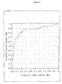
- FIG. 1 ROC-analysis of patients diagnosed with CRC versus healthy controls and disease controls using log CYFRA 21-1 alone.
- FIG. 2 ROC-analysis of patients diagnosed with CRC versus healthy controls and disease controls using the combination of log CYFRA 21-1 and log NSE.
- FIG. 3 ROC-analysis of patients diagnosed with CRC versus healthy controls and disease controls using the combination of log CYFRA 21-1 and log ASC.
- FIG. 4 ROC-analysis of patients diagnosed with CRC versus healthy controls and disease controls using the combination of log CYFRA 21-1, log NSE and log ASC.
- FIG. 5 ROC-analysis of patients diagnosed with CRC versus healthy controls and disease controls using the combination of log CYFRA 21-1, log NSE, log ASC and log NNMT.
- a marker means one marker or more than one marker.
- marker refers to a molecules to be used as a target for analyzing patient test samples.
- molecular targets are proteins or polypeptides themselves as well as antibodies present in a sample.
- Proteins or polypeptides used as a marker in the present invention are contemplated to include any variants of said protein as well as fragments of said protein or said variant, in particular, immunologically detectable fragments.
- proteins which are released by cells or present in the extracellular matrix which become damaged, e.g., during inflammation could become degraded or cleaved into such fragments.
- Certain markers are synthesized in an inactive form, which may be subsequently activated by proteolysis.
- proteins or fragments thereof may also be present as part of a complex.
- Such complex also may be used as a marker in the sense of the present invention.
- Variants of a marker polypeptide are encoded by the same gene, but differ in their PI or MW, or both (e.g., as a result of alternative mRNA or pre-mRNA processing, e.g. alternative splicing or limited proteolysis) and in addition, or in the alternative, may arise from differential post-translational modification (e.g., glycosylation, acylation, and/or phosphorylation).
- assessing colorectal cancer is used to indicate that the method according to the present invention will, alone or together with other markers or variables, e.g., the criteria set forth by the UICC (see above), e.g., aid the physician to establish or confirm the absence or presence of CRC or aid the physician in the prognosis, the monitoring of therapy efficacy (e.g. after surgery, chemotherapy or radiotherapy) and the detection of recurrence (follow-up of patients after therapy).
- markers or variables e.g., the criteria set forth by the UICC (see above), e.g., aid the physician to establish or confirm the absence or presence of CRC or aid the physician in the prognosis, the monitoring of therapy efficacy (e.g. after surgery, chemotherapy or radiotherapy) and the detection of recurrence (follow-up of patients after therapy).
- sample refers to a biological sample obtained for the purpose of evaluation in vitro.
- the sample or patient sample preferably may comprise any body fluid.
- Preferred test samples include blood, serum, plasma, urine, saliva, and synovial fluid.
- Preferred samples are whole blood, serum, plasma or synovial fluid, with plasma or serum being most preferred.
- any such assessment is made in vitro.
- the patient sample is discarded afterwards.
- the patient sample is solely used for the in vitro diagnostic method of the invention and the material of the patient sample is not transferred back into the patient's body.
- the sample is a liquid sample, e.g., whole blood, serum, or plasma.
- the present invention relates to a method for assessing CRC in vitro by biochemical markers, comprising measuring in a sample the concentration of CYFRA 21-1 and using the concentration determined in the assessment of CRC.
- An assay for CYFRA 21-1 specifically measures a soluble fragment of cytokeratin 19 as present in the circulation.
- the measurement of CYFRA 21-1 is typically based upon two monoclonal antibodies (Bodenmueller, H., et al., Int. J. Biol. Markers 9 (1994) 75-81).
- the two specific monoclonal antibodies (KS 19.1 and BM 19.21) are used and a soluble fragment of cytokeratin 19 having a molecular weight of approx. 30,000 daltons is measured.
- Cytokeratins are structural proteins forming the subunits of epithelial intermediary filaments. Twenty different cytokeratin polypeptides have so far been identified. Due to their specific distribution patterns they are eminently suitable for use as differentiation markers in tumor pathology. Intact cytokeratin polypeptides are poorly soluble, but soluble fragments can be detected in serum (Bodenmueller, H., et al., supra).
- CYFRA 21-1 is a well-established marker for Non-Small-Cell Lung Carcinoma (NSCLC).
- NSCLC Non-Small-Cell Lung Carcinoma
- the main indication for CYFRA 21-1 is monitoring the course of non-small cell lung cancer (NSCLC) (Sturgeon, C., Clinical Chemistry 48 (2002) 1151-1159).
- CYFRA 21-1 is also suitable for course-monitoring in myoinvasive cancer of the bladder. Good specificity is shown by CYFRA 21-1 relative to benign lung diseases (pneumonia, sarcoidosis, tuberculosis, chronic bronchitis, bronchial asthma, emphysema).
- CYFRA also is of use in detecting disease relapse and assessing treatment efficacy in the field of breast cancer (Nakata, B., et al., British J. of Cancer (2004) 1-6).
- CYFRA 21-1 has been measured on an Elecsys® analyzer using Roche product number 11820966 according to the manufacturers instructions.
- CYFRA 21-1 is an established marker in the field of NSCLC.
- non-malignant disease controls derived from patients with certain lung non-malignant diseases have been used. This has been considered important to differentiate benign from malign lung diseases (H. Bodenmüller, et al., supra).
- the inventors of the present invention have surprisingly been able to detect the marker CYFRA 21-1 in a significant percentage of samples derived from patients with CRC. Even more surprising they have been able to demonstrate that the presence of CYFRA 21-1 in such liquid sample obtained from an individual can be used in the assessment of colorectal cancer.
- the ideal scenario for diagnosis would be a situation wherein a single event or process would cause the respective disease as, e.g., in infectious diseases. In all other cases correct diagnosis can be very difficult, especially when the etiology of the disease is not fully understood as is the case for CRC.
- no biochemical marker for example in the field of CRC, is diagnostic with 100% specificity and at the same time 100% sensitivity for a given disease. Rather biochemical markers e.g. are used to assess with a certain likelihood or predictive value the presence or absence of a disease. Therefore in routine clinical diagnosis, generally various clinical symptoms and biological markers are considered together in the diagnosis, treatment and management of the underlying disease.
- Biochemical markers can either be determined individually or in a preferred embodiment of the invention they can be measured simultaneously using a chip or a bead based array technology. The concentrations of the biomarkers are then interpreted independently using an individual cut-off for each marker or they are combined for interpretation.
- the assessment of CRC according to the present invention is performed in a method comprising measuring in a sample the concentration of a) CYFRA 21-1, b) optionally one or more other marker of colorectal cancer, and c) using the concentration determined in step (a) and optionally step (b) in the assessment of colorectal cancer.
- the method for assessment of CRC is performed by measuring the concentration of CYFRA 21-1 and of one or more other marker and by using the concentration of CYFRA 21-1 and of the one or more other marker in the assessment of CRC.
- the present invention is also directed to a method for assessing CRC in vitro by biochemical markers, comprising measuring in a sample the concentration of CYFRA 21-1 and of one or more other marker of CRC and using the concentrations determined in the assessment of CRC.
- the marker CYFRA 21-1 both in the univariate analysis as well as in the multivariate analysis performed, has (at a specificity of about 90%) a remarkable sensitivity for CRC of almost 60% and in this respect was found superior as compared to other markers investigated.
- the marker CYFRA 21-1 will be of advantage in one or more of the following aspects: screening; diagnostic aid; prognosis; monitoring of therapy, and follow-up.
- CRC is the second most common malignancy of both males and females in developed countries. Because of its high prevalance, its long asymptomatic phase and the presence of premalignant lesions, CRC meets many of the criteria for screening. Clearly, a serum tumour marker which has acceptable sensitivity and specificity would be more suitable for screening than either FOB testing or endoscopy.
- CYFRA 21-1 alone will not suffice to allow for a general screening e.g. of the at risk population for CRC. Most likely no single biochemical marker in the circulation will ever meet the sensitivity and specificity criteria required for screening purposes. Rather it has to be expected that a marker panel will have to be used in CRC screening.
- the data established in the present invention indicate that the marker CYFRA 21-1 will form an integral part of a marker panel appropriate for screening purposes.
- the present invention therefore relates to the use of CYFRA 21-1 as one marker of a CRC marker panel for CRC screening purposes.
- the present data further indicate that certain combinations of markers will be advantageous in the screening for CRC.
- the present invention also relates to the use of a marker panel comprising CYFRA 21-1 and NSE, or of a marker panel comprising CYFRA 21-1 and ASC, or of a marker panel comprising CYFRA 21-1 and NSE and ASC for the purpose of screening for CRC.
- CEA 21-1 as a single marker according to the data of the present invention might be at least as good a single marker as CEA or even superior it has to be expected that CYFRA 21-1 will be used as a diagnostic aid, especially by establishing a baseline value before surgery.
- the present invention thus also relates to the use of CYFRA 21-1 for establishing a baseline value before surgery for CRC.
- the gold standard for determining prognosis in patients with CRC is the extend of disease as defined by the Dukes', TNM or other staging systems. If a marker such as CEA is to be used for predicting outcome, it must: provide stronger prognostic information than that offered by existing staging systems, provide information independent of the existing systems or provide prognostic data within specific subgroups defined by existing criteria, e.g. in Dukes' B or node-negative patients.
- CEA should be added to the TNM staging system for colorectal cancer.
- the CEA level should be designated as follows: CX, CEA cannot be assessed; CO, CEA not elevated ( ⁇ 5 ⁇ g/l) or CEA1, CEA elevated (>5 ⁇ g/l) (Compton, C., et al., Cancer 88 (2000) 1739-1757).
- CYFRA 21-1 alone significantly contributes to the differentiation of CRC patients from healthy controls or from healthy controls plus non-malignant colon diseases, it has to be expected that it will aid in assessing the prognosis of patients suffering from CRC.
- the level of preoperative CYFRA 21-1 will most likely be combined with one or more other marker for CRC and/or the TNM staging system, as recommended for CEA by the AJCC.
- CYFRA 21-1 is used in the prognosis of patients with CRC.
- CYFRA 21-1 will be at least as good a marker for monitoring of chemotherapy as CEA.
- the present invention therefore also relates to the use of CYFRA 21-1 in the monitoring of CRC patients under chemotherapy.
- CEA Serial monitoring with CEA has been shown to detect recurrent/metastatic disease with a sensitivity of approximately of 80%, specificity of approximately 70% and provides an average lead-time of 5 months (for review, see Duffy, M. J., et al., supra, and Fletcher, R. H., supra). Furthermore, CEA was the most frequent indicator of recurrence in asymptomatic patients (Pietra, N., et al., Dis. Colon Rectum 41 (1998) 1127-1133 and Graham, R. A., et al., Ann. Surg. 228 (1998) 59-63) and was more cost-effective than radiology for the detection of potentially curable recurrent disease.
- CEA was most sensitive (almost 100%) for the detection of liver metastasis.
- CEA was less reliable for diagnosing locoregional recurrences, the sensitivity being only approximately 60% (Moertel, C. G., et al., Jama 270 (1993) 943-947).
- the present invention discloses and therefore in a preferred embodiment relates to the use of CYFRA 21-1 in the diagnostic field of CRC or in the assessment of CRC, respectively.
- the present invention relates to the use of CYFRA 21-1 as a marker molecule for colorectal cancer in combination with one or more marker molecules for colorectal cancer in the assessment of colorectal cancer from a liquid sample obtained from an individual.
- the expression “one or more” denotes 1 to 20, preferably 1 to 10, preferably 1 to 5, more preferred 3 or 4.
- CYFRA 21-1 and the one or more other marker form a CRC marker panel.
- a preferred embodiment of the present invention is the use of CYFRA 21-1 as a marker molecule for colorectal cancer in combination with one or more marker molecules for colorectal cancer in the assessment of colorectal cancer from a liquid sample obtained from an individual.
- Preferred selected other CRC markers with which the measurement of CYFRA 21-1 may be combined are NSE, ASC, NMMT, CA 19-9, CA 72-4, and/or CEA.
- the marker panel used in the assessment of CRC comprises CYFRA 21-1 and at least one other marker molecule selected from the group consisting of NSE, ASC and NMMT.
- markers which preferably are combined with CYFRA 21-1 or which form part of the CRC marker panel comprising CYFRA 21-1, respectively, are discussed in more detail below.
- OPN osteopontin
- OPN is a glyco-phosphoprotein that is expressed and secreted by numerous human cancers. OPN is found in normal plasma, urine, milk and bile (U.S. Pat. No. 6,414,219; U.S. Pat. No. 5,695,761; Denhardt, D. T. & Guo, X., FASEB J. 7 (1993) 1475-1482; Oldberg, A., et al., PNAS 83 (1986) 8819-8823; Oldberg, A., et al., J. Biol. Chem. 263 (1988) 19433-19436; Giachelli, C. M., et al., Trends Cardiovasc. Med. 5 (1995) 88-95).
- the human OPN protein and cDNA have been isolated and sequenced (Kiefer M. C., et al., Nucl. Acids Res. 17 (1989) 3306.
- OPN functions in cell adhesion, chemotaxis, macrophage-directed interleukin-10 (IL-10) suppression, stress-dependent angiogenesis, prevention of apoptosis, and anchorage-independent growth of tumor cells by regulating cell-matrix interactions and cellular signaling through binding with integrin and CD44 receptors. While constitutive expression of OPN exists in several cell types, induced expression has been detected in T-lymphocytes, epidermal cells, bone cells, macrophages, and tumor cells in remodeling processes such as inflammation, ischemia-reperfusion, bone resorption, and tumor progression (reviewed by Wai, P. Y. & Kuo P. C. J. Surg. Res. 121 (2004) 228-241).
- OPN is known to interact with a number of integrin receptors. Increased OPN expression has been reported in a number of human cancers, and its cognate receptors (av-b3, av-b5, and av-b1 integrins and CD44) have been identified.
- av-b3, av-b5, and av-b1 integrins and CD44 cognate receptors
- Irby, R. B., et al., Clinical & Experimental Metastasis 21 (2004) 515-523 indicate that both endogenous OPN expression (via stable transfection) as well as exogenous OPN (added to culture medium) enhanced the motility and invasive capacity of human colon cancer cells in vitro. OPN appeared to regulate motility though interaction with CD44.
- OPN expression also reduced intercellular (homotypic) adhesion, which is regarded as a characteristic of metastatic cancer cells. Stable transfection of four poorly tumorigenic human colon cancer cell lines with OPN also resulted in enhanced tumorigenicity in vivo with increased proliferation and increased CD31 positive microvessel counts, concordant with the degree of OPN expression.
- NSE neuro-specific enolase
- glycolytic enzyme enolase also known as the glycolytic enzyme enolase (2-phospho-D-glycerate hydrolase, EC 4.2.1.11, molecular weight approx. 80 kD
- ⁇ , ⁇ , and ⁇ the glycolytic enzyme enolase
- the ⁇ -subunit of enolase occurs in numerous types of tissue in mammals, whereas the ⁇ -subunits found mainly in the heart and in striated musculature.
- the enolase isoforms ⁇ and ⁇ which are referred to as neuron-specific enolase (NSE) or ⁇ -enolase, are primarily detectable in high concentrations in neurons and neuro-endocrine cells as well as in tumors originating from them (Lamerz R., NSE (Neuronen-spezifische Enolase), ⁇ -Enolase, In: Thomas L. (ed.), Clinical Laboratory Diagnosis, TH-Books, Frankfurt, 1 st English Edition 1998: 979-981, 5. yer Auflage 1998: 1000-1003).
- NSE Neuron-specific enolase
- ⁇ -Enolase In: Thomas L. (ed.), Clinical Laboratory Diagnosis, TH-Books, Frankfurt, 1 st English Edition 1998: 979-981, 5. yer Auflage 1998: 1000-1003).
- NSE is described as the marker of first choice in the monitoring of small cell bronchial carcinoma (Lamerz R., supra), whereas CYFRA 21-1 is superior to NSE for non-small cell bronchial carcinoma (Ebert, W., et al., Eur. J. Clin. Chem. Clin. Biochem 32 (1994) 189-199).
- Elevated NSE concentrations are found in 60-81% of cases of small cell bronchial carcinoma.
- NSE serum values above 30 ng/ml are found in 62% of the affected children.
- the medians rise in accordance with the stage of the disease.
- NSE has also been measured in other tumors: Non-pulmonary malignant diseases show values above 25 ng/ml in 22% of the cases (carcinomas in all stages). Brain tumors such as glioma, miningioma, neurofibroma, and neurinoma are only occasionally accompanied by elevated serum NSE values. In primary brain tumors or brain metastasis and in malignant melanoma and phaeochromocytoma, elevated NSE-values can occur in the CSF (cerebrospinal fluid). Increased NSE concentrations have been reported for 14% of organ-restricted and 46% of metastasizing renal carcinomas, with a correlation to the grade as an independent prognosis factor.
- NSE has been measured on an Elecsys® analyzer using Roche product number 12133113 according to the manufacturers instructions.
- the CA 19-9 (carbohydrate antigen 19-9) values measured are defined by the use of the monoclonal antibody 1116-NS-19-9.
- the 1116-NS-19-9-reactive determinant in serum is mainly expressed on a mucin-like protein that contains a high number of CA 19-9 epitopes (Magnani J. L., Arch. Biochem. Biophys. 426 (2004) 122-131).
- CA 19-9 containing mucins are expressed in fetal gastric, intestinal and pancreatic epithelia. Low concentrations can also be found in adult tissue in the liver, lungs, and pancreas (Stieber, P. and Fateh-Moghadam, A., Boeringer Mannheim, Cat. No. 1536869 (engl), 1320947 (dtsch). ISBN 3-926725-07-9 dtsch/engl., Juergen Hartmann Verlag, Marloffstein-Rathsberg (1993); Herlyn, M., et al., J. Clin. Immunol. 2 (1982) 135-140).
- CA 19-9 assay values can assist in the differential diagnosis and monitoring of patients with pancreatic carcinoma (sensitivity 70-87%) (Ritts, R. E., Jr., et al., Int. J. Cancer 33 (1984) 339-345). There is no correlation between tumor mass and the CA 19-9 assay values. However, patients with CA 19-9 serum levels above 10,000 U/mL almost always have distal metastasis.
- CA 19-9 cannot be used for the early detection of pancreatic carcinoma (Steinberg, W. M., et al., Gastroenterology 90 (1986) 343-349).
- CA 19-9 values provide a sensitivity of 50-75%.
- the concomitant determination of CA 72-4 and CEA is recommended in case of gastric carcinoma.
- determination of CEA alone is adequate; only in a limited number of the CEA-negative cases the determination of CA 19-9 can be useful.
- CA 19-9 has been measured on a Elecsys® analyzer using Roche product number 11776193 according to the manufacturers instructions.
- CEA carcinomaembryonic antigen
- a monomeric glycoprotein molecular weight approx. 180,000 dalton
- a variable carbohydrate component of approx. 45-60%
- CEA like AFP, belongs to the group of carcinofetal antigens that are produced during the embryonic and fetal period.
- the CEA gene family consists of about 17 active genes in two subgroups. The first group contains CEA and the non-specific cross-reacting antigens (NCA); the second group contains the pregnancy-specific glycoproteins (PSG).
- NCA non-specific cross-reacting antigens
- PSG pregnancy-specific glycoproteins
- CEA is mainly found in the fetal gastrointestinal tract and in fetal serum. It also occurs in slight quantities in intestinal, pancreatic, and hepatic tissue of healthy adults. The formation of CEA is repressed after birth, and accordingly serum CEA values are hardly measurable in healthy adults.
- High CEA concentrations are frequently found in cases of colorectal adenocarcinoma (Stieber, P. and Fateh-Moghadam, A., supra).
- Slight to moderate CEA elevations occur in 20-50% of benign diseases of the intestine, the pancreas, the liver, and the lungs (e.g. liver cirrhosis, chronic hepatitis, pancreatitis, ulcerative colitis, Crohn's Disease, emphysema (Stieber, P., and Fateh-Moghadam, A., supra).
- Smokers also have elevated CEA values.
- the main indication for CEA determinations is therapy management and the follow-up of patients with colorectal carcinoma.
- CEA determinations are not recommended for cancer-screening in the general population.
- CEA concentrations within the normal range do not exclude the possible presence of a malignant disease.
- NCA2 merconium antigen
- CEA has been measured on an Elecsys® analyzer using Roche product number 11731629 according to the manufacturers instructions.
- ASC apoptosis-associated speck-like protein containing a caspase-associated recruitment domain
- TMS1 target of methylation-induced silencing 1
- ASC has a theoretical molecular weight of 21,627 Da and a theoretical isoelectric point of pH 6.29.
- Caspase-associated recruitment domains mediate the interaction between adaptor proteins such as APAF1 (apoptotic protease activating factor 1) and the pro-form of caspases (e.g., CASP 9) participating in apoptosis.
- ASC is a member of the CARD-containing adaptor protein family.
- DNMT1 DNA cytosine-5-methyltransferase-1
- breast cancer cell lines, but not normal breast tissue exhibited complete methylation of ASC and expressed no ASC message. Expression of ASC in breast cancer cell lines inhibited growth and reduced the number of surviving colonies.
- Conway et al. concluded that ASC functions in the promotion of caspase-dependent apoptosis and that overexpression of ASC inhibits the growth of breast cancer cells (Conway, K. E., et al., Cancer Research 60 (2000) 6236-6242).
- McConnell and Vertino showed that inducible expression of ASC inhibits cellular proliferation and induces DNA fragmentation that can be blocked by caspase inhibitor.
- Immunofluorescence microscopy demonstrated that induction of apoptosis causes a CARD-dependent shift from diffuse cytoplasmic expression to spherical perinuclear aggregates (McConnell, B. B., and Vertino, P. M., Cancer Research 60 (2000) 6243-6247).
- Moriani et al. observed methylation of ASC gene not only in breast cancer cells but also in gastric cancer.
- Conway et al. examined primary breast tissues for TMS1 methylation and compared the results to methylation in healthy tissues (Conway K. E., et al., Cancer Research 60 (2000) 6236-6242). Levine et al. found that ASC silencing was not correlated with methylation of specific CpG sites, but rather was associated with dense methylation of ASC CpG island. Breast tumor cell lines containing exclusively methylated ASC copies do not express ASC, while in partially methylated cell lines the levels of ASC expression are directly related to the percentage of methylated ASC allels present in the cell population (Levine, J. J., et al., Oncogene 22 (2003) 3475-3488).
- Virmani et al. examined the methylation status of ASC in lung cancer and breast cancer tissue. They found that aberrant methylation of ASC was present in 46% of breast cancer cell lines and in 32% of breast tumor tissue. Methylation was rare in non-malignant breast tissue (7% (Virmani, A., et al., Int. J. Cancer 106 (2003) 198-204).
- Shiohara et al. found out that up-regulation of ASC is closely associated with inflammation and apoptosis in human neutrophils (Shiohara, M., et al., Blood 98 (2001) 229a).
- Masumoto et al. observed that high levels of ASC are abundantly expressed in epithelial cells and leucocytes (Masumoto, J., et al., Journal Histochem. Cytochem. 49 (2001) 1269-1275).
- An in-house sandwich immunoassay has been developed for measurement of ASC. This assay is performed in a microtiter plate format. Streptavidin-coated microtiter plates are used. A biotinylated polyclonal antibody to ASC is used as a capturing antibody and a digoxigenylated polyclonal antibody to ASC is used as the second specific binding partner in this sandwich assay. The sandwich complex formed is finally visualized by an anti-digoxigenin horseradish peroxidase conjugate and an appropriate peroxidase substrate.
- the protein MASP (maspin precursor; Swiss-PROT: P36952) is a 42-kDa protein that shares homology with the serpin superfamily of protease inhibitors. Immunostaining studies demonstrate that maspin is found in the extracellular matrix and at the plasma membrane (Zou, Z., et al., Science 263 (1994) 526-529).
- the human MASP gene (SERPINB5 of PI5) was originally isolated from normal mammary epithelium by subtractive hybridization on the basis of its expression at the mRNA level (Zou et al., supra). Maspin was expressed in normal mammary epithelial cells but not in most mammary carcinoma cell lines. Zou et al. (supra) showed that its expression reduces the ability of transformed cells to induce tumor formation and metastasis, suggesting that the maspin gene encodes a tumor suppressor.
- Ferritin is a protein containing about 20% iron and is found in the intestines, the liver and the spleen. It is one of the chief forms in which iron is stored in the body. Body iron stores have been reported to increase the risk of colorectal neoplasms. In a study by Scholefield, J. H., et al. (Dis. Colon Rectum 41 (1998) 1029-1032) using samples from 148 patients (50 patients with proven colorectal cancer, 49 patients without colon disease, and patients with adenomas of the colon) serum ferritin was assayed. There were no significant differences in serum ferritin levels among any of the three groups.
- the protein nicotinamide N-methyltransferase (NNMT; Swiss-PROT; P40261) has an apparent molecular weight of 29.6 kDa and an isoelectric point of 5.56.
- NNMT catalyzes the N-methylation of nicotinamide and other pyridines. This activity is important for biotransformation of many drugs and xenobiotic compounds.
- the protein has been reported to be predominantly expressed in liver and is located in the cytoplasm.
- NNMT has been cloned from cDNA from human liver and contained a 792-nucleotide open reading frame that encoded a 264-amino acid protein with a calculated molecular mass of 29.6 kDa, (Aksoy, S., et al., J. Biol. Chem. 269 (1994) 14835-14840). Little is known in the literature about a potential role of the enzyme in human cancer.
- markers of a marker panel e.g. for CYFRA 21-1 and NSE, or for CYFRA 21-1 and OPN are mathematically combined and the combined value is correlated to the underlying diagnostic question.
- Marker values may be combined by any appropriate state of the art mathematical method.
- Well-known mathematical methods for correlating a marker combination to a disease employ methods like, discriminant analysis (DA) (i.e. linear-, quadratic-, regularized-DA), Kernel Methods (i.e. SVM), Nonparametric Methods (i.e. k-Nearest-Neighbor Classifiers), PLS (Partial Least Squares), Tree-Based Methods (i.e.
- Logic Regression CART, Random Forest Methods, Boosting/Bagging Methods
- Generalized Linear Models i.e. Logistic Regression
- Principal Components based Methods i.e. SIMCA
- Generalized Additive Models Fuzzy Logic based Methods, Neural Networks and Genetic Algorithms based Methods.
- DA i.e. Linear-, Quadratic-, Regularized Discriminant Analysis
- Kernel Methods i.e. SVM
- Nonparametric Methods i.e.
- k-Nearest-Neighbor Classifiers PLS (Partial Least Squares), Tree-Based Methods (i.e. Logic Regression, CART, Random Forest Methods, Boosting Methods), or Generalized Linear Models (i.e. Logistic Regression). Details relating to these statistical methods are found in the following references: Ruczinski, I., et al., J. of Computational and Graphical Statistics, 12 (2003) 475-511; Friedman, J. H., J.
- state A e.g. diseased from healthy.
- the markers are no longer independent but form a marker panel.
- combining the measurements of CYFRA 21-1 and NSE or ASC, respectively does significantly improve the diagnostic accuracy for CRC as compared to either healthy controls or, as also assessed, as compared to healthy controls plus non-malignant disease controls.
- combining CYFRA 21-1 with OPN or with OPN and FERR improves diagnostic accuracy in a likewise setting. Especially the later finding is of great importance, because a patient with a non-malignant disease may require quite a different treatment as a patient with CRC.
- ROC receiver-operating characteristics
- the clinical performance of a laboratory test depends on its diagnostic accuracy, or the ability to correctly classify subjects into clinically relevant subgroups. Diagnostic accuracy measures the test's ability to correctly distinguish two different conditions of the subjects investigated. Such conditions are for example health and disease or benign versus malignant disease.
- the ROC plot depicts the overlap between the two distributions by plotting the sensitivity versus 1 ⁇ specificity for the complete range of decision thresholds.
- sensitivity or the true-positive fraction [defined as (number of true-positive test results)/(number of true-positive+number of false-negative test results)]. This has also been referred to as positivity in the presence of a disease or condition. It is calculated solely from the affected subgroup.
- false-positive fraction or 1 ⁇ specificity [defined as (number of false-positive results)/(number of true-negative+number of false-positive results)]. It is an index of specificity and is calculated entirely from the unaffected subgroup.
- the ROC plot is independent of the prevalence of disease in the sample.
- Each point on the ROC plot represents a sensitivity/1 ⁇ specificity pair corresponding to a particular decision threshold.
- a test with perfect discrimination has an ROC plot that passes through the upper left corner, where the true-positive fraction is 1.0, or 100% (perfect sensitivity), and the false-positive fraction is 0 (perfect specificity).
- the theoretical plot for a test with no discrimination is a 45° diagonal line from the lower left corner to the upper right corner. Most plots fall in between these two extremes.
- One convenient goal to quantify the diagnostic accuracy of a laboratory test is to express its performance by a single number.
- the combination of the two markers CYFRA 21-1 and NSE significantly improves the diagnostic accuracy for CRC.
- the combination of the two markers CYFRA 21-1 and ASC also significantly improves the diagnostic accuracy for CRC.
- the combination of the two markers CYFRA 21-1 and OPN significantly improves the diagnostic accuracy for CRC.
- the present invention relates to a method for improving the diagnostic accuracy for CRC versus healthy controls and/or patients suffering from non-malignant colon disease by measuring in a sample the concentration of at least CYFRA 21-1 and NSE, OPN or ASC, respectively and correlating the concentrations determined to the presence or absence of CRC, the improvement resulting in more patients being correctly classified as suffering from CRC versus healthy controls and/or patients suffering from non-malignant colon disease as compared to a classification based on any single marker investigated alone.
- At least the concentration of the biomarkers CYFRA 21-1 and NSE, respectively, is determined and the marker combination is used in the assessment of CRC.
- At least the concentration of the biomarkers CYFRA 21-1 and ASC, respectively, is determined and the marker combination is used in the assessment of CRC.
- At least the concentration of the biomarkers CYFRA 21-1 and OPN, respectively, is determined and the marker combination is used in the assessment of CRC.
- At least the concentration of the biomarkers CYFRA 21-1, NSE and ASC, respectively, is determined and the marker combination is used in the assessment of CRC.
- At least the concentration of the biomarkers CYFRA 21-1, OPN and ASC, respectively, is determined and the marker combination is used in the assessment of CRC.
- At least the concentration of the biomarkers CYFRA 21-1, NSE and OPN, respectively, is determined and the marker combination is used in the assessment of CRC.
- At least the concentration of the biomarkers CYFRA 21-1, NNMT and OPN, respectively, is determined and the marker combination is used in the assessment of CRC.
- At least the concentration of the biomarkers CYFRA 21-1, MASP and OPN, respectively, is determined and the marker combination is used in the assessment of CRC.
- At least the concentration of the biomarkers CYFRA 21-1, FERR and OPN, respectively, is determined and the marker combination is used in the assessment of CRC.
- the CRC samples of Table 1 have been evaluated in comparison to control samples obtained from healthy individual, patients with non-malignant colon diseases or in comparison to the pooled data obtained with the healthy and the non-malignant control samples.
- Table 2 gives an overview over the controls used.
- Sensitivity for each marker has been calculated at a common specificity level of 90% for each individual marker tested.
- Table 3 gives the sensitivity over all UICC stages in percent for each of the most promising CRC markers.
- the markers NNMT and ASC appear to have a comparable but slightly lower sensitivity and are followed by CEA.
- RDA Regularized Discriminant Analysis
- McLachlan, G. J., Discriminant Analysis and Statistical Pattern Recognition, Wiley Series in probability and mathematical statistics, 1992 The classification algorithms were generated with the Regularized Discriminant Analysis (RDA), which is a generalization of the common Discriminant Analysis, i.e. Quadratic- and Linear Discriminant Analysis (McLachlan, G. J., Discriminant Analysis and Statistical Pattern Recognition, Wiley Series in probability and mathematical statistics, 1992).
- plug-in maximum likelihood
- Support Vector Machines algorithms (Hastie, Trevor, Tibshirani, Robert, Friedman, Jerome, The Elements of Statistical Learning, Springer Series in Statistics, 2001) can be fitted with comparable classification results.
- the analysis by RDA has been based on 106 CRC samples, because for 3 out of the original 109 samples a marker value was missing.
- the marker panels were stepwise constructed starting from the best single marker for the classification problem and ending when the increase in the sensitivity at a specificity level of about 90% does not change remarkably any more.
- every single marker was transformed with the natural logarithmic function. 5-fold cross validation was used.
- Table 4 presents the classification results of patients diagnosed with CRC versus controls including non-malignant colon diseases.
- the first marker selected by RDA was CYFRA 21-1, the second one NSE.
- the marker panel CYFRA 21-1, NSE, and ASC in this analysis did yield the highest sensitivity at a specificity level of about 90%.
- RDA was performed using a marker panel comprising CYFRA 21-1, OPN, FERR, NNMT, NSE and MASP.
- the second study population comprised serum samples from 254 patients diagnosed with CRC (see Table 6) and 391 control samples. These were split into a training set and a test set. The analysis was based on a training set of 128 CRC samples and 195 controls. Of the controls 16 were from individuals without any gastro-intestinal disease, 50 from individuals with hemorrhoids, 5 from patients with other bowel diseases; 63 controls came from individuals with diverticulosis, 61 from healthy blood donors. The test set consisted of 126 CRC samples and 196 controls. Of the controls 20 were from individuals without any gastro-intestinal disease, 43 from individuals with hemorrhoids, 8 from patients with other bowel diseases; 65 controls came from individuals with diverticulosis, 60 from healthy blood donors.
Landscapes
- Health & Medical Sciences (AREA)
- Life Sciences & Earth Sciences (AREA)
- Engineering & Computer Science (AREA)
- Immunology (AREA)
- Urology & Nephrology (AREA)
- Hematology (AREA)
- Biomedical Technology (AREA)
- Chemical & Material Sciences (AREA)
- Molecular Biology (AREA)
- Medicinal Chemistry (AREA)
- Biochemistry (AREA)
- Cell Biology (AREA)
- Hospice & Palliative Care (AREA)
- Biotechnology (AREA)
- Food Science & Technology (AREA)
- Oncology (AREA)
- Physics & Mathematics (AREA)
- Analytical Chemistry (AREA)
- Microbiology (AREA)
- General Health & Medical Sciences (AREA)
- General Physics & Mathematics (AREA)
- Pathology (AREA)
- Measuring Or Testing Involving Enzymes Or Micro-Organisms (AREA)
- Investigating Or Analysing Biological Materials (AREA)
- Medicines Containing Antibodies Or Antigens For Use As Internal Diagnostic Agents (AREA)
- Medicines Containing Material From Animals Or Micro-Organisms (AREA)
Applications Claiming Priority (4)
| Application Number | Priority Date | Filing Date | Title |
|---|---|---|---|
| EP04030619 | 2004-12-23 | ||
| EP04030619 | 2004-12-23 | ||
| EP04030619.3 | 2004-12-23 | ||
| PCT/EP2005/013867 WO2006066915A1 (fr) | 2004-12-23 | 2005-12-22 | Utilisation de cyfra 21-1 comme marqueur du cancer colorectal |
Related Parent Applications (1)
| Application Number | Title | Priority Date | Filing Date |
|---|---|---|---|
| PCT/EP2005/013867 Continuation WO2006066915A1 (fr) | 2004-12-23 | 2005-12-22 | Utilisation de cyfra 21-1 comme marqueur du cancer colorectal |
Publications (2)
| Publication Number | Publication Date |
|---|---|
| US20080020414A1 US20080020414A1 (en) | 2008-01-24 |
| US7731938B2 true US7731938B2 (en) | 2010-06-08 |
Family
ID=34927964
Family Applications (1)
| Application Number | Title | Priority Date | Filing Date |
|---|---|---|---|
| US11/764,913 Active 2026-02-27 US7731938B2 (en) | 2004-12-23 | 2007-06-19 | CYFRA 21-1 as a marker for colorectal cancer |
Country Status (10)
| Country | Link |
|---|---|
| US (1) | US7731938B2 (fr) |
| EP (1) | EP1831695B1 (fr) |
| JP (1) | JP4606469B2 (fr) |
| CN (2) | CN101088012A (fr) |
| AT (1) | ATE430936T1 (fr) |
| CA (1) | CA2585788C (fr) |
| DE (1) | DE602005014386D1 (fr) |
| ES (1) | ES2325277T3 (fr) |
| HK (1) | HK1115636A1 (fr) |
| WO (1) | WO2006066915A1 (fr) |
Cited By (4)
| Publication number | Priority date | Publication date | Assignee | Title |
|---|---|---|---|---|
| US20100021928A1 (en) * | 2006-06-26 | 2010-01-28 | Technion Research & Development Foundation Ltd. | Methods and kits for diagnosing cancer |
| US20100317040A1 (en) * | 2009-06-16 | 2010-12-16 | Technion Research & Develepment Foundation Ltd. | Methods and kits for diagnosing cancer |
| US20140004543A1 (en) * | 2011-03-11 | 2014-01-02 | Roche Diagnostics Operations, Inc. | Asc as a marker for chronic obstructive pulmonary disease (copd) |
| KR20200103431A (ko) * | 2019-02-25 | 2020-09-02 | 가톨릭대학교 산학협력단 | Cyfra 21-1을 이용한 갑상선암 림프절 전이 진단 방법 |
Families Citing this family (12)
| Publication number | Priority date | Publication date | Assignee | Title |
|---|---|---|---|---|
| CA2655851A1 (fr) * | 2006-08-01 | 2008-02-07 | F. Hoffmann-La Roche Ag | Utilisation de la nnmt en tant que marqueur pour le cancer du poumon |
| US20100196889A1 (en) * | 2006-11-13 | 2010-08-05 | Bankaitis-Davis Danute M | Gene Expression Profiling for Identification, Monitoring and Treatment of Colorectal Cancer |
| CN101896817A (zh) * | 2007-12-10 | 2010-11-24 | 霍夫曼-拉罗奇有限公司 | 用于结直肠癌的标记物组 |
| JP5298188B2 (ja) | 2008-07-03 | 2013-09-25 | エフ.ホフマン−ラ ロシュ アーゲー | 肺癌用マーカーとしてのasc |
| US20130095503A1 (en) * | 2010-01-05 | 2013-04-18 | University Of Cincinnati | Serum spla2-iia as diagnosis marker for prostate and lung cancer |
| CN103476428B (zh) * | 2010-09-09 | 2016-10-26 | 北京同为时代生物技术有限公司 | 用于诊断上皮源性癌症的血液标志物及其单克隆抗体 |
| JP6026645B2 (ja) * | 2012-04-24 | 2016-11-16 | ボアジチ・ユニヴェルシテシBogazici Universitesi | 抗原送達方法 |
| KR101619122B1 (ko) | 2013-08-14 | 2016-05-10 | 연세대학교 산학협력단 | 탈메틸화 의존적 asc 발현을 촉진시켜서 대장암 종양세포의 사멸감수성을 증진시키는 항암제 병용 투여용 후보물질의 스크리닝 방법 |
| EP3183578B8 (fr) * | 2014-08-22 | 2020-07-15 | Abbott Laboratories | Méthodes de détection précoce du cancer colorectal |
| WO2019120527A1 (fr) * | 2017-12-20 | 2019-06-27 | Michael Heneka | Nouveaux moyens et méthodes permettant de traiter des maladies neurodégénératives |
| KR20210134946A (ko) * | 2019-03-01 | 2021-11-11 | 어드밴스드 마커 디스커버리 에스.엘. | 대장암 및/또는 이의 전암 단계 진단을 위한 단백질 시그니처 |
| CA3175753A1 (fr) * | 2020-03-19 | 2021-09-23 | Advanced Marker Discovery S.l. | Signature proteque pour cribler une population globale pour un cancer colorectal et/ou un stade precancereux correspondant |
Citations (4)
| Publication number | Priority date | Publication date | Assignee | Title |
|---|---|---|---|---|
| US20040038225A1 (en) | 2002-08-26 | 2004-02-26 | Markowitz Sanford D. | Methods and compositions for categorizing patients |
| WO2004057336A2 (fr) | 2002-12-20 | 2004-07-08 | Roche Diagnostics Gmbh | Utilisation de la nicotinamide n-methyltransferase comme marqueur du cancer colorectal |
| EP1439393A2 (fr) | 2002-12-13 | 2004-07-21 | Bayer Healthcare LLC | Procédés de détection utilisants TIMP 1 pour le diagnostic du cancer du colon |
| WO2004104593A1 (fr) | 2003-05-26 | 2004-12-02 | Roche Diagnostics Gmbh | Utilisation de la proteine masp en tant que marqueur pour le cancer colorectal |
Family Cites Families (1)
| Publication number | Priority date | Publication date | Assignee | Title |
|---|---|---|---|---|
| US6165740A (en) * | 1998-09-30 | 2000-12-26 | Sysmex Corporation | Method and device for flow-cytometric microorganism analysis |
-
2005
- 2005-12-22 EP EP05819706A patent/EP1831695B1/fr not_active Not-in-force
- 2005-12-22 CA CA2585788A patent/CA2585788C/fr not_active Expired - Fee Related
- 2005-12-22 AT AT05819706T patent/ATE430936T1/de active
- 2005-12-22 JP JP2007543807A patent/JP4606469B2/ja not_active Expired - Fee Related
- 2005-12-22 WO PCT/EP2005/013867 patent/WO2006066915A1/fr active Application Filing
- 2005-12-22 CN CNA2005800445589A patent/CN101088012A/zh active Pending
- 2005-12-22 ES ES05819706T patent/ES2325277T3/es active Active
- 2005-12-22 CN CN200580044411XA patent/CN101088011B/zh not_active Expired - Fee Related
- 2005-12-22 DE DE602005014386T patent/DE602005014386D1/de active Active
-
2007
- 2007-06-19 US US11/764,913 patent/US7731938B2/en active Active
-
2008
- 2008-05-30 HK HK08106060.4A patent/HK1115636A1/xx not_active IP Right Cessation
Patent Citations (4)
| Publication number | Priority date | Publication date | Assignee | Title |
|---|---|---|---|---|
| US20040038225A1 (en) | 2002-08-26 | 2004-02-26 | Markowitz Sanford D. | Methods and compositions for categorizing patients |
| EP1439393A2 (fr) | 2002-12-13 | 2004-07-21 | Bayer Healthcare LLC | Procédés de détection utilisants TIMP 1 pour le diagnostic du cancer du colon |
| WO2004057336A2 (fr) | 2002-12-20 | 2004-07-08 | Roche Diagnostics Gmbh | Utilisation de la nicotinamide n-methyltransferase comme marqueur du cancer colorectal |
| WO2004104593A1 (fr) | 2003-05-26 | 2004-12-02 | Roche Diagnostics Gmbh | Utilisation de la proteine masp en tant que marqueur pour le cancer colorectal |
Non-Patent Citations (24)
| Title |
|---|
| Ahlquist, D., "Fecal Occult Blood Testing for Colorectal Cancer," Colorectal Neoplasia, Part II, 26;1 (Mar. 1997) 41/55. |
| Bernick et al, "Neuroendocrine Carcinomas of the Colon and Rectum," (Dis of the Colon and Rectum), 2004, vol. 47, pp. 163-169. * |
| Bodenmuller, H. et al., "Lung Cancer-Associated Keratin 19 Fragments: Development and Biochemical Characterisation of the New Serum Assay Enzymun-Test CYFRA 21-1," The International Journal of Biological Markers, 9;2 (1994) 75-81. |
| Duffy, M. et al., "Clinical utility of biochemical markers in colorectal cancer: European Group on Tumour Markers (EGTM) guidelines," European Journal of Cancer, 39 (2003) 718-727. |
| Ebert, W. et al., "Cytokeratin 19 Fragment CYFRA 21-1 Compared with Carcinoembryonic Antigen, Squamous Cell Carcinoma Antigen and Neuron-Specific Enolase in Lung Cancer," Eur. J. Clin. Chem. Clin. Biochem, 32 (1994) 189-199. |
| Hoffmann, D. et al., "The Prognostic Value of CEA, CA242, CA19-9, CA72-4, CYFRA 21-1 and S100 in Colorectal Cancer," Tumor Biol, 24 (2003) 49-Abstract P-14 CEA. |
| Kashihara et al, "Intrahepatic cholangiocarcinoma with increased serum CYFRA 21-1 level," (J. Gastroenterol) 1998, vol. 33, pp. 447-453. * |
| Kojima, O. et al., "Tumor Marker, Serological Detection of Cancer," Rinsho Byori. Jap. Clin. Pathol. 34 (2004) 337-339. |
| Lamerz, R., Clinical Laboratory Diagnostics: Use and Assessment of Clinical Laboratory Results, First Edition, TH-Books Verlagsgesellschaft mbH, Frankfurt/Main, Germany. |
| Martell, R.E. et al., "OVX1 and CEA in Patients with Colon Carcinoma, Colon Polyps and Benign Colon Disorders," The International Journal of Biological Markers, 13;3 (1998) 145-149. |
| Matsumoto, H., Database Biosci, Info. Servc. Philadelphia (2002)-Database accession No. PREV200200296087-XP002329221. |
| Matsumoto, H., Journal of Japan Society of Coloproctology, 55 (2002) 136-144. |
| Miyashita, T. et al., Database Biosci, Info. Servc. Philadelphia (2002)-Database accession No. PREV200000195119-XP002329220. |
| Miyashita, T. et al., Journal of Japan Society of Coloproctology,53 (2000)76-82. |
| Molina et al. "Study of a new tumor marker, CYFRA 21-1, in malignant and nonmalignant diseases", Tumor Biol., 1994, 15:318-325. * |
| Nakata, B. et al., "Serum CYFRA 21-1 (cytokeratin-19 fragments) is a useful tumour marker for detecting disease relapse and assessing treatment efficacy in breast cancer," British Journal of Cancer, 91 (2004) 873-878. |
| Qiang, Z. et a., "Diagnostics Value of Multi-Tumor Markers Protein Biochip Detective System for Digestive Tract Cancer," Zhongguo Zhongliu Linchaung, 31 (2004) 337-339. |
| Sakahara, H. et al., Kakuigaku, 30 (1993) 1475-1479. |
| Sturgeon, C., "Practice Guidelines for Tumor Marker Use in the Clinic," Clinical Chemistry, 48:8 (2002) 1151-1159. |
| Van Der Gaast, A. et al., "Evaluation of a New Tumour Marker in Patients with Non-Small-Cell Lung Cancer: Cyfra 21.1," Br. J. Cancer, 69 (1994) 525-528. |
| Vaughn, C. et al., "Ferritin, A Potential Marker for Cancer of the Colon," Proc. Am. Assoc. Cancer Res. Annual Meeting, 30 (19989) 26-Abstract No. 101. |
| Yeh et al, "Monitoring Cytokeratin Fragment 10 (CYFRA 21-1) Serum Levels for Early Prediction of Recurrence of Adenocarcinoma and Squamous Cell Carcinom in the Lung after Surgical Resection," (Lung), 2002, vol. 180, pp. 273-279. * |
| Yokoyama, T. et a., "Methylation of ASC/TMS1, a proapoptotic gene responsible for activating procaspase-1, in human colorectal cancer," Cancer Letters, 202 (2003) 101-108. |
| Zweig, M. et al., "Receiver-Operating Characteristic (ROC) Plots: A fundamental Evaluation Tool in Clinical Medicine," Clin. Chem., 39;4 (1993) 561-577. |
Cited By (7)
| Publication number | Priority date | Publication date | Assignee | Title |
|---|---|---|---|---|
| US20100021928A1 (en) * | 2006-06-26 | 2010-01-28 | Technion Research & Development Foundation Ltd. | Methods and kits for diagnosing cancer |
| US20100317040A1 (en) * | 2009-06-16 | 2010-12-16 | Technion Research & Develepment Foundation Ltd. | Methods and kits for diagnosing cancer |
| US9075060B2 (en) | 2009-06-16 | 2015-07-07 | Technion Research & Development Foundation Limited | Methods for diagnosing oral or oral-pharyngeal cancer |
| US20140004543A1 (en) * | 2011-03-11 | 2014-01-02 | Roche Diagnostics Operations, Inc. | Asc as a marker for chronic obstructive pulmonary disease (copd) |
| US9116156B2 (en) * | 2011-03-11 | 2015-08-25 | Roche Diagnostics Operations, Inc. | ASC as a marker for chronic obstructive pulmonary disease (COPD) |
| KR20200103431A (ko) * | 2019-02-25 | 2020-09-02 | 가톨릭대학교 산학협력단 | Cyfra 21-1을 이용한 갑상선암 림프절 전이 진단 방법 |
| KR102293271B1 (ko) | 2019-02-25 | 2021-08-23 | 가톨릭대학교 산학협력단 | Cyfra 21-1을 이용한 갑상선암 림프절 전이 진단 방법 |
Also Published As
| Publication number | Publication date |
|---|---|
| CN101088011B (zh) | 2012-07-18 |
| JP4606469B2 (ja) | 2011-01-05 |
| JP2008522172A (ja) | 2008-06-26 |
| US20080020414A1 (en) | 2008-01-24 |
| ES2325277T3 (es) | 2009-08-31 |
| CA2585788A1 (fr) | 2006-06-29 |
| HK1115636A1 (en) | 2008-12-05 |
| EP1831695B1 (fr) | 2009-05-06 |
| WO2006066915A1 (fr) | 2006-06-29 |
| EP1831695A1 (fr) | 2007-09-12 |
| ATE430936T1 (de) | 2009-05-15 |
| CN101088012A (zh) | 2007-12-12 |
| DE602005014386D1 (de) | 2009-06-18 |
| CA2585788C (fr) | 2013-03-05 |
| CN101088011A (zh) | 2007-12-12 |
Similar Documents
| Publication | Publication Date | Title |
|---|---|---|
| US7731938B2 (en) | CYFRA 21-1 as a marker for colorectal cancer | |
| US20090075312A1 (en) | Assessing colorectal cancer by measuring osteopontin and carcinoembryonic antigen | |
| EP2223116B1 (fr) | Panel de marqueurs pour le cancer colorectal | |
| EP2223115B1 (fr) | Séprase comme marqueur du cancer | |
| US20070161062A1 (en) | Protein CBP2 as a marker for colorectal cancer | |
| US7846653B2 (en) | ASC as a marker for colorectal cancer | |
| US10175241B2 (en) | ASC as a marker for lung cancer | |
| EP1761780A1 (fr) | Utilisation de cyfra 21-1 comme marqueur du cancer colorectal | |
| US20070184498A1 (en) | Protein RS15A as a marker for colorectal cancer | |
| WO2006066916A1 (fr) | Utilisation de dipeptidase microsomale comme marqueur du cancer colorectal |
Legal Events
| Date | Code | Title | Description |
|---|---|---|---|
| AS | Assignment |
Owner name: ROCHE DIAGNOSTICS GMBH, GERMANY Free format text: ASSIGNMENT OF ASSIGNORS INTEREST;ASSIGNORS:KARL, JOHANN;ANDRES, HERBERT;GRUNERT, VEIT PETER;AND OTHERS;REEL/FRAME:019946/0580;SIGNING DATES FROM 20070719 TO 20070723 Owner name: ROCHE DIAGNOSTICS OPERATIONS, INC., INDIANA Free format text: ASSIGNMENT OF ASSIGNORS INTEREST;ASSIGNOR:ROCHE DIAGNOSTICS GMBH;REEL/FRAME:019946/0601 Effective date: 20070731 Owner name: ROCHE DIAGNOSTICS GMBH,GERMANY Free format text: ASSIGNMENT OF ASSIGNORS INTEREST;ASSIGNORS:KARL, JOHANN;ANDRES, HERBERT;GRUNERT, VEIT PETER;AND OTHERS;SIGNING DATES FROM 20070719 TO 20070723;REEL/FRAME:019946/0580 Owner name: ROCHE DIAGNOSTICS OPERATIONS, INC.,INDIANA Free format text: ASSIGNMENT OF ASSIGNORS INTEREST;ASSIGNOR:ROCHE DIAGNOSTICS GMBH;REEL/FRAME:019946/0601 Effective date: 20070731 |
|
| STCF | Information on status: patent grant |
Free format text: PATENTED CASE |
|
| FPAY | Fee payment |
Year of fee payment: 4 |
|
| MAFP | Maintenance fee payment |
Free format text: PAYMENT OF MAINTENANCE FEE, 8TH YEAR, LARGE ENTITY (ORIGINAL EVENT CODE: M1552) Year of fee payment: 8 |
|
| MAFP | Maintenance fee payment |
Free format text: PAYMENT OF MAINTENANCE FEE, 12TH YEAR, LARGE ENTITY (ORIGINAL EVENT CODE: M1553); ENTITY STATUS OF PATENT OWNER: LARGE ENTITY Year of fee payment: 12 |