EP2223116B1 - Panel de marqueurs pour le cancer colorectal - Google Patents
Panel de marqueurs pour le cancer colorectal Download PDFInfo
- Publication number
- EP2223116B1 EP2223116B1 EP08860118.2A EP08860118A EP2223116B1 EP 2223116 B1 EP2223116 B1 EP 2223116B1 EP 08860118 A EP08860118 A EP 08860118A EP 2223116 B1 EP2223116 B1 EP 2223116B1
- Authority
- EP
- European Patent Office
- Prior art keywords
- seprase
- crc
- marker
- ferritin
- cea
- Prior art date
- Legal status (The legal status is an assumption and is not a legal conclusion. Google has not performed a legal analysis and makes no representation as to the accuracy of the status listed.)
- Not-in-force
Links
Images


Classifications
-
- G—PHYSICS
- G01—MEASURING; TESTING
- G01N—INVESTIGATING OR ANALYSING MATERIALS BY DETERMINING THEIR CHEMICAL OR PHYSICAL PROPERTIES
- G01N33/00—Investigating or analysing materials by specific methods not covered by groups G01N1/00 - G01N31/00
- G01N33/48—Biological material, e.g. blood, urine; Haemocytometers
- G01N33/50—Chemical analysis of biological material, e.g. blood, urine; Testing involving biospecific ligand binding methods; Immunological testing
- G01N33/53—Immunoassay; Biospecific binding assay; Materials therefor
- G01N33/574—Immunoassay; Biospecific binding assay; Materials therefor for cancer
- G01N33/57407—Specifically defined cancers
- G01N33/57419—Specifically defined cancers of colon
Definitions
- the present invention relates to a method for assessing colorectal cancer (CRC) in vitro comprising measuring in a sample the concentration and/or activity of a seprase polypeptide and/or fragments thereof and of either anti-p53 and/or osteopontin and/or Ferritin, of optionally one or more other marker of CRC, and using the combined measurement result in the assessment of CRC. Furthermore, it especially relates to a method for assessing CRC from a liquid sample, derived from an individual by measuring seprase and at least one marker selected from the group consisting of anti-p53, Ferritin and osteopontin in said sample.
- the method according to the present invention can, e.g., be used in the early detection of cancer by screening of asymptomatic individuals or in the surveillance of patients who undergo surgery.
- Cancer remains a major public health challenge despite progress in detection and therapy.
- Cancer cells are characterized by the production of cancer-associated marker proteins. Cancer-associated proteins are found both in the tissues and in the bodily fluids of an individual who carries cancer cells. Their levels usually are low at the early stages of the carcinogenic progress and increase during the disease's progression and only in rare cases proteins are observed showing a decreased level in the course of disease progression.
- the sensitive detection of these proteins is an advantageous and promising approach for the diagnosis of cancer, in particular in an early stage diagnosis of cancer.
- the most prevalent cancer types are breast cancer (BC), lung cancer (LC) and colorectal cancer (CRC).
- Surgical resection of the tumors is widely accepted as a first line treatment for early stage solid tumors.
- Most cancers are detected only when they become symptomatic, i.e. when patients already are in a rather late stage of disease progression.
- Colorectal cancer most frequently progresses from adenomas (polyps) to malignant carcinomas.
- the staging of cancer is the classification of the disease in terms of extent, progression, and severity. It groups cancer patients so that generalizations can be made about prognosis and the choice of therapy.
- TNM the TNM system
- N the status of regional lymph nodes
- M the presence or absence of distant metastases
- TNM criteria are published by the UICC ( International Union against Cancer), Sobin, L.H., Wittekind, Ch. (eds): TNM Classification of Malignant Tumours, sixth edition, 2002 ).
- UICC International Union against Cancer
- Sobin L.H.
- TNM staging and UICC disease stages correspond to each other as shown in the following Table taken from Sobin L.H. and Wittekind (eds.) supra.
- tumor marker which is detectable in body fluids, e.g. blood or serum or plasma or a panel of such markers, would be desirable.
- CEA carcinoembryonic antigen
- a tumor-associated glycoprotein a tumor-associated glycoprotein
- CEA carcinoembryonic antigen
- serum CEA determination possesses neither the sensitivity nor the specificity to enable its use as a screening test for colorectal cancer in the asymptomatic population ( Reynoso, G. et al., JAMA 220 (1972) 361-365 ; Sturgeon, C., Clin. Chem. 48 (2002) 1151-1159 ).
- the guaiac test is currently most widely used as a screening assay for CRC from stool.
- the guaiac test however, has both poor sensitivity as well as poor specificity.
- the sensitivity of the guaiac-based fecal occult blood tests is ⁇ 26%, which means 74% of patients with malignant lesions will remain undetected ( Ahlquist, D.A., Gastroenterol. Clin. North Am. 26 (1997) 41-55 ).
- the sensitivity and specificity of diagnostic alternatives to the guaiac test have been recently investigated by Sieg, A. et al., Int. J. Colorectal Dis. 14 (1999) 267-271 .
- the hemoglobin assay has an unsatisfactory sensitivity for the detection of colorectal neoplasms. Whereas cancer in its progressed carcinoma stage is detected with a sensitivity of about 87% the earlier tumor stages are not detected with a sufficient sensitivity.
- the hemoglobin-haptoglobin complex assay was more sensitive in the detection of earlier stages of CRC. This more sensitive detection was accompanied by a poor specificity. Since poor specificity, however, translates to a high number of unnecessary secondary investigations, like colonoscopy, an assay with a poor specificity also does not meet the requirements of a generally accepted screening assay.
- Calprotectin has been described as an alternative biomarker for the detection of CRC from stool samples in US 5,455,160 and correspondingly in the scientific literature by Roseth, A.G., et al. (Scand. J. Gastroenterol. 27 (1992) 793-798 ; Scand. J. Gastroenterol. 28 (1993) 1073-1076 ). Although calprotectin is a marker of inflammatory diseases its potential as a marker for the detection of CRC from stool is documented by several publications ( Johne, B., et al., Scand. J. Gastroenterol. 36 (2001) 291-296 ; Limburg, P.J., et al., Am. J. Gastroenterol.
- calprotectin appears to have some characteristics favorable for a diagnostic biomarker as compared to hemoglobin. It is evenly distributed in feces, it is stable at room temperature making mail delivery of the sample to the laboratory feasible and it shows no interference with food components or pharmaceutical compounds ( Ton, H., et al., Clin. Chim. Acta 292 (2000) 41-54 ).
- a new diagnostic marker as a single marker should be comparable to other markers known in the art, or better. Or, a new marker should lead to a progress in diagnostic sensitivity and/or specificity either if used alone or in combination with one or more other markers, respectively.
- the diagnostic sensitivity and/or specificity of a test is best assessed by its receiver-operating characteristics, which will be described in detail below.
- the new marker panels of the present invention have surprisingly been found that significantly improve the diagnostic accuracy, especially in the screening for CRC.
- the present invention relates to a method for assessing colorectal cancer (CRC) in vitro comprising measuring in a sample the concentration and/or activity of a seprase polypeptide and/or fragments thereof, of anti-p53, osteopontin and/or Ferritin, of optionally one or more other marker of CRC, and using the combined measurement result for seprase, anti-p53, osteopontin and/or Ferritin and optionally of the one or more other marker in the assessment of CRC.
- CRC colorectal cancer
- a decreased concentration and/or activity of a seprase polypeptide and/or fragments thereof in the test sample is associated with the risk and/or occurrence of cancer, especially of CRC.
- the marker seprase can be combined with the marker anti-p53, osteopontin and/or Ferritin and, if required, with other markers for CRC, like CEA and/or CYFRA21-1.
- One embodiment of the present invention refers to the mass screening of a population to distinguish between individuals that are probably free from colorectal cancer and individuals which might be classified as "suspect" cases, i.e. individuals, which might have CRC. The latter group of individuals could then be subjected to further diagnostic procedures, e.g. by imaging methods or other suitable means.
- a further embodiment of the present invention refers to an improvement of tumor marker panels which are suitable for the assessment of colorectal cancer.
- the present invention is directed to a method for assessing CRC in vitro by biochemical marker, comprising measuring in a sample the concentration and/or activity of seprase and using the measurement results, particularly the concentration or activity determined in the assessment of colorectal cancer.
- the present invention is also directed to a method for assessing CRC in vitro by biochemical marker, comprising measuring in a sample the concentration and/or activity of seprase, of a marker selected from the group consisting of anti-p53, osteopontin and Ferritin and of one or more other markers, and using the measurement results, particularly concentrations, determined in the assessment of colorectal cancer.
- Preferred markers for use in combination with seprase, anti-p53, osteopontin and/or Ferritin are markers which are general tumor markers (i.e. markers which are not specific for a single tumor type) or, on the other hand, specific tumor markers (markers which are specific for a single tumor type).
- Preferred the one or more other marker is Carcinoembryonic antigen (CEA) and/or CYFRA21-1. Preferably, these markers are used in any combination together with seprase.
- a cut-off value for an individual marker or a marker combination can for example be determined from the testing of a group of healthy individuals.
- the cut-off is set to result in a specificity of 90%, also preferred the cut-off is set to result in a specificity of 95%, or also preferred the cut-off is set to result in a specificity of 98%.
- a value for an individual marker or a marker combination above the cut-off value can for example be indicative for the presence of cancer.
- the present invention in a preferred embodiment, relates to a method for assessing CRC, comprising measuring seprase and anti-p53 and using the combined measurement values for assessment of CRC.
- the present invention in a preferred embodiment, relates to a method for assessing CRC, comprising measuring seprase and osteopontin and using the combined measurement values for assessment of CRC.
- the present invention in a preferred embodiment, relates to a method for assessing CRC, comprising measuring seprase and Ferritin and using the combined measurement values for assessment of CRC.
- the present invention in a further preferred embodiment, relates to a method for assessing CRC, comprising measuring seprase, Ferritin, CEA and osteopontin and using the combined measurement values for assessment of CRC.
- the present invention in a further preferred embodiment, relates to a method for assessing CRC, comprising measuring seprase, Ferritin, CEA, osteopontin and anti-p53 and using the combined measurement values for assessment of CRC.
- the present invention in a further preferred embodiment, relates to a method for assessing CRC, comprising measuring seprase, Ferritin, CEA, CYFRA21-1, osteopontin and anti-p53 and using the combined measurement values for assessment of CRC.
- the present invention in a preferred embodiment, also relates to the use of a marker panel comprising at least seprase and anti-p53 in the assessment of colorectal cancer.
- the present invention in a preferred embodiment, also relates to the use of a marker panel comprising at least seprase and anti-p53 in the assessment of colorectal cancer.
- the present invention in a preferred embodiment, also relates to the use of a marker panel comprising at least seprase and osteopontin in the assessment of colorectal cancer.
- the present invention in a preferred embodiment, also relates to the use of a marker panel comprising at least seprase and Ferritin in the assessment of colorectal cancer.
- the present invention in a preferred embodiment, also relates to the use of a marker panel comprising at least seprase and CEA in the assessment of colorectal cancer.
- the present invention in a preferred embodiment, also relates to the use of a marker panel comprising at least seprase and CYFRA21-1 in the assessment of colorectal cancer.
- the present invention also relates to the use of a seprase polypeptide and/or fragments thereof in the assessment of colon cancer, wherein a decreased concentration and/or activity of seprase and/or fragments thereof is indicative for cancer.
- the present invention also relates to the use of an antibody directed against a seprase polypeptide and/or fragments thereof in the assessment of colon cancer, wherein a decreased concentration and/or activity of seprase and/or fragments thereof is indicative for colon cancer.
- the present invention also relates to the use of a reagent for measuring the enzymatic activity of a seprase polypeptide and/or fragments thereof in the assessment of colon cancer, wherein a decreased concentration and/or activity of seprase and/or of enzymatically active fragments thereof is indicative for colon cancer.
- the present invention also relates to a device comprising the means for performing the method according to the present invention or for making use of the marker combinations disclosed.
- the present invention also provides for a kit comprising the means for performing the method according to the present invention, i.e. the means for at least measuring seprase and a marker selected from the group consisting of anti-p53, osteopontin and Ferritin.
- the present invention also provides a kit for performing the method according to the present invention comprising at least the reagents required to specifically measure seprase and anti-p53, respectively, and optionally auxiliary reagents for performing the measurement.
- the present invention also provides a kit for performing the method according to the present invention comprising at least the reagents required to specifically measure seprase and osteopontin, respectively, and optionally auxiliary reagents for performing the measurement.
- the present invention also provides a kit for performing the method according to the present invention comprising at least the reagents required to specifically measure seprase and Ferritin, respectively, and optionally auxiliary reagents for performing the measurement.
- the present invention also provides a kit for performing the method according to the present invention comprising at least the reagents required to specifically measure seprase and CYFRA21-1, respectively, and optionally auxiliary reagents for performing the measurement.
- the present invention also provides a kit for performing the method according to the present invention comprising at least the reagents required to specifically measure seprase and CEA, respectively, and optionally auxiliary reagents for performing the measurement.
- the present invention relates to a method for assessing colorectal cancer in vitro comprising measuring in a sample the concentration and/or activity of (a) a seprase polypeptide and/or fragments thereof, (b) optionally one or more other marker of CRC, and (c) using the measurement result of step (a) and optionally of step (b) in the assessment of CRC.
- the present invention relates to a method for assessing colorectal cancer (CRC) in vitro comprising measuring in a sample the concentration and/or activity of (a) a seprase polypeptide and/or fragments thereof, (b) anti-p53, osteopontin and/or Ferritin, (c) optionally one or more other marker of CRC, and (d) using the combined measurement result of step (a) and (b) and optionally of step (c) in the assessment of CRC.
- CRC colorectal cancer
- the concentration and/or activity of seprase is combined with the measurement for at least anti-p53, osteopontin or Ferritin and the combined value is correlated to an underlying diagnostic question like e.g. likelihood of presence or absence of CRC, stage of disease, disease progression, or response to therapy.
- Human seprase (alias fibroblast activation protein (FAP)) is a 170 kDa membrane-bound glycoprotein having gelatinase and dipeptidyl peptidase activity. It consists of two identical monomeric units (Uni Prot KB database Accession No. P 27487) characterized by the sequence given in SEQ ID NO:1.
- the seprase monomer comprises 766 amino acids and has an apparent molecular weight of 97 kDa (SDS-PAGE) ( Pineiro-Sanchez, et al., J. Biol. Chem. 272 (1997) 7595-7601 ; Park, et al., J. Biol. Chem. 274 (1999) 36505-36512 ).
- a soluble form of seprase is the antiplasmin-cleaving enzyme APCE ( Lee, et al., Blood 103 (2004) 3783-3788 ; Lee, et al., Blood 107 (2006) 1397-1404 ).
- the N-terminal amino acid sequence of APCE corresponds to residues 24-38 of seprase which is predicted to have its first 6 N-terminal residues within the fibroblast cytoplasm, followed by a 20-residue transmembrane domain and then a 734 residue extracellular C-terminal catalytic domain ( Goldstein, et al., Biochim Biophys Acta. 1361 (1997) 11-19 ; Scanlan, et al., Proc Natl Acad Sci USA 91 (1994) 5657-5661 ).
- Iwasa et al., 2005 discloses that the increased expression of seprase activity is associated with lymph node metastasis in human colorectal cancer.
- a determination of the concentration and/or the activity e.g. the enzymatic activity of a seprase polypeptide and/or fragments thereof, particularly of APCE in a body fluid, allows the assessment of cancer disease, e.g. of lung, breast, colon, or kidney cancer disease, and in particular of lung cancer disease. Even more surprisingly, it was found that a decreased concentration and/or activity of seprase or fragments thereof in a sample compared to normal controls is indicative for the risk or occurrence of cancer.
- a marker means one marker or more than one marker.
- the term “at least” is used to indicate that optionally one or more further objects may be present.
- a marker panel comprising at least (the markers) seprase and CYFRA21-1 may optionally comprise one or more other marker.
- one or more denotes 1 to 50, preferably 1 to 20 also preferred 2, 3, 4, 5, 6, 7, 8, 9, 10, 12, or 15.
- marker refers to a molecule to be used as a target for analyzing a patient's test sample.
- molecular targets are proteins or polypeptides.
- Proteins or polypeptides used as a marker in the present invention are contemplated to include naturally occurring variants of said protein as well as fragments of said protein or said variant, in particular, immunologically detectable fragments.
- Immunologically detectable fragments preferably comprise at least 6, 7, 8, 10, 12, 15 or 20 contiguous amino acids of said marker polypeptide.
- proteins which are released by cells or present in the extracellular matrix may be damaged, e.g., during inflammation, and could become degraded or cleaved into such fragments.
- markers are synthesized in an inactive form, which may be subsequently activated by proteolysis.
- proteins or fragments thereof may also be present as part of a complex.
- Such complex also may be used as a marker in the sense of the present invention.
- the amino acid sequence of a variant is to 95% or more identical to the corresponding marker sequence.
- a marker polypeptide or a variant thereof may carry a post-translational modification.
- posttranslational modifications are glycosylation, acylation, and/or phosphorylation.
- a “marker of cancer” and in particular a “marker of colon cancer or colorectal cancer” in the sense of the present invention is any marker that if combined with the marker combination comprising seprase and a marker selected from the group consisting of anti-p53, osteopontin and Ferritin adds relevant information in the assessment of CRC.
- the information is considered relevant or of additive value if at a given specificity the sensitivity, or if at a given sensitivity the specificity, respectively, for the assessment of CRC can be improved by including said marker into a marker combination already comprising the marker seprase and a marker selected from the group consisting of anti-p53, osteopontin and Ferritin.
- sample refers to a biological sample obtained for the purpose of evaluation in vitro.
- the sample or patient sample preferably comprises any body fluid.
- Preferred samples are whole blood, serum, plasma with plasma or serum being most preferred.
- assessing cancer and in particular "assessing colon cancer or colorectal cancer” is used to indicate that the method according to the present invention will (alone or together with other markers or variables, e.g., the criteria set forth by the UICC (see above)) e.g., aid the physician to establish or confirm the absence or presence of CRC or aid the physician in the prognosis, the detection of recurrence (follow-up of patients after surgery) and/or the monitoring of treatment, especially of chemotherapy.
- markers or variables e.g., the criteria set forth by the UICC (see above)
- aid the physician to establish or confirm the absence or presence of CRC or aid the physician in the prognosis the detection of recurrence (follow-up of patients after surgery) and/or the monitoring of treatment, especially of chemotherapy.
- any such assessment is made in vitro.
- the patient sample is discarded afterwards.
- the patient sample is solely used for the in vitro diagnostic method of the invention and the material of the patient sample is not transferred back into the patient's body.
- the sample is a liquid sample, e.g., whole blood, serum, or plasma.
- measurement preferably comprises a qualitative, semi-quantitative or a quantitative measurement of a marker in a sample.
- the measurements in a method according to the present invention are quantitative measurements.
- the term “seprase polypeptide and/or fragments thereof” refers to monomeric or multimeric, particularly dimeric polypeptides. Further, the term “seprase polypeptide and/or fragments thereof” particularly refers to soluble forms of seprase and/or fragments thereof, particularly antiplasmin-cleaving enzyme (APCE) as well as to seprase and/or seprase fragments present in form of a complex with another polypeptide.
- APCE antiplasmin-cleaving enzyme
- Seprase polypeptides are preferably detected in appropriate samples, particularly in body fluids.
- Preferred samples are body fluids, such as blood, plasma, serum, sputum and urine.
- the sample is derived from a human subject, e.g. a tumor patient or a person in risk of a tumor or a person suspected of having a tumor.
- the concentration and/or activity of a seprase polypeptide and/or fragments thereof is determined.
- the marker seprase is specifically measured from a sample by use of a specific binding agent.
- immunodiagnostic procedures may be used to reach a result comparable to the achievements of the present invention.
- alternative strategies to generate antibodies may be used.
- Such strategies comprise amongst others the use of synthetic peptides, representing an epitope of seprase for immunization.
- DNA immunization also known as DNA vaccination may be used.
- seprase is detected in a sandwich-type assay format.
- a first specific binding agent is used to capture seprase on the one side and a second specific binding agent, which is labeled to be directly or indirectly detectable, is used on the other side.
- the specific binding agents used in a sandwich-type assay format may be antibodies specifically directed against seprase.
- the detection may be carried out by using different capturing and labeled antibodies, i.e. antibodies which recognize different epitopes on the seprase polypeptide.
- a sandwich-type assay may also be carried out with a capture and labeling antibody which is directed against the same epitope of seprase. In this embodiment, only di- and multimeric forms of seprase may be detected.
- the marker seprase is specifically measured from a sample by use of a test regimen suitable for detecting the enzymatic activity of seprase.
- the enzymatic activity may e.g. be a gelatinase activity or a peptidase activity.
- Gelatinase activity may e.g. be determined by zymography using gelatine zymograms.
- Peptidase activity may e.g. be determined by fluorescent assays using appropriate labeled peptide substrates.
- Assays for the measurement of seprase activity are e.g. described by Lee et al. (2006), supra.
- OPN is found in normal plasma, urine, milk and bile ( US 6,414,219 ; US 5,695,761 ; Denhardt, D.T. and Guo, X., FASEB J. 7 (1993) 1475-1482 ; Oldberg, A., et al., PNAS 83 (1986) 8819-8823 ; Oldberg, A., et al., J. Biol. Chem. 263 (1988) 19433-19436 ; Giachelli, C.M., et al., Trends Cardiovasc. Med. 5 (1995) 88-95 ).
- the human OPN protein and cDNA have been isolated and sequenced ( Kiefer M.C., et al., Nucl. Acids Res. 17 (1989) 3306 ).
- OPN functions in cell adhesion, chemotaxis, macrophage-directed interleukin-10 (IL-10) suppression, stress-dependent angiogenesis, prevention of apoptosis, and anchorage-independent growth of tumor cells by regulating cell-matrix interactions and cellular signaling through binding with integrin and CD44 receptors. While constitutive expression of OPN exists in several cell types, induced expression has been detected in T-lymphocytes, epidermal cells, bone cells, macrophages, and tumor cells in remodeling processes such as inflammation, ischemia-reperfusion, bone resorption, and tumor progression (reviewed by Wai, P.Y. and Kuo, P.C., J. Surg. Res. 121 (2004) 228-241 ).
- IL-10 interleukin-10
- OPN is known to interact with a number of integrin receptors. Increased OPN expression has been reported in a number of human cancers, and its cognate receptors (av-b3, av-b5, and av-b1 integrins and CD44) have been identified.
- av-b3, av-b5, and av-b1 integrins and CD44 cognate receptors.
- Irby, R.B., et al., Clin. Exp. Metastasis 21 (2004) 515-523 indicate that both endogenous OPN expression (via stable transfection) as well as exogenous OPN (added to culture medium) enhanced the motility and invasive capacity of human colon cancer cells in vitro. OPN appeared to regulate motility though interaction with CD44.
- OPN expression also reduced intercellular (homotypic) adhesion, which is regarded as a characteristic of metastatic cancer cells. Stable transfection of four poorly tumorigenic human colon cancer cell lines with OPN also resulted in enhanced tumorigenicity in vivo with increased proliferation and increased CD31 positive micro vessel counts, concordant with the degree of OPN expression. Fragments of OPN may also be used in a method according to the present invention.
- the present invention relates to a method for assessing CRC in vitro by biochemical markers, comprising measuring in a sample the concentration of osteopontin and of seprase, combining the measured values and using the combined value in the assessment of CRC.
- CEA Carcinoembryonic antigen
- CEA cancerembryonic antigen
- CEA is a monomeric glycoprotein (molecular weight approx. 180.000 Dalton) with a variable carbohydrate component of approx. 45-60% ( Gold, P. and Freedman, S.O., J. Exp Med 121 (1965) 439-462 ).
- CEA like AFP, belongs to the group of carcinofetal antigens that are produced during the embryonic and fetal period.
- the CEA gene family consists of about 17 active genes in two subgroups. The first group contains CEA and the Non-specific Cross-reacting Antigens (NCA); the second group contains the Pregnancy-Specific Glycoproteins (PSG).
- NCA Non-specific Cross-reacting Antigens
- PSG Pregnancy-Specific Glycoproteins
- CEA is mainly found in the fetal gastrointestinal tract and in fetal serum. It also occurs in slight quantities in intestinal, pancreatic, and hepatic tissue of healthy adults. The formation of CEA is repressed after birth, and accordingly serum CEA values are hardly measurable in healthy adults.
- CEA determinations are therapy management and the follow-up of patients with colorectal carcinoma.
- CEA determinations are not recommended for cancer-screening in the general population.
- CEA concentrations within the normal range do not exclude the possible presence of a malignant disease.
- NCA2 meconium antigen
- CYFRA21-1 specifically measures a soluble fragment of cytokeratin 19 as present in the circulation.
- the measurement of CYFRA21-1 is typically based upon two monoclonal antibodies ( Bodenmueller, H., et al., Int. J. Biol. Markers 9 (1994) 75-81 ).
- the two specific monoclonal antibodies (KS 19.1 and BM 19.21) are used and a soluble fragment of cytokeratin 19 having a molecular weight of approx. 30,000 Daltons is measured.
- Cytokeratins are structural proteins forming the subunits of epithelial intermediary filaments.
- cytokeratin polypeptides Twenty different cytokeratin polypeptides have so far been identified. Due to their specific distribution patterns they are eminently suitable for use as differentiation markers in tumor pathology. Intact cytokeratin polypeptides are poorly soluble, but soluble fragments can be detected in serum (Bodenmueller, H., et al., supra).
- CYFRA21-1 is a well-established marker for Non-Small-Cell Lung Carcinoma (NSCLC).
- NSCLC Non-Small-Cell Lung Carcinoma
- the main indication for CYFRA21-1 is monitoring the course of non-small cell lung cancer (NSCLC) ( Sturgeon, C., Clinical Chemistry 48 (2002) 1151-1159 ).
- CYFRA21-1 serum levels indicate an advanced tumor stage and a poor prognosis in patients with non-small-cell lung cancer ( van der Gaast, A.., et al., Br. J. Cancer 69 (1994) 525-528 ).
- a normal or only slightly elevated value does not rule out the presence of a tumor.
- Successful therapy is documented by a rapid fall in the CYFRA21-1 serum level into the normal range.
- a constant CYFRA21-1 value or a slight or only slow decrease in the CYFRA21-1 value indicates incomplete removal of a tumor or the presence of multiple tumors with corresponding therapeutic and prognostic consequences. Progression of the disease is often shown earlier by increasing CYFRA21-1 values than by clinical symptomatology and imaging procedures.
- CYFRA21-1 preferably is measured on an Elecsys ® analyzer using Roche product number 11820966 according to the manufacturer's instructions.
- Ferritin is a macromolecule with a molecular weight of at least 440 kD (depending on the iron content) and consists of a protein shell (apoferritin) of 24 subunits and an iron core containing an average of approx. 2500 Fe 3+ ions (in liver and spleen ferritin) ( Wick, M. et al., Ferritin in Iron Metabolism - Diagnosis of Anemieas (second edition).
- Ferritin tends to form oligomers, and when it is present in excess in the cells of the storage organs there is a tendency for condensation to semicrystalline hemosiderin to occur in the lysosomes.
- At least 20 isoferritins can be distinguished with the aid of isoelectric focusing ( Arosio, P. et al., Heterogeneity of ferritin II: Immunological aspects. In: Albertini A, Arosio P, Chiancone E, Drysdale J (eds). Ferritins and isoferritins as biochemical markers. Elsevier, Amsterdam 1984;33-47 ). This microheterogeneity is due to differences in the contents of the acidic H and weakly basic L subunits. The basic isoferritins are responsible for the long-term iron storage function, and are found mainly in the liver, spleen, and bone marrow ( Kaltwasser, J.P.
- Acidic isoferritins are found mainly in the myocardium, placenta, and tumor tissue. They have a lower iron content and presumably function as intermediaries for the transfer of iron in various syntheses ( Morikawa, K. et al., Leuk. Lymphoma 18(5-6) (1995) 429-433 ; Borch-Iohnson, B., Analyst 120(3) (1995) 891-903 ; Cook, J. et al., Adv. Exp. Med. Biol. 356 (1994) 219-228 ).
- ferritin is a suitable method for ascertaining the iron metabolism situation. Determination of ferritin at the beginning of therapy provides a representative measure of the body's iron reserves. Clinically, a threshold value of 20 ⁇ g/L (ng/mL) has proved useful in the detection of prelatent iron deficiency. This value provides a reliable indication of exhaustion of the iron reserves that can be mobilized for hemoglobin synthesis. When the ferritin level is elevated and the possibility of a distribution disorder can be ruled out, this is a manifestation of iron overloading in the body. 400 ⁇ g/L (ng/mL) ferritin is used as the threshold value.
- Elevated ferritin values are also encountered with the following tumors: acute leukemia, Hodgkin's disease and carcinoma of the lung, liver and prostate.
- the determination of ferritin has proved to be of value in liver metastasis.
- Reasons for the elevated values could be cell necrosis, blocked erythropoiesis or increased synthesis in tumor tissue.
- Ferritin preferably is measured on an Elecsys ® analyzer using Roche product number 11820982 according to the manufacturer's instructions.
- p53 antibodies were discovered during the course of tumor-associated antigen screening in breast cancer patients without the knowledge of mutations of the p53 gene ( Crawford, L. et al., Int. J. Cancer 30 (1982) 403-408 ).
- the tumor suppressor p53 is a phosphoprotein barely detectable in the nucleus of normal cells ( Benchimol, S. et al., EMBO J. 1 (1982) 1055-1062 ).
- p53 can arrest cell cycle progression, thus allowing the DNA to be repaired or it can lead to apoptosis.
- Anti-p53 preferably is measured using two immuno-dominant peptides of the wild type p53 protein ( Lubin et. al., Cancer Res. 53 (1993) 5872-5876 ).
- polypeptides like seprase, are preferably measured by use of one or more specific binding agent(s).
- a specific binding agent is, e.g., a receptor for seprase, a lectin binding to seprase or an antibody to seprase.
- a specific binding agent has at least an affinity of 10 7 l/mol for its corresponding target molecule.
- the specific binding agent preferably has an affinity of 10 8 l/mol or also preferred of 10 9 l/mol for its target molecule.
- specific is used to indicate that other biomolecules present in the sample do not significantly bind to the binding agent specific for seprase.
- the level of binding to a biomolecule other than the target molecule results in a binding affinity which is at most only 10% or less, only 5% or less only 2% or less or only 1% or less of the affinity to the target molecule, respectively.
- a preferred specific binding agent will fulfill both the above minimum criteria for affinity as well as for specificity.
- a specific binding agent preferably is an antibody reactive with seprase.
- the term antibody refers to a polyclonal antibody, a monoclonal antibody, antigen binding fragments of such antibodies, single chain antibodies as well as to genetic constructs comprising the binding domain of an antibody.
- Antibodies are generated by state of the art procedures, e.g., as described in Tijssen ( Tijssen, P., Practice and theory of enzyme immunoassays, 11, Elsevier Science Publishers B.V., Amsterdam, the whole book, especially pages 43-78 ).
- Tijssen Tijssen, P., Practice and theory of enzyme immunoassays, 11, Elsevier Science Publishers B.V., Amsterdam, the whole book, especially pages 43-78 ).
- the skilled artisan is well aware of methods based on immunosorbents that can be used for the specific isolation of antibodies. By these means the quality of polyclonal antibodies and hence their performance in immunoassays can be enhanced. (Tijssen, P., supra, pages 108-115).
- monoclonal as well as polyclonal antibodies e.g. as raised in rabbits may be used.
- polyclonal antibodies from different species e.g., rats or guinea pigs can also be used. Since monoclonal antibodies can be produced in any amount required with constant properties, they represent ideal tools in development of an assay for clinical routine.
- the generation and the use of monoclonal antibodies to seprase in a method according to the present invention, respectively, represent yet other preferred embodiments.
- Preferred examples of antibodies suitable for the detection of seprase are disclosed in Pi ⁇ eiro-Sánchez et al., supra, e.g. the antibodies D8, D28 and D43.
- an appropriate amount or aliquot of the sample obtained from an individual is incubated with the specific binding agents for different markers of the marker panel under conditions appropriate for formation of a binding agent marker complex. Such conditions need not be specified, since the skilled artisan without any inventive effort can easily identify such appropriate incubation conditions.
- the amount of binding agent marker complex is measured and used in the assessment of cancer, preferably of CRC. As the skilled artisan will appreciate there are numerous methods to measure the amount of the specific binding agent marker complex all described in detail in relevant textbooks (cf., e.g., Tijssen P., supra, or Diamandis, E.P. and Christopoulos, T.K. (eds.), Immunoassay, Academic Press, Boston (1996 )).
- the marker of interest is an antibody or an auto-antibody
- various detection method with which the skilled artisan is fully familiar are at hand.
- Non-limiting and preferred methods used in the detection of antibodies or as the case may be of auto-antibodies are the indirect sandwich assay method or the double-antigen-sandwich (DAGS) method.
- DGS double-antigen-sandwich
- the indirect sandwich method the (auto-)antibody (first antibody) is bound to the (auto-)antigen and indirectly detected via a labeled antibody (secondary antibody) to this (auto-)antibody or first antibody.
- the assay is set up to provide for two antigens each comprising an epitope for the (auto-)antibody wherein one of the two antigens is bound to a solid phase or capable of binding to a solid phase and wherein the second antigen is detectably labeled.
- the (auto-)antibody is present a bridge it will bridge the first and the second antigen and the complex formed can be detected/measured.
- the present invention relates to a method for assessing CRC, in vitro by biochemical markers, comprising measuring in a sample the concentration of seprase and using the concentration and/or activity determined in the assessment of CRC.
- the present invention relates to a method for assessing CRC, in vitro by biochemical markers, comprising measuring in a sample the concentration of seprase and of at least anti-p53, osteopontin or Ferritin, combining the concentration(s) and/or activity(ies) determined and using the combined value in the assessment of CRC.
- the inventors of the present invention have surprisingly been able to detect a decreased concentration and/or activity of the marker seprase in a significant percentage of samples derived from patients with colon cancer. Again surprisingly they have been able to demonstrate that the decreased concentration and/or activity of seprase in such sample obtained from an individual can be used in the assessment of CRC. Even more surprising they have been able to demonstrate that the decreased concentration and/or activity of seprase in such sample obtained from an individual can be combined with at least a measurement of anti-p53, Ferritin and/or osteopontin and used in the assessment of CRC.
- the ideal scenario for diagnosis would be a situation wherein a single event or process would cause the respective disease as, e.g., in infectious diseases. In all other cases correct diagnosis can be very difficult, especially when the etiology of the disease is not fully understood as is the case for many cancer types, e.g. for CRC.
- no biochemical marker is diagnostic with 100% specificity and at the same time 100% sensitivity for a given multifactorial disease, for example for CRC.
- biochemical markers e.g., CYFRA21-1 or CEA, or as shown here certain marker combinations comprising seprase can be used to assess with a certain likelihood or predictive value e.g., the presence, absence, or the severity of a disease. Therefore in routine clinical diagnosis, generally various clinical symptoms and biological markers are considered together in the diagnosis, treatment and management of the underlying disease.
- Biochemical markers can either be determined individually or in a preferred embodiment of the invention they can be measured simultaneously using a chip or a bead based array technology. The concentrations of the biomarkers are then combined for interpretation.
- the marker seprase or a panel comprising seprase and at least one marker selected from the group consisting of anti-p53, Ferritin and osteopontin will be of advantage in one or more of the following aspects: screening; diagnostic aid; prognosis; monitoring of therapy such as chemotherapy, radiotherapy, and immunotherapy.
- Screening is defined as the systematic application of a test to identify individuals e.g. at risk individuals, for indicators of a disease, e.g., the presence of cancer.
- the screening population is composed of individuals known to be at higher than average risk of cancer.
- a screening population for CRC may be composed of individuals known to be at higher than average risk for CRC, like persons at an age of 45 and above, or 50 and above or with a family history of CRC.
- a body fluid such as whole blood, serum or plasma is used as a sample in the screening for colorectal cancer.
- a marker panel comprising a plurality of markers will have to be used in cancer screening.
- the data established in the present invention indicate that the marker seprase, preferably together with a marker selected from the group consisting of anti-p53, Ferritin and osteopontin will form an integral part of a marker panel appropriate for CRC screening purposes.
- the present invention therefore relates to the use of seprase as one marker of a CRC marker panel, for example a marker panel comprising the marker seprase together with a marker selected from the group consisting of anti-p53, Ferritin and osteopontin for CRC screening purposes.
- a marker panel comprising seprase and CYFRA21-1, or of a marker panel comprising seprase and CEA, or to a marker panel comprising seprase and two or more markers selected from the group consisting of anti-p53, Ferritin, osteopontin, CYFRA21-1 and CEA.
- Markers may either aid the differential diagnosis of benign vs. malignant disease in a particular organ, help to distinguish between different histological types of a tumor, or to establish baseline marker values before surgery.
- CT computed tomography
- seprase as a single marker might be superior to other markers, e.g. in the case of CRC to other markers, like CEA or CYFRA21-1, it has to be expected that seprase alone or together with other preferred markers as described in the present invention will be used as a diagnostic aid, especially by establishing a baseline value before surgery.
- the present invention thus also relates to the use of seprase or of marker combinations comprising seprase for establishing a baseline value before surgery for cancer.
- Prognostic indicators can be defined as clinical, pathological, or biochemical features of cancer patients and their tumors that predict with certain likelihood the disease outcome. Their main use is to help to rationally plan patient management, i.e. to avoid undertreatment of aggressive disease and overtreatment of indolent disease, respectively.
- a marker panel according to the present invention significantly contributes to the differentiation of CRC patients, from controls, it has to be expected that it will aid in assessing the prognosis of patients suffering from CRC.
- the level of preoperative seprase will most likely be combined with one or more other marker for CRC and/or the TNM staging system.
- a marker panel according to the present invention is used in the prognosis of patients with CRC.
- a CRC marker panel according to present invention i.e., a marker panel comprising seprase will be at least as good a marker for monitoring of chemotherapy in CRC as CEA.
- the present invention therefore also relates to a marker panel comprising seprase and at least one marker selected from the group consisting of anti-p53, Ferritin and/or osteopontin in the monitoring of cancer patients, preferably of CRC patients, under chemotherapy.
- the measurements for seprase and for at least one marker selected from the group consisting of anti-p53, Ferritin and/or osteopontin and of optionally CYFRA 21-1 and/or CEA will be combined and used in the monitoring of CRC therapy.
- a marker panel comprising seprase and one or more other marker of CRC in the follow-up of CRC patients represents a further preferred embodiment of the present invention.
- a further preferred embodiment relates to the use of a marker panel comprising seprase and at least one marker selected from the group consisting of anti-p53, Ferritin and/or osteopontin in the follow-up of CRC patients.
- the present invention also relates to the use of seprase in the assessment of CRC.
- certain preferred embodiments relate to the measurement values for seprase and for at least one marker selected from the group consisting of anti-p53, Ferritin and/or osteopontin to the combination of these measurement values and to the use of the combined value in the assessment of CRC.
- the combination of markers is evaluated.
- the values measured for markers of a marker panel e.g. for seprase and anti-p53, are mathematically combined and the combined value is correlated to the underlying diagnostic question.
- Marker values may be combined by any appropriate state of the art mathematical method.
- Well-known mathematical methods for correlating a marker combination to a disease employ methods like, discriminant analysis (DA) (i.e. linear-, quadratic-, regularized-DA), Kernel Methods (i.e. SVM), Nonparametric Methods (i.e. k-Nearest-Neighbor Classifiers), PLS (Partial Least Squares), Tree-Based Methods (i.e.
- Logic Regression CART, Random Forest Methods, Boosting/Bagging Methods
- Generalized Linear Models i.e. Logistic Regression
- Principal Components based Methods i.e. SIMCA
- Generalized Additive Models Fuzzy Logic based Methods, Neural Networks and Genetic Algorithms based Methods.
- DA i.e. Linear-, Quadratic-, Regularized Discriminant Analysis
- Kernel Methods i.e. SVM
- Nonparametric Methods i.e.
- k-Nearest-Neighbor Classifiers PLS (Partial Least Squares), Tree-Based Methods (i.e. Logic Regression, CART, Random Forest Methods, Boosting Methods), or Generalized Linear Models (i.e. Logistic Regression). Details relating to these statistical methods are found in the following references: Ruczinski, I., et al, J. of Computational and Graphical Statistics, 12 (2003) 475-511 ; Friedman, J. H., J.
- state A e.g. diseased from no-diseased.
- state B e.g. diseased from no-diseased.
- the markers are no longer independent but form a marker panel.
- ROC receiver-operating characteristics
- the clinical performance of a laboratory test depends on its diagnostic accuracy, or the ability to correctly classify subjects into clinically relevant subgroups. Diagnostic accuracy measures the test's ability to correctly distinguish two different conditions of the subjects investigated. Such conditions are for example health and disease or benign versus malignant disease.
- the ROC plot depicts the overlap between the two distributions by plotting the sensitivity versus 1 - specificity for the complete range of decision thresholds.
- sensitivity or the true-positive fraction [defined as (number of true-positive test results)/(number of true-positive + number of false-negative test results)]. This has also been referred to as positivity in the presence of a disease or condition. It is calculated solely from the affected subgroup.
- false-positive fraction or 1 - specificity [defined as (number of false-positive results)/(number of true-negative + number of false-positive results)]. It is an index of specificity and is calculated entirely from the unaffected subgroup.
- the ROC plot is independent of the prevalence of disease in the sample.
- Each point on the ROC plot represents a sensitivity/1-specificity pair corresponding to a particular decision threshold.
- a test with perfect discrimination has an ROC plot that passes through the upper left corner, where the true-positive fraction is 1.0, or 100% (perfect sensitivity), and the false-positive fraction is 0 (perfect specificity).
- the theoretical plot for a test with no discrimination is a 45° diagonal line from the lower left corner to the upper right corner. Most plots fall in between these two extremes.
- One preferred way to quantify the diagnostic accuracy of a laboratory test is to express its performance by a single number.
- the present invention relates to a method for improving the diagnostic accuracy for CRC versus controls by measuring in a sample the concentration of at least seprase and a marker selected from the group consisting of anti-p53, Ferritin and/or osteopontin, respectively, mathematically combining the concentrations determined and correlating the combined value to the presence or absence of CRC, the improvement resulting in more patients being correctly classified as being at risk or suffering from CRC versus controls as compared to a classification based on any single marker investigated alone.
- At least the concentration of the biomarkers seprase and CYFRA21-1, respectively, is determined and the marker combination is used in the assessment of CRC.
- At least the concentration of the biomarkers seprase and CEA, respectively, is determined and the marker combination is used in the assessment of CRC.
- At least the measurement values for the biomarkers seprase, CEA, anti-p53 and Ferritin are combined for the assessment of CRC.
- At least the measurement values for the biomarkers seprase, CEA, anti-p53 and osteopontin are combined for the assessment of CRC.
- At least the measurement results for seprase, CEA, anti-p53, CYFRA21-1 and osteopontin are determined, mathematically combined, and this marker combination is used in the assessment of CRC.
- At least the measurement results for seprase, CEA, anti-p53, CYFRA21-1 and Ferritin are determined, mathematically combined, and this marker combination is used in the assessment of CRC.
- At least the measurement results for seprase, CEA, anti-p53, CYFRA21-1, osteopontin and Ferritin are determined, mathematically combined, and this marker combination is used in the assessment of CRC.
- Blood samples were drawn into a serum tube and allowed to clot for at least 1h and up to 2 h at room temperature. After centrifugation the supernatant (serum) was frozen at -25°C in 1 ml aliquots and transferred within two weeks to a -70°C freezer. Before measurement the serum samples were thawed and re-aliquoted into smaller volumes for convenience and refrozen. The samples were then thawed immediately before analysis. Therefore, every sample in the set was frozen and thawed only twice before analysis.
- Elecsys ® kits were acquired for following tumor markers: CEA, CA 15-3, CA 19-9, CA125, CA 72-4, Ferritin, ß-hCG, NSE, AFP and CYFRA21-1 (Cytokeratin-19). All assays were run according to the manufacturer's instructions on the Elecsys ® 2010 system (Roche Diagnostics GmbH, Mannheim, Germany). The concentrations measured by the instrument were used to calculate/generate the AUC data shown in Table 1.
- osteopontin and anti-p53 micro titer assays were developed in-house.
- two different antibodies were used. These antibodies were selected to have different non-overlapping epitopes. The epitopes of the two antibodies used are between amino acid 167 and the carboxy terminus of the osteopontin sequence (Kiefer, M.C. et al., Nucl. Acids Res. 17 (1989) 3306). One antibody had been biotinylated and was used as a capture antibody. The second antibody had been digoxigenylated. The digoxigenylated antibody was then detected by use of an appropriate anti-DIG secondary antibody.
- Streptavidin-coated 96-well microtiter plates were incubated with 100 ⁇ l biotinylated anti-OPN polyclonal antibody for 60 min at 10 ⁇ g/mL in 10 mM phosphate buffered saline (PBS), pH 7.4, 1% BSA and 0.1% Tween 20. After incubation, plates were washed three times with phosphate buffered saline pH 7.4 (PBS), 0.1% Tween 20. Wells were then incubated for 2 h with either a serial dilution of the recombinant protein as standard antigen or with diluted plasma samples from patients. After binding of OPN, plates were washed three times with PBS, 0.1% Tween 20.
- wells were incubated with 100 ⁇ l of digoxigenylated anti-OPN polyclonal antibody for 60 min at 10 ⁇ g/ml in 10 mM phosphate, pH 7.4, 1% BSA, 0.9% NaCl and 0.1% Tween 20. Thereafter, plates were washed three times to remove unbound antibody.
- wells were incubated with 20 mU/ml anti-digoxigenin-POD conjugates (Roche Diagnostics GmbH, Mannheim, Germany, Catalogue No. 1633716) for 60 min in 10 mM phosphate, pH 7.4, 1% BSA, 0,9% NaCl and 0.1% Tween 20. Plates were subsequently washed three times with the same buffer.
- ABTS solution (Roche Diagnostics GmbH, Mannheim, Germany, Catalog No. 11685767) and OD was measured after 30-60 min at 405 nm with an ELISA reader.
- TMB tetramethylbenzidine/hydrogenperoxide
- Monoclonal rat IgG (clone D28) was brought to 10 mg/ml in 10 mM NaH 2 PO 4 /NaOH, pH 7.5, 30 mM NaCl. Per ml IgG solution 50 ⁇ l Biotin-N-hydroxysuccinimide (3.6 mg/ml in DMSO) were added. After 30 min at room temperature, the sample was chromatographed on Superdex 200 (10 mM NaH 2 PO 4 /NaOH, pH 7.5, 30 mM NaCl). The fractions containing biotinylated IgG were collected.
- Monoclonal rat IgG (clone D8) was brought to 10 mg/ml in 10 mM NaH 2 PO 4 /NaOH, 30 mM NaCl, pH 7.5.
- Per ml IgG solution 50 ⁇ l of digoxigenin-3-O-methylcarbonyl- ⁇ -aminocaproic acid-N-hydroxysuccinimide ester (Roche Diagnostics, Mannheim, Germany, Cat. No. 1 333 054) (3.8 mg/ml in DMSO) were added. After 30 min at room temperature, the sample was chromatographed on Superdex ® 200 (10 mM NaH 2 PO 4 /NaOH, pH 7.5, 30 mM NaCl). The fractions containing digoxigenylated IgG were collected.
- Samples (75 ⁇ l) were mixed with 150 ⁇ l of antibody reagent containing 0.375 ⁇ g of each, D28-biotin and D8-digoxigenin antibodies in incubation buffer (comprising amongst other additives 40 mM phosphate, 200 mM sodium tartrate, 10 mM EDTA, 0.1% Tween 20, and adjusted to pH 7.4). After incubation for two hours, 2x100 ⁇ l of the mixture was transferred into separate wells of a streptavidin-coated microtiter plate and incubated for another hour. Plates were washed three times with washing buffer (10 mM Tris, 150 mM NaCl, 0.05% Tween 20).
- the equation of the calibration curve was calculated by linear regression analysis and used for converting the absorbance reading of a well into the corresponding concentration value. The result was multiplied by the pre-dilution factor to get the arbitrary seprase concentration of the respective sample itself.
- the cut-off for each assay was determined in the control collective A by cakulation of the 95% quantile resulting in a 95% specificity.
- the diagnostic potential of the biomarkers was evaluated either by calculating the receiver operator characteristic curves (ROC) or the clinical sensitivity at the preset specificity of 95% (Table 1).
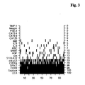
- Table 1 ROC analysis and sensitivities of all single markers (301 CRC, 266 controls, at 95% specificity) Marker Assay AUC (%) Sensitivity all UICC stages (%) Sensitivity UICC stages I-III (%) CEA Elecsys 77 43,9 30,9 CA 15-3 Elecsys 53 4,0 3,4 CA 125 Elecsys 56 17,6 10,3 CA 19-9 Elecsys 64 22,6 15,0 CA 72-4 Elecsys 66 20,6 12,9 CYFRA21-1 Elecsys 78 35,5 22,3 Ferritin Elecsys 65 23,9 27,0 NSE Elecsys 57 14,0 6,4 AFP Elecsys 56 6,6 5,6 ß-hCG Elecsys 51 3,7 3,4 OPN Roche-MTP 77 30,2 23,2 Anti-p53 Roche-MTP 57 20,0 18,8 Seprase Roche-MTP 79 42,4 45,4 S100 A12 Roche-MTP 69 20,3 12,9 HGF M
- Penalized Logistic Regression was used as a mathematical model for marker combinations as implemented in the R-toolbox "penalized” (http://cran.r-project.org/).
- the total sample collective was randomly divided in a training set (2/3 of samples) and a test set (1/3 of samples).
- the algorithm optimisation namely the selection of the penalization type and its penalization parameter was carried out by 100 runs in a Monte-Carlo cross-validation design within the training set.
Landscapes
- Health & Medical Sciences (AREA)
- Life Sciences & Earth Sciences (AREA)
- Engineering & Computer Science (AREA)
- Immunology (AREA)
- Urology & Nephrology (AREA)
- Chemical & Material Sciences (AREA)
- Biomedical Technology (AREA)
- Molecular Biology (AREA)
- Hematology (AREA)
- Medicinal Chemistry (AREA)
- Analytical Chemistry (AREA)
- Biotechnology (AREA)
- Hospice & Palliative Care (AREA)
- Oncology (AREA)
- Food Science & Technology (AREA)
- Microbiology (AREA)
- Physics & Mathematics (AREA)
- Cell Biology (AREA)
- Biochemistry (AREA)
- General Health & Medical Sciences (AREA)
- General Physics & Mathematics (AREA)
- Pathology (AREA)
- Investigating Or Analysing Biological Materials (AREA)
- Measuring Or Testing Involving Enzymes Or Micro-Organisms (AREA)
Claims (14)
- Procédé pour évaluer un cancer colorectal (CRC) in vitro comprenant la mesure dans un échantillon de sang entier, de sérum ou de plasma, la concentration et/ou l'activitéa) d'un polypeptide de séprase et/ou d'un de ses fragments ;b) de l'anticorps anti-p53 et/ou de l'ostéopontine et/ou de la ferritine ;c) de manière facultative, d'un ou de plusieurs autres marqueurs du CRC ; etd) l'utilisation des résultats de mesure combinés des étapes (a) et (b) et de manière facultative de l'étape (c) dans l'évaluation du CRC, dans lequel une réduction de la concentration pour la séprase, une réduction de la concentration pour la ferritine, une augmentation de la concentration pour l'anticorps anti-p53 et une augmentation de la concentration pour l'ostéopontine sont symptomatiques d'un CRC.
- Procédé selon la revendication 1, dans lequel au moins les résultats de mesure pour la séprase et pour l'anticorps anti-p53 sont combinés pour l'évaluation du CRC.
- Procédé selon la revendication 1, dans lequel au moins les résultats de mesure pour la séprase et pour l'ostéopontine sont combinés pour l'évaluation du CRC.
- Procédé selon la revendication 1, dans lequel au moins les résultats de mesure pour la séprase et pour la ferritine sont combinés pour l'évaluation du CRC.
- Procédé selon la revendication 1, dans lequel lesdits un ou plusieurs autres marqueurs facultatifs sont choisis parmi le groupe constitué par l'antigène carcinoembryonnaire (CEA) et le CYFRA21-1.
- Procédé selon l'une quelconque des revendications 1 à 3 et 5, dans lequel au moins les résultats de mesure pour la séprase, le CEA, l'ostéopontine et la ferritine sont combinés.
- Procédé selon l'une quelconque des revendications 1 à 6, dans lequel au moins les résultats de mesure pour la séprase, le CEA, l'anticorps anti-p53, le CYFRA21-1, le CEA et la ferritine sont combinés.
- Procédé selon l'une quelconque des revendications 1 à 7, dans lequel on mesure la concentration d'un polypeptide de séprase et/ou d'un de ses fragments et/ou de complexes comprenant la séprase et/ou un de ses fragments.
- Utilisation in vitro d'un panel de marqueurs comprenant au moins les résultats de mesure pour la séprase et l'anticorps anti-p53 pour l'évaluation du CRC.
- Utilisation in vitro d'un panel de marqueurs comprenant au moins les résultats de mesure pour la séprase et l'ostéopontine pour l'évaluation du CRC.
- Utilisation in vitro d'un panel de marqueurs comprenant au moins les résultats de mesure pour la séprase et la ferritine pour l'évaluation du CRC.
- Utilisation in vitro d'un panel de marqueurs comprenant au moins les résultats de mesure pour la séprase, le CEA, l'anticorps anti-p53 et la ferritine pour l'évaluation du CRC.
- Utilisation in vitro d'un panel de marqueurs comprenant au moins les résultats de mesure pour la séprase, le CEA, l'anticorps anti-p53, le CYFRA21-1, le CEA et la ferritine pour l'évaluation du CRC.
- Trousse comprenant des anticorps pour la mesure des marqueurs selon les revendications 1 à 8 ou pour la mesure des panels de marqueurs selon les utilisations des revendications 9 à 13.
Priority Applications (1)
| Application Number | Priority Date | Filing Date | Title |
|---|---|---|---|
| EP08860118.2A EP2223116B1 (fr) | 2007-12-10 | 2008-12-08 | Panel de marqueurs pour le cancer colorectal |
Applications Claiming Priority (4)
| Application Number | Priority Date | Filing Date | Title |
|---|---|---|---|
| EP07023897A EP2071337A1 (fr) | 2007-12-10 | 2007-12-10 | La séprase en tant que marqueur pour le cancer |
| EP08017119 | 2008-09-29 | ||
| EP08860118.2A EP2223116B1 (fr) | 2007-12-10 | 2008-12-08 | Panel de marqueurs pour le cancer colorectal |
| PCT/EP2008/010386 WO2009074276A2 (fr) | 2007-12-10 | 2008-12-08 | Panel de marqueurs pour le cancer colorectal |
Publications (2)
| Publication Number | Publication Date |
|---|---|
| EP2223116A2 EP2223116A2 (fr) | 2010-09-01 |
| EP2223116B1 true EP2223116B1 (fr) | 2014-11-19 |
Family
ID=40344417
Family Applications (1)
| Application Number | Title | Priority Date | Filing Date |
|---|---|---|---|
| EP08860118.2A Not-in-force EP2223116B1 (fr) | 2007-12-10 | 2008-12-08 | Panel de marqueurs pour le cancer colorectal |
Country Status (7)
| Country | Link |
|---|---|
| US (1) | US20100240068A1 (fr) |
| EP (1) | EP2223116B1 (fr) |
| JP (2) | JP5591711B2 (fr) |
| CN (1) | CN101896817A (fr) |
| CA (1) | CA2701970A1 (fr) |
| ES (1) | ES2527649T3 (fr) |
| WO (1) | WO2009074276A2 (fr) |
Families Citing this family (19)
| Publication number | Priority date | Publication date | Assignee | Title |
|---|---|---|---|---|
| WO2010105235A2 (fr) | 2009-03-12 | 2010-09-16 | Cancer Prevention And Cure, Ltd. | Procédés d'identification, d'évaluation, de prévention et de thérapie de maladies des poumons et leurs kits comprenant une identification, une évaluation, une prévention et une thérapie de maladies basées sur le sexe |
| EP2446272A2 (fr) * | 2009-06-23 | 2012-05-02 | Eventus Diagnostics Israel Ltd. | Procédé et système de détection du cancer |
| EP2426615A1 (fr) | 2010-08-06 | 2012-03-07 | Roche Diagnostics GmbH | Dispositif d'évaluation pour aider à l'estimation du cancer colorectal |
| WO2012047899A2 (fr) * | 2010-10-04 | 2012-04-12 | The Johns Hopkins University | Nouveaux biomarqueurs de diagnostic de cancer colorectal par l'hyperméthylation de l'adn |
| EP2463659A1 (fr) * | 2010-12-13 | 2012-06-13 | Université de Liège | Biomarqueurs pour le diagnostic du cancer. |
| WO2012123293A1 (fr) * | 2011-03-11 | 2012-09-20 | Roche Diagnostics Gmbh | Séprase en tant que marqueur pour une maladie pulmonaire obstructive chronique (copd) |
| CN102183662A (zh) * | 2011-03-22 | 2011-09-14 | 浙江大学 | 一种大肠癌预后预测模型的建立方法 |
| IL278227B (en) * | 2011-04-29 | 2022-07-01 | Cancer Prevention & Cure Ltd | Data classification systems for identifying biomarkers and diagnosing diseases |
| EP2926138A4 (fr) * | 2012-11-30 | 2016-09-14 | Applied Proteomics Inc | Procédé d'évaluation de présence ou de risque de tumeurs du côlon |
| EP2916134B1 (fr) | 2014-03-05 | 2017-08-16 | Roche Diagnostics GmbH | Utilisation de la séprase pour le diagnostic différentiel de la dyspnée aiguë |
| EP3183578B8 (fr) * | 2014-08-22 | 2020-07-15 | Abbott Laboratories | Méthodes de détection précoce du cancer colorectal |
| WO2016164815A1 (fr) | 2015-04-10 | 2016-10-13 | Applied Proteomics, Inc. | Panels de biomarqueurs protéiques pour détecter le cancer colorectal et l'adénome avancé |
| EP3427057A1 (fr) | 2016-03-07 | 2019-01-16 | H. Hoffnabb-La Roche Ag | Détection d'anticorps anti-p53 |
| EP3607089A4 (fr) | 2017-04-04 | 2020-12-30 | Lung Cancer Proteomics, LLC | Profilage de protéine à base de plasma pour le pronostic précoce du cancer du poumon |
| KR20210134946A (ko) * | 2019-03-01 | 2021-11-11 | 어드밴스드 마커 디스커버리 에스.엘. | 대장암 및/또는 이의 전암 단계 진단을 위한 단백질 시그니처 |
| CN109902411B (zh) * | 2019-03-07 | 2020-08-11 | 三峡大学 | 土壤重金属含量检测建模方法及装置、检测方法及装置 |
| WO2021024009A1 (fr) * | 2019-08-02 | 2021-02-11 | Shanghai Yunxiang Medical Technology Co., Ltd. | Procédés et compositions pour fournir une évaluation du cancer du côlon à l'aide de biomarqueurs protéiques |
| WO2023282916A1 (fr) | 2021-07-09 | 2023-01-12 | Guardant Health, Inc. | Procédés de détection de réarrangements génomiques à l'aide d'acides nucléiques acellulaires |
| CN112951332A (zh) * | 2021-02-25 | 2021-06-11 | 北京博富瑞基因诊断技术有限公司 | 一种基于aGVHD biomarker的重度肠道aGVHD模型的方法 |
Family Cites Families (9)
| Publication number | Priority date | Publication date | Assignee | Title |
|---|---|---|---|---|
| US5455160A (en) * | 1993-05-27 | 1995-10-03 | Fagerhol; Magne K. | Diagnostic test and kit for disease or disorders in the digestive system |
| US5695761A (en) * | 1993-12-23 | 1997-12-09 | Rutgers University | Suppression of nitric oxide production by osteopontin |
| EP0953639A1 (fr) * | 1998-04-30 | 1999-11-03 | Boehringer Ingelheim International GmbH | Anticorps dirigé contre FAPalpha pouvant être produit avec une efficacité accrue |
| US6414219B1 (en) * | 1998-06-30 | 2002-07-02 | Rutgers, The State University Of New Jersey | Osteopontin knock-out mouse and methods of use thereof |
| EP1315829B1 (fr) * | 2000-09-09 | 2010-07-28 | The Research Foundation Of State University Of New York | Methodes et compositions d'isolation de cellules cancereuses metastatiques, et utilisation correspondante lors de la mesure du potentiel metastatique d'un cancer |
| EP1831695B1 (fr) * | 2004-12-23 | 2009-05-06 | F. Hoffmann-La Roche AG | Utilisation de cyfra 21-1 et osteopontine comme marqueurs du cancer colorectal |
| AU2006246719A1 (en) * | 2005-05-19 | 2006-11-23 | Genentech, Inc. | Fibroblast activation protein inhibitor compounds and methods |
| CN101346628A (zh) * | 2005-12-22 | 2009-01-14 | 霍夫曼-拉罗奇有限公司 | 包含骨桥蛋白和癌胚抗原的标记物组合在结肠直肠癌的评估中的用途 |
| AU2007225054B2 (en) * | 2006-03-13 | 2012-08-16 | Becton, Dickinson And Company | Diagnosis and prognosis of dipeptidyl peptidase-associated disease states |
-
2008
- 2008-12-08 CN CN2008801199284A patent/CN101896817A/zh active Pending
- 2008-12-08 ES ES08860118.2T patent/ES2527649T3/es active Active
- 2008-12-08 WO PCT/EP2008/010386 patent/WO2009074276A2/fr active Application Filing
- 2008-12-08 CA CA2701970A patent/CA2701970A1/fr not_active Abandoned
- 2008-12-08 JP JP2010536378A patent/JP5591711B2/ja not_active Expired - Fee Related
- 2008-12-08 EP EP08860118.2A patent/EP2223116B1/fr not_active Not-in-force
-
2010
- 2010-06-02 US US12/791,929 patent/US20100240068A1/en not_active Abandoned
-
2014
- 2014-07-30 JP JP2014155200A patent/JP2014211452A/ja active Pending
Also Published As
| Publication number | Publication date |
|---|---|
| US20100240068A1 (en) | 2010-09-23 |
| JP5591711B2 (ja) | 2014-09-17 |
| WO2009074276A3 (fr) | 2009-08-20 |
| CN101896817A (zh) | 2010-11-24 |
| CA2701970A1 (fr) | 2009-06-18 |
| EP2223116A2 (fr) | 2010-09-01 |
| WO2009074276A2 (fr) | 2009-06-18 |
| JP2011506917A (ja) | 2011-03-03 |
| JP2014211452A (ja) | 2014-11-13 |
| ES2527649T3 (es) | 2015-01-28 |
Similar Documents
| Publication | Publication Date | Title |
|---|---|---|
| EP2223116B1 (fr) | Panel de marqueurs pour le cancer colorectal | |
| EP2223115B1 (fr) | Séprase comme marqueur du cancer | |
| JP4606469B2 (ja) | 直腸結腸癌用マーカーとしてのcyfra21−1の使用 | |
| EP2356461B1 (fr) | Pacap en tant que marqueur pour le cancer | |
| EP2435830B1 (fr) | Secernin-1 en tant que marqueur pour le cancer | |
| EP2071337A1 (fr) | La séprase en tant que marqueur pour le cancer | |
| JP2009520958A (ja) | 結腸直腸癌の評価におけるオステオポンチンおよび癌胎児性抗原を含むマーカー組合せの使用 | |
| JP4673895B2 (ja) | 直腸結腸癌用マーカーとしてのascの使用 | |
| EP2297581B1 (fr) | Asc en tant que marqueur du cancer du poumon | |
| EP2430450B1 (fr) | Cybp en tant que marqueur du cancer du poumon |
Legal Events
| Date | Code | Title | Description |
|---|---|---|---|
| PUAI | Public reference made under article 153(3) epc to a published international application that has entered the european phase |
Free format text: ORIGINAL CODE: 0009012 |
|
| 17P | Request for examination filed |
Effective date: 20100712 |
|
| AK | Designated contracting states |
Kind code of ref document: A2 Designated state(s): AT BE BG CH CY CZ DE DK EE ES FI FR GB GR HR HU IE IS IT LI LT LU LV MC MT NL NO PL PT RO SE SI SK TR |
|
| AX | Request for extension of the european patent |
Extension state: AL BA MK RS |
|
| DAX | Request for extension of the european patent (deleted) | ||
| 17Q | First examination report despatched |
Effective date: 20110221 |
|
| GRAP | Despatch of communication of intention to grant a patent |
Free format text: ORIGINAL CODE: EPIDOSNIGR1 |
|
| INTG | Intention to grant announced |
Effective date: 20140624 |
|
| GRAS | Grant fee paid |
Free format text: ORIGINAL CODE: EPIDOSNIGR3 |
|
| GRAA | (expected) grant |
Free format text: ORIGINAL CODE: 0009210 |
|
| RIN1 | Information on inventor provided before grant (corrected) |
Inventor name: ANDRES, HERBERT Inventor name: GARCZAREK, URSULA Inventor name: WILD, NORBERT Inventor name: KARL, JOHANN Inventor name: ROLLINGER, WOLFGANG |
|
| AK | Designated contracting states |
Kind code of ref document: B1 Designated state(s): AT BE BG CH CY CZ DE DK EE ES FI FR GB GR HR HU IE IS IT LI LT LU LV MC MT NL NO PL PT RO SE SI SK TR |
|
| REG | Reference to a national code |
Ref country code: GB Ref legal event code: FG4D |
|
| REG | Reference to a national code |
Ref country code: CH Ref legal event code: EP |
|
| REG | Reference to a national code |
Ref country code: AT Ref legal event code: REF Ref document number: 697310 Country of ref document: AT Kind code of ref document: T Effective date: 20141215 |
|
| REG | Reference to a national code |
Ref country code: IE Ref legal event code: FG4D |
|
| REG | Reference to a national code |
Ref country code: DE Ref legal event code: R096 Ref document number: 602008035518 Country of ref document: DE Effective date: 20141231 |
|
| REG | Reference to a national code |
Ref country code: ES Ref legal event code: FG2A Ref document number: 2527649 Country of ref document: ES Kind code of ref document: T3 Effective date: 20150128 |
|
| REG | Reference to a national code |
Ref country code: NL Ref legal event code: VDEP Effective date: 20141119 |
|
| REG | Reference to a national code |
Ref country code: LT Ref legal event code: MG4D |
|
| PG25 | Lapsed in a contracting state [announced via postgrant information from national office to epo] |
Ref country code: NO Free format text: LAPSE BECAUSE OF FAILURE TO SUBMIT A TRANSLATION OF THE DESCRIPTION OR TO PAY THE FEE WITHIN THE PRESCRIBED TIME-LIMIT Effective date: 20150219 Ref country code: IS Free format text: LAPSE BECAUSE OF FAILURE TO SUBMIT A TRANSLATION OF THE DESCRIPTION OR TO PAY THE FEE WITHIN THE PRESCRIBED TIME-LIMIT Effective date: 20150319 Ref country code: LT Free format text: LAPSE BECAUSE OF FAILURE TO SUBMIT A TRANSLATION OF THE DESCRIPTION OR TO PAY THE FEE WITHIN THE PRESCRIBED TIME-LIMIT Effective date: 20141119 Ref country code: PT Free format text: LAPSE BECAUSE OF FAILURE TO SUBMIT A TRANSLATION OF THE DESCRIPTION OR TO PAY THE FEE WITHIN THE PRESCRIBED TIME-LIMIT Effective date: 20150319 Ref country code: FI Free format text: LAPSE BECAUSE OF FAILURE TO SUBMIT A TRANSLATION OF THE DESCRIPTION OR TO PAY THE FEE WITHIN THE PRESCRIBED TIME-LIMIT Effective date: 20141119 Ref country code: NL Free format text: LAPSE BECAUSE OF FAILURE TO SUBMIT A TRANSLATION OF THE DESCRIPTION OR TO PAY THE FEE WITHIN THE PRESCRIBED TIME-LIMIT Effective date: 20141119 |
|
| PG25 | Lapsed in a contracting state [announced via postgrant information from national office to epo] |
Ref country code: SE Free format text: LAPSE BECAUSE OF FAILURE TO SUBMIT A TRANSLATION OF THE DESCRIPTION OR TO PAY THE FEE WITHIN THE PRESCRIBED TIME-LIMIT Effective date: 20141119 Ref country code: LV Free format text: LAPSE BECAUSE OF FAILURE TO SUBMIT A TRANSLATION OF THE DESCRIPTION OR TO PAY THE FEE WITHIN THE PRESCRIBED TIME-LIMIT Effective date: 20141119 Ref country code: CY Free format text: LAPSE BECAUSE OF FAILURE TO SUBMIT A TRANSLATION OF THE DESCRIPTION OR TO PAY THE FEE WITHIN THE PRESCRIBED TIME-LIMIT Effective date: 20141119 Ref country code: HR Free format text: LAPSE BECAUSE OF FAILURE TO SUBMIT A TRANSLATION OF THE DESCRIPTION OR TO PAY THE FEE WITHIN THE PRESCRIBED TIME-LIMIT Effective date: 20141119 Ref country code: GR Free format text: LAPSE BECAUSE OF FAILURE TO SUBMIT A TRANSLATION OF THE DESCRIPTION OR TO PAY THE FEE WITHIN THE PRESCRIBED TIME-LIMIT Effective date: 20150220 Ref country code: PL Free format text: LAPSE BECAUSE OF FAILURE TO SUBMIT A TRANSLATION OF THE DESCRIPTION OR TO PAY THE FEE WITHIN THE PRESCRIBED TIME-LIMIT Effective date: 20141119 |
|
| PG25 | Lapsed in a contracting state [announced via postgrant information from national office to epo] |
Ref country code: BE Free format text: LAPSE BECAUSE OF NON-PAYMENT OF DUE FEES Effective date: 20141231 |
|
| PG25 | Lapsed in a contracting state [announced via postgrant information from national office to epo] |
Ref country code: RO Free format text: LAPSE BECAUSE OF FAILURE TO SUBMIT A TRANSLATION OF THE DESCRIPTION OR TO PAY THE FEE WITHIN THE PRESCRIBED TIME-LIMIT Effective date: 20141119 Ref country code: DK Free format text: LAPSE BECAUSE OF FAILURE TO SUBMIT A TRANSLATION OF THE DESCRIPTION OR TO PAY THE FEE WITHIN THE PRESCRIBED TIME-LIMIT Effective date: 20141119 Ref country code: SK Free format text: LAPSE BECAUSE OF FAILURE TO SUBMIT A TRANSLATION OF THE DESCRIPTION OR TO PAY THE FEE WITHIN THE PRESCRIBED TIME-LIMIT Effective date: 20141119 Ref country code: EE Free format text: LAPSE BECAUSE OF FAILURE TO SUBMIT A TRANSLATION OF THE DESCRIPTION OR TO PAY THE FEE WITHIN THE PRESCRIBED TIME-LIMIT Effective date: 20141119 Ref country code: CZ Free format text: LAPSE BECAUSE OF FAILURE TO SUBMIT A TRANSLATION OF THE DESCRIPTION OR TO PAY THE FEE WITHIN THE PRESCRIBED TIME-LIMIT Effective date: 20141119 |
|
| REG | Reference to a national code |
Ref country code: DE Ref legal event code: R097 Ref document number: 602008035518 Country of ref document: DE |
|
| PG25 | Lapsed in a contracting state [announced via postgrant information from national office to epo] |
Ref country code: MC Free format text: LAPSE BECAUSE OF FAILURE TO SUBMIT A TRANSLATION OF THE DESCRIPTION OR TO PAY THE FEE WITHIN THE PRESCRIBED TIME-LIMIT Effective date: 20141119 |
|
| REG | Reference to a national code |
Ref country code: IE Ref legal event code: MM4A |
|
| PLBE | No opposition filed within time limit |
Free format text: ORIGINAL CODE: 0009261 |
|
| STAA | Information on the status of an ep patent application or granted ep patent |
Free format text: STATUS: NO OPPOSITION FILED WITHIN TIME LIMIT |
|
| 26N | No opposition filed |
Effective date: 20150820 |
|
| PG25 | Lapsed in a contracting state [announced via postgrant information from national office to epo] |
Ref country code: IE Free format text: LAPSE BECAUSE OF NON-PAYMENT OF DUE FEES Effective date: 20141208 |
|
| REG | Reference to a national code |
Ref country code: FR Ref legal event code: PLFP Year of fee payment: 8 |
|
| PGFP | Annual fee paid to national office [announced via postgrant information from national office to epo] |
Ref country code: CH Payment date: 20151026 Year of fee payment: 8 Ref country code: GB Payment date: 20151125 Year of fee payment: 8 |
|
| REG | Reference to a national code |
Ref country code: AT Ref legal event code: UEP Ref document number: 697310 Country of ref document: AT Kind code of ref document: T Effective date: 20141119 |
|
| PG25 | Lapsed in a contracting state [announced via postgrant information from national office to epo] |
Ref country code: SI Free format text: LAPSE BECAUSE OF FAILURE TO SUBMIT A TRANSLATION OF THE DESCRIPTION OR TO PAY THE FEE WITHIN THE PRESCRIBED TIME-LIMIT Effective date: 20141119 |
|
| PGFP | Annual fee paid to national office [announced via postgrant information from national office to epo] |
Ref country code: ES Payment date: 20151211 Year of fee payment: 8 Ref country code: AT Payment date: 20151124 Year of fee payment: 8 Ref country code: FR Payment date: 20151124 Year of fee payment: 8 |
|
| PGFP | Annual fee paid to national office [announced via postgrant information from national office to epo] |
Ref country code: DE Payment date: 20151230 Year of fee payment: 8 Ref country code: IT Payment date: 20151210 Year of fee payment: 8 |
|
| PG25 | Lapsed in a contracting state [announced via postgrant information from national office to epo] |
Ref country code: BG Free format text: LAPSE BECAUSE OF FAILURE TO SUBMIT A TRANSLATION OF THE DESCRIPTION OR TO PAY THE FEE WITHIN THE PRESCRIBED TIME-LIMIT Effective date: 20141119 |
|
| PG25 | Lapsed in a contracting state [announced via postgrant information from national office to epo] |
Ref country code: TR Free format text: LAPSE BECAUSE OF FAILURE TO SUBMIT A TRANSLATION OF THE DESCRIPTION OR TO PAY THE FEE WITHIN THE PRESCRIBED TIME-LIMIT Effective date: 20141119 Ref country code: HU Free format text: LAPSE BECAUSE OF FAILURE TO SUBMIT A TRANSLATION OF THE DESCRIPTION OR TO PAY THE FEE WITHIN THE PRESCRIBED TIME-LIMIT; INVALID AB INITIO Effective date: 20081208 Ref country code: MT Free format text: LAPSE BECAUSE OF FAILURE TO SUBMIT A TRANSLATION OF THE DESCRIPTION OR TO PAY THE FEE WITHIN THE PRESCRIBED TIME-LIMIT Effective date: 20141119 Ref country code: LU Free format text: LAPSE BECAUSE OF NON-PAYMENT OF DUE FEES Effective date: 20141208 |
|
| REG | Reference to a national code |
Ref country code: DE Ref legal event code: R119 Ref document number: 602008035518 Country of ref document: DE |
|
| REG | Reference to a national code |
Ref country code: CH Ref legal event code: PL |
|
| REG | Reference to a national code |
Ref country code: AT Ref legal event code: MM01 Ref document number: 697310 Country of ref document: AT Kind code of ref document: T Effective date: 20161208 |
|
| GBPC | Gb: european patent ceased through non-payment of renewal fee |
Effective date: 20161208 |
|
| REG | Reference to a national code |
Ref country code: FR Ref legal event code: ST Effective date: 20170831 |
|
| PG25 | Lapsed in a contracting state [announced via postgrant information from national office to epo] |
Ref country code: CH Free format text: LAPSE BECAUSE OF NON-PAYMENT OF DUE FEES Effective date: 20161231 Ref country code: LI Free format text: LAPSE BECAUSE OF NON-PAYMENT OF DUE FEES Effective date: 20161231 Ref country code: FR Free format text: LAPSE BECAUSE OF NON-PAYMENT OF DUE FEES Effective date: 20170102 Ref country code: AT Free format text: LAPSE BECAUSE OF NON-PAYMENT OF DUE FEES Effective date: 20161208 Ref country code: IT Free format text: LAPSE BECAUSE OF NON-PAYMENT OF DUE FEES Effective date: 20161208 |
|
| PG25 | Lapsed in a contracting state [announced via postgrant information from national office to epo] |
Ref country code: GB Free format text: LAPSE BECAUSE OF NON-PAYMENT OF DUE FEES Effective date: 20161208 Ref country code: DE Free format text: LAPSE BECAUSE OF NON-PAYMENT OF DUE FEES Effective date: 20170701 |
|
| PG25 | Lapsed in a contracting state [announced via postgrant information from national office to epo] |
Ref country code: ES Free format text: LAPSE BECAUSE OF NON-PAYMENT OF DUE FEES Effective date: 20161209 |
|
| REG | Reference to a national code |
Ref country code: ES Ref legal event code: FD2A Effective date: 20181116 |