US6413778B1 - Methods for the detection and identification of crystals in biological fluids - Google Patents
Methods for the detection and identification of crystals in biological fluids Download PDFInfo
- Publication number
- US6413778B1 US6413778B1 US09/235,124 US23512499A US6413778B1 US 6413778 B1 US6413778 B1 US 6413778B1 US 23512499 A US23512499 A US 23512499A US 6413778 B1 US6413778 B1 US 6413778B1
- Authority
- US
- United States
- Prior art keywords
- crystals
- filter
- present
- urine
- indicator
- Prior art date
- Legal status (The legal status is an assumption and is not a legal conclusion. Google has not performed a legal analysis and makes no representation as to the accuracy of the status listed.)
- Expired - Fee Related
Links
- 239000013078 crystal Substances 0.000 title claims abstract description 164
- 238000000034 method Methods 0.000 title claims abstract description 60
- 239000013060 biological fluid Substances 0.000 title claims abstract description 34
- 238000001514 detection method Methods 0.000 title abstract description 7
- 239000003153 chemical reaction reagent Substances 0.000 claims abstract description 43
- 210000002700 urine Anatomy 0.000 claims abstract description 30
- 229910052567 struvite Inorganic materials 0.000 claims description 17
- CKMXBZGNNVIXHC-UHFFFAOYSA-L ammonium magnesium phosphate hexahydrate Chemical compound [NH4+].O.O.O.O.O.O.[Mg+2].[O-]P([O-])([O-])=O CKMXBZGNNVIXHC-UHFFFAOYSA-L 0.000 claims description 16
- 238000005406 washing Methods 0.000 claims description 15
- FYYHWMGAXLPEAU-UHFFFAOYSA-N Magnesium Chemical compound [Mg] FYYHWMGAXLPEAU-UHFFFAOYSA-N 0.000 claims description 12
- MUBZPKHOEPUJKR-UHFFFAOYSA-N Oxalic acid Chemical compound OC(=O)C(O)=O MUBZPKHOEPUJKR-UHFFFAOYSA-N 0.000 claims description 12
- 239000011777 magnesium Substances 0.000 claims description 12
- 229910052749 magnesium Inorganic materials 0.000 claims description 12
- 108010063734 Oxalate oxidase Proteins 0.000 claims description 7
- 230000009471 action Effects 0.000 claims description 7
- NGPGYVQZGRJHFJ-UHFFFAOYSA-N chembl1604790 Chemical group OC1=CC(O)=CC=C1N=NC1=CC=C([N+]([O-])=O)C=C1 NGPGYVQZGRJHFJ-UHFFFAOYSA-N 0.000 claims description 7
- 108010001336 Horseradish Peroxidase Proteins 0.000 claims description 5
- 239000008280 blood Substances 0.000 claims description 5
- 210000004369 blood Anatomy 0.000 claims description 5
- 102000005701 Calcium-Binding Proteins Human genes 0.000 claims description 4
- 108010045403 Calcium-Binding Proteins Proteins 0.000 claims description 4
- 102000003992 Peroxidases Human genes 0.000 claims description 4
- 239000010836 blood and blood product Substances 0.000 claims description 4
- 229940125691 blood product Drugs 0.000 claims description 4
- 210000003722 extracellular fluid Anatomy 0.000 claims description 4
- 108040007629 peroxidase activity proteins Proteins 0.000 claims description 4
- 238000012800 visualization Methods 0.000 claims description 4
- 239000000463 material Substances 0.000 abstract description 53
- 239000002250 absorbent Substances 0.000 abstract description 40
- 230000002745 absorbent Effects 0.000 abstract description 40
- 239000012530 fluid Substances 0.000 abstract description 16
- 230000002401 inhibitory effect Effects 0.000 abstract description 10
- 239000012528 membrane Substances 0.000 abstract description 6
- 238000010186 staining Methods 0.000 abstract description 6
- 239000000975 dye Substances 0.000 description 19
- HEMHJVSKTPXQMS-UHFFFAOYSA-M Sodium hydroxide Chemical compound [OH-].[Na+] HEMHJVSKTPXQMS-UHFFFAOYSA-M 0.000 description 18
- 239000011148 porous material Substances 0.000 description 11
- 241001465754 Metazoa Species 0.000 description 9
- 239000000126 substance Substances 0.000 description 9
- 238000003556 assay Methods 0.000 description 7
- QXDMQSPYEZFLGF-UHFFFAOYSA-L calcium oxalate Chemical class [Ca+2].[O-]C(=O)C([O-])=O QXDMQSPYEZFLGF-UHFFFAOYSA-L 0.000 description 7
- 239000007788 liquid Substances 0.000 description 7
- VTYYLEPIZMXCLO-UHFFFAOYSA-L Calcium carbonate Chemical compound [Ca+2].[O-]C([O-])=O VTYYLEPIZMXCLO-UHFFFAOYSA-L 0.000 description 6
- PHOLIFLKGONSGY-CSKARUKUSA-N (e)-(3-methyl-1,3-benzothiazol-2-ylidene)hydrazine Chemical compound C1=CC=C2S\C(=N\N)N(C)C2=C1 PHOLIFLKGONSGY-CSKARUKUSA-N 0.000 description 5
- 206010011509 Crystalluria Diseases 0.000 description 5
- 241000282326 Felis catus Species 0.000 description 5
- 239000004698 Polyethylene Substances 0.000 description 5
- -1 polyethylene Polymers 0.000 description 5
- 229920000573 polyethylene Polymers 0.000 description 5
- 230000000717 retained effect Effects 0.000 description 5
- OYPRJOBELJOOCE-UHFFFAOYSA-N Calcium Chemical compound [Ca] OYPRJOBELJOOCE-UHFFFAOYSA-N 0.000 description 4
- LEVWYRKDKASIDU-QWWZWVQMSA-N D-cystine Chemical compound OC(=O)[C@H](N)CSSC[C@@H](N)C(O)=O LEVWYRKDKASIDU-QWWZWVQMSA-N 0.000 description 4
- MHAJPDPJQMAIIY-UHFFFAOYSA-N Hydrogen peroxide Chemical compound OO MHAJPDPJQMAIIY-UHFFFAOYSA-N 0.000 description 4
- 239000004695 Polyether sulfone Substances 0.000 description 4
- 238000004458 analytical method Methods 0.000 description 4
- 239000011575 calcium Substances 0.000 description 4
- 229910052791 calcium Inorganic materials 0.000 description 4
- 238000006243 chemical reaction Methods 0.000 description 4
- 239000000356 contaminant Substances 0.000 description 4
- 229960003067 cystine Drugs 0.000 description 4
- 230000002452 interceptive effect Effects 0.000 description 4
- 230000000670 limiting effect Effects 0.000 description 4
- 229920006393 polyether sulfone Polymers 0.000 description 4
- 239000000047 product Substances 0.000 description 4
- 229920002799 BoPET Polymers 0.000 description 3
- 241000282472 Canis lupus familiaris Species 0.000 description 3
- 239000005041 Mylar™ Substances 0.000 description 3
- 239000000020 Nitrocellulose Substances 0.000 description 3
- 229910000019 calcium carbonate Inorganic materials 0.000 description 3
- 229920002678 cellulose Polymers 0.000 description 3
- 239000001913 cellulose Substances 0.000 description 3
- 230000008859 change Effects 0.000 description 3
- 230000002255 enzymatic effect Effects 0.000 description 3
- 239000003365 glass fiber Substances 0.000 description 3
- 230000005484 gravity Effects 0.000 description 3
- 239000000203 mixture Substances 0.000 description 3
- 229920001220 nitrocellulos Polymers 0.000 description 3
- 230000001376 precipitating effect Effects 0.000 description 3
- 239000000758 substrate Substances 0.000 description 3
- 238000012360 testing method Methods 0.000 description 3
- NEGFNJRAUMCZMY-UHFFFAOYSA-N 3-(dimethylamino)benzoic acid Chemical compound CN(C)C1=CC=CC(C(O)=O)=C1 NEGFNJRAUMCZMY-UHFFFAOYSA-N 0.000 description 2
- BPYKTIZUTYGOLE-IFADSCNNSA-N Bilirubin Chemical compound N1C(=O)C(C)=C(C=C)\C1=C\C1=C(C)C(CCC(O)=O)=C(CC2=C(C(C)=C(\C=C/3C(=C(C=C)C(=O)N\3)C)N2)CCC(O)=O)N1 BPYKTIZUTYGOLE-IFADSCNNSA-N 0.000 description 2
- 206010007027 Calculus urinary Diseases 0.000 description 2
- OKTJSMMVPCPJKN-UHFFFAOYSA-N Carbon Chemical compound [C] OKTJSMMVPCPJKN-UHFFFAOYSA-N 0.000 description 2
- CURLTUGMZLYLDI-UHFFFAOYSA-N Carbon dioxide Chemical compound O=C=O CURLTUGMZLYLDI-UHFFFAOYSA-N 0.000 description 2
- KCXVZYZYPLLWCC-UHFFFAOYSA-N EDTA Chemical compound OC(=O)CN(CC(O)=O)CCN(CC(O)=O)CC(O)=O KCXVZYZYPLLWCC-UHFFFAOYSA-N 0.000 description 2
- LEHOTFFKMJEONL-UHFFFAOYSA-N Uric Acid Chemical compound N1C(=O)NC(=O)C2=C1NC(=O)N2 LEHOTFFKMJEONL-UHFFFAOYSA-N 0.000 description 2
- 238000010521 absorption reaction Methods 0.000 description 2
- MXZRMHIULZDAKC-UHFFFAOYSA-L ammonium magnesium phosphate Chemical compound [NH4+].[Mg+2].[O-]P([O-])([O-])=O MXZRMHIULZDAKC-UHFFFAOYSA-L 0.000 description 2
- 239000012491 analyte Substances 0.000 description 2
- 230000015572 biosynthetic process Effects 0.000 description 2
- 235000005911 diet Nutrition 0.000 description 2
- 230000037213 diet Effects 0.000 description 2
- 239000003814 drug Substances 0.000 description 2
- 239000011521 glass Substances 0.000 description 2
- 238000012986 modification Methods 0.000 description 2
- 230000004048 modification Effects 0.000 description 2
- 239000004033 plastic Substances 0.000 description 2
- 229920003023 plastic Polymers 0.000 description 2
- 230000035945 sensitivity Effects 0.000 description 2
- 230000002485 urinary effect Effects 0.000 description 2
- 239000011534 wash buffer Substances 0.000 description 2
- QZHXKQKKEBXYRG-UHFFFAOYSA-N 4-n-(4-aminophenyl)benzene-1,4-diamine Chemical compound C1=CC(N)=CC=C1NC1=CC=C(N)C=C1 QZHXKQKKEBXYRG-UHFFFAOYSA-N 0.000 description 1
- QGZKDVFQNNGYKY-UHFFFAOYSA-O Ammonium Chemical compound [NH4+] QGZKDVFQNNGYKY-UHFFFAOYSA-O 0.000 description 1
- 241000894006 Bacteria Species 0.000 description 1
- 241000283707 Capra Species 0.000 description 1
- 241000700198 Cavia Species 0.000 description 1
- 102000004190 Enzymes Human genes 0.000 description 1
- 108090000790 Enzymes Proteins 0.000 description 1
- 241000283086 Equidae Species 0.000 description 1
- 208000034826 Genetic Predisposition to Disease Diseases 0.000 description 1
- 241000282412 Homo Species 0.000 description 1
- 208000000913 Kidney Calculi Diseases 0.000 description 1
- 241000124008 Mammalia Species 0.000 description 1
- 206010029148 Nephrolithiasis Diseases 0.000 description 1
- BPQQTUXANYXVAA-UHFFFAOYSA-N Orthosilicate Chemical compound [O-][Si]([O-])([O-])[O-] BPQQTUXANYXVAA-UHFFFAOYSA-N 0.000 description 1
- 241000283973 Oryctolagus cuniculus Species 0.000 description 1
- 108010092464 Urate Oxidase Proteins 0.000 description 1
- TVWHNULVHGKJHS-UHFFFAOYSA-N Uric acid Natural products N1C(=O)NC(=O)C2NC(=O)NC21 TVWHNULVHGKJHS-UHFFFAOYSA-N 0.000 description 1
- 239000011358 absorbing material Substances 0.000 description 1
- 238000004026 adhesive bonding Methods 0.000 description 1
- 229960000723 ampicillin Drugs 0.000 description 1
- AVKUERGKIZMTKX-NJBDSQKTSA-N ampicillin Chemical compound C1([C@@H](N)C(=O)N[C@H]2[C@H]3SC([C@@H](N3C2=O)C(O)=O)(C)C)=CC=CC=C1 AVKUERGKIZMTKX-NJBDSQKTSA-N 0.000 description 1
- 239000003242 anti bacterial agent Substances 0.000 description 1
- 229940088710 antibiotic agent Drugs 0.000 description 1
- QVGXLLKOCUKJST-UHFFFAOYSA-N atomic oxygen Chemical compound [O] QVGXLLKOCUKJST-UHFFFAOYSA-N 0.000 description 1
- 230000008901 benefit Effects 0.000 description 1
- 239000000872 buffer Substances 0.000 description 1
- 239000001506 calcium phosphate Substances 0.000 description 1
- 229910000389 calcium phosphate Inorganic materials 0.000 description 1
- 235000011010 calcium phosphates Nutrition 0.000 description 1
- UGSQEBVMGSXVSH-UHFFFAOYSA-L calcium;oxalate;dihydrate Chemical compound O.O.[Ca+2].[O-]C(=O)C([O-])=O UGSQEBVMGSXVSH-UHFFFAOYSA-L 0.000 description 1
- 239000001569 carbon dioxide Substances 0.000 description 1
- 229910002092 carbon dioxide Inorganic materials 0.000 description 1
- 238000004113 cell culture Methods 0.000 description 1
- 230000001413 cellular effect Effects 0.000 description 1
- 229920002301 cellulose acetate Polymers 0.000 description 1
- 238000005119 centrifugation Methods 0.000 description 1
- 150000005829 chemical entities Chemical group 0.000 description 1
- 239000003795 chemical substances by application Substances 0.000 description 1
- 235000019504 cigarettes Nutrition 0.000 description 1
- 238000004891 communication Methods 0.000 description 1
- 239000012141 concentrate Substances 0.000 description 1
- 238000011161 development Methods 0.000 description 1
- 235000021045 dietary change Nutrition 0.000 description 1
- 230000004069 differentiation Effects 0.000 description 1
- 201000010099 disease Diseases 0.000 description 1
- 208000037265 diseases, disorders, signs and symptoms Diseases 0.000 description 1
- 238000004090 dissolution Methods 0.000 description 1
- 229940079593 drug Drugs 0.000 description 1
- 238000000855 fermentation Methods 0.000 description 1
- 230000004151 fermentation Effects 0.000 description 1
- 239000000835 fiber Substances 0.000 description 1
- 239000002657 fibrous material Substances 0.000 description 1
- 238000001914 filtration Methods 0.000 description 1
- 230000002706 hydrostatic effect Effects 0.000 description 1
- 229910052500 inorganic mineral Inorganic materials 0.000 description 1
- 238000003780 insertion Methods 0.000 description 1
- 230000037431 insertion Effects 0.000 description 1
- 230000003993 interaction Effects 0.000 description 1
- 230000001788 irregular Effects 0.000 description 1
- UHNWOJJPXCYKCG-UHFFFAOYSA-L magnesium oxalate Chemical class [Mg+2].[O-]C(=O)C([O-])=O UHNWOJJPXCYKCG-UHFFFAOYSA-L 0.000 description 1
- 230000014759 maintenance of location Effects 0.000 description 1
- 239000011707 mineral Substances 0.000 description 1
- 210000003097 mucus Anatomy 0.000 description 1
- 150000003891 oxalate salts Chemical class 0.000 description 1
- 229910052760 oxygen Inorganic materials 0.000 description 1
- 239000001301 oxygen Substances 0.000 description 1
- 230000036961 partial effect Effects 0.000 description 1
- 239000008188 pellet Substances 0.000 description 1
- 210000002381 plasma Anatomy 0.000 description 1
- 229920000728 polyester Polymers 0.000 description 1
- 229920000098 polyolefin Polymers 0.000 description 1
- 239000002244 precipitate Substances 0.000 description 1
- DQMZLTXERSFNPB-UHFFFAOYSA-N primidone Chemical compound C=1C=CC=CC=1C1(CC)C(=O)NCNC1=O DQMZLTXERSFNPB-UHFFFAOYSA-N 0.000 description 1
- 229960002393 primidone Drugs 0.000 description 1
- 210000002966 serum Anatomy 0.000 description 1
- 239000002195 soluble material Substances 0.000 description 1
- 241000894007 species Species 0.000 description 1
- 239000008362 succinate buffer Substances 0.000 description 1
- UEUXEKPTXMALOB-UHFFFAOYSA-J tetrasodium;2-[2-[bis(carboxylatomethyl)amino]ethyl-(carboxylatomethyl)amino]acetate Chemical compound [Na+].[Na+].[Na+].[Na+].[O-]C(=O)CN(CC([O-])=O)CCN(CC([O-])=O)CC([O-])=O UEUXEKPTXMALOB-UHFFFAOYSA-J 0.000 description 1
- 238000012549 training Methods 0.000 description 1
- QORWJWZARLRLPR-UHFFFAOYSA-H tricalcium bis(phosphate) Chemical compound [Ca+2].[Ca+2].[Ca+2].[O-]P([O-])([O-])=O.[O-]P([O-])([O-])=O QORWJWZARLRLPR-UHFFFAOYSA-H 0.000 description 1
- 229940116269 uric acid Drugs 0.000 description 1
- 210000001635 urinary tract Anatomy 0.000 description 1
- 238000013022 venting Methods 0.000 description 1
- 230000000007 visual effect Effects 0.000 description 1
Images
Classifications
-
- G—PHYSICS
- G01—MEASURING; TESTING
- G01N—INVESTIGATING OR ANALYSING MATERIALS BY DETERMINING THEIR CHEMICAL OR PHYSICAL PROPERTIES
- G01N33/00—Investigating or analysing materials by specific methods not covered by groups G01N1/00 - G01N31/00
- G01N33/48—Biological material, e.g. blood, urine; Haemocytometers
- G01N33/483—Physical analysis of biological material
- G01N33/487—Physical analysis of biological material of liquid biological material
- G01N33/493—Physical analysis of biological material of liquid biological material urine
-
- Y—GENERAL TAGGING OF NEW TECHNOLOGICAL DEVELOPMENTS; GENERAL TAGGING OF CROSS-SECTIONAL TECHNOLOGIES SPANNING OVER SEVERAL SECTIONS OF THE IPC; TECHNICAL SUBJECTS COVERED BY FORMER USPC CROSS-REFERENCE ART COLLECTIONS [XRACs] AND DIGESTS
- Y10—TECHNICAL SUBJECTS COVERED BY FORMER USPC
- Y10T—TECHNICAL SUBJECTS COVERED BY FORMER US CLASSIFICATION
- Y10T436/00—Chemistry: analytical and immunological testing
- Y10T436/25—Chemistry: analytical and immunological testing including sample preparation
- Y10T436/25375—Liberation or purification of sample or separation of material from a sample [e.g., filtering, centrifuging, etc.]
Definitions
- This invention relates in general to devices for separating crystals from urine or other biological fluids and ascertaining their chemical identity. Also provided are methods for detecting and identifying crystals which may be present in urine or other biological fluids, and kits containing the devices and other materials and reagents needed to conduct the methods.
- Crystalluria the presence of crystals in the urine, is a common problem which afflicts household pets such as cats and dogs.
- the formation of crystals and mucus in the urinary tract is a potentially life threatening condition in these animals. It is indicative of the predilection to form uroliths or stones, and can ultimately lead to a complete obstruction of the urinary system.
- Urine crystals may be of several different types. Most commonly crystals are formed of struvite (magnesium-ammonium-phosphate), oxalate, urate, cystine, or silicate, but may also be composed of other materials such as bilirubin, calcium carbonate, or calcium phosphate. Struvites and calcium oxalates combined comprise over 88% of the uroliths found in cats and dogs and therefore represent the cause of the great majority of these animal medical problems. The occurrence of crystalluria within these animal populations varies according to species, breed, diet, sex, age, and genetic pre-disposition. Crystalluria also occurs in a variety of other mammals. For example, it is known that calcium carbonate crystals can form in horses, rabbits, guinea pigs, and goats.
- the term “habit” is commonly used by minerologists to refer to the characteristic shape or shapes of mineral crystals. Different crystal types exhibit different habits which a skilled person is able to differentiate. The judgment is based largely on the apparent structure or habit of the crystals. These methods involve specific skills, significant handling of the specimen, and are both time consuming and expensive. Microscopic detection and identification of the crystals is further complicated by the fact that their appearance can be influenced by the variable conditions of their formation, growth, and dissolution. The sensitivity of this method is also limited as it is not uncommon for the technician to fail to detect small numbers of crystals which may be present.
- This method of identifying the crystals also involves significant handling of the sample since it typically requires a volume of 5-10 mls of urine, which then must be concentrated to enhance the population of crystals within the examined specimen. Concentration is normally accomplished by centrifugation at 2500 rpm for 5-10 minutes, aspiration of all but 0.5 to 1.0 ml of fluid, and resuspension of the pellet.
- the microscopist bases the judgment of crystal type on the physical characteristics or habit of the crystals present.
- Calcium oxalate dihydrate crystals typically are colorless and have a characteristic octahedral or envelope shape, having the appearance under a light microscope of squares whose corners are connected by intersecting diagonal lines.
- Struvite crystals are known for their colorless, orthorhombic, “coffinlid” shape, although frequently other, irregular forms are seen. They often have three to six or more sides and often have oblique ends.
- Cystine crystals exhibit a colorless hexagonal shape with equal or unequal sides. They may appear singly but usually aggregate in layers. As is evident from these descriptions, the differentiation of these different crystal types is based largely on subjective criteria, and is therefore prone to human error.
- Calcium carbonate crystals may form as large yellow-brown or colorless spheroids with radial striations, or smaller crystals with round, ovoid, or dumbbell shapes.
- the outer appearance of a crystal may not always correlate with its true chemical identity.
- kidney stones are usually comprised of calcium oxalate, struvite, or cystine.
- crystals sometimes are comprised of a calcium oxalate core covered by an outer layer of struvite.
- they may be comprised of a struvite core covered by an outer layer of calcium oxalate. Therefore, such crystals can be very deceiving even to the skilled person who is trying to ascertain their identity based largely on the external appearance of the crystals.
- the distinction is important, since the treatment programs for struvite and calcium oxalate crystals are very different.
- the present invention provides a device for the convenient, rapid, and accurate determination of crystals which may be present in urine or other biological fluids and their identity.
- the device is inexpensive, disposable, specific, and requires only very small sample volumes. It also eliminates the need and consequent delay and expense of transmitting samples to a commercial laboratory for analysis. This results in quicker treatment for the affected animal and avoids additional stress on the animal caused by unnecessary dietary changes and inconvenience to the animal owner.
- Another important advantage is that the analysis of the urine crystals is based on more objective criteria, sharply reducing human error as a source of inaccuracy. Using the present invention, one is able to determine the presence of urinary crystals and identify the crystal type with almost 100% sensitivity and accuracy.
- the present invention also discloses methods for detecting and identifying crystals which may be present in biological fluids, including urine.
- the methods may be conveniently performed in the veterinarian's office during the time typically taken for an office visit. Unlike currently available methods, a high level of training and skill is not necessary to successfully and confidently identify the crystal types, and the method can be learned in a matter of minutes.
- kits which may include the devices of the present invention and reagents necessary for conducting the methods of the present invention.
- the kits enable the veterinarian or other animal caretaker to have conveniently available everything needed to conduct the assay and detect and identify any crystals which may be present.
- the kits have a shelf life of at least six months and may last one year or more, and may be conveniently stored in a small space until needed.
- the kits may include reagents necessary to conduct the assay in a ready-to-use format, thereby eliminating the need for mixing or preparing reagents.
- the present invention relates to devices, methods, and kits for the rapid detection and identification of crystals which are suspected of being present in urine or other biological fluids.
- the devices of the present invention contain a filter, and may also contain an absorbent material. Crystals which are suspected of being present in urine or other biological fluids are isolated on or within the filter. The crystals may be isolated on the surface of or within the structure of the filter.
- the absorbent material serves to facilitate the drawing of liquid through the filter and into its absorbent material, thus isolating the crystal on or within the structure of the filter. Once isolated, the crystals will be available for staining with an indicator reagent specific for a determining component of the crystal.
- the device may further contain a member which inhibits flow from the absorbent material to the filter and which may be placed between the absorbent material and the filter.
- the filter, absorbent material, and member for inhibiting flow from the absorbent material to the filter may be in positional relationship such that fluid passes through the filter, through the member for inhibiting flow from the absorbent material to the filter, and into the absorbent material.
- the member for inhibiting flow from the filter to the absorbing material may be made of polyethylene.
- the member may be a disk of porous polyethylene.
- the filter may be a filter with a graded pore structure.
- the filter may be selected from, but is not limited to, an A/E glass filter, glass fiber, mylar, WHATMAN D28® filter, WHATMAN GD-1® filter, nitrocellulose, HEMASEP V®, HEMASEP L® filter, or SUPOR® polyethersulfone filter.
- the biological fluid may be urine, blood, blood products, or interstitial fluid.
- the present invention provides methods of detecting and isolating crystals which may be present in urine or other biological fluids.
- the methods of the present invention may consist of the steps of isolating the crystals on or within a filter, contacting the crystals with an indicator reagent which is specific for a determining component of the crystals, and determining the presence and identity of the crystals.
- the methods may further consist of a step of washing the crystals to wash away contaminants which may interfere with the indicator reaction.
- This wash step may be necessary in some chemistries to obtain an optimal indicator reaction.
- This washing step may further accomplish a disintegration action on the crystals thereby exposing a determining component of the crystals.
- This step may be combined with the initial wash step by using a wash that both washes and acts to partially disintegrate the crystal.
- the determining component thus exposed may react with dye or substrate for that specific determining component of the crystal.
- the presence of the determining component will cause a visible color change on the surface of or within the filter or membrane.
- the additional wash step may clarify the color change for easier interpretation.
- the crystals which are suspected of being present may be oxalate crystals, or struvite crystals.
- the indicating system may be oxalate oxidase, a peroxidase and an indicator.
- the peroxidase may be horseradish peroxidase.
- the indicator may also be a precipitating magnesium binding dye, such as magneson dye, or a calcium binding dye.
- the indicator may be these or other precipitating dyes which form insoluble complexes with various analytes.
- kits for detecting and identifying crystals which are suspected of being present in urine or another biological fluid may include a device of the present invention for detecting and identifying crystals which are suspected of being present in a biological fluid and at least one reagent for detecting and identifying the crystals.
- the reagent(s) provided in the kit may be an indicator reagent or system useful for practicing the methods of the present invention.
- the kit may also include other reagents useful for practicing the methods of the present invention.
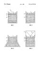
- FIG. 1 is a schematic of one embodiment of the present invention for detecting and identifying crystals which may be present in a biological fluid.
- the Figure is an exposed side view showing the interior portion of the device.
- FIG. 2 is a schematic showing an exposed side view of an embodiment of the device.
- the member for inhibiting flow from the absorbent material to the filter is not present.
- FIG. 3 is a schematic illustrating that the device may take a variety of shapes.
- the Figure is an exposed side view and the shape here depicted may be useful for absorbing larger quantities of liquids where desirable.
- FIG. 4 is a schematic illustrating an exposed side view of an embodiment of the device where a funnel may be attached to the device for receiving the sample of biological fluid.
- the device of the present invention comprises a filter which is capable of retaining crystals which may be present in a biological fluid on its surface or within its structure.
- the device may also comprise an absorbent material 3 which serves to draw fluid through the filter 1 .
- the filter may have pore sizes which are small enough to retain the crystals on its surface or within its structure.
- the filter will have a graded pore structure with pore diameters which generally decrease as fluid travels further into the filter. This type of pore structure enables the introduction of the crystals into the filter and their retention in its structure as the pore sizes generally decrease further into the structure of the filter, and also permits the crystals to be held in place and washed while still being retained in the filter.
- filter is meant anything that will function to separate components of a mixture by filtration, such as filters or membranes.
- optical filter is meant a HEMASEP V®, HEMASEP L®, SUPOR® polyethersulfone, WHATMAN D28®, WHATMAN GD-1®, nitrocellulose, or mylar filter.
- preferred filter is meant a HEMASEP V®, HEMASEP L®, SUPOR® polyethersulfone, WHATMAN D28®, or WHATMAN GD-1® filter.
- the filter be capable of resisting staining or of being destained without appreciably removing stain from the crystals, so that stained crystals will be more easily visible in or on the filter.
- the filter when the crystal is struvite the filter will be a HEMASEP V® membrane or its equivalent.
- the filter when the crystal is oxalate, the filter will be a HEMASEP L® membrane or its equivalent.
- filters may be used in the present invention including, but not limited to, A/E glass fiber, GF/DVA glass fiber, mylar, WHATMAN D28®, WHATMAN GD-1®, nitrocellulose, SUPOR® polyethersulfone, or any of their equivalents.
- the filter may be retained within a housing or holding member 2 .
- the housing 2 may be made of glass, plastic, or other suitable material.
- the housing 2 may comprise an ICON® filter device or its equivalent.
- Absorbent material 3 may be packed into the housing member 2 with one of the filters described above, or its equivalent.
- the housing member 2 may comprise any type of filter or device which is capable of holding the filter 1 in place while the biological fluid is applied to and allowed to pass through it.
- the filter 1 may be held in direct contact with the absorbent material 3 .
- the present invention may also comprise an absorbent material 3 adjoining the filter 1 which is able to draw fluid through the filter 1 and absorb the fluid.
- the absorbent material 3 may be an absorbent block.
- the selection of the absorbent material is not critical and a variety of fibrous materials may be used.
- the absorbent material 3 may be a cellulose-based absorbent material.
- the material may be cellulose acetate with the fibers oriented as in a cigarette filter.
- the absorbent material may be a cellulose block.
- the material may be any material capable of drawing fluid through the filter 1 and absorbing the fluid including, but not limited to, polyester or polyolefin.
- the person of ordinary skill in the art will also readily realize that other methods of drawing fluid through the filter may be used, such as by suction, gravity, the application of external pressure, or any method capable of drawing or forcing fluid through the filter 1 .
- a member 4 for inhibiting flow from the absorbent material to the filter may be placed in between the filter 1 and the absorbent material 3 .
- This member 4 for inhibiting flow may serve to reduce backflow in the filter device.
- the member 4 for reducing backflow may be a porous polyethylene disk placed in between the filter 1 and the absorbent material 3 . The disk may enhance the drawing of the fluid into the absorbent material 3 and minimizes backflow toward the filter 1 .
- the porous polyethylene disk may be a POREX® polyethylene disk, or its equivalent.
- the device may also function without the member 4 for reducing backflow.
- the device may utilize a variety of other filters for retaining the crystals.
- the filter may be placed into a bowl-shaped or conical-shaped device and the urine or biological fluid drawn through by suction, gravity, external pressure or other means. This would enable almost any filter to be used in the device which had pores small enough to retain the crystals suspected of being present and was, preferably, capable of resisting staining or of being destained. Typically, this would enable any filters with pores of at least approximately 0.2 ⁇ m or larger to be used. The selection of an optimal filter can result in greater ease of reading the assay results.
- the filter should be selected based on the type of crystals being detected since each crystal has its own typical range of size where it becomes clinically important. Optimal results are obtained by selecting a filter which has pore sizes which are the largest that will still capture the crystal of interest. Smaller pore sizes than necessary may reduce flow efficiency and also capture interfering substances, such as cellular debris, casts, bacteria, sperm, or other biological products which may interfere with the indicator reaction since they may contain magnesium, calcium, or other components which may be detected by the indicator.
- the housing 2 may have a lower opening into which is inserted a removable plug to permit insertion of the filter 1 , the member 4 for inhibiting flow from the absorbent material to the filter, and the absorbent material 3 .
- the size of the absorbent material 3 is preferably selected such that all of the liquid to be added to the apparatus during an assay can be received in and retained in the absorbent material 3 .
- Small ports for venting air may be provided in the housing 2 near the bottom to allow displaced air to escape.
- Other embodiments may include, but are not limited to, shapes which facilitate the use of the device under vacuum or positive pressure.
- FIG. 3 depicts an embodiment of the device shaped to maximize the absorptive capability of the device.
- the funnel 4 depicts an embodiment of the device with a funnel 6 for directing sample to the filter 1 , so as to concentrate crystals at one spot for better detection.
- the funnel may have a side 7 which is secured to the device by any suitable fastening method such as clipping or gluing.
- the funnel may be made of plastic.
- a filter 1 may be used which has the qualities of retaining crystals by electrostatic attractions, or by chemical affinity, or any method which serves to secure the crystals which may be present to the surface of the filter 1 or within its structure, for staining and detection.
- the present invention is also directed to methods of detecting and identifying crystals which may be present in urine or other biological fluid.
- a method which consists of the steps of applying the sample to the receiving area of the device 5 of the present invention (referring to FIG. 1 throughout); a first wash step which may be desirable in some chemistries (such as when assaying for struvite or oxalate crystals) to wash away contaminants or non-crystal material which may be present from the biological fluid, and which may also expose a determining component of the crystals; a step whereby an indicator reagent or system is applied to the crystals which may be present on the surface of or within the structure of the filter 1 ; in some chemistries, such as when using dye binding as a means of visualization, as in a preferred test for struvite crystals, an additional wash step such as to destain the filter 1 which may be desirable for optimal visualization of crystal coloration which may have occurred; and, a step in which coloration and identity of the crystals which may be present
- the sample is applied directly to the filter 1 of the device. This may be accomplished by dropwise addition or simply by pouring or pipetting the sample into the receiving vessel or area of the device 5 .
- the biological fluid is drawn through the filter 1 and into the absorbent material 3 and the crystals are retained on the surface of the filter 1 or within its structure.
- the biological fluid is drawn through the filter 1 and through the member 4 for inhibiting flow from the absorbent material to the filter (if present), and into the absorbent material 3 .
- the member 4 for inhibiting flow may serve to reduce backflow in the device.
- the fluid may be passed through the filter 1 by the use of suction, gravity, external pressure, or any other means which will cause the fluid to pass through the filter 1 .
- the filter 1 is assembled with the absorbent material 3 in a manner which permits direct communication between the pores or interstices of the filter 1 and the capillaries of the absorbent material 3 .
- the filter 1 is assembled with the absorbent material 3 in a manner which permits direct communication between the pores or interstices of the filter 1 and the capillaries of the absorbent material 3 .
- a wash step may then be carried out to remove soluble material and any material other than the crystals from the surface. This step is desirable when the sample contains soluble cross reactive or interfering substances.
- the wash step may also accomplish a disintegration action upon the crystals and help to expose a determining component of the crystals such as Ca +2 or oxalate. By partially disintegrating the crystals the available concentration of analyte is dramatically increased.
- a disintegration action is an action which at least partially disintegrates the crystal. The disintegration action releases free analyte into a larger area and facilitates visualization of crystals which may be present.
- a determining component is a property of the crystal which is particular for a specific crystal type or a group of crystal types such that identification of the presence of the determining component aids in some way in identifying the crystal type.
- An indicator system is a reagent or group of reagents which produce a detectable signal in the presence of a determining component.
- An indicator reagent is a chemical entity which is able to produce a detectable signal in the presence of a determining component.
- An indicator reagent such as a dye or enzymatic substrate which is specific for a determining component of the type of crystal being tested for may then be contacted with the crystals.
- Suitable indicator reagents may include, but are not limited to, the combination of oxalate oxidase, horseradish peroxidase, and an indicator dye, magnesium binding dyes (such as magneson dye) which bind to magnesium, precipitating dyes, or calcium binding dyes which bind to calcium.
- magnesium binding dyes such as magneson dye
- indicator reagents are available and may be utilized, as long as the presence of some determining component of the crystals which may be present is detected by the indicator reagent. Therefore, the above indicators are provided as examples, and are not intended to be limiting.
- a final wash step may be desirable to provide optimal viewing of any color change or other indicating signal which may be present.
- the presence of crystals and their composition can then be determined by noting coloration of the filter 1 or other indicating signals which may have occurred due to the action of a specific dye, enzyme, or other indicator reagent on the crystals or crystal components.
- the methods of the present invention fill the need for a method to quickly detect the presence of crystalluria, and for determining the type of crystal which may be present in a novel, heretofore undisclosed way.
- the methods and principles disclosed herein may be adapted and applied for use in detecting crystals from any biological fluid.
- many valuable products may be produced in crystalline form from cell culture or fermentation processes. These products may include biological components, antibiotics, or any product produced in crystalline form. Crystalluria also occurs in humans and may be the result of the presence of various drugs which are excreted in the urine, such as ampicillin, primidone, and ciprofloxicin.
- the devices and methods of the present invention can be adapted and applied to detect these crystals as well. These methods and principles may also be applied to detect crystals which may be present in whole blood, blood products such as plasma or serum, or interstitial fluid.
- kits with materials and reagents necessary for conducting the method.
- the kits may contain devices and reagents useful for detecting and identifying crystals which may be present in urine or other biological fluids.
- the kit may contain: a) a device for detecting crystals which may be present in a biological fluid, b) a container of 0.1N HCl, c) a container of 0.5% magneson dye in 1% NaOH and d) a container of 0.1N NaOH.
- the kit may contain a) a device, b) a container of 10 mmol/L EDTA wash buffer at approximately pH 7.6, c) a container of 3-methyl-2-benzothiazolinone hydrazone (MBTH), d) a container of 3-(dimethylamino) benzoic acid (DMAB) in a buffer, e) a container of oxalate oxidase, and f) a container of horseradish peroxidase.
- DMAB 3-(dimethylamino) benzoic acid
- This example illustrates how the present invention can be employed to detect the presence of struvite (magnesium-ammonium-phosphate) crystals in urine.
- Reagent 1 consisted of 0.1N HCl
- Reagent 2 was 0.5% magneson dye in 1% NaOH
- Reagent 3 was 0.1N NaOH.
- This example illustrates how the invention may be employed to test for the presence of oxalate crystals, which may occur as calcium, ammonium or magnesium oxalates.
- oxalate crystals which may occur as calcium, ammonium or magnesium oxalates.
- Calcium oxalates are the most commonly found oxalate crystal in veterinary medicine.
- Reagent 1 was a 10 mM EDTA wash buffer at pH 7.6
- Reagent 2 was prepared immediately before use from its separate compositions, and contained 3-methyl-2-benzothiazolinone hydrazone (MBTH) and 3-(dimethylamino) benzoic acid (DMAB) in succinate buffer, combined with oxalate oxidase and horseradish peroxidase.
- Oxalate oxidase acts upon calcium oxalate crystals in the presence of oxygen to convert oxalate to carbon dioxide and hydrogen peroxide.
- a detectable indamine dye is produced by the interaction of the DMAB/MBTH substrate with hydrogen peroxide in the presence of peroxidase.
- the invention is broadly applicable for detecting and identifying a variety of crystal types using various indicator reagents.
- crystals containing calcium may also be detected by using cresolpthalein complexone or arsenazo III. With these indicators, a disintegration step may be necessary to break up the crystals.
- calgamite may be used to detect and stain crystals containing magnesium.
- crystals containing uric acid may be detectable in the blood, urine, or other biological fluid with the enzyme uricase.
Landscapes
- Health & Medical Sciences (AREA)
- Engineering & Computer Science (AREA)
- Life Sciences & Earth Sciences (AREA)
- Biomedical Technology (AREA)
- Urology & Nephrology (AREA)
- Physics & Mathematics (AREA)
- Chemical & Material Sciences (AREA)
- Molecular Biology (AREA)
- Hematology (AREA)
- Biophysics (AREA)
- Food Science & Technology (AREA)
- Medicinal Chemistry (AREA)
- Analytical Chemistry (AREA)
- Biochemistry (AREA)
- General Health & Medical Sciences (AREA)
- General Physics & Mathematics (AREA)
- Immunology (AREA)
- Pathology (AREA)
- Investigating Or Analysing Biological Materials (AREA)
Priority Applications (8)
| Application Number | Priority Date | Filing Date | Title |
|---|---|---|---|
| US09/235,124 US6413778B1 (en) | 1999-01-21 | 1999-01-21 | Methods for the detection and identification of crystals in biological fluids |
| PCT/US2000/000875 WO2000043777A1 (en) | 1999-01-21 | 2000-01-13 | Devices and methods for the detection and identification of crystals in biological fluids |
| DE60031204T DE60031204T2 (de) | 1999-01-21 | 2000-01-13 | VERFAHREN und KIT ZUM NACHWEIS UND ZUR IDENTIFIZIERUNG VON KRISTALLEN IN BIOLOGISCHEN FLÜSSIGKEITEN |
| EP00903288A EP1066516B1 (de) | 1999-01-21 | 2000-01-13 | VERFAHREN und KIT ZUM NACHWEIS UND ZUR IDENTIFIZIERUNG VON KRISTALLEN IN BIOLOGISCHEN FLÜSSIGKEITEN |
| AT00903288T ATE342506T1 (de) | 1999-01-21 | 2000-01-13 | Verfahren und kit zum nachweis und zur identifizierung von kristallen in biologischen flüssigkeiten |
| AU25059/00A AU774699B2 (en) | 1999-01-21 | 2000-01-13 | Devices and methods for the detection and identification of crystals in biological fluids |
| JP2000595147A JP2002535652A (ja) | 1999-01-21 | 2000-01-13 | 体液中の結晶を検出および同定するための装置および方法 |
| CA002324947A CA2324947A1 (en) | 1999-01-21 | 2000-01-13 | Devices and methods for the detection and identification of crystals in biological fluids |
Applications Claiming Priority (1)
| Application Number | Priority Date | Filing Date | Title |
|---|---|---|---|
| US09/235,124 US6413778B1 (en) | 1999-01-21 | 1999-01-21 | Methods for the detection and identification of crystals in biological fluids |
Publications (1)
| Publication Number | Publication Date |
|---|---|
| US6413778B1 true US6413778B1 (en) | 2002-07-02 |
Family
ID=22884196
Family Applications (1)
| Application Number | Title | Priority Date | Filing Date |
|---|---|---|---|
| US09/235,124 Expired - Fee Related US6413778B1 (en) | 1999-01-21 | 1999-01-21 | Methods for the detection and identification of crystals in biological fluids |
Country Status (8)
| Country | Link |
|---|---|
| US (1) | US6413778B1 (de) |
| EP (1) | EP1066516B1 (de) |
| JP (1) | JP2002535652A (de) |
| AT (1) | ATE342506T1 (de) |
| AU (1) | AU774699B2 (de) |
| CA (1) | CA2324947A1 (de) |
| DE (1) | DE60031204T2 (de) |
| WO (1) | WO2000043777A1 (de) |
Cited By (14)
| Publication number | Priority date | Publication date | Assignee | Title |
|---|---|---|---|---|
| US20020164812A1 (en) * | 1999-04-06 | 2002-11-07 | Uab Research Foundation | Method for screening crystallization conditions in solution crystal growth |
| US20030022384A1 (en) * | 1999-04-06 | 2003-01-30 | Uab Research Foundation | Method for screening crystallization conditions in solution crystal growth |
| US20030027348A1 (en) * | 1999-04-06 | 2003-02-06 | Uab Research Foundation | Method for screening crystallization conditions in solution crystal growth |
| US20030087449A1 (en) * | 2001-10-26 | 2003-05-08 | Asplin John R. | Urine cystine quantitation and supersaturation assay |
| US20030180960A1 (en) * | 2001-07-30 | 2003-09-25 | Larry Cosenza | Use of dye to distinguish salt and protein crystals under microcrystallization conditions |
| US20030232967A1 (en) * | 1999-04-06 | 2003-12-18 | Arnon Chait | Method for preparation of microarrays for screening of crystal growth conditions |
| US20040007672A1 (en) * | 2002-07-10 | 2004-01-15 | Delucas Lawrence J. | Method for distinguishing between biomolecule and non-biomolecule crystals |
| US20050178317A1 (en) * | 2001-04-05 | 2005-08-18 | The California Institute Of Technology | High throughput screening of crystallization of materials |
| US20070026528A1 (en) * | 2002-05-30 | 2007-02-01 | Delucas Lawrence J | Method for screening crystallization conditions in solution crystal growth |
| US20070202602A1 (en) * | 1999-04-06 | 2007-08-30 | Delucas Lawrence J | Method for screening crystallization conditions in solution crystal growth |
| US20080194042A1 (en) * | 2007-02-14 | 2008-08-14 | Cheung Herman S | Assays for disease-associated crystals in biological samples |
| US9228996B2 (en) | 2013-05-31 | 2016-01-05 | Empire Technology Development Llc | Method and device for detecting device colonization |
| US9535043B2 (en) | 2013-05-31 | 2017-01-03 | Empire Technology Development Llc | Color change indicator of biofilm formation |
| CN112076359A (zh) * | 2020-09-14 | 2020-12-15 | 江苏恰瑞生物科技有限公司 | 用于肾结石治疗的血浆吸附过滤装置及其过滤方法 |
Families Citing this family (1)
| Publication number | Priority date | Publication date | Assignee | Title |
|---|---|---|---|---|
| WO2014201088A1 (en) | 2013-06-11 | 2014-12-18 | Case Western Reserve University | Methods and devices for diagnosis of particles in biological fluids |
Citations (16)
| Publication number | Priority date | Publication date | Assignee | Title |
|---|---|---|---|---|
| US3731806A (en) | 1971-03-01 | 1973-05-08 | Pelam Inc | Specimen analysis device |
| US3768978A (en) | 1971-03-01 | 1973-10-30 | Hamilton Co | Disposable pipette |
| US4225669A (en) * | 1979-04-27 | 1980-09-30 | Melnick Joseph L | Staining and analysis of bacteria |
| US4235601A (en) * | 1979-01-12 | 1980-11-25 | Thyroid Diagnostics, Inc. | Test device and method for its use |
| US4455371A (en) * | 1982-03-03 | 1984-06-19 | The Ohio State University Research Foundation | Oxalate oxidase composition for assay of oxalate |
| US4683209A (en) * | 1985-02-26 | 1987-07-28 | Miles Laboratories, Inc. | Viability test device |
| US4727019A (en) | 1984-05-11 | 1988-02-23 | Hybritech Incorporated | Method and apparatus for immunoassays |
| EP0307566A2 (de) | 1987-07-28 | 1989-03-22 | Heinz Spagyrik Institut Ag | Verfahren zur kristallmorphologischen Blut- und Harnanalyse, zur Frühdiagnose und zur Herstellung von Arzneimitteln |
| US4912035A (en) * | 1987-06-11 | 1990-03-27 | Eastman Kodak Company | Method for minimizing interference by reductants when detecting cells in biological fluids |
| US4992365A (en) * | 1984-04-23 | 1991-02-12 | Hyman Edward S | Method of detecting bacteria in urine |
| US5264348A (en) * | 1991-05-13 | 1993-11-23 | Miles Inc. | Ascorbate interference-resistant composition, device and method of assaying for predetermined analyte |
| US5352410A (en) | 1993-06-03 | 1994-10-04 | Hansen Warren D | Fluid specimen collection and testing apparatus |
| EP0780678A2 (de) | 1995-12-19 | 1997-06-25 | TOA MEDICAL ELECTRONICS CO., Ltd. | Analysator zur Analyse von Urinbestandteilen |
| EP0841403A2 (de) | 1996-11-08 | 1998-05-13 | Idemitsu Kosan Company Limited | Farbzusammensetzung für Microorganismen, Filtervorrichtung zum Einfangen von Bakterien, sowie Testsatz zum Bestimmung von Bakterienanzahl |
| US5824495A (en) * | 1992-11-20 | 1998-10-20 | Southpac Trust International, Inc. | Cell fixative and preparation, kit and method |
| US5891733A (en) * | 1994-10-20 | 1999-04-06 | Toa Medical Electronics Co., Ltd. | Reagent for analyzing solid components in urine and method for analyzing solid components by employing the same |
Family Cites Families (6)
| Publication number | Priority date | Publication date | Assignee | Title |
|---|---|---|---|---|
| US5006474A (en) * | 1987-12-16 | 1991-04-09 | Disease Detection International Inc. | Bi-directional lateral chromatographic test device |
| JPH0484898A (ja) * | 1990-06-26 | 1992-03-18 | Terumo Corp | 試験具 |
| JPH0792160A (ja) * | 1991-08-15 | 1995-04-07 | La Mina Ltd | 採取装置、試験及び採取装置、試験方法、測定方法 |
| JPH05312809A (ja) * | 1992-03-10 | 1993-11-26 | Mochida Pharmaceut Co Ltd | 免疫学的簡易測定方法および装置 |
| JP2634759B2 (ja) * | 1993-08-02 | 1997-07-30 | 前田 浩志 | 愛玩動物尿症病の体外診断方法 |
| US5776701A (en) * | 1996-05-31 | 1998-07-07 | University Of Florida | Materials and methods for detecting oxalate |
-
1999
- 1999-01-21 US US09/235,124 patent/US6413778B1/en not_active Expired - Fee Related
-
2000
- 2000-01-13 AT AT00903288T patent/ATE342506T1/de not_active IP Right Cessation
- 2000-01-13 JP JP2000595147A patent/JP2002535652A/ja active Pending
- 2000-01-13 CA CA002324947A patent/CA2324947A1/en not_active Abandoned
- 2000-01-13 EP EP00903288A patent/EP1066516B1/de not_active Expired - Lifetime
- 2000-01-13 DE DE60031204T patent/DE60031204T2/de not_active Expired - Lifetime
- 2000-01-13 AU AU25059/00A patent/AU774699B2/en not_active Ceased
- 2000-01-13 WO PCT/US2000/000875 patent/WO2000043777A1/en active IP Right Grant
Patent Citations (16)
| Publication number | Priority date | Publication date | Assignee | Title |
|---|---|---|---|---|
| US3731806A (en) | 1971-03-01 | 1973-05-08 | Pelam Inc | Specimen analysis device |
| US3768978A (en) | 1971-03-01 | 1973-10-30 | Hamilton Co | Disposable pipette |
| US4235601A (en) * | 1979-01-12 | 1980-11-25 | Thyroid Diagnostics, Inc. | Test device and method for its use |
| US4225669A (en) * | 1979-04-27 | 1980-09-30 | Melnick Joseph L | Staining and analysis of bacteria |
| US4455371A (en) * | 1982-03-03 | 1984-06-19 | The Ohio State University Research Foundation | Oxalate oxidase composition for assay of oxalate |
| US4992365A (en) * | 1984-04-23 | 1991-02-12 | Hyman Edward S | Method of detecting bacteria in urine |
| US4727019A (en) | 1984-05-11 | 1988-02-23 | Hybritech Incorporated | Method and apparatus for immunoassays |
| US4683209A (en) * | 1985-02-26 | 1987-07-28 | Miles Laboratories, Inc. | Viability test device |
| US4912035A (en) * | 1987-06-11 | 1990-03-27 | Eastman Kodak Company | Method for minimizing interference by reductants when detecting cells in biological fluids |
| EP0307566A2 (de) | 1987-07-28 | 1989-03-22 | Heinz Spagyrik Institut Ag | Verfahren zur kristallmorphologischen Blut- und Harnanalyse, zur Frühdiagnose und zur Herstellung von Arzneimitteln |
| US5264348A (en) * | 1991-05-13 | 1993-11-23 | Miles Inc. | Ascorbate interference-resistant composition, device and method of assaying for predetermined analyte |
| US5824495A (en) * | 1992-11-20 | 1998-10-20 | Southpac Trust International, Inc. | Cell fixative and preparation, kit and method |
| US5352410A (en) | 1993-06-03 | 1994-10-04 | Hansen Warren D | Fluid specimen collection and testing apparatus |
| US5891733A (en) * | 1994-10-20 | 1999-04-06 | Toa Medical Electronics Co., Ltd. | Reagent for analyzing solid components in urine and method for analyzing solid components by employing the same |
| EP0780678A2 (de) | 1995-12-19 | 1997-06-25 | TOA MEDICAL ELECTRONICS CO., Ltd. | Analysator zur Analyse von Urinbestandteilen |
| EP0841403A2 (de) | 1996-11-08 | 1998-05-13 | Idemitsu Kosan Company Limited | Farbzusammensetzung für Microorganismen, Filtervorrichtung zum Einfangen von Bakterien, sowie Testsatz zum Bestimmung von Bakterienanzahl |
Non-Patent Citations (2)
| Title |
|---|
| Chelfouh, N. et al., "Characterization of Urinary Calculi: In Vitro Study of Twinkling Artifact" Revealed by Color-Flow Sonography, AJR. American Journal of Roentgenology, (Oct. 1998) 171 (4) 1055-60, XP-000901742, the whole document. |
| Tisellus, H., "Crystalluria in Patients With Calcium Stone Disease," The Journal of Urology, 161(5) (1999). |
Cited By (20)
| Publication number | Priority date | Publication date | Assignee | Title |
|---|---|---|---|---|
| US7244396B2 (en) | 1999-04-06 | 2007-07-17 | Uab Research Foundation | Method for preparation of microarrays for screening of crystal growth conditions |
| US20020164812A1 (en) * | 1999-04-06 | 2002-11-07 | Uab Research Foundation | Method for screening crystallization conditions in solution crystal growth |
| US20030027348A1 (en) * | 1999-04-06 | 2003-02-06 | Uab Research Foundation | Method for screening crystallization conditions in solution crystal growth |
| US20030022384A1 (en) * | 1999-04-06 | 2003-01-30 | Uab Research Foundation | Method for screening crystallization conditions in solution crystal growth |
| US20070202602A1 (en) * | 1999-04-06 | 2007-08-30 | Delucas Lawrence J | Method for screening crystallization conditions in solution crystal growth |
| US20030232967A1 (en) * | 1999-04-06 | 2003-12-18 | Arnon Chait | Method for preparation of microarrays for screening of crystal growth conditions |
| US7700363B2 (en) | 1999-04-06 | 2010-04-20 | Uab Research Foundation | Method for screening crystallization conditions in solution crystal growth |
| US20050178317A1 (en) * | 2001-04-05 | 2005-08-18 | The California Institute Of Technology | High throughput screening of crystallization of materials |
| US7250305B2 (en) | 2001-07-30 | 2007-07-31 | Uab Research Foundation | Use of dye to distinguish salt and protein crystals under microcrystallization conditions |
| US20030180960A1 (en) * | 2001-07-30 | 2003-09-25 | Larry Cosenza | Use of dye to distinguish salt and protein crystals under microcrystallization conditions |
| US6991937B2 (en) * | 2001-10-26 | 2006-01-31 | Litholink Corporation | Urine cystine quantitation and supersaturation assay |
| US20030087449A1 (en) * | 2001-10-26 | 2003-05-08 | Asplin John R. | Urine cystine quantitation and supersaturation assay |
| US20070026528A1 (en) * | 2002-05-30 | 2007-02-01 | Delucas Lawrence J | Method for screening crystallization conditions in solution crystal growth |
| US20040007672A1 (en) * | 2002-07-10 | 2004-01-15 | Delucas Lawrence J. | Method for distinguishing between biomolecule and non-biomolecule crystals |
| US20080194042A1 (en) * | 2007-02-14 | 2008-08-14 | Cheung Herman S | Assays for disease-associated crystals in biological samples |
| US8153436B2 (en) * | 2007-02-14 | 2012-04-10 | The United States Of America As Represented By The Department Of Veterans Affairs | Assays for disease-associated crystals in biological samples |
| US9228996B2 (en) | 2013-05-31 | 2016-01-05 | Empire Technology Development Llc | Method and device for detecting device colonization |
| US9535043B2 (en) | 2013-05-31 | 2017-01-03 | Empire Technology Development Llc | Color change indicator of biofilm formation |
| CN112076359A (zh) * | 2020-09-14 | 2020-12-15 | 江苏恰瑞生物科技有限公司 | 用于肾结石治疗的血浆吸附过滤装置及其过滤方法 |
| CN112076359B (zh) * | 2020-09-14 | 2022-10-28 | 江苏恰瑞生物科技有限公司 | 用于肾结石治疗的血浆吸附过滤装置及其过滤方法 |
Also Published As
| Publication number | Publication date |
|---|---|
| JP2002535652A (ja) | 2002-10-22 |
| AU774699B2 (en) | 2004-07-01 |
| WO2000043777A1 (en) | 2000-07-27 |
| DE60031204T2 (de) | 2007-08-23 |
| CA2324947A1 (en) | 2000-07-27 |
| EP1066516B1 (de) | 2006-10-11 |
| DE60031204D1 (de) | 2006-11-23 |
| ATE342506T1 (de) | 2006-11-15 |
| EP1066516A1 (de) | 2001-01-10 |
| AU2505900A (en) | 2000-08-07 |
Similar Documents
| Publication | Publication Date | Title |
|---|---|---|
| US6413778B1 (en) | Methods for the detection and identification of crystals in biological fluids | |
| JP3214854B2 (ja) | カード上での微量分析 | |
| JP2818191B2 (ja) | 固相分析装置 | |
| AU2003239507B2 (en) | Method and apparatus for measuring white blood cell count | |
| US8003399B2 (en) | Nitrite detection technique | |
| DK1824991T3 (en) | Device and method for the detection of analytes | |
| US6562581B2 (en) | Method for quantitative determination of glycated hemoglobin | |
| EP0269876B1 (de) | Proberichtwirkung für analytisches Festphasengerät | |
| US20070244368A1 (en) | Diagnostic Test Devices | |
| JP2001520892A (ja) | 溶液中の検体を同定するための分析装置およびその方法 | |
| AU629087B2 (en) | Device and method of separating and assaying whole blood | |
| PL191687B1 (pl) | Urządzenie testowe do oznaczania zawartości analitów w płynnych produktach nabiałowych | |
| JPH05249122A (ja) | 尿タンパク質検定のための改良された組成物および試験具ならびにそれを用いる方法 | |
| CA2113101A1 (en) | Diagnostic kits and methods for making granulocyte cell counts | |
| US6596502B2 (en) | Kit and method for detecting fecal parasites | |
| JP2007139556A (ja) | 新規な分析方法およびキット | |
| US8703101B2 (en) | Methods of determining NOx in a wound sample | |
| JP4864615B2 (ja) | 多項目試験紙および検体中の被検物質の検出方法 | |
| WO2005061723A1 (en) | Microorganism detector | |
| US20110287461A1 (en) | Enzymatic analytical membrane, test device and method | |
| JPH0659235B2 (ja) | アイソザイム混合物から1つの酵素を測定する方法及びテスト担体 | |
| JP2913219B2 (ja) | 尿沈渣検査成績管理方法 | |
| WO1999061892A1 (en) | Estimation of active infection by helicobacter pylori | |
| M. Abdelaziz et al. | Follow-up testing for ketonuria: is it necessary? | |
| IL157742A (en) | Kit and method for detecting fecal parasites |
Legal Events
| Date | Code | Title | Description |
|---|---|---|---|
| AS | Assignment |
Owner name: IDEXX LABORATORIES, INC., MAINE Free format text: ASSIGNMENT OF ASSIGNORS INTEREST;ASSIGNORS:CARPENTER, CHARLES;TORNBERG, MELANIE;CLARK, GENEVIEVE;REEL/FRAME:009892/0208;SIGNING DATES FROM 19990329 TO 19990330 |
|
| FPAY | Fee payment |
Year of fee payment: 4 |
|
| FPAY | Fee payment |
Year of fee payment: 8 |
|
| REMI | Maintenance fee reminder mailed | ||
| LAPS | Lapse for failure to pay maintenance fees | ||
| STCH | Information on status: patent discontinuation |
Free format text: PATENT EXPIRED DUE TO NONPAYMENT OF MAINTENANCE FEES UNDER 37 CFR 1.362 |
|
| FP | Lapsed due to failure to pay maintenance fee |
Effective date: 20140702 |