US5256642A - Compositions of soluble complement receptor 1 (CR1) and a thrombolytic agent, and the methods of use thereof - Google Patents
Compositions of soluble complement receptor 1 (CR1) and a thrombolytic agent, and the methods of use thereof Download PDFInfo
- Publication number
- US5256642A US5256642A US07/588,128 US58812890A US5256642A US 5256642 A US5256642 A US 5256642A US 58812890 A US58812890 A US 58812890A US 5256642 A US5256642 A US 5256642A
- Authority
- US
- United States
- Prior art keywords
- scr1
- protein
- lhr
- soluble
- fragment
- Prior art date
- Legal status (The legal status is an assumption and is not a legal conclusion. Google has not performed a legal analysis and makes no representation as to the accuracy of the status listed.)
- Expired - Lifetime
Links
- VQJOPRHZJAIGEW-UHFFFAOYSA-N CCN(C)C1CC1 Chemical compound CCN(C)C1CC1 VQJOPRHZJAIGEW-UHFFFAOYSA-N 0.000 description 1
Images


Classifications
-
- C—CHEMISTRY; METALLURGY
- C07—ORGANIC CHEMISTRY
- C07K—PEPTIDES
- C07K14/00—Peptides having more than 20 amino acids; Gastrins; Somatostatins; Melanotropins; Derivatives thereof
- C07K14/195—Peptides having more than 20 amino acids; Gastrins; Somatostatins; Melanotropins; Derivatives thereof from bacteria
- C07K14/315—Peptides having more than 20 amino acids; Gastrins; Somatostatins; Melanotropins; Derivatives thereof from bacteria from Streptococcus (G), e.g. Enterococci
- C07K14/3153—Streptokinase
-
- A—HUMAN NECESSITIES
- A61—MEDICAL OR VETERINARY SCIENCE; HYGIENE
- A61P—SPECIFIC THERAPEUTIC ACTIVITY OF CHEMICAL COMPOUNDS OR MEDICINAL PREPARATIONS
- A61P29/00—Non-central analgesic, antipyretic or antiinflammatory agents, e.g. antirheumatic agents; Non-steroidal antiinflammatory drugs [NSAID]
-
- A—HUMAN NECESSITIES
- A61—MEDICAL OR VETERINARY SCIENCE; HYGIENE
- A61P—SPECIFIC THERAPEUTIC ACTIVITY OF CHEMICAL COMPOUNDS OR MEDICINAL PREPARATIONS
- A61P37/00—Drugs for immunological or allergic disorders
- A61P37/02—Immunomodulators
- A61P37/06—Immunosuppressants, e.g. drugs for graft rejection
-
- A—HUMAN NECESSITIES
- A61—MEDICAL OR VETERINARY SCIENCE; HYGIENE
- A61P—SPECIFIC THERAPEUTIC ACTIVITY OF CHEMICAL COMPOUNDS OR MEDICINAL PREPARATIONS
- A61P7/00—Drugs for disorders of the blood or the extracellular fluid
- A61P7/02—Antithrombotic agents; Anticoagulants; Platelet aggregation inhibitors
-
- A—HUMAN NECESSITIES
- A61—MEDICAL OR VETERINARY SCIENCE; HYGIENE
- A61P—SPECIFIC THERAPEUTIC ACTIVITY OF CHEMICAL COMPOUNDS OR MEDICINAL PREPARATIONS
- A61P9/00—Drugs for disorders of the cardiovascular system
-
- A—HUMAN NECESSITIES
- A61—MEDICAL OR VETERINARY SCIENCE; HYGIENE
- A61P—SPECIFIC THERAPEUTIC ACTIVITY OF CHEMICAL COMPOUNDS OR MEDICINAL PREPARATIONS
- A61P9/00—Drugs for disorders of the cardiovascular system
- A61P9/10—Drugs for disorders of the cardiovascular system for treating ischaemic or atherosclerotic diseases, e.g. antianginal drugs, coronary vasodilators, drugs for myocardial infarction, retinopathy, cerebrovascula insufficiency, renal arteriosclerosis
-
- C—CHEMISTRY; METALLURGY
- C07—ORGANIC CHEMISTRY
- C07K—PEPTIDES
- C07K14/00—Peptides having more than 20 amino acids; Gastrins; Somatostatins; Melanotropins; Derivatives thereof
- C07K14/435—Peptides having more than 20 amino acids; Gastrins; Somatostatins; Melanotropins; Derivatives thereof from animals; from humans
- C07K14/705—Receptors; Cell surface antigens; Cell surface determinants
- C07K14/70596—Molecules with a "CD"-designation not provided for elsewhere
-
- C—CHEMISTRY; METALLURGY
- C07—ORGANIC CHEMISTRY
- C07K—PEPTIDES
- C07K16/00—Immunoglobulins [IGs], e.g. monoclonal or polyclonal antibodies
- C07K16/18—Immunoglobulins [IGs], e.g. monoclonal or polyclonal antibodies against material from animals or humans
- C07K16/28—Immunoglobulins [IGs], e.g. monoclonal or polyclonal antibodies against material from animals or humans against receptors, cell surface antigens or cell surface determinants
- C07K16/2896—Immunoglobulins [IGs], e.g. monoclonal or polyclonal antibodies against material from animals or humans against receptors, cell surface antigens or cell surface determinants against molecules with a "CD"-designation, not provided for elsewhere
-
- C—CHEMISTRY; METALLURGY
- C12—BIOCHEMISTRY; BEER; SPIRITS; WINE; VINEGAR; MICROBIOLOGY; ENZYMOLOGY; MUTATION OR GENETIC ENGINEERING
- C12N—MICROORGANISMS OR ENZYMES; COMPOSITIONS THEREOF; PROPAGATING, PRESERVING, OR MAINTAINING MICROORGANISMS; MUTATION OR GENETIC ENGINEERING; CULTURE MEDIA
- C12N9/00—Enzymes; Proenzymes; Compositions thereof; Processes for preparing, activating, inhibiting, separating or purifying enzymes
- C12N9/14—Hydrolases (3)
- C12N9/48—Hydrolases (3) acting on peptide bonds (3.4)
- C12N9/50—Proteinases, e.g. Endopeptidases (3.4.21-3.4.25)
- C12N9/64—Proteinases, e.g. Endopeptidases (3.4.21-3.4.25) derived from animal tissue
- C12N9/6421—Proteinases, e.g. Endopeptidases (3.4.21-3.4.25) derived from animal tissue from mammals
- C12N9/6424—Serine endopeptidases (3.4.21)
- C12N9/6456—Plasminogen activators
- C12N9/6462—Plasminogen activators u-Plasminogen activator (3.4.21.73), i.e. urokinase
-
- C—CHEMISTRY; METALLURGY
- C12—BIOCHEMISTRY; BEER; SPIRITS; WINE; VINEGAR; MICROBIOLOGY; ENZYMOLOGY; MUTATION OR GENETIC ENGINEERING
- C12Y—ENZYMES
- C12Y304/00—Hydrolases acting on peptide bonds, i.e. peptidases (3.4)
- C12Y304/21—Serine endopeptidases (3.4.21)
- C12Y304/21073—Serine endopeptidases (3.4.21) u-Plasminogen activator (3.4.21.73), i.e. urokinase
-
- A—HUMAN NECESSITIES
- A61—MEDICAL OR VETERINARY SCIENCE; HYGIENE
- A61K—PREPARATIONS FOR MEDICAL, DENTAL OR TOILETRY PURPOSES
- A61K38/00—Medicinal preparations containing peptides
Definitions
- the present invention relates to the C3b/C4b receptor (CR1) gene and its encoded protein.
- the invention also relates to CR1 nucleic acid sequences and fragments thereof comprising 70 nucleotides, and their encoded peptides or proteins comprising 24 amino acids.
- the invention also provides for the expression of the CR1 protein and fragments thereof.
- the CR1 nucleic acids and proteins have use in the diagnosis or therapy of disorders involving complement activity, and various inflammatory and immune disorders.
- the complement system is a group of proteins that constitutes about 10 percent of the globulins in the normal serum of humans (Hood, L. E., et al., 1984, Immunology, 2d Ed., The Benjamin/Cummings Publishing Co., Menlo Park, Calif., p. 339).
- Complement (C) plays an important role in the mediation of immune and allergic reactions (Rapp, H. J. and Borsos, T, 1970, Molecular Basis of Complement Action, Appleton-Century-Crofts (Meredith), New York).
- the activation of complement components leads to the generation of a group of factors, including chemotactic peptides that mediate the inflammation associated with complement-dependent diseases.
- the sequential activation of the complement cascade may occur via the classical pathway involving antigen-antibody complexes, or by an alternative pathway which involves the recognition of certain cell wall polysaccharides.
- the activities mediated by activated complement proteins include lysis of target cells, chemotaxis, opsonization, stimulation of vascular and other smooth muscle cells, and functional aberrations such as degranulation of mast cells, increased permeability of small blood vessels, directed migration of leukocytes, and activation of B lymphocytes and macrophages (Eisen, H. N., 1974, Immunology, Harper & Row Publishers, Inc. Hagerstown, Md., p. 512).
- the human C3b/C4b receptor termed CR1
- CR1 The human C3b/C4b receptor, termed CR1
- erythrocytes is present on erythrocytes, monocytes/macrophages, granulocytes, B cells, some T cells, splenic follicular dendritic cells, and glomerular podocytes
- erythrocytes is present on monocytes/macrophages, granulocytes, B cells, some T cells, splenic follicular dendritic cells, and glomerular podocytes
- CR1 specifically binds C3b, C4b, and iC3b.
- a soluble form of the receptor has been found in plasma that has ligand binding activity and the same molecular weight as membrane-associated CR1 (Yoon, S. H. and Fearon, D. T., 1985, J. Immunol. 134:3332).
- CR1 binds C3b and C4b that have covalently attached to immune complexes and other complement activators, and the consequences of these interactions depend upon the cell type bearing the receptor (Fearon, D. T. and Wong, W. W., 1983, Ann. Rev. Immunol. 1:243).
- Erythrocyte CR1 binds immune complexes for transport to the liver (Cornacoff, J. B., et al., 1983, J. Clin. Invest. 71:236; Medof, M. E., et al., 1982, J. Exp. Med. 156:1739)
- CR1 on neutrophils and monocytes internalizes bound complexes, either by adsorptive endocytosis through coated pits (Fearon, D. T., et al., 1981, J. Exp. Med. 153:1615; Abrahamson, D. R. and Fearon, D. T., 1983, Lab. Invest.
- CR1 on B lymphocytes is less defined, although treatment of these cells with antibody to CR1 enhanced their response to suboptimal doses of pokeweed mitogen (Daha, M. R., et al., 1983, Immunobiol. 164:227 (Abstr.)).
- CR1 on follicular dendritic cells may subserve an antigen presentation role (Klaus, G. G. B., et al., 1980, Immunol. Rev. 53:3).
- CR1 can also inhibit the classical and alternative pathway C3/C5 convertases and act as a cofactor for the cleavage of C3b and C4b by factor I, indicating that CR1 also has complement regulatory functions in addition to serving as a receptor (Fearon, D. T., 1979, Proc. Natl. Acad. Sci. U.S.A. 76:5867; Iida, K. and Nussenzweig, V., 1981, J. Exp. Med. 153:1138).
- the bimolecular complex C3b,Bb is a C3 activating enzyme (convertase).
- CR1 (and factor H, at higher concentrations) can bind to C3b and can also promote the dissociation of C3b,Bb. Furthermore, formation of C3b,CR1 (and C3b,H) renders C3b susceptible to irreversible proteolytic inactivation by factor I, resulting in the formation of inactivated C3b (iC3b).
- the complex C4b,2a is the C3 convertase.
- CR1 (and C4 binding protein, C4bp, at higher concentrations) can bind to C4b, and can also promote the dissociation of C4b,2a. The binding renders C4b susceptible to irreversible proteolytic inactivation by factor I through cleavage to C4c and C4d (inactivated complement proteins.)
- CR1 is a glycoprotein composed of a single polypeptide chain.
- Four allotypic forms of CR1 have been found, differing by increments of ⁇ 40,000-50,000 daltons molecular weight.
- the two most common forms, the F and S allotypes, also termed the A and B allotypes have molecular weights of 250,000 and 290,000 daltons (Dykman, T. R., et al., 1983, Proc. Natl. Acad. Sci. U.S.A. 80:1698; Wong, W. W., et al., 1983, J. Clin. Invest.
- the CR1 gene has been shown to have repetitive intervening sequences by the demonstration of crosshybridization of a genomic probe lacking coding sequences to several genomic restriction fragments (Wong, W. W., et al., 1986, J. Exp. Med. 164:1531). Further, DNA from an individual having the larger S allotype had an additional restriction fragment hybridizing to this genomic probe when compared with DNA from an individual having the F allotype, suggesting that duplication of genomic sequences occurred in association with the higher molecular weight CR1 allele (id.).
- CR1 has been shown to have homology to complement receptor type 2 (CR2) (Weis, J. J., et al., 1986, Proc. Natl. Acad. Sci. U.S.A. 83:5639-5643).
- CR1 number has also been found to correlate inversely with serum levels of immune complexes, with serum levels of C3d, and with the amounts of erythrocyte-bound C3dg, perhaps reflecting uptake of complement-activating immune complexes and deposition on the erythrocyte as an "innocent bystander" (Ross et al., 1985, J. Immunol. 135:2005-2014; Holme et al., 1986, Clin. Exp. Immunol. 63:41-48; Walport et al., 1985, Clin. Exp. Immunol. 59:547).
- HIV Human Immunodeficiency Virus
- Abnormalities of complement receptor expression in SLE are not limited to erythrocyte CR1. Relative deficiencies of total cellular CR1 of neutrophils and plasma membrane CR1 of B lymphocytes of the SLE patients have been shown to occur (Wilson et al., 1986, Arthr. Rheum. 29:739-747).
- CR1 expression on glomerular podocytes does not differ from normal (Kazatchkine et al., 1982, J. Clin. Invest. 69:900-912; Emancipator et al., 1983, Clin. Immunol. Immunopathol. 27:170-175).
- Complement activation has also been associated with disease states involving inflammation.
- the intestinal inflammation of Crohn's disease is characterized by the lymphoid infiltration of mononuclear and polymorphonuclear leukocytes. It was found recently (Ahrenstedt et al., 1990, New Engl. J. Med. 322:1345-9) that the complement C4 concentration in the jejunal fluid of Crohn's disease patients increased compared to normal controls.
- Other disease states implicating the complement system in inflammation include thermal injury (burns, frostbite) (Gelfand et al., 1982, J. Clin. Invest.
- Complement may also play a role in diseases involving immune complexes.
- Immune complexes are found in many pathological states including but not limited to autoimmune diseases such as rheumatoid arthritis or SLE, hematologic malignancies such as AIDS (Tayler et al., 1983, Arthritis Rheum. 26:736-44; Inada et al., 1986, AIDS Research 2:235-247) and disorders involving autoantibodies and/or complement activation (Ross et al., 1985, J. Immunol. 135:2005-14). Inada et al.
- erythrocyte CR1 has a functional role in the removal of circulating immune complexes in autoimmune patients and may thereby inhibit the disposition of immune complexes within body tissue (Inada et al., 1989, Ann. Rheum. Dis 4:287).
- a decrease in CR1 activity has been associated with clinical disease state in ARC and AIDS patients (Inada et al., 1986, AIDS Res. 2:235).
- the present invention relates to the C3b/C4b receptor (CR1) gene and its encoded protein.
- the invention also relates to CR1 nucleic acid sequences and fragments thereof comprising 70 nucleotides and their encoded peptides or proteins comprising 24 amino acids.
- the invention further provides for the expression of the CR1 protein and fragments thereof.
- the genes and proteins of the invention have uses in diagnosis and therapy of disorders involving complement activity, and various immune system or inflammatory disorders.
- the cloning, nucleotide sequence, and deduced amino acid sequence of a full-length CR1 cDNA and fragments thereof are described.
- the expression of the CR1 protein and fragments thereof is also described. Expression of the CR1 protein and its fragments which contain binding sites for C3b and/or C4b, and which exhibit factor I cofactor activity, is obtained.
- soluble CR1 molecules Also described in the examples infra are the production and purification of soluble CR1 molecules, which molecules are shown to be therapeutically useful for the treatment of inflammatory reactions and in the reduction of myocardial infarct size and prevention of reperfusion injury.
- Ad2 MLP adenovirus 2 major late promoter
- CMV cytomegalovirus
- CR1 complement receptor type 1
- C3b/C4b receptor the C3b/C4b receptor
- CR2 complement receptor, type 2
- mAb monoclonal antibody
- FIG. 1 Nucleotide and amino acid sequence of the entire CR1 coding region. The sequence begins with the first nucleotide following the octamer EcoRI linker in clone ⁇ T109.1. Nucleotide number 1531 of this sequence is the first nucleotide 5' of nucleotide number 1 of the sequence depicted in FIG. 3. The strand corresponding to the mRNA is shown, with the deduced amino acid sequence presented below. The putative signal sequence encoded by nucleotide numbers 28-147 is bracketed.
- FIG. 2 Restriction map of 5.5 kb of human CR1 cDNA.
- the black bar indicates the cDNA, restriction sites are H, HindIII; B, BamHI; R, EcoRI; P, PstI; A, ApaI; S, SacI; G, BglII; K, KpnI.
- the cDNA clones from which the sequence was derived are shown below the map.
- the arrows indicate the direction and extent of sequence analysis by the dideoxynucleotide chain termination method.
- cDNA clones were oriented on the basis of restriction maps and overlapping sequence identity.
- FIG. 3 Nucleotide sequence of 5.5 kb of human CR1 cDNA. The strand corresponding to the mRNA is shown and base number 1 (corresponding to base number 1532 of FIG. 1) is the first base after the EcoRI linker in the most 5' clone. The stop codon is underlined. The 110-bp sequence in the box was found between nucleotides 147 and 148 (arrow) and is believed to represent a portion of an intervening sequence.
- FIG. 4 Dot matrix analysis of the nucleotide sequence of 5.5 kb of human CR1 cDNA. A dot was plotted if there was at least a 40 bp of 90 bp match. The dark line bisecting the square diagonally indicates the identity of the sequence with itself. The two additional parallel dark lines 1.35 and 2.7 kb from the line of identity represent two tandem, direct long homologous repeats (LHRs) of 1.35 kb each. The six lighter, dashed lines between two LHRs correspond to short consensus repeats of ⁇ 0.2 kb. The short consensus repeats (SCRs) extend 0.4 kb beyond the long homologous repeats.
- LHRs direct long homologous repeats
- FIG. 5 Deduced amino acid sequence of human CR1. Each residue is shown in the one letter code (Lehninger, A. L., 1975, Biochemistry, 2d Ed., Worth Publishers, Inc., New York, p. 72). The residues in the long homologous repeats have been aligned to illustrate their homology. All the residues in LHR-B are shown, and a residue is given for LHR-C and LHR-D only where it is different from that in LHR-B. A hydropathy profile is aligned under the COOH-terminus of the protein to illustrate the presumptive transmembrane region. A stretch of four positively charged residues immediately after the hydrophobic sequence is overlined.
- FIG. 6 (A) Alignment of the SCRs of CR1. The repeats are numbered 1-23 from NH 2 -terminal to COOH-termnal. Spaces have been introduced to maximize the alignment. A residue is deemed conserved if it, or a conservative substitution, is present in at least half of the SCRs. The horizontal arrow indicates an SCR that was also sequenced from CR1 genomic clone 2.38 and is encoded by a single exon.
- FIG. 7 Alignment of the consensus sequence of the SCRs of proteins known to have this structure. Spaces were introduced to maximize the alignment. A residue is deemed conserved as in FIG. 5, except for those proteins having only one or two SCRs, in which a residue is conserved if it is present in at least half of the other proteins. The dashes correspond to nonconserved positions. The underlined portions of CR2 and C2b indicate that no sequence information has been published in this region for these proteins. The boxes indicate the invariant half-cystines. The number to the right of the sequence represents the number of SCRs used to generate the consensus sequence.
- the protein abbreviations and references for the sequence data used to determine the consensus sequences are: (CR1) complement receptor type 1, (H) factor H (Kristensen, T., et al., 1986, J. Immunol. 136:3407), (C4bp) C4 binding protein (Chung, L. P., et al., 1985, Biochem. J. 230:133), (CR2) complement receptor type 2 (Weis, J. J., et al., 1986, Proc. Natl. Acad. Sci. U.S.A. 83:5639), (Ba) proteolytic fragment of factor B (Morley, B. J. and Campbell, R. D., 1984, EMBO J.
- FIG. 8 Schematic diagram of the proposed structure of human CR1.
- the COOH-terminal cytoplasmic region is on the right side of the lipid bilayer.
- 30 SCRs are arrayed linearly on the extracellular side of the plasma membrane.
- the brackets indicate the LHRs.
- the inset is an enlargement of a single SCR to illustrate the triple loop structure.
- FIG. 9 Restriction map of the insert of the plasmid, pBSABCD, encoding human CR1.
- the brackets designate the positions of LHR-A, -B, -C, and -D, respectively.
- the lines below the box represent the positions of the newly isolated 5' cDNA clones.
- the restriction sites are: A, ApaI, B, BamHI; G, BglII; H, HindIII; K, KpnI; M, BspMII; P, PstI; R, EcoRI; and S, SacI.
- FIG. 10 The deduced amino acid sequence of the 5' cDNA clones encoding the seven SCRs of LHR-A, and alignment of this sequence with the corresponding SCRs of LHR-B, -C, and -D.
- the four cysteines that are conserved in each SCR are underlined.
- a residue is shown for LHR-B, -C and -D only where it is different from that in LHR-A.
- FIG. 11 Restriction maps of the expression plasmids, piABCD and pMTABCD.
- Pm MT and p CMV represent the murine metallothionein and cytomegalovirus immediate early promoters, respectively.
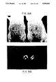
- FIG. 12 Analysis by phase contrast (panels a and c) and immunofluorescent (panels b and d) microscopy of COS cells transfected with piABCD (panels a and b) and CDM8 vector alone (panels c and d), respectively, and indirectly stained with YZ1 monoclonal anti-CR1 antibody and fluorescein-labelled goat anti-mouse F(ab') 2 .
- FIG. 13 Analysis of C3b- and C4b-binding by COS cells expressing recombinant CR1.
- COS cells transfected with piABCD panels a and c) or with the CDM8 vector alone (panels b and d) were incubated with EAC4b(lim),3b (panels a and b) or with EAC4b (panels c and d) and examined for formation of rosettes by phase contrast microscopy.
- FIG. 14 Analysis of recombinant CR1 expressed by transfected COS cells by SDS-PAGE.
- Detergent lysates of the cells were sequentially immunoadsorbed with Sepharose-UPC10 (lanes 1-3) and Sepharose-YZ1 (lanes 4-6) and the eluates analyzed by SDS-PAGE under non-reducing conditions and autoradiography.
- FIG. 15 Cleavage of 125 I-C3(ma) by factor I in the presence of immunoimmobilized recombinant CR1.
- Replicate samples of 125 I-C3(ma) were treated with factor I in the presence of factor H (lane 1), Sepharose-UPC10 preincubated with the lysate of COS cells transfected with the CDM8 vector alone (lane 2), Sepharose-UPC10 preincubated with the lysate of piABCD-transfected COS cells (lane 3), Sepharose-YZ1 preincubated with the lysate of CDM8-transfected COS cells (lane 4), and 6 ⁇ l (lane 5), 12 ⁇ l (lane 6) and 25 ⁇ l (lane 7) of Sepharose-YZ1 that had been preincubated with the lysate of piABCD-transfected COS cells.
- Samples of 125 I-labelled C3(ma) were also treated in the absence of factor I with 25 ⁇ l of Sepharose-YZ1 that had been preincubated with the lysate of piABCD-transfected COS cells (lane 8). After reduction, the 125 I-C3(ma) was analyzed by SDS-PAGE and autoradiography.
- FIG. 16 The cDNA constructs encoding the CR1 deletion mutants.
- the positions of the cDNA segments encoding the four LHRs are indicated by the brackets above the full length piABCD construct on which are shown the restriction sites used for preparation of the deletion mutants.
- the cDNA restriction fragments remaining in each of the mutants are indicated by the solid lines.
- the restriction sites are: A, ApaI; B, BsmI; E, BstEII; and P, PstI.
- FIG. 17 Comparison of recombinant deletion mutants of CR1 with the wild type F and S allotypes of CR1.
- the eluates were subjected to SDS-PAGE under reducing conditions and autoradiography.
- FIG. 18 Cleavage of 125 I-C3(ma) by factor I in the presence of COS cells expressing full length and deletion mutants of CR1.
- Replicate samples of 125 I-C3(ma) were incubated with COS cells transfected with the CDM8 vector alone (lanes 1 and 7), piABCD (lanes 2 and 8), piAD (lanes 3 and 9), piBD (lanes 4 and 10), piCD (lanes 5 and 11), and piD (lanes 6 and 12), respectively, in the absence (lanes 1-6) or presence (lanes 7-12) of factor I.
- Samples of 125 I-C3(ma) also were incubated with factor H and factor I (lane 13) and with factor I alone (lane 14), respectively. After reduction, the 125 I-C3(ma) was analyzed by SDS-PAGE and autoradiography.
- FIG. 19 Schematic model depicting the types of SCRs comprising each LHR of CR1, and the predicted sites determining the specificities of the receptor for C3b and C4b. The secondary binding specificities of these are indicated by the parentheses.
- FIG. 20 A schematic diagram illustrating the DNA regions remaining in the soluble CR1 DNA constructs. The regions of the full length CR1 cDNA are indicated by the boxes along the top of the figure.
- FIG. 21 A schematic diagram illustrating the major elements in the pTCS series of expression vectors.
- FIG. 22 A diagram of the expression vector pTCSgpt.
- the polyadenylation site is from the murine Ig kappa sequences (NBRF Nucleic database accession #Kcms, bp 1306-714); the Ad2 MLP and tripartite regions are from the Ad2 sequence (NBRF Nucleic database accession #Gdad2, bp 5791-6069); the SV40 early promoter is from the SV40 genome (NBRF Nucleic Database accession #GSV40W).
- the gpt gene, ampicillin gene and bacterial origin of replication are from the vector pSV2gpt (ATCC Accession No. 37145).
- FIG. 23 4-20% SDS-PAGE of antibody affinity purified sCR1.
- FIG. 24 Cation exchange HPLC elution profile. Eluted protein was monitored by absorbance at 280 nm (y-axis). The absorbance of both the flow-through (0-100 minutes) and the eluted sCR1 (150-165 minutes) were both offscale. The x-axis represents the elution time in minutes.
- FIG. 25 4-20% gradient SDS-PAGE of cation and anion exchange HPLC purified sCR1.
- SDS-polyacryamide gels were run under non-reducing conditions.
- Lane 1 an aliquot of bioreactor supernatant;
- lane 2 an aliquot of bioreactor supernatant dialyzed against cation HPLC starting buffer;
- lane 3 an aliquot of the eluted sCR1 peak from a cation exchange HPLC column;
- lane 4 an aliquot of the sCR1 peak from the cation exchange HPLC column dialyzed into starting buffer for anion HPLC;
- lanes 5 and 6 aliquots of two different fractions of eluted sCR1 from anion HPLC.
- FIG. 26 C5a induction of an oxygen burst in human neutrophils. Following a C5a induced oxygen burst, DCFDA became oxidized and brightly fluoresced. Fluorescent intensity, as determined by flow cytometry, is measured on the x-axis and number of cells on the y-axis. Panel a, profile and gate for the cells; panel b, 0 minutes after C5a addition; panel c, 1 minute; panel d, 2 minutes; panel e, 3 minutes; panel f, 4 minutes; panel g, 20 minutes. This DCFDA assay gives a sensitive indication of C5a.
- FIG. 27 Activation of human complement in the presence of sCR1 shows reduced C5a activity in the DCFDA assay.
- Panel a unstimulated cells
- panel b control without sCR1 showing a high degree of fluorescence
- panel c DCFDA assay in the presence of sCR1 showing a reduction of 75% in fluorescent intensity.
- y-axis is number of cells and x-axis is fluorescent intensity.
- FIG. 28 Inhibition of classical pathway C5a and C3a production in human serum by sCR1. Similar profiles were observed for either antibody affinity purified or HPLC purified sCR1.
- FIG. 29 Inhibition of complement-mediated hemolysis by recombinant sCR1. Similar profiles were observed for antibody affinity purified or HPLC purified sCR1.
- FIG. 30 Gross morphology of RPAR in sCR1-treated (left) and untreated (right) rats.
- rats received an intravenous injection of ovalbumin, followed by an intradermal injection of a mixture of either sCR1 (left rat) or PBS (right rat) with anti-ovalbumin, neat (left site); anti-ovalbumin, 1/2 dilution (middle site) or rabbit IgG (right site). The injections were performed in duplicate; top and bottom rows gave identical results.
- the rat which received sCR1 had barely visible changes, while the untreated rat developed full symptoms of RPAR.
- FIG. 31 Light microscopy of skin biopsies from sCR1-treated (a) and untreated (b) rats.
- FIG. 32 The clearance of injected sCR1 from the blood of rats and monkeys showing biphasic, ⁇ and ⁇ , clearance phases.
- FIG. 33 Autoradiographs of the Southern blots in which the CR1 cDNA and intron probes were hybridized to the EcoRV digests of the DNA from individuals who expressed the F or the F' allotypes.
- the positions of the Hind III fragments of ⁇ DNA are designated in kilobases on the left.
- the position of the F'-specific fragment is designated by a single arrow.
- FIG. 34 EcoRV restriction map of the F allele of CR1.
- the white boxes represent the positions of the exons and the stippled boxes represent the sites of hybridizations of the intron probes.
- the brackets over the LHR-B and -C indicate two possible regions of deletion.
- V represents an EcoRV site.
- FIG. 35 The cDNA inserts for the different forms of recombinant rCR1.
- the restriction sites shown are: A, ApaL I; B, BamH I; C, Sac I; H, Hind III; L, Bgl I; P, Pst I; R, EcoR I; and S, Sma I.
- the diagram at the top represents the CR1 protein, and the SCR with identical sequences are filled in by the same patterns.
- FIG. 36 Coomassie Blue-stained SDS-PAGE under nonreducing conditions of recombinant sCR1 purified by absorption on YZ-1-Sepharose.
- Each lane contains 10 ⁇ g of recombinant sCR1 purified from the culture supernatants of COS cells that have been transfected with pasecABBCD (lane 1), pasecABCD (lane 2), or pasecACD (lane 3).
- the position of the Mr markers are indicated on the right in kD.
- FIG. 37 Cofactor activity of recombinant sCR1. Cleavage of the ⁇ ' chain of C3b was measured in the presence of increasing amounts of recombinant sCR1 derived from COS cells transfected with pasecABBCD, pasecABCD, or pasecACD.
- FIG. 38 Inhibition of 125 I-C3b dimer uptake on erythrocytes by recombinant sCR1. Erythrocyte-bound ligand was measured in the presence of increasing concentrations of C3b dimer, C3b monomer, and recombinant sCR1 derived from COS cells transfected with pasecABBCD, pasecABCD, or pasecACD.
- FIG. 39 Inhibition of the alternative (A) and classical (B) C3 convertases by recombinant sCR1 purified from COS cells transfected with the different plasmids encoding the CR1 variants.
- FIG. 40 Inhibition of the alternative (A) and classical (B) C5 convertases by recombinant sCR1 purified from COS cells transfected with the different plasmids encoding the CR1 variants.
- the present invention is directed to the C3b/C4b receptor (CR1) gene and its encoded protein.
- the invention is also directed to CR1 nucleic acid sequences and fragments thereof comprising 70 nucleotides and their encoded peptides or proteins comprising 24 amino acids.
- the invention further provides for the expression of the CR1 protein and fragments thereof.
- Such CR1 sequences and proteins have value in diagnosis and therapy of inflammatory or immune system disorders, and disorders involving complement activity.
- the invention relates to soluble CR1 molecules and the expression, purification, and uses thereof.
- soluble CR1 molecules shall mean portions of the CR1 protein which, in contrast to the native CR1 proteins, are not expressed on the cell surface as membrane proteins.
- CR1 molecules which substantially lack a transmembrane region are soluble CR1 molecules.
- the soluble CR1 molecules are secreted by a cell in which they are expressed.
- the cloning and complete nucleotide and deduced amino acid sequence of the full-length CR1 cDNA, and of fragments thereof, and the expression of the encoded CR1 products are described.
- the expression of CR1 and fragments thereof, with binding sites for C3b and/or C4b, and which inhibit factor I cofactor activity, is also described.
- the invention is further illustrated by the production and purification of soluble, truncated CR1 molecules. In specific examples, such molecules are demonstrated to be therapeutically useful in reducing inflammation, and in reducing myocardial infarct size and preventing reperfusion injury.
- Any human cell can potentially serve as the nucleic acid source for the molecular cloning of the CR1 gene.
- Isolation of the CR1 gene involves the isolation of those DNA sequences which encode a protein displaying CR1-associated structure or properties, e.g., binding of C3b or C4b or immune complexes, modulating phagocytosis, immune stimulation or proliferation, and regulation of complement.
- the DNA may be obtained by standard procedures known in the art from cloned DNA (e.g., a DNA "library”), by chemical synthesis, by cDNA cloning, or by the cloning of genomic DNA, or fragments thereof, purified from the desired human cell.
- Cells which can serve as sources of nucleic acid for cDNA cloning of the CR1 gene include but are not limited to monocytes/macrophages, granulocytes, B cells, T cells, splenic follicular dendritic cells, and glomerular podocytes.
- Clones derived from genomic DNA may contain regulatory and intron DNA regions in addition to coding regions; clones derived from cDNA will contain only exon sequences. Whatever the source, the CR1 gene should be molecularly cloned into a suitable vector for propagation of the gene.
- DNA fragments are generated, some of which will encode the desired CR1 gene.
- the DNA may be cleaved at specific sites using various restriction enzymes. Alternatively, one may use DNAse in the presence of manganese to fragment the DNA, or the DNA can be physically sheared, as for example, by sonication.
- the linear DNA fragments can then be separated according to size by standard techniques, including but not limited to, agarose and polyacrylamide gel electrophoresis and column chromatography.
- identification of the specific DNA fragment containing the CR1 gene may be accomplished in a number of ways. For example, if an amount of a CR1 gene or its specific RNA, or a fragment thereof, is available and can be purified and labeled, the generated DNA fragments may be screened by nucleic acid hybridization to the labeled probe (Benton, W. and Davis, R., 1977, Science 196:180; Grunstein, M. and Hogness, D., 1975, Proc. Natl. Acad. Sci. U.S.A. 72:3961). Those DNA fragments with substantial homology to the probe will hybridize.
- nucleic acid fractions enriched in CR1 may be used as a probe, as an initial selection procedure.
- the probe representing B cell cDNA from which messages expressed by fibroblasts have been subtracted can be used. It is also possible to identify the appropriate fragment by restriction enzyme digestion(s) and comparison of fragment sizes with those expected according to a known restriction map if such is available. Further selection on the basis of the properties of the gene, or the physical, chemical, or immunological properties of its expressed product, as described infra, can be employed after the initial selection.
- the CR1 gene can also be identified by mRNA selection by nucleic acid hybridization followed by in vitro translation. In this procedure, fragments are used to isolate complementary mRNAs by hybridization. Such DNA fragments may represent available, purified CR1 DNA, or DNA that has been enriched for CR1 sequences. Immunoprecipitation analysis or functional assays (e.g., for C3b or C4b binding, or promotion of phagocytosis or immune stimulation, or complement regulation, etc.) of the in vitro translation products of the isolated mRNAs identifies the mRNA and, therefore, the complementary DNA fragments that contain the CR1 sequences.
- specific mRNAs may be selected by adsorption of polysomes isolated from cells to immobilized antibodies specifically directed against CR1.
- a radiolabeled CR1 cDNA can be synthesized using the selected mRNA (from the adsorbed polysomes) as a template. The radiolabeled mRNA or cDNA may then be used as a probe to identify the CR1 DNA fragments from among other genomic DNA fragments.
- RNA for cDNA cloning of the CR1 gene can be isolated from cells including but not limited to monocytes/macrophages, granulocytes, B cells, T cells, dendritic cells, and podocytes.
- tonsilar cells can serve as the source of mRNA for cDNA cloning (See Section 6.1.2, infra). Other methods are possible and within the scope of the invention.
- the identified and isolated gene can then be inserted into an appropriate cloning vector.
- vector-host systems known in the art may be used. Possible vectors include, but are not limited to, plasmids or modified viruses, but the vector system must be compatible with the host cell used. Such vectors include, but are not limited to, bacteriophages such as lambda derivatives, or plasmids such as pBR322 or pUC plasmid or CDM8 plasmid (Seed, B., 1987, Nature 329:840-842) or derivatives. Recombinant molecules can be introduced into host cells via transformation, transfection, infection, electroporation, etc.
- the CR1 gene may be identified and isolated after insertion into a suitable cloning vector, in a "shot gun" approach. Enrichment for the CR1 gene, for example, by size fractionation, can be done before insertion into the cloning vector.
- the CR1 gene is inserted into a cloning vector which can be used to transform, transfect, or infect appropriate host cells so that many copies of the gene sequences are generated.
- the cloning vector can be the CDM8 vector, which can be used to achieve expression in a mammalian host cell.
- the insertion into a cloning vector can, for example, be accomplished by ligating the DNA fragment into a cloning vector which has complementary cohesive termini. However, if the complementary restriction sites used to fragment the DNA are not present in the cloning vector, the ends of the DNA molecules may be enzymatically modified.
- any site desired may be produced by ligating nucleotide sequences (linkers) onto the DNA termini; these ligated linkers may comprise specific chemically synthesized oligonucleotides encoding restriction endonuclease recognition sequences.
- the cleaved vector and CR1 gene may be modified by homopolymeric tailing.
- Identification of the cloned CR1 gene can be accomplished in a number of ways based on the properties of the DNA itself, or alternatively, on the physical, immunological, or functional properties of its encoded protein.
- the DNA itself may be detected by plaque or colony nucleic acid hybridization to labeled probes (Benton, W. and Davis, R., 1977, Science 196:180; Grunstein, M. and Hogness, D., 1975, Proc. Natl. Acad. Sci. U.S.A. 72:3961).
- the presence of the CR1 gene may be detected by assays based on properties of its expressed product.
- cDNA clones or DNA clones which hybrid-select the proper mRNAs, can be selected which produce a protein that, e.g., has similar or identical electrophoretic migration, isoelectric focusing behavior, proteolytic digestion maps, C3b and/or C4b and/or immune complex binding activity, complement regulatory activity, effects on phagocytosis or immune stimulation, or antigenic properties as known for CR1.
- the CR1 protein may be identified by binding of labeled antibody to the putatively CR1-synthesizing clones, in an ELISA (enzyme-linked immunosorbent assay)-type procedure.
- transformation of host cells with recombinant DNA molecules that incorporate the isolated CR1 gene, cDNA, or synthesized DNA sequence enables generation of multiple copies of the gene.
- the gene may be obtained in large quantities by growing transformants, isolating the recombinant DNA molecules from the transformants and, when necessary, retrieving the inserted gene from the isolated recombinant DNA.
- CR1 cDNA clones in a CDM8 vector can be transfected into COS (monkey kidney) cells for large-scale expression under the control of the cytomegalovirus promoter (see Section 8, infra).
- the recombinant DNA molecule that incorporates the CR1 gene can be modified so that the gene is flanked by virus sequences that allow for genetic recombination in cells infected with the virus so that the gene can be inserted into the viral genome.
- promoter DNA may be ligated 5' of the CR1-coding sequence, in addition to or replacement of the native promoter to provide for increased expression of the protein.
- Expression vectors which express CR1 deletion mutants can also be made, to provide for expression of defined fragments of the CR1 sequence (see the example sections, infra).
- deletion mutants can be constructed which encode fragments of the CR1 protein that exhibit the desired C3b and/or C4b binding activity (see Section 9, infra), e.g., LHR-A for binding of C4b, or LHR-C for binding of C3b.
- an expression vector which encodes a CR1 molecule with a deletion of the transmembrane region can be used to produce a soluble CR1 molecule (see the examples sections 11-14, infra). Many manipulations are possible, and within the scope of the present invention.
- the nucleotide sequence coding for the CR1 protein (FIG. 1) or a portion thereof, can be inserted into an appropriate expression vector, i.e., a vector which contains the necessary elements for the transcription and translation of the inserted protein-coding sequence.
- the necessary transcriptional and translation signals can also be supplied by the native CR1 gene and/or its flanking regions.
- a variety of host-vector systems may be utilized to express the protein-coding sequence.
- mammalian cell systems infected with virus e.g., vaccinia virus, adenovirus, etc.
- insect cell systems infected with virus e.g., baculovirus
- microorganisms such as yeast containing yeast vectors, or bacteria transformed with bacteriophage DNA, plasmid DNA or cosmid DNA.
- the expression elements of these vectors vary in their strength and specificities. Depending on the host-vector system utilized, any one of a number of suitable transcription and translation elements may be used.
- promoters isolated from the genome of mammalian cells or from viruses that grow in these cells may be used. Promoters produced by recombinant DNA or synthetic techniques may also be used to provide for transcription of the inserted sequences.
- Specific initiation signals are also required for efficient translation of inserted protein coding sequences. These signals include the ATG initiation codon and adjacent sequences. In cases where the entire CR1 gene including its own initiation codon and adjacent sequences are inserted into the appropriate expression vectors, no additional translational control signals may be needed. However, in cases where only a portion of the CR1 coding sequence is inserted, exogenous translational control signals, including the ATG initiation codon, must be provided. The initiation codon must furthermore be in phase with the reading frame of the protein coding sequences to ensure translation of the entire insert. These exogenous translational control signals and initiation codons can be of a variety of origins, both natural and synthetic.
- Any of the methods previously described for the insertion of DNA fragments into a vector may be used to construct expression vectors containing a chimeric gene consisting of appropriate transcriptional/translational control signals and the protein coding sequences. These methods may include in vitro recombinant DNA and synthetic techniques and in vivo recombinations (genetic recombination).
- a soluble CR1 molecule can be expressed.
- Such a soluble molecule can be produced by use of recombinant DNA techniques to delete the DNA sequences encoding the CR1 transmembrane region (see Sections 11-14, infra).
- the ability to express a soluble CR1 molecule is not limited to any one genetic modification of the CR1 nucleic acid sequence; as long as the nucleic acid sequence encoding a substantial portion of the CR1 transmembrane region is deleted, soluble CR1 constructs can be obtained.
- Expression vectors containing CR1 gene inserts can be identified by three general approaches: (a) DNA-DNA hybridization, (b) presence or absence of "marker" gene functions, and (c) expression of inserted sequences.
- the presence of a foreign gene inserted in an expression vector can be detected by DNA-DNA hybridization using probes comprising sequences that are homologous to the inserted CR1 gene.
- the recombinant vector/host system can be identified and selected based upon the presence or absence of certain "marker" gene functions (e.g., thymidine kinase activity, resistance to antibiotics, transformation phenotype, occlusion body formation in baculovirus, etc.) caused by the insertion of foreign genes into the vector.
- certain "marker" gene functions e.g., thymidine kinase activity, resistance to antibiotics, transformation phenotype, occlusion body formation in baculovirus, etc.
- recombinant expression vectors can be identified by assaying the foreign gene product expressed by the recombinant. Such assays can be based on the physical, immunological, or functional properties of the gene product.
- recombinant DNA molecule Once a particular recombinant DNA molecule is identified and isolated, several methods known in the art may be used to propagate it. Once a suitable host system and growth conditions are established, recombinant expression vectors can be propagated and prepared in quantity.
- CDM8 vectors with an CR1 cDNA insert can be transfected into COS cells, in which the CR1 cDNA insert is expressed to produce the CR1 protein.
- CDM8 vectors with a CR1 cDNA insert corresponding to a portion of the CR1 coding region can be transfected into COS cells, where the CR1 or fragment is expressed.
- infra, truncated, soluble CR1 molecules can be expressed in mammalian cells by use of expression vectors such as the pTCS vectors described in Section 11.3.1.
- the expression vectors which can be used include, but are not limited to, the following vectors or their derivatives: human or animal viruses such as vaccinia virus or adenovirus; insect viruses such as baculovirus; yeast vectors; bacteriophage vectors (e.g., lambda), and plasmid and cosmid DNA vectors, to name but a few.
- a host cell strain may be chosen which modulates the expression of the inserted sequences, or modifies and processes the chimeric gene product in the specific fashion desired. Expression from certain promoters can be elevated in the presence of certain inducers; thus, expression of the genetically engineered CR1 protein may be controlled.
- different host cells have characteristic and specific mechanisms for the translational and post-translational processing and modification of proteins. Appropriate cell lines or host systems can be chosen to ensure the desired modification and processing of the expressed heterologous protein. For example, in one embodiment, expression in a bacterial system can be used to produce an unglycosylated CR1 protein with the deduced amino acid sequence of FIG. 1. Expression in yeast will produce a glycosylated product.
- mammalian COS cells can be used to ensure "native" glycosylation of the heterologous CR1 protein.
- different vector/host expression systems may effect processing reactions such as proteolytic cleavages to different extents. Many such variously processed CR1 proteins can be produced and are within the scope of the present invention.
- large scale production of soluble CR1 molecules may be carried out as described infra in Section 12.1 et seq.
- the gene product should be analyzed. This can be achieved by assays based on the physical, immunological, or functional properties of the product.
- the CR1 proteins may be isolated and purified by standard methods including chromatography (e.g., ion exchange, affinity, and sizing column chromatography, high pressure liquid chromatography), centrifugation, differential solubility, or by any other standard technique for the purification of proteins.
- chromatography e.g., ion exchange, affinity, and sizing column chromatography, high pressure liquid chromatography
- centrifugation e.g., centrifugation, differential solubility, or by any other standard technique for the purification of proteins.
- large quantities of soluble CR1 can be purified by procedures involving HPLC (see Section 12.2 et seq.).
- HPLC large-scale production of purified CR1 can be achieved by using an expression system which produces soluble CR1 as starting material, thus eliminating the requirement of solubilizing membrane-bound CR1 with detergents.
- the reduction of fetal calf serum concentrations in the bioreactor cultures and/or the use of alternative culture medias in these cultures eliminates the need to remove high concentrations of extraneous proteins from the soluble CR1-containing starting material during subsequent purification.
- Either cation HPLC or a combination of cation HPLC followed by anion exchange HPLC can be used for purification in this preferred aspect.
- Substantially pure soluble CR1 in high yield can thus be achieved in only one or two steps.
- the amino acid sequence of the protein can be deduced from the nucleotide sequence of the chimeric gene contained in the recombinant.
- the protein can be synthesized by standard chemical methods known in the art (e.g., see Hunkapiller, M., et al., 1984, Nature 310:105-111).
- such CR1 proteins include but are not limited to those containing, as a primary amino acid sequence, all or part of the amino acid sequence substantially as depicted in FIG. 1, including altered sequences in which functionally equivalent amino acid residues are substituted for residues within the sequence resulting in a silent change.
- one or more amino acid residues within the sequence can be substituted by another amino acid of a similar polarity which acts as a functional equivalent, resulting in a silent alteration.
- Nonconservative substitutions can also result in functionally equivalent proteins.
- substitutes for an amino acid within the CR1 sequence may be selected from other members of the class to which the amino acid belongs.
- the nonpolar (hydrophobic) amino acids include alanine, leucine, isoleucine, valine, proline, phenylalanine, tryptophan and methionine.
- the polar neutral amino acids include glycine, serine, threonine, cysteine, tyrosine, asparagine, and glutamine.
- the positively charged (basic) amino acids include arginine, lysine and histidine.
- the negatively charged (acidic) amino acids include aspartic acid and glutamic acid.
- CR1 proteins which are differentially modified during or after translation, e.g., by glycosylation, proteolytic cleavage, etc.
- cloned recombinant CR1 expressed by transfected cells was shown to be indistinguishable from the F allotype of erythrocytes by SDS-PAGE (FIG. 14), capable of mediating the binding of sheep erythrocytes bearing either C4b or C3b, and able to reproduce the ligand specificity of CR1 (FIG. 13), and exhibit factor I co-factor activity for cleavage of the alpha polypeptide of C3(ma) (FIG. 15).
- the structure of the CR1 gene and protein can be analyzed by various methods known in the art, including but not limited to those described infra.
- the cloned DNA or cDNA corresponding to the CR1 gene can be analyzed by methods including but not limited to Southern hybridization (Southern, E. M., 1975, J. Mol. Biol. 98:503-517), Northern hybridization (see e.g., Freeman et al., 1983, Proc. Natl. Acad. Sci. U.S.A. 80:4094-4098), restriction endonuclease mapping (Maniatis, T., 1982, Molecular Cloning, A Laboratory Manual, Cold Spring Harbor Laboratory, Cold Spring Harbor, N.Y.), and DNA sequence analysis.
- the stringency of the hybridization conditions for both Southern and Northern hybridization can be manipulated to ensure detection of nucleic acids with the desired degree of relatedness to the specific CR1 probe used. For example, hybridization under low stringency conditions with a probe containing CR1 gene sequences encoding LHR-B and LHR-C, can be used to detect CR2 nucleic acid sequences.
- Restriction endonuclease mapping can be used to roughly determine the genetic structure of the CR1 gene.
- cleavage with restriction enzymes can be used to derive the restriction map shown in FIG. 2, infra. Restriction maps derived by restriction endonuclease cleavage can be confirmed by DNA sequence analysis.
- DNA sequence analysis can be performed by any techniques known in the art, including but not limited to the method of Maxam and Gilbert (1980, Meth. Enzymol. 65:499-560), the Sanger dideoxy method (Sanger, F., et al., 1977, Proc. Natl. Acad. Sci. U.S.A. 74:5463), or use of an automated DNA sequenator (e.g., Applied Biosystems, Foster City, Calif.).
- the cDNA sequence of the CR1 gene comprises the sequence substantially as depicted in FIG. 1, and described in Sections 6 and 7, infra.
- the amino acid sequence of the CR1 protein can be derived by deduction from the DNA sequence, or alternatively, by direct sequencing of the protein, e.g., with an automated amino acid sequencer.
- the amino acid sequence of a representative CR1 protein comprises the sequence substantially as depicted in FIG. 1, and detailed in Section 6, infra. As described infra, all of the coding sequence of the F allotype CR1 has been cloned and, after cleavage of the signal peptide of 41 amino acids, the mature receptor contained 1998 amino acids including an extracellular domain of 1930 residues that forms 30 SCRs, 28 of which are organized into LHRs-A, -B, -C and -D, (FIG. 10), a single membrane spanning domain of 25 amino acids and a relatively short cytoplasmic domain of 43 amino acids.
- CR1 is unique in having groups of SCRs organized into LHRs. Comparison of the four LHRs of CR1 reveals that each is a composite of four types of SCRs: types a, b, c and d (FIG. 19). For example, the sequences of SCR-1 and -2 of LHR-A are only 62%, 62% and 57% identical to the first two SCRs of LHR-B, -C and -D, respectively. However, SCR-3 through SCR-7 differ from the corresponding SCRs of LHR-B at only a single position, and SCR-3 and -4 differ from those of LHR-C at only three positions (FIG. 10).
- LHR-B and -C some of the type "a" SCRs of LHR-A are also present in LHR-B and -C.
- the first two SCRs of LHR-B which differ from those of LHR-A, are 99% identical with the corresponding SCRs of LHR-C, so that LHR-B and -C share the type "b" SCR at these positions.
- the fifth, sixth and seventh SCR of LHR-C are only 77% identical to the type "a" SCRs in LHR-A and -B at these positions, and are considered as type "c” SCRs.
- the first through fourth SCRs of LHR-D are relatively unique and are type "d", while the fifth through seventh SCRs are approximately 93% identical to the "c" type found in LHR-C.
- the CR1 protein sequence can be further characterized by a hydrophilicity analysis (Hopp, T. and Woods, K., 1981, Proc. Natl. Acad. Sci. U.S.A. 78:3824).
- a hydrophilicity profile can be used to identify the hydrophobic and hydrophilic regions of the CR1 protein and the corresponding regions of the gene sequence which encode such regions.
- a hydrophilicity profile of the COOH-terminus of the CR1 protein is depicted in FIG. 5.
- derivatives, analogues, and peptides related to CR1 are also envisioned, and within the scope of the present invention.
- Such derivatives, analogues, or peptides which have the desired immunogenicity or antigenicity can be used, for example, in immunoassays, for immunization, therapeutically, etc.
- Such molecules which retain, or alternatively inhibit, a desired CR1 property e.g., binding of C3b or C4b, regulation of complement activity, or promotion of immune stimulation or phagocytosis, etc., can be used as inducers, or inhibitors, respectively, of such property.
- the CR1-related derivatives, analogues, and peptides of the invention can be produced by various methods known in the art.
- the manipulations which result in their production can occur at the gene or protein level.
- the cloned CR1 gene can be modified by any of numerous strategies known in the art (Maniatis, T., 1982, Molecular Cloning, A Laboratory Manual, Cold Spring Harbor Laboratory, Cold Spring Harbor, N.Y.).
- the CR1 sequence can be cleaved at appropriate sites with restriction endonuclease(s), followed by further enzymatic modification if desired, isolated, and ligated in vitro (see Section 8, infra).
- nucleic acid sequences encoding a fusion protein consisting of a molecule comprising a portion of the CR1 sequence plus a non-CR1 sequence, can be produced.
- the CR1 gene can be mutated in vitro or in vivo, to create and/or destroy translation, initiation, and/or termination sequences, or to create variations in coding regions and/or form new restriction endonuclease sites or destroy preexisting ones, to facilitate further in vitro modification.
- Any technique for mutagenesis known in the art can be used, including but not limited to, in vitro site-directed mutagenesis (Hutchinson, C., et al., 1978, J. Biol. Chem. 253:6551), use of TAB® linkers (Pharmacia), etc.
- Manipulations of the CR1 sequence may also be made at the protein level. Any of numerous chemical modifications may be carried out by known techniques, including but not limited to specific chemical cleavage by cyanogen bromide, trypsin, chymotrypsin, papain, V8 protease, NaBH 4 ; acetylation, formylation, oxidation, reduction; metabolic synthesis in the presence of tunicamycin; etc.
- analogues and peptides related to CR1 can be chemically synthesized.
- a peptide corresponding to a portion of CR1 which mediates the desired activity e.g., C3b and/or C4b binding, immune stimulation, complement regulation, etc.
- a peptide synthesizer can be synthesized by use of a peptide synthesizer.
- nucleotide sequence of CR1 can be made by recombinant DNA procedures that result in sequences encoding a protein having multiple LHR-B sequences. Such valency modifications alter the extent of C3b binding.
- CR1 proteins, analogues, derivatives, and subsequences thereof, and anti-CR1 antibodies have uses in assays and in diagnostics.
- the molecules of the invention which demonstrate the desired CR1 property or function can be used to assay such property or function.
- CR1 proteins or fragments thereof, which exhibit binding of C3b and/or C4b, in free and/or in complex forms can be used in assays to measure the amount of such substances in a sample, e.g., a body fluid of a patient.
- full-length CR1 or a CR1 deletion mutant expressed on the cell surface (e.g., those described in Section 8, infra) having the ability to bind C3b (e.g., see Table II, Section 9, infra), iC3b or C4b (e.g., see Table II) can be used in assays to measure the levels of C3b, iC3b, or C4b, respectively, in a sample.
- a CR1 protein or fragment thereof which is constructed by recombinant DNA technology to lack a transmembrane sequence, and is thus secreted, can be used.
- such a measurement of C3b and/or C4b can be relied on as an indication of complement activity, and can be useful in the diagnosis of inflammatory and immune system disorders.
- Such disorders include but are not limited to tissue damage due to burn--or myocardial infarct-induced trauma, adult respiratory distress syndrome (shock lung), autoimmune disorders such as rheumatoid arthritis, systemic lupus erythematosus, and other diseases or disorders involving undesirable or inappropriate complement activity (see, e.g., Miescher, P. A. and Muller-Eberhard, H. J., eds., 1976, Text Book of Immunopathology, 2d Ed., Vols.
- the CR1 protein and fragments thereof containing an epitope have uses in assays including but not limited to immunoassays.
- the immunoassays which can be used include but are not limited to competitive and non-competitive assay systems using techniques such as radioimmunoassays, ELISA (enzyme linked immunosorbent assay), "sandwich” immunoassays, precipitin reactions, gel diffusion precipitin reactions, immunodiffusion assays, agglutination assays, complement-fixation assays, immunoradiometric assays, fluorescent immunoassays, protein A immunoassays, and immunoelectrophoresis assays, to name but a few.
- CR1 genes and related nucleic acid sequences and subsequences can be used in hybridization assays.
- Such hybridization assays can be used to monitor inflammatory or immune responses associated with CR1 expression, to diagnose certain disease states associated with changes in CR1 expression, to determine the CR1 allotype of a patient, and to detect the presence and/or expression of the CR1 gene and related genes (e.g., CR2).
- Kits for practicing the assays for use in the present invention are also provided.
- the CR1 protein and fragments, derivatives, and analogues thereof can be therapeutically useful in the modulation of functions mediated by CR1.
- functions include but are not limited to binding of C3b and/or C4b, in free or in complex forms, promotion of phagocytosis, complement regulation, immune stimulation, etc.
- Effective doses of the CR1 proteins and related molecules of the invention have therapeutic value for many of the diseases or disorders associated with such functions, such as immune or inflammatory disorders (e.g., those described supra in Section 5.6.1).
- full-length CR1 or fragments thereof and related molecules which exhibit the desired activity can have therapeutic uses in the inhibition of complement by their ability to act as a factor I cofactor, promoting the irreversible inactivation of complement components C3b or C4b (Fearon, D. T., 1979, Proc. Natl. Acad. Sci. U.S.A. 76:5867; Iida, K. and Nussenzweig, V., 1981, J. Exp. Med. 153:1138), and/or by the ability to inhibit the alternative or classical C3 or C5 convertases.
- an expression vector can be constructed to encode a CR1 molecule which lacks the transmembrane region (e.g., by deletion carboxy-terminal to the arginine encoded by the most C-terminal SCR), resulting in the production of a soluble CR1 fragment.
- a fragment can retain the ability to bind C3b and/or C4b, in free or in complex forms.
- such a soluble CR1 protein may no longer exhibit factor I cofactor activity.
- the soluble CR1 product can be administered in vivo to a patient, so that the soluble CR1 can effectively compete out binding of the C3b and/or C4b to the native cell-surface CR1, thus blocking cell-surface CR1 factor I cofactor activity, and increasing complement activity.
- C3b After C3b has covalently attached to particles and soluble immune complexes, the inactivation of C3b by proteolytic processing into iC3b and C3dg has two biologic consequences: preventing excessive activation of the complement system via the amplification pathway, and formation of ligands that can engage receptors other than CR1.
- the iC3b fragment cannot bind factor B so that conversion to this state blocks additional complement activation via the alternative pathway amplification loop.
- iC3b can be bound by CR1 and CR3, the two complement receptors that mediate phagocytosis by myelomonocytic cells.
- C3b to iC3b conversion cessation of complement activation without interference with CR1- and CR3-mediated clearance of the C3-coated complex.
- additional conversion of iC3b to C3dg creates a fragment that interacts only with CR2 and not with CR1 and CR3. This circumstance limits complement-dependent binding of the C3dg-bearing complex to cell types expressing CR2, which include B lymphocytes, follicular dendritic cells and perhaps epithelial cells of the dermis, and diminishes or excludes interaction with phagocytic cell types.
- CR1 molecules may be used therapeutically not only to affect the clearance process, but also in the targeting of complexes to the CR2-bearing cell types that participate in antigen presentation and antibody production.
- a CR1 protein or fragment thereof which can bind C3b or C4b, and/or retains the ability to inhibit the alternative or classical C3 or C5 convertases, or retains factor I cofactor activity, can be used to promote complement inactivation.
- the CR1 protein or fragment can be valuable in the treatment of disorders which involve undesirable or inappropriate complement activity (e.g., shock lung, tissue damage due to burn or ischemic heart conditions, autoimmune disorders, inflammatory conditions, etc.).
- a soluble CR1 molecule can be expressed which retains a desired functional activity, as demonstrated, e.g., by the ability to inhibit classical complement-mediated hemolysis, classical C5a production, classical C3a production, or neutrophil oxidative burst in vitro.
- a soluble CR1 molecule can be used to reduce inflammation and its detrimental effects, or to reduce myocardial infarct size or prevent reperfusion injury, etc.
- Such CR1 molecules useful for in vivo therapy may be tested in various model systems known in the art, including but not limited to the reversed passive Arthrus reaction (see Section 14.1) and a rat myocardial infarct model (see Section 14.3).
- a fragment of CR1 or an analogue or derivative thereof, which is shown to inhibit a desired CR1 property or function can be used to prevent or treat diseases or disorders associated with that function.
- CR1 and related molecules e.g., encapsulation in liposomes, microparticles, or microcapsules, expression by hematopoietic stem cell progeny in gene therapy, etc.
- Other methods of introduction include but are not limited to intradermal, intramuscular, intraperitoneal, intravenous, subcutaneous, intranasal, and oral routes.
- compositions comprise a therapeutically effective amount of a CR1 protein, or an analogue, derivative, or fragment thereof, and a pharmaceutically acceptable carrier.
- a carrier includes but is not limited to saline, buffered saline, dextrose, and water.
- a method of treating thrombotic conditions, especially acute myocardial infarction, in humans and animals comprises administering to a human or animal in need thereof an effective amount of a soluble CR1 protein according to the invention and an effective amount of a thrombolytic agent.
- the invention also provides the use of a soluble CR1 protein and a thrombolytic agent in the manufacture of a medicament for the treatment of thrombotic conditions in humans and animals.
- the compounds may be administered by any convenient route, for example by infusion or bolus injection, and may be administered sequentially or together.
- the soluble CR1 protein according to the invention and the thrombolytic agent are administered sequentially, the soluble CR1 protein may be administered either before or after the thrombolytic agent.
- the soluble CR1 protein and the thrombolytic agent are administered together they are preferably given in the form of a pharmaceutical composition comprising both agents.
- a pharmaceutical composition comprising a soluble CR1 protein and a thrombolytic agent together with a pharmaceutically acceptable carrier.
- the composition may be formulated in accordance with routine procedures as a pharmaceutical composition adapted for intravenous administration to human beings.
- compositions for intravenous administration are solutions in sterile isotonic aqueous buffer.
- the composition may also include a solubilizing agent and a local anaesthetic such as lignocaine to ease pain at the site of the injection.
- the ingredients will be supplied either separately or mixed together in unit dosage form, for example, as a dry lyophilised powder or water free concentrate in a hermetically sealed container such as an ampoule or sachette indicating the quantity of active agent in activity units.
- the composition is to be administered by infusion, it can be dispensed with an infusion bottle containing sterile pharmaceutical grade ⁇ Water for Injection ⁇ or saline.
- an ampoule of sterile water for injection or saline may be provided so that the ingredients may be mixed prior to administration.
- a pharmaceutical pack comprising one or more containers filled with one or more of the ingredients of the pharmaceutical composition is also within the scope of the invention.
- the quantity of material administered, and the ratio of thrombolytic agent to CR1 protein, will depend upon the seriousness of the thromboembolic condition and position and size of the clot.
- the precise dose to be employed and mode of administration must per force in view of the nature of the complaint be decided according to the circumstances by the physician supervising treatment.
- a patient being treated for a thrombus will generally receive a dose of from 0.5 to 50 mg of complement inhibitor (soluble CR1 component) per standard dose of thrombolytic agent.
- thrombolytic agents for use in combination therapy as described above are fibrinolytic enzymes, including plasminogen activators.
- plasminogen activator includes but is not limited to streptokinase, human tissue plasminogen activator (t-PA) and urokinase (u-PA) (both single and two-chain forms).
- t-PA human tissue plasminogen activator
- u-PA urokinase
- Such enzymes are obtained from natural sources or tissues or by recombinant DNA methods where heterologous host organisms such as bacteria, yeasts, fungi or mammalian cells express genes specifyng the enzymes.
- heterologous host organisms such as bacteria, yeasts, fungi or mammalian cells express genes specifyng the enzymes.
- the term also includes:
- conjugates comprising a fibrinolytic enzyme linked to a water-soluble polymer by means of a reversible linking group as disclosed in EP-A-0183503;
- the plasminogen activator is a hybrid molecule as described in EP-A-0297882 which comprises the five kringle domains of plasminogen linked to the B-chain of t-PA or u-PA via an amino acid sequence comprising, respectively, the t-PA cleavage site between residues 275 and 276 and the cysteine residue 264 of t-PA or the u-PA cleavage site between residues 158 and 159 and the cysteine residue 148 of u-PA.
- hybrids examples include plasminogen 1-544/t-PA 262-527 including one and two chain variants, lys 78 and glu 1 variants, and mixtures thereof;
- plasminogen 1-544/t-PA 262-527 (arg 275 gln) including one and two chain variants, lys 78 and glu 1 variants, and mixtures thereof;
- plasminogen 1-541/t-PA 262-527 including one and two chain variants, lys 78 and glu 1 variants, and mixtures thereof;
- t-PA 1-50/t-PA 88-91/pro-gly-ser/plasminogen 84-544/t-PA 262-527 including one and two chain variants, gly -3 , ser 1 and val 4 variants, and mixtures thereof;
- t-PA 1-91/pro-gly-ser/plasminogen 84-544/t-PA 262-527 including one and two chain variants, gly -3 , ser 1 and val 4 variants, and mixtures thereof; or
- plasminogen 1-546/u-PA 137-411 including one and two chain variants, lys 78 and glu 1 variants, and mixtures thereof.
- the thrombolytic agent for use in combination therapy is a reversibly blocked in vivo fibrinolytic enzyme having the meaning given by Smith in U.S. Pat. No. 4,285,932, i.e., an in vivo fibrinolytic enzyme wherein the catalytic site essential for fibrinolytic activity is blocked by a group which is removable by hydrolysis at a rate such that the pseudo-first order rate constant for hydrolysis is in the range 10 -6 sec -1 to 10 -3 sec -1 in isotonic aqueous media at pH 7.4 at 37° C.
- the fibrinolytic enzyme is a plasminogen activator comprising a serine protease domain of t-PA or urokinase
- an example of a removable blocking group is a 2-aminobenzoyl group substituted in the 3- or 4-position with a halogen atom and optionally further substituted with one or more weakly electron-withdrawing or electon-donating groups, wherein the pseudo first order rate constant for hydrolysis of the derivative is in the range of 6.0 ⁇ 10 -5 to 4.0 ⁇ 10 -4 sec -1 when measured in a buffer system consisting of 0.05M sodium phosphate, 0.1M sodium chloride, 0.01% v/v detergent comprising polyoxyethylenesorbitan monoleate having a molecular weight of approximately 1300, at pH 7.4 at 37° C.
- the reversibly blocked in vivo fibrinolytic enzyme is a binary complex between streptokinase and plasminogen, most preferably a p-anisoyl streptokinase/plasminogen complex without internal bond cleavage as described in U.S. Pat. No. 4,808,405, marketed by Beecham Group plc under the Trademark EMINASE (generic name anistreplase, hereinafter referred to as APSAC, i.e. anisoylated human plasminogen-streptokinase-activator complex; see for example J. P. Monk and R. C. Heel, 1987, Drugs 34:25-49).
- EMINASE generic name anistreplase, hereinafter referred to as APSAC, i.e. anisoylated human plasminogen-streptokinase-activator complex; see for example J. P. Monk and R. C. Heel, 1987,
- the soluble CR1 component used in combination therapy is encoded by a nucleic acid vector selected from the group consisting of pBSCR1c, pBSCR1s, pBM-CR1c, pBSCR1c/pTCSgpt and pBSCR1s/PTCSgpt and is especially that prepared from pBSCR1c/pTCSgpt as described above (see Section 12).
- thrombolytics for use in combination therapy (with examples of dose and method of administration) are as follows:
- Amino acid identity between the LHRs ranged from 70% between the first and third repeats to 99% between the NH 2 -terminal 250 amino acids of the first and second repeats.
- Each LHR comprises seven short consensus repeats (SCRs) of 60-70 amino acids that resemble the SCRs of other C3/C4 binding proteins, such as complement receptor type 2, factors B and H, C4 binding protein, and C2.
- SCRs short consensus repeats
- Two additional SCRs join the LHRs to a single membrane-spanning domain of 25 amino acids: thus, the F allotype of CR1 probably contains at least 30 SCRs, 23 of which have been sequenced.
- Each SCR is predicted to form a triple loop structure in which the four conserved half-cystines form disulfide linkages.
- the linear alignment of 30 SCRs as a semi-rigid structure would extend 1,140 Angstroms from the plasma membrane and might facilitate the interaction of CR1 with C3b and C4b located within the interstices of immune complexes and microbial cell walls.
- the COOH-terminal cytoplasmic domain of 43 residues contains a six amino acid sequence that is homologous to the sequence in the epidermal growth factor receptor that is phosphorylated by protein kinase C.
- CR1 was purified from washed human erythrocyte membranes by sequential Matrex Red A and YZ-1 monoclonal antibody affinity chromatography (Wong, W. W., et al., 1985, J. Immunol. Methods 82:303). Tryptic peptides were prepared and isolated by sequential gradient and isocratic reverse-phase HPLC (high performance liquid chromatography) as described (Wong, W. W., et al., 1985, Proc. Natl. Acad. Sci. U.S.A. 82:7711).
- Tryptic peptide analysis was performed with a 470A Protein Sequencer (Applied Biosystems, Inc., Foster City, Calif.), and analysis of each degradative cycle was achieved using a 120 PTH-amino acid analyzer (Applied Biosystems, Inc.).
- a cDNA library was constructed in ⁇ gt11 from human tonsilar poly(A) + RNA as described (Wong, W. W., et al., 1985, Proc. Natl. Acad. Sci. U.S.A. 82:7711). By RNA blot hybridization, the tonsil donor was homozygous for the F allele of CR1 (id.). The cDNA was selected on an agarose gel to include fractions between 2 and 7 kb before the cloning steps. The initial complexity of the library was 4.5 ⁇ 10 6 recombinants per 100 ng cDNA and the library was amplified in Escherichia coli strain Y1088.
- the library was screened (Maniatis, T., et al., 1982, Molecular Cloning, A Laboratory Manual, Cold Spring Harbor Laboratory, Cold Spring Harbor, N.Y.) with CR1 probes, CR1-1 (ATCC accession nos. 57330 (E. coli containing CR1-1 plasmid), 57331 (purified CR1-1 DNA)) and CR1-2 (Wong, W. W., et al., 1985, Proc. Natl. Acad. Sci. U.S.A. 82:7711), that had been radiolabeled to a specific activity of 2-8 ⁇ 10 8 cpm/ ⁇ g by nick translation.
- Hybridization was performed in 50% formamide, 5 ⁇ SSC (1 ⁇ SSC: 15 mM sodium citrate, 150 mM sodium chloride) at 43° C. and filters were washed at 60° C. in 0.2 ⁇ SSC, conditions that do not allow the detection of CR2 cDNA clones (Weis, J. J., et al., 1986, Proc. Natl. Acad. Sci. U.S.A. 83:5639). Positive clones were plaque-purified twice before restriction mapping and DNA sequence analysis.
- a genomic library was constructed in EMBL-3 with 15-20 kb fragments produced by partial digestion of human leukocyte DNA with Sau3AI. The initial complexity was 1.2 ⁇ 10 6 , and the library was amplified in E. coli strain P2392. The library was also screened with the cDNA probes CR1-1 and CR1-2 (Wong, W. W., et al., 1985, Proc. Natl. Acad. Sci. U.S.A. 82:7711).
- a size-selected tonsillar cDNA library was screened with the CR1-1 and CR1-2 probes obtained from the CR1 cDNA clone, ⁇ T8.3 (Wong, W. W., et al., 1985, Proc. Natl. Acad. Sci. U.S.A. 82:7711). Fifteen positive phage were identified out of 1.5 ⁇ 10 6 recombinants and 13 of these represented distinct clones. Ten were restriction mapped and sequenced in whole or in part by the dideoxynucleotide chain termination method. The cDNA clones were aligned on the basis of overlapping sequence identity (FIG. 2) and were found to span 5.5 kb (FIG. 3).
- a single long open reading frame was identified beginning at the 5' end of the cDNA clones and extending 4.7 kb downstream to a stop codon.
- the coding sequence for CR1 in this library is expected to be 6 kb, based on an estimated 220,000 dalton molecular weight for the nonglycosylated receptor (Wong, W. W., et al., 1983, J. Clin. Invest. 72:685). Thus, these clones span ⁇ 80% of the estimated coding sequence.
- Clones T49.1 and T55.1 contain coding sequence at their 5' ends, indicating that additional 5, coding and noncoding sequences remain to be identified.
- the overlapping clones, T8.2, T43.1 and T87.1 contain the transmembrane and cytoplasmic regions encoded by an identical sequence in each clone.
- the clone extending most 3', T8.2, contains 807 bp of untranslated sequence without a poly(A) sequence.
- Clone T8.3 contains a 91-bp deletion of nucleotides 1,406-1,497 and clone T40.1 contains a 9-bp deletion of nucleotides 1,498-1,507 relative to the sequences found in clones T6.1 and T55.1. These deletions occurred in regions having sequences homologous to 5' splice sites and may represent splicing errors in the mRNA.
- Clones T49.1 and T55.1 contain a 110 bp insertion between nucleotides 147 and 148 of the open reading frame (FIG. 3).
- This sequence is judged to be a portion of an intron because it did not hybridize to blots of tonsillar poly(A) + RNA, it contains a 5' splice site (Breathnach, R., et al., 1978, Proc. Natl. Acad. Sci. U.S.A. 75:4853) (FIG. 3), it is flanked by cDNA sequences in CR1 genomic clones, and it shifts the reading frame. Clone T9.4 contains 0.88 kb of intervening sequence at the 3' end that does not hybridize to blots of tonsillar poly(A) + RNA.
- Dot matrix analysis of the nucleotide sequence of CR1 revealed two types of internal homologies (FIG. 4).
- the first type of internal homology is represented by the bold, uninterrupted lines that indicate the presence of three tandem, direct, highly homologous repeats of 1.35 kb. These nucleotide sequences encode the long homologous repeats (LHRs) of CR1.
- the second type of repeat is represented by the dashed parallel lines that indicate regions of lesser homology. These sequences occur every 190-210 nucleotides and encode the short consensus repeats (SCRs) of CR1.
- LHR-B extends from residue 1 through residue 438
- LHR-C corresponds to residues 439-891
- LHR-D extends from residue 892 through 1,341.
- Residues 451-694 of LHR-C are 99% identical to residues 1-244 of LHR-B, but only 61% identical to the corresponding residues of LHR-D.
- residues 695-891 of LHR-C are 91% identical to residues 1,148-1,341 of LHR-D but only 76% identical to the corresponding region of LHR-B.
- LHR-C appears to be a hybrid that comprises sequences most homologous to the first half of LHR-B and the second half of LHR-D.
- the LHRs are followed by two SCRs that are not repeated, a 25 residue hydrophobic segment and a 43 amino acid COOH-terminal region with no sequence homology to the SCRs (FIG. 5).
- the 5' 1.3 kb of the CR1 coding sequence represents a fourth LHR, LHR-A (see FIG. 1, supra, and Section 7, infra). This conclusion was supported by analysis of tryptic peptides of erythrocyte CR1. Ten tryptic peptides have sequences identical to the amino acid sequences derived from the cDNA clones (Table I).
- Each LHR comprises seven 60-70 amino acid SCRs that characterize the family of C3 and C4 binding proteins (C4bp) (FIG. 6A). Maximal homology between the 23 SCRs of CR1 was observed by introducing spaces in the alignment of the sequences (FIG. 6A). Altogether, 29 of the average 65 residues in each repeat are conserved. There are six residues that are present in all SCRs: the four half-cystines that are in similar relative positions suggesting that each may be involved in a critical disulfide linkage, and the tryptophan and the second glycine after the second half-cystine (FIG. 6A).
- the consensus sequence of the CR1 SCRs is compared with the SCRs of the other members of the superfamily having this characteristic structure (FIG. 7).
- These members include not only proteins having C3/C4 binding function, CR2 (Weis, J. J., et al., 1986, Proc. Natl. Acad. Sci. U.S.A. 83:5639), C4bp (Chung, L. P., et al., 1985, Biochem. J. 230:133), factor H (Kristensen, T., et al., 1986, J. Immunol. 136:3407), factor B (Morley, B. J. and Campbell, R. D., 1984, EMBO J.
- the tryptophan is also invariant with the exception of the fifth SCR in ⁇ 2 -glycoprotein I and two of the repeats in factor XIIIb. Other residues that are conserved but not present in each SCR tend to cluster about the half-cystines. There is only one free thiol group in factor B and C2 (Christie, D. L. and Gagnon, J., 1982, Biochem. J. 201:555; Parkes, C., et al., 1983, Biochem. J.
- the first half-cystine is disulfide-linked to the third and the second to the fourth (Lozier, J., et al., 1984, Proc. Natl. Acad. Sci. U.S.A. 81:3640).
- the presumed cytoplasmic region of CR1 comprises 43 residues and contains a six amino acid sequence, VHPRTL, which is homologous to the sequence VRKRTL, a site of protein kinase C phosphorylation in the epidermal growth factor (EGF) receptor and the erbB oncogene product (Hunter, T., et al., 1984, Nature 311:480; Davis, R. J. and Czech, M. P., 1985, Proc. Natl. Acad. Sci. U.S.A. 82:1974). There are no tyrosine residues in the cytoplasmic region of tonsillar CR1.
- Each LHR is comprised of seven SCRs which are the basic structural elements of other C3/C4 binding proteins.
- the conservation of the four half-cystines per SCR, the probable involvement of the first and third and the second and fourth half-cystines in disulfide linkages (Lozier, J., et al., 1984, Proc. Natl. Acad. Sci. U.S.A. 81:3640) and the presence of conserved amino acids such as proline, glycine and asparagine which are frequently found in ⁇ -turns (Rose, G. D., et al., 1985, Adv. Protein Chem.
- FIG. 8 This series of tandemly repeated SCRs is predicted to form an elongated structure (FIG. 8) as has been proposed for factor H and for each subunit of human C4bp (Sim, R. B. and DiScipio, R. G., 1982, Biochem. J. 205:285; Whaley, K. and Ruddy, S., 1976, J. Exp. Med. 144:1147; Dahlback, B., et al., 1983, Proc. Natl. Acad. Sci. U.S.A. 80:3461). Electron microscopic studies of the subunits of C4bp have indicated dimensions of 300 ⁇ 30 Angstroms (Dahlback, B., et al., 1983, Proc. Natl.
- SCR1 is the major, and perhaps only, extracytoplasmic element of CR1 provides structural evidence for a close relationship between the receptor and factor H and C4bp, two plasma proteins that are exclusively or predominantly composed of SCRs (Chung, L. P., et al., 1985, Biochem. J. 230:133; Kristensen, T., et al., 1986, J. Immunol. 136:3407).
- CR1 was initially isolated as an erythrocyte membrane protein having factor H-like activity after detergent solubilization (Fearon, D. T., 1979, Proc. Natl. Acad. Sci. U.S.A.
- CR1 is unique in having organized this basic structure and genetic unit into the higher order structural unit of the LHR.
- Analysis of a 14.5 kb BamHI fragment of genomic DNA that is associated with expression of the S allotype has suggested that at least one repeating genomic unit in CR1 is an extended segment of DNA containing the exons encoding at least five SCRs and their flanking introns (Wong, W. W., et al., 1986, J. Exp. Med. 164:1531).
- S allele contains an additional copy of this genomic unit compared with the number present in the F allele. This observation, combined with a tryptic peptide mapping study (Nickells, M. W.
- an LHR represents a peptide of ⁇ 40-50 kD allows us to predict the presence in the S allotype (290 kD) of an additional LHR relative to the estimate of four LHRs in the F allotype (250,000 daltons molecular weight).
- LHR-B and -D are 67% identical to each other throughout their length, whereas LHR-C is 99% identical to LHR-B in the NH 2 -terminal four SCRs and 91% identical to LHR-D in the COOH-terminal three SCRs. This organization could not have occurred by a single recombinational event between identical parental alleles in the origin of this hybrid LHR. Rather, the hybrid LHR may have arisen by gene conversion (Atchison, M. and Adesnik, M., 1986, Proc. Natl. Acad. Sci. U.S.A.
- each LHR might represent a single C3b/C4b binding domain, which would make the receptor multivalent and adapted for the binding of complexes bearing multiple molecules of C3b and C4b.
- distinct LHRs might be responsible for binding C3b and C4b, respectively (see Section 9, infra), providing a structural basis for the combination of factor H and C4bp activities in CR1.
- the LHRs of CR1 may represent structural domains that serve to extend CR1 from the plasma membrane, as suggested by the proposed structural model (FIG.
- Activation of protein kinase C by phorbol esters induces phosphorylation of CR1 in neutrophils, monocytes, and eosinophils (Changelian, P. S. and Fearon, D. T., 1986, J. Exp. Med. 163:101) and the CR1 cytoplasmic domain of 43 amino acids has a sequence that is homologous to a site that is phosphorylated by protein kinase C in the epidermal growth factor receptor (Hunger, T., et al., 1984, Nature 311:480; Davis, R. J. and Czech, M. P., 1985, Proc. Natl. Acad. Sci. U.S.A. 82:1974).
- a selectively primed cDNA library, ⁇ HH was constructed from 3 ⁇ g of poly (A) + RNA purified from DMSO-induced cells as described (Chirgwin, J. M. et al., 1979, Biochemistry 18:5290; Aviv, H. and Leder, P., 1972, Proc. Natl. Acad. Sci. U.S.A. 69:1408; Ausubel, F. M., et al., 1987, Current Protocols in Molecular Biology, John Wiley & Sons, New York) with the following modifications.
- LK35.1 a 35-mer oligonucleotide, 5'-TGAAGTCATC ACAGGATTTC ACTTCACATG TGGGG-3', was used in place of oligo(dT) 12-18 and 40 ⁇ Ci of ⁇ 32 P-dCTP were added during second strand synthesis.
- One third of the cDNA was cloned in ⁇ gt11 and a cDNA library was constructed from human tonsilar poly(A) + RNA as described in Section 6.1.2, supra. 750,000 independent recombinants were obtained.
- the probes used for screening cDNA libraries were CR1-1 (Wong, W. W., et al., 1985, Proc. Natl. Acad. Sci. U.S.A. 82:7711) (ATCC accession no. 57331), CR-2 (Wong, W. W., et al., supra), CR1-4 (Wong, W. W. et al., 1986, J. Exp. Med. 164:1531), and CR1-18, a 252 bp Sau3AI fragment from the 0.5 kb EcoRI fragment of cDNA clone ⁇ H3.1 corresponding to nucleotides 101-352 in FIG. 1.
- CR1-18 hybridizes only to cDNA clones encoding either the NH 2 -terminal SCR of LHR-A or the signal peptide.
- the inserts of the cDNA clones were sequenced by the dideoxynucleotide technique (Sanger, F., et al., 1977, Proc. Natl. Acad. Sci. U.S.A. 74:5463) after subcloning fragments into M13mp18 and M13mp19 (Yanisch-Perron, C. et al., 1985, Gene 28:351).
- a specifically primed ⁇ gt11 cDNA library, ⁇ HH, that contained 7.5 ⁇ 10 5 recombinants was prepared with cDNA synthesized from poly (A) + RNA from DMSO induced HL-60 cells. These cells express only the F allotype of CR1 (Lublin, D. M., et al., 1986, J. Biol. Chem. 261:5736) which is predicted to have four LHRs (Lapata, M. A., et al., 1984, Nucl. Acids Res. 12:5707).
- the primer, LK35.1 was an antisense 35-mer corresponding to nucleotides 896-930 of the partial cDNA sequence of CR1 presented in FIG. 3.
- This oligonucleotide was shown to hybridize to LHR-B, LHR-C and LHR-D under the conditions of reverse transcription. Two hundred and fifty positive clones were identified in a plating of 3.8 ⁇ 10 5 unamplified recombinant phage screened with a mixture of the CR1 cDNA probes, CR1-1 and CR1-4. Thirty-eight positive clones were picked and plaque purified.
- clones ⁇ H10.3 and ⁇ T109.1 contain identical putative hydrophobic leader sequences (Von Heijne, G., 1986, Nucl. Acids Res. 14:4683) encoding 41 amino acids, including an ATG matching the consensus NNA/GNNATGG proposed for eukaryotic translation initiation sites (FIG. 10) (Kozak, M., 1986, Cell 44:283).
- a second ATG located six codons upstream of the chosen ATG and just downstream of an in-frame stop codon, is a poor match for this consensus sequence.
- the first three amino acids of this leader sequence for CR1 MGA are the same as those reported for CR2.
- the sequences of these two clones diverge upstream of the ATG and that from clone ⁇ 10.3 is believed to represent a portion of an intervening sequence, as has been described for other CR1 cDNA clones in Section 6, supra.
- the signal peptidase cleavage is predicted (Von Heijne, G., 1986, Nucl. Acids Res. 14:4683) to occur between glycine-46 and glutamine-47, suggesting that the blocked NH 2 -terminus of CR1 (Wong., W. W., et al., 1985, J. Immunol. Methods 82:303; Holeis, V. M., et al., 1986, Complement 3:63) may be due to the presence of a pyrrolidone amide.
- the first two SCRs of the NH 2 -terminal LHR-A contained in these clones are only 61% identical to the corresponding region of LHR-B, whereas SCRs 3-7 of LHR-A are 99% identical to the corresponding SCRs of LHR-B (FIG. 10).
- Comparison of LHR-A with LHR-C reveals that only the third and fourth SCRs of each are highly homologous (99% identical).
- LHR-A and -D have only 68% overall identity, with maximal identity of 81% between the sixth SCR of each LHR.
- F allotype is comprised of 2039 amino acids including a 41 amino acid signal peptide, four LHRs of seven SCRs each, two additional COOH-terminal SCRs, a 25 residue transmembrane region and a 43 amino acid cytoplasmic domain. There are 25 potential N-linked glycosylation sites.
- cDNA library was prepared using as a primer a 35-mer oligonucleotide known to hybridize under the conditions of reverse transcription to LHR-B, -C and -D; the possibility was considered that this primer might hybridize also to LHR-A that had been predicted to be highly homologous to LHR-B (see Section 6 supra).
- cDNA clones were identified by the use of another oligonucleotide, KS23.1, that hybridizes only to LHR-B under stringent conditions, thereby increasing the probability of finding 5' cDNA clones. Two clones were found that encompassed almost all of the residual sequence of CR1 and a Sau3AI fragment of one of these, CR1-18, had sequence sufficiently unique to permit its use in the identification of the remaining 5' clones (FIGS. 9, 10).
- CR1 contains sequences homologous to two additional B cell proteins, one that is encoded by this newly recognized mRNA, and CR2.
- CR1 cDNA clones have been isolated that span 7.0 kb and contain an open reading frame encoding 2039 amino acids (FIG. 1).
- the proposed precursor form of the receptor includes a 41 amino acid signal peptide, four long homologous repeats (LHRs) of 450 amino acids with each LHR comprised of 7 short consensus repeats (SCRs), two COOH-terminal SCRs of 65 amino acids, a 25 amino acid transmembrane domain, and a 43 amino acid cytoplasmic region.
- LHRs long homologous repeats
- SCRs 7 short consensus repeats
- COOH-terminal SCRs of 65 amino acids
- 25 amino acid transmembrane domain a 43 amino acid cytoplasmic region.
- the CR1 F allotype contains 30 SCRs.
- the NH 2 -terminal LHR, LHR-A (see Section 7, supra), is 61% identical to the corresponding region of LHR-B in the first two SCRs and 99% identical in the COOH-terminal five SCRs.
- Restriction fragments of eight CR1 cDNA clones were spliced to form a full length construct of 6.9 kb and placed downstream of a mouse metallothionein promoter or a cytomegalovirus promoter, and transfected into L (mouse) cells or COS (monkey) cells.
- Recombinant cell surface CR1 was detected by indirect radioimmunoassay and immunofluorescence. No antigen was detected on cells transfected with the parental vector (CR1 - ) only.
- plasmid pBSABCD a vector encoding the full length (SCRs 1-30) CR1 protein.
- the 2.3 kb insert from cDNA clone ⁇ T8.2 was subcloned into pUC18 as an EcoRI fragment, such that the 5' end was proximal to the HindIII site in the plasmid polylinker.
- This plasmid was named p188.2.
- p188.2 was cut with ApaI and HindIII, and the large 4.7 kb fragment containing CR1 sequence from SCR 26 through the 3' untranslated region plus vector sequences was gel-purified.
- the 0.75 kb and the 0.93 kb EcoRI fragments from cDNA clone ⁇ T8.3 were subcloned into plasmid pBR327. These subclones were called pCR1-1 and pCR1-2, respectively, and contained SCRs 11-14 and SCRs 17-21, respectively.
- the EcoRI inserts were purified from each.
- the 0.75 kb pCR1-1 fragment was digested with SmaI, and the digest was ligated to pUC18 DNA cut with EcoRI and SmaI.
- the 0.93 kb fragment of pCR1-2 was digested with HindIII, and ligated to pUC19 cut with EcoRI and HindIII, and a subclone, p191-2.1, was isolated that contained a 0.27 kb insert containing SCR 17.
- the cDNA clone ⁇ T6.1 (See Section 6, supra; Vogelstein, L. B., et al., 1987, J. Exp. Med. 165:1095; Wong, W. W., et al., 1987, J. Exp. Med. 164:1531) was digested with EcoRI, and the 0.37 kb fragment corresponding to CR1 SCRs 15 and 16 was subcloned into pBR322. This clone was called pCR1-4. Clone p181-1.1 was cut with EcoRI and ScaI, and the 1.4 kb fragment was isolated. Clone p191-2.1 (Klickstein, L. B., et al., 1987, J. Exp. Med.
- Plasmid p1-11-2 was digested with EcoRI, and the 0.37 kb insert fragment from pCR1-4 was inserted by ligation. The resulting plasmid was used to transform E. coli DH5 ⁇ .
- a subclone was chosen that contained a 0.39 kb BamHI-HindIII fragment.
- This plasmid was called p142 and contained CR1 SCRs 12-17.
- the 3.5 kb EcoRI-HindIII insert fragment from p8.250.1 was transferred to pGEM3b.
- This plasmid was called pG8.250.1.
- the 1.2 HindIII fragment from p142 was purified and ligated to pG8.250.1 that had been cut with HindIII.
- a subclone was chosen that contained a 2.4 kb PstI-ApaI insert, thus selecting the correct orientation.
- This plasmid was called pCD and contained CR1 sequences from SCR 12 through the 3' end.
- the cDNA clone ⁇ 5'7.1 (Klickstein, L. B., et al., September 1987, Complement 4:180; see Section 7, supra) was cut with PstI, and the 1.35 kb fragment corresponding to SCRs 6-12 was isolated and ligated to PstI-cut pCD. The mixture was transformed, and a subclone was selected which contained 1.35 kb and 1.1 kb HindIII fragments. This clone was called pBCD.
- the cDNA clone ⁇ 5'3.1 (Klickstein, L. B., et al., 1987, Complement 4:180; see Section 7, supra) was cut with EcoRI, and the digest was ligated to EcoRI-cut pUC18. A subclone, p3.11-1, was isolated, that contained a 1.0 kb insert corresponding to SCRs 3-7, which insert was gel-purified.
- the cDNA clone ⁇ 5'10.3 was cut with EcoRI, and the 0.63 kb insert containing SCRs 1 and 2 was subcloned into pUC18.
- Plasmid p10.3.5 was partially digested with EcoRI, and a 3.4 kb fragment corresponding to linear plasmid was isolated and ligated with the 1 kb fragment from p3.11-1. A subclone, pLA, was picked, which contained a 1.3 kb PstI fragment, in the correct site of insertion and orientation.
- the cDNA clone ⁇ T109.4 (Klickstein, L. B., et al., 1987, Complement 4:180; see Section 7, supra) was digested with EcoRI, and subcloned into pUC18. A subclone was chosen that contained a 0.55 kb EcoRI fragment corresponding to the 5' untranslated region through the leader sequence and SCRs 1 and 2.
- the plasmid p109.4 was cut with PstI and BspMII, and a 3.0 kb fragment containing the vector, leader sequence, and SCR 1, was isolated. The fragment was ligated to a 0.81 kb PstI-BspMII fragment from pLA that contained SCRs 2-5.
- This new plasmid was called pNLA.
- the plasmid pNLA was partially digested with EcoRI and completely digested with PstI, and a 1.1 kb EcoRI-PstI fragment containing CR1 sequence from the leader sequence through SCR 5 was isolated and ligated to pBluescript KS+(Stratagene, San Diego, Calif.) to put an XhoI site on the 5' side of the cDNA.
- This plasmid was called pXLA.
- the plasmid pBCD was cut with EcoRV and then partially digested with PstI, and a 6.0 kb PstI-EcoRV fragment containing CR1 sequence from SCR 6 through the 3' untranslated region was isolated and ligated to PstI+SmaI-digested pXLA.
- the resulting bacterial expression plasmid which contains the entire CR1 cDNA coding sequence, was called pBSABCD.
- the pBSABCD plasmid was digested with XhoI and NotI, and the insert was ligated downstream from the CMV promotor in the 4.4 kb fragment of the expression vector, CDM8 (Seed, B., 1987, Nature 329:840-842), which also had been cut with these restriction enzymes.
- the resulting construction was termed piABCD (FIG. 11).
- the 6.9 kb XhoI-NotI fragment was ligated downstream from the metallothionein promoter in the expression vector, pMT.neol, which had also been cut with these restriction enzymes.
- the resulting construction was termed pMTABCD (FIG. 11).
- Sheep erythrocytes sensitized with rabbit antibody (EA) and limited amounts of C4b [EAC4b(lim)] and 12,000 cpm 125 I-C3b per cell [EAcC4b(lim),3b] were prepared by sequential treatment of EAC4b(lim) (Diamedix) with C1, C2 and 125 I-C3 followed by incubation for 60 minutes at 37° C. in gelatin veronal-buffered saline containing 40 mM EDTA.
- methylamine-treated C3 [C3(ma)] were covalently attached to sheep E (erythrocytes) treated with 3-(2-pyridyldithio) propionic acid N-hydroxysuccinimide ester (Sigma) (Lambris, J. D., et al., 1983, J. Immunol. Methods 65:277).
- EAC4b were prepared with purified C4 (Hammer, C. H., et al., 1981, J. Biol. Chem. 256:3995).
- Both piABCD and pMTABCD were transfected by the DEAE (diethylaminoethyl)-dextran method into COS (monkey) cells.
- Recombinant CR1 was detected on the surface of the transfected cells by immunofluorescence using the anti-CR1 monoclonal antibody, YZ-1; and by immunoprecipitation of 125 I-labeled cells followed by non-reducing SDS-PAGE, which revealed a protein having a mobility identical to that of CR1 immunoprecipitated from human erythrocytes of a donor homozygous for the F allotype (Wong, W. W., et al., 1983, J. Clin. Invest.
- murine L cells were co-transfected by the DEAE-dextran method (Ausubel, F. M., et al., 1987, Current Protocols in Molecular Biology, Seidman, J. G. and Struhl, K., eds., John Wiley & Sons, New York; Seed, B., 1987, Nature 329:840) in duplicate with 0, 2, or 4 ⁇ g of either piABCD or pMTABCD and 2 ⁇ g of pXGH5, a reporter plasmid that directs the expression of growth hormone (Selden, R. F., et al., 1986,Mol. Cell. Biol. 6:3173).
- the cells were harvested after two days and assayed for expression of CR1 by binding of YZ1 monoclonal anti-CR1 antibody. There was a dose response relationship between recombinant plasmid DNA and the expression of CR1 antigen (Table II).
- piABCD directed the expression of nearly three-fold more CR1 antigen than did pMTABCD.
- the growth hormone concentration in the culture medium varied by less than two-fold with the exception of plate 5. Additional experiments revealed that piABCD directed the transient expression of three-fold more CR1 antigen in COS cells than in L cells.
- CR1 antigen was present in clusters on the surface of the transfected COS cells when assessed by indirect immunofluorescence of cells stained with YZ1 anti-CR1 mAB (FIG. 12). This distribution of recombinant CR1 on COS cells resembles that of wild type CR1 on human leukocytes (Fearon et al., 1981, J. Exp. Med. 153:1615).
- the molecular weight of the recombinant CR1 was determined by surface iodination of COS cells transfected with piABCD, immunoprecipitation of cell lysates with Sepharose-YZ1, SDS-PAGE and autoradiography.
- the recombinant CR1 had a molecular weight of 190,000 unreduced which is equivalent to that of the F allotype and less than that of the S allotype of erythrocyte CR1 (FIG. 14).
- the C3b-binding and C4b-binding function of recombinant CR1 was assayed by the formation of rosettes between the transfected COS cells and EAC4b or EAC4b(lim),3b.
- 5%-50% of COS cells transfected with the plasmid, piABCD bound five or more EAC4b or EAC4b(lim),3b (FIG. 13).
- the COS cells expressing CR1 did not form rosettes with EAC4b(lim),3bi, although this intermediate did form rosettes with Raji B lymphoblastoid cells expressing CR2.
- Expression vectors encoding part of the CR1 coding sequence were constructed as described infra, and found to express their respective CR1 inserts when transformed into COS cells.
- the CR1 fragments were expressed as cell-surface proteins.
- deletion mutants were constructed by taking advantage of the presence of a single BsmI site in a homologous position near the amino-terminus of each of the four CR1 long homologous repeats (LHRs), and the absence of BsmI sites elsewhere in the CR1 cDNA and Bluescript vector (Stratagene, San Diego, Calif.).
- Ten micrograms of the plasmid pBSABCD were partially digested with 50 units of the restriction enzyme BsmI for 45 minutes, and the digest was fractionated by agarose gel electrophoresis. DNA fragments of 8.55 kb, 7.20 kb and 5.85 kb were purified that corresponded to linear LHRs, respectively. Each of the three fragments was ligated to itself and the ligations used separately to transform competent E. coli DH5 ⁇ to ampicillin resistance.
- the 8.55 kb fragment was generated as the consequence of cleavage of pBSABCD at two adjacent BsmI sites, thus there are three possible product plasmids after ligation, pBCD, pACD or pABD, where the capital letters represent the LHRs that remain in the plasmid. These were distinguishable by restriction mapping with SmaI. DNA was prepared from 12 colonies, digested with SmaI, and separated by agarose gel electrophoresis. Five clones had two SmaI fragments of 2.5 kb and 6.1 kb, corresponding to deletion of the coding sequence of LHR-A, thus representing pBCD.
- Three clones had a single linear fragment of 8.5 kb corresponding to pACD.
- Four clones had two SmaI fragments of 1.2 kb and 7.4 kb, which was expected for the deletion of the coding sequence of LHR-C, producing pABD.
- the 5.6 kb insert of each of these three constructions was gel-purified after double digestion with XhoI and NotI, and ligated to the expression vector CDM8 that had been gel-purified after digestion with the same restriction enzymes.
- E. coli DK1/P3 was transformed with the ligation mixtures and DNA was prepared from five colonies of each.
- the 7.20 kb fragment from the partial digestion of pBSABCD was a consequence of BsmI digestion at three adjacent sites or, equivalently with respect to the large fragment, at two sites with a single uncut site between them, thus there were two possible products obtainable after transformation, pAD and pCD. These were distinguished by double digestion with XhoI and PstI, which yielded two fragments of 1.0 kb and 6.2 kb in the case of pAD, and a linear fragment of 7.2 kb for pCD.
- the 4.2 kb insert from each of these plasmids was gel-purified after double digestion with XhoI and NotI, and subcloned into CDM8 as above.
- the presence of the deleted CR1 cDNA in the expression vector was shown by double digestion with PstI and BglII.
- the clone piAD had fragments of 2.4 kb and 6.2 kb, while piCD had a single fragment of 8.6 kb.
- the 5.85 kb fragment from the BsmI digestion of pBSABCD represents a product of complete digestion and a single clone, pD, was obtained after transformation of E. coli DH5 ⁇ . This was confirmed by double digestion with HindIII and BglII which yielded the expected 3.7 kb and 2.2 kb fragments.
- the 2.9 kb insert of the clone was gel-purified after double digestion with XhoI and NotI and ligated to the expression vector as above.
- the plasmid pBD was prepared by BsmI partial digestion of pBCD.
- the linear 7.2 kb fragment corresponding to cleavage of two adjacent BsmI sites was gel-purified, self-ligated as above, and E. coli DH5 ⁇ was transformed to ampicillin resistance.
- pBD was identified by the presence of 1.2 kb and 6.0 kb fragments upon SmaI digestion.
- the 4.2 kb insert was purified after double digestion with XhoI and NotI, and transferred to CDM8 as above.
- the clone piBD was confirmed by observation of the expected 0.8 kb and 7.8 kb fragments after HindIII digestion.
- the product of the piABCD construct comigrated with the F allotype of CR1, while the deletion mutants demonstrated stepwise decrements of approximately 45,000 daltons, indicative of the deletion of one, two and three LHRs, respectively (FIG. 17).
- the plasmid piABCD was completely digested with BstEII and the two fragments at 1.35 kb (a doublet) and 8.6 kb were gel-purified, mixed, and ligated, and E. coli DK1/P3 was transformed to ampicillin and tetracycline resistance. Colonies were screened by hybridization with the CR1 cDNA probe CR1-4 (see Section 8.1, supra), and strongly positive clones were picked and further screened by digestion with SmaI. piE1 was identified by the presence of two fragments at 2.7 kb and 7.3 kb, and piE2 was identified by a single 10.0 kb linear fragment. piE-2 was identified as a weakly CR1-4 positive clone that contained a single 8.6 kb SmaI fragment
- the plasmid piP1 was obtained by complete digestion of piABCD with PstI and gel-purification of the large, 10.0 kb fragment. This fragment was ligated and E. coli DK1/P3 was transformed with the mixture. The resulting plasmid, piP1, contained a single, 10.0 kb SmaI fragment.
- the plasmids piU1 and piU-2 were prepared by first transforming the dcm - strain GM271/P3 with the plasmid pXLA, and isolating DNA. This DNA was double digested with StuI and NotI, and the 3.3 kb fragment was gel-purified. The plasmid pBSABCD was partially digested with NsiI, and the resulting four base pair 3' overhangs were removed by treatment with the Klenow fragment of E. coli DNA polymerase I. The DNA was then digested to completion with NotI, and fragments of 5.4 kb and 4.0 kb were gel-purified.
- the plasmid piA/D was prepared by first digesting piABCD with PstI to completion. The PstI digest was then partially digested with ApaI, and the 3' overhangs were removed with the Klenow fragment of E. coli DNA polymerase I. The DNA was then fractionated by agarose gel electrophoresis and the 7.5 kb fragment was isolated,
- Plasmids piABCD, piAD, piCD, and piD containing the LHR(s) denoted by the capital letter(s) of their names, were transformed into COS cells, which were used in assays to assess the ability of their encoded CR1 fragments to bind C3b or C4b. Binding assays were carried out by observation of erythrocyte rosetting resulting from the binding of C3b or C4b-coated red cells by COS cells expressing a full-length CR1 molecule or a CR1 deletion mutant on their cell surface (transient expression).
- Transfected cells 1-4 ⁇ 10 6 /ml, were incubated with C3- or C4-bearing erythrocytes, 2-6 ⁇ 10 8 /ml in 0.02 ml for 60 minutes at 20° C.
- the percentage of transfected cells forming rosettes was evaluated microscopically with a transfected cell scored as a rosette if there were at least five adherent erythrocytes. The results are shown in Table III.
- piE-2 construct differs from piCD only in having SCR-1 an -2 of LHR-A instead of the first two SCRs of LHR-C, the function of the C3-binding site in LHR-C must require these NH 2 -terminal SCRS.
- the proportion of COS cells expressing the full length piABCD recombinant that formed rosettes with EC4(ma) was less than the fraction rosetting with EC3(ma), perhaps reflecting fewer C4(ma) per erythrocyte (Table III) or fewer C4-binding sites per receptor.
- the C4-binding site of CR1 resides primarily in LHR-A, although secondary sites may be present in LHR-B and -C.
- the improved rosetting capability of the piE-2 construct relative to that of piCD suggests that SCR-1 and -2 of LHR-A are involved in the C4 binding site.
- Radioimmunoassay of the binding of YZ1 monoclonal anti-CR1 antibody indicated significant uptake by COS cells expressing the piABCD, piAD, piBD, and piCD constructs (Table IV).
- Cells transfected with piD or piA/D which is comprised of the five NH 2 -terminal SCRs of LHR-A and the three COOH-terminal SCRs of LHR-D, did not bind YZ1 anti-CR1 antibody, although the products of these constructs bound polyclonal anti-CR1 antiserum (Table IV).
- the YZ1 epitope is repeated in LHR-A, -B and -C, is not present in the NH 2 -terminal SCRs of LHR-A, and is not present or is inaccessible in LHR-D.
- the capacity of the COS cells expressing the piBD and piCD constructs to bind EC4(ma) may have been caused by the transfer of nucleotides encoding the NH 2 -terminal 36 amino acids from SCR-1 of LHR-A to LHR-B and -C through the ligation of the BsmI fragments.
- these 36 amino acids alone did not confer on the piD product C4-rosetting function.
- the finding of three distinct ligand recognition sites in CR1, two for C3b and one for C4b (FIG.
- each receptor molecule may be capable of effectively binding complexes bearing multiple C4b and C3b molecules despite having a relatively low affinity for monovalent ligands (Arnaout, M. A., et al., 1983, Immunology 48:229). This finding also provides an explanation for the inability of soluble C4b to inhibit formation of rosettes between erythrocytes bearing C3b and a human B lymphoblastoid cell line (Gaither, T. A., et al., 1983, J. Immunol. 131:899).
- Possible ligands for which CR1 would be especially adapted may be the molecular complexes, C4b/C3b and C3b/C3b, that are generated during activation of the classical and alternative pathways, respectively. Since there are distinct binding sites in three of the four LHRs, the CR1 structural allotypes differing by their number of LHRs may have significant functional differences caused by variations in the number of ligand binding sites. Although in vitro studies have not reported differing binding activities of the F, S and F' (A, B and C, respectively) allotypes, the smaller F' allotype presumably having only three LHRs might have an impaired capability to clear immune complexes. The F' allotype has been reported possibly to be associated with systemic lupus erythematosus (van Dyne, S., et al., 1987, Clin. Exp. Immunol. 68:570).
- cell-surface CR1 protein and fragments were solubilized with Nonidet P-40, and the lysate was immunoprecipitated with anti-CR1 monoclonal antibody YZ-1 coupled to Sepharose beads.
- Detergent lysates of 1 ⁇ 10 6 transfected COS cells were immunoprecipitated sequentially with Sepharose UPC10 anti-levan and Sepharose- YZ-1. The immunoprecipitate was then assayed for factor I cofactor activity by incubation of the washed beads for 60 minutes at 37° C.
- transfected COS cells carrying a CR1 expression vector (piABCD, piAD, piBD, piCD, or piD, described supra) were incubated with 0.5 ⁇ g 125 I-C3(ma) and 0.2 ⁇ g factor I (Fearon, D. T., 1977, J. Immunol. 119:1248), and analyzed as described supra.
- the factor I-cofactor activity of cell-surface recombinant CR1 is shown in FIG. 15.
- Factor I cleaved the alpha chain of C3(ma) into fragments of molecular weights 76,000 and 46,000 only in the presence of immunoimmobilized, recombinant CR1 or factor H (FIG. 15).
- the regions corresponding to bands from the autoradiogram were excised from the gel and assayed for 125 I to determine the amount of alpha chain cleaved.
- factor H 91% of the alpha chain was cleaved while in the presence of increasing amounts of recombinant CR1, 26%, 41%, and 55%, respectively, was cleaved.
- the CR1 cDNA was modified by recombinant DNA procedures so that a soluble form (sCR1) of CR1 or CR1 fragments was produced.
- the sCR1 constructs were expressed in a mammalian system where the expressed protein was secreted from the cells. Large quantities of the soluble polypeptides were produced, which, in contrast to the membrane bound form of CR1 proteins, did not have to be solubilized to obtain them in solution.
- E. coli DH1 or DH5 ⁇ were made competent by the procedure of Morrison, D. A., 1979, Meth. Enzymol 68:326-331. Competent bacterial cells were transformed with DNA according to Maniatis, T., et al., 1982, Molecular Cloning, A Laboratory Manual, Cold Spring Harbor Laboratory, Cold Spring Harbor, N.Y. Plasmids were purified by alkaline lysis or by the boiling method (Maniatis, T., et al., supra).
- DNA fragments were purified from agarose (BioRad, Richmond, Calif.) gels as follows. The appropriate DNA band was excised from the gel using a blade, and the agarose slice was placed on a piece of parafilm, sliced into very small pieces, and transferred to a new piece of parafilm. The agarose pieces were crushed, and the agarose transferred to a 1.5 ml tube. An equal volume of phenol (Ultra pure, BRL, Gaithersburg, Md.) was added, the mixture vortexed, then frozen at -70° C. for 10 minutes, and centrifuged for 10 minutes. The aqueous phase was further extracted twice with phenol/chloroform (1:1), and twice with chloroform. The DNA was then ethanol precipitated, the pellet washed, dried in vacuo, and resuspended in 10 mM Tris-HCl, pH 7.0, 1 mM EDTA.
- agarose BioRad, Richmond, Calif.
- DNA fragments were isolated from low gelling temperature agarose (FMC, Corp., Rockland, Me.) as follows. The appropriate DNA band was excised from the agarose gel, placed in a 1.5 ml tube, and melted a 65° C for 15 minutes. The liquified gel was extracted with phenol containing 0.1% sodium dodecyl sulfate (SDS, ultra pure, BRL, Gaithersburg, Md.). The aqueous phase was further extracted once with phenol-SDS and twice with chloroform. The DNA was then ethanol precipitated in 2.0M NH 4 Acetate, dried, and resuspended in water.
- SDS sodium dodecyl sulfate
- DUX B11 CHO cells after being incubated with the DNA-calcium phosphate preparation for 4 to 6 hours, were subjected to glycerol shock by removing the growth medium by aspiration and adding 5 ml of 20% glycerol DMEM medium for 1 minute. Cells were then washed twice in complete alpha MEM and incubated in this medium for 48 hours.
- DUX B11 CHO cell transfectants were grown in DHFR (dihydrofolate reductase) selection medium consisting of alpha MEM medium (Gibco) without nucleosides, supplemented with 10% dialyzed fetal calf serum (Gibco) and 4 mM L-glutamine. Amplification was carried out by growing cells in increasing concentrations of methotrexate (Sigma, #A-6770, Amethopterin) (Kaufman, R. J., et al., 1985, Molec. Cell Biol. 5:1750-1759).
- methotrexate Sigma, #A-6770, Amethopterin
- the red cells were washed three times in PBS, then lysed in 6 volumes of hypotonic lysis buffer (10 mM Tris pH 8, 0.1 mM PMSF (phenyl methyl sulfonyl fluoride), 0.1 mM TPCK (tosylamidephenylethyl chloromethyl ketone), aprotonin, 2 mM EDTA).
- hypotonic lysis buffer 10 mM Tris pH 8, 0.1 mM PMSF (phenyl methyl sulfonyl fluoride), 0.1 mM TPCK (tosylamidephenylethyl chloromethyl ketone), aprotonin, 2 mM EDTA.
- the ghosts were washed several times in lysis buffer, counted in a hemocytometer, aliquoted and frozen at -70° C. until needed.
- ghosts were diluted to 1.6 ⁇ 10 8 ghosts/ml in solubilizing buffer (10 mM Tris pH 8, 50 mM KCl, 0.2% NP40, 0.3% DOC, 6.2 mM PMSF, 0.2 mM iodacetamide, aprotonin, 0.1 mM TPCK, 2 mM EDTA, 0.2% NaN3) and serially diluted to 2.5 ⁇ 10 6 ghosts/ml for use as standards in the ELISA. Absorbances at 490 nm were plotted and any unknown sample run was referred to the plot to obtain ghost equivalents/ml.
- solubilizing buffer 10 mM Tris pH 8, 50 mM KCl, 0.2% NP40, 0.3% DOC, 6.2 mM PMSF, 0.2 mM iodacetamide, aprotonin, 0.1 mM TPCK, 2 mM EDTA, 0.2% NaN3
- Immulon-II plates were coated with 100 ⁇ l/well of a 0.4 ⁇ g/ml concentration of an anti-CR1 monoclonal antibody (clone J3D3, AMAC IOT 17) (Cook, J., et al., 1985, Molec. Immunol. 22:531-538) in PBS and incubated overnight at 4° C. The antibody solution was then discarded and the plates were blocked by the addition of blocking buffer (1.0% BSA in PBS) at 300 ⁇ l /well and incubation at 37° C. for 2 hours. After blocking, plates were used immediately or stored at 4° C. until needed.
- an anti-CR1 monoclonal antibody clone J3D3, AMAC IOT 17
- Immunol 184:1851-1858 was diluted 1:8000 in 50% FCS, 50% blocking buffer and added at 100 ⁇ l /well. After incubating for two hours at 37° C., the plates were again washed three times with PBS containing 0.05% Tween-20.
- the substrate orthophenylenediamine (OPD) was added at 0.2% concentration in substrate buffer (0.36% citric acid H 2 O, 1.74% Na 2 HPO 4 .7H 2 O, 0.1% thimerosal, 0.4% H 2 O 2 , pH 6.3) at 100 ⁇ l /well. The reaction was stopped after 20 minutes at room temperature using 50 ⁇ l /well of 2N H 2 SO 4 . Absorbances at 490 nm were read.
- CR1 cDNA is composed of approximately 6,951 nucleotide base pairs (FIG. 1, Sections 6, 7, supra).
- the translational stop signal of the native cDNA is located at base pair 6145.
- the protein is a membrane-bound receptor molecule composed of four long homologous repeats (LHRs) which are exposed on the exterior surface of the cell membrane, plus a membrane-spanning domain of approximately 25 amino acids, followed by a carboxyl terminal region extending into the cytoplasm. This cytoplasmic domain consists of forty-three amino acids.
- LHRs long homologous repeats
- This cytoplasmic domain consists of forty-three amino acids.
- the strategy we used to produce soluble CR1 molecules (sCR1) was to remove the transmembrane region that anchors a protein in the cell membrane and then to express the truncated constructs as secreted polypeptides.
- Plasmid pBSABCD contains the CR1 cDNA from nucleotides 1 to 6860 and lacks the untranslated sequences 3' to the EcoRV site at nucleotide 6860.
- CR1 cDNA possesses a unique BalI restriction endonuclease recognition site at base pair 5914, twenty-nine base pairs away from the start of the transmembrane domain.
- pBSABCD was first digested with BalI to produce a linear molecule with flush ends and was then ligated using T4 DNA ligase to a synthetic oligonucleotide consisting of two 38 nucleotide complementary strands with the following sequence:
- the resulting molecule had a restored BalI site and an altered sequence which reproduced the native CR1 sequence up to and including the alanine residue at the start of the transmembrane domain.
- a translational stop signal (in lower case and underlined above) had been introduced immediately after the alanine, followed by an XhoI restriction site to faciliate subcloning the altered cDNA.
- pBSCR1c XhoI digestion of this plasmid (designated pBSCR1c) excised the cDNA insert (designated sCR1c) by cutting at the oligonucleotide-added XhoI site in the cDNA and at the XhoI site in the pBSKS+® multiple cloning site at the 5' end of the CR1 cDNA.
- pBSCR1c contains the following C-terminal sequences:
- a second sCR1 construct lacking a transmembrane region was generated as follows.
- pBSABCD was digested with SacI which cut at the unique SacI site at nucleotide base pair 5485 in the CR1 cDNA and at the SacI site in the multiple cloning site of the host plasmid, located at the 3' end of the CR1 cDNA. This digestion resulted in the excision of 1375 nucleotides of DNA sequence from the 3' end of the cDNA. This fragment was then removed electrophoretically. The exposed ends of the resulting plasmid, containing the remaining sCR1 cDNA, were made flush using T4 DNA polymerase and a blunt-end ligation was performed.
- the Pharmacia univeral translation terminator (catalog #27-4890-01, Pharmacia, Inc., Piscataway, N.J.), a self-complementary oligomer which contains translational stop signals in all three reading frames, was also included in the ligation. Upon ligation, the inserted oligomer provided a new translation stop signal for the sCR1 cDNA.
- pBMT3X is a eukaryotic expression vector (Krystal, M., et al., 1986, Proc. Natl. Acad. Sci. USA 83:2709-2713) that contains the human metallothionein--1A gene, which confers to cells resistance to increased levels of heavy metals such as cadmium.
- the vector also contains the mouse metallothionein-1 gene that contains an engineered XhoI site preceding the initiation codon for the Mt-1 protein. The XhoI site is used as the insertion site for expression of genes under the control of the mouse Mt-I promoter.
- sCR1c insert (approximately 5.9 kb) was excised from pBSCR1c using XhoI and then ligated to the unique XhoI site of vector pBMT3X.
- the correct orientation of the sCR1c insert in pBMT3X was determined by restriction digestion (Maniatis, T., et al., 1982, Molecular Cloning, A Laboratory Manual, Cold Spring Harbor Laboratory, Cold Spring Harbor, N.Y.). The resulting plasmid was named pBM-CR1c.
- deletion mutants were also constructed that specifically deleted portions of the sCR1 cDNA (FIG. 20). Each deletion mutant lacked the transmembrane region of the full length cDNA so that expression of the mutants would yield soluble polypeptides.
- pBSCR1c was digested with SmaI, resulting in two fragments of size 2.56 kb and 7.3 kb. These fragments were separated by agarose gel electrophoresis, and the 7.3 kb fragment was purified and religated to itself.
- E. coli DH5 ⁇ cells were made competent (Morrison, D. A., 1979, Meth. Enzymol. 68:326-331) and then transformed with the ligation mix. The resulting plasmid was named pBL-CR1cl. This construct removed 38% of LHR-B, 100% of LHR-C, and 51% of LHR-D of the CR1c insert.
- pBL-CR1cl was digested with XhoI and the CR1 insert was separated from the pBluescript® vector. The isolated CR1 fragment was then inserted into the unique XhoI site of expression vector pTCSgpt to produce plasmid pT-CR1cl.
- pBSCR1c was digested with ClaI and BalI, resulting in two fragments of size 3.96 kb and 5.9 kb. These fragments were purified from an agarose gel. Plasmid pBR322 was digested with ClaI and BalI and the 2.9 kb pBR322 fragment was purified and ligated to the 5.9 kb fragment from pBSCR1c. E. coli DH5 ⁇ cells were transformed with the ligation mix and the resulting plasmid was termed pBR8.8. This plasmid was digested with XbaI, generating two fragments of size 7.45 kb and 1.35 kb.
- the 7.45 kb fragment was purified from an agarose gel and religated to itself.
- the resulting plasmid, pBR7.45 was digested with ClaI and BalI, and the isolated 4.5 kb fragment containing the sCR1 cDNA was ligated to the 3.96 kb fragment from pBSCR1c, resulting in plsmid pBL-CR1c2.
- This construct removed 90% of LHR-B in the sCR1 insert, regenerated the XbaI site at junction 1637/2987 bp, and maintained the correct reading frame.
- pBL-CR1c2 was digested with XhoI, and the sCR1 insert was separated from the pBluescript® vector. The isolated sCR1 fragment was then inserted into the unique XhoI site of expression vector pTCSgpt to produce plasmid pT-CR1c2.
- pBSCR1c was digested with NsiI resulting in three fragments of sizes 1.09 kb, 1.35 kb, and 7.46 kb.
- the 7.46 kb fragment was purified from an agarose gel and religated to itself, thus generating plasmid pBL-CR1c3.
- This construction removed 77% of LHR-A and 100% of LHR-B in the sCR1 insert.
- the NsiI site was regenerated at junction 463/2907 bp while maintaining the correct translation frame.
- pBL-CR1c3 was digested with XhoI and the sCR1 insert separated from the pBluescript® vector. The isolated sCR1 fragment was then inserted into the unique XhoI site of expression vector pTCSgpt to produce plasmid pT-CR1c3.
- pBSCR1c digested with PstI.
- the PstI site in the polylinker region of pBluescript® had been removed during ligation of the CR1 cDNA to this vector (Example 8.1, supra).
- the resulting fragments of size 1.35 kb and 8.5 kb were separated by gel electrophoresis, and the 8.5 kb fragment was purified and religated to itself, generating plasmid pBL-CR1c4.
- This construction removed 31% of LHR-A and 69% of LHR-B of the sCR1 insert.
- the PstI site was regenerated at junction 1074/2424 bp, thus maintaining the correct reading frame.
- pBL-CR1c4 was digested with XhoI and the sCR1 insert separated from the pBluescript® vector. The isolated sCR1 fragment was then inserted into the unique XhoI site of expression vector pTCSgpt to produce plasmid pT-CR1c4.
- pBL-CR1c1 was digested with SmaI, thus linearizing the plasmid at the unique SmaI site.
- the plasmid was dephosphorylated, and ligated to phosphorylated NheI linker containing a Nonsense codon (New England Biolabs, Beverley, Mass.).
- This type of linker contains a translational stop codon in all three possible reading frames, and it also contains an NheI restriction site, which faciliates confirming the presence of the nonsense linker in the sCR1 cDNA.
- the resulting plasmid was named pBL-CR1c5, and it retained LHR-A and 62% of LHR-B of the sCR1 cDNA.
- pBL-CR1c5 was digested with XhoI, and the sCR1 insert was separated from the pBluescript® vector. The isolated sCR1 fragment was then inserted into the unique XhoI site of expression vector pTCSgpt to produce plasmid pT-CR1c5.
- the expression of a soluble form of CR1 that can be secreted from cells in high yield is (i) not limited to one exact site in the CR1 cDNA to be used for deletion or truncation, and (ii) is also not limited to the use of a particular expression vector (see infra).
- the ability to produce secreted sCR1 was demonstrated in two different expression systems.
- the pTCS series of expression vectors which were used consists of three plasmids, each with a unique XhoI cloning site for insertion of cDNAs (FIG. 21). Transcription of the inserted cDNA is driven by a set of tandem promotors.
- the SV40 early promoter which is located upstream of the adenovirus 2 major late promotor (AD2 MLP). Between the beginning of the cDNA and the AD2 MLP is the adenovirus tripartite leader. Transcribed mRNAs are terminated at a polyadenylation signal provided by the murine immunoglobulin kappa (Ig ⁇ ) sequences located downstream of the XhoI cDNA cloning site.
- Ig ⁇ murine immunoglobulin kappa
- gpt xanthine-guanine phosphoribosyltransferase
- dhfr dihydrofolate reductase
- neomycin resistance neo r
- gpt xanthine-guanine phosphoribosyltransferase
- dhfr dihydrofolate reductase
- neomycin resistance neomycin resistance
- DNA sequences are known for adenovirus 2 (Ad2), SV40, pSV2cat (Gorman, C., 1985, DNA Cloning, Volume II, A Practical Approach, ed. D. M. Glover, IRL Press, pp. 143-190), and murine immunoglobulin kappa. Sequences are located in the GenBank® database and the National Biomedical Research annotation and references. Any of these sequences could also serve as a source for the appropriate segments of the pTCS vectors.
- the vectors pTCSgpt, pTCSneo, and pTCS dhfr were constructed from the intermediate plasmids pEAXgpt and pMLEgpt as follows:
- the Ad2 MLP DNA fragment was derived from M13 mp9/MLP (Concino, M. F., et al., 1983, J. Biol. Chem. 258:8493-8496).
- This plasmid contains adenovirus 2 sequences of nucleotides 5778 (XhoI site) to 6231 (HindIII site) including the PvuII restriction site at nucleotide 6069 and the SacII site at nucleotide 5791 (see NBRF Nucleic database, accession #Gdad2).
- the XhoI to HindIII fragment had been cloned into the HindIII and SalI sites of M13 mp9 to generate plasmid M13 mp9/MLP.
- Plasmid M13 mp9/MLP was digested with EcoRI and HindIII and the smaller MLP containing fragment isolated.
- a pUC plasmid (Pharmacia, Inc., Piscataway, NJ) was also digested with EcoRI and HindIII and the larger fragment from this plasmid was then ligated to the EcoRI to HindIII MLP fragment. This resulted in a new MLP-containing plasmid with the plasmid backbone of pUC. This plasmid was digested with SmaI, ligated to SalI linkers, and recircularized.
- This new plasmid was then digested with PvuII which cleaved the plasmid at the PvuII site located at position #6069 within the adenovirus 2 insert sequences. The resulting linear fragment was ligated to XhoI linkers and recircularized. This plasmid was then digested with XhoI and SalI and the smaller fragment containing MLP DNA was isolated (fragment #1).
- Step 2 Plasmid, pSV2gpt (American Type Culture Collection (ATCC) Accession No. 37145), was digested with PvuII, ligated to SalI linkers, and digested with SalI. The final product was a linear pSV2gpt fragment that served as the source of the gpt gene (fragment #2).
- a murine immunoglobulin Ig ⁇ fragment (Hieter, P. A., et al., 1980, Cell 22:197-207) was digested with HaeIII and AvaII and the fragment containing the polyadenylation sequences isolated.
- the Ig stop codon is at position 1296, followed by the AvaII site at 1306, the AATAAA polyadenylation site at 1484, and the HaeIII site at 1714.
- the overhanging ends of this fragment were filled in with E. coli DNA polymerase, and the fragment was then ligated to XhoI linkers, and digested with XhoI.
- This fragment (fragment #3) served as the source of the polyadenylation site.
- Step 4 Fragments 1, 2, and 3 were ligated together with T4 DNA ligase to produce a circular plasmid. The correct orientation of the fragments in this plasmid was confirmed by restriction enzyme analysis. Downstream of the XhoI cDNA cloning site was the murine kappa polyadenylation site, and further downstream from this site was the SV40 promoter and gpt gene. Upstream of the XhoI site was the MLP promoter and further upstream from this promoter was the bacterial origin of replication and ampicillin gene. This plasmid was then digested with SalI and the overhanging ends filled in with E. coli DNA polymerase. The resulting blunt end fragment was ligated to EcoRI linkers and recircularized with T4 DNA ligase. This final plasmid was designated pEAXgpt.
- Plasmid pMLP CAT (Lee, R. F., et al., 1988, Virology, 165:51-56) is an expression plasmid with a pML vector backbone and contains the adenovirus 2 MLP and tripartite leader sequences 5, to the CAT gene.
- pMLP CAT was digested with XhoI and SacII; the XhoI cut at a site between the CAT gene and the L3 region of the tripartite leader, and SacII cut at position #5791 within the adenovirus DNA but 5' of the MLP.
- the AD2 MLP and tripartite leader were thus located on this small XhoI to SacII fragment (fragment #4).
- Step 2 Plasmid pEAXgpt was digested with XhoI and SacII, and the smaller MLP containing fragment was
- fragment #5 The larger fragment (fragment #5) was isolated. Fragments 4 and 5, both with SacII and XhoI ends, were ligated to produce plasmid pMLEgpt.
- Step 1 pMLEgpt was digested with SacII and the ends filled in with T4 DNA polymerase to yield a blunt end fragment (fragment #6).
- SacII site is located at nucleotide 5791 in the Adenovirus 2 sequence, 5' of MLP-tripartite leader.
- Step 2 pSV2dhfr (ATCC Accession No. 37146) was digested with HindIII and PvuII. The smaller 342 nucleotide fragment containing the SV40 early promoter was blunt ended using the Klenow fragment of E. coli DNA polymerase (fragment #7). Fragments 6 and 7 were ligated with T4 DNA ligase. Restriction enzyme analysis confirmed that the fragments were correctly oriented to give two, tandem promoters upstream of the XhoI cDNA cloning site, each promoter able to prime RNA synthesis in the same direction. This plasmid was named pTCSgpt (FIG. 22).
- Step 1 pSV2dhfr was digested with HindIII and PvuII, and the larger fragment was then purified from an agarose gel (fragment #8). The smaller SV40 early promoter containing fragment was discarded.
- Step 2 pTCSgpt was digested with EcoRI and then filled in with the Klenow fragment of E. coli DNA polymerase to generate blunt ends. This linear fragment was then digested with HindIII, and the fragment (about 1600 nucleotides) containing the pTCS transcription unit of SV40 promoter, MLP, tripartite leader, XhoI cDNA cloning site, murine Ig ⁇ sequences, and second SV40 promoter was isolated (fragment #9). This fragment had one flush end and one HindIII overhanging end. Ligation of fragments 8 and 9 generated plasmid pTCSdhfr.
- Step 1 pSV2neo (ATCC No. 37149) was digested with HindIII and BamHI, and the larger fragment (fragment #10) was isolated. This fragment contained the plasmid backbone and neo gene.
- Step 2 pTCSdhfr was digested with HindIII and BamHI, and the pTCS transcription unit (fragment #11) was isolated from an agarose gel after electrophoresis of the digestion products. Ligation of fragments 10 and 11 generated plasmid pTCSneo.
- Plasmids pBSCR1c and pBSCR1s were constructed (Section 11.1, supra) such that most of the cDNA coding regions, except the transmembrane and cytoplasmic regions were preserved (FIG. 20).
- pBSCR1 s is shorter than pBSCR1c since it is also missing a portion of LHR-D and SCRs 29 and 30 that are present in pBSCR1c.
- the sCR1 portions of these plasmids were inserted into pTCSgpt, followed by transfection and expression as described infra.
- pBSCR1c/pTCSgpt construction pBSCR1c was digested with XhoI to yield the 5.9 kb insert, sCR1c. sCR1c was inserted into the XhoI cDNA cloning site of pTCSgpt to produce pBSCR1c/pTCSgpt.
- pBSCR1s/pTCSgpt construction pBSCR1s was digested with XhoI and PvuI to release the sCR1s insert. The ends of the insert were made blunt with T4 DNA polymerase. This insert was purified from an agarose gel. Vector pTCSgpt was digested with XhoI, and the overhanging XhoI ends were filled in with E. coli DNA polymerase I. Next, the sCR1s insert was ligated to the blunt end vector to produce pBSCR1s/pTCSgpt.
- Plasmids pBSCR1c/pTCSgpt and pBSCR1s/pTCSgpt were digested with FspI, and the resultant linear DNA's were transfected into Chinese Hamster Ovary cells that were mutant in the dhfr gene (CHO DUX B11 cells) via calcium phosphate coprecipitation with plasmid pSV2dhfr. Transfectants were selected by their ability to grow in DHFR selection medium. Culture supernatants of transfectant clones were assayed for secreted sCR1 by ELISA.
- pBSCR1c/pTCSgpt and pBSCR1s/pTCSgpt recombinants produced soluble CR1 with similar levels of production. This indicated that the ability to produce a soluble CR1 polypeptide was not dependent upon an exact truncation point within the CR1 cDNA.
- sCR1c The truncated CR1 cDNA insert, sCR1c, was inserted into the expression vector pTCSgpt and expressed as described above. It was also inserted into the expression vector pBMT3X as described supra in Section 11.2.3, to yield pBM-CR1c. Both these expression vectors have very strong promoters. Expression of soluble CR1 was tested in both systems to determine whether one system would produce better yields of secreted polypeptide.
- C127I mouse cells (ATCC Accession No. CRL 1616, Rockville, Md.) were transfected with pBM-CR1c using the calcium phosphate method (Graham, F. L. and van der Eb, A. J., 1973, Virology 52:456-467). After glycerol shock, the cells were refed with D-MEM medium containing 10% fetal bovine serum and 2 mM L-glutamine, and incubated at 37° C. for 48 hours. Thereafter, the cells were trypsinized, and split at 1:5 and 1:10 ratios into complete D-MEM medium plus 10 ⁇ M cadmium chloride. Cadmium-resistant colonies appeared within 10 days.
- the pT-CR1c series of deletion mutants were missing the transmembrane and cytoplasmic domains, as were the constructs, pBSCR1c and pBSCR1s.
- the deletion mutants also contained fairly large deletions of various LHR regions of the CR1 cDNA (see FIG. 20).
- the deletion mutants were expressed in CHO DUX B11 cells and the levels of soluble CR1 polypeptide produced were measured.
- deletion mutants were also able to produce soluble CR1, further demonstrated that the ability to express sCR1 was not dependent upon one exact genetic modification of the CR1 cDNA. As long as the transmembrane regions were deleted, all constructs were able to produce a soluble polypeptide.
- sCR1 Large quantities of sCR1 were produced in a hollow fiber bioreactor system. The quantities of sCR1 obtained were proportional to the relative yield of the inoculated recombinant clones. For optimal purification results, a serum-free medium was chosen that resulted in high production levels of sCR1 in the absence of large quantities of exogenously added fetal calf serum polypeptides.
- a Cell-PharmTM Cell Culture System I (CD Medical, Inc., Miami Lakes, Fla.), equipped with a model IV-L hollow fiber bioreactor (30 kD molecular weight cutoff), was assembled under sterile conditions.
- Two clones (clone 2 and clone 35 of pBSCR1c/pTCSgpt) were expanded into eight T-225 flasks. At confluency, the cells were trypsinized, washed, pelleted, and resuspended in culture media. Approximately 5 ⁇ 10 8 cells of clone 2 and 10 ⁇ 10 8 cells of clone 35 were inoculated into two separate hollow fiber bioreactors.
- Premixed gas 5% CO 2 in air was bubbled into the reservoir medium through the oxygenator to maintain pH. Media recirculation, replacement and gas flow rates were adjusted to yield maximum production. Samples were harvested through inoculating ports, centrifuged at 1000 rpm for 10 minutes, filtered through a 0.22 ⁇ M pore size filter, and kept at 4° C. before purification.
- a confluent T75 flask of pBSCR1c/pTCSgpt clone 35 was divided into two T75 flasks.
- One flask was cultured with alpha MEM, supplemented with 10% fetal calf serum, L-glutamine, antibiotics, and 500 nM methotrexate.
- the other flask was weaned stepwise from 5%, 1%, 0.5% and no fetal calf serum in alpha MEM plus L-glutamine, antibiotics, 500 nM methotrexate plus HB CHO growth supplement (Hana Biologics, Inc., Alameda, Calif.).
- the cell growth and sCR1 production levels of the two flasks were compared.
- the growth of the cells in the serum-free media never reached confluency.
- the levels of sCR1 production are given in Table VIII.
- the level of sCR1 production was best when cells were grown in 10% fetal calf serum.
- the levels found at day 14 in serum-free media were 1.4 ⁇ 10 10 ghosts/ml as compared to 4.2 ⁇ 10 10 ghosts/ml for 10 % fetal calf serum supplemented media.
- CHO-1 Ventrex Laboratories, Inc., Portland, Me.
- This media consists of a DME-F12 base and a growth additive. Equal numbers of cells were thawed and seeded into separate wells in a 24-well plate. After the cells had attached, the media was discarded, and either 10% fetal calf serum containing media or serum-free media was added to appropriate wells. Each condition was performed in duplicate.
- the CHO-1, Ventrex Laboratories media yielded similar levels of cell growth as did the fetal calf serum containing media.
- Soluble CR1 produced by recombinant transfectants does not have to be solubilized with detergents for purification; it is already soluble.
- soluble CR1 can be purified by anti-CR1 antibody chromatography (see below), this procedure does not lend itself easily to large-scale production.
- the extent of scale-up is limited by the amount of anti-CR1 antibody that can be obtained for preparing the antibody matrix of the antibody purification columns.
- the high binding affinity of an antibody such as YZ-1 for CR1 means that rather harsh conditions, for example pH 12.2, have to be used to remove the bound sCR1 product from the antibody matrix (Wong, W. W., et al., 1985, J. Immunol. Methods 82:303-313).
- HPLC columns can easily be scaled up to produce even larger quantities of purified soluble CR1. In addition, they do not require harsh conditions for the elution and recovery of sCR1.
- sCR1 For antibody affinity purification of sCR1, 100 mg of monoclonal antibody YZ-1 was covalently coupled to 7 mg of AffiGel-10 (BioRad, Richmond, CA) according to the manufacturer's instructions. CR1 containing supernatant from cell cultures was incubated with the immobilized YZ-1 in a flask rocking at 4° C overnight. The material was poured into a glass column and washed extensively with 10 mM Hepes, 0.1M NaCl, pH 7. The sCR1 was eluted using 20 mM sodium phosphate, 0.7M NaCl, pH 12 (Yoon, S. H. and Fearon, D. T., 1985, J. Immunol. 134:3332-3338).
- the CHO cell culture supernatants from these cultures could be dialyzed directly into starting buffer for cation exchange chromatography.
- Samples were dialyzed into starting buffer (0.02M sodium phosphate, 0.06 N sodium chloride, pH 7.0) and then filtered through a 0.2 ⁇ m filter to remove any particulate material. The sample was then loaded onto a cation exchange high pressure liquid chromatography column (10 cm ⁇ 10 mm, Hydropore-SCX HPLC column from Rainin). The column was washed and eluted with a sodium chloride gradient developed using 0.02M phosphate, 0.5N NaCl, pH 7.0. The sCR1 eluted somewhere between 0.06 N and 0.25 N NaCl. Elution was monitored by absorbance at 280 nm and by ELISA.
- Western blotting was performed using a modified procedure from Towbin, H., et al., 1979, Proc. Natl. Acad. Sci. USA, 76:4350-4354. Briefly, purified sCR1 was run on a 4-20% SDS-PAGE, transferred to nitrocellulose, specifically probed with anti-CR1 (mouse mAb YZ-1 or J3D3), and detected with goat anti-mouse antibody conjugated with alkaline phosphatase.
- 100 ml of culture supernatant from transfectant pBSCR1c/pTCSgpt clone 2 produced 22 mg of purified sCR1, as determined by absorbance at 280 nm, when purified by cation HPLC (FIG. 24).
- the yield was calculated to be 202% with another 13% in the flow-through or column wash fraction. The greater than 100% yield probably reflects matrix effects in the ELISA.
- this level of methotrexate amplification Given the rates that culture supernatant can be withdrawn from a bioreactor, it should be possible at this level of methotrexate amplification to produce about 100 mg of purified soluble CR1 per week per bioreactor.
- Some ways in which this level of production can be scaled up include amplifying the starting cultures to a maximum extent with methotrexate prior to seeding the bioreactor, increasing the number of bioreactors in production at any one time, and using larger capacity HPLC columns.
- the sCR1 containing peak fraction from the cation HPLC (FIG. 24) was further purified on an anion HPLC.
- the purity of the sCR1 material at the various steps was tested by SDS-PAGE (FIG. 25).
- the smaller bands seen in these heavily loaded gels represent fragments of sCR1 as determined by Western Blot analysis using anti-CR1 monoclonal antibodies, YZ1 or J3D3. The fragment sCR1 bands were not seen in most preparations.
- the functional activity of purified sCR1 was tested by its ability to inhibit classical complement-mediated hemolysis by 50% at a purified sCR1 concentration of 0.25 ⁇ g/ml.
- the purified soluble CR1 was also able to inhibit classical complement C5a production by 50% at 5 ⁇ g/ml and C3a production by 50% at 13 ⁇ g/ml (see Section 13, infra).
- Soluble CR1 may reduce the area of damaged tissue by preventing the generation of C3a and C5a, the complement components involved in neutrophil activation.
- a bioassay which can quantitate the generation of oxygen radicals produced by neutrophils during a C5a induced oxygen burst was used (Bass, D. A., et al., 1983, J. Immunol. 130:1910-1917).
- This assay employs dichlorofluorescin diacetate (DCFDA), a lipid soluble molecule which can enter cells, become trapped, and turn highly fluorescent upon oxidation.
- DCFDA dichlorofluorescin diacetate
- Fresh whole blood, human complement sources (Beth Israel Hospital, Boston, Mass.), dried Baker's yeast, PBS with 0.1% gelatin and 5 mM glucose, 100 mM EDTA, 10 mM DCFDA in HBSS (Kodak), Red blood cell (RBC) lysing buffer (Ortho Diagnostics), purified C5a (Sigma Chemical Co., St. Louis, Mo.), and soluble CR1 were used.
- Neutrophils were prepared as described by Bass 30 (1983, J. lmmunol. 130:1910-1917). 2.0 ml of Whole blood was washed 3 times in PBS-gelatin-glucose, resuspended in 5 ml of 10 ⁇ M DCFDA in HBSS plus 5 ml PBS-gelatin-glucose and incubated for 15 minutes at 37° C. Cells were then centrifuged and resuspended in 2.0 ml PBS-gelatin-glucose plus 5 mM EDTA.
- DCFDA-loaded cells 100 ⁇ l were incubated with 50 ⁇ l of C5a diluted 1:1 in human serum or heparinized plasma (100 ng/ml) or control at 37° C. for 30 minutes. The RBC's were lysed out, and the neutrophils were analyzed on a flow cytometer.
- FIG. 26 shows a rapid increase in fluorescence intensity of the human neutrophils after stimulation with purified C5a.
- C5a 20 ng/ml final concentration
- the neutrophils were 10-fold brighter than control DCFDA-loaded neutrophils.
- the neutrophils were 20-fold as bright as controls. This assay seems to be a sensitive indicator of C5a.
- C5a diluted 1:1 in heparinized plasma induced an oxygen burst in DCFDA loaded neutrophils.
- C5a in buffer there was a ten-fold increase in fluorescent intensity after a 30 minute incubation with the neutrophils.
- the decreased signal may be caused by PDGF release during phlebotomy or plasma isolation. More gentle and rapid isolation of the plasma from the cellular components of blood may minimize the release of PDGF and allow for better C5a function.
- the ability to inhibit complement was tested by assaying for inhibition of complement-mediated red cell lysis (hemolysis).
- the inhibition of hemolysis was determined as a function of soluble CR1 concentration.
- the sCR1 samples to be tested were diluted in 0.1M Hepes buffer (0.15N NaCl, pH 7.4), and 50 ⁇ l were added to each well of a V-bottom microtiter plate typically in triplicate.
- Human serum, used as the complement source was diluted 1 to 125 in Hepes buffer, and 50 ⁇ l were added to each well.
- commercially available sheep erythrocytes with anti-sheep antibody (Diamedix Cat. No. 789-002) were used as received and added 100 ⁇ l /well to initiate the complement pathway leading to hemolysis.
- the plate was incubated for 60 minutes at 37° C. and then centrifuged at 500 ⁇ g for 10 minutes. The supernatants were removed and placed in a flat-bottom microtiter plate. The extent of hemolysis was measured as a function of the sample absorbance at 410 nm.
- serum-free controls were not included and anti-complement activity was monitored qualitatively as a decrease in the absorbance at 410 nm of the sample.
- the hemolytic assay described above was also used to assess the capability of human recombinant sCR1 to inhibit sheep red cell lysis by complement from other species, such as guinea pig and rat.
- complement from other species, such as guinea pig and rat.
- fresh-frozen serum or freshly lyophilized serum or plasma was used as a complement source.
- sera were obtained commercially (Sigma Chemical Company, St. Louis, Mo.).
- the serum was first titered for its capacity to lyse activated red cells. The greatest dilution which yielded at least 80% maximal red cell lysis was chosen to assess the effects of added human sCR1. The assay was then performed as described above, substituting animal for human serum at the preferred dilution.
- purified sCR1 inhibited classical complement-mediated lysis by 50% at a sCR1 concentration of 0.12 ⁇ g/ml.
- the ability of antibody affinity purified sCR1 to inhibit the hemolytic assay was compared to that of unpurified material (sCR1 containing cell culture supernatant).
- the purified sCR1 had activity comparable to that of the unpurified sCR1, with both producing 50% inhibition in the hemolytic assay at 1.6 ⁇ 10 8 ghosts/ml. This indicated that the purification procedure was not substantially diminishing the functional activity of the final sCR1 product.
- purified sCR1 could be stored frozen, an aliquot was stored at -70° C. for one week.
- concentration of the frozen sCR1 was the same as the nonfrozen sCR1, as determined by absorbance at 280 nm and CR1 ELISA.
- the frozen sCR1 also had the same activity as the nonfrozen sCR1 as determined by inhibition of hemolysis.
- Both guinea pig and rat complement appeared to be inhibited by human sCR1.
- the lack of clear inhibition for other species may reflect (a) the inappropriateness of using rabbit antibodies and sheep erythrocytes in the assay system, or (b) the high concentration of serum required for hemolysis in this system.
- the ability to inhibit complement was also tested by assaying for specific inhibition of C3a and C5a production.
- a single human serum pool to be used as a source of complement, was aliquoted and stored frozen at -70° C.
- Human IgG was heat-aggregated, aliquoted, and stored frozen at -70° C.
- serum aliquots were equilibrated at 37° C. with varying concentrations of sCR1 to be tested.
- the complement pathway was initiated by the addition of aggregated human IgG. Control samples containing no IgG were always included.
- the levels of the released complement peptides were determined by radioimmunoassay using commercially available radioimmunoassay (RIA) kits (C5a RIA, Amersham Cat No. RPA.520; C3a RIA, Amersham Cat. No. RPA.518) in modified procedures.
- RIA radioimmunoassay
- CB counts bound
- the y-axis in FIG. 29 represents the fraction inhibition.
- the fraction inhibition is equal to the counts bound (CB) for a "sample", less the CB in the "sample with no sCR1", divided by the CB for the "no IgG control" less the CB in the "sample with no sCR1.” ##EQU1##
- the activity of purified sCR1 was assayed by testing its ability to inhibit C5a and C3a production in an activated human serum sample.
- the Arthus reaction is a classic immunologically induced inflammatory response caused by injecting antigen locally that then reacts with antibodies in circulation.
- the major biological response is characterized by immune complex deposition, complement fixation, polymorphonuclear (PMN) leukocyte infiltration, release of lysosomal enzymes, vasoactive amine, and local tissue damage (Uriuhura, T. and Movat, H. Z., 1966, Exp. Mol. Pathol. 5:539-558; Cochrane, C. G., 1968, Adv. Immunol. 9:97-162).
- a modification of the direct Arthus reaction, the reversed passive Arthus reaction (RPAR) has been used as a model for identifying anti-inflammatory agents (Pflum, L. R. and Graeme, M. L., 1979, Agents and Actions 9:184-189).
- RPAR reversed passive Arthus reaction
- soluble CR1s When tested in a rat RPAR model, soluble CR1s were able to block the local inflammatory reaction. The mechanism of the action of this soluble CR1 function in vivo may be mediated through the inhibition of complement pathway enzymes.
- a weak RPAR reaction (e.g., edema and erythema) began to be visible after 3 to 5 hours following intradermal injection of anti-ovalbumin antibody. The intensity of the reaction gradually increased until the size of the reaction reached 3-5 mm in diameter after 24 hours (FIG. 30b). No reactions were observed in the rat skin where only non-immune rabbit IgG or PBS was injected.
- tissue sections prepared from the site of the lesion Under microscopic examination of the tissue sections prepared from the site of the lesion, many acute inflammatory cells were visible in the dermis, particularly around the blood vessels (FIG. 31b). This is typically recognized as vasculitis and perivasculitis.
- the tissue indicated a typical inflammatory condition with extensive infiltration of PMN outside of the blood vessels, the presence of erythrocytes in the connective tissue, and the loosening of collagen fibers.
- a mixture of purified sCR1 was prepared by combining 40 ⁇ l of 0.75 mg/ml sCR1 with an equal volume of anti-ovalbumin or normal rabbit IgG or PBS. Either the sCR1:anti-ovalbumin mixture or the sCR1:rabbit IgG mixture, or the sCR1:PBS mixture was injected intradermally into intravenously ovalbumin primed rats. Barely visible lesions developed in the injection sites that received sCR1 plus anti-ovalbumin antibody (FIG. 30a). As expected, no lesions developed in the injection sites that received sCR1:rabbit IgG or sCR1:PBS.
- the biological half-life of sCR1 in vivo was determined as follows. Rats of similar age (6 weeks) and body weight (110-125 g) were injected intravenously with 250 ⁇ g of sCR1 in 0.35 ml. At 2 minutes, 5 minutes, 10 minutes, 60 minutes, and 24 hours post-injection, the rats were sacrificed and blood was obtained from vena cava puncture. 1-2 ml of sera from each rat was obtained by centrifugation at 1800 rpm for 10 minutes, and the amount of sCR1 in each sample was determined by CR1 ELISA.
- the first phase ( ⁇ ) had a short half-life measured in minutes. This half-life was dose dependent as shown in monkey study 2-431 where a 1 mg/kg dose gave t 1/2 ⁇ of 9.13 min and a 10 mg/kg dose gave t 1/2 ⁇ of 29 min.
- the second phase ( ⁇ ) showed a much longer half-life measured in hours (see Table XII and FIG. 32).
- sCR1 which was able to inhibit the activity of the complement pathway C3/C5 convertase in vitro was also able to reduce the extent of reperfusion injury in an in vivo rat myocardial infarct model.
- Myocardial infarction can be induced in a rat by coronary ligation. If established within the first few hours after myocardial infarction, reperfusion has been shown to reduce the infarct size, to improve the left ventricular function, and to reduce mortality (Braunwald, E. and Kloner, R. A., 1985, J. Clin. Invest. 76:1713-1719). However, reperfusion to a myocardium that is severely ischemic but not irreversibly injured, can itself produce and extend injury. The mechanisms responsible for the reperfusion-induced injury may include injury mediated by oxygen free radicals and cellular calcium overload. Leukocytes acting either alone or in concert with microvascular endothelial cells may contribute to this injury.
- Ligation of the coronary artery was judged to be successful in 22 animals in each group that met all of the following criteria: immediate electrocardiographic changes compatible with ischemia; cyanosis of the anterior left ventricular wall; and histologic evidence of myocardial necrosis post mortem.
- the structure was released successfully in all rats except two of the sCR1 treated animals. Analysis of all survivors, including the two rats in whom reperfusion was not achieved, demonstrated that treatment with sCR1 reduced the size of myocardial infarction from a mean of 16 ⁇ 2 percent of the left ventricular mass in the control rats to 9 ⁇ 2 percent in the sCR1 group (P ⁇ 0.01).
- the frequency of transmural infarction also was lower in the sCR1-treated (6 of 25) than in the control rats (12 of 24) (P ⁇ 0.04).
- the infarct segment thickness summed over four sections of hearts from all rats treated with sCR1 was 7.8 ⁇ 0.4 mm, which was not significantly different from that of all untreated animals 7.3 ⁇ 0.4 mm, but slightly less than that of the remote, uninfarcted interventricular septum from the hearts of these two groups of rats (sCR1 rats, 9.3 ⁇ 0.2 mm; untreated rats, 9.4 ⁇ 0.2 mm). There was also no difference in the intraventricular cavity size of these two groups (sCR1 rats, 64.9 ⁇ 3.6 mm 3 ; untreated rats, 68.9 ⁇ 2.6 mm 3 ). Therefore, sCR1 suppresses myocardial infarct size but does not interfere with healing in a manner that causes ventricular dilation and left ventricular wall thinning, as judged from observation of hearts one week after infarction.
- C5b-9 complexes along endothelial surfaces suggests that these cells may be the primary site of complement activation in the pathogenesis of reflow injury to ischemic myocardium, and contrasts with the more diffuse distribution of complement proteins throughout infarcted myocardium 24 hours following coronary artery occlusion (McManus et al., 1983, Lab. Invest. 48:436). While the latter may simply reflect the capacity of necrotic tissue to activate complement, the former may indicate that ischemically stressed endothelium acquires a complement-activating function.
- Complement activation by endothelial cells would be an especially potent stimulus for the early localization of neutrophils to ischemic myocardium, with C5a activating intravascular leukocytes and causing their rapid upregulation of cellular receptors, including CR1 and CR3 (Fearon & Collins, 1983, J. Immunol. 130:370; Arnaout et al., 1984, J. Clin. Invest. 74:1291). The latter receptor has been shown to promote the attachment of neutrophils to complement-activating endothelial cells bearing the ligand for CR3, iC3b (Marks et al., 1989, Nature 339:314). Therefore, suppression of complement activation by sCR1 may account for the decreased numbers of neutrophils apparently adherent to endothelial cells.
- Reperfusion of ischemic myocardium by thrombolytic agents reduces infarct size, improves left ventricular function, and reduces mortality if established within a few hours of coronary artery occlusion (Guerci et al., 1987, N. Engl. J. Med. 317:1613; ISIS-2 Collaborative Group, 1988, Lancet ii:49; Van de Werf & Arnold, 1988, Br. Med. J. 279:1374).
- the potential benefits of reperfusion may not be fully achieved because reflow into myocardium that is severely ischemic, but not irreversibly injured, may induce necrosis (Becker & Amorosio, 1987, Prog. Cardiovasc. Dis.
- sCR1 treatment is effective in reducing reperfusion injury in vivo and in ameliorating the effects of myocardial infarction.
- reperfusion injury can be ameliorated, the absolute amount of salvaged myocardium can be increased and the time window for which reperfusion is clinically useful can be extended.
- Treatment with sCR1 should be a useful concomitant therapy with thrombolytics as described in the next section or balloon coronary angioplasty during acute infarction.
- EXAMPLE CO-FORMULATION OF SOLUBLE COMPLEMENT RECEPTOR 1 (sCR-1) WITH p-ANISOYLATED HUMAN PLASMINOGEN-STREPTOKINASE-ACTIVATOR COMPLEX (APSAC)
- sCR-1 prepared as described in section 12.2 [0.93 mg in sterile Dulbecco's phosphate-buffered saline, 1.0 ml], was added to a vial of APSAC which had been reconstituted in sterile water (4.0 ml).
- the APSAC preparation contained the following:
- EXAMPLE MOLECULAR DEFINITION OF THE F' ALLOTYPE OF HUMAN CR1: LOSS OF A C3b BINDING SITE IS ASSOCIATED WITH ALTERED FUNCTION
- Human CR1 is composed of tandem long homologous repeating (LHR) segments that encode separate binding sites for C3b or C4b. Homologous recombination with unequal crossover has been proposed as the genetic mechanism that gave rise to the CR1 alleles that differ in their total numbers of LHR.
- the F allotype has four LHR, maned LHR-A, -B, -C, -D, 5' to 3'.
- the site in LHR-A preferentially binds C4b, and those in LHR-B and -C prefer C3b.
- a previous study revealed the presence of a fifth LHR with sequences similar to LHR-B and a third C3b binding site in the S allotype of higher molecular weight.
- the variant with only one C3b binding site was at least 10 fold less effective in the inhibition of the alternative pathway C3 and C5 convertases.
- LHR-A, -B, -C, and -D LHR in the F (or A) allotype of -250 kD, termed LHR-A, -B, -C, and -D, respectively, 5, to 3, (Examples 6 and 7, supra; Wong et al., 1989, J. Exp. Med. 169:847). While the first two SCR in LHR-A determine its ability to bind C4b, the corresponding units in LHR-B and -C determine their higher affinities for C3b (Example 9, supra).
- Immunol. 68:570 may have resulted from the deletion of one LHR and may be impaired in its capacity to bind efficiently to immune complexes coated with complement fragments.
- the data presented infra define the molecular vasis of the F' allele by using intron probes specific for each LHR to analyze an EcoRV restriction fragment length polymorphism (RFLP) of the CR1 that is present in individuals who express this allotype.
- RFLP EcoRV restriction fragment length polymorphism
- soluble sCR1 variants having one, two or three C3b recognition sites and corresponding to the predicted structures of the F', F, S allotypes, respectively, were compared for their abilities to bind dimeric ligand, to act as cofactor for the cleavage of C3b, and to inhibit the alternative and classical pathway C3 and C5 convertases.
- Genomic DNA was prepared from peripheral blood leukocytes, digested with EcoRV, electrophoresed and analysed by Southern blotting as previously described (Wong et al., 1986, J. Exp. Med. 164:1531).
- the CR1 cDNA probe 1-1 hybridizes to SCR-4 to -7 of all the LHR while the probe 1-4 specifically hybridizes to SCR-1 and -2 of LHR-B and -C (Klickstein et al., 1987, J. Exp. Med. 165:1095; Lanestein et al., 1988, J. Exp. Med. 168:1699; Wong et al., 1989, J. Exp. Med. 169:847).
- the noncoding probe PE hybridizes to the intron between the two exons that encode SCR-2 in LHR-B and -C.
- PX hybridizes to the intron between the two exons that encode SCR-6 in LHR-A and -B, and HE hybridizes to the intron 5' of SCR-7 in LHR-A and -B (Wong et al., 1989, J. Exp. Med. 169:847) (FIG. 34).
- a genomic library derived from the DNA of an individual homozygous for the F allele was screened for CR1 clones by hybridization to cDNA probes that span the entire coding sequence. Restriction mapping of the overlapping phage clones that span the F allele was performed as previously described (Wong et al., 1989, J. Exp. Med. 169:847).
- a clone named piABBCD with an in-frame insertion was selected by restriction mapping, and the segment encoding the entire extracellular domain was excised by digestion with Xho I and Apa LI and treated with the Klenow DNA polymerase. The blunt ended fragment was ligated to the linkers, 5'-TGAGCTAGCTCA-3', digested with Nhe I, and inserted into the Xba I site of the expression vector Ap r M8, a derivative of CDM8 (Seed, 1987, Nature 329:840).
- This plasmid, named pasecABBCD has a stop codon inserted after the thirty-seventh SCR and lacks the sequences encoding the transmembrane and cytoplasmic domains.
- the plasmid, pasecABCD was made by the transfer of the four LHR of the F allotype from pBSABCD into Ap r M8 using a similar strategy.
- Thirty to sixty ⁇ g of each of the above plasmids were used to transfect 2 ⁇ 10 7 COS-1 cells (American Type Culture Collection, Rockville, Md.) in the presence of 400 ⁇ g/ml DEAE-dextran, and 100 ⁇ M chloroquine for 4 hours at 37° C.
- DMEM Dulbecco's modified Eagle medium with high glucose
- sCR1 was purified by affinity chromatography on mAb YZ-1-Sepharose as described except that detergents were omitted from the eluting buffer (Wong et al., 1985, J. Immunol. Meth. 82:303; Example 12.2.1, supra).
- the purified proteins were dialyzed twice against 1,000 volumes of PBS and frozen in small aliquots at -70° C. This procedure routinely yielded 150-200 ⁇ g of sCR1 as determined by the Micro BCA kit (Pierce, Rockford, IL) using BSA as a standard.
- the protein was analyzed by SDS-PAGE on a gel containing a linear gradient of 5% to 15% acrylamide.
- Cofactor activity of sCR1 Purified human C3 (Tack & Prahl, 1976, Biochemistry 15:4512) was treated with 0.5% TPCK-trypsin (Sigma, St. Louis, Mo.) for 5 minutes at 37° C. and the reaction was stopped by the addition of a four fold molar excess of soy bean trypsin inhibitor.
- the C3b was labeled with 125I to a specific activity of 5 ⁇ 10 5 cpm/ ⁇ g using Iodogen (Pierce, Rockford, Ill.).
- Cofactor activity of sCR1 was assessed by incubation of 200 ng of C3b, 100 ng of factor I (Fearon, 1977, J. Immunol.
- C3b Capacity of sCR1 to bind dimeric C3b.
- C3b was cross-linked by dimethyl suberimidate (Sigma, St. Louis, Mo.) (Wilson et al., 1982, New Engl. J. Med. 307:981) or 1,6-bismaleimidohexane (Pierce, Rockford, Ill.) (Weisman et al., 1990, Science 249:146), and dimers were selected by sedimentation on a linear gradient of 4.5% to 30% sucrose in PBS (Wilson et al., 1982, New Engl. J. Med. 307:981).
- Soluble recombinant CR1 was isolated by chromatography on YZ-1-Sepharose from the culture supernatants of COS cells that had been transfected with the CR1 plasmids. Each sCR1 protein was greater than 95% pure and the three forms exhibited incremental M r differences of ⁇ 30 kd under nonreducing conditions on SDS-PAGE, similar to those observed for the naturally occurring allotypes (FIG. 36). Biosynthetic labeling with 35S-cysteine of the COS cells transfected with the vector Ap r M8 alone showed no absorption of CR1-like proteins to the YZ-1 Sepharose.
- the soluble sCR1 had M that were similar to the CR1 isolated from 125 I-labeled erythrocytes, consistent with deletion of only 70 amino acids from each molecule.
- the lanes that contained sCR1 isolated from pasecABBCD- or pasecABCD-transfected cells small amounts of protein with M r similar to the smaller forms were observed (FIG. 36, lanes 2 and 3). These may represent the products of homologous recombination that were spontaneously generated within the transfected COS cells.
- Cofactor activity of sCR1. The functional integrity of the different forms of sCR1 was measured in a cofactor assay in which radiolabeled C3b was converted to the iC3b and C3dg fragments.
- the amounts of sCR1 that were required for 50% factor-I-mediated cleavage of the ⁇ ' chain of C3b ranged from 3.5 nM for the pasecABBCD-derived protein to 8 nM for the pasecACD-derived protein, and differed only slightly for the three forms (FIG. 37).
- conversion of C3b to C3dg was seen with the addition of 10 nM to 20 nM of all forms of sCR1.
- soluble recombinant CR1 irrespective of the number of C3b binding sites, retained the capacity of the native molecule to bind C3b and served as a cofactor for the factor-I cleavage.
- Capacity of sCR1 to bind dimeric C3b In order to assess the effect of having different numbers of LHR-B on the binding of C3b-coated targets, the sCR1 variants were used to inhibit the uptake of 125 I-C3b dimer by erythrocyte CR1. The concentrations required for 50% inhibition reflected the relative binding of either the ligand or the cell bound receptor. In the experiment shown in FIG. 38, 10 nM of unlabeled C3b dimer was required for 50% inhibition of the interaction between 125 I-C3b dimer and erythrocyte CR1. The interaction of the receptor with monomeric C3b was much weaker, requiring 1 ⁇ M, or 100 fold more of this ligand to achieve similar inhibition (FIG.
- each polymorphic variant of human CR1 is encoded by a different number of LHR is predicted by the ⁇ 1.3 kb differences in the transcripts associated with each allotype (lWong et al., 1986, J. Exp. Med. 164:1531; Holers et al., 1987, Proc. Natl. Acad. Sci. USA 84:2459).
- the parallels between the homologies in the coding regions and the homologies in the corresponding noncoding regions of the different LHR of the CR1 gene enabled us to predict the coding sequences based on a restriction map of the genomic clones.
- E. coli strain DK1/P3 carrying plasmid piABCD (designated pCR1-piABCD), encoding the full-length CR1 protein, was deposited with the Agricultural Research Culture Collection (NRRL), Peoria, Ill., on Mar. 31, 1988 and was assigned accession number B-18355.
Priority Applications (17)
| Application Number | Priority Date | Filing Date | Title |
|---|---|---|---|
| US07/588,128 US5256642A (en) | 1988-04-01 | 1990-09-24 | Compositions of soluble complement receptor 1 (CR1) and a thrombolytic agent, and the methods of use thereof |
| EP90917286A EP0502892B1 (en) | 1989-09-26 | 1990-09-25 | THE HUMAN C3b/C4b RECEPTOR (CR1) |
| AT90917286T ATE256181T1 (de) | 1989-09-26 | 1990-09-25 | Menschlicher c3b/c4b-rezeptor (cr1) |
| PCT/US1990/005454 WO1991005047A1 (en) | 1989-09-26 | 1990-09-25 | THE HUMAN C3b/C4b RECEPTOR (CR1) |
| JP03500358A JP3097753B2 (ja) | 1989-09-26 | 1990-09-25 | ヒトC3b/C4bレセプター(CR1) |
| NZ235445A NZ235445A (en) | 1989-09-26 | 1990-09-25 | Human c3b/c4b receptor (cr1) gene, its encoded protein and pharmaceutical compositions |
| AU67550/90A AU656312B2 (en) | 1989-09-26 | 1990-09-25 | The human C3b/C4b receptor (CR1) |
| CA002067744A CA2067744C (en) | 1989-09-26 | 1990-09-25 | The human c3b/c4b receptor (cr1) |
| DE69034122T DE69034122D1 (de) | 1989-09-26 | 1990-09-25 | Menschlicher c3b/c4b-rezeptor (cr1) |
| IE344790A IE903447A1 (en) | 1989-09-26 | 1990-09-25 | THE HUMAN C3b/C4b RECEPTOR (CR1) |
| PT95437A PT95437B (pt) | 1989-09-26 | 1990-09-26 | Processo de obtencao de sequencia de acido nucleico da proteina de cr1, de vectores contendo a referida sequencia, de celulas recombinantes, de preparacao e purificacao de proteinas de cr1 e de composicoes farmaceuticas |
| GR900100716A GR900100716A (el) | 1989-09-26 | 1990-09-26 | C3B/C4B υποδοχέας (CR1). |
| IL95806A IL95806A (en) | 1989-09-26 | 1990-09-26 | A medicinal product containing the human B4C / B3C receptor |
| CN90109580A CN1060112A (zh) | 1989-09-26 | 1990-11-30 | 人体C3b/C4b受体(CR1) |
| FI921291A FI921291A0 (fi) | 1989-09-26 | 1992-03-25 | Human c3b/c4b-receptor (cri). |
| US08/138,825 US5472939A (en) | 1988-04-01 | 1993-10-19 | Method of treating complement mediated disorders |
| US08/472,038 US5856297A (en) | 1988-04-01 | 1995-06-06 | Human C3b/C4b receptor (CR1) |
Applications Claiming Priority (4)
| Application Number | Priority Date | Filing Date | Title |
|---|---|---|---|
| US17653288A | 1988-04-01 | 1988-04-01 | |
| US07/332,865 US5212071A (en) | 1988-04-01 | 1989-04-03 | Nucleic acids encoding a human C3b/C4b receptor (CR1) |
| US41274589A | 1989-09-26 | 1989-09-26 | |
| US07/588,128 US5256642A (en) | 1988-04-01 | 1990-09-24 | Compositions of soluble complement receptor 1 (CR1) and a thrombolytic agent, and the methods of use thereof |
Related Parent Applications (1)
| Application Number | Title | Priority Date | Filing Date |
|---|---|---|---|
| US41274589A Continuation-In-Part | 1988-04-01 | 1989-09-26 |
Related Child Applications (1)
| Application Number | Title | Priority Date | Filing Date |
|---|---|---|---|
| US08/138,825 Division US5472939A (en) | 1988-04-01 | 1993-10-19 | Method of treating complement mediated disorders |
Publications (1)
| Publication Number | Publication Date |
|---|---|
| US5256642A true US5256642A (en) | 1993-10-26 |
Family
ID=27021901
Family Applications (3)
| Application Number | Title | Priority Date | Filing Date |
|---|---|---|---|
| US07/588,128 Expired - Lifetime US5256642A (en) | 1988-04-01 | 1990-09-24 | Compositions of soluble complement receptor 1 (CR1) and a thrombolytic agent, and the methods of use thereof |
| US08/138,825 Expired - Lifetime US5472939A (en) | 1988-04-01 | 1993-10-19 | Method of treating complement mediated disorders |
| US08/472,038 Expired - Lifetime US5856297A (en) | 1988-04-01 | 1995-06-06 | Human C3b/C4b receptor (CR1) |
Family Applications After (2)
| Application Number | Title | Priority Date | Filing Date |
|---|---|---|---|
| US08/138,825 Expired - Lifetime US5472939A (en) | 1988-04-01 | 1993-10-19 | Method of treating complement mediated disorders |
| US08/472,038 Expired - Lifetime US5856297A (en) | 1988-04-01 | 1995-06-06 | Human C3b/C4b receptor (CR1) |
Country Status (15)
| Country | Link |
|---|---|
| US (3) | US5256642A (ja) |
| EP (1) | EP0502892B1 (ja) |
| JP (1) | JP3097753B2 (ja) |
| CN (1) | CN1060112A (ja) |
| AT (1) | ATE256181T1 (ja) |
| AU (1) | AU656312B2 (ja) |
| CA (1) | CA2067744C (ja) |
| DE (1) | DE69034122D1 (ja) |
| FI (1) | FI921291A0 (ja) |
| GR (1) | GR900100716A (ja) |
| IE (1) | IE903447A1 (ja) |
| IL (1) | IL95806A (ja) |
| NZ (1) | NZ235445A (ja) |
| PT (1) | PT95437B (ja) |
| WO (1) | WO1991005047A1 (ja) |
Cited By (19)
| Publication number | Priority date | Publication date | Assignee | Title |
|---|---|---|---|---|
| WO1995001797A1 (en) * | 1993-07-09 | 1995-01-19 | Smithkline Beecham Corporation | Protein purification |
| US5480773A (en) * | 1992-09-30 | 1996-01-02 | Senju Pharmaceutical Co., Ltd. | Method for preserving blood using ascorbic acid tocopheryl phosphate esters |
| US5520912A (en) * | 1993-07-02 | 1996-05-28 | Immuno Aktiengesellschaft | Prevention and treatment of ischemic events and reperfusion injury resulting therefrom using lys-plasminogen |
| US5545619A (en) * | 1991-05-03 | 1996-08-13 | Washington University | Modified complement system regulators |
| US5858690A (en) * | 1995-04-12 | 1999-01-12 | Oy Aboatech Ab | Method for detecting allergy |
| US5858969A (en) * | 1992-08-07 | 1999-01-12 | T Cell Sciences, Inc. | Therapeutic methods employing sialylated glycoforms of soluble complement receptor 1 (SCR 1) |
| US6319897B1 (en) | 1996-03-13 | 2001-11-20 | John D. Lambris | Peptides which inhibit complement activation |
| US6458360B1 (en) | 1990-04-25 | 2002-10-01 | The Johns Hopkins University | Soluble complement regulatory molecules |
| US6503947B1 (en) | 1998-01-27 | 2003-01-07 | Brigham And Women's Hospital | Method of treating cytotoxic damage |
| US20060079477A1 (en) * | 1996-02-01 | 2006-04-13 | Gilead Sciences, Inc. | High affinity nucleic acid ligands of complement system proteins |
| US20080227717A1 (en) * | 2005-11-28 | 2008-09-18 | The Trustees Of The University Of Pennsylvania | Potent compstatin analogs |
| US20090269356A1 (en) * | 2006-03-08 | 2009-10-29 | David Epstein | Complement Binding Aptamers and Anti-C5 Agents Useful in the Treatment of Ocular Disorders |
| US20100104542A1 (en) * | 2008-10-21 | 2010-04-29 | Austen Jr William G | Cell transplantation |
| US20100222550A1 (en) * | 2002-09-20 | 2010-09-02 | Lambris John D | Compstatin analogs with improved activity |
| US20110060027A1 (en) * | 2005-02-14 | 2011-03-10 | Claude Benedict | Aptamer Therapeutics Useful in the Treatment of Complement-Related Disorders |
| US20120226020A1 (en) * | 2004-01-21 | 2012-09-06 | Medof M Edward | Hybrid and chimeric polypeptides that regulate activation of complement |
| US9630992B2 (en) | 2011-09-07 | 2017-04-25 | The Trustees Of The University Of Pennsylvania | Compstatin analogs with improved pharmacokinetic properties |
| US10184110B2 (en) | 2010-08-06 | 2019-01-22 | The General Hospital Corporation | System and apparatus for cell treatment |
| US10800812B2 (en) | 2018-04-06 | 2020-10-13 | The Trustees Of The University Of Pennsylvania | Compstatin analogs with increased solubility and improved pharmacokinetic properties |
Families Citing this family (55)
| Publication number | Priority date | Publication date | Assignee | Title |
|---|---|---|---|---|
| WO1992010205A1 (en) * | 1990-12-06 | 1992-06-25 | T Cell Sciences, Inc. | Synergistic compositions of soluble complement receptors and compounds that inhibit complement and/or suppress immune activity |
| ES2168095T3 (es) * | 1992-05-11 | 2002-06-01 | Corvas Int Inc | Inhibidores de neutrofilos procedentes de nematodos. |
| GB9301289D0 (en) * | 1993-01-22 | 1993-03-17 | Smithkline Beecham Plc | Novel composition |
| CA2155933C (en) | 1993-02-12 | 2000-04-04 | James L. Levin | Pulmonary administration of scr1 and other complement inhibitory proteins |
| US5861272A (en) * | 1995-06-02 | 1999-01-19 | Human Genome Sciences, Inc. | C5A receptor |
| GB9604518D0 (en) * | 1996-03-02 | 1996-05-01 | Smithkline Beecham Plc | Novel compounds |
| WO1997035619A1 (en) | 1996-03-28 | 1997-10-02 | Genitrix, L.L.C. | Opsonin-enhanced cells, and methods of modulating an immune response to an antigen |
| TW555765B (en) | 1996-07-09 | 2003-10-01 | Amgen Inc | Low molecular weight soluble tumor necrosis factor type-I and type-II proteins |
| GB9614871D0 (en) * | 1996-07-15 | 1996-09-04 | Smithkline Beecham Plc | Compounds |
| US6319890B1 (en) * | 1996-12-30 | 2001-11-20 | Manfred P. Dierich | Inhibition of binding of complement Factor H |
| AU6451998A (en) * | 1997-03-06 | 1998-09-22 | Bard Diagnostic Sciences, Inc. | Screening and treatment using complement regulator or receptor proteins |
| US6221621B1 (en) | 1997-03-06 | 2001-04-24 | Bard Diagnostic Sciences, Inc. | Methods of screening for colorectal cancers in which a complement Factor I or related protein is associated |
| AU6549398A (en) * | 1997-03-10 | 1998-09-29 | Beth Israel Deaconess Medical Center | Immune complexes and methods of detection and treatment thereof |
| US8088386B2 (en) * | 1998-03-20 | 2012-01-03 | Genentech, Inc. | Treatment of complement-associated disorders |
| US6956107B2 (en) * | 1998-02-20 | 2005-10-18 | Tanox, Inc. | Inhibitors of complement activation |
| US7112327B2 (en) | 1998-02-20 | 2006-09-26 | Tanox, Inc. | Inhibition of complement activation |
| ES2317689T3 (es) * | 1998-02-20 | 2009-04-16 | Genentech, Inc. | Inhibidores de la activacion del complemento. |
| US6297024B1 (en) | 1998-10-15 | 2001-10-02 | Cell Activation, Inc. | Methods for assessing complement activation |
| US6235494B1 (en) | 1999-02-08 | 2001-05-22 | The Scripps Research Institute | Substrates for assessing mannan-binding protein-associated serine protease activity and methods using the substrates |
| US20020102581A1 (en) * | 1999-02-19 | 2002-08-01 | Hageman Gregory S. | Diagnostics and therapeutics for ocular disorders |
| GB9905503D0 (en) | 1999-03-10 | 1999-05-05 | Adprotech Plc | Novel compound formulations and methods of delivery |
| US7011952B2 (en) | 2000-02-22 | 2006-03-14 | University Of Iowa Research Foundation | Diagnostics and therapeutics for macular degeneration-related disorders |
| US20030118983A1 (en) * | 2000-03-09 | 2003-06-26 | Cassidy Richard A. | Rapid screening procedure for inflammation mediators |
| ES2316446T3 (es) | 2000-04-29 | 2009-04-16 | University Of Iowa Research Foundation | Diagnostico y terapeutica para trastornos relacionados con la degeneracion macular. |
| GB0016811D0 (en) * | 2000-07-07 | 2000-08-30 | Adprotech Ltd | Lipid rafts |
| US20030050692A1 (en) * | 2000-12-22 | 2003-03-13 | Avantec Vascular Corporation | Delivery of therapeutic capable agents |
| US20020082679A1 (en) * | 2000-12-22 | 2002-06-27 | Avantec Vascular Corporation | Delivery or therapeutic capable agents |
| US6820011B2 (en) | 2001-04-11 | 2004-11-16 | The Regents Of The University Of Colorado | Three-dimensional structure of complement receptor type 2 and uses thereof |
| PL372246A1 (en) * | 2002-05-28 | 2005-07-11 | 3-Dimensional Pharmaceuticals, Inc. | Novel thiophene amidines, compositions thereof, and methods of treating complement-mediated diseases and conditions |
| DK1569685T3 (da) | 2002-11-15 | 2012-11-12 | Univ Colorado Regents | Komplementmodulatorer rettet mod komplementreceptor-2 |
| WO2006012373A2 (en) * | 2004-07-20 | 2006-02-02 | Critical Therapeutics, Inc. | Combination therapies of hmgb and complement inhibitors against inflammation |
| EP1738763A1 (en) | 2005-06-30 | 2007-01-03 | AVANT Immunotherapeutics, Inc. | Use of complement inhibitory proteins to treat spinal cord injury |
| CN1314811C (zh) * | 2005-07-25 | 2007-05-09 | 中国人民解放军第四军医大学 | 重组嵌合分子Cb1-Grb7(SH2)-RING真核表达质粒及抗肿瘤基因药物用途 |
| KR20180002911A (ko) * | 2005-11-04 | 2018-01-08 | 제넨테크, 인크. | 안질환 치료를 위한 보체 경로 억제제의 용도 |
| US20070196367A1 (en) * | 2006-02-22 | 2007-08-23 | Valentin Dinu | Methods of preventing and treating Alzheimer's disease, age related macular degeneration and other diseases involving extra-cellular debris through the inhibition of the complement system |
| DE102006019296A1 (de) | 2006-04-26 | 2007-10-31 | Robert Bosch Gmbh | Zündspule, insbesondere für eine Brennkraftmaschine eines Kraftfahrzeugs |
| SI2044111T1 (sl) | 2006-06-21 | 2015-02-27 | Musc Foundation For Research Development | Ciljanje komplementa faktorja H za zdravljenje bolezni |
| PL2698166T3 (pl) | 2006-10-10 | 2016-03-31 | Regenesance B V | Hamowanie układu dopełniacza służące do lepszej regeneracji nerwów |
| KR20160092061A (ko) * | 2006-11-02 | 2016-08-03 | 제넨테크, 인크. | 인간화 항-d 인자 항체 |
| JP2010513530A (ja) * | 2006-12-22 | 2010-04-30 | エンサイシブ・ファーマシューティカルズ・インコーポレイテッド | C3a受容体の調節剤およびその使用方法 |
| CR20170001A (es) | 2008-04-28 | 2017-08-10 | Genentech Inc | Anticuerpos anti factor d humanizados |
| GB0904427D0 (en) | 2009-03-13 | 2009-04-29 | Lachmann Peter | Treatment of diseases related to hyperactivity of the complement system |
| RS20120461A1 (en) | 2009-07-02 | 2013-06-28 | Musc Foundation For Research Development | METHODS FOR STIMULATION OF LIVER REGENERATION |
| KR20120130748A (ko) | 2009-11-05 | 2012-12-03 | 알렉시온 캠브리지 코포레이션 | 발작성 야간 혈색소뇨증, 용혈성 빈혈, 및 혈관내 및 혈관외 용혈을 수반한 질환 상태의 치료 |
| ES2717912T3 (es) | 2010-05-14 | 2019-06-26 | Univ Colorado Regents | Grupos dirigidos al receptor del complemento 2 (cr2) mejorados |
| KR20130036276A (ko) | 2010-06-22 | 2013-04-11 | 더 리젠츠 오브 더 유니버시티 오브 콜로라도, 어 바디 코포레이트 | 보체 결합 3의 C3d 조각에 대한 항체들 |
| CN102702339B (zh) * | 2011-05-24 | 2013-10-30 | 华南师范大学 | 斜带石斑鱼补体c3基因、载体、重组菌株和蛋白及其应用 |
| US10531655B2 (en) | 2011-12-02 | 2020-01-14 | The Regents Of The University Of California | Reperfusion protection solution and uses thereof |
| US10413620B2 (en) | 2012-08-17 | 2019-09-17 | The Regents Of The University Of Colorado, A Body Corporate | Light-emitting versions of the monoclonal antibody to C3D (MAB 3D29) for imaging |
| WO2014028865A1 (en) | 2012-08-17 | 2014-02-20 | The Regents Of The University Of Colorado, A Body Corporate | Compositions and methods for detecting complement activation |
| CN107318267B (zh) | 2013-08-12 | 2021-08-17 | 豪夫迈·罗氏有限公司 | 用于治疗补体相关的病症的组合物和方法 |
| BR112016025312A2 (pt) | 2014-05-01 | 2017-10-17 | Genentech Inc | variantes de anticorpo, anticorpo anti-fator d, formulação farmacêutica, dispositivo de distribuição, utilização da formulação e de uma composição, composição e método de tratamento de uma desordem |
| JP2018534930A (ja) | 2015-10-30 | 2018-11-29 | ジェネンテック, インコーポレイテッド | 抗d因子抗体及びコンジュゲート |
| CN108472382A (zh) | 2015-10-30 | 2018-08-31 | 豪夫迈·罗氏有限公司 | 抗-因子d抗体变体缀合物及其用途 |
| US20210238238A1 (en) * | 2018-05-16 | 2021-08-05 | Csl Limited | Soluble complement receptor type i variants and uses thereof |
Citations (8)
| Publication number | Priority date | Publication date | Assignee | Title |
|---|---|---|---|---|
| US4285932A (en) * | 1978-09-07 | 1981-08-25 | Beecham Group Limited | Enzyme derivatives for use in the treatment of venous thrombosis |
| US4642284A (en) * | 1983-06-13 | 1987-02-10 | Scripps Clinic And Research Foundation | Method and system for detection of complement pathway activation |
| US4672044A (en) * | 1984-08-24 | 1987-06-09 | Scripps Clinic & Research Foundation | Murine monoclonal antibody combining site to human C3b receptor (CR1) |
| US4761371A (en) * | 1985-02-12 | 1988-08-02 | Genentech, Inc. | Insulin receptor |
| US4808405A (en) * | 1979-11-05 | 1989-02-28 | Beecham Group P.L.C. | P-anisoyl streptokinase/plasminogen complex |
| WO1989009220A1 (en) * | 1988-04-01 | 1989-10-05 | The Johns Hopkins University | THE HUMAN C3b/C4b RECEPTOR (CR1) |
| US4883784A (en) * | 1985-11-08 | 1989-11-28 | Sankyo Company Limited | Human complement factors and their therapeutic use |
| US5143901A (en) * | 1989-06-26 | 1992-09-01 | Immuno Aktiengesellschaft | Preparation for treating and preventing thromboses and thromboembolic complications, use of such a preparation and a method of producing the same |
Family Cites Families (4)
| Publication number | Priority date | Publication date | Assignee | Title |
|---|---|---|---|---|
| US5212071A (en) * | 1988-04-01 | 1993-05-18 | The Johns Hopkins University | Nucleic acids encoding a human C3b/C4b receptor (CR1) |
| US4713332A (en) * | 1984-01-13 | 1987-12-15 | The Ontario Cancer Institute | T cell specific CDNA clone |
| US4886743A (en) * | 1985-04-24 | 1989-12-12 | California Institute Of Technology | Diagnostic reagents based on unique sequences within the variable region of the T cell receptor and uses thereof |
| IL94522A (en) * | 1990-05-28 | 1994-02-27 | Joseph Eldor | Device for combined spinal and epidural anesthesia |
-
1990
- 1990-09-24 US US07/588,128 patent/US5256642A/en not_active Expired - Lifetime
- 1990-09-25 NZ NZ235445A patent/NZ235445A/en unknown
- 1990-09-25 WO PCT/US1990/005454 patent/WO1991005047A1/en active IP Right Grant
- 1990-09-25 JP JP03500358A patent/JP3097753B2/ja not_active Expired - Fee Related
- 1990-09-25 IE IE344790A patent/IE903447A1/en not_active Application Discontinuation
- 1990-09-25 AU AU67550/90A patent/AU656312B2/en not_active Ceased
- 1990-09-25 DE DE69034122T patent/DE69034122D1/de not_active Expired - Lifetime
- 1990-09-25 AT AT90917286T patent/ATE256181T1/de not_active IP Right Cessation
- 1990-09-25 CA CA002067744A patent/CA2067744C/en not_active Expired - Fee Related
- 1990-09-25 EP EP90917286A patent/EP0502892B1/en not_active Expired - Lifetime
- 1990-09-26 GR GR900100716A patent/GR900100716A/el not_active IP Right Cessation
- 1990-09-26 IL IL95806A patent/IL95806A/en not_active IP Right Cessation
- 1990-09-26 PT PT95437A patent/PT95437B/pt active IP Right Grant
- 1990-11-30 CN CN90109580A patent/CN1060112A/zh active Pending
-
1992
- 1992-03-25 FI FI921291A patent/FI921291A0/fi unknown
-
1993
- 1993-10-19 US US08/138,825 patent/US5472939A/en not_active Expired - Lifetime
-
1995
- 1995-06-06 US US08/472,038 patent/US5856297A/en not_active Expired - Lifetime
Patent Citations (8)
| Publication number | Priority date | Publication date | Assignee | Title |
|---|---|---|---|---|
| US4285932A (en) * | 1978-09-07 | 1981-08-25 | Beecham Group Limited | Enzyme derivatives for use in the treatment of venous thrombosis |
| US4808405A (en) * | 1979-11-05 | 1989-02-28 | Beecham Group P.L.C. | P-anisoyl streptokinase/plasminogen complex |
| US4642284A (en) * | 1983-06-13 | 1987-02-10 | Scripps Clinic And Research Foundation | Method and system for detection of complement pathway activation |
| US4672044A (en) * | 1984-08-24 | 1987-06-09 | Scripps Clinic & Research Foundation | Murine monoclonal antibody combining site to human C3b receptor (CR1) |
| US4761371A (en) * | 1985-02-12 | 1988-08-02 | Genentech, Inc. | Insulin receptor |
| US4883784A (en) * | 1985-11-08 | 1989-11-28 | Sankyo Company Limited | Human complement factors and their therapeutic use |
| WO1989009220A1 (en) * | 1988-04-01 | 1989-10-05 | The Johns Hopkins University | THE HUMAN C3b/C4b RECEPTOR (CR1) |
| US5143901A (en) * | 1989-06-26 | 1992-09-01 | Immuno Aktiengesellschaft | Preparation for treating and preventing thromboses and thromboembolic complications, use of such a preparation and a method of producing the same |
Non-Patent Citations (81)
| Title |
|---|
| Andreatta et al. (Chemical Abstracts vol. 94(21), p. 303, abstract 170110u; 1980, in Enzyme Inhibitors, Proc. Meet., Brodbeck, U.(ed), Verlag Chem., pp. 261 272). * |
| Andreatta et al. (Chemical Abstracts vol. 94(21), p. 303, abstract 170110u; 1980, in Enzyme Inhibitors, Proc. Meet., Brodbeck, U.(ed), Verlag Chem., pp. 261-272). |
| Atkinson and Jones (1984, J. Clin. Invest. 74:1649 1657). * |
| Atkinson and Jones (1984, J. Clin. Invest. 74:1649-1657). |
| Chung and Reid, 1985, Structural and functional studies on C4b binding protein, a regulatory component of the human complement system, Bioscience Reports 5:255 865. * |
| Chung and Reid, 1985, Structural and functional studies on C4b-binding protein, a regulatory component of the human complement system, Bioscience Reports 5:255-865. |
| Chung et al. (1985, Biochem. J. 230:133 141). * |
| Chung et al. (1985, Biochem. J. 230:133-141). |
| Dahlback et al. (1983, Proc. Natl. Acad. Sci. U.S.A. 80:3461 3465). * |
| Dahlback et al. (1983, Proc. Natl. Acad. Sci. U.S.A. 80:3461-3465). |
| Dykman et al. (1983, Proc. Natl. Acad. Sci. U.S.A. 80:1698 1702). * |
| Dykman et al. (1983, Proc. Natl. Acad. Sci. U.S.A. 80:1698-1702). |
| Fearon (1979, Proc. Natl. Acad. Sci. U.S.A. 16:5867 5871). * |
| Fearon (1979, Proc. Natl. Acad. Sci. U.S.A. 16:5867-5871). |
| Frank (1987, New Eng. J. Med. 316:1525 1530). * |
| Frank (1987, New Eng. J. Med. 316:1525-1530). |
| Fries et al. (1985, J. Immol. 135:2673 2679). * |
| Fries et al. (1985, J. Immol. 135:2673-2679). |
| Glover et al. (1988, Mol. Immunol. 25:1261 1267). * |
| Glover et al. (1988, Mol. Immunol. 25:1261-1267). |
| Hewick et al. (1981, J. Biol. Chem. 256:7990 7997.). * |
| Hewick et al. (1981, J. Biol. Chem. 256:7990-7997.). |
| Hiraki et al. (1986, J. Immunol. 136:4291 4296). * |
| Hiraki et al. (1986, J. Immunol. 136:4291-4296). |
| Holers et al. (1987, Proc. Natl. Acad. Sci. U.S.A. 84:2459 2463). * |
| Holers et al. (1987, Proc. Natl. Acad. Sci. U.S.A. 84:2459-2463). |
| Hourcade et al. (1987a, Abstracts: XIIth Int. Complement Workshop, Arlaud et al. (eds.), pp. 171 172, Abstract 128). * |
| Hourcade et al. (1987a, Abstracts: XIIth Int. Complement Workshop, Arlaud et al. (eds.), pp. 171-172, Abstract 128). |
| Hourcade et al. (1987b, ibid, p. 172, Abstract 129). * |
| Hourcade et al. (1988, J. Exp. Med. 168:1255 1270). * |
| Hourcade et al. (1988, J. Exp. Med. 168:1255-1270). |
| Iida and Nussenzweig (1981, J. Exp. Med. 153:1138 1150). * |
| Iida and Nussenzweig (1981, J. Exp. Med. 153:1138-1150). |
| Iida et al. (1982, J. Exp. Med. 155:1427 1438). * |
| Iida et al. (1982, J. Exp. Med. 155:1427-1438). |
| Klickstein et al. (1987a, J. Exp. Med. 165:1095 1112). * |
| Klickstein et al. (1987a, J. Exp. Med. 165:1095-1112). |
| Klickstein et al. (1987b, ibid, p. 180, Abstract 149). * |
| Klickstein et al. (1988, J. Exp. Med. 168:1699 1717). * |
| Klickstein et al. (1988, J. Exp. Med. 168:1699-1717). |
| Kristensen et al. (1987, Biochem. 26:4668 4674). * |
| Kristensen et al. (1987, Biochem. 26:4668-4674). |
| Krych et al., 1989, A secreted small form of human complement C3b/C4b receptor (CR1) that binds to C4b but not C3b, F.A.S.E.B. J. 3:A368. * |
| Kumar et al. (1989, Biochem. 28:4040 4046). * |
| Kumar et al. (1989, Biochem. 28:4040-4046). |
| Matzner et al. (Chemical Abstracts vol. 106(1), abstract 3626b, 1986, J. Cell. Physiol. 129:215 220). * |
| Matzner et al. (Chemical Abstracts vol. 106(1), abstract 3626b, 1986, J. Cell. Physiol. 129:215-220). |
| McCluskey et al. (1985, Cell 40:247 257). * |
| McCluskey et al. (1985, Cell 40:247-257). |
| Medof et al. (1983, Ann. N.Y. Acad. Sci. 421:299 306). * |
| Medof et al. (1983, Ann. N.Y. Acad. Sci. 421:299-306). |
| Schasteen et al. (1988, Mol. Immunol. 25:1269 1275). * |
| Schasteen et al. (1988, Mol. Immunol. 25:1269-1275). |
| Seya et al. (1985, J. Immunol. 135:2661 2667). * |
| Seya et al. (1985, J. Immunol. 135:2661-2667). |
| Sim (1985, Biochem. J. 232:883 889). * |
| Sim (1985, Biochem. J. 232:883-889). |
| Smith et al. (1987, Science 238:1704 1707). * |
| Smith et al. (1987, Science 238:1704-1707). |
| Stenlund et al. (1983, EMBO J. 2:669 673). * |
| Stenlund et al. (1983, EMBO J. 2:669-673). |
| Suggs et al. (1981, Proc. Natl. Acad. Sci. USA 78:6613 6617). * |
| Suggs et al. (1981, Proc. Natl. Acad. Sci. USA 78:6613-6617). |
| Weis et al. (1986, Proc. Natl. Acad. Sci. USA 83:5639 5643). * |
| Weis et al. (1986, Proc. Natl. Acad. Sci. USA 83:5639-5643). |
| Weis et al. (1987, J. Immunol. 138:312 315). * |
| Weis et al. (1987, J. Immunol. 138:312-315). |
| Weisman et al. (1990, Science 249:146 151). * |
| Weisman et al. (1990, Science 249:146-151). |
| Wilson et al. (Chemical Abstracts vol. 108(1), p. 421, abstract 4304y; 1987, Immunol. Res. 6:192 209). * |
| Wilson et al. (Chemical Abstracts vol. 108(1), p. 421, abstract 4304y; 1987, Immunol. Res. 6:192-209). |
| Wong and Fearon (1985, J. Immunol. 134:4048 4056). * |
| Wong and Fearon (1985, J. Immunol. 134:4048-4056). |
| Wong et al. (1985, J. Immunol. Meth. 82:303 313). * |
| Wong et al. (1985, J. Immunol. Meth. 82:303-313). |
| Wong et al. (1985, Proc. Natl. Acad. Sci. USA 82:7711 7715). * |
| Wong et al. (1985, Proc. Natl. Acad. Sci. USA 82:7711-7715). |
| Yoon and Fearon (1985, J. Immunol. 134:3332 3338). * |
| Yoon and Fearon (1985, J. Immunol. 134:3332-3338). |
| Young and Davis (1983, Science 222:778 782). * |
| Young and Davis (1983, Science 222:778-782). |
Cited By (48)
| Publication number | Priority date | Publication date | Assignee | Title |
|---|---|---|---|---|
| US6458360B1 (en) | 1990-04-25 | 2002-10-01 | The Johns Hopkins University | Soluble complement regulatory molecules |
| US6897290B1 (en) * | 1991-05-03 | 2005-05-24 | Washington University | Modified RCA proteins |
| US5545619A (en) * | 1991-05-03 | 1996-08-13 | Washington University | Modified complement system regulators |
| US6521450B1 (en) | 1991-05-03 | 2003-02-18 | Washington University | Cells expressing a modified regulator of complement activation |
| US5719127A (en) * | 1991-05-03 | 1998-02-17 | Washington University | Modified complement system regulators |
| US5858969A (en) * | 1992-08-07 | 1999-01-12 | T Cell Sciences, Inc. | Therapeutic methods employing sialylated glycoforms of soluble complement receptor 1 (SCR 1) |
| US6057131A (en) * | 1992-08-07 | 2000-05-02 | Avant Immunotherapeutics, Inc. | Preparation and use of sialylated glycoforms of soluble complement receptor 1 (CR1) |
| US5480773A (en) * | 1992-09-30 | 1996-01-02 | Senju Pharmaceutical Co., Ltd. | Method for preserving blood using ascorbic acid tocopheryl phosphate esters |
| US5597800A (en) * | 1993-07-02 | 1997-01-28 | Immuno Aktiengesellschaft | Treatment with lys-plasminogen of reperfusion injury and brain edema caused by ischemia |
| US5520912A (en) * | 1993-07-02 | 1996-05-28 | Immuno Aktiengesellschaft | Prevention and treatment of ischemic events and reperfusion injury resulting therefrom using lys-plasminogen |
| US5840858A (en) * | 1993-07-09 | 1998-11-24 | T Cell Sciences, Inc. | Protein purification using immobilized metal affinity chromatography for complement receptor proteins |
| AU688778B2 (en) * | 1993-07-09 | 1998-03-19 | Avant Immunotherapeutics, Inc. | Protein purification |
| WO1995001797A1 (en) * | 1993-07-09 | 1995-01-19 | Smithkline Beecham Corporation | Protein purification |
| US5858690A (en) * | 1995-04-12 | 1999-01-12 | Oy Aboatech Ab | Method for detecting allergy |
| US7964572B2 (en) | 1996-02-01 | 2011-06-21 | Gilead Sciences, Inc. | High affinity nucleic acid ligands of complement system proteins |
| US20060079477A1 (en) * | 1996-02-01 | 2006-04-13 | Gilead Sciences, Inc. | High affinity nucleic acid ligands of complement system proteins |
| US20110196021A1 (en) * | 1996-02-01 | 2011-08-11 | Gilead Sciences, Inc. | High Affinity Nucleic Acid Ligands of Complement System Proteins |
| US6319897B1 (en) | 1996-03-13 | 2001-11-20 | John D. Lambris | Peptides which inhibit complement activation |
| US6503947B1 (en) | 1998-01-27 | 2003-01-07 | Brigham And Women's Hospital | Method of treating cytotoxic damage |
| US7989589B2 (en) | 2002-09-20 | 2011-08-02 | The Trustees Of The University Of Pennsylvania | Compstatin analogs with improved activity |
| US8883413B2 (en) | 2002-09-20 | 2014-11-11 | The Trustees Of The University Of Pennsylvania | Compstatin analogs with improved activity |
| US20100222550A1 (en) * | 2002-09-20 | 2010-09-02 | Lambris John D | Compstatin analogs with improved activity |
| US20120226020A1 (en) * | 2004-01-21 | 2012-09-06 | Medof M Edward | Hybrid and chimeric polypeptides that regulate activation of complement |
| US8932601B2 (en) * | 2004-01-21 | 2015-01-13 | Case Western Reserve University | Hybrid and chimeric polypeptides that regulate activation of complement |
| US11913000B2 (en) | 2005-02-14 | 2024-02-27 | Iveric Bio, Inc. | Aptamer therapeutics useful in the treatment of complement-related disorders |
| US20110060027A1 (en) * | 2005-02-14 | 2011-03-10 | Claude Benedict | Aptamer Therapeutics Useful in the Treatment of Complement-Related Disorders |
| US10947544B2 (en) | 2005-02-14 | 2021-03-16 | Archemix Llc | Aptamer therapeutics useful in the treatment of complement-related disorders |
| US9617546B2 (en) | 2005-02-14 | 2017-04-11 | Archemix Llc | Aptamer therapeutics useful in the treatment of complement-related disorders |
| US8236773B2 (en) | 2005-02-14 | 2012-08-07 | Archemix Llc | Aptamer therapeutics useful in the treatment of complement-related disorders |
| US8436164B2 (en) | 2005-02-14 | 2013-05-07 | Archemix Llc | Aptamer therapeutics useful in the treatment of complement-related disorders |
| US8946184B2 (en) | 2005-02-14 | 2015-02-03 | Archemix Llc | Aptamer therapeutics useful in the treatment of complement-related disorders |
| US9169307B2 (en) | 2005-11-28 | 2015-10-27 | The Trustees Of The University Of Pennsylvania | Potent compstatin analogs |
| US20080227717A1 (en) * | 2005-11-28 | 2008-09-18 | The Trustees Of The University Of Pennsylvania | Potent compstatin analogs |
| US7888323B2 (en) | 2005-11-28 | 2011-02-15 | The Trustees Of The University Of Pennsylvania | Potent compstatin analogs |
| EP3360582A1 (en) | 2006-03-08 | 2018-08-15 | Archemix LLC | Complement binding aptamers and anti-c5 agents useful in the treatment of ocular disorders |
| EP2596807A1 (en) | 2006-03-08 | 2013-05-29 | Archemix LLC | Complement binding aptamers and anti-C5 agents useful in the treatment of ocular disorders |
| EP3034094A1 (en) | 2006-03-08 | 2016-06-22 | Archemix LLC | Complement binding aptamers and anti-c5 agents useful in the treatment of ocular disorders |
| US20090269356A1 (en) * | 2006-03-08 | 2009-10-29 | David Epstein | Complement Binding Aptamers and Anti-C5 Agents Useful in the Treatment of Ocular Disorders |
| EP4159220A1 (en) | 2006-03-08 | 2023-04-05 | Archemix LLC | Complement binding aptamers and anti-c5 agents useful in the treatment of ocular disorders |
| US20100104542A1 (en) * | 2008-10-21 | 2010-04-29 | Austen Jr William G | Cell transplantation |
| US8512695B2 (en) | 2008-10-21 | 2013-08-20 | The General Hospital Corporation | Method of preventing fat graft resorption by administering fat-derived cells and poloxamer P 188 |
| US9730963B2 (en) | 2008-10-21 | 2017-08-15 | The General Hospital Corporation | Cell transplantation |
| US10184110B2 (en) | 2010-08-06 | 2019-01-22 | The General Hospital Corporation | System and apparatus for cell treatment |
| US10174079B2 (en) | 2011-09-07 | 2019-01-08 | The Trustees Of The University Of Pennsylvania | Compstatin analogs with improved pharmacokinetic properties |
| US10745442B2 (en) | 2011-09-07 | 2020-08-18 | The Trustees Of The University Of Pennsylvania | Compstatin analogs with improved pharmacokinetic properties |
| US9630992B2 (en) | 2011-09-07 | 2017-04-25 | The Trustees Of The University Of Pennsylvania | Compstatin analogs with improved pharmacokinetic properties |
| US10800812B2 (en) | 2018-04-06 | 2020-10-13 | The Trustees Of The University Of Pennsylvania | Compstatin analogs with increased solubility and improved pharmacokinetic properties |
| US11884747B2 (en) | 2018-04-06 | 2024-01-30 | The Trustees Of The University Of Pennsylvania | Compstatin analogs with increased solubility and improved pharmacokinetic properties |
Also Published As
| Publication number | Publication date |
|---|---|
| EP0502892B1 (en) | 2003-12-10 |
| EP0502892A1 (en) | 1992-09-16 |
| NZ235445A (en) | 1993-02-25 |
| GR900100716A (el) | 1992-01-20 |
| EP0502892A4 (en) | 1992-11-19 |
| US5856297A (en) | 1999-01-05 |
| PT95437A (pt) | 1991-08-14 |
| CA2067744A1 (en) | 1991-03-27 |
| FI921291A (fi) | 1992-03-25 |
| FI921291A0 (fi) | 1992-03-25 |
| JP3097753B2 (ja) | 2000-10-10 |
| US5472939A (en) | 1995-12-05 |
| WO1991005047A1 (en) | 1991-04-18 |
| CA2067744C (en) | 2001-02-06 |
| IL95806A (en) | 1998-04-05 |
| ATE256181T1 (de) | 2003-12-15 |
| IL95806A0 (en) | 1991-06-30 |
| DE69034122D1 (de) | 2004-01-22 |
| AU6755090A (en) | 1991-04-28 |
| PT95437B (pt) | 1998-12-31 |
| IE903447A1 (en) | 1991-04-10 |
| AU656312B2 (en) | 1995-02-02 |
| CN1060112A (zh) | 1992-04-08 |
| JPH05504053A (ja) | 1993-07-01 |
Similar Documents
| Publication | Publication Date | Title |
|---|---|---|
| US5256642A (en) | Compositions of soluble complement receptor 1 (CR1) and a thrombolytic agent, and the methods of use thereof | |
| US5212071A (en) | Nucleic acids encoding a human C3b/C4b receptor (CR1) | |
| US5981481A (en) | Human C3b/C4b receptor (CR1) | |
| CA1340866C (en) | Human c3b/c4b receptor (cr1) | |
| JP3860605B2 (ja) | 補体活性化をブロックするキメラタンパク質 | |
| JPH05501398A (ja) | 組換え産生したヒト細胞膜補助因子タンパク(mcp) | |
| JP2004194664A (ja) | 新規p−セレクチンリガンド蛋白 | |
| CA2129370A1 (en) | Protease-resistant thrombomodulin analogs | |
| WO1999040115A9 (en) | COMPOSITIONS AND METHODS TO INHIBIT FORMATION OF THE C5b-9 COMPLEX OF COMPLEMENT | |
| EP0913470A2 (en) | The human C3b/C4b receptor (CR1) | |
| AU647371C (en) | The human C3b/C4b receptor (CR1) | |
| WO1993011778A1 (en) | Bifunctional antithrombotic molecules and antithrombotic polypeptides | |
| US6750323B1 (en) | Platelet activation protein | |
| NO310078B1 (no) | Nukleinsyresekvens kodende for CR1-protein, ekspresjonsvektor samt rekombinant celle omfattende nevnte sekvens og en fremgangsmåte for å fremstille nevnte protein | |
| NZ244853A (en) | Antibodies and fragments to icam-1, pharmaceutical composition |
Legal Events
| Date | Code | Title | Description |
|---|---|---|---|
| AS | Assignment |
Owner name: BRIGHAM AND WOMEN'S HOSPITAL, THE, 75 FRANCIS STRE Free format text: ASSIGNMENT OF ASSIGNORS INTEREST.;ASSIGNOR:FEARON, DOUGLAS T.;REEL/FRAME:005536/0511 Effective date: 19901120 Owner name: BRIGHAM AND WOMEN'S HOSPITAL, THE, 75 FRANCIS STRE Free format text: ASSIGNMENT OF ASSIGNORS INTEREST.;ASSIGNORS:KLICKSTEIN, LLOYD B.;WONG, WINNIE W.;REEL/FRAME:005536/0518;SIGNING DATES FROM 19901024 TO 19901030 Owner name: BRIGHAM AND WOMEN'S HOSPITAL, THE, 75 FRANCIS STRE Free format text: ASSIGNMENT OF ASSIGNORS INTEREST.;ASSIGNORS:CARSON, GERALD R.;CONCINO, MICHAEL F.;IP, STEPHEN H.;AND OTHERS;REEL/FRAME:005536/0525;SIGNING DATES FROM 19901029 TO 19901105 Owner name: JOHNS HOPKINS UNIVERSITY, THE, 720 RUTLAND AVENUE, Free format text: ASSIGNMENT OF ASSIGNORS INTEREST.;ASSIGNORS:CARSON, GERALD R.;CONCINO, MICHAEL F.;IP, STEPHEN H.;AND OTHERS;REEL/FRAME:005536/0525;SIGNING DATES FROM 19901029 TO 19901105 Owner name: JOHNS HOPKINS UNIVERSITY, THE, 720 RUTLAND AVENUE, Free format text: ASSIGNMENT OF ASSIGNORS INTEREST.;ASSIGNOR:FEARON, DOUGLAS T.;REEL/FRAME:005536/0511 Effective date: 19901120 Owner name: T CELL SCIENCES, INC., 38 SIDNEY STREET, CAMBRIDGE Free format text: ASSIGNMENT OF ASSIGNORS INTEREST.;ASSIGNORS:KLICKSTEIN, LLOYD B.;WONG, WINNIE W.;REEL/FRAME:005536/0518;SIGNING DATES FROM 19901024 TO 19901030 Owner name: T CELL SCIENCES, INC., 38 SIDNEY STREET, CAMBRIDGE Free format text: ASSIGNMENT OF ASSIGNORS INTEREST.;ASSIGNOR:FEARON, DOUGLAS T.;REEL/FRAME:005536/0511 Effective date: 19901120 Owner name: T CELL SCIENCES, INC., 38 SIDNEY STREET, CAMBRIDGE Free format text: ASSIGNMENT OF ASSIGNORS INTEREST.;ASSIGNORS:CARSON, GERALD R.;CONCINO, MICHAEL F.;IP, STEPHEN H.;AND OTHERS;REEL/FRAME:005536/0525;SIGNING DATES FROM 19901029 TO 19901105 Owner name: JOHNS HOPKINS UNIVERSITY, THE, 720 RUTLAND AVENUE, Free format text: ASSIGNMENT OF ASSIGNORS INTEREST.;ASSIGNORS:KLICKSTEIN, LLOYD B.;WONG, WINNIE W.;REEL/FRAME:005536/0518;SIGNING DATES FROM 19901024 TO 19901030 |
|
| STCF | Information on status: patent grant |
Free format text: PATENTED CASE |
|
| FPAY | Fee payment |
Year of fee payment: 4 |
|
| AS | Assignment |
Owner name: AVANT IMMUNOTHERAPEUTICS, INC., MASSACHUSETTS Free format text: CHANGE OF NAME;ASSIGNOR:T CELL SCIENCES, INC, (NEEDHAM, MASSACHUSETTS);REEL/FRAME:010086/0348 Effective date: 19980821 |
|
| AS | Assignment |
Owner name: AVANT IMMUNOTHERAPEUTICS, INC., MASSACHUSETTS Free format text: CHANGE OF NAME;ASSIGNOR:T CELL SCIENCES, INC. (NEEDHAM, MASSACHUSETTS);REEL/FRAME:010238/0102 Effective date: 19980821 |
|
| FPAY | Fee payment |
Year of fee payment: 8 |
|
| FEPP | Fee payment procedure |
Free format text: PAT HOLDER CLAIMS SMALL ENTITY STATUS, ENTITY STATUS SET TO SMALL (ORIGINAL EVENT CODE: LTOS); ENTITY STATUS OF PATENT OWNER: SMALL ENTITY |
|
| REFU | Refund |
Free format text: REFUND - PAYMENT OF MAINTENANCE FEE, 12TH YEAR, LARGE ENTITY (ORIGINAL EVENT CODE: R1553); ENTITY STATUS OF PATENT OWNER: SMALL ENTITY |
|
| FPAY | Fee payment |
Year of fee payment: 12 |
|
| AS | Assignment |
Owner name: CELLDEX THERAPEUTICS, INC., MASSACHUSETTS Free format text: CHANGE OF NAME;ASSIGNOR:AVANT IMMUNOTHERAPEUTICS, INC. (NEEDHAM, MASSACHUSETTS);REEL/FRAME:022380/0239 Effective date: 20080925 Owner name: CELLDEX THERAPEUTICS, INC.,MASSACHUSETTS Free format text: CHANGE OF NAME;ASSIGNOR:AVANT IMMUNOTHERAPEUTICS, INC. (NEEDHAM, MASSACHUSETTS);REEL/FRAME:022380/0239 Effective date: 20080925 |