US20160015701A1 - Methods of Treating Acute Kidney Injury - Google Patents
Methods of Treating Acute Kidney Injury Download PDFInfo
- Publication number
- US20160015701A1 US20160015701A1 US14/772,621 US201414772621A US2016015701A1 US 20160015701 A1 US20160015701 A1 US 20160015701A1 US 201414772621 A US201414772621 A US 201414772621A US 2016015701 A1 US2016015701 A1 US 2016015701A1
- Authority
- US
- United States
- Prior art keywords
- injury
- subject
- renal
- ischemia
- kidney
- Prior art date
- Legal status (The legal status is an assumption and is not a legal conclusion. Google has not performed a legal analysis and makes no representation as to the accuracy of the status listed.)
- Abandoned
Links
- MOTJMGVDPWRKOC-ISJPVSPGSA-N CCCCN(CCCC)C(=O)CN1CC(C2=CC=C3OCOC3=C2)[C@@H](C(=O)O)[C@@H]1C1=CC=C(OC)C=C1 Chemical compound CCCCN(CCCC)C(=O)CN1CC(C2=CC=C3OCOC3=C2)[C@@H](C(=O)O)[C@@H]1C1=CC=C(OC)C=C1 MOTJMGVDPWRKOC-ISJPVSPGSA-N 0.000 description 1
- KJCYAMFOYVFXMW-NLEMLSRKSA-N CCCC[C@H]([Na])NC(=O)[C@@H](CC1=CN(C(C)=O)C2=C1C=CC=C2)NC(=O)[C@H](CC)CC(=O)N1[C@@H](C)CCC[C@H]1C.O=C=O Chemical compound CCCC[C@H]([Na])NC(=O)[C@@H](CC1=CN(C(C)=O)C2=C1C=CC=C2)NC(=O)[C@H](CC)CC(=O)N1[C@@H](C)CCC[C@H]1C.O=C=O KJCYAMFOYVFXMW-NLEMLSRKSA-N 0.000 description 1
Images


Classifications
-
- A—HUMAN NECESSITIES
- A61—MEDICAL OR VETERINARY SCIENCE; HYGIENE
- A61K—PREPARATIONS FOR MEDICAL, DENTAL OR TOILETRY PURPOSES
- A61K31/00—Medicinal preparations containing organic active ingredients
- A61K31/33—Heterocyclic compounds
- A61K31/395—Heterocyclic compounds having nitrogen as a ring hetero atom, e.g. guanethidine or rifamycins
- A61K31/435—Heterocyclic compounds having nitrogen as a ring hetero atom, e.g. guanethidine or rifamycins having six-membered rings with one nitrogen as the only ring hetero atom
- A61K31/44—Non condensed pyridines; Hydrogenated derivatives thereof
- A61K31/445—Non condensed piperidines, e.g. piperocaine
- A61K31/4523—Non condensed piperidines, e.g. piperocaine containing further heterocyclic ring systems
- A61K31/454—Non condensed piperidines, e.g. piperocaine containing further heterocyclic ring systems containing a five-membered ring with nitrogen as a ring hetero atom, e.g. pimozide, domperidone
-
- A—HUMAN NECESSITIES
- A61—MEDICAL OR VETERINARY SCIENCE; HYGIENE
- A61K—PREPARATIONS FOR MEDICAL, DENTAL OR TOILETRY PURPOSES
- A61K31/00—Medicinal preparations containing organic active ingredients
- A61K31/33—Heterocyclic compounds
- A61K31/395—Heterocyclic compounds having nitrogen as a ring hetero atom, e.g. guanethidine or rifamycins
- A61K31/40—Heterocyclic compounds having nitrogen as a ring hetero atom, e.g. guanethidine or rifamycins having five-membered rings with one nitrogen as the only ring hetero atom, e.g. sulpiride, succinimide, tolmetin, buflomedil
- A61K31/4025—Heterocyclic compounds having nitrogen as a ring hetero atom, e.g. guanethidine or rifamycins having five-membered rings with one nitrogen as the only ring hetero atom, e.g. sulpiride, succinimide, tolmetin, buflomedil not condensed and containing further heterocyclic rings, e.g. cromakalim
-
- A—HUMAN NECESSITIES
- A61—MEDICAL OR VETERINARY SCIENCE; HYGIENE
- A61P—SPECIFIC THERAPEUTIC ACTIVITY OF CHEMICAL COMPOUNDS OR MEDICINAL PREPARATIONS
- A61P13/00—Drugs for disorders of the urinary system
- A61P13/12—Drugs for disorders of the urinary system of the kidneys
-
- A—HUMAN NECESSITIES
- A61—MEDICAL OR VETERINARY SCIENCE; HYGIENE
- A61P—SPECIFIC THERAPEUTIC ACTIVITY OF CHEMICAL COMPOUNDS OR MEDICINAL PREPARATIONS
- A61P43/00—Drugs for specific purposes, not provided for in groups A61P1/00-A61P41/00
-
- C—CHEMISTRY; METALLURGY
- C12—BIOCHEMISTRY; BEER; SPIRITS; WINE; VINEGAR; MICROBIOLOGY; ENZYMOLOGY; MUTATION OR GENETIC ENGINEERING
- C12Q—MEASURING OR TESTING PROCESSES INVOLVING ENZYMES, NUCLEIC ACIDS OR MICROORGANISMS; COMPOSITIONS OR TEST PAPERS THEREFOR; PROCESSES OF PREPARING SUCH COMPOSITIONS; CONDITION-RESPONSIVE CONTROL IN MICROBIOLOGICAL OR ENZYMOLOGICAL PROCESSES
- C12Q1/00—Measuring or testing processes involving enzymes, nucleic acids or microorganisms; Compositions therefor; Processes of preparing such compositions
- C12Q1/68—Measuring or testing processes involving enzymes, nucleic acids or microorganisms; Compositions therefor; Processes of preparing such compositions involving nucleic acids
- C12Q1/6876—Nucleic acid products used in the analysis of nucleic acids, e.g. primers or probes
- C12Q1/6883—Nucleic acid products used in the analysis of nucleic acids, e.g. primers or probes for diseases caused by alterations of genetic material
-
- G—PHYSICS
- G01—MEASURING; TESTING
- G01N—INVESTIGATING OR ANALYSING MATERIALS BY DETERMINING THEIR CHEMICAL OR PHYSICAL PROPERTIES
- G01N33/00—Investigating or analysing materials by specific methods not covered by groups G01N1/00 - G01N31/00
- G01N33/48—Biological material, e.g. blood, urine; Haemocytometers
- G01N33/50—Chemical analysis of biological material, e.g. blood, urine; Testing involving biospecific ligand binding methods; Immunological testing
- G01N33/68—Chemical analysis of biological material, e.g. blood, urine; Testing involving biospecific ligand binding methods; Immunological testing involving proteins, peptides or amino acids
- G01N33/6893—Chemical analysis of biological material, e.g. blood, urine; Testing involving biospecific ligand binding methods; Immunological testing involving proteins, peptides or amino acids related to diseases not provided for elsewhere
-
- G—PHYSICS
- G01—MEASURING; TESTING
- G01N—INVESTIGATING OR ANALYSING MATERIALS BY DETERMINING THEIR CHEMICAL OR PHYSICAL PROPERTIES
- G01N2800/00—Detection or diagnosis of diseases
- G01N2800/34—Genitourinary disorders
- G01N2800/347—Renal failures; Glomerular diseases; Tubulointerstitial diseases, e.g. nephritic syndrome, glomerulonephritis; Renovascular diseases, e.g. renal artery occlusion, nephropathy
-
- G—PHYSICS
- G01—MEASURING; TESTING
- G01N—INVESTIGATING OR ANALYSING MATERIALS BY DETERMINING THEIR CHEMICAL OR PHYSICAL PROPERTIES
- G01N2800/00—Detection or diagnosis of diseases
- G01N2800/70—Mechanisms involved in disease identification
- G01N2800/7019—Ischaemia
-
- G—PHYSICS
- G01—MEASURING; TESTING
- G01N—INVESTIGATING OR ANALYSING MATERIALS BY DETERMINING THEIR CHEMICAL OR PHYSICAL PROPERTIES
- G01N2800/00—Detection or diagnosis of diseases
- G01N2800/70—Mechanisms involved in disease identification
- G01N2800/7038—Hypoxia
Definitions
- the present disclosure is directed to methods for treating or preventing acute kidney injury.
- Acute kidney injury occurs when one or both kidneys are injured from one or more various causes and may result in a rapid loss of kidney function such as filtering waste products from blood.
- causes of AKI include, but are not limited to, (1) exposure to a nephrotoxic agent; (2) systemic inflammatory response syndrome due to trauma, burns, pancreatitis, sepsis, or infection; (3) any physiologic condition that results in low blood volume, including peripheral arterial occlusive disease, arteriosclerosis obliterans, low cardiac output, volume redistribution, or altered vascular resistance; (4) traumatic rhabdomyolysis; (5) persistence or aggravation of inflammatory cytokinemia; (6) obstruction of the urinary tract; and (7) other intrinsic renal causes of acute kidney injury.
- kidney transplantation surgery (as donor or recipient), bilateral arterial occlusion, bilateral acute renal vein thrombosis, acute uric acid nephropathy, hypovolemia, cardiovascular collapse, acute bilateral upper tract obstruction, hypercalcemic nephropathy, hemolytic uremic syndrome, acute urinary retention, malignant nephrosclerosis, essential mixed cyroimmunoglobulinemia, oxalate nephropathy, cortical necrosis, postpartum glomerulosclerosis, hypersensitivity nephropathy, scleroderma, idiopathic rapidly progressive glomerulonephritis, Goodpasture's syndrome, non-Goodpasture's anti-GBM disease, acute bacterial endocarditis or visceral sepsis, microscopic polyarteritis nodosa, Wegener's granulomatosis, allergic granulomatosis, acute radiation nephritis, post
- AKI etiology of AKI
- physicians attempt to manage the disease with fluid modulation and treat the underlying cause to minimize renal damage.
- a myriad of different types of compounds have been investigated to treat acute kidney injury with limited success.
- Proposed treatments of AKI include diuretics, caspase inhibitors, minocycline, guanosine, pifithrin-alpha, PARP inhibitors, sphingosine 1 phosphate analogs, adenosine 2A agonists, alpha-MSH, IL-10, fibrate, PPAR-gamma agonists, activated C protein, iNOS inhibitors, insulin, ethyl pyruvate, recombinant EPO, hepatocyte growth factor, carbon monoxide release compound and bilirubin, fenoldopam, and atrial natriuretic peptide.
- AKI can be clinically diagnosed and evaluated by assessing certain laboratory parameters (e.g., serum creatinine (SCr), glomerular filtration rate (GFR), blood urea nitrogen (BUN), markers of inflammation). Kidney injury resulting from AKI can further be assessed via certain biomarkers, including kidney injury molecule 1 (KIM-1), human neutrophil gelatinase-associated lipocalin (NGAL), interleukin-18 (IL-18), cystatin C, clusterin, fatty acid binding protein, and osteopontin. (Cruz D N, et al., Neutrophil gelatinase - associated lipocalin as a biomarker of cardiovascular disease: a systematic review . Clin Chem Lab Med. 50: 1533-1545, 2012.)
- Ischemia occurs when there is insufficient blood flow to provide adequate oxygenation, which results in tissue hypoxia (reduced oxygen) or anoxia (absence of oxygen) as the most severe form of hypoxia, and ultimately tissue necrosis, and to a lesser extent apoptosis. Ischemia always results in hypoxia; however, hypoxia can occur without ischemia if, for example, the oxygen content of the arterial blood decreases, such as occurs with anemia.
- a therapy that is efficacious in models of ischemia-induced kidney injury would also be efficacious in models of hypoxia-induced kidney injury because both models cause tissue damage by depriving the tissue of essential nutrients and by causing endothelial dysfunction, oxidative stress, and inflammation.
- the present disclosure is directed to methods of treating acute kidney injury with an endothelin receptor antagonist, such as atrasentan.
- methods of treating acute kidney injury in a subject comprise administering to the subject an ETA receptor antagonist, such as atrasentan or a pharmaceutically acceptable salt thereof.
- the methods are suitable for treating kidney injury that is an ischemia-induced kidney injury or hypoxia-induced kidney injury.
- the ETA receptor antagonist may be administered after the kidney injury, for example at least 24 hours after the kidney injury and/or after the subject develops clinical acute renal failure.
- the ETA receptor antagonist is not administered to the subject before the onset and/or diagnosis of acute kidney injury, and in some embodiments, the methods exclude such administration.
- methods of treating ischemia-induced renal injury or hypoxia-induced renal injury in a subject are provided.
- methods of delaying progression to chronic kidney disease in a subject having ischemia-induced kidney injury or hypoxia-induced kidney injury are provided.
- methods of reversing post-ischemic or post-hypoxic kidney damage in a subject are provided. In each of these aspects, the methods comprise administering to the subject an ETA receptor antagonist.
- methods of reducing the loss of renal mass in a subject having an ischemia-induced kidney injury or hypoxia-induced kidney injury comprise diagnosing the subject as having acute kidney injury; making a first measurement of an AKI indicator; administering to the subject an ETA receptor antagonist; and making a later second measurement of the AKI indicator after the subject has been administered the ETA receptor antagonist for a period of time.
- the difference between the first measurement and second measurement of the AKI indicator is not significant.
- the AKI indicator is kidney mass, kidney volume, glomerular filtration rate, serum creatinine, blood urea nitrogen, or markers of inflammation.
- methods of diagnosing and treating acute kidney injury in a subject comprise measuring a level of an indicator of ischemia-induced renal injury or hypoxia-induced renal injury; determining whether the measured level indicates ischemia-induced renal injury or hypoxia-induced renal injury; and administering to the subject suffering from ischemia-induced renal injury or hypoxia-induced renal injury an ETA receptor antagonist.
- indicators of ischemia-induced renal injury or hypoxia-induced renal injury include urinary tubular injury residue (i.e., urine samples showing tubular injury residue (e.g., tubular cell casts)), ET-1 mRNA levels expressed in the kidney, ETA receptor mRNA levels expressed in the kidney, NGAL mRNA levels expressed in the kidney, lactate, or markers of inflammation.
- the mRNA expression may be localized within the renal cortex of the kidney.
- Other indicators may be protein levels of ET-1 or NGAL or other measures of ETA receptor expression or presence on cell surfaces.
- the ETA receptor antagonist may be atrasentan or a pharmaceutically acceptable salt thereof.
- the ETA receptor antagonist can be administered after the kidney injury, for example at least 24 hours after the kidney injury, or after the subject develops clinical acute renal failure.
- FIG. 1 depicts renal cortical and plasma endothelin 1 mRNA levels following ischemic kidney injury.
- FIG. 2 depicts renal cortical and plasma endothelin 1 protein levels at two weeks post-ischemic kidney injury.
- FIG. 3 illustrates ChIP assay assessments of Pol II binding, histone 3 methylation, acetylation, and histone variant H2.Z exchange at exon 1 of the ET-1 gene.
- FIG. 4 depicts renal cortical ETA and ETB receptor mRNA levels at 24 hrs and two weeks post ischemic injury.
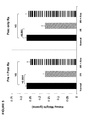
- FIG. 5 shows renal weights two weeks after the induction of unilateral ischemic injury+/ ⁇ atrasentan treatment in the pre+post (left panel), or just the post ischemic period (right panel).
- FIG. 6 shows photographs of the kidneys whose weights are shown in FIG. 5 .
- FIG. 7 depicts renal proliferation following the unilateral ischemic protocol with and without atrasentan treatment.
- the left hand panel depicts the weights of the contralateral (non ischemic) kidneys from the unilateral ischemic injury experiments ⁇ atrasentan treatment.
- the right hand panel depicts KI-67 staining of a normal kidney (A), a 2 week (left) post ischemic kidney (B), and a 2 week left post ischemic kidney with pre+post atrasentan treatment (C).
- FIG. 8 illustrates the effect of atrasentan on blood urea nitrogen (BUN) and plasma creatinine twenty-four hours after ischemia of different durations.
- FIG. 9 illustrates the effect of atrasentan on NGAL twenty-four hours after ischemia of different durations.
- the term “about” is used synonymously with the term “approximately.”
- the use of the term “about” indicates that values slightly outside the cited values, namely, plus or minus 10%. Such dosages are thus encompassed by the scope of the claims reciting the terms “about” and “approximately.”
- administer refers to any manner of providing a drug (such as astrasentan or a pharmaceutically acceptable salt thereof) to a subject or patient.
- routes of administration can be accomplished through any means known by those skilled in the art. Such means include, but are not limited to, oral, buccal, intravenous, subcutaneous, intramuscular, transdermal, by inhalation and the like.
- active agent refers to an agent that achieves a desired biological effect or a pharmaceutically acceptable salt thereof.
- active agent and “drug” are used interchangeably herein.
- the solid state form of the active agent used in preparing the dosage forms of the present disclosure is not critical.
- active agent used in preparing the dosage forms of the present disclosure can be amorphous or crystalline.
- the final dosage form contains at least a detectable amount of crystalline active agent.
- the crystalline nature of the active agent can be detected using powder X-ray diffraction analysis, by differential scanning calorimetry or any other techniques known in the art.
- ABT-627 refers to (2R,3R,4S)-4-(1,3-benzodioxol-5-yl)-1-[2-(dibutylamino)-2-oxoethyl]-2-(4-methoxyphenyl)pyrrolidine-3-carboxylic acid having the structure shown below:
- BQ-788 refers to N-cis-2,6-dimethylpiperidinocarbonyl-L-gamma-methylleucyl-D-1-methoxycarbonyltryptophanyl-D-norleucine having the structure shown below:
- an “effective amount” or a “therapeutically effective amount” of an active agent is meant a nontoxic but sufficient amount of the active agent to provide the desired effect.
- the amount of active agent that is “effective” will vary from subject to subject, depending on the age and general condition of the individual, the particular active agent or agents, and the like. Thus, it is not always possible to specify an exact “effective amount.” However, an appropriate “effective amount” in any individual case may be determined by using routine experimentation.
- the ETA receptor antagonist may be administered upon detection of renal ischemia or hypoxia or within 24 hours after such detection. Alternatively or additionally, the ETA receptor antagonist may be administered for up to one week, or two weeks, or four weeks, or eight weeks, or longer. Or the ETA receptor antagonist may be administered for as long as the acute kidney injury exists or until the injury is resolved or until ischemia or hypoxia is no longer detected.
- endothelin subtype A receptor antagonist or “ETA receptor antagonist” or “ETA receptor inhibitor” refers to any compound that inhibits the effect of ET-1 signaling through the endothelin subtype A receptor.
- ETA receptor antagonists include, but are not limited to, ambrisentan, atrasentan, avosentan, BMS 193884, BQ-123, CI-1020, clazosentan, darusentan, edonentan, S-0139, SB-209670, sitaxsentan, TA-0201, tarasentan, TBC 3711, tezosentan, YM-598, ZD-1611, ZD-4054, and salts, esters, prodrugs, metabolites, tautomers, racemates and enantiomers thereof.
- endothelin antagonist or “ET-1 antagonist” or “ET-1 inhibitor” refers to any compound that inhibits ET-1.
- Acute kidney injury or “acute kidney failure” is typically identified by a rapid deterioration in renal function sufficient to result in the accumulation of nitrogenous wastes in the body (see, e.g., Anderson and Schrier (1994), in Harrison's Principles of Internal Medicine, 13th edition, Isselbacher et al., eds., McGraw Hill Text, New York). Rates of increase in BUN of at least 4 to 8 mmol/L/day (10 to 20 mg/dL/day), and rates of increase of serum creatinine of at least 40 to 80 ⁇ mol/L/day (0.5 to 1.0 mg/dL/day), are typical in acute renal failure. Urinary samples also may contain tubular injury residue in patients suffering from acute kidney injury.
- rates of increase in BUN may exceed 100/mg/dL/day. Rates of increase in BUN or serum creatinine may be determined by serial blood tests and, preferably, at least two blood tests are conducted over a period of between 6 and 72 hours or, more preferably, 12 and 24 hours.
- acute kidney injury is intended to embrace both syndromes. Acute kidney injury is regularly identified by clinicians, as discussed above.
- ischemia-induced kidney injury refers to renal injury due to one or more identified occurrences of renal ischemia or ischemia and reperfusion.
- ischemic kidney injury may be used interchangeably herein.
- hypoxia-induced kidney injury refers to renal injury due to ischemia, ischemia and reperfusion, and/or hypoxia, even if the hypoxia or apoxia is due to causes unknown or other than ischemia.
- Ischemia-induced kidney injury can be identified by clinicians, for example by recognizing ischemic conditions or by reference to certain intrinsic renal causes of acute kidney injury.
- Hypoxia-induced kidney injury can be identified by clinicians, for example by recognizing hypoxic conditions or by reference to certain intrinsic renal causes of acute kidney injury. The extent of the renal injury would be clinically assessed by tracking increases in BUN and serum creatinine.
- markers of inflammation refers to peptides and chemicals produced by a patient's body when some aspect of the patient's body is in an inflammatory state.
- exemplary markers of inflammation include triglycerides, non-high-density lipoprotein (HDL), apoprotein B, fibrinogen, soluble tumor necrosis factor receptor (sTNFR-2), monocyte chemoattractant protein-1, interferon-gamma-inducible protein (IP-10), macrophage inflammatory protein-1delta, vascular cell adhesion molecule-1 (VCAM), serum amyloid A, heme oxygenase 1, soluble intercellular molecule type 1 (sICAM-1), C-reactive protein (CRP) and members of the interleukin family.
- HDL non-high-density lipoprotein
- sTNFR-2 soluble tumor necrosis factor receptor
- IP-10 interferon-gamma-inducible protein
- VCAM vascular cell adhesion molecule-1
- serum amyloid A heme oxygen
- CRP C - Reactive Protein
- NEJM NEJM
- AKI indicator refers to a clinical measurement that reflects the presence of acute kidney injury.
- Contemplated AKI indicators include certain laboratory parameters, including but not limited to, GFR, serum cystatin C, urinary albumin excretion, BUN, serum creatinine, urinary NGAL, urinary KIM-1, markers of inflammation and certain morphological markers, including, but not limited to, kidney mass and urine samples showing renal tubular cells and cellular residue.
- GFR glomerular filtration rate
- MDRD Modification of Diet in Renal Disease
- Bedside Schwartz equation the Cockcroft-Gault formula, or any other clinically acceptable formula or equation.
- Clinical acute renal failure refers to a decrease in renal function that is or can be detected clinically, such as by a general practitioner or a nephrologist.
- Clinical acute renal failure may be assessed by decreased glomerular filtration rate, decrease in or absence of urine production, increased waste products (especially creatinine or urea) in the blood, hematuria (blood loss in the urine), proteinuria (protein loss in the urine), or other clinical indicators.
- intracranial renal causes of acute kidney injury include:
- pharmaceutically acceptable such as in the recitation of a “pharmaceutically acceptable excipient,” or a “pharmaceutically acceptable additive,” is meant a material that is not biologically or otherwise undesirable, i.e., the material may be incorporated into a pharmaceutical composition administered to a patient without causing any undesirable biological effects.
- RAAS inhibitor refers to any compound that inhibits one or more elements of the renin-angiotensin-aldosterone system (RAAS).
- RAAS inhibitors include ACE inhibitors, ARBs, rennin inhibitors, aldosterone antagonists and others.
- subject refers to an animal.
- the animal is a mammal, including a human or non-human, preferably a human subject.
- patient and subject may be used interchangeably herein.
- a diagnosis that a subject is in acute renal failure, or at risk of entering acute renal failure is made on the basis of serial blood tests measuring, among other factors, the circulating levels of serum creatinine and blood urea nitrogen.
- serial blood tests may be taken every few hours immediately upon admittance of an undiagnosed patient presenting with symptoms of acute renal failure. More typically, however, consecutive serial blood tests are separated by a period of at least 6 hours, not more than 72 hours, and preferably 12-24 hours. On the basis of two or more blood tests within a 24 or 72 hour period, it is possible to calculate a rate of increase of serum creatinine or BUN. Additionally, or alternatively, diagnosis can be made by assessing the presence of tubule injury residue in urinary samples.
- Subjects possessing a single kidney may be considered to be at increased risk of acute renal failure. This is particularly true for those subjects in which one kidney has been lost due to a disease or condition which may afflict the remaining kidney.
- subjects which are already recipients of a renal transplant, or which are receiving chronic dialysis e.g., chronic hemodialysis or continuous ambulatory peritoneal dialysis
- Other groups that are at an increased risk of developing acute kidney injury include those with chronic kidney disease, diabetes mellitus, and the elderly. Therefore, for these subjects, the clinical indications discussed above may need to be more carefully monitored, and earlier or more aggressive intervention with renal therapeutic agent treatment may be advisable.
- treating and “treatment” refer to reduction in severity and/or frequency of symptoms, elimination of symptoms and/or underlying cause, prevention of the occurrence of symptoms and/or their underlying cause, and improvement or remediation of damage.
- “treating” a patient involves prevention of a particular disorder or adverse physiological event in a susceptible individual as well as treatment of a clinically symptomatic individual by inhibiting or causing regression of a disorder or disease.
- the present methods are based, in part, upon the surprising discovery that therapeutic use of ETA receptor antagonists to subjects suffering from acute kidney injury, including after the onset or diagnosis of acute kidney injury, can reduce mortality and/or morbidity rates, and prevent, inhibit, delay, or alleviate the permanent and/or progressive loss of renal function associated with acute kidney injury.
- the kidney injury can be hypoxia-induced kidney injury or, more specifically, an ischemia-induced kidney injury.
- kidney loss prevents or reduces physical kidney loss as well as functional renal loss associated with ischemic-induced and/or hypoxia-induced kidney injury.
- kidney loss can be evaluated by assessing kidney mass or kidney volume using ultrasound technology or other appropriate visualization technology.
- renoprotective effects from the disclosed methods may occur before, during and/or after the initial ischemic/reperfusion injury phase.
- the ETA receptor antagonist is administered to accelerate renal recovery after AKI.
- renoprotective effects from the disclosed methods result in a reduction in markers of inflammation and certain laboratory parameters that indicate kidney stress and dysfuntion (e.g., serum creatinine, GFR, BUN). Therefore, in some embodiments of the present invention, measurements of these markers of inflammation and/or certain laboratory parameters at some time after the administration of an ETA receptor antagonist show a reduction over time, which would correlate to an increase in renal function.
- kidney stress and dysfuntion e.g., serum creatinine, GFR, BUN
- renoprotection afforded by ETA receptor antagonism is not clear. However, without being bound to any particular theory or mechanism, it is believed that the renoprotection may be driven by one or more of the following: direct ETA antagonism, interactions with nitric oxide signaling, the angiotensin II system, and TGFbeta-mediated fibrosis. If persistent tissue ischemia is, in fact, a mediator of progressive renal damage, as suggested above, then ET-1-mediated renal vasoconstriction could potentially play an important pathogenic role. It does appear that some of the renoprotective efficacy observed in ETA receptor antagonism is due to renal microvascular dilation and preservation of renal tubular cell structure and function. Therefore, in some embodiments of the present methods, a combination of an ETA receptor antagonist and RAAS inhibitor may be administered to provide added renoprotection over the treatment of only an ETA receptor antagonist or RAAS inhibitor alone.
- mice Male 30-45 gram CD-1 mice, obtained from Charles River Laboratories, Wilmington, Mass. They were housed under routine vivarium conditions with free food and water access. Surgeries were conducted under deep pentobarbital anesthesia (40-50 mg/Kg IP). Post surgical analgesia was provided with buprenorphine (0.1 mg/Kg IP) at the completion of surgery. All procedures were approved by the institution's IACUC, in accordance with NIH guidelines.
- the scores were graded on a semiquantitative scale of 1+ to 4+, or least to most severe renal injury observed (based on the extent of proximal tubule necrosis). Continuous variable results were compared by Student's T test. The histologic data were judged by Wilcoxon rank sum test. Significance was judged by a p value of ⁇ 0.05.
- ET-1 and ETA/ETB receptor expression were quantified following ischemic renal injury, and ET-1 gene chromatin remodeling and RNA polymerase II (Pol II) binding were evaluated.
- Ten mice were subjected to a midline laparotomy, and the left renal pedicles were exposed and occluded ⁇ 30 min using atraumatic microvascular clamps. Uniform ischemia was confirmed by the development of total kidney cyanosis (indicating hypoxia). Body temperature was monitored with an intra-abdominal thermometer and maintained at 37° C. with an external heating source. At the completion of the ischemic period, the vascular clamp was removed, and uniform reperfusion was confirmed by loss of kidney cyanosis.
- mice were then allowed to recover from anesthesia. Ten additional mice, subjected to the same surgical procedure, but not to renal pedicle occlusion, served as sham-operated controls.
- a blood sample was obtained from the inferior vena cava and both kidneys were resected.
- the kidneys were iced, and the renal cortical samples were obtained with a razor blade, and they were extracted for both total RNA (RNeasy; Qiagen), and total protein.
- the RNA samples were used to determine the mRNAs for ET-1, and the for ETA and ETB receptors.
- ET-1 protein concentrations in renal cortical extracts and plasma were determined by ELISA.
- renal cortical chromatin extracts were prepared from the following kidneys: kidneys from three sham operated mice (2 weeks post surgery); three 2 week post-ischemic kidneys; and the three corresponding contralateral kidneys.
- RNA polymerase II (Pol II) binding and histone modifications at the ET-1 gene following ischemic renal injury
- histone modifying enzyme systems can be activated and induce chromatin remodeling at pro-inflammatory genes. These changes include histone H3 trimethylation, acetylation, and histone H2A.Z exchange.
- Naito M, et al., Endotoxin mediates recruitment of RNA polymerase II to target genes in acute renal failure . J Am Soc Nephrol 19: 1321-1330, 2008; Naito M, et al., BRG1 increases transcription of proinflammatory genes in renal ischemia.
- ChIP assay was applied to two week post ischemic kidney samples, and significant increases in H3K4m3, H3K9/14 acetylation and H2A.Z levels were observed.
- the functional significance of these changes was implied by parallel increases in binding of Pol II (the enzyme that drives transcription) to the ET-1 gene. While it cannot be absolutely assumed that there are cause and effect relationships between these histone changes and gene transcription rates, the fact that they are known to be gene activating events in a variety of biologic systems certainly suggest that this is the case.
- Renal cortical ETA and ETB receptor mRNA expression were also assessed post ischemia. As shown in FIG. 4 , a 3 fold increase in ETA receptor mRNA was apparent by 24 hours post ischemia. By 2 weeks post ischemia, a further 8 fold increase in ETA receptor mRNA was observed. Thus, compared to basal values, ETA receptor mRNA levels rose ⁇ 25 fold over the course of the experiment. In sharp contrast, no increase in ETB receptor mRNA was observed at 24 hours post ischemia. By two weeks post ischemia, a significant ETB receptor mRNA increase was observed, but it was quantitatively trivial compare to the ETA mRNA values (25 ⁇ vs. 2 ⁇ , respectively).
- mice were subjected to a unilateral ischemia/reperfusion (I/R) protocol as described in Example 1.
- I/R ischemia/reperfusion
- mice received the highly potent and specific ETA receptor antagonist, atrasentan.
- Atrasentan was administered in the drinking water (25 ⁇ g/mL; designed to equate with a dose of ⁇ 5 mg/Kg/day).
- Atrasentan dosing was started one day before surgery, and continued throughout the remainder of the experiment.
- Fresh atrasentan was provided ⁇ 2-3 ⁇ per week for two weeks. The remaining nine mice received only free food and water access, serving as controls.
- mice Upon completion of a two week post ischemic recovery period, the mice were re-anesthetized with pentobarbital, the abdominal incision was re-opened, a terminal blood sample was obtained from the inferior vena cava for BUN and ET-1 analysis, and then the left (post ischemic) kidneys and the right (contralateral) kidneys were removed, and weighed. The degree of post-ischemic loss of renal mass was assessed by comparing the weights of kidneys from sham operated mice, control post-ischemic mice, and post ischemic mice that had received atrasentan treatment. Finally, frontal sections of post-ischemic kidneys were taken from five control mice and five atrasentan-treated mice, fixed in 10% buffered formalin, and used for subsequent histochemical analyses.
- the unilateral I/R injury protocol induced an approximate 45% reduction in renal mass (renal weight), in comparison to the weights of kidneys extracted from sham operated mice (p ⁇ 0.0001). Sham surgery did not independently affect renal weight, compared to those obtained from normal mice (not shown).
- Administration of atrasentan started before renal ischemia and continued throughout the two week post-ischemic period, conferred marked protection, as judged by the fact that the post ischemic kidney weights did not significantly differ from that of normal kidneys.
- FIG. 6 A graphic depiction of this result is presented in FIG. 6 : the unilateral ischemic/reperfusion (I/R) kidney (far left) was markedly reduced in size and volume, compared to a normal kidney (far right).
- the pre- and post-ischemia atrasentan treated kidney manifested a near normal size.
- the administration of atrasentan before and after renal ischemia can reduce the loss of kidney volume or mass.
- ischemia without drug treatment caused a 40% reduction in renal weight.
- Post ischemic atrasentan completely blocked this loss of renal mass, thereby recapitulating the protection seen in the pre+post treatment experiment.
- administering atrasentan to a subject suffering ischemic kidney injury at 24 hours or more after that injury is beneficial in providing renal protection from the effects of ischemia and hypoxia.
- the contralateral kidneys manifested an approximate 25% increase in renal size, compared to normal kidneys. This is consistent with renal hypertrophy in response to a reduction in post-ischemic kidney function, as previously described.
- Zager R A, et al. Acute unilateral ischemic renal injury induces progressive renal inflammation, lipid accumulation, histone modification, and “end-stage” kidney disease. Am J Physiol 301: F1334-1345, 2011.) Atrasentan had no effect on this hypertrophic response, given that contralateral kidney weights were essentially identical in the no-drug vs. drug treatment groups.
- This example examined renal histology on kidneys from five control mice and five atrasentan-treated mice from Example 2. More particularly, to confirm atrasentan's protective effect against ongoing post ischemic injury, renal histology was examined in kidneys obtained two weeks post ischemia with (combined pre and post) and without atrasentan treatment. Four micron sections were cut and stained with hematoxylin and eosin for overall assessment of the severity of tissue injury. In addition, renal tubular cell proliferation was assessed by immunohistochemical staining for KI-67, a nuclear protein marker of all active cell cycle phases (G1, S, G2, mitosis; but not Go). (Scholzen T, et al., The Ki -67 protein: from the known and the unknown .
- the unilateral ischemia protocol caused marked proximal tubule dropout, extensive interstitial inflammation, ongoing proximal tubule necrosis and extensive intratubular cast formation. These changes were observed throughout the renal cortex and outer medullary stripe. Atrasentan caused a marked reduction in each of these changes. Blinded grading the severity of these changes using a semiquantitative score (1+ to 4+; least to most severe injury observed) revealed a marked diminution in injury scores in the atrasentan group (3.4 ⁇ 0.3 vs 1.7 ⁇ 0.4; p ⁇ 0.01). Thus, the histologic correlated with the preserved renal mass/renal weight with atrasentan treatment.
- KI-67 immunohistochemical staining demonstrated a marked increase in renal tubular cell proliferation in all two week post ischemic kidneys, compared to normal kidneys (p ⁇ 0.001).
- the right hand panel depicts KI-67 staining of a normal kidney (A), a two week (left) post ischemic kidney (B), and a two week left post ischemic kidney with pre+post atrasentan treatment (C).
- the post ischemic kidneys manifested a marked increase in nuclear KI-67 staining, compared to that seen in normal kidneys.
- Atrasentan did not appear to alter this proliferative response, as denoted by the frequency of KI-67 nuclear staining (% KI-67 nuclear positivity: normal kidneys, 2.4 ⁇ 0.6%; control ischemia, 11.4 ⁇ 0.8%; ischemia+atrasentan, 10.8 ⁇ 1.2%).
- mice were subjected to the same bilateral ischemia protocols without atrasentan treatment. Atrasentan was continued during the post ischemic period. Twenty four hours later, the mice were re-anesthetized, the abdominal incisions were re-opened, a blood sample was obtained from the inferior vena cava, and the kidneys were resected.
- the severity of injury was assessed at 24 hours post ischemia by BUN and plasma creatinine concentrations. Atrasentan failed to mitigate the severity of AKI with either ischemic challenge. As shown in FIG. 8 , no protection was observed with either the 22.5 min or the 25 min ischemic challenges (which induced moderate and severe azotemia, respectively).
- renal cortical NGAL mRNA levels were also assessed. Measurement of NGAL mRNA represents a more sensitive biomarker of kidney injury than BUN and Plasma Creatinine measured in FIG. 8 and was measured to investigate the effect of atrasentan on the induction phase of ischemic injury. As shown in FIG. 9 , both I/R protocols induced marked NGAL mRNA increases.
- proximal tubule (HK-2) cells derived from normal human kidney were incubated in T24 well plates in keratinocyte serum free medium, as previously described in detail.
- HK-2 cells cultured proximal tubule cells derived from normal human kidney were incubated in T24 well plates in keratinocyte serum free medium, as previously described in detail.
- Iwata M, et al. Sphingosine: a mediator of acute renal tubular injury and subsequent cytoresistance. Proc Natl Acad Sci, USA 9 2: 8970-8974, 1995
- Iwata M et al., Protein synthesis inhibition induces cytoresistance in cultured human proximal tubular ( HK -2) cells .
- ATP depletion antimycin/deoxyglucose/Ca ionophore; “CAD”) addition to HK-2 cells induced modest tubular cell death, raising LDH release from a control value of 8.6 ⁇ 0.1% to 22.8 ⁇ 0.2%.
- Atrasentan did not alter LDH release, either under basal conditions (9.0 ⁇ 0.1%) or with the ATP depletion protocol (23.2 ⁇ 0.3%). This is again consistent with the in vivo data that atrasentan was unable to block an acute ATP depletion injury phase.
Landscapes
- Health & Medical Sciences (AREA)
- Life Sciences & Earth Sciences (AREA)
- Chemical & Material Sciences (AREA)
- General Health & Medical Sciences (AREA)
- Engineering & Computer Science (AREA)
- Medicinal Chemistry (AREA)
- Animal Behavior & Ethology (AREA)
- Veterinary Medicine (AREA)
- Public Health (AREA)
- Pharmacology & Pharmacy (AREA)
- Organic Chemistry (AREA)
- Proteomics, Peptides & Aminoacids (AREA)
- Epidemiology (AREA)
- Analytical Chemistry (AREA)
- Molecular Biology (AREA)
- Immunology (AREA)
- Urology & Nephrology (AREA)
- Bioinformatics & Cheminformatics (AREA)
- Biochemistry (AREA)
- Physics & Mathematics (AREA)
- Hematology (AREA)
- Biotechnology (AREA)
- Genetics & Genomics (AREA)
- Microbiology (AREA)
- Biomedical Technology (AREA)
- Wood Science & Technology (AREA)
- Zoology (AREA)
- Pathology (AREA)
- General Chemical & Material Sciences (AREA)
- Nuclear Medicine, Radiotherapy & Molecular Imaging (AREA)
- Chemical Kinetics & Catalysis (AREA)
- General Engineering & Computer Science (AREA)
- Cell Biology (AREA)
- Biophysics (AREA)
- Food Science & Technology (AREA)
- General Physics & Mathematics (AREA)
- Medicines That Contain Protein Lipid Enzymes And Other Medicines (AREA)
- Measuring Or Testing Involving Enzymes Or Micro-Organisms (AREA)
- Pharmaceuticals Containing Other Organic And Inorganic Compounds (AREA)
Priority Applications (1)
| Application Number | Priority Date | Filing Date | Title |
|---|---|---|---|
| US14/772,621 US20160015701A1 (en) | 2013-03-08 | 2014-03-10 | Methods of Treating Acute Kidney Injury |
Applications Claiming Priority (3)
| Application Number | Priority Date | Filing Date | Title |
|---|---|---|---|
| US201361775174P | 2013-03-08 | 2013-03-08 | |
| US14/772,621 US20160015701A1 (en) | 2013-03-08 | 2014-03-10 | Methods of Treating Acute Kidney Injury |
| PCT/US2014/022688 WO2014138738A1 (en) | 2013-03-08 | 2014-03-10 | Methods of treating acute kidney injury |
Publications (1)
| Publication Number | Publication Date |
|---|---|
| US20160015701A1 true US20160015701A1 (en) | 2016-01-21 |
Family
ID=51492045
Family Applications (1)
| Application Number | Title | Priority Date | Filing Date |
|---|---|---|---|
| US14/772,621 Abandoned US20160015701A1 (en) | 2013-03-08 | 2014-03-10 | Methods of Treating Acute Kidney Injury |
Country Status (10)
| Country | Link |
|---|---|
| US (1) | US20160015701A1 (enExample) |
| EP (1) | EP2964671A4 (enExample) |
| JP (1) | JP2016512201A (enExample) |
| CN (1) | CN105324395A (enExample) |
| AU (1) | AU2014225320A1 (enExample) |
| BR (1) | BR112015020788A2 (enExample) |
| CA (1) | CA2901922A1 (enExample) |
| HK (1) | HK1220209A1 (enExample) |
| MX (1) | MX2015011730A (enExample) |
| WO (1) | WO2014138738A1 (enExample) |
Cited By (5)
| Publication number | Priority date | Publication date | Assignee | Title |
|---|---|---|---|---|
| WO2020006571A1 (en) * | 2018-06-29 | 2020-01-02 | pulseData Inc. | Machine learning systems and methods for predicting risk of renal function decline |
| WO2021126977A1 (en) * | 2019-12-17 | 2021-06-24 | Chinook Therapeutics, Inc. | Methods of treating iga nephropathy with atrasentan |
| WO2021207723A3 (en) * | 2020-04-10 | 2021-12-16 | Chinook Therapeutics, Inc. | Methods of treating diabetic kidney disease |
| US20220304979A1 (en) * | 2019-12-17 | 2022-09-29 | Chinook Therapeutics, Inc. | Methods of reducing disease flares |
| TWI914232B (zh) | 2019-12-17 | 2026-02-01 | 美商奇努克醫療股份有限公司 | 用阿曲生坦(ATRASENTAN)治療IgA腎病的方法 |
Families Citing this family (9)
| Publication number | Priority date | Publication date | Assignee | Title |
|---|---|---|---|---|
| CA2909871A1 (en) | 2013-04-30 | 2014-11-06 | Abbvie Inc. | Methods for improving lipid profiles using atrasentan |
| MX2017005884A (es) * | 2014-11-07 | 2017-06-26 | Abbvie Inc | Metodos para el tratamiento de la erc, utilizando predictores de la retencion de liquidos. |
| EP3235496A1 (en) | 2016-04-19 | 2017-10-25 | Noorik Biopharmaceuticals AG | Treatment of acute renal failure |
| WO2018067857A1 (en) * | 2016-10-05 | 2018-04-12 | Mitobridge, Inc. | Methods of treating acute kidney injury |
| KR102177349B1 (ko) * | 2018-02-13 | 2020-11-12 | 서울대학교병원 | 체세포 복제 효율 증진용 조성물 |
| WO2021091791A1 (en) * | 2019-11-06 | 2021-05-14 | Northwestern University | Inhibition of the ve-ptp phosphatase protects the kidney from ischemia-reperfusion injury |
| CN113209111A (zh) * | 2021-05-14 | 2021-08-06 | 上海海糖生物医药科技有限公司 | 一种褐藻寡糖的应用 |
| CN115005157B (zh) * | 2022-06-02 | 2023-12-26 | 上海交通大学医学院附属新华医院 | 一种低氧致急性肾损伤动物模型的构建方法 |
| CN116731117B (zh) * | 2023-06-19 | 2024-05-10 | 武汉大学 | Kim-1靶向性多肽及其在急性肾损伤中肾靶向性的应用 |
Family Cites Families (4)
| Publication number | Priority date | Publication date | Assignee | Title |
|---|---|---|---|---|
| US5273961A (en) * | 1992-09-22 | 1993-12-28 | Genentech, Inc. | Method of prophylaxis of acute renal failure |
| CA2563996A1 (en) * | 2004-04-23 | 2005-11-10 | Renal Diagnostic, Inc. | An automated non- invasive real-time acute renal failure detection system |
| US20060205733A1 (en) * | 2004-08-26 | 2006-09-14 | Encysive Pharmaceuticals | Endothelin a receptor antagonists in combination with phosphodiesterase 5 inhibitors and uses thereof |
| US9255931B2 (en) * | 2010-06-24 | 2016-02-09 | Morehouse School Of Medicine | Method and compositions for the treatment and detection of endothelin-1 related kidney diseases |
-
2014
- 2014-03-10 WO PCT/US2014/022688 patent/WO2014138738A1/en not_active Ceased
- 2014-03-10 BR BR112015020788A patent/BR112015020788A2/pt not_active IP Right Cessation
- 2014-03-10 AU AU2014225320A patent/AU2014225320A1/en not_active Abandoned
- 2014-03-10 HK HK16108216.3A patent/HK1220209A1/zh unknown
- 2014-03-10 CA CA2901922A patent/CA2901922A1/en not_active Abandoned
- 2014-03-10 CN CN201480011012.2A patent/CN105324395A/zh active Pending
- 2014-03-10 US US14/772,621 patent/US20160015701A1/en not_active Abandoned
- 2014-03-10 JP JP2015561753A patent/JP2016512201A/ja active Pending
- 2014-03-10 EP EP14760019.1A patent/EP2964671A4/en not_active Withdrawn
- 2014-03-10 MX MX2015011730A patent/MX2015011730A/es unknown
Non-Patent Citations (2)
| Title |
|---|
| Kuro et al. Jpn. J. Pharmacology, 2000, Vol. 82, pgs. 307-316. * |
| Legrand et al. Mol. Med., 2008, Vol. 14, Iss. 7-8, pgs. 502-516. * |
Cited By (14)
| Publication number | Priority date | Publication date | Assignee | Title |
|---|---|---|---|---|
| US10978176B2 (en) | 2018-06-29 | 2021-04-13 | pulseData Inc. | Machine learning systems and methods for predicting risk of renal function decline |
| WO2020006571A1 (en) * | 2018-06-29 | 2020-01-02 | pulseData Inc. | Machine learning systems and methods for predicting risk of renal function decline |
| US12521369B2 (en) | 2019-12-17 | 2026-01-13 | Chinook Therapeutics, Inc. | Methods of improving renal function |
| WO2021126977A1 (en) * | 2019-12-17 | 2021-06-24 | Chinook Therapeutics, Inc. | Methods of treating iga nephropathy with atrasentan |
| US20220304979A1 (en) * | 2019-12-17 | 2022-09-29 | Chinook Therapeutics, Inc. | Methods of reducing disease flares |
| US11491137B2 (en) | 2019-12-17 | 2022-11-08 | Chinook Therapeutics, Inc. | Methods of improving renal function |
| TWI914232B (zh) | 2019-12-17 | 2026-02-01 | 美商奇努克醫療股份有限公司 | 用阿曲生坦(ATRASENTAN)治療IgA腎病的方法 |
| US11998526B2 (en) | 2019-12-17 | 2024-06-04 | Chinook Therapeutics, Inc. | Methods of improving renal function |
| US12121509B2 (en) | 2019-12-17 | 2024-10-22 | Chinook Therapeutics, Inc. | Methods of improving renal function |
| TWI875895B (zh) * | 2019-12-17 | 2025-03-11 | 美商奇努克醫療股份有限公司 | 用阿曲生坦(ATRASENTAN)治療IgA腎病的方法 |
| US12370174B2 (en) | 2019-12-17 | 2025-07-29 | Chinook Therapeutics, Inc. | Methods of improving renal function |
| WO2021207723A3 (en) * | 2020-04-10 | 2021-12-16 | Chinook Therapeutics, Inc. | Methods of treating diabetic kidney disease |
| CN115768421A (zh) * | 2020-04-10 | 2023-03-07 | 切诺克治疗公司 | 治疗糖尿病肾病的方法 |
| US12582631B2 (en) | 2022-08-16 | 2026-03-24 | Chinook Therapeutics, Inc. | Methods of improving renal function |
Also Published As
| Publication number | Publication date |
|---|---|
| AU2014225320A1 (en) | 2015-08-06 |
| BR112015020788A2 (pt) | 2017-10-10 |
| EP2964671A1 (en) | 2016-01-13 |
| WO2014138738A1 (en) | 2014-09-12 |
| WO2014138738A8 (en) | 2015-12-30 |
| EP2964671A4 (en) | 2016-08-31 |
| CA2901922A1 (en) | 2014-09-12 |
| CN105324395A (zh) | 2016-02-10 |
| JP2016512201A (ja) | 2016-04-25 |
| HK1220209A1 (zh) | 2017-04-28 |
| MX2015011730A (es) | 2016-04-04 |
Similar Documents
| Publication | Publication Date | Title |
|---|---|---|
| US20160015701A1 (en) | Methods of Treating Acute Kidney Injury | |
| Calvin et al. | Contrast-induced acute kidney injury and diabetic nephropathy | |
| Bachorzewska-Gajewska et al. | NGAL (neutrophil gelatinase-associated lipocalin) and L-FABP after percutaneous coronary interventions due to unstable angina in patients with normal serum creatinine | |
| Heering et al. | Cyclosporine A and chlorambucil in the treatment of idiopathic focal segmental glomerulosclerosis | |
| Worcester et al. | A test of the hypothesis that oxalate secretion produces proximal tubule crystallization in primary hyperoxaluria type I | |
| TWI900606B (zh) | 用於治療內皮素相關疾病之齊泊騰坦和達格列淨的組合 | |
| KR20190113955A (ko) | 장-외 증상을 갖는 염증성 장질환의 치료를 위한 화합물 및 방법 | |
| Nakashima et al. | Clinical evaluation of tocilizumab for patients with active rheumatoid arthritis refractory to anti-TNF biologics: tocilizumab in combination with methotrexate | |
| Kunzendorf et al. | The Th1-Th2 paradigm in 1998: law of nature or rule with exceptions. | |
| Joannidis | Drug-induced renal failure in the ICU | |
| CN103429242A (zh) | 用于预防和治疗心脏肥大的方法和组合物 | |
| Rusai et al. | Immunosuppression with 4SC-101, a novel inhibitor of dihydroorotate dehydrogenase, in a rat model of renal transplantation | |
| KR20180121539A (ko) | 급성 신부전의 치료에 사용하기 위한 암브리센탄 | |
| Almeida et al. | Successful use of mycophenolate mofetil as adjunct to prednisolone for treatment of acute kidney injury secondary to human serum albumin administration in a dog | |
| Koç et al. | Severe hyperkalemia in two renal transplant recipients treated with standard dose of trimethoprim-sulfamethoxazole | |
| JP2007512381A (ja) | 医薬的組み合わせ | |
| JP6679096B2 (ja) | Rageアプタマーおよびその用途 | |
| WO2022105870A1 (en) | Methods of treating systemic lupus erythematosus using btk inhibitors | |
| Ma et al. | Pentoxifylline protects against loss of function and renal interstitial fibrosis in chronic experimental partial ureteral obstruction | |
| TWI821836B (zh) | 一種用於緩解或預防慢性腎臟病進展的藥物組合物及其用途 | |
| Joy et al. | Miscellaneous Etiologies of Acute Kidney Injury | |
| Antoun et al. | Sodium-Glucose Cotransporter 2 Inhibitors in Autosomal Dominant Polycystic Kidney Disease: Mechanistic Insights and Therapeutic Promise | |
| Pereira et al. | Hemodynamics and renal function during administration of low-dose dopamine in severely ill patients | |
| Hwang et al. | Protective Effect of Resveratrol on Glycocalyx Loss due to Endothelial Cell Dysfunction in Renal Aging: FR-PO1036 | |
| Sandery | Acute Kidney Injury and Glomerular Hyperfiltration in Children |
Legal Events
| Date | Code | Title | Description |
|---|---|---|---|
| AS | Assignment |
Owner name: ABBIVE INC., ILLINOIS Free format text: ASSIGNMENT OF ASSIGNORS INTEREST;ASSIGNOR:ANDRESS, DENNIS;REEL/FRAME:037677/0574 Effective date: 20150918 |
|
| AS | Assignment |
Owner name: NATIONAL INSTITUTES OF HEALTH (NIH), U.S. DEPT. OF Free format text: CONFIRMATORY LICENSE;ASSIGNOR:FRED HUTCHINSON CANCER RESEARCH CENTER;REEL/FRAME:041300/0332 Effective date: 20170106 |
|
| AS | Assignment |
Owner name: FRED HUTCHINSON CANCER RESEARCH CENTER, WASHINGTON Free format text: ASSIGNMENT OF ASSIGNORS INTEREST;ASSIGNOR:ZAGER, RICHARD A.;REEL/FRAME:044832/0884 Effective date: 20171220 |
|
| STCB | Information on status: application discontinuation |
Free format text: ABANDONED -- FAILURE TO RESPOND TO AN OFFICE ACTION |