US12129496B2 - Haplotype-based treatment of RP1 associated retinal degenerations - Google Patents
Haplotype-based treatment of RP1 associated retinal degenerations Download PDFInfo
- Publication number
- US12129496B2 US12129496B2 US17/175,234 US202117175234A US12129496B2 US 12129496 B2 US12129496 B2 US 12129496B2 US 202117175234 A US202117175234 A US 202117175234A US 12129496 B2 US12129496 B2 US 12129496B2
- Authority
- US
- United States
- Prior art keywords
- grna
- intron
- exon
- cas9
- seq
- Prior art date
- Legal status (The legal status is an assumption and is not a legal conclusion. Google has not performed a legal analysis and makes no representation as to the accuracy of the status listed.)
- Active, expires
Links
Images


Classifications
-
- C—CHEMISTRY; METALLURGY
- C12—BIOCHEMISTRY; BEER; SPIRITS; WINE; VINEGAR; MICROBIOLOGY; ENZYMOLOGY; MUTATION OR GENETIC ENGINEERING
- C12N—MICROORGANISMS OR ENZYMES; COMPOSITIONS THEREOF; PROPAGATING, PRESERVING, OR MAINTAINING MICROORGANISMS; MUTATION OR GENETIC ENGINEERING; CULTURE MEDIA
- C12N9/00—Enzymes; Proenzymes; Compositions thereof; Processes for preparing, activating, inhibiting, separating or purifying enzymes
- C12N9/14—Hydrolases (3)
- C12N9/16—Hydrolases (3) acting on ester bonds (3.1)
- C12N9/22—Ribonucleases [RNase]; Deoxyribonucleases [DNase]
-
- A—HUMAN NECESSITIES
- A61—MEDICAL OR VETERINARY SCIENCE; HYGIENE
- A61K—PREPARATIONS FOR MEDICAL, DENTAL OR TOILETRY PURPOSES
- A61K38/00—Medicinal preparations containing peptides
- A61K38/16—Peptides having more than 20 amino acids; Gastrins; Somatostatins; Melanotropins; Derivatives thereof
- A61K38/164—Peptides having more than 20 amino acids; Gastrins; Somatostatins; Melanotropins; Derivatives thereof from bacteria
-
- A—HUMAN NECESSITIES
- A61—MEDICAL OR VETERINARY SCIENCE; HYGIENE
- A61K—PREPARATIONS FOR MEDICAL, DENTAL OR TOILETRY PURPOSES
- A61K9/00—Medicinal preparations characterised by special physical form
- A61K9/0012—Galenical forms characterised by the site of application
- A61K9/0048—Eye, e.g. artificial tears
-
- C—CHEMISTRY; METALLURGY
- C12—BIOCHEMISTRY; BEER; SPIRITS; WINE; VINEGAR; MICROBIOLOGY; ENZYMOLOGY; MUTATION OR GENETIC ENGINEERING
- C12N—MICROORGANISMS OR ENZYMES; COMPOSITIONS THEREOF; PROPAGATING, PRESERVING, OR MAINTAINING MICROORGANISMS; MUTATION OR GENETIC ENGINEERING; CULTURE MEDIA
- C12N15/00—Mutation or genetic engineering; DNA or RNA concerning genetic engineering, vectors, e.g. plasmids, or their isolation, preparation or purification; Use of hosts therefor
- C12N15/09—Recombinant DNA-technology
- C12N15/11—DNA or RNA fragments; Modified forms thereof; Non-coding nucleic acids having a biological activity
-
- C—CHEMISTRY; METALLURGY
- C12—BIOCHEMISTRY; BEER; SPIRITS; WINE; VINEGAR; MICROBIOLOGY; ENZYMOLOGY; MUTATION OR GENETIC ENGINEERING
- C12N—MICROORGANISMS OR ENZYMES; COMPOSITIONS THEREOF; PROPAGATING, PRESERVING, OR MAINTAINING MICROORGANISMS; MUTATION OR GENETIC ENGINEERING; CULTURE MEDIA
- C12N15/00—Mutation or genetic engineering; DNA or RNA concerning genetic engineering, vectors, e.g. plasmids, or their isolation, preparation or purification; Use of hosts therefor
- C12N15/09—Recombinant DNA-technology
- C12N15/87—Introduction of foreign genetic material using processes not otherwise provided for, e.g. co-transformation
- C12N15/90—Stable introduction of foreign DNA into chromosome
- C12N15/902—Stable introduction of foreign DNA into chromosome using homologous recombination
- C12N15/907—Stable introduction of foreign DNA into chromosome using homologous recombination in mammalian cells
-
- A—HUMAN NECESSITIES
- A61—MEDICAL OR VETERINARY SCIENCE; HYGIENE
- A61K—PREPARATIONS FOR MEDICAL, DENTAL OR TOILETRY PURPOSES
- A61K38/00—Medicinal preparations containing peptides
-
- A—HUMAN NECESSITIES
- A61—MEDICAL OR VETERINARY SCIENCE; HYGIENE
- A61K—PREPARATIONS FOR MEDICAL, DENTAL OR TOILETRY PURPOSES
- A61K9/00—Medicinal preparations characterised by special physical form
- A61K9/0012—Galenical forms characterised by the site of application
- A61K9/0019—Injectable compositions; Intramuscular, intravenous, arterial, subcutaneous administration; Compositions to be administered through the skin in an invasive manner
-
- C—CHEMISTRY; METALLURGY
- C12—BIOCHEMISTRY; BEER; SPIRITS; WINE; VINEGAR; MICROBIOLOGY; ENZYMOLOGY; MUTATION OR GENETIC ENGINEERING
- C12N—MICROORGANISMS OR ENZYMES; COMPOSITIONS THEREOF; PROPAGATING, PRESERVING, OR MAINTAINING MICROORGANISMS; MUTATION OR GENETIC ENGINEERING; CULTURE MEDIA
- C12N2310/00—Structure or type of the nucleic acid
- C12N2310/10—Type of nucleic acid
- C12N2310/20—Type of nucleic acid involving clustered regularly interspaced short palindromic repeats [CRISPR]
-
- C—CHEMISTRY; METALLURGY
- C12—BIOCHEMISTRY; BEER; SPIRITS; WINE; VINEGAR; MICROBIOLOGY; ENZYMOLOGY; MUTATION OR GENETIC ENGINEERING
- C12N—MICROORGANISMS OR ENZYMES; COMPOSITIONS THEREOF; PROPAGATING, PRESERVING, OR MAINTAINING MICROORGANISMS; MUTATION OR GENETIC ENGINEERING; CULTURE MEDIA
- C12N2320/00—Applications; Uses
- C12N2320/30—Special therapeutic applications
- C12N2320/34—Allele or polymorphism specific uses
-
- C—CHEMISTRY; METALLURGY
- C12—BIOCHEMISTRY; BEER; SPIRITS; WINE; VINEGAR; MICROBIOLOGY; ENZYMOLOGY; MUTATION OR GENETIC ENGINEERING
- C12N—MICROORGANISMS OR ENZYMES; COMPOSITIONS THEREOF; PROPAGATING, PRESERVING, OR MAINTAINING MICROORGANISMS; MUTATION OR GENETIC ENGINEERING; CULTURE MEDIA
- C12N2800/00—Nucleic acids vectors
- C12N2800/80—Vectors containing sites for inducing double-stranded breaks, e.g. meganuclease restriction sites
Definitions
- RP1 retinitis pigmentosa 1 protein
- AdRP mutation-associated autosomal dominant Retinitis Pigmentosa
- Retinitis pigmentosa is an important cause of vision loss for people of all ages. As a group, these diseases are characterized by the progressive death of the light sensitive photoreceptor cells of the retina, which are required for vision.
- RP is highly heterogeneous genetically, and mutations in the RP genes can be passed from parent to offspring in multiple genetic inheritance modes—autosomal recessive, autosomal dominant, or X-linked.
- mutations in more than 60 genes are known to cause non-syndromic RP, and mutations in an additional 73 genes cause syndromic forms of RP, such as Usher syndrome and Bardet-Biedl syndrome.
- CRISPR/Cas9 Clustered Regularly Interspaced Short Palindromic Repeat-associated protein 9 genome editing technology has been shown to be an efficient tool for editing the genetic code in mammalian cells and many organisms. Described herein are methods for the specific abolishment of the mutant copy of dominant RP genes via CRISPR/Cas9 system to eliminate the production of pathogenic proteins, and thus prevent photoreceptor cell death in dominant RP patients. Thus described herein is the use of CRISPR/Cas9 technology for treating RP1 mutation-associated autosomal dominant Retinitis Pigmentosa (adRP).
- adRP RP1 mutation-associated autosomal dominant Retinitis Pigmentosa
- nucleic acids comprising sequences encoding a Cas9 protein, and a first guide RNA (gRNA), and a second gRNA, wherein the first and/or second RNAs are targeted to sequences comprising single nucleotide polymorphisms (SNPs) in a mutated allele of a RP1 gene of a subject, wherein the mutated allele is associated with autosomal dominant retinitis pigmentosa (adRP).
- the first gRNA comprises a target sequence in intron 1 or 3
- the second gRNA comprises a target sequence in intron 3 or exon 4, preferably wherein one or both gRNAs comprise a sequence shown in Table 6.
- the first or second gRNAs are targeted to bi-allelic sequences, e.g., in intron 3, while the other gRNA is targeted to a sequence comprising a SNP.
- nucleic acids comprising sequences encoding a Cas9 protein, and optionally at least one gRNA targeted to a sequence comprising a single nucleotide polymorphism (SNP) in intron 1 or 3 or exon 4, and optionally comprising a sequence encoding a second gRNA, optionally wherein the first and second gRNAs targeted to sequences in intron 1 or 3 and/or in intron 3 or exon 4, wherein the second gRNA is targeted to a bi-allelic sequence or sequence comprising a SNP of an RP1 gene of a human subject.
- the gRNA comprises a target sequence shown in Table 6.
- the nucleic acids encodes S. aureus Cas9 or S. pyogenes Cas9.
- the Cas9 comprises a nuclear localization signal, optionally a C-terminal nuclear localization signal and/or an N-terminal nuclear localization signal; and/or wherein the sequences encoding Cas9 comprises a polyadenylation signal.
- the gRNA is a unimolecular S. aureus gRNA comprising SEQ ID NO: 1 or SEQ ID NO: 2 (which correspond to SEQ ID NO: 7 or SEQ ID NO: 8 of WO 2018/026976 respectively), or the corresponding two-part modular S. aureus gRNA, wherein the crRNA component comprises SEQ ID NO: 3 or SEQ ID NO: 4 (the bold section of SEQ ID NO: 1 or SEQ ID NO: 2) and the tracrRNA component comprises SEQ ID NO: 5 or SEQ ID NO: 6 (the underlined section of SEQ ID NO: 1 or SEQ ID NO: 2).
- SEQ ID NO: 1 S . aureus gRNA component [N] 16-24 GTTTTAGTACTCTG GAAA CAGAATCTACTAAAACAAGGCAAAATGCC GTGTTTATCTCGTCAACTTGTTGGCGAGATTTTTT SEQ ID NO: 2: S .
- the gRNA is an S. pyogenes gRNA comprising any one of the sequences set forth in SEQ ID NOs: 7-16 (which correspond to SEQ ID NOs: 2404, 2407, 2408, and 1-7 of WO 2014/144592)
- SEQ ID NO: 7 S . pyogenes gRNA (X 17-20 )GUUUUAGAGCUA; SEQ ID NO: 8: S . pyogenes gRNA (X 17-20 )GUUUUAGAGCUAUGCUGUUUUG SEQ ID NO: 9: S . pyogenes gRNA (X 17-20 )GUUUUAGAGCUAUGCU SEQ ID NO: 10: S . pyogenes gRNA (X 17-20 )GUUUUAGAGCUAGAAAUAGCAAGUUAAAAUAAGGCUAGUCCG SEQ ID NO: 11: S .
- pyogenes gRNA (X 17-20 ) GUUUUAGAGCUAUGCUGAAAAGCAUAGCAAGUUAAAAUAAGGCUAG UCCGUUAUC SEQ ID NO: 12: S .
- pyogenes gRNA (X 17-20 ) GUUUUAGAGCUAUGCUGUUUUGGAAACAAAACAGCAUAGCAAGUUAA AAUAAGGCUAGUCCGUUAUC SEQ ID NO: 13: S .
- pyogenes gRNA (X 17-20 ) GUUUUAGAGCUAGAAAUAGCAAGUUAAAAUAAGGCUAGUCCGUUAU CAACUUGAAAAAGUGGCACCGAGUCGGUGC SEQ ID NO: 14: S .
- pyogenes gRNA (X 17-20 ) GUUUAAGAGCUAGAAAUAGCAAGUUUAAAUAAGGCUAGUCCGUUAUC AACUUGAAAAAGUGGCACCGAGUCGGUGC SEQ ID NO: 15: S . pyogenes gRNA (X 17-20 ) GUUUUAGAGCUAUGCUGGAAACAGCAUAGCAAGUUUAAAUAAGGCUAG UCCGUUAUCAACUUGAAAAAGUGGCACCGAGUCGGUGC SEQ ID NO: 16: S . pyogenes gRNA (X 17-20 ) GUUUAAGAGCUAUGCUGGAAACAGCAUAGCAAGUUUAAAUAAGGCUAG UCCGUUAUCAACUUGAAAAAGUGGCACCGAGUCGGUGC
- the nucleic acids comprise a vector, e.g., a viral delivery vector.
- the viral delivery vector comprises a promoter for Cas9, e.g., a CMV, EFS, or hGRK1 promoter.
- the viral delivery vector comprises a promoter for the gRNA, e.g., a U6 promoter.
- the viral delivery vector comprises an adeno-associated virus (AAV) vector.
- AAV adeno-associated virus
- the nucleic acids comprise (i) a first guide RNA (gRNA) comprising a sequence targeting a domain in intron 1 or 3 of the human RP1 gene, and a second gRNA comprising a sequence targeting a domain in intron 3 or exon 4 of the human RP1 gene, or a single guide RNA comprising a sequence selected from the group listed in Table 6; (ii) a first and a second inverted terminal repeat sequence (ITR); (iii) a promoter, e.g., a U6 promoter, for driving expression of the first and second gRNAs; and
- nucleic acids described herein for use in therapy as well as the nucleic acids described herein for use in preparation of a medicament, or for use in a method of treating a subject who has a condition associated with a mutation in RP1.
- condition is autosomal dominant retinitis pigmentosa (adRP).
- the AAV vector is delivered to a retina of a subject by injection, such as by subretinal injection.
- the methods comprise using CRISPR editing to form a first double strand break within intron 1 or 3 of the human RP1 gene and a second double strand within intron 3 or 4 of the human RP1 gene.
- the cell is a cell of the eye of a mammal, e.g., human.
- the first and second double strand breaks are generated using a pair of gRNAs comprising sequences selected from Table 6.
- the first and second double strand breaks are repaired by non-homologous end joining (NHEJ) in a manner that results in removal of all or part of exon 2, exon 3, and exon 4 of a mutant allele of an RP1 gene on chromosome 8, sufficient to disrupt expression of the RP1 protein from that allele.
- NHEJ non-homologous end joining
- the first and second double strand breaks are repaired by non-homologous end joining (NHEJ) in a manner that results in removal of (a) all of exon 2 and exon 3; (b) part of exon 4; or (c) all of exon 2 and exon 3 and part of exon 4; of a mutant allele of an RP1 gene on chromosome 8, sufficient to disrupt expression of the RP1 protein from that allele.
- NHEJ non-homologous end joining
- the first and second double strand breaks are repaired by non-homologous end joining (NHEJ) in a manner that results in removal of all or part of exon 2 and exon 3, or exon 4 of a mutant allele of an RP1 gene on chromosome 8, sufficient to disrupt expression of the RP1 protein from that allele.
- NHEJ non-homologous end joining
- the first and/or second gRNA is configured to form a complex with a Cas9 molecule.
- the cell is from a subject suffering from autosomal dominant retinitis pigmentosa (adRP).
- adRP autosomal dominant retinitis pigmentosa
- the cell is a retinal cell or a photoreceptor cell.
- the photoreceptor cell is a cone photoreceptor cell or a cone cell, a rod photoreceptor cell or a rod cell or a macular cone photoreceptor cell.
- the methods include contacting the cell with a recombinant viral particle comprising a nucleotide sequence encoding a first gRNA comprising a sequence targeting a domain in intron 1 or 3 of the human RP1 gene, preferably as shown in Table 6; a nucleotide sequence encoding a second gRNA molecule comprising a sequence targeting a domain in intron 3 or exon 4 of the human RP1 gene, preferably as shown in Table 6; and a nucleotide sequence encoding a Cas9 molecule; wherein said viral particle is capable of delivery to a non-dividing cell, and wherein said contacting results in removal of all or part of exon 2, exon 3, and exon 4 of a mutant allele of an RP1 gene on chromosome 8, sufficient to disrupt expression of the RP1 protein from that allele.
- the methods include contacting the cell with a recombinant viral particle comprising a nucleotide sequence encoding a first gRNA comprising a sequence targeting a domain in intron 1 or 3 of the human RP1 gene, preferably as shown in Table 6; a nucleotide sequence encoding a second gRNA molecule and a second gRNA comprising a sequence targeting a domain in intron 3 or exon 4 of the human RP1 gene, preferably as shown in Table 6; and a nucleotide sequence encoding a Cas9 molecule; wherein said viral particle is capable of delivery to a non-dividing cell, and wherein said contacting results in removal of all or part of exon 2 and exon 3, or exon 4 of a mutant allele of an RP1 gene on chromosome 8, sufficient to disrupt expression of the RP1 protein from that allele.
- a recombinant viral particle comprising a nucleotide sequence encoding a first gRNA comprising
- the viral particle is an adeno-associated virus (AAV) viral particle.
- AAV adeno-associated virus
- the first gRNA and the second gRNA are shown in Table 6.
- FIG. 1 shows common SNP-based CRISPR/Cas9 targeting strategy for mutant RP1 alleles.
- One of the RP1 haplotype has 3 common SNPs in intron 1, 3 SNPs in intron 3, and 4 SNPs in the last exon. Simultaneously targeting of one SNP in intron 1 or 3, and the other SNPs in intron 3 or exon 4, will delete the RP1 genomic regions (top), which will result in knockout of the RP1 allele.
- An example of combined target of SNP rs436527 in intron 1 and rs444772 in exon 4 is shown at the bottom.
- FIGS. 2 A- 2 B show editing efficiencies of RP1 gRNAs in cultured human cells.
- FIG. 2 A shows screening of individual sgRNAs for Haplotype 1 variants.
- FIG. 2 B shows screening of individual sgRNAs for Haplotype 2 and 3 variants.
- Plasmids encoding individual sgRNAs were co-transfected with Cas9 expressing plasmids (SpCas9-WT and SpCas9-VRQR), into human cell lines carrying hetero- or homozygous RP1 haplotypes.
- Target spanning polymerase chain reaction (PCR) was performed, and amplicons were subjected to next-generation sequencing (NGS). Editing efficiency was calculated as the percentage of edited reads/total reads per allele.
- FIG. 3 shows quantification of genomic deletion in HAP-1 cells treated with pairs of sgRNAs targeting RP1 haplotype 1. Amplicons within the predicted deletion region were quantified by qPCR, relative to unedited samples. Deletion efficiency was calculated from the reduction in amplicon quantity.
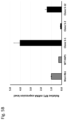
- FIG. 4 shows quantification of RP1 expression by qRT-PCR in Weri-Rb1 cells treated with pairs of sgRNAs targeting RP1 haplotype 1. Specific pairs of sgRNAs were able to decrease endogenous RP1 expression in heterozygous (H1/H2) Weri-Rb1 cells.
- FIGS. 5 A- 5 B show generation of RP1 expression cell lines.
- FIG. 5 A shows screening of RP1 expression in near-haploid (HAP-1) clonal cell-lines engineered with an EF1a promoter insertion.
- FIG. 5 B shows RP1 expression level in engineered HAP-1 stable cell-lines relative to Weri-Rb1 cells. RP1 expression was quantified by qRT-PCR.
- Engineered HAP1-EF1a-RP1 Clone 5-1 has the highest expression of RP1 transcripts, with a four-fold increase over endogenous RP1 expression in Weri-Rb1 cells.
- the effects of dominant mutations can arise via gain-of-function, dominant-negative, or haploinsufficiency mechanisms.
- the primary challenge in developing a gene therapy for autosomal dominant RP (adRP) is that, in addition to delivery of a healthy gene, the mutant gene may also need to be suppressed (Lewin, A. S., et al., (1998). Nat. Med. 4(8): p. 967-971; O'Reilly, M., et al., (2008). Vision Research, 48(3): p. 386-91; and O'Reilly, M., et al., (2007) American Journal of Human Genetics, 81(1): p. 127-35).
- the second challenge is that, for many IRD genes, the therapeutic dosage falls within a narrow window such that overexpression of the normal gene can lead to retinal degeneration (O'Reilly, M., et al., (2008) Vision Research. 48(3): p. 386-91; Wen, X. H., et al., (2009) Biophys J, 96(3): p. 939-50; and Liu, Q., et al., (2012) PLoS One, 7(8): p. e43251).
- CRISPR/Cas9 based genome editing tools provide a reliable and practical means to conceivably edit the disease-causing mutations in mammalian cells by introducing double strand breaks (DSBs) near the mutation, which are then repaired through the non-homologous end joining (NHEJ) or homology-directed repair (HDR) pathway, leading to the loss or correction of the mutant allele (Yin, H., et al., (2014) Nat Biotechnol, 2014. 32(6): p. 551-3; Ran, F. A., et al., (2015) Nature, 2015. 520(7546): p. 186-91; Swiech, L., et al., (2015) Nat Biotechnol, 2015. 33(1): p.
- DSBs double strand breaks
- HDR homology-directed repair
- the CRISPR/Cas9 system is capable of maintaining the edited gene under its endogenous expression stoichiometry, thereby avoiding ectopic expression and abnormal gene production that may occur with conventional gene augmentation therapies.
- Dominant mutations identified to date in RP1 are either nonsense or frameshift mutations that cluster at the N-terminal half of the last exon, exon 4 (Gandra, M., et al., (2008). Mol. Vis., 14: p. 1105-1113; Chen, L. J., et al., (2009). Invest Ophthalmol.Vis.Sci. 51(4): p. 2236-2242; Liu, Q., et al, (2009) Investigative ophthalmology & visual science 50(4): p. 1566-74; Liu, Q., et al., (2012). PLoS One, 7(8): p. e43251).
- the mutant RP1 alleles are predicted to encode truncated proteins that play a dominant negative role in photoreceptors (Liu, Q., et al., (2012). PLoS One, 7(8): p. e43251; Liu, Q., A. et al (2009), Invest Ophthalmol Vis Sci, 50(4): p. 1566-74; and Liu, Q., et al. (2004) J Neurosci, 24(29): p. 6427-36).
- the most common mutation in RP1, R677X is present in approximately 3% of patients with adRP in the United States, making it the third most common adRP mutation (Jacobson, S.
- the present methods employ a paired-sgRNA targeting approach using common SNPs as surrogate targeting sites to disrupt the expression of mutant RP1 alleles ( FIG. 1 ).
- This strategy could be applied to any autosomal dominant mutation in RP1, and would avoid the need for additional gene augmentation therapies, since the wild-type allele remains under its endogenous expression stoichiometry, and 50% of RP1 expression is sufficient for normal retinal integrity and function.
- the combined frequency of the three most common RP1 haplotypes is 93%, and the total homozygous frequency of the three common haplotypes is 35%. This indicates that approximately two thirds of the general population is heterozygous for RP1 haplotypes, which can be specifically targeted by the SNP-based CRISRP/Cas9 gene editing technology.
- RNA-Guided Nucleases RGNs
- Guide RNAs RGNs
- the present methods include the use of paired RNA-guided nucleases (RGNs) targeted to SNPs in a mutant allele of RP1, to excise portions of and disrupt the expression of the mutant RP1 alleles.
- RGNs paired RNA-guided nucleases
- the methods can include the use of RGNs including Cas9, Cpf1, and orthologs thereof.
- CRISPR from Prevotella and Francisella 1 (Cpf1, also known as Cas12a) nuclease can also be used, e.g., as described in Zetsche et al., (2015) Cell 163, 759-771; Schunder et al., (2013) Int J Med Microbiol 303, 51-60; Makarova et al., (2015) Nat Rev Microbiol 13, 722-736; Fagerlund et al., (2015) Genome Biol 16, 251.
- Cpf1/Cas12a requires only a single 42-nt crRNA, which has 23 nt at its 3′ end that are complementary to the protospacer of the target DNA sequence (Zetsche et al., (2015) Cell 163, 759-771). Furthermore, whereas SpCas9 recognizes an NGG PAM sequence that is 3′ of the protospacer, AsCpf1 and LbCp1 recognize TTTN PAMs that are found 5′ of the protospacer (Id.).
- the present system utilizes a wild type or variant Cas9 protein from S. pyogenes or Staphylococcus aureus , or a wild type or variant Cpf1 protein from Acidaminococcus sp. BV3L6 or Lachnospiraceae bacterium ND2006 either as encoded in bacteria or codon-optimized for expression in mammalian cells and/or modified in its PAM recognition specificity and/or its genome-wide specificity.
- a number of variants have been described; see, e.g., WO 2016/141224, PCT/US2016/049147, Kleinstiver et al., (2016) Nat Biotechnol .
- Exemplary guide RNAs used with S. aureus include those set forth in SEQ ID NOs: 1-6.
- pyogenes Cas9 29431739 M495V/Y515N/K526E/R661Q; (SpCas9) (M495V/Y515N/K526E/R661S; evoCas9 M495V/Y515N/K526E/R661L) S .
- pyogenes Cas9 26735016 N497A/R661A/Q695A/Q926A (SpCas9) HF1 S .
- pyogenes Cas9 30166441 R1335V, L1111R, D1135V, G1218R, (SpCas9) E1219F, A1322R, T1337R SpCas9-NG S .
- an RGN sequence is modified to include a nuclear localization sequences (NLSs), e.g., at the C- and/or N-terminus of the RGN protein, and a mini-polyadenylation signal (or Poly-A sequence).
- NLSs include SV40 large T antigen NLS (PKKKRRV (SEQ ID NO: 17)); PKKKRKV (SEQ ID NO: 18); KRTADGSEFESPKKKRKV (SEQ ID NO: 19); and nucleoplasmin NLS (KRPAATKKAGQAKKKK (SEQ ID NO:20)).
- Other NLSs are known in the art; see, e.g., Cokol et al., (2000) EMBO Rep .
- An exemplary polyadenylation signal is TAGCAATAAAGGATCGTTTATTTTCATTGGAAGCGTGTG TTGGTTTTTTGATCAGGCGCG (SEQ ID NO: 21)).
- gRNAs appropriate for the RGN should be used.
- the gRNAs used in the present disclosure can be unimolecular or modular, as known in the art.
- Sequences encoding the RGN and guide RNA can be delivered to the retina, e.g., using a viral vector. Described herein are targeted expression vectors for in vivo transfection and expression of a polynucleotide that encodes a RGN and guide RNAs as described herein, in the retina, e.g., in photoreceptors, e.g., primarily or only in photoreceptors. Expression constructs of such components can be administered in any effective carrier, e.g., any formulation or composition capable of effectively delivering the component gene to cells in vivo.
- Approaches include insertion of the gene in viral vectors, including recombinant retroviruses, adenovirus, adeno-associated virus, lentivirus, and herpes simplex virus-1, alphavirus, vaccinia virus, or recombinant bacterial or eukaryotic plasmids; preferred viral vectors are adeno-associated virus type 2 (AAV2), AAV5 or AAV8.
- retroviruses including recombinant retroviruses, adenovirus, adeno-associated virus, lentivirus, and herpes simplex virus-1, alphavirus, vaccinia virus, or recombinant bacterial or eukaryotic plasmids
- preferred viral vectors are adeno-associated virus type 2 (AAV2), AAV5 or AAV8.
- Viral vectors transfect cells directly; plasmid DNA can be delivered naked or with the help of, for example, cationic liposomes (lipofectamine) or derivatized (e.g., antibody conjugated), cationic dendrimers, inorganic vectors (e.g., iron oxide magnetofection), lipidoids, cell-penetrating peptides, cyclodextrin polymer (CDP), polylysine conjugates, gramacidin S, artificial viral envelopes or other such intracellular carriers, as well as direct injection of the gene construct or CaPO4 precipitation carried out in vivo.
- liposomes lipofectamine
- derivatized e.g., antibody conjugated
- cationic dendrimers e.g., inorganic vectors (e.g., iron oxide magnetofection)
- lipidoids e.g., cell-penetrating peptides, cyclodextrin polymer (CDP), polylysine conjugates,
- An exemplary approach for in vivo introduction of nucleic acid into a cell is by use of a viral vector containing nucleic acid, e.g., a cDNA.
- a viral vector containing nucleic acid e.g., a cDNA.
- Infection of cells with a viral vector has the advantage that a large proportion of the targeted cells can receive the nucleic acid.
- molecules encoded within the viral vector e.g., by a cDNA contained in the viral vector, are expressed efficiently in cells that have taken up viral vector nucleic acid.
- Viral vectors can be used as a recombinant gene delivery system for the transfer of exogenous genes in vivo, particularly into humans. These vectors provide efficient delivery of genes into cells, and in some cases the transferred nucleic acids are stably integrated into the chromosomal DNA of the host. Protocols for producing recombinant viruses and for infecting cells in vitro or in vivo with such viruses can be found in Ausubel, et al., eds., Gene Therapy Protocols Volume 1 : Production and In Vivo Applications of Gene Transfer Vectors , Humana Press, (2008), pp. 1-32 and other standard laboratory manuals.
- a preferred viral vector system useful for delivery of nucleic acids is the adeno-associated virus (AAV).
- Adeno-associated virus is a naturally occurring defective virus that requires another virus, such as an adenovirus or a herpes virus, as a helper virus for efficient replication and a productive life cycle.
- AAV vectors efficiently transduce various cell types and can produce long-term expression of transgenes in vivo.
- AAV vector genomes can persist within cells as episomes, vector integration has been observed (see for example Deyle and Russell, Curr Opin Mol Ther. 2009 August; 11(4): 442-447; Asokan et al., Mol Ther. 2012 April; 20(4): 699-708; Flotte et al., Am. J. Respir. Cell. Mol. Biol. 7:349-356 (1992); Samulski et al., J. Virol. 63:3822-3828 (1989); and McLaughlin et al., J. Virol. 62:1963-1973 (1989)).
- AAV vectors particularly AAV2 have been extensively used for gene augmentation or replacement and have shown therapeutic efficacy in a range of animal models as well as in the clinic; see, e.g., Mingozzi and High, (2011) Nature Reviews Genetics 12, 341-355; Deyle and Russell, (2009) Curr Opin Mol Ther . August; 11(4): 442-447; Asokan et al., (2012) Mol Ther. April; 20(4): 699-708.
- AAV vectors containing as little as 300 base pairs of AAV can be packaged and can produce recombinant protein expression. Space for exogenous DNA is limited to about 4.5 kb.
- an AAV1, 2, 4, 5, or 8 vector can be used to introduce DNA into the retina, e.g., into photoreceptors, inner retinal cells, or RPE cells (such as those described in Maguire et al. (2008). N Engl J Med 358: 2240-2248; Maguire et al. (2009). Lancet 374: 1597-1605; Bainbridge et al. (2008). N Engl J Med 358: 2231-2239; Hauswirth et al. (2008). Hum Gene Ther 19: 979-990; Cideciyan et al. (2008). Proc Natl Acad Sci USA 105: 15112-15117; Cideciyan et al. (2009).
- the AAV vector can include (or include a sequence encoding) an AAV capsid polypeptide described in WO 2015054653; for example, a virus particle comprising an AAV capsid polypeptide having an amino acid sequence selected from the group consisting of SEQ ID NOs: 1, 3, 5, 7, 9, 11, 13, 15, and 17 of WO 2015054653, and a RGN and guide RNA-encoding sequences as described herein.
- the AAV capsid polypeptide is as shown in Table 1 of WO 2015054653, reproduced here:
- the AAV capsid polypeptide is an Anc80 polypeptide, e.g., an exemplary polypeptide shown in SEQ ID NO: 19 (Anc80L27); SEQ ID NO: 20 (Anc80L59); SEQ ID NO: 21 (Anc80L60); SEQ ID NO: 22 (Anc80L62); SEQ ID NO: 23 (Anc80L65); SEQ ID NO: 24 (Anc80L33); SEQ ID NO: 25 (Anc80L36); and SEQ ID NO: 26 (Anc80L44) of WO 2015054653.
- a variety of nucleic acids have been introduced into different cell types using AAV vectors (see for example the references cited above and those cited in Asokan et al., (2012) Molecular Therapy; 20 4, 699-708; and Hermonat et al., (1984) Proc. Nat. Acad. Sci. USA 81:6466-6470; Tratschin et al., (1985) Mol. Cell. Biol. 4:2072-2081; Wondisford et al., (1988) Mol. Endocrinol. 2:32-39; Tratschin et al., (1984) J. Virol. 51:611-619; and Flotte et al., (1993) J. Biol. Chem. 268:3781-3790.
- a self-complementary AAV which contains an inverted repeat genome that folds to make double-stranded DNA.
- Retroviruses can also be used.
- the development of specialized cell lines (termed “packaging cells”) which produce only replication-defective retroviruses has increased the utility of retroviruses for gene therapy, and defective retroviruses are characterized for use in gene transfer for gene therapy purposes (for a review see Katz et al., (2013) Human Gene Therapy 24:914).
- a replication defective retrovirus can be packaged into virions, which can be used to infect a target cell through the use of a helper virus by standard techniques.
- suitable retroviruses include pLJ, pZIP, pWE and pEM, which are known to those skilled in the art.
- Retroviruses have been used to introduce a variety of genes into many different cell types, including epithelial cells, in vitro and/or in vivo (see for example Eglitis, et al. (1985) Science 230:1395-1398; Danos and Mulligan (1988) Proc. Natl. Acad. Sci. USA 85:6460-6464; Wilson et al. (1988) Proc. Natl. Acad. Sci. USA 85:3014-3018; Armentano et al. (1990) Proc. Natl. Acad. Sci.
- adenovirus-derived vectors The genome of an adenovirus can be manipulated, such that it encodes and expresses a gene product of interest, but is inactivated in terms of its ability to replicate in a normal lytic viral life cycle. See, for example, Berkner et al., (1988) BioTechniques 6:616; Rosenfeld et al., (1991) Science 252:431-434; and Rosenfeld et al., (1992) Cell 68:143-155.
- adenoviral vectors derived from the adenovirus strain Ad type 5 d1324 or other strains of adenovirus are known to those skilled in the art.
- Recombinant adenoviruses can be advantageous in certain circumstances, in that they are not capable of infecting non-dividing cells and can be used to infect a wide variety of cell types, including epithelial cells (Rosenfeld et al., (1992) supra).
- the virus particle is relatively stable and amenable to purification and concentration, and as above, can be modified so as to affect the spectrum of infectivity.
- introduced adenoviral DNA (and foreign DNA contained therein) is not integrated into the genome of a host cell but remains episomal, thereby avoiding potential problems that can occur as a result of insertional mutagenesis in situ, where introduced DNA becomes integrated into the host genome (e.g., retroviral DNA).
- the carrying capacity of the adenoviral genome for foreign DNA is large (up to 8 kilobases) relative to other gene delivery vectors (Berkner et al., supra; Haj-Ahmand and Graham, (1986) J. Virol. 57:267.
- sequences encoding RGN and guide RNAs is entrapped in liposomes bearing positive charges on their surface (e.g., lipofectins), which can be tagged with antibodies against cell surface antigens of the target tissue (Mizuno et al., (1992) No Shinkei Geka 20:547-551; PCT publication WO91/06309; Japanese patent application 1047381; and European patent publication EP-A-43075).
- lipofectins e.g., lipofectins
- the vectors can also include promoters, enhancers (e.g., CMV enhancer), other cis-regulatory elements, and/or capsid serotype variants.
- promoters can include promoters that drive expression in many cell types (e.g., cytomegalovirus (CMV), chicken ⁇ -actin (CBA), cytomegalovirus (CMV) enhancer fused to the chicken beta-actin promoter (CAG), CBh, elongation factor alpha 1 (EFalpha1), EF-1 Alpha Short (EFS) or CASI) or specifically in photoreceptor cells (e.g., RHO, beta phosphodiesterase (PDE), retinitis pigmentosa (RP1), rhodopsin kinase (hGRK1) and cone arrestin (CAR)) (see, e.g., Gray et al., (2011) Hum Gene Ther .
- CMV cytomegalovirus
- CBA chicken ⁇
- cis-regulatory elements can include enhancer elements derived from the interphotoreceptor retinoid-binding protein gene (IRBP), woodchuck hepatitis virus post-transcriptional regulatory element (WPRE), or minute virus of mice (MVM) intron (see Domenger and Grimm, (2019) Human Molecular Genetics, 28(R1):R3-R14 (October)).
- IRBP interphotoreceptor retinoid-binding protein gene
- WPRE woodchuck hepatitis virus post-transcriptional regulatory element
- MVM minute virus of mice
- the pharmaceutical preparation of the gene therapy construct can consist essentially of the gene delivery system (viral vector and any associated agents such as helper viruses, proteins, lipids, and so on) in an acceptable diluent, or can comprise a slow release matrix in which the gene delivery vehicle is embedded.
- the pharmaceutical preparation can comprise one or more cells, which produce the gene delivery system.
- the methods can include delivering the RGN and guide RNA together, e.g., as a complex.
- the RGN and gRNA can be can be overexpressed in a host cell and purified, then complexed with the guide RNA (e.g., in a test tube) to form a ribonucleoprotein (RNP), and delivered to cells.
- the RGN can be expressed in and purified from bacteria through the use of bacterial expression plasmids.
- His-tagged deaminase fusion protein can be expressed in bacterial cells and then purified using nickel affinity chromatography.
- RNPs circumvents the necessity of delivering plasmid DNAs encoding the nuclease or the guide, or encoding the nuclease as an mRNA. RNP delivery may also improve specificity, presumably because the half-life of the RNP is shorter and there is no persistent expression of the nuclease and guide (in contrast to the sustained expression from a plasmid).
- the RNPs can be delivered to the cells in vivo or in vitro, e.g., using lipid-mediated transfection or electroporation. See, e.g., Liang et al. (2015) Journal of biotechnology 208: 44-53; Zuris, John A., et al. (2015) Nature biotechnology 33.1: 73-80; Kim et al. (2014) Genome research 24.6: 1012-1019.
- RP Retinitis pigmentosa
- Suitable subjects can be identified by one of skill in the art and a diagnosis confirmed by genetic testing (e.g., sequencing to identify the presence of a mutation in the subject's RP1 gene).
- a reference sequence for RP1 can be found in NCBI GenBank at RefSeqGene ID NG_009840.2 (Range 5030-19768).
- the methods include administering an effective amount of an RGN and paired guide RNAs that excise portions of and disrupt the expression of the mutant RP1 alleles.
- the vectors can be introduced into a subject by any of a number of methods, each of which familiar in the art. Although other methods can be used, in some embodiments, the route of choice for delivery of gene therapy vectors to the retina is via sub-retinal injection. This provides access to the RPE and photoreceptor cells of the retina. Different serotypes of AAV have been shown to transduce these cell populations effectively after sub-retinal injection in animal studies (Vandenberghe et al., (2013) PLoS One. 8:e53463.
- Sub-retinal injections can be performed using a standard surgical approach (e.g., as described in Maguire et al., 2008 supra; Bainbridge et al., 2008 supra; Cideciyan et al., 2008 supra; MacLaren et al., 2014 supra). See also WO2019/183641.
- any region of the retina may be targeted, though the fovea (which extends approximately 1 degree out from the center of the eye) may be preferred in certain instances due to its role in central visual acuity and the relatively high concentration of cone photoreceptors there relative to peripheral regions of the retina.
- injections may be targeted to parafoveal regions (extending between approximately 2 and 10 degrees off center), which are characterized by the presence of all three types of retinal photoreceptor cells.
- injections into the parafoveal region may be made at comparatively acute angles using needle paths that cross the midline of the retina.
- injection paths may extend from the nasal aspect of the sclera near the limbus through the vitreal chamber and into the parafoveal retina on the temporal side, from the temporal aspect of the sclera to the parafoveal retina on the nasal side, from a portion of the sclera located superior to the cornea to an inferior parafoveal position, and/or from an inferior portion of the sclera to a superior parafoveal position.
- the use of relatively small angles of injection relative to the retinal surface may advantageously reduce or limit the potential for spillover of vector from the bleb into the vitreous body and, consequently, reduce the loss of the vector during delivery.
- the macula (inclusive of the fovea) can be targeted, and in other cases, additional retinal regions can be targeted, or can receive spillover doses.
- compositions comprising AAV vectors can be administered to subjects by any suitable means, including without limitation injection, for example, sub-retinal injection.
- concentration of AAV vector within the composition is selected to ensure, among other things, that a sufficient AAV dose is administered to the retina of the subject, taking account of dead volume within the injection apparatus and the relatively limited volume that can be safely administered.
- Suitable doses may include, for example, 1 ⁇ 10 11 viral genomes (vg)/mL, 2 ⁇ 10 11 viral genomes (vg)/mL, 3 ⁇ 10 11 viral genomes (vg)/mL, 4 ⁇ 10 11 viral genomes (vg)/mL, 5 ⁇ 10 11 viral genomes (vg)/mL, 6 ⁇ 10 11 viral genomes (vg)/mL, 7 ⁇ 10 11 viral genomes (vg)/mL, 8 ⁇ 10 11 viral genomes (vg)/mL, 9 ⁇ 10 11 viral genomes (vg)/mL, 1 ⁇ 10 12 vg/mL, 2 ⁇ 10 12 viral genomes (vg)/mL, 3 ⁇ 10 12 viral genomes (vg)/mL, 4 ⁇ 10 12 viral genomes (vg)/mL, 5 ⁇ 10 12 viral genomes (vg)/mL, 6 ⁇ 10 12 viral genomes (vg)/mL, 7 ⁇ 10 12 viral genomes (vg)/mL, 8 ⁇ 10 12 viral genomes (
- any suitable volume of the composition may be delivered to the subretinal or cochlear space.
- the volume is selected to form a bleb in the subretinal space, for example 1 microliter, 10 microliters, 50 microliters, 100 microliters, 150 microliters, 200 microliters, 250 microliters, 300 microliters, etc.
- viruses comprising sequences encoding an RGN and guide RNAs as described herein (e.g., one or more viruses comprising sequences one, two, or all three of the RGN and paired gRNAs, e.g., wherein a single virus comprises sequences encodes one, two (e.g., two gRNAs), or all three, as well as compositions comprising one or more of such viruses (e.g., a composition comprising one virus comprising sequences encoding the two gRNAs and a separate virus comprising a sequence encoding the RGN; a composition comprising one virus comprising sequences encoding the two gRNAs and the RGN; or a composition comprising one virus comprising a sequence encoding a first gRNA, a second virus comprising a sequence encoding a second gRNA, and a third virus comprising a sequence encoding the RGN).
- RNPs comprising the RGN and gRNAs
- H1 is the reference sequence.
- Common SNPs on H2 include G at rs702761, G at rs145290, A at rs436527, A at rs428854, C at rs424499, C at rs429668, A at rs444772, A at rs446227, C at rs414352, and G at rs441800.
- Common SNPs on H3 include A for rs62514616, T for rs2293869, and A for rs61739567.
- the combined frequency of these three common haplotypes is 93% and the total homozygous frequency of the three common haplotypes is 35%. This indicates that approximately two thirds of the general population is heterozygous for RP1 haplotypes, which can be specifically targeted by the SNP-based CRISPR/Cas9 gene editing technology.
- the allele frequencies for the four common SNPs (rs444772, rs446227, rs414352 and rs441800) on H2 is 0.25-0.27 in European Americans based on the 1000 Genomes Project (Table 4).
- the two SNPs (rs2293869 and rs61739567) for H3 have an allele frequency of 0.44 in European Americans.
- LD linkage disequilibrium
- Cas9-sgRNAs for targeting SNPs on human RP1 haplotypes sgRNA Targeted Site SNPs Seq Cas9 ID Protospacers # PAM haplotypes 1 rs702761 A/G Sp-WT 1A1 and 1G1 ATGCTGATATTGAAR 22 TGG H1 or H3/H2 TAGAA Sp-WT 1A2 and 1G2 TGCTGATATTG 23 GGAA H1 or H3/H2 AARTAGAAT Sp-VRQR 1A3 and 1G3 CAATATGCTGAT 24 AGAA H1 or H3/H2 ATTGAART Sa-WT 1A4 and 1G4 TCACAATATGCT 25 TAGAAT H1 or H3/H2 GATATTGAAR Sa-KKH 1A5 and 1G5 TGATATTGAARTAG 26 TTCAGT H1 or H3/H2 AATGGAAC Sa-KKH 1A6 and 1G6 TGATACTGAAGTT 27 TTCAAT H1 or
- All Cas9 expression plasmids were constructed into a CAG-Cas9-T2A-EGFP backbone modified from a pSQT817 vector (Addgene #53373).
- EGFP and Cas9 were co-expressed as a single protein before being separated by a self-cleaving T2A peptide; EGFP served as the marker for the targeted cells and for the downstream FACS sorting.
- the sgRNAs for wild-type and variant SpCas9 were cloned into the expression vector BPK1520 (Addgene #65777), and the sgRNAs for wild-type and variant SaCas9 were cloned into the BPK2660 vector (Addgene #70709).
- H2/H3 HEK293T
- ARPE-19 H1/H2
- hTERT RPE-1 H3/H3
- Min38931 fibroblast H2/H2 from RP1 patient
- HL-60 H1/H1
- HT-1080 H1/H2
- C3A H2/H3
- Y79 H1/H3
- Weri-Rb1 H1/H2
- H1/H2 haploid human cell-line HAP1
- HEK293T cells were selected for the initial screening step, for their efficiency of transfection, and heterozygous H2/H3 haplotype, such that all sgRNAs could be tested in these cells (in the H3 positions, H2 SNPs are the same as H1, and vice versa.
- H1 has 13 targetable sites
- H2 has 10 sites
- H3 has 3 sites
- NGS deep sequencing was utilized to quantitatively analyze the specific DSBs-NHEJ events in the genome, and to predict the cleavage specificity. Briefly, short PCR products spanning the targeted site were amplified and purified, and dual-indexed TruSeq 250 bp paired end sequencing was performed on an Illumina MiSeq Sequencer by MGH CCIB. Paired-end reads were mapped to the human genome reference and reads with an average quality score >30 were analyzed for indels that overlap the intended target nuclease binding site. Indel analyses were conducted using an in-house algorithm at the MGH CCIB.
- SpCas9 wild-type and variant guides for all three haplotypes have been tested in HEK293T, ARPE19, hTert-RPE1, U20S, HAP1 and Weri-Rb1 cell lines, and analyzed for edited and unedited reads per allele. Editing efficiency was calculated as a ratio of edited alleles/total reads per allele, and are presented in Table 7 below, and FIGS. 2 A and 2 B .
- sgRNAs with the highest targeting efficiency and specificity were selected, and their ability to target and knockout RP1 alleles via paired sgRNA directed editing in heterozygous or haploid cell lines was evaluated. Paired guide deletion strategies for H1 were first tested. Editing by a single gRNA targeted to an intronic region was repaired by NHEJ, and did not affect RP1 expression ( FIG. 4 , single guide 14N2 only), therefore highly efficient guides targeting regions of intron 3 in any allele were paired with allele-specific sgRNAs targeting H1 SNPs in intron 1, or exon 4.
- bi-allele targeting guides will edit both wild type and mutant alleles, but when paired with allele specific guides in intron 1, or exon 4, will delete a large genomic portion form the mutant allele only. Larger deletions are also possible via the pairing of allele specific guides in intron 1 and exon 4 (Table 8 and Table 9).
- a qPCR strategy was developed to quantify editing and RP1 allele deletion efficiency at the genomic level.
- qPCR primers were designed to amplify regions within the predicted deletion.
- the ‘internal’ amplicon was produced only from remaining ‘undeleted’ alleles, and after normalization to a reference gene on another chromosome, the remaining ‘undeleted’ allele was quantified relative to unedited cells ( FIG. 3 ). All combinations for sgRNAs tested were able to decrease RP1 allele content in edited cells, up to 58% for 14N2+12A2 gRNA pair. However, this quantification strategy may not capture all editing events.
- inversions after dual DSBs may still allow amplification of ‘internal’ amplicons, if they have not been damaged by editing.
- Quantification of a reduction of RP1 expression is a more accurate measure of allele disruption, as described below.
- RP1 is specifically expressed in specialized post-mitotic photoreceptors.
- the retinoblastoma cell line Weri-Rb1 expresses low levels of RP1, but is difficult to transfect. Cell-lines were therefore generated with exogenous expression of RP1, by insertion of a promoter upstream of the RP1 transcriptional start site (TSS).
- TSS transcriptional start site
- a plasmid encoding 100 bp homology arms and the EF1a promoter followed by a neomycin resistance gene and a P2A peptide cleavage motif was co-transfected with plasmids which express an RP1 targeting sgRNA, and SpCas9, into wild-type Hap1 cells. After 3 weeks of G418 selection at 50 ug/mL, 15 individual colonies were picked and analyzed for targeted genomic integration of the promoter cassette, and for RP1 expression by semi-quantitative RT-PCR. Clone 5 was then selected for stable cell line generation from single cells, and 12 clones were tested for relative RP1 expression by qRT-PCR ( FIG. 5 A ).
- iPSCs have also been generated from fibroblasts obtained from two RP1 patients (OGI1557-002771 and OGI1781-003109) with dominant mutations selected from the probands in the IRD Biobank at MEEI.
- Patient OGI1557-002771 is heterozygous for H1/H3 haplotype, and carries the c. 2103delAATA mutation in cis with the SNPs for H3.
- Patient OGI1781-003109 is heterozygous for H2/H3, and carries the mutation c.2029C>T in cis with the SNPs for H2.
- Cells have undergone reprogramming with the non-integrating Sendaii virus method at Harvard iPSC Core Facility. These iPS cells and the differentiated retinal organoids are used to evaluate the lead Cas9/dual sgRNA pairs regarding their efficacy and safety.
- Table 10 presents a summary of the SNPs and their presence in each haplotype, with the genomic information included.
- RP1 Haplotype SNPs with Genomic Location RP1 Haplotype variant Genome SNP H1 H2 H3 location rs702761 A G A Intron 1 rs145290 A G A Intron 1 rs436527 G A G Intron 1 rs428854 G A G Intron 3 rs424499 T C T Intron 3 rs429668 T C T Intron 3 rs62514616 G G A Intron 3 rs444772 G A G Exon 4 rs446227 G A G Exon 4 rs414352 T C T Exon 4 rs441800 A G A Exon 4 rs2293869 A A T Exon 4 rs61739567 G G A Exon 4
- Table 11 presents a summary of first and second gRNA targets by haplotype.
Landscapes
- Health & Medical Sciences (AREA)
- Life Sciences & Earth Sciences (AREA)
- Genetics & Genomics (AREA)
- Engineering & Computer Science (AREA)
- Chemical & Material Sciences (AREA)
- Bioinformatics & Cheminformatics (AREA)
- Zoology (AREA)
- Molecular Biology (AREA)
- Wood Science & Technology (AREA)
- Organic Chemistry (AREA)
- Biomedical Technology (AREA)
- General Engineering & Computer Science (AREA)
- Biotechnology (AREA)
- General Health & Medical Sciences (AREA)
- Microbiology (AREA)
- Biochemistry (AREA)
- Medicinal Chemistry (AREA)
- Plant Pathology (AREA)
- Physics & Mathematics (AREA)
- Biophysics (AREA)
- Pharmacology & Pharmacy (AREA)
- Veterinary Medicine (AREA)
- Public Health (AREA)
- Animal Behavior & Ethology (AREA)
- Epidemiology (AREA)
- Cell Biology (AREA)
- Mycology (AREA)
- Ophthalmology & Optometry (AREA)
- Immunology (AREA)
- Gastroenterology & Hepatology (AREA)
- Proteomics, Peptides & Aminoacids (AREA)
- Medicines Containing Material From Animals Or Micro-Organisms (AREA)
- Medicines That Contain Protein Lipid Enzymes And Other Medicines (AREA)
- Micro-Organisms Or Cultivation Processes Thereof (AREA)
- Pharmaceuticals Containing Other Organic And Inorganic Compounds (AREA)
Priority Applications (2)
| Application Number | Priority Date | Filing Date | Title |
|---|---|---|---|
| US17/175,234 US12129496B2 (en) | 2020-02-12 | 2021-02-12 | Haplotype-based treatment of RP1 associated retinal degenerations |
| US18/890,708 US20250115887A1 (en) | 2020-02-12 | 2024-09-19 | Haplotype-based treatment of rp1 associated retinal degenerations |
Applications Claiming Priority (2)
| Application Number | Priority Date | Filing Date | Title |
|---|---|---|---|
| US202062975636P | 2020-02-12 | 2020-02-12 | |
| US17/175,234 US12129496B2 (en) | 2020-02-12 | 2021-02-12 | Haplotype-based treatment of RP1 associated retinal degenerations |
Related Child Applications (1)
| Application Number | Title | Priority Date | Filing Date |
|---|---|---|---|
| US18/890,708 Division US20250115887A1 (en) | 2020-02-12 | 2024-09-19 | Haplotype-based treatment of rp1 associated retinal degenerations |
Publications (2)
| Publication Number | Publication Date |
|---|---|
| US20210269783A1 US20210269783A1 (en) | 2021-09-02 |
| US12129496B2 true US12129496B2 (en) | 2024-10-29 |
Family
ID=77292955
Family Applications (2)
| Application Number | Title | Priority Date | Filing Date |
|---|---|---|---|
| US17/175,234 Active 2041-10-05 US12129496B2 (en) | 2020-02-12 | 2021-02-12 | Haplotype-based treatment of RP1 associated retinal degenerations |
| US18/890,708 Pending US20250115887A1 (en) | 2020-02-12 | 2024-09-19 | Haplotype-based treatment of rp1 associated retinal degenerations |
Family Applications After (1)
| Application Number | Title | Priority Date | Filing Date |
|---|---|---|---|
| US18/890,708 Pending US20250115887A1 (en) | 2020-02-12 | 2024-09-19 | Haplotype-based treatment of rp1 associated retinal degenerations |
Country Status (6)
| Country | Link |
|---|---|
| US (2) | US12129496B2 (https=) |
| EP (1) | EP4103238A4 (https=) |
| JP (1) | JP2023519101A (https=) |
| CN (1) | CN115427568A (https=) |
| AU (1) | AU2021219795A1 (https=) |
| WO (1) | WO2021163550A1 (https=) |
Families Citing this family (1)
| Publication number | Priority date | Publication date | Assignee | Title |
|---|---|---|---|---|
| US12171813B2 (en) | 2021-02-05 | 2024-12-24 | Christiana Care Gene Editing Institute, Inc. | Methods of and compositions for reducing gene expression and/or activity |
Citations (19)
| Publication number | Priority date | Publication date | Assignee | Title |
|---|---|---|---|---|
| EP0043075A2 (de) | 1980-06-25 | 1982-01-06 | Studiengesellschaft Kohle mbH | Verfahren zur Erhöhung der Inkorporation und der Expression von genetischem Material in die Kerne von intakten Zellen mit Hilfe von Liposomen |
| JPS57124055A (en) | 1981-01-27 | 1982-08-02 | Toyota Motor Corp | Air-fuel ratio control device for engine |
| WO1989002468A1 (en) | 1987-09-11 | 1989-03-23 | Whitehead Institute For Biomedical Research | Transduced fibroblasts and uses therefor |
| WO1989005345A1 (en) | 1987-12-11 | 1989-06-15 | Whitehead Institute For Biomedical Research | Genetic modification of endothelial cells |
| WO1989007136A2 (en) | 1988-02-05 | 1989-08-10 | Whitehead Institute For Biomedical Research | Modified hepatocytes and uses therefor |
| US4868116A (en) | 1985-07-05 | 1989-09-19 | Whitehead Institute For Biomedical Research | Introduction and expression of foreign genetic material in epithelial cells |
| US4980286A (en) | 1985-07-05 | 1990-12-25 | Whitehead Institute For Biomedical Research | In vivo introduction and expression of foreign genetic material in epithelial cells |
| WO1991006309A1 (en) | 1989-11-03 | 1991-05-16 | Vanderbilt University | Method of in vivo delivery of functioning foreign genes |
| WO1992007573A1 (en) | 1990-10-31 | 1992-05-14 | Somatix Therapy Corporation | Genetic modification of endothelial cells |
| WO2014144592A2 (en) | 2013-03-15 | 2014-09-18 | The General Hospital Corporation | Using truncated guide rnas (tru-grnas) to increase specificity for rna-guided genome editing |
| WO2015054653A2 (en) | 2013-10-11 | 2015-04-16 | Massachusetts Eye & Ear Infirmary | Methods of predicting ancestral virus sequences and uses thereof |
| WO2016141224A1 (en) | 2015-03-03 | 2016-09-09 | The General Hospital Corporation | Engineered crispr-cas9 nucleases with altered pam specificity |
| WO2017040348A1 (en) | 2015-08-28 | 2017-03-09 | The General Hospital Corporation | Engineered crispr-cas9 nucleases |
| WO2018026976A1 (en) | 2016-08-02 | 2018-02-08 | Editas Medicine, Inc. | Compositions and methods for treating cep290 associated disease |
| US20190010481A1 (en) | 2017-04-21 | 2019-01-10 | The General Hospital Corporation | Variants of CPF1 (CAS12a) With Altered PAM Specificity |
| WO2019165322A1 (en) | 2018-02-22 | 2019-08-29 | Moore Tara | Crispr/cas9 systems, and methods of use thereof |
| WO2019183641A1 (en) | 2018-03-23 | 2019-09-26 | Massachusetts Eye And Ear Infirmary | Crispr/cas9-mediated exon-skipping approach for ush2a-associated usher syndrome |
| WO2019183630A2 (en) | 2018-03-23 | 2019-09-26 | The Trustees Of Columbia University In The City Of New York | Gene editing for autosomal dominant diseases |
| US20190345501A1 (en) | 2017-02-07 | 2019-11-14 | Massachusetts Institute Of Technology | Methods and compositions for rna-guided genetic circuits |
-
2021
- 2021-02-12 CN CN202180027183.4A patent/CN115427568A/zh active Pending
- 2021-02-12 EP EP21753483.3A patent/EP4103238A4/en active Pending
- 2021-02-12 WO PCT/US2021/017942 patent/WO2021163550A1/en not_active Ceased
- 2021-02-12 AU AU2021219795A patent/AU2021219795A1/en active Pending
- 2021-02-12 JP JP2022548612A patent/JP2023519101A/ja active Pending
- 2021-02-12 US US17/175,234 patent/US12129496B2/en active Active
-
2024
- 2024-09-19 US US18/890,708 patent/US20250115887A1/en active Pending
Patent Citations (20)
| Publication number | Priority date | Publication date | Assignee | Title |
|---|---|---|---|---|
| EP0043075A2 (de) | 1980-06-25 | 1982-01-06 | Studiengesellschaft Kohle mbH | Verfahren zur Erhöhung der Inkorporation und der Expression von genetischem Material in die Kerne von intakten Zellen mit Hilfe von Liposomen |
| JPS57124055A (en) | 1981-01-27 | 1982-08-02 | Toyota Motor Corp | Air-fuel ratio control device for engine |
| US4868116A (en) | 1985-07-05 | 1989-09-19 | Whitehead Institute For Biomedical Research | Introduction and expression of foreign genetic material in epithelial cells |
| US4980286A (en) | 1985-07-05 | 1990-12-25 | Whitehead Institute For Biomedical Research | In vivo introduction and expression of foreign genetic material in epithelial cells |
| WO1989002468A1 (en) | 1987-09-11 | 1989-03-23 | Whitehead Institute For Biomedical Research | Transduced fibroblasts and uses therefor |
| WO1989005345A1 (en) | 1987-12-11 | 1989-06-15 | Whitehead Institute For Biomedical Research | Genetic modification of endothelial cells |
| WO1989007136A2 (en) | 1988-02-05 | 1989-08-10 | Whitehead Institute For Biomedical Research | Modified hepatocytes and uses therefor |
| WO1991006309A1 (en) | 1989-11-03 | 1991-05-16 | Vanderbilt University | Method of in vivo delivery of functioning foreign genes |
| WO1992007573A1 (en) | 1990-10-31 | 1992-05-14 | Somatix Therapy Corporation | Genetic modification of endothelial cells |
| WO2014144592A2 (en) | 2013-03-15 | 2014-09-18 | The General Hospital Corporation | Using truncated guide rnas (tru-grnas) to increase specificity for rna-guided genome editing |
| WO2015054653A2 (en) | 2013-10-11 | 2015-04-16 | Massachusetts Eye & Ear Infirmary | Methods of predicting ancestral virus sequences and uses thereof |
| WO2016141224A1 (en) | 2015-03-03 | 2016-09-09 | The General Hospital Corporation | Engineered crispr-cas9 nucleases with altered pam specificity |
| WO2017040348A1 (en) | 2015-08-28 | 2017-03-09 | The General Hospital Corporation | Engineered crispr-cas9 nucleases |
| WO2018026976A1 (en) | 2016-08-02 | 2018-02-08 | Editas Medicine, Inc. | Compositions and methods for treating cep290 associated disease |
| US20190345501A1 (en) | 2017-02-07 | 2019-11-14 | Massachusetts Institute Of Technology | Methods and compositions for rna-guided genetic circuits |
| US20190010481A1 (en) | 2017-04-21 | 2019-01-10 | The General Hospital Corporation | Variants of CPF1 (CAS12a) With Altered PAM Specificity |
| WO2019165322A1 (en) | 2018-02-22 | 2019-08-29 | Moore Tara | Crispr/cas9 systems, and methods of use thereof |
| WO2019183641A1 (en) | 2018-03-23 | 2019-09-26 | Massachusetts Eye And Ear Infirmary | Crispr/cas9-mediated exon-skipping approach for ush2a-associated usher syndrome |
| WO2019183630A2 (en) | 2018-03-23 | 2019-09-26 | The Trustees Of Columbia University In The City Of New York | Gene editing for autosomal dominant diseases |
| US20210017509A1 (en) * | 2018-03-23 | 2021-01-21 | The Trustees Of Columbia University In The City Of New York | Gene Editing for Autosomal Dominant Diseases |
Non-Patent Citations (122)
Also Published As
| Publication number | Publication date |
|---|---|
| WO2021163550A1 (en) | 2021-08-19 |
| AU2021219795A1 (en) | 2022-08-25 |
| US20210269783A1 (en) | 2021-09-02 |
| JP2023519101A (ja) | 2023-05-10 |
| EP4103238A1 (en) | 2022-12-21 |
| CA3169991A1 (en) | 2021-08-19 |
| US20250115887A1 (en) | 2025-04-10 |
| CN115427568A (zh) | 2022-12-02 |
| EP4103238A4 (en) | 2024-03-20 |
Similar Documents
| Publication | Publication Date | Title |
|---|---|---|
| US20250235556A1 (en) | Gene therapy for autosomal dominant diseases | |
| EP3768303B1 (en) | Crispr/cas9-mediated exon-skipping approach for ush2a-associated usher syndrome | |
| Tornabene et al. | Therapeutic homology-independent targeted integration in retina and liver | |
| US12285495B2 (en) | Genome editing methods and constructs | |
| KR20210049833A (ko) | Mma의 치료를 위한 비-붕괴적 유전자 요법 | |
| KR20230029891A (ko) | 전이유전자 발현 시스템 | |
| JP2025175283A (ja) | Amdの処置のためのhtra1調節 | |
| US20250115887A1 (en) | Haplotype-based treatment of rp1 associated retinal degenerations | |
| EP3821019A1 (en) | Methods of treating non-syndromic sensorineural hearing loss | |
| KR20220018495A (ko) | 영양요구성 선택 방법 | |
| BR112020015798A2 (pt) | Composições de vírus adeno-associado para restaurar função de gene da pah e métodos de uso das mesmas | |
| CA3169991C (en) | Haplotype-based treatment of rp1 associated retinal degenerations | |
| US20230313235A1 (en) | Compositions for use in treating autosomal dominant best1-related retinopathies | |
| US20250302994A1 (en) | Erythroparvovirus with a modified genome for gene therapy | |
| LLADO SANTAEULARIA | THERAPEUTIC GENOME EDITING IN RETINA AND LIVER | |
| WO2026025441A1 (zh) | 一种用于显性遗传的视网膜色素变性基因编辑药物载体 | |
| WO2025080973A1 (en) | Chst6 mini gene - chst6 expression construct with native promoter | |
| JP2026511058A (ja) | ゲノム工学を使用して制御性t細胞(treg)を作製するための方法 | |
| HK40075787A (en) | Gene therapy for autosomal dominant diseases | |
| HK1251615B (en) | Gene therapy for autosomal dominant diseases |
Legal Events
| Date | Code | Title | Description |
|---|---|---|---|
| FEPP | Fee payment procedure |
Free format text: ENTITY STATUS SET TO UNDISCOUNTED (ORIGINAL EVENT CODE: BIG.); ENTITY STATUS OF PATENT OWNER: SMALL ENTITY |
|
| FEPP | Fee payment procedure |
Free format text: ENTITY STATUS SET TO SMALL (ORIGINAL EVENT CODE: SMAL); ENTITY STATUS OF PATENT OWNER: SMALL ENTITY |
|
| AS | Assignment |
Owner name: MASSACHUSETTS EYE AND EAR INFIRMARY, MASSACHUSETTS Free format text: ASSIGNMENT OF ASSIGNORS INTEREST;ASSIGNORS:LIU, QIN;COLLIN, CAITLIN;SIGNING DATES FROM 20210408 TO 20210427;REEL/FRAME:056234/0310 |
|
| STPP | Information on status: patent application and granting procedure in general |
Free format text: DOCKETED NEW CASE - READY FOR EXAMINATION |
|
| STPP | Information on status: patent application and granting procedure in general |
Free format text: NON FINAL ACTION MAILED |
|
| STPP | Information on status: patent application and granting procedure in general |
Free format text: RESPONSE TO NON-FINAL OFFICE ACTION ENTERED AND FORWARDED TO EXAMINER |
|
| STPP | Information on status: patent application and granting procedure in general |
Free format text: RESPONSE TO NON-FINAL OFFICE ACTION ENTERED AND FORWARDED TO EXAMINER |
|
| STPP | Information on status: patent application and granting procedure in general |
Free format text: FINAL REJECTION MAILED |
|
| STPP | Information on status: patent application and granting procedure in general |
Free format text: DOCKETED NEW CASE - READY FOR EXAMINATION |
|
| STPP | Information on status: patent application and granting procedure in general |
Free format text: NOTICE OF ALLOWANCE MAILED -- APPLICATION RECEIVED IN OFFICE OF PUBLICATIONS |
|
| ZAAB | Notice of allowance mailed |
Free format text: ORIGINAL CODE: MN/=. |
|
| STPP | Information on status: patent application and granting procedure in general |
Free format text: AWAITING TC RESP., ISSUE FEE NOT PAID |
|
| STPP | Information on status: patent application and granting procedure in general |
Free format text: NOTICE OF ALLOWANCE MAILED -- APPLICATION RECEIVED IN OFFICE OF PUBLICATIONS |
|
| STPP | Information on status: patent application and granting procedure in general |
Free format text: PUBLICATIONS -- ISSUE FEE PAYMENT VERIFIED |
|
| STCF | Information on status: patent grant |
Free format text: PATENTED CASE |
|
| CC | Certificate of correction |