US11072792B2 - Materials and methods for treatment of usher syndrome type 2A - Google Patents
Materials and methods for treatment of usher syndrome type 2A Download PDFInfo
- Publication number
- US11072792B2 US11072792B2 US16/905,145 US202016905145A US11072792B2 US 11072792 B2 US11072792 B2 US 11072792B2 US 202016905145 A US202016905145 A US 202016905145A US 11072792 B2 US11072792 B2 US 11072792B2
- Authority
- US
- United States
- Prior art keywords
- grna
- seq
- sequence
- cells
- cell
- Prior art date
- Legal status (The legal status is an assumption and is not a legal conclusion. Google has not performed a legal analysis and makes no representation as to the accuracy of the status listed.)
- Active
Links
Images


Classifications
-
- C—CHEMISTRY; METALLURGY
- C12—BIOCHEMISTRY; BEER; SPIRITS; WINE; VINEGAR; MICROBIOLOGY; ENZYMOLOGY; MUTATION OR GENETIC ENGINEERING
- C12N—MICROORGANISMS OR ENZYMES; COMPOSITIONS THEREOF; PROPAGATING, PRESERVING, OR MAINTAINING MICROORGANISMS; MUTATION OR GENETIC ENGINEERING; CULTURE MEDIA
- C12N15/00—Mutation or genetic engineering; DNA or RNA concerning genetic engineering, vectors, e.g. plasmids, or their isolation, preparation or purification; Use of hosts therefor
- C12N15/09—Recombinant DNA-technology
- C12N15/11—DNA or RNA fragments; Modified forms thereof; Non-coding nucleic acids having a biological activity
- C12N15/113—Non-coding nucleic acids modulating the expression of genes, e.g. antisense oligonucleotides; Antisense DNA or RNA; Triplex- forming oligonucleotides; Catalytic nucleic acids, e.g. ribozymes; Nucleic acids used in co-suppression or gene silencing
- C12N15/1138—Non-coding nucleic acids modulating the expression of genes, e.g. antisense oligonucleotides; Antisense DNA or RNA; Triplex- forming oligonucleotides; Catalytic nucleic acids, e.g. ribozymes; Nucleic acids used in co-suppression or gene silencing against receptors or cell surface proteins
-
- A—HUMAN NECESSITIES
- A61—MEDICAL OR VETERINARY SCIENCE; HYGIENE
- A61P—SPECIFIC THERAPEUTIC ACTIVITY OF CHEMICAL COMPOUNDS OR MEDICINAL PREPARATIONS
- A61P27/00—Drugs for disorders of the senses
-
- C—CHEMISTRY; METALLURGY
- C12—BIOCHEMISTRY; BEER; SPIRITS; WINE; VINEGAR; MICROBIOLOGY; ENZYMOLOGY; MUTATION OR GENETIC ENGINEERING
- C12N—MICROORGANISMS OR ENZYMES; COMPOSITIONS THEREOF; PROPAGATING, PRESERVING, OR MAINTAINING MICROORGANISMS; MUTATION OR GENETIC ENGINEERING; CULTURE MEDIA
- C12N9/00—Enzymes; Proenzymes; Compositions thereof; Processes for preparing, activating, inhibiting, separating or purifying enzymes
- C12N9/14—Hydrolases (3)
- C12N9/16—Hydrolases (3) acting on ester bonds (3.1)
- C12N9/22—Ribonucleases RNAses, DNAses
-
- C—CHEMISTRY; METALLURGY
- C12—BIOCHEMISTRY; BEER; SPIRITS; WINE; VINEGAR; MICROBIOLOGY; ENZYMOLOGY; MUTATION OR GENETIC ENGINEERING
- C12N—MICROORGANISMS OR ENZYMES; COMPOSITIONS THEREOF; PROPAGATING, PRESERVING, OR MAINTAINING MICROORGANISMS; MUTATION OR GENETIC ENGINEERING; CULTURE MEDIA
- C12N2310/00—Structure or type of the nucleic acid
- C12N2310/10—Type of nucleic acid
- C12N2310/20—Type of nucleic acid involving clustered regularly interspaced short palindromic repeats [CRISPRs]
-
- C—CHEMISTRY; METALLURGY
- C12—BIOCHEMISTRY; BEER; SPIRITS; WINE; VINEGAR; MICROBIOLOGY; ENZYMOLOGY; MUTATION OR GENETIC ENGINEERING
- C12N—MICROORGANISMS OR ENZYMES; COMPOSITIONS THEREOF; PROPAGATING, PRESERVING, OR MAINTAINING MICROORGANISMS; MUTATION OR GENETIC ENGINEERING; CULTURE MEDIA
- C12N2800/00—Nucleic acids vectors
- C12N2800/80—Vectors containing sites for inducing double-stranded breaks, e.g. meganuclease restriction sites
Definitions
- the present application provides materials and methods for treating Usher Syndrome Type 2A.
- Usher syndrome is a condition that affects both hearing and vision.
- the major symptoms of Usher syndrome are hearing loss and an eye disorder called retinitis pigmentosa, which causes night-blindness and a loss of peripheral vision through the progressive degeneration of the retina.
- Many people with Usher syndrome also have severe balance problems.
- the present disclosure presents a novel method to ameliorate, if not eliminate, Usher Syndrome Type 2A.
- the novel approach targets a mutation in the USH2A gene, such as an IVS40 mutation, with a method resulting in the disruption of a sequence used as a splice donor site encoded by a gene containing the mutation.
- the splice donor site causes incorrect splicing.
- the treatment can be effected with a small number of treatments and, in some cases, with a single treatment.
- the resulting therapy can ameliorate Usher Syndrome Type 2A associated with an IVS40 mutation, or in some cases, can eliminate Usher Syndrome Type 2A associated with an IVS40 mutation.
- a method for editing an USH2A gene in a human cell comprises: introducing into the human cell one or more deoxyribonucleic acid (DNA) endonuclease, thereby effecting one or more single-strand breaks (SSBs) or double-strand breaks (DSBs) within or near the USH2A gene or a DNA sequence encoding a regulatory sequence of the USH2A gene that results in a correction thereby creating an edited human cell.
- DNA deoxyribonucleic acid
- the method comprises: introducing into the human cell one or more DNA endonuclease, thereby effecting one or more SSBs or DSBs within or near intron 40 of the USH2A gene that results in a correction thereby creating an edited human cell.
- Also provided herein is a method for editing an USH2A gene in a human cell comprises: introducing into the human cell one or more DNA endonuclease, thereby effecting one or more SSBs or DSBs within or near the USH2A gene or a DNA sequence encoding regulatory sequence of the USH2A gene that results in a modulation of expression or function of the USH2A gene thereby creating an edited human cell.
- the method comprises: introducing into the human cell one or more DNA endonuclease, thereby effecting one or more SSBs or DSBs within or near intron 40 of the USH2A gene that results in a modulation of expression or function of the USH2A gene thereby creating an edited human cell.
- the method comprises: editing an USH2A gene containing an IVS40 mutation in a cell of the patient.
- gRNAs guide ribonucleic acids
- the one or more gRNAs comprise a spacer sequence selected from the group consisting of nucleic acid sequences in SEQ ID NOs: 5272-5319, 5321, 5323, 5325, 5327-5328, 5443 and 5446-5461 of the Sequence Listing.
- kits for treating a patient with Usher Syndrome Type 2A in vivo comprises one or more gRNAs or sgRNAs for editing an USH2A gene containing an IVS40 mutation, one or more DNA endonucleases; and optionally, one or more donor template.
- the one or more gRNAs or sgRNAs comprise a spacer sequence selected from the group consisting of nucleic acid sequences in SEQ ID NOs: 5272-5319, 5321, 5323, 5325, 5327-5328, 5443 and 5446-5461 of the Sequence Listing.
- gRNA or sgRNA comprising SEQ ID NO: 5321.
- gRNA or sgRNA comprising SEQ ID NO: 5323.
- gRNA or sgRNA comprising SEQ ID NO: 5325.
- gRNA or sgRNA comprising SEQ ID NO: 5327.
- gRNA or sgRNA comprising SEQ ID NO: 5328.
- gRNA or sgRNA comprising SEQ ID NO: 5321 and any one of SEQ ID NOs: 5267-5269.
- gRNA or sgRNA comprising SEQ ID NO: 5323 and any one of SEQ ID NOs: 5267-5269.
- gRNA or sgRNA comprising SEQ ID NO: 5325 and any one of SEQ ID NOs: 5267-5269.
- gRNA or sgRNA comprising SEQ ID NO: 5327 and any one of SEQ ID NOs: 5267-5269.
- gRNA or sgRNA comprising SEQ ID NO: 5328 and any one of SEQ ID NOs: 5267-5269.
- the method comprises: editing the USH2A gene containing the IVS40 mutation using a gRNA or sgRNA comprising SEQ ID NO: 5321.
- Also provided herein is a method for editing an USH2A gene containing an IVS40 mutation.
- the method comprises: editing the USH2A gene containing the IVS40 mutation using a gRNA or sgRNA comprising SEQ ID NO: 5323.
- the method comprises: editing the USH2A gene containing the IVS40 mutation using a gRNA or sgRNA comprising SEQ ID NO: 5325.
- the method comprises: editing the USH2A gene containing the IVS40 mutation using a gRNA or sgRNA comprising SEQ ID NO: 5327.
- Also provided herein is a method for editing an USH2A gene containing an IVS40 mutation.
- the method comprises: editing the USH2A gene containing the IVS40 mutation using a gRNA or sgRNA comprising SEQ ID NO: 5328.
- the method comprises: editing the USH2A gene containing the IVS40 mutation using a gRNA or sgRNA comprising SEQ ID NO: 5321 and any one of SEQ ID NOs: 5267-5269.
- the method comprises: editing the USH2A gene containing the IVS40 mutation using a gRNA or sgRNA comprising SEQ ID NO: 5323 and any one of SEQ ID NOs: 5267-5269.
- the method comprises: editing the USH2A gene containing the IVS40 mutation using a gRNA or sgRNA comprising SEQ ID NO: 5325 and any one of SEQ ID NOs: 5267-5269.
- the method comprises: editing the USH2A gene containing the IVS40 mutation using a gRNA or sgRNA comprising SEQ ID NO: 5327 and any one of SEQ ID NOs: 5267-5269.
- the method comprises: editing the USH2A gene containing the IVS40 mutation using a gRNA or sgRNA comprising SEQ ID NO: 5328 and any one of SEQ ID NOs: 5267-5269.
- Also provided herein is a method for treating a patient with an USH2A gene containing an IVS40 mutation.
- the method comprises: administering a gRNA or sgRNA to the patient, wherein the gRNA or sgRNA comprises SEQ ID NO: 5321.
- Also provided herein is a method for treating a patient with an USH2A gene containing an IVS40 mutation.
- the method comprises: administering a gRNA or sgRNA to the patient, wherein the gRNA or sgRNA comprise SEQ ID NO: 5323.
- Also provided herein is a method for treating a patient with an USH2A gene containing an IVS40 mutation.
- the method comprises: administering a gRNA or sgRNA to the patient, wherein the gRNA or sgRNA comprise SEQ ID NO: 5325.
- Also provided herein is a method for treating a patient with an USH2A gene containing an IVS40 mutation.
- the method comprises: administering a gRNA or sgRNA to the patient, wherein the gRNA or sgRNA comprise SEQ ID NO: 5327.
- Also provided herein is a method for treating a patient with an USH2A gene containing an IVS40 mutation.
- the method comprises: administering a gRNA or sgRNA to the patient, wherein the gRNA or sgRNA comprise SEQ ID NO: 5328.
- Also provided herein is a method for treating a patient with an USH2A gene containing an IVS40 mutation.
- the method comprises: administering a gRNA or sgRNA to the patient, wherein the gRNA or sgRNA comprise SEQ ID NO: 5321 and any one of SEQ ID NOs: 5267-5269.
- Also provided herein is a method for treating a patient with an USH2A gene containing an IVS40 mutation.
- the method comprises: administering a gRNA or sgRNA to the patient, wherein the gRNA or sgRNA comprise SEQ ID NO: 5323 and any one of SEQ ID NOs: 5267-5269.
- Also provided herein is a method for treating a patient with an USH2A gene containing an IVS40 mutation.
- the method comprises: administering a gRNA or sgRNA to the patient, wherein the gRNA or sgRNA comprise SEQ ID NO: 5325 and any one of SEQ ID NOs: 5267-5269.
- Also provided herein is a method for treating a patient with an USH2A gene containing an IVS40 mutation.
- the method comprises: administering a gRNA or sgRNA to the patient, wherein the gRNA or sgRNA comprise SEQ ID NO: 5327 and any one of SEQ ID NOs: 5267-5269.
- Also provided herein is a method for treating a patient with an USH2A gene containing an IVS40 mutation.
- the method comprises: administering a gRNA or sgRNA to the patient, wherein the gRNA or sgRNA comprise SEQ ID NO: 5328 and any one of SEQ ID NOs: 5267-5269.
- the method comprises: deleting a sequence comprising the IVS40 mutation using a first gRNA or sgRNA comprising SEQ ID NO: 5295 and a second gRNA or sgRNA comprising SEQ ID NO: 5279.
- the method comprises: deleting a sequence comprising the IVS40 mutation using a first gRNA or sgRNA comprising SEQ ID NO: 5294 and a second gRNA or sgRNA comprising SEQ ID NO: 5300.
- the method comprises: deleting a sequence comprising the IVS40 mutation using a first gRNA or sgRNA comprising SEQ ID NO: 5295 and a second gRNA or sgRNA comprising SEQ ID NO: 5300.
- the method comprises: deleting a sequence comprising the IVS40 mutation using a first gRNA or sgRNA comprising SEQ ID NO: 5290 and a second gRNA or sgRNA comprising SEQ ID NO: 5300.
- the method comprises: deleting a sequence comprising the IVS40 mutation using a first gRNA or sgRNA comprising SEQ ID NO: 5277 and a second gRNA or sgRNA comprising SEQ ID NO: 5300.
- Also provided herein is a method for treating a patient with an USH2A gene containing an IVS40 mutation.
- the method comprises: administering a first gRNA or sgRNA and second gRNA or sgRNA to the patient, wherein the first gRNA or sgRNA comprise SEQ ID NO: 5295 and the second gRNA or sgRNA comprise SEQ ID NO: 5279.
- Also provided herein is a method for treating a patient with an USH2A gene containing an IVS40 mutation.
- the method comprises: administering a first gRNA or sgRNA and second gRNA or sgRNA to the patient, wherein the first gRNA or sgRNA comprise SEQ ID NO: 5294 and the second gRNA or sgRNA comprise SEQ ID NO: 5300.
- Also provided herein is a method for treating a patient with an USH2A gene containing an IVS40 mutation.
- the method comprises: administering a first gRNA or sgRNA and second gRNA or sgRNA to the patient, wherein the first gRNA or sgRNA comprise SEQ ID NO: 5295 and the second gRNA or sgRNA comprise SEQ ID NO: 5300.
- Also provided herein is a method for treating a patient with an USH2A gene containing an IVS40 mutation.
- the method comprises: administering a first gRNA or sgRNA and second gRNA or sgRNA to the patient, wherein the first gRNA or sgRNA comprise SEQ ID NO: 5290 and the second gRNA or sgRNA comprise SEQ ID NO: 5300.
- Also provided herein is a method for treating a patient with an USH2A gene containing an IVS40 mutation.
- the method comprises: administering a first gRNA or sgRNA and second gRNA or sgRNA to the patient, wherein the first gRNA or sgRNA comprise SEQ ID NO: 5277 and the second gRNA or sgRNA comprise SEQ ID NO: 5300.
- the method comprises: deleting a sequence comprising the IVS40 mutation using a first gRNA or sgRNA comprising SEQ ID NO: 5452 and a second gRNA or sgRNA comprising SEQ ID NO: 5449.
- the method comprises: deleting a sequence comprising the IVS40 mutation using a first gRNA or sgRNA comprising SEQ ID NO: 5453 and a second gRNA or sgRNA comprising SEQ ID NO: 5449.
- the method comprises: deleting a sequence comprising the IVS40 mutation using a first gRNA or sgRNA comprising SEQ ID NO: 5455 and a second gRNA or sgRNA comprising SEQ ID NO: 5457.
- the method comprises: deleting a sequence comprising the IVS40 mutation using a first gRNA or sgRNA comprising SEQ ID NO: 5452 and a second gRNA or sgRNA comprising SEQ ID NO: 5451.
- the method comprises: deleting a sequence comprising the IVS40 mutation using a first gRNA or sgRNA comprising SEQ ID NO: 5448 and a second gRNA or sgRNA comprising SEQ ID NO: 5449.
- Also provided herein is a method for treating a patient with an USH2A gene containing an IVS40 mutation.
- the method comprises: administering a first gRNA or sgRNA and second gRNA or sgRNA to the patient, wherein the first gRNA or sgRNA comprise SEQ ID NO: 5452 and the second gRNA or sgRNA comprise SEQ ID NO: 5449.
- Also provided herein is a method for treating a patient with an USH2A gene containing an IVS40 mutation.
- the method comprises: administering a first gRNA or sgRNA and second gRNA or sgRNA to the patient, wherein the first gRNA or sgRNA comprise SEQ ID NO: 5453 and the second gRNA or sgRNA comprise SEQ ID NO: 5449.
- Also provided herein is a method for treating a patient with an USH2A gene containing an IVS40 mutation.
- the method comprises: administering a first gRNA or sgRNA and second gRNA or sgRNA to the patient, wherein the first gRNA or sgRNA comprise SEQ ID NO: 5455 and the second gRNA or sgRNA comprise SEQ ID NO: 5457.
- Also provided herein is a method for treating a patient with an USH2A gene containing an IVS40 mutation.
- the method comprises: administering a first gRNA or sgRNA and second gRNA or sgRNA to the patient, wherein the first gRNA or sgRNA comprise SEQ ID NO: 5452 and the second gRNA or sgRNA comprise SEQ ID NO: 5451.
- Also provided herein is a method for treating a patient with an USH2A gene containing an IVS40 mutation
- the method comprises: administering a first gRNA or sgRNA and second gRNA or sgRNA to the patient, wherein the first gRNA or sgRNA comprise SEQ ID NO: 5448 and the second gRNA or sgRNA comprise SEQ ID NO: 5449.
- FIGS. 1A-B depict the type II CRISPR/Cas system.
- FIG. 1A depicts the type II CRISPR/Cas system including gRNA.
- FIG. 1B depicts the type II CRISPR/Cas system including sgRNA.
- FIGS. 2A-F show the single guide RNA (sgRNA) sequence, the target DNA sequence, and the reverse strand of the target DNA sequence to which the sgRNA binds, for each of 57 sgRNA sequences.
- sgRNA single guide RNA
- FIGS. 2C-D show the target DNA sequence, for each of 57 sgRNA sequences.
- FIGS. 2E-F show the reverse strand of the target DNA sequence to which the sgRNA binds, for each of 57 sgRNA sequences.
- FIGS. 2G-I show the single guide RNA (sgRNA) sequence, the target DNA sequence, and the reverse strand of the target DNA sequence to which the sgRNA binds, for a sgRNA sequence.
- sgRNA single guide RNA
- FIG. 2G shows the single guide RNA (sgRNA) sequence for a sgRNA sequence.
- FIG. 2H shows the target DNA sequence for a sgRNA sequence.
- FIG. 2I shows the reverse strand of the target DNA sequence to which the sgRNA binds, for a sgRNA sequence.
- FIGS. 2J-L show the single guide RNA (sgRNA) sequence, the target DNA sequence, and the reverse strand of the target DNA sequence to which the sgRNA binds, for each of 16 sgRNA sequences.
- sgRNA single guide RNA
- FIG. 2K shows the target DNA sequence, for each of 16 sgRNA sequences.
- FIG. 2L shows the reverse strand of the target DNA sequence to which the sgRNA binds, for each of 16 sgRNA sequences.
- FIGS. 4A-B show an IVS40 mutation introduced into genomic DNA via homology directed repair (HDR); the IVS40 mutation is a single nucleotide mutation (A to G) in intron 40 of the human USH2A gene.
- HDR homology directed repair
- FIG. 4A shows an IVS40 mutation introduced into genomic DNA via HDR.
- FIG. 4B shows the IVS40 mutation as a single nucleotide mutation (A to G) in intron 40 of the human USH2A gene.
- FIGS. 5A-B depict the binding regions of several USH2A IVS40 mutation targeting sgRNAs and the on-target and off-target editing efficiencies for each of the USH2A IVS40 mutation targeting sgRNAs.
- FIG. 5A depicts the binding regions of several sgRNA spacer regions (SEQ ID NOs: 5321, 5323, 5325, 5327, and 5328) around the IVS40 mutation of the USH2A gene.
- the pointed end is the region adjacent to the PAM sequence in the genomic DNA.
- FIG. 5B shows on-target and off-target editing efficiencies for each of the USH2A IVS40 mutation targeting sgRNAs depicted in FIG. 5A .
- FIGS. 6A-C show the location relative to the IVS40 mutation, the position relative to the IVS40 nucleotide, and the on-target editing efficiency for sgRNAs that (1) associate with SpCas9 or SaCas9 and either (2) overlap with the IVS40 mutation, bind upstream of the IVS40 mutation, or bind downstream of the IVS40 mutation.
- ⁇ 8-(+11) indicates that the sgRNA binds between position 8 nucleotides upstream of the IVS40 mutation to 11 nucleotides downstream of the IVS40.
- ⁇ 571-590 indicates that the sgRNA binds between position 571 nucleotides upstream of the IVS40 mutation to 590 nucleotides upstream of the IVS40.
- +744-763 indicates that the sgRNA binds between position 744 nucleotides downstream of the IVS40 mutation to 763 nucleotides downstream of the IVS40.
- FIG. 6C shows the location relative to the IVS40 mutation, the position relative to the IVS40 nucleotide, and the on-target editing efficiency for each of 16 sgRNAs that associate with SaCas9 and either (1) bind upstream of the IVS40 mutation, or (2) bind downstream of the IVS40 mutation.
- FIGS. 7A-F show the binding of a first sgRNA upstream of the IVS40 mutation and the binding of a second sgRNA downstream of the IVS40 mutation and 5 possible editing outcomes from using dual sgRNAs.
- FIG. 7B depicts unedited genomic DNA comprising the IVS40 mutation.
- FIG. 7D depicts the editing of genomic DNA by both (1) a first sgRNA that binds upstream of the IVS40 mutation; and (2) a second sgRNA that binds downstream of the IVS40 mutation, but editing does not result in a deletion.
- FIG. 7E depicts the editing of genomic DNA by both (1) a first sgRNA that binds upstream of the IVS40 mutation; and (2) a second sgRNA that binds downstream of the IVS40 mutation and the editing results in a deletion.
- FIG. 7F depicts the editing of genomic DNA by both (1) a first sgRNA that binds upstream of the IVS40 mutation; and (2) a second sgRNA that binds downstream of the IVS40 mutation, but editing does not result in a deletion. Instead, editing results in an inversion.
- FIG. 8 is a scheme for performing quantitative analysis of deletions using ddPCR.
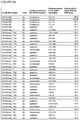
- FIG. 9A is a table showing the deletion frequency and resulting deletion size for selected different dual sgRNA.
- FIGS. 11A-B show the deletion frequency and resulting deletion size for selected different dual sgRNA.
- FIG. 11A is a table showing the deletion frequency and resulting deletion size for selected different dual sgRNA.
- FIG. 11B is a graph showing the deletion frequency and resulting deletion size for selected different dual sgRNA.
- FIGS. 12A-C show the splicing reporter plasmid, pET01; a schematic representation of two different USH2A DNA inserts; and a gel image of RT-PCR products amplified from mRNA of HEK 293 SpCas9 positive cells transfected with 1 of 2 different splice reporter plasmids and 1 of 4 different USH2A IVS40 mutation targeting sgRNAs.
- FIG. 12B depicts two different USH2A DNA inserts (part of mutant intron 40 or part of wild-type intron 40) and vector elements around the inserts.
- the corresponding RNA splice products (mutant splice product or wild-type splice product) are also depicted.
- FIGS. 13A-C show constructs used for a blue fluorescent protein (BFP) splicing reporter assay and possible effects that genome editing can have on the constructs and BFP gene expression therefrom.
- BFP blue fluorescent protein
- FIG. 13C depicts the construct from FIG. 13B in a first configuration, before genome editing, and in a second configuration, after genome editing. After genome editing, BFP expression is expected.
- FIG. 14 shows the percent of live cells that expressed BFP after being subjected to a BFP splicing reporter assay. Data for editing strategies that use single sgRNAs and dual sgRNAs are shown. sgRNAs were paired with SpCas9.
- FIG. 15 shows the percent of live cells that expressed BFP after being subjected to a BFP splicing reporter assay. Data for editing strategies that use dual sgRNAs are shown. These pairs of sgRNAs were paired with SaCas9.
- FIG. 17B shows a sequence of mRNA transcribed from a wild-type USH2A gene (or an edited USH2A gene). Also shown are locations where primers and probes bind to the corresponding cDNA.
- FIG. 18 shows the percent of corrected and uncorrected transcripts in IVS40 mutant cells that have been subjected to genome editing according to the present disclosure. Data for editing strategies that use single sgRNAs and dual sgRNAs are shown. These sgRNAs were paired with SpCas9. Negative controls are also shown.
- SEQ ID NOs: 1-612 are Cas endonuclease ortholog sequences.
- SEQ ID Nos: 613-4696 are microRNA sequences.
- SEQ ID Nos: 4697-5265 are AAV serotype sequences.
- SEQ ID NO: 5266 is a humanUSH2A nucleotide sequence.
- SEQ ID NOs: 5267-5269 show sample sgRNA backbone sequences that SpCas9 is complexed with.
- SEQ ID NO: 5270 is a sample guide RNA (gRNA) for a Streptococcus pyogenes Cas9 endonuclease.
- gRNA sample guide RNA
- SEQ ID NO: 5271 shows a known family of homing endonuclease, as classified by its structure.
- SEQ ID NOs: 5272-5319 are 20 bp spacer sequences for targeting regions upstream and downstream of the IVS40 mutation, within or near intron 40 of the USH2A gene with a S. pyogenes Cas9 endonuclease.
- SEQ ID NO: 5320 is a 20 bp spacer sequence for targeting within or near a USH2A gene or other DNA sequence that encodes a regulatory sequence of the USH2A gene with a S. pyogenes Cas9 endonuclease.
- SEQ ID NO: 5321 is a 20 bp spacer sequence for targeting regions upstream and downstream of the IVS40 mutation, within or near intron 40 of the USH2A gene with a S. pyogenes Cas9 endonuclease.
- SEQ ID NO: 5322 is a 20 bp spacer sequence for targeting within or near a USH2A gene or other DNA sequence that encodes a regulatory sequence of the USH2A gene with a S. pyogenes Cas9 endonuclease.
- SEQ ID NO: 5323 is a 20 bp spacer sequence for targeting regions upstream and downstream of the IVS40 mutation, within or near intron 40 of the USH2A gene with a S. pyogenes Cas9 endonuclease.
- SEQ ID NO: 5324 is a 20 bp spacer sequence for targeting within or near a USH2A gene or other DNA sequence that encodes a regulatory sequence of the USH2A gene with a S. pyogenes Cas9 endonuclease.
- SEQ ID NO: 5325 is a 20 bp spacer sequence for targeting regions upstream and downstream of the IVS40 mutation, within or near intron 40 of the USH2A gene with a S. pyogenes Cas9 endonuclease.
- SEQ ID NO: 5326 is a 20 bp spacer sequence for targeting within or near a USH2A gene or other DNA sequence that encodes a regulatory sequence of the USH2A gene with a S. pyogenes Cas9 endonuclease.
- SEQ ID NOs: 5327-5328 are 20 bp spacer sequences for targeting regions upstream and downstream of the IVS40 mutation, within or near intron 40 of the USH2A gene with a S. pyogenes Cas9 endonuclease.
- SEQ ID NOs: 5329-5385 are sequences that represent the target DNA sequences, for each of 57 sgRNA sequences.
- SEQ ID NOs: 5386-5442 are sequences that represent the reverse strands of the target DNA sequence to which the sgRNA will bind, for each of 57 sgRNA sequences.
- SEQ ID NO: 5443 is an 18 bp spacer sequence for targeting regions downstream of the IVS40 mutation with a S. pyogenes Cas9 endonuclease.
- SEQ ID NO: 5444 is a sequence that represents the target DNA sequence for an 18 bp sgRNA sequence.
- SEQ ID NO: 5445 is a sequence that represents the reverse strand of the target DNA sequence to which the sgRNA will bind for an 18 bp sgRNA sequence.
- SEQ ID NOs: 5446-5461 are 20 bp spacer sequences for targeting regions upstream and downstream of the IVS40 mutation, within or near intron 40 of the USH2A gene with a Staphylococcus aureus Cas9 endonuclease.
- SEQ ID NOs: 5462-5477 are sequences that represent the target DNA sequences, for each of 16 sgRNA sequences.
- SEQ ID NOs: 5478-5493 are sequences that represent the reverse strands of the target DNA sequence to which the sgRNA will bind, for each of 16 sgRNA sequences.
- SEQ ID NO: 5494 is a single-stranded HDR donor sequence.
- SEQ ID NOs: 5495-5506 are PCR primer sequences.
- SEQ ID NOs: 5507-5523 are sgRNA expressing plasmid sequences.
- SEQ ID NO: 5524 is a sequence for pET01 comprising part of wild-type intron 40 of USH2A.
- SEQ ID NO: 5525 is a sequence for pET01 comprising part of mutant intron 40 of USH2A.
- SEQ ID NOs: 5526-5537 show sample sgRNA backbone sequences that SaCas9 is complexed with.
- SEQ ID NOs: 5538-5549 are sgRNA expressing plasmid sequences.
- the sgRNAs associate with SpCas9.
- SEQ ID NOs: 5560 and 5561 are oligonucleotide probes used to detect corrected or uncorrected USH2A cDNA sequences.
- Applicants have discovered a novel method for treating Usher Syndrome Type 2A, e.g., an Usher Syndrome Type 2A associated with an IVS40 mutation in a USH2A gene.
- the method can result in slowing or reversing the development of Usher Syndrome Type 2A or preventing development of disease in an individual.
- a cellular, ex vivo or in vivo method can involve one or a combination of the following methods.
- One method involves disrupting the consensus sequence used as a splice donor site within or near the IVS40 mutation in the USH2A gene by insertions and/or deletions that arise due to the non-homologous end joining (NHEJ) pathway.
- NHEJ non-homologous end joining
- the IVS40 mutation located in intron 40 of the USH2A gene is excised.
- a mutant allele e.g., an IVS40 mutation
- the NHEJ strategy can involve inducing one single-stranded break or double-stranded break within or near the IVS40 mutation in the USH2A gene with one or more CRISPR endonucleases and a gRNA (e.g., cRNA+tracrRNA, or sgRNA).
- a gRNA e.g., cRNA+tracrRNA, or sgRNA.
- This approach edits the sequence within or near the IVS40 mutation and can disrupt the sequence that is causing the incorrect splicing.
- This method utilizes gRNAs or sgRNAs specific for the IVS40 mutation in the USH2A gene.
- the excision strategy can include a set of guide RNAs or sgRNAs that bind upstream and downstream of the IVS40 mutation, within intron 40 of the USH2A gene and excise an area of the genome containing the IVS40 mutation.
- This strategy can be expected to affect both the mutant (Mut) and the wild-type (WT) alleles, which is permissible with intronic mutations.
- the excision strategy can result in a shorter version of the nascent precursor messenger RNA (pre-mRNA, missing the dominant splice donor creating mutation), which can be spliced correctly, leading to WT mRNA and expression of WT usherin protein in edited cells, as depicted in FIG. 3 .
- the edited cell's function and survival can be expected to improve in cases where enough supporting retinal structure is still available.
- This method utilizes gRNAs or sgRNAs specific for the regions upstream and downstream of the IVS40 mutation, within intron 40 of the USH2A gene.
- deletions created by the excision strategy can be from 50 to 5000 base pairs (bp) in size.
- deletions can range from 50-100; 50-250; 50-500; 50-1000; 50-1500; 50-2000; 200-500; 200-750; 200-1000; 200-1100; 500-1,000; 1,000-1,500; 1,500-2,000; 1,000-2,000; 2,000-2,500; 2,500-3,000; 3,000-3,500; 3,500-4,000; 4,000-4,500; 4,500-5,000 or 50-2,900 base pairs in size.
- the HDR strategy can involve inducing one or more single-stranded breaks or double-stranded breaks upstream and downstream of the IVS40 mutation, within or near intron 40 of the USH2A gene with one or more CRISPR endonucleases and a gRNA (e.g., crRNA+tracrRNA, or sgRNA), or two or more single-stranded breaks or double-stranded breaks upstream and downstream of the IVS40 mutation within or near intron 40 of the USH2A gene with one or more CRISPR endonucleases (Cas9, Cpf1 and the like) and two or more gRNAs, in the presence of a donor DNA template introduced exogenously to direct the cellular DSB response to Homology-Directed Repair.
- gRNA e.g., crRNA+tracrRNA, or sgRNA
- the donor DNA template can be a short single-stranded oligonucleotide, a short double-stranded oligonucleotide, a long single or double-stranded DNA molecule.
- the methods can provide gRNA pairs that make a deletion by cutting the gene twice, one gRNA cutting at the 5′ end of the IVS40 mutation and the other gRNA cutting at the 3′ end of the IVS40 mutation that facilitates insertion of a new sequence from a polynucleotide donor template to replace the IVS40 mutation in the USH2A gene.
- the cutting can be accomplished by a pair of DNA endonucleases that each makes a DSB (one DSB on each end of the IVS40 mutation within or near intron 40), or by multiple nickases that together make a DSB (one DSB on each end of the IVS40 mutation within or near intron 40).
- This method utilizes gRNAs or sgRNAs specific for regions upstream and downstream of the IVS40 mutation, within intron 40 of the USH2A gene. This method also utilizes donor DNA molecules.
- Such methods use endonucleases, such as CRISPR-associated (Cas9, Cpf1 and the like) nucleases, to stably correct the IVS40 mutation within the genomic locus of the USH2A gene.
- CRISPR-associated (Cas9, Cpf1 and the like) nucleases Any CRISPR endonuclease can be used in the methods of the present disclosure, each CRISPR endonuclease having its own associated PAM, which can or cannot be disease specific.
- pyogenes have been identified in SEQ ID NOs: 5272-5319, 5321, 5323, 5325, 5327-5328, and 5443 of the Sequence Listing.
- gRNA spacer sequences for targeting the IVS40 mutation in the USH2A gene with a CRISPR/Cas9 endonuclease from S. aureus have been identified in SEQ ID NOs: 5446-5461 of the Sequence Listing.
- Usher syndrome is an autosomal recessive disease, characterized by sensorineural hearing loss, retinitis pigmentosa (RP) and in some cases, vestibular dysfunction.
- RP retinitis pigmentosa
- the prevalence of Usher Syndrome has been estimated to be between 1/6000 and 1/25000.
- Usher Syndrome is a clinically and genetically heterogeneous disease, accounting for about half of all cases of combined hereditary deafness-blindness. To date the disease has been associated with 13 genes. Three clinical forms of the disease have been identified (USH I, II, and III) based on the severity of the hearing impairment, the presence or absence of vestibular dysfunction, and the age of onset of the disease.
- Usher Syndrome Type II is the most frequent clinical form accounting for approximately 50% of all Usher Syndrome cases.
- Usher Syndrome Type II is characterized by congenital hearing loss and progressive vision loss starting in adolescence or adulthood. The hearing loss ranges from mild to severe and mainly affects the ability to hear high-frequency sounds. Vision loss occurs as the light-sensing cells of the retina gradually deteriorate. Night vision loss begins first, followed by loss of the peripheral vision. With time, these blind areas enlarge and merge to produce tunnel vision. In some cases, vision is further impaired by cataracts. Many patients become legally blind in the 5 th decade of life.
- Usher Syndrome Type 2A is due to a mutation in the USH2A gene and accounts for approximately 80% of all Usher Syndrome Type II cases and 40% of all Usher Syndrome cases.
- the usherin protein is located next to vesicle loading point at the periciliary membrane and may play a role in vesicle transport between the inner segments and the outer segments of photoreceptors.
- the C.7595-2144A>G (IVS40 mutation) in the USH2A gene leads to the creation of a splice donor site and insertion of 152 bp into the USH2A mRNA, which in turn leads to a frameshift and a truncated and non-functional protein.
- any one or more of the mutations can be repaired in order to restore the usherin protein function.
- the pathological variant, IVS40 can be excised or corrected to restore the usherin protein expression (See Table 1).
- the method is an in vivo cell-based therapy.
- Chromosomal DNA of the cells in the Usher Syndrome type 2A patient can be edited using the materials and methods described herein.
- the in-vivo method can comprise editing an IVS40 mutation in a USH2A gene in a cell of a patient, such as photoreceptor cells or retinal progenitor cells.
- RNA and protein remain in the cell can also be adjusted using treatments or domains added to change the half-life.
- In vivo treatment would eliminate a number of treatment steps, but a lower rate of delivery can require higher rates of editing. In vivo treatment can eliminate problems and losses from ex vivo treatment and engraftment.
- An advantage of in vivo gene therapy can be the ease of therapeutic production and administration.
- the same therapeutic approach and therapy will have the potential to be used to treat more than one patient, for example a number of patients who share the same or similar genotype or allele.
- ex vivo cell therapy typically requires using a patient's own cells, which are isolated, manipulated and returned to the same patient.
- a patient-specific induced pluripotent stem cell iPSC
- the chromosomal DNA of these iPSC cells can be edited using the materials and methods described herein.
- the method can comprise editing within or near an IVS40 mutation in a USH2A gene of the iPSC.
- the genome-edited iPSCs can be differentiated into other cells, such as photoreceptor cells or retinal progenitor cells.
- the differentiated cells such as photoreceptor cell or retinal progenitor cell, can be implanted into the patient (i.e., implanted into the patient's eye).
- photoreceptor cells or retinal progenitor cells can be isolated from the patient.
- the chromosomal DNA of these photoreceptor cells or retinal progenitor cells can be edited using the materials and methods described herein.
- the method can comprise editing within or near an IVS40 mutation in a USH2A gene of the photoreceptor cells or retinal progenitor cells.
- the genome-edited photoreceptor cells or retinal progenitor cells can be implanted into the patient (i.e., implanted into the patient's eye).
- a mesenchymal stem cell can be isolated from the patient, which can be isolated from the patient's bone marrow, peripheral blood, adipose tissue, or umbilical cord.
- the chromosomal DNA of these mesenchymal stem cells can be edited using the materials and methods described herein.
- the method can comprise editing within or near an IVS40 mutation in a USH2A gene of the mesenchymal stem cells.
- the genome-edited mesenchymal stem cells can be differentiated into any type of cell, e.g., photoreceptor cells or retinal progenitor cells.
- the differentiated cells e.g., photoreceptor cells or retinal progenitor cells can be implanted into the patient (i.e., implanted into the patient's eye).
- Nuclease-based therapeutics can have some level of off-target effects.
- Performing gene correction ex vivo allows one to characterize the corrected cell population prior to implantation.
- the present disclosure includes sequencing the entire genome of the corrected cells to ensure that the off-target effects, if any, can be in genomic locations associated with minimal risk to the patient.
- populations of specific cells, including clonal populations can be isolated prior to implantation.
- iPSCs are prolific, making it easy to obtain the large number of cells that will be required for a cell-based therapy.
- iPSCs are an ideal cell type for performing clonal isolations. This allows screening for the correct genomic correction, without risking a decrease in viability.
- other primary cells such as photoreceptor cells or retinal progenitor cells, are viable for only a few passages and difficult to clonally expand.
- manipulation of iPSCs for the treatment of Usher Syndrome Type 2A can be much easier, and can shorten the amount of time needed to make the desired genetic correction.
- Genome editing refers to the process of modifying the nucleotide sequence of a genome, such as in a precise or pre-determined manner.
- methods of genome editing described herein include methods of using site-directed nucleases to cut DNA at precise target locations in the genome, thereby creating single-strand or double-strand DNA breaks at particular locations within the genome. Such breaks can be and regularly are repaired by natural, endogenous cellular processes, such as HDR and NHEJ. These two main DNA repair processes consist of a family of alternative pathways. NHEJ directly joins the DNA ends resulting from a double-strand break, sometimes with the loss or addition of nucleotide sequence, which may disrupt or enhance gene expression.
- HDR utilizes a homologous sequence, or donor sequence, as a template for inserting a defined DNA sequence at the break point.
- the homologous sequence can be in the endogenous genome, such as a sister chromatid.
- the donor can be an exogenous nucleic acid, such as a plasmid, a single-strand oligonucleotide, a double-stranded oligonucleotide, a duplex oligonucleotide or a virus, that has regions of high homology with the nuclease-cleaved locus, but which can also contain additional sequence or sequence changes including deletions that can be incorporated into the cleaved target locus.
- a third repair mechanism can be microhomology-mediated end joining (MMEJ), also referred to as “Alternative NHEJ (ANHEJ)”, in which the genetic outcome is similar to NHEJ in that small deletions and insertions can occur at the cleavage site.
- MMEJ can make use of homologous sequences of a few base pairs flanking the DNA break site to drive a more favored DNA end joining repair outcome, and recent reports have further elucidated the molecular mechanism of this process. In some instances, it may be possible to predict likely repair outcomes based on analysis of potential microhomologies at the site of the DNA break.
- a step in the genome editing process can be to create one or two DNA breaks, the latter as double-strand breaks or as two single-stranded breaks, in the target locus as near the site of intended mutation. This can be achieved via the use of site-directed polypeptides, as described and illustrated herein.
- Site-directed polypeptides can introduce double-strand breaks or single-strand breaks in nucleic acids, e.g., genomic DNA.
- the double-strand break can stimulate a cell's endogenous DNA-repair pathways [e.g., homology-dependent repair (HDR) or non-homologous end joining (NHEJ) or (ANHEJ) or (MMEJ)].
- NHEJ can repair cleaved target nucleic acid without the need for a homologous template. This can sometimes result in small deletions or insertions (indels) in the target nucleic acid at the site of cleavage, and can lead to disruption or alteration of gene expression.
- HDR can occur when a homologous repair template, or donor, is available.
- the homologous donor template can comprise at least a portion of the wild-type USH2A gene, or cDNA.
- the at least a portion of the wild-type USH2A gene or cDNA can be exon 1, exon 2, exon 3, exon 4, exon 5, exon 6, exon 7, exon 8, exon 9, exon 10, exon 11, exon 12, exon 13, exon 14, exon 15, exon 16, exon 17, exon 18, exon 19, exon 20, exon 21, exon 22, exon 23, exon 24, exon 25, exon 26, exon 27, exon 28, exon 29, exon 30, exon 31, exon 32, exon 33, exon 34, exon 35, exon 36, exon 37, exon 38, exon 39, exon 40, exon 41, exon 42, exon 43, exon 44, exon 45, exon 46, exon 47, exon 48, exon 49, exon 50, exon 51, exon 52, exon 53
- the donor template can be 250 bp to 500 bp.
- the donor template can be 100 to 250 bp.
- the donor template can be delivered by AAV.
- the homologous donor template can comprise sequences that can be homologous to sequences flanking the target nucleic acid cleavage site.
- the donor template can have homologous arms to the 1q41 region.
- the donor template can also have homologous arms to the pathological variant IVS40.
- the sister chromatid can be used by the cell as the repair template.
- the repair template can be supplied as an exogenous nucleic acid, such as a plasmid, duplex oligonucleotide, single-strand oligonucleotide, double-stranded oligonucleotide, or viral nucleic acid.
- an additional nucleic acid sequence such as a transgene
- modification such as a single or multiple base change or a deletion
- MMEJ can result in a genetic outcome that is similar to NHEJ in that small deletions and insertions can occur at the cleavage site.
- MMEJ can make use of homologous sequences of a few base pairs flanking the cleavage site to drive a favored end-joining DNA repair outcome. In some instances, it may be possible to predict likely repair outcomes based on analysis of potential microhomologies in the nuclease target regions.
- homologous recombination can be used to insert an exogenous polynucleotide sequence into the target nucleic acid cleavage site.
- An exogenous polynucleotide sequence is termed a donor polynucleotide (or donor or donor sequence or polynucleotide donor template) herein.
- the donor polynucleotide, a portion of the donor polynucleotide, a copy of the donor polynucleotide, or a portion of a copy of the donor polynucleotide can be inserted into the target nucleic acid cleavage site.
- the donor polynucleotide can be an exogenous polynucleotide sequence, i.e., a sequence that does not naturally occur at the target nucleic acid cleavage site.
- the modifications of the target DNA due to NHEJ and/or HDR can lead to, for example, gene correction.
- a CRISPR (Clustered Regularly Interspaced Short Palindromic Repeats) genomic locus can be found in the genomes of many prokaryotes (e.g., bacteria and archaea). In prokaryotes, the CRISPR locus encodes products that function as a type of immune system to help defend the prokaryotes against foreign invaders, such as virus and phage. There are three stages of CRISPR locus function: integration of new sequences into the CRISPR locus, expression of CRISPR RNA (crRNA), and silencing of foreign invader nucleic acid. Five types of CRISPR systems (e.g., Type I, Type II, Type III, Type U, and Type V) have been identified.
- a CRISPR locus includes a number of short repeating sequences referred to as “repeats.” When expressed, the repeats can form secondary structures (e.g., hairpins) and/or comprise unstructured single-stranded sequences.
- the repeats usually occur in clusters and frequently diverge between species.
- the repeats are regularly interspaced with unique intervening sequences referred to as “spacers,” resulting in a repeat-spacer-repeat locus architecture.
- the spacers are identical to or have high homology with known foreign invader sequences.
- a spacer-repeat unit encodes a crisprRNA (crRNA), which is processed into a mature form of the spacer-repeat unit.
- crRNA crisprRNA
- a CRISPR locus also comprises polynucleotide sequences encoding CRISPR Associated (Cas) genes.
- Cas genes encode endonucleases involved in the biogenesis and the interference stages of crRNA function in prokaryotes. Some Cas genes comprise homologous secondary and/or tertiary structures.
- the crRNA of the crRNA-tracrRNA-Cas9 complex can guide the complex to a target nucleic acid to which the crRNA can hybridize. Hybridization of the crRNA to the target nucleic acid can activate Cas9 for targeted nucleic acid cleavage.
- the target nucleic acid in a Type II CRISPR system is referred to as a protospacer adjacent motif (PAM).
- PAM protospacer adjacent motif
- the PAM is essential to facilitate binding of a site-directed polypeptide (e.g., Cas9) to the target nucleic acid.
- Type II systems also referred to as Nmeni or CASS4 are further subdivided into Type II-A (CASS4) and II-B (CASS4a).
- Type V CRISPR systems have several important differences from Type II systems.
- Cpf1 is a single RNA-guided endonuclease that, in contrast to Type II systems, lacks tracrRNA.
- Cpf1-associated CRISPR arrays can be processed into mature crRNAs without the requirement of an additional trans-activating tracrRNA.
- the Type V CRISPR array can be processed into short mature crRNAs of 42-44 nucleotides in length, with each mature crRNA beginning with 19 nucleotides of direct repeat followed by 23-25 nucleotides of spacer sequence.
- mature crRNAs in Type II systems can start with 20-24 nucleotides of spacer sequence followed by about 22 nucleotides of direct repeat.
- Cpf1 can utilize a T-rich protospacer-adjacent motif such that Cpf1-crRNA complexes efficiently cleave target DNA preceded by a short T-rich PAM, which is in contrast to the G-rich PAM following the target DNA for Type II systems.
- Type V systems cleave at a point that is distant from the PAM
- Type II systems cleave at a point that is adjacent to the PAM.
- Cpf1 cleaves DNA via a staggered DNA double-stranded break with a 4 or 5 nucleotide 5′ overhang.
- Type II systems cleave via a blunt double-stranded break.
- Cpf1 contains a predicted RuvC-like endonuclease domain, but lacks a second HNH endonuclease domain, which is in contrast to Type II systems.
- Exemplary CRISPR/Cas polypeptides include the Cas9 polypeptides in FIG. 1 of Fonfara et al., Nucleic Acids Research, 42: 2577-2590 (2014).
- the CRISPR/Cas gene naming system has undergone extensive rewriting since the Cas genes were discovered.
- FIG. 5 of Fonfara, supra provides PAM sequences for the Cas9 polypeptides from various species.
- a site-directed polypeptide is a nuclease used in genome editing to cleave DNA.
- the site-directed nuclease can be administered to a cell or a patient as either: one or more polypeptides, or one or more mRNAs encoding the polypeptide. Any of the enzymes or orthologs listed in SEQ ID NOs: 1-612, or disclosed herein, can be utilized in the methods herein.
- a site-directed polypeptide can comprise a plurality of nucleic acid-cleaving (i.e., nuclease) domains. Two or more nucleic acid-cleaving domains can be linked together via a linker.
- the linker can comprise a flexible linker.
- Linkers can comprise 1, 2, 3, 4, 5, 6, 7, 8, 9, 10, 11, 12, 13, 14, 15, 16, 17, 18, 19, 20, 21, 22, 23, 24, 25, 30, 35, 40 or more amino acids in length.
- Naturally-occurring wild-type Cas9 enzymes comprise two nuclease domains, a HNH nuclease domain and a RuvC domain.
- the “Cas9” refers to both naturally occurring and recombinant Cas9s.
- Cas9 enzymes contemplated herein can comprise a HNH or HNH-like nuclease domain, and/or a RuvC or RuvC-like nuclease domain.
- HNH or HNH-like domains comprise a McrA-like fold.
- HNH or HNH-like domains comprises two antiparallel ⁇ -strands and an ⁇ -helix.
- HNH or HNH-like domains comprises a metal binding site (e.g., a divalent cation binding site).
- HNH or HNH-like domains can cleave one strand of a target nucleic acid (e.g., the complementary strand of the crRNA targeted strand).
- RuvC or RuvC-like domains comprise an RNaseH or RNaseH-like fold.
- RuvC/RNaseH domains are involved in a diverse set of nucleic acid-based functions including acting on both RNA and DNA.
- the RNaseH domain comprises 5 ⁇ -strands surrounded by a plurality of ⁇ -helices.
- RuvC/RNaseH or RuvC/RNaseH-like domains comprise a metal binding site (e.g., a divalent cation binding site).
- RuvC/RNaseH or RuvC/RNaseH-like domains can cleave one strand of a target nucleic acid (e.g., the non-complementary strand of a double-stranded target DNA).
- Site-directed polypeptides can introduce double-strand breaks or single-strand breaks in nucleic acids, e.g., genomic DNA.
- the double-strand break can stimulate a cell's endogenous DNA-repair pathways (e.g., HDR or NHEJ or ANHEJ or MMEJ).
- NHEJ can repair cleaved target nucleic acid without the need for a homologous template. This can sometimes result in small deletions or insertions (indels) in the target nucleic acid at the site of cleavage, and can lead to disruption or alteration of gene expression.
- HDR can occur when a homologous repair template, or donor, is available.
- the homologous donor template can comprise sequences that are homologous to sequences flanking the target nucleic acid cleavage site.
- the sister chromatid can be used by the cell as the repair template.
- the repair template can be supplied as an exogenous nucleic acid, such as a plasmid, duplex oligonucleotide, single-strand oligonucleotide or viral nucleic acid.
- an additional nucleic acid sequence such as a transgene
- modification such as a single or multiple base change or a deletion
- homologous recombination can be used to insert an exogenous polynucleotide sequence into the target nucleic acid cleavage site.
- An exogenous polynucleotide sequence is termed a donor polynucleotide (or donor or donor sequence) herein.
- the donor polynucleotide, a portion of the donor polynucleotide, a copy of the donor polynucleotide, or a portion of a copy of the donor polynucleotide can be inserted into the target nucleic acid cleavage site.
- the donor polynucleotide can be an exogenous polynucleotide sequence, i.e., a sequence that does not naturally occur at the target nucleic acid cleavage site.
- the site-directed polypeptide can comprise an amino acid sequence having at least 10%, at least 15%, at least 20%, at least 30%, at least 40%, at least 50%, at least 60%, at least 70%, at least 75%, at least 80%, at least 85%, at least 90%, at least 95%, at least 99%, or 100% amino acid sequence identity to a wild-type exemplary site-directed polypeptide [e.g., Cas9 from S. pyogenes , US2014/0068797 Sequence ID No. 8 or Sapranauskas et al., Nucleic Acids Res, 39(21): 9275-9282 (2011)], and various other site-directed polypeptides.
- a wild-type exemplary site-directed polypeptide e.g., Cas9 from S. pyogenes , US2014/0068797 Sequence ID No. 8 or Sapranauskas et al., Nucleic Acids Res, 39(21): 9275-9282 (2011)
- the site-directed polypeptide can comprise at least 70, 75, 80, 85, 90, 95, 97, 99, or 100% identity to a wild-type site-directed polypeptide (e.g., Cas9 from S. pyogenes , supra) over 10 contiguous amino acids.
- the site-directed polypeptide can comprise at most: 70, 75, 80, 85, 90, 95, 97, 99, or 100% identity to a wild-type site-directed polypeptide (e.g., Cas9 from S. pyogenes , supra) over 10 contiguous amino acids.
- the site-directed polypeptide can comprise at least: 70, 75, 80, 85, 90, 95, 97, 99, or 100% identity to a wild-type site-directed polypeptide (e.g., Cas9 from S. pyogenes , supra) over 10 contiguous amino acids in a RuvC nuclease domain of the site-directed polypeptide.
- the site-directed polypeptide can comprise at most: 70, 75, 80, 85, 90, 95, 97, 99, or 100% identity to a wild-type site-directed polypeptide (e.g., Cas9 from S. pyogenes , supra) over 10 contiguous amino acids in a RuvC nuclease domain of the site-directed polypeptide.
- the site-directed polypeptide can comprise a modified form of a wild-type exemplary site-directed polypeptide.
- the modified form of the wild-type exemplary site-directed polypeptide can comprise a mutation that reduces the nucleic acid-cleaving activity of the site-directed polypeptide.
- the modified form of the wild-type exemplary site-directed polypeptide can have less than 90%, less than 80%, less than 70%, less than 60%, less than 50%, less than 40%, less than 30%, less than 20%, less than 10%, less than 5%, or less than 1% of the nucleic acid-cleaving activity of the wild-type exemplary site-directed polypeptide (e.g., Cas9 from S. pyogenes , supra).
- the modified form of the site-directed polypeptide can have no substantial nucleic acid-cleaving activity.
- a site-directed polypeptide is a modified form that has no substantial nucleic acid-cleaving activity, it is referred to herein as “enzymatically inactive.”
- the modified form of the site-directed polypeptide can comprise a mutation such that it can induce a SSB on a target nucleic acid (e.g., by cutting only one of the sugar-phosphate backbones of a double-strand target nucleic acid).
- the mutation can result in less than 90%, less than 80%, less than 70%, less than 60%, less than 50%, less than 40%, less than 30%, less than 20%, less than 10%, less than 5%, or less than 1% of the nucleic acid-cleaving activity in one or more of the plurality of nucleic acid-cleaving domains of the wild-type site directed polypeptide (e.g., Cas9 from S. pyogenes , supra).
- the mutation can result in one or more of the plurality of nucleic acid-cleaving domains retaining the ability to cleave the complementary strand of the target nucleic acid, but reducing its ability to cleave the non-complementary strand of the target nucleic acid.
- the mutation can result in one or more of the plurality of nucleic acid-cleaving domains retaining the ability to cleave the non-complementary strand of the target nucleic acid, but reducing its ability to cleave the complementary strand of the target nucleic acid. For example, residues in the wild-type exemplary S.
- pyogenes Cas9 polypeptide such as Asp10, His840, Asn854 and Asn856, are mutated to inactivate one or more of the plurality of nucleic acid-cleaving domains (e.g., nuclease domains).
- the residues to be mutated can correspond to residues Asp10, His840, Asn854 and Asn856 in the wild-type exemplary S. pyogenes Cas9 polypeptide (e.g., as determined by sequence and/or structural alignment).
- Non-limiting examples of mutations include D10A, H840A, N854A or N856A. Mutations other than alanine substitutions can be suitable.
- a D10A mutation can be combined with one or more of H840A, N854A, or N856A mutations to produce a site-directed polypeptide substantially lacking DNA cleavage activity.
- a H840A mutation can be combined with one or more of D10A, N854A, or N856A mutations to produce a site-directed polypeptide substantially lacking DNA cleavage activity.
- a N854A mutation can be combined with one or more of H840A, D10A, or N856A mutations to produce a site-directed polypeptide substantially lacking DNA cleavage activity.
- a N856A mutation can be combined with one or more of H840A, N854A, or D10A mutations to produce a site-directed polypeptide substantially lacking DNA cleavage activity.
- Site-directed polypeptides that comprise one substantially inactive nuclease domain are referred to as “nickases”.
- RNA-guided endonucleases for example Cas9
- Wild type Cas9 is typically guided by a single guide RNA designed to hybridize with a specified ⁇ 20 nucleotide sequence in the target sequence (such as an endogenous genomic locus).
- a specified ⁇ 20 nucleotide sequence in the target sequence such as an endogenous genomic locus.
- several mismatches can be tolerated between the guide RNA and the target locus, effectively reducing the length of required homology in the target site to, for example, as little as 13 nt of homology, and thereby resulting in elevated potential for binding and double-strand nucleic acid cleavage by the CRISPR/Cas9 complex elsewhere in the target genome—also known as off-target cleavage.
- nickase variants of Cas9 each only cut one strand, in order to create a double-strand break it is necessary for a pair of nickases to bind in close proximity and on opposite strands of the target nucleic acid, thereby creating a pair of nicks, which is the equivalent of a double-strand break.
- nickases can also be used to promote HDR versus NHEJ.
- HDR can be used to introduce selected changes into target sites in the genome through the use of specific donor sequences that effectively mediate the desired changes.
- Mutations contemplated can include substitutions, additions, and deletions, or any combination thereof.
- the mutation converts the mutated amino acid to alanine.
- the mutation converts the mutated amino acid to another amino acid (e.g., glycine, serine, threonine, cysteine, valine, leucine, isoleucine, methionine, proline, phenylalanine, tyrosine, tryptophan, aspartic acid, glutamic acid, asparagine, glutamine, histidine, lysine, or arginine).
- the mutation converts the mutated amino acid to a non-natural amino acid (e.g., selenomethionine).
- the mutation converts the mutated amino acid to amino acid mimics (e.g., phosphomimics).
- the mutation can be a conservative mutation.
- the mutation converts the mutated amino acid to amino acids that resemble the size, shape, charge, polarity, conformation, and/or rotamers of the mutated amino acids (e.g., cysteine/serine mutation, lysine/asparagine mutation, histidine/phenylalanine mutation).
- the mutation can cause a shift in reading frame and/or the creation of a premature stop codon. Mutations can cause changes to regulatory regions of genes or loci that affect expression of one or more genes.
- the site-directed polypeptide (e.g., variant, mutated, enzymatically inactive and/or conditionally enzymatically inactive site-directed polypeptide) can target nucleic acid.
- the site-directed polypeptide (e.g., variant, mutated, enzymatically inactive and/or conditionally enzymatically inactive endoribonuclease) can target DNA.
- the site-directed polypeptide e.g., variant, mutated, enzymatically inactive and/or conditionally enzymatically inactive endoribonuclease
- the site-directed polypeptide can comprise one or more non-native sequences (e.g., the site-directed polypeptide is a fusion protein).
- the site-directed polypeptide can comprise an amino acid sequence comprising at least 15% amino acid identity to a Cas9 from a bacterium (e.g., S. pyogenes ), a nucleic acid binding domain, and two nucleic acid cleaving domains (i.e., a HNH domain and a RuvC domain).
- a Cas9 from a bacterium e.g., S. pyogenes
- a nucleic acid binding domain e.g., S. pyogenes
- two nucleic acid cleaving domains i.e., a HNH domain and a RuvC domain
- the site-directed polypeptide can comprise an amino acid sequence comprising at least 15% amino acid identity to a Cas9 from a bacterium (e.g., S. pyogenes ), and two nucleic acid cleaving domains (i.e., a HNH domain and a RuvC domain).
- a Cas9 from a bacterium e.g., S. pyogenes
- two nucleic acid cleaving domains i.e., a HNH domain and a RuvC domain
- the site-directed polypeptide can comprise an amino acid sequence comprising at least 15% amino acid identity to a Cas9 from a bacterium (e.g., S. pyogenes ), and two nucleic acid cleaving domains, wherein one or both of the nucleic acid cleaving domains comprise at least 50% amino acid identity to a nuclease domain from Cas9 from a bacterium (e.g., S. pyogenes ).
- a bacterium e.g., S. pyogenes
- the site-directed polypeptide can comprise an amino acid sequence comprising at least 15% amino acid identity to a Cas9 from a bacterium (e.g., S. pyogenes ), two nucleic acid cleaving domains (i.e., a HNH domain and a RuvC domain), and non-native sequence (for example, a nuclear localization signal) or a linker linking the site-directed polypeptide to a non-native sequence.
- a Cas9 from a bacterium (e.g., S. pyogenes ), two nucleic acid cleaving domains (i.e., a HNH domain and a RuvC domain), and non-native sequence (for example, a nuclear localization signal) or a linker linking the site-directed polypeptide to a non-native sequence.
- a bacterium e.g., S. pyogenes
- two nucleic acid cleaving domains i.e
- the site-directed polypeptide can comprise an amino acid sequence comprising at least 15% amino acid identity to a Cas9 from a bacterium (e.g., S. pyogenes ), two nucleic acid cleaving domains (i.e., a HNH domain and a RuvC domain), wherein the site-directed polypeptide comprises a mutation in one or both of the nucleic acid cleaving domains that reduces the cleaving activity of the nuclease domains by at least 50%.
- a bacterium e.g., S. pyogenes
- two nucleic acid cleaving domains i.e., a HNH domain and a RuvC domain
- the site-directed polypeptide can comprise an amino acid sequence comprising at least 15% amino acid identity to a Cas9 from a bacterium (e.g., S. pyogenes ), and two nucleic acid cleaving domains (i.e., a HNH domain and a RuvC domain), wherein one of the nuclease domains comprises mutation of aspartic acid 10, and/or wherein one of the nuclease domains can comprise a mutation of histidine 840, and wherein the mutation reduces the cleaving activity of the nuclease domain(s) by at least 50%.
- a Cas9 from a bacterium
- two nucleic acid cleaving domains i.e., a HNH domain and a RuvC domain
- one of the nuclease domains comprises mutation of aspartic acid 10
- one of the nuclease domains can comprise a mutation of histidine 840, and wherein the mutation reduces the cle
- the one or more site-directed polypeptides can comprise two nickases that together effect one double-strand break at a specific locus in the genome, or four nickases that together effect or cause two double-strand breaks at specific loci in the genome.
- one site-directed polypeptide e.g., DNA endonuclease
- Non-limiting examples of Cas9 orthologs from other bacterial strains including but not limited to, Cas proteins identified in Acaryochloris marina MBIC11017; Acetohalobium arabaticum DSM 5501; Acidithiobacillus caldus; Acidithiobacillus ferrooxidans ATCC 23270; Alicyclobacillus acidocaldarius LAA1; Alicyclobacillus acidocaldarius subsp. acidocaldarius DSM 446; Allochromatium vinosum DSM 180; Ammonifex degensii KC4; Anabaena variabilis ATCC 29413; Arthrospira maxima CS-328; Arthrospira platensis str.
- PCC 6506 Pelotomaculum _ thermopropionicum _SI; Petrotoga mobilis SJ95; Polaromonas naphthalenivorans CJ2; Polaromonas sp. JS666; Pseudoalteromonas haloplanktis TAC125; Streptomyces pristinaespiralis ATCC 25486; Streptomyces pristinaespiralis ATCC 25486; Streptococcus thermophilus; Streptomyces viridochromogenes DSM 40736; Streptosporangium roseum DSM 43021; Synechococcus sp. PCC 7335; and Thermosipho africanus TCF52B (Chylinski et al., RNA Biol., 2013; 10(5): 726-737.
- Cas9 orthologs In addition to Cas9 orthologs, other Cas9 variants such as fusion proteins of inactive dCas9 and effector domains with different functions can be served as a platform for genetic modulation. Any of the foregoing enzymes can be useful in the present disclosure.
- endonucleases that can be utilized in the present disclosure are provided in SEQ ID NOs: 1-612. These proteins can be modified before use or can be encoded in a nucleic acid sequence such as a DNA, RNA or mRNA or within a vector construct such as the plasmids or adeno-associated virus (AAV) vectors taught herein. Further, they can be codon optimized.
- AAV adeno-associated virus
- SpCas9 indicates that the Cas9 gene/protein in question originated in Streptococcus pyogenes and was modified, such as by addition of NLS(s) and/or the performance of codon optimization.
- SpCas9 indicates that the Cas9 gene/protein in question originated in Staphylococcus aureus and was modified, such as by addition of NLS(s) and/or the performance of codon optimization.
- the present disclosure provides a genome-targeting nucleic acid that can direct the activities of an associated polypeptide (e.g., a site-directed polypeptide) to a specific target sequence within a target nucleic acid.
- the genome-targeting nucleic acid can be an RNA.
- a genome-targeting RNA is referred to as a “guide RNA” or “gRNA” herein.
- a guide RNA can comprise at least a spacer sequence that hybridizes to a target nucleic acid sequence of interest, and a CRISPR repeat sequence.
- the gRNA also comprises a second RNA called the tracrRNA sequence.
- the CRISPR repeat sequence and tracrRNA sequence hybridize to each other to form a duplex.
- the crRNA forms a duplex.
- the duplex can bind a site-directed polypeptide, such that the guide RNA and site-direct polypeptide form a complex.
- the genome-targeting nucleic acid can provide target specificity to the complex by virtue of its association with the site-directed polypeptide. The genome-targeting nucleic acid thus can direct the activity of the site-directed polypeptide.
- Exemplary guide RNAs include the spacer sequences in SEQ ID NOs: 5272-5319, 5321, 5323, 5325, 5327-5328, 5443 and 5446-5461 of the Sequence Listing ( FIGS. 2A-B , G, and J).
- the target DNA sequence (5′-3′) (See SEQ ID NOs: 5329-5385, 5444, and 5462-5477) can be found in FIGS. 2C-D , 2 H, and 2 K.
- the reverse strand of target DNA sequence to which the sgRNA will bind (5′-3′) can be found in FIGS. 2E-F , I, and L.
- Each guide RNA can be designed to include a spacer sequence complementary to its genomic target sequence.
- each of the spacer sequences in SEQ ID NOs: 5272-5319, 5321, 5323, 5325, 5327-5328, 5443 and 5446-5461 of the Sequence Listing can be put into a single RNA chimera or a crRNA (along with a corresponding tracrRNA). See Jinek et al., Science, 337, 816-821 (2012) and Deltcheva et al., Nature, 471, 602-607 (2011).
- gRNAs or sgRNAs disclosed herein can associate with a DNA endonuclease to form a ribonucleoprotein complex, which can cause stable edits within or near the IVS40 mutation in a USH2A gene. Changes in DNA sequences (e.g., edits) brought about by this editing can be non-transient.
- the genome-targeting nucleic acid can be a double-molecule guide RNA ( FIG. 1A ).
- the genome-targeting nucleic acid can be a single-molecule guide RNA ( FIG. 1B ).
- the double-molecule guide RNA or single-molecule guide RNA can be modified.
- a double-molecule guide RNA can comprise two strands of RNA.
- the first strand comprises in the 5′ to 3′ direction, an optional spacer extension sequence, a spacer sequence and a minimum CRISPR repeat sequence.
- the second strand can comprise a minimum tracrRNA sequence (complementary to the minimum CRISPR repeat sequence), a 3′ tracrRNA sequence and an optional tracrRNA extension sequence.
- a single-molecule guide RNA (sgRNA) in a Type II system can comprise, in the 5′ to 3′ direction, an optional spacer extension sequence, a spacer sequence, a minimum CRISPR repeat sequence, a single-molecule guide linker, a minimum tracrRNA sequence, a 3′ tracrRNA sequence and an optional tracrRNA extension sequence.
- the optional tracrRNA extension can comprise elements that contribute additional functionality (e.g., stability) to the guide RNA.
- the single-molecule guide linker can link the minimum CRISPR repeat and the minimum tracrRNA sequence to form a hairpin structure.
- the optional tracrRNA extension can comprise one or more hairpins.
- the sgRNA can comprise a variable length spacer sequence with 17-30 nucleotides at the 5′ end of the sgRNA sequence (Table 2). In other examples, the sgRNA can comprise a variable length spacer sequence with 17-24 nucleotides at the 5′ end of the sgRNA sequence.
- the sgRNA can comprise a 20 nucleotide spacer sequence at the 5′ end of the sgRNA sequence.
- the sgRNA can comprise a less than 20 nucleotide spacer sequence at the 5′ end of thesgRNA sequence.
- the sgRNA can comprise a 19 nucleotide spacer sequence at the 5′ end of thesgRNA sequence.
- the sgRNA can comprise a 18 nucleotide spacer sequence at the 5′ end of thesgRNA sequence.
- the sgRNA can comprise a 17 nucleotide spacer sequence at the 5′ end of thesgRNA sequence.
- the sgRNA can comprise a more than 20 nucleotide spacer sequence at the 5′end of the sgRNA sequence.
- the sgRNA can comprise a 21 nucleotide spacer sequence at the 5′end of the sgRNA sequence.
- the sgRNA can comprise a 22 nucleotide spacer sequence at the 5′end of the sgRNA sequence.
- the sgRNA can comprise a 23 nucleotide spacer sequence at the 5′end of the sgRNA sequence.
- the sgRNA can comprise a 24 nucleotide spacer sequence at the 5′end of the sgRNA sequence.
- the sgRNA can comprise a 25 nucleotide spacer sequence at the 5′end of the sgRNA sequence.
- the sgRNA can comprise a 26 nucleotide spacer sequence at the 5′end of the sgRNA sequence.
- the sgRNA can comprise a 27 nucleotide spacer sequence at the 5′end of the sgRNA sequence.
- the sgRNA can comprise a 28 nucleotide spacer sequence at the 5′end of the sgRNA sequence.
- the sgRNA can comprise a 29 nucleotide spacer sequence at the 5′end of the sgRNA sequence.
- the sgRNA can comprise a 30 nucleotide spacer sequence at the 5′ end of the sgRNA sequence.
- the sgRNA can comprise no uracil at the 3′end of the sgRNA sequence, such as in SEQ ID NOs: 5268, 5527, 5530, 5533, or 5536 of Table 2.
- the sgRNA can comprise one or more uracil at the 3′end of the sgRNA sequence, such as in SEQ ID NOs: 5267, 5269, 5526, 5528, 5529, 5531, 5532, 5534, 5535, or 5537 in Table 2.
- the sgRNA can comprise 1 uracil (U) at the 3′ end of the sgRNA sequence.
- the sgRNA can comprise 2 uracil (UU) at the 3′ end of the sgRNA sequence.
- the sgRNA can comprise 3 uracil (UUU) at the 3′ end of the sgRNA sequence.
- the sgRNA can comprise 4 uracil (UUUU) at the 3′ end of the sgRNA sequence.
- the sgRNA can comprise 5 uracil (UUUUU) at the 3′ end of the sgRNA sequence.
- the sgRNA can comprise 6 uracil (UUUUUU) at the 3′ end of the sgRNA sequence.
- the sgRNA can comprise 7 uracil (UUUUUUU) at the 3′ end of the sgRNA sequence.
- the sgRNA can comprise 8 uracil (UUUUUUUU) at the 3′ end of the sgRNA sequence.
- modified sgRNAs can comprise one or more 2′-O-methyl phosphorothioate nucleotides.
- guide RNAs used in the CRISPR/Cas/Cpf1 system can be readily synthesized by chemical means, as illustrated below and described in the art. While chemical synthetic procedures are continually expanding, purifications of such RNAs by procedures such as high performance liquid chromatography (HPLC, which avoids the use of gels such as PAGE) tends to become more challenging as polynucleotide lengths increase significantly beyond a hundred or so nucleotides.
- HPLC high performance liquid chromatography
- One approach used for generating RNAs of greater length is to produce two or more molecules that are ligated together. Much longer RNAs, such as those encoding a Cas9 or Cpf1 endonuclease, are more readily generated enzymatically.
- a spacer extension sequence can modify activity, provide stability and/or provide a location for modifications of a genome-targeting nucleic acid.
- a spacer extension sequence can modify on- or off-target activity or specificity.
- a spacer extension sequence can be provided.
- the spacer extension sequence can have a length of more than 1, 5, 10, 15, 20, 25, 30, 35, 40, 45, 50, 60, 70, 80, 90, 100, 120, 140, 160, 180, 200, 220, 240, 260, 280, 300, 320, 340, 360, 380, 400, 1000, 2000, 3000, 4000, 5000, 6000, or 7000 or more nucleotides.
- the spacer extension sequence can have a length of less than 1, 5, 10, 15, 20, 25, 30, 35, 40, 45, 50, 60, 70, 80, 90, 100, 120, 140, 160, 180, 200, 220, 240, 260, 280, 300, 320, 340, 360, 380, 400, 1000, 2000, 3000, 4000, 5000, 6000, 7000 or more nucleotides.
- the spacer extension sequence can be less than 10 nucleotides in length.
- the spacer extension sequence can be between 10-30 nucleotides in length.
- the spacer extension sequence can be between 30-70 nucleotides in length.
- the spacer extension sequence can comprise another moiety (e.g., a stability control sequence, an endoribonuclease binding sequence, a ribozyme).
- the moiety can decrease or increase the stability of a nucleic acid targeting nucleic acid.
- the moiety can be a transcriptional terminator segment (i.e., a transcription termination sequence).
- the moiety can function in a eukaryotic cell.
- the moiety can function in a prokaryotic cell.
- the moiety can function in both eukaryotic and prokaryotic cells.
- Non-limiting examples of suitable moieties include: a 5′ cap (e.g., a 7-methylguanylate cap (m7 G)), a riboswitch sequence (e.g., to allow for regulated stability and/or regulated accessibility by proteins and protein complexes), a sequence that forms a dsRNA duplex (i.e., a hairpin), a sequence that targets the RNA to a subcellular location (e.g., nucleus, mitochondria, chloroplasts, and the like), a modification or sequence that provides for tracking (e.g., direct conjugation to a fluorescent molecule, conjugation to a moiety that facilitates fluorescent detection, a sequence that allows for fluorescent detection, etc.), and/or a modification or sequence that provides a binding site for proteins (e.g., proteins that act on DNA, including transcriptional activators, transcriptional repressors, DNA methyltransferases, DNA demethylases, histone acetyltransferases, histone deacet
- the spacer sequence hybridizes to a sequence in a target nucleic acid of interest.
- the spacer of a genome-targeting nucleic acid can interact with a target nucleic acid in a sequence-specific manner via hybridization (i.e., base pairing).
- the nucleotide sequence of the spacer can vary depending on the sequence of the target nucleic acid of interest.
- the spacer sequence can be designed to hybridize to a target nucleic acid that is located 5′ of a PAM of the Cas9 enzyme used in the system.
- the spacer can perfectly match the target sequence or can have mismatches.
- Each Cas9 enzyme has a particular PAM sequence that it recognizes in a target DNA.
- S. pyogenes recognizes in a target nucleic acid a PAM that comprises the sequence 5′-NRG-3′, where R comprises either A or G, where N is any nucleotide and N is immediately 3′ of the target nucleic acid sequence targeted by the spacer sequence.
- S. pyogenes recognizes in a target nucleic acid a PAM that comprises the sequence 5′-NRG-3′, where R comprises either A or G, where N is any nucleotide and N is immediately 3′ of the target nucleic acid sequence targeted by the spacer sequence.
- S. pyogenes recognizes in a target nucleic acid a PAM
- aureus Cas9 recognizes in a target nucleic acid a PAM that comprises the sequence 5′-NNGRRT-3′, where R comprises either A or G, where N is any nucleotide and N is immediately 3′ of the target nucleic acid sequence targeted by the spacer sequence.
- S. aureus Cas9 recognizes in a target nucleic acid a PAM that comprises the sequence 5′-NNGRRN-3′, where R comprises either A or G, where N is any nucleotide and the N is immediately 3′ of the target nucleic acid sequence targeted by the spacer sequence.
- the target nucleic acid in a sequence comprising 5′-NNNNNNNNNNNNNNNNNNNNNNNRG-3′ (SEQ ID NO: 5270), can comprise the sequence that corresponds to the Ns, wherein N is any nucleotide, and the underlined NRG sequence is the S. pyogenes PAM.
- the spacer sequence can comprise 24 nucleotides. In some examples, the spacer sequence can comprise 23 nucleotides. In some examples, the spacer sequence can comprise 22 nucleotides. In some examples, the spacer sequence can comprise 21 nucleotides. In some examples, the spacer sequence can comprise 20 nucleotides. In some examples, the spacer sequence can comprise 19 nucleotides. In some examples, the spacer sequence can comprise 18 nucleotides. In some examples, the spacer sequence can comprise 17 nucleotides.
- the percent complementarity between the spacer sequence and the target nucleic acid is at least about 30%, at least about 40%, at least about 50%, at least about 60%, at least about 65%, at least about 70%, at least about 75%, at least about 80%, at least about 85%, at least about 90%, at least about 95%, at least about 97%, at least about 98%, at least about 99%, or 100%.
- the spacer sequence can be designed or chosen using a computer program.
- the computer program can use variables, such as predicted melting temperature, secondary structure formation, predicted annealing temperature, sequence identity, genomic context, chromatin accessibility, % GC, frequency of genomic occurrence (e.g., of sequences that are identical or are similar but vary in one or more spots as a result of mismatch, insertion or deletion), methylation status, presence of SNPs, and the like.
- a minimum CRISPR repeat sequence can be a sequence with at least about 30%, about 40%, about 50%, about 60%, about 65%, about 70%, about 75%, about 80%, about 85%, about 90%, about 95%, or 100% sequence identity to a reference CRISPR repeat sequence (e.g., crRNA from S. pyogenes ).
- a reference CRISPR repeat sequence e.g., crRNA from S. pyogenes
- a minimum CRISPR repeat sequence can comprise nucleotides that can hybridize to a minimum tracrRNA sequence in a cell.
- the minimum CRISPR repeat sequence and a minimum tracrRNA sequence can form a duplex, i.e. a base-paired double-stranded structure. Together, the minimum CRISPR repeat sequence and the minimum tracrRNA sequence can bind to the site-directed polypeptide. At least a part of the minimum CRISPR repeat sequence can hybridize to the minimum tracrRNA sequence.
- At least a part of the minimum CRISPR repeat sequence can comprise at least about 30%, about 40%, about 50%, about 60%, about 65%, about 70%, about 75%, about 80%, about 85%, about 90%, about 95%, or 100% complementary to the minimum tracrRNA sequence. At least a part of the minimum CRISPR repeat sequence can comprise at most about 30%, about 40%, about 50%, about 60%, about 65%, about 70%, about 75%, about 80%, about 85%, about 90%, about 95%, or 100% complementary to the minimum tracrRNA sequence.
- the minimum CRISPR repeat sequence can have a length from about 7 nucleotides to about 100 nucleotides.
- the length of the minimum CRISPR repeat sequence is from about 7 nucleotides (nt) to about 50 nt, from about 7 nt to about 40 nt, from about 7 nt to about 30 nt, from about 7 nt to about 25 nt, from about 7 nt to about 20 nt, from about 7 nt to about 15 nt, from about 8 nt to about 40 nt, from about 8 nt to about 30 nt, from about 8 nt to about 25 nt, from about 8 nt to about 20 nt, from about 8 nt to about 15 nt, from about 15 nt to about 100 nt, from about 15 nt to about 80 nt, from about 15 nt to about 50 nt, from about 15 nt to about 40 nt, from about 15 nt to about 30 n
- the minimum CRISPR repeat sequence can be at least about 60% identical to a reference minimum CRISPR repeat sequence (e.g., wild-type crRNA from S. pyogenes ) over a stretch of at least 6, 7, or 8 contiguous nucleotides.
- the minimum CRISPR repeat sequence can be at least about 65% identical, at least about 70% identical, at least about 75% identical, at least about 80% identical, at least about 85% identical, at least about 90% identical, at least about 95% identical, at least about 98% identical, at least about 99% identical or 100% identical to a reference minimum CRISPR repeat sequence over a stretch of at least 6, 7, or 8 contiguous nucleotides.
- a minimum tracrRNA sequence can be a sequence with at least about 30%, about 40%, about 50%, about 60%, about 65%, about 70%, about 75%, about 80%, about 85%, about 90%, about 95%, or 100% sequence identity to a reference tracrRNA sequence (e.g., wild type tracrRNA from S. pyogenes ).
- a reference tracrRNA sequence e.g., wild type tracrRNA from S. pyogenes
- a minimum tracrRNA sequence can comprise nucleotides that hybridize to a minimum CRISPR repeat sequence in a cell.
- a minimum tracrRNA sequence and a minimum CRISPR repeat sequence form a duplex, i.e. a base-paired double-stranded structure. Together, the minimum tracrRNA sequence and the minimum CRISPR repeat can bind to a site-directed polypeptide. At least a part of the minimum tracrRNA sequence can hybridize to the minimum CRISPR repeat sequence.
- the minimum tracrRNA sequence can be at least about 30%, about 40%, about 50%, about 60%, about 65%, about 70%, about 75%, about 80%, about 85%, about 90%, about 95%, or 100% complementary to the minimum CRISPR repeat sequence.
- the minimum tracrRNA sequence can have a length from about 7 nucleotides to about 100 nucleotides.
- the minimum tracrRNA sequence can be from about 7 nucleotides (nt) to about 50 nt, from about 7 nt to about 40 nt, from about 7 nt to about 30 nt, from about 7 nt to about 25 nt, from about 7 nt to about 20 nt, from about 7 nt to about 15 nt, from about 8 nt to about 40 nt, from about 8 nt to about 30 nt, from about 8 nt to about 25 nt, from about 8 nt to about 20 nt, from about 8 nt to about 15 nt, from about 15 nt to about 100 nt, from about 15 nt to about 80 nt, from about 15 nt to about 50 nt, from about 15 nt to about 40 nt, from about 15 nt to about 30 nt or
- the minimum tracrRNA sequence can be approximately 9 nucleotides in length.
- the minimum tracrRNA sequence can be approximately 12 nucleotides.
- the minimum tracrRNA can consist of tracrRNA nt 23-48 described in Jinek et al., supra.
- the minimum tracrRNA sequence can be at least about 60% identical to a reference minimum tracrRNA (e.g., wild type, tracrRNA from S. pyogenes ) sequence over a stretch of at least 6, 7, or 8 contiguous nucleotides.
- a reference minimum tracrRNA e.g., wild type, tracrRNA from S. pyogenes
- the minimum tracrRNA sequence can be at least about 65% identical, about 70% identical, about 75% identical, about 80% identical, about 85% identical, about 90% identical, about 95% identical, about 98% identical, about 99% identical or 100% identical to a reference minimum tracrRNA sequence over a stretch of at least 6, 7, or 8 contiguous nucleotides.
- the duplex between the minimum CRISPR RNA and the minimum tracrRNA can comprise a double helix.
- the duplex between the minimum CRISPR RNA and the minimum tracrRNA can comprise at least about 1, 2, 3, 4, 5, 6, 7, 8, 9, or 10 or more nucleotides.
- the duplex between the minimum CRISPR RNA and the minimum tracrRNA can comprise at most about 1, 2, 3, 4, 5, 6, 7, 8, 9, or 10 or more nucleotides.
- the duplex can comprise a mismatch (i.e., the two strands of the duplex are not 100% complementary).
- the duplex can comprise at least about 1, 2, 3, 4, or 5 or mismatches.
- the duplex can comprise at most about 1, 2, 3, 4, or 5 or mismatches.
- the duplex can comprise no more than 2 mismatches.
- a bulge is an unpaired region of nucleotides within the duplex.
- a bulge can contribute to the binding of the duplex to the site-directed polypeptide.
- the bulge can comprise, on one side of the duplex, an unpaired 5′-XXXY-3′ where X is any purine and Y comprises a nucleotide that can form a wobble pair with a nucleotide on the opposite strand, and an unpaired nucleotide region on the other side of the duplex.
- the number of unpaired nucleotides on the two sides of the duplex can be different.
- the bulge can comprise an unpaired purine (e.g., adenine) on the minimum CRISPR repeat strand of the bulge.
- the bulge can comprise an unpaired 5′-AAGY-3′ of the minimum tracrRNA sequence strand of the bulge, where Y comprises a nucleotide that can form a wobble pairing with a nucleotide on the minimum CRISPR repeat strand.
- a bulge on the minimum CRISPR repeat side of the duplex can comprise at least 1, 2, 3, 4, or 5 or more unpaired nucleotides.
- a bulge on the minimum CRISPR repeat side of the duplex can comprise at most 1, 2, 3, 4, or 5 or more unpaired nucleotides.
- a bulge on the minimum CRISPR repeat side of the duplex can comprise 1 unpaired nucleotide.
- a bulge on the minimum tracrRNA sequence side of the duplex can comprise at least 1, 2, 3, 4, 5, 6, 7, 8, 9, or 10 or more unpaired nucleotides.
- a bulge on the minimum tracrRNA sequence side of the duplex can comprise at most 1, 2, 3, 4, 5, 6, 7, 8, 9, or 10 or more unpaired nucleotides.
- a bulge on a second side of the duplex (e.g., the minimum tracrRNA sequence side of the duplex) can comprise 4 unpaired nucleotides.
- a bulge can comprise at least one wobble pairing. In some examples, a bulge can comprise at most one wobble pairing. A bulge can comprise at least one purine nucleotide. A bulge can comprise at least 3 purine nucleotides. A bulge sequence can comprise at least 5 purine nucleotides. A bulge sequence can comprise at least one guanine nucleotide. In some examples, a bulge sequence can comprise at least one adenine nucleotide.
- one or more hairpins can be located 3′ to the minimum tracrRNA in the 3′ tracrRNA sequence.
- the hairpin can start at least about 1, 2, 3, 4, 5, 6, 7, 8, 9, 10, 15, or 20 or more nucleotides 3′ from the last paired nucleotide in the minimum CRISPR repeat and minimum tracrRNA sequence duplex.
- the hairpin can start at most about 1, 2, 3, 4, 5, 6, 7, 8, 9 or 10 or more nucleotides 3′ of the last paired nucleotide in the minimum CRISPR repeat and minimum tracrRNA sequence duplex.
- the hairpin can comprise a CC dinucleotide (i.e., two consecutive cytosine nucleotides).
- One or more of the hairpins can interact with guide RNA-interacting regions of a site-directed polypeptide.
- the 3′ tracrRNA sequence can have a length from about 6 nucleotides to about 100 nucleotides.
- the 3′ tracrRNA sequence can have a length from about 6 nucleotides (nt) to about 50 nt, from about 6 nt to about 40 nt, from about 6 nt to about 30 nt, from about 6 nt to about 25 nt, from about 6 nt to about 20 nt, from about 6 nt to about 15 nt, from about 8 nt to about 40 nt, from about 8 nt to about 30 nt, from about 8 nt to about 25 nt, from about 8 nt to about 20 nt, from about 8 nt to about 15 nt, from about 15 nt to about 100 nt, from about 15 nt to about 80 nt, from about 15 nt to about 50 nt, from about 15 nt to about 40 nt, from about 15 nt to about
- the tracrRNA extension sequence can comprise a functional moiety (e.g., a stability control sequence, ribozyme, endoribonuclease binding sequence).
- the functional moiety can comprise a transcriptional terminator segment (i.e., a transcription termination sequence).
- the functional moiety can have a total length from about 10 nucleotides (nt) to about 100 nucleotides, from about 10 nt to about 20 nt, from about 20 nt to about 30 nt, from about 30 nt to about 40 nt, from about 40 nt to about 50 nt, from about 50 nt to about 60 nt, from about 60 nt to about 70 nt, from about 70 nt to about 80 nt, from about 80 nt to about 90 nt, or from about 90 nt to about 100 nt, from about 15 nt to about 80 nt, from about 15 nt to about 50 nt, from about 15 nt to about 40 nt, from about 15 nt to about 30 nt, or from about 15 nt to about 25 nt.
- the functional moiety can function in a eukaryotic cell.
- the functional moiety can function in a prokaryotic cell.
- the linker can have a length from about 3 nt to about 5 nt, from about 5 nt to about 10 nt, from about 10 nt to about 15 nt, from about 15 nt to about 20 nt, from about 20 nt to about 25 nt, from about 25 nt to about 30 nt, from about 30 nt to about 35 nt, from about 35 nt to about 40 nt, from about 40 nt to about 50 nt, from about 50 nt to about 60 nt, from about 60 nt to about 70 nt, from about 70 nt to about 80 nt, from about 80 nt to about 90 nt, or from about 90 nt to about 100 nt.
- the linker of a single-molecule guide nucleic acid can be between 4 and 40 nucleotides.
- the linker can be at least about 100, 500, 1000, 1500, 2000, 2500, 3000, 3500, 4000, 4500, 5000, 5500, 6000, 6500, or 7000 or more nucleotides.
- the linker can be at most about 100, 500, 1000, 1500, 2000, 2500, 3000, 3500, 4000, 4500, 5000, 5500, 6000, 6500, or 7000 or more nucleotides.
- Linkers can comprise any of a variety of sequences, although in some examples the linker will not comprise sequences that have extensive regions of homology with other portions of the guide RNA, which might cause intramolecular binding that could interfere with other functional regions of the guide.
- a simple 4 nucleotide sequence -GAAA- was used, Science, 337(6096):816-821 (2012), but numerous other sequences, including longer sequences can likewise be used.
- a genome-targeting nucleic acid interacts with a site-directed polypeptide (e.g., a nucleic acid-guided nuclease such as Cas9), thereby forming a complex.
- the genome-targeting nucleic acid guides the site-directed polypeptide to a target nucleic acid.
- RNPs Ribonucleoprotein Complexes
- the site-directed polypeptide and genome-targeting nucleic acid can each be administered separately to a cell or a patient.
- the site-directed polypeptide can be pre-complexed with one or more guide RNAs, or one or more crRNA together with a tracrRNA.
- the site-directed polypeptide can be pre-complexed with one or more sgRNA.
- the pre-complexed material can then be administered to a cell or a patient.
- Such pre-complexed material is known as a ribonucleoprotein particle (RNP).
- the site-directed polypeptide in the RNP can be, for example, a Cas9 endonuclease or a Cpf1 endonuclease.
- the site-directed polypeptide can be flanked at the N-terminus, the C-terminus, or both the N-terminus and C-terminus by one or more nuclear localization signals (NLSs).
- NLSs nuclear localization signals
- a Cas9 endonuclease can be flanked by two NLSs, one NLS located at the N-terminus and the second NLS located at the C-terminus.
- the NLS can be any NLS known in the art, such as a SV40 NLS.
- the weight ratio of genome-targeting nucleic acid to site-directed polypeptide in the RNP can be 1:1.
- the weight ratio of sgRNA to Cas9 endonuclease in the RNP can be 1:1.
- Shifts in the location of the 5′ boundary and/or the 3′ boundary relative to particular reference loci can be used to facilitate or enhance particular applications of gene editing, which depend in part on the endonuclease system selected for the editing, as further described and illustrated herein.
- many endonuclease systems have rules or criteria that can guide the initial selection of potential target sites for cleavage, such as the requirement of a PAM sequence motif in a particular position adjacent to the DNA cleavage sites in the case of CRISPR Type II or Type V endonucleases.
- the frequency of off-target activity for a particular combination of target sequence and gene editing endonuclease can be assessed relative to the frequency of on-target activity.
- cells that have been correctly edited at the desired locus can have a selective advantage relative to other cells.
- Illustrative, but nonlimiting, examples of a selective advantage include the acquisition of attributes such as enhanced rates of replication, persistence, resistance to certain conditions, enhanced rates of successful engraftment or persistence in vivo following introduction into a patient, and other attributes associated with the maintenance or increased numbers or viability of such cells.
- cells that have been correctly edited at the desired locus can be positively selected for by one or more screening methods used to identify, sort or otherwise select for cells that have been correctly edited.
- Both selective advantage and directed selection methods can take advantage of the phenotype associated with the correction.
- cells can be edited two or more times in order to create a second modification that creates a new phenotype that is used to select or purify the intended population of cells.
- Such a second modification could be created by adding a second gRNA for a selectable or screenable marker.
- cells can be correctly edited at the desired locus using a DNA fragment that contains the cDNA and also a selectable marker.
- target sequence selection can also be guided by consideration of off-target frequencies in order to enhance the effectiveness of the application and/or reduce the potential for undesired alterations at sites other than the desired target.
- off-target frequencies can be influenced by a number of factors including similarities and dissimilarities between the target site and various off-target sites, as well as the particular endonuclease used.
- Bioinformatics tools are available that assist in the prediction of off-target activity, and frequently such tools can also be used to identify the most likely sites of off-target activity, which can then be assessed in experimental settings to evaluate relative frequencies of off-target to on-target activity, thereby allowing the selection of sequences that have higher relative on-target activities. Illustrative examples of such techniques are provided herein, and others are known in the art.
- gRNAs of the present disclosure can direct editing at a genetic locus where editing is desired (e.g., a mutant allele of the USH2A gene).
- editing e.g., a mutant allele of the USH2A gene.
- on-target editing means editing at a genetic locus where editing is desired.
- gRNAs disclosed herein can have on-target activity when the gRNA directs editing of the corresponding mutant allele within or near the IVS40 mutation.
- gRNAs of the present disclosure can also direct editing at a genetic locus where editing is not desired.
- off-target editing means editing at a genetic locus where editing is not desired.
- Off-target editing can be editing of a wild-type allele of the USH2A gene.
- this type of off-target editing is termed “wild-type off-target editing,” “wild-type off-target activity,” or “wild-type off-target cleavage.”
- a gRNA disclosed herein can have wild-type off-target activity when the gRNA directs editing of a wild-type USH2A allele.
- Off-target editing can be editing of a second gene or locus (e.g., editing of a genomic sequence that is not a sequence of the USH2A gene or a regulatory sequence of the USH2A gene).
- this type of off-target editing is termed “genomic off-target editing,” “genomic off-target activity,” or “genomic off-target cleavage.”
- gRNAs disclosed herein have genomic off-target activity when the gRNA directs editing of a genomic sequence that is not a sequence of the USH2A gene or a regulatory sequence of the USH2A gene.
- wild-type off-target activity of a gRNA can be “minimal.”
- gRNAs with minimal wild-type off-target activity can be determined using methods known in the art, for example, methods based on in silico analysis, in vitro methods, or in vivo methods of determining the amount of wild-type off-target editing caused by a gRNA.
- a gRNA with minimal wild-type off-target activity can cause off-target editing in 30% or less of cells, for example, 25% or less of cells, 20% or less of cells, 15% or less of cells 10% or less of cells, 5% or less of cells, 4% or less of cells, 3% or less of cells, 2% or less of cells, 1% or less of cells, 0.5% or less of cells, 0.25% or less of cells, or 0.1% or less of cells.
- Such determinations can, in some cases, be determined using in vitro systems.
- genomic off-target activity of a gRNA can be “minimal.”
- gRNAs with minimal genomic off-target activity can be determined based on in silico analysis, in vitro methods, or in vivo methods of determining the amount of genomic off-target editing caused by a gRNA.
- a gRNA with minimal genomic off-target activity can cause at least one instance of genomic off-target editing in 30% or less of cells such as, for example, 25% or less of cells, 20% or less of cells, 15% or less of cells 10% or less of cells, 5% or less of cells, 4% or less of cells, 3% or less of cells, 2% or less of cells, 1% or less of cells, 0.5% or less of cells, 0.25% or less of cells, or 0.1% or less of cells.
- Such determinations can, in some cases, be determined using in vitro systems.
- DSBs can be regularly induced and repaired in normal cells.
- indels small insertions or deletions
- DSBs can also be specifically induced at particular locations, as in the case of the endonucleases systems described herein, which can be used to cause directed or preferential gene modification events at selected chromosomal locations.
- the tendency for homologous sequences to be subject to recombination in the context of DNA repair (as well as replication) can be taken advantage of in a number of circumstances, and is the basis for one application of gene editing systems, such as CRISPR, in which homology directed repair is used to insert a sequence of interest, provided through use of a “donor” polynucleotide, into a desired chromosomal location.
- Regions of homology between particular sequences which can be small regions of “microhomology” that can comprise as few as ten base pairs or less, can also be used to bring about desired deletions.
- a single DSB can be introduced at a site that exhibits microhomology with a nearby sequence.
- a result that occurs with high frequency is the deletion of the intervening sequence as a result of recombination being facilitated by the DSB and concomitant cellular repair process.
- selecting target sequences within regions of homology can also give rise to much larger deletions, including gene fusions (when the deletions are in coding regions), which may or may not be desired given the particular circumstances.
- the examples provided herein further illustrate the selection of various target regions for the creation of DSBs designed to induce replacements that result in restoration of usherin protein function, as well as the selection of specific target sequences within such regions that are designed to minimize off-target events relative to on-target events.
- Homology direct repair is a cellular mechanism for repairing double-stranded breaks (DSBs).
- the most common form is homologous recombination.
- Genome engineering tools allow researchers to manipulate the cellular homologous recombination pathways to create site-specific modifications to the genome. It has been found that cells can repair a double-stranded break using a synthetic donor molecule provided in trans. Therefore, by introducing a double-stranded break near a specific mutation and providing a suitable donor, targeted changes can be made in the genome. Specific cleavage increases the rate of HDR more than 1,000 fold above the rate of 1 in 10 6 cells receiving a homologous donor alone. The rate of HDR at a particular nucleotide is a function of the distance to the cut site, so choosing overlapping or nearest target sites is important.
- Gene editing offers the advantage over gene addition, as correcting in situ leaves the rest of the genome unperturbed.
- Supplied donors for editing by HDR vary markedly but can contain the intended sequence with small or large flanking homology arms to allow annealing to the genomic DNA.
- the homology regions flanking the introduced genetic changes can be 30 bp or smaller, or as large as a multi-kilobase cassette that can contain promoters, cDNAs, etc.
- Both single-stranded and double-stranded oligonucleotide donors have been used. These oligonucleotides range in size from less than 100 nt to over many kb, though longer ssDNA can also be generated and used. Double-stranded donors can be used, including PCR amplicons, plasmids, and mini-circles.
- an AAV vector can be a very effective means of delivery of a donor template, though the packaging limits for individual donors is ⁇ 5 kb. Active transcription of the donor increased HDR three-fold, indicating the inclusion of promoter may increase conversion. Conversely, CpG methylation of the donor decreased gene expression and HDR.
- Donor nucleotides for correcting mutations often are small ( ⁇ 300 bp). This is advantageous, as HDR efficiencies may be inversely related to the size of the donor molecule. Also, it is expected that the donor templates can fit into size constrained AAV molecules, which have been shown to be an effective means of donor template delivery.
- nickase variants exist that have one or the other nuclease domain inactivated resulting in cutting of only one DNA strand.
- HDR can be directed from individual Cas nickases or using pairs of nickases that flank the target area.
- Donors can be single-stranded, nicked, or dsDNA.
- the donor DNA can be supplied with the nuclease or independently by a variety of different methods, for example by transfection, nanoparticle, microinjection, or viral transduction.
- a range of tethering options have been proposed to increase the availability of the donors for HDR. Examples include attaching the donor to the nuclease, attaching to DNA binding proteins that bind nearby, or attaching to proteins that are involved in DNA end binding or repair.
- the repair pathway choice can be guided by a number of culture conditions, such as those that influence cell cycling, or by targeting of DNA repair and associated proteins.
- a number of culture conditions such as those that influence cell cycling, or by targeting of DNA repair and associated proteins.
- key NHEJ molecules can be suppressed, such as KU70, KU80 or DNA ligase IV.
- the ends from a DNA break or ends from different breaks can be joined using the several non-homologous repair pathways in which the DNA ends are joined with little or no base-pairing at the junction.
- NHEJ was used to insert a 15-kb inducible gene expression cassette into a defined locus in human cell lines after nuclease cleavage.
- the methods of insertion of large inducible gene expression cassettes have been described [Maresca, M., Lin, V. G., Guo, N. & Yang, Y., Genome Res 23, 539-546 (2013), Suzuki et al. Nature, 540, 144-149 (2016)].
- NHEJ may prove effective for ligation in the intron, while the error-free HDR may be better suited in the coding region.
- Illustrative modifications within the USH2A gene include replacements within or near (proximal) to the mutations referred to above, such as within the region of less than 3 kb, less than 2 kb, less than 1 kb, less than 0.5 kb upstream or downstream of the specific mutation. Given the relatively wide variations of mutations in the USH2A gene, it will be appreciated that numerous variations of the replacements referenced above (including without limitation larger as well as smaller deletions), would be expected to result in restoration of the usherin protein function.
- Such variants can include replacements that are larger in the 5′ and/or 3′ direction than the specific mutation in question, or smaller in either direction. Accordingly, by “near” or “proximal” with respect to specific replacements, it is intended that the SSB or DSB locus associated with a desired replacement boundary (also referred to herein as an endpoint) can be within a region that is less than about 3 kb from the reference locus. e.g., the mutation site.
- the SSB or DSB locus can be more proximal and within 2 kb, within 1 kb, within 0.5 kb, or within 0.1 kb.
- the desired endpoint can be at or “adjacent to” the reference locus, by which it is intended that the endpoint can be within 100 bp, within 50 bp, within 25 bp, or less than about 10 bp to 5 bp from the reference locus.
- proximal refers to the SSBs or DSBs being within 2 kb, within 1 kb, within 0.5 kb, within 0.25 kb, within 0.2 kb, within 0.1 kb, within 50 bp, within 25 bp, within 20 bp, within 15 bp, within 10 bp, within 5 bp of the IVS40 mutation.
- the SSB or DSB locus can also be within 2 kb, within 1 kb, within 0.5 kb, within 0.25 kb, within 0.2 kb, or within 0.1 kb, within 50 bp, within 25 bp, within 20 bp, within 15 bp, within 10 bp, within 5 bp of intron 40.
- polynucleotides introduced into cells can comprise one or more modifications that can be used individually or in combination, for example, to enhance activity, stability or specificity, alter delivery, reduce innate immune responses in host cells, or for other enhancements, as further described herein and known in the art.
- modified polynucleotides can be used in the CRISPR/Cas9/Cpf1 system, in which case the guide RNAs (either single-molecule guides or double-molecule guides) and/or a DNA or an RNA encoding a Cas or Cpf1 endonuclease introduced into a cell can be modified, as described and illustrated below.
- modified polynucleotides can be used in the CRISPR/Cas9/Cpf1 system to edit any one or more genomic loci.
- modifications of guide RNAs can be used to enhance the formation or stability of the CRISPR/Cas9/Cpf1 genome editing complex comprising guide RNAs, which can be single-molecule guides or double-molecule, and a Cas or Cpf1 endonuclease.
- Modifications of guide RNAs can also or alternatively be used to enhance the initiation, stability or kinetics of interactions between the genome editing complex with the target sequence in the genome, which can be used, for example, to enhance on-target activity.
- Modifications of guide RNAs can also or alternatively be used to enhance specificity, e.g., the relative rates of genome editing at the on-target site as compared to effects at other (off-target) sites.
- Modifications can also or alternatively be used to increase the stability of a guide RNA, e.g., by increasing its resistance to degradation by ribonucleases (RNases) present in a cell, thereby causing its half-life in the cell to be increased.
- RNases ribonucleases
- RNA interference including small-interfering RNAs (siRNAs), as described below and in the art, tend to be associated with reduced half-life of the RNA and/or the elicitation of cytokines or other factors associated with immune responses.
- RNAs encoding an endonuclease that are introduced into a cell including, without limitation, modifications that enhance the stability of the RNA (such as by increasing its degradation by RNAses present in the cell), modifications that enhance translation of the resulting product (i.e. the endonuclease), and/or modifications that decrease the likelihood or degree to which the RNAs introduced into cells elicit innate immune responses.
- modifications such as the foregoing and others, can likewise be used.
- CRISPR/Cas9/Cpf1 for example, one or more types of modifications can be made to guide RNAs (including those exemplified above), and/or one or more types of modifications can be made to RNAs encoding Cas endonuclease (including those exemplified above).
- guide RNAs used in the CRISPR/Cas9/Cpf1 system can be readily synthesized by chemical means, enabling a number of modifications to be readily incorporated, as illustrated below and described in the art. While chemical synthetic procedures are continually expanding, purifications of such RNAs by procedures such as high-performance liquid chromatography (HPLC, which avoids the use of gels such as PAGE) tends to become more challenging as polynucleotide lengths increase significantly beyond a hundred or so nucleotides.
- HPLC high-performance liquid chromatography
- One approach that can be used for generating chemically modified RNAs of greater length is to produce two or more molecules that are ligated together.
- RNAs such as those encoding a Cas9 endonuclease
- RNAs are more readily generated enzymatically. While fewer types of modifications are available for use in enzymatically produced RNAs, there are still modifications that can be used to, e.g., enhance stability, reduce the likelihood or degree of innate immune response, and/or enhance other attributes, as described further below and in the art; and new types of modifications are regularly being developed.
- modifications can comprise one or more nucleotides modified at the 2′ position of the sugar, in some aspects a 2′-O-alkyl, 2′-O-alkyl-O-alkyl, or 2′-fluoro-modified nucleotide.
- RNA modifications can comprise 2′-fluoro, 2′-amino or 2′-O-methyl modifications on the ribose of pyrimidines, abasic residues, or an inverted base at the 3′ end of the RNA.
- modified oligonucleotides include those comprising modified backbones, for example, phosphorothioates, phosphotriesters, methyl phosphonates, short chain alkyl or cycloalkyl intersugar linkages or short chain heteroatomic or heterocyclic intersugar linkages.
- Some oligonucleotides are oligonucleotides with phosphorothioate backbones and those with heteroatom backbones, particularly CH 2 —NH—O—CH 2 , CH, ⁇ N(CH 3 ) ⁇ O ⁇ CH 2 (known as a methylene(methylimino) or MMI backbone), CH 2 —O—N (CH 3 )—CH 2 , CH 2 —N(CH 3 )—N(CH 3 )—CH 2 and O—N(CH 3 )— CH 2 —CH 2 backbones, wherein the native phosphodiester backbone is represented as O— P— O— CH); amide backbones [see De Mesmaeker et al., Ace. Chem.
- morpholino backbone structures see Summerton and Weller, U.S. Pat. No. 5,034,506
- PNA peptide nucleic acid
- Phosphorus-containing linkages include, but are not limited to, phosphorothioates, chiral phosphorothioates, phosphorodithioates, phosphotriesters, aminoalkylphosphotriesters, methyl and other alkyl phosphonates comprising 3′alkylene phosphonates and chiral phosphonates, phosphinates, phosphoramidates comprising 3′-amino phosphoramidate and aminoalkylphosphoramidates, thionophosphoramidates, thionoalkylphosphonates, thionoalkylphosphotriesters, and boranophosphates having normal 3′-5′ linkages, 2′-5′ linked analogs of these, and those having inverted polarity wherein the adjacent pairs of nucleoside units are linked 3′-5′ to 5′-3′ or 2′-5′ to 5′-2′; see U.S.
- Morpholino-based oligomeric compounds are described in Braasch and Corey, Biochemistry, 41(14): 4503-4510 (2002); Genesis, Volume 30, Issue 3, (2001); Heasman, Dev. Biol., 243: 209-214 (2002); Nasevicius et al., Nat. Genet., 26:216-220 (2000); Lacerra et al., Proc. Natl. Acad. Sci., 97: 9591-9596 (2000); and U.S. Pat. No. 5,034,506, issued Jul. 23, 1991.
- Cyclohexenyl nucleic acid oligonucleotide mimetics are described in Wang et al., J. Am. Chem. Soc., 122: 8595-8602 (2000).
- Modified oligonucleotide backbones that do not include a phosphorus atom therein have backbones that are formed by short chain alkyl or cycloalkyl internucleoside linkages, mixed heteroatom and alkyl or cycloalkyl internucleoside linkages, or one or more short chain heteroatomic or heterocyclic internucleoside linkages.
- These comprise those having morpholino linkages (formed in part from the sugar portion of a nucleoside); siloxane backbones; sulfide, sulfoxide and sulfone backbones; formacetyl and thioformacetyl backbones; methylene formacetyl and thioformacetyl backbones; alkene containing backbones; sulfamate backbones; methyleneimino and methylenehydrazino backbones; sulfonate and sulfonamide backbones; amide backbones; and others having mixed N, O, S, and CH 2 component parts; see U.S. Pat. Nos.
- One or more substituted sugar moieties can also be included, e.g., one of the following at the 2′ position: OH, SH, SCH 3 , F, OCN, OCH 3 , OCH 3 O(CH 2 )n CH 3 , O(CH 2 )n NH 2 , or O(CH 2 )n CH 3 , where n is from 1 to about 10; C1 to C10 lower alkyl, alkoxyalkoxy, substituted lower alkyl, alkaryl or aralkyl; Cl; Br; CN; CF 3 ; OCF 3 ; O-, S-, or N-alkyl; O-, S-, or N-alkenyl; SOCH 3 ; SO 2 CH 3 ; ONO 2 ; NO 2 ; N 3 ; NH 2 ; heterocycloalkyl; heterocycloalkaryl; aminoalkylamino; polyalkylamino; substituted silyl; an RNA cleaving group; a reporter group; an
- a modification includes 2′-methoxyethoxy (2′-O—CH 2 CH 2 OCH 3 , also known as 2′-O-(2-methoxyethyl)) (Martin et al, HeIv. Chim. Acta, 1995, 78, 486).
- Other modifications include 2′-methoxy (2′-O—CH 3 ), 2′-propoxy (2′-OCH 2 CH 2 CH 3 ) and 2′-fluoro (2′-F).
- Similar modifications can also be made at other positions on the oligonucleotide, particularly the 3′ position of the sugar on the 3′ terminal nucleotide and the 5′ position of 5′ terminal nucleotide.
- Oligonucleotides can also have sugar mimetics, such as cyclobutyls in place of the pentofuranosyl group.
- both a sugar and an internucleoside linkage, i.e., the backbone, of the nucleotide units can be replaced with novel groups.
- the base units can be maintained for hybridization with an appropriate nucleic acid target compound.
- an oligomeric compound an oligonucleotide mimetic that has been shown to have excellent hybridization properties, is referred to as a peptide nucleic acid (PNA).
- PNA peptide nucleic acid
- the sugar-backbone of an oligonucleotide can be replaced with an amide containing backbone, for example, an aminoethylglycine backbone.
- the nucleobases can be retained and bound directly or indirectly to aza nitrogen atoms of the amide portion of the backbone.
- Guide RNAs can also include, additionally or alternatively, nucleobase (often referred to in the art simply as “base”) modifications or substitutions.
- nucleobases include adenine (A), guanine (G), thymine (T), cytosine (C), and uracil (U).
- Modified nucleobases include nucleobases found only infrequently or transiently in natural nucleic acids, e.g., hypoxanthine, 6-methyladenine, 5-Me pyrimidines, particularly 5-methylcytosine (also referred to as 5-methyl-2′ deoxycytosine and often referred to in the art as 5-Me-C), 5-hydroxymethylcytosine (HMC), glycosyl HMC and gentobiosyl HMC, as well as synthetic nucleobases, e.g., 2-aminoadenine, 2-(methylamino) adenine, 2-(imidazolylalkyl)adenine, 2-(aminoalklyamino) adenine or other heterosubstituted alkyladenines, 2-thiouracil, 2-thiothymine, 5-bromouracil, 5-hydroxymethyluracil, 8-azaguanine, 7-deazaguanine, N6 (6-aminohexyl)
- Modified nucleobases can comprise other synthetic and natural nucleobases, such as 5-methylcytosine (5-me-C), 5-hydroxymethyl cytosine, xanthine, hypoxanthine, 2-aminoadenine, 6-methyl and other alkyl derivatives of adenine and guanine, 2-propyl and other alkyl derivatives of adenine and guanine, 2-thiouracil, 2-thiothymine and 2-thiocytosine, 5-halouracil and cytosine, 5-propynyl uracil and cytosine, 6-azo uracil, cytosine and thymine, 5-uracil (pseudo-uracil), 4-thiouracil, 8-halo, 8-amino, 8-thiol, 8-thioalkyl, 8-hydroxyl and other 8-substituted adenines and guanines, 5-halo particularly 5-bromo, 5-trifluoromethyl and other 5-
- nucleobases can comprise those disclosed in U.S. Pat. No. 3,687,808, those disclosed in ‘The Concise Encyclopedia of Polymer Science and Engineering’, pages 858-859, Kroschwitz, J. I., ed. John Wiley & Sons, 1990, those disclosed by Englisch et al., Angewandle Chemie, International Edition', 1991, 30, page 613, and those disclosed by Sanghvi, Y. S., Chapter 15, Antisense Research and Applications', pages 289-302, Crooke, S. T. and Lebleu, B. ea., CRC Press, 1993. Certain of these nucleobases are particularly useful for increasing the binding affinity of the oligomeric compounds of the invention.
- 5-substituted pyrimidines 6-azapyrimidines and N-2, N-6 and 0-6 substituted purines, comprising 2-aminopropyladenine, 5-propynyluracil and 5-propynylcytosine.
- 5-methylcytosine substitutions have been shown to increase nucleic acid duplex stability by 0.6-1.2° C. (Sanghvi, Y. S., Crooke, S. T. and Lebleu, B., eds, ‘Antisense Research and Applications’, CRC Press, Boca Raton, 1993, pp. 276-278) and are aspects of base substitutions, even more particularly when combined with 2′-O-methoxyethyl sugar modifications.
- modified refers to a non-natural sugar, phosphate, or base that is incorporated into a guide RNA, an endonuclease, or both a guide RNA and an endonuclease. It is not necessary for all positions in a given oligonucleotide to be uniformly modified, and in fact more than one of the aforementioned modifications can be incorporated in a single oligonucleotide, or even in a single nucleoside within an oligonucleotide.
- the guide RNAs and/or mRNA (or DNA) encoding an endonuclease can be chemically linked to one or more moieties or conjugates that enhance the activity, cellular distribution, or cellular uptake of the oligonucleotide.
- moieties comprise, but are not limited to, lipid moieties such as a cholesterol moiety [Letsinger et al., Proc. Natl. Acad. Sci. USA, 86: 6553-6556 (1989)]; cholic acid [Manoharan et al., Bioorg. Med. Chem.
- Sugars and other moieties can be used to target proteins and complexes comprising nucleotides, such as cationic polysomes and liposomes, to particular sites.
- nucleotides such as cationic polysomes and liposomes
- hepatic cell directed transfer can be mediated via asialoglycoprotein receptors (ASGPRs); see, e.g., Hu, et al., Protein Pept Lett. 21(10):1025-30 (2014).
- GAGPRs asialoglycoprotein receptors
- Other systems known in the art and regularly developed can be used to target biomolecules of use in the present case and/or complexes thereof to particular target cells of interest.
- targeting moieties or conjugates can include conjugate groups covalently bound to functional groups, such as primary or secondary hydroxyl groups.
- Conjugate groups of the present disclosure include intercalators, reporter molecules, polyamines, polyamides, polyethylene glycols, polyethers, groups that enhance the pharmacodynamic properties of oligomers, and groups that enhance the pharmacokinetic properties of oligomers.
- Typical conjugate groups include cholesterols, lipids, phospholipids, biotin, phenazine, folate, phenanthridine, anthraquinone, acridine, fluoresceins, rhodamines, coumarins, and dyes.
- Groups that enhance the pharmacodynamic properties include groups that improve uptake, enhance resistance to degradation, and/or strengthen sequence-specific hybridization with the target nucleic acid.
- Groups that enhance the pharmacokinetic properties include groups that improve uptake, distribution, metabolism or excretion of the compounds of the present disclosure. Representative conjugate groups are disclosed in International Patent Application No. PCT/US92/09196, filed Oct. 23, 1992 (published as WO1993007883), and U.S. Pat. No. 6,287,860.
- Conjugate moieties include, but are not limited to, lipid moieties such as a cholesterol moiety, cholic acid, a thioether, e.g., hexyl-5-tritylthiol, a thiocholesterol, an aliphatic chain, e.g., dodecandiol or undecyl residues, a phospholipid, e.g., di-hexadecyl-rac-glycerol or triethylammonium 1,2-di-O-hexadecyl-rac-glycero-3-H-phosphonate, a polyamine or a polyethylene glycol chain, or adamantane acetic acid, a palmityl moiety, or an octadecylamine or hexylamino-carbonyl-oxy cholesterol moiety.
- lipid moieties such as a cholesterol moiety, cholic acid, a thioether,
- Longer polynucleotides that are less amenable to chemical synthesis and are typically produced by enzymatic synthesis can also be modified by various means. Such modifications can include, for example, the introduction of certain nucleotide analogs, the incorporation of particular sequences or other moieties at the 5′ or 3′ ends of molecules, and other modifications.
- the mRNA encoding Cas9 is approximately 4 kb in length and can be synthesized by in vitro transcription.
- Modifications to the mRNA can be applied to, e.g., increase its translation or stability (such as by increasing its resistance to degradation with a cell), or to reduce the tendency of the RNA to elicit an innate immune response that is often observed in cells following introduction of exogenous RNAs, particularly longer RNAs such as that encoding Cas9.
- TriLink can be used to impart desirable characteristics, such as increased nuclease stability, increased translation or reduced interaction of innate immune receptors with in vitro transcribed RNA.
- 5-Methylcytidine-5′-Triphosphate 5-Methyl-CTP
- N6-Methyl-ATP 5-Methyl-ATP
- Pseudo-UTP and 2-Thio-UTP have also been shown to reduce innate immune stimulation in culture and in vivo while enhancing translation, as illustrated in publications by Kormann et al. and Warren et al. referred to below.
- RNAs incorporating modifications designed to bypass innate anti-viral responses can reprogram differentiated human cells to pluripotency. See, e.g., Warren, et al., Cell Stem Cell, 7(5):618-30 (2010).
- modified mRNAs that act as primary reprogramming proteins can be an efficient means of reprogramming multiple human cell types.
- RNA interference including small-interfering RNAs (siRNAs).
- siRNAs present particular challenges in vivo because their effects on gene silencing via mRNA interference are generally transient, which can require repeat administration.
- siRNAs are double-stranded RNAs (dsRNA) and mammalian cells have immune responses that have evolved to detect and neutralize dsRNA, which is often a by-product of viral infection.
- dsRNA double-stranded RNAs
- mammalian cells have immune responses that have evolved to detect and neutralize dsRNA, which is often a by-product of viral infection.
- RNAs can enhance their delivery and/or uptake by cells, including for example, cholesterol, tocopherol and folic acid, lipids, peptides, polymers, linkers and aptamers; see, e.g., the review by Winkler, Ther. Deliv. 4:791-809 (2013).
- a polynucleotide encoding a site-directed polypeptide can be codon-optimized according to methods standard in the art for expression in the cell containing the target DNA of interest. For example, if the intended target nucleic acid is in a human cell, a human codon-optimized polynucleotide encoding Cas9 is contemplated for use for producing the Cas9 polypeptide.
- the present disclosure provides a nucleic acid comprising a nucleotide sequence encoding a genome-targeting nucleic acid of the disclosure, a site-directed polypeptide of the disclosure, and/or any nucleic acid or proteinaceous molecule necessary to carry out the aspects of the methods of the disclosure.
- vector refers to a nucleic acid molecule capable of transporting another nucleic acid to which it has been linked.
- plasmid refers to a circular double-stranded DNA loop into which additional nucleic acid segments can be ligated.
- viral vector Another type of vector is a viral vector; wherein additional nucleic acid segments can be ligated into the viral genome.
- Certain vectors are capable of autonomous replication in a host cell into which they are introduced (e.g., bacterial vectors having a bacterial origin of replication and episomal mammalian vectors). Other vectors (e.g., non-episomal mammalian vectors) are integrated into the genome of a host cell upon introduction into the host cell, and thereby are replicated along with the host genome.
- vectors can be capable of directing the expression of nucleic acids to which they are operatively linked. Such vectors are referred to herein as “recombinant expression vectors”, or more simply “expression vectors”, which serve equivalent functions.
- operably linked means that the nucleotide sequence of interest is linked to regulatory sequence(s) in a manner that allows for expression of the nucleotide sequence.
- regulatory sequence is intended to include, for example, promoters, enhancers and other expression control elements (e.g., polyadenylation signals). Such regulatory sequences are described, for example, in Goeddel; Gene Expression Technology: Methods in Enzymology 185, Academic Press, San Diego, Calif. (1990). Regulatory sequences include those that direct constitutive expression of a nucleotide sequence in many types of host cells, and those that direct expression of the nucleotide sequence only in certain host cells (e.g., tissue-specific regulatory sequences). It will be appreciated by those skilled in the art that the design of the expression vector can depend on such factors as the choice of the target cell, the level of expression desired, and the like.
- Expression vectors contemplated include, but are not limited to, viral vectors based on vaccinia virus, poliovirus, adenovirus, adeno-associated virus, SV40, herpes simplex virus, human immunodeficiency virus, retrovirus (e.g., Murine Leukemia Virus, spleen necrosis virus, and vectors derived from retroviruses such as Rous Sarcoma Virus, Harvey Sarcoma Virus, avian leukosis virus, a lentivirus, human immunodeficiency virus, myeloproliferative sarcoma virus, and mammary tumor virus) and other recombinant vectors.
- retrovirus e.g., Murine Leukemia Virus, spleen necrosis virus, and vectors derived from retroviruses such as Rous Sarcoma Virus, Harvey Sarcoma Virus, avian leukosis virus, a lentivirus, human immunodeficiency virus, myeloprolif
- vectors contemplated for eukaryotic target cells include, but are not limited to, the vectors pXT1, pSG5, pSVK3, pBPV, pMSG, and pSVLSV40 (Pharmacia). Additional vectors contemplated for eukaryotic target cells include, but are not limited to, the vectors pCTx-1, pCTx-2, and pCTx-3. Other vectors can be used so long as they are compatible with the host cell.
- a vector can comprise one or more transcription and/or translation control elements.
- any of a number of suitable transcription and translation control elements including constitutive and inducible promoters, transcription enhancer elements, transcription terminators, etc. can be used in the expression vector.
- the vector can be a self-inactivating vector that either inactivates the viral sequences or the components of the CRISPR machinery or other elements.
- Non-limiting examples of suitable eukaryotic promoters include those from cytomegalovirus (CMV) immediate early, herpes simplex virus (HSV) thymidine kinase, early and late SV40, long terminal repeats (LTRs) from retrovirus, human elongation factor-1 promoter (EF1), a hybrid construct comprising the cytomegalovirus (CMV) enhancer fused to the chicken beta-actin promoter (CAG), murine stem cell virus promoter (MSCV), phosphoglycerate kinase-1 locus promoter (PGK), and mouse metallothionein-I.
- CMV cytomegalovirus
- HSV herpes simplex virus
- LTRs long terminal repeats
- EF1 human elongation factor-1 promoter
- CAG chicken beta-actin promoter
- MSCV murine stem cell virus promoter
- PGK phosphoglycerate kinase-1 locus promoter
- RNA polymerase III promoters For expressing small RNAs, including guide RNAs used in connection with Cas endonuclease, various promoters such as RNA polymerase III promoters, including for example U6 and H1, can be advantageous. Descriptions of and parameters for enhancing the use of such promoters are known in art, and additional information and approaches are regularly being described; see, e.g., Ma, H. et al., Molecular Therapy—Nucleic Acids 3, e161 (2014) doi:10.1038/mtna.2014.12.
- the expression vector can also contain a ribosome binding site for translation initiation and a transcription terminator.
- the expression vector can also comprise appropriate sequences for amplifying expression.
- the expression vector can also include nucleotide sequences encoding non-native tags (e.g., histidine tag, hemagglutinin tag, green fluorescent protein, etc.) that are fused to the site-directed polypeptide, thus resulting in a fusion protein.
- a promoter can be an inducible promoter (e.g., a heat shock promoter, tetracycline-regulated promoter, steroid-regulated promoter, metal-regulated promoter, estrogen receptor-regulated promoter, etc.).
- the promoter can be a constitutive promoter (e.g., CMV promoter, UBC promoter).
- the promoter can be a spatially restricted and/or temporally restricted promoter (e.g., a tissue specific promoter, a cell type specific promoter, etc.).
- nucleic acid encoding a genome-targeting nucleic acid of the disclosure and/or a site-directed polypeptide can be packaged into or on the surface of delivery vehicles for delivery to cells.
- Delivery vehicles contemplated include, but are not limited to, nanospheres, liposomes, quantum dots, nanoparticles, polyethylene glycol particles, hydrogels, and micelles.
- targeting moieties can be used to enhance the preferential interaction of such vehicles with desired cell types or locations.
- Introduction of the complexes, polypeptides, and nucleic acids of the disclosure into cells can occur by viral or bacteriophage infection, transfection, conjugation, protoplast fusion, lipofection, electroporation, nucleofection, calcium phosphate precipitation, polyethyleneimine (PEI)-mediated transfection, DEAE-dextran mediated transfection, liposome-mediated transfection, particle gun technology, calcium phosphate precipitation, direct micro-injection, nanoparticle-mediated nucleic acid delivery, and the like.
- PEI polyethyleneimine
- microRNA miRNA
- miRNAs are non-coding RNAs that play key roles in post-transcriptional gene regulation. miRNAs reportedly regulate the expression of a large number of mammalian protein-encoding genes. Specific and potent gene silencing by double-stranded RNA (RNAi) was discovered, plus additional small noncoding RNA (Canver, M. C. et al., Nature (2015)). The largest class of non-coding RNAs important for gene silencing is miRNAs. In mammals, miRNAs are first transcribed as long RNA transcripts, which can be separate transcriptional units, part of protein introns, or other transcripts.
- the long transcripts are called primary miRNA (pri-miRNA) that include imperfectly base-paired hairpin structures. These pri-miRNA can be cleaved into one or more shorter precursor miRNAs (pre-miRNAs) by Microprocessor, a protein complex in the nucleus, involving Drosha.
- pri-miRNA primary miRNA
- pre-miRNAs shorter precursor miRNAs
- Pre-miRNAs are short stem loops ⁇ 70 nucleotides in length with a 2-nucleotide 3′-overhang that are exported into the mature 19-25 nucleotide miRNA:miRNA* duplexes.
- the miRNA strand with lower base pairing stability (the guide strand) can be loaded onto the RNA-induced silencing complex (RISC).
- the passenger guide strand (marked with *), can be functional, but is usually degraded.
- miRNAs can be important in development, differentiation, cell cycle and growth control, and in virtually all biological pathways in mammals and other multicellular organisms. miRNAs can also be involved in cell cycle control, apoptosis and stem cell differentiation, hematopoiesis, hypoxia, muscle development, neurogenesis, insulin secretion, cholesterol metabolism, aging, viral replication and immune responses.
- a single miRNA can target hundreds of different mRNA transcripts, while an individual transcript can be targeted by many different miRNAs. More than 28645 microRNAs have been annotated in the latest release of miRBase (v.21). Some miRNAs can be encoded by multiple loci, some of which can be expressed from tandemly co-transcribed clusters. The features allow for complex regulatory networks with multiple pathways and feedback controls. miRNAs can be integral parts of these feedback and regulatory circuits and can help regulate gene expression by keeping protein production within limits (Herranz, H. & Cohen, S. M. Genes Dev 24, 1339-1344 (2010); Posadas, D. M. & Carthew, R. W. Curr Opin Genet Dev 27, 1-6 (2014)).
- miRNA can also be important in a large number of human diseases that are associated with abnormal miRNA expression. This association underscores the importance of the miRNA regulatory pathway. Recent miRNA deletion studies have linked miRNA with regulation of the immune responses (Stern-Ginossar, N. et al., Science 317, 376-381 (2007)).
- miRNA also have a strong link to cancer and can play a role in different types of cancer. miRNAs have been found to be downregulated in a number of tumors. miRNA can be important in the regulation of key cancer-related pathways, such as cell cycle control and the DNA damage response, and can therefore be used in diagnosis and can be targeted clinically. miRNAs can delicately regulate the balance of angiogenesis, such that experiments depleting all miRNAs suppress tumor angiogenesis (Chen, S. et al., Genes Dev 28, 1054-1067 (2014)).
- miRNA genes can also be subject to epigenetic changes occurring with cancer. Many miRNA loci can be associated with CpG islands increasing their opportunity for regulation by DNA methylation (Weber, B., Stresemann, C., Brueckner, B. & Lyko, F. Cell Cycle 6, 1001-1005 (2007)). The majority of studies have used treatment with chromatin remodeling drugs to reveal epigenetically silenced miRNAs.
- miRNAs can also activate translation (Posadas, D. M. & Carthew, R. W. Curr Opin Genet Dev 27, 1-6 (2014)). Knocking out these sites can lead to decreased expression of the targeted gene, while introducing these sites can increase expression.
- miRNAs could also be inhibited by specific targeting of the special loop region adjacent to the palindromic sequence.
- Catalytically inactive Cas9 can also be used to inhibit shRNA expression (Zhao, Y. et al., Sci Rep 4, 3943 (2014)).
- the binding sites can also be targeted and mutated to prevent the silencing by miRNA.
- any of the miRNAs or their binding sites can be incorporated into the compositions of the invention.
- compositions can have a region such as, but not limited to, a region comprising the sequence of any of the miRNAs listed in SEQ ID NOs: 613-4696, the reverse complement of the miRNAs listed in SEQ ID NOs: 613-4696, or the miRNA anti-seed region of any of the miRNAs listed in SEQ ID NOs: 613-4696.
- compositions of the invention can comprise one or more miRNA target sequences, miRNA sequences, or miRNA seeds.
- sequences can correspond to any known miRNA such as those taught in US Publication No. 2005/0261218 and US Publication No. 2005/0059005.
- known miRNAs, their sequences, and their binding site sequences in the human genome are listed in SEQ ID NOs: 613-4696.
- a miRNA sequence comprises a “seed” region, i.e., a sequence in the region of positions 2-8 of the mature miRNA, which sequence has perfect Watson-Crick complementarity to the miRNA target sequence.
- a miRNA seed can comprise positions 2-8 or 2-7 of the mature microRNA.
- a miRNA seed can comprise 7 nucleotides (e.g., nucleotides 2-8 of the mature microRNA), wherein the seed-complementary site in the corresponding miRNA target is flanked by an adenine (A) opposed to miRNA position 1.
- a miRNA seed can comprise 6 nucleotides (e.g., nucleotides 2-7 of the mature microRNA), wherein the seed-complementary site in the corresponding miRNA target is flanked by an adenine (A) opposed to microRNA position 1.
- A adenine
- the bases of the miRNA seed have complete complementarity with the target sequence.
- miR-122 a miRNA abundant in liver, can inhibit the expression of the sequence delivered if one or multiple target sites of miR-122 are engineered into the polynucleotide encoding that target sequence.
- Introduction of one or multiple binding sites for different miRNA can be engineered to further decrease the longevity, stability, and protein translation hence providing an additional layer of tenability.
- miRNA site refers to a miRNA target site or a miRNA recognition site, or any nucleotide sequence to which a miRNA binds or associates. It should be understood that “binding” can follow traditional Watson-Crick hybridization rules or can reflect any stable association of the miRNA with the target sequence at or adjacent to the miRNA site.
- miRNA binding sites can be engineered out of (i.e., removed from) sequences in which they naturally occur in order to increase protein expression in specific tissues.
- miR-122 binding sites can be removed to improve protein expression in the liver.
- miRNAs are known to be differentially expressed in immune cells (also called hematopoietic cells), such as antigen presenting cells (APCs) (e.g., dendritic cells and macrophages), macrophages, monocytes, B lymphocytes, T lymphocytes, granulocytes, natural killer cells, etc.
- APCs antigen presenting cells
- Immune cell specific miRNAs are involved in immunogenicity, autoimmunity, the immune-response to infection, inflammation, as well as unwanted immune response after gene therapy and tissue/organ transplantation. Immune cells specific microRNAs also regulate many aspects of development, proliferation, differentiation and apoptosis of hematopoietic cells (immune cells).
- miR-142 and miR-146 are exclusively expressed in the immune cells, particularly abundant in myeloid dendritic cells.
- Introducing the miR-142 binding site into the 3′-UTR of a polypeptide of the present disclosure can selectively suppress the gene expression in the antigen presenting cells through miR-142 mediated mRNA degradation, limiting antigen presentation in professional APCs (e.g., dendritic cells) and thereby preventing antigen-mediated immune response after gene delivery (see, Annoni A et al., blood, 2009, 114, 5152-5161.
- miRNAs are abnormally over-expressed in certain cancer cells and others are under-expressed.
- miRNAs are differentially expressed in cancer cells (WO2008/154098, US2013/0059015, US2013/0042333, WO2011/157294); cancer stem cells (US2012/0053224); pancreatic cancers and diseases (US2009/0131348, US2011/0171646, US2010/0286232, U.S. Pat. No. 8,389,210); asthma and inflammation (U.S. Pat. No.
- the principal targets for gene editing are human cells.
- the human cells can be somatic cells, which after being modified using the techniques as described, can give rise to differentiated cells, e.g., photoreceptor cells or retinal progenitor cells.
- the human cells can be photoreceptor cells or retinal progenitor cells.
- Progenitor cells are capable of both proliferation and giving rise to more progenitor cells, these in turn having the ability to generate a large number of mother cells that can in turn give rise to differentiated or differentiable daughter cells.
- the daughter cells themselves can be induced to proliferate and produce progeny that subsequently differentiate into one or more mature cell types, while also retaining one or more cells with parental developmental potential.
- stem cell refers then, to a cell with the capacity or potential, under particular circumstances, to differentiate to a more specialized or differentiated phenotype, and which retains the capacity, under certain circumstances, to proliferate without substantially differentiating.
- progenitor or stem cell refers to a generalized mother cell whose descendants (progeny) specialize, often in different directions, by differentiation, e.g., by acquiring completely individual characters, as occurs in progressive diversification of embryonic cells and tissues.
- Cellular differentiation is a complex process typically occurring through many cell divisions.
- a differentiated cell can derive from a multipotent cell that itself is derived from a multipotent cell, and so on. While each of these multipotent cells can be considered stem cells, the range of cell types that each can give rise to can vary considerably.
- Some differentiated cells also have the capacity to give rise to cells of greater developmental potential. Such capacity can be natural or can be induced artificially upon treatment with various factors.
- stem cells can also be “multipotent” because they can produce progeny of more than one distinct cell type, but this is not required for “stem-ness.”
- differentiated is a cell that has progressed further down the developmental pathway than the cell to which it is being compared.
- stem cells can differentiate into lineage-restricted precursor cells (such as a myocyte progenitor cell), which in turn can differentiate into other types of precursor cells further down the pathway (such as a myocyte precursor), and then to an end-stage differentiated cell, such as a myocyte, which plays a characteristic role in a certain tissue type, and can or cannot retain the capacity to proliferate further.
- the genetically engineered human cells described herein can be induced pluripotent stem cells (iPSCs).
- iPSCs induced pluripotent stem cells
- An advantage of using iPSCs is that the cells can be derived from the same subject to which the progenitor cells are to be administered. That is, a somatic cell can be obtained from a subject, reprogrammed to an induced pluripotent stem cell, and then re-differentiated into a progenitor cell to be administered to the subject (e.g., autologous cells). Because the progenitors are essentially derived from an autologous source, the risk of engraftment rejection or allergic response can be reduced compared to the use of cells from another subject or group of subjects. In addition, the use of iPSCs negates the need for cells obtained from an embryonic source. Thus, in one aspect, the stem cells used in the disclosed methods are not embryonic stem cells.
- the cell to be reprogrammed can be either partially or terminally differentiated prior to reprogramming.
- Reprogramming can encompass complete reversion of the differentiation state of a differentiated cell (e.g., a somatic cell) to a pluripotent state or a multipotent state.
- Reprogramming can encompass complete or partial reversion of the differentiation state of a differentiated cell (e.g., a somatic cell) to an undifferentiated cell (e.g., an embryonic-like cell).
- Reprogramming can result in expression of particular genes by the cells, the expression of which further contributes to reprogramming.
- reprogramming of a differentiated cell can cause the differentiated cell to assume an undifferentiated state (e.g., is an undifferentiated cell).
- the resulting cells are referred to as “reprogrammed cells,” or “induced pluripotent stem cells (iPSCs or iPS cells).”
- Reprogramming can involve alteration, e.g., reversal, of at least some of the heritable patterns of nucleic acid modification (e.g., methylation), chromatin condensation, epigenetic changes, genomic imprinting, etc., that occur during cellular differentiation.
- Reprogramming is distinct from simply maintaining the existing undifferentiated state of a cell that is already pluripotent or maintaining the existing less than fully differentiated state of a cell that is already a multipotent cell (e.g., a myogenic stem cell).
- Reprogramming is also distinct from promoting the self-renewal or proliferation of cells that are already pluripotent or multipotent, although the compositions and methods described herein can also be of use for such purposes, in some examples.
- mouse iPSCs satisfy all the standard assays for pluripotency: specifically, in vitro differentiation into cell types of the three germ layers, teratoma formation, contribution to chimeras, germline transmission [see, e.g., Maherali and Hochedlinger, Cell Stem Cell. 3(6):595-605 (2008)], and tetraploid complementation.
- iPSCs Human iPSCs can be obtained using similar transduction methods, and the transcription factor trio, OCT4, SOX2, and NANOG, has been established as the core set of transcription factors that govern pluripotency; see, e.g., Budniatzky and Gepstein, Stem Cells Transl Med. 3(4):448-57 (2014); Barrett et al., Stem Cells Trans Med 3:1-6 sctm.2014-0121 (2014); Focosi et al., Blood Cancer Journal 4: e211 (2014).
- the production of iPSCs can be achieved by the introduction of nucleic acid sequences encoding stem cell-associated genes into an adult, somatic cell, historically using viral vectors.
- iPSCs can be generated or derived from terminally differentiated somatic cells, as well as from adult stem cells, or somatic stem cells. That is, a non-pluripotent progenitor cell can be rendered pluripotent or multipotent by reprogramming. In such instances, it cannot be necessary to include as many reprogramming factors as required to reprogram a terminally differentiated cell.
- reprogramming can be induced by the non-viral introduction of reprogramming factors, e.g., by introducing the proteins themselves, or by introducing nucleic acids that encode the reprogramming factors, or by introducing messenger RNAs that upon translation produce the reprogramming factors (see e.g., Warren et al., Cell Stem Cell, 7(5):618-30 (2010).
- Reprogramming can be achieved by introducing a combination of nucleic acids encoding stem cell-associated genes, including, for example, Oct-4 (also known as Oct-3/4 or Pouf51), Sox1, Sox2, Sox3, Sox 15, Sox 18, NANOG, Klf1, Klf2, Klf4, Klf5, NR5A2, c-Myc, 1-Myc, n-Myc, Rem2, Tert, and LIN28.
- Reprogramming using the methods and compositions described herein can further comprise introducing one or more of Oct-3/4, a member of the Sox family, a member of the Klf family, and a member of the Myc family to a somatic cell.
- the methods and compositions described herein can further comprise introducing one or more of each of Oct-4, Sox2, Nanog, c-MYC and Klf4 for reprogramming.
- the exact method used for reprogramming is not necessarily critical to the methods and compositions described herein.
- the reprogramming is not effected by a method that alters the genome.
- reprogramming can be achieved, e.g., without the use of viral or plasmid vectors.
- the efficiency of reprogramming i.e., the number of reprogrammed cells derived from a population of starting cells can be enhanced by the addition of various agents, e.g., small molecules, as shown by Shi et al., Cell-Stem Cell 2:525-528 (2008); Huangfu et al., Nature Biotechnology 26(7):795-797 (2008) and Marson et al., Cell-Stem Cell 3: 132-135 (2008).
- agents that enhance the efficiency or rate of induced pluripotent stem cell production can be used in the production of patient-specific or disease-specific iPSCs.
- agents that enhance reprogramming efficiency include soluble Wnt, Wnt conditioned media, BIX-01294 (a G9a histone methyltransferase), PD0325901 (a MEK inhibitor), DNA methyltransferase inhibitors, histone deacetylase (HDAC) inhibitors, valproic acid, 5′-azacytidine, dexamethasone, suberoylanilide, hydroxamic acid (SAHA), vitamin C, and trichostatin (TSA), among others.
- reprogramming enhancing agents include: Suberoylanilide Hydroxamic Acid (SAHA (e.g., MK0683, vorinostat) and other hydroxamic acids), BML-210, Depudecin (e.g., ( ⁇ )-Depudecin), HC Toxin, Nullscript (4-(1,3-Dioxo-1H,3H-benzo[de]isoquinolin-2-yl)-N-hydroxybutanamide), Phenylbutyrate (e.g., sodium phenylbutyrate) and Valproic Acid ((VP A) and other short chain fatty acids), Scriptaid, Suramin Sodium, Trichostatin A (TSA), APHA Compound 8, Apicidin, Sodium Butyrate, pivaloyloxymethyl butyrate (Pivanex, AN-9), Trapoxin B, Chlamydocin, Depsipeptide (also known as FR901228 or FK228),
- reprogramming enhancing agents include, for example, dominant negative forms of the HDACs (e.g., catalytically inactive forms), siRNA inhibitors of the HDACs, and antibodies that specifically bind to the HDACs.
- HDACs e.g., catalytically inactive forms
- siRNA inhibitors of the HDACs e.g., siRNA inhibitors of the HDACs
- antibodies that specifically bind to the HDACs are available, e.g., from BIOMOL International, Fukasawa, Merck Biosciences, Novartis, Gloucester Pharmaceuticals, Titan Pharmaceuticals, MethylGene, and Sigma Aldrich.
- isolated clones can be tested for the expression of a stem cell marker.
- a stem cell marker can be selected from the non-limiting group including SSEA3, SSEA4, CD9, Nanog, Fbx15, Ecat1, Esg1, Eras, Gdf3, Fgf4, Cripto, Dax1, Zpf296, S1c2a3, Rex1, Utf1, and Nat1.
- a cell that expresses Oct4 or Nanog is identified as pluripotent.
- Methods for detecting the expression of such markers can include, for example, RT-PCR and immunological methods that detect the presence of the encoded polypeptides, such as Western blots or flow cytometric analyses. Detection can involve not only RT-PCR, but can also include detection of protein markers. Intracellular markers can be best identified via RT-PCR, or protein detection methods such as immunocytochemistry, while cell surface markers are readily identified, e.g., by immunocytochemistry.
- the pluripotent stem cell character of isolated cells can be confirmed by tests evaluating the ability of the iPSCs to differentiate into cells of each of the three germ layers.
- teratoma formation in nude mice can be used to evaluate the pluripotent character of the isolated clones.
- the cells can be introduced into nude mice and histology and/or immunohistochemistry can be performed on a tumor arising from the cells.
- the growth of a tumor comprising cells from all three germ layers, for example, further indicates that the cells are pluripotent stem cells.
- the genetically engineered human cells described herein are photoreceptor cells or retinal progenitor cells (RPCs).
- RPCs are multipotent progenitor cells that can give rise to all the six neurons of the retina as well as the Müller glia.
- Müller glia are a type of retinal glial cells and are the major glial component of the retina. Their function is to support the neurons of the retina and to maintain retinal homeostasis and integrity. Müller glia isolated from adult human retinas have been shown to differentiate into rod photoreceptors.
- Müller glia-derived photoreceptors by patch-clamp recordings has revealed that their electrical properties are comparable to those of adult rods (Giannelli et al., 2011, Stem Cells, (2):344-56).
- RPCs are gradually specified into lineage-restricted precursor cells during retinogenesis, which then maturate into the terminally differentiated neurons or Müller glia.
- Fetal-derived human retinal progenitor cells (hRPCs) exhibit molecular characteristics indicative of a retinal progenitor state up to the sixth passage. They demonstrate a gradual decrease in the percentages of KI67-, SOX2-, and vimentin-positive cells from passages 1 to 6, whereas a sustained expression of nestin and PAX6 is seen through passage 6.
- Microarray analysis of passage 1 hRPCs demonstrate the expression of early retinal developmental genes: VIM (vimentin), KI67, NES (nestin), PAX6, SOX2, HESS, GNL3, OTX2, DACH1, SIX6, and CHX10 (VSX2).
- the hRPCs are functional in nature and respond to excitatory neurotransmitters (Schmitt et al., 2009, Investigative Ophthalmology and Visual Sciences. 2009; 50(12):5901-8).
- the outermost region of the retina contains a supportive retinal pigment epithelium (RPE) layer, which maintains photoreceptor health by transporting nutrients and recycling shed photoreceptor parts.
- RPE retinal pigment epithelium
- the RPE is attached to Bruch's membrane, an extracellular matrix structure at the interface between the choroid and retina.
- the genetically engineered human cells described herein are photoreceptor cells, which are specialized types of neurons found in the retina. Photoreceptors convert light into signals that are able to stimulate biological processes and are responsible for sight. Rods and cones are the two classic photoreceptor cells that contribute information to the visual system.
- cones neural retina is identified, cut-out, and placed on 10% gelatin.
- the inner retinal layers are isolated using a laser.
- the isolated cone monolayers are cultured for 18 hours and compared with untreated retinas by light microscopy and transmission microscopy to check for any structural damage.
- the cells are characterized for expression of cone-specific markers (Salchow et al., Current Eye Research 2001; 22).
- the biopsied retina is minced with dual scalpels and digested enzymatically in an incubator at 37° C.
- the supernatants of the digested cells are centrifuged and the cells are resuspended in cell-free retinal progenitor-conditioned medium.
- the cells are transferred to fibronectin-coated tissue culture flasks containing fresh media and cultured (Karteries et al., Journal of Neuroscience Research 2004; 77:334-343).
- One step of the ex vivo methods of the present disclosure can involve creating a patient-specific iPS cell, patient-specific iPS cells, or a patient-specific iPS cell line.
- the creating step can comprise: a) isolating a somatic cell, such as a skin cell or fibroblast, from the patient; and b) introducing a set of pluripotency-associated genes into the somatic cell in order to induce the cell to become a pluripotent stem cell.
- the set of pluripotency-associated genes can be one or more of the genes selected from the group consisting of OCT4, SOX1, SOX2, SOX3, SOX15, SOX18, NANOG, KLF1, KLF2, KLF4, KLF5, c-MYC, n-MYC, REM2, TERT and LIN28.
- a biopsy or aspirate is a sample of tissue or fluid taken from the body.
- biopsies or aspirates There are many different kinds of biopsies or aspirates. Nearly all of them involve using a sharp tool to remove a small amount of tissue. If the biopsy will be on the skin or other sensitive area, numbing medicine can be applied first.
- a biopsy or aspirate can be performed according to any of the known methods in the art. For example, in a bone marrow aspirate, a large needle is used to enter the pelvis bone to collect bone marrow.
- Mesenchymal stem cells can be isolated according to any method known in the art, such as from a patient's bone marrow or peripheral blood. For example, marrow aspirate can be collected into a syringe with heparin. Cells can be washed and centrifuged on a PercollTM density gradient. Cells, such as blood cells, liver cells, interstitial cells, macrophages, mast cells, and thymocytes, can be separated using density gradient centrifugation media, PercollTM.
- the cells can then be cultured in Dulbecco's modified Eagle's medium (DMEM) (low glucose) containing 10% fetal bovine serum (FBS) (Pittinger M F, Mackay A M, Beck S C et al., Science 1999; 284:143-147).
- DMEM Dulbecco's modified Eagle's medium
- FBS fetal bovine serum
- Another step of the ex vivo methods of the present disclosure can comprise differentiating the genome-edited iPSCs into photoreceptor cells or retinal progenitor cells.
- the differentiating step may be performed according to any method known in the art.
- iPSCs can be used to generate retinal organioids and photoreceptors as described in the art (Phillips et al., Stem Cells, June 2014, 32(6): pgs. 1480-1492; Zhong et al. Nat. Commun., 2014, 5: pg 4047; Tucker et al., PLoS One, April 2011, 6(4): e18992).
- hiPSC are differentiated into retinal progenitor cells using various treatments, including Wnt, Nodal, and Notch pathway inhibitors (Noggin, Dk1, LeftyA, and DAPT) and other growth factors.
- the retinal progenitor cells are further differentiated into photoreceptor cells, the treatment including: exposure to native retinal cells in coculture systems, RX+ or Mitf+ by subsequent treatment with retinoic acid and taurine, or exposure to several exogenous factors including Noggin, Dkk1, DAPT, and insulin-like growth factor (Yang et al., Stem Cells International 2016).
- Another step of the ex vivo methods of the present disclosure can comprise differentiating the genome-edited mesenchymal stem cells into photoreceptor cells or retinal progenitor cells.
- the differentiating step can be performed according to any method known in the art.
- Another step of the ex vivo methods of the present disclosure can comprise implanting the photoreceptor cells or retinal progenitor cells into patients.
- This implanting step can be accomplished using any method of implantation known in the art.
- the genetically modified cells can be injected directly in the patient's blood or otherwise administered to the patient.
- Another step of the ex vivo methods of the invention involves implanting the photoreceptor cells or retinal progenitor cells into patients.
- This implanting step can be accomplished using any method of implantation known in the art.
- the genetically modified cells can be injected directly in the patient's eye or otherwise administered to the patient.
- genetically modified cell refers to a cell that comprises at least one genetic modification introduced by genome editing (e.g., using the CRISPR/Cas9/Cpf1 system).
- the genetically modified cell can be genetically modified progenitor cell.
- the genetically modified cell can be a genetically modified photoreceptor cell or retinal progenitor cell.
- a genetically modified cell comprising an exogenous genome-targeting nucleic acid and/or an exogenous nucleic acid encoding a genome-targeting nucleic acid is contemplated herein.
- control treated population describes a population of cells that has been treated with identical media, viral induction, nucleic acid sequences, temperature, confluency, flask size, pH, etc., with the exception of the addition of the genome editing components. Any method known in the art can be used to measure restoration of USH2A gene or usherin protein expression or activity, for example Western Blot analysis of the usherin protein or real time PCR for quantifying USH2A mRNA.
- isolated cell refers to a cell that has been removed from an organism in which it was originally found, or a descendant of such a cell.
- the cell can be cultured in vitro, e.g., under defined conditions or in the presence of other cells.
- the cell can be later introduced into a second organism or re-introduced into the organism from which it (or the cell from which it is descended) was isolated.
- isolated population refers to a population of cells that has been removed and separated from a mixed or heterogeneous population of cells.
- the isolated population can be a substantially pure population of cells, as compared to the heterogeneous population from which the cells were isolated or enriched.
- the isolated population can be an isolated population of human progenitor cells, e.g., a substantially pure population of human progenitor cells, as compared to a heterogeneous population of cells comprising human progenitor cells and cells from which the human progenitor cells were derived.
- substantially enhanced refers to a population of cells in which the occurrence of a particular type of cell is increased relative to pre-existing or reference levels, by at least 2-fold, at least 3-, at least 4-, at least 5-, at least 6-, at least 7-, at least 8-, at least 9, at least 10-, at least 20-, at least 50-, at least 100-, at least 400-, at least 1000-, at least 5000-, at least 20000-, at least 100000- or more fold depending, e.g., on the desired levels of such cells for ameliorating Usher Syndrome Type 2A.
- substantially pure with respect to a particular cell population, refers to a population of cells that is at least about 75%, at least about 85%, at least about 90%, or at least about 95% pure, with respect to the cells making up a total cell population. That is, the terms “substantially pure” or “essentially purified,” with regard to a population of progenitor cells, refers to a population of cells that contain fewer than about 20%, about 15%, about 10%, about 9%, about 8%, about 7%, about 6%, about 5%, about 4%, about 3%, about 2%, about 1%, or less than 1%, of cells that are not progenitor cells as defined by the terms herein.
- RNA polynucleotides RNA or DNA
- endonuclease polynucleotide(s) RNA or DNA
- endonuclease polypeptide(s) can be delivered by viral or non-viral delivery vehicles known in the art, such as electroporation or lipid nanoparticles.
- the DNA endonuclease can be delivered as one or more polypeptides, either alone or pre-complexed with one or more guide RNAs, or one or more crRNA together with a tracrRNA.
- Polynucleotides can be delivered by non-viral delivery vehicles including, but not limited to, nanoparticles, liposomes, ribonucleoproteins, positively charged peptides, small molecule RNA-conjugates, aptamer-RNA chimeras, and RNA-fusion protein complexes.
- non-viral delivery vehicles including, but not limited to, nanoparticles, liposomes, ribonucleoproteins, positively charged peptides, small molecule RNA-conjugates, aptamer-RNA chimeras, and RNA-fusion protein complexes.
- Polynucleotides such as guide RNA, sgRNA, and mRNA encoding an endonuclease, can be delivered to a cell or a patient by a lipid nanoparticle (LNP).
- LNP lipid nanoparticle
- a LNP refers to any particle having a diameter of less than 1000 nm, 500 nm, 250 nm, 200 nm, 150 nm, 100 nm, 75 nm, 50 nm, or 25 nm.
- a nanoparticle can range in size from 1-1000 nm, 1-500 nm, 1-250 nm, 25-200 nm, 25-100 nm, 35-75 nm, or 25-60 nm.
- LNPs can be made from cationic, anionic, or neutral lipids.
- Neutral lipids such as the fusogenic phospholipid DOPE or the membrane component cholesterol, can be included in LNPs as ‘helper lipids’ to enhance transfection activity and nanoparticle stability.
- Limitations of cationic lipids include low efficacy owing to poor stability and rapid clearance, as well as the generation of inflammatory or anti-inflammatory responses.
- LNPs can also be comprised of hydrophobic lipids, hydrophilic lipids, or both hydrophobic and hydrophilic lipids.
- DPSC DPSC
- POPC DOPE
- SM SM
- PEG-modified lipids are: PEG-DMG, PEG-CerC14, and PEG-CerC20.
- the lipids can be combined in any number of molar ratios to produce a LNP.
- the polynucleotide(s) can be combined with lipid(s) in a wide range of molar ratios to produce a LNP.
- the site-directed polypeptide and genome-targeting nucleic acid can each be administered separately to a cell or a patient.
- the site-directed polypeptide can be pre-complexed with one or more guide RNAs, or one or more crRNA together with a tracrRNA.
- the pre-complexed material can then be administered to a cell or a patient.
- Such pre-complexed material is known as a ribonucleoprotein particle (RNP).
- RNA is capable of forming specific interactions with RNA or DNA. While this property is exploited in many biological processes, it also comes with the risk of promiscuous interactions in a nucleic acid-rich cellular environment.
- One solution to this problem is the formation of ribonucleoprotein particles (RNPs), in which the RNA is pre-complexed with an endonuclease.
- RNPs ribonucleoprotein particles
- Another benefit of the RNP is protection of the RNA from degradation.
- the endonuclease, crRNA and tracrRNA can be generally combined in a 1:1:1 molar ratio.
- a wide range of molar ratios can be used to produce a RNP.
- a recombinant adeno-associated virus (AAV) vector can be used for delivery.
- Techniques to produce rAAV particles, in which an AAV genome to be packaged that includes the polynucleotide to be delivered, rep and cap genes, and helper virus functions are provided to a cell are standard in the art. Production of rAAV typically requires that the following components are present within a single cell (denoted herein as a packaging cell): a rAAV genome, AAV rep and cap genes separate from (i.e., not in) the rAAV genome, and helper virus functions.
- the AAV rep and cap genes can be from any AAV serotype for which recombinant virus can be derived, and can be from a different AAV serotype than the rAAV genome ITRs, including, but not limited to, AAV serotypes described herein. Production of pseudotyped rAAV is disclosed in, for example, international patent application publication number WO 01/83692.
- the AAV serotype can be, or have, a mutation in the AAV9 sequence as described by N Pulichla et al. (Molecular Therapy 19(6):1070-1078 (2011), such as but not limited to, AAV9.9, AAV9.11, AAV9.13, AAV9.16, AAV9.24, AAV9.45, AAV9.47, AAV9.61, AAV9.68, AAV9.84.
- the AAV serotype can be, or have, a sequence as described in U.S. Pat. No. 6,156,303, such as, but not limited to, AAV3B (SEQ ID NO: 1 and 10 of U.S. Pat. No. 6,156,303), AAV6 (SEQ ID NO: 2, 7 and 11 of U.S. Pat. No. 6,156,303), AAV2 (SEQ ID NO: 3 and 8 of U.S. Pat. No. 6,156,303), AAV3A (SEQ ID NO: 4 and 9, of U.S. Pat. No. 6,156,303), or derivatives thereof.
- AAV3B SEQ ID NO: 1 and 10 of U.S. Pat. No. 6,156,303
- AAV6 SEQ ID NO: 2, 7 and 11 of U.S. Pat. No. 6,156,303
- AAV2 SEQ ID NO: 3 and 8 of U.S. Pat. No. 6,156,303
- AAV3A SEQ ID NO: 4 and 9, of U.S. Pat. No.
- the serotype can be AAVDJ or a variant thereof, such as AAVDJ8 (or AAV-DJ8), as described by Grimm et al. (Journal of Virology 82(12): 5887-5911 (2008)).
- the amino acid sequence of AAVDJ8 can comprise two or more mutations in order to remove the heparin binding domain (HBD).
- HBD heparin binding domain
- 7,588,772 can comprise two mutations: (1) R587Q where arginine (R; Arg) at amino acid 587 is changed to glutamine (Q; Gln) and (2) R590T where arginine (R; Arg) at amino acid 590 is changed to threonine (T; Thr).
- K406R where lysine (K; Lys) at amino acid 406 is changed to arginine (R; Arg)
- R587Q where arginine (R; Arg) at amino acid 587 is changed to glutamine (Q; Gln)
- R590T where arginine (R; Arg) at amino acid 590 is changed to threonine (T; Thr).
- the AAV serotype can be, or have, a sequence as described in International Publication No. WO2015121501, such as, but not limited to, true type AAV (ttAAV) (SEQ ID NO: 2 of WO2015121501), “UPenn AAV10” (SEQ ID NO: 8 of WO2015121501), “Japanese AAV10” (SEQ ID NO: 9 of WO2015121501), or variants thereof.
- true type AAV ttAAV
- UPenn AAV10 SEQ ID NO: 8 of WO2015121501
- Japanese AAV10 Japanese AAV10
- AAV capsid serotype selection or use can be from a variety of species.
- the AAV can be an avian AAV (AAAV).
- the AAAV serotype can be, or have, a sequence as described in U.S. Pat. No. 9,238,800, such as, but not limited to, AAAV (SEQ ID NO: 1, 2, 4, 6, 8, 10, 12, and 14 of U.S. Pat. No. 9,238,800), or variants thereof.
- the AAV can be a bovine AAV (BAAV).
- BAAV serotype can be, or have, a sequence as described in U.S. Pat. No. 9,193,769, such as, but not limited to, BAAV (SEQ ID NO: 1 and 6 of U.S. Pat. No. 9,193,769), or variants thereof.
- BAAV serotype can be or have a sequence as described in U.S. Pat. No. 7,427,396, such as, but not limited to, BAAV (SEQ ID NO: 5 and 6 of U.S. Pat. No. 7,427,396), or variants thereof.
- the AAV can be a caprine AAV.
- the caprine AAV serotype can be, or have, a sequence as described in U.S. Pat. No. 7,427,396, such as, but not limited to, caprine AAV (SEQ ID NO: 3 of U.S. Pat. No. 7,427,396), or variants thereof.
- the AAV can be engineered as a hybrid AAV from two or more parental serotypes.
- the AAV can be AAV2G9 which comprises sequences from AAV2 and AAV9.
- the AAV2G9 AAV serotype can be, or have, a sequence as described in United States Patent Publication No. US20160017005.
- the AAV can be a serotype generated by the AAV9 capsid library with mutations in amino acids 390-627 (VP1 numbering) as described by Pulichla et al. (Molecular Therapy 19(6):1070-1078 (2011).
- the serotype and corresponding nucleotide and amino acid substitutions can be, but is not limited to, AAV9.1 (G1594C; D532H), AAV6.2 (T1418A and T1436X; V473D and I479K), AAV9.3 (T1238A; F413Y), AAV9.4 (T1250C and A1617T; F417S), AAV9.5 (A1235G, A1314T, A1642G, C1760T; Q412R, T548A, A587V), AAV9.6 (T1231A; F411I), AAV9.9 (G1203A, G1785T; W595C), AAV9.10 (A1500G, T1676C; M559
- the AAV can be a serotype comprising at least one AAV capsid CD8+ T-cell epitope.
- the serotype can be AAV1, AAV2 or AAV8.
- the AAV can be a variant, such as PHP.A or PHP.B as described in Deverman. 2016. Nature Biotechnology. 34(2): 204-209.
- the AAV can be a serotype selected from any of those found in SEQ ID NOs: 4697-5265 and Table 3.
- the AAV can be encoded by a sequence, fragment or variant as described in SEQ ID NOs: 4697-5265 and Table 3.
- a method of generating a packaging cell involves creating a cell line that stably expresses all of the necessary components for AAV particle production.
- a plasmid (or multiple plasmids) comprising a rAAV genome lacking AAV rep and cap genes, AAV rep and cap genes separate from the rAAV genome, and a selectable marker, such as a neomycin resistance gene, are integrated into the genome of a cell.
- AAV genomes have been introduced into bacterial plasmids by procedures such as GC tailing (Samulski et al., 1982, Proc. Natl. Acad. S6.
- the packaging cell line can then be infected with a helper virus, such as adenovirus.
- a helper virus such as adenovirus.
- AAV vector serotypes can be matched to target cell types.
- the following exemplary cell types can be transduced by the indicated AAV serotypes among others.
- Tissue/Cell Type Serotype Liver AAV3, AA5, AAV8, AAV9 Skeletal muscle AAV1, AAV7, AAV6, AAV8, AAV9 Central nervous system AAV1, AAV4, AAV5, AAV8, AAV9 RPE AAV5, AAV4, AAV2, AAV8, AAV9 AAVrh8r Photoreceptor cells AAV5, AAV8, AAV9, AAVrh8R Lung AAV9, AAV5 Heart AAV8 Pancreas AAV8 Kidney AAV2, AAV8
- viral vectors include, but are not limited to, lentivirus, alphavirus, enterovirus, pestivirus, baculovirus, herpesvirus, Epstein Barr virus, papovavirusr, poxvirus, vaccinia virus, and herpes simplex virus.
- Cas9 mRNA, sgRNA targeting one or two loci in USH2A gene, and donor DNA can each be separately formulated into lipid nanoparticles, or are all co-formulated into one lipid nanoparticle.
- Cas9 mRNA can be formulated in a lipid nanoparticle, while sgRNA and donor DNA can be delivered in an AAV vector.
- LV's are capable of infecting both dividing and non-dividing cells due to their unique ability to pass through a target cell's intact nuclear membrane Greenberg et al., University of Berkeley, Calif.; 2006).
- Lentiviral particles that form the gene delivery vehicle are replication defective and are generated by attenuating the HIV virulence genes.
- the genes Vpu, Vpr, Nef, Env, and Tat are excised making the vector biologically safe.
- Lentiviral vehicles for example, derived from HIV-1/HIV-2 can mediate the efficient delivery, integration and long-term expression of transgenes into non-dividing cells.
- the term “recombinant” refers to a vector or other nucleic acid containing both lentiviral sequences and non-lentiviral retroviral sequences.
- Cell lines that can be used to produce high-titer lentiviral particles may include, but are not limited to 293T cells, 293FT cells, and 293SF-3F6 cells (Witting et al., Human Gene Therapy, 2012; 23: 243-249; Ansorge et al., Journal of Genetic Medicine, 2009; 11: 868-876).
- LV lentivirus
- ex vivo methods of administering progenitor cells to a subject contemplated herein involve the use of therapeutic compositions comprising progenitor cells.
- Therapeutic compositions can contain a physiologically tolerable carrier together with the cell composition, and optionally at least one additional bioactive agent as described herein, dissolved or dispersed therein as an active ingredient.
- the therapeutic composition is not substantially immunogenic when administered to a mammal or human patient for therapeutic purposes, unless so desired.
- Additional agents included in a cell composition can include pharmaceutically acceptable salts of the components therein.
- Pharmaceutically acceptable salts include the acid addition salts (formed with the free amino groups of the polypeptide) that are formed with inorganic acids, such as, for example, hydrochloric or phosphoric acids, or such organic acids as acetic, tartaric, mandelic and the like. Salts formed with the free carboxyl groups can also be derived from inorganic bases, such as, for example, sodium, potassium, ammonium, calcium or ferric hydroxides, and such organic bases as isopropylamine, trimethylamine, 2-ethylamino ethanol, histidine, procaine and the like.
- Guide RNAs of the present disclosure can be formulated with pharmaceutically acceptable excipients such as carriers, solvents, stabilizers, adjuvants, diluents, etc., depending upon the particular mode of administration and dosage form.
- Guide RNA compositions can be formulated to achieve a physiologically compatible pH, and range from a pH of about 3 to a pH of about 11, about pH 3 to about pH 7, depending on the formulation and route of administration.
- the pH can be adjusted to a range from about pH 5.0 to about pH 8.
- the compositions can comprise a therapeutically effective amount of at least one compound as described herein, together with one or more pharmaceutically acceptable excipients.
- Suitable excipients include, for example, carrier molecules that include large, slowly metabolized macromolecules such as proteins, polysaccharides, polylactic acids, polyglycolic acids, polymeric amino acids, amino acid copolymers, and inactive virus particles.
- Other exemplary excipients can include antioxidants (for example and without limitation, ascorbic acid), chelating agents (for example and without limitation, EDTA), carbohydrates (for example and without limitation, dextrin, hydroxyalkylcellulose, and hydroxyalkylmethylcellulose), stearic acid, liquids (for example and without limitation, oils, water, saline, glycerol and ethanol), wetting or emulsifying agents, pH buffering substances, and the like.
- administering can be used interchangeably in the context of the placement of cells, e.g., progenitor cells, into a subject, by a method or route that results in at least partial localization of the introduced cells at a desired site, such as a site of injury or repair, such that a desired effect(s) is produced.
- the cells, e.g., progenitor cells, or their differentiated progeny can be administered by any appropriate route that results in delivery to a desired location in the subject where at least a portion of the implanted cells or components of the cells remain viable.
- the period of viability of the cells after administration to a subject can be as short as a few hours, e.g., twenty-four hours, to a few days, to as long as several years, or even the life time of the patient, i.e., long-term engraftment.
- an effective amount of photoreceptor cells or retinal progenitor cells is administered via a systemic route of administration, such as an intraperitoneal or intravenous route.
- administering can also be used interchangeably in the context of the placement of at least one of a gRNA, sgRNA, and an endonuclease into a subject, by a method or route that results in at least partial localization of the introduced gRNA, sgRNA, and/or endonuclease at a desired site, such as a site of injury or repair, such that a desired effect(s) is produced.
- a desired site such as a site of injury or repair, such that a desired effect(s) is produced.
- the gRNA, sgRNA, and/or endonuclease can be administered by any appropriate route that results in delivery to a desired location in the subject.
- the terms “individual,” “subject,” “host” and “patient” are used interchangeably herein and refer to any subject for whom diagnosis, treatment or therapy is desired.
- the subject is a mammal.
- the subject is a human being.
- progenitor cells described herein can be administered to a subject in advance of any symptom of Usher Syndrome Type 2A. Accordingly, the prophylactic administration of a progenitor cell population serves to prevent Usher Syndrome Type 2A.
- the term “effective amount” refers to the amount of a population of progenitor cells or their progeny needed to prevent or alleviate at least one or more signs or symptoms of Usher Syndrome Type 2A, and relates to a sufficient amount of a composition to provide the desired effect, e.g., to treat a subject having Usher Syndrome Type 2A.
- the term “therapeutically effective amount” therefore refers to an amount of progenitor cells or a composition comprising progenitor cells that is sufficient to promote a particular effect when administered to a typical subject, such as one who has or is at risk for Usher Syndrome Type 2A.
- An effective amount would also include an amount sufficient to prevent or delay the development of a symptom of the disease, alter the course of a symptom of the disease (for example but not limited to, slow the progression of a symptom of the disease), or reverse a symptom of the disease. It is understood that for any given case, an appropriate “effective amount” can be determined by one of ordinary skill in the art using routine experimentation.
- administering refers to the delivery of a progenitor cell composition into a subject by a method or route that results in at least partial localization of the cell composition at a desired site.
- a cell composition can be administered by any appropriate route that results in effective treatment in the subject, i.e., administration results in delivery to a desired location in the subject where at least a portion of the composition delivered, i.e. at least 1 ⁇ 10 4 cells are delivered to the desired site for a period of time.
- the cells can be administered systemically.
- systemic administration refers to the administration of a population of progenitor cells other than directly into a target site, tissue, or organ, such that it enters, instead, the subject's circulatory system and, thus, is subject to metabolism and other like processes.
- the efficacy of a treatment comprising a composition for the treatment of Usher Syndrome Type 2A can be determined by the skilled clinician. However, a treatment is considered “effective treatment,” if any one or all of the signs or symptoms of, as but one example, levels of functional usherin are altered in a beneficial manner (e.g., increased by at least 10%), or other clinically accepted symptoms or markers of disease are improved or ameliorated. Efficacy can also be measured by failure of an individual to worsen as assessed by hospitalization or need for medical interventions (e.g., progression of the disease is halted or at least slowed). Methods of measuring these indicators are known to those of skill in the art and/or described herein.
- Treatment includes any treatment of a disease in an individual or an animal (some non-limiting examples include a human, or a mammal) and includes: (1) inhibiting the disease, e.g., arresting, or slowing the progression of symptoms; or (2) relieving the disease, e.g., causing regression of symptoms; and (3) preventing or reducing the likelihood of the development of symptoms.
- the treatment according to the present disclosure can ameliorate one or more symptoms associated with Usher Syndrome Type 2A by increasing, decreasing or altering the amount of functional usherin in the individual.
- Signs typically associated with Usher Syndrome Type 2A include for example, hearing loss and an eye disorder called retinitis pigmentosa, which causes night-blindness and a loss of peripheral vision through the progressive degeneration of the retina. Many people with Usher syndrome also have severe balance problems.
- kits for carrying out the methods described herein.
- a kit can include one or more of a genome-targeting nucleic acid, a polynucleotide encoding a genome-targeting nucleic acid, a site-directed polypeptide, a polynucleotide encoding a site-directed polypeptide, and/or any nucleic acid or proteinaceous molecule necessary to carry out the aspects of the methods described herein, or any combination thereof.
- a kit can comprise: (1) a vector comprising a nucleotide sequence encoding a genome-targeting nucleic acid, (2) the site-directed polypeptide or a vector comprising a nucleotide sequence encoding the site-directed polypeptide, and (3) a reagent for reconstitution and/or dilution of the vector(s) and or polypeptide.
- a kit can comprise: (1) a vector comprising (i) a nucleotide sequence encoding a genome-targeting nucleic acid, and (ii) a nucleotide sequence encoding the site-directed polypeptide; and (2) a reagent for reconstitution and/or dilution of the vector.
- the kit can comprise a single-molecule guide genome-targeting nucleic acid. In any of the above kits, the kit can comprise a double-molecule genome-targeting nucleic acid. In any of the above kits, the kit can comprise two or more double-molecule guides or single-molecule guides.
- the kits can comprise a vector that encodes the nucleic acid targeting nucleic acid.
- the kit can further comprise a polynucleotide to be inserted to effect the desired genetic modification.
- Components of a kit can be in separate containers, or combined in a single container.
- kit described above can further comprise one or more additional reagents, where such additional reagents are selected from a buffer, a buffer for introducing a polypeptide or polynucleotide into a cell, a wash buffer, a control reagent, a control vector, a control RNA polynucleotide, a reagent for in vitro production of the polypeptide from DNA, adaptors for sequencing and the like.
- a buffer can be a stabilization buffer, a reconstituting buffer, a diluting buffer, or the like.
- a kit can also comprise one or more components that can be used to facilitate or enhance the on-target binding or the cleavage of DNA by the endonuclease, or improve the specificity of targeting.
- a kit can further comprise instructions for using the components of the kit to practice the methods.
- the instructions for practicing the methods can be recorded on a suitable recording medium.
- the instructions can be printed on a substrate, such as paper or plastic, etc.
- the instructions can be present in the kits as a package insert, in the labeling of the container of the kit or components thereof (i.e., associated with the packaging or sub packaging), etc.
- the instructions can be present as an electronic storage data file present on a suitable computer readable storage medium, e.g., CD-ROM, diskette, flash drive, etc.
- the actual instructions are not present in the kit, but means for obtaining the instructions from a remote source (e.g., via the Internet), can be provided.
- An example of this case is a kit that comprises a web address where the instructions can be viewed and/or from which the instructions can be downloaded. As with the instructions, this means for obtaining the instructions can be recorded on a suitable substrate.
- nucleases engineered to target specific sequences there are four major types of nucleases: meganucleases and their derivatives, zinc finger nucleases (ZFNs), transcription activator like effector nucleases (TALENs), and CRISPR-Cas9 nuclease systems.
- the nuclease platforms vary in difficulty of design, targeting density and mode of action, particularly as the specificity of ZFNs and TALENs is through protein-DNA interactions, while RNA-DNA interactions primarily guide Cas9.
- Cas9 cleavage also requires an adjacent motif, the PAM, which differs between different CRISPR systems.
- Cas9 from Streptococcus pyogenes cleaves using a NGG PAM
- CRISPR from Neisseria meningitidis can cleave at sites with PAMs including NNNNGATT, NNNNNGTTT and
- NNNNGCTT A number of other Cas9 orthologs target protospacer adjacent to alternative PAMs.
- CRISPR endonucleases such as Cas9
- Cas9 can be used in the methods of the present disclosure.
- teachings described herein, such as therapeutic target sites could be applied to other forms of endonucleases, such as ZFNs, TALENs, HEs, or MegaTALs, or using combinations of nucleases.
- endonucleases such as ZFNs, TALENs, HEs, or MegaTALs, or using combinations of nucleases.
- Additional binding domains can be fused to the Cas9 protein to increase specificity.
- the target sites of these constructs would map to the identified gRNA specified site, but would require additional binding motifs, such as for a zinc finger domain.
- a meganuclease can be fused to a TALE DNA-binding domain.
- the meganuclease domain can increase specificity and provide the cleavage.
- inactivated or dead Cas9 dCas9
- dCas9 inactivated or dead Cas9
- dCas9 can be fused to a cleavage domain and require the sgRNA/Cas9 target site and adjacent binding site for the fused DNA-binding domain. This likely would require some protein engineering of the dCas9, in addition to the catalytic inactivation, to decrease binding without the additional binding site.
- Zinc finger nucleases are modular proteins comprised of an engineered zinc finger DNA binding domain linked to the catalytic domain of the type II endonuclease FokI. Because FokI functions only as a dimer, a pair of ZFNs must be engineered to bind to cognate target “half-site” sequences on opposite DNA strands and with precise spacing between them to enable the catalytically active FokI dimer to form. Upon dimerization of the FokI domain, which itself has no sequence specificity per se, a DNA double-strand break is generated between the ZFN half-sites as the initiating step in genome editing.
- each ZFN is typically comprised of 3-6 zinc fingers of the abundant Cys2-His2 architecture, with each finger primarily recognizing a triplet of nucleotides on one strand of the target DNA sequence, although cross-strand interaction with a fourth nucleotide also can be important. Alteration of the amino acids of a finger in positions that make key contacts with the DNA alters the sequence specificity of a given finger. Thus, a four-finger zinc finger protein will selectively recognize a 12 bp target sequence, where the target sequence is a composite of the triplet preferences contributed by each finger, although triplet preference can be influenced to varying degrees by neighboring fingers.
- ZFNs can be readily re-targeted to almost any genomic address simply by modifying individual fingers, although considerable expertise is required to do this well.
- proteins of 4-6 fingers are used, recognizing 12-18 bp respectively.
- a pair of ZFNs will typically recognize a combined target sequence of 24-36 bp, not including the typical 5-7 bp spacer between half-sites.
- the binding sites can be separated further with larger spacers, including 15-17 bp.
- a target sequence of this length is likely to be unique in the human genome, assuming repetitive sequences or gene homologs are excluded during the design process.
- the ZFN protein-DNA interactions are not absolute in their specificity so off-target binding and cleavage events do occur, either as a heterodimer between the two ZFNs, or as a homodimer of one or the other of the ZFNs.
- the latter possibility has been effectively eliminated by engineering the dimerization interface of the FokI domain to create “plus” and “minus” variants, also known as obligate heterodimer variants, which can only dimerize with each other, and not with themselves. Forcing the obligate heterodimer prevents formation of the homodimer. This has greatly enhanced specificity of ZFNs, as well as any other nuclease that adopts these FokI variants.
- TALENs Transcription Activator-Like Effector Nucleases
- ZFNs Transcription Activator-Like Effector Nucleases
- the major difference from ZFNs is the nature of the DNA binding domain and the associated target DNA sequence recognition properties.
- the TALEN DNA binding domain derives from TALE proteins, which were originally described in the plant bacterial pathogen Xanthomonas sp.
- TALEs are comprised of tandem arrays of 33-35 amino acid repeats, with each repeat recognizing a single base pair in the target DNA sequence that is typically up to 20 bp in length, giving a total target sequence length of up to 40 bp.
- Nucleotide specificity of each repeat is determined by the repeat variable diresidue (RVD), which includes just two amino acids at positions 12 and 13.
- RVD repeat variable diresidue
- the bases guanine, adenine, cytosine and thymine are predominantly recognized by the four RVDs: Asn-Asn, Asn-Ile, His-Asp and Asn-Gly, respectively.
- RVD repeat variable diresidue
- FokI domains have been created that are deactivated in their catalytic function. If one half of either a TALEN or a ZFN pair contains an inactive FokI domain, then only single-strand DNA cleavage (nicking) will occur at the target site, rather than a DSB. The outcome is comparable to the use of CRISPR/Cas9/Cpf1 “nickase” mutants in which one of the Cas9 cleavage domains has been deactivated. DNA nicks can be used to drive genome editing by HDR, but at lower efficiency than with a DSB. The main benefit is that off-target nicks are quickly and accurately repaired, unlike the DSB, which is prone to NHEJ-mediated mis-repair.
- TALEN-based systems have been described in the art, and modifications thereof are regularly reported; see, e.g., Boch, Science 326(5959):1509-12 (2009); Mak et al., Science 335(6069):716-9 (2012); and Moscou et al., Science 326(5959):1501 (2009).
- the use of TALENs based on the “Golden Gate” platform, or cloning scheme, has been described by multiple groups; see, e.g., Cermak et al., Nucleic Acids Res 39(12):e82 (2011); Li et al., Nucleic Acids Res. 39(14):6315-25 (2011); Weber et al., PLoS One. 6(2): e16765 (2011); Wang et al., J Genet Genomics 41(6):339-47, Epub 2014 May 17 (2014); and Cermak T et al., Methods Mol Biol 1239:133-59 (2015).
- Homing endonucleases are sequence-specific endonucleases that have long recognition sequences (14-44 base pairs) and cleave DNA with high specificity—often at sites unique in the genome.
- HEs can be used to create a DSB at a target locus as the initial step in genome editing.
- some natural and engineered HEs cut only a single strand of DNA, thereby functioning as site-specific nickases.
- the large target sequence of HEs and the specificity that they offer have made them attractive candidates to create site-specific DSBs.
- the MegaTAL platform and Tev-mTALEN platform use a fusion of TALE DNA binding domains and catalytically active HEs, taking advantage of both the tunable DNA binding and specificity of the TALE, as well as the cleavage sequence specificity of the HE; see, e.g., Boissel et al., NAR 42: 2591-2601 (2014); Kleinstiver et al., G3 4:1155-65 (2014); and Boissel and Scharenberg, Methods Mol. Biol. 1239: 171-96 (2015).
- the MegaTev architecture is the fusion of a meganuclease (Mega) with the nuclease domain derived from the GIY-YIG homing endonuclease I-TevI (Tev).
- the two active sites are positioned ⁇ 30 bp apart on a DNA substrate and generate two DSBs with non-compatible cohesive ends; see, e.g., Wolfs et al., NAR 42, 8816-29 (2014). It is anticipated that other combinations of existing nuclease-based approaches will evolve and be useful in achieving the targeted genome modifications described herein.
- the CRISPR genome editing system typically uses a single Cas9 endonuclease to create a DSB.
- the specificity of targeting is driven by a 20 or 24 nucleotide sequence in the guide RNA that undergoes Watson-Crick base-pairing with the target DNA (plus an additional 2 bases in the adjacent NAG or NGG PAM sequence in the case of Cas9 from S. pyogenes ).
- RNA/DNA interaction is not absolute, with significant promiscuity sometimes tolerated, particularly in the 5′ half of the target sequence, effectively reducing the number of bases that drive specificity.
- One solution to this has been to completely deactivate the Cas9 or Cpf1 catalytic function—retaining only the RNA-guided DNA binding function—and instead fusing a FokI domain to the deactivated Cas9; see, e.g., Tsai et al., Nature Biotech 32: 569-76 (2014); and Guilinger et al., Nature Biotech 32: 577-82 (2014).
- FokI must dimerize to become catalytically active, two guide RNAs are required to tether two FokI fusions in close proximity to form the dimer and cleave DNA. This essentially doubles the number of bases in the combined target sites, thereby increasing the stringency of targeting by CRISPR-based systems.
- fusion of the TALE DNA binding domain to a catalytically active HE takes advantage of both the tunable DNA binding and specificity of the TALE, as well as the cleavage sequence specificity of I-TevI, with the expectation that off-target cleavage can be further reduced.
- Method 1 provides a method for editing an USH2A gene in a human cell, the method comprising: introducing into the human cell one or more DNA endonucleases, thereby effecting one or more SSBs or DSBs within or near the USH2A gene or a DNA sequence encoding a regulatory sequence of the USH2A gene that results in a correction thereby creating an edited human cell.
- Method 2 provides a method for editing an USH2A gene containing an IVS40 mutation, the method comprising: introducing into the human cell one or more DNA endonucleases to effect one or more SSBs or DSBs within or near intron 40 of the USH2A gene that results in a correction thereby creating an edited human cell.
- Method 3 provides a method for editing an USH2A gene containing an IVS40 mutation in a human cell as provided in Method 2, wherein the IVS40 mutation is located within intron 40 of the USH2A gene.
- Method 4 provides an in vivo method for treating a patient with Usher Syndrome Type 2A, the method comprising: editing an USH2A gene containing an IVS40 mutation in a cell of the patient.
- Method 5 provides an in vivo method for treating a patient with Usher Syndrome Type 2A as provided in Method 4, wherein the editing comprises: introducing into the cell one or more DNA endonucleases to effect one or more SSBs or DSBs within or near intron 40 of the USH2A gene that results in a correction and results in restoration of usherin protein function.
- Method 6 provides an in vivo method for treating a patient with Usher Syndrome Type 2A as provided in any one of Methods 4 or 5, wherein the IVS40 mutation is located within intron 40 of the USH2A gene.
- Method 7 provides a method as provided in any one of Methods 1-2 or 5, wherein the one or more DNA endonucleases is a Cas1, Cas1B, Cas2, Cas3, Cas4, Cas5, Cas6, Cas7, Cas8, Cas9 (also known as Csn1 and Csx12), Cas100, Csy1, Csy2, Csy3, Cse1, Cse2, Csc1, Csc2, Csa5, Csn2, Csm2, Csm3, Csm4, Csm5, Csm6, Cmr1, Cmr3, Cmr4, Cmr5, Cmr6, Csb1, Csb2, Csb3, Csx17, Csx14, Csx10, Csx16, CsaX, Csx3, Csx1, Csx15, Csf1, Csf2, Csf3, Csf4, or Cpf1 endonuclease; a homolog thereof, a
- Method 8 provides an in vivo method for treating a patient with Usher Syndrome Type 2A as provided in Method 7, wherein the method comprises introducing into the cell one or more polynucleotides encoding the one or more DNA endonucleases.
- Method 9 provides an in vivo method for treating a patient with Usher Syndrome Type 2A as provided in Method 7, wherein the method comprises introducing into the cell one or more RNAs encoding the one or more DNA endonucleases.
- Method 10 provides an in vivo method for treating a patient with Usher Syndrome Type 2A as provided in any one of Methods 8 or 9, wherein the one or more polynucleotides or one or more RNAs is one or more modified polynucleotides or one or more modified RNAs.
- Method 11 provides an in vivo method for treating a patient with Usher Syndrome Type 2A as provided in Method 7, wherein the DNA endonuclease is one or more proteins or polypeptides.
- Method 12 provides an in vivo method for treating a patient with Usher Syndrome Type 2A as provided in any one of Methods 1-11, wherein the method further comprises: introducing into the cell one or more gRNAs.
- Method 13 provides an in vivo method for treating a patient with Usher Syndrome Type 2A as provided in Method 12, wherein the one or more gRNAs are sgRNAs.
- Method 14 provides an in vivo method for treating a patient with Usher Syndrome Type 2A as provided in any one of Methods 12-13, wherein the one or more gRNAs or one or more sgRNAs is one or more modified gRNAs or one or more modified sgRNAs.
- Method 15 provides an in vivo method for treating a patient with Usher Syndrome Type 2A as provided in any one of Methods 11-13, wherein the one or more DNA endonucleases is pre-complexed with one or more gRNAs or one or more sgRNAs.
- Method 16 provides an in vivo method for treating a patient with Usher Syndrome Type 2A as provided in any one of Methods 1-15, further comprising: introducing into the cell a polynucleotide donor template comprising at least a portion of the wild-type USH2A gene, or cDNA.
- Method 17 provides an in vivo method for treating a patient with Usher Syndrome Type 2A as provided in Method 16, wherein the at least a portion of the wild-type USH2A gene or cDNA is exon 1, exon 2, exon 3, exon 4, exon 5, exon 6, exon 7, exon 8, exon 9, exon 10, exon 11, exon 12, exon 13, exon 14, exon 15, exon 16, exon 17, exon 18, exon 19, exon 20, exon 21, exon 22, exon 23, exon 24, exon 25, exon 26, exon 27, exon 28, exon 29, exon 30, exon 31, exon 32, exon 33, exon 34, exon 35, exon 36, exon 37, exon 38, exon 39, exon 40, exon 41, exon 42, exon 43, exon 44, exon 45, exon 46, exon 47, exon 48, exon 49, exon 50, exon 51, exon 52, exon 53, exon 54, exon 55, exon 56, exon 57, ex
- Method 18 provides an in vivo method for treating a patient with Usher Syndrome Type 2A as provided in any one of Methods 16-17, wherein the donor template is either a single or double-stranded polynucleotide.
- Method 19 provides an in vivo method for treating a patient with Usher Syndrome Type 2A as provided in any one of Methods 16-17, wherein the donor template has homologous arms to the 1q41 region.
- Method 20 provides a method as provided in any one of Methods 2 or 5, further comprising: introducing into the cell one gRNA; wherein the one or more DNA endonucleases is one or more Cas9 or Cpf1 endonucleases that effect one SSB or DSB at a locus located within or near intron 40 of the USH2A gene; and wherein the gRNA comprises a spacer sequence that is complementary to a segment of the locus located within intron 40.
- Method 21 provides a method as provided in any one of Methods 2 or 5, further comprising: introducing into the cell one or more gRNAs; wherein the one or more DNA endonucleases is one or more Cas9 or Cpf1 endonucleases that effect a pair of SSBs or DSBs, the first at a 5′ locus and the second at a 3′ locus, within or near intron 40 of the USH2A gene; and wherein the first guide RNA comprises a spacer sequence that is complementary to a segment of the 5′ locus and the second guide RNA comprises a spacer sequence that is complementary to a segment of the 3′ locus.
- Method 22 provides an in vivo method for treating a patient with Usher Syndrome Type 2A as provided in any one of Methods 20 or 21, wherein the IVS40 mutation is located within intron 40 of the USH2A gene.
- Method 23 provides an in vivo method for treating a patient with Usher Syndrome Type 2A as provided in any one of Methods 20-22, wherein the one or more gRNAs are one or more sgRNAs.
- Method 24 provides an in vivo method for treating a patient with Usher Syndrome Type 2A as provided in any one of Methods 20-23, wherein the one or more gRNAs or one or more sgRNAs is one or more modified gRNAs or one or more modified sgRNAs.
- Method 25 provides an in vivo method for treating a patient with Usher Syndrome Type 2A as provided in any one of Methods 20-24, wherein the one or more DNA endonucleases is pre-complexed with one or more gRNAs or one or more sgRNAs.
- Method 26 provides an in vivo method for treating a patient with Usher Syndrome Type 2A as provided in Method 20, further comprising: a polynucleotide donor template comprising at least a portion of the wild-type USH2A gene; wherein a new sequence from the polynucleotide donor template is inserted into the chromosomal DNA at the locus located within or near intron 40 of the USH2A gene that results in a correction of the IVS40 mutation in the USH2A gene.
- Method 27 provides an in vivo method for treating a patient with Usher Syndrome Type 2A as provided in Method 21, further comprising: a polynucleotide donor template comprising at least a portion of the wild-type USH2A gene; wherein a new sequence from the polynucleotide donor template is inserted into the chromosomal DNA between the 5′ locus and the 3′ locus, within or near intron 40 of the USH2A gene that results in a correction of the chromosomal DNA between the 5′ locus and the 3′ locus within or near intron 40 of the USH2A gene.
- Method 28 provides an in vivo method for treating a patient with Usher Syndrome Type 2A as provided in any one of Methods 20-27, wherein the at least a portion of the wild-type USH2A gene or cDNA is exon 1, exon 2, exon 3, exon 4, exon 5, exon 6, exon 7, exon 8, exon 9, exon 10, exon 11, exon 12, exon 13, exon 14, exon 15, exon 16, exon 17, exon 18, exon 19, exon 20, exon 21, exon 22, exon 23, exon 24, exon 25, exon 26, exon 27, exon 28, exon 29, exon 30, exon 31, exon 32, exon 33, exon 34, exon 35, exon 36, exon 37, exon 38, exon 39, exon 40, exon 41, exon 42, exon 43, exon 44, exon 45, exon 46, exon 47, exon 48, exon 49, exon 50, exon 51, exon 52, exon 53, exon 54, exon 55, exon 56
- Method 29 provides an in vivo method for treating a patient with Usher Syndrome Type 2A as provided in any one of Methods 20-28, wherein the polynucleotide donor template is either a single or double-stranded polynucleotide.
- Method 30 provides an in vivo method for treating a patient with Usher Syndrome Type 2A as provided in any one of Methods 20-29, wherein the polynucleotide donor template has homologous arms to the 1q41 region.
- Method 31 provides an in vivo method for treating a patient with Usher Syndrome Type 2A as provided in any one of Methods 20-30, wherein the SSB or DSB is located within intron 40, 0-1800 nucleotides upstream of the IVS40 mutation.
- Method 32 provides an in vivo method for treating a patient with Usher Syndrome Type 2A as provided in any one of Methods 20-30, wherein the SSB or DSB is located within intron 40, 0-1100 nucleotides downstream of the IVS40 mutation.
- Method 33 provides an in vivo method for treating a patient with Usher Syndrome Type 2A as provided in any one of Methods 12-15 or 23-25, wherein the gRNA or sgRNA is complementary to a segment of intron 40 of the USH2A gene.
- Method 34 provides an in vivo method for treating a patient with Usher Syndrome Type 2A as provided in any one of Methods 1-3 or 5-33, wherein the correction is by HDR.
- Method 35 provides an in vivo method for treating a patient with Usher Syndrome Type 2A as provided in any one of Methods 26-27, wherein the donor template has homologous arms to the IVS40 mutation.
- Method 36 provides a method as provided in any one of Methods 2 or 5, further comprising: introducing into the cell two gRNAs; wherein the one or more DNA endonucleases is one or more Cas9 or Cpf1 endonucleases that effect a pair of double-strand breaks (DSBs), the first at a 5′ DSB locus and the second at a 3′ DSB locus, within or near intron 40 of the USH2A gene that causes a deletion of the chromosomal DNA between the 5′ DSB locus and the 3′ DSB locus that results in a deletion of the chromosomal DNA between the 5′ DSB locus and the 3′ DSB locus within or near intron 40 of the USH2A gene; and wherein the first guide RNA comprises a spacer sequence that is complementary to a segment of the 5′ DSB locus and the second guide RNA comprises a spacer sequence that is complementary to a segment of the 3′
- Method 37 the present disclosure provides an in vivo method for treating a patient with Usher Syndrome Type 2A as provided in Method 36, wherein the IVS40 mutation is located within intron 40 of the USH2A gene.
- Method 38 provides an in vivo method for treating a patient with Usher Syndrome Type 2A as provided in any one of Methods 36 or 37, wherein the two gRNAs are two sgRNAs.
- Method 39 the present disclosure provides an in vivo method for treating a patient with Usher Syndrome Type 2A as provided in any one of Methods 36-38, wherein the two gRNAs or two sgRNAs are two modified gRNAs or two modified sgRNAs.
- Method 40 provides an in vivo method for treating a patient with Usher Syndrome Type 2A as provided in any one of Methods 36-39, wherein the one or more DNA endonucleases is pre-complexed with two gRNAs or two sgRNAs.
- Method 41 provides an in vivo method for treating a patient with Usher Syndrome Type 2A as provided in any one of Methods 36-40, wherein the 5′ DSB within intron 40 is located 0-1800 nucleotides upstream of the IVS40 mutation.
- Method 42 provides an in vivo method for treating a patient with Usher Syndrome Type 2A as provided in any one of Methods 36-40, wherein 3′ DSB within intron 40 is located 0-1100 nucleotides downstream of the IVS40 mutation.
- Method 43 provides an in vivo method for treating a patient with Usher Syndrome Type 2A as provided in any one of Methods 36-42, wherein the deletion is a deletion of 50 bp to 2900 bp.
- Method 44 provides an in vivo method for treating a patient with Usher Syndrome Type 2A as provided in any one of Methods 36-42, wherein the deletion is a deletion of 50 bp to 2000 bp.
- Method 45 the present disclosure provides an in vivo method for treating a patient with Usher Syndrome Type 2A as provided in any one of Methods 36-42, wherein the deletion is a deletion of 50 bp to 1000 bp.
- Method 46 provides an in vivo method for treating a patient with Usher Syndrome Type 2A as provided in any one of Methods 36-42, wherein the deletion is a deletion of 50 bp to 500 bp.
- Method 47 the present disclosure provides an in vivo method for treating a patient with Usher Syndrome Type 2A as provided in any one of Methods 36-42, wherein the deletion is a deletion of 50 bp to 250 bp.
- Method 48 provides an in vivo method for treating a patient with Usher Syndrome Type 2A as provided in any one of Methods 36-47, further comprising: a polynucleotide donor template comprising at least a portion of the wild-type USH2A gene.
- Method 49 provides an in vivo method for treating a patient with Usher Syndrome Type 2A as provided in any one of Methods 20-22 and 36-37, wherein the Cas9 or Cpf1 mRNA, gRNA, and donor template are either each formulated into separate lipid nanoparticles or all co-formulated into a lipid nanoparticle.
- Method 50 provides an in vivo method for treating a patient with Usher Syndrome Type 2A as provided in any one of Methods 20-22 and 36-37, wherein the Cas9 or Cpf1 mRNA, gRNA, and donor template are either each formulated into separate AAV vectors or all co-formulated into an AAV vector.
- Method 51 provides an in vivo method for treating a patient with Usher Syndrome Type 2A as provided in any one of Methods 20-22 and 36-37, wherein the Cas9 or Cpf1 mRNA is formulated into a lipid nanoparticle, and both the gRNA and donor template are delivered to the cell by an AAV vector.
- Method 52 provides an in vivo method for treating a patient with Usher Syndrome Type 2A as provided in any one of Methods 20-22 and 36-37, wherein the Cas9 or Cpf1 mRNA is formulated into a lipid nanoparticle, and the gRNA is delivered to the cell by electroporation and donor template is delivered to the cell by an AAV vector.
- Method 53 the present disclosure provides an in vivo method for treating a patient with Usher Syndrome Type 2A as provided in any one of Methods 50-52, wherein the AAV vector is a self-inactivating AAV vector.
- Method 54 provides an in vivo method for treating a patient with Usher Syndrome Type 2A as provided in any one of Methods 1-53, wherein the USH2A gene is located on Chromosome 1: 215,622,893-216,423,395 (Genome Reference Consortium—GRCh38/hg38).
- Method 55 the present disclosure provides a method as provided in any one of Methods 1-3 or 5-54, wherein the restoration of usherin protein function is compared to wild-type or normal usherin protein function.
- Method 56 provides an in vivo method for treating a patient with Usher Syndrome Type 2A as provided in Method 16, wherein the polynucleotide donor template is up to 11 kb.
- Method 57 the present disclosure provides an in vivo method for treating a patient with Usher Syndrome Type 2A as provided in Method 56, wherein the polynucleotide donor template is delivered by AAV.
- Method 58 provides a method as provided in any one of Methods 1-3, wherein the human cell is a photoreceptor cell or retinal progenitor cell.
- Method 59 provides an in vivo method for treating a patient with Usher Syndrome Type 2A as provided in Methods 4-57, wherein the cell is a photoreceptor cell or retinal progenitor cell.
- Method 60 provides a method for editing an USH2A gene containing an IVS40 mutation, the method comprising: editing the USH2A gene containing the IVS40 mutation using a gRNA or sgRNA comprising SEQ ID NO: 5321.
- Method 61 provides a method for editing an USH2A gene containing an IVS40 mutation, the method comprising: editing the USH2A gene containing the IVS40 mutation using a gRNA or sgRNA comprising SEQ ID NO: 5323.
- Method 62 provides a method for editing an USH2A gene containing an IVS40 mutation, the method comprising: editing the USH2A gene containing the IVS40 mutation using a gRNA or sgRNA comprising SEQ ID NO: 5325.
- Method 63 provides a method for editing an USH2A gene containing an IVS40 mutation, the method comprising: editing the USH2A gene containing the IVS40 mutation using a gRNA or sgRNA comprising SEQ ID NO: 5327.
- Method 64 provides a method for editing an USH2A gene containing an IVS40 mutation, the method comprising: editing the USH2A gene containing the IVS40 mutation using a gRNA or sgRNA comprising SEQ ID NO: 5328.
- Method 65 provides a method for editing an USH2A gene containing an IVS40 mutation, the method comprising: editing the USH2A gene containing the IVS40 mutation using a gRNA or sgRNA comprising SEQ ID NO: 5321 and any one of SEQ ID NOs: 5267-5269.
- Method 66 provides a method for editing an USH2A gene containing an IVS40 mutation, the method comprising: editing the USH2A gene containing the IVS40 mutation using a gRNA or sgRNA comprising SEQ ID NO: 5323 and any one of SEQ ID NOs: 5267-5269.
- Method 67 provides a method for editing an USH2A gene containing an IVS40 mutation, the method comprising: editing the USH2A gene containing the IVS40 mutation using a gRNA or sgRNA comprising SEQ ID NO: 5325 and any one of SEQ ID NOs: 5267-5269.
- Method 68 provides a method for editing an USH2A gene containing an IVS40 mutation, the method comprising: editing the USH2A gene containing the IVS40 mutation using a gRNA or sgRNA comprising SEQ ID NO: 5327 and any one of SEQ ID NOs: 5267-5269.
- Method 69 provides a method for editing an USH2A gene containing an IVS40 mutation, the method comprising: editing the USH2A gene containing the IVS40 mutation using a gRNA or sgRNA comprising SEQ ID NO: 5328 and any one of SEQ ID NOs: 5267-5269.
- Method 70 provides a method for treating a patient with an USH2A gene containing an IVS40 mutation, the method comprising: administering a gRNA or sgRNA to the patient, wherein the gRNA or sgRNA comprises SEQ ID NO: 5321.
- Method 71 provides a method for treating a patient with an USH2A gene containing an IVS40 mutation, the method comprising: administering a gRNA or sgRNA to the patient, wherein the gRNA or sgRNA comprises SEQ ID NO: 5323.
- Method 72 provides a method for treating a patient with an USH2A gene containing an IVS40 mutation, the method comprising: administering a gRNA or sgRNA to the patient, wherein the gRNA or sgRNA comprises SEQ ID NO: 5325.
- Method 73 provides a method for treating a patient with an USH2A gene containing an IVS40 mutation, the method comprising: administering a gRNA or sgRNA to the patient, wherein the gRNA or sgRNA comprises SEQ ID NO: 5327.
- Method 74 provides a method for treating a patient with an USH2A gene containing an IVS40 mutation, the method comprising: administering a gRNA or sgRNA to the patient, wherein the gRNA or sgRNA comprises SEQ ID NO: 5328.
- Method 75 provides a method for treating a patient with an USH2A gene containing an IVS40 mutation, the method comprising: administering a gRNA or sgRNA to the patient, wherein the gRNA or sgRNA comprises SEQ ID NO: 5321 and any one of SEQ ID NOs: 5267-5269.
- Method 76 provides a method for treating a patient with an USH2A gene containing an IVS40 mutation, the method comprising: administering a gRNA or sgRNA to the patient, wherein the gRNA or sgRNA comprises SEQ ID NO: 5323 and any one of SEQ ID NOs: 5267-5269.
- Method 77 provides a method for treating a patient with an USH2A gene containing an IVS40 mutation, the method comprising: administering a gRNA or sgRNA to the patient, wherein the gRNA or sgRNA comprises SEQ ID NO: 5325 and any one of SEQ ID NOs: 5267-5269.
- Method 78 provides a method for treating a patient with an USH2A gene containing an IVS40 mutation, the method comprising: administering a gRNA or sgRNA to the patient, wherein the gRNA or sgRNA comprises SEQ ID NO: 5327 and any one of SEQ ID NOs: 5267-5269.
- Method 79 provides a method for treating a patient with an USH2A gene containing an IVS40 mutation, the method comprising: administering a gRNA or sgRNA to the patient, wherein the gRNA or sgRNA comprises SEQ ID NO: 5328 and any one of SEQ ID NOs: 5267-5269.
- Method 80 provides a method for editing an USH2A gene containing an IVS40 mutation, the method comprising: deleting a sequence comprising the IVS40 mutation using a first gRNA or sgRNA comprising SEQ ID NO: 5295 and a second gRNA or sgRNA comprising SEQ ID NO: 5279.
- Method 81 provides a method for editing an USH2A gene containing an IVS40 mutation, the method comprising: deleting a sequence comprising the IVS40 mutation using a first gRNA or sgRNA comprising SEQ ID NO: 5294 and a second gRNA or sgRNA comprising SEQ ID NO: 5300.
- Method 82 provides a method for editing an USH2A gene containing an IVS40 mutation, the method comprising: deleting a sequence comprising the IVS40 mutation using a first gRNA or sgRNA comprising SEQ ID NO: 5295 and a second gRNA or sgRNA comprising SEQ ID NO: 5300.
- Method 83 provides a method for editing an USH2A gene containing an IVS40 mutation, the method comprising: deleting a sequence comprising the IVS40 mutation using a first gRNA or sgRNA comprising SEQ ID NO: 5290 and a second gRNA or sgRNA comprising SEQ ID NO: 5300.
- Method 84 provides a method for editing an USH2A gene containing an IVS40 mutation, the method comprising: deleting a sequence comprising the IVS40 mutation using a first gRNA or sgRNA comprising SEQ ID NO: 5277 and a second gRNA or sgRNA comprising SEQ ID NO: 5300.
- Method 86 provides a method for treating a patient with an USH2A gene containing an IVS40 mutation, the method comprising: administering a first gRNA or sgRNA and second gRNA or sgRNA to the patient, wherein the first gRNA or sgRNA comprises SEQ ID NO: 5295 and the second gRNA or sgRNA comprises SEQ ID NO: 5279.
- Method 87 provides a method for treating a patient with an USH2A gene containing an IVS40 mutation, the method comprising: administering a first gRNA or sgRNA and second gRNA or sgRNA to the patient, wherein the first gRNA or sgRNA comprises SEQ ID NO: 5294 and the second gRNA or sgRNA comprises SEQ ID NO: 5300.
- Method 88 provides a method for treating a patient with an USH2A gene containing an IVS40 mutation, the method comprising: administering a first gRNA or sgRNA and second gRNA or sgRNA to the patient, wherein the first gRNA or sgRNA comprises SEQ ID NO: 5295 and the second gRNA or sgRNA comprises SEQ ID NO: 5300.
- Method 90 provides a method for treating a patient with an USH2A gene containing an IVS40 mutation, the method comprising: administering a first gRNA or sgRNA and second gRNA or sgRNA to the patient, wherein the first gRNA or sgRNA comprises SEQ ID NO: 5277 and the second gRNA or sgRNA comprises SEQ ID NO: 5300.
- Method 91 the present disclosure provides a method of any one of Methods 86-90, wherein the first gRNA or sgRNA and second gRNA or sgRNA are administered simultaneously; the first gRNA or sgRNA is administered prior to the second gRNA or sgRNA; or the second gRNA or sgRNA is administered prior to the first gRNA or sgRNA to the patient.
- Method 92 provides a method for editing an USH2A gene containing an IVS40 mutation, the method comprising: deleting a sequence comprising the IVS40 mutation using a first gRNA or sgRNA comprising SEQ ID NO: 5452 and a second gRNA or sgRNA comprising SEQ ID NO: 5449.
- Method 93 provides a method for editing an USH2A gene containing an IVS40 mutation, the method comprising: deleting a sequence comprising the IVS40 mutation using a first gRNA or sgRNA comprising SEQ ID NO: 5453 and a second gRNA or sgRNA comprising SEQ ID NO: 5449.
- Method 94 provides a method for editing an USH2A gene containing an IVS40 mutation, the method comprising: deleting a sequence comprising the IVS40 mutation using a first gRNA or sgRNA comprising SEQ ID NO: 5455 and a second gRNA or sgRNA comprising SEQ ID NO: 5457.
- Method 95 provides a method for editing an USH2A gene containing an IVS40 mutation, the method comprising: deleting a sequence comprising the IVS40 mutation using a first gRNA or sgRNA comprising SEQ ID NO: 5452 and a second gRNA or sgRNA comprising SEQ ID NO: 5451.
- Method 98 provides a method for treating a patient with an USH2A gene containing an IVS40 mutation, the method comprising: administering a first gRNA or sgRNA and second gRNA or sgRNA to the patient, wherein the first gRNA or sgRNA comprises SEQ ID NO: 5452 and the second gRNA or sgRNA comprises SEQ ID NO: 5449.
- Method 99 provides a method for treating a patient with an USH2A gene containing an IVS40 mutation, the method comprising: administering a first gRNA or sgRNA and second gRNA or sgRNA to the patient, wherein the first gRNA or sgRNA comprises SEQ ID NO: 5453 and the second gRNA or sgRNA comprises SEQ ID NO: 5449.
- Method 100 provides a method for treating a patient with an USH2A gene containing an IVS40 mutation, the method comprising: administering a first gRNA or sgRNA and second gRNA or sgRNA to the patient, wherein the first gRNA or sgRNA comprises SEQ ID NO: 5455 and the second gRNA or sgRNA comprises SEQ ID NO: 5457.
- Method 101 provides a method for treating a patient with an USH2A gene containing an IVS40 mutation, the method comprising: administering a first gRNA or sgRNA and second gRNA or sgRNA to the patient, the first gRNA or sgRNA comprises SEQ ID NO: 5452 and the second gRNA or sgRNA comprises SEQ ID NO: 5451.
- Method 102 provides a method for treating a patient with an USH2A gene containing an IVS40 mutation, the method comprising: administering a first gRNA or sgRNA and second gRNA or sgRNA to the patient, wherein the first gRNA or sgRNA comprises SEQ ID NO: 5448 and the second gRNA or sgRNA comprises SEQ ID NO: 5449.
- Method 103 provides a method of any one of Methods 98-102, wherein the first gRNA or sgRNA and second gRNA or sgRNA are administered simultaneously; the first gRNA or sgRNA is administered prior to the second gRNA or sgRNA; or the second gRNA or sgRNA is administered prior to the first gRNA or sgRNA.
- Method 104 provides a method for editing a USH2A gene in a human cell as provided in Method 1, wherein the human cell has defective activity and the edited human cell expresses a functional USH2A.
- Method 106 provides a method for editing a USH2A gene in a human cell as provided in Method 1, wherein the correction results in a modulation of expression or function of the USH2A gene.
- Method 107 provides a method for editing an USH2A gene containing an IVS40 mutation in a human cell as provided in Method 2, wherein the correction results in a modulation of expression or function of the USH2A gene.
- Method 108 provides an in vivo method for treating a patient with Usher Syndrome Type 2A as provided in Method 5, wherein the correction results in a modulation of expression or function of the USH2A gene and results in restoration of usherin protein function.
- Method 109 provides an in vivo method for treating a patient with Usher Syndrome Type 2A as provided in Method 4, wherein the editing comprises: introducing into the cell one or more DNA endonucleases to effect one or more SSBs or DSBs within or near intron 40 of the USH2A gene that results in a modulation of expression or function of the USH2A gene and results in restoration of usherin protein function.
- Method 110 provides a method for editing a USH2A gene in a human cell, the method comprising: introducing into the human cell one or more DNA endonucleases to effect one or more SSBs or DSBs within or near the USH2A gene or other DNA sequences that encode regulatory sequence of the USH2A gene that results in a modulation of expression or function of the USH2A gene thereby creating an edited human cell.
- Method 111 provides a method for editing an USH2A gene containing an IVS40 mutation in a human cell, the method comprising: introducing into the human cell one or more DNA endonucleases to effect one or more SSBs or DSBs within or near intron 40 of the USH2A gene that results in a modulation of expression or function of the USH2A gene thereby creating an edited human cell.
- Composition 1 provides one or more gRNAs for editing an IVS40 mutation in a USH2A gene in a cell from a patient with Usher Syndrome Type 2A, the one or more gRNAs comprising a spacer sequence selected from the group consisting of nucleic acid sequences in SEQ ID NOs: 5272-5319, 5321, 5323, 5325, 5327-5328, 5443, and 5446-5461 of the Sequence Listing.
- composition 2 the present disclosure provides one or more gRNAs of Composition 1, wherein the IVS40 mutation is located within intron 40 of the USH2A gene.
- composition 3 the present disclosure provides one or more gRNAs of any of Compositions 1 or 2, wherein the one or more gRNAs are one or more sgRNAs.
- composition 4 provides one or more gRNAs of any of Compositions 1-3, wherein the one or more gRNAs or one or more sgRNAs is one or more modified gRNAs or one or more modified sgRNAs.
- Composition 5 provides one or more gRNAs of any of Compositions 1-4, wherein the cell is a photoreceptor cell, retinal progenitor cell, mesenchymal stem cell (MSC), or induced pluripotent stem cell (iPSC).
- the cell is a photoreceptor cell, retinal progenitor cell, mesenchymal stem cell (MSC), or induced pluripotent stem cell (iPSC).
- composition 6 provides a single-molecule guide RNA (sgRNA) for editing an IVS40 mutation in a USH2A gene in a cell from a patient with Usher Syndrome Type 2A, the sgRNA comprising SEQ ID NO: 5321.
- sgRNA single-molecule guide RNA
- composition 7 provides a single-molecule guide RNA (sgRNA) for editing an IVS40 mutation in a USH2A gene in a cell from a patient with Usher Syndrome Type 2A, the sgRNA comprising SEQ ID NO: 5323.
- sgRNA single-molecule guide RNA
- composition 8 provides a single-molecule guide RNA (sgRNA) for editing an IVS40 mutation in a USH2A gene in a cell from a patient with Usher Syndrome Type 2A, the sgRNA comprising SEQ ID NO: 5325.
- sgRNA single-molecule guide RNA
- composition 9 provides a single-molecule guide RNA (sgRNA) for editing an IVS40 mutation in a USH2A gene in a cell from a patient with Usher Syndrome Type 2A, the sgRNA comprising SEQ ID NO: 5327.
- sgRNA single-molecule guide RNA
- composition 10 provides a single-molecule guide RNA (sgRNA) for editing an IVS40 mutation in a USH2A gene in a cell from a patient with Usher Syndrome Type 2A, the sgRNA comprising SEQ ID NO: 5328.
- sgRNA single-molecule guide RNA
- composition 11 provides a single-molecule guide RNA (sgRNA) for editing an IVS40 mutation in a USH2A gene in a cell from a patient with Usher Syndrome Type 2A, the sgRNA comprising SEQ ID NO: 5321 and any one of SEQ ID NOs: 5267-5269.
- sgRNA single-molecule guide RNA
- composition 12 provides a single-molecule guide RNA (sgRNA) for editing an IVS40 mutation in a USH2A gene in a cell from a patient with Usher Syndrome Type 2A, the sgRNA comprising SEQ ID NO: 5323 and any one of SEQ ID NOs: 5267-5269.
- sgRNA single-molecule guide RNA
- composition 13 provides a single-molecule guide RNA (sgRNA) for editing an IVS40 mutation in a USH2A gene in a cell from a patient with Usher Syndrome Type 2A, the sgRNA comprising SEQ ID NO: 5325 and any one of SEQ ID NOs: 5267-5269.
- sgRNA single-molecule guide RNA
- composition 14 provides a single-molecule guide RNA (sgRNA) for editing an IVS40 mutation in a USH2A gene in a cell from a patient with Usher Syndrome Type 2A, the sgRNA comprising SEQ ID NO: 5327 and any one of SEQ ID NOs: 5267-5269.
- sgRNA single-molecule guide RNA
- composition 15 provides a single-molecule guide RNA (sgRNA) for editing an IVS40 mutation in a USH2A gene in a cell from a patient with Usher Syndrome Type 2A, the sgRNA comprising SEQ ID NO: 5328 and any one of SEQ ID NOs: 5267-5269.
- sgRNA single-molecule guide RNA
- composition 16 provides one or more gRNAs for editing an IVS40 mutation in a USH2A gene, the one or more gRNAs comprising a spacer sequence selected from the group consisting of nucleic acid sequences in SEQ ID NOs: 5272-5319, 5321, 5323, 5325, 5327-5328, 5443, and 5446-5461 of the Sequence Listing.
- Therapeutic 1 provides a therapeutic for treating a patient with Usher Syndrome Type 2A, the therapeutic comprising at least one or more gRNAs for editing an IVS40 mutation in a USH2A gene, the one or more gRNAs comprising a spacer sequence selected from the group consisting of nucleic acid sequences in SEQ ID NOs: 5272-5319, 5321, 5323, 5325, 5327-5328, 5443, and 5446-5461 of the Sequence Listing.
- Therapeutic 2 the present disclosure provides the therapeutic of Therapeutic 1, wherein the IVS40 mutation is located within intron 40 of the USH2A gene.
- Therapeutic 3 the present disclosure provides the therapeutic of any one of Therapeutics 1 or 2, wherein the one or more gRNAs are one or more sgRNAs.
- Therapeutic 4 provides the therapeutic of any one of Therapeutics 1-3, wherein the one or more gRNAs or one or more sgRNAs is one or more modified gRNAs or one or more modified sgRNAs.
- Therapeutic 5 provides a therapeutic for treating a patient with Usher Syndrome Type 2A, the therapeutic formed by a method comprising: introducing one or more DNA endonucleases; introducing one or more gRNA or one or more sgRNA for editing an IVS40 mutation in a USH2A gene; optionally introducing one or more donor template; wherein the one or more gRNAs or sgRNAs comprise a spacer sequence selected from the group consisting of nucleic acid sequences in SEQ ID NOs: 5272-5319, 5321, 5323, 5325, 5327-5328, 5443, and 5446-5461 of the Sequence Listing.
- Therapeutic 6 the present disclosure provides the therapeutic of Therapeutic 5, wherein the IVS40 mutation is located within intron 40 of the USH2A gene.
- Kit 1 provides a kit for treating a patient with Usher Syndrome Type 2A in vivo, the kit comprising one or more gRNAs or sgRNAs for editing an IVS40 mutation in a USH2A gene wherein the one or more gRNAs or sgRNAs comprise a spacer sequence selected from the group consisting of nucleic acid sequences in SEQ ID NOs: 5272-5319, 5321, 5323, 5325, 5327-5328, 5443, and 5446-5461 of the Sequence Listing; one or more DNA endonucleases; and optionally, one or more donor template.
- Kit 2 the present disclosure provides the kit of Kit 1, wherein the IVS40 mutation is located within intron 40 of the USH2A gene.
- Kit 3 the present disclosure provides the kit of any one of Kits 1 or 2, wherein the one or more DNA endonucleases is a Cas1, Cas1B, Cas2, Cas3, Cas4, Cas5, Cas6, Cas7, Cas8, Cas9 (also known as Csn1 and Csx12), Cas100, Csy1, Csy2, Csy3, Cse1, Cse2, Csc1, Csc2, Csa5, Csn2, Csm2, Csm3, Csm4, Csm5, Csm6, Cmr1, Cmr3, Cmr4, Cmr5, Cmr6, Csb1, Csb2, Csb3, Csx17, Csx14, Csx10, Csx16, CsaX, Csx3, Csx1, Csx15, Csf1, Csf2, Csf3, Csf4, or Cpf1 endonuclease; a homolog thereof, a recombin
- Kit 4 the present disclosure provides the kit of any one of Kits 1-3, comprising one or more donor template.
- Kit 5 the present disclosure provides the kit of Kit 4, wherein the donor template has homologous arms to the 1q41 region.
- Kit 6 the present disclosure provides the kit of Kit 4, wherein the donor template has homologous arms to the IVS40 mutation.
- Nucleic Acid 1 provides a nucleic acid encoding a gRNA comprising a spacer sequence selected from the group consisting of SEQ ID NOs: 5272-5319, 5321, 5323, 5325, 5327-5328, 5443, and 5446-5461.
- Nucleic Acid 2 the present disclosure provides the nucleic acid of Nucleic Acid 1, wherein the gRNA is a sgRNA.
- Vector 1 provides a vector encoding a gRNA comprising a spacer sequence selected from the group consisting of SEQ ID NOs: 5272-5319, 5321, 5323, 5325, 5327-5328, 5443, and 5446-5461.
- Vector 2 the present disclosure provides the vector of Vector 1, wherein the gRNA is a sgRNA.
- Vector 3 the present disclosure provides the vector of any one of Vectors 1 or 2, wherein the vector is an AAV.
- Vector 4 the present disclosure provides the vector of any one of Vectors 1-3, wherein the vector is an AAV5 serotype capsid vector.
- gene refers to a segment of nucleic acid which encodes and is capable of expressing a specific gene product.
- a gene often produces a protein or polypeptide as its gene product, but a gene can produce any desired polypeptide or nucleic acid product.
- alteration or “alteration of genetic information” refers to any change in the genome of a cell.
- Insertions refers to an addition of one or more nucleotides in a DNA sequence. Insertions can range from small insertions of a few nucleotides to insertions of large segments such as a cDNA or a gene.
- deletion refers to a loss or removal of one or more nucleotides in a DNA sequence or a loss or removal of the function of a gene.
- a deletion can include, for example, a loss of a few nucleotides, an exon, an intron, a gene segment, or the entire sequence of a gene.
- deletion of a gene refers to the elimination or reduction of the function or expression of a gene or its gene product. This can result from not only a deletion of sequences within or near the gene, but also other events (e.g., insertion, nonsense mutation) that disrupt the expression of the gene.
- correction refers to a change of one or more nucleotides of a genome in a cell.
- Non-limiting examples of a change include an insertion, deletion, substitution, integration, inversion, or duplication. Such correction may result in a more favorable genotypic or phenotypic outcome, whether in structure or function, for the genomic site which is corrected.
- One non-limiting example of a “correction” includes the change of a mutant or defective sequence which restores structure or function to a gene or its gene product(s). Depending on the nature of the mutation, correction may be achieved via various strategies disclosed herein.
- hybridizable or “complementary” or “substantially complementary” it is meant that a nucleic acid (e.g., RNA) comprises a sequence of nucleotides that enables it to non-covalently bind, e.g.: form Watson-Crick base pairs, “anneal”, or “hybridize,” to another nucleic acid in a sequence-specific, antiparallel manner (i.e., a nucleic acid specifically binds to a complementary nucleic acid) under the appropriate in vitro and/or in vivo conditions of temperature and solution ionic strength.
- RNA complementary nucleic acid
- a spacer sequence of a gRNA or sgRNA may be fully complementary to a nucleotide sequence.
- a spacer sequence of a gRNA or sgRNA may be fully complementary to a nucleotide sequence except for in at least one location.
- a spacer sequence of a gRNA or sgRNA may be fully complementary to the nucleotide sequence except for in at least two locations.
- knock-in refers to an addition of a DNA sequence, or fragment thereof into a genome.
- DNA sequences to be knocked-in may include an entire gene or genes, may include regulatory sequences associated with a gene or any portion or fragment of the foregoing.
- a cDNA encoding the wild-type protein may be inserted into the genome of a cell carrying a mutant gene.
- Knock-in strategies need not replace the defective gene, in whole or in part.
- a knock-in strategy may further involve substitution of an existing sequence with the provided sequence, e.g., substitution of a mutant allele with a wild-type copy.
- the term “knock-out” refers to the elimination of a gene or the expression of a gene.
- compositions, therapeutics, kits, methods, and respective component(s) thereof are essential to the present disclosure, yet open to the inclusion of unspecified elements, whether essential or not.
- any numerical range recited in this specification describes all sub-ranges of the same numerical precision (i.e., having the same number of specified digits) subsumed within the recited range.
- a recited range of “1.0 to 10.0” describes all sub-ranges between (and including) the recited minimum value of 1.0 and the recited maximum value of 10.0, such as, for example, “2.4 to 7.6,” even if the range of “2.4 to 7.6” is not expressly recited in the text of the specification. Accordingly, the Applicant reserves the right to amend this specification, including the claims, to expressly recite any sub-range of the same numerical precision subsumed within the ranges expressly recited in this specification.
- the examples describe the use of the CRISPR system as an illustrative genome editing technique to create defined therapeutic genomic deletions, insertions, or replacements within or near the IVS40 mutation in the USH2A gene that lead to a disruption or excision of the IVS40 mutation in the genomic locus that restores usherin protein function.
- Introduction of the defined therapeutic modifications represents a novel therapeutic strategy for the potential amelioration, if not elimination, of Usher Syndrome Type 2A, as described and illustrated herein.
- SaCas9 To discover target sites for genome editing by SaCas9, a region spanning 1100 nucleotides upstream and 550 nucleotides downstream of the IVS40 mutation in intron 40 of the USH2A gene was scanned for SaCas9 PAMs. The area was scanned for PAMs having the sequence NNGRRT. gRNA spacer sequences (17-24 bps) located immediately upstream of the NNGRRT PAM were then identified. These sequences are candidates for use in editing the gene.
- StCas9 To discover target sites for genome editing by StCas9, a region spanning 1800 nucleotides upstream and 1100 nucleotides downstream of the IVS40 mutation in intron 40 of the USH2A gene (2.9 kB total) is scanned for StCas9 PAMs. The area is scanned for PAMs having the sequence NNAGAAW. gRNA spacer sequences (17-24 bps) located immediately upstream of the NNAGAAW PAM are then identified. These sequences are candidates for use in editing the gene.
- TdCas9 To discover target sites for genome editing by TdCas9, a region spanning 1800 nucleotides upstream and 1100 nucleotides downstream of the IVS40 mutation in intron 40 of the USH2A gene (2.9 kB total) is scanned for TdCas9 PAMs. The area is scanned for PAMs having the sequence NAAAAC. gRNA spacer sequences (17-24 bps) located immediately upstream of the NAAAAC PAM are then identified. These sequences are candidates for use in editing the gene.
- NmCas9 To discover target sites for genome editing by NmCas9, a region spanning 1800 nucleotides upstream and 1100 nucleotides downstream of the IVS40 mutation in intron 40 of the USH2A gene (2.9 kB total) is scanned for NmCas9 PAMs. The area is scanned for PAMs having the sequence NNNNGHTT. gRNA spacer sequences (17-24 bps) located immediately upstream of the NNNNGHTT PAMs are then identified. These sequences are candidates for use in editing the gene.
- Cpf-1 To discover target sites for genome editing by Cpf-1, a region spanning 1800 nucleotides upstream and 1100 nucleotides downstream of the IVS40 mutation in intron 40 of the USH2A gene (2.9 kB total) is scanned for Cpf1 PAMs. The area is scanned for PAMs having the sequence YTN. gRNA spacer sequences (17-24 bps.) located immediately upstream of the YTN PAM are then identified. These sequences are candidates for use in editing the gene.
- a gRNA or sgRNA can direct an RNP complex to an on-target site such as a genomic sequence for which editing is desired but may also have the potential to interact with an off-target site for which editing is not desired.
- on-target site such as a genomic sequence for which editing is desired but may also have the potential to interact with an off-target site for which editing is not desired.
- candidate gRNAs were screened and selected in a single process or multi-step process that used both in silico analysis of binding and experimentally assessed activity at both on-target and off-target sites.
- candidate gRNAs having sequences that match a particular on-target site such as a site within or near the IVS40 mutation in the USH2A gene, with an adjacent PAM were assessed for their potential to cleave at off-target sites having similar sequences, using one or more of a variety of bioinformatics tools available for assessing off-target binding, as described and illustrated in more detail below, in order to assess the likelihood of effects at chromosomal positions other than those intended.
- Candidates predicted to have relatively lower potential for off-target activity were then assessed experimentally to measure their on-target activity, and then off-target activities at various sites.
- gRNAs having sufficiently high on-target activity to achieve desired levels of gene editing at the selected locus, and relatively lower off-target activity to reduce the likelihood of alterations at other chromosomal loci were preferred.
- the ratio of on-target to off-target activity is often referred to as the “specificity” of a gRNA.
- COSMID CRISPR Off-target Sites with Mismatches, Insertions and Deletions
- Other bioinformatics tools include, but are not limited to autoCOSMID and CCTop.
- Bioinformatic tools were used to minimize off-target cleavage in order to reduce the detrimental effects of mutations and chromosomal rearrangements.
- Studies on CRISPR/Cas9 systems suggested the possibility of off-target activity due to non-specific hybridization of the guide strand to DNA sequences with base pair mismatches and/or bulges, particularly at positions distal from the PAM region. Therefore, it was important to have a bioinformatics tool that identified potential off-target sites that have insertions and/or deletions between the RNA guide strand and genomic sequences, in addition to base-pair mismatches.
- Bioinformatics tools based upon the off-target prediction algorithm CCTop were used to search genomes for potential CRISPR off-target sites (CCTop is available on the web at crispr.cos.uni-heidelberg.de/).
- Additional bioinformatics pipelines were employed that weigh the estimated on- and/or off-target activity of gRNA targeting sites in a region.
- Other features that were used to predict activity include information about the cell type in question, DNA accessibility, chromatin state, transcription factor binding sites, transcription factor binding data, and other CHIP-seq data. Additional factors were weighed that predict editing efficiency, such as relative positions and directions of pairs of gRNAs, local sequence features and micro-homologies.
- HEK 293 cell line with a homozygous C.7595-2144A>G mutation (IVS40 USH2A mutant cell line) was generated using wild-type HEK 293 cells.
- Wild-type HEK 293 cells contain two wild-type alleles of the human USH2A gene, and endogenously express low levels of USH2A.
- the wild-type HEK 293 cells were transfected with ribonucleoproteins (RNPs) and single-stranded DNA oligos (3-6 ⁇ g) using Lonza's NucleofectorTM kit, Lonza's nucleofector machine, and the recommended program for HEK 293 cells (available from Lonza, Switzerland).
- RNPs ribonucleoproteins
- 3-6 ⁇ g single-stranded DNA oligos
- RNPs Ribonucleoproteins
- 2.5 ⁇ g TrueCutTM Cas9 Protein v2 available from ThermoFisher Scientific, Massachusetts, US
- a 1 ⁇ g ( ⁇ 25 pM) synthetic gRNA from ThermoFisher Scientific.
- the synthetic gRNA comprised the following unmodified protospacer region: UAAAGAUGAUCUCUUAUCUU (SEQ ID NO: 5326) and
- ThermoFisher's proprietary tracrRNA sequence ThermoFisher's proprietary tracrRNA sequence.
- the synthetic gRNA used to introduce a double-stranded break in the wild-type HEK 293 genome was designed so that the PAM sequence recognized by the RNP would no longer serve as a PAM sequence once the desired IVS40 mutation was introduced to the genome, thus preventing further editing of the genomes of cells that have undergone successful HDR ( FIG. 4A ).
- the single-stranded DNA oligo used as a template for HDR was: GCACTTCAAACCCCCACAATACACAGCCTTTTCTTAAAGATGATCTCTTACCTTGGG AAAGGAGAGGTGTTCAATTTCAATTTCATGATTTGTTTCCCCCT (SEQ ID NO: 5494).
- the transfected cells were allowed to recover in culture for 3-7 days post-transfection. Single cells were automatically sorted into 96-well plates and colonies originated from the single cells. Genomic DNA was isolated from each colony of cells, PCR amplified using a forward primer AGTTGCAGGCCAGTTGATTTGTAT (SEQ ID NO: 5495) and reverse primer CAAAATGGGGATACAGCTCCTTTC (SEQ ID NO: 5496), and the PCR product was sequenced for the presence of the desired C.7595-2144A>G (IVS40) single nucleotide mutation in the human USH2A gene. Several clones were isolated that had the IVS40 mutation introduced into both alleles ( FIG. 4B ). Thus, applicants generated a cell line appropriate for use in on-target editing experiments.
- the IVS40 USH2A mutant cell line was seeded in 200 ⁇ l of 10% heat inactivated (HI)FBS/90% DMEM at 200,000 cells per well in a 96-well plate and nucleofected with 1 ⁇ g of sgRNA and 2.5 ⁇ g SpCas9 protein as an RNP complex.
- HI heat inactivated
- sgRNAs used for this assay were purchased as non-modified synthetic sgRNA from Thermo Fisher Scientific.
- the transfected IVS40 USH2A mutant cells were compared to control cells, which are IVS40 USH2A mutant cells transfected with a plasmid that encodes green fluorescent protein (GFP) (to visually confirm transfection efficiency), or not transfected at all (data not shown).
- GFP green fluorescent protein
- Genomic DNA was harvested from the IVS40 USH2A mutant cells 48-96 hours after transfection and PCR amplified around the IVS40 mutation of the USH2A gene. The resulting genome-specific PCR products were then sequenced with a primer located internally in the respective amplified PCR product and the sequences were subjected to a TIDE analysis.
- TIDE is a web tool to rapidly assess genome editing by CRISPR-Cas9 of a target locus determined by a guide RNA (gRNA or sgRNA). Based on quantitative sequence trace data from two standard capillary sequencing reactions, the TIDE software quantifies the editing efficacy and identifies the predominant types of insertions and deletions (indels) in the DNA of a targeted cell pool. See Brinkman et al., Nucl. Acids Res. (2014) for a detailed explanation and examples.
- gRNAs provided herein can direct editing within or near the mutant IVS40 USH2A locus. Editing by gRNAs such as those tested in this Example can contribute to correction of the IVS40 mutation via at least the NHEJ strategy.
- HEK 293 cells were seeded in 200 ⁇ l of 10% HIFBS/DMEM at 200,000 cells per well in a 96-well plate, and nucleofected with 1 ⁇ g of sgRNA and 2.5 ⁇ g SpCas9 protein as an RNP complex.
- sgRNAs used for this assay were purchased as non-modified synthetic sgRNA from Thermofisher.
- the transfected HEK 293 cells were compared to control cells, which are HEK 293 cells (Wild-type for USH2A) transfected with a plasmid that encodes GFP (to visually confirm transfection efficiency), or not transfected at all (data not shown).
- Genomic DNA was harvested from the HEK 293 cells 48-96 hours after transfection and PCR amplified around the IVS40 mutation of the USH2A gene. The resulting genome-specific PCR products were then sequenced with a primer located internally in the respective amplified PCR product and the sequences were subjected to a TIDE analysis.
- gRNAs provided herein can have minimal wild-type off-target activity and that these gRNAs can be used to correct the IVS40 mutation with specificity.
- gRNAs characterized in Examples 9 and 10 overlap with the IVS40 mutation when hybridized to their genomic target
- another group of gRNAs that hybridize either upstream or downstream of the IVS40 mutation are also provided herein.
- gRNAs from this group that were predicted to have the lowest off-target activity were tested for on-target editing efficiency in wild-type HEK 293FT cells, using SpCas9. Because the gRNAs bind to DNA sequences near, but not overlapping with, the IVS40 mutation, Applicants performed these experiments in wild-type HEKFT 293 cells.
- HEK 293FT cells with SpCas9 open reading frame (ORF) regulated by a constitutive promoter integrated into the AAVS1 locus were cultured in 10% HI FBS/90% DMEM supplemented with 1 ⁇ g/ml puromycin and passaged every 3-4 days.
- ORF open reading frame
- the HEK 293FT cell line expressing SpCas9 were seeded in 200 ⁇ l of 10% HIFBS/DMEM at 50,000 cells per well in a 96-well plate, and transfected with 1 ⁇ g of sgRNA using Lipofectamine® Messenger-MaxTM (available from Thermo Fisher Scientific, Massachusetts, US).
- sgRNAs used for this assay were synthesized by in vitro transcription (IVT).
- IVT in vitro transcription
- the sgRNAs target locations downstream or upstream of the IVS40 mutation ( FIGS. 6A-B ).
- On-target editing efficiency was determined within intron 40 of the USH2A gene via TIDE analysis for sgRNAs (a sgRNA comprising any one of SEQ ID NOs: 5272-5319) ( FIGS. 2A-B ).
- upstream and downstream gRNAs provided herein can display on-target activity at the USH2A locus. This on-target activity can contribute to IVS40 mutation correction via at least the excision strategy and/or the HDR strategy.
- gRNAs that hybridize either upstream or downstream of the IVS40 mutation were tested for on-target activity. These gRNAs associate with SaCas9 and were used in genetically engineered K562 cells. K562 cells express SaCas9 protein from an inducible promoter, and have the wild-type USH2A gene.
- the wild-type K562 cells expressing SaCas9 were treated with 1-10 ⁇ g/mL of Doxycycline to induce SaCas9 expression, 48 hours prior to transfection.
- the cell line was seeded in 200 ⁇ l of 10% FBS/IMDM at 200,000 cells per well in a 96-well plate, and nucleofected with 1 ⁇ g total plasmid that encodes sgRNA.
- the sgRNAs target locations downstream or upstream of the IVS40 mutation ( FIG. 6C ).
- the edited K562 cells were compared to control cells, which were transfected with a plasmid that encodes GFP (to visually confirm transfection efficiency), or not transfected at all (data not shown).
- Genomic DNA was harvested from the K562 cells 48-96 hours after transfection and PCR amplified around the IVS40 mutation of the USH2A gene. The resulting genome-specific PCR products were then sequenced with a primer located internally in the respective amplified PCR product and subjected to a TIDE analysis.
- upstream and downstream gRNAs of the present disclosure can display on-target activity at the USH2A locus.
- This on-target activity can contribute to IVS40 mutation correction via at least the excision strategy and/or the HDR strategy.
- two plasmids were constructed from pET01.
- the two plasmids that pET01 was used to create are: (1) pET01-IVS40 (SEQ ID NO: 5525); and (2) pET01-WT (SEQ ID NO: 5524).
- pET01 ( FIG. 12A ) is a plasmid that includes rat pre-proinsulin 5′ and 3′ exons separated by an intron sequence.
- the intron sequence contains a multiple cloning site (MCS).
- MCS multiple cloning site
- the intron is flanked by a 5′ splice donor site and a 3′ splice acceptor site of a eukaryotic exon.
- the 3′splice acceptor site is followed by a 3′ polyadenylation site (poly A).
- the vector contains prokaryotic and eukaryotic genetic elements for replication in prokaryotic and eukaryotic cells.
- the expression of this vector sequence is driven by a promoter present in the long terminal repeat (LTR) of Rous Sarcoma Virus (RSV) followed by a short stretch of a eukaryotic gene (phosphatase).
- LTR long terminal repeat
- RSV Rous Sarcoma Virus
- a 3150 bp region of USH2A intron 40 comprising the IVS40 mutation was cloned into the MCS of pET01 to create pET01-IVS40.
- the IVS40 mutation in the USH2A intron 40 leads to the creation of a splice donor site and insertion of 152 bp into the USH2A mRNA making the amplified USH2A mRNA fragment 387 total base pairs ( FIG. 12B ).
- a 3150 bp region of USH2A intron 40 comprising the wild-type sequence was cloned into the MCS of pET01 to create pET01-WT.
- USH2A mRNA is transcribed from pET01-WT and introns are removed from the mRNA making the amplified USH2A mRNA fragment 235 total base pairs ( FIG. 12B ).
- transcripts from the pET01-IVS40 and the pET01-WT plasmids can be analyzed to investigate correction of the IVS40 mutation and the status of splicing in the USH2A gene at intron 40.
- Wild-type HEK 293 cells with SpCas9 open reading frame (ORF) regulated by a constitutive promoter integrated into the AAVS1 locus were cultured in 10% Heat inactivated (HI) FBS/DMEM supplemented with 1 ⁇ g/ml puromycin, and passaged every 3-4 days.
- ORF open reading frame
- the wild-type HEK 293 cell line expressing SpCas9 was seeded in 200 ⁇ l of 10% HIFBS/DMEM at 200,000 cells per well in a 96-well plate, and transfected with 1 ⁇ g of either: (1) a pET01-IVS40; or (2) a pET01-WT and transfected with 1 ⁇ g of a sgRNA comprising any one of: USH2A MP (SEQ ID NO: 5327), the USH2A MO (SEQ ID NO: 5321), the USH2A MG (SEQ ID NO: 5323), or the USH2A MB (SEQ ID NO: 5325).
- sgRNAs used for this assay were synthesized by in vitro transcription (IVT) by Thermofisher.
- the cDNA was then PCR amplified using forward primer GGTGACAGCTGCCAGGATCG (SEQ ID NO: 5499) and reverse primer GCCACCTCCAGTGCCAAGGT (SEQ ID NO: 5500).
- the PCR products were run on a Bioanalyzer (available from Agilent Technologies, California, US), a chip-based capillary electrophoresis machine used to analyze RNA.
- Bioanalyzer results showed a 235 bp product when wild-type HEK 293 cells were transfected with pET01-IVS40 and a sgRNA comprising USH2A MP (SEQ ID NO: 5327) ( FIG. 12C , lane 4). These results indicate that the IVS40 mutation in the USH2A intron 40 had been edited, which led to a disruption of the splice donor site and removal of 152 bp from the USH2A mRNA fragment making the USH2A mRNA fragment 235 total base pairs.
- Bioanalyzer results showed a 235 bp product when wild-type HEK 293 cells were transfected with pET01-IVS40 and a sgRNA comprising the USH2A MO (SEQ ID NO: 5321) ( FIG. 12C , lane 6). These results indicate that the IVS40 mutation in the USH2A intron 40 had been edited, which led to a disruption of the splice donor site and removal of 152 bp from the USH2A mRNA fragment making the USH2A mRNA fragment 235 total base pairs.
- Bioanalyzer results showed a 235 bp product when wild-type HEK 293 cells were transfected with pET01-IVS40 and a sgRNA comprising the USH2A MG (SEQ ID NO: 5323) ( FIG. 12C , lane 8). These results indicate that the IVS40 mutation in the USH2A intron 40 had been edited, which led to a disruption of the splice donor site and removal of 152 bp from the USH2A mRNA fragment making the USH2A mRNA fragment less than 235 total base pairs. It is possible that a Bioanalyzer error led to a downward shift in the band.
- Bioanalyzer results showed a 235 bp product when wild-type HEK 293 cells were transfected with pET01-IVS40 and a sgRNA comprising the USH2A MB (SEQ ID NO: 5325) ( FIG. 12C , lane 10). These results indicate that the IVS40 mutation in the USH2A intron 40 had been edited, which led to a disruption of the splice donor site and removal of 152 bp from the USH2A mRNA fragment making the USH2A mRNA fragment 235 total base pairs.
- Bioanalyzer results showed a 235 bp product when wild-type HEK 293 cells were transfected with pET01-WT and a sgRNA comprising USH2A MP (SEQ ID NO: 5327) ( FIG. 12C , lane 3). These results indicate that the sgRNA comprising USH2A MP (SEQ ID NO: 5327) do not affect the RNA splicing of USH2A intron 40 comprising a wild-type sequence.
- Bioanalyzer results showed a 235 bp product when wild-type HEK 293 cells were transfected with pET01-WT and a sgRNA comprising the USH2A MO (SEQ ID NO: 5321) ( FIG. 12C , lane 5). These results indicate that the sgRNA comprising the USH2A MO (SEQ ID NO: 5321) do not affect the RNA splicing of USH2A intron 40 comprising a wild-type sequence.
- Bioanalyzer results showed a 235 bp product when wild-type HEK 293 cells were transfected with pET01-WT and a sgRNA comprising the USH2A MG (SEQ ID NO: 5323) ( FIG. 12C , lane 7). These results indicate that the sgRNA comprising the USH2A MG (SEQ ID NO: 5323) do not affect the RNA splicing of USH2A intron 40 comprising a wild-type sequence.
- Bioanalyzer results showed a 235 bp product when wild-type HEK 293 cells were transfected with pET01-WT and a sgRNA comprising the USH2A MB (SEQ ID NO: 5325) ( FIG. 12C , lane 9). These results indicate that the sgRNA comprising the USH2A MB (SEQ ID NO: 5325) do not affect the RNA splicing of USH2A intron 40 comprising a wild-type sequence.
- the transfected wild-type HEK 293FT cells expressing SpCas9 were compared to control cells, which are wild-type HEK 293 cells expressing SpCas9 and transfected with a pET01-IVS40 or pET01-WT, but not transfected with any sgRNA.
- Bioanalyzer results showed a 235 bp product when wild-type HEK 293 cells were transfected with pET01-WT and no sgRNA ( FIG. 12C , lane 1).
- Bioanalyzer results showed a 387 bp product when wild-type HEK 293 cells were transfected with pET01-IVS40 and no sgRNA ( FIG. 12C , lane 2).
- gRNAs provided herein can correct the splicing phenotype associated with the IVS40 mutation at least via the NHEJ strategy. Additionally, the gRNAs do not affect splicing of transcripts with a USH2A intron 40 comprising a wild-type sequence.
- gRNAs or sgRNAs demonstrated to have on-target activity are tested for targeted-genome-wide off-target editing using GUIDE-seq, Amplicon-seq, and/or Digenome-seq.
- Off-target effects are tested in human or non-human primate (NHP) photoreceptor cells.
- NHS non-human primate
- sgRNAs that can associate with SpCas9, were used in pairs to delete the IVS40 mutation within intron 40 of the USH2A gene ( FIG. 7A ).
- “Dual sgRNA editing” refers to a first sgRNA that targets a location downstream of the IVS40 mutation and a second sgRNA that targets a location upstream of the IVS40 mutation.
- FIG. 7D (4) the genomic DNA is edited at both a location upstream of the IVS40 mutation and at a location downstream of the IVS40 mutation, and editing results in a deletion ( FIG. 7E ); and (5) the genomic DNA is edited at both a location upstream of the IVS40 mutation and at a location downstream of the IVS40 mutation, but editing does not result in a deletion. Instead, editing results in an inversion ( FIG. 7F ).
- the size of the deletion product (in the case of FIG. 7E ) and editing efficiency is important for each of the dual sgRNAs.
- the size of the deletion products generated from edits between the first sgRNA and second sgRNA was calculated from theoretically predicted DNA break sites located three nucleotides upstream of the PAM sequence for each of the first sgRNAs and second sgRNAs.
- the size of the deletion products ranged from 70 to 1422 bp (Table 4, FIG. 9A ).
- Wild-type HEK 293 cells with SpCas9 open reading frame (ORF) regulated by a constitutive promoter integrated into the AAVS1 locus were cultured in 10% Heat inactivated (HI) FBS/DMEM supplemented with 1 ⁇ g/ml puromycin and passaged every 3-4 days.
- HI Heat inactivated
- the use of wild-type HEK 293 cells (which express SpCas9) for this experiment is possible because the dual sgRNAs bind to regions upstream and downstream of the IVS40 mutation and these regions do not differ between the wild-type cell line and the IVS40 USH2A mutant cell line.
- Genetically engineered HEK 293 cells that have a homozygous C.7595-2144A>G mutation could be used for these experiments, but these cells do not express SpCas9.
- the wild-type HEK 293 cell line expressing SpCas9 was seeded in 200 ⁇ l of 10% HIFBS/DMEM at 200,000 cells per well in a 96-well plate and transfected with 0.5 ⁇ g of a first sgRNA that targets a location upstream of the IVS40 mutation and 0.5 ⁇ g of a second sgRNA that targets a location downstream of the IVS40 mutation.
- sgRNAs targeting a location upstream or downstream of the IVS40 mutation that were used for this assay were synthesized by Thermofisher. These sgRNAs comprise a specific protospacer sequence and ThermoFisher's proprietary tracrRNA sequence.
- the transfected wild-type HEK 293 cells expressing SpCas9 were compared to control cells, which are wild-type HEK 293 cells expressing SpCas9, but not transfected with any sgRNA, or were transfected with a GFP-encoding plasmid (data not shown).
- Genomic DNA was harvested from transfected wild-type HEK 293 cells 48-96 hours after transfection, digested with BamHI restriction enzyme, and 70 ng was used for ddPCR.
- two PCR products (a first PCR product and second PCR product) could be amplified ( FIG. 8 ).
- the genomic DNA was digested with BamHI, which does not cut in the immediate vicinity of the two PCR products or the regions between them.
- the first PCR product (the target PCR product) was amplified using a forward primer CCAGAGCAGGAAGCTAATAAA (SEQ ID NO: 5501) and a reverse primer GATGAACTTGCACTTCAAACC (SEQ ID NO: 5503).
- the target PCR product was bound by a USH2A target probe labeled with 6-Carboxyfluorescein (FAM) AATTGAACACCTCTCCTTTCCCAAG (SEQ ID NO: 5502).
- FAM 6-Carboxyfluorescein
- AATTGAACACCTCTCCTTTCCCAAG SEQ ID NO: 5502
- the forward and reverse primers amplified the genomic DNA creating a target PCR product
- the USH2A target probe (TaqMan type) labeled/quantified the target PCR product.
- part of the genomic DNA was deleted and the USH2A target probe cannot bind the edited DNA because the probe's binding site has been excised.
- a second PCR product (the reference PCR product) was amplified using forward primer ACCTACCTATGTTACCACTCA (SEQ ID NO: 5504) and reverse primer GTCACCTTCTCTTACCTCAAAT (SEQ ID NO: 5506).
- the reference PCR product was bound by a USH2A reference probe labeled with 2′-chloro-7′phenyl-1,4-dichloro-6-carboxy-fluorescein (VIC) CTTAGTGGAATCACAGACAATGGGC (SEQ ID NO: 5505).
- the editing efficiency of selected dual sgRNAs that associate with SpCas9 was determined to be in the range of 18.5 to 66.3% ( FIGS. 9A-B ).
- the size of the deletion product (in the case of FIG. 7E ) and the editing efficiency is important for each of the dual sgRNAs.
- the size of the deletion products generated from edits between the first sgRNA and second sgRNA was calculated from theoretically predicted DNA break sites located three nucleotides upstream of the PAM sequence for each of the first sgRNAs and second sgRNAs.
- the size of the deletion products ranged from 211 to 1078 bp (Table 5, FIG. 11A ).
- Wild-type K562 cells contain two wild-type alleles of the USH2A gene, and the cells were engineered to stably express Staphylococcus aureus Cas9 endonuclease under a Doxycycline inducible promoter.
- the use of wild-type K562 cells (which express SaCas9) for this experiment is possible because the dual sgRNAs bind to regions upstream and downstream of the IVS40 mutation, and these regions do not differ between the wild-type cell line and the IVS40 USH2A mutant cell line.
- Genetically engineered HEK 293 cells that have a homozygous C.7595-2144A>G mutation IVS40 USH2A mutant cell line) could be used, but these cells do not express SaCas9.
- the wild-type K562 cells expressing SaCas9 were treated with 1-10 ⁇ g/mL of Doxycycline to induce SaCas9 expression, 48 hours prior to transfection.
- the wild-type K562 cells expressing SaCas9 were then seeded in 200 ⁇ l of 10% FBS/IMDM at 200,000 cells per well in a 96-well plate, and nucleofected with 1 ⁇ g of a plasmid that encodes a first sgRNA that targets a location upstream of the IVS40 mutation ( FIG.
- transfected wild-type K562 cells expressing SaCas9 were compared to control cells, which are wild-type K562 cells expressing SaCas9, but not transfected with any plasmids that encode sgRNA (data not shown).
- transfected wild-type K562 cells were treated with 1-10 ⁇ g/mL Doxycycline for an additional 48-96 hours post-transfection.
- Genomic DNA was harvested from transfected wild-type K562 cells 48-96 hours post-transfection, digested with BamHI restriction enzyme, and 40-70 ng was used for ddPCR analysis as described in Example 16.
- the editing efficiency of several dual sgRNAs that associate with SaCas9 was determined to be in the range of 8.7 to 30.8% ( FIGS. 11A-B ).
- BFP blue fluorescent protein
- a Jump-InTM GripTiteTM HEK 293 Kit (available from Thermo Fisher Scientific) was used to integrate a reporter construct into the HEK 293 genome.
- This kit comprises HEK 293 cells comprising an R4 attP site that can be targeted by an R4 integrase to promote integration of the reporter construct into the cellular genome at an unspecified site.
- a plasmid encoding the integrase and a second plasmid engineered to carry the reporter construct were co-transfected into cells. After integration, cells comprising the reporter construct were selected using 10 ⁇ g/mL blasticidin.
- the integrated reporter construct comprises a phosphoglycerate kinase promoter operably linked to a BFP gene to allow for transcription of the BFP gene.
- the BFP gene comprises part of intron 40 of the USH2A gene.
- Two versions of the construct exist.
- a first version (the “wild-type version”) comprises a wild-type intron 40 sequence ( FIG. 13A ).
- the wild-type version served to validate the BFP splicing reporter assay and confirm that the construct could express BFP (data not shown).
- a second version (the “IVS40 mutant version”) comprises an IVS40 mutant intron 40 sequence ( FIGS. 13B-C ).
- FIGS. 13B-C show a diagram of the how results from the BFP splicing reporter assay can be interpreted.
- a cell comprising the IVS40 mutant version of the construct will not express detectable levels of BFP.
- the phosphoglycerate kinase promoter activates transcription of the BFP gene
- the IVS40 mutation in the intron 40 sequence causes aberrant splicing and the inclusion of the flanking, intronic sequence of IVS40, which makes the reporter BFP gene out of frame ( FIG. 13B ).
- each sgRNA was encoded on a separate plasmid comprising (1) the sgRNA gene, operably linked to a U6 promoter and (2) a SpCas9 gene, operably linked to a chicken beta-actin promoter.
- a separate plasmid comprising (1) the sgRNA gene, operably linked to a U6 promoter and (2) a SpCas9 gene, operably linked to a chicken beta-actin promoter.
- each plasmid was transfected when two plasmids were transfected and 2 ⁇ g of plasmid were transfected when a single plasmid was transfected.
- Each plasmid also encodes a GFP gene operably linked to Cas9 genes through T2A peptide. This can allow for monitoring of the transfection efficiency via measurement of GFP signal.
- Engineered HEK 293 cells from Example 18 comprising the IVS40 mutant version of the construct described in Example 18 were used during the BFP splicing reporter assay.
- Engineered HEK 293 Cells were grown as in Example 19. Dual sgRNAs were used to test the excision strategy.
- Each sgRNA was encoded on a separate plasmid comprising the (1) sgRNA gene, operably linked to a U6 promoter and (2) a SaCas9 gene, operably linked to a chicken beta-actin promoter.
- cells were transfected with two plasmids (one plasmid for each of the pair of dual sgRNAs to be tested).
- Transfections used Lipofectamine® 3000 (available from Thermo Fisher Scientific). To control for gene dosage, cells were transfected with 1 ⁇ g of each plasmid.
- Each plasmid also encodes a GFP gene operably linked to a Cas9 through T2A peptide. This can allow for monitoring of the transfection efficiency via measurement of GFP signal.
Priority Applications (2)
| Application Number | Priority Date | Filing Date | Title |
|---|---|---|---|
| US16/905,145 US11072792B2 (en) | 2017-12-21 | 2020-06-18 | Materials and methods for treatment of usher syndrome type 2A |
| US17/360,578 US20210403905A1 (en) | 2017-12-21 | 2021-06-28 | Materials and methods for treatment of usher syndrome type 2a |
Applications Claiming Priority (4)
| Application Number | Priority Date | Filing Date | Title |
|---|---|---|---|
| US201762609333P | 2017-12-21 | 2017-12-21 | |
| US201862746226P | 2018-10-16 | 2018-10-16 | |
| PCT/IB2018/060546 WO2019123429A1 (fr) | 2017-12-21 | 2018-12-21 | Matériaux et méthodes de traitement du syndrome d'usher de type 2a |
| US16/905,145 US11072792B2 (en) | 2017-12-21 | 2020-06-18 | Materials and methods for treatment of usher syndrome type 2A |
Related Parent Applications (1)
| Application Number | Title | Priority Date | Filing Date |
|---|---|---|---|
| PCT/IB2018/060546 Continuation WO2019123429A1 (fr) | 2017-12-21 | 2018-12-21 | Matériaux et méthodes de traitement du syndrome d'usher de type 2a |
Related Child Applications (1)
| Application Number | Title | Priority Date | Filing Date |
|---|---|---|---|
| US17/360,578 Continuation US20210403905A1 (en) | 2017-12-21 | 2021-06-28 | Materials and methods for treatment of usher syndrome type 2a |
Publications (2)
| Publication Number | Publication Date |
|---|---|
| US20200354717A1 US20200354717A1 (en) | 2020-11-12 |
| US11072792B2 true US11072792B2 (en) | 2021-07-27 |
Family
ID=65269008
Family Applications (2)
| Application Number | Title | Priority Date | Filing Date |
|---|---|---|---|
| US16/905,145 Active US11072792B2 (en) | 2017-12-21 | 2020-06-18 | Materials and methods for treatment of usher syndrome type 2A |
| US17/360,578 Pending US20210403905A1 (en) | 2017-12-21 | 2021-06-28 | Materials and methods for treatment of usher syndrome type 2a |
Family Applications After (1)
| Application Number | Title | Priority Date | Filing Date |
|---|---|---|---|
| US17/360,578 Pending US20210403905A1 (en) | 2017-12-21 | 2021-06-28 | Materials and methods for treatment of usher syndrome type 2a |
Country Status (7)
| Country | Link |
|---|---|
| US (2) | US11072792B2 (fr) |
| EP (1) | EP3728594A1 (fr) |
| JP (1) | JP7402163B2 (fr) |
| CN (1) | CN111836892A (fr) |
| AU (1) | AU2018393050A1 (fr) |
| CA (1) | CA3084632A1 (fr) |
| WO (1) | WO2019123429A1 (fr) |
Cited By (1)
| Publication number | Priority date | Publication date | Assignee | Title |
|---|---|---|---|---|
| US20210403905A1 (en) * | 2017-12-21 | 2021-12-30 | Crispr Therapeutics Ag | Materials and methods for treatment of usher syndrome type 2a |
Families Citing this family (12)
| Publication number | Priority date | Publication date | Assignee | Title |
|---|---|---|---|---|
| EP3728595A1 (fr) | 2017-12-21 | 2020-10-28 | CRISPR Therapeutics AG | Substances et méthodes pour le traitement du syndrome d'usher de type 2a et/ou de la rétinite pigmentaire autosomique récessive (arrp) non syndromique |
| US20220145305A1 (en) * | 2019-02-12 | 2022-05-12 | Universita' Degli Studi Di Trento | CAS12a GUIDE RNA MOLECULES AND USES THEREOF |
| KR20220004674A (ko) | 2019-04-15 | 2022-01-11 | 에디진 인크. | Rna를 편집하기 위한 방법 및 조성물 |
| EP3963075A1 (fr) * | 2019-04-30 | 2022-03-09 | INSERM (Institut National de la Santé et de la Recherche Médicale) | Correction des deux mutations ush2a les plus prédominantes par édition du génome |
| PE20220300A1 (es) | 2019-07-12 | 2022-03-09 | Univ Beijing | Edicion de acido ribonucleico (arn) dirigido aprovechando la adenosina desaminasa que actua sobre acido ribonucleico endogeno (adar) utilizando acidos ribonucleicos (arn) modificados geneticamente |
| CR20220366A (es) * | 2019-12-30 | 2022-11-28 | Edigene Therapeutics Beijing Inc | Método para el tratamiento del síndrome de usher y composición del mismo |
| WO2023150272A2 (fr) * | 2022-02-04 | 2023-08-10 | Massachusetts Eye And Ear Infirmary | Saut d'exon pour traiter le syndrome d'usher |
| EP4223877A1 (fr) * | 2022-02-08 | 2023-08-09 | Eberhard Karls Universität Tübingen Medizinische Fakultät | Système et procédé d'édition d'adn génomique pour moduler l'épissage |
| WO2023191570A1 (fr) * | 2022-03-30 | 2023-10-05 | 주식회사 진코어 | Système d'édition génique pour le traitement du syndrome d'usher |
| WO2023194359A1 (fr) * | 2022-04-04 | 2023-10-12 | Alia Therapeutics Srl | Compositions et méthodes de traitement du syndrome d'usher de type 2a |
| WO2023201270A2 (fr) * | 2022-04-13 | 2023-10-19 | Caribou Biosciences, Inc. | Applications thérapeutiques de systèmes de type crispr de type v |
| CN115806989B (zh) * | 2022-11-25 | 2023-08-08 | 昆明理工大学 | 针对DMD基因5号外显子突变的sgRNA及载体和应用 |
Citations (5)
| Publication number | Priority date | Publication date | Assignee | Title |
|---|---|---|---|---|
| WO2015134812A1 (fr) | 2014-03-05 | 2015-09-11 | Editas Medicine, Inc. | Méthodes et compositions liées à crispr/cas et destinées à traiter le syndrome de usher et la rétinite pigmentaire |
| WO2017070633A2 (fr) | 2015-10-23 | 2017-04-27 | President And Fellows Of Harvard College | Protéines cas9 évoluées pour l'édition génétique |
| WO2017186739A1 (fr) | 2016-04-25 | 2017-11-02 | Proqr Therapeutics Ii B.V. | Utilisation d'oligonucléotides pour traiter une maladie oculaire |
| WO2019123430A1 (fr) | 2017-12-21 | 2019-06-27 | Casebia Therapeutics Llp | Substances et méthodes pour le traitement du syndrome d'usher de type 2a et/ou de la rétinite pigmentaire autosomique récessive (arrp) non syndromique |
| WO2019123429A1 (fr) | 2017-12-21 | 2019-06-27 | Casebia Therapeutics Llp | Matériaux et méthodes de traitement du syndrome d'usher de type 2a |
Family Cites Families (174)
| Publication number | Priority date | Publication date | Assignee | Title |
|---|---|---|---|---|
| US3687808A (en) | 1969-08-14 | 1972-08-29 | Univ Leland Stanford Junior | Synthetic polynucleotides |
| US4469863A (en) | 1980-11-12 | 1984-09-04 | Ts O Paul O P | Nonionic nucleic acid alkyl and aryl phosphonates and processes for manufacture and use thereof |
| US5023243A (en) | 1981-10-23 | 1991-06-11 | Molecular Biosystems, Inc. | Oligonucleotide therapeutic agent and method of making same |
| US4476301A (en) | 1982-04-29 | 1984-10-09 | Centre National De La Recherche Scientifique | Oligonucleotides, a process for preparing the same and their application as mediators of the action of interferon |
| JPS5927900A (ja) | 1982-08-09 | 1984-02-14 | Wakunaga Seiyaku Kk | 固定化オリゴヌクレオチド |
| FR2540122B1 (fr) | 1983-01-27 | 1985-11-29 | Centre Nat Rech Scient | Nouveaux composes comportant une sequence d'oligonucleotide liee a un agent d'intercalation, leur procede de synthese et leur application |
| US4605735A (en) | 1983-02-14 | 1986-08-12 | Wakunaga Seiyaku Kabushiki Kaisha | Oligonucleotide derivatives |
| US4948882A (en) | 1983-02-22 | 1990-08-14 | Syngene, Inc. | Single-stranded labelled oligonucleotides, reactive monomers and methods of synthesis |
| US4824941A (en) | 1983-03-10 | 1989-04-25 | Julian Gordon | Specific antibody to the native form of 2'5'-oligonucleotides, the method of preparation and the use as reagents in immunoassays or for binding 2'5'-oligonucleotides in biological systems |
| US4587044A (en) | 1983-09-01 | 1986-05-06 | The Johns Hopkins University | Linkage of proteins to nucleic acids |
| US5118802A (en) | 1983-12-20 | 1992-06-02 | California Institute Of Technology | DNA-reporter conjugates linked via the 2' or 5'-primary amino group of the 5'-terminal nucleoside |
| US5550111A (en) | 1984-07-11 | 1996-08-27 | Temple University-Of The Commonwealth System Of Higher Education | Dual action 2',5'-oligoadenylate antiviral derivatives and uses thereof |
| US5430136A (en) | 1984-10-16 | 1995-07-04 | Chiron Corporation | Oligonucleotides having selectably cleavable and/or abasic sites |
| US5258506A (en) | 1984-10-16 | 1993-11-02 | Chiron Corporation | Photolabile reagents for incorporation into oligonucleotide chains |
| US5367066A (en) | 1984-10-16 | 1994-11-22 | Chiron Corporation | Oligonucleotides with selectably cleavable and/or abasic sites |
| US4828979A (en) | 1984-11-08 | 1989-05-09 | Life Technologies, Inc. | Nucleotide analogs for nucleic acid labeling and detection |
| FR2575751B1 (fr) | 1985-01-08 | 1987-04-03 | Pasteur Institut | Nouveaux nucleosides de derives de l'adenosine, leur preparation et leurs applications biologiques |
| US5405938A (en) | 1989-12-20 | 1995-04-11 | Anti-Gene Development Group | Sequence-specific binding polymers for duplex nucleic acids |
| US5034506A (en) | 1985-03-15 | 1991-07-23 | Anti-Gene Development Group | Uncharged morpholino-based polymers having achiral intersubunit linkages |
| US5166315A (en) | 1989-12-20 | 1992-11-24 | Anti-Gene Development Group | Sequence-specific binding polymers for duplex nucleic acids |
| US5235033A (en) | 1985-03-15 | 1993-08-10 | Anti-Gene Development Group | Alpha-morpholino ribonucleoside derivatives and polymers thereof |
| US5185444A (en) | 1985-03-15 | 1993-02-09 | Anti-Gene Deveopment Group | Uncharged morpolino-based polymers having phosphorous containing chiral intersubunit linkages |
| US4762779A (en) | 1985-06-13 | 1988-08-09 | Amgen Inc. | Compositions and methods for functionalizing nucleic acids |
| US5317098A (en) | 1986-03-17 | 1994-05-31 | Hiroaki Shizuya | Non-radioisotope tagging of fragments |
| JPS638396A (ja) | 1986-06-30 | 1988-01-14 | Wakunaga Pharmaceut Co Ltd | ポリ標識化オリゴヌクレオチド誘導体 |
| US5264423A (en) | 1987-03-25 | 1993-11-23 | The United States Of America As Represented By The Department Of Health And Human Services | Inhibitors for replication of retroviruses and for the expression of oncogene products |
| US5276019A (en) | 1987-03-25 | 1994-01-04 | The United States Of America As Represented By The Department Of Health And Human Services | Inhibitors for replication of retroviruses and for the expression of oncogene products |
| US4904582A (en) | 1987-06-11 | 1990-02-27 | Synthetic Genetics | Novel amphiphilic nucleic acid conjugates |
| JP2828642B2 (ja) | 1987-06-24 | 1998-11-25 | ハワード フローレイ インスティテュト オブ イクスペリメンタル フィジオロジー アンド メディシン | ヌクレオシド誘導体 |
| US5585481A (en) | 1987-09-21 | 1996-12-17 | Gen-Probe Incorporated | Linking reagents for nucleotide probes |
| US5188897A (en) | 1987-10-22 | 1993-02-23 | Temple University Of The Commonwealth System Of Higher Education | Encapsulated 2',5'-phosphorothioate oligoadenylates |
| US4924624A (en) | 1987-10-22 | 1990-05-15 | Temple University-Of The Commonwealth System Of Higher Education | 2,',5'-phosphorothioate oligoadenylates and plant antiviral uses thereof |
| US5525465A (en) | 1987-10-28 | 1996-06-11 | Howard Florey Institute Of Experimental Physiology And Medicine | Oligonucleotide-polyamide conjugates and methods of production and applications of the same |
| DE3738460A1 (de) | 1987-11-12 | 1989-05-24 | Max Planck Gesellschaft | Modifizierte oligonukleotide |
| US5082830A (en) | 1988-02-26 | 1992-01-21 | Enzo Biochem, Inc. | End labeled nucleotide probe |
| JPH03503894A (ja) | 1988-03-25 | 1991-08-29 | ユニバーシィティ オブ バージニア アランミ パテンツ ファウンデイション | オリゴヌクレオチド n‐アルキルホスホラミデート |
| US5278302A (en) | 1988-05-26 | 1994-01-11 | University Patents, Inc. | Polynucleotide phosphorodithioates |
| US5109124A (en) | 1988-06-01 | 1992-04-28 | Biogen, Inc. | Nucleic acid probe linked to a label having a terminal cysteine |
| US5216141A (en) | 1988-06-06 | 1993-06-01 | Benner Steven A | Oligonucleotide analogs containing sulfur linkages |
| US5175273A (en) | 1988-07-01 | 1992-12-29 | Genentech, Inc. | Nucleic acid intercalating agents |
| US5262536A (en) | 1988-09-15 | 1993-11-16 | E. I. Du Pont De Nemours And Company | Reagents for the preparation of 5'-tagged oligonucleotides |
| US5512439A (en) | 1988-11-21 | 1996-04-30 | Dynal As | Oligonucleotide-linked magnetic particles and uses thereof |
| US5457183A (en) | 1989-03-06 | 1995-10-10 | Board Of Regents, The University Of Texas System | Hydroxylated texaphyrins |
| US5599923A (en) | 1989-03-06 | 1997-02-04 | Board Of Regents, University Of Tx | Texaphyrin metal complexes having improved functionalization |
| US5391723A (en) | 1989-05-31 | 1995-02-21 | Neorx Corporation | Oligonucleotide conjugates |
| US4958013A (en) | 1989-06-06 | 1990-09-18 | Northwestern University | Cholesteryl modified oligonucleotides |
| US5451463A (en) | 1989-08-28 | 1995-09-19 | Clontech Laboratories, Inc. | Non-nucleoside 1,3-diol reagents for labeling synthetic oligonucleotides |
| US5134066A (en) | 1989-08-29 | 1992-07-28 | Monsanto Company | Improved probes using nucleosides containing 3-dezauracil analogs |
| US5254469A (en) | 1989-09-12 | 1993-10-19 | Eastman Kodak Company | Oligonucleotide-enzyme conjugate that can be used as a probe in hybridization assays and polymerase chain reaction procedures |
| US5399676A (en) | 1989-10-23 | 1995-03-21 | Gilead Sciences | Oligonucleotides with inverted polarity |
| US5264562A (en) | 1989-10-24 | 1993-11-23 | Gilead Sciences, Inc. | Oligonucleotide analogs with novel linkages |
| US5264564A (en) | 1989-10-24 | 1993-11-23 | Gilead Sciences | Oligonucleotide analogs with novel linkages |
| US5292873A (en) | 1989-11-29 | 1994-03-08 | The Research Foundation Of State University Of New York | Nucleic acids labeled with naphthoquinone probe |
| US5177198A (en) | 1989-11-30 | 1993-01-05 | University Of N.C. At Chapel Hill | Process for preparing oligoribonucleoside and oligodeoxyribonucleoside boranophosphates |
| US5130302A (en) | 1989-12-20 | 1992-07-14 | Boron Bilogicals, Inc. | Boronated nucleoside, nucleotide and oligonucleotide compounds, compositions and methods for using same |
| US5486603A (en) | 1990-01-08 | 1996-01-23 | Gilead Sciences, Inc. | Oligonucleotide having enhanced binding affinity |
| US5681941A (en) | 1990-01-11 | 1997-10-28 | Isis Pharmaceuticals, Inc. | Substituted purines and oligonucleotide cross-linking |
| US5459255A (en) | 1990-01-11 | 1995-10-17 | Isis Pharmaceuticals, Inc. | N-2 substituted purines |
| US5578718A (en) | 1990-01-11 | 1996-11-26 | Isis Pharmaceuticals, Inc. | Thiol-derivatized nucleosides |
| US5587470A (en) | 1990-01-11 | 1996-12-24 | Isis Pharmaceuticals, Inc. | 3-deazapurines |
| US5587361A (en) | 1991-10-15 | 1996-12-24 | Isis Pharmaceuticals, Inc. | Oligonucleotides having phosphorothioate linkages of high chiral purity |
| US5214136A (en) | 1990-02-20 | 1993-05-25 | Gilead Sciences, Inc. | Anthraquinone-derivatives oligonucleotides |
| AU7579991A (en) | 1990-02-20 | 1991-09-18 | Gilead Sciences, Inc. | Pseudonucleosides and pseudonucleotides and their polymers |
| US5321131A (en) | 1990-03-08 | 1994-06-14 | Hybridon, Inc. | Site-specific functionalization of oligodeoxynucleotides for non-radioactive labelling |
| US5470967A (en) | 1990-04-10 | 1995-11-28 | The Dupont Merck Pharmaceutical Company | Oligonucleotide analogs with sulfamate linkages |
| ES2116977T3 (es) | 1990-05-11 | 1998-08-01 | Microprobe Corp | Soportes solidos para ensayos de hibridacion de acidos nucleicos y metodos para inmovilizar oligonucleotidos de modo covalente. |
| US5608046A (en) | 1990-07-27 | 1997-03-04 | Isis Pharmaceuticals, Inc. | Conjugated 4'-desmethyl nucleoside analog compounds |
| US5489677A (en) | 1990-07-27 | 1996-02-06 | Isis Pharmaceuticals, Inc. | Oligonucleoside linkages containing adjacent oxygen and nitrogen atoms |
| US5610289A (en) | 1990-07-27 | 1997-03-11 | Isis Pharmaceuticals, Inc. | Backbone modified oligonucleotide analogues |
| US5677437A (en) | 1990-07-27 | 1997-10-14 | Isis Pharmaceuticals, Inc. | Heteroatomic oligonucleoside linkages |
| US5618704A (en) | 1990-07-27 | 1997-04-08 | Isis Pharmacueticals, Inc. | Backbone-modified oligonucleotide analogs and preparation thereof through radical coupling |
| US5541307A (en) | 1990-07-27 | 1996-07-30 | Isis Pharmaceuticals, Inc. | Backbone modified oligonucleotide analogs and solid phase synthesis thereof |
| US5138045A (en) | 1990-07-27 | 1992-08-11 | Isis Pharmaceuticals | Polyamine conjugated oligonucleotides |
| US5218105A (en) | 1990-07-27 | 1993-06-08 | Isis Pharmaceuticals | Polyamine conjugated oligonucleotides |
| US5688941A (en) | 1990-07-27 | 1997-11-18 | Isis Pharmaceuticals, Inc. | Methods of making conjugated 4' desmethyl nucleoside analog compounds |
| BR9106702A (pt) | 1990-07-27 | 1993-06-08 | Isis Pharmaceuticals Inc | Analogo de oligonucleotideos e processos para modular a producao de uma proteina por um organismo e para tratar um organismo |
| US5602240A (en) | 1990-07-27 | 1997-02-11 | Ciba Geigy Ag. | Backbone modified oligonucleotide analogs |
| US5623070A (en) | 1990-07-27 | 1997-04-22 | Isis Pharmaceuticals, Inc. | Heteroatomic oligonucleoside linkages |
| IL113519A (en) | 1990-08-03 | 1997-11-20 | Sterling Winthrop Inc | Oligonucleoside sequences of from about 6 to about 200 bases having a three atom internucleoside linkage, their preparation and pharmaceutical compositions for inhibiting gene expression containing said oligonucleosides |
| US5245022A (en) | 1990-08-03 | 1993-09-14 | Sterling Drug, Inc. | Exonuclease resistant terminally substituted oligonucleotides |
| US5177196A (en) | 1990-08-16 | 1993-01-05 | Microprobe Corporation | Oligo (α-arabinofuranosyl nucleotides) and α-arabinofuranosyl precursors thereof |
| US5512667A (en) | 1990-08-28 | 1996-04-30 | Reed; Michael W. | Trifunctional intermediates for preparing 3'-tailed oligonucleotides |
| US5214134A (en) | 1990-09-12 | 1993-05-25 | Sterling Winthrop Inc. | Process of linking nucleosides with a siloxane bridge |
| US5561225A (en) | 1990-09-19 | 1996-10-01 | Southern Research Institute | Polynucleotide analogs containing sulfonate and sulfonamide internucleoside linkages |
| CA2092002A1 (fr) | 1990-09-20 | 1992-03-21 | Mark Matteucci | Liaisons internucleosidiques modifiees |
| US5432272A (en) | 1990-10-09 | 1995-07-11 | Benner; Steven A. | Method for incorporating into a DNA or RNA oligonucleotide using nucleotides bearing heterocyclic bases |
| US5173414A (en) | 1990-10-30 | 1992-12-22 | Applied Immune Sciences, Inc. | Production of recombinant adeno-associated virus vectors |
| ATE198598T1 (de) | 1990-11-08 | 2001-01-15 | Hybridon Inc | Verbindung von mehrfachreportergruppen auf synthetischen oligonukleotiden |
| US5714331A (en) | 1991-05-24 | 1998-02-03 | Buchardt, Deceased; Ole | Peptide nucleic acids having enhanced binding affinity, sequence specificity and solubility |
| US5719262A (en) | 1993-11-22 | 1998-02-17 | Buchardt, Deceased; Ole | Peptide nucleic acids having amino acid side chains |
| US5539082A (en) | 1993-04-26 | 1996-07-23 | Nielsen; Peter E. | Peptide nucleic acids |
| US5371241A (en) | 1991-07-19 | 1994-12-06 | Pharmacia P-L Biochemicals Inc. | Fluorescein labelled phosphoramidites |
| US5571799A (en) | 1991-08-12 | 1996-11-05 | Basco, Ltd. | (2'-5') oligoadenylate analogues useful as inhibitors of host-v5.-graft response |
| ATE239484T1 (de) | 1991-10-24 | 2003-05-15 | Isis Pharmaceuticals Inc | Derivatisierte oligonukleotide mit verbessertem aufnahmevermögen |
| US5484908A (en) | 1991-11-26 | 1996-01-16 | Gilead Sciences, Inc. | Oligonucleotides containing 5-propynyl pyrimidines |
| TW393513B (en) | 1991-11-26 | 2000-06-11 | Isis Pharmaceuticals Inc | Enhanced triple-helix and double-helix formation with oligomers containing modified pyrimidines |
| US5565552A (en) | 1992-01-21 | 1996-10-15 | Pharmacyclics, Inc. | Method of expanded porphyrin-oligonucleotide conjugate synthesis |
| US5595726A (en) | 1992-01-21 | 1997-01-21 | Pharmacyclics, Inc. | Chromophore probe for detection of nucleic acid |
| US5633360A (en) | 1992-04-14 | 1997-05-27 | Gilead Sciences, Inc. | Oligonucleotide analogs capable of passive cell membrane permeation |
| US5434257A (en) | 1992-06-01 | 1995-07-18 | Gilead Sciences, Inc. | Binding compentent oligomers containing unsaturated 3',5' and 2',5' linkages |
| US5272250A (en) | 1992-07-10 | 1993-12-21 | Spielvogel Bernard F | Boronated phosphoramidate compounds |
| US5574142A (en) | 1992-12-15 | 1996-11-12 | Microprobe Corporation | Peptide linkers for improved oligonucleotide delivery |
| US5476925A (en) | 1993-02-01 | 1995-12-19 | Northwestern University | Oligodeoxyribonucleotides including 3'-aminonucleoside-phosphoramidate linkages and terminal 3'-amino groups |
| GB9304618D0 (en) | 1993-03-06 | 1993-04-21 | Ciba Geigy Ag | Chemical compounds |
| WO1994022891A1 (fr) | 1993-03-31 | 1994-10-13 | Sterling Winthrop Inc. | Oligonucleotides a liaisons d'amides remplacant les liaisons de phosphodiesters |
| US5502177A (en) | 1993-09-17 | 1996-03-26 | Gilead Sciences, Inc. | Pyrimidine derivatives for labeled binding partners |
| PT728214E (pt) | 1993-11-09 | 2004-11-30 | Ohio Med College | Linhas celulares estaveis capazes de expressar o gene de replicacao do virus adeno-associado |
| AU688428B2 (en) | 1993-11-09 | 1998-03-12 | Johns Hopkins University, The | Generation of high titers of recombinant AAV vectors |
| US5457187A (en) | 1993-12-08 | 1995-10-10 | Board Of Regents University Of Nebraska | Oligonucleotides containing 5-fluorouracil |
| US5596091A (en) | 1994-03-18 | 1997-01-21 | The Regents Of The University Of California | Antisense oligonucleotides comprising 5-aminoalkyl pyrimidine nucleotides |
| US5625050A (en) | 1994-03-31 | 1997-04-29 | Amgen Inc. | Modified oligonucleotides and intermediates useful in nucleic acid therapeutics |
| US5525711A (en) | 1994-05-18 | 1996-06-11 | The United States Of America As Represented By The Secretary Of The Department Of Health And Human Services | Pteridine nucleotide analogs as fluorescent DNA probes |
| US5658785A (en) | 1994-06-06 | 1997-08-19 | Children's Hospital, Inc. | Adeno-associated virus materials and methods |
| US5597696A (en) | 1994-07-18 | 1997-01-28 | Becton Dickinson And Company | Covalent cyanine dye oligonucleotide conjugates |
| US5580731A (en) | 1994-08-25 | 1996-12-03 | Chiron Corporation | N-4 modified pyrimidine deoxynucleotides and oligonucleotide probes synthesized therewith |
| US5856152A (en) | 1994-10-28 | 1999-01-05 | The Trustees Of The University Of Pennsylvania | Hybrid adenovirus-AAV vector and methods of use therefor |
| WO1996017947A1 (fr) | 1994-12-06 | 1996-06-13 | Targeted Genetics Corporation | Lignees cellulaires d'encapsidation utilisees pour la generation de titres hauts de vecteurs aav recombinants |
| FR2737730B1 (fr) | 1995-08-10 | 1997-09-05 | Pasteur Merieux Serums Vacc | Procede de purification de virus par chromatographie |
| CA2625279A1 (fr) | 1995-08-30 | 1997-03-06 | Genzyme Corporation | Purification d'adenovirus et de virus adeno-associe (aav) par voie chromatographique |
| EP1983057A3 (fr) | 1995-09-08 | 2009-01-07 | Genzyme Corporation | Vecteurs AAV améliorés pour la thérapie génique |
| US5910434A (en) | 1995-12-15 | 1999-06-08 | Systemix, Inc. | Method for obtaining retroviral packaging cell lines producing high transducing efficiency retroviral supernatant |
| US6156303A (en) | 1997-06-11 | 2000-12-05 | University Of Washington | Adeno-associated virus (AAV) isolates and AAV vectors derived therefrom |
| EP1009808B1 (fr) | 1997-09-05 | 2012-12-05 | Genzyme Corporation | Procedes de generation de preparations de vecteurs aav recombinants dont le titre est eleve et qui sont exemptes de "helper"virus |
| US6258595B1 (en) | 1999-03-18 | 2001-07-10 | The Trustees Of The University Of Pennsylvania | Compositions and methods for helper-free production of recombinant adeno-associated viruses |
| US6287860B1 (en) | 2000-01-20 | 2001-09-11 | Isis Pharmaceuticals, Inc. | Antisense inhibition of MEKK2 expression |
| WO2001083692A2 (fr) | 2000-04-28 | 2001-11-08 | The Trustees Of The University Of Pennsylvania | Vecteurs aav recombinants dotes de capsides aav5 et vecteurs aav5 pseudotypes dans des capsides heterologues |
| US6808905B2 (en) | 2001-05-14 | 2004-10-26 | Cell Genesys, Inc. | Lentiviral vectors encoding clotting factors for gene therapy |
| US20030158403A1 (en) | 2001-07-03 | 2003-08-21 | Isis Pharmaceuticals, Inc. | Nuclease resistant chimeric oligonucleotides |
| ATE527347T1 (de) | 2001-08-02 | 2011-10-15 | Inst Clayton De La Rech | Verfahren und zusammensetzungen im zusammenhang mit verbesserten lentivirusvektor- produktionssystemen |
| EP2428571B1 (fr) | 2001-09-28 | 2018-07-18 | Max-Planck-Gesellschaft zur Förderung der Wissenschaften e.V. | Molécules de micro ARN |
| US8927269B2 (en) | 2003-05-19 | 2015-01-06 | The United States Of America, As Represented By The Secretary, Department Of Health And Human Services | Avian adenoassociated virus and uses thereof |
| CA2533701A1 (fr) | 2003-07-31 | 2005-02-17 | Isis Pharmaceuticals, Inc. | Composes oligomeres et compositions utilisables pour moduler des petits arn non-codants |
| WO2005056807A2 (fr) | 2003-12-04 | 2005-06-23 | The Government Of The United States Of America, As Represented By The Secretary, Department Of Health And Human Services, National Institutes Of Health | Vecteur viral associe aux adenovirus bovins et leurs utilisations |
| US7427396B2 (en) | 2004-06-03 | 2008-09-23 | Genzyme Corporation | AAV vectors for gene delivery to the lung |
| CA2633754C (fr) | 2006-01-05 | 2013-06-18 | The Ohio State University Research Foundation | Procedes et compositions faisant intervenir des micro-arn pour le diagnostic et le traitement des cancers solides |
| EP2591794A1 (fr) | 2006-01-05 | 2013-05-15 | The Ohio State University Research Foundation | Anomalies d'expressions de MicroARN dans des tumeurs endocrines pancréatiques acinaires |
| EP2522749A1 (fr) | 2006-03-02 | 2012-11-14 | The Ohio State University | Profil d'expression de micro-ARN associé au cancer du pancréas |
| EP2007795B1 (fr) | 2006-03-30 | 2016-11-16 | The Board Of Trustees Of The Leland Stanford Junior University | Protéines de capsides aav |
| EP2115138A2 (fr) | 2006-09-19 | 2009-11-11 | Asuragen, Inc. | Micro arn exprimés par différenciation dans les maladies du pancréas, et leur utilisation |
| WO2008054828A2 (fr) | 2006-11-01 | 2008-05-08 | The Ohio State University Research Foundation | Signature de l'expression de microarn pour la prédiction de la survie et des métastases dans le carcinome hépato-cellulaire |
| CN101622348A (zh) | 2006-12-08 | 2010-01-06 | 奥斯瑞根公司 | 作为治疗性干预靶标的miR-20调节的基因和途径 |
| WO2008147974A1 (fr) | 2007-05-23 | 2008-12-04 | University Of South Florida | Micro-arn modulant l'immunité et l'inflammation |
| WO2008154098A2 (fr) | 2007-06-07 | 2008-12-18 | Wisconsin Alumni Research Foundation | Réactifs et procédés permettant une analyse d'expression d'arnmi et identification de biomarqueurs de cancer |
| JP2011505143A (ja) | 2007-11-30 | 2011-02-24 | ジ・オハイオ・ステイト・ユニバーシティ・リサーチ・ファウンデイション | マイクロrna発現プロファイリング及び肺癌における末梢血ターゲティング |
| WO2009100430A2 (fr) | 2008-02-08 | 2009-08-13 | Asuragen, Inc | Micro arn (mirna) exprimés différentiellement dans des nœuds lymphoïdes prélevés chez des patients atteints d’un cancer |
| US20110052502A1 (en) | 2008-02-28 | 2011-03-03 | The Ohio State University Research Foundation | MicroRNA Signatures Associated with Human Chronic Lymphocytic Leukemia (CCL) and Uses Thereof |
| EP2112235A1 (fr) | 2008-04-24 | 2009-10-28 | Max-Planck-Gesellschaft zur Förderung der Wissenschaften e.V. | Compositions et procédés pour le profilage de l'expression de microARN du carcinome du nasopharynx |
| ES2762864T3 (es) | 2008-06-18 | 2020-05-26 | Oxford Biomedica Ltd | Purificación de virus |
| WO2010018563A2 (fr) | 2008-08-12 | 2010-02-18 | Rosetta Genomics Ltd. | Compositions et procédés de pronostic d'un lymphome |
| EP2358902A1 (fr) | 2008-12-10 | 2011-08-24 | Universität Regensburg | Compositions et procédés pour le profilage de l'expression de micro-arn de cellules souches cancéreuses |
| WO2010129860A2 (fr) | 2009-05-08 | 2010-11-11 | The Ohio State University Research Foundation | Profil d'expression et ciblage de micro-arn dans un tissu pulmonaire atteint d'une bronchopneumopathie chronique obstructive (bpco) et leurs méthodes d'utilisation |
| KR100990111B1 (ko) | 2009-08-19 | 2010-10-29 | 엘지전자 주식회사 | 태양전지 |
| WO2011076143A1 (fr) | 2009-12-24 | 2011-06-30 | Fudan University | Compositions et méthodes de profilage du cancer du poumon par expression de microarn |
| WO2011076142A1 (fr) | 2009-12-24 | 2011-06-30 | Fudan University | Compositions et procédés pour le profilage de l'expression de micro-arn dans un plasma d'un cancer colorectal |
| EP2341145A1 (fr) | 2009-12-30 | 2011-07-06 | febit holding GmbH | Empreinte ARNm dans le diagnostic de maladies |
| WO2011094683A2 (fr) | 2010-01-29 | 2011-08-04 | H. Lee Moffitt Cancer Center And Research Institute, Inc. | Méthode d'identification des syndromes myélodysplasiques |
| EP2354246A1 (fr) | 2010-02-05 | 2011-08-10 | febit holding GmbH | ARNm dans le diagnostic du cancer ovarien |
| WO2011113030A2 (fr) | 2010-03-11 | 2011-09-15 | H.Lee Moffitt Cancer Center & Research Institute | Profils d'expression de micro-arn de cancer humain prédictifs de réponse à la chimiothérapie |
| WO2011157294A1 (fr) | 2010-06-16 | 2011-12-22 | Universita' Degli Studi Di Padova | Compositions destinées à être utilisées dans le traitement ou la prévention de cancer, de cancer du sein, de cancer du poumon, de cancer de l'ovaire, de métastase, d'insuffisance cardiaque, de remodelage cardiaque, de myocardiopathie dilatée, de maladies auto-immunes ou de maladies ou de troubles apparentés |
| WO2012151212A1 (fr) | 2011-05-01 | 2012-11-08 | University Of Rochester | SCHÉMAS D'EXPRESSION DU microARN DANS LE CARCINOME HÉPATOCELLULAIRE MULTIFOCAL ET LEURS UTILISATIONS |
| WO2012153187A2 (fr) | 2011-05-06 | 2012-11-15 | Xentech | Marqueurs pour le pronostic et la thérapie du cancer, et procédés d'utilisation |
| ES2610245T3 (es) | 2011-07-15 | 2017-04-26 | Leo Pharma A/S | Perfilado de microARN para el diagnóstico en linfoma cutáneo de células T (CTCL) |
| US20150080243A1 (en) | 2011-09-01 | 2015-03-19 | Allegro Diagnostics Corp. | Methods and compositions for detecting cancer based on mirna expression profiles |
| WO2013066678A1 (fr) | 2011-10-26 | 2013-05-10 | Georgetown University | Profilage de l'expression du microarn du cancer de la thyroïde |
| CN109097398A (zh) | 2011-11-24 | 2018-12-28 | 吉尼松公司 | 与工业制药应用相容的可放大的慢病毒载体生产系统 |
| US9209196B2 (en) | 2011-11-30 | 2015-12-08 | Sharp Kabushiki Kaisha | Memory circuit, method of driving the same, nonvolatile storage device using the same, and liquid crystal display device |
| DE102012007232B4 (de) | 2012-04-07 | 2014-03-13 | Susanne Weller | Verfahren zur Herstellung von rotierenden elektrischen Maschinen |
| EA038924B1 (ru) | 2012-05-25 | 2021-11-10 | Те Риджентс Оф Те Юниверсити Оф Калифорния | Способы и композиции рнк-специфической модификации днк-мишени и рнк-специфической модуляции транскрипции |
| AU2014227766B2 (en) | 2013-03-15 | 2018-10-04 | The University Of North Carolina At Chapel Hill | Methods and compositions for dual glycan binding AAV vectors |
| JP2015092462A (ja) | 2013-09-30 | 2015-05-14 | Tdk株式会社 | 正極及びそれを用いたリチウムイオン二次電池 |
| AU2014361781B2 (en) * | 2013-12-12 | 2021-04-01 | Massachusetts Institute Of Technology | Delivery, use and therapeutic applications of the CRISPR -Cas systems and compositions for genome editing |
| GB201403684D0 (en) | 2014-03-03 | 2014-04-16 | King S College London | Vector |
| WO2015141521A1 (fr) | 2014-03-21 | 2015-09-24 | 株式会社日立国際電気 | Appareil de traitement de substrat, procédé de fabrication de dispositif semi-conducteur et support d'enregistrement |
| JP6197169B2 (ja) | 2014-09-29 | 2017-09-20 | 東芝メモリ株式会社 | 半導体装置の製造方法 |
-
2018
- 2018-12-21 AU AU2018393050A patent/AU2018393050A1/en not_active Abandoned
- 2018-12-21 CA CA3084632A patent/CA3084632A1/fr active Pending
- 2018-12-21 CN CN201880089894.2A patent/CN111836892A/zh active Pending
- 2018-12-21 JP JP2020534230A patent/JP7402163B2/ja active Active
- 2018-12-21 WO PCT/IB2018/060546 patent/WO2019123429A1/fr unknown
- 2018-12-21 EP EP18842660.5A patent/EP3728594A1/fr active Pending
-
2020
- 2020-06-18 US US16/905,145 patent/US11072792B2/en active Active
-
2021
- 2021-06-28 US US17/360,578 patent/US20210403905A1/en active Pending
Patent Citations (6)
| Publication number | Priority date | Publication date | Assignee | Title |
|---|---|---|---|---|
| WO2015134812A1 (fr) | 2014-03-05 | 2015-09-11 | Editas Medicine, Inc. | Méthodes et compositions liées à crispr/cas et destinées à traiter le syndrome de usher et la rétinite pigmentaire |
| WO2017070633A2 (fr) | 2015-10-23 | 2017-04-27 | President And Fellows Of Harvard College | Protéines cas9 évoluées pour l'édition génétique |
| WO2017186739A1 (fr) | 2016-04-25 | 2017-11-02 | Proqr Therapeutics Ii B.V. | Utilisation d'oligonucléotides pour traiter une maladie oculaire |
| WO2019123430A1 (fr) | 2017-12-21 | 2019-06-27 | Casebia Therapeutics Llp | Substances et méthodes pour le traitement du syndrome d'usher de type 2a et/ou de la rétinite pigmentaire autosomique récessive (arrp) non syndromique |
| WO2019123429A1 (fr) | 2017-12-21 | 2019-06-27 | Casebia Therapeutics Llp | Matériaux et méthodes de traitement du syndrome d'usher de type 2a |
| US20200318108A1 (en) | 2017-12-21 | 2020-10-08 | Crispr Therapeutics Ag | Materials and methods for treatment of usher syndrome type 2a and/or non-syndromic autosomal recessive retinitis pigmentosa (arrp) |
Non-Patent Citations (19)
| Title |
|---|
| Burnight, E. et al., "CRISPR/Cas9-mediated genome editing for correction of inherited retinal disease mutations," IOVS, vol. 57 (12):p. 1157 (2016). |
| Caldecott (2008) "Single-strand break repair and genetic disease", Nature Reviews, Genetics, 9: 619-31. (Year: 2008). * |
| Caldecott, K. "Single-strand break repair and genetic disease," Nature Reviews, Genetics, vol. 9: 619-631 (2008). |
| French, L. et al., "A Review of Gene, Drug and Cell-Based Therapies for Usher Syndrome," Frontiers in Cellular Neuroscience, vol. 14: Article 183: 17 pages (2020). |
| Fuster-Garcia, C. et al., "USH2A Gene Editing Using the CRISPR System," Molecular Therapy-Necleic Acids, vol. 8: 529-541 (2017). |
| https://www.fightingblindness.org/research/usher-syndrome-research-advances-7 (Jan. 2021) "Usher Syndrome Research Advances", Retinal Disease Research Advances, Authors unknown, volume and issue not applicable, published by Foundation Fighting Blindness, Columbia, MD, 7 pages as printed. (Year: 2021). * |
| International Preliminary Report on Patentability, PCT/IB2018/060546, dated Jun. 23, 2020, 9 pages. |
| International Preliminary Report on Patentability, PCT/IB2018/060547, dated Jun. 23, 2020, 8 pages. |
| International Search Report and Written Opinion, PCT/IB2018/060546, dated Apr. 3, 2019, 13 pages. |
| International Search Report and Written Opinion, PCT/IB2018/060547, dated Apr. 2, 2019, 11 pages. |
| Pendse, et al. (2020) "Exon 13-skipped USH2A protein retains functional integrity in mice, suggesting an exo-skipping therapeutic approach to treat USH2A-associated disease", bioRxiv, 2020.02.04.934240; doi: https://doi.org/10.1101/2020.02.04.934240. 34 pages. (Year: 2020). * |
| Pendse, N. et al., "Exon 13-skipped USH2A protein retains functional integrity in mice, suggesting an exo-skipping therapeutic approach to treat USH2A-associated disease," BioRxiv, 2020.02.04.934240; doi: https://doi.org/10.1101/2020.02.04.934240, 34 pages (2020). |
| Sanjurjo-Soriano, C. et al., "Genome Editing in Patient iPSCs Corrects the Most Prevalent USH2A Mutations and Reveals Intriguing Mutant mRNA Expression Profiles," Molecular Therapy, Methods & Clinical Development, vol. 17: 156-173 (2019). |
| Sanjurjo-Soriano, et al. (2019) "Genome Editing in Patient iPSCs Corrects the Most Prevalent USH2A Mutations and Reveals Intriguing Mutant mRNA Expression Profiles", Molecular Therapy, Methods & Clinical Development, 17: 156-73. (Year: 2019). * |
| Sengillo, et al. (2017) "Electroretinography Reveals Difference in Cone Function between Syndromic and Nonsydromic USH2A Patients", Scientific Reports, 7: 11170 (11 pages). (Year: 2017). * |
| Sengillo, J. et al. "Electroretinography Reveals Difference in Cone Function between Syndromic and Nonsydromic USH2A Patients," Scientific Reports, vol. 7: 11170: 11 pages (2017). |
| U.S. Appl. No. 16/905,258, Feb. 2, 2021. |
| U.S. Appl. No. 16/905,258, filed Jun. 18, 2020, Mariacarmela Allocca. |
| U.S. Appl. No. 16/905,258, Oct. 14, 2020. |
Cited By (1)
| Publication number | Priority date | Publication date | Assignee | Title |
|---|---|---|---|---|
| US20210403905A1 (en) * | 2017-12-21 | 2021-12-30 | Crispr Therapeutics Ag | Materials and methods for treatment of usher syndrome type 2a |
Also Published As
| Publication number | Publication date |
|---|---|
| US20210403905A1 (en) | 2021-12-30 |
| JP2021508451A (ja) | 2021-03-11 |
| CN111836892A (zh) | 2020-10-27 |
| AU2018393050A1 (en) | 2020-06-18 |
| WO2019123429A1 (fr) | 2019-06-27 |
| CA3084632A1 (fr) | 2019-06-27 |
| EP3728594A1 (fr) | 2020-10-28 |
| JP7402163B2 (ja) | 2023-12-20 |
| US20200354717A1 (en) | 2020-11-12 |
Similar Documents
| Publication | Publication Date | Title |
|---|---|---|
| US11118177B2 (en) | Materials and methods for treatment of Usher syndrome type 2A and/or non-syndromic autosomal recessive retinitis pigmentosa (ARRP) | |
| US10662425B2 (en) | Materials and methods for treatment of autosomal dominant retinitis pigmentosa | |
| US11072792B2 (en) | Materials and methods for treatment of usher syndrome type 2A | |
| US20230279439A1 (en) | Materials and methods for treatment of pain related disorders | |
| US20200095579A1 (en) | Materials and methods for treatment of merosin-deficient cogenital muscular dystrophy (mdcmd) and other laminin, alpha 2 (lama2) gene related conditions or disorders | |
| US20200216857A1 (en) | Materials and methods for treatment of spinocerebellar ataxia type 2 (sca2) and other spinocerebellar ataxia type 2 protein (atxn2) gene related conditions or disorders | |
| WO2018154439A1 (fr) | Matériaux et procédés pour le traitement de l'ataxie spinocérébelleuse de type 1 (sca1) et d'autres états ou troubles liés au gène de l'ataxie spinocérébelleuse de type 1 (atxn1) | |
| US10995328B2 (en) | Materials and methods for treatment of autosomal dominant cone-rod dystrophy | |
| US20230330270A1 (en) | Materials and methods for treatment of friedreich ataxia and other related disorders | |
| US20240066150A1 (en) | Materials and methods for treatment of pain related disorders | |
| WO2018002886A1 (fr) | Matériaux et méthodes de traitement de l'ataxie spinocérébelleuse de type 3 (sca3) et d'autres troubles associés |
Legal Events
| Date | Code | Title | Description |
|---|---|---|---|
| FEPP | Fee payment procedure |
Free format text: ENTITY STATUS SET TO UNDISCOUNTED (ORIGINAL EVENT CODE: BIG.); ENTITY STATUS OF PATENT OWNER: LARGE ENTITY |
|
| AS | Assignment |
Owner name: BAYER HEALTHCARE LLC, NEW JERSEY Free format text: ASSIGNMENT OF ASSIGNORS INTEREST;ASSIGNOR:CASEBIA THERAPEUTICS LIMITED LIABILITY PARTNERSHIP;REEL/FRAME:053254/0696 Effective date: 20200116 Owner name: CASEBIA THERAPEUTICS LLP, GREAT BRITAIN Free format text: ASSIGNMENT OF ASSIGNORS INTEREST;ASSIGNORS:KANTARDZHIEVA, ALBENA;NOMA, AKIKO;SCARIA, ABRAHAM;SIGNING DATES FROM 20190110 TO 20190111;REEL/FRAME:053254/0445 Owner name: CRISPR THERAPEUTICS AG, SWITZERLAND Free format text: ASSIGNMENT OF ASSIGNORS INTEREST;ASSIGNOR:CASEBIA THERAPEUTICS LIMITED LIABILITY PARTNERSHIP;REEL/FRAME:053254/0696 Effective date: 20200116 |
|
| STPP | Information on status: patent application and granting procedure in general |
Free format text: NON FINAL ACTION MAILED |
|
| STPP | Information on status: patent application and granting procedure in general |
Free format text: RESPONSE TO NON-FINAL OFFICE ACTION ENTERED AND FORWARDED TO EXAMINER |
|
| STPP | Information on status: patent application and granting procedure in general |
Free format text: NOTICE OF ALLOWANCE MAILED -- APPLICATION RECEIVED IN OFFICE OF PUBLICATIONS |
|
| STPP | Information on status: patent application and granting procedure in general |
Free format text: PUBLICATIONS -- ISSUE FEE PAYMENT VERIFIED |
|
| STCF | Information on status: patent grant |
Free format text: PATENTED CASE |