US10828195B2 - Uveoscleral shunt and methods for implanting same - Google Patents
Uveoscleral shunt and methods for implanting same Download PDFInfo
- Publication number
- US10828195B2 US10828195B2 US15/971,553 US201815971553A US10828195B2 US 10828195 B2 US10828195 B2 US 10828195B2 US 201815971553 A US201815971553 A US 201815971553A US 10828195 B2 US10828195 B2 US 10828195B2
- Authority
- US
- United States
- Prior art keywords
- shunt
- eye
- lumen
- distal end
- distal
- Prior art date
- Legal status (The legal status is an assumption and is not a legal conclusion. Google has not performed a legal analysis and makes no representation as to the accuracy of the status listed.)
- Active, expires
Links
Images


Classifications
-
- A—HUMAN NECESSITIES
- A61—MEDICAL OR VETERINARY SCIENCE; HYGIENE
- A61F—FILTERS IMPLANTABLE INTO BLOOD VESSELS; PROSTHESES; DEVICES PROVIDING PATENCY TO, OR PREVENTING COLLAPSING OF, TUBULAR STRUCTURES OF THE BODY, e.g. STENTS; ORTHOPAEDIC, NURSING OR CONTRACEPTIVE DEVICES; FOMENTATION; TREATMENT OR PROTECTION OF EYES OR EARS; BANDAGES, DRESSINGS OR ABSORBENT PADS; FIRST-AID KITS
- A61F9/00—Methods or devices for treatment of the eyes; Devices for putting-in contact lenses; Devices to correct squinting; Apparatus to guide the blind; Protective devices for the eyes, carried on the body or in the hand
- A61F9/007—Methods or devices for eye surgery
- A61F9/00781—Apparatus for modifying intraocular pressure, e.g. for glaucoma treatment
-
- A—HUMAN NECESSITIES
- A61—MEDICAL OR VETERINARY SCIENCE; HYGIENE
- A61P—SPECIFIC THERAPEUTIC ACTIVITY OF CHEMICAL COMPOUNDS OR MEDICINAL PREPARATIONS
- A61P27/00—Drugs for disorders of the senses
- A61P27/02—Ophthalmic agents
- A61P27/06—Antiglaucoma agents or miotics
Definitions
- This disclosure relates to reducing intraocular pressure within the eye.
- This disclosure also relates to a treatment of glaucoma and/or other ocular disorders wherein aqueous humor is permitted to flow out of an anterior chamber of the eye through a surgically implanted pathway.
- a human eye is a specialized sensory organ capable of light reception and is able to receive visual images.
- Aqueous humor is a transparent liquid that fills at least the region between the cornea, at the front of the eye, and the lens.
- a trabecular meshwork located in an anterior chamber angle, which is formed between the iris and the cornea, normally serves as a drainage channel for aqueous humor from the anterior chamber so as to maintain a balanced pressure within the anterior chamber of the eye.
- Glaucoma is a group of eye diseases encompassing a broad spectrum of clinical presentations, etiologies, and treatment modalities. Glaucoma causes pathological changes in the optic nerve, visible on the optic disk, and it causes corresponding visual field loss, resulting in blindness if untreated. Lowering intraocular pressure is the major treatment goal in all glaucomas.
- aqueous aqueous humor
- Schlemm's canal aqueous aqueous humor
- Schlemm's canal aqueous collector channels in the posterior wall of Schlemm's canal
- aqueous veins which form the episcleral venous system.
- Aqueous is continuously secreted by a ciliary body around the lens, so there is a constant flow of aqueous from the ciliary body to the anterior chamber of the eye.
- Pressure within the eye is determined by a balance between the production of aqueous and its exit through the trabecular meshwork (major route) and uveoscleral outflow (minor route).
- the portion of the trabecular meshwork adjacent to Schlemm's canal causes most of the resistance to aqueous outflow.
- the term “uveoscleral outflow pathway” is to be given its ordinary and customary meaning to a person of ordinary skill in the art (and it is not to be limited to a special or customized meaning), and refers without limitation to the space or passageway whereby aqueous exits the eye by passing through the ciliary muscle bundles located angle of the anterior chamber and into the tissue planes between the choroid and the sclera, which extend posteriorly to the optic nerve. From these tissue planes, it is believed that the aqueous travels through the surrounding scleral tissue and drains via the scleral and conjunctival vessels, or is absorbed by the uveal blood vessels.
- the term “supraciliary space” is to be given its ordinary and customary meaning to a person of ordinary skill in the art (and it is not to be limited to a special or customized meaning), and refers without limitation to the portion of the uveoscleral pathway through the ciliary muscle and between the ciliary body and the sclera
- the term “suprachoroidal space” is to be given its ordinary and customary meaning to a person of ordinary skill in the art (and it is not to be limited to a special or customized meaning), and refers without limitation to the portion of the uveoscleral pathway between the choroid and sclera.
- the ora serrata may act as a site of resistance along the uveoscleral outflow pathway.
- the ora serrata can vary in length from about 5.75 mm to 7.5 mm nasally to about 6.5 mm to about 8.5 mm temporally.
- Other studies suggest that the ciliary muscle bundles are the primary site of resistance.
- Certain therapeutic agents have been shown to reduce intraocular pressure by increasing uveoscleral outflow, but the mechanism by which uveoscleral outflow is increased is unclear. Some studies have suggested that relaxation of the ciliary muscle may reduce resistance through the ciliary muscle bundles to increase flow. Other studies suggest that dilation of the post-capillary venules or constriction of the pre-capillary arterioles may reduce downstream fluid pressure and increase uveoscleral outflow.
- Glaucoma is broadly classified into two categories: closed-angle glaucoma, also known as angle closure glaucoma, and open-angle glaucoma. Closed-angle glaucoma is caused by closure of the anterior chamber angle by contact between the iris and the inner surface of the trabecular meshwork. Closure of this anatomical angle prevents normal drainage of aqueous from the anterior chamber of the eye. Open-angle glaucoma is any glaucoma in which the exit of aqueous through the trabecular meshwork is diminished while the angle of the anterior chamber remains open. For most cases of open-angle glaucoma, the exact cause of diminished filtration is unknown.
- Primary open-angle glaucoma is the most common of the glaucomas, and is often asymptomatic in the early to moderately advanced stages of glaucoma. Patients may suffer substantial, irreversible vision loss prior to diagnosis and treatment.
- secondary open-angle glaucomas may include edema or swelling of the trabecular spaces (e.g., from corticosteroid use), abnormal pigment dispersion, or diseases such as hyperthyroidism that produce vascular congestion.
- Miotics e.g., pilocarpine, carbachol, and acetylcholinesterase inhibitors
- Sympathomimetics e.g., epinephrine and dipivalylepinephxine
- Beta-blockers e.g., betaxolol, levobunolol and timolol
- Carbonic anhydrase inhibitors e.g., acetazolamide, methazolamide and ethoxzolamide
- Prostaglandins e.g., metabolite derivatives of arachindonic acid.
- Medical therapy includes topical ophthalmic drops or oral medications that reduce the production of aqueous or increase the outflow of aqueous.
- drug therapies for glaucoma are sometimes associated with significant side effects.
- the most frequent and perhaps most serious drawback to drug therapy, especially the elderly, is patient compliance. Patients often forget to take their medication at the appropriate times or else administer eye drops improperly, resulting in under- or overdosing.
- Patient compliance is particularly problematic with therapeutic agents requiring dosing frequencies of three times a day or more, such as pilocarpine. Because the effects of glaucoma are irreversible, when patients dose improperly, allowing ocular concentrations to drop below appropriate therapeutic levels, further permanent damage to vision occurs.
- current drug therapies are targeted to be deposited directly into the ciliary body where the aqueous is produced. And current therapies do not provide for a continuous slow-release of the drug. When drug therapy fails, surgical therapy is pursued.
- Surgical therapy for open-angle glaucoma consists of laser trabeculoplasty, trabeculectomy, and implantation of aqueous shunts after failure of trabeculectomy or if trabeculectomy is unlikely to succeed.
- Trabeculectomy is a major surgery that is widely used and is augmented with topically applied anticancer drugs, such as 5-flurouracil or mitomycin-C to decrease scarring and increase the likelihood of surgical success.
- Approximately 100,000 trabeculectomies are performed on Medicare-age patients per year in the United States. This number would likely increase if ocular morbidity associated with trabeculectomy could be decreased.
- the current morbidity associated with trabeculectomy consists of failure (10-15%); infection (a life long risk of 2-5%); choroidal hemorrhage, a severe internal hemorrhage from low intraocular pressure, resulting in visual loss (1%); cataract formation; and hypotony maculopathy (potentially reversible visual loss from low intraocular pressure). For these reasons, surgeons have tried for decades to develop a workable surgery for reducing intraocular pressure.
- goniotomy/trabeculotomy and other mechanical disruptions of the trabecular meshwork, such as trabeculopuncture, goniophotoablation, laser trabecular ablation, and goniocurretage. These are all major operations and are briefly described below.
- Goniotomy and trabeculotomy are simple and directed techniques of microsurgical dissection with mechanical disruption of the trabecular meshwork. These initially had early favorable responses in the treatment of open-angle glaucoma. However, long-term review of surgical results showed only limited success in adults. In retrospect, these procedures probably failed due to cellular repair and fibrosis mechanisms and a process of “filling in.” Filling in is a detrimental effect of collapsing and closing in of the created opening in the trabecular meshwork. Once the created openings close, the pressure builds back up and the surgery fails.
- Neodymium (Nd) YAG lasers also have been investigated as an optically invasive trabeculopuncture technique for creating full-thickness holes in trabecular meshwork.
- Nd Neodymium
- the relatively small hole created by this trabeculopuncture technique exhibits a filling-in effect and fails.
- Goniophotoablation is disclosed by Berlin in U.S. Pat. No. 4,846,172 and involves the use of an excimer laser to treat glaucoma by ablating the trabecular meshwork. This method did not succeed in a clinical trial. Hill et al. used an Erbium YAG laser to create full-thickness holes through trabecular meshwork (Hill et al., Lasers in Surgery and Medicine 11:341346, 1991). This laser trabecular ablation technique was investigated in a primate model and a limited human clinical trial at the University of California, Irvine. Although ocular morbidity was zero in both trials, success rates did not warrant further human trials. Failure was again from filling in of surgically created defects in the trabecular meshwork by repair mechanisms. Neither of these is a viable surgical technique for the treatment of glaucoma.
- Goniocurretage is an “ab interno” (from the inside), mechanically disruptive technique that uses an instrument similar to a cyclodialysis spatula with a microcurette at the tip.
- Initial results were similar to trabeculotomy: it failed due to repair mechanisms and a process of filling in.
- trabeculectomy is the most commonly performed filtering surgery
- viscocanalostomy (VC) and nonpenetrating trabeculectomy (NPT) are two new variations of filtering surgery. These are “ab externo” (from the outside), major ocular procedures in which Schlemm's canal is surgically exposed by making a large and very deep scleral flap.
- Schlemm's canal is cannulated and viscoelastic substance injected (which dilates Schlemm's canal and the aqueous collector channels).
- NPT nonpenetrating trabeculectomy
- Trabeculectomy, VC, and NPT involve the formation of an opening or hole under the conjunctiva and scleral flap into the anterior chamber, such that aqueous is drained onto the surface of the eye or into the tissues located within the lateral wall of the eye.
- These surgical operations are major procedures with significant ocular morbidity.
- a number of implantable drainage devices have been used to ensure that the desired filtration and outflow of aqueous through the surgical opening will continue.
- the risk of placing a glaucoma drainage device also includes hemorrhage, infection, and diplopia (double vision).
- the trabecular meshwork and juxtacanalicular tissue together provide the majority of resistance to the outflow of aqueous, they are logical targets for surgical removal in the treatment of open-angle glaucoma. In addition, minimal amounts of tissue need be altered and existing physiologic outflow pathways can be utilized. Some procedures bypass the trabecular meshwork and juxtacanalicular tissue to drain fluid to physiologic outflow channels. However, in severe cases, it has been found that these procedures do not sufficiently reduce intraocular pressure.
- a drainage implant preferably by an ab interno implantation procedure
- One such location disclosed herein is the uveoscleral outflow pathway, which comprises the supraciliary space and/or the suprachoroidal space.
- a method is provided for implanting a drainage implant ab interno in an eye to divert aqueous humor from the anterior chamber to the supraciliary space.
- a method for reducing intraocular pressure in an eye of a mammal comprising introducing an ocular implant into the anterior chamber of the eye, the ocular implant having proximal and distal ends, cutting eye tissue using a distal portion of the implant at a location posterior of a scleral spur of the eye, advancing the implant from the anterior chamber into the cut eye tissue such that the distal end is located in the suprachoroidal space and the proximal end is located in the anterior chamber, and conducting aqueous humor between the proximal and distal ends of the implant.
- a mammal e.g., human
- the implant comprises a substantially straight, rigid, generally cylindrical body of a length no greater than 7 mm, preferably not greater than about 5 mm, and more preferably not greater than about 4 mm and not shorter than about 2 mm.
- the body has a tip that narrows toward an end, at least one inlet communicating with at least one inner lumen that terminates at one or more outlets.
- the lumen is of a sufficient length to extend from an anterior chamber to a suprachoroidal space of an eye. Means are provided for regulating fluid flow through the lumen.
- a method for regulating intraocular pressure comprises placing an elongated implant in eye tissue with an inlet in an anterior chamber and an outlet in a uveoscleral outflow pathway of an eye, and utilizing intraocular pressure to apply a force to move a valve surface within the implant in a direction transverse to a longitudinal axis of the implant such that aqueous humor flows from the anterior chamber to the uveoscleral outflow pathway at intraocular pressures greater than a threshold pressure.
- the intraocular implant comprises an inlet portion that provides an ingress flow path comprising one or more influent openings that have a total cross-sectional flow area and communicate with an interior chamber within the implant, an outlet portion providing an egress flow path comprising one or more effluent openings, and a pressure regulation valve having a deflectable plate or diaphragm with a surface area exposed to fluid within the interior chamber. The surface area is substantially greater than the total cross-sectional flow area.
- the valve is disposed between the interior chamber and the one or more effluent openings such that movement of the deflectable plate regulates flow from the interior chamber to the one or more effluent openings.
- the plate extends in a direction generally parallel to the inlet flow path and to the outlet flow path.
- the method comprises providing an opening into an anterior chamber of the eye, inserting an instrument into the anterior chamber through said opening to perform a cataract extraction from the eye, providing an ocular implant having an inflow portion in fluid communication with an outflow portion, transporting the ocular implant from the opening through the anterior chamber of the eye to the anterior chamber angle of the eye, positioning the ocular implant such that the inflow portion of the ocular implant is positioned in the anterior chamber and the outflow portion of the ocular implant is positioned in the suprachoroidal space, and permitting aqueous humor to flow from the anterior chamber of the eye through the inflow portion of the ocular implant to the outflow portion of the ocular implant and into the suprachoroidal space of the eye.
- the system comprises a drainage implant which, following implantation at an implantation site, conducts fluid from the anterior chamber to a uveoscleral outflow pathway, such as the supraciliary space and a delivery instrument for implanting the drainage implant.
- the instrument has a distal end sufficiently sharp to penetrate eye tissue at an insertion site near the limbus of the patient's eye, and is sufficiently long to advance the implant transocularly from the insertion site across the anterior chamber to the implantation site.
- the instrument also has a sufficiently small cross section such that the insertion site self seals without suturing upon withdrawal of the instrument from the eye.
- the instrument comprises a plurality of members longitudinally moveable relative to each other and a seal between the members to prevent aqueous humor from passing between the members proximal the seal when the instrument is in the eye.
- the system comprises a shunt that includes a central lumen that terminates at an outlet opening at a distal end of the shunt and a delivery instrument for implanting the shunt.
- the shunt further includes a transitional region that continually decreases in the radial dimension toward the distal end.
- the shunt also has a sufficient length such that, following implantation at an implantation site, the lumen conducts fluid from the anterior chamber to a uveoscleral outflow pathway of an eye.
- the delivery instrument comprises an outer needle, an implantation member and a trocar.
- the outer needle has a distal end sufficiently sharp to penetrate eye tissue at an insertion site near the limbus of the patient's eye.
- the implantation member is sufficiently long to advance the shunt transocularly from the insertion site across the anterior chamber to the implantation site, and is movable along an axis of the delivery instrument.
- the trocar cooperates with the implantation member and is movable relative to the implantation member.
- the trocar is sized to extend through the central lumen of the shunt and has a distal portion that narrows toward a distal end of the trocar. The distal end of the trocar is rounded.
- a method for treating glaucoma comprises forming as incision in eye tissue located near the limbus of the eye, introducing a delivery instrument through the incision, the delivery instrument carrying a drainage device, implanting the drainage device in eye tissue at a location posterior of a scleral spur of the eye, without introducing a viscoelectic material into the anterior chamber, to establish a flow path for aqueous humor from the anterior chamber to a physiologic outflow path, and withdrawing the delivery instrument from the eye, wherein the incision is sufficient small that it is self-sealing once the delivery instrument is withdrawn.
- a method for lowering intraocular pressure in a patient having at least one ocular shunt implanted in the trabecular meshwork to drain aqueous humor from the anterior chamber towards Schlemm's canal is disclosed in accordance with some embodiments of the present invention.
- the method comprises introducing a drainage device through tissue adjacent the limbus into the anterior chamber, advancing the drainage device across the anterior chamber to a location near the scleral spur, and implanting the drainage device in eye tissue at a location spaced from the at least one ocular shunt and the trabecular meshwork to establish a flow path from the anterior chamber towards the suprachoroidal space.
- a further aspect of the invention involves a system for treating glaucoma.
- the system comprises a plurality of implants, each implant has a distal end sufficiently sharp to extend through tissue into a physiologic outflow pathway, and an instrument that has a chamber in which the implants are loaded for serial delivery into eye tissue.
- At least a first implant of the plurality of implants includes a recess that is sized to receive a distal end of a second implant of the plurality of implants.
- the recess is shaped so that with the implants contacting each other when placed in tandem in the instrument, the distal end of the second implant does not bear against the first implant.
- FIG. 1 illustrates a schematic cross-sectional view of an eye with a delivery device containing an implant being advanced across the anterior chamber.
- FIG. 2 illustrates a schematic cross-sectional view of an eye with a delivery device being advanced adjacent the anterior chamber angle.
- the size of the shunt is exaggerated for illustration purposes.
- FIG. 3 illustrates a schematic cross-section view of an eye with a delivery device implanting an implant that extends between the anterior chamber and the uveoscleral outflow pathway.
- FIG. 4 illustrates a drainage implant in accordance with embodiments disclosed herein.
- FIG. 5 illustrates another drainage implant in accordance with embodiments disclosed herein.
- FIG. 6 illustrates another drainage implant in accordance with embodiments disclosed herein.
- FIG. 7 illustrates another drainage implant in accordance with embodiments disclosed herein including a core extending through a lumen of the implant.
- FIG. 8 illustrates the implant of FIG. 7 with the core removed from the lumen of the implant.
- FIG. 9 illustrates another drainage implant in accordance with embodiments disclosed herein including a ball-check pressure regulator.
- FIG. 10 illustrates an exploded view of the implant of FIG. 9 .
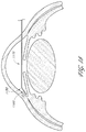
- FIG. 11 illustrates another drainage implant in accordance with embodiments disclosed herein.
- FIG. 12 illustrates an exploded view of the implant of FIG. 11 .
- FIG. 13 illustrates another drainage implant in accordance with embodiments disclosed herein.
- FIG. 14 illustrates an exploded view of the implant of FIG. 13 .
- FIG. 15 illustrates a cross-sectional view of one embodiment of a delivery device with an implant extending therefrom.
- FIG. 16 illustrates a perspective view of another embodiment of a delivery device.
- FIG. 17 illustrates a schematic cross-sectional view of an eye with another delivery device being advanced across the anterior chamber.
- FIG. 18 illustrates a schematic cross-sectional view of an eye with another delivery device being advanced across the anterior chamber.
- FIG. 19 illustrates a cross-sectional view of another drainage implant in accordance with embodiments disclosed herein.
- FIG. 20 illustrates a perspective view of another drainage implant in accordance with embodiments disclosed herein.
- FIG. 21 illustrates a cross-sectional view of another embodiment of a delivery device.
- FIG. 22 illustrates another delivery device in accordance with embodiments disclosed herein.
- FIGS. 23A-B illustrates side views of the delivery device of FIG. 22 .
- FIG. 24 illustrates another delivery device in accordance with embodiments disclosed herein.
- FIG. 25 illustrates a cross-sectional view of another drainage implant in accordance with embodiments disclosed herein.
- FIGS. 26A-C illustrates another drainage implant in accordance with embodiments disclosed herein including a cap.
- FIGS. 27A-C illustrates another drainage implant in accordance with embodiments disclosed herein including a flexible portion.
- FIGS. 28A-B illustrates a reed-type valve in accordance with embodiments disclosed herein.
- FIG. 29 illustrates another delivery device in accordance with embodiments disclosed herein.
- FIG. 30 illustrates a cross-sectional view of another embodiment of a delivery device.
- FIG. 31 illustrates a cross-sectional view of another embodiment of a delivery device.
- FIG. 32 illustrates a cross-sectional view of another embodiment of a delivery device.
- FIG. 33 illustrates a cross-sectional view of another embodiment of a delivery device and an associated shunt.
- FIGS. 34A and 34B are cross-sectional views of a shunt with side ports.
- FIG. 35 is a cross-sectional view of another embodiment of a shunt with side ports.
- FIG. 36 is a cross-sectional view of another embodiment of a shunt with side ports.
- FIGS. 37A and 37B illustrate cross-sectional views of other drainage implants in accordance with embodiments disclosed herein.
- FIG. 38 illustrates a cross-sectional view of another drainage implant in accordance with embodiments disclosed herein.
- FIG. 39 is a perspective view of an implant configured in accordance with another embodiment of the present invention.
- FIGS. 40A and 40B illustrate sectional views of the implant of FIG. 39 and an associated drainage implant as implanted into any eye in accordance with embodiments disclosed herein.
- FIGS. 41A to 41H illustrate cross-sectional views of other drainage implants in accordance with embodiments disclosed herein.
- FIGS. 42A to 42D illustrate cross-sectional views of other drainage implants in accordance with embodiments disclosed herein.
- An ophthalmic implant system comprises a shunt and a delivery instrument for implanting the shunt. While this and other systems and associated methods are described herein in connection with glaucoma treatment, the disclosed systems and methods can be used to treat other types of ocular disorders in addition to glaucoma.
- the shunt following implantation at an implantation site, drains fluid from the anterior chamber into a physiologic outflow space.
- the shunt is configured to provide a fluid flow path for draining aqueous humor from the anterior chamber of an eye to the uveoscleral outflow pathway to reduce intraocular pressure.
- an instrument is provided for delivering and/or implanting the drainage shunt ab interno in an eye to divert aqueous humor from the anterior chamber to the uveoscleral outflow pathway.
- a method is provided for implanting a drainage shunt ab interno in an eye to divert aqueous humor from the anterior chamber to the uveoscleral outflow pathway.
- the aqueous humor is diverted to the supraciliary space or the suprachoroidal space of the uveoscleral outflow pathway.
- shunt as used herein is a broad term, and is to be given its ordinary and customary meaning to a person of ordinary skill in the art (and it is not to be limited to a special or customized meaning), and refers without limitation to an implant defining one or more fluid passages.
- the fluid passage(s) in some embodiments remains patent and, in other embodiments, the passage(s) is fully or partially occluded under at least some circumstances (e.g., at lower intraocular pressure levels).
- the shunts may feature a variety of characteristics, described in more detail below, which facilitate the regulation of intraocular pressure.
- the mechanical aspects and material composition of the shunt can be important for controlling the amount and direction of fluid flow. Therefore, various examples of shunt dimensions, features, tip configurations, material flexibility, coatings, and valve design, in accordance with some embodiments of the present disclosure, are discussed in detail below.
- the delivery instruments may be used to facilitate delivery and/or implantation of the shunt to the desired location of the eye.
- the delivery instrument preferably is used to place the shunt into a desired position by application of a continual implantation force, by tapping the shunt into place using a distal portion of the delivery instrument, or by a combination of these methods.
- the design of the delivery instruments may take into account, for example, the angle of implantation and the location of the shunt relative to an incision.
- the delivery instrument may have a fixed geometry, be shape-set, or actuated.
- the delivery instrument may have adjunctive or ancillary functions.
- the delivery instrument may additionally be used to, for example, inject dye and/or viscoelastic fluid, to dissect, or be used as a guidewire.
- the shunt can be advanced through the ciliary attachment tissue, which lies to the posterior of the scleral spur, during implantation.
- This tissue typically is fibrous or porous, which is relatively easy to pierce or cut with a surgical device, and lies inward of the scleral spur.
- the shunt can be advanced through this tissue and abut against the sclera once the shunt extends into the uveoscleral outflow pathway. The shunt can then slide within the uveoscleral outflow pathway along the interior wall of the sclera.
- the shunt As the shunt is advanced into the uveoscleral outflow pathway and against the sclera, the shunt will likely be oriented at an angle with respect to the interior wall of the sclera.
- the shunt is advanced until it reaches the desired implantation site within the uveoscleral outflow pathway.
- the shunt is advanced into the ciliary body or ciliary muscle bundles to achieve drainage into the supraciliary space.
- the shunt is advanced through the ciliary body or ciliary muscle bundles to achieve fluid communication between the anterior chamber and the suprachoroidal space.
- the shunt is advanced into the compact zone or through the compact to drain aqueous humor into the more distal portions of the suprachoroidal space.
- the disclosed embodiments include shunts that provide a fluid flow path for conducting aqueous humor from the anterior chamber of an eye to the uveoscleral outflow pathway to reduce intraocular pressure, preferably below episcleral venous pressure without hypotony.
- the shunts can have an inflow portion and an outflow portion.
- the outflow portion of the shunt preferably is disposed at or near a distal end of the shunt.
- the inflow portion may be sized and configured to reside in the anterior chamber of the eye and the outflow portion may be sized and configured to reside in the uveoscleral outflow pathway.
- the outflow portion may be sized and configured to reside in the supraciliary region of the uveoscleral outflow pathway or in the suprachoroidal space.
- One or more lumens can extend through the shunt to form at least a portion of the flow path.
- Each lumen preferably extends from an inflow end to an outflow end along a lumen axis.
- the lumen extends substantially through the longitudinal center of the shunt.
- the lumen can be offset from the longitudinal center of the shunt.
- the flow path can be defined by grooves, channel or reliefs formed on an outer surface of the shunt body.
- One or more openings can extend through the wall of the shunt. In some embodiments, the openings can extend through a middle portion of the shunt. In other embodiments the openings can extend through other portions of the shunt.
- the openings can be one or more of a variety of functions. One such function is that when the shunt is inserted into the suprachoroidal or supraciliary space, the openings provide a plurality of routes through which the aqueous humor can drain.
- the shunt when the shunt is inserted into the eye, if the shunt only has one outflow channel (e.g., one end of a lumen), that outflow channel can be plugged, for example, by the shunt's abutment against the interior surface of the sclera or the outer surface of the choroid. Additionally, the outflow channel can be clogged with tissue that is accumulated or cored during the advancement of the shunt through the fibrous or porous tissue.
- a plurality of openings provides a plurality of routes through which the fluid may flow to maintain patency and operability of the drainage shunt. In embodiments where the shunt has a porous body, the openings can define surface discontinuities to assist in anchoring the shunt once implanted.
- the shunt in some embodiments can include a distal portion that is sufficiently sharp to pierce eye tissue near the scleral spur of the eye, and that is disposed closer to the outlet portion than to the inlet portion.
- the distal portion is located at the distal end of the implant.
- the distal portion can be sufficiently blunt so as not to substantially penetrate scleral tissue of the eye.
- the shunts have a generally sharpened forward end and are self-trephinating, i.e., self-penetrating, so as to pass through tissue without pre-forming an incision, hole or aperture.
- the sharpened forward end can be, for example, conical or tapered.
- the tip can be sufficiently sharp to pierce eye tissue near the scleral spur of the eye.
- the tip also can be sufficiently blunt so as not to substantially penetrate scleral tissue of the eye.
- the taper angle of the sharpened end can be, for example, about 30° ⁇ 15° in some embodiments.
- the radius of the tip can be about 70 to about 200 microns. In other embodiments, where an outlet opening is formed at the distal end of the shunt, the distal portion gradually increases in cross-sectional size in the proximal direction, preferably at a generally constant taper or radius or in a parabolic manner.
- the body of the shunt can include at least one surface irregularity.
- the surface irregularity can comprise, for example, a ridge, groove, relief, hole, or annular groove.
- the surface discontinuities or irregularities can also be formed by barbs or other projections, which extend from the outer surface of the shunt, to inhibit migration of the shunt from its implanted position.
- the projections may comprise external ribbing to resist displacement of the shunt.
- the surface irregularity in some embodiments can interact with the tissue of the interior wall of the sclera and/or with the tissue of the ciliary attachment tissue.
- the shunts are anchored by mechanical interlock between tissue and an irregular surface and/or by friction fit.
- the shunt includes cylindrical recessed portions (e.g., annular groves) along an elongate body to provide enhanced gripping features during implantation and anchoring following implantation within the eye tissue.
- the shunt may also incorporate fixation features, such as flexible radial (i.e., outwardly extending) extensions.
- the extensions may be separate pieces attached to the shunt, or may be formed by slitting the shunt wall, and thermally forming or mechanically deforming the extensions radially outward. If the extensions are separate pieces, they may be comprised of flexible material such as nitinol or polyimide.
- the extensions may be located at the proximal or distal ends of the shunt, or both, to prevent extrusion of the shunt from its intended location. The flexibility of the fixation features will facilitate entry through the corneal incision, and also through the ciliary muscle attachment tissue.
- the body of the shunt has an outlet opening on a side surface to allow fluid flow. In some embodiments, the body of the shunt has a plurality of outlet openings on a side surface to allow fluid flow. In other embodiments, there is a plurality of outlet openings at one end of the shunt, such as the distal end. The openings can facilitate fluid flow through the shunt.
- the shunt may have a cap, or tip, at one end.
- the cap can include a tissue-piercing end and one or more outlet openings. Each of the one or more outlet openings can communicate with at least one of the one or more lumens.
- the cap can have a conically shaped tip with a plurality of outlet openings disposed proximal of the tip's distal end.
- the cap can have a tapered angle tip.
- the tip can be sufficiently sharp to pierce eye tissue near the scleral spur of the eye.
- the tip also can be sufficiently blunt so as not to substantially penetrate scleral tissue of the eye.
- the conically shaped tip facilitates delivery of the shunt to the desired location.
- the cap has an outlet opening on a side surface to allow fluid flow. In some embodiments, the cap has a plurality of outlet openings on a side surface to allow fluid flow. In other embodiments, there is a plurality of outlet openings on the conical surface of the cap.
- the openings on the cap can facilitate fluid flow through the shunt. The opening may provide an alternate route for fluid flow which is beneficial in case the primary outflow portion of the shunt becomes blocked.
- multiple shunts are configured to be delivered during a single procedure. In some embodiments when multiple shunts are delivered, the shunts are arranged tandemly.
- the shunt can include a tip protector at one end.
- the tip protector can comprise a recess shaped to receive and protect, for example, the tip of an adjacent shunt.
- the tip of the adjacent shunt has a conical shape.
- the recess may be shaped to contact the sides of the conical tip while protecting the more tapered tip, or end, from impact. The tip protector is particularly useful for delivery of multiple shunts.
- the shunts may be of varied lengths to optimize flows.
- the shunt has sufficient length such that the outflow portion resides in the suprachoroidal space and the inflow portion is exposed to the anterior chamber.
- the length of the shunt is a length such that the outflow portion resides in the supraciliary space of the uveoscleral outflow pathway.
- the length of the shunt is a length such that the outflow portion resides in the membranous region of the uveoscleral outflow pathway adjacent to the retina, while in other embodiments, the shunt has a length that extends distally past the membranous region.
- the length of the shunt from the portion residing in the anterior chamber to the portion residing in the uveoscleral outflow pathway may be about 0.5 mm to about 5 mm. In preferred embodiments, the length of the shunt may be about 1.5 mm to about 5 mm. In more preferred embodiments, the length of the shunt may be about 2.0 mm.
- the length of the shunt is about 0.5, 0.6, 0.7, 0.8, 0.9, 1.0, 1.1, 1.2, 1.3, 1.4, 1.5, 1.6, 1.7, 1.8, 1.9, 2.0, 2.1, 2.2, 2.3, 2.4, 2.5, 2.6, 2.7, 2.8, 2.9, 3.0, 3.1, 3.2, 3.3, 3.4, 3.5, 3.6, 3.7, 3.8, 3.9, 4.0, 4.1, 4.2, 4.3, 4.4, 4.5, 4.6, 4.7, 4.8, 4.9, or 5.0 mm.
- the shunt can have an outer diameter that will permit the shunt to fit within a 23-gauge needle during implantation.
- the shunt can also have a diameter that is designed to be inserted with larger needles.
- the shunt can also be delivered with 18-, 19- or 20-gauge needles.
- smaller gauge applicators such as a 23-gauge (or smaller) applicator, may be used.
- the shunt can have a substantially constant cross-sectional shape through most of the length of the shunt, or the shunt can have portions of reduced or enlarged cross-sectional size (e.g., diameter), or cylindrical channels, e.g., annular grooves, disposed on the outer surface between the proximal end and the distal end.
- the distal end of the shunt can have a tapered portion, or a portion having a continually decreasing radial dimension with respect to the lumen axis along the length of the axis.
- the tapered portion preferably in some embodiments terminates with a smaller radial dimension at the outflow end.
- the tapered portion can operate to form, dilate, and/or increase the size of, an incision or puncture created in the tissue.
- the tapered portion may have a diameter of about 23 gauge to about 30 gauge, and preferably about 25 gauge.
- the diameter of one or more drainage lumens within the shunt may be varied to alter flow characteristics.
- the cross-sectional size of a shunt may be, for example, 0.1 mm to about 1.0 mm, or preferably about 0.3 mm to about 0.4 mm. A small cross-sectional size can be used to restrict flow.
- the cross-sectional shape of the shunt or a shunt may be any of a variety of cross-sectional shapes suitable for allowing fluid flow.
- the cross-sectional shape of the shunt or shunt may be circular, oval, square, trapezoidal, rectangular, or any combination thereof.
- the shunt is configured to expand, either radially or axially, or both radially and axially.
- the shunt may be self-expanding.
- the shunt may be expanded by, for example, using a balloon device.
- the structure of the shunt may be flexible. At least a portion of the structure of the shunt may be flexible, or the whole structure may be flexible. In some embodiments, the structure of the shunt is accordion- or balloon-like. This pleated like structure provides flexibility. In other embodiments, at least a portion of the shunt is curved. In some embodiments, at least a portion of the shunt is straight. In some embodiments, the shunt has both curved and straight portions, and in some embodiments, the shunt is generally rigid (i.e., maintains its preformed shape when implanted).
- the shunt is preferably made of one or more biocompatible materials. Suitable biocompatible materials include polypropylene, polyimide, glass, nitinol, polyvinyl alcohol, polyvinyl pyrrolidone, collagen, chemically-treated collagen, polyethersulfone (PES), poly(styrene-isobutyl-styrene), Pebax, acrylic, polyolefin, polysilicon, polypropylene, hydroxyapatite, titanium, gold, silver, platinum, other metals, ceramics, plastics and a mixture thereof.
- the shunts can be manufactured by conventional sintering, micro machining, laser machining, and/or electrical discharge machining.
- the shunt is made of a flexible material. In other embodiments, the shunt is made of a rigid material. In some embodiments, a portion of the shunt is made from flexible material while another portion of the shunt is made from rigid material.
- the body can have an outer surface of which at least a portion is porous. Some embodiments include porosity that can be varied by masking a portion of the exterior with a band. Where the shunts include a porous body, the cross-section and porosity can be calibrated (down to 0.5 micrometers) to control the flow rates of aqueous humor through the shunt.
- At least a portion of the shunt (e.g., an internal spine or an anchor) is made of a material capable of shape memory.
- a material capable of shape memory may be compressed and, upon release, may expand axially or radially, or both axially and radially, to assume a particular shape.
- at least a portion of the shunt has a preformed shape.
- at least a portion of the shunt is made of a superelastic material.
- at least a portion of the shunt is made up nitinol.
- at least a portion of the shunt is made of a deformable material.
- the body of the shunt can comprise material that includes a therapeutic agent, and/or can house, anchor, or support a therapeutic agent, or can include a coating.
- the coating can include a therapeutic agent.
- the coatings can be, for example, a drug eluting coating, an antithrombogenic coating, and a lubricious coating.
- the therapeutic agent can be selected from the group consisting of: heparin, TGF-beta, an intraocular pressure-lowering drug, and an anti-proliferative agent.
- Materials that may be used for a drug-eluting coating include parylene C, poly (butyl methacrylate), poly (methyl methacrylate), polyethylene-co-vinyl acetate, and other materials known in the art.
- the shunt can further comprise a biodegradable material in or on the shunt.
- the biodegradable material can be selected from the group consisting of poly(lactic acid), polyethylene-vinyl acetate, poly(lactic-co-glycolic acid), poly(D,L-lactide), poly(D,L-lactide-co-trimethylene carbonate), collagen, heparinized collagen, poly(caprolactone), poly(glycolic acid), and a copolymer. All or a portion of the shunt may be coated with a therapeutic agent, e.g. with heparin, preferably in the flow path, to reduce blood thrombosis or tissue restenosis.
- a therapeutic agent e.g. with heparin
- the flow path through the shunt can be configured to be regulated to a flow rate that will reduce the likelihood of hypotony in the eye.
- the intraocular pressure is maintained at about 8 mm Hg. In other embodiments, the intraocular pressure is maintained at pressures less than about 8 mmHg, for example the intraocular pressure may be maintained between about 6 mm Hg and about 8 mm Hg. In other embodiments, the intraocular pressure is maintained at pressures greater than about 8 mm Hg. For example, the pressures may be maintained between about 8 mmHg and about 18 mm Hg, and more preferably between 8 mm Hg and 16 mm Hg, and most preferably not greater than 12 mm Hg.
- the flow rate can be limited to about 2.5 ⁇ L/min or less. In some embodiments the flow rate can be limited to between about 1.9 ⁇ L/min and about 3.1 ⁇ L/min.
- the Hagen-Poiseuille equation suggests that a 4 mm long stent at a flow rate of 2.5 ⁇ L/min should have an inner diameter of 52 mm to create a pressure gradient of 5 mm Hg above the pressure in the suprachoroidal space.
- the shunt may or may not comprise means for regulating fluid flow through the shunt.
- Means for regulating fluid flow can include flow restrictors, pressure regulators, or both.
- the shunt has neither a flow restrictor nor a pressure regulator.
- Regulating flow of aqueous humor can comprise varying between at least first and second operational states in which aqueous humor flow is more restricted in a first state and less restricted in a second state. Increasing the restriction to flow when changing from the second state to the first state can involve moving a valve toward a valve seat in a direction generally parallel or generally normal to a line connecting the proximal and distal ends of the shunt.
- the outflow portion of the shunt in some embodiments is sized and configured to reside in the supraciliary region of the uveoscleral outflow pathway. In such embodiments, there is a lesser need for means for regulating fluid flow through the shunt.
- the means for flow restriction may be, for example, a valve, a long lumen length, small lumen cross section, or any combination thereof.
- the flow of fluid is restricted by the size of a lumen within the shunt, which produces a capillary effect that limits the fluid flow for given pressures.
- the capillary effect of the lumen allows the shunt to restrict flow and provides a valveless regulation of fluid flow.
- the flow path length may be increased without increasing the overall length of the shunt by creating a lumen with a spiral flow path.
- a lumen within the shunt is configured to accommodate placement therein of a spiral flow channel core that is configured to provide preferred flow restriction.
- the spiral flow channel provides an extended path for the flow of fluid between the inlet(s) and outlet(s) of the shunt that is greater than a straight lumen extending between the ends of the shunt.
- the extended path provides a greater potential resistance of fluid flow through the shunt without increasing the length of the shunt.
- the core could have a single spiral flow channel, or a plurality of spiral flow channels for providing a plurality of flow paths through which fluid may flow through the shunt.
- the core can have two or more spiral flow channels, which can intersect.
- the means for flow regulation comprises a pressure regulating valve.
- the valve can open when fluid pressure within the anterior chamber exceeds a preset level.
- Intraocular pressure may be used to apply a force to move a valve surface within the shunt in a direction transverse to a longitudinal axis of the shunt such that aqueous humor flows from the anterior chamber to the uveoscleral outflow pathway at intraocular pressures greater than a threshold pressure.
- a shunt may have any number of valves to restrict flow and/or regulate pressure.
- the valve is preferably located between the anterior chamber and one or more effluent openings such that movement of the valve regulates flow from the interior chamber to the one or more effluent openings.
- a variety of valves are useful with the shunt for restricting flow.
- the valve is a unidirectional valve and/or is a pressure relief valve.
- the pressure relief valve can comprise a ball, a ball seat and a biasing member urging the ball towards the ball seat.
- the valve is a reed-type valve. In a reed valve, for example, one end of the valve may be fixed to a portion of the shunt.
- the body of the reed valve is capable of being deflected in order to allow flow. Pressure from fluid in the anterior chamber can deflect the body of the reed valve, thereby causing the valve to open.
- the shunt includes a pressure regulation valve having a deflectable plate or diaphragm with a surface area exposed to fluid within the interior chamber, the surface area being substantially greater than the total cross-sectional flow area of the one or more influent openings of the shunt.
- a pressure regulation valve having a deflectable plate or diaphragm with a surface area exposed to fluid within the interior chamber, the surface area being substantially greater than the total cross-sectional flow area of the one or more influent openings of the shunt.
- a valve can be disposed between an interior chamber of the shunt and the one or more effluent openings such that movement of the deflectable plate regulates flow from the interior chamber to the one or more effluent openings.
- the plate can extend in a direction generally parallel to the inlet flow path and to the outlet flow path.
- the check pressure relief valve When the intraocular pressure exceeds a particular pressure, the check pressure relief valve will open and permit fluid to flow between the anterior chamber and the uveoscleral outflow pathway. When the intraocular pressure reaches a second, lower pressure, the valve will close and limit or inhibit fluid from being conducted to the suprachoroidal space. The valve will remain closed until the intraocular pressure again reaches the particular pressure, and at which time the valve will reopen to permit or enhance drainage of fluid to the uveoscleral outflow pathway. Accordingly, the shunt provides drainage of the anterior chamber through the shunt based on the intraocular pressure levels and reduces the likelihood for over-draining the anterior chamber and causing hypotony.
- the shunt is inserted from a site transocularly situated from the implantation site.
- the delivery instrument can be sufficiently long to advance the shunt transocularly from the insertion site across the anterior chamber to the implantation site.
- At least a portion of the instrument can be flexible.
- the instrument can be rigid.
- the instrument can comprise a plurality of members longitudinally moveable relative to each other.
- at least a portion of the delivery instrument is curved or angled.
- a portion of the delivery instrument is rigid and another portion of the instrument is flexible.
- the delivery instrument has a distal curvature.
- the distal curvature of the delivery instrument may be characterized as a radius of approximately 10 to 30 mm, and preferably about 20 mm.
- the delivery instrument has a distal angle.
- the distal angle may be characterized as approximately 90 to 170 degrees relative to an axis of the proximal segment of the delivery instrument, and preferably about 145 degrees.
- the angle can incorporate a small radius of curvature at the “elbow” so as to make a smooth transition from the proximal segment of the delivery instrument to the distal segment.
- the length of the distal segment may be approximately 0.5 to 7 mm, and preferably about 2 to 3 mm.
- the instruments have a sharpened forward end and are self-trephinating, i.e., self-penetrating, so as to pass through tissue without pre-forming an incision, hole or aperture.
- a trocar, scalpel, or similar instrument can be used to pre-form an incision in the eye tissue before passing the shunt into such tissue.
- the instrument can have a sufficiently small cross section such that the insertion site self seals without suturing upon withdrawal of the instrument from the eye.
- An outer diameter of the delivery instrument preferably is no greater than about 18 gauge and is not smaller than about 27 gauge.
- the incision in the corneal tissue is preferable made with a hollow needle through which the shunt is passed.
- the needle has a small diameter size (e.g., 18 or 19 or 20 or 21 or 22 or 23 or 24 or 25 or 26 or 27 gauge) so that the incision is self sealing and the implantation occurs in a closed chamber with or without viscoelastic.
- a self-sealing incision also can be formed using a conventional “tunneling” procedure in which a spatula-shaped scalpel is used to create a generally inverted V-shaped incision through the cornea.
- the instrument used to form the incision through the cornea remains in place (that is, extends through the corneal incision) during the procedure and is not removed until after implantation.
- Such incision-forming instrument either can be used to carry the ocular shunt or can cooperate with a delivery instrument to allow implantation through the same incision without withdrawing the incision-forming instrument.
- various surgical instruments can be passed through one or more corneal incisions multiple times.
- a delivery instrument can be advanced from the insertion site transocularly into the anterior chamber angle and positioned at a location near the scleral spur. Using the scleral spur as a reference point, the delivery instrument can be advanced further in a generally posterior direction to drive the shunt into eye tissue at a location just inward of the scleral spur toward the iris.
- the placement and implantation of the shunt can be performed using a gonioscope or other conventional imaging equipment.
- the delivery instrument preferably is used to force the shunt into a desired position by application of a continual implantation force, by tapping the shunt into place using a distal portion of the delivery instrument, or by a combination of these methods. Once the shunt is in the desired position, it may be further seated by tapping using a distal portion of the delivery instrument.
- the delivery instrument can include an open distal end with a lumen extending therethrough. Positioned within the lumen is preferably a pusher tube that is axially movable within the lumen.
- the pusher tube can be any device suitable for pushing or manipulating the shunt in relation to the delivery instrument, such as, for example, but without limitation a screw, a rod, a stored energy device such as a spring.
- a wall of the delivery instrument preferably extends beyond pusher tube to accommodate placement within the lumen of a shunt.
- the shunt can be secured in position. For example, the shunt can be secured by viscoelastic or mechanical interlock with the pusher tube or wall.
- the pusher tube When the shunt is brought into position adjacent the tissue in the anterior chamber angle, the pusher tube is advanced axially toward the open distal end of the delivery instrument. As the pusher tube is advanced, the shunt is also advanced. When the shunt is advanced through the tissue and such that it is no longer in the lumen of the delivery instrument, the delivery instrument is retracted, leaving the shunt in the eye tissue.
- Some embodiments can include a spring-loaded or stored-energy pusher system.
- the spring-loaded pusher preferably includes a button operably connected to a hinged rod device.
- the rod of the hinged rod device engages a depression in the surface of the pusher, keeping the spring of the pusher in a compressed conformation.
- the button When the user pushes the button, the rod is disengaged from the depression, thereby allowing the spring to decompress, thereby advancing the pusher forward.
- an over-the wire system is used to deliver the shunt.
- the shunt can be delivered over a wire.
- the wire is self-trephinating.
- the wire can function as a trocar.
- the wire can be superelastic, flexible, or relatively inflexible with respect to the shunt.
- the wire can be pre-formed to have a certain shape.
- the wire can be curved.
- the wire can have shape memory, or be elastic.
- the wire is a pull wire.
- the wire can be a steerable catheter.
- the wire is positioned within a lumen in the shunt.
- the wire can be axially movable within the lumen.
- the lumen may or may not include valves or other flow regulatory devices.
- the delivery instrument comprises is a trocar.
- the trocar may be angled or curved.
- the trocar can be rigid, semi-rigid or flexible.
- the shunt can be, but need not be relatively flexible.
- the diameter of the trocar can be about 0.001 inches to about 0.01 inches. In some embodiments, the diameter of the trocar is 0.001, 0.002, 0.004, 0.005, 0.006, 0.007, 0.008, 0.009, or 0.01 inches.
- delivery of the shunt is achieved by applying a driving force at or near the distal end of the shunt.
- the driving force can be a pulling or a pushing applied generally to the end of the shunt.
- the instrument can include a seal to prevent aqueous humor from passing through the delivery instrument and/or between the members of the instrument when the instrument is in the eye.
- the seal can also aid in preventing backflow of aqueous humor through the instrument and out the eye.
- Suitable seals for inhibiting leakage include, for example, an o-ring, a coating, a hydrophilic agent, a hydrophobic agent, and combinations thereof.
- the coating can be, for example, a silicone coat such as MDXTM silicone fluid.
- the instrument is coated with the coating and a hydrophilic or hydrophobic agent.
- one region of the instrument is coated with the coating plus the hydrophilic agent, and another region of the instrument is coated with the coating plus the hydrophobic agent.
- the delivery instrument can additionally comprise a seal between various members comprising the instrument.
- the seal can comprise a hydrophobic or hydrophilic coating between slip-fit surfaces of the members of the instrument.
- the seal can be disposed proximate of the drainage shunt when carried by the delivery instrument.
- the seal is present on at least a section of each of two devices that are machined to fit closely with one another.
- the delivery instrument can include a distal end having a beveled shape.
- the delivery instrument can include a distal end having a spatula shape.
- the beveled or spatula shape can have a sharpened edge.
- the beveled or spatula shape can include a recess to contain the shunt.
- the recess can include a pusher or other suitable means to push out or eject the shunt.
- the delivery instrument further can be configured to deliver multiple shunts.
- the shunts can be arranged in tandem, as described in greater detail below.
- the implantation occurs in a closed chamber with or without viscoelastic.
- the shunts may be placed using an applicator, such as a pusher, or they may be placed using a delivery instrument having energy stored in the instrument, such as disclosed in U.S. Patent Publication 2004/0050392, filed Aug. 28, 2002, the entirety of which is incorporated herein by reference and made a part of this specification and disclosure.
- fluid may be infused through the delivery instrument or another instrument used in the procedure to create an elevated fluid pressure at the distal end of the shunt to ease implantation.
- the shunt is implanted through the fibrous attachment of the ciliary muscle to the sclera.
- This fibrous attachment zone extends about 0.5 mm posteriorly from the scleral spur, as shown between the two arrows ( 1020 ) in FIG. 17 .
- the shunt can be flexible to facilitate delivery along the curvature or can be more loosely held to move easily along an accurate path.
- the shunt can be relatively rigid.
- the delivery instrument can incorporate a shunt advancement element (e.g. pusher) that is flexible enough to pass through the distal angle.
- the shunt and delivery instrument can be advanced together through the anterior chamber 32 from an incision at or near the limbus, across the iris, and through the ciliary muscle attachment until the shunt outlet portion is located in the uveoscleral outflow pathway (e.g. exposed to the suprachoroidal space 34 defined between the sclera 38 and the choroid 40 ).
- FIG. 2 illustrates a transocular implantation approach with the delivery instrument inserted well above the limbus. The incision, however, can be more posterior and closer to the limbus.
- the operator can then simultaneously push on a pusher device while pulling back on the delivery instrument, such that the shunt outlet portion maintains its location in the uveoscleral outflow pathway.
- the shunt is released from the delivery instrument, and the delivery instrument is retracted proximally, as illustrated in FIG. 3 .
- the delivery instrument then can be withdrawn from the anterior chamber through the incision.
- a viscoelastic can be injected into the suprachoroidal space to create a chamber or pocket between the choroid and sclera which can be accessed by a shunt. Such a pocket could expose more of the choroidal and scleral tissue area, and increase uveoscleral outflow, causing a lower TOP.
- the viscoelastic material can be injected with a 25 or 27 G cannula, for example, through an incision in the ciliary muscle attachment or through the sclera (e.g. from outside the eye). The viscoelastic material can also be injected through the shunt itself either before, during or after implantation is completed.
- a hyperosmotic agent can be injected into the suprachoroidal space. Such an injection can delay TOP reduction. Thus, hypotony can be avoided in the acute postoperative period by temporarily reducing choroidal absorption.
- the hyperosmotic agent can be, for example glucose, albumin, HYPAQUETM medium, glycerol, or poly(ethylene glycol). The hyperosmotic agent can breakdown or wash out as the patient heals, resulting in a stable, acceptably low TOP, and avoiding transient hypotony.
- the therapeutic agents that may be utilized with the ophthalmic implant system may include one or more agents provided below.
- the therapeutic agents include but are not limited to pharmaceutical agents, biological agents including hormones, enzyme or antibody-related components, oligonucleotides, DNA/RNA vectors and live cells configured to produce one or more biological components.
- the use of any particular therapeutic agent is not limited to their primary effect or regulatory body-approved treatment indication or manner of use.
- the listing of any particular agent within any one therapeutic class below is only representative of one possible use of the agent and is not intended to limit the scope of its use with the ophthalmic implant system.
- Exemplary therapeutic agents may include: anti-angiogenesis agents, including VEGF receptor tyrosine kinase inhibitors and anti-vascular endothelial growth factor (anti-VEGF) agents such as ranibizumab (LUCENTIS®) and bevacizumab (AVASTIN®), pegaptanib (MACUGEN®), sunitinib and sorafenib and any of a variety of known small-molecule and transcription inhibitors having anti-angiogenesis effect; classes of known ophthalmic agents, including: glaucoma agents, such as beta-blocker agents such as betaxolol, carteolol, levobetaxolol, levobunolol and timolol; carbonic anhydrase inhibitor agents such as acetozolamide, brinzolamide, dorzolamide and methazolamide; mydriatic-cycloplegic agents such as atropine, cyclopentolate, succinylcholine, homatropine
- Other therapeutic agents of the same class as the ophthalmic agents listed above that may be used include: other beta-blocker agents such as acebutolol, atenolol, bisoprolol, carvedilol, asmolol, labetalol, nadolol, penbutolol, pindolol and propranolol; other corticosteroidal and non-steroidal anti-inflammatory agents such aspirin, betamethasone, cortisone, diflunisal, etodolac, fenoprofen, fludrocortisone, flurbiprofen, hydrocortisone, ibuprofen, indomethacine, ketoprofen, meclofenamate, mefenamic acid, meloxicam, methylprednisolone, nabumetone, naproxen, oxaprozin, prednisolone, prioxicam, salsalate, sulindac and
- Valdecoxib other immune-modulating agents such as aldesleukin, adalimumab (HUMIRA®), azathioprine, basiliximab, daclizumab, etanercept (ENBREL®), hydroxychloroquine, infliximab (REMICADE®), leflunomide, methotrexate, mycophenolate mofetil, and sulfasalazine; other anti-histamine agents such as loratadine, desloratadine, cetirizine, diphenhydramine, chlorpheniramine, dexchlorpheniramine, clemastine, cyproheptadine, fexofenadine, hydroxyzine and promethazine; other anti-infective agents such as aminoglycosides such as amikacin and streptomycin; anti-fungal agents such as amphotericin B, caspofungin, clotrimazole, fluconazole
- the therapeutic agents may be released or eluted from the ophthalmic implant system, bound to a surface of the ophthalmic implant, and/or used in conjunction with the ophthalmic implant through injection, oral or eye drop delivery routes.
- the therapeutic agents may also be released from a separate drug eluting device that is implantable in the same or a different location in the eye or orbital cavity.
- the separate drug eluting device may be located in a physiologic outflow pathway or physiologic cavity of the eye or body, or may be implanted into an artificially formed site of the eye or body.
- a variety of controlled-release technologies that are known in the art may be used with the ophthalmic implant system, including non-degradable and biodegradable polymeric and non-polymeric release platforms.
- Injection/infusion/implantation routes or sites include intravenous and intravitreal routes, choroidal, scleral, conjunctival, retinal, ciliary body, posterior chamber, anterior chamber (including the angle), trabecular meshwork, Schlemm's canal, suprachoroidal, and other sites along the uveoscleral pathway.
- the vascular routes or sites include but are not limited to the ophthalmic artery, the lacrimal artery, the short posterior ciliary arteries, the long posterior ciliary arteries, the anterior ciliary arteries, the central retinal arteries, the central retinal veins, and episcleral arteries and veins.
- combinations of agents having synergistic and/or complementary effects for a particular disease or set of related conditions or symptoms may be used.
- a disease-treating agent may be used in combination with a metabolism-altering agent affecting the cytochrome P450 system to affect the pharmacokinetics of the disease-treating agent.
- an anti-infective agent may be combined with an anti-inflammatory agent to treat inflammation resulting from the infection.
- FIG. 4 illustrates one embodiment of a shunt 130 that is operable to drain fluid from the anterior chamber to the uveoscleral outflow pathway (e.g., the suprachoroidal space).
- the shunt 130 has an inflow portion 132 and an outflow portion 134 .
- the inflow portion 132 is sized and configured to reside in the anterior chamber of the eye and the outflow portion 134 is sized and configured to reside in the uveoscleral outflow pathway.
- Extending through the shunt 130 is preferably at least one lumen 136 that operates to conduct the fluid through the shunt 130 .
- Each lumen 136 preferably extends from an inflow end 138 to an outflow end 140 along a lumen axis 142 .
- the shunt 130 preferably has an outer diameter that will permit the shunt 130 to fit within a 21-gauge or 23-gauge needle or hollow instrument during implantation; however, larger or smaller gauge instruments may also be used.
- the shunt 130 can also have a diameter that is designed to be delivered with larger needles.
- the shunt 130 can also be delivered with 18-, 19- or 20-gauge needles.
- the shunt 130 can have a constant diameter through most of the length of the shunt 130 , or the shunt 130 can have portions of reduced diameter, e.g., annular grooves 146 , between the proximal end 138 and the distal end 140 .
- the annular grooves 146 produce an irregular outer surface that can operate to mechanically lock or anchor the shunt 130 in place when implanted.
- surface discontinuities or irregularities can also be formed by barbs or other projections, which extend from the outer surface of the shunt, to inhibit migration of the shunt 130 from its implanted position, as described above.
- the outflow portion 134 of the shunt 130 preferably is disposed at or near the distal end 140 of the shunt 130 .
- the outflow portion 134 has a tapered portion 144 ; however, it may also have other shapes (e.g. semi-sphere, a paraboloid, a hyperboloid) with a continually decreasing radial dimension with respect to the lumen axis 142 along the length of the axis 142 .
- the tapered portion 144 preferably terminates with a smaller radial dimension at the outflow end 140 .
- the tapered portion 144 can operate to form, dilate, and/or increase the size of, an incision or puncture created in the tissue.
- the distal end 140 can operate as a trocar to puncture or create an incision in the tissue.
- the tapered portion 144 can be advanced through the puncture or incision.
- the tapered portion 144 will operate to stretch or expand the tissue around the puncture or incision to accommodate the increasing size of the tapered portion 144 as it is advanced through the tissue.
- the stretched tissue passes over the cylindrical channels 146 having a reduced diameter, the stretched tissue will retract generally to fill the cylindrical channels 146 and will abut the edges of the shunt 130 having a greater diameter.
- the interaction of the tissue and the edges of the shunt 130 will provide an anchor for the shunt 130 following implantation to inhibit shunt migration.
- the tapered portion 144 can also facilitate proper location of the shunt 130 into the supraciliary or suprachoroidal spaces.
- the shunt 130 is preferably advanced through the tissue within the anterior chamber angle during implantation. This tissue typically is fibrous or porous, which is relatively easy to pierce or cut with a surgical device, such as the tip of the shunt 130 .
- the shunt 130 can be advanced through this tissue and abut against the sclera once the shunt extends into the uveoscleral outflow pathway.
- the tapered portion 144 preferably provides a generally rounded edge or surface that facilitates sliding of the shunt 130 within the suprachoroidal space along the interior wall of the sclera.
- the shunt 130 will likely be oriented at an angle with respect to the interior wall of the sclera.
- the tip of the shunt 130 engages the sclera the tip preferably has a radius that will permit the shunt 130 to slide along the sclera instead of piercing or substantially penetrating the sclera.
- the tapered portion 144 will provide an edge against which the shunt 130 can abut against the sclera and reduce the likelihood that the shunt will pierce the sclera.
- aqueous humor flows from the anterior chamber to the uveoscleral outflow pathway through the lumen 136 of the shunt.
- the flow of fluid is preferably restricted by the size of the lumen 136 , which produces a capillary effect that limits the fluid flow for given pressures.
- the capillary effect of the lumen allows the shunt to restrict flow and provides a valveless regulation of fluid flow.
- the flow of fluid through the shunt 130 is preferably configured to be restricted to flow rated that will reduce the likelihood of hypotony in the eye.
- the flow rate can be limited to about 2.5 ⁇ L/min or less. In some embodiments the flow rate can be limited to between about 1.9 ⁇ L/min and about 3.1 ⁇ L/min.
- a plurality of shunts 130 can be used in a single eye to conduct fluid from the anterior chamber to the uveoscleral outflow pathway. In such applications, the cumulative flow rate through the shunts preferably is within the range of about 1.9 ⁇ L/min to about 3.1 ⁇ L/min, although the flow rate for each of the shunts can be significantly less than about 2.5 ⁇ L/min. For example, if an application called for implantation of five shunts, then each shunt 130 can be configured to have a flow rate of about 0.5 ⁇ L/min.
- the lumen can be offset from the longitudinal center of the shunt.
- FIG. 4 depicts the shunt as having a tapered portion 144 that terminates substantially where the tapered portion 144 meets the lumen 136
- the lumen 136 can be offset from the center of the shunt 130 such that lumen 136 opens along one of the sides of the tapered portion 144 .
- the tapered portion 144 can terminate at a location offset from the lumen axis 142 and can extend beyond the point at which the interior lumen 136 and the exterior tapered portion 144 meet.
- the lumen can vary in direction along its length.
- the shunt 130 preferably comprises any of the materials previously described above.
- the shunt 130 can be fabricated through conventional micro machining techniques or through procedures commonly used for fabricating optical fibers.
- the shunts 130 are drawn with a bore, or lumen, extending therethrough.
- the tapered portion 144 at the outflow portion 134 can be constructed by shearing off an end tubular body. This can create a tapered portion 144 that can be used to puncture or incise the tissue during implantation and dilate the puncture or incision during advancement of the shunt 130 .
- Other materials can be used for the shunt of FIG. 4 , and other methods of manufacturing the shunt 130 can also be used.
- the shunt 130 can be constructed of metals or plastics, and the shunts can be machined with a bore that is drilled as described above.
- the shunt 130 of FIG. 4 represents a shunt having a construction that provides for the opportunity to vary the size of the shunt 130 or the lumen 136 . Additionally, the shunt 130 is able to be delivered in small needles. For example, the shunt 130 can fit within a needle for the implantation procedure. The needle preferably has a size of about 18 gauge to about 23 gauge, and most preferably about 23 gauge. The shunt also need not have a unitary configuration; that is, be formed of the same piece of material. For example, a proximal portion of the shunt can be formed of glass drawn to have at least one small diameter lumen. A distal portion of the shunt can be a cap formed of a different material.
- the cap includes a tissue-piercing end and one or more outlet openings. Each of the one or more outlet openings communicates with at least one of the one or more lumens in the proximal portion.
- the cap has a conically shaped tip with a plurality of outlet openings disposed proximal of the tip's distal end.
- FIG. 5 illustrates a shunt 230 having a relatively similar construction as that of FIG. 4 .
- FIG. 5 illustrates an embodiment of a shunt 230 having an elongate body with an inflow portion 232 and an outflow portion 234 .
- a lumen(s) 236 preferably extends between an inflow end 238 and an outflow end 240 .
- Proximate the outflow end 240 is preferably a tapered portion 244 having a construction similar to the embodiments described above with respect to FIG. 4 .
- the bodies of the shunts can be formed of a porous material which has one or more flow paths from the inflow portion 232 to the outflow portion 240 .
- FIG. 5 depicts a plurality of apertures 246 extending through the wall of the shunt 230 . While the apertures 246 are depicted as extending through a middle portion of the shunt 230 , the apertures can extend through other portions of the shunt 230 . For example, the apertures 246 can also extend through the outflow portion 234 , or more particularly, through the tapered portion 244 .
- the plurality of apertures 246 can provide several functions. One such function is that when the shunt 230 is inserted into the uveoscleral outflow pathway, the apertures 246 provide a plurality of routes through which the aqueous humor can drain.
- the shunt 230 can be plugged, for example, by the shunt's abutment against the interior wall of the sclera or the outer wall of the choroid. Additionally, the outflow channel can be clogged with tissue that is accumulated during the advancement of the shunt 230 through the fibrous or porous tissue.
- the plurality of apertures 246 provides a plurality of routes through which the fluid may flow to maintain patency and operability of the drainage shunt 230 . In embodiments where the shunt has a porous body, the apertures 246 can define surface discontinuities to assist in anchoring the shunt once implanted.
- FIG. 6 depicts embodiments of a shunt 330 having an elongate body with an inflow portion 332 and an outflow portion 334 .
- the body of the shunt 330 is formed of a porous material. Proximate the outflow end 340 is preferably a tapered portion 344 having a construction similar to the embodiments described above with respect to FIG. 4 .
- the shunt 330 includes cylindrical recessed portions along the elongate body to provide enhanced gripping features during implantation and anchoring following implantation within the eye tissue.
- the shunts depicted in FIGS. 5 and 6 are preferably constructed of metals, ceramics, or plastics; although several of the other materials noted herein can also be used.
- the shunts 230 , 330 can be constructed of titanium and manufactured by conventional sintering, micro machining, laser machining, and/or electrical discharge machining.
- the shunt 230 of FIG. 5 preferably restricts fluid flow in similar manners described above with respect to the embodiments of FIG. 4 .
- the shunts 230 , 330 include a porous body the cross-section and porosity can be calibrated (done to 0.5 micrometers) to control the flow rates of aqueous humor through the shunt.
- the flow rates through the shunts illustrated in FIGS. 5 and 6 preferably are similar to the rates specified above.
- FIGS. 7-8 depict embodiments of another shunt 430 having an elongate body with an inflow portion 432 and an outflow portion 434 .
- a lumen 436 preferably extends between an inflow end 438 and an outflow end 440 .
- the illustrated embodiment includes just one lumen, other embodiments can include multiple lumens, each including the flow restriction described below.
- Proximate the outflow end 440 is preferably a tapered portion 444 that decreases in a radial dimension along a lumen axis 442 .
- the shunt 430 includes cylindrical recessed portions 446 along the elongate body to provide enhanced gripping features during implantation and anchoring following implantation within the eye tissue.
- the lumen 436 is preferably configured to accommodate placement therein of a spiral flow channel core 448 that is configured to provide preferred flow restriction.
- the core 448 is preferably configured to extend through the lumen 436 between the inflow end 438 and the outflow end 440 and includes a tortuous or spiral flow channel 450 extending generally along the exterior of the core 448 .
- the spiral flow channel 450 provides an extended path for the flow of fluid between the two ends of the shunt 430 that is greater than a straight lumen extending between the ends of the shunt 430 .
- the extended path provides a greater potential resistance of fluid flow through the shunt without increasing the length of the shunt.
- the core 448 is depicted in FIGS. 7 and 8 as having only a single spiral flow channel 450 , the core 448 could have a plurality of spiral flow channels 450 for providing a plurality of flow paths through which fluid may flow through the shunt 430 .
- the core 448 can have two or more spiral flow channels 450 .
- the core 448 can also have one or more straight lumens extending through the core 448 .
- the shunt 430 is preferably manufactured of metals, ceramics, or plastics through conventional micro machining, laser machining, or electrical discharge machining.
- the shunt 430 can be constructed of titanium, glass, or noble metals.
- the core 448 is made of the same material as the body of the shunt 430 while in yet further embodiments, the core 448 includes a material that is different than the body of the shunt 430 .
- FIGS. 9-10 depicts embodiment of another shunt 530 having an elongate body with an inflow portion 532 and an outflow portion 534 .
- the shunt 530 preferably includes a lumen 536 that extends between an inflow end 538 and an outflow end 540 .
- the shunt 530 preferably includes a tapered portion 544 at the outflow end 540 that decreases in a radial dimension along a lumen axis 542 .
- the shunt 530 includes cylindrical recessed portions 546 along the elongate body to provide enhanced gripping features during implantation and anchoring following implantation within the eye tissue.
- the shunt 530 is preferably configured to conduct fluid between the anterior chamber and the uveoscleral outflow pathway with the inflow end 538 exposed to the anterior chamber and the outflow end 540 exposed to the suprachoroidal space.
- the shunt 530 preferably reduces the likelihood of hypotony of the eye by providing a ball-check pressure regulator. For example, when the intraocular pressure exceeds a particular pressure, the ball-check pressure regulator will open and permit fluid to flow between the anterior chamber and the uveoscleral outflow pathway. When the intraocular pressure reaches a second, lower pressure, the ball-check pressure regulator will close and limit or inhibit fluid from being conducted to the uveoscleral outflow pathway.
- the ball-check pressure regulator will remain closed until the intraocular pressure again reaches the particular pressure, and at which time the ball-check valve will reopen to permit or enhance drainage of fluid to the uveoscleral outflow pathway. Accordingly, the shunt 530 provides drainage of the anterior chamber through the shunt 530 based on the intraocular pressure levels and reduces the likelihood for over-draining the anterior chamber and causing hypotony.
- the ball-check regulator is preferably configured to be positioned within the lumen 536 of the shunt 530 and includes a luminal spring 552 that is configured to reside within the lumen.
- the luminal spring 552 is depicted as a coil spring, but the luminal spring 552 can be any type of spring or biasing member that is resilient or reversibly compressible.
- the spring 552 can comprise Nitinol or other flexible or resilient materials.
- the ball-check regulator also preferably includes a ball 554 that preferably has a diameter less than the diameter of the lumen 536 of the shunt 530 so as to permit movement of the ball 554 within the lumen 536 and to permit the flow of fluid between the ball 554 and the inner wall of the lumen 536 when the ball 554 resides within the lumen 536 .
- the luminal spring 552 is preferably configured to engage a ball 554 at one end of the luminal spring 552 and to permit the ball 554 to move between different positions within the lumen 536 .
- a ball sleeve 556 is preferably provided within at least a portion of the lumen 536 and is positioned adjacent to the ball 554 opposite the luminal spring 552 .
- FIGS. 9 and 10 depict the ball sleeve 556 positioned adjacent the inflow end 538 .
- the luminal spring 552 is depicted as extending from the outflow portion 534 toward the inflow portion 532 with the ball 554 interposed between one end of the luminal spring 552 and the ball sleeve 556 .
- the portion of the ball sleeve 556 that is adjacent the ball 554 preferably has a lumen that has a diameter less than that of the ball 554 and limits movement of the ball 554 so the ball is unable to pass through the ball sleeve lumen.
- This end of the ball sleeve 556 preferably provides a ball seat 558 against which the ball 554 can rest when urged against the ball sleeve 556 by the luminal spring 552 .
- the ball 554 prevents flow when contacting seat of the ball sleeve 556 ; however, in other embodiments, some restricted flow can occur through the shunt even when the ball 554 rests against the seat. Such flow can occur through one or more parallel flow paths or through one or more relatively small flow paths that extend around the ball 554 and remain open when the ball 554 contacts the seat of the ball sleeve 556 .
- the shunt 530 also preferably includes a distal taper or cone 560 that is configured to reside at least partially within the lumen 536 .
- the distal cone 560 preferably includes radial flanges 562 that provide a means for securing the cone 560 in place by engaging the inner wall of the lumen 536 while providing a space between the distal cone 560 and the inner wall of the lumen 536 .
- the distal cone 560 provides radial channels 562 instead of flanges through which the fluid can be conducted. The space between the inner wall of the lumen 536 and the cone 560 or the channels 562 permits fluid conducted through the lumen 536 to exit the shunt by flowing around the distal cone 560 .
- the luminal spring 552 When the ball-check pressure regulator is assembled, the luminal spring 552 is preferably seated against the distal cone 560 on one end and presses against the ball 554 on the other end with a determined force. The ball 554 is moved against the ball seat 558 of the ball sleeve 556 as a reaction to the force of the luminal spring 552 .
- the shunt 530 When the shunt 530 is inserted within the eye with the inflow end 538 exposed to the anterior chamber and the outflow end 540 exposed to the suprachoroidal space, the ball 554 will be exposed to the intraocular pressure of the anterior chamber.
- the ball 554 will be pressed against the ball seat 558 and limit or inhibit flow of fluid past the ball 554 until the intraocular pressure exerts a force upon the ball 554 that is greater than the force applied by the luminal spring 552 .
- the luminal spring 552 force is overpowered by the intraocular pressure, the ball 554 will be moved down the lumen 536 away from the ball seat 558 , thus permitting fluid to pass around the ball 554 , through the lumen 536 , and out the outflow portion 534 between the radial flanges 562 of the distal cone 560 .
- the force pressing against the ball 554 will be reduced, and when the force applied on the ball 554 by the intraocular pressure is less than the force applied on the ball 554 by the luminal spring 552 , the ball 554 will be moved through the lumen 536 until it is pressed against the ball seat 558 , thus stopping the flow of fluid through the lumen 536 .
- FIGS. 11 and 12 illustrate embodiments of a generally flat pressure regulator shunt 630 .
- the shunt 630 preferably includes an inflow portion 632 and an outflow portion 634 .
- the inflow portion 632 preferably includes a plurality of inlets along an inflow end 638
- the outflow portion 634 preferably includes a plurality of outlets along an outflow end 640 .
- the shunt 630 is preferably constructed of three portions: a top portion 642 , a bottom portion 644 , and a middle portion 646 .
- the top portion 642 and the bottom portion 644 are preferably substantially rigid and provide a housing for the shunt 630 .
- the top portion 642 is engageable with the bottom portion 644 by aligning a plurality of apertures 651 extending along the edges of the portions 642 , 644 .
- the two portions 642 , 644 can be secured together by glue, solder, or other means for connection the portions.
- the bottom portion 644 preferably includes inflow apertures 648 that are configured to permit fluid to enter into a chamber 650 formed by the edges of the shunt 630 .
- the top portion 642 preferably includes a plurality of outflow apertures 652 through which fluid can exit the chamber 650 and be discharged from the shunt 630 .
- a flexible or resilient middle portion 646 is preferably positioned between the two portions 642 , 644 .
- the middle portion 646 is preferably a biased membrane that is biased toward the bottom portion 644 when the shunt 630 is assembled and rests on a membrane seat 654 .
- a plurality of apertures 653 along the edges of the membrane preferably coincides with a plurality of protrusions 655 on the top and bottom portions 642 , 644 .
- the middle portion 646 is preferably constructed of a nitinol sputter deposited silicone membrane.
- the membrane preferably pressed against the bottom portion 644 and has an aperture 656 extending therethrough.
- the aperture 656 provides a flow path through which fluid conducted through the shunt 630 can pass when the membrane does not rest on the membrane seat 654 .
- the shunt is inserted into the eye with the inflow portion 632 exposed to the anterior chamber and the outflow portion 634 exposed to the uveoscleral outflow pathway. Fluid from the anterior chamber will enter into the inflow apertures 648 and fill the chamber 650 on one side of the membrane of the middle portion 646 . Because the middle portion membrane 646 is biased toward the membrane seat 654 , the aperture 656 will not permit fluid to flow to the other side of the membrane. When the intraocular pressure reaches an elevated level, the fluid pressure within the chamber 650 will create a force against the membrane 646 and cause the membrane 646 to disengage the membrane seat 654 .
- the membrane aperture 656 permits fluid to flow through the membrane 646 into the other side of the chamber 650 and out the outflow apertures 652 .
- the pressure at which the membrane will be deflected from the membrane seat 654 preferably corresponds to acceptable intraocular pressure levels.
- the large surface area of the membrane 646 will provide for a low tolerance of pressure regulation.
- the shunt 630 is preferably implanted following the creation of an incision in the tissue.
- the delivery instrument may create the incision for the shunt 630 and insert the shunt 630 into the incision.
- the shunt 630 can have a sharpened outflow end 640 and create the incision itself as it is advanced through the tissue.
- FIGS. 13 and 14 depict a shunt 730 that operates under similar principles as that of the embodiments depicted in FIGS. 11 and 12 .
- the shunt has an inflow portion 732 and an outflow portion 734 .
- the inflow portion 732 includes an inflow end 738 and inflow apertures 748 .
- the outflow portion 734 includes an outflow end 740 and outflow apertures 752 .
- the inflow apertures 748 and the outflow apertures 752 are in fluid communication with a shunt chamber 750 .
- the shunt 730 preferably includes four portions: a top portion 742 , a spring or biasing portion 736 , a membrane portion 746 , and a bottom portion 744 .
- the sprint or biasing portion 736 When the shunt is assembled, the sprint or biasing portion 736 preferably presses the membrane portion 746 against the bottom portion 744 , thus restricting the fluid communication through the shunt 730 .
- the intraocular pressure reaches a certain level, the resultant force exerted against the membrane portion 746 will exceed that of the spring or biasing portion 736 and cause the membrane portion 746 to disengage the bottom portion 744 .
- an aperture 756 will permit fluid to flow through the membrane 746 and through an aperture 758 in the spring or biasing portion 736 .
- the aqueous humor will flow through the chamber 750 and out the shunt 730 through the outflow apertures 752 .
- an intraocular pressure regulator having an inlet portion that provide at least one ingress flow path that include one or more influent openings.
- the openings preferably have a total cross-sectional flow area and communicate with an interior chamber within the shunt.
- the shunts include an outlet portion that provides an egress flow path that has one or more effluent openings.
- the shunts have a pressure regulation valve that includes a deflectable plate with a surface area exposed to fluid within the interior chamber. The surface area preferably is substantially greater than the total cross-sectional flow area.
- the valve is preferably located between the interior chamber and one or more effluent openings such that movement of the deflectable plate regulates flow from the interior chamber to the one or more effluent openings.
- the plate preferably extends in a direction generally parallel to the inlet flow path and to the outlet flow path.
- FIG. 15 illustrates one embodiment of a delivery instrument 830 that can be used with at least several of the shunt embodiments described herein.
- the delivery instrument 830 preferably includes an open distal end 832 with a lumen 834 extending therethrough. Positioned within the lumen 834 is preferably a pusher tube 836 that is axially movable within the lumen 834 , as indicated by the arrows A.
- a wall 838 of the delivery instrument 830 preferably extends beyond pusher tube 836 to accommodate placement within the lumen 834 of a shunt 840 .
- the shunt 840 can be secured in position. For example, the shunt 840 can be secured by viscoelastic or mechanical interlock with the pusher tube 836 or wall 838 .
- the pusher tube 836 When the shunt is brought into position adjacent the tissue in the anterior chamber angle, the pusher tube 836 is advanced axially toward the open distal end 832 of the delivery instrument 830 . As the pusher tube 836 is advanced, the shunt 840 is also advanced. When the shunt 840 is advanced through the tissue and such that it is no longer in the lumen 834 of the delivery instrument 830 , the delivery instrument 830 is retracted, leaving the shunt 840 in the eye tissue.
- FIG. 16 illustrates another embodiment of a delivery instrument 930 that can be used with embodiments of shunts described herein.
- the delivery instrument 930 preferably has an open distal end 932 that is configured to receive a shunt (not shown).
- the delivery instrument 930 preferably has a plurality of prongs 934 that are separated circumferentially by axially-extending slots 936 in an inner cylinder 938 of the delivery instrument.
- the prongs 934 are preferably slightly biased radially outward and are able to be forced radially inward to grasp a shunt that resides within the open distal end 932 .
- a slider tube 940 is preferably positioned around the inner cylinder 938 and has an inner diameter that is slightly larger than the outer diameter of the inner cylinder 938 .
- the slider tube 940 is preferably axially movable over the inner cylinder 938 in the direction of the arrows B. As the slider tube 940 is advanced over the prongs 934 , the prongs 934 are forced radially inward and the gaps created by the slots 936 are reduced. As the prongs 934 are forced radially inward, the inner diameter of the inner cylinder 938 is reduced, and the prongs 934 can firmly grasp a shunt that is positioned therein. When the shunt is properly positioned within the eye tissue, the slider tube 940 is withdrawn to permit the prongs 934 to expand radially outwardly, and the shunt is released from the grip of the prongs 934 .
- the delivery instrument 930 is then removed from the eye. If the shunt needs to be repositioned, the delivery instrument 930 can re-grip the shunt by placing the prongs 934 over the shunt and advancing the slider tuber 940 over the prongs. The shunt can be release following its repositioning or orienting in the same manner as described above. If multiple shunts are required, a new shunt can be inserted into the delivery instrument and deliver in the same manner as described above.
- FIG. 17 shows a meridional section of the anterior segment of the human eye and schematically illustrates another embodiment of a delivery instrument 1130 that can be used with embodiments of shunts described herein.
- arrows 1020 show the fibrous attachment zone of the ciliary muscle 1030 to the sclera 1040 .
- the ciliary muscle is part of the choroid 1050 .
- the suprachoroidal space 34 is the interface between the choroid and the sclera.
- Other structures in the eye include the lens 1060 , the cornea 1070 , the anterior chamber 32 , the iris 1080 , and Schlemm's canal 1090 .
- the height of the delivery instrument/shunt assembly (dimension 1095 in FIG. 17 ) preferably is less than about 3 mm, and more preferably less than 2 mm.
- the suprachoroidal space between the choroid and the sclera generally forms an angle 1110 of about 55 degrees with the optical axis 1115 of the eye.
- This angle in addition to the height requirement described in the preceding paragraph, are features to consider in the geometrical design of the delivery instrument/shunt assembly.
- the delivery instrument 1130 incorporates a distal curvature 1140 , as shown in FIG. 17 , or a distal angle 1150 , as shown in FIG. 18 .
- the distal curvature ( FIG. 17 ) is expected to pass more smoothly through the corneal or scleral incision at the limbus.
- the shunt preferably is curved or flexible in this case.
- the shunt may be mounted on the straight segment of the delivery instrument, distal of the “elbow” or angle 1150 .
- the shunt may be straight and relatively inflexible, and the delivery instrument can incorporate a delivery mechanism that is flexible enough to advance through the angle.
- the shunt is a rigid tube, provided that the shunt is no longer than the length of the distal segment 1160 .
- the distal curvature 1140 of delivery instrument 1130 may be characterized as a radius of approximately 10 to 30 mm, and preferably about 20 mm.
- the distal angle of the delivery instrument depicted in FIG. 18 may be characterized as approximately 90 to 170 degrees relative to an axis of the proximal segment 1170 of the delivery instrument, and preferably about 145 degrees.
- the angle incorporates a small radius of curvature at the “elbow” so as to make a smooth transition from the proximal segment 1170 of the delivery instrument to the distal segment 1160 .
- the length of the distal segment 1160 may be approximately 0.5 to 7 mm, and preferably about 2 to 3 mm.
- FIG. 19 illustrates in cross-section another embodiment of a shunt 2000 that is operable to drain fluid from the anterior chamber to the suprachoroidal space.
- the shunt can include one or more lumens 2010 , a circumferential wall 2020 , and a tip 2030 .
- the tip may be pointed (for pushing through resistant tissue), or rounded (to be incapable of penetrating through tissue such as the sclera).
- One or more side holes 2040 in the wall permit the egress of aqueous fluid flowing from the anterior chamber through lumen(s) 2010 .
- the tip 2030 may be comprised of, for example, but without limitation, a thermoplastic material such as polyurethane or Pebax or polymethylmethacrylate or polyimide, or elastomeric material such as silicone, or metal material such as titanium, steel, or nitinol.
- the tip 2030 may be unitary with or be attached to the longitudinal body section 2050 of the shunt by molding, or adhesive bonding, or thermal bonding.
- the longitudinal body may be comprised of, for example, but without limitation, a thermoplastic material such as polyurethane or Pebax or polymethylmethacrylate or polyimide, or elastomeric material such as silicone, or metal material such as titanium, steel, or nitinol.
- the body material is preferably flexible, such as polyurethane or Pebax or silicone. However, it may be comprised of rigid material such as polymethylmethacrylate or metal.
- the shunt may be made flexible by creating one or more indentations, or by etching or machining or laser processing a relief pattern in the wall of the shunt, such is known in the art of design and fabrication of shunts for the coronary arteries.
- the shunt does not need to provide a solid tubular conduit between the anterior chamber and the suprachoroidal space, as the shunt will be surrounded by tissue, and the fluid flow will thus be constrained within the tubular envelope created by the shunt.
- the flexible shunt has an outer diameter of approximately 0.1 to 2.0 mm diameter, preferably about 0.4 mm.
- the length of the shunt is approximately 0.5 to 7 mm, preferably about 2 to 4 mm.
- the shunt may also incorporate fixation features 2060 , such as flexible radial extensions.
- the extensions may be separate pieces attached to the shunt, or may be formed by slitting the shunt wall, and thermally forming or mechanically deforming the extensions radially outward, as shown in FIG. 20 . If the extensions 2060 are separate pieces, they may be comprised of flexible material such as nitinol or polyimide (and may assume an extended shape once implanted in tissue).
- the extensions 2060 may be located at the anterior or posterior ends of the shunt, or both, to inhibit extrusion of the shunt from its intended location. The flexibility of the fixation features will facilitate entry through the corneal incision, and also through the ciliary muscle attachment tissue.
- FIG. 21 shows another embodiment of a system that can be used to perform a variety of methods or procedures.
- the curved shaft of a delivery instrument 2100 can be hollow, and the shunt 2110 can be slidably mounted on the outer diameter of the delivery instrument.
- the shunt is preferably flexible.
- a flexible, slidable stylet 2120 can be inserted through the shaft of the delivery instrument, and pushes against the inner wall of shunt tip 2130 .
- the stylet 2120 can be comprised of a flexible material with a high modulus of elasticity, such as stainless steel, and preferably nitinol.
- the proximal end of the delivery instrument is not shown, but provides for a sliding mechanism to advance and retract the stylet 2120 by the operator.
- the mechanism may be incorporated into a handle, such as the push-pull controls in the handles of electrophysiology catheters known in the art; or the proximal end of the stylet 2120 may extend outward from the proximal end of the shaft, such that the operator may grasp it directly to push and pull it.
- the shunt and shaft assembly can be advanced together through the limbus, across the iris, and through the ciliary muscle until the shunt tip is located in the suprachoroidal space.
- the operator then simultaneously pushes on the stylet 2120 while pulling back on the delivery instrument 2100 , such that the shunt tip maintains its location in the suprachoroidal space.
- the shunt 2110 is released distally from the delivery instrument 2100 , and the delivery instrument 2100 is retracted proximally. At this point, the shunt 2110 is still riding on the distal end of the stylet 2120 .
- the next step is to withdraw the stylet 2120 , leaving the shunt 2110 in place in the tissue.
- the delivery instrument 2100 is withdrawn from the anterior chamber through the incision.
- a shunt and delivery instrument assembly including a flexible stylet, similar to that shown in FIG. 21 can also be used in conjunction with the angled delivery instrument of FIG. 18 and a rigid tube shunt. The operation is similar to that described in the preceding paragraph.
- FIGS. 22, 23A and 23B show an example of a delivery instrument for a shunt.
- the shunt is delivered through a needle with a cutting tip 2140 .
- the shunt can be loaded inside of the shaft of the needle for delivery through the eye.
- the needle can be curved on the side of the needle opposite to the beveled opening 2150 , as illustrated in FIG. 23( a ) . This allows the curved part of the needle to take a “downward” direction without appreciably affecting the effective height of the device.
- This geometry can be advantageous for passage through the anterior chamber between the iris and the cornea.
- the curve permits the sharp tip of the needle to follow the angle of the ciliary muscle/sclera interface (angle 1110 shown in FIG.
- the design of the curved tip as shown in FIG. 23A can limit the depth of the dissection of the ciliary muscle from the sclera to the minimum depth necessary to cut through the fibrous attachment tissue. This depth is estimated to be less than about 0.5 mm.
- the curvature of the tip act as a baffle to redirect the shunt as it is pushed distally outward through the needle.
- the needle cutting tip is straight, as illustrated in FIG. 23B .
- FIG. 24 shows another embodiment of a system that can be used to perform a variety of methods or procedures.
- the shunt 2200 is deflected “downward” at an angle that parallels the suprachoroidal space.
- the depth of insertion can be determined by the length of the pushrod 2220 , whose travel can be limited by the stop 2230 . It is preferred that the pushrod ends at the proximal edge of the opening of the needle 2240 . In this way, the shunt will not be pushed below the anterior surface of the ciliary muscle.
- FIG. 25 shows another embodiment of a system that can be used to perform a variety of methods or procedures.
- the shunt 2200 is mounted on a curved or angled shaft 2250 .
- the shaft 2250 can be tubular (as shown), or solid and the distal end 2260 can be sharpened.
- the shunt 2200 can be curved with approximately the same radius as the delivery device, so that the shunt can be relatively stiff and still slide along the shaft.
- a pusher tube 2270 causes the shunt to slide distally along the shaft and be released.
- the sharpened end 2260 makes an incision in the fibrous tissue attaching the ciliary muscle and the sclera.
- the distance between the sharpened tip 2260 and the distal end of the shunt determines how deeply the tissue may be incised.
- the operator can advance the pusher tube 2270 while holding the mounting shaft 2250 fixed. This action causes the shunt 2200 to be advanced into the incision.
- the distance of shunt advance can be determined by the length of the pusher tube 2270 , whose travel can be limited by a stop, as depicted in FIG. 24 .
- FIG. 1 Further embodiments of the invention incorporate injection of viscoelastic through the shunt or through the shaft 2250 in order to accomplish posterior dissection of the suprachoroidal tissue, thereby creating a volumetric chamber or reservoir for aqueous humor.
- therapeutic agents e.g., a hyperosmatic agent
- FIG. 26 illustrates various embodiments of a cap 2280 for a shunt 2290 that is operable to drain fluid from the anterior chamber to the suprachoroidal space.
- the cap 2280 can include a tissue-piercing end 2300 and one or more outlet openings 2310 . Each of the one or more outlet openings 2310 can communicate with at least one of the one or more lumens 2320 .
- cap can have a conically shaped tip 2330 with a plurality of outlet openings 2310 disposed proximal of the tip's distal end.
- the cap can have a tapered angle tip 2330 .
- the tip 2330 can be sufficiently sharp to pierce eye tissue near the scleral spur of the eye.
- the tip also can be sufficiently blunt so as not to substantially penetrate scleral tissue of the eye.
- the conically shaped tip 2330 facilitates delivery of the shunt to the desired location.
- the cap 2280 has an outlet opening 2310 on a side surface to allow fluid flow.
- the cap has a plurality of outlet openings 2310 on a side surface to allow fluid flow.
- the openings 2310 on the cap can facilitate fluid flow through the shunt.
- the openings 2310 may provide an alternate route for fluid flow which is beneficial in case the primary outflow portion of the shunt becomes blocked.
- FIG. 27 shows another embodiment of a system that can be used to perform a variety of methods or procedures.
- the shunt 2350 illustrated in FIG. 27 has a portion 2360 which has an accordion-like structure.
- the accordion-like structure provides flexibility.
- FIG. 27( a ) depicts the accordion-like portion 2360 in an expanded configuration.
- FIG. 27( b ) depicts the accordion-like portion 2360 in a compressed configuration.
- FIG. 27( c ) depicts the accordion-like portion 2360 in a curved or bended configuration.
- FIG. 28 illustrates another embodiment of a shunt 2370 that is operable to drain fluid from the anterior chamber to the suprachoroidal space.
- the shunt 2370 has a reed-type valve 2380 to regulate flow.
- One end 2390 of the reed valve 2380 may be fixed to a portion of the shunt.
- the body of the reed valve 2380 is capable of being deflected 2400 in order to allow flow.
- the reed valve 2380 illustrated in FIG. 28 a is shown in a closed configuration. Pressure from fluid in the anterior chamber can deflect the body of the reed valve 2380 , thereby causing the valve to open, as depicted in FIG. 28 b.
- FIG. 29 shows another embodiment of a system that can be used to perform a variety of methods or procedures.
- a delivery instrument includes a distal end 2500 having a spatula shape.
- the spatula shape can have a sharpened forward edge 2510 .
- the spatula shape can include a recess 2520 to contain the shunt.
- the recess can include a pusher 2530 or other suitable means to push out or eject the shunt.
- FIG. 30 shows another embodiment of a system that can be used to perform a variety of methods or procedures.
- Multiple shunts 2600 are configured to be delivered during a single procedure.
- the shunts 2600 (which are schematically shown) are arranged tandemly.
- the shunt can include a tip protector 2610 at one end.
- the tip protector 2610 can comprise a recess shaped to receive and protect, for example, the tip 2620 of an adjacent shunt.
- the tip protector 2610 is shaped to contact the sides 2630 of the generally conical tip while protecting the more tapered tip, or end 2640 , from impact.
- the delivery instrument 2650 can include a pusher 2660 (e.g. a threaded push rod) or other suitable means to push out or eject each shunt 2600 .
- a pusher 2660 e.g. a threaded push rod
- FIG. 31 shows another embodiment of a system that can be used to perform a variety of methods or procedures.
- Delivery of the shunt 2700 is achieved by applying a driving force at or near the distal end 2710 of the shunt 2700 using, for example, a pusher 2720 .
- the driving force can be a pushing force applied to the distal end 2710 of the shunt 2700 .
- the delivery device alternatively can extend through or around the shunt to supply a pulling force to draw the shunt through tissue.
- FIG. 32 shows another embodiment of a system 2800 that can be used to perform a variety of methods or procedures.
- a spring-loaded pusher system 2800 can be used for delivery of a shunt.
- the spring-loaded pusher 2810 preferably includes a button 2820 operably connected to a hinged rod device 2830 .
- the distal portion 2835 of the hinged rod device 2830 engages a depression 2840 in the surface of the pusher 2810 , keeping the spring 2850 of the pusher 2810 in a compressed conformation.
- the distal portion 2835 of the hinged rod device 2830 is disengaged from the depression 2840 , thereby allowing the spring 2850 to decompress, thereby advancing the pusher 2810 forward.
- FIG. 33 shows another embodiment of a system that can be used to perform a variety of methods or procedures.
- an over-the-wire system 2920 is used to deliver the shunt 2900 .
- the shunt 2900 can have a generally rounded distal portion 2915 at the distal end.
- the radius of the distal portion can be about 70 to about 500 microns.
- the distal portion 2915 can gradually increase in cross-sectional size towards the proximal direction, preferably at a generally constant taper or radius or in a parabolic manner as shown.
- the implant comprises one or more openings 2905 communicating with an interior chamber, or lumen, within the implant.
- the edges of the openings are rounded as shown.
- the implant can include other exterior surface irregularities (e.g., annular grooves) to anchor the implant, as described above.
- the shunt can have a flange 2910 at a proximal portion of the implant.
- the flange has sharp edges and corners as shown. The sharp edges and corners tend to inhibit cell proliferation near the influent end of the implant.
- the wire or similar elongated structure 2920 can function as a trocar.
- the wire 2920 is self-trephinating.
- the radius of the tip of the distal portion 2930 of the wire 2920 can be about 10 to about 500 microns. In some embodiments, the radius of the tip of the distal portion 2930 of the wire 2920 can be about 70 to about 200 microns.
- the distal portion 2930 of wire 2920 can increase in cross-sectional size towards the proximal direction. In some embodiments, the increase can be in a parabolic manner. In the depicted embodiment, the wire 2920 has a distal portion 2930 having a gradual increase in cross-sectional size in a parabolic manner towards the proximal direction.
- the wire 2920 can have a rounded distal tip of the distal portion 2930 .
- the distal portion can be tapered.
- the wire can be superelastic, flexible, or relatively inflexible with respect to the shunt.
- the wire can be pre-formed to have a certain shape.
- the wire can be curved.
- the wire can have shape memory, or be elastic.
- the wire is a pull wire.
- the wire can be a steerable catheter.
- a pusher 2950 can be used in conjunction with the wire 2920 to aid in delivery of the shunt 2900 .
- the pusher 2950 can be used to hold the shunt 2900 in place as the wire 2920 is withdrawn proximally after the shunt 2900 has been delivered to a desired location.
- the pusher 2950 , trocar 2920 and implant 2900 preferably are sized to fit and move (e.g., slide) within an outer sheath or needle.
- the needle preferably includes a sharpened distal end to penetrate tissue (e.g., corneal tissue) when accessing the anterior chamber of the eye.
- FIGS. 34-36 illustrate other embodiments of a shunt that is operable to conduct fluid from the anterior chamber to the suprachoroidal space.
- the shunt 3000 has an outflow configuration 3010 wherein the flow exits normal (+/ ⁇ 90°) from the axis of the shunt through a side port exit hole 3020 .
- This outflow configuration can prevent adhesion and/or encapsulation of the tissues that make up the supraciliary or suprachoroidal space (e.g., choroid and sclera and any other membranes within) by, for example, the fluid pressure created, and/or a rinsing effect. As such, the outflow pathway is kept clear and unobstructed.
- the flow can exit and directly impinge the tissues that form the uveoscleral outflow pathway.
- This flow can push and hold the surrounding tissue away form the stent, thereby preventing tissue adhesion to the shunt 3000 at the location of the fluid path.
- the flow can also help to create a stenting effect, i.e., holding the space open and enlarging.
- the stenting can facilitate absorption into the choroid and/or the sclera by increasing the contact area between the pool of aqueous humor and the tissues.
- the side port exit holes 3020 also can prevent tissues and cells from accumulating in an axial hole during the insertion operation, i.e., the scraping/snowplowing of cells/tissues that could get lodged in the tip and block flow.
- the shunt 3000 has an outflow configuration 3100 wherein the flow exits the device not axially, nor at a 90° angle to the device main axis, but at an angle that bisects the two options, i.e., 30-60°.
- This outflow configuration can help to prevent tissue adhesion and provide the other benefits described above.
- this outflow configuration allows the flow to exit the shunt without the slowdown that occurs when it is turned at a 90° angle. That is, the configuration presents a less restrictive flow path. This outflow configuration consequently can provide a greater opportunity for the flow to be directed deeper into the suprachoroidal space.
- the outflow configuration shown in FIGS. 34A, 34B and 35 can be combined with one or more axial outlets, as shown in FIG. 36 .
- the shunt 3000 has a combination of axial 3200 and side port 3300 flow.
- the relative sizes of the ports 3400 , 3500 can be varied to achieve the correct balance between the two flow directions.
- the axial flow lumen can be sized down so that the side port openings 3400 receive an adequate amount of flow to realize the advantages of side port flow 3300 without sacrificing axial flow 3200 and maximal flow penetration deep into the uveoscleral outflow pathway.
- each side port opening 3400 has a size 3300 larger than the size 3200 of the axial 3300 port 3500 .
- the axial port 3500 can be larger than one or more, or all of the side port 3400 .
- FIGS. 34-36 The shunt aspects described in connection with FIGS. 34-36 can be incorporated into other embodiments of the shunt, such as the embodiment depicted in FIG. 33 .
- FIGS. 37A and 37B show another embodiments of a system that can be used to perform a variety of methods and procedures.
- the shunt 3600 illustrated in these figures has a solid and rounded tip with a central lumen 3800 (as illustrated in FIG. 37A ) or without a central lumen (as illustrated in FIG. 38B ).
- FIG. 38 shows another embodiment of a system that can be used to perform a variety of methods or procedures.
- the shunt 3900 illustrated in FIG. 37 has a reduced diameter B at the point where the fluid exits the stent, compared with the inlet orifice diameter A.
- the reduced diameter B can result in an increased fluid velocity V B , compared to the fluid inlet velocity V A .
- the increased fluid velocity can help to keep tissue at bay, thereby preventing adhesion to the shunt.
- the increased fluid velocity can also create space for absorption of fluid into the choroid and sclera.
- the increased fluid velocity can also cause deeper penetration of the fluid once it exits the shunt.
- FIGS. 39, 40A and 40B shows another embodiment of a system that can be used to perform a variety of methods or procedures.
- a matrix, or grating 4100 can be positioned within the uveoscleral outflow pathway to create, and hold open a space between the ciliary muscle bundlres, or the choroid and the sclera, into which fluid can flow.
- the grating 4100 can effectively decrease or essentially eliminate the resistance that the fluid would encounter upon entering the uveoscleral outflow pathway from a single or double entry point from the anterior chamber, and establish open fluid communication, or, in longer embodiments, contact to a large surface area of choroid and/or sclera for absorption and dissipation.
- the grating 4100 can be made from a number of biocompatible materials such as, for example, metals like gold, platinum, tantalum, titanium, etc., or from a biocompatible polymer such as silicone, PMMA, polyimide, polyether sulfone (PES), styrene-b-isobutylene-b-styrene (SIBS), ceramic, or from a combination of materials, such as a coating, plating, or coextrusion of one of the mentioned materials with another one of the mentioned or other materials.
- the grating 4100 can be separate, or integral with a shunt that establishes a patent opening from the anterior chamber 4105 to the suprachoroidal space 4110 . As shown in FIG.
- the grating 4100 can be injected or otherwise placed through a small opening such as a tube 4120 and unfold or otherwise expand once in the suprachoroidal space so that it can be delivered ab interno. In other embodiments, the grating can be placed ab externo.
- the grating 4100 can also include one or more therapeutic agent, which is carried by, embedded within, integrated with and/or coated on the grating 4100 .
- FIGS. 41A-J show other embodiments of a system that can be used to perform a variety of methods or procedures.
- the shunts 4200 illustrated in FIGS. 41A-J include a retention feature(s) to engage the fibrous muscle adhesion 4210 ( FIG. 41A ) that attaches the choroid to the sclera at its furthest anterior extent of the choroid.
- the feature can help prevent the stent from moving once implanted.
- the feature can also give the surgeon tactile feedback as to the ideal axial positioning of the device.
- Such a feature can be in the form of a circumferential groove, a protruding anchor, a flange, etc.
- the shunt 4200 a illustrated in FIG. 41A includes an annular barb 4215 that projects from the outer surface of the shunt 4200 a .
- the shunt 4200 b illustrated in FIG. 41B includes a feature formed by wire 4232 which is placed through a hole 4230 in the shunt 4232 .
- the wire is preformed and elastic, which allows it to fold down during implantation using a delivery device.
- the shunt 4200 c illustrated in FIG. 41C includes two annular barbs 4234 , 4236 that are arranged back-to-back. As such, the barbs 4234 , 4236 form an annular groove about the shunt 4200 c .
- FIG. 41D illustrates another embodiment of a shunt 4200 d with an annular barb 4252 .
- the barb 4252 is formed apart from a tubular body of the implant 4250 and is attached thereto with suitable mechanical fasteners (e.g., detents and grooves) or chemical adhesives (e.g., cyanoacrylate).
- FIG. 41E illustrates another embodiment of the barb 4252 ′ with a cylindrical shape.
- FIG. 41F illustrates an embodiment of a shunt 4200 f that includes a plurality of barbs 4256 , 4260 that are arranged to form an annular groove on the exterior of the shunt 4200 f .
- FIG. 41G illustrates yet another embodiment of a shunt 4200 g with a plurality of annular ribs 4270 formed on an exterior surface of the shunt 4200 g .
- the retention feature(s) may be cut deep enough to make the body of a shunt made from plastic or metal flexible enough for insertion through bent tube, such as a cannula, and to conform to anatomy after placement.
- FIG. 41H shows a plurality of retention features 4280 on the body of a shunt 4200 H which have been sized to weaken the wall of the shunt and to provide flexibility of the tube. Any of these retention features can be used with the above-described embodiments of shunts and delivery devices.
- FIGS. 42A-D show other embodiments of a system that can be used to perform a variety of methods or procedures.
- the shunts illustrated in FIGS. 42A-D are made of a swellable hydrophilic polymer 4300 .
- the swellable hydrophilic polymer can be, for example, swellable hydrophilic aliphatic polyurethane. Swelling of the polymer after insertion of the shunt can create a tight fit in the tissue, as shown in FIGS. 42A and 42B .
- the swellable material can be applied by, for example, dip coating, spray coating, or coextrusion to a core tubular structure comprised of a nonswellable polymeric or metal or ceramic material.
- the stent can be molded or extruded from the swellable hydrophilic material.
- the outer surface can be covered by a thin layer of a biodegradable polymer 4310 such as polylactic acid, as shown in FIG. 42A .
- the layer of biodegradable polymer can prevent the swellable polymer from swelling until after it is implanted.
- a layer of viscoelastic may also accomplish this purpose.
- the swellable material may be formed as one or two “donuts” 4320 to further enhance retention of the stent, and prevention of anterior or posterior migration.
- the swellable material may also be designed to form a flow-dispersing component upon swelling.
- the shunt can facilitate delivery of a therapeutic agent.
- the therapeutic agent can be, for example, heparin, TGF-beta, an intraocular pressure-lowering drug, and an anti-proliferative agent.
- the therapeutic agent is introduced concurrently with the delivery of the shunt to the eye.
- the therapeutic agent can be part of the shunt itself.
- the therapeutic agent can be embedded in the material of the shunt, or coat at least a portion of the shunt.
- the therapeutic agent may be present on various portions of the shunt.
- the therapeutic agent may be present on the distal end of the shunt, or the proximal end of the shunt.
- the shunt can include combination of therapeutic agents.
- the different therapeutic agents can be separated or combined.
- One kind of therapeutic agent can be present at the proximal end of the shunt, and a different kind of therapeutic agent can be present at the distal end of the shunt.
- an anti-proliferative agent may be present at the distal end of the shunt to prevent growth
- a growth-promoting agent may be applied to the proximal end of the shunt to promote growth.
- the therapeutic agent is delivered through the implant to the desired location in the eye, such as the uveoscleral outflow pathway.
- the therapeutic agent is delivered to the uveoscleral outflow pathway in combination with a therapeutic agent delivered via trans pars plana vitrectomy, thereby delivering a therapeutic agent to both sides of the retina.
- the shunt can improve access of topical medication to the posterior uvea.
- the shunt is used to delivery a topical medication to treat a chorio-retinal disease.
- shunts of the same or different type may be implanted.
- the shunts disclosed herein may be used in combination with trabecular bypass shunts, such as those disclosed in U.S. Patent Publication 2004/0050392 (Appendix A), and those described in U.S. Patent Publication 2005/0271704, filed Mar. 18, 2005, the entirety of which is incorporated herein by reference and made a part of this specification and disclosure.
- implantation may be performed in combination with other surgical procedures, such as cataract surgery. All or a portion of the shunt may be coated, e.g. with heparin, preferably in the flow path, to reduce blood thrombosis or tissue restenosis.
- a multiplicity of shunts having different flow capacities and/or lumen sizes may be implanted.
- a single “large” lumen stent can be implanted first, and subsequent, depending on the pressure response to the first stent, a second can be added with potentially smaller flow capacity in order to “fine tune” the desired IOP.
- the IOP of a first patient can safely be brought down to approximately 12-18 mm Hg, and once the flow capacity of the first stent is matched with the IOP reduction, a calculation can be made as to what additional outflow is required to achieve target pressures of, for example, approximately 8-12 mmHg.
- An appropriately sized stent can be added to accomplish the target pressure.
- Both stents can be proactively added at the same time based on calculated outflow requirements.
- the stents can be added sequentially as described above based on the measured effect of the first stent.
Abstract
Description
Claims (20)
Priority Applications (2)
| Application Number | Priority Date | Filing Date | Title |
|---|---|---|---|
| US15/971,553 US10828195B2 (en) | 2006-11-10 | 2018-05-04 | Uveoscleral shunt and methods for implanting same |
| US17/089,150 US20210137737A1 (en) | 2006-11-10 | 2020-11-04 | Uveoscleral shunt and methods for implanting same |
Applications Claiming Priority (7)
| Application Number | Priority Date | Filing Date | Title |
|---|---|---|---|
| US85787206P | 2006-11-10 | 2006-11-10 | |
| US88009107P | 2007-01-11 | 2007-01-11 | |
| US89061007P | 2007-02-19 | 2007-02-19 | |
| US94794207P | 2007-07-03 | 2007-07-03 | |
| US11/938,238 US8506515B2 (en) | 2006-11-10 | 2007-11-09 | Uveoscleral shunt and methods for implanting same |
| US13/964,579 US9962290B2 (en) | 2006-11-10 | 2013-08-12 | Uveoscleral shunt and methods for implanting same |
| US15/971,553 US10828195B2 (en) | 2006-11-10 | 2018-05-04 | Uveoscleral shunt and methods for implanting same |
Related Parent Applications (1)
| Application Number | Title | Priority Date | Filing Date |
|---|---|---|---|
| US13/964,579 Continuation US9962290B2 (en) | 2006-11-10 | 2013-08-12 | Uveoscleral shunt and methods for implanting same |
Related Child Applications (1)
| Application Number | Title | Priority Date | Filing Date |
|---|---|---|---|
| US17/089,150 Continuation US20210137737A1 (en) | 2006-11-10 | 2020-11-04 | Uveoscleral shunt and methods for implanting same |
Publications (2)
| Publication Number | Publication Date |
|---|---|
| US20180325732A1 US20180325732A1 (en) | 2018-11-15 |
| US10828195B2 true US10828195B2 (en) | 2020-11-10 |
Family
ID=39226823
Family Applications (4)
| Application Number | Title | Priority Date | Filing Date |
|---|---|---|---|
| US11/938,238 Active 2029-02-23 US8506515B2 (en) | 2006-11-10 | 2007-11-09 | Uveoscleral shunt and methods for implanting same |
| US13/964,579 Active US9962290B2 (en) | 2006-11-10 | 2013-08-12 | Uveoscleral shunt and methods for implanting same |
| US15/971,553 Active 2028-09-05 US10828195B2 (en) | 2006-11-10 | 2018-05-04 | Uveoscleral shunt and methods for implanting same |
| US17/089,150 Pending US20210137737A1 (en) | 2006-11-10 | 2020-11-04 | Uveoscleral shunt and methods for implanting same |
Family Applications Before (2)
| Application Number | Title | Priority Date | Filing Date |
|---|---|---|---|
| US11/938,238 Active 2029-02-23 US8506515B2 (en) | 2006-11-10 | 2007-11-09 | Uveoscleral shunt and methods for implanting same |
| US13/964,579 Active US9962290B2 (en) | 2006-11-10 | 2013-08-12 | Uveoscleral shunt and methods for implanting same |
Family Applications After (1)
| Application Number | Title | Priority Date | Filing Date |
|---|---|---|---|
| US17/089,150 Pending US20210137737A1 (en) | 2006-11-10 | 2020-11-04 | Uveoscleral shunt and methods for implanting same |
Country Status (6)
| Country | Link |
|---|---|
| US (4) | US8506515B2 (en) |
| EP (1) | EP2088976B1 (en) |
| JP (2) | JP5748407B2 (en) |
| AU (1) | AU2007319383A1 (en) |
| CA (1) | CA2668954C (en) |
| WO (1) | WO2008061043A2 (en) |
Cited By (12)
| Publication number | Priority date | Publication date | Assignee | Title |
|---|---|---|---|---|
| US11019996B2 (en) | 2015-03-20 | 2021-06-01 | Glaukos Corporation | Gonioscopic devices |
| US11116625B2 (en) | 2017-09-28 | 2021-09-14 | Glaukos Corporation | Apparatus and method for controlling placement of intraocular implants |
| USD938585S1 (en) | 2017-10-27 | 2021-12-14 | Glaukos Corporation | Implant delivery apparatus |
| US11197780B2 (en) | 2012-03-26 | 2021-12-14 | Glaukos Corporation | System and method for delivering multiple ocular implants |
| US11253394B2 (en) | 2013-03-15 | 2022-02-22 | Dose Medical Corporation | Controlled drug delivery ocular implants and methods of using same |
| US11318043B2 (en) | 2016-04-20 | 2022-05-03 | Dose Medical Corporation | Bioresorbable ocular drug delivery device |
| US11376040B2 (en) | 2017-10-06 | 2022-07-05 | Glaukos Corporation | Systems and methods for delivering multiple ocular implants |
| US11523938B2 (en) | 2013-03-15 | 2022-12-13 | Glaukos Corporation | Systems and methods for delivering an ocular implant to the suprachoroidal space within an eye |
| US11559430B2 (en) | 2013-03-15 | 2023-01-24 | Glaukos Corporation | Glaucoma stent and methods thereof for glaucoma treatment |
| US11564833B2 (en) | 2015-09-25 | 2023-01-31 | Glaukos Corporation | Punctal implants with controlled drug delivery features and methods of using same |
| US11744458B2 (en) | 2017-02-24 | 2023-09-05 | Glaukos Corporation | Gonioscopes |
| US11925578B2 (en) | 2015-09-02 | 2024-03-12 | Glaukos Corporation | Drug delivery implants with bi-directional delivery capacity |
Families Citing this family (210)
| Publication number | Priority date | Publication date | Assignee | Title |
|---|---|---|---|---|
| US8313454B2 (en) * | 1997-11-20 | 2012-11-20 | Optonol Ltd. | Fluid drainage device, delivery device, and associated methods of use and manufacture |
| MXPA01010832A (en) | 1999-04-26 | 2003-06-30 | Gmp Vision Solutions Inc | Shunt device and method for treating glaucoma. |
| US6638239B1 (en) * | 2000-04-14 | 2003-10-28 | Glaukos Corporation | Apparatus and method for treating glaucoma |
| US7867186B2 (en) | 2002-04-08 | 2011-01-11 | Glaukos Corporation | Devices and methods for treatment of ocular disorders |
| EP1977724A1 (en) | 2001-04-07 | 2008-10-08 | Glaukos Corporation | System for treating ocular disorders |
| US7431710B2 (en) | 2002-04-08 | 2008-10-07 | Glaukos Corporation | Ocular implants with anchors and methods thereof |
| US7488303B1 (en) * | 2002-09-21 | 2009-02-10 | Glaukos Corporation | Ocular implant with anchor and multiple openings |
| US7331984B2 (en) | 2001-08-28 | 2008-02-19 | Glaukos Corporation | Glaucoma stent for treating glaucoma and methods of use |
| US9301875B2 (en) | 2002-04-08 | 2016-04-05 | Glaukos Corporation | Ocular disorder treatment implants with multiple opening |
| US20040225250A1 (en) | 2003-05-05 | 2004-11-11 | Michael Yablonski | Internal shunt and method for treating glaucoma |
| US7291125B2 (en) | 2003-11-14 | 2007-11-06 | Transcend Medical, Inc. | Ocular pressure regulation |
| US9186274B2 (en) * | 2005-02-23 | 2015-11-17 | Camras Vision Inc. | Method and apparatus for reducing intraocular pressure |
| WO2007084582A2 (en) | 2006-01-17 | 2007-07-26 | Forsight Labs, Llc | Drug delivery treatment device |
| EP3632385A1 (en) | 2006-01-17 | 2020-04-08 | Novartis AG | Glaucoma treatment device |
| US20070202186A1 (en) | 2006-02-22 | 2007-08-30 | Iscience Interventional Corporation | Apparatus and formulations for suprachoroidal drug delivery |
| US8197435B2 (en) | 2006-05-02 | 2012-06-12 | Emory University | Methods and devices for drug delivery to ocular tissue using microneedle |
| US7909789B2 (en) | 2006-06-26 | 2011-03-22 | Sight Sciences, Inc. | Intraocular implants and methods and kits therefor |
| US8974511B2 (en) * | 2010-11-15 | 2015-03-10 | Aquesys, Inc. | Methods for treating closed angle glaucoma |
| US10085884B2 (en) | 2006-06-30 | 2018-10-02 | Aquesys, Inc. | Intraocular devices |
| US20120123317A1 (en) * | 2010-11-15 | 2012-05-17 | Aquesys, Inc. | Methods for implanation of glaucoma shunts |
| US8758290B2 (en) * | 2010-11-15 | 2014-06-24 | Aquesys, Inc. | Devices and methods for implanting a shunt in the suprachoroidal space |
| US8721702B2 (en) | 2010-11-15 | 2014-05-13 | Aquesys, Inc. | Intraocular shunt deployment devices |
| US8852256B2 (en) | 2010-11-15 | 2014-10-07 | Aquesys, Inc. | Methods for intraocular shunt placement |
| US20120123315A1 (en) * | 2010-11-15 | 2012-05-17 | Aquesys, Inc. | Intraocular shunts |
| US8828070B2 (en) | 2010-11-15 | 2014-09-09 | Aquesys, Inc. | Devices for deploying intraocular shunts |
| US8801766B2 (en) | 2010-11-15 | 2014-08-12 | Aquesys, Inc. | Devices for deploying intraocular shunts |
| US8852137B2 (en) | 2010-11-15 | 2014-10-07 | Aquesys, Inc. | Methods for implanting a soft gel shunt in the suprachoroidal space |
| US8308701B2 (en) | 2010-11-15 | 2012-11-13 | Aquesys, Inc. | Methods for deploying intraocular shunts |
| US20120123316A1 (en) | 2010-11-15 | 2012-05-17 | Aquesys, Inc. | Intraocular shunts for placement in the intra-tenon's space |
| US8663303B2 (en) | 2010-11-15 | 2014-03-04 | Aquesys, Inc. | Methods for deploying an intraocular shunt from a deployment device and into an eye |
| CA2668954C (en) | 2006-11-10 | 2020-09-08 | Glaukos Corporation | Uveoscleral shunt and methods for implanting same |
| EP2173289A4 (en) | 2007-07-17 | 2010-11-24 | Transcend Medical Inc | Ocular implant with hydrogel expansion capabilities |
| US20100324543A1 (en) * | 2007-09-18 | 2010-12-23 | Kurtz Ronald M | Method And Apparatus For Integrating Cataract Surgery With Glaucoma Or Astigmatism Surgery |
| US20090082862A1 (en) * | 2007-09-24 | 2009-03-26 | Schieber Andrew T | Ocular Implant Architectures |
| US7740604B2 (en) | 2007-09-24 | 2010-06-22 | Ivantis, Inc. | Ocular implants for placement in schlemm's canal |
| US8425449B2 (en) | 2009-07-09 | 2013-04-23 | Ivantis, Inc. | Ocular implants and methods for delivering ocular implants into the eye |
| US8734377B2 (en) | 2007-09-24 | 2014-05-27 | Ivantis, Inc. | Ocular implants with asymmetric flexibility |
| US20170360609A9 (en) | 2007-09-24 | 2017-12-21 | Ivantis, Inc. | Methods and devices for increasing aqueous humor outflow |
| US8512404B2 (en) | 2007-11-20 | 2013-08-20 | Ivantis, Inc. | Ocular implant delivery system and method |
| US8808222B2 (en) | 2007-11-20 | 2014-08-19 | Ivantis, Inc. | Methods and apparatus for delivering ocular implants into the eye |
| EP2417946A1 (en) * | 2007-11-23 | 2012-02-15 | Ecole Polytechnique Fédérale de Lausanne (EPFL) | Non-invasively adjustable drainage device |
| US8109896B2 (en) | 2008-02-11 | 2012-02-07 | Optonol Ltd. | Devices and methods for opening fluid passageways |
| CA2717441A1 (en) * | 2008-03-05 | 2009-09-11 | Ivantis, Inc. | Methods and apparatus for treating glaucoma |
| ES2640867T3 (en) | 2008-06-25 | 2017-11-07 | Novartis Ag | Eye implant with ability to change shape |
| US8628492B2 (en) * | 2008-07-01 | 2014-01-14 | California Institute Of Technology | Implantable glaucoma drainage device |
| CA2745884C (en) | 2008-12-05 | 2017-08-01 | Ivantis, Inc. | Cannula for ocular implant delivery system |
| AU2015218475B2 (en) * | 2008-12-05 | 2017-11-02 | Alcon Inc. | Methods and apparatus for delivering ocular implants into the eye |
| JP5524983B2 (en) | 2009-01-28 | 2014-06-18 | トランセンド・メディカル・インコーポレイテッド | Implant system |
| US8623395B2 (en) | 2010-01-29 | 2014-01-07 | Forsight Vision4, Inc. | Implantable therapeutic device |
| CA2757037C (en) | 2009-01-29 | 2019-08-06 | Forsight Vision4, Inc. | Posterior segment drug delivery |
| US9636255B2 (en) * | 2009-02-13 | 2017-05-02 | Dose Medical Corporation | Uveoscleral drug delivery implant and methods for implanting the same |
| US8182435B2 (en) | 2009-05-04 | 2012-05-22 | Alcon Research, Ltd. | Intraocular pressure sensor |
| US8123687B2 (en) * | 2009-05-07 | 2012-02-28 | Alcon Research, Ltd. | Intraocular pressure sensor |
| WO2010135369A1 (en) * | 2009-05-18 | 2010-11-25 | Dose Medical Corporation | Drug eluting ocular implant |
| AU2010271274B2 (en) | 2009-07-09 | 2015-05-21 | Alcon Inc. | Single operator device for delivering an ocular implant |
| US8535333B2 (en) | 2009-07-29 | 2013-09-17 | Transcend Medical, Inc. | Ocular implant applier and methods of use |
| US8951221B2 (en) * | 2009-08-20 | 2015-02-10 | Grieshaber Ophthalmic Research Foundation | Method and device for the treatment of glaucoma |
| US8545431B2 (en) * | 2009-09-21 | 2013-10-01 | Alcon Research, Ltd. | Lumen clearing valve for glaucoma drainage device |
| US20110071454A1 (en) * | 2009-09-21 | 2011-03-24 | Alcon Research, Ltd. | Power Generator For Glaucoma Drainage Device |
| US8257295B2 (en) | 2009-09-21 | 2012-09-04 | Alcon Research, Ltd. | Intraocular pressure sensor with external pressure compensation |
| US8419673B2 (en) | 2009-09-21 | 2013-04-16 | Alcon Research, Ltd. | Glaucoma drainage device with pump |
| US8721580B2 (en) * | 2009-09-21 | 2014-05-13 | Alcon Research, Ltd. | Power saving glaucoma drainage device |
| CA2778452A1 (en) * | 2009-10-23 | 2011-04-28 | Ivantis, Inc. | Ocular implant system and method |
| US8845572B2 (en) | 2009-11-13 | 2014-09-30 | Grieshaber Ophthalmic Research Foundation | Method and device for the treatment of glaucoma |
| US8343106B2 (en) | 2009-12-23 | 2013-01-01 | Alcon Research, Ltd. | Ophthalmic valved trocar vent |
| BR112012017771B1 (en) | 2009-12-23 | 2020-07-14 | Alcon Research, Llc | VALVE TROCART CANNULA |
| US8529492B2 (en) | 2009-12-23 | 2013-09-10 | Trascend Medical, Inc. | Drug delivery devices and methods |
| US10166142B2 (en) * | 2010-01-29 | 2019-01-01 | Forsight Vision4, Inc. | Small molecule delivery with implantable therapeutic device |
| WO2011097408A1 (en) | 2010-02-05 | 2011-08-11 | Sight Sciences, Inc | Intraocular implants and related kits and methods |
| WO2011163505A1 (en) | 2010-06-23 | 2011-12-29 | Ivantis, Inc. | Ocular implants deployed in schlemm's canal of the eye |
| RS61601B1 (en) | 2010-08-05 | 2021-04-29 | Forsight Vision4 Inc | Injector apparatus for drug delivery |
| AU2011285548B2 (en) | 2010-08-05 | 2014-02-06 | Forsight Vision4, Inc. | Combined drug delivery methods and apparatus |
| RS62540B1 (en) | 2010-08-05 | 2021-12-31 | Forsight Vision4 Inc | Apparatus to treat an eye |
| WO2012040380A1 (en) * | 2010-09-21 | 2012-03-29 | The Regents Of The University Of Colorado, A Body Corporate | Aqueous humor micro bypass shunt |
| US9370444B2 (en) * | 2010-10-12 | 2016-06-21 | Emmett T. Cunningham, JR. | Subconjunctival conformer device and uses thereof |
| US8915877B2 (en) | 2010-10-12 | 2014-12-23 | Emmett T. Cunningham, JR. | Glaucoma drainage device and uses thereof |
| US20120271272A1 (en) | 2010-10-15 | 2012-10-25 | Iscience Interventional Corporation | Device for ocular access |
| US20160256317A1 (en) | 2010-11-15 | 2016-09-08 | Aquesys, Inc. | Methods for implanting intraocular shunts |
| US8585629B2 (en) | 2010-11-15 | 2013-11-19 | Aquesys, Inc. | Systems for deploying intraocular shunts |
| AU2011329656B2 (en) | 2010-11-19 | 2017-01-05 | Forsight Vision4, Inc. | Therapeutic agent formulations for implanted devices |
| US9668915B2 (en) | 2010-11-24 | 2017-06-06 | Dose Medical Corporation | Drug eluting ocular implant |
| US8403920B2 (en) * | 2010-12-01 | 2013-03-26 | Alcon Research, Ltd. | Laser trabeculectomy with energy dissipating injection |
| CA2827306C (en) | 2011-02-23 | 2018-03-27 | Grieshaber Ophthalmic Research Foundation | Implant for the treatment of glaucoma |
| EP2517619B1 (en) | 2011-04-27 | 2013-05-22 | Istar Medical | Improvements in or relating to glaucoma management and treatment |
| US20120277544A1 (en) * | 2011-04-28 | 2012-11-01 | Medtronic, Inc. | Biodegradable insertion guide for the insertion of a medical device |
| US20120310137A1 (en) * | 2011-06-02 | 2012-12-06 | Silvestrini Thomas A | Eye shunt with porous structure |
| US8657776B2 (en) | 2011-06-14 | 2014-02-25 | Ivantis, Inc. | Ocular implants for delivery into the eye |
| EP2726016B1 (en) | 2011-06-28 | 2023-07-19 | ForSight Vision4, Inc. | An apparatus for collecting a sample of fluid from a reservoir chamber of a therapeutic device for the eye |
| WO2013011511A1 (en) | 2011-07-18 | 2013-01-24 | Mor Research Applications Ltd. | A device for adjusting the intraocular pressure |
| WO2013040247A2 (en) | 2011-09-16 | 2013-03-21 | Forsight Vision4, Inc. | Fluid exchange apparatus and methods |
| US9072588B2 (en) | 2011-10-03 | 2015-07-07 | Alcon Research, Ltd. | Selectable varied control valve systems for IOP control systems |
| US8585631B2 (en) | 2011-10-18 | 2013-11-19 | Alcon Research, Ltd. | Active bimodal valve system for real-time IOP control |
| US8834406B2 (en) * | 2011-10-21 | 2014-09-16 | Snyder Biomedical Corporation | Biocompatible glaucoma drainage device |
| US8753305B2 (en) | 2011-12-06 | 2014-06-17 | Alcon Research, Ltd. | Bubble-driven IOP control system |
| US9610195B2 (en) | 2013-02-27 | 2017-04-04 | Aquesys, Inc. | Intraocular shunt implantation methods and devices |
| US8765210B2 (en) | 2011-12-08 | 2014-07-01 | Aquesys, Inc. | Systems and methods for making gelatin shunts |
| US8852136B2 (en) * | 2011-12-08 | 2014-10-07 | Aquesys, Inc. | Methods for placing a shunt into the intra-scleral space |
| US10080682B2 (en) | 2011-12-08 | 2018-09-25 | Aquesys, Inc. | Intrascleral shunt placement |
| US9808373B2 (en) | 2013-06-28 | 2017-11-07 | Aquesys, Inc. | Intraocular shunt implantation |
| US8579848B2 (en) | 2011-12-09 | 2013-11-12 | Alcon Research, Ltd. | Active drainage systems with pressure-driven valves and electronically-driven pump |
| US8840578B2 (en) | 2011-12-09 | 2014-09-23 | Alcon Research, Ltd. | Multilayer membrane actuators |
| US8603024B2 (en) | 2011-12-12 | 2013-12-10 | Alcon Research, Ltd. | Glaucoma drainage devices including vario-stable valves and associated systems and methods |
| WO2013090197A1 (en) | 2011-12-12 | 2013-06-20 | Alcon Research, Ltd. | Active drainage systems with dual-input pressure-driven valves |
| WO2013090231A1 (en) | 2011-12-13 | 2013-06-20 | Alcon Research, Ltd. | Active drainage systems with dual-input pressure-driven valves |
| US9339187B2 (en) | 2011-12-15 | 2016-05-17 | Alcon Research, Ltd. | External pressure measurement system and method for an intraocular implant |
| US8663150B2 (en) * | 2011-12-19 | 2014-03-04 | Ivantis, Inc. | Delivering ocular implants into the eye |
| AU2015201008B2 (en) * | 2011-12-19 | 2017-01-19 | Alcon Inc. | Delivering ocular implants into the eye |
| WO2013116061A1 (en) | 2012-02-03 | 2013-08-08 | Forsight Vision4, Inc. | Insertion and removal methods and apparatus for therapeutic devices |
| US8986240B2 (en) | 2012-02-14 | 2015-03-24 | Alcon Research, Ltd. | Corrugated membrane actuators |
| US9155653B2 (en) | 2012-02-14 | 2015-10-13 | Alcon Research, Ltd. | Pressure-driven membrane valve for pressure control system |
| WO2013126498A1 (en) | 2012-02-22 | 2013-08-29 | Schachar Ira H | Device and method for treatment of retinal detachment and other maladies of the eye |
| US10342700B2 (en) | 2012-02-22 | 2019-07-09 | Ira H. Schachar | Device and method for treatment of retinal detachment and other maladies of the eye |
| EP2844200B1 (en) | 2012-03-20 | 2020-11-25 | Sight Sciences, Inc | Ocular delivery systems |
| US8998838B2 (en) * | 2012-03-29 | 2015-04-07 | Alcon Research, Ltd. | Adjustable valve for IOP control with reed valve |
| US9358156B2 (en) | 2012-04-18 | 2016-06-07 | Invantis, Inc. | Ocular implants for delivery into an anterior chamber of the eye |
| US10085633B2 (en) | 2012-04-19 | 2018-10-02 | Novartis Ag | Direct visualization system for glaucoma treatment |
| US9241832B2 (en) | 2012-04-24 | 2016-01-26 | Transcend Medical, Inc. | Delivery system for ocular implant |
| US20130317412A1 (en) * | 2012-05-23 | 2013-11-28 | Bruno Dacquay | Flow Control For Treating A Medical Condition |
| US8652085B2 (en) | 2012-07-02 | 2014-02-18 | Alcon Research, Ltd. | Reduction of gas escape in membrane actuators |
| US9501823B2 (en) * | 2012-08-02 | 2016-11-22 | Agency For Science, Technology And Research | Methods and systems for characterizing angle closure glaucoma for risk assessment or screening |
| RU2650203C2 (en) | 2012-09-17 | 2018-04-11 | Новартис Аг | Expanding ocular implant devices |
| CN104884049A (en) | 2012-11-08 | 2015-09-02 | 克莱尔塞德生物医学股份有限公司 | Methods and devices for the treatment of ocular diseases in human subjects |
| US9763829B2 (en) | 2012-11-14 | 2017-09-19 | Novartis Ag | Flow promoting ocular implant |
| US10617558B2 (en) | 2012-11-28 | 2020-04-14 | Ivantis, Inc. | Apparatus for delivering ocular implants into an anterior chamber of the eye |
| US9572712B2 (en) | 2012-12-17 | 2017-02-21 | Novartis Ag | Osmotically actuated fluidic valve |
| US9528633B2 (en) | 2012-12-17 | 2016-12-27 | Novartis Ag | MEMS check valve |
| US9295389B2 (en) | 2012-12-17 | 2016-03-29 | Novartis Ag | Systems and methods for priming an intraocular pressure sensor in an intraocular implant |
| US10159600B2 (en) | 2013-02-19 | 2018-12-25 | Aquesys, Inc. | Adjustable intraocular flow regulation |
| US9125723B2 (en) | 2013-02-19 | 2015-09-08 | Aquesys, Inc. | Adjustable glaucoma implant |
| US9707128B2 (en) * | 2013-02-27 | 2017-07-18 | James M. Rynerson | Method and apparatus for reducing intraocular pressure of an eye |
| CA2905496A1 (en) | 2013-03-14 | 2014-09-25 | Forsight Vision4, Inc. | Systems for sustained intraocular delivery of low solubility compounds from a port delivery system implant |
| WO2014145021A1 (en) * | 2013-03-15 | 2014-09-18 | Orange County Glaucoma, Pc | Enhancement of aqueous flow |
| US9259310B2 (en) | 2013-03-15 | 2016-02-16 | Ira H. Schachar | Device and method for ciliary muscle compression for increasing the amplitude of accommodation |
| AU2014241163B2 (en) | 2013-03-28 | 2018-09-27 | Forsight Vision4, Inc. | Ophthalmic implant for delivering therapeutic substances |
| US9987163B2 (en) | 2013-04-16 | 2018-06-05 | Novartis Ag | Device for dispensing intraocular substances |
| AU2014259694B2 (en) | 2013-05-03 | 2018-11-08 | Clearside Biomedical, Inc. | Apparatus and methods for ocular injection |
| US10188550B2 (en) | 2013-06-03 | 2019-01-29 | Clearside Biomedical, Inc. | Apparatus and methods for drug delivery using multiple reservoirs |
| US9649223B2 (en) * | 2013-06-13 | 2017-05-16 | Innfocus, Inc. | Inserter for tubular medical implant devices |
| US9226851B2 (en) | 2013-08-24 | 2016-01-05 | Novartis Ag | MEMS check valve chip and methods |
| US9289324B2 (en) | 2013-08-26 | 2016-03-22 | Novartis Ag | Externally adjustable passive drainage device |
| US9283115B2 (en) | 2013-08-26 | 2016-03-15 | Novartis Ag | Passive to active staged drainage device |
| US9585790B2 (en) | 2013-11-14 | 2017-03-07 | Aquesys, Inc. | Intraocular shunt inserter |
| US9044301B1 (en) | 2013-11-25 | 2015-06-02 | Innfocus, Inc. | Methods, systems and devices for treating glaucoma |
| US9681983B2 (en) | 2014-03-13 | 2017-06-20 | Novartis Ag | Debris clearance system for an ocular implant |
| US9603742B2 (en) | 2014-03-13 | 2017-03-28 | Novartis Ag | Remote magnetic driven flow system |
| WO2015184173A1 (en) | 2014-05-29 | 2015-12-03 | Dose Medical Corporation | Implants with controlled drug delivery features and methods of using same |
| RU2710491C2 (en) | 2014-06-20 | 2019-12-26 | Клиасайд Байомедикал, Инк. | Device for drug injection into ocular tissue and method for drug injection into ocular tissue |
| JP2017527329A (en) | 2014-07-01 | 2017-09-21 | インジェクトセンス, インコーポレイテッド | Hermetic sealed implant sensor with vertical stacking structure |
| WO2016004223A1 (en) | 2014-07-01 | 2016-01-07 | Cao Ariel | Methods and devices for implantation of intraocular pressure sensors |
| WO2016011056A1 (en) | 2014-07-14 | 2016-01-21 | Ivantis, Inc. | Ocular implant delivery system and method |
| WO2016011191A1 (en) | 2014-07-15 | 2016-01-21 | Forsight Vision4, Inc. | Ocular implant delivery device and method |
| SG11201700943TA (en) | 2014-08-08 | 2017-03-30 | Forsight Vision4 Inc | Stable and soluble formulations of receptor tyrosine kinase inhibitors, and methods of preparation thereof |
| US10342702B2 (en) | 2014-08-29 | 2019-07-09 | Camras Vision Inc. | Apparatus and method for reducing intraocular pressure |
| US10201451B2 (en) | 2014-08-29 | 2019-02-12 | Camras Vision Inc. | Device and method for reducing intraocular pressure |
| US9572713B2 (en) * | 2014-09-10 | 2017-02-21 | Novartis Ag | Intraocular pressure sensing system for posterior segment drainage |
| USD750223S1 (en) | 2014-10-14 | 2016-02-23 | Clearside Biomedical, Inc. | Medical injector for ocular injection |
| WO2016060538A1 (en) * | 2014-10-17 | 2016-04-21 | Manuel Humberto Gallardo Inzunza | Valve for compensating and regulating intraocular pressure |
| CA2967330A1 (en) | 2014-11-10 | 2016-05-19 | Forsight Vision4, Inc. | Expandable drug delivery devices and methods of use |
| US10299958B2 (en) | 2015-03-31 | 2019-05-28 | Sight Sciences, Inc. | Ocular delivery systems and methods |
| US9655777B2 (en) | 2015-04-07 | 2017-05-23 | Novartis Ag | System and method for diagphragm pumping using heating element |
| US10952897B1 (en) * | 2015-05-06 | 2021-03-23 | S. Gregory Smith | Eye implant devices and method and device for implanting such devices for treatment of glaucoma |
| WO2016187355A1 (en) * | 2015-05-20 | 2016-11-24 | Glaukos Corporation | Therapeutic drug compositions and implants for delivery of same |
| BR112017025859A2 (en) | 2015-06-03 | 2018-08-14 | Aquesys, Inc. | external ab intraocular shunt placement |
| CN107809977B (en) | 2015-06-24 | 2021-04-09 | 希里奥尼克斯公司 | Injectable porous device for the treatment of dry and wet age-related macular degeneration or diabetic retinopathy |
| WO2017015633A1 (en) * | 2015-07-22 | 2017-01-26 | Glaukos Corporation | Ocular implants for reduction of intraocular pressure |
| CN108135470B (en) | 2015-08-14 | 2021-03-09 | 伊万提斯公司 | Ocular implant with pressure sensor and delivery system |
| WO2017049248A1 (en) | 2015-09-16 | 2017-03-23 | Orange County Glaucoma, Pc | Shunt for vascular flow enhancement |
| US10524958B2 (en) | 2015-09-30 | 2020-01-07 | Alievio, Inc. | Method and apparatus for reducing intraocular pressure |
| CN113069681B (en) | 2015-11-20 | 2022-12-23 | 弗赛特影像4股份有限公司 | Method of manufacturing a therapeutic device for sustained drug delivery |
| WO2017106517A1 (en) | 2015-12-15 | 2017-06-22 | Ivantis, Inc. | Ocular implant and delivery system |
| PT3393412T (en) * | 2015-12-24 | 2022-11-15 | Istar Medical | Ocular implant systems |
| US10390901B2 (en) | 2016-02-10 | 2019-08-27 | Clearside Biomedical, Inc. | Ocular injection kit, packaging, and methods of use |
| ES2837524T3 (en) | 2016-04-05 | 2021-06-30 | Forsight Vision4 Inc | Implantable ocular drug delivery devices |
| JP2019514581A (en) | 2016-05-02 | 2019-06-06 | クリアサイド バイオメディカル,インコーポレイテッド | Systems and methods for ocular drug delivery |
| CN114532976A (en) | 2016-05-31 | 2022-05-27 | 酷拉公司 | Implantable intraocular pressure sensor and method of use |
| WO2017210627A1 (en) | 2016-06-02 | 2017-12-07 | Aquesys, Inc. | Intraocular drug delivery |
| US10596035B2 (en) * | 2016-06-06 | 2020-03-24 | Ecole Polytechnique Federale De Lausanne (Epfl) | Apparatus for treating excess intraocular fluid |
| IL305537A (en) | 2016-08-12 | 2023-10-01 | Clearside Biomedical Inc | Devices and methods for adjusting the insertion depth of a needle for medicament delivery |
| BR112019008830A2 (en) | 2016-11-02 | 2019-07-09 | Liqid Medical Pty Limited | a shunt system, shunt and method for treating an eye disorder |
| CN114848286A (en) * | 2016-12-19 | 2022-08-05 | 新世界医学有限公司 | Ocular treatment devices and related methods of use |
| US11925579B2 (en) * | 2017-02-22 | 2024-03-12 | Board Of Regents, The University Of Texas System | Flexible microfluidic meshwork for glaucoma surgery |
| WO2018231485A1 (en) * | 2017-06-13 | 2018-12-20 | Innfocus, Inc. | Systems, methods, and apparatus for treatment of glaucoma |
| US11166849B2 (en) | 2017-07-20 | 2021-11-09 | Shifamed Holdings, Llc | Adjustable flow glaucoma shunts and methods for making and using same |
| EP4218692A3 (en) | 2017-07-20 | 2023-09-06 | Shifamed Holdings, LLC | Adjustable flow glaucoma shunts and methods for making and using same |
| BR112020005375A2 (en) * | 2017-09-20 | 2020-10-13 | Sinopsys Surgical, Inc. | tools, sets, kits and methods of implanting fluid access to the paranasal sinus |
| US11246753B2 (en) | 2017-11-08 | 2022-02-15 | Aquesys, Inc. | Manually adjustable intraocular flow regulation |
| CN111655206B (en) | 2017-11-21 | 2022-10-14 | 弗赛特影像4股份有限公司 | Fluid exchange device for expandable port delivery system and method of use |
| CA3091154C (en) * | 2018-02-22 | 2023-10-03 | Ivantis, Inc. | Ocular implant and delivery system |
| US10952898B2 (en) | 2018-03-09 | 2021-03-23 | Aquesys, Inc. | Intraocular shunt inserter |
| US11135089B2 (en) | 2018-03-09 | 2021-10-05 | Aquesys, Inc. | Intraocular shunt inserter |
| WO2019195419A1 (en) * | 2018-04-03 | 2019-10-10 | Jack Chu | A new ocular device and method for glaucoma treatment |
| CN108743014A (en) * | 2018-06-11 | 2018-11-06 | 温州医科大学 | For lasting sexual balance across sieve plate pressure difference across the sieve plate pressure difference method of across sieve plate pressure balance maintainer and implanted device and balance |
| TWI695716B (en) * | 2019-03-26 | 2020-06-11 | 巨晰光纖股份有限公司 | Diversion bracket for eyeball drainage |
| JP2022537948A (en) * | 2019-06-14 | 2022-08-31 | イアンテック・インコーポレイテッド | Systems for the formation and preparation of implantable biological stents and biological materials in the treatment of glaucoma |
| US11504270B1 (en) | 2019-09-27 | 2022-11-22 | Sight Sciences, Inc. | Ocular delivery systems and methods |
| WO2021072315A1 (en) | 2019-10-10 | 2021-04-15 | Shifamed Holdings, Llc | Adjustable flow glaucoma shunts and associated systems and methods |
| CA3058571A1 (en) * | 2019-10-11 | 2021-04-11 | Houfar SEKHAVAT | Glaucoma shunts and related methods of use |
| US11529258B2 (en) | 2020-01-23 | 2022-12-20 | Shifamed Holdings, Llc | Adjustable flow glaucoma shunts and associated systems and methods |
| AU2021219845A1 (en) | 2020-02-14 | 2022-09-01 | Shifamed Holdings, Llc | Shunting systems with rotation-based flow control assemblies, and associated systems and methods |
| EP4106695A4 (en) | 2020-02-18 | 2024-03-20 | Shifamed Holdings Llc | Adjustable flow glaucoma shunts having non-linearly arranged flow control elements, and associated systems and methods |
| US11766355B2 (en) | 2020-03-19 | 2023-09-26 | Shifamed Holdings, Llc | Intraocular shunts with low-profile actuation elements and associated systems and methods |
| US11439537B2 (en) * | 2020-04-10 | 2022-09-13 | Paul Phillips | Opthalmic cannulated wound-stabilizing blade and method of using same |
| CN115867237A (en) | 2020-04-16 | 2023-03-28 | 施菲姆德控股有限责任公司 | Adjustable glaucoma treatment devices and related systems and methods |
| US11672702B2 (en) * | 2020-05-15 | 2023-06-13 | Eyeflow, Inc. | Method and apparatus for implant in the conventional aqueous humor outflow pathway of a mammalian eye |
| JP7220688B2 (en) * | 2020-09-16 | 2023-02-10 | 巨晰光纖股▲ふん▼有限公司 | Diversion support frame for eyeball drainage |
| JP2024503989A (en) | 2021-01-11 | 2024-01-30 | アルコン インコーポレイティド | Systems and methods for viscoelastic delivery |
| EP4281144A1 (en) | 2021-01-22 | 2023-11-29 | Shifamed Holdings, LLC | Adjustable shunting systems with plate assemblies, and associated systems and methods |
| CN113288580A (en) * | 2021-06-24 | 2021-08-24 | 明澈生物科技(广州)有限公司 | A aqueous humor drainage ware for implanting glaucoma patient is intraocular |
| WO2023039133A1 (en) * | 2021-09-10 | 2023-03-16 | Iantrek, Inc. | Methods and devices for increasing aqueous drainage of the eye |
| WO2023199125A1 (en) * | 2022-04-15 | 2023-10-19 | Mentor Worldwide Llc | Systems, devices and methods for aspirating fluids that collect around tissue expanders that are implanted in breast tissue |
Citations (602)
| Publication number | Priority date | Publication date | Assignee | Title |
|---|---|---|---|---|
| US2031754A (en) | 1932-11-04 | 1936-02-25 | Ernest J Sweetland | Extensible coupling |
| US2127903A (en) | 1936-05-05 | 1938-08-23 | Davis & Geck Inc | Tube for surgical purposes and method of preparing and using the same |
| US2269963A (en) | 1940-06-01 | 1942-01-13 | Wappler Frederick Charles | Implanting device |
| US3416530A (en) | 1966-03-02 | 1968-12-17 | Richard A. Ness | Eyeball medication dispensing tablet |
| US3439675A (en) | 1966-06-14 | 1969-04-22 | Becton Dickinson Co | Deformable needle assembly |
| US3717151A (en) | 1971-03-11 | 1973-02-20 | R Collett | Flesh penetrating apparatus |
| US3788327A (en) | 1971-03-30 | 1974-01-29 | H Donowitz | Surgical implant device |
| US3809093A (en) | 1972-04-14 | 1974-05-07 | S Abraham | Surgical tool |
| US3827700A (en) | 1971-10-01 | 1974-08-06 | S Kaller | Sealing device |
| US3863623A (en) | 1972-06-19 | 1975-02-04 | Medical College Of Georgia Fou | Method for microscintigraphic evaluation studies |
| US3915172A (en) | 1970-05-27 | 1975-10-28 | Ceskoslovenska Akademie Ved | Capillary drain for glaucoma |
| US3948871A (en) | 1972-04-21 | 1976-04-06 | George H. Butterfield And Son | Composition for hard type contact lens with wettable surface |
| US3948271A (en) | 1972-11-07 | 1976-04-06 | Taichiro Akiyama | Drain for the eardrum and apparatus for introducing the same |
| US3976077A (en) | 1975-02-03 | 1976-08-24 | Kerfoot Jr Franklin W | Eye surgery device |
| US4030480A (en) | 1976-05-13 | 1977-06-21 | Ernst Jochen Meyer | Ocular decompression process |
| US4037604A (en) | 1976-01-05 | 1977-07-26 | Newkirk John B | Artifical biological drainage device |
| US4043346A (en) | 1976-03-10 | 1977-08-23 | Baylor College Of Medicine | Catheter |
| US4113088A (en) | 1977-06-06 | 1978-09-12 | Binkhorst Richard D | Sterile package |
| US4168697A (en) | 1977-01-17 | 1979-09-25 | Cantekin Erdem I | Middle ear ventilating tube and method |
| US4175563A (en) | 1977-10-05 | 1979-11-27 | Arenberg Irving K | Biological drainage shunt |
| US4299227A (en) | 1979-10-19 | 1981-11-10 | Lincoff Harvey A | Ophthalmological appliance |
| US4328803A (en) | 1980-10-20 | 1982-05-11 | Opthalmic Systems, Inc. | Ophthalmological procedures |
| US4366582A (en) | 1980-12-01 | 1983-01-04 | Faulkner Gerald D | Posterior chamber intraocular lens |
| US4402681A (en) | 1980-08-23 | 1983-09-06 | Haas Joseph S | Artificial implant valve for the regulation of intraocular pressure |
| US4428746A (en) | 1981-07-29 | 1984-01-31 | Antonio Mendez | Glaucoma treatment device |
| US4449529A (en) | 1981-11-18 | 1984-05-22 | Becton Dickinson And Company | Automatic retractable lancet assembly |
| US4449974A (en) | 1982-02-26 | 1984-05-22 | Walter Messingschlager | Body fluid drainage tube |
| US4501274A (en) | 1981-03-12 | 1985-02-26 | Finn Skjaerpe | Microsurgical instrument |
| FR2553658A1 (en) | 1983-10-19 | 1985-04-26 | Neidich Warren | Implant valve for curing glaucoma |
| US4521210A (en) | 1982-12-27 | 1985-06-04 | Wong Vernon G | Eye implant for relieving glaucoma, and device and method for use therewith |
| US4554918A (en) | 1982-07-28 | 1985-11-26 | White Thomas C | Ocular pressure relief device |
| US4560383A (en) | 1983-10-27 | 1985-12-24 | Leiske Larry G | Anterior chamber intraocular lens |
| US4578058A (en) | 1984-04-02 | 1986-03-25 | Grandon Stanley C | Intraocular catheter apparatus and method of use |
| US4583224A (en) | 1982-11-08 | 1986-04-15 | Hitachi, Ltd. | Fault tolerable redundancy control |
| US4604087A (en) | 1985-02-26 | 1986-08-05 | Joseph Neil H | Aqueous humor drainage device |
| US4632842A (en) | 1985-06-20 | 1986-12-30 | Atrium Medical Corporation | Glow discharge process for producing implantable devices |
| US4634418A (en) | 1984-04-06 | 1987-01-06 | Binder Perry S | Hydrogel seton |
| US4642090A (en) | 1985-03-04 | 1987-02-10 | Utrata Peter J | Disposable combination scalpel blade and incision irrigator for ophthalmological use |
| US4692142A (en) | 1986-02-24 | 1987-09-08 | Dignam Bernard J | Sutureless infusion cannula for ophthalmic surgery |
| US4718907A (en) | 1985-06-20 | 1988-01-12 | Atrium Medical Corporation | Vascular prosthesis having fluorinated coating with varying F/C ratio |
| US4722724A (en) | 1986-06-23 | 1988-02-02 | Stanley Schocket | Anterior chamber tube shunt to an encircling band, and related surgical procedure |
| US4733665A (en) | 1985-11-07 | 1988-03-29 | Expandable Grafts Partnership | Expandable intraluminal graft, and method and apparatus for implanting an expandable intraluminal graft |
| US4750901A (en) | 1986-03-07 | 1988-06-14 | Molteno Anthony C B | Implant for drainage of aqueous humour |
| US4782819A (en) | 1987-02-25 | 1988-11-08 | Adair Edwin Lloyd | Optical catheter |
| US4787885A (en) | 1984-04-06 | 1988-11-29 | Binder Perry S | Hydrogel seton |
| US4800870A (en) | 1988-03-11 | 1989-01-31 | Reid Jr Ben A | Method and apparatus for bile duct exploration |
| US4800890A (en) | 1984-12-28 | 1989-01-31 | Cramer Bernhard M | Steerable guide wire for catheters |
| WO1989000869A1 (en) | 1987-08-06 | 1989-02-09 | White Thomas C | Glaucoma drainage in the lacrimal system |
| US4804382A (en) | 1986-06-02 | 1989-02-14 | Sulzer Brothers Limited | Artificial vessel |
| US4820626A (en) | 1985-06-06 | 1989-04-11 | Thomas Jefferson University | Method of treating a synthetic or naturally occuring surface with microvascular endothelial cells, and the treated surface itself |
| US4846793A (en) | 1987-03-18 | 1989-07-11 | Endocon, Inc. | Injector for implanting multiple pellet medicaments |
| US4846172A (en) | 1987-05-26 | 1989-07-11 | Berlin Michael S | Laser-delivery eye-treatment method |
| US4853224A (en) | 1987-12-22 | 1989-08-01 | Visionex | Biodegradable ocular implants |
| US4863457A (en) | 1986-11-24 | 1989-09-05 | Lee David A | Drug delivery device |
| US4867173A (en) | 1986-06-30 | 1989-09-19 | Meadox Surgimed A/S | Steerable guidewire |
| US4870953A (en) | 1987-11-13 | 1989-10-03 | Donmicheal T Anthony | Intravascular ultrasonic catheter/probe and method for treating intravascular blockage |
| US4883864A (en) | 1985-09-06 | 1989-11-28 | Minnesota Mining And Manufacturing Company | Modified collagen compound and method of preparation |
| US4886488A (en) | 1987-08-06 | 1989-12-12 | White Thomas C | Glaucoma drainage the lacrimal system and method |
| US4900300A (en) | 1987-07-06 | 1990-02-13 | Lee David A | Surgical instrument |
| US4905667A (en) | 1987-05-12 | 1990-03-06 | Ernst Foerster | Apparatus for endoscopic-transpapillary exploration of biliary tract |
| US4936825A (en) | 1988-04-11 | 1990-06-26 | Ungerleider Bruce A | Method for reducing intraocular pressure caused by glaucoma |
| US4946436A (en) | 1989-11-17 | 1990-08-07 | Smith Stewart G | Pressure-relieving device and process for implanting |
| US4968296A (en) | 1989-12-20 | 1990-11-06 | Robert Ritch | Transscleral drainage implant device for the treatment of glaucoma |
| US4986810A (en) | 1989-09-01 | 1991-01-22 | Neal Semrad | Toggle catheter |
| US4991602A (en) | 1989-06-27 | 1991-02-12 | Flexmedics Corporation | Flexible guide wire with safety tip |
| US4997652A (en) | 1987-12-22 | 1991-03-05 | Visionex | Biodegradable ocular implants |
| US5005577A (en) | 1988-08-23 | 1991-04-09 | Frenkel Ronald E P | Intraocular lens pressure monitoring device |
| EP0436232A1 (en) | 1990-01-05 | 1991-07-10 | Hermeking, Heino, Dr. | Instrument for the implantation of an artificial lense |
| US5041081A (en) | 1990-05-18 | 1991-08-20 | Odrich Ronald B | Ocular implant for controlling glaucoma |
| US5053040A (en) | 1989-11-09 | 1991-10-01 | Goldsmith Iii Manning M | Method of performing a myringotomy |
| US5053044A (en) | 1988-01-11 | 1991-10-01 | Devices For Vascular Intervention, Inc. | Catheter and method for making intravascular incisions |
| WO1991018568A1 (en) | 1990-05-31 | 1991-12-12 | Wright Medical, Inc. | Glaucoma implant |
| US5073163A (en) | 1990-01-29 | 1991-12-17 | Lippman Myron E | Apparatus for treating glaucoma |
| US5092837A (en) | 1989-12-20 | 1992-03-03 | Robert Ritch | Method for the treatment of glaucoma |
| US5095887A (en) | 1989-09-12 | 1992-03-17 | Claude Leon | Microscope-endoscope assembly especially usable in surgery |
| US5116327A (en) | 1989-06-05 | 1992-05-26 | Helix Medical, Inc. | Hysterectomy drain appliance |
| WO1992008406A1 (en) | 1990-11-14 | 1992-05-29 | The University Of Rochester | Intraretinal delivery and withdrawal instruments |
| US5127901A (en) | 1990-05-18 | 1992-07-07 | Odrich Ronald B | Implant with subconjunctival arch |
| US5129895A (en) | 1990-05-16 | 1992-07-14 | Sunrise Technologies, Inc. | Laser sclerostomy procedure |
| US5139502A (en) | 1987-08-19 | 1992-08-18 | Atos Medical Ab | Drainage tube for sinus maxillaris, a means for its insertion and a means for making a hole for its positioning |
| WO1992019294A1 (en) | 1991-05-08 | 1992-11-12 | Prywes Arnold S | Shunt valve and therapeutic delivery systems |
| US5164188A (en) | 1989-11-22 | 1992-11-17 | Visionex, Inc. | Biodegradable ocular implants |
| US5169386A (en) | 1989-09-11 | 1992-12-08 | Bruce B. Becker | Method and catheter for dilatation of the lacrimal system |
| US5171213A (en) | 1991-08-14 | 1992-12-15 | Price Jr Francis W | Technique for fistulization of the eye and an eye filtration prosthesis useful therefor |
| US5180362A (en) | 1990-04-03 | 1993-01-19 | Worst J G F | Gonio seton |
| US5207685A (en) | 1992-02-11 | 1993-05-04 | Cinberg James Z | Tympanic ventilation tube and related technique |
| US5221255A (en) | 1990-01-10 | 1993-06-22 | Mahurkar Sakharam D | Reinforced multiple lumen catheter |
| EP0550791A1 (en) | 1992-01-10 | 1993-07-14 | GRIESHABER & CO. AG SCHAFFHAUSEN | Device to keep intraocular pressure at its natural level |
| US5246451A (en) | 1991-04-30 | 1993-09-21 | Medtronic, Inc. | Vascular prosthesis and method |
| US5248231A (en) | 1991-10-09 | 1993-09-28 | Avdel System Limited | Self-plugging blind rivet |
| WO1994002081A1 (en) | 1992-07-16 | 1994-02-03 | Wong Vernon G | Eye implant suitable for relief of glaucoma |
| US5284476A (en) | 1992-03-20 | 1994-02-08 | Koch Paul S | Nuclear hydrolysis cannula |
| US5290295A (en) | 1992-07-15 | 1994-03-01 | Querals & Fine, Inc. | Insertion tool for an intraluminal graft procedure |
| US5300020A (en) | 1991-05-31 | 1994-04-05 | Medflex Corporation | Surgically implantable device for glaucoma relief |
| US5318513A (en) | 1992-09-24 | 1994-06-07 | Leib Martin L | Canalicular balloon fixation stent |
| WO1994013234A1 (en) | 1992-12-17 | 1994-06-23 | Michael Andrew Coote | Implant device and method for treatment of glaucoma |
| US5324306A (en) | 1991-10-30 | 1994-06-28 | Howmedica, Inc. | Hemostatic implant introducer |
| US5334137A (en) | 1992-02-21 | 1994-08-02 | Eagle Vision, Inc. | Lacrimal fluid control device |
| US5338291A (en) | 1993-02-03 | 1994-08-16 | Pudenz-Schulte Medical Research Corporation | Glaucoma shunt and method for draining aqueous humor |
| US5342370A (en) | 1993-03-19 | 1994-08-30 | University Of Miami | Method and apparatus for implanting an artifical meshwork in glaucoma surgery |
| US5346464A (en) | 1992-03-10 | 1994-09-13 | Camras Carl B | Method and apparatus for reducing intraocular pressure |
| WO1994021205A1 (en) | 1993-03-16 | 1994-09-29 | Photogenesis, Incorporated | Method for preparation and transplantation of volute grafts and surgical instrument therefor |
| US5358492A (en) | 1991-05-02 | 1994-10-25 | Feibus Miriam H | Woven surgical drain and method of making |
| RU2022539C1 (en) | 1990-01-29 | 1994-11-15 | Мир Сергеевич Ремизов | Method of treating glaucoma |
| US5370607A (en) | 1992-10-28 | 1994-12-06 | Annuit Coeptis, Inc. | Glaucoma implant device and method for implanting same |
| US5370641A (en) | 1992-05-22 | 1994-12-06 | O'donnell, Jr.; Francis E. | Laser trabeculodissection |
| US5397300A (en) | 1990-05-31 | 1995-03-14 | Iovision, Inc. | Glaucoma implant |
| WO1995008310A1 (en) | 1993-09-22 | 1995-03-30 | Voir Et Vivre E U R L | Implantable device for the treatment of ×demas |
| US5415666A (en) | 1992-03-23 | 1995-05-16 | Advanced Surgical, Inc. | Tethered clamp retractor |
| US5433701A (en) | 1994-12-21 | 1995-07-18 | Rubinstein; Mark H. | Apparatus for reducing ocular pressure |
| US5443505A (en) | 1993-11-15 | 1995-08-22 | Oculex Pharmaceuticals, Inc. | Biocompatible ocular implants |
| US5445637A (en) | 1993-12-06 | 1995-08-29 | American Cyanamid Company | Method and apparatus for preventing posterior capsular opacification |
| US5454796A (en) | 1991-04-09 | 1995-10-03 | Hood Laboratories | Device and method for controlling intraocular fluid pressure |
| US5462558A (en) | 1994-08-29 | 1995-10-31 | United States Surgical Corporation | Suture clip applier |
| US5472440A (en) | 1991-04-29 | 1995-12-05 | Beckman; Hugh | Apparatus and method for surgically performing a filtering operation on an eye for glaucoma |
| US5476445A (en) | 1990-05-31 | 1995-12-19 | Iovision, Inc. | Glaucoma implant with a temporary flow restricting seal |
| FR2721499A1 (en) | 1994-06-22 | 1995-12-29 | Opsia | Trabeculectomy implant. |
| US5502052A (en) | 1988-04-26 | 1996-03-26 | Alcon Laboratories, Inc. | Use of a combination of apraclonidine and timolol to control intraocular pressure |
| US5516522A (en) | 1994-03-14 | 1996-05-14 | Board Of Supervisors Of Louisiana State University | Biodegradable porous device for long-term drug delivery with constant rate release and method of making the same |
| US5520631A (en) | 1994-07-22 | 1996-05-28 | Wound Healing Of Oklahoma | Method and apparatus for lowering the intraocular pressure of an eye |
| GB2296663A (en) | 1995-01-03 | 1996-07-10 | Ahmed Salih Mahmud | Drainage device for alleviating excess ophthalmic fluid pressure |
| WO1996020742A1 (en) | 1995-01-06 | 1996-07-11 | Wong Vernon G | Improve eye implant for relief of glaucoma |
| US5547993A (en) | 1995-10-24 | 1996-08-20 | Mitsubishi Chemical Corporation | Therapeutic agent for glaucoma |
| US5557453A (en) | 1992-06-12 | 1996-09-17 | Leica Mikroskopie Und Systeme Gmbh | Microscope that displays superimposed data |
| US5556400A (en) | 1994-12-27 | 1996-09-17 | Tunis; Scott W. | Methods of preparing and inserting flexible intraocular lenses and a configuration for flexible intraocular lenses |
| US5558637A (en) | 1993-10-13 | 1996-09-24 | Leiras Oy | Implant injection device |
| US5558630A (en) | 1994-12-30 | 1996-09-24 | Fisher; Bret L. | Intrascleral implant and method for the regulation of intraocular pressure |
| US5562641A (en) | 1993-05-28 | 1996-10-08 | A Bromberg & Co. Ltd. | Two way shape memory alloy medical stent |
| USRE35390E (en) | 1989-11-17 | 1996-12-03 | Smith; Stewart G. | Pressure relieving device and process for implanting |
| US5601094A (en) | 1994-11-22 | 1997-02-11 | Reiss; George R. | Ophthalmic shunt |
| US5601549A (en) | 1994-11-17 | 1997-02-11 | Machida Endoscope Co., Ltd. | Medical observing instrument |
| US5626558A (en) | 1995-05-05 | 1997-05-06 | Suson; John | Adjustable flow rate glaucoma shunt and method of using same |
| US5626588A (en) | 1992-04-30 | 1997-05-06 | Lasersurge, Inc. | Trocar wound closure device |
| US5626559A (en) | 1994-05-02 | 1997-05-06 | Ramot University Authority For Applied Research And Industrial Development Ltd. | Ophthalmic device for draining excess intraocular fluid |
| US5639278A (en) | 1993-10-21 | 1997-06-17 | Corvita Corporation | Expandable supportive bifurcated endoluminal grafts |
| US5643321A (en) | 1994-11-10 | 1997-07-01 | Innovasive Devices | Suture anchor assembly and methods |
| US5651783A (en) | 1995-12-20 | 1997-07-29 | Reynard; Michael | Fiber optic sleeve for surgical instruments |
| US5652236A (en) | 1994-12-08 | 1997-07-29 | Allergan | Method for reducing intraocular pressure in the mammalian eye by administration of guanylate cyclase inhibitors |
| US5653724A (en) | 1993-08-18 | 1997-08-05 | Imonti; Maurice M. | Angled phacoemulsifier tip |
| US5663205A (en) | 1992-05-22 | 1997-09-02 | Senju Pharmaceutical Co. Ltd. | Pharmaceutical composition for use in glaucoma treatment |
| US5665114A (en) | 1994-08-12 | 1997-09-09 | Meadox Medicals, Inc. | Tubular expanded polytetrafluoroethylene implantable prostheses |
| US5670161A (en) | 1996-05-28 | 1997-09-23 | Healy; Kevin E. | Biodegradable stent |
| US5669501A (en) | 1996-06-05 | 1997-09-23 | Xomed Surgical Products, Inc. | Package and method for delivering a medical implant |
| US5681275A (en) | 1988-10-07 | 1997-10-28 | Ahmed; Abdul Mateen | Ophthalmological device with adaptable multiple distribution plates |
| US5681323A (en) | 1996-07-15 | 1997-10-28 | Arick; Daniel S. | Emergency cricothyrotomy tube insertion |
| US5695479A (en) | 1993-11-01 | 1997-12-09 | Jagpal; Ravindar | Instrument, system, kit and method for catheterization procedures |
| US5702414A (en) | 1995-05-14 | 1997-12-30 | Optonol Ltd | Method of implanting an intraocular implant |
| US5702419A (en) | 1994-09-21 | 1997-12-30 | Wake Forest University | Expandable, intraluminal stents |
| US5704907A (en) | 1994-07-22 | 1998-01-06 | Wound Healing Of Oklahoma | Method and apparatus for lowering the intraocular pressure of an eye |
| US5713844A (en) | 1997-01-10 | 1998-02-03 | Peyman; Gholam A. | Device and method for regulating intraocular pressure |
| US5723005A (en) | 1995-06-07 | 1998-03-03 | Herrick Family Limited Partnership | Punctum plug having a collapsible flared section and method |
| US5725546A (en) | 1994-06-24 | 1998-03-10 | Target Therapeutics, Inc. | Detachable microcoil delivery catheter |
| US5725529A (en) | 1990-09-25 | 1998-03-10 | Innovasive Devices, Inc. | Bone fastener |
| US5725493A (en) | 1994-12-12 | 1998-03-10 | Avery; Robert Logan | Intravitreal medicine delivery |
| US5733256A (en) | 1996-09-26 | 1998-03-31 | Micro Medical Devices | Integrated phacoemulsification system |
| US5741333A (en) | 1995-04-12 | 1998-04-21 | Corvita Corporation | Self-expanding stent for a medical device to be introduced into a cavity of a body |
| US5741292A (en) | 1995-10-26 | 1998-04-21 | Eagle Vision | Punctum dilating and plug inserting instrument with push-button plug release |
| US5743868A (en) | 1994-02-14 | 1998-04-28 | Brown; Reay H. | Corneal pressure-regulating implant device |
| US5752928A (en) | 1997-07-14 | 1998-05-19 | Rdo Medical, Inc. | Glaucoma pressure regulator |
| CA2273331A1 (en) | 1996-11-29 | 1998-06-04 | The Lions Eye Institute Of Western Australia Incorporated | Biological microfistula tube and implantation method and apparatus |
| US5762625A (en) | 1992-09-08 | 1998-06-09 | Kabushikikaisha Igaki Iryo Sekkei | Luminal stent and device for inserting luminal stent |
| US5767079A (en) | 1992-07-08 | 1998-06-16 | Celtrix Pharmaceuticals, Inc. | Method of treating ophthalmic disorders using TGF -β |
| US5766243A (en) | 1995-08-21 | 1998-06-16 | Oasis Medical, Inc. | Abrasive polished canalicular implant |
| FR2757068A1 (en) | 1996-12-13 | 1998-06-19 | Jussmann Alberto | Self fixing medical drain |
| WO1998030181A1 (en) | 1997-01-10 | 1998-07-16 | University College London | Device for use in the eye |
| US5785674A (en) | 1988-10-07 | 1998-07-28 | Mateen; Ahmed Abdul | Device and method for treating glaucoma |
| US5792099A (en) | 1995-02-14 | 1998-08-11 | Decamp; Dennis | Syringe and cannula for insertion of viscoelastic material into an eye and method of using same |
| EP0858788A1 (en) | 1997-02-15 | 1998-08-19 | Heidelberg Engineering Optische Messsysteme GmbH | Medical device, especially for reducing the intraocular pressure |
| WO1998035639A1 (en) | 1997-02-17 | 1998-08-20 | Corneal Industrie | Implant for deep sclerectomy |
| US5800376A (en) | 1995-09-29 | 1998-09-01 | Medtronic, Inc. | Burr-hole flow control device |
| WO1998037831A2 (en) | 1997-02-28 | 1998-09-03 | Staar Surgical Company, Inc. | A glaucoma drain implanting device and method |
| US5807244A (en) | 1996-11-15 | 1998-09-15 | Barot; Jagdish Shantilal | Single use disposable iris retractor |
| US5807302A (en) | 1996-04-01 | 1998-09-15 | Wandel; Thaddeus | Treatment of glaucoma |
| US5810870A (en) | 1993-08-18 | 1998-09-22 | W. L. Gore & Associates, Inc. | Intraluminal stent graft |
| US5814620A (en) | 1993-07-27 | 1998-09-29 | Hybridon, Inc. | Inhibition of neovascularization using vegf-specific oligonucleotides |
| US5817100A (en) | 1994-02-07 | 1998-10-06 | Kabushikikaisya Igaki Iryo Sekkei | Stent device and stent supplying system |
| US5824071A (en) | 1996-09-16 | 1998-10-20 | Circulation, Inc. | Apparatus for treatment of ischemic heart disease by providing transvenous myocardial perfusion |
| US5830139A (en) | 1996-09-04 | 1998-11-03 | Abreu; Marcio M. | Tonometer system for measuring intraocular pressure by applanation and/or indentation |
| US5830171A (en) | 1997-08-12 | 1998-11-03 | Odyssey Medical, Inc. | Punctal occluder |
| US5833694A (en) | 1995-05-25 | 1998-11-10 | Medtronic, Inc. | Stent assembly and method of use |
| US5836939A (en) | 1995-10-25 | 1998-11-17 | Plc Medical Systems, Inc. | Surgical laser handpiece |
| US5840041A (en) | 1994-09-16 | 1998-11-24 | Fraunhofer-Gesellschaft Zur Foerderung Der Angewandten Forschung E.V. | Device for measuring the mechanical properties of biological tissue |
| US5846199A (en) | 1996-04-18 | 1998-12-08 | Cordis Europa N.V. | Catheter with marker sleeve |
| US5865831A (en) | 1996-04-17 | 1999-02-02 | Premier Laser Systems, Inc. | Laser surgical procedures for treatment of glaucoma |
| US5868697A (en) | 1995-05-14 | 1999-02-09 | Optonol Ltd. | Intraocular implant |
| US5869468A (en) | 1994-04-04 | 1999-02-09 | Freeman; William R. | Treatment of conditions of abnormally increased intraocular pressure by administration of phosphonylmethoxyalkyl nucleoside analogs and related nucleoside analogs |
| CA2244646A1 (en) | 1997-08-15 | 1999-02-15 | Grieshaber & Co. Ag Schaffhausen | Method and device to improve aqueous humor drainage in an eye |
| US5882327A (en) | 1997-04-17 | 1999-03-16 | Jacob; Jean T. | Long-term glaucoma drainage implant |
| US5886822A (en) | 1996-10-08 | 1999-03-23 | The Microoptical Corporation | Image combining system for eyeglasses and face masks |
| US5891084A (en) | 1994-12-27 | 1999-04-06 | Lee; Vincent W. | Multiple chamber catheter delivery system |
| US5908449A (en) | 1994-04-29 | 1999-06-01 | W. L. Gore & Associates, Inc. | Blood contact surfaces using extracellular matrix synthesized in vitro |
| CA2311244A1 (en) | 1997-11-20 | 1999-06-03 | Optonol Ltd. | Flow regulating implant, method of manufacturing, and delivery device |
| US5913852A (en) | 1995-07-21 | 1999-06-22 | Nemours Foundation | Drain cannula |
| WO1999030641A1 (en) | 1997-12-15 | 1999-06-24 | Prolifix Medical, Inc. | Vascular stent for reduction of restenosis |
| WO1999030644A1 (en) | 1997-12-18 | 1999-06-24 | Keravision, Inc. | Corneal implant introducer |
| US5925342A (en) | 1996-11-13 | 1999-07-20 | Allergan | Method for reducing intraocular pressure in the mammalian eye by administration of potassium channel blockers |
| US5927585A (en) | 1997-12-17 | 1999-07-27 | Senco Products, Inc. | Electric multiple impact fastener driving tool |
| US5932299A (en) | 1996-04-23 | 1999-08-03 | Katoot; Mohammad W. | Method for modifying the surface of an object |
| WO1999038470A2 (en) | 1998-01-29 | 1999-08-05 | David Pyam Soltanpour | Method and apparatus for controlling intraocular pressure |
| US5941250A (en) | 1996-11-21 | 1999-08-24 | University Of Louisville Research Foundation Inc. | Retinal tissue implantation method |
| US5952378A (en) | 1994-08-24 | 1999-09-14 | Pharmacia & Upjohn Ab | Methods and means for drug administration |
| US5968058A (en) | 1996-03-27 | 1999-10-19 | Optonol Ltd. | Device for and method of implanting an intraocular implant |
| US5980928A (en) | 1997-07-29 | 1999-11-09 | Terry; Paul B. | Implant for preventing conjunctivitis in cattle |
| US5981598A (en) | 1995-02-10 | 1999-11-09 | The University Of Toronto Innovations Foundation | Deprenyl compounds for treatment of glaucoma |
| US5984913A (en) | 1995-07-27 | 1999-11-16 | Michiel S. Kritzinger | Corneal aspiration cannula and method of using |
| CA2643357A1 (en) | 1998-05-19 | 1999-11-25 | Bausch & Lomb Incorporated | Device for inserting a flexible intraocular lens |
| US6004302A (en) | 1997-08-28 | 1999-12-21 | Brierley; Lawrence A. | Cannula |
| RU2143250C1 (en) | 1996-09-25 | 1999-12-27 | Астахов Сергей Юрьевич | Method for treating patients suffering from a combination of glaucoma and cataract |
| US6007510A (en) | 1996-10-25 | 1999-12-28 | Anamed, Inc. | Implantable devices and methods for controlling the flow of fluids within the body |
| US6030416A (en) | 1998-02-13 | 2000-02-29 | Pharmacia & Upjohn Ab | Medical implants of stretch-crystallizable elastomers and methods of implantation |
| US6033434A (en) | 1995-06-08 | 2000-03-07 | Ave Galway Limited | Bifurcated endovascular stent and methods for forming and placing |
| US6036682A (en) | 1997-12-02 | 2000-03-14 | Scimed Life Systems, Inc. | Catheter having a plurality of integral radiopaque bands |
| US6036678A (en) | 1995-02-28 | 2000-03-14 | Photogenesis, Inc. | Method for preparation and transplantation of planar implants and surgical instrument therefor |
| WO2000013627A1 (en) | 1998-09-02 | 2000-03-16 | Thomas Neuhann | Device for improving in a targeted manner and/or permanently ensuring the ability of the aqueous humor to pass through the trabecular meshwork |
| US6045557A (en) | 1995-11-10 | 2000-04-04 | Baxter International Inc. | Delivery catheter and method for positioning an intraluminal graft |
| US6050970A (en) | 1997-05-08 | 2000-04-18 | Pharmacia & Upjohn Company | Method and apparatus for inserting a glaucoma implant in an anterior and posterior segment of the eye |
| US6059812A (en) | 1997-03-21 | 2000-05-09 | Schneider (Usa) Inc. | Self-expanding medical device for centering radioactive treatment sources in body vessels |
| US6059772A (en) | 1995-03-10 | 2000-05-09 | Candela Corporation | Apparatus and method for treating glaucoma using a gonioscopic laser trabecular ablation procedure |
| US6063116A (en) | 1994-10-26 | 2000-05-16 | Medarex, Inc. | Modulation of cell proliferation and wound healing |
| US6063396A (en) | 1994-10-26 | 2000-05-16 | Houston Biotechnology Incorporated | Methods and compositions for the modulation of cell proliferation and wound healing |
| US6071286A (en) | 1997-02-19 | 2000-06-06 | Mawad; Michel E. | Combination angioplasty balloon/stent deployment device |
| US6074395A (en) | 1999-02-02 | 2000-06-13 | Linvatec Corporation | Cannulated tissue anchor insertion system |
| US6077299A (en) | 1998-06-22 | 2000-06-20 | Eyetronic, Llc | Non-invasively adjustable valve implant for the drainage of aqueous humor in glaucoma |
| US6102045A (en) | 1994-07-22 | 2000-08-15 | Premier Laser Systems, Inc. | Method and apparatus for lowering the intraocular pressure of an eye |
| US6110912A (en) | 1996-02-21 | 2000-08-29 | Wisconsin Alumni Research Foundation | Cytoskeletal active agents for glaucoma therapy |
| US6123668A (en) | 1996-09-04 | 2000-09-26 | Abreu; Marcio Marc | Method and apparatus for signal transmission and detection using a contact device |
| US6135977A (en) | 1994-02-16 | 2000-10-24 | Possis Medical, Inc. | Rheolytic catheter |
| WO2000064393A1 (en) | 1999-04-26 | 2000-11-02 | Lynch Mary G | Shunt device and method for treating glaucoma |
| US6146387A (en) | 1998-08-26 | 2000-11-14 | Linvatec Corporation | Cannulated tissue anchor system |
| WO2000067687A1 (en) | 1999-05-05 | 2000-11-16 | Glautec Ag | Device for treating glaucoma of the eye |
| WO2000072788A1 (en) | 1999-06-02 | 2000-12-07 | Optonol Ltd. | Flow control device, introducer and method of implanting |
| US6159458A (en) | 1997-11-04 | 2000-12-12 | Insite Vision | Sustained release ophthalmic compositions containing water soluble medicaments |
| RU2160573C1 (en) | 2000-03-30 | 2000-12-20 | Российская медицинская академия последипломного образования | Surgical method for treating the cases of glaucoma and cataract combination |
| US6165210A (en) | 1994-04-01 | 2000-12-26 | Gore Enterprise Holdings, Inc. | Self-expandable helical intravascular stent and stent-graft |
| US6174305B1 (en) | 1996-04-09 | 2001-01-16 | Endocare, Inc. | Urological stent therapy system and method |
| US6177427B1 (en) | 1994-06-28 | 2001-01-23 | Alcon Laboratories, Inc. | Treatment of glaucoma and ocular hypertension |
| US6184250B1 (en) | 1993-08-03 | 2001-02-06 | Alcon Laboratories, Inc. | Use of cloprostenol and fluprostenol analogues to treat glaucoma and ocular hypertension |
| US6187016B1 (en) | 1999-09-14 | 2001-02-13 | Daniel G. Hedges | Stent retrieval device |
| US6193656B1 (en) | 1999-02-08 | 2001-02-27 | Robert E. Jeffries | Intraocular pressure monitoring/measuring apparatus and method |
| US6194415B1 (en) | 1995-06-28 | 2001-02-27 | Allergan Sales, Inc. | Method of using (2-imidazolin-2-ylamino) quinoxoalines in treating neural injury |
| US6197056B1 (en) | 1992-07-15 | 2001-03-06 | Ras Holding Corp. | Segmented scleral band for treatment of presbyopia and other eye disorders |
| US6201001B1 (en) | 1999-08-02 | 2001-03-13 | Abbott Laboratories | Imidazole antiproliferative agents |
| US6217895B1 (en) | 1999-03-22 | 2001-04-17 | Control Delivery Systems | Method for treating and/or preventing retinal diseases with sustained release corticosteroids |
| US6221078B1 (en) | 1999-06-25 | 2001-04-24 | Stephen S. Bylsma | Surgical implantation apparatus |
| US6224570B1 (en) | 1998-02-06 | 2001-05-01 | Possis Medical, Inc. | Rheolytic thrombectomy catheter and method of using same |
| US6228873B1 (en) | 1994-12-09 | 2001-05-08 | The Regents Of The University Of California | Method for enhancing outflow of aqueous humor in treatment of glaucoma |
| US6231597B1 (en) | 1999-02-16 | 2001-05-15 | Mark E. Deem | Apparatus and methods for selectively stenting a portion of a vessel wall |
| US6231853B1 (en) | 1998-06-01 | 2001-05-15 | Incyte Pharmaceuticals, Inc. | Human glutathione peroxidase-6 |
| US6241721B1 (en) | 1998-10-09 | 2001-06-05 | Colette Cozean | Laser surgical procedures for treatment of glaucoma |
| US6254612B1 (en) | 1998-10-22 | 2001-07-03 | Cordis Neurovascular, Inc. | Hydraulic stent deployment system |
| EP1114627A1 (en) | 2000-01-05 | 2001-07-11 | GRIESHABER & CO. AG SCHAFFHAUSEN | Method and apparatus to improve the outflow of the aqueous humor of an eye |
| US6261256B1 (en) | 1996-12-20 | 2001-07-17 | Abdul Mateen Ahmed | Pocket medical valve & method |
| WO2001050943A2 (en) | 2000-01-12 | 2001-07-19 | Becton, Dickinson And Company | Systems and methods for reducing intraocular pressure |
| US6264668B1 (en) | 1998-09-16 | 2001-07-24 | Arnold S. Prywes | Ophthalmologic instrument for producing a fistula in the sclera |
| US6266182B1 (en) | 1997-04-03 | 2001-07-24 | Olympus Optical Co., Ltd. | Operating microscope |
| US6268398B1 (en) | 1998-04-24 | 2001-07-31 | Mitokor | Compounds and methods for treating mitochondria-associated diseases |
| US6274138B1 (en) | 1997-09-03 | 2001-08-14 | Incyte Genomics, Inc. | Human mitochondrial malate dehydrogenase |
| US6287313B1 (en) | 1999-11-23 | 2001-09-11 | Sdgi Holdings, Inc. | Screw delivery system and method |
| US6287256B1 (en) | 1998-09-03 | 2001-09-11 | Korea Advanced Institute Of Science And Technology | Sealed-type remote pressure-monitoring device and method for fabricating the same |
| WO2001068016A2 (en) | 2000-03-11 | 2001-09-20 | Johns Hopkins University | Sutureless ocular surgical methods and instruments |
| US20010025150A1 (en) | 2000-01-03 | 2001-09-27 | De Juan Eugene | Surgical devices and methods of use thereof for enhanced tactile perception |
| US6299603B1 (en) | 1998-08-03 | 2001-10-09 | Karl I. Hecker | Injection apparatus and method of using same |
| US6299895B1 (en) | 1997-03-24 | 2001-10-09 | Neurotech S.A. | Device and method for treating ophthalmic diseases |
| US6306114B1 (en) | 1998-06-16 | 2001-10-23 | Eagle Vision, Inc. | Valved canalicular plug for lacrimal duct occlusion |
| WO2001078656A2 (en) | 2000-04-14 | 2001-10-25 | The Regents Of The University Of California | Device for glaucoma treatment and methods thereof |
| WO2001078631A2 (en) | 2000-04-14 | 2001-10-25 | Glaukos Corporation | Apparatus and method for treating glaucoma |
| WO2001085065A1 (en) | 2000-05-06 | 2001-11-15 | Duckworth & Kent Limited | Ophthalmic injector |
| WO2001094784A1 (en) | 2000-06-05 | 2001-12-13 | Mohsen Shahinpoor | Synthetic muscle based diaphragm pump apparatuses |
| US6331313B1 (en) | 1999-10-22 | 2001-12-18 | Oculex Pharmaceticals, Inc. | Controlled-release biocompatible ocular drug delivery implant devices and methods |
| US20010053873A1 (en) | 1999-11-24 | 2001-12-20 | Hansgeorg Schaaf | Device for improving drainage of the aqueous humor within the eye of a living being |
| WO2001097727A1 (en) | 2000-06-19 | 2001-12-27 | Glaukos Corporation | Stented trabecular shunt and methods thereof |
| US6342058B1 (en) | 1999-05-14 | 2002-01-29 | Valdemar Portney | Iris fixated intraocular lens and instrument for attaching same to an iris |
| US20020013572A1 (en) | 2000-05-19 | 2002-01-31 | Berlin Michael S. | Delivery system and method of use for the eye |
| US6348042B1 (en) | 1999-02-02 | 2002-02-19 | W. Lee Warren, Jr. | Bioactive shunt |
| US20020026200A1 (en) | 2000-08-22 | 2002-02-28 | Savage James A. | Method and apparatus for treatment of glaucoma |
| US6355033B1 (en) | 1999-06-17 | 2002-03-12 | Vivant Medical | Track ablation device and methods of use |
| DE10042310A1 (en) | 2000-08-29 | 2002-03-14 | Aixmed Ges Fuer Medizintechnik | Aqueous humor drainage device |
| US6358222B1 (en) | 1996-12-16 | 2002-03-19 | Adeva Medical Gmbh & Co. | Shunt valve |
| US6361519B1 (en) | 1996-08-13 | 2002-03-26 | Heartstent Corporation | Mesh tip myocardial implant |
| US6363938B2 (en) | 1998-12-22 | 2002-04-02 | Angiotrax, Inc. | Methods and apparatus for perfusing tissue and/or stimulating revascularization and tissue growth |
| US6375642B1 (en) | 2000-02-15 | 2002-04-23 | Grieshaber & Co. Ag Schaffhausen | Method of and device for improving a drainage of aqueous humor within the eye |
| US6378526B1 (en) | 1998-08-03 | 2002-04-30 | Insite Vision, Incorporated | Methods of ophthalmic administration |
| US20020052640A1 (en) | 2000-08-04 | 2002-05-02 | Steve Bigus | Sheath for self-expanding stents |
| WO2002036052A1 (en) | 2000-11-01 | 2002-05-10 | Glaukos Corporation | Glaucoma treatment device |
| US6402734B1 (en) | 1998-07-02 | 2002-06-11 | Jeffrey N. Weiss | Apparatus and method for cannulating retinal blood vessels |
| US20020072673A1 (en) | 1999-12-10 | 2002-06-13 | Yamamoto Ronald K. | Treatment of ocular disease |
| US6405732B1 (en) | 1994-06-24 | 2002-06-18 | Curon Medical, Inc. | Method to treat gastric reflux via the detection and ablation of gastro-esophageal nerves and receptors |
| US6413540B1 (en) | 1999-10-21 | 2002-07-02 | Alcon Universal Ltd. | Drug delivery device |
| US6416777B1 (en) | 1999-10-21 | 2002-07-09 | Alcon Universal Ltd. | Ophthalmic drug delivery device |
| US20020099434A1 (en) | 1991-10-04 | 2002-07-25 | Scimed Life Systems, Inc., Boston Scientific Corporation | Biodegradable drug delivery vascular stent |
| US6428566B1 (en) | 2000-10-31 | 2002-08-06 | Advanced Cardiovascular Systems, Inc. | Flexible hoop and link sheath for a stent delivery system |
| US6428501B1 (en) | 2000-09-19 | 2002-08-06 | K2 Limited Partnership U/A/D | Surgical instrument sleeve |
| US20020111608A1 (en) | 2001-01-18 | 2002-08-15 | George Baerveldt | Minimally invasive glaucoma surgical instrument and method |
| US20020120284A1 (en) | 2001-02-23 | 2002-08-29 | Ras Holding Corp | System and method for making incisions for scleral eye implants |
| US6450937B1 (en) | 1999-12-17 | 2002-09-17 | C. R. Bard, Inc. | Needle for implanting brachytherapy seeds |
| US20020133168A1 (en) | 2001-03-16 | 2002-09-19 | Smedley Gregory T. | Applicator and methods for placing a trabecular shunt for glaucoma treatment |
| US6454787B1 (en) | 1998-12-11 | 2002-09-24 | C. R. Bard, Inc. | Collagen hemostatic foam |
| US20020143284A1 (en) | 2001-04-03 | 2002-10-03 | Hosheng Tu | Drug-releasing trabecular implant for glaucoma treatment |
| US6471666B1 (en) | 2000-02-24 | 2002-10-29 | Steven A. Odrich | Injectable glaucoma device |
| US20020165522A1 (en) | 2001-05-03 | 2002-11-07 | Jorgen Holmen | Method for use in cataract surgery |
| US20020188308A1 (en) | 2001-04-07 | 2002-12-12 | Hosheng Tu | Glaucoma stent and methods thereof for glaucoma treatment |
| WO2002102274A2 (en) | 2001-05-01 | 2002-12-27 | Glaukos Corporation | Glaucoma device and methods thereof |
| DE10127666A1 (en) | 2001-06-07 | 2003-01-09 | Glautec Ag | Apparatus for glaucoma treatment by means of a laser catheter includes a stent made of a material which dissolves after a certain time |
| US20030014092A1 (en) | 2001-04-18 | 2003-01-16 | Thomas Neuhann | Apparatus for the treatment of glaucoma |
| US20030019833A1 (en) | 1999-06-28 | 2003-01-30 | California Institute Of Technology | Microfabricated elastomeric valve and pump systems |
| WO2003015659A2 (en) | 2001-08-16 | 2003-02-27 | Gmp Vision Solutions, Inc. | Improved shunt device and method for treating glaucoma |
| US6530896B1 (en) | 1996-05-13 | 2003-03-11 | James B. Elliott | Apparatus and method for introducing an implant |
| US20030055372A1 (en) | 1999-04-26 | 2003-03-20 | Lynch Mary G. | Shunt device and method for treating glaucoma |
| US20030060752A1 (en) | 2000-04-14 | 2003-03-27 | Olav Bergheim | Glaucoma device and methods thereof |
| US20030088260A1 (en) | 2001-11-08 | 2003-05-08 | Smedley Gregory T. | Combined treatment for cataract and glaucoma treatment |
| US6561974B1 (en) | 2000-05-31 | 2003-05-13 | Grieshaber & Co. Ag Schaffhausen | Device for use in a surgical procedure on an eye of a living being, and method of retracting the iris |
| US20030093084A1 (en) | 2001-11-13 | 2003-05-15 | Optonol Ltd. | Delivery devices for flow regulating implants |
| US20030097117A1 (en) | 2001-11-16 | 2003-05-22 | Buono Lawrence M. | Spray device |
| US20030097151A1 (en) | 2001-10-25 | 2003-05-22 | Smedley Gregory T. | Apparatus and mitochondrial treatment for glaucoma |
| WO2003045290A1 (en) | 2001-11-21 | 2003-06-05 | Iscience Corporation | Ophthalmic microsurgical system |
| US20030105456A1 (en) | 2001-12-04 | 2003-06-05 | J.T. Lin | Apparatus and methods for prevention of age-related macular degeneration and other eye diseases |
| US20030109907A1 (en) | 1998-06-22 | 2003-06-12 | Shadduck John H. | Devices and techniques for light-mediated stimulation of trabecular meshwork in glaucoma therapy |
| US6579235B1 (en) | 1999-11-01 | 2003-06-17 | The Johns Hopkins University | Method for monitoring intraocular pressure using a passive intraocular pressure sensor and patient worn monitoring recorder |
| US6582453B1 (en) | 2000-07-14 | 2003-06-24 | Opus Medical, Inc. | Method and apparatus for attaching connective tissues to bone using a suture anchoring device |
| US6585680B2 (en) | 1999-11-29 | 2003-07-01 | Scan-Mark, Inc. | Suction tube for surgical purposes |
| US6585753B2 (en) | 2001-03-28 | 2003-07-01 | Scimed Life Systems, Inc. | Expandable coil stent |
| US6589203B1 (en) | 2000-01-26 | 2003-07-08 | Peter Mitrev | Glaucoma drainage device implant |
| US20030135149A1 (en) | 2002-01-13 | 2003-07-17 | Cheryl Cullen | Sinus valved glaucoma shunt |
| US6595945B2 (en) | 2001-01-09 | 2003-07-22 | J. David Brown | Glaucoma treatment device and method |
| US6605053B1 (en) | 1999-09-10 | 2003-08-12 | Percardia, Inc. | Conduit designs and related methods for optimal flow control |
| US6607542B1 (en) | 1998-12-11 | 2003-08-19 | Andrew Michael Wild | Surgical apparatus and method for occluding or encircling a body passageway |
| US6613343B2 (en) | 2000-04-12 | 2003-09-02 | Pharmacia Groningen Bv | Injectable intraocular accommodating lens |
| WO2003073968A2 (en) | 2002-02-28 | 2003-09-12 | Gmp Vision Solutions, Inc. | Device and method for monitoring aqueous flow within the eye |
| US6620154B1 (en) | 1990-11-07 | 2003-09-16 | Lares Research | Laser surgical probe |
| US6629981B2 (en) | 2000-07-06 | 2003-10-07 | Endocare, Inc. | Stent delivery system |
| US20030195438A1 (en) | 2002-04-12 | 2003-10-16 | Petillo Phillip J. | Method and apparatus to treat glaucoma |
| US20030208217A1 (en) | 2002-05-01 | 2003-11-06 | D.O.T. Dan Ophthalmic Technologies Ltd. | Surgical tool and method for extracting tissue from wall of an organ |
| US20030212383A1 (en) | 2001-01-05 | 2003-11-13 | Dana Cote | System and methods for reducing intraocular pressure |
| US20030229303A1 (en) | 2002-03-22 | 2003-12-11 | Haffner David S. | Expandable glaucoma implant and methods of use |
| US6666841B2 (en) | 2001-05-02 | 2003-12-23 | Glaukos Corporation | Bifurcatable trabecular shunt for glaucoma treatment |
| US20030236483A1 (en) | 2002-06-25 | 2003-12-25 | Ren David H | Dual drainage ocular shunt for glaucoma |
| US20040015140A1 (en) | 2002-07-19 | 2004-01-22 | Shields Milton B. | Uveoscleral drainage device |
| US6682500B2 (en) | 1998-01-29 | 2004-01-27 | David Soltanpour | Synthetic muscle based diaphragm pump apparatuses |
| US20040024345A1 (en) | 2002-04-19 | 2004-02-05 | Morteza Gharib | Glaucoma implant with valveless flow bias |
| US6699272B2 (en) | 1988-08-24 | 2004-03-02 | Endoluminal Therapeutics, Inc. | Biodegradable polymeric endoluminal sealing process, apparatus and polymeric products for use therein |
| US20040050392A1 (en) | 2001-08-28 | 2004-03-18 | Hosheng Tu | Glaucoma stent for treating glaucoma and methods of use |
| USD490152S1 (en) | 2003-02-28 | 2004-05-18 | Glaukos Corporation | Surgical handpiece |
| US20040098122A1 (en) | 2002-11-18 | 2004-05-20 | William Lee | One-piece minicapsulorhexis valve |
| US20040102729A1 (en) | 2002-04-08 | 2004-05-27 | David Haffner | Devices and methods for glaucoma treatment |
| US20040111050A1 (en) | 2000-04-14 | 2004-06-10 | Gregory Smedley | Implantable ocular pump to reduce intraocular pressure |
| US20040127843A1 (en) | 2000-04-14 | 2004-07-01 | Hosheng Tu | Glaucoma implant with therapeutic agents |
| US6763833B1 (en) | 1999-08-23 | 2004-07-20 | Conceptus, Inc. | Insertion/deployment catheter system for intrafallopian contraception |
| US6767346B2 (en) | 2001-09-20 | 2004-07-27 | Endocare, Inc. | Cryosurgical probe with bellows shaft |
| US20040147870A1 (en) | 2002-04-08 | 2004-07-29 | Burns Thomas W. | Glaucoma treatment kit |
| US20040154946A1 (en) | 2002-11-06 | 2004-08-12 | Kenneth Solovay | Storage apparatus for surgical implant device |
| US6780165B2 (en) | 1997-01-22 | 2004-08-24 | Advanced Medical Optics | Micro-burst ultrasonic power delivery |
| US20040193095A1 (en) | 2003-03-29 | 2004-09-30 | Shadduck John H. | Implants for treating ocular hypertension, methods of use and methods of fabrication |
| US20040193262A1 (en) | 2003-03-29 | 2004-09-30 | Shadduck John H. | Implants for treating ocular hypertension, methods of use and methods of fabrication |
| US20040215126A1 (en) | 2003-04-22 | 2004-10-28 | Ahmed A. Mateen | Device for treating glaucoma & method of manufacture |
| WO2004093761A1 (en) | 2003-04-16 | 2004-11-04 | Iscience Surgical Corporation | Opthalmic microsurgical instruments |
| US20040225250A1 (en) | 2003-05-05 | 2004-11-11 | Michael Yablonski | Internal shunt and method for treating glaucoma |
| US20040236343A1 (en) | 2003-05-23 | 2004-11-25 | Taylor Jon B. | Insertion tool for ocular implant and method for using same |
| US20040243227A1 (en) | 2002-06-13 | 2004-12-02 | Guided Delivery Systems, Inc. | Delivery devices and methods for heart valve repair |
| US6827738B2 (en) | 2001-01-30 | 2004-12-07 | Timothy R. Willis | Refractive intraocular implant lens and method |
| US20040249404A1 (en) | 2001-11-22 | 2004-12-09 | Haefliger Eduard Anton | Device and method for the performance of ophthalmological operations |
| US20040254517A1 (en) | 2003-02-18 | 2004-12-16 | Hugo Quiroz-Mercado | Methods and devices for draining fluids and lowering intraocular pressure |
| US20040254521A1 (en) | 2003-06-16 | 2004-12-16 | Solx, Inc. | Shunt for the treatment of glaucoma |
| US20040260227A1 (en) | 2002-12-19 | 2004-12-23 | Lisk James R. | Article and method for ocular aqueous drainage |
| US20050038498A1 (en) | 2003-04-17 | 2005-02-17 | Nanosys, Inc. | Medical device applications of nanostructured surfaces |
| US20050049578A1 (en) | 2000-04-14 | 2005-03-03 | Hosheng Tu | Implantable ocular pump to reduce intraocular pressure |
| US20050055075A1 (en) | 2003-09-08 | 2005-03-10 | Leonard Pinchuk | Methods for the manufacture of porous prostheses |
| US20050096639A1 (en) | 2000-05-08 | 2005-05-05 | Michael Slatkine | Non-penetrating filtration surgery |
| US6893413B2 (en) | 2002-01-07 | 2005-05-17 | Eric C. Martin | Two-piece stent combination for percutaneous arterialization of the coronary sinus and retrograde perfusion of the myocardium |
| US20050107734A1 (en) | 2003-11-14 | 2005-05-19 | Coroneo Minas T. | Ocular pressure regulation |
| US20050119737A1 (en) | 2000-01-12 | 2005-06-02 | Bene Eric A. | Ocular implant and methods for making and using same |
| US6902577B2 (en) | 2002-03-29 | 2005-06-07 | Isaac Lipshitz | Intraocular lens implant with mirror |
| US20050125003A1 (en) | 2003-12-05 | 2005-06-09 | Leonard Pinchuk | Glaucoma implant device |
| US20050165385A1 (en) | 2004-01-22 | 2005-07-28 | Solx, Inc. | Glaucoma treatment method |
| US20050171562A1 (en) | 2002-06-11 | 2005-08-04 | Criscuolo Christopher J. | Hernia mesh tacks |
| US20050192527A1 (en) | 2001-05-02 | 2005-09-01 | Morteza Gharib | Glaucoma implant with extending members |
| US20050209672A1 (en) | 2004-03-02 | 2005-09-22 | Cardiomind, Inc. | Sliding restraint stent delivery systems |
| US20050240222A1 (en) | 2004-04-27 | 2005-10-27 | Shipp John I | Surgery delivery device and mesh anchor |
| US20050240143A1 (en) | 2004-04-23 | 2005-10-27 | Dohlman Claes H | Shunt with enclosed pressure-relief valve |
| US20050250788A1 (en) | 2004-01-30 | 2005-11-10 | Hosheng Tu | Aqueous outflow enhancement with vasodilated aqueous cavity |
| JP2005533619A (en) | 2002-09-18 | 2005-11-10 | アラーガン、インコーポレイテッド | Device for delivering an ophthalmic implant |
| WO2005107845A1 (en) | 2004-04-29 | 2005-11-17 | Iscience Interventional Corporation | Apparatus and method for ocular treatment |
| WO2005107664A2 (en) | 2004-04-29 | 2005-11-17 | Iscience Interventional Corporation | Apparatus and method for surgical enhancement of aqueous humor drainage |
| US20050267478A1 (en) | 2003-06-13 | 2005-12-01 | Corradi Ralph R | Surgical fastener with predetermined resorption rate |
| US20050271704A1 (en) | 2002-04-08 | 2005-12-08 | Hosheng Tu | Injectable glaucoma implants with multiple openings |
| US20050277864A1 (en) | 2000-04-14 | 2005-12-15 | David Haffner | Injectable gel implant for glaucoma treatment |
| US20060032507A1 (en) | 2004-08-11 | 2006-02-16 | Hosheng Tu | Contrast-enhanced ocular imaging |
| US20060069340A1 (en) | 2003-06-16 | 2006-03-30 | Solx, Inc. | Shunt for the treatment of glaucoma |
| US7033603B2 (en) | 1999-08-06 | 2006-04-25 | Board Of Regents The University Of Texas | Drug releasing biodegradable fiber for delivery of therapeutics |
| US20060116626A1 (en) | 2002-03-07 | 2006-06-01 | Gregory Smedley | Fluid infusion methods for glaucoma treatment |
| US20060155300A1 (en) | 2002-09-17 | 2006-07-13 | Iscience Surgical, Inc. | Apparatus and method for surgical bypass of aqueous humor |
| US7077821B2 (en) | 1998-07-06 | 2006-07-18 | Boston Scientific Scimed, Inc. | Implant system and method for bulking tissue |
| US20060173397A1 (en) | 2004-11-23 | 2006-08-03 | Hosheng Tu | Ophthalmology implants and methods of manufacture |
| US7094225B2 (en) | 2001-05-03 | 2006-08-22 | Glaukos Corporation | Medical device and methods of use of glaucoma treatment |
| US7101402B2 (en) | 1998-09-10 | 2006-09-05 | Percardia, Inc. | Designs for left ventricular conduit |
| US20060200113A1 (en) | 2002-03-07 | 2006-09-07 | David Haffner | Liquid jet for glaucoma treatment |
| US20060210605A1 (en) | 2004-04-21 | 2006-09-21 | Acclarent, Inc. | Devices, systems and methods useable for treating sinusitis |
| US20060217741A1 (en) | 2005-03-28 | 2006-09-28 | Ghannoum Ziad R | Irrigation tip |
| US20060241580A1 (en) | 2003-06-10 | 2006-10-26 | Neomedix Corporation | Device and methods useable for treatment of glaucoma and other surgical procedures |
| US7135016B1 (en) | 2000-05-08 | 2006-11-14 | Optotech Ltd. | Non-penetrating filtration surgery |
| US7144616B1 (en) | 1999-06-28 | 2006-12-05 | California Institute Of Technology | Microfabricated elastomeric valve and pump systems |
| US20070004998A1 (en) | 2005-06-21 | 2007-01-04 | Rodgers M S | Glaucoma implant having MEMS flow module with flexing diaphragm for pressure regulation |
| US20070021653A1 (en) | 2005-06-27 | 2007-01-25 | Lars-Olof Hattenbach | Device for the injection of drugs into microvessels |
| US7192484B2 (en) | 2002-09-27 | 2007-03-20 | Surmodics, Inc. | Advanced coating apparatus and method |
| US7192412B1 (en) | 2002-09-14 | 2007-03-20 | Glaukos Corporation | Targeted stent placement and multi-stent therapy |
| US20070073390A1 (en) | 2005-09-23 | 2007-03-29 | Medlogics Device Corporation | Methods and devices for enhanced adhesion between metallic substrates and bioactive material-containing coatings |
| US20070093740A1 (en) | 2005-09-28 | 2007-04-26 | Shetty Rajesh K | Fluid shunting apparatus and methods |
| US20070106199A1 (en) | 1998-01-29 | 2007-05-10 | Daniel Krivoy | Implantable pump apparatuses |
| US7217263B2 (en) | 2000-01-03 | 2007-05-15 | The Johns Hopkins University | Device and method for manual retinal vein catheterization |
| US20070118066A1 (en) | 2004-12-03 | 2007-05-24 | Leonard Pinchuk | Glaucoma Implant Device |
| US20070118065A1 (en) | 2004-12-03 | 2007-05-24 | Leonard Pinchuk | Glaucoma Implant Device |
| US20070123812A1 (en) | 2004-12-03 | 2007-05-31 | Leonard Pinchuk | Glaucoma Implant Device |
| US20070149927A1 (en) | 2003-12-15 | 2007-06-28 | Terumo Kabushiki Kaisha | Catheter assembly |
| US20070154621A1 (en) | 2005-11-18 | 2007-07-05 | Issam Raad | Methods for coating surfaces with antimicrobial agents |
| US20070156079A1 (en) | 2005-09-16 | 2007-07-05 | Bg Implant, Inc. | Glaucoma Treatment Devices and Methods |
| US20070161981A1 (en) | 2006-01-06 | 2007-07-12 | Arthrocare Corporation | Electrosurgical method and systems for treating glaucoma |
| WO2007084582A2 (en) | 2006-01-17 | 2007-07-26 | Forsight Labs, Llc | Drug delivery treatment device |
| US20070179426A1 (en) | 2004-05-11 | 2007-08-02 | Selden Nathan R | Interfacial stent and method of maintaining patency of surgical fenestrations |
| WO2007087061A2 (en) | 2006-01-17 | 2007-08-02 | Transcend Medical, Inc. | Glaucoma treatment device |
| US20070179471A1 (en) | 2004-01-23 | 2007-08-02 | Iscience Surgical Corporation | Composite ophthalmic microcannula |
| US20070202186A1 (en) | 2006-02-22 | 2007-08-30 | Iscience Interventional Corporation | Apparatus and formulations for suprachoroidal drug delivery |
| US20070207186A1 (en) | 2006-03-04 | 2007-09-06 | Scanlon John J | Tear and abrasion resistant expanded material and reinforcement |
| US20070212388A1 (en) | 2006-03-08 | 2007-09-13 | Sahajanand Medical Technologies Pvt. Ltd. | Compositions comprising porous articles and uses in implantable medical devices |
| US7294115B1 (en) | 1998-01-30 | 2007-11-13 | Percardia, Inc. | Methods of providing direct blood flow between a heart chamber and a coronary vessel |
| US20070287958A1 (en) | 2004-01-12 | 2007-12-13 | Iscience Surgical Corporation | Injector for Viscous Materials |
| US20070292470A1 (en) | 2006-06-15 | 2007-12-20 | Medtronic Vascular, Inc. | Implantable Medical Devices and Methods for Making the Same |
| US20070293873A1 (en) | 2006-06-19 | 2007-12-20 | Allergan, Inc. | Apparatus and methods for implanting particulate ocular implants |
| US20070293807A1 (en) | 2006-05-01 | 2007-12-20 | Lynch Mary G | Dual drainage pathway shunt device and method for treating glaucoma |
| US20080027304A1 (en) | 2006-04-18 | 2008-01-31 | Pardo Geoffrey B | Intraocular pressure attenuation device |
| US20080033351A1 (en) | 2006-08-04 | 2008-02-07 | Allergan, Inc. | Ocular implant delivery assemblies with distal caps |
| US20080039931A1 (en) | 2006-04-25 | 2008-02-14 | Surmodics, Inc. | Hydrophilic shape memory insertable medical articles |
| US20080051681A1 (en) | 2006-08-22 | 2008-02-28 | Schwartz Donald N | Ultrasonic treatment of glaucoma |
| US20080058704A1 (en) | 2004-04-29 | 2008-03-06 | Michael Hee | Apparatus and Method for Ocular Treatment |
| US7344528B1 (en) | 2003-02-24 | 2008-03-18 | Maxwell Sensors Inc | Optic fiber probe |
| US20080082078A1 (en) | 2001-05-21 | 2008-04-03 | Eyelight, Inc. | Glaucoma surgery methods and systems |
| US20080091224A1 (en) | 2006-07-11 | 2008-04-17 | Refocus Group, Inc. | Apparatus and method for securing ocular tissue |
| US20080097214A1 (en) | 2006-09-05 | 2008-04-24 | Capistrano Labs, Inc. | Ophthalmic ultrasound probe assembly |
| US20080109037A1 (en) | 2006-11-03 | 2008-05-08 | Musculoskeletal Transplant Foundation | Press fit suture anchor and inserter assembly |
| US20080108932A1 (en) | 2005-08-24 | 2008-05-08 | Rodgers M Steven | MEMS filter module with multi-level filter traps |
| US20080108933A1 (en) | 2006-06-30 | 2008-05-08 | Dao-Yi Yu | Methods, Systems and Apparatus for Relieving Pressure in an Organ |
| US20080114440A1 (en) | 2006-11-13 | 2008-05-15 | Sage Medical Technologies, Inc | Methods and devices for deploying an implant in curved anatomy |
| WO2008061043A2 (en) | 2006-11-10 | 2008-05-22 | Glaukos Corporation | Uveoscleral shunt and methods for implanting same |
| US20080140059A1 (en) | 2002-02-22 | 2008-06-12 | Refocus Group, Inc. | System and method for determining a position for a scleral pocket for a scleral prosthesis |
| US20080147083A1 (en) | 2006-11-09 | 2008-06-19 | Vold Steven D | Method and device for fixation of ophthalmic tissue |
| US20080161907A1 (en) | 2001-09-17 | 2008-07-03 | Psivida Inc. | Stent coated with a sustained-release drug delivery and method for use thereof |
| WO2008083118A1 (en) | 2006-12-26 | 2008-07-10 | Qlt Plug Delivery, Inc. | Drug delivery implants for inhibition of optical defects |
| US20080183289A1 (en) | 2007-01-29 | 2008-07-31 | Werblin Research & Development Corp. | Intraocular lens system |
| US20080188860A1 (en) | 2007-02-07 | 2008-08-07 | Vold Steven D | Ophthalmic surgical apparatus |
| US20080200923A1 (en) | 2007-01-09 | 2008-08-21 | Richard Beckman | Insertion tool for ocular implant and method for using same |
| US20080208176A1 (en) | 2007-02-27 | 2008-08-28 | Ih-Houng Loh | Instrument for injecting an ophthalmic device into an eye |
| US20080243243A1 (en) | 2006-07-07 | 2008-10-02 | Williams Stuart K | Implantable medical articles having pro-healing coatings |
| US20080236669A1 (en) | 1999-06-28 | 2008-10-02 | California Institute Of Technology | Microfabricated elastomeric valve and pump systmes |
| US20080243156A1 (en) | 2007-03-30 | 2008-10-02 | Thomas John | Ophthalmic surgical instrument & surgical methods |
| US7431710B2 (en) | 2002-04-08 | 2008-10-07 | Glaukos Corporation | Ocular implants with anchors and methods thereof |
| US20080255545A1 (en) | 2007-04-10 | 2008-10-16 | Mansfield John M | Apparatus and method for treating the inside of an eye |
| US20080269730A1 (en) | 2005-04-14 | 2008-10-30 | Dotson Robert S | Ophthalmic Phototherapy Device and Associated Treatment Method |
| US20080277007A1 (en) | 1999-06-28 | 2008-11-13 | California Institute Of Technology | Microfabricated elastomeric valve and pump systems |
| US20080281250A1 (en) | 2005-05-10 | 2008-11-13 | Marvin Bergsneider | Self-Clearing Catheter for Clinical Implantation |
| US20080289710A1 (en) | 1999-06-28 | 2008-11-27 | California Institute Of Technology | Microfabricated elastomeric valve and pump systems |
| US7458953B2 (en) | 2006-06-20 | 2008-12-02 | Gholam A. Peyman | Ocular drainage device |
| US20080306429A1 (en) | 2007-06-07 | 2008-12-11 | Shields Milton B | Uveoscleral drainage device |
| US7468065B2 (en) | 2002-09-18 | 2008-12-23 | Allergan, Inc. | Apparatus for delivery of ocular implants |
| WO2009012406A1 (en) | 2007-07-17 | 2009-01-22 | Transcend Medical, Inc. | Ocular implant with hydrogel expansion capabilities reference to priority document |
| US7488303B1 (en) | 2002-09-21 | 2009-02-10 | Glaukos Corporation | Ocular implant with anchor and multiple openings |
| US20090043242A1 (en) | 2007-08-07 | 2009-02-12 | Becton, Dickinson And Company | Instruments and methods for implanting corneal implant via extra-and intra-cameral routes |
| US20090043365A1 (en) | 2005-07-18 | 2009-02-12 | Kolis Scientific, Inc. | Methods, apparatuses, and systems for reducing intraocular pressure as a means of preventing or treating open-angle glaucoma |
| US7520876B2 (en) | 2006-04-21 | 2009-04-21 | Entellus Medical, Inc. | Device and method for treatment of sinusitis |
| US20090124973A1 (en) | 2007-11-09 | 2009-05-14 | D Agostino Eduardo | Insertion mechanism for use with a syringe |
| USD592746S1 (en) | 2007-11-08 | 2009-05-19 | Alimera Sciences | Ocular implantation device |
| US20090132040A1 (en) | 2007-11-20 | 2009-05-21 | Ivantis, Inc. | Ocular Implant Delivery System and Method |
| US20090137989A1 (en) | 2005-05-10 | 2009-05-28 | Takuya Kataoka | Ophthalmic laser treatment device |
| USRE40722E1 (en) | 2002-09-27 | 2009-06-09 | Surmodics, Inc. | Method and apparatus for coating of substrates |
| US20090198213A1 (en) | 2008-01-30 | 2009-08-06 | Takao Tanaka | Method of ophthalmic surgery and kit therefor |
| US20090204053A1 (en) | 2008-02-11 | 2009-08-13 | Optonol Ltd. | Devices and methods for opening fluid passageways |
| US20090227933A1 (en) | 2003-02-18 | 2009-09-10 | S.K. Pharmaceuticals | Optic nerve implants |
| US20090227934A1 (en) | 2008-03-05 | 2009-09-10 | Euteneuer Charles L | Methods and Apparatus for Treating Glaucoma |
| US20090264813A1 (en) | 2006-06-19 | 2009-10-22 | Allergan, Inc. | Apparatus and methods for implanting particulate ocular implants |
| US20090287233A1 (en) | 2008-05-15 | 2009-11-19 | Huculak John C | Small Gauge Mechanical Tissue Cutter/Aspirator Probe For Glaucoma Surgery |
| USD606190S1 (en) | 2007-02-08 | 2009-12-15 | Aderans Research Institute, Inc. | Device for delivering cellular material and physiologic fluids to a subject |
| WO2009151543A1 (en) | 2008-05-27 | 2009-12-17 | Dober Chemical Corporation | Controlled release of microbiocides |
| US20090326432A1 (en) | 2007-01-25 | 2009-12-31 | Wolfram Schmidt | Ocular implant |
| US20100004581A1 (en) | 2008-01-07 | 2010-01-07 | Salutarismd | Methods and devices for minimally-invasive extraocular delivery of radiation to the posterior portion of the eye |
| US20100010452A1 (en) | 2007-01-09 | 2010-01-14 | Fovea Pharmaceuticals | Apparatus for intra-ocular injection |
| US20100025613A1 (en) | 2006-08-10 | 2010-02-04 | California Institute Of Technology | Microfluidic valve having free-floating member and method of fabrication |
| US20100042209A1 (en) | 2007-01-08 | 2010-02-18 | Fabio Ariel Guarnieri | Implantable ocular microapparatus to ameliorate glaucoma or an ocular overpressure causing disease |
| US20100057055A1 (en) | 2005-02-23 | 2010-03-04 | Camras Carl B | Method and apparatus for reducing intraocular pressure |
| US20100057093A1 (en) | 2007-02-08 | 2010-03-04 | Takeshi Ide | Injector for eye |
| US7678065B2 (en) | 2001-05-02 | 2010-03-16 | Glaukos Corporation | Implant with intraocular pressure sensor for glaucoma treatment |
| US20100076419A1 (en) | 2007-10-30 | 2010-03-25 | Iridex Corporation | Contact Probe for the Delivery of Laser Energy |
| US7713228B2 (en) | 2005-01-28 | 2010-05-11 | Alcon, Inc. | Surgical method |
| US20100121342A1 (en) | 2007-11-20 | 2010-05-13 | Schieber Andrew T | Methods and Apparatus for Delivering Ocular Implants Into the Eye |
| US20100137981A1 (en) | 2008-06-25 | 2010-06-03 | Silvestrini Thomas A | Ocular implant with shape change capabilities |
| US20100152626A1 (en) | 2006-08-22 | 2010-06-17 | Schwartz Donald N | Ultrasonic treatment of glaucoma |
| US20100158980A1 (en) | 2008-12-18 | 2010-06-24 | Casey Kopczynski | Drug delivery devices for delivery of therapeutic agents |
| US20100173866A1 (en) | 2004-04-29 | 2010-07-08 | Iscience Interventional Corporation | Apparatus and method for ocular treatment |
| US7758624B2 (en) | 2000-11-13 | 2010-07-20 | C. R. Bard, Inc. | Implant delivery device |
| US20100185205A1 (en) | 2009-01-16 | 2010-07-22 | Allergan, Inc. | Interocular injector |
| US7771388B2 (en) | 2005-10-12 | 2010-08-10 | Daniel Olsen | Steerable catheter system |
| WO2010093945A2 (en) | 2009-02-13 | 2010-08-19 | Glaukos Corporation | Uveoscleral drug delivery implant and methods for implanting the same |
| US20100225061A1 (en) | 2009-03-09 | 2010-09-09 | Bath David L | System and method for gaming with an engagable projectile |
| US20100241046A1 (en) | 2006-09-06 | 2010-09-23 | Innfocus, Llc | Apparatus, methods and devices for treatment of ocular disorders |
| US20100255061A1 (en) | 2009-01-29 | 2010-10-07 | Forsight Labs, Llc | Posterior Segment Drug Delivery |
| US20100262174A1 (en) | 2006-08-11 | 2010-10-14 | The Regents Of The University Of California | Microsurgery for Treatment of Glaucoma |
| US20100274258A1 (en) | 2009-01-28 | 2010-10-28 | Silvestrini Thomas A | Ocular implant with stiffness qualities, methods of implantation and system |
| US20100274259A1 (en) | 1997-11-20 | 2010-10-28 | Optonol Ltd. | Fluid drainage device, delivery device, and associated methods of use and manufacture |
| US20100280317A1 (en) | 2009-04-03 | 2010-11-04 | Silvestrini Thomas A | Ocular implant delivery systems and methods |
| US20100278898A1 (en) | 2004-04-30 | 2010-11-04 | Allergan, Inc. | Intraocular pressure reduction with intracameral bimatoprost implants |
| WO2010135369A1 (en) | 2009-05-18 | 2010-11-25 | Dose Medical Corporation | Drug eluting ocular implant |
| US20110009874A1 (en) | 2009-07-09 | 2011-01-13 | John Wardle | Single Operator Device for Delivering an Ocular Implant |
| US20110009958A1 (en) | 2009-07-09 | 2011-01-13 | John Wardle | Ocular Implants and Methods for Delivering Ocular Implants Into the Eye |
| US20110022065A1 (en) | 2004-04-27 | 2011-01-27 | Shipp John I | Absorbable anchor for hernia mesh fixation |
| US20110046728A1 (en) | 2007-03-13 | 2011-02-24 | University Of Rochester | Intraocular pressure regulating device |
| US20110046536A1 (en) | 2009-08-20 | 2011-02-24 | Grieshaber Ophthalmic Research Foundation | Method and device for the treatment of glaucoma |
| US7905904B2 (en) | 2006-02-03 | 2011-03-15 | Biomet Sports Medicine, Llc | Soft tissue repair device and associated methods |
| US20110066098A1 (en) | 2007-11-23 | 2011-03-17 | Ecole Polytechnique Federale De Lausanne (Epfl) | Non-invasively adjustable drainage device |
| US20110071524A1 (en) | 2008-05-15 | 2011-03-24 | Mynosys Cellular Devices, Inc. | Ophthalmic surgical device for capsulotomy |
| US20110071459A1 (en) | 2009-09-21 | 2011-03-24 | Alcon Research, Ltd. | Power Saving Glaucoma Drainage Device |
| US20110071454A1 (en) | 2009-09-21 | 2011-03-24 | Alcon Research, Ltd. | Power Generator For Glaucoma Drainage Device |
| US20110071458A1 (en) | 2009-09-21 | 2011-03-24 | Alcon Research, Ltd. | Glaucoma Drainage Device with Pump |
| US20110071505A1 (en) | 2009-09-21 | 2011-03-24 | Matthew Rickard | Intraocular Pressure Sensor with External Pressure Compensation |
| US20110071456A1 (en) | 2009-09-21 | 2011-03-24 | Rickard Matthew J A | Lumen Clearing Valve For Glaucoma Drainage Device |
| US20110082385A1 (en) | 2008-04-17 | 2011-04-07 | Yale University | Method for implanting intraocular pressure sensor |
| US7931660B2 (en) | 2007-05-10 | 2011-04-26 | Tyco Healthcare Group Lp | Powered tacker instrument |
| US20110098809A1 (en) | 2009-10-23 | 2011-04-28 | John Wardle | Ocular Implant System and Method |
| US20110112546A1 (en) | 2009-07-29 | 2011-05-12 | Juan Jr Eugene De | Ocular implant applier and methods of use |
| US7945336B2 (en) | 2006-12-18 | 2011-05-17 | Commissariat A L'energie Atomique | Probe with multiple arms and system for deep electrical neurostimulation comprising such a probe |
| US20110118835A1 (en) | 2009-08-13 | 2011-05-19 | Matthew Silvestrini | Branched ocular implant |
| US20110118649A1 (en) | 2009-11-13 | 2011-05-19 | Grieshaber Ophthalmic Research Foundation | Method and device for the treatment of glaucoma |
| US7959632B2 (en) | 2005-10-20 | 2011-06-14 | Fugo Richard J | Plasma incising device including disposable incising tips for performing surgical procedures |
| US20110144641A1 (en) | 2009-12-15 | 2011-06-16 | Alcon Research, Ltd. | High-Intensity Pulsed Electric Field Vitrectomy Apparatus |
| US7997460B2 (en) | 2005-11-03 | 2011-08-16 | Reseal International Limited Partnership | Continuously sealing one way valve assembly and fluid delivery system and formulations for use therein |
| US20110202049A1 (en) | 2010-02-18 | 2011-08-18 | Alcon Research, Ltd. | Small Gauge Ablation Probe For Glaucoma Surgery |
| US20110224597A1 (en) | 2008-12-22 | 2011-09-15 | GRIESHABAR OPHTHALMIC RESARCH FOUNDATION C/O PRICEWATERHOUSE COOPERS AG NEUMARKET 4/KorNHAUSST | Implant for inserting into the schlemm's canal of an eye |
| US20110230877A1 (en) | 2010-03-16 | 2011-09-22 | Alcon Research, Ltd. | Pulsed Electric Field Probe for Glaucoma Surgery |
| US20110244014A1 (en) | 2005-02-23 | 2011-10-06 | Williams Stuart K | Implantable medical articles having laminin coatings and methods of use |
| US20110245753A1 (en) | 2010-04-05 | 2011-10-06 | Sunalp Murad A | Apparatus and method for lowering intraocular pressure in an eye |
| US20110257623A1 (en) | 2009-11-25 | 2011-10-20 | Healionics Corporation | Implantable medical devices having microporous surface layers and method for reducing foreign body response to the same |
| US20110319806A1 (en) | 2010-06-23 | 2011-12-29 | John Wardle | Ocular Implants Deployed in Schlemm's Canal of the Eye |
| US20110319793A1 (en) | 2010-06-29 | 2011-12-29 | Sunnybrook Research Institute | Thermal therapy apparatus and method using focused ultrasonic sound fields |
| US20120022409A1 (en) | 2009-10-12 | 2012-01-26 | Kona Medical, Inc. | Energetic modulation of nerves |
| US20120022424A1 (en) | 2010-05-27 | 2012-01-26 | Iscience Interventional Corporation | Device for placing circumferential implant in schlemm's canal |
| US20120035524A1 (en) | 2010-06-09 | 2012-02-09 | Silvestrini Thomas A | Expandable ocular devices |
| US20120035525A1 (en) | 2010-06-09 | 2012-02-09 | Silvestrini Thomas A | Ocular implant with fluid outflow pathways having microporous membranes |
| US20120065570A1 (en) | 2010-09-11 | 2012-03-15 | Yeung Jeffrey E | Disc shunt delivery with stepped needle |
| US20120071908A1 (en) | 2003-06-10 | 2012-03-22 | Neomedix Corporation | Tubular cutter device and methods for cutting and removing strips of tissue from the body of a patient |
| US20120078281A1 (en) | 2007-11-02 | 2012-03-29 | Refocus Ocular, Inc. | Apparatuses and methods for forming incisions in ocular tissue |
| US20120089072A1 (en) | 2010-10-12 | 2012-04-12 | Cunningham Jr Emmett T | Subconjunctival conformer device and uses thereof |
| US20120089073A1 (en) | 2010-10-12 | 2012-04-12 | Cunningham Jr Emmett T | Glaucoma drainage device and uses thereof |
| US20120109040A1 (en) | 2002-03-15 | 2012-05-03 | Glaukos Corporation | Combined treatment for cataract and glaucoma treatment |
| US20120123440A1 (en) | 2010-11-15 | 2012-05-17 | Aquesys, Inc. | Methods for deploying an intraocular shunt from a deployment device and into an eye |
| US20120123439A1 (en) | 2010-11-15 | 2012-05-17 | Aquesys, Inc. | Intraocular shunt deployment devices |
| US8197418B2 (en) | 2007-06-08 | 2012-06-12 | Cornell University | Microprobes |
| US20120165723A1 (en) | 2010-11-15 | 2012-06-28 | Aquesys, Inc. | Methods for implanting a soft gel shunt in the suprachoroidal space |
| US20120165721A1 (en) | 2010-11-15 | 2012-06-28 | Aquesys, Inc. | Systems for deploying intraocular shunts |
| US20120165722A1 (en) | 2010-11-15 | 2012-06-28 | Aquesys, Inc. | Devices and methods for implanting a shunt in the suprachoroidal space |
| US20120197175A1 (en) | 2006-06-30 | 2012-08-02 | Aquesys, Inc. | Methods, systems and apparatus for relieving pressure in an organ |
| US20120203262A1 (en) | 2000-04-14 | 2012-08-09 | Attenuex Technologies, Inc. | Delivery system for inflatable implant |
| US20120232570A1 (en) | 2004-06-24 | 2012-09-13 | Boston Scientific Scimed, Inc. | Apparatus and method for treating occluded vasculature |
| US20120238994A1 (en) | 2010-12-22 | 2012-09-20 | Martin Nazzaro | Two-piece injectable drug delivery device with heat-cured seal |
| JP2012198134A (en) | 2011-03-22 | 2012-10-18 | Chugoku Electric Power Co Inc:The | Fault point locating device and program |
| US20120271272A1 (en) | 2010-10-15 | 2012-10-25 | Iscience Interventional Corporation | Device for ocular access |
| US20120283557A1 (en) | 2011-05-05 | 2012-11-08 | Berlin Michael S | Methods and Apparatuses for the Treatment of Glaucoma using visible and infrared ultrashort laser pulses |
| US20120289883A1 (en) | 2004-08-17 | 2012-11-15 | California Institute Of Technology | Implantable Intraocular Pressure Drain |
| US20120310137A1 (en) | 2011-06-02 | 2012-12-06 | Silvestrini Thomas A | Eye shunt with porous structure |
| US20120323159A1 (en) | 2011-06-14 | 2012-12-20 | John Wardle | Ocular Implants for Delivery Into the Eye |
| US20130018412A1 (en) | 2011-01-18 | 2013-01-17 | Michelle Journey | Surgical implantation instrument |
| US20130079759A1 (en) | 2005-04-14 | 2013-03-28 | Robert S. Dotson | Ophthalmic Phototherapy Device and Associated Treatment Method |
| US20130090534A1 (en) | 2011-09-13 | 2013-04-11 | Thomas W. Burns | Intraocular physiological sensor |
| US20130131577A1 (en) | 2009-09-21 | 2013-05-23 | Ben Bronstein | Uveoscleral drainage device |
| US20130144202A1 (en) | 2011-12-06 | 2013-06-06 | Leslie Field | Bubble-Driven IOP Control System |
| US20130150770A1 (en) | 2011-12-08 | 2013-06-13 | Aquesys, Inc. | Methods for placing a shunt into the intra-scleral space |
| US20130150773A1 (en) | 2011-12-07 | 2013-06-13 | Oded M. Nissan | Glaucoma Active Pressure Regulation Shunt |
| US20130150777A1 (en) | 2011-12-12 | 2013-06-13 | Sebastian Böhm | Glaucoma Drainage Devices Including Vario-Stable Valves and Associated Systems and Methods |
| US20130150774A1 (en) | 2011-12-09 | 2013-06-13 | Leslie Field | Active Drainage Systems With Pressure-Driven Valves and Electronically-Driven Pump |
| US20130150776A1 (en) | 2011-12-12 | 2013-06-13 | Sebastian Böhm | Glaucoma Drainage Devices Including Vario-Stable Valves and Associated Systems and Methods |
| US20130158381A1 (en) | 2011-12-15 | 2013-06-20 | Matthew J.A. Rickard | External Pressure Measurement System and Method for an Intraocular Implant |
| US20130165840A1 (en) | 2009-11-06 | 2013-06-27 | University Hospitals Of Cleveland | Fluid communication device and method of use thereof |
| US20130184631A1 (en) | 2012-01-12 | 2013-07-18 | Leonard Pinchuk | Method, Surgical Kit and Device for Treating Glaucoma |
| US20130253528A1 (en) | 2012-03-26 | 2013-09-26 | Glaukos Corporation | System and method for delivering multiple ocular implants |
| US20130289467A1 (en) | 2010-11-24 | 2013-10-31 | David Haffner | Drug eluting ocular implant |
| US8585664B2 (en) | 2011-12-12 | 2013-11-19 | Alcon Research, Ltd | System and method for powering ocular implants |
| US20140052046A1 (en) | 2012-02-13 | 2014-02-20 | Iridex Corporation | Reduction of intraocular pressure in the eye using a tubular clip |
| US20140135916A1 (en) | 2012-11-14 | 2014-05-15 | Transcend Medical, Inc. | Flow promoting ocular implant |
| US20140276332A1 (en) | 2013-03-15 | 2014-09-18 | Glaukos Corporation | Glaucoma stent and methods thereof for glaucoma treatment |
| US8840578B2 (en) | 2011-12-09 | 2014-09-23 | Alcon Research, Ltd. | Multilayer membrane actuators |
| WO2014151070A1 (en) | 2013-03-15 | 2014-09-25 | Glaukos Corporation | Systems and methods for delivering an ocular implant to the suprachoroidal space within an eye |
| US8852266B2 (en) | 1998-09-30 | 2014-10-07 | Bard Peripheral Vascular, Inc. | Delivery mechanism for implantable stent |
| US8864701B2 (en) | 2012-08-13 | 2014-10-21 | Alcon Research, Ltd. | Implantable MEMS device and method |
| US8956320B2 (en) | 2012-08-28 | 2015-02-17 | Alcon Research, Ltd. | Capillary valve |
| US8986240B2 (en) | 2012-02-14 | 2015-03-24 | Alcon Research, Ltd. | Corrugated membrane actuators |
| US8998983B2 (en) | 2012-06-04 | 2015-04-07 | Altaviz, Llc | Intraocular lens inserters |
| US8998838B2 (en) | 2012-03-29 | 2015-04-07 | Alcon Research, Ltd. | Adjustable valve for IOP control with reed valve |
| US9072588B2 (en) | 2011-10-03 | 2015-07-07 | Alcon Research, Ltd. | Selectable varied control valve systems for IOP control systems |
| US9125721B2 (en) | 2011-12-13 | 2015-09-08 | Alcon Research, Ltd. | Active drainage systems with dual-input pressure-driven valves |
| US9132034B2 (en) | 2012-10-01 | 2015-09-15 | Alcon Research, Ltd. | Valve position sensor |
| US9155653B2 (en) | 2012-02-14 | 2015-10-13 | Alcon Research, Ltd. | Pressure-driven membrane valve for pressure control system |
| US20150374546A1 (en) | 2013-03-15 | 2015-12-31 | Orange County Glaucoma, Pc | Enhancement of aqueous flow |
| US9226851B2 (en) | 2013-08-24 | 2016-01-05 | Novartis Ag | MEMS check valve chip and methods |
| US9283115B2 (en) | 2013-08-26 | 2016-03-15 | Novartis Ag | Passive to active staged drainage device |
| US9289324B2 (en) | 2013-08-26 | 2016-03-22 | Novartis Ag | Externally adjustable passive drainage device |
| US9603738B2 (en) | 2013-03-15 | 2017-03-28 | Dose Medical Corporation | Implants with controlled drug delivery features and methods of using same |
| US9603741B2 (en) | 2000-05-19 | 2017-03-28 | Michael S. Berlin | Delivery system and method of use for the eye |
| US9849027B2 (en) | 2007-11-08 | 2017-12-26 | Alimera Sciences, Inc. | Ocular implantation device |
Family Cites Families (10)
| Publication number | Priority date | Publication date | Assignee | Title |
|---|---|---|---|---|
| US5321130A (en) * | 1992-02-10 | 1994-06-14 | Molecular Probes, Inc. | Unsymmetrical cyanine dyes with a cationic side chain |
| US5410030A (en) * | 1993-04-05 | 1995-04-25 | Molecular Probes, Inc. | Dimers of unsymmetrical cyanine dyes containing pyridinium moieties |
| US5658751A (en) * | 1993-04-13 | 1997-08-19 | Molecular Probes, Inc. | Substituted unsymmetrical cyanine dyes with selected permeability |
| US5656449A (en) * | 1995-03-06 | 1997-08-12 | Molecular Probes, Inc. | Neutral unsymmetrical cyanine dyes |
| CA2310088C (en) * | 1997-11-14 | 2008-12-16 | Carl E. Yee | Multi-sheath delivery catheter |
| US6664047B1 (en) * | 1999-04-30 | 2003-12-16 | Molecular Probes, Inc. | Aza-benzazolium containing cyanine dyes |
| CA2423360A1 (en) * | 2000-09-21 | 2002-03-28 | Atritech, Inc. | Apparatus for implanting devices in atrial appendages |
| FR2817912B1 (en) | 2000-12-07 | 2003-01-17 | Hispano Suiza Sa | REDUCER TAKING OVER THE AXIAL EFFORTS GENERATED BY THE BLOWER OF A TURBO-JET |
| DE10120366A1 (en) | 2001-04-25 | 2002-10-31 | Convenience Food Sys Wallau | Plastic packaging with at least one sealed knob |
| JP4067912B2 (en) * | 2002-08-19 | 2008-03-26 | 日本ワイパブレード株式会社 | Wiper blade coupling member and wiper blade |
-
2007
- 2007-11-09 CA CA2668954A patent/CA2668954C/en active Active
- 2007-11-09 WO PCT/US2007/084362 patent/WO2008061043A2/en active Application Filing
- 2007-11-09 EP EP07845045.9A patent/EP2088976B1/en active Active
- 2007-11-09 US US11/938,238 patent/US8506515B2/en active Active
- 2007-11-09 AU AU2007319383A patent/AU2007319383A1/en not_active Abandoned
- 2007-11-09 JP JP2009536528A patent/JP5748407B2/en active Active
-
2013
- 2013-08-12 US US13/964,579 patent/US9962290B2/en active Active
-
2014
- 2014-09-16 JP JP2014188283A patent/JP2014240022A/en active Pending
-
2018
- 2018-05-04 US US15/971,553 patent/US10828195B2/en active Active
-
2020
- 2020-11-04 US US17/089,150 patent/US20210137737A1/en active Pending
Patent Citations (868)
| Publication number | Priority date | Publication date | Assignee | Title |
|---|---|---|---|---|
| US2031754A (en) | 1932-11-04 | 1936-02-25 | Ernest J Sweetland | Extensible coupling |
| US2127903A (en) | 1936-05-05 | 1938-08-23 | Davis & Geck Inc | Tube for surgical purposes and method of preparing and using the same |
| US2269963A (en) | 1940-06-01 | 1942-01-13 | Wappler Frederick Charles | Implanting device |
| US3416530A (en) | 1966-03-02 | 1968-12-17 | Richard A. Ness | Eyeball medication dispensing tablet |
| US3439675A (en) | 1966-06-14 | 1969-04-22 | Becton Dickinson Co | Deformable needle assembly |
| US3915172A (en) | 1970-05-27 | 1975-10-28 | Ceskoslovenska Akademie Ved | Capillary drain for glaucoma |
| US3717151A (en) | 1971-03-11 | 1973-02-20 | R Collett | Flesh penetrating apparatus |
| US3788327A (en) | 1971-03-30 | 1974-01-29 | H Donowitz | Surgical implant device |
| US3827700A (en) | 1971-10-01 | 1974-08-06 | S Kaller | Sealing device |
| US3809093A (en) | 1972-04-14 | 1974-05-07 | S Abraham | Surgical tool |
| US3948871A (en) | 1972-04-21 | 1976-04-06 | George H. Butterfield And Son | Composition for hard type contact lens with wettable surface |
| US3863623A (en) | 1972-06-19 | 1975-02-04 | Medical College Of Georgia Fou | Method for microscintigraphic evaluation studies |
| US3948271A (en) | 1972-11-07 | 1976-04-06 | Taichiro Akiyama | Drain for the eardrum and apparatus for introducing the same |
| US3976077A (en) | 1975-02-03 | 1976-08-24 | Kerfoot Jr Franklin W | Eye surgery device |
| US4037604A (en) | 1976-01-05 | 1977-07-26 | Newkirk John B | Artifical biological drainage device |
| US4043346A (en) | 1976-03-10 | 1977-08-23 | Baylor College Of Medicine | Catheter |
| US4030480A (en) | 1976-05-13 | 1977-06-21 | Ernst Jochen Meyer | Ocular decompression process |
| US4168697A (en) | 1977-01-17 | 1979-09-25 | Cantekin Erdem I | Middle ear ventilating tube and method |
| US4113088A (en) | 1977-06-06 | 1978-09-12 | Binkhorst Richard D | Sterile package |
| US4175563A (en) | 1977-10-05 | 1979-11-27 | Arenberg Irving K | Biological drainage shunt |
| US4299227A (en) | 1979-10-19 | 1981-11-10 | Lincoff Harvey A | Ophthalmological appliance |
| US4402681A (en) | 1980-08-23 | 1983-09-06 | Haas Joseph S | Artificial implant valve for the regulation of intraocular pressure |
| US4328803A (en) | 1980-10-20 | 1982-05-11 | Opthalmic Systems, Inc. | Ophthalmological procedures |
| US4328803B1 (en) | 1980-10-20 | 1994-01-11 | Opthalmic Systems, Inc. | Opthalmological procedures |
| US4366582A (en) | 1980-12-01 | 1983-01-04 | Faulkner Gerald D | Posterior chamber intraocular lens |
| US4501274A (en) | 1981-03-12 | 1985-02-26 | Finn Skjaerpe | Microsurgical instrument |
| US4428746A (en) | 1981-07-29 | 1984-01-31 | Antonio Mendez | Glaucoma treatment device |
| US4449529A (en) | 1981-11-18 | 1984-05-22 | Becton Dickinson And Company | Automatic retractable lancet assembly |
| US4449974A (en) | 1982-02-26 | 1984-05-22 | Walter Messingschlager | Body fluid drainage tube |
| US4554918A (en) | 1982-07-28 | 1985-11-26 | White Thomas C | Ocular pressure relief device |
| US4583224A (en) | 1982-11-08 | 1986-04-15 | Hitachi, Ltd. | Fault tolerable redundancy control |
| US4521210A (en) | 1982-12-27 | 1985-06-04 | Wong Vernon G | Eye implant for relieving glaucoma, and device and method for use therewith |
| FR2553658A1 (en) | 1983-10-19 | 1985-04-26 | Neidich Warren | Implant valve for curing glaucoma |
| US4560383A (en) | 1983-10-27 | 1985-12-24 | Leiske Larry G | Anterior chamber intraocular lens |
| US4578058A (en) | 1984-04-02 | 1986-03-25 | Grandon Stanley C | Intraocular catheter apparatus and method of use |
| US4787885A (en) | 1984-04-06 | 1988-11-29 | Binder Perry S | Hydrogel seton |
| US4634418A (en) | 1984-04-06 | 1987-01-06 | Binder Perry S | Hydrogel seton |
| US4800890A (en) | 1984-12-28 | 1989-01-31 | Cramer Bernhard M | Steerable guide wire for catheters |
| US4604087A (en) | 1985-02-26 | 1986-08-05 | Joseph Neil H | Aqueous humor drainage device |
| US4642090A (en) | 1985-03-04 | 1987-02-10 | Utrata Peter J | Disposable combination scalpel blade and incision irrigator for ophthalmological use |
| US4820626A (en) | 1985-06-06 | 1989-04-11 | Thomas Jefferson University | Method of treating a synthetic or naturally occuring surface with microvascular endothelial cells, and the treated surface itself |
| US4718907A (en) | 1985-06-20 | 1988-01-12 | Atrium Medical Corporation | Vascular prosthesis having fluorinated coating with varying F/C ratio |
| US4632842A (en) | 1985-06-20 | 1986-12-30 | Atrium Medical Corporation | Glow discharge process for producing implantable devices |
| US4883864A (en) | 1985-09-06 | 1989-11-28 | Minnesota Mining And Manufacturing Company | Modified collagen compound and method of preparation |
| US4733665A (en) | 1985-11-07 | 1988-03-29 | Expandable Grafts Partnership | Expandable intraluminal graft, and method and apparatus for implanting an expandable intraluminal graft |
| US4733665B1 (en) | 1985-11-07 | 1994-01-11 | Expandable Grafts Partnership | Expandable intraluminal graft,and method and apparatus for implanting an expandable intraluminal graft |
| US4733665C2 (en) | 1985-11-07 | 2002-01-29 | Expandable Grafts Partnership | Expandable intraluminal graft and method and apparatus for implanting an expandable intraluminal graft |
| US4692142A (en) | 1986-02-24 | 1987-09-08 | Dignam Bernard J | Sutureless infusion cannula for ophthalmic surgery |
| US4750901A (en) | 1986-03-07 | 1988-06-14 | Molteno Anthony C B | Implant for drainage of aqueous humour |
| US4804382A (en) | 1986-06-02 | 1989-02-14 | Sulzer Brothers Limited | Artificial vessel |
| US4722724A (en) | 1986-06-23 | 1988-02-02 | Stanley Schocket | Anterior chamber tube shunt to an encircling band, and related surgical procedure |
| US4867173A (en) | 1986-06-30 | 1989-09-19 | Meadox Surgimed A/S | Steerable guidewire |
| US4863457A (en) | 1986-11-24 | 1989-09-05 | Lee David A | Drug delivery device |
| US4782819A (en) | 1987-02-25 | 1988-11-08 | Adair Edwin Lloyd | Optical catheter |
| US4846793A (en) | 1987-03-18 | 1989-07-11 | Endocon, Inc. | Injector for implanting multiple pellet medicaments |
| US4905667A (en) | 1987-05-12 | 1990-03-06 | Ernst Foerster | Apparatus for endoscopic-transpapillary exploration of biliary tract |
| US4846172A (en) | 1987-05-26 | 1989-07-11 | Berlin Michael S | Laser-delivery eye-treatment method |
| US4900300A (en) | 1987-07-06 | 1990-02-13 | Lee David A | Surgical instrument |
| US4886488A (en) | 1987-08-06 | 1989-12-12 | White Thomas C | Glaucoma drainage the lacrimal system and method |
| WO1989000869A1 (en) | 1987-08-06 | 1989-02-09 | White Thomas C | Glaucoma drainage in the lacrimal system |
| US5139502A (en) | 1987-08-19 | 1992-08-18 | Atos Medical Ab | Drainage tube for sinus maxillaris, a means for its insertion and a means for making a hole for its positioning |
| US4870953A (en) | 1987-11-13 | 1989-10-03 | Donmicheal T Anthony | Intravascular ultrasonic catheter/probe and method for treating intravascular blockage |
| US4853224A (en) | 1987-12-22 | 1989-08-01 | Visionex | Biodegradable ocular implants |
| US4997652A (en) | 1987-12-22 | 1991-03-05 | Visionex | Biodegradable ocular implants |
| US5053044A (en) | 1988-01-11 | 1991-10-01 | Devices For Vascular Intervention, Inc. | Catheter and method for making intravascular incisions |
| US4800870A (en) | 1988-03-11 | 1989-01-31 | Reid Jr Ben A | Method and apparatus for bile duct exploration |
| US5372577A (en) | 1988-04-11 | 1994-12-13 | Ungerleider; Bruce A. | Apparatus for reducing intraocular pressure |
| US4936825A (en) | 1988-04-11 | 1990-06-26 | Ungerleider Bruce A | Method for reducing intraocular pressure caused by glaucoma |
| US5502052A (en) | 1988-04-26 | 1996-03-26 | Alcon Laboratories, Inc. | Use of a combination of apraclonidine and timolol to control intraocular pressure |
| US5005577A (en) | 1988-08-23 | 1991-04-09 | Frenkel Ronald E P | Intraocular lens pressure monitoring device |
| US6699272B2 (en) | 1988-08-24 | 2004-03-02 | Endoluminal Therapeutics, Inc. | Biodegradable polymeric endoluminal sealing process, apparatus and polymeric products for use therein |
| US5785674A (en) | 1988-10-07 | 1998-07-28 | Mateen; Ahmed Abdul | Device and method for treating glaucoma |
| US5681275A (en) | 1988-10-07 | 1997-10-28 | Ahmed; Abdul Mateen | Ophthalmological device with adaptable multiple distribution plates |
| US5116327A (en) | 1989-06-05 | 1992-05-26 | Helix Medical, Inc. | Hysterectomy drain appliance |
| US4991602A (en) | 1989-06-27 | 1991-02-12 | Flexmedics Corporation | Flexible guide wire with safety tip |
| US4986810A (en) | 1989-09-01 | 1991-01-22 | Neal Semrad | Toggle catheter |
| US5169386A (en) | 1989-09-11 | 1992-12-08 | Bruce B. Becker | Method and catheter for dilatation of the lacrimal system |
| US5095887A (en) | 1989-09-12 | 1992-03-17 | Claude Leon | Microscope-endoscope assembly especially usable in surgery |
| US5053040A (en) | 1989-11-09 | 1991-10-01 | Goldsmith Iii Manning M | Method of performing a myringotomy |
| USRE35390E (en) | 1989-11-17 | 1996-12-03 | Smith; Stewart G. | Pressure relieving device and process for implanting |
| US4946436A (en) | 1989-11-17 | 1990-08-07 | Smith Stewart G | Pressure-relieving device and process for implanting |
| US5164188A (en) | 1989-11-22 | 1992-11-17 | Visionex, Inc. | Biodegradable ocular implants |
| US4968296A (en) | 1989-12-20 | 1990-11-06 | Robert Ritch | Transscleral drainage implant device for the treatment of glaucoma |
| WO1991008784A1 (en) | 1989-12-20 | 1991-06-27 | Klein Edward E | Method and apparatus for the treatment of glaucoma |
| US5092837A (en) | 1989-12-20 | 1992-03-03 | Robert Ritch | Method for the treatment of glaucoma |
| EP0436232A1 (en) | 1990-01-05 | 1991-07-10 | Hermeking, Heino, Dr. | Instrument for the implantation of an artificial lense |
| US5221255A (en) | 1990-01-10 | 1993-06-22 | Mahurkar Sakharam D | Reinforced multiple lumen catheter |
| US5073163A (en) | 1990-01-29 | 1991-12-17 | Lippman Myron E | Apparatus for treating glaucoma |
| RU2022539C1 (en) | 1990-01-29 | 1994-11-15 | Мир Сергеевич Ремизов | Method of treating glaucoma |
| US5180362A (en) | 1990-04-03 | 1993-01-19 | Worst J G F | Gonio seton |
| US5129895A (en) | 1990-05-16 | 1992-07-14 | Sunrise Technologies, Inc. | Laser sclerostomy procedure |
| US5041081A (en) | 1990-05-18 | 1991-08-20 | Odrich Ronald B | Ocular implant for controlling glaucoma |
| US5127901A (en) | 1990-05-18 | 1992-07-07 | Odrich Ronald B | Implant with subconjunctival arch |
| US5558629A (en) | 1990-05-31 | 1996-09-24 | Iovision, Inc. | Glaucoma implant |
| US5397300A (en) | 1990-05-31 | 1995-03-14 | Iovision, Inc. | Glaucoma implant |
| US5178604A (en) | 1990-05-31 | 1993-01-12 | Iovision, Inc. | Glaucoma implant |
| WO1991018568A1 (en) | 1990-05-31 | 1991-12-12 | Wright Medical, Inc. | Glaucoma implant |
| US5476445A (en) | 1990-05-31 | 1995-12-19 | Iovision, Inc. | Glaucoma implant with a temporary flow restricting seal |
| US5725529A (en) | 1990-09-25 | 1998-03-10 | Innovasive Devices, Inc. | Bone fastener |
| US6620154B1 (en) | 1990-11-07 | 2003-09-16 | Lares Research | Laser surgical probe |
| WO1992008406A1 (en) | 1990-11-14 | 1992-05-29 | The University Of Rochester | Intraretinal delivery and withdrawal instruments |
| US5409457A (en) | 1990-11-14 | 1995-04-25 | The University Of Rochester | Intraretinal delivery and withdrawal instruments |
| US5454796A (en) | 1991-04-09 | 1995-10-03 | Hood Laboratories | Device and method for controlling intraocular fluid pressure |
| US5472440A (en) | 1991-04-29 | 1995-12-05 | Beckman; Hugh | Apparatus and method for surgically performing a filtering operation on an eye for glaucoma |
| US5246451A (en) | 1991-04-30 | 1993-09-21 | Medtronic, Inc. | Vascular prosthesis and method |
| US5358492A (en) | 1991-05-02 | 1994-10-25 | Feibus Miriam H | Woven surgical drain and method of making |
| WO1992019294A1 (en) | 1991-05-08 | 1992-11-12 | Prywes Arnold S | Shunt valve and therapeutic delivery systems |
| US6007511A (en) | 1991-05-08 | 1999-12-28 | Prywes; Arnold S. | Shunt valve and therapeutic delivery system for treatment of glaucoma and methods and apparatus for its installation |
| US5300020A (en) | 1991-05-31 | 1994-04-05 | Medflex Corporation | Surgically implantable device for glaucoma relief |
| US5171213A (en) | 1991-08-14 | 1992-12-15 | Price Jr Francis W | Technique for fistulization of the eye and an eye filtration prosthesis useful therefor |
| US20020099434A1 (en) | 1991-10-04 | 2002-07-25 | Scimed Life Systems, Inc., Boston Scientific Corporation | Biodegradable drug delivery vascular stent |
| US5248231A (en) | 1991-10-09 | 1993-09-28 | Avdel System Limited | Self-plugging blind rivet |
| US5324306A (en) | 1991-10-30 | 1994-06-28 | Howmedica, Inc. | Hemostatic implant introducer |
| US5360399A (en) | 1992-01-10 | 1994-11-01 | Robert Stegmann | Method and apparatus for maintaining the normal intraocular pressure |
| US5486165A (en) | 1992-01-10 | 1996-01-23 | Stegmann; Robert | Method and appliance for maintaining the natural intraocular pressure |
| EP0550791A1 (en) | 1992-01-10 | 1993-07-14 | GRIESHABER & CO. AG SCHAFFHAUSEN | Device to keep intraocular pressure at its natural level |
| US5207685A (en) | 1992-02-11 | 1993-05-04 | Cinberg James Z | Tympanic ventilation tube and related technique |
| US5334137A (en) | 1992-02-21 | 1994-08-02 | Eagle Vision, Inc. | Lacrimal fluid control device |
| US5346464A (en) | 1992-03-10 | 1994-09-13 | Camras Carl B | Method and apparatus for reducing intraocular pressure |
| US5284476A (en) | 1992-03-20 | 1994-02-08 | Koch Paul S | Nuclear hydrolysis cannula |
| US5415666A (en) | 1992-03-23 | 1995-05-16 | Advanced Surgical, Inc. | Tethered clamp retractor |
| US5626588A (en) | 1992-04-30 | 1997-05-06 | Lasersurge, Inc. | Trocar wound closure device |
| US5370641A (en) | 1992-05-22 | 1994-12-06 | O'donnell, Jr.; Francis E. | Laser trabeculodissection |
| US5663205A (en) | 1992-05-22 | 1997-09-02 | Senju Pharmaceutical Co. Ltd. | Pharmaceutical composition for use in glaucoma treatment |
| US5557453A (en) | 1992-06-12 | 1996-09-17 | Leica Mikroskopie Und Systeme Gmbh | Microscope that displays superimposed data |
| US5767079A (en) | 1992-07-08 | 1998-06-16 | Celtrix Pharmaceuticals, Inc. | Method of treating ophthalmic disorders using TGF -β |
| US5290295A (en) | 1992-07-15 | 1994-03-01 | Querals & Fine, Inc. | Insertion tool for an intraluminal graft procedure |
| US6197056B1 (en) | 1992-07-15 | 2001-03-06 | Ras Holding Corp. | Segmented scleral band for treatment of presbyopia and other eye disorders |
| WO1994002081A1 (en) | 1992-07-16 | 1994-02-03 | Wong Vernon G | Eye implant suitable for relief of glaucoma |
| US5762625A (en) | 1992-09-08 | 1998-06-09 | Kabushikikaisha Igaki Iryo Sekkei | Luminal stent and device for inserting luminal stent |
| US5318513A (en) | 1992-09-24 | 1994-06-07 | Leib Martin L | Canalicular balloon fixation stent |
| US5370607A (en) | 1992-10-28 | 1994-12-06 | Annuit Coeptis, Inc. | Glaucoma implant device and method for implanting same |
| WO1994013234A1 (en) | 1992-12-17 | 1994-06-23 | Michael Andrew Coote | Implant device and method for treatment of glaucoma |
| US5338291A (en) | 1993-02-03 | 1994-08-16 | Pudenz-Schulte Medical Research Corporation | Glaucoma shunt and method for draining aqueous humor |
| WO1994021205A1 (en) | 1993-03-16 | 1994-09-29 | Photogenesis, Incorporated | Method for preparation and transplantation of volute grafts and surgical instrument therefor |
| US5342370A (en) | 1993-03-19 | 1994-08-30 | University Of Miami | Method and apparatus for implanting an artifical meshwork in glaucoma surgery |
| US5651782A (en) | 1993-03-19 | 1997-07-29 | University Of Miami | Method and apparatus for implanting an artificial meshwork in glaucoma surgery |
| EP0881055A1 (en) | 1993-03-19 | 1998-12-02 | The University Of Miami | Method and apparatus for implanting an artificial meshwork in glaucoma surgery |
| US5676679A (en) | 1993-03-19 | 1997-10-14 | University Of Miami | Apparatus for implanting an artifical meshwork in glaucoma surgery |
| US5562641A (en) | 1993-05-28 | 1996-10-08 | A Bromberg & Co. Ltd. | Two way shape memory alloy medical stent |
| US5814620A (en) | 1993-07-27 | 1998-09-29 | Hybridon, Inc. | Inhibition of neovascularization using vegf-specific oligonucleotides |
| US6184250B1 (en) | 1993-08-03 | 2001-02-06 | Alcon Laboratories, Inc. | Use of cloprostenol and fluprostenol analogues to treat glaucoma and ocular hypertension |
| US5653724A (en) | 1993-08-18 | 1997-08-05 | Imonti; Maurice M. | Angled phacoemulsifier tip |
| US5810870A (en) | 1993-08-18 | 1998-09-22 | W. L. Gore & Associates, Inc. | Intraluminal stent graft |
| FR2710269A1 (en) | 1993-09-22 | 1995-03-31 | Voir Vivre | Implantable device for the treatment of edemas. |
| WO1995008310A1 (en) | 1993-09-22 | 1995-03-30 | Voir Et Vivre E U R L | Implantable device for the treatment of ×demas |
| US5558637A (en) | 1993-10-13 | 1996-09-24 | Leiras Oy | Implant injection device |
| US5639278A (en) | 1993-10-21 | 1997-06-17 | Corvita Corporation | Expandable supportive bifurcated endoluminal grafts |
| US5695479A (en) | 1993-11-01 | 1997-12-09 | Jagpal; Ravindar | Instrument, system, kit and method for catheterization procedures |
| US5766242A (en) | 1993-11-15 | 1998-06-16 | Oculex Pharmaceuticals, Inc. | Biocompatible ocular implants |
| US5824072A (en) | 1993-11-15 | 1998-10-20 | Oculex Pharmaceuticals, Inc. | Biocompatible ocular implants |
| US5443505A (en) | 1993-11-15 | 1995-08-22 | Oculex Pharmaceuticals, Inc. | Biocompatible ocular implants |
| US5445637A (en) | 1993-12-06 | 1995-08-29 | American Cyanamid Company | Method and apparatus for preventing posterior capsular opacification |
| US5817100A (en) | 1994-02-07 | 1998-10-06 | Kabushikikaisya Igaki Iryo Sekkei | Stent device and stent supplying system |
| US5743868A (en) | 1994-02-14 | 1998-04-28 | Brown; Reay H. | Corneal pressure-regulating implant device |
| US6135977A (en) | 1994-02-16 | 2000-10-24 | Possis Medical, Inc. | Rheolytic catheter |
| US5516522A (en) | 1994-03-14 | 1996-05-14 | Board Of Supervisors Of Louisiana State University | Biodegradable porous device for long-term drug delivery with constant rate release and method of making the same |
| US6165210A (en) | 1994-04-01 | 2000-12-26 | Gore Enterprise Holdings, Inc. | Self-expandable helical intravascular stent and stent-graft |
| US5869468A (en) | 1994-04-04 | 1999-02-09 | Freeman; William R. | Treatment of conditions of abnormally increased intraocular pressure by administration of phosphonylmethoxyalkyl nucleoside analogs and related nucleoside analogs |
| US6060463A (en) | 1994-04-04 | 2000-05-09 | William Freeman | Treatment of conditions of abnormally increased intraocular pressure by administration of phosphonylmethoxyalkyl nucleoside analogs and related nucleoside analogs |
| US5908449A (en) | 1994-04-29 | 1999-06-01 | W. L. Gore & Associates, Inc. | Blood contact surfaces using extracellular matrix synthesized in vitro |
| US5626559A (en) | 1994-05-02 | 1997-05-06 | Ramot University Authority For Applied Research And Industrial Development Ltd. | Ophthalmic device for draining excess intraocular fluid |
| FR2721499A1 (en) | 1994-06-22 | 1995-12-29 | Opsia | Trabeculectomy implant. |
| US5879319A (en) | 1994-06-22 | 1999-03-09 | Chauvin Opsia | Sclerotomy implant |
| US6405732B1 (en) | 1994-06-24 | 2002-06-18 | Curon Medical, Inc. | Method to treat gastric reflux via the detection and ablation of gastro-esophageal nerves and receptors |
| US5725546A (en) | 1994-06-24 | 1998-03-10 | Target Therapeutics, Inc. | Detachable microcoil delivery catheter |
| US6177427B1 (en) | 1994-06-28 | 2001-01-23 | Alcon Laboratories, Inc. | Treatment of glaucoma and ocular hypertension |
| US5704907A (en) | 1994-07-22 | 1998-01-06 | Wound Healing Of Oklahoma | Method and apparatus for lowering the intraocular pressure of an eye |
| US5520631A (en) | 1994-07-22 | 1996-05-28 | Wound Healing Of Oklahoma | Method and apparatus for lowering the intraocular pressure of an eye |
| US6102045A (en) | 1994-07-22 | 2000-08-15 | Premier Laser Systems, Inc. | Method and apparatus for lowering the intraocular pressure of an eye |
| US5665114A (en) | 1994-08-12 | 1997-09-09 | Meadox Medicals, Inc. | Tubular expanded polytetrafluoroethylene implantable prostheses |
| US5952378A (en) | 1994-08-24 | 1999-09-14 | Pharmacia & Upjohn Ab | Methods and means for drug administration |
| US5462558A (en) | 1994-08-29 | 1995-10-31 | United States Surgical Corporation | Suture clip applier |
| US5840041A (en) | 1994-09-16 | 1998-11-24 | Fraunhofer-Gesellschaft Zur Foerderung Der Angewandten Forschung E.V. | Device for measuring the mechanical properties of biological tissue |
| US5702419A (en) | 1994-09-21 | 1997-12-30 | Wake Forest University | Expandable, intraluminal stents |
| US6063116A (en) | 1994-10-26 | 2000-05-16 | Medarex, Inc. | Modulation of cell proliferation and wound healing |
| US6063396A (en) | 1994-10-26 | 2000-05-16 | Houston Biotechnology Incorporated | Methods and compositions for the modulation of cell proliferation and wound healing |
| US5643321A (en) | 1994-11-10 | 1997-07-01 | Innovasive Devices | Suture anchor assembly and methods |
| US5601549A (en) | 1994-11-17 | 1997-02-11 | Machida Endoscope Co., Ltd. | Medical observing instrument |
| US5601094A (en) | 1994-11-22 | 1997-02-11 | Reiss; George R. | Ophthalmic shunt |
| US5652236A (en) | 1994-12-08 | 1997-07-29 | Allergan | Method for reducing intraocular pressure in the mammalian eye by administration of guanylate cyclase inhibitors |
| US6228873B1 (en) | 1994-12-09 | 2001-05-08 | The Regents Of The University Of California | Method for enhancing outflow of aqueous humor in treatment of glaucoma |
| US5725493A (en) | 1994-12-12 | 1998-03-10 | Avery; Robert Logan | Intravitreal medicine delivery |
| US6251090B1 (en) | 1994-12-12 | 2001-06-26 | Robert Logan Avery | Intravitreal medicine delivery |
| US5433701A (en) | 1994-12-21 | 1995-07-18 | Rubinstein; Mark H. | Apparatus for reducing ocular pressure |
| US5891084A (en) | 1994-12-27 | 1999-04-06 | Lee; Vincent W. | Multiple chamber catheter delivery system |
| US5556400A (en) | 1994-12-27 | 1996-09-17 | Tunis; Scott W. | Methods of preparing and inserting flexible intraocular lenses and a configuration for flexible intraocular lenses |
| US5558630A (en) | 1994-12-30 | 1996-09-24 | Fisher; Bret L. | Intrascleral implant and method for the regulation of intraocular pressure |
| GB2296663A (en) | 1995-01-03 | 1996-07-10 | Ahmed Salih Mahmud | Drainage device for alleviating excess ophthalmic fluid pressure |
| WO1996020742A1 (en) | 1995-01-06 | 1996-07-11 | Wong Vernon G | Improve eye implant for relief of glaucoma |
| US5981598A (en) | 1995-02-10 | 1999-11-09 | The University Of Toronto Innovations Foundation | Deprenyl compounds for treatment of glaucoma |
| US5792099A (en) | 1995-02-14 | 1998-08-11 | Decamp; Dennis | Syringe and cannula for insertion of viscoelastic material into an eye and method of using same |
| US6036678A (en) | 1995-02-28 | 2000-03-14 | Photogenesis, Inc. | Method for preparation and transplantation of planar implants and surgical instrument therefor |
| US6059772A (en) | 1995-03-10 | 2000-05-09 | Candela Corporation | Apparatus and method for treating glaucoma using a gonioscopic laser trabecular ablation procedure |
| US5741333A (en) | 1995-04-12 | 1998-04-21 | Corvita Corporation | Self-expanding stent for a medical device to be introduced into a cavity of a body |
| US5626558A (en) | 1995-05-05 | 1997-05-06 | Suson; John | Adjustable flow rate glaucoma shunt and method of using same |
| US20040088048A1 (en) | 1995-05-14 | 2004-05-06 | Jacob Richter | Intraocular implant, delivery device, and method of implantation |
| US20020177856A1 (en) | 1995-05-14 | 2002-11-28 | Jacob Richter | Intraocular implant, delivery device, and method of implantation |
| US5868697A (en) | 1995-05-14 | 1999-02-09 | Optonol Ltd. | Intraocular implant |
| US5702414A (en) | 1995-05-14 | 1997-12-30 | Optonol Ltd | Method of implanting an intraocular implant |
| US5833694A (en) | 1995-05-25 | 1998-11-10 | Medtronic, Inc. | Stent assembly and method of use |
| US5723005A (en) | 1995-06-07 | 1998-03-03 | Herrick Family Limited Partnership | Punctum plug having a collapsible flared section and method |
| US6033434A (en) | 1995-06-08 | 2000-03-07 | Ave Galway Limited | Bifurcated endovascular stent and methods for forming and placing |
| US6194415B1 (en) | 1995-06-28 | 2001-02-27 | Allergan Sales, Inc. | Method of using (2-imidazolin-2-ylamino) quinoxoalines in treating neural injury |
| US5913852A (en) | 1995-07-21 | 1999-06-22 | Nemours Foundation | Drain cannula |
| US5984913A (en) | 1995-07-27 | 1999-11-16 | Michiel S. Kritzinger | Corneal aspiration cannula and method of using |
| US5766243A (en) | 1995-08-21 | 1998-06-16 | Oasis Medical, Inc. | Abrasive polished canalicular implant |
| US5800376A (en) | 1995-09-29 | 1998-09-01 | Medtronic, Inc. | Burr-hole flow control device |
| US5547993A (en) | 1995-10-24 | 1996-08-20 | Mitsubishi Chemical Corporation | Therapeutic agent for glaucoma |
| US5836939A (en) | 1995-10-25 | 1998-11-17 | Plc Medical Systems, Inc. | Surgical laser handpiece |
| US5741292A (en) | 1995-10-26 | 1998-04-21 | Eagle Vision | Punctum dilating and plug inserting instrument with push-button plug release |
| US6045557A (en) | 1995-11-10 | 2000-04-04 | Baxter International Inc. | Delivery catheter and method for positioning an intraluminal graft |
| US5651783A (en) | 1995-12-20 | 1997-07-29 | Reynard; Michael | Fiber optic sleeve for surgical instruments |
| US6110912A (en) | 1996-02-21 | 2000-08-29 | Wisconsin Alumni Research Foundation | Cytoskeletal active agents for glaucoma therapy |
| US6436427B1 (en) | 1996-03-22 | 2002-08-20 | Neurotech S.A. | Device and method for treating ophthalmic diseases |
| US5968058A (en) | 1996-03-27 | 1999-10-19 | Optonol Ltd. | Device for and method of implanting an intraocular implant |
| US5807302A (en) | 1996-04-01 | 1998-09-15 | Wandel; Thaddeus | Treatment of glaucoma |
| US6174305B1 (en) | 1996-04-09 | 2001-01-16 | Endocare, Inc. | Urological stent therapy system and method |
| US5865831A (en) | 1996-04-17 | 1999-02-02 | Premier Laser Systems, Inc. | Laser surgical procedures for treatment of glaucoma |
| US5846199A (en) | 1996-04-18 | 1998-12-08 | Cordis Europa N.V. | Catheter with marker sleeve |
| US5932299A (en) | 1996-04-23 | 1999-08-03 | Katoot; Mohammad W. | Method for modifying the surface of an object |
| US6530896B1 (en) | 1996-05-13 | 2003-03-11 | James B. Elliott | Apparatus and method for introducing an implant |
| US5670161A (en) | 1996-05-28 | 1997-09-23 | Healy; Kevin E. | Biodegradable stent |
| US5669501A (en) | 1996-06-05 | 1997-09-23 | Xomed Surgical Products, Inc. | Package and method for delivering a medical implant |
| US5681323A (en) | 1996-07-15 | 1997-10-28 | Arick; Daniel S. | Emergency cricothyrotomy tube insertion |
| US6361519B1 (en) | 1996-08-13 | 2002-03-26 | Heartstent Corporation | Mesh tip myocardial implant |
| US6123668A (en) | 1996-09-04 | 2000-09-26 | Abreu; Marcio Marc | Method and apparatus for signal transmission and detection using a contact device |
| US5830139A (en) | 1996-09-04 | 1998-11-03 | Abreu; Marcio M. | Tonometer system for measuring intraocular pressure by applanation and/or indentation |
| US5824071A (en) | 1996-09-16 | 1998-10-20 | Circulation, Inc. | Apparatus for treatment of ischemic heart disease by providing transvenous myocardial perfusion |
| RU2143250C1 (en) | 1996-09-25 | 1999-12-27 | Астахов Сергей Юрьевич | Method for treating patients suffering from a combination of glaucoma and cataract |
| US5733256A (en) | 1996-09-26 | 1998-03-31 | Micro Medical Devices | Integrated phacoemulsification system |
| US5886822A (en) | 1996-10-08 | 1999-03-23 | The Microoptical Corporation | Image combining system for eyeglasses and face masks |
| US6007510A (en) | 1996-10-25 | 1999-12-28 | Anamed, Inc. | Implantable devices and methods for controlling the flow of fluids within the body |
| US5925342A (en) | 1996-11-13 | 1999-07-20 | Allergan | Method for reducing intraocular pressure in the mammalian eye by administration of potassium channel blockers |
| US5807244A (en) | 1996-11-15 | 1998-09-15 | Barot; Jagdish Shantilal | Single use disposable iris retractor |
| US5941250A (en) | 1996-11-21 | 1999-08-24 | University Of Louisville Research Foundation Inc. | Retinal tissue implantation method |
| WO1998023237A1 (en) | 1996-11-29 | 1998-06-04 | The Lions Eye Institute Of Western Australia Incorporated | Biological microfistula tube and implantation method and apparatus |
| US6544249B1 (en) | 1996-11-29 | 2003-04-08 | The Lions Eye Institute Of Western Australia Incorporated | Biological microfistula tube and implantation method and apparatus |
| CA2273331A1 (en) | 1996-11-29 | 1998-06-04 | The Lions Eye Institute Of Western Australia Incorporated | Biological microfistula tube and implantation method and apparatus |
| JP2001504732A (en) | 1996-11-29 | 2001-04-10 | ザ ライオンズ アイ インスティチュート オブ ウェスタン オーストラリア インコーポレイテッド | Biological micro fistula tube, transplantation method and transplantation device |
| FR2757068A1 (en) | 1996-12-13 | 1998-06-19 | Jussmann Alberto | Self fixing medical drain |
| US6358222B1 (en) | 1996-12-16 | 2002-03-19 | Adeva Medical Gmbh & Co. | Shunt valve |
| US6261256B1 (en) | 1996-12-20 | 2001-07-17 | Abdul Mateen Ahmed | Pocket medical valve & method |
| US6186974B1 (en) | 1997-01-10 | 2001-02-13 | University College London And Moorfields Eye Hospital Nhs Trust | Device for use in the eye |
| WO1998030181A1 (en) | 1997-01-10 | 1998-07-16 | University College London | Device for use in the eye |
| US5713844A (en) | 1997-01-10 | 1998-02-03 | Peyman; Gholam A. | Device and method for regulating intraocular pressure |
| US6780165B2 (en) | 1997-01-22 | 2004-08-24 | Advanced Medical Optics | Micro-burst ultrasonic power delivery |
| US6142990A (en) | 1997-02-15 | 2000-11-07 | Heidelberg Engineering Optische Messsysteme Gmbh | Medical apparatus, especially for reducing intraocular pressure |
| EP0858788A1 (en) | 1997-02-15 | 1998-08-19 | Heidelberg Engineering Optische Messsysteme GmbH | Medical device, especially for reducing the intraocular pressure |
| WO1998035639A1 (en) | 1997-02-17 | 1998-08-20 | Corneal Industrie | Implant for deep sclerectomy |
| US6071286A (en) | 1997-02-19 | 2000-06-06 | Mawad; Michel E. | Combination angioplasty balloon/stent deployment device |
| WO1998037831A2 (en) | 1997-02-28 | 1998-09-03 | Staar Surgical Company, Inc. | A glaucoma drain implanting device and method |
| US5893837A (en) | 1997-02-28 | 1999-04-13 | Staar Surgical Company, Inc. | Glaucoma drain implanting device and method |
| US6059812A (en) | 1997-03-21 | 2000-05-09 | Schneider (Usa) Inc. | Self-expanding medical device for centering radioactive treatment sources in body vessels |
| US6299895B1 (en) | 1997-03-24 | 2001-10-09 | Neurotech S.A. | Device and method for treating ophthalmic diseases |
| US6266182B1 (en) | 1997-04-03 | 2001-07-24 | Olympus Optical Co., Ltd. | Operating microscope |
| US5882327A (en) | 1997-04-17 | 1999-03-16 | Jacob; Jean T. | Long-term glaucoma drainage implant |
| US6050970A (en) | 1997-05-08 | 2000-04-18 | Pharmacia & Upjohn Company | Method and apparatus for inserting a glaucoma implant in an anterior and posterior segment of the eye |
| US5752928A (en) | 1997-07-14 | 1998-05-19 | Rdo Medical, Inc. | Glaucoma pressure regulator |
| US5980928A (en) | 1997-07-29 | 1999-11-09 | Terry; Paul B. | Implant for preventing conjunctivitis in cattle |
| US5830171A (en) | 1997-08-12 | 1998-11-03 | Odyssey Medical, Inc. | Punctal occluder |
| US20020013546A1 (en) | 1997-08-15 | 2002-01-31 | Grieshaber & Co. Ag Schaffhausen | Method and device to improve aqueous humor drainage in an eye |
| EP0898947A2 (en) | 1997-08-15 | 1999-03-03 | GRIESHABER & CO. AG SCHAFFHAUSEN | Method and apparatus to improve the outflow of the aqueous humor of an eye |
| JPH11123205A (en) | 1997-08-15 | 1999-05-11 | Grieshaber & Co Ag Schatzhausen | Device for improving drainage of aqueous humor in eyeball |
| CA2244646A1 (en) | 1997-08-15 | 1999-02-15 | Grieshaber & Co. Ag Schaffhausen | Method and device to improve aqueous humor drainage in an eye |
| US6004302A (en) | 1997-08-28 | 1999-12-21 | Brierley; Lawrence A. | Cannula |
| US6274138B1 (en) | 1997-09-03 | 2001-08-14 | Incyte Genomics, Inc. | Human mitochondrial malate dehydrogenase |
| US6159458A (en) | 1997-11-04 | 2000-12-12 | Insite Vision | Sustained release ophthalmic compositions containing water soluble medicaments |
| US6510600B2 (en) | 1997-11-20 | 2003-01-28 | Optonol, Ltd. | Method for manufacturing a flow regulating implant |
| US20030079329A1 (en) | 1997-11-20 | 2003-05-01 | Ira Yaron | Flow regulating implant, method of manufacture, and delivery device |
| CA2311244A1 (en) | 1997-11-20 | 1999-06-03 | Optonol Ltd. | Flow regulating implant, method of manufacturing, and delivery device |
| WO1999026567A1 (en) | 1997-11-20 | 1999-06-03 | Optonol Ltd. | Flow regulating implant, method of manufacturing, and delivery device |
| US20100185138A1 (en) | 1997-11-20 | 2010-07-22 | Optonol Ltd. | Flow regulating implant, method of manufacture, and delivery device |
| US6203513B1 (en) | 1997-11-20 | 2001-03-20 | Optonol Ltd. | Flow regulating implant, method of manufacture, and delivery device |
| US20080125691A1 (en) | 1997-11-20 | 2008-05-29 | Optonol Ltd. | Flow regulating implants |
| US20100274259A1 (en) | 1997-11-20 | 2010-10-28 | Optonol Ltd. | Fluid drainage device, delivery device, and associated methods of use and manufacture |
| US20010000527A1 (en) | 1997-11-20 | 2001-04-26 | Optonol Ltd. | Flow regulating implant, method of manufacture, and delivery device |
| US6036682A (en) | 1997-12-02 | 2000-03-14 | Scimed Life Systems, Inc. | Catheter having a plurality of integral radiopaque bands |
| WO1999030641A1 (en) | 1997-12-15 | 1999-06-24 | Prolifix Medical, Inc. | Vascular stent for reduction of restenosis |
| US5927585A (en) | 1997-12-17 | 1999-07-27 | Senco Products, Inc. | Electric multiple impact fastener driving tool |
| US6050999A (en) | 1997-12-18 | 2000-04-18 | Keravision, Inc. | Corneal implant introducer and method of use |
| WO1999030644A1 (en) | 1997-12-18 | 1999-06-24 | Keravision, Inc. | Corneal implant introducer |
| WO1999038470A2 (en) | 1998-01-29 | 1999-08-05 | David Pyam Soltanpour | Method and apparatus for controlling intraocular pressure |
| US6168575B1 (en) | 1998-01-29 | 2001-01-02 | David Pyam Soltanpour | Method and apparatus for controlling intraocular pressure |
| US20070106199A1 (en) | 1998-01-29 | 2007-05-10 | Daniel Krivoy | Implantable pump apparatuses |
| US6682500B2 (en) | 1998-01-29 | 2004-01-27 | David Soltanpour | Synthetic muscle based diaphragm pump apparatuses |
| US6589198B1 (en) | 1998-01-29 | 2003-07-08 | David Soltanpour | Implantable micro-pump assembly |
| US7294115B1 (en) | 1998-01-30 | 2007-11-13 | Percardia, Inc. | Methods of providing direct blood flow between a heart chamber and a coronary vessel |
| US6224570B1 (en) | 1998-02-06 | 2001-05-01 | Possis Medical, Inc. | Rheolytic thrombectomy catheter and method of using same |
| US6030416A (en) | 1998-02-13 | 2000-02-29 | Pharmacia & Upjohn Ab | Medical implants of stretch-crystallizable elastomers and methods of implantation |
| US6268398B1 (en) | 1998-04-24 | 2001-07-31 | Mitokor | Compounds and methods for treating mitochondria-associated diseases |
| CA2643357A1 (en) | 1998-05-19 | 1999-11-25 | Bausch & Lomb Incorporated | Device for inserting a flexible intraocular lens |
| US6231853B1 (en) | 1998-06-01 | 2001-05-15 | Incyte Pharmaceuticals, Inc. | Human glutathione peroxidase-6 |
| US6306114B1 (en) | 1998-06-16 | 2001-10-23 | Eagle Vision, Inc. | Valved canalicular plug for lacrimal duct occlusion |
| US6077299A (en) | 1998-06-22 | 2000-06-20 | Eyetronic, Llc | Non-invasively adjustable valve implant for the drainage of aqueous humor in glaucoma |
| US20030109907A1 (en) | 1998-06-22 | 2003-06-12 | Shadduck John H. | Devices and techniques for light-mediated stimulation of trabecular meshwork in glaucoma therapy |
| US6402734B1 (en) | 1998-07-02 | 2002-06-11 | Jeffrey N. Weiss | Apparatus and method for cannulating retinal blood vessels |
| US7077821B2 (en) | 1998-07-06 | 2006-07-18 | Boston Scientific Scimed, Inc. | Implant system and method for bulking tissue |
| US6378526B1 (en) | 1998-08-03 | 2002-04-30 | Insite Vision, Incorporated | Methods of ophthalmic administration |
| US6299603B1 (en) | 1998-08-03 | 2001-10-09 | Karl I. Hecker | Injection apparatus and method of using same |
| US6146387A (en) | 1998-08-26 | 2000-11-14 | Linvatec Corporation | Cannulated tissue anchor system |
| WO2000013627A1 (en) | 1998-09-02 | 2000-03-16 | Thomas Neuhann | Device for improving in a targeted manner and/or permanently ensuring the ability of the aqueous humor to pass through the trabecular meshwork |
| US6494857B1 (en) | 1998-09-02 | 2002-12-17 | Thomas Neuhann | Device for improving in a targeted manner and/or permanently ensuring the ability of the aqueous humor to pass through the trabecular meshwork |
| DE19840047A1 (en) | 1998-09-02 | 2000-03-23 | Thomas Neuhann | Device for the targeted improvement and / or permanent guarantee of the permeability for eye chamber water through the trabecular mechanism in the Schlemm canal |
| US6517483B2 (en) | 1998-09-03 | 2003-02-11 | Korea Advanced Institute Of Science And Technology | Sealed-type remote pressure-monitoring device and method for fabricating the same |
| US6287256B1 (en) | 1998-09-03 | 2001-09-11 | Korea Advanced Institute Of Science And Technology | Sealed-type remote pressure-monitoring device and method for fabricating the same |
| US7101402B2 (en) | 1998-09-10 | 2006-09-05 | Percardia, Inc. | Designs for left ventricular conduit |
| US6264668B1 (en) | 1998-09-16 | 2001-07-24 | Arnold S. Prywes | Ophthalmologic instrument for producing a fistula in the sclera |
| US8852266B2 (en) | 1998-09-30 | 2014-10-07 | Bard Peripheral Vascular, Inc. | Delivery mechanism for implantable stent |
| US6241721B1 (en) | 1998-10-09 | 2001-06-05 | Colette Cozean | Laser surgical procedures for treatment of glaucoma |
| US6254612B1 (en) | 1998-10-22 | 2001-07-03 | Cordis Neurovascular, Inc. | Hydraulic stent deployment system |
| US6607542B1 (en) | 1998-12-11 | 2003-08-19 | Andrew Michael Wild | Surgical apparatus and method for occluding or encircling a body passageway |
| US6454787B1 (en) | 1998-12-11 | 2002-09-24 | C. R. Bard, Inc. | Collagen hemostatic foam |
| US6363938B2 (en) | 1998-12-22 | 2002-04-02 | Angiotrax, Inc. | Methods and apparatus for perfusing tissue and/or stimulating revascularization and tissue growth |
| US6074395A (en) | 1999-02-02 | 2000-06-13 | Linvatec Corporation | Cannulated tissue anchor insertion system |
| US6348042B1 (en) | 1999-02-02 | 2002-02-19 | W. Lee Warren, Jr. | Bioactive shunt |
| US6193656B1 (en) | 1999-02-08 | 2001-02-27 | Robert E. Jeffries | Intraocular pressure monitoring/measuring apparatus and method |
| US6231597B1 (en) | 1999-02-16 | 2001-05-15 | Mark E. Deem | Apparatus and methods for selectively stenting a portion of a vessel wall |
| US6217895B1 (en) | 1999-03-22 | 2001-04-17 | Control Delivery Systems | Method for treating and/or preventing retinal diseases with sustained release corticosteroids |
| US6548078B2 (en) | 1999-03-22 | 2003-04-15 | Control Delivery Systems | Method for treating and/or preventing retinal diseases with sustained release corticosteroids |
| US6464724B1 (en) | 1999-04-26 | 2002-10-15 | Gmp Vision Solutions, Inc. | Stent device and method for treating glaucoma |
| US6783544B2 (en) | 1999-04-26 | 2004-08-31 | Gmp Vision Solutions, Inc. | Stent device and method for treating glaucoma |
| US20030055372A1 (en) | 1999-04-26 | 2003-03-20 | Lynch Mary G. | Shunt device and method for treating glaucoma |
| WO2000064391A1 (en) | 1999-04-26 | 2000-11-02 | Lynch Mary G | Stent device and method for treating glaucoma |
| US20030069637A1 (en) | 1999-04-26 | 2003-04-10 | Lynch Mary G. | Stent device and method for treating glaucoma |
| US6524275B1 (en) | 1999-04-26 | 2003-02-25 | Gmp Vision Solutions, Inc. | Inflatable device and method for treating glaucoma |
| US20050038334A1 (en) | 1999-04-26 | 2005-02-17 | Lynch Mary G. | Shunt device and method for treating glaucoma |
| WO2000064390A1 (en) | 1999-04-26 | 2000-11-02 | Lynch Mary G | Inflatable device and method for treating glaucoma |
| US10492950B2 (en) | 1999-04-26 | 2019-12-03 | Glaukos Corporation | Shunt device and method for treating ocular disorders |
| US6827699B2 (en) | 1999-04-26 | 2004-12-07 | Gmp Vision Solutions, Inc. | Shunt device and method for treating glaucoma |
| WO2000064389A1 (en) | 1999-04-26 | 2000-11-02 | Lynch Mary G | Trabeculotomy device and method for treating glaucoma |
| US6827700B2 (en) | 1999-04-26 | 2004-12-07 | Gmp Vision Solutions, Inc. | Shunt device and method for treating glaucoma |
| US6626858B2 (en) | 1999-04-26 | 2003-09-30 | Gmp Vision Solutions, Inc. | Shunt device and method for treating glaucoma |
| US7850637B2 (en) | 1999-04-26 | 2010-12-14 | Glaukos Corporation | Shunt device and method for treating glaucoma |
| US7220238B2 (en) | 1999-04-26 | 2007-05-22 | Gmp Vision Solutions, Inc. | Shunt device and method for treating glaucoma |
| US6450984B1 (en) | 1999-04-26 | 2002-09-17 | Gmp Vision Solutions, Inc. | Shunt device and method for treating glaucoma |
| WO2000064393A1 (en) | 1999-04-26 | 2000-11-02 | Lynch Mary G | Shunt device and method for treating glaucoma |
| US20030236484A1 (en) | 1999-04-26 | 2003-12-25 | Gmp Vision Solutions, Inc. | Inflatable device and method for treating glaucoma |
| WO2000067687A1 (en) | 1999-05-05 | 2000-11-16 | Glautec Ag | Device for treating glaucoma of the eye |
| US6342058B1 (en) | 1999-05-14 | 2002-01-29 | Valdemar Portney | Iris fixated intraocular lens and instrument for attaching same to an iris |
| US6726664B2 (en) | 1999-06-02 | 2004-04-27 | Optonol Ltd. | Flow control device, introducer and method of implanting |
| US20030208163A1 (en) | 1999-06-02 | 2003-11-06 | Optonol Ltd. | Flow control device, introducer and method of implanting |
| WO2000072788A1 (en) | 1999-06-02 | 2000-12-07 | Optonol Ltd. | Flow control device, introducer and method of implanting |
| US6558342B1 (en) | 1999-06-02 | 2003-05-06 | Optonol Ltd. | Flow control device, introducer and method of implanting |
| US6355033B1 (en) | 1999-06-17 | 2002-03-12 | Vivant Medical | Track ablation device and methods of use |
| US6582426B2 (en) | 1999-06-17 | 2003-06-24 | Vivant Medical, Inc. | Needle kit and method for microwave ablation, track coagulation, and biopsy |
| US6221078B1 (en) | 1999-06-25 | 2001-04-24 | Stephen S. Bylsma | Surgical implantation apparatus |
| US20080236669A1 (en) | 1999-06-28 | 2008-10-02 | California Institute Of Technology | Microfabricated elastomeric valve and pump systmes |
| US20080210322A1 (en) | 1999-06-28 | 2008-09-04 | California Institute Of Technology | Microfabricated elastomeric valve and pump systems |
| US20090151422A1 (en) | 1999-06-28 | 2009-06-18 | California Institute Of Technology | Microfabricated elastomeric valve and pump systems |
| US20030019833A1 (en) | 1999-06-28 | 2003-01-30 | California Institute Of Technology | Microfabricated elastomeric valve and pump systems |
| US20080277007A1 (en) | 1999-06-28 | 2008-11-13 | California Institute Of Technology | Microfabricated elastomeric valve and pump systems |
| US8656958B2 (en) | 1999-06-28 | 2014-02-25 | California Institue Of Technology | Microfabricated elastomeric valve and pump systems |
| US7144616B1 (en) | 1999-06-28 | 2006-12-05 | California Institute Of Technology | Microfabricated elastomeric valve and pump systems |
| US20080289710A1 (en) | 1999-06-28 | 2008-11-27 | California Institute Of Technology | Microfabricated elastomeric valve and pump systems |
| US20100175767A1 (en) | 1999-06-28 | 2010-07-15 | California Institute Of Technology | Microfabricated Elastomeric Valve and Pump Systems |
| US6201001B1 (en) | 1999-08-02 | 2001-03-13 | Abbott Laboratories | Imidazole antiproliferative agents |
| US7033603B2 (en) | 1999-08-06 | 2006-04-25 | Board Of Regents The University Of Texas | Drug releasing biodegradable fiber for delivery of therapeutics |
| US6763833B1 (en) | 1999-08-23 | 2004-07-20 | Conceptus, Inc. | Insertion/deployment catheter system for intrafallopian contraception |
| US6605053B1 (en) | 1999-09-10 | 2003-08-12 | Percardia, Inc. | Conduit designs and related methods for optimal flow control |
| US6187016B1 (en) | 1999-09-14 | 2001-02-13 | Daniel G. Hedges | Stent retrieval device |
| US6413540B1 (en) | 1999-10-21 | 2002-07-02 | Alcon Universal Ltd. | Drug delivery device |
| US6416777B1 (en) | 1999-10-21 | 2002-07-09 | Alcon Universal Ltd. | Ophthalmic drug delivery device |
| US6331313B1 (en) | 1999-10-22 | 2001-12-18 | Oculex Pharmaceticals, Inc. | Controlled-release biocompatible ocular drug delivery implant devices and methods |
| US6579235B1 (en) | 1999-11-01 | 2003-06-17 | The Johns Hopkins University | Method for monitoring intraocular pressure using a passive intraocular pressure sensor and patient worn monitoring recorder |
| US6287313B1 (en) | 1999-11-23 | 2001-09-11 | Sdgi Holdings, Inc. | Screw delivery system and method |
| US6764439B2 (en) | 1999-11-24 | 2004-07-20 | Grieshaber & Co. Ag Schaffhausen | Device for improving drainage of the aqueous humor within the eye of a living being |
| US20010053873A1 (en) | 1999-11-24 | 2001-12-20 | Hansgeorg Schaaf | Device for improving drainage of the aqueous humor within the eye of a living being |
| US6585680B2 (en) | 1999-11-29 | 2003-07-01 | Scan-Mark, Inc. | Suction tube for surgical purposes |
| US20020072673A1 (en) | 1999-12-10 | 2002-06-13 | Yamamoto Ronald K. | Treatment of ocular disease |
| US6450937B1 (en) | 1999-12-17 | 2002-09-17 | C. R. Bard, Inc. | Needle for implanting brachytherapy seeds |
| US6676607B2 (en) | 2000-01-03 | 2004-01-13 | The Johns Hopkins University | Intraoperative microsurgical ultrasonic device and methods related thereto |
| US20010025150A1 (en) | 2000-01-03 | 2001-09-27 | De Juan Eugene | Surgical devices and methods of use thereof for enhanced tactile perception |
| US6969384B2 (en) | 2000-01-03 | 2005-11-29 | The Johns Hopkins University | Surgical devices and methods of use thereof for enhanced tactile perception |
| US7217263B2 (en) | 2000-01-03 | 2007-05-15 | The Johns Hopkins University | Device and method for manual retinal vein catheterization |
| US6726676B2 (en) | 2000-01-05 | 2004-04-27 | Grieshaber & Co. Ag Schaffhausen | Method of and device for improving the flow of aqueous humor within the eye |
| EP1114627A1 (en) | 2000-01-05 | 2001-07-11 | GRIESHABER & CO. AG SCHAFFHAUSEN | Method and apparatus to improve the outflow of the aqueous humor of an eye |
| US20030139729A1 (en) | 2000-01-05 | 2003-07-24 | Prof M.D. Robert Stegmann | Method of and device for improving the flow of aqueous humor within the eye |
| AU7205900A (en) | 2000-01-05 | 2001-07-12 | Grieshaber & Co. Ag Schaffhausen | A method of and device for improving the flow of aqueous humor within the eye |
| JP2003520077A (en) | 2000-01-12 | 2003-07-02 | ベクトン・ディキンソン・アンド・カンパニー | Systems and methods for reducing intraocular pressure |
| WO2001050943A2 (en) | 2000-01-12 | 2001-07-19 | Becton, Dickinson And Company | Systems and methods for reducing intraocular pressure |
| US20080221501A1 (en) | 2000-01-12 | 2008-09-11 | Becton, Dickinson And Company | Systems and Methods for Reducing Intraocular Pressure |
| US20050119737A1 (en) | 2000-01-12 | 2005-06-02 | Bene Eric A. | Ocular implant and methods for making and using same |
| US6589203B1 (en) | 2000-01-26 | 2003-07-08 | Peter Mitrev | Glaucoma drainage device implant |
| US6375642B1 (en) | 2000-02-15 | 2002-04-23 | Grieshaber & Co. Ag Schaffhausen | Method of and device for improving a drainage of aqueous humor within the eye |
| US6471666B1 (en) | 2000-02-24 | 2002-10-29 | Steven A. Odrich | Injectable glaucoma device |
| WO2001068016A2 (en) | 2000-03-11 | 2001-09-20 | Johns Hopkins University | Sutureless ocular surgical methods and instruments |
| US7077848B1 (en) | 2000-03-11 | 2006-07-18 | John Hopkins University | Sutureless occular surgical methods and instruments for use in such methods |
| RU2160573C1 (en) | 2000-03-30 | 2000-12-20 | Российская медицинская академия последипломного образования | Surgical method for treating the cases of glaucoma and cataract combination |
| US6613343B2 (en) | 2000-04-12 | 2003-09-02 | Pharmacia Groningen Bv | Injectable intraocular accommodating lens |
| US20120203262A1 (en) | 2000-04-14 | 2012-08-09 | Attenuex Technologies, Inc. | Delivery system for inflatable implant |
| US9066782B2 (en) | 2000-04-14 | 2015-06-30 | Dose Medical Corporation | Ocular implant with therapeutic agents and methods thereof |
| US20030187385A1 (en) | 2000-04-14 | 2003-10-02 | Bergheim Olav B. | Implant with anchor |
| WO2001078656A2 (en) | 2000-04-14 | 2001-10-25 | The Regents Of The University Of California | Device for glaucoma treatment and methods thereof |
| US8801648B2 (en) | 2000-04-14 | 2014-08-12 | Glaukos Corporation | Ocular implant with anchor and methods thereof |
| US6638239B1 (en) | 2000-04-14 | 2003-10-28 | Glaukos Corporation | Apparatus and method for treating glaucoma |
| US20030181848A1 (en) | 2000-04-14 | 2003-09-25 | Bergheim Olav B. | Implant with drug coating |
| US8808219B2 (en) | 2000-04-14 | 2014-08-19 | Glaukos Corporation | Implant delivery device and methods thereof for treatment of ocular disorders |
| JP2013208434A (en) | 2000-04-14 | 2013-10-10 | Glaukos Corp | Implant containing therapeutic agent for eye |
| US20060195056A1 (en) | 2000-04-14 | 2006-08-31 | Bergheim Olav B | Shunt device and method for treating glaucoma |
| US20060195055A1 (en) | 2000-04-14 | 2006-08-31 | Bergheim Olav B | Shunt device and method for treating glaucoma |
| US8814820B2 (en) | 2000-04-14 | 2014-08-26 | Glaukos Corporation | Ocular implant with therapeutic agent and methods thereof |
| WO2001078631A2 (en) | 2000-04-14 | 2001-10-25 | Glaukos Corporation | Apparatus and method for treating glaucoma |
| JP2004500220A (en) | 2000-04-14 | 2004-01-08 | グローコス コーポレーション | Glaucoma treatment apparatus and method |
| US20130018296A1 (en) | 2000-04-14 | 2013-01-17 | Glaukos Corporation | Ocular implant with therapeutic agent and methods thereof |
| US8348877B2 (en) | 2000-04-14 | 2013-01-08 | Dose Medical Corporation | Ocular implant with therapeutic agents and methods thereof |
| US8333742B2 (en) | 2000-04-14 | 2012-12-18 | Glaukos Corporation | Method of delivering an implant for treating an ocular disorder |
| US8273050B2 (en) | 2000-04-14 | 2012-09-25 | Glaukos Corporation | Ocular implant with anchor and therapeutic agent |
| US20030060752A1 (en) | 2000-04-14 | 2003-03-27 | Olav Bergheim | Glaucoma device and methods thereof |
| US20060084907A1 (en) | 2000-04-14 | 2006-04-20 | Bergheim Olav B | Shunt device and method for treating glaucoma |
| US6533768B1 (en) | 2000-04-14 | 2003-03-18 | The Regents Of The University Of California | Device for glaucoma treatment and methods thereof |
| US20110105987A1 (en) | 2000-04-14 | 2011-05-05 | Glaukos Corporation | System and method for treating an ocular disorder |
| US20050049578A1 (en) | 2000-04-14 | 2005-03-03 | Hosheng Tu | Implantable ocular pump to reduce intraocular pressure |
| US7297130B2 (en) | 2000-04-14 | 2007-11-20 | Glaukos Corporation | Implant with anchor |
| US20070282244A1 (en) | 2000-04-14 | 2007-12-06 | Glaukos Corporation | Glaucoma implant with anchor |
| US20030187384A1 (en) | 2000-04-14 | 2003-10-02 | Bergheim Olav B. | Implant with a micropump |
| US6736791B1 (en) | 2000-04-14 | 2004-05-18 | Glaukos Corporation | Glaucoma treatment device |
| US20050277864A1 (en) | 2000-04-14 | 2005-12-15 | David Haffner | Injectable gel implant for glaucoma treatment |
| US7867205B2 (en) | 2000-04-14 | 2011-01-11 | Glaukos Corporation | Method of delivering an implant for treating an ocular disorder |
| US20040111050A1 (en) | 2000-04-14 | 2004-06-10 | Gregory Smedley | Implantable ocular pump to reduce intraocular pressure |
| US20040127843A1 (en) | 2000-04-14 | 2004-07-01 | Hosheng Tu | Glaucoma implant with therapeutic agents |
| EP2260803A2 (en) | 2000-04-14 | 2010-12-15 | Glaukos Corporation | Ocular implant system for treating glaucoma |
| US20080045878A1 (en) | 2000-04-14 | 2008-02-21 | Glaukos Corporation | Therapeutic shunt device and method for treating glaucoma |
| EP2985012A1 (en) | 2000-04-14 | 2016-02-17 | Glaukos Corporation | Implant for treating ocular disorders |
| US6955656B2 (en) | 2000-04-14 | 2005-10-18 | Glaukos Corporation | Apparatus and method for treating glaucoma |
| US20050209549A1 (en) | 2000-04-14 | 2005-09-22 | Bergheim Olav B | Glaucoma implant with multiple openings |
| US6780164B2 (en) | 2000-04-14 | 2004-08-24 | Glaukos Corporation | L-shaped implant with bi-directional flow |
| US7708711B2 (en) | 2000-04-14 | 2010-05-04 | Glaukos Corporation | Ocular implant with therapeutic agents and methods thereof |
| US20090138081A1 (en) | 2000-04-14 | 2009-05-28 | Glaukos Copporation | Ocular implant with anchor and methods thereof |
| US9993368B2 (en) | 2000-04-14 | 2018-06-12 | Glaukos Corporation | System and method for treating an ocular disorder |
| US20090137983A1 (en) | 2000-04-14 | 2009-05-28 | Glaukos Corporation | Implant delivery device and methods thereof for treatment of ocular disorders |
| WO2001085065A1 (en) | 2000-05-06 | 2001-11-15 | Duckworth & Kent Limited | Ophthalmic injector |
| US20050096639A1 (en) | 2000-05-08 | 2005-05-05 | Michael Slatkine | Non-penetrating filtration surgery |
| US20110092965A1 (en) | 2000-05-08 | 2011-04-21 | I Optima Ltd. | Non-penetrating filtration surgery |
| US7135016B1 (en) | 2000-05-08 | 2006-11-14 | Optotech Ltd. | Non-penetrating filtration surgery |
| US9833357B2 (en) | 2000-05-19 | 2017-12-05 | Michael S. Berlin | Delivery system and method of use for the eye |
| US9603741B2 (en) | 2000-05-19 | 2017-03-28 | Michael S. Berlin | Delivery system and method of use for the eye |
| US20020013572A1 (en) | 2000-05-19 | 2002-01-31 | Berlin Michael S. | Delivery system and method of use for the eye |
| US8540659B2 (en) | 2000-05-19 | 2013-09-24 | Michael S. Berlin | Delivery system and method of use for the eye |
| US6561974B1 (en) | 2000-05-31 | 2003-05-13 | Grieshaber & Co. Ag Schaffhausen | Device for use in a surgical procedure on an eye of a living being, and method of retracting the iris |
| WO2001094784A1 (en) | 2000-06-05 | 2001-12-13 | Mohsen Shahinpoor | Synthetic muscle based diaphragm pump apparatuses |
| WO2001097727A1 (en) | 2000-06-19 | 2001-12-27 | Glaukos Corporation | Stented trabecular shunt and methods thereof |
| US6629981B2 (en) | 2000-07-06 | 2003-10-07 | Endocare, Inc. | Stent delivery system |
| US6582453B1 (en) | 2000-07-14 | 2003-06-24 | Opus Medical, Inc. | Method and apparatus for attaching connective tissues to bone using a suture anchoring device |
| US20020052640A1 (en) | 2000-08-04 | 2002-05-02 | Steve Bigus | Sheath for self-expanding stents |
| US20020026200A1 (en) | 2000-08-22 | 2002-02-28 | Savage James A. | Method and apparatus for treatment of glaucoma |
| US6699211B2 (en) | 2000-08-22 | 2004-03-02 | James A. Savage | Method and apparatus for treatment of glaucoma |
| DE10042310A1 (en) | 2000-08-29 | 2002-03-14 | Aixmed Ges Fuer Medizintechnik | Aqueous humor drainage device |
| US6428501B1 (en) | 2000-09-19 | 2002-08-06 | K2 Limited Partnership U/A/D | Surgical instrument sleeve |
| US6428566B1 (en) | 2000-10-31 | 2002-08-06 | Advanced Cardiovascular Systems, Inc. | Flexible hoop and link sheath for a stent delivery system |
| WO2002036052A1 (en) | 2000-11-01 | 2002-05-10 | Glaukos Corporation | Glaucoma treatment device |
| US7758624B2 (en) | 2000-11-13 | 2010-07-20 | C. R. Bard, Inc. | Implant delivery device |
| US20030212383A1 (en) | 2001-01-05 | 2003-11-13 | Dana Cote | System and methods for reducing intraocular pressure |
| US6595945B2 (en) | 2001-01-09 | 2003-07-22 | J. David Brown | Glaucoma treatment device and method |
| US20020111608A1 (en) | 2001-01-18 | 2002-08-15 | George Baerveldt | Minimally invasive glaucoma surgical instrument and method |
| US20110077626A1 (en) | 2001-01-18 | 2011-03-31 | The Regents Of The University Of California | Minimally invasive glaucoma surgical instrument and method |
| US20060106370A1 (en) | 2001-01-18 | 2006-05-18 | The Regents Of The University Of California | Minimally invasive glaucoma surgical instrument and method |
| US6827738B2 (en) | 2001-01-30 | 2004-12-07 | Timothy R. Willis | Refractive intraocular implant lens and method |
| US20070123919A1 (en) | 2001-02-23 | 2007-05-31 | Ras Holding Corp | System and method for making incisions for scleral eye implants |
| US20020120285A1 (en) | 2001-02-23 | 2002-08-29 | Ras Holding Corp | Surgical blade for use with a surgical tool for making incisions for scleral eye implants |
| US20020120284A1 (en) | 2001-02-23 | 2002-08-29 | Ras Holding Corp | System and method for making incisions for scleral eye implants |
| US20070078471A1 (en) | 2001-02-23 | 2007-04-05 | Ras Holding Corp | System and method for making incisions for scleral eye implants |
| WO2002074052A2 (en) | 2001-03-16 | 2002-09-26 | Glaukos Corporation | Applicator and methods for placing a trabecular shunt for glaucoma treatment |
| US20020133168A1 (en) | 2001-03-16 | 2002-09-19 | Smedley Gregory T. | Applicator and methods for placing a trabecular shunt for glaucoma treatment |
| US6585753B2 (en) | 2001-03-28 | 2003-07-01 | Scimed Life Systems, Inc. | Expandable coil stent |
| US20020143284A1 (en) | 2001-04-03 | 2002-10-03 | Hosheng Tu | Drug-releasing trabecular implant for glaucoma treatment |
| US7135009B2 (en) | 2001-04-07 | 2006-11-14 | Glaukos Corporation | Glaucoma stent and methods thereof for glaucoma treatment |
| US20020188308A1 (en) | 2001-04-07 | 2002-12-12 | Hosheng Tu | Glaucoma stent and methods thereof for glaucoma treatment |
| US20040254520A1 (en) | 2001-04-07 | 2004-12-16 | Eric Porteous | Coil implant for glaucoma treatment |
| EP2982354A1 (en) | 2001-04-07 | 2016-02-10 | Glaukos Corporation | System for treating ocular disorders |
| US20130245532A1 (en) | 2001-04-07 | 2013-09-19 | Glaukos Corporation | Ocular implant system |
| US9572963B2 (en) | 2001-04-07 | 2017-02-21 | Glaukos Corporation | Ocular disorder treatment methods and systems |
| US8075511B2 (en) | 2001-04-07 | 2011-12-13 | Glaukos Corporation | System for treating ocular disorders and methods thereof |
| US20080200860A1 (en) | 2001-04-07 | 2008-08-21 | Glaukos Corporation | System for treating ocular disorders and methods thereof |
| US10406029B2 (en) | 2001-04-07 | 2019-09-10 | Glaukos Corporation | Ocular system with anchoring implant and therapeutic agent |
| US9155654B2 (en) | 2001-04-07 | 2015-10-13 | Glaukos Corporation | Ocular system with anchoring implant and therapeutic agent |
| US9987472B2 (en) | 2001-04-07 | 2018-06-05 | Glaukos Corporation | Ocular implant delivery systems |
| JP2005512607A (en) | 2001-04-07 | 2005-05-12 | グローコス コーポレーション | Glaucoma stent and glaucoma treatment method |
| US20130281910A1 (en) | 2001-04-07 | 2013-10-24 | Glaukos Corporation | Ocular implant system |
| US8118768B2 (en) | 2001-04-07 | 2012-02-21 | Dose Medical Corporation | Drug eluting ocular implant with anchor and methods thereof |
| US7857782B2 (en) | 2001-04-07 | 2010-12-28 | Glaukos Corporation | Ocular implant delivery system and method thereof |
| US8062244B2 (en) | 2001-04-07 | 2011-11-22 | Glaukos Corporation | Self-trephining implant and methods thereof for treatment of ocular disorders |
| US8579846B2 (en) | 2001-04-07 | 2013-11-12 | Glaukos Corporation | Ocular implant systems |
| US20110092878A1 (en) | 2001-04-07 | 2011-04-21 | Glaukos Corporation | Ocular implant delivery system and methods thereof |
| US7563241B2 (en) | 2001-04-07 | 2009-07-21 | Glaukos Corporation | Implant and methods thereof for treatment of ocular disorders |
| US20120071809A1 (en) | 2001-04-07 | 2012-03-22 | Glaukos Corporation | Ocular implant systems |
| US20030014092A1 (en) | 2001-04-18 | 2003-01-16 | Thomas Neuhann | Apparatus for the treatment of glaucoma |
| WO2002102274A2 (en) | 2001-05-01 | 2002-12-27 | Glaukos Corporation | Glaucoma device and methods thereof |
| US8142364B2 (en) | 2001-05-02 | 2012-03-27 | Dose Medical Corporation | Method of monitoring intraocular pressure and treating an ocular disorder |
| US20120259195A1 (en) | 2001-05-02 | 2012-10-11 | Dose Medical Corporation | System and method of monitoring intraocular pressure and treating an ocular disorder |
| US7678065B2 (en) | 2001-05-02 | 2010-03-16 | Glaukos Corporation | Implant with intraocular pressure sensor for glaucoma treatment |
| US6666841B2 (en) | 2001-05-02 | 2003-12-23 | Glaukos Corporation | Bifurcatable trabecular shunt for glaucoma treatment |
| US6981958B1 (en) | 2001-05-02 | 2006-01-03 | Glaukos Corporation | Implant with pressure sensor for glaucoma treatment |
| US20050192527A1 (en) | 2001-05-02 | 2005-09-01 | Morteza Gharib | Glaucoma implant with extending members |
| US20100106073A1 (en) | 2001-05-02 | 2010-04-29 | Glaukos Corporation | Method of monitoring intraocular pressure and treating an ocular disorder |
| US7273475B2 (en) | 2001-05-03 | 2007-09-25 | Glaukos Corporation | Medical device and methods of use for glaucoma treatment |
| US20030014021A1 (en) | 2001-05-03 | 2003-01-16 | Jorgen Holmen | Methods and compositions usable in cataract surgery |
| US8337445B2 (en) | 2001-05-03 | 2012-12-25 | Glaukos Corporation | Ocular implant with double anchor mechanism |
| US7094225B2 (en) | 2001-05-03 | 2006-08-22 | Glaukos Corporation | Medical device and methods of use of glaucoma treatment |
| US20020165522A1 (en) | 2001-05-03 | 2002-11-07 | Jorgen Holmen | Method for use in cataract surgery |
| US20080082078A1 (en) | 2001-05-21 | 2008-04-03 | Eyelight, Inc. | Glaucoma surgery methods and systems |
| US8679089B2 (en) | 2001-05-21 | 2014-03-25 | Michael S. Berlin | Glaucoma surgery methods and systems |
| DE10127666A1 (en) | 2001-06-07 | 2003-01-09 | Glautec Ag | Apparatus for glaucoma treatment by means of a laser catheter includes a stent made of a material which dissolves after a certain time |
| WO2003015659A2 (en) | 2001-08-16 | 2003-02-27 | Gmp Vision Solutions, Inc. | Improved shunt device and method for treating glaucoma |
| US7331984B2 (en) | 2001-08-28 | 2008-02-19 | Glaukos Corporation | Glaucoma stent for treating glaucoma and methods of use |
| US9561131B2 (en) | 2001-08-28 | 2017-02-07 | Glaukos Corporation | Implant delivery system and methods thereof for treating ocular disorders |
| US7879079B2 (en) | 2001-08-28 | 2011-02-01 | Glaukos Corporation | Implant delivery system and methods thereof for treating ocular disorders |
| US20040050392A1 (en) | 2001-08-28 | 2004-03-18 | Hosheng Tu | Glaucoma stent for treating glaucoma and methods of use |
| US20060241749A1 (en) | 2001-08-28 | 2006-10-26 | Hosheng Tu | Glaucoma stent system |
| US20080161907A1 (en) | 2001-09-17 | 2008-07-03 | Psivida Inc. | Stent coated with a sustained-release drug delivery and method for use thereof |
| US6767346B2 (en) | 2001-09-20 | 2004-07-27 | Endocare, Inc. | Cryosurgical probe with bellows shaft |
| US20030097151A1 (en) | 2001-10-25 | 2003-05-22 | Smedley Gregory T. | Apparatus and mitochondrial treatment for glaucoma |
| US20030088260A1 (en) | 2001-11-08 | 2003-05-08 | Smedley Gregory T. | Combined treatment for cataract and glaucoma treatment |
| US7163543B2 (en) | 2001-11-08 | 2007-01-16 | Glaukos Corporation | Combined treatment for cataract and glaucoma treatment |
| WO2003041622A2 (en) | 2001-11-13 | 2003-05-22 | Optonol Ltd. | Delivery devices for flow regulating implants |
| US20030093084A1 (en) | 2001-11-13 | 2003-05-15 | Optonol Ltd. | Delivery devices for flow regulating implants |
| US20030097117A1 (en) | 2001-11-16 | 2003-05-22 | Buono Lawrence M. | Spray device |
| WO2003045290A1 (en) | 2001-11-21 | 2003-06-05 | Iscience Corporation | Ophthalmic microsurgical system |
| US20040249404A1 (en) | 2001-11-22 | 2004-12-09 | Haefliger Eduard Anton | Device and method for the performance of ophthalmological operations |
| US20090112245A1 (en) | 2001-11-22 | 2009-04-30 | Eduard Anton Haefliger | Device and method for the performance of ophthalmological operations |
| US20030105456A1 (en) | 2001-12-04 | 2003-06-05 | J.T. Lin | Apparatus and methods for prevention of age-related macular degeneration and other eye diseases |
| US6893413B2 (en) | 2002-01-07 | 2005-05-17 | Eric C. Martin | Two-piece stent combination for percutaneous arterialization of the coronary sinus and retrograde perfusion of the myocardium |
| US20030135149A1 (en) | 2002-01-13 | 2003-07-17 | Cheryl Cullen | Sinus valved glaucoma shunt |
| US6966888B2 (en) | 2002-01-13 | 2005-11-22 | Eagle Vision, Inc. | Sinus valved glaucoma shunt |
| US20080140059A1 (en) | 2002-02-22 | 2008-06-12 | Refocus Group, Inc. | System and method for determining a position for a scleral pocket for a scleral prosthesis |
| WO2003073968A2 (en) | 2002-02-28 | 2003-09-12 | Gmp Vision Solutions, Inc. | Device and method for monitoring aqueous flow within the eye |
| US9220632B2 (en) | 2002-03-07 | 2015-12-29 | Glaukos Corporation | Fluid infusion methods for ocular disorder treatment |
| US20060116626A1 (en) | 2002-03-07 | 2006-06-01 | Gregory Smedley | Fluid infusion methods for glaucoma treatment |
| US20060200113A1 (en) | 2002-03-07 | 2006-09-07 | David Haffner | Liquid jet for glaucoma treatment |
| US7186232B1 (en) | 2002-03-07 | 2007-03-06 | Glaukoa Corporation | Fluid infusion methods for glaucoma treatment |
| US8617094B2 (en) | 2002-03-07 | 2013-12-31 | Glaukos Corporation | Fluid infusion methods for glaucoma treatment |
| US20120109040A1 (en) | 2002-03-15 | 2012-05-03 | Glaukos Corporation | Combined treatment for cataract and glaucoma treatment |
| US20030229303A1 (en) | 2002-03-22 | 2003-12-11 | Haffner David S. | Expandable glaucoma implant and methods of use |
| US6902577B2 (en) | 2002-03-29 | 2005-06-07 | Isaac Lipshitz | Intraocular lens implant with mirror |
| US20200155349A1 (en) | 2002-04-08 | 2020-05-21 | Glaukos Corporation | Devices and methods for glaucoma treatment |
| US20070276315A1 (en) | 2002-04-08 | 2007-11-29 | Glaukos Corporation | Devices and methods for glaucoma treatment |
| US20130253404A1 (en) | 2002-04-08 | 2013-09-26 | Glaukos Corporation | Ocular implant systems |
| US20050271704A1 (en) | 2002-04-08 | 2005-12-08 | Hosheng Tu | Injectable glaucoma implants with multiple openings |
| US20040102729A1 (en) | 2002-04-08 | 2004-05-27 | David Haffner | Devices and methods for glaucoma treatment |
| US10485701B2 (en) | 2002-04-08 | 2019-11-26 | Glaukos Corporation | Devices and methods for glaucoma treatment |
| US20130253405A1 (en) | 2002-04-08 | 2013-09-26 | Glaukos Corporation | Ocular implant systems |
| US7867186B2 (en) | 2002-04-08 | 2011-01-11 | Glaukos Corporation | Devices and methods for treatment of ocular disorders |
| US9597230B2 (en) | 2002-04-08 | 2017-03-21 | Glaukos Corporation | Devices and methods for glaucoma treatment |
| US7879001B2 (en) | 2002-04-08 | 2011-02-01 | Glaukos Corporation | Devices and methods for treatment of ocular disorders |
| US20070276316A1 (en) | 2002-04-08 | 2007-11-29 | Glaukos Corporation | Devices and methods for glaucoma treatment |
| US20040147870A1 (en) | 2002-04-08 | 2004-07-29 | Burns Thomas W. | Glaucoma treatment kit |
| US9301875B2 (en) | 2002-04-08 | 2016-04-05 | Glaukos Corporation | Ocular disorder treatment implants with multiple opening |
| US7431710B2 (en) | 2002-04-08 | 2008-10-07 | Glaukos Corporation | Ocular implants with anchors and methods thereof |
| US20030195438A1 (en) | 2002-04-12 | 2003-10-16 | Petillo Phillip J. | Method and apparatus to treat glaucoma |
| US20040024345A1 (en) | 2002-04-19 | 2004-02-05 | Morteza Gharib | Glaucoma implant with valveless flow bias |
| US20030208217A1 (en) | 2002-05-01 | 2003-11-06 | D.O.T. Dan Ophthalmic Technologies Ltd. | Surgical tool and method for extracting tissue from wall of an organ |
| US20050171562A1 (en) | 2002-06-11 | 2005-08-04 | Criscuolo Christopher J. | Hernia mesh tacks |
| US20040243227A1 (en) | 2002-06-13 | 2004-12-02 | Guided Delivery Systems, Inc. | Delivery devices and methods for heart valve repair |
| US20030236483A1 (en) | 2002-06-25 | 2003-12-25 | Ren David H | Dual drainage ocular shunt for glaucoma |
| US7662123B2 (en) | 2002-07-19 | 2010-02-16 | Yale University | Uveoscleral drainage device |
| EP1534363A2 (en) | 2002-07-19 | 2005-06-01 | Yale University | Uveoscleral drainage device |
| US20040015140A1 (en) | 2002-07-19 | 2004-01-22 | Shields Milton B. | Uveoscleral drainage device |
| WO2004008945A2 (en) | 2002-07-19 | 2004-01-29 | Yale University | Uveoscleral drainage device |
| US7041077B2 (en) | 2002-07-19 | 2006-05-09 | Yale University | Uveoscleral drainage device |
| US20060155238A1 (en) | 2002-07-19 | 2006-07-13 | Shields Milton B | Uveoscleral drainage device |
| US7192412B1 (en) | 2002-09-14 | 2007-03-20 | Glaukos Corporation | Targeted stent placement and multi-stent therapy |
| US20060155300A1 (en) | 2002-09-17 | 2006-07-13 | Iscience Surgical, Inc. | Apparatus and method for surgical bypass of aqueous humor |
| US20100191103A1 (en) | 2002-09-17 | 2010-07-29 | Iscience Interventional Corporation | Apparatus and method for surgical bypass of aqueous humor |
| US7468065B2 (en) | 2002-09-18 | 2008-12-23 | Allergan, Inc. | Apparatus for delivery of ocular implants |
| JP2005533619A (en) | 2002-09-18 | 2005-11-10 | アラーガン、インコーポレイテッド | Device for delivering an ophthalmic implant |
| US7090681B2 (en) | 2002-09-18 | 2006-08-15 | Allergan, Inc. | Methods and apparatus for delivery of ocular implants |
| US8007459B2 (en) | 2002-09-21 | 2011-08-30 | Glaukos Corporation | Ocular implant with anchoring mechanism and multiple outlets |
| US20100087774A1 (en) | 2002-09-21 | 2010-04-08 | Glaukos Corporation | Ocular implant with anchoring mechanism and multiple outlets |
| US20130018295A1 (en) | 2002-09-21 | 2013-01-17 | Glaukos Corporation | Ocular implant with anchoring mechanism and multiple outlets |
| US7488303B1 (en) | 2002-09-21 | 2009-02-10 | Glaukos Corporation | Ocular implant with anchor and multiple openings |
| US20120078158A1 (en) | 2002-09-21 | 2012-03-29 | Glaukos Corporation | Ocular implant with anchoring mechanism and multiple outlets |
| US7192484B2 (en) | 2002-09-27 | 2007-03-20 | Surmodics, Inc. | Advanced coating apparatus and method |
| USRE40722E1 (en) | 2002-09-27 | 2009-06-09 | Surmodics, Inc. | Method and apparatus for coating of substrates |
| US20040154946A1 (en) | 2002-11-06 | 2004-08-12 | Kenneth Solovay | Storage apparatus for surgical implant device |
| US20040098122A1 (en) | 2002-11-18 | 2004-05-20 | William Lee | One-piece minicapsulorhexis valve |
| US20040260227A1 (en) | 2002-12-19 | 2004-12-23 | Lisk James R. | Article and method for ocular aqueous drainage |
| US20040254517A1 (en) | 2003-02-18 | 2004-12-16 | Hugo Quiroz-Mercado | Methods and devices for draining fluids and lowering intraocular pressure |
| US20090227933A1 (en) | 2003-02-18 | 2009-09-10 | S.K. Pharmaceuticals | Optic nerve implants |
| US7344528B1 (en) | 2003-02-24 | 2008-03-18 | Maxwell Sensors Inc | Optic fiber probe |
| USD490152S1 (en) | 2003-02-28 | 2004-05-18 | Glaukos Corporation | Surgical handpiece |
| US20040193262A1 (en) | 2003-03-29 | 2004-09-30 | Shadduck John H. | Implants for treating ocular hypertension, methods of use and methods of fabrication |
| US20040193095A1 (en) | 2003-03-29 | 2004-09-30 | Shadduck John H. | Implants for treating ocular hypertension, methods of use and methods of fabrication |
| WO2004093761A1 (en) | 2003-04-16 | 2004-11-04 | Iscience Surgical Corporation | Opthalmic microsurgical instruments |
| US20070073275A1 (en) | 2003-04-16 | 2007-03-29 | Conston Stanley R | Ophthalmic microsurgical instruments |
| US20050038498A1 (en) | 2003-04-17 | 2005-02-17 | Nanosys, Inc. | Medical device applications of nanostructured surfaces |
| US20040215126A1 (en) | 2003-04-22 | 2004-10-28 | Ahmed A. Mateen | Device for treating glaucoma & method of manufacture |
| US20040225250A1 (en) | 2003-05-05 | 2004-11-11 | Michael Yablonski | Internal shunt and method for treating glaucoma |
| US20100152641A1 (en) | 2003-05-05 | 2010-06-17 | Michael Yablonski | Internal shunt and method for treating glaucoma |
| US20070149915A1 (en) | 2003-05-05 | 2007-06-28 | Judith Yablonski | Internal shunt and method for treating glaucoma |
| US20040236343A1 (en) | 2003-05-23 | 2004-11-25 | Taylor Jon B. | Insertion tool for ocular implant and method for using same |
| US20120071908A1 (en) | 2003-06-10 | 2012-03-22 | Neomedix Corporation | Tubular cutter device and methods for cutting and removing strips of tissue from the body of a patient |
| US20060241580A1 (en) | 2003-06-10 | 2006-10-26 | Neomedix Corporation | Device and methods useable for treatment of glaucoma and other surgical procedures |
| US20050267478A1 (en) | 2003-06-13 | 2005-12-01 | Corradi Ralph R | Surgical fastener with predetermined resorption rate |
| US20040254521A1 (en) | 2003-06-16 | 2004-12-16 | Solx, Inc. | Shunt for the treatment of glaucoma |
| US20060069340A1 (en) | 2003-06-16 | 2006-03-30 | Solx, Inc. | Shunt for the treatment of glaucoma |
| AU2004264913A1 (en) | 2003-08-05 | 2005-02-24 | Glaukos Corporation | Devices and methods for glaucoma treatment |
| EP2351589A1 (en) | 2003-08-05 | 2011-08-03 | Glaukos Corporation | Devices for glaucoma treatment |
| JP2014193366A (en) | 2003-08-05 | 2014-10-09 | Glaukos Corp | Device for glaucoma treatment |
| US20050055075A1 (en) | 2003-09-08 | 2005-03-10 | Leonard Pinchuk | Methods for the manufacture of porous prostheses |
| US20070106236A1 (en) | 2003-11-14 | 2007-05-10 | Coroneo Minas T | Ocular Pressure Regulation |
| US20110028884A1 (en) | 2003-11-14 | 2011-02-03 | Minas Theodore Coroneo | Ocular pressure regulation |
| US20050107734A1 (en) | 2003-11-14 | 2005-05-19 | Coroneo Minas T. | Ocular pressure regulation |
| US8808220B2 (en) | 2003-11-14 | 2014-08-19 | Transcend Medical, Inc. | Ocular pressure regulation |
| WO2005046782A1 (en) | 2003-11-14 | 2005-05-26 | Minas Theodore Coroneo | Ocular pressure regulation |
| US7291125B2 (en) | 2003-11-14 | 2007-11-06 | Transcend Medical, Inc. | Ocular pressure regulation |
| US20070088242A1 (en) | 2003-11-14 | 2007-04-19 | Coroneo Minas T | Ocular pressure regulation |
| US7815592B2 (en) | 2003-11-14 | 2010-10-19 | Transcend Medical, Inc. | Ocular pressure regulation |
| US20080195027A1 (en) | 2003-11-14 | 2008-08-14 | Minas Theodore Coroneo | Ocular pressure regulation |
| US20070106235A1 (en) | 2003-11-14 | 2007-05-10 | Coroneo Minas T | Ocular Pressure Regulation |
| US20110087151A1 (en) | 2003-11-14 | 2011-04-14 | Minas Theodore Coroneo | Ocular pressure regulation |
| US7850638B2 (en) | 2003-11-14 | 2010-12-14 | Transcend Medical, Inc. | Ocular pressure regulation |
| US20050125003A1 (en) | 2003-12-05 | 2005-06-09 | Leonard Pinchuk | Glaucoma implant device |
| US20070149927A1 (en) | 2003-12-15 | 2007-06-28 | Terumo Kabushiki Kaisha | Catheter assembly |
| US20070287958A1 (en) | 2004-01-12 | 2007-12-13 | Iscience Surgical Corporation | Injector for Viscous Materials |
| US7967772B2 (en) | 2004-01-12 | 2011-06-28 | Iscience Interventional Corporation | Injector for viscous materials |
| US20050165385A1 (en) | 2004-01-22 | 2005-07-28 | Solx, Inc. | Glaucoma treatment method |
| US20070179471A1 (en) | 2004-01-23 | 2007-08-02 | Iscience Surgical Corporation | Composite ophthalmic microcannula |
| US20100240987A1 (en) | 2004-01-23 | 2010-09-23 | Christian Jeffrey J | Composite ophthalmic microcannula |
| US20050250788A1 (en) | 2004-01-30 | 2005-11-10 | Hosheng Tu | Aqueous outflow enhancement with vasodilated aqueous cavity |
| US20050209672A1 (en) | 2004-03-02 | 2005-09-22 | Cardiomind, Inc. | Sliding restraint stent delivery systems |
| US20060210605A1 (en) | 2004-04-21 | 2006-09-21 | Acclarent, Inc. | Devices, systems and methods useable for treating sinusitis |
| US20050240143A1 (en) | 2004-04-23 | 2005-10-27 | Dohlman Claes H | Shunt with enclosed pressure-relief valve |
| US20050240222A1 (en) | 2004-04-27 | 2005-10-27 | Shipp John I | Surgery delivery device and mesh anchor |
| US20110022065A1 (en) | 2004-04-27 | 2011-01-27 | Shipp John I | Absorbable anchor for hernia mesh fixation |
| US20080058704A1 (en) | 2004-04-29 | 2008-03-06 | Michael Hee | Apparatus and Method for Ocular Treatment |
| US20090043321A1 (en) | 2004-04-29 | 2009-02-12 | Iscience Interventional Corporation | Apparatus And Method For Surgical Enhancement Of Aqueous Humor Drainage |
| WO2005107664A2 (en) | 2004-04-29 | 2005-11-17 | Iscience Interventional Corporation | Apparatus and method for surgical enhancement of aqueous humor drainage |
| WO2005107845A1 (en) | 2004-04-29 | 2005-11-17 | Iscience Interventional Corporation | Apparatus and method for ocular treatment |
| US20100173866A1 (en) | 2004-04-29 | 2010-07-08 | Iscience Interventional Corporation | Apparatus and method for ocular treatment |
| US20100278898A1 (en) | 2004-04-30 | 2010-11-04 | Allergan, Inc. | Intraocular pressure reduction with intracameral bimatoprost implants |
| US20070179426A1 (en) | 2004-05-11 | 2007-08-02 | Selden Nathan R | Interfacial stent and method of maintaining patency of surgical fenestrations |
| WO2005117780A2 (en) | 2004-06-01 | 2005-12-15 | Becton, Dickinson And Company | Ocular implant and methods for making and using same |
| US20120232570A1 (en) | 2004-06-24 | 2012-09-13 | Boston Scientific Scimed, Inc. | Apparatus and method for treating occluded vasculature |
| US20060032507A1 (en) | 2004-08-11 | 2006-02-16 | Hosheng Tu | Contrast-enhanced ocular imaging |
| US20120289883A1 (en) | 2004-08-17 | 2012-11-15 | California Institute Of Technology | Implantable Intraocular Pressure Drain |
| US20140034607A1 (en) | 2004-08-17 | 2014-02-06 | Ellis Fan-chuin Meng | Implantable intraocular pressure drain |
| US20060173397A1 (en) | 2004-11-23 | 2006-08-03 | Hosheng Tu | Ophthalmology implants and methods of manufacture |
| US20070118065A1 (en) | 2004-12-03 | 2007-05-24 | Leonard Pinchuk | Glaucoma Implant Device |
| US20070118066A1 (en) | 2004-12-03 | 2007-05-24 | Leonard Pinchuk | Glaucoma Implant Device |
| US20070123812A1 (en) | 2004-12-03 | 2007-05-31 | Leonard Pinchuk | Glaucoma Implant Device |
| US7713228B2 (en) | 2005-01-28 | 2010-05-11 | Alcon, Inc. | Surgical method |
| US20110244014A1 (en) | 2005-02-23 | 2011-10-06 | Williams Stuart K | Implantable medical articles having laminin coatings and methods of use |
| US20100057055A1 (en) | 2005-02-23 | 2010-03-04 | Camras Carl B | Method and apparatus for reducing intraocular pressure |
| US20060217741A1 (en) | 2005-03-28 | 2006-09-28 | Ghannoum Ziad R | Irrigation tip |
| US20130079759A1 (en) | 2005-04-14 | 2013-03-28 | Robert S. Dotson | Ophthalmic Phototherapy Device and Associated Treatment Method |
| US20080269730A1 (en) | 2005-04-14 | 2008-10-30 | Dotson Robert S | Ophthalmic Phototherapy Device and Associated Treatment Method |
| US20090137989A1 (en) | 2005-05-10 | 2009-05-28 | Takuya Kataoka | Ophthalmic laser treatment device |
| US20080281250A1 (en) | 2005-05-10 | 2008-11-13 | Marvin Bergsneider | Self-Clearing Catheter for Clinical Implantation |
| US20070004998A1 (en) | 2005-06-21 | 2007-01-04 | Rodgers M S | Glaucoma implant having MEMS flow module with flexing diaphragm for pressure regulation |
| US20070021653A1 (en) | 2005-06-27 | 2007-01-25 | Lars-Olof Hattenbach | Device for the injection of drugs into microvessels |
| US20090043365A1 (en) | 2005-07-18 | 2009-02-12 | Kolis Scientific, Inc. | Methods, apparatuses, and systems for reducing intraocular pressure as a means of preventing or treating open-angle glaucoma |
| US20080108932A1 (en) | 2005-08-24 | 2008-05-08 | Rodgers M Steven | MEMS filter module with multi-level filter traps |
| US20070156079A1 (en) | 2005-09-16 | 2007-07-05 | Bg Implant, Inc. | Glaucoma Treatment Devices and Methods |
| US20070073390A1 (en) | 2005-09-23 | 2007-03-29 | Medlogics Device Corporation | Methods and devices for enhanced adhesion between metallic substrates and bioactive material-containing coatings |
| US20070093740A1 (en) | 2005-09-28 | 2007-04-26 | Shetty Rajesh K | Fluid shunting apparatus and methods |
| US7771388B2 (en) | 2005-10-12 | 2010-08-10 | Daniel Olsen | Steerable catheter system |
| US7959632B2 (en) | 2005-10-20 | 2011-06-14 | Fugo Richard J | Plasma incising device including disposable incising tips for performing surgical procedures |
| US7997460B2 (en) | 2005-11-03 | 2011-08-16 | Reseal International Limited Partnership | Continuously sealing one way valve assembly and fluid delivery system and formulations for use therein |
| US20070154621A1 (en) | 2005-11-18 | 2007-07-05 | Issam Raad | Methods for coating surfaces with antimicrobial agents |
| US20070161981A1 (en) | 2006-01-06 | 2007-07-12 | Arthrocare Corporation | Electrosurgical method and systems for treating glaucoma |
| US20110098629A1 (en) | 2006-01-17 | 2011-04-28 | Juan Jr Eugene De | Glaucoma treatment device |
| US20070233037A1 (en) | 2006-01-17 | 2007-10-04 | Gifford Hanson S Iii | Drug Delivery Treatment Device |
| US20100010416A1 (en) | 2006-01-17 | 2010-01-14 | Juan Jr Eugene De | Glaucoma treatment device |
| US20110306915A1 (en) | 2006-01-17 | 2011-12-15 | De Juan Jr Eugene | Glaucoma Treatment Device |
| US20070191863A1 (en) | 2006-01-17 | 2007-08-16 | De Juan Eugene Jr | Glaucoma Treatment Device |
| WO2007087061A2 (en) | 2006-01-17 | 2007-08-02 | Transcend Medical, Inc. | Glaucoma treatment device |
| WO2007084582A2 (en) | 2006-01-17 | 2007-07-26 | Forsight Labs, Llc | Drug delivery treatment device |
| US20110028883A1 (en) | 2006-01-17 | 2011-02-03 | Juan Jr Eugene De | Glaucoma treatment device |
| US7905904B2 (en) | 2006-02-03 | 2011-03-15 | Biomet Sports Medicine, Llc | Soft tissue repair device and associated methods |
| US20070202186A1 (en) | 2006-02-22 | 2007-08-30 | Iscience Interventional Corporation | Apparatus and formulations for suprachoroidal drug delivery |
| US20070207186A1 (en) | 2006-03-04 | 2007-09-06 | Scanlon John J | Tear and abrasion resistant expanded material and reinforcement |
| US20070212387A1 (en) | 2006-03-08 | 2007-09-13 | Sahajanand Medical Technologies Pvt. Ltd. | Coatings for implantable medical devices |
| US20070212388A1 (en) | 2006-03-08 | 2007-09-13 | Sahajanand Medical Technologies Pvt. Ltd. | Compositions comprising porous articles and uses in implantable medical devices |
| US20070212386A1 (en) | 2006-03-08 | 2007-09-13 | Sahajanand Medical Technologies Pvt. Ltd. | Coatings for implantable medical devices |
| US20070212393A1 (en) | 2006-03-08 | 2007-09-13 | Sahajanand Medical Technologies Pvt. Ltd. | Compositions and coatings for implantable medical devices |
| US20080027304A1 (en) | 2006-04-18 | 2008-01-31 | Pardo Geoffrey B | Intraocular pressure attenuation device |
| US7520876B2 (en) | 2006-04-21 | 2009-04-21 | Entellus Medical, Inc. | Device and method for treatment of sinusitis |
| US20080039931A1 (en) | 2006-04-25 | 2008-02-14 | Surmodics, Inc. | Hydrophilic shape memory insertable medical articles |
| US20100234791A1 (en) | 2006-05-01 | 2010-09-16 | Glaukos Corporation | Dual drainage pathway shunt device |
| US20070293807A1 (en) | 2006-05-01 | 2007-12-20 | Lynch Mary G | Dual drainage pathway shunt device and method for treating glaucoma |
| US20070292470A1 (en) | 2006-06-15 | 2007-12-20 | Medtronic Vascular, Inc. | Implantable Medical Devices and Methods for Making the Same |
| US20090264813A1 (en) | 2006-06-19 | 2009-10-22 | Allergan, Inc. | Apparatus and methods for implanting particulate ocular implants |
| US20070293873A1 (en) | 2006-06-19 | 2007-12-20 | Allergan, Inc. | Apparatus and methods for implanting particulate ocular implants |
| US7458953B2 (en) | 2006-06-20 | 2008-12-02 | Gholam A. Peyman | Ocular drainage device |
| US20080108933A1 (en) | 2006-06-30 | 2008-05-08 | Dao-Yi Yu | Methods, Systems and Apparatus for Relieving Pressure in an Organ |
| US20100121249A1 (en) | 2006-06-30 | 2010-05-13 | Aquesys, Inc. | Methods for reducing pressure in an organ |
| US20100121248A1 (en) | 2006-06-30 | 2010-05-13 | Aquesys, Inc. | Apparatus for reducing pressure in an organ |
| US20120197175A1 (en) | 2006-06-30 | 2012-08-02 | Aquesys, Inc. | Methods, systems and apparatus for relieving pressure in an organ |
| US20080243243A1 (en) | 2006-07-07 | 2008-10-02 | Williams Stuart K | Implantable medical articles having pro-healing coatings |
| US20080091224A1 (en) | 2006-07-11 | 2008-04-17 | Refocus Group, Inc. | Apparatus and method for securing ocular tissue |
| US20080033351A1 (en) | 2006-08-04 | 2008-02-07 | Allergan, Inc. | Ocular implant delivery assemblies with distal caps |
| US20080097335A1 (en) | 2006-08-04 | 2008-04-24 | Allergan, Inc. | Ocular implant delivery assemblies |
| US20100025613A1 (en) | 2006-08-10 | 2010-02-04 | California Institute Of Technology | Microfluidic valve having free-floating member and method of fabrication |
| US20100262174A1 (en) | 2006-08-11 | 2010-10-14 | The Regents Of The University Of California | Microsurgery for Treatment of Glaucoma |
| US20100152626A1 (en) | 2006-08-22 | 2010-06-17 | Schwartz Donald N | Ultrasonic treatment of glaucoma |
| US20080051681A1 (en) | 2006-08-22 | 2008-02-28 | Schwartz Donald N | Ultrasonic treatment of glaucoma |
| US20080097214A1 (en) | 2006-09-05 | 2008-04-24 | Capistrano Labs, Inc. | Ophthalmic ultrasound probe assembly |
| US20100241046A1 (en) | 2006-09-06 | 2010-09-23 | Innfocus, Llc | Apparatus, methods and devices for treatment of ocular disorders |
| US20080109037A1 (en) | 2006-11-03 | 2008-05-08 | Musculoskeletal Transplant Foundation | Press fit suture anchor and inserter assembly |
| US20080147083A1 (en) | 2006-11-09 | 2008-06-19 | Vold Steven D | Method and device for fixation of ophthalmic tissue |
| US8506515B2 (en) * | 2006-11-10 | 2013-08-13 | Glaukos Corporation | Uveoscleral shunt and methods for implanting same |
| US20140081194A1 (en) | 2006-11-10 | 2014-03-20 | Glaukos Corporation | Uveoscleral shunt and methods for implanting same |
| US20080228127A1 (en) | 2006-11-10 | 2008-09-18 | Glaukos Corporation | Uveoscleral shunt and methods for implanting same |
| US9962290B2 (en) * | 2006-11-10 | 2018-05-08 | Glaukos Corporation | Uveoscleral shunt and methods for implanting same |
| WO2008061043A2 (en) | 2006-11-10 | 2008-05-22 | Glaukos Corporation | Uveoscleral shunt and methods for implanting same |
| JP2014240022A (en) | 2006-11-10 | 2014-12-25 | グローコス コーポレーション | Uveoscleral shunt and methods for implanting the shunt |
| EP2088976A2 (en) | 2006-11-10 | 2009-08-19 | Glaukos Corporation | Uveoscleral shunt and methods for implanting same |
| US20080114440A1 (en) | 2006-11-13 | 2008-05-15 | Sage Medical Technologies, Inc | Methods and devices for deploying an implant in curved anatomy |
| US7945336B2 (en) | 2006-12-18 | 2011-05-17 | Commissariat A L'energie Atomique | Probe with multiple arms and system for deep electrical neurostimulation comprising such a probe |
| WO2008083118A1 (en) | 2006-12-26 | 2008-07-10 | Qlt Plug Delivery, Inc. | Drug delivery implants for inhibition of optical defects |
| US20100042209A1 (en) | 2007-01-08 | 2010-02-18 | Fabio Ariel Guarnieri | Implantable ocular microapparatus to ameliorate glaucoma or an ocular overpressure causing disease |
| US20100010452A1 (en) | 2007-01-09 | 2010-01-14 | Fovea Pharmaceuticals | Apparatus for intra-ocular injection |
| US20100030150A1 (en) | 2007-01-09 | 2010-02-04 | Fovea Pharmaceuticals | Apparatus for intra-ocular injection |
| US20080200923A1 (en) | 2007-01-09 | 2008-08-21 | Richard Beckman | Insertion tool for ocular implant and method for using same |
| US20080215062A1 (en) | 2007-01-09 | 2008-09-04 | Mark Bowen | Packaging for holding an ophthalmic shunt |
| US20090326432A1 (en) | 2007-01-25 | 2009-12-31 | Wolfram Schmidt | Ocular implant |
| US20080183289A1 (en) | 2007-01-29 | 2008-07-31 | Werblin Research & Development Corp. | Intraocular lens system |
| US20080188860A1 (en) | 2007-02-07 | 2008-08-07 | Vold Steven D | Ophthalmic surgical apparatus |
| US20100057093A1 (en) | 2007-02-08 | 2010-03-04 | Takeshi Ide | Injector for eye |
| USD606190S1 (en) | 2007-02-08 | 2009-12-15 | Aderans Research Institute, Inc. | Device for delivering cellular material and physiologic fluids to a subject |
| US20080208176A1 (en) | 2007-02-27 | 2008-08-28 | Ih-Houng Loh | Instrument for injecting an ophthalmic device into an eye |
| US20110046728A1 (en) | 2007-03-13 | 2011-02-24 | University Of Rochester | Intraocular pressure regulating device |
| US20080243156A1 (en) | 2007-03-30 | 2008-10-02 | Thomas John | Ophthalmic surgical instrument & surgical methods |
| US20080255545A1 (en) | 2007-04-10 | 2008-10-16 | Mansfield John M | Apparatus and method for treating the inside of an eye |
| US7931660B2 (en) | 2007-05-10 | 2011-04-26 | Tyco Healthcare Group Lp | Powered tacker instrument |
| US20080306429A1 (en) | 2007-06-07 | 2008-12-11 | Shields Milton B | Uveoscleral drainage device |
| US8197418B2 (en) | 2007-06-08 | 2012-06-12 | Cornell University | Microprobes |
| JP5328788B2 (en) | 2007-07-17 | 2013-10-30 | トランセンド・メディカル・インコーポレイテッド | Intraocular implant with hydrogel expansion capability |
| US9585789B2 (en) * | 2007-07-17 | 2017-03-07 | Novartis Ag | Ocular implant with hydrogel expansion capabilities |
| US20090182421A1 (en) | 2007-07-17 | 2009-07-16 | Tom Silvestrini | Ocular implant with hydrogel expansion capabilities |
| US20120220917A1 (en) | 2007-07-17 | 2012-08-30 | Tom Silvestrini | Ocular implant with hydrogel expansion capabilities |
| WO2009012406A1 (en) | 2007-07-17 | 2009-01-22 | Transcend Medical, Inc. | Ocular implant with hydrogel expansion capabilities reference to priority document |
| US20090043242A1 (en) | 2007-08-07 | 2009-02-12 | Becton, Dickinson And Company | Instruments and methods for implanting corneal implant via extra-and intra-cameral routes |
| US20100076419A1 (en) | 2007-10-30 | 2010-03-25 | Iridex Corporation | Contact Probe for the Delivery of Laser Energy |
| US20120078281A1 (en) | 2007-11-02 | 2012-03-29 | Refocus Ocular, Inc. | Apparatuses and methods for forming incisions in ocular tissue |
| USD592746S1 (en) | 2007-11-08 | 2009-05-19 | Alimera Sciences | Ocular implantation device |
| US9849027B2 (en) | 2007-11-08 | 2017-12-26 | Alimera Sciences, Inc. | Ocular implantation device |
| US20090124973A1 (en) | 2007-11-09 | 2009-05-14 | D Agostino Eduardo | Insertion mechanism for use with a syringe |
| US20100121342A1 (en) | 2007-11-20 | 2010-05-13 | Schieber Andrew T | Methods and Apparatus for Delivering Ocular Implants Into the Eye |
| US20090132040A1 (en) | 2007-11-20 | 2009-05-21 | Ivantis, Inc. | Ocular Implant Delivery System and Method |
| US20130079701A1 (en) | 2007-11-20 | 2013-03-28 | Andrew T. Schieber | Methods and Apparatus for Delivering Ocular Implants Into the Eye |
| US20110066098A1 (en) | 2007-11-23 | 2011-03-17 | Ecole Polytechnique Federale De Lausanne (Epfl) | Non-invasively adjustable drainage device |
| US20100004581A1 (en) | 2008-01-07 | 2010-01-07 | Salutarismd | Methods and devices for minimally-invasive extraocular delivery of radiation to the posterior portion of the eye |
| US20090198213A1 (en) | 2008-01-30 | 2009-08-06 | Takao Tanaka | Method of ophthalmic surgery and kit therefor |
| US20090204053A1 (en) | 2008-02-11 | 2009-08-13 | Optonol Ltd. | Devices and methods for opening fluid passageways |
| US8267882B2 (en) | 2008-03-05 | 2012-09-18 | Ivantis, Inc. | Methods and apparatus for treating glaucoma |
| US20090227934A1 (en) | 2008-03-05 | 2009-09-10 | Euteneuer Charles L | Methods and Apparatus for Treating Glaucoma |
| US20130006165A1 (en) | 2008-03-05 | 2013-01-03 | Euteneuer Charles L | Methods and Apparatus for Treating Glaucoma |
| WO2009126569A1 (en) | 2008-04-07 | 2009-10-15 | S.K. Pharmaceuticals, Inc. | Optic nerve implants |
| US20110082385A1 (en) | 2008-04-17 | 2011-04-07 | Yale University | Method for implanting intraocular pressure sensor |
| US20090287233A1 (en) | 2008-05-15 | 2009-11-19 | Huculak John C | Small Gauge Mechanical Tissue Cutter/Aspirator Probe For Glaucoma Surgery |
| US20110071524A1 (en) | 2008-05-15 | 2011-03-24 | Mynosys Cellular Devices, Inc. | Ophthalmic surgical device for capsulotomy |
| WO2009151543A1 (en) | 2008-05-27 | 2009-12-17 | Dober Chemical Corporation | Controlled release of microbiocides |
| US20100137981A1 (en) | 2008-06-25 | 2010-06-03 | Silvestrini Thomas A | Ocular implant with shape change capabilities |
| US20100158980A1 (en) | 2008-12-18 | 2010-06-24 | Casey Kopczynski | Drug delivery devices for delivery of therapeutic agents |
| US20110224597A1 (en) | 2008-12-22 | 2011-09-15 | GRIESHABAR OPHTHALMIC RESARCH FOUNDATION C/O PRICEWATERHOUSE COOPERS AG NEUMARKET 4/KorNHAUSST | Implant for inserting into the schlemm's canal of an eye |
| US20100185205A1 (en) | 2009-01-16 | 2010-07-22 | Allergan, Inc. | Interocular injector |
| US20120016286A1 (en) | 2009-01-28 | 2012-01-19 | Silvestrini Thomas A | Ocular implant with stiffness qualities, methods of implantation and system |
| US20110028983A1 (en) | 2009-01-28 | 2011-02-03 | Silvestrini Thomas A | Ocular implant with stiffness qualities, methods of implantation and system |
| US20120022429A1 (en) | 2009-01-28 | 2012-01-26 | Silvestrini Thomas A | Ocular implant with stiffness qualities, methods of implantation and system |
| US20100274258A1 (en) | 2009-01-28 | 2010-10-28 | Silvestrini Thomas A | Ocular implant with stiffness qualities, methods of implantation and system |
| US20100255061A1 (en) | 2009-01-29 | 2010-10-07 | Forsight Labs, Llc | Posterior Segment Drug Delivery |
| US9636255B2 (en) | 2009-02-13 | 2017-05-02 | Dose Medical Corporation | Uveoscleral drug delivery implant and methods for implanting the same |
| WO2010093945A2 (en) | 2009-02-13 | 2010-08-19 | Glaukos Corporation | Uveoscleral drug delivery implant and methods for implanting the same |
| US20120165933A1 (en) | 2009-02-13 | 2012-06-28 | Dose Medical Corporation | Uveoscleral drug delivery implant and methods for implanting the same |
| US20100225061A1 (en) | 2009-03-09 | 2010-09-09 | Bath David L | System and method for gaming with an engagable projectile |
| US20100280317A1 (en) | 2009-04-03 | 2010-11-04 | Silvestrini Thomas A | Ocular implant delivery systems and methods |
| US20130110125A1 (en) | 2009-04-03 | 2013-05-02 | Transcend Medical, Inc. | Ocular Implant Delivery Systems And Methods |
| WO2010135369A1 (en) | 2009-05-18 | 2010-11-25 | Dose Medical Corporation | Drug eluting ocular implant |
| US10206813B2 (en) | 2009-05-18 | 2019-02-19 | Dose Medical Corporation | Implants with controlled drug delivery features and methods of using same |
| US20120078362A1 (en) | 2009-05-18 | 2012-03-29 | Dose Medical Corporation | Drug eluting ocular implant |
| US20110009874A1 (en) | 2009-07-09 | 2011-01-13 | John Wardle | Single Operator Device for Delivering an Ocular Implant |
| US20110009958A1 (en) | 2009-07-09 | 2011-01-13 | John Wardle | Ocular Implants and Methods for Delivering Ocular Implants Into the Eye |
| US20110112546A1 (en) | 2009-07-29 | 2011-05-12 | Juan Jr Eugene De | Ocular implant applier and methods of use |
| US20110118835A1 (en) | 2009-08-13 | 2011-05-19 | Matthew Silvestrini | Branched ocular implant |
| WO2011020633A1 (en) | 2009-08-20 | 2011-02-24 | Grieshaber Ophthalmic Research Foundation | Method and device for the treatment of glaucoma |
| US20110046536A1 (en) | 2009-08-20 | 2011-02-24 | Grieshaber Ophthalmic Research Foundation | Method and device for the treatment of glaucoma |
| US20110071505A1 (en) | 2009-09-21 | 2011-03-24 | Matthew Rickard | Intraocular Pressure Sensor with External Pressure Compensation |
| US8721580B2 (en) | 2009-09-21 | 2014-05-13 | Alcon Research, Ltd. | Power saving glaucoma drainage device |
| US20130131577A1 (en) | 2009-09-21 | 2013-05-23 | Ben Bronstein | Uveoscleral drainage device |
| US8419673B2 (en) | 2009-09-21 | 2013-04-16 | Alcon Research, Ltd. | Glaucoma drainage device with pump |
| US8808224B2 (en) | 2009-09-21 | 2014-08-19 | Alcon Research, Ltd. | Glaucoma drainage device with pump |
| US20110071458A1 (en) | 2009-09-21 | 2011-03-24 | Alcon Research, Ltd. | Glaucoma Drainage Device with Pump |
| US20110071454A1 (en) | 2009-09-21 | 2011-03-24 | Alcon Research, Ltd. | Power Generator For Glaucoma Drainage Device |
| US20110071459A1 (en) | 2009-09-21 | 2011-03-24 | Alcon Research, Ltd. | Power Saving Glaucoma Drainage Device |
| US20110071456A1 (en) | 2009-09-21 | 2011-03-24 | Rickard Matthew J A | Lumen Clearing Valve For Glaucoma Drainage Device |
| US8545431B2 (en) | 2009-09-21 | 2013-10-01 | Alcon Research, Ltd. | Lumen clearing valve for glaucoma drainage device |
| US20120022409A1 (en) | 2009-10-12 | 2012-01-26 | Kona Medical, Inc. | Energetic modulation of nerves |
| US20110098809A1 (en) | 2009-10-23 | 2011-04-28 | John Wardle | Ocular Implant System and Method |
| US20130165840A1 (en) | 2009-11-06 | 2013-06-27 | University Hospitals Of Cleveland | Fluid communication device and method of use thereof |
| US20110118649A1 (en) | 2009-11-13 | 2011-05-19 | Grieshaber Ophthalmic Research Foundation | Method and device for the treatment of glaucoma |
| US20110257623A1 (en) | 2009-11-25 | 2011-10-20 | Healionics Corporation | Implantable medical devices having microporous surface layers and method for reducing foreign body response to the same |
| US20110144641A1 (en) | 2009-12-15 | 2011-06-16 | Alcon Research, Ltd. | High-Intensity Pulsed Electric Field Vitrectomy Apparatus |
| US20110202049A1 (en) | 2010-02-18 | 2011-08-18 | Alcon Research, Ltd. | Small Gauge Ablation Probe For Glaucoma Surgery |
| US20110230877A1 (en) | 2010-03-16 | 2011-09-22 | Alcon Research, Ltd. | Pulsed Electric Field Probe for Glaucoma Surgery |
| US20130006164A1 (en) | 2010-03-26 | 2013-01-03 | Optonol Ltd. | Fluid drainage device, delivery device, and associated methods of use and manufacture |
| US20110245753A1 (en) | 2010-04-05 | 2011-10-06 | Sunalp Murad A | Apparatus and method for lowering intraocular pressure in an eye |
| US20120022424A1 (en) | 2010-05-27 | 2012-01-26 | Iscience Interventional Corporation | Device for placing circumferential implant in schlemm's canal |
| US20140155803A1 (en) | 2010-06-09 | 2014-06-05 | Transcend Medical Inc. | Ocular implant with fluid outflow pathways having microporous membranes |
| US20120035524A1 (en) | 2010-06-09 | 2012-02-09 | Silvestrini Thomas A | Expandable ocular devices |
| US20120035525A1 (en) | 2010-06-09 | 2012-02-09 | Silvestrini Thomas A | Ocular implant with fluid outflow pathways having microporous membranes |
| US20110319806A1 (en) | 2010-06-23 | 2011-12-29 | John Wardle | Ocular Implants Deployed in Schlemm's Canal of the Eye |
| US20110319793A1 (en) | 2010-06-29 | 2011-12-29 | Sunnybrook Research Institute | Thermal therapy apparatus and method using focused ultrasonic sound fields |
| US20120065570A1 (en) | 2010-09-11 | 2012-03-15 | Yeung Jeffrey E | Disc shunt delivery with stepped needle |
| US20120089073A1 (en) | 2010-10-12 | 2012-04-12 | Cunningham Jr Emmett T | Glaucoma drainage device and uses thereof |
| US20120089072A1 (en) | 2010-10-12 | 2012-04-12 | Cunningham Jr Emmett T | Subconjunctival conformer device and uses thereof |
| US20120271272A1 (en) | 2010-10-15 | 2012-10-25 | Iscience Interventional Corporation | Device for ocular access |
| US8852137B2 (en) | 2010-11-15 | 2014-10-07 | Aquesys, Inc. | Methods for implanting a soft gel shunt in the suprachoroidal space |
| US20120123440A1 (en) | 2010-11-15 | 2012-05-17 | Aquesys, Inc. | Methods for deploying an intraocular shunt from a deployment device and into an eye |
| US20120123439A1 (en) | 2010-11-15 | 2012-05-17 | Aquesys, Inc. | Intraocular shunt deployment devices |
| US20120165723A1 (en) | 2010-11-15 | 2012-06-28 | Aquesys, Inc. | Methods for implanting a soft gel shunt in the suprachoroidal space |
| US20120165721A1 (en) | 2010-11-15 | 2012-06-28 | Aquesys, Inc. | Systems for deploying intraocular shunts |
| US20120165722A1 (en) | 2010-11-15 | 2012-06-28 | Aquesys, Inc. | Devices and methods for implanting a shunt in the suprachoroidal space |
| US9668915B2 (en) | 2010-11-24 | 2017-06-06 | Dose Medical Corporation | Drug eluting ocular implant |
| US20130289467A1 (en) | 2010-11-24 | 2013-10-31 | David Haffner | Drug eluting ocular implant |
| US20120238994A1 (en) | 2010-12-22 | 2012-09-20 | Martin Nazzaro | Two-piece injectable drug delivery device with heat-cured seal |
| US20130018412A1 (en) | 2011-01-18 | 2013-01-17 | Michelle Journey | Surgical implantation instrument |
| JP2012198134A (en) | 2011-03-22 | 2012-10-18 | Chugoku Electric Power Co Inc:The | Fault point locating device and program |
| US20120283557A1 (en) | 2011-05-05 | 2012-11-08 | Berlin Michael S | Methods and Apparatuses for the Treatment of Glaucoma using visible and infrared ultrashort laser pulses |
| US20120310137A1 (en) | 2011-06-02 | 2012-12-06 | Silvestrini Thomas A | Eye shunt with porous structure |
| US20120323159A1 (en) | 2011-06-14 | 2012-12-20 | John Wardle | Ocular Implants for Delivery Into the Eye |
| US20130090534A1 (en) | 2011-09-13 | 2013-04-11 | Thomas W. Burns | Intraocular physiological sensor |
| US9072588B2 (en) | 2011-10-03 | 2015-07-07 | Alcon Research, Ltd. | Selectable varied control valve systems for IOP control systems |
| US8753305B2 (en) | 2011-12-06 | 2014-06-17 | Alcon Research, Ltd. | Bubble-driven IOP control system |
| US20130144202A1 (en) | 2011-12-06 | 2013-06-06 | Leslie Field | Bubble-Driven IOP Control System |
| US20130150773A1 (en) | 2011-12-07 | 2013-06-13 | Oded M. Nissan | Glaucoma Active Pressure Regulation Shunt |
| US8771220B2 (en) | 2011-12-07 | 2014-07-08 | Alcon Research, Ltd. | Glaucoma active pressure regulation shunt |
| US20130150770A1 (en) | 2011-12-08 | 2013-06-13 | Aquesys, Inc. | Methods for placing a shunt into the intra-scleral space |
| US20130150774A1 (en) | 2011-12-09 | 2013-06-13 | Leslie Field | Active Drainage Systems With Pressure-Driven Valves and Electronically-Driven Pump |
| US8579848B2 (en) | 2011-12-09 | 2013-11-12 | Alcon Research, Ltd. | Active drainage systems with pressure-driven valves and electronically-driven pump |
| US8840578B2 (en) | 2011-12-09 | 2014-09-23 | Alcon Research, Ltd. | Multilayer membrane actuators |
| US8603024B2 (en) | 2011-12-12 | 2013-12-10 | Alcon Research, Ltd. | Glaucoma drainage devices including vario-stable valves and associated systems and methods |
| US20130150776A1 (en) | 2011-12-12 | 2013-06-13 | Sebastian Böhm | Glaucoma Drainage Devices Including Vario-Stable Valves and Associated Systems and Methods |
| US20130150777A1 (en) | 2011-12-12 | 2013-06-13 | Sebastian Böhm | Glaucoma Drainage Devices Including Vario-Stable Valves and Associated Systems and Methods |
| US8585664B2 (en) | 2011-12-12 | 2013-11-19 | Alcon Research, Ltd | System and method for powering ocular implants |
| US9125721B2 (en) | 2011-12-13 | 2015-09-08 | Alcon Research, Ltd. | Active drainage systems with dual-input pressure-driven valves |
| US20130158381A1 (en) | 2011-12-15 | 2013-06-20 | Matthew J.A. Rickard | External Pressure Measurement System and Method for an Intraocular Implant |
| US20130184631A1 (en) | 2012-01-12 | 2013-07-18 | Leonard Pinchuk | Method, Surgical Kit and Device for Treating Glaucoma |
| US20140052046A1 (en) | 2012-02-13 | 2014-02-20 | Iridex Corporation | Reduction of intraocular pressure in the eye using a tubular clip |
| US9155653B2 (en) | 2012-02-14 | 2015-10-13 | Alcon Research, Ltd. | Pressure-driven membrane valve for pressure control system |
| US8986240B2 (en) | 2012-02-14 | 2015-03-24 | Alcon Research, Ltd. | Corrugated membrane actuators |
| US20130253528A1 (en) | 2012-03-26 | 2013-09-26 | Glaukos Corporation | System and method for delivering multiple ocular implants |
| US20160151204A1 (en) | 2012-03-26 | 2016-06-02 | Glaukos Corporation | System and method for delivering multiple ocular implants |
| US9554940B2 (en) | 2012-03-26 | 2017-01-31 | Glaukos Corporation | System and method for delivering multiple ocular implants |
| WO2013148275A2 (en) | 2012-03-26 | 2013-10-03 | Glaukos Corporation | System and method for delivering multiple ocular implants |
| US10271989B2 (en) | 2012-03-26 | 2019-04-30 | Glaukos Corporation | System and method for delivering multiple ocular implants |
| US9173775B2 (en) | 2012-03-26 | 2015-11-03 | Glaukos Corporation | System for delivering multiple ocular implants |
| US8998838B2 (en) | 2012-03-29 | 2015-04-07 | Alcon Research, Ltd. | Adjustable valve for IOP control with reed valve |
| US8998983B2 (en) | 2012-06-04 | 2015-04-07 | Altaviz, Llc | Intraocular lens inserters |
| US8864701B2 (en) | 2012-08-13 | 2014-10-21 | Alcon Research, Ltd. | Implantable MEMS device and method |
| US8956320B2 (en) | 2012-08-28 | 2015-02-17 | Alcon Research, Ltd. | Capillary valve |
| US9132034B2 (en) | 2012-10-01 | 2015-09-15 | Alcon Research, Ltd. | Valve position sensor |
| US20140135916A1 (en) | 2012-11-14 | 2014-05-15 | Transcend Medical, Inc. | Flow promoting ocular implant |
| US20180177633A1 (en) | 2013-03-15 | 2018-06-28 | Dose Medical Corporation | Controlled drug delivery ocular implants and methods of using same |
| US20180021170A1 (en) | 2013-03-15 | 2018-01-25 | Dose Medical Corporation | Implants with controlled drug delivery features and methods of using same |
| US9603738B2 (en) | 2013-03-15 | 2017-03-28 | Dose Medical Corporation | Implants with controlled drug delivery features and methods of using same |
| WO2014151070A1 (en) | 2013-03-15 | 2014-09-25 | Glaukos Corporation | Systems and methods for delivering an ocular implant to the suprachoroidal space within an eye |
| US20140276332A1 (en) | 2013-03-15 | 2014-09-18 | Glaukos Corporation | Glaucoma stent and methods thereof for glaucoma treatment |
| US10188551B2 (en) | 2013-03-15 | 2019-01-29 | Glaukos Corporation | Systems and methods for delivering an ocular implant to the suprachoroidal space within an eye |
| US20150374546A1 (en) | 2013-03-15 | 2015-12-31 | Orange County Glaucoma, Pc | Enhancement of aqueous flow |
| US9592151B2 (en) | 2013-03-15 | 2017-03-14 | Glaukos Corporation | Systems and methods for delivering an ocular implant to the suprachoroidal space within an eye |
| US20150065940A1 (en) | 2013-03-15 | 2015-03-05 | Glaukos Corporation | Systems and methods for delivering an ocular implant to the suprachoroidal space within an eye |
| US20160038338A1 (en) | 2013-03-15 | 2016-02-11 | Glaukos Corporation | Systems and methods for delivering an ocular implant to the suprachoroidal space within an eye |
| US20200179171A1 (en) | 2013-03-15 | 2020-06-11 | Glaukos Corporation | Glaucoma stent and methods thereof for glaucoma treatment |
| US10517759B2 (en) | 2013-03-15 | 2019-12-31 | Glaukos Corporation | Glaucoma stent and methods thereof for glaucoma treatment |
| US9226851B2 (en) | 2013-08-24 | 2016-01-05 | Novartis Ag | MEMS check valve chip and methods |
| US9289324B2 (en) | 2013-08-26 | 2016-03-22 | Novartis Ag | Externally adjustable passive drainage device |
| US9283115B2 (en) | 2013-08-26 | 2016-03-15 | Novartis Ag | Passive to active staged drainage device |
Non-Patent Citations (92)
| Title |
|---|
| Bae, et al., "In vitro experiment of the pressure regulating valve for a glaucoma implant", Journal of Micromechanics and Microengineering 13.5, 13:613-619, No. 5, Sep. 2003. |
| Bahler, Cindy K., BS, Gregrory T. Smedley, PhD, Jianbo Zhou, PhD, Douglas H. Johnson, MD., Trabecular Bypass Stents Decrease Intraocular Pressure in Cultured Human Anterior Segments, American Journal of Ophthalmology, Dec. 2004, vol. 138, pp. 988-994. |
| Bartolomei, et al., "Seton implantation to divert aqueous humor", Journal of Glaucoma, 13:348-349, No. 4, Aug. 2004. |
| Bucciarelli, Patrice D., Working Model is Next Step in Team's Long Journey to Commercial Product, Healthfirst, Business First of Louisville, louisville.bizjournals.com, Feb. 27, 2004. |
| Chen, et al., "Trabeculetomy combined with implantation of sil-icon rubber slice for intractable glaucoma", Eye Science, 18:95-98, vol. 2, Jun. 2002. |
| Chen, P.-J., Rodger, D.C., Meng, E., Humayun, M.S., Tai, Y.-C., "Implantable Unpowered Parylene MEMS Intraocular Pressure Sensor", Microtechnologies in Medicine and Biology, 2006 International Conference on Publication Date: May 9-12, 2006, 5pp., downloaded from http://ieeezxplore.ieee.org/xpl/freeabs_all.jsp?arnumber=4281361. |
| Chu, Jennifer, "Detecting the Danger Signs of Glaucoma", Technology Review Published by MIT, Aug. 15, 2007, 2 pp., http://www.technologyreview.com/printer_friendly_article.aspx?id=19257. |
| Constad, William H., et al., Use of an Angiotensin Converting Enzyme Inhibitor in Ocular Hypertension and Primary Open-Angle Glaucoma, 103 AM J Opthalmol 674 (1988). |
| Coote, "Glaucoma Hollow Fiber Filters-A New Glaucoma Seton. Preliminary Results," J. Glaucoma, vol. 8, No. 1, Supplement (1999), p. S4 (1 page). |
| Coote, "Glaucoma Hollow Fiber Filters—A New Glaucoma Seton. Preliminary Results," J. Glaucoma, vol. 8, No. 1, Supplement (1999), p. S4 (1 page). |
| De Juan et al., "Refinements in microinstrumentation for vitrous surgery," Am. J. Ophthalmol. 109:218-20 (1990). |
| Dorland's Illustrated Medical Dictionary, 28th Edition, Philadelphia: W.B. Saunders Company, 1994, p. 167. |
| Duane's Ophthalmology on CD-ROM, 2006 Edition, Chapter 56-Medical Therapy of Glaucoma by Marc Weitzman and Joseph Caprioli. |
| Duane's Ophthalmology on CD-ROM, 2006 Edition, Chapter 56—Medical Therapy of Glaucoma by Marc Weitzman and Joseph Caprioli. |
| Ellis, R., "Reduction of Intraocular Pressure Using Plastics in Surgery," American Journal of Ophthalmology, vol. 50, pp. 733-743 (1960). |
| Emi, Kazayuki, et al., Hydrostatic Pressure of the Suprachoroidal Space, Investigative Ophthalmology & Visual Science, vol. 30, No. 2, Feb. 1989 (pp. 233-239). |
| Fletcher, Daniel A., Ph.D., Daniel V. Palanker, Ph.D., Philip Hule, M.D., Jason Miller, MS, Michael F. Marmor, M.D. and Mark S. Blumenkranz, M.D.; Intravascular Drug Delivery With a Pulsed Liquid Microjet; (Reprinted) Arch Ophthalmology; vol. 120, Sep. 2002, pp. 1206-1208. |
| Gal, "A novel glaucoma drainage valve", ProQuest Dissertations Publishing, 131 pages, 1999. |
| Grant, W.M., MD, Further Studies on Facility of Flow Through the Trabecular Meshwork, AMA Archives of Ophthalmology, Oct. 1958, vol. 60, pp. 523-533. |
| Grierson, I., R.C. Howes, and Q. Wang, Age-related Changes in the Canal of Schlemm, Exp. Eye Res., 1984, vol. 39, pp. 505-512. |
| Hill, R.A., Q. Ren, D.C. Nguyen, L.H. Liaw, & M.W. Berns, Free-electron Laser (FEL) Ablation of Ocular Tissues, Lasers Med Sci 1998, vol. 13, pp. 219-226. |
| Hill, Richard A., MD, George Baerveldt, MD, Serdar A. Ozler, MD, Michael Pickford, BA, Glen A. Profeta, BS, & Michael W. Berns, PhD, Laser Ablation (LTA), Lasers in Trabecular Surgery and Medicine, 1991, vol. 11, pp. 341-346. |
| Hoeh et al., Early Postoperative Safety and Surgical Outcomes After Implantation of a Suprachoroidal Micro-Stent for the Treatment of Open-Angle Glaucoma Concomitant with Cataract Surgery, 39 J. Cataract Refract. Surg. 431 (2013). |
| Hoerauf, Hans, Christopher Wirbelauer, Christian Scholz, Ralf Engelhardt, Peter Koch, Horst Laqua, and Reginald Birngruber, Slit-lamp-adapted optical coherence tomography of the anterior segment, Graefe's Arch Clin Exp Ophthalmol, 2000, vol. 238, pp. 8-18. |
| Hoskins, et al., "Aqueous Humor Outflow", Becker-Shaffer's Diagnosis and Therapy of the Glaucomas, 6th Edition, Chapter 4, pp. 41-66, 1989. |
| Hoskins, H. Dunbar, et al., Diagnosis and Therapy of the Glaucomas, Chapter 4: Aqueous Humor Outflow, 61 edition, pp. 41-66 (1989) (28 pages). |
| Jacobi, Phillip C., MD, Thomas S. Dietlein, MD and Gunter K. Krieglstein, MD, Bimanual Trabecular Aspiration in Pseudoexfoliation Glaucoma, Ophthalmology, 1998, vol. 105, No. 5, May 1998, pp. 886-894. |
| Jacobi, Phillip C., MD, Thomas S. Dietlein, MD and Gunter K. Krieglstein, MD, Goniocurettage for Removing Trabecular Meshwork: Clinical Results of a new Surgical Technique in Advanced Chronic Open-Angle Glaucoma, American Journal of Ophthalmology, May 1999, pp. 505-510. |
| Jacobi, Phillip C., MD, Thomas S. Dietlein, MD and Gunter K. Krieglstein, MD, Microendoscopic Trabecular Surgery in Glaucoma Management, Ophthalmology, 1999 vol. 106, No. 3, pp. 538-544. |
| Jocson, Vincente, L., M.D.; Air Trabeculotomy, American Journal of Ophthalmolgy: vol. 79, No. 1, Jan.-Jun. 1975; pp. 107-111. |
| Johnson, Douglas H., M.D., et al.: Basic Sciences in Clinical Glaucomal How Does Nonpenetrating Glaucoma Surgery Work? Aqueous Outflow Resistance and Glaucoma Surgery; Journal of Glaucoma; 2001, vol. 10, No. 1, pp. 55-67. |
| Johnson, et al., Schlemm's Canal Becomes Smaller After Successful Filtration Surgery, (reprinted) ARCM Ophthalmol/vol. 118, Sep. 2000 (www.archophthalmol.com) p. 1251-1256. |
| Johnstone, M.A., R. Stegmann, and B.A. Smit, American Glaucoma Society, 12th Annual Meeting, Cylindrical Tubular Structures Spanning from Trabecular Meshwork Across SC, Laboratory Studies with SEM, TEM and Tracers Correlated with Clinical Findings, Abstract No. 18., p. 39, 2002. |
| Jordan et al., Cyclodialysis ab interno as a surgical approach to intractable glaucoma, Graefe's Arch Clin Exp Opthalmol (2007) 245, pp. 1071-1076. |
| Jordon, et al., "A Novel Approach to Suprachoroidal Drainage for the Surgical Treatment of Intractable Glaucoma," J Glaucoma 15(3): 200-205 (2006). |
| Karlen, M. E., et al., "Deep sclerectomy with collagen implant: medium term results", Br. J. Ophthalmol. vol. 83, No. 1, Jan. 1999, pp. 6-11 (abstract only). |
| Katuri, Kalyan C., Asrani, Sanjay and Ramasubramanian, Melur K., "Intraocular Pressure Monitoring Sensors", IEEE Sensors Journal, vol. 8, No. 1, Jan. 2008, 8 pp. |
| Katz, L. Jay, MD, A Call for Innovative Operations for Glaucoma, Arch Ophthalmology, Mar. 2000, vol. 118, pp. 412-413. |
| Kim et al., Controlled Drug Release from an Ocular Implant: An Evaluation Using Dynamic Three-Dimensional Magnetic Resonance Imaging. Invest Ophthalmol Vis Sci. 2004;45:2722-2731. |
| Kimura, T., et al.; The Efficacy of Isopropyl Unoppostone in the Concomitant Application of B Blocker, Dipivefrin and Pilocarpine; Glaucoma Clinical Pharmacology II, Abstract B56, IVOS 1998 vol. 39, (cover page and p. No. S258). |
| Klemm, A. Balazs, J. Draeger, R. Wiezorrek, Experimental use of space-retaining substances with extended duration: functional and morphological results, Graefe's Arch Clin Exp Ophthalmol (1995) 233:592-597. |
| Krejci, "Cyclodialysis with Hydroxyethyl Methacrylate Capillary Strip (HCS)," Opthalmologica, vol. 164 (1972), pp. 113-121 (9 pages). |
| Lim, "Development of a new glaucoma drainage device", ProQuest Dissertations Publishing, 147 pages, 2001. |
| Llobet, et al., Understanding Trabecular Meshwork Physiology: A Key to the Control of Intraocular Pressure?, News Physiol Sci vol. 18, pp. 205-209 (2003). |
| Matsumoto, Yasuhiro and Douglas H. Johnson, Trabecular Meshwork Phagocytosis in Glaucomatous Eyes, Ophthalmologica 1977, vol. 211, pp. 147-152. |
| Moses, Robert A., et al., "Blood Reflux in Schlemm's Canal", Arch Ophthamol., vol. 97, Jul 1979, pp. 1307-1310. |
| Nickells, Robert W., Apoptosis of Retinal Ganglion Cells in Glaucoma: An Update of the Molecular Pathways Involved in Cell Death, Survey of Ophthalmology, vol. 43, Supplement 1, Jun. 1999, pp. S-151 through S-161. |
| Ning, "Optimum Design of a New Aqueous Humor Drainage Implant for Glaucoma and the Animal", ProQuest Dissertations Publishing, 2004. |
| Oatts et al., "In vitro an in vivo comparison of two suprachoroidal shunts," Invest. Opthalmol. Vis. Sci. 54:5416-23 (2013). |
| Ocular Swan Autoclavable Gonioprism from at least as early as Jun. 29, 2007 in 3 pages, downloaded from http://ocularinc.com. |
| Olsen, et al., "Cannulation of the Suprachoroidal Space: A Novel Drug Delivary Methodology to the Posterior Segment", American Journal of Ophthalmology, Nov. 2006, pp. 777-787.e2 (13 pages). |
| Ozdamar, et al., "Suprachoroidal Seton Implantation in Refractory Glaucoma: A novel Surgical Technique", Journal of Glaucoma 12:354-359, 2003. |
| Pajic, Bojan et al., "A novel technique of ab interno glaucoma surgery: follow-up results after 24 months", Graefe's Arch Clin Exp Ophthalmol, Jul. 2005, (2006) 244:22-27. |
| Pederson, Jonathan et al., "Uveoscleral Aqueous Outflow in the Rhesus Monkey: Importance of Uveal Reabsorption," Invest. Ophthalmol, Visual Sci. Nov. 1977, Uveal Reabsorption of Aqueous Humor, vol. 16, No. 11, pp. 1008-1017. |
| Pinnas, G., et al., "Cyclodialysis With Teflon Tube Implants, "American Journal of Ophthalmology, vol. 68, No. 5 pp. 879-883 (Nov. 1969). |
| Portney, G., M.D., "Silicone Elastomer Implantation Cyclodialysis: A Negative Report," Arch. Opthalmol., vol. 89, pp. 10-12 (Jan. 1973). |
| Putney, Luanna K., Cecile Rose T. Vibat, and Martha E. O'Donnell, Intracellular C1 Regulates Na-K-C1 Cotransport Activity in Human Trabecular Meshwork Cells, 1999 American Physiological Society, Sep. 1999, pp. C373 through C383. |
| Putney, Luanna K., Cecile Rose T. Vibat, and Martha E. O'Donnell, Intracellular C1 Regulates Na—K—C1 Cotransport Activity in Human Trabecular Meshwork Cells, 1999 American Physiological Society, Sep. 1999, pp. C373 through C383. |
| Qu, I., et al., Isolation and characterization of noncytopathic pestivirus mutants reveals a role for nonstructural protein NS4B in viral cytopathogenicity. Nov. 2001 Journal of Virology. vol. 75, No. 22, 10651-62, see Fig. 1 and p. 10654. |
| Radhakrishnan, Sumita, Andrew M. Rollins, Jonathan E. Roth, S. Yazddanfar, Volker Westphal, David Bardenstein, and Joseph Izatt, Real-Time Optical Coherence Tomography of the Anterior Segment at 1310 nm, Arch Ophthalmology, Aug. 2001, vol. 119, pp. 1179-1185. |
| Refojo, "Current status of biomaterials in ophthalmology", Survey of ophthalmology, 26:257-265, No. 5, 1982. |
| Ritch, et al., "Uveoscleral Outflow," The Glaucomas, 2nd Edition, Chapter 15, pp. 337-343, 1996. |
| Rizq et al., Intraocular Pressure Measurement at the Choroid Surface: A Feasibility Study with Implications for Implantable Microsystems, Br J Ophthalmol 2001; 85:868-871, Jul. 2001. |
| Rohen, Johannes W., Grune & Stratton, Harcourt Brace Jovanovich Publishers, edited by J.E. Cairns, Glaucoma, vol. 1, Chapter 14, Anatomy of the Aqueous Outflow Channels, 1986 pp. 277-296. |
| Rosenberg, et al., "Implants in Glaucoma Surgery", The Glaucomas 1996, Chapter 88, pp. 1783-1807 (27 pages). |
| Rowan, MD, Combined Cyclodialysis and Cataract Surgery, Ophthalmic Surgery and Lasers, Dec. 1998, vol. 29, No. 12, pp. 962-968 (9 pages). |
| Schwartz, Arthur L., MD, & Douglas R. Anderson, MD, Trabecular Surgery, Arch Ophthalmol, vol. 92, Aug. 1974, pp. 134-138. |
| Scott, et al., "Use of glaucoma drainage devices in the management of glaucoma associated with aniridia", American Journal of Ophthalmology, 135:155-159, No. 2, Feb. 1, 2003. |
| Sherman, Steven H., et al., "The Fate of Anterior Chamber Fluorescein in the Monkey Eye 1. The Anterior Chamber Outflow Pathways", Exp. Eye Res. vol. 27, pp. 159-173 (1978) (15 pages). |
| Shields, M. Bruce, Aqueous Humor Dynamics, Textbook of Glaucoma, Fourth Ed., Williams & Wilkins Publishers, 1998, Ch. 2, pp. 5-31. |
| Shields, M. Bruce, MD, A Study Guide for Glaucoma: Aqueous Humor Dynamics, Copyright 1982, pp. 6-43. |
| Spiegel, Detlev, 7 chirurgische Glaukomtherapie, (English translation enclosed) pp. 79-88 Spiegel, D., "Surgical Glaucoma Therapy" in Benefits and Risks of Ophthalmological Therapy (Kampik & Grehn, Eds.) Ch. 7 (Germany 1998). |
| Spiegel, Detliev, MD, Karin Kobuch, MD, Richard A. Hill, MD, Ronald L. Gross, MD, Schlemm's Canal Implant: A New Method to Lower Intraocular Pressure in Patients With POAG?, Opthalmic Surgery and Lasers, Jun. 1999, vol. 30, No. 6, pp. 492-494. |
| Strange, Kevin (edited by), Cellular and Molecular Physiology of Cell Volume Regulation, Library of Congress Cataloging in-Publication Data, CRC Press, Inc., 1994 pp. 312-321. |
| Tatton, W.G., Apoptotic Mechanisms in Neurodegeneration: Possible Relevance to Glaucoma, European Journal of Ophthalmology, Jan.-Mar. 1999, vol. 9, Supplement 1, pp. S22 through S29. |
| Tatton, William, Ruth M.E. Chalmers-Redman, Ajay Sud, Steven M. Podos, and Thomas Mittag, Maintaining Mitochondrial Membrane Impermeability: An Opportunity for New Therapy in Glaucoma, Survey of Ophthalmology, vol. 45, Supplement 3, May 2001, pp. S277 through S283. |
| Tham, et al., "Incisional surgery for angle closure glaucoma", Seminars in Ophthalmology, 17:92-99, No. 2, Jun. 2002. |
| Timmermans, et al., Possible Subdivion of Postsynapic Adrenoceptors Mediating Pressor Responses in the Pithed Rat; Nauyn-Schmeideberg's Arch. Pharmacol., 310, pp. 189-193 (1979). |
| Troncoso, M.D., Cyclodialysis with Insertion of a Metal Implant in the Treatment of Glaucoma, Read before the Section on Ophthalmology at the Ninetieth Annual Session of the American Medical Association, St. Louis, May 17, 1939, Archives of Ophthalmology, pp. 270-300, downloaded from www.archophthalmol.com on Aug. 5, 2010. |
| Troncoso, Manuel U., Use of tantalum implants for inducing a permanent hypotony in rabbits' eyes, American Journal of Ophthalmology, vol. 32, No. 4, Apr. 1949, pp. 499-508 (11 pages). |
| Tsontcho Ianchulev, Chapter 21: The CyPass Suprachoroidal Micro-Stent, in J.R. Samples &I.I.K. Ahmed (eds.), Surgical Innovations in Glaucoma 229 (Springer Science+Business Media 2014). |
| Tsontcho Ianchulev, Chapter 3: Suprachoroidal Space as a Therapeutic Target, in J.R. Samples & I.I.K. Ahmed (eds.), Surgical Innovations in Glaucoma 33 (Springer Science + Business Media 2014). |
| Van Der Veen, G., et al., "The Gonioseton, A Surgical Treatment for Chronic Glaucoma," Documenta Ophthalmologica, 1990 (75) pp. 365-375. |
| Wagner, Justin A., Edwards, Aurélie, and Schuman, Joel S., Characterization of Uveoscleral Outflow in Enucleated Porcine Eyes Perfused Under Constant Pressure, Invest Ophthalmol Vis Sci. Sep. 2004; 45(9): 3203-3206 (9 pages). |
| Walter, et al., Development of a Completely Encapsulated Intraocular Pressure Sensor, Ophthalmic Research Nov.-Dec. 2000; 32:278-284. |
| Webster's Third New International Dictionary of the English Language (Unabridged), definitions of "deploy" and "deployment", p. 605 (2002) (4 pages). |
| Welsh, N. H., et al., "The 'deroofing' of Schlemm's canal in patients with open-angle glaucoma through placement of a collagen drainage device", Ophthalmic Surg. Lasers, vol. 29, No. 3, Mar. 1998,pp. 216-226 (abstract only). |
| Welsh, N. H., et al., "The ‘deroofing’ of Schlemm's canal in patients with open-angle glaucoma through placement of a collagen drainage device", Ophthalmic Surg. Lasers, vol. 29, No. 3, Mar. 1998,pp. 216-226 (abstract only). |
| Yablonski, "Internal tube shunt from anterior chamber to suprachoroidal space: A novel glaucoma surgery", IOVS, vol. 46, No. Suppl., p. 1223, 2005. |
| Yablonski, "Trabeculectomy with internal Tube Shunt-A novel Glaucoma surgery", Journal of Glaucoma, vol. 14, No. 2:91-97, 2005. |
| Yablonski, "Trabeculectomy with internal Tube Shunt—A novel Glaucoma surgery", Journal of Glaucoma, vol. 14, No. 2:91-97, 2005. |
| Zhou, Jianbo, PhD, Gregory T. Smedley, PhD., A Trabecular Bypass Flow Hypothesis, Feb. 2005, vol. 14 No. 1, pp. 74-83. |
Cited By (16)
| Publication number | Priority date | Publication date | Assignee | Title |
|---|---|---|---|---|
| US11426306B2 (en) | 2009-05-18 | 2022-08-30 | Dose Medical Corporation | Implants with controlled drug delivery features and methods of using same |
| US11197780B2 (en) | 2012-03-26 | 2021-12-14 | Glaukos Corporation | System and method for delivering multiple ocular implants |
| US11944573B2 (en) | 2012-03-26 | 2024-04-02 | Glaukos Corporation | System and method for delivering multiple ocular implants |
| US11523938B2 (en) | 2013-03-15 | 2022-12-13 | Glaukos Corporation | Systems and methods for delivering an ocular implant to the suprachoroidal space within an eye |
| US11253394B2 (en) | 2013-03-15 | 2022-02-22 | Dose Medical Corporation | Controlled drug delivery ocular implants and methods of using same |
| US11559430B2 (en) | 2013-03-15 | 2023-01-24 | Glaukos Corporation | Glaucoma stent and methods thereof for glaucoma treatment |
| US11019996B2 (en) | 2015-03-20 | 2021-06-01 | Glaukos Corporation | Gonioscopic devices |
| US11019997B2 (en) | 2015-03-20 | 2021-06-01 | Glaukos Corporation | Gonioscopic devices |
| US11826104B2 (en) | 2015-03-20 | 2023-11-28 | Glaukos Corporation | Gonioscopic devices |
| US11925578B2 (en) | 2015-09-02 | 2024-03-12 | Glaukos Corporation | Drug delivery implants with bi-directional delivery capacity |
| US11564833B2 (en) | 2015-09-25 | 2023-01-31 | Glaukos Corporation | Punctal implants with controlled drug delivery features and methods of using same |
| US11318043B2 (en) | 2016-04-20 | 2022-05-03 | Dose Medical Corporation | Bioresorbable ocular drug delivery device |
| US11744458B2 (en) | 2017-02-24 | 2023-09-05 | Glaukos Corporation | Gonioscopes |
| US11116625B2 (en) | 2017-09-28 | 2021-09-14 | Glaukos Corporation | Apparatus and method for controlling placement of intraocular implants |
| US11376040B2 (en) | 2017-10-06 | 2022-07-05 | Glaukos Corporation | Systems and methods for delivering multiple ocular implants |
| USD938585S1 (en) | 2017-10-27 | 2021-12-14 | Glaukos Corporation | Implant delivery apparatus |
Also Published As
| Publication number | Publication date |
|---|---|
| US9962290B2 (en) | 2018-05-08 |
| EP2088976B1 (en) | 2019-07-03 |
| US20180325732A1 (en) | 2018-11-15 |
| JP5748407B2 (en) | 2015-07-15 |
| JP2014240022A (en) | 2014-12-25 |
| EP2088976A2 (en) | 2009-08-19 |
| AU2007319383A1 (en) | 2008-05-22 |
| WO2008061043A3 (en) | 2008-09-25 |
| CA2668954C (en) | 2020-09-08 |
| CA2668954A1 (en) | 2008-05-22 |
| US8506515B2 (en) | 2013-08-13 |
| US20210137737A1 (en) | 2021-05-13 |
| US20080228127A1 (en) | 2008-09-18 |
| US20140081194A1 (en) | 2014-03-20 |
| JP2010509003A (en) | 2010-03-25 |
| WO2008061043A2 (en) | 2008-05-22 |
Similar Documents
| Publication | Publication Date | Title |
|---|---|---|
| US20210137737A1 (en) | Uveoscleral shunt and methods for implanting same | |
| US11944573B2 (en) | System and method for delivering multiple ocular implants | |
| US9636255B2 (en) | Uveoscleral drug delivery implant and methods for implanting the same | |
| AU2014201621B2 (en) | Uveoscleral shunt and methods for implanting same | |
| JP7148670B2 (en) | Implants with controlled drug delivery and methods of using same | |
| US20240065887A1 (en) | Drug delivery implants as intraocular drug depots and methods of using same | |
| US20190000673A1 (en) | Ocular implants for reduction of intraocular pressure and methods for implanting same | |
| US11925578B2 (en) | Drug delivery implants with bi-directional delivery capacity |
Legal Events
| Date | Code | Title | Description |
|---|---|---|---|
| FEPP | Fee payment procedure |
Free format text: ENTITY STATUS SET TO UNDISCOUNTED (ORIGINAL EVENT CODE: BIG.); ENTITY STATUS OF PATENT OWNER: LARGE ENTITY |
|
| FEPP | Fee payment procedure |
Free format text: ENTITY STATUS SET TO SMALL (ORIGINAL EVENT CODE: SMAL); ENTITY STATUS OF PATENT OWNER: LARGE ENTITY |
|
| STPP | Information on status: patent application and granting procedure in general |
Free format text: DOCKETED NEW CASE - READY FOR EXAMINATION |
|
| STPP | Information on status: patent application and granting procedure in general |
Free format text: NON FINAL ACTION MAILED |
|
| FEPP | Fee payment procedure |
Free format text: ENTITY STATUS SET TO UNDISCOUNTED (ORIGINAL EVENT CODE: BIG.); ENTITY STATUS OF PATENT OWNER: LARGE ENTITY |
|
| STPP | Information on status: patent application and granting procedure in general |
Free format text: PUBLICATIONS -- ISSUE FEE PAYMENT VERIFIED |
|
| STCF | Information on status: patent grant |
Free format text: PATENTED CASE |