RU2589664C1 - Method of treating consequences of fractures of forearm bones in gerontological patients - Google Patents
Method of treating consequences of fractures of forearm bones in gerontological patients Download PDFInfo
- Publication number
- RU2589664C1 RU2589664C1 RU2015130164/14A RU2015130164A RU2589664C1 RU 2589664 C1 RU2589664 C1 RU 2589664C1 RU 2015130164/14 A RU2015130164/14 A RU 2015130164/14A RU 2015130164 A RU2015130164 A RU 2015130164A RU 2589664 C1 RU2589664 C1 RU 2589664C1
- Authority
- RU
- Russia
- Prior art keywords
- forearm
- fracture
- bones
- fractures
- projection
- Prior art date
Links
Abstract
Description
Изобретение относится к медицине, в частности физиотерапии в системе реабилитации после переломов костей предплечья у геронтологических больных.The invention relates to medicine, in particular physiotherapy in the rehabilitation system after fractures of the forearm in gerontological patients.
Частота переломов костей скелета составляет 8-15%. Из них на долю костей конечностей приходится 65-70%. Чаще всего наблюдают переломы кистей и стопы (более 60%); переломы костей предплечья и голени по частоте распределяются одинаково и составляют вместе 20%, ребер и грудины - 6%; значительно реже встречаются переломы лопатки (0,3%), позвонков (0,5%), таза (0,6%), бедренной кости (0,9%). Переломы костей предплечья составляют порядка 30% от всех возможных. [Шаров Д.В, Иванюк А.С. Реабилитация после переломов и травм. - 2014. - fb2].The frequency of skeletal bone fractures is 8-15%. Of these, the share of limb bones accounts for 65-70%. Most often, fractures of the hands and feet are observed (more than 60%); fractures of the forearm and lower leg bones are equally distributed in frequency and together account for 20%, ribs and sternum - 6%; fractures of the scapula (0.3%), vertebrae (0.5%), pelvis (0.6%), and femur (0.9%) are much less common. Fractures of the forearm bones account for about 30% of all possible. [Sharov D.V., Ivanyuk A.S. Rehabilitation after fractures and injuries. - 2014. - fb2].
Переломы лучевой кости в типичном месте одно из наиболее частых повреждений костей скелета, Переломы дистального метаэпифиза лучевой кости по данным различных авторов занимают от 15% до 70% всех переломов костей. Большинство пострадавших являются людьми трудоспособного возраста. Немаловажным является и то обстоятельство, что у них надолго снижается трудоспособность, они длительно болеют и не могут приступать к работе при появлении таких осложнений как нейродистрофический синдром. Как известно, этот вид травмы характеризуется половым диморфизмом с преобладанием в структуре пострадавших женщин до 82%. Большинство переломов у женщин возникает в постмонопаузальном периоде. Это объясняется тем, что у них развивается дисгормональное расстройство, при котором снижется плотность костной ткани, одновременно в сочетании с плохим усвоением и всасыванием кальция с пищей. В связи с расширением жизнедеятельности человека и увеличением срока выхода на пенсию имеет место повышение социальной активности женщин, которые участвуют в производстве. Так, результаты всероссийской переписи населения 2002 г. показали, что среди лиц старше 60 лет женщины составляют 66%, а старше 85 лет - 78%. По данным Центра демографии и экологии человека, люди старше 60 лет - самая быстрорастущая группа населения, и она уже сейчас в России составляет 16% от всех жителей страны, а к 2015 г. составит 20%. Эпидемиологические исследования, проведенные на выборках населения в возрасте 50 лет и старше в отдельных городах России, показали, что частота остеопороза (ОП) у женщин составила 30,5-33,1%, при этом распространенность ОП у них увеличивалась до 46,5% в возрасте 65 лет и старше. После наступления менопаузы возрастает скорость резорбции костной ткани и, как следствие, уменьшается масса кости. Увеличение порозности костной ткани повышает риск возникновения переломов в наиболее типичных местах. Неудовлетворительный результат лечения и неадекватный подход к выбору метода лечения приводят к развитию осложнения, снижению трудоспособности и к инвалидизации. Учитывая, что большую часть больных составляют люди трудоспособного возраста, это, в свою очередь, приводит к медико-экономическому ущербу как самому больному, так и государству.Radial bone fractures in a typical place are one of the most common injuries of skeleton bones. Fractures of the distal metaepiphysis of the radial bone, according to various authors, occupy from 15% to 70% of all bone fractures. Most of the victims are people of working age. Also important is the fact that their ability to work is reduced for a long time, they are ill for a long time and cannot start working when such complications as neurodystrophic syndrome appear. As you know, this type of injury is characterized by sexual dimorphism with a predominance of up to 82% in the structure of affected women. Most fractures in women occur in the postmonopausal period. This is due to the fact that they develop a dishormonal disorder in which bone density decreases, simultaneously in combination with poor absorption and absorption of calcium with food. In connection with the expansion of human life and the extension of the retirement period, there is an increase in the social activity of women who participate in production. So, the results of the All-Russian population census of 2002 showed that among people over 60 years old women make up 66%, and over 85 years old - 78%. According to the Center for Human Demography and Ecology, people over 60 are the fastest growing group of people, and now in Russia it is 16% of all residents of the country, and by 2015 it will be 20%. Epidemiological studies conducted on samples of the population aged 50 years and older in selected cities of Russia showed that the frequency of osteoporosis (OP) in women was 30.5-33.1%, while the prevalence of OP in them increased to 46.5% at the age of 65 years and older. After the onset of menopause, the rate of bone resorption increases and, as a result, bone mass decreases. An increase in bone porosity increases the risk of fractures in the most typical places. An unsatisfactory treatment outcome and an inadequate approach to the choice of treatment method lead to the development of complications, reduced ability to work and disability. Given that the majority of patients are people of working age, this, in turn, leads to medical and economic damage to both the patient himself and the state.
На процесс регенерации кости влияют как общие явления на системном организменном уровне, так и местные изменения тканевого метаболизма. От стабильности отломков кости, темпа биосинтеза белков и специфических ферментов зависит весь последующий ход репаративного процесса, а в конечном итоге скорость заживления перелома кости. Условия могут считаться оптимальными, если отломки кости хорошо сопоставлены и достаточно прочно зафиксированы и при этом отсутствуют выраженные патологические изменения со стороны важнейших систем организма, которые могли бы нарушить процессы восстановления костной ткани [Михайлов Е.Е., Беневоленская Л.И. Эпидемиология остеопороза и переломов. Руководство по остеопорозу /под ред. Л.И. Беневоленской. - М.: БИНОМ, 2003. с. 10-53].The process of bone regeneration is affected by both general phenomena at the systemic body level and local changes in tissue metabolism. The entire subsequent course of the reparative process, and ultimately the rate of healing of a bone fracture, depends on the stability of bone fragments, the rate of biosynthesis of proteins and specific enzymes. Conditions can be considered optimal if bone fragments are well correlated and firmly fixed and there are no pronounced pathological changes on the part of the most important body systems that could disrupt bone tissue repair processes [Mikhailov E.E., Benevolenskaya L.I. Epidemiology of osteoporosis and fractures. Guide to osteoporosis / ed. L.I. Benevolenskaya. - M .: BINOM, 2003.S. 10-53].
Цель физиотерапевтических воздействий заключается в завершении процесса консолидации перелома с максимально возможным восстановлением функции травмированной конечности и восстановление работоспособности пациента, устранение мышечной гипотрофии и развития остеопороза.The goal of physiotherapeutic effects is to complete the process of fracture consolidation with the maximum possible restoration of the function of the injured limb and restoration of the patient's working capacity, elimination of muscular hypotrophy and the development of osteoporosis.
Основными симптомами после снятия иммобилизационной повязки с места перелома являются: ограничение двигательной функции, реже болезненность, нарушение трофики тканей в форме отечности и изменения цвета кожи. При остаточных явлениях отека мягких тканей в области повреждения после удаления гипсовой повязки очень важно активизировать лимфоотток, улучшить дренажную функцию глубоких сосудов. После длительной иммобилизации конечности в суставах часто развиваются контрактуры. Физиотерапия в этих случаях должна быть направлена на улучшение кровообращения в суставах и прилежащих зонах, повышение эластичности рубцово-измененных тканей, уменьшение болей при разработке суставов [Виды реабилитации: физиотерапия, лечебная физкультура, массаж: учеб. пособие / Т.Ю. Быковская [и др.]; под общ. ред. Б.В. Кабарухина. - Ростов н/Д: Феникс, 2010. - 557, [1] с: ил. - (Медицина). С. 69-72].The main symptoms after removing the immobilization dressing from the fracture site are: restriction of motor function, less pain, violation of trophic tissue in the form of swelling and discoloration of the skin. With the residual effects of soft tissue edema in the area of damage after removal of the plaster cast, it is very important to activate the lymphatic drainage and improve the drainage function of the deep vessels. After prolonged immobilization of the limb, contractures often develop in the joints. Physiotherapy in these cases should be aimed at improving blood circulation in the joints and adjacent areas, increasing the elasticity of scarred tissues, reducing pain during joint development [Types of rehabilitation: physiotherapy, physiotherapy, massage: training. allowance / T.Yu. Bykovskaya [et al.]; under the general. ed. B.V. Kabarukhina. - Rostov n / a: Phoenix, 2010. - 557, [1] with: ill. - (The medicine). S. 69-72].
Большинство пациентов возвращаются к своей повседневной деятельности после перелома дистального отдела костей предплечья через 1,5-2 месяца. Безусловно сроки реабилитации после перелома зависят от многих факторов: от характера травмы, метода лечения, реакции организма на повреждение. Пострадавшие пациенты на фоне остеопороза почти все имеют ограничение движений в запястье после иммобилизации. Синдром Зудека является одним из значимых осложнений при травмах конечностей. Это осложнение в последнее время выявляется все чаще. Синдром Зудека - это патологическое состояние, вызванное воспалением или повреждением мягких тканей, нервов, костей и суставов. Наиболее ярким проявлением синдрома Зудека у данной группы больных являются остро развивающаяся выраженная костная атрофия (остеопороз), нарушения местного кровотока, с последствиями в виде тугоподвижности суставов и ограничением их функции. Сиптомокомплекс заболевания складывается из проявлений трофического порядка. Конечность теряет свою форму. Контуры сглажены, кожа истончается, становится цианотичной, холодной на ощупь, изменяется форма и окраска ногтей, они становятся ломкими, снижается мышечная сила. Это заболевание характерно постоянной болью, испытываемой больным.Most patients return to their daily activities after a fracture of the distal forearm bones after 1.5-2 months. Of course, the timing of rehabilitation after a fracture depends on many factors: the nature of the injury, the method of treatment, the body's response to damage. Affected patients with osteoporosis almost all have a restriction of movement in the wrist after immobilization. Sudeck's syndrome is one of the significant complications of injuries to the limbs. This complication has recently been identified more and more often. Sudeck's syndrome is a pathological condition caused by inflammation or damage to soft tissues, nerves, bones, and joints. The most striking manifestation of Sudek syndrome in this group of patients is acute developing pronounced bone atrophy (osteoporosis), local blood flow disorders, with consequences in the form of joint stiffness and limitation of their function. The symptom complex of the disease consists of manifestations of the trophic order. The limb loses its shape. The contours are smoothed out, the skin becomes thinner, it becomes cyanotic, cold to the touch, the shape and color of the nails changes, they become brittle, muscle strength decreases. This disease is characterized by constant pain experienced by the patient.
Для пациентов старше 40 лет после перелома дистального метаэпифиза лучевой кости частота развития синдрома Зудека составляет 10-40% случаев. Риск развития этого состояния повышает отсутствие физиолечения и кинезотерапии.For patients older than 40 years after fracture of the distal metaepiphysis of the radius, the incidence of Sudeck syndrome is 10–40% of cases. The risk of developing this condition increases the lack of physiotherapy and kinesitherapy.
Известен способ лечения осложнений переломов костей предплечья с помощью ультразвукового воздействия на место перелома [Частная физиотерапия. // Под ред. Г.Н. Пономаренко. - С.-П. - 2005. - с. 594-630]. Озвучивание проводится над проекцией перелома при интенсивности 0,4 Вт/см2 в непрерывном режиме по 5-7 мин на поле, ежедневно до 10 процедур. Способ оказывает воздействие на усиление кровообращения и развитие коллатералей в месте перелома, способствует более быстрой консолидации костной мозоли и ее укреплению, предупреждает развитие спаечного процесса в окружающих тканях.There is a method of treating complications of fractures of the forearm using ultrasound at the fracture site [Private physiotherapy. // Ed. G.N. Ponomarenko. - S.-P. - 2005. - p. 594-630]. Sound is carried out over the projection of the fracture at an intensity of 0.4 W / cm 2 in continuous mode for 5-7 minutes on the field, daily up to 10 procedures. The method has an effect on enhancing blood circulation and the development of collaterals at the fracture site, contributes to faster consolidation of bone callus and its strengthening, prevents the development of adhesions in the surrounding tissues.
Однако данный способ имеет недостатки: ультразвук нельзя применять у пациентов (чаще женщин в постменопаузальном периоде) с остеопорозом, когда повышается хрупкость костей на фоне ультразвукового воздействия при уменьшении костной массы.However, this method has disadvantages: ultrasound cannot be used in patients (usually women in the postmenopausal period) with osteoporosis, when bone fragility increases due to ultrasound exposure with a decrease in bone mass.
Наиболее близким по технической сущности является способ лечения после снятия гипсовой повязки или фиксирующих спиц при переломе костей предплечья с целью восстановления функции конечности и снятия болевого и отечного синдрома в форме низкоинтенсивного магнитного поля и импульсных токов. Сочетание данных факторов применяется с целью купирования отечности тканей, снятия проявлений болевого синдрома при разработке формирующейся контрактуры сустава, стимуляции кровообращения для улучшения трофики поврежденных тканей, восстановления работоспособности мышц конечности [Белянин О.Л. // Травматология и ортопедия России. - №1. - 2006. - с. 75-78]. Воздействие оказывают от аппаратов серии «Полюс» синусоидальными магнитными полями над областью перелома в непрерывном режиме, напряженностью магнитной индукции до 50 мТл, по 10 мин ежедневных процедур до 10 раз. После курса магнитотерапии назначаются импульсные токи от аппаратов диадинамотерапии от аппарата «Тонус-2» на мышцы предплечья формой тока Ритм Синкопа (Однотактный непрерывный ток) до 12 мин на процедуру при силе тока до видимого сокращения мышц до 10 ежедневных процедур.The closest in technical essence is the method of treatment after removing the plaster cast or fixing knitting needles for fractures of the forearm with the aim of restoring the function of the limb and relieving pain and swelling syndrome in the form of a low-intensity magnetic field and pulsed currents. The combination of these factors is used to stop swelling of tissues, relieve manifestations of pain in the development of emerging contracture of the joint, stimulate blood circulation to improve trophism of damaged tissues, restore the working capacity of limb muscles [Belyanin OL // Traumatology and orthopedics of Russia. - No. 1. - 2006. - p. 75-78]. The effect is exerted by the devices of the Polyus series with sinusoidal magnetic fields over the fracture area in a continuous mode, magnetic induction intensity of up to 50 mT, 10 minutes of daily procedures up to 10 times. After a course of magnetotherapy, impulse currents from the diadynamic therapy apparatus from the Tonus-2 apparatus to the forearm muscles are prescribed by the current shape Rhythm Syncope (Single-cycle continuous current) up to 12 min per procedure with current strength until visible muscle contraction up to 10 daily procedures.
Данный метод способствует улучшению трофики травмированных мягких тканей в месте образования костной мозоли. Способствует профилактике развития фиброзных образований, ухудшающих функциональность близлежащего сустава (лучезапястного), снижает болевой синдром и местную метеолабильность, оказывает профилактику развития контрактур.This method improves trophism of injured soft tissues at the site of bone callus formation. It helps prevent the development of fibrotic formations that worsen the functionality of a nearby joint (wrist), reduces pain and local meteorolability, and prevents the development of contractures.
Однако данный метод имеет недостатки:However, this method has disadvantages:
- низкоинтенсивное магнитное поле оказывает выраженный эффект только при назначении до 18-20 процедур, что делает трудоемким процесс лечения в амбулаторных условиях, особенно в зимний период, когда чаще случаются переломы;- a low-intensity magnetic field has a pronounced effect only when up to 18-20 procedures are prescribed, which makes the treatment process labor intensive on an outpatient basis, especially in winter, when fractures are more likely to occur;
- стимуляция диадинамическими токами травмированных мышц имеет эффективность всего в 49% у травмированных в возрасте до 40 лет;- stimulation by diadynamic currents of injured muscles has an efficiency of only 49% in injured under the age of 40 years;
- болевой синдром сохраняется в виде хронической боли, который возобновляется при провоцирующих обстоятельствах: переохлаждении, тяжелом физическом труде, интеркурентных заболеваниях, наличии метеолабильности;- the pain syndrome persists in the form of chronic pain, which resumes under provoking circumstances: hypothermia, hard physical labor, intercurrent diseases, the presence of meteorolability;
- наличие остеопороза прогрессирует в позднем посттравматическом периоде с формированием контрактуры близлежащего сустава, ограничивая участие пострадавшего в социальной жизни в 78%.- the presence of osteoporosis progresses in the late post-traumatic period with the formation of contracture of the nearby joint, limiting the participation of the victim in social life at 78%.
Задача изобретения - повышение эффективности лечения в восстановительном периоде после переломов костей предплечья за счет снижения развития остеопороза и изменений фиброзного характера в мягких тканях места перелома и восстановления адаптационных резервов больного.The objective of the invention is to increase the effectiveness of treatment in the recovery period after fractures of the forearm bones by reducing the development of osteoporosis and changes in the fibrous nature in the soft tissues of the fracture site and the restoration of the patient's adaptive reserves.
Поставленная задача достигается способом лечения последствий переломов костей предплечья у геронтологических больных, включающим магнитотерапию и диадинамотерапию. Сразу после снятия гипсовой повязки проводят диадинамофорез в положении больного сидя на стуле. При этом располагают первый электрод (+) с прокладкой, смоченной в 5% растворе гидролизата плаценты, размером 6×10 см над проекцией костной мозоли в месте перелома костей предплечья с внутренней стороны предплечья. Второй электрод (-) размером 8×10 см располагают на наружной поверхности предплечья. Диадинамофорез проводят от аппарата ДТ-50-3 постоянным электрическим током полусинусоидальной формы частотой 100 Гц 15 мин, силу тока плавно увеличивают до появления выраженных, но неболезненных ощущений вибрации под электродами. Сразу после процедуры диадинамофореза проводят магнитоакустическое воздействие над проекцией костной мозоли в месте перелома костей предплечья от аппарата Магофон-01 при величине магнитной индукции 30±9 мТл, с частотой акустического диапазона 0,02-20 кГц 10 мин. Курс 10 процедур ежедневно.The task is achieved by a method of treating the consequences of forearm fractures in gerontological patients, including magnetotherapy and diadynamic therapy. Immediately after removal of the plaster cast, diadynamophoresis is performed in the position of the patient sitting on a chair. In this case, the first electrode (+) is placed with a pad soaked in a 5% placenta hydrolyzate solution 6 × 10 cm in size over the projection of bone callus at the site of fracture of the forearm bones from the inside of the forearm. A second electrode (-) measuring 8 × 10 cm is placed on the outer surface of the forearm. Diadynamophoresis is carried out from the DT-50-3 apparatus with a constant electric current of a half-sinusoidal shape with a frequency of 100 Hz for 15 minutes, the current strength is gradually increased until there are pronounced, but not painful sensations of vibration under the electrodes. Immediately after the diadynamophoresis procedure, a magnetoacoustic effect is performed over the projection of bone callus at the site of fracture of the forearm bones from the Magofon-01 apparatus with a magnetic induction value of 30 ± 9 mT, with an acoustic frequency of 0.02-20 kHz for 10 minutes. A course of 10 procedures daily.
Новизна способа:The novelty of the method:
- Вводят 5% раствор гидролизата плаценты методом диадинамофореза над проекцией костной мозоли в месте перелома костей предплечья.- A 5% placenta hydrolyzate solution is administered by the method of diadynamophoresis over the projection of bone callus at the site of fracture of the forearm bones.
- Проводят магнитоакустическое воздействие от аппарата Магофон-01 контактным методом в положении больного сидя над проекцией костной мозоли в месте перелома костей предплечья, ежедневно на курс до 10 процедур.- The magnetoacoustic effect is carried out from the Magofon-01 apparatus by the contact method in the position of the patient sitting over the projection of bone callus at the site of fracture of the forearm bones, daily for a course of up to 10 procedures.
Технический результат. Способ оказывает выраженный обезболивающий эффект, нормализует функцию лучезапястного сустава травмированной конечности, применяется у геронтологических больных.The technical result. The method has a pronounced analgesic effect, normalizes the function of the wrist joint of an injured limb, is used in gerontological patients.
Способ уменьшает болевой синдром в покое и при выполнении различных видов работ, предупреждает развитие контрактур лучезапястного сустава, а также повышает реакцию адаптации на функциональные нагрузки, способствует восстановлению нарушенных корко-висцеральных взаимосвязей, нормализует метаболические процессы.The method reduces pain at rest and when performing various types of work, prevents the development of contractures of the wrist joint, and also increases the adaptation reaction to functional loads, helps restore disturbed cortico-visceral interconnections, normalizes metabolic processes.
Введение 5% раствора гидролизата плаценты в месте перелома у лиц старшей возрастной группы обосновано компенсацией истощенных резервных компонентов клеточных теломеров, отвечающих за коммуникативные связи и проведение импульсов от периферии к центру и обратно. При этом диадинамические токи способствуют расширению кровотока и усилению трофики тканей за счет увеличения количества сосудистой сети, находящейся у геронтологических больных в спавшемся состоянии, характерного для данной возрастной группы, что препятствует регенеративным процессам: увеличивается количество функционирующих анастомозов и коллатералей. В то же время диадинамические токи могут провоцировать повышение артериального давления, что требует компенсации по ходу приема процедур, а этот эффект нивелируется низкочастотными магнитными полями, которые усиливают образование релизинг-факторов в гипоталямусе и тропных гормонов гипофиза, стимулирующих функцию надпочечников, щитовидной железы, половых органов и других эндокринных желез, имеющих дефицит функции из-за возрастных изменений. Применение ультразвука как профилактического фактора для предупреждения дистрофических изменений нервной, соединительной и мышечной тканей в старшей возрастной группе противопоказано из-за развития остеопороза, в связи с чем актуально акустическое воздействие, которое вызывает аналогичное действие на соединительнотканные элементы, действуя как механический фактор регенерации.The introduction of a 5% placental hydrolyzate solution at the fracture site in people of an older age group is justified by compensating for the depleted reserve components of cell telomeres, which are responsible for communicative communications and conducting impulses from the periphery to the center and back. At the same time, diadynamic currents contribute to the expansion of blood flow and increased tissue trophism due to an increase in the number of vasculature in gerontological patients in a collapsed state, characteristic of this age group, which prevents regenerative processes: the number of functioning anastomoses and collaterals increases. At the same time, diadynamic currents can provoke an increase in blood pressure, which requires compensation during the procedure, and this effect is offset by low-frequency magnetic fields, which enhance the formation of release factors in the hypothalamus and tropic pituitary hormones that stimulate the function of the adrenal glands, thyroid gland, genitals and other endocrine glands with deficiency of function due to age-related changes. The use of ultrasound as a prophylactic factor for the prevention of dystrophic changes in the nervous, connective and muscle tissues in the older age group is contraindicated due to the development of osteoporosis, which is why the acoustic effect is relevant, which causes a similar effect on connective tissue elements, acting as a mechanical regeneration factor.
Критерием выбора воздействия на проекцию перелома диадинамических токов явился механизм действия фактора: диадинамические токи (ДДТ) возбуждают миелинизированные проводники соматосенсорной системы (кожные и мышечные афференты), принадлежащие к Аβ-волокнам. Возникающие импульсы возбуждения по толстым миелинизированным волокнам распространяются по направлению к желатинозной субстанции задних рогов спинного мозга и далее в ЦНС, активируя эндогенные опиоидные и серотонинэргические системы ствола головного мозга и вызывая формирование доминантного очага возбуждения в его коре. Последний по закону отрицательной индукции вызывает подавление болевой доминанты в коре и активирует центры парасимпатической нервной системы, что приводит к уменьшению болевых ощущений пациента, вплоть до полной анальгезии. Развитию обезболевающего действия способствует также вызываемый ДДТ усиленный выброс эндорфинов. Имеет значение и уменьшение проводимости и изменение лабильности Аδ- и С-волокон, благодаря чему импульсация из болевого очага не поступает в ЦНС.The criterion for selecting the effect on the projection of the fracture of diadynamic currents was the mechanism of the factor: diadynamic currents (DDT) excite myelinated conductors of the somatosensory system (skin and muscle afferents) belonging to Aβ fibers. The resulting excitation pulses along the thick myelinated fibers propagate towards the gelatinous substance of the posterior horns of the spinal cord and further to the central nervous system, activating the endogenous opioid and serotonergic systems of the brain stem and causing the formation of a dominant focus of excitation in its cortex. The latter, according to the law of negative induction, causes the suppression of the pain dominant in the cortex and activates the centers of the parasympathetic nervous system, which leads to a decrease in the patient's pain, up to complete analgesia. The development of an analgesic effect is also facilitated by the increased release of endorphins caused by DDT. A decrease in conductivity and a change in the lability of Aδ and C fibers are also important, due to which the impulse from the pain focus does not enter the central nervous system.
Формируемые в результате активации корковых и подкорковых центров нисходящие эфферентные импульсные потоки усиливают скорость кровотока в пораженных органах и тканях, активируют трофические влияния симпатической нервной системы и местные защитные механизмы.The descending efferent impulse flows formed as a result of activation of the cortical and subcortical centers increase the blood flow velocity in the affected organs and tissues, activate the trophic effects of the sympathetic nervous system and local defense mechanisms.
ДДТ непосредственно вызывают также ритмические сокращения миофибрилл скелетных мышц и гладких мышц сосудов. Это вместе с рефлекторным механизмом вызывает усиление местного кровотока и улучшение микроциркуляции, а также увеличивает количество функционирующих анастомозов и коллатералей, что, в свою очередь, активирует обмен веществ и несколько повышает температуру тканей. Сокращение гладких мышц сосудов вызывает увеличение венозного оттока, перераспределение содержания ионов и воды в интерстиции, способствует удалению продуктов аутолиза клеток, дегидратации тканей и уменьшению их отека, что сопровождается улучшением их функциональных свойств.DDT also directly causes rhythmic contractions of skeletal muscle myofibrils and vascular smooth muscle. This, together with the reflex mechanism, causes an increase in local blood flow and an improvement in microcirculation, and also increases the number of functioning anastomoses and collaterals, which, in turn, activates the metabolism and slightly increases the temperature of tissues. Contraction of vascular smooth muscle causes an increase in venous outflow, redistribution of the content of ions and water in the interstitium, helps to remove products of cell autolysis, tissue dehydration and reduce their edema, which is accompanied by an improvement in their functional properties.
Результатом действия ДДТ является достижение эффекта обезболивания, мионейростимуляции, вазоактивации и трофостимуляции в травмированных тканях (Улащик В.С. Физиотерапия. - Универсальная медицинская энциклопедия. - Минск: Книжный дом. - 2008. - с. 469).The result of DDT is to achieve the effect of analgesia, myoneurostimulation, vasoactivation and trophostimulation in injured tissues (Ulashchik V.S. Physiotherapy. - Universal Medical Encyclopedia. - Minsk: Book House. - 2008. - p. 469).
Процедуры отпускались от аппарата диадинамотерапии ДТ 50-3 ТУ4.7506165,1-90 (рег. №2249. 1991).The procedures were dispensed from the DAD 50-3 TU4.7506165.1-90 Dyadynamic Apparatus (reg. No. 2249. 1991).
Критерием выбора 5% раствора «Гидролизата плаценты» явилась патогенетическая обоснованность метода воздействия препарата при развитии дегенеративно-воспалительных изменений в травмированных тканях (особенно старшей возрастной группе) в качестве противовоспалительного, стимулирующего, иммуномодулирующего фактора. Препарат «Гидролизат плаценты» получен из плаценты домашнего скота, разработан коллективом создателей и ученых на базе производственного ООО «Компания Ялма» с последующим лабораторным и клиническим испытанием в центре пластической хирургии и косметологии МЗ России (аттестат № ГСЭН. Ru. ЦОА. 158 Государственный реестр № РОСС RU. 0001.510346. Протокол №46 от 19.09.2003. Сопроводительное письмо от 26.08.2003 г. сан. эпид. закл. №77.99.03.915.Д005721.10.01. от 01.10.2001 г. Заключение института пластической хирургии и косметологии МЗ России от 19.09.2003). Препарат содержит высокоактивные пептиды, аминокислоты, мукополисахариды, гиалуроновую и нуклеиновые кислоты, микроэлементы. Перечисленные компоненты обеспечивают мощное антиоксидантное действие, стимулирующее не только процессы местного иммуногенеза, регенерации и фагоцитоза, но и тонизирующее центральную нервную систему, улучшающее работу сердечной мышцы. [Дериглазова Н.А., 2006, Козырева Л.Г., 2005]. Попадание частиц гидролизата плаценты в кровяное русло и региональные лимфатические железы оказывает значительный иммуномодулирующий эффект не только на местный неспецифический защитный барьер, но и на системный иммунитет в целом.The criterion for choosing a 5% placental hydrolyzate solution was the pathogenetic validity of the method of drug exposure during the development of degenerative-inflammatory changes in injured tissues (especially the older age group) as an anti-inflammatory, stimulating, immunomodulating factor. The placenta hydrolyzate preparation was obtained from the placenta of livestock, developed by a team of creators and scientists on the basis of the production company Yalma LLC, followed by laboratory and clinical testing at the Center for Plastic Surgery and Cosmetology of the Ministry of Health of Russia (certificate No. GSEN. Ru. ЦОА. 158 State Register No. ROSS RU. 0001.510346. Minutes No. 46 dated 09/19/2003. Covering letter dated August 26, 2003. Sanitary Epidemiological order No. 77.99.03.915. Д005721.10.01. Dated 01.10.2001. Conclusion of the Institute of Plastic Surgery and Cosmetology of the Ministry of Health Russia from 09/19/2003). The drug contains highly active peptides, amino acids, mucopolysaccharides, hyaluronic and nucleic acids, trace elements. These components provide a powerful antioxidant effect that stimulates not only the processes of local immunogenesis, regeneration and phagocytosis, but also tones the central nervous system, which improves the functioning of the heart muscle. [Deriglazova N.A., 2006, Kozyreva L.G., 2005]. The ingress of particles of the placenta hydrolyzate into the bloodstream and regional lymph glands has a significant immunomodulating effect not only on the local nonspecific protective barrier, but also on the systemic immunity as a whole.
Критерием выбора воздействия переменным низкочастотным магнитным полем на проекцию перелома костей предплечья явился механизм действия фактора. Доказано, что в низкочастотных магнитных полях увеличивается скорость проведения потенциалов действия по нервным проводникам, повышается их возбудимость, уменьшается периневральный отек. Происходит восстановление измененных функциональных свойств нейролеммы афферентных проводников болевой чувствительности, что приводит к ослаблению, а затем и прекращению импульсации из болевого очага. Таким образом, магнитное поле, оказывает тормозное влияние на периферическую нервную систему. Кроме того, оно нормализует вегетативные функции организма, уменьшает повышенный тонус сосудов. За счет увеличения колебательных движений форменных элементов и белков плазмы крови происходит активация локального кровотока, усиление кровоснабжения различных органов и тканей, а также их трофики. Следует отметить, что восстановление нарушенного локального кровотока во многих случаях составляет основу клинической эффективности данного фактора. Низкочастотные магнитные поля усиливают образование релизинг-факторов в гипоталямусе и тропных гормонов гипофиза, которые стимулируют функцию надпочечников, щитовидной железы, половых органов и других эндокринных желез. В результате формируются общие приспособительные реакции организма, направленные на повышение его резистентности и толерантности к физическим нагрузкам, стимуляцию половой активности. Кроме того, активация низкочастотными магнитными полями центральных звеньев нейроэндокринной регуляции деятельности внутренних органов приводит к усилению в них преимущественно катаболических реакций. За счет расслабления гладких мышц периферических сосудов такие поля обладают слабым гипотензивным действием. Северный полюс магнита своим влиянием вызывает анальгетический эффект. Нервные клетки имеют внутренний отрицательный, а внешний положительный заряды. Под влиянием магнитных полей внешний положительный заряд становится очень сильным. При возникновении биоэлектрического обмена, т.е. взаимодействие отрицательного и положительного зарядов вызывает слабый по мощности импульс, который направляется в мозг. Такой физиологический процесс сопровождается анестезирующим эффектом. Северный полюс магнита особенно эффективен при болевом синдроме.The criterion for choosing the impact of an alternating low-frequency magnetic field on the projection of a fracture of the forearm bones was the mechanism of action of the factor. It is proved that in low-frequency magnetic fields the speed of carrying out action potentials along nerve conductors increases, their excitability increases, perineural edema decreases. The restoration of the altered functional properties of the neurolemma of afferent conductors of pain sensitivity occurs, which leads to the weakening, and then the termination of impulse from the pain focus. Thus, the magnetic field has a inhibitory effect on the peripheral nervous system. In addition, it normalizes the vegetative functions of the body, reduces the increased vascular tone. Due to the increase in the oscillatory movements of the formed elements and plasma proteins, the local blood flow is activated, the blood supply to various organs and tissues, as well as their trophism is increased. It should be noted that the restoration of impaired local blood flow in many cases forms the basis of the clinical effectiveness of this factor. Low-frequency magnetic fields enhance the formation of release factors in the hypothalamus and pituitary tropic hormones that stimulate the function of the adrenal glands, thyroid gland, genitals, and other endocrine glands. As a result, the general adaptive reactions of the body are formed, aimed at increasing its resistance and tolerance to physical activity, stimulating sexual activity. In addition, the activation of the central links of the neuroendocrine regulation of the activity of internal organs by low-frequency magnetic fields leads to an increase in predominantly catabolic reactions. Due to the relaxation of the smooth muscles of peripheral vessels, such fields have a weak hypotensive effect. The north pole of the magnet by its influence causes an analgesic effect. Nerve cells have internal negative and external positive charges. Under the influence of magnetic fields, the external positive charge becomes very strong. When bioelectric exchange occurs, i.e. the interaction of negative and positive charges causes a weak impulse, which is sent to the brain. Such a physiological process is accompanied by an anesthetic effect. The north pole of the magnet is especially effective for pain.
Виброакустический эффект от встроенного источника в аппарат Магофон-01 создает синергическое усиление действия низкочастотного магнитного поля. Одномоментная генерация звуков частотой от 20 до 20000 Гц вызывает механическое возбуждение рецепторов и периферическое сжатие и растяжение тканей, что предупреждает развитие склеротических процессов в соединительнотканных элементах окружающих тканей. В результате воздействие оказывается не только на клеточном уровне, но и молекулярном. Особенно важно это для реабилитации периферической и вегетативной нервной системы, нейроэндокринной (которая активно участвует в процессе костной регенерации).The vibro-acoustic effect from the built-in source in the device Magofon-01 creates a synergistic amplification of the low-frequency magnetic field. The simultaneous generation of sounds with a frequency of 20 to 20,000 Hz causes mechanical excitation of the receptors and peripheral compression and stretching of the tissues, which prevents the development of sclerotic processes in the connective tissue elements of surrounding tissues. As a result, the effect is not only at the cellular level, but also molecular. This is especially important for the rehabilitation of the peripheral and autonomic nervous system, the neuroendocrine (which is actively involved in the process of bone regeneration).
Процедуры отпускались от аппарата низкочастотной магнитотерапии магнитной индукции 30±9 мТл и частотой акустического диапазона 0,02-20 кГц - Магофон-01 (регистрационный №90/345-107, Россия, г. Елатьма, Рязанская область).The procedures were dispensed from the low-frequency magnetotherapy apparatus of magnetic induction 30 ± 9 mT and the frequency of the acoustic range 0.02-20 kHz - Magofon-01 (registration No. 90 / 345-107, Russia, Elatma, Ryazan region).
Применение локального воздействия 5% раствора гидролизата плаценты с помощью диадинамических токов на проекцию костной мозоли в месте перелома костей предплечья с последующим использованием контактным методом магнитоакстического воздействия позволило получить новый результат:The use of local exposure to a 5% placenta hydrolyzate solution using diadynamic currents on the projection of bone callus at the site of fracture of the forearm bones, followed by the use of the magnetoacoustic contact method, allowed us to obtain a new result:
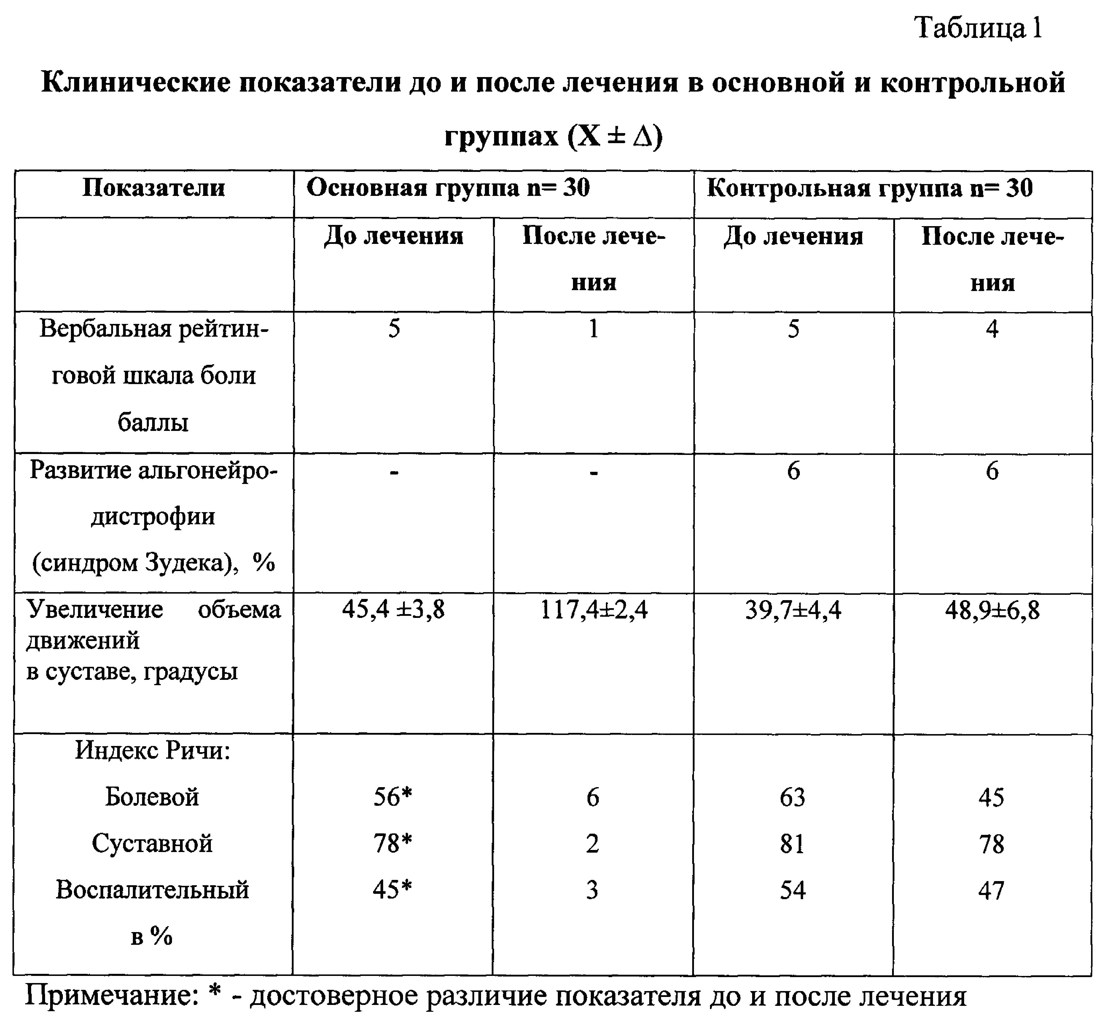
- Значительно уменьшить болевой синдром: у всех пациентов основной группы (по вербальной рейтинговой шкале, учитывающей силу боли, в основной группе соответствовала 1, а в контроле - 4-5 Р<0,01) к концу процедур.- Significantly reduce pain: in all patients of the main group (on a verbal rating scale that takes into account the strength of pain, in the main group corresponded to 1, and in the control - 4-5 P <0.01) by the end of the procedures.
- Увеличить объем движений в лучезапястном суставе за счет активности пронаторов и супинаторов у пациентов основной группы против контроля к концу курса (объем движений в основной группе через 10 дней составил 117,4±12,7 градусов, против 67,4±5,8 градусов в контроле Р<0,01).- To increase the range of motion in the wrist joint due to the activity of pronators and arch supports in patients of the main group against control by the end of the course (the range of movements in the main group after 10 days was 117.4 ± 12.7 degrees, versus 67.4 ± 5.8 degrees in control P <0.01).
- Значительно уменьшить активность воспалительно-дегенеративного процесса, о чем можно судить по суставному, болевому и воспалительному индексу Ричи в лучезапястном суставе (у всех пациентов основной группы отмечена достоверная положительная динамика индексов Ричи: болевой индекс снизился в среднем на 63%, суставной - на 78%, воспалительный - на 75% против 34%, 43% и 54% соответственно у больных, получавших лечение согласно прототипу Р<,01).- Significantly reduce the activity of the inflammatory-degenerative process, as can be judged by the articular, pain and inflammatory Richie index in the wrist joint (in all patients of the main group there was a significant positive dynamics of the Richie index: pain index decreased by an average of 63%, articular - by 78 %, inflammatory - by 75% against 34%, 43% and 54%, respectively, in patients receiving treatment according to the prototype P <, 01).
- Предупредить развитие альгонейродистрофии (синдром Зудека), о чем можно судить по отсутствию развития синдрома в основной группе, в то время как в контроле имело место проявление в 6% случаев.- Prevent the development of algoneurodystrophy (Sudek syndrome), as can be judged by the absence of the development of the syndrome in the main group, while in the control there was a manifestation in 6% of cases.
Способ осуществляется следующим образом.The method is as follows.
Воздействуют физиотерапевтическим фактором в положении больного сидя на стуле, располагают первый электрод (+) с прокладкой, смоченной в 5% растворе гидролизата плаценты, размером 6×10 см над проекцией костной мозоли в месте перелома костей предплечья с внутренней стороны предплечья, второй электрод (-) размером 8×10 см располагают на наружной поверхности предплечья. Диадинамотерапию проводят от аппарата «ДТ-50-3». Параметры тока при отпуске процедуры: постоянный ток полусинусоидальной формы частотой 100 Гц по 15 мин. Силу тока плавно увеличивают до появления выраженных, но неболезненных ощущений вибрации под электродами. Дополнительно сразу после процедуры диадинамофореза проводят магнитоакустическое воздействие над проекцией костной мозоли в месте перелома костей предплечья от аппарата Магофон-01 при величине магнитной индукции 30±9 мТл, с частотой акустического диапазона 0,02-20 кГц по 10 мин на процедуру. Процедуры проводят ежедневно на курс до 10 процедур.They influence the physiotherapeutic factor in the patient’s position while sitting on a chair, place the first electrode (+) with a pad soaked in a 5% placenta hydrolyzate solution 6 × 10 cm in size over the projection of bone callus at the site of fracture of the forearm bones from the inside of the forearm, the second electrode (- ) 8 × 10 cm in size are placed on the outer surface of the forearm. Diadynamic therapy is carried out from the device "DT-50-3". Current parameters during tempering: DC half-sine wave with a frequency of 100 Hz for 15 min. The current strength is gradually increased until the appearance of pronounced, but non-painful sensations of vibration under the electrodes. Additionally, immediately after the diadynamophoresis procedure, a magnetoacoustic effect is performed over the projection of bone callus at the site of fracture of the forearm bones from the Magofon-01 apparatus with a magnetic induction value of 30 ± 9 mT, with an acoustic frequency of 0.02-20 kHz for 10 min per procedure. Procedures are carried out daily for a course of up to 10 procedures.
Способ применяют для предупреждения развития осложнений после переломов костей предплечья у лиц старшей возрастной группы.The method is used to prevent the development of complications after fractures of the forearm bones in persons of an older age group.
Под наблюдением находилось 30 пациентов с диагнозом закрытый перелом лучевой кости без смещения в типичном месте 14 человек; закрытый перелом костей предплечья со смещением в типичном месте 14 человек; открытый перелом со смещением костей предплечья в типичном месте 2 человека, которые составили основную группу, получавших лечение по заявляемому способу. В контрольную группу вошли 30 больных с аналогичным диагнозом (22 человека с закрытым переломом лучевой кости без смещения в типичном месте, перелом костей предплечья со смещением в типичном месте 8 человек), получавших лечение согласно прототипу. Все пациенты находились под наблюдением травматолога в амбулаторных условиях после наложения гипсовой повязки. В 24 случаях имело место применение репозиции с целью совмещения осколков костей предплечья.Under observation were 30 patients with a diagnosis of closed fracture of the radius without displacement in a typical place of 14 people; closed fracture of the forearm with a displacement in a typical place of 14 people; open fracture with displacement of the bones of the forearm in a typical place of 2 people who made up the main group who received treatment by the present method. The control group included 30 patients with a similar diagnosis (22 people with a closed fracture of the radius without displacement in a typical place, fractures of the forearm with a displacement in a typical place of 8 people) who received treatment according to the prototype. All patients were under the supervision of a traumatologist on an outpatient basis after applying a plaster cast. In 24 cases, reposition was used to combine fragments of the bones of the forearm.
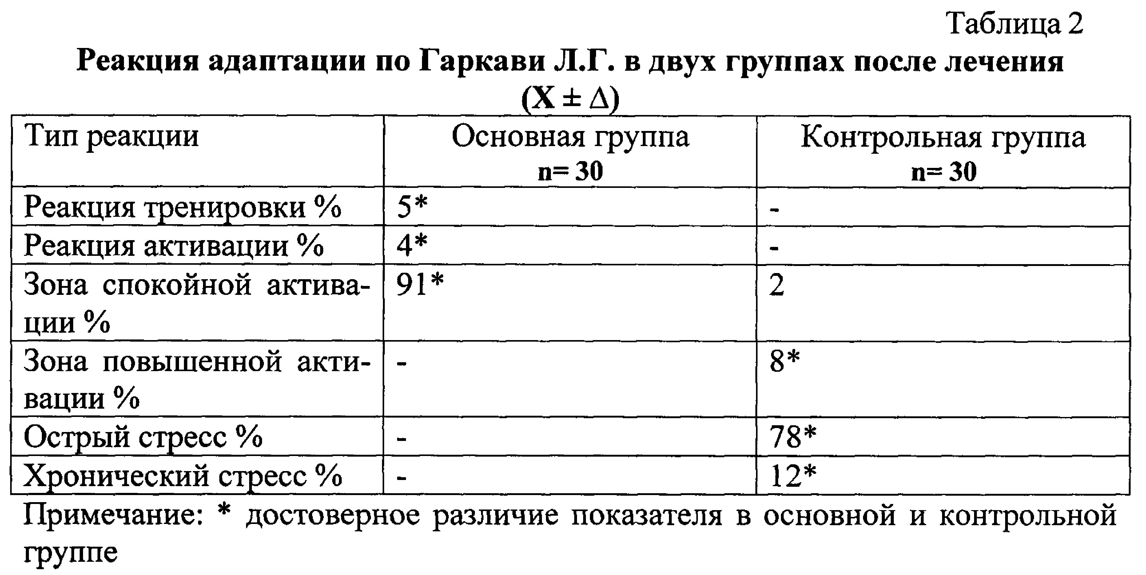
Всем пациентам проводилась рентгенография костей предплечья с лучезапястным суставом в прямой и боковой проекции трехкратно. После снятия гипсовой повязки до и после курса лечения по предлагаемому способу проводилась оценка болевого синдрома по вербальной шкале. Вербальная рейтинговая шкала содержит ряд слов, описывающих силу боли: боль отсутствует (1), легкая (2), дискомфортная (3), раздражающая (4), тяжелая (4), невыносимая (5). Оценкой силы боли служит порядковый номер выбранного определения (Павленко С.С. и соавт., 2002). Объем движений в лучезапястном суставе оценивался в градусах. Выраженность изменений в лучезапястном суставе определяли по оценке синдрома Ричи, учитывающего болевой, суставной и воспалительный характер изменений. С целью контроля за восстановлением адаптационно-резервных возможностей организма на фоне физиопроцедур определялась реакция Л.Г. Гаркави.All patients underwent x-ray of the forearm bones with the wrist joint in the direct and lateral projection three times. After removing the plaster cast before and after the course of treatment according to the proposed method, pain was assessed on a verbal scale. The verbal rating scale contains a number of words describing the strength of pain: pain is absent (1), mild (2), uncomfortable (3), irritating (4), severe (4), unbearable (5). Assessment of the strength of pain is the serial number of the selected definition (Pavlenko S.S. et al., 2002). The range of motion in the wrist joint was evaluated in degrees. The severity of changes in the wrist joint was determined by the evaluation of Richie's syndrome, taking into account the pain, articular and inflammatory nature of the changes. In order to control the restoration of the adaptive-reserve capabilities of the body against the background of physiotherapy, the reaction of L.G. Garkavi.
Полученные результаты показали, что предлагаемый способ имеет выраженный положительный эффект на восстановление функционального состояния лучезапястного сустава, вовлеченного в патологический процесс в результате перелома костей предплечья в типичном месте.The results showed that the proposed method has a pronounced positive effect on the restoration of the functional state of the wrist joint involved in the pathological process as a result of fracture of the forearm bones in a typical place.
Пример 1. Больная А. Год рождения 1956. Поступила под наблюдение с диагнозом основным: перелом костей предплечья в нижней трети со смещением отломков. Была проведена репозиция отломков костей предплечья с целью их совмещения. Была наложена соответствующая гипсовая циркулярная повязка. Контрольные рентгенограммы через 10, 21 и 30 дней после репозиции проводились соответственно: рентгенограмму лучезапястного сустава в двух проекциях - в переднезадней и боковой проекции. На 10 сутки проведена репозиция костных отломков, так как возникло вторичное смешение отломков в гипсовой повязке. Через 8 недель гипсовую повязку сняли. При осмотре окружающие ткани лучезапястного сустава имели отечность, багровосинюшную окраску, отсутствие сгибания и разгибания, отсутствие сгибания пальцев кисти, болезненность при попытке совершать движения в суставе. Пациентке сразу оказали воздействие по заявляемому способу: диадинамофорез в положении сидя на стуле, располагали первый электрод со смоченной прокладкой в 5% растворе гидролизата плаценты (подключают «+»), размером 6×10 см над проекцией костной мозоли в месте перелома костей предплечья с внутренней стороны предплечья, второй электрод размером 8×10 см - на наружной поверхности предплечья (подключают «-»). Диадинамофорез проводили от аппарата ДТ-50-3. Параметры тока при отпуске процедуры: постоянный ток полусинусоидальной формы частотой 100 Гц по 15 мин. Силу тока плавно увеличивали до появления выраженных, но неболезненных ощущений вибрации под электродами. Дополнительно сразу после процедуры диадинамофореза проводили магнитоакустическое воздействие над проекцией костной мозоли в месте перелома костей предплечья от аппарата Магофон-01, при величине магнитной индукции 30±9 мТл, с частотой акустического диапазона 0,02-20 кГц по 10 мин на процедуру. Процедуры проводили ежедневно на курс до 10 процедур. После окончания курса лечения через 10 дней локальная динамика была выражена в форме исчезновения отечности сустава, нормализации цвета кожи, отсутствии болезненности в суставе как в покое, так и при выполнении движений. При этом угол сгибания и разгибания доходил до 106 градусов. Наблюдения за пациенткой в течение месяца отмечали стойкую положительную динамику за регенерацией костных тканей и восстановлением функции лучезапястного сустава без явлений остеопороза. Реакция адаптации по Л.Г. Гаркави находилась в пределах зоны спокойной активации, что говорило о выраженном эффекте физиопроцедур на резистентность организма в целом, хотя пациентка была в постменопаузальном периоде, что значительно снижает приспособительные реакции. Амбулаторные наблюдения в течение 2-х лет с проведением двух курсов процедур в год по заявляемому способу позволили полностью восстановить функцию лучезапястного сустава и избежать осложнений.Example 1. Patient A. Year of birth 1956. Was admitted under observation with a diagnosis of the main: fracture of the forearm bones in the lower third with the displacement of fragments. Reposition of fragments of the bones of the forearm was carried out in order to combine them. An appropriate gypsum circular dressing was applied. Control radiographs 10, 21, and 30 days after the reposition were carried out, respectively: the radiograph of the wrist joint in two projections - in the anteroposterior and lateral projection. On day 10, the bone fragments were repositioned, as secondary mixing of fragments in a plaster cast occurred. After 8 weeks, the plaster cast was removed. On examination, the surrounding tissues of the wrist joint had swelling, a purplish-blue color, lack of flexion and extension, lack of flexion of the fingers, pain when trying to make movements in the joint. The patient immediately had an effect according to the claimed method: diadynamophoresis while sitting on a chair, placed the first electrode with a moistened pad in a 5% placenta hydrolyzate solution (connect “+”), 6 × 10 cm in size over the projection of bone callus at the site of fracture of the forearm bones with an internal sides of the forearm, the second electrode measuring 8 × 10 cm on the outer surface of the forearm (connect “-”). Diadynamophoresis was performed from a DT-50-3 apparatus. Current parameters during tempering: DC half-sine wave with a frequency of 100 Hz for 15 min. The current strength was gradually increased until the appearance of pronounced, but not painful sensations of vibration under the electrodes. In addition, immediately after the diadynamophoresis procedure, a magnetoacoustic effect was performed on the projection of bone callus at the site of fracture of the forearm bones from the Magofon-01 apparatus, with a magnetic induction value of 30 ± 9 mT, with an acoustic frequency of 0.02-20 kHz for 10 min per procedure. The procedures were carried out daily for a course of up to 10 procedures. After completing the course of treatment after 10 days, local dynamics was expressed in the form of the disappearance of swelling of the joint, normalization of skin color, the absence of pain in the joint both at rest and when performing movements. In this case, the angle of flexion and extension reached 106 degrees. Observations of the patient during the month noted persistent positive dynamics in bone tissue regeneration and restoration of the wrist joint function without osteoporosis. Adaptation reaction according to L.G. Garkavi was within the zone of calm activation, which indicated a pronounced effect of physiotherapy on the resistance of the organism as a whole, although the patient was in the postmenopausal period, which significantly reduces adaptive reactions. Outpatient observations for 2 years with two courses of procedures per year by the present method allowed to fully restore the function of the wrist joint and to avoid complications.
Пример 2. Больная В. Год рождения 1952. Поступила под наблюдение с диагнозом основным: перелом лучевой кости правого предплечья в нижней трети со смещением отломков. Была проведена репозиция отломков костей предплечья с целью их совмещения. Была наложена соответствующая гипсовая циркулярная повязка. Контрольные рентгенограммы через 10, 21 и 30 дней после репозиции проводились соответственно: рентгенограмму лучезапястного сустава в двух проекциях - в передне-задней и боковой проекции. На 11 сутки проведена репозиция костных отломков, так как возникло вторичное смешение отломков в гипсовой повязке. Через 5 недель гипсовую повязку сняли. При осмотре окружающие ткани лучезапястного сустава имели отечность, багровосинюшную окраску, отсутствие сгибания и разгибания, отсутствие сгибания пальцев кисти, болезненность при попытке совершать движения в суставе. Пациентке сразу оказали воздействие по заявляемому способу: диадинамофорез в положении сидя на стуле. Располагали первый электрод со смоченной прокладкой в 5% растворе гидролизата плаценты (подключают «+»), размером 6×10 см над проекцией костной мозоли в месте перелома костей предплечья с внутренней стороны предплечья. Второй электрод размером 8×10 см располагают на наружной поверхности предплечья (подключают «-»). Диадинамофорез проводили от аппарата ДТ-50-3. Параметры тока при отпуске процедуры: постоянный электрический ток полусинусоидальной формы частотой 100 Гц 15 мин. Силу тока плавно увеличивали до появления выраженных, но неболезненных ощущений вибрации под электродами. Дополнительно сразу после процедуры диадинамофореза проводили магнитоакустическое воздействие над проекцией костной мозоли в месте перелома костей предплечья от аппарата Магафон-01 при величине магнитной индукции 30±9 мТл, с частотой акустического диапазона 0,02-20 кГц 10 мин на процедуру. Процедуры проводили ежедневно на курс до 10 процедур. После окончания курса лечения через 10 дней локальная динамика была выражена в форме исчезновения отечности сустава, нормализации цвета кожи, отсутствии болезненности в суставе как в покое, так и при выполнении движений. При этом угол сгибания и разгибания доходил до 111 градусов. Наблюдения за пациенткой в течение месяца отмечали стойкую положительную динамику за регенерацией костных тканей и восстановлением функции лучезапястного сустава без явлений остеопороза. Реакция адаптации по Л.Г. Гаркави находилась в пределах зоны спокойной активации, что говорило о выраженном эффекте физиопроцедур на резистентность организма в целом, хотя пациентка была в постменопаузальном периоде, что значительно снижает приспособительные реакции. Амбулаторные наблюдения в течение 2-х лет с проведением двух курсов процедур в год по заявляемому способу позволили полностью восстановить функцию лучезапястного сустава и избежать осложнений.Example 2. Patient B. Year of birth 1952. Was admitted under observation with a diagnosis of the main: a fracture of the radial bone of the right forearm in the lower third with the displacement of fragments. Reposition of fragments of the bones of the forearm was carried out in order to combine them. An appropriate gypsum circular dressing was applied. Control radiographs 10, 21 and 30 days after the reposition were carried out, respectively: the radiograph of the wrist joint in two projections - in the anteroposterior and lateral projection. On day 11, bone fragments were repositioned, as secondary mixing of fragments in a plaster cast occurred. After 5 weeks, the plaster cast was removed. On examination, the surrounding tissues of the wrist joint had swelling, a purplish-blue color, lack of flexion and extension, lack of flexion of the fingers, pain when trying to make movements in the joint. The patient immediately had an effect according to the claimed method: diadynamophoresis in a sitting position on a chair. The first electrode was placed with a moistened pad in a 5% placental hydrolyzate solution (“+” plugged in), 6 × 10 cm in size over the projection of bone callus at the site of fracture of the forearm bones from the inside of the forearm. The second electrode measuring 8 × 10 cm is placed on the outer surface of the forearm (connect “-”). Diadynamophoresis was performed from a DT-50-3 apparatus. Current parameters during tempering: DC electric current of a half-sinusoidal shape with a frequency of 100 Hz 15 min. The current strength was gradually increased until the appearance of pronounced, but not painful sensations of vibration under the electrodes. In addition, immediately after the diadynamophoresis procedure, a magnetoacoustic effect was performed over the projection of bone callus at the site of fracture of the forearm bones from the Magafon-01 apparatus with a magnetic induction value of 30 ± 9 mT, with an acoustic frequency of 0.02-20 kHz for 10 min per procedure. The procedures were carried out daily for a course of up to 10 procedures. After completing the course of treatment after 10 days, local dynamics was expressed in the form of the disappearance of swelling of the joint, normalization of skin color, the absence of pain in the joint both at rest and when performing movements. In this case, the angle of flexion and extension reached 111 degrees. Observations of the patient during the month noted persistent positive dynamics in bone tissue regeneration and restoration of the wrist joint function without osteoporosis. Adaptation reaction according to L.G. Garkavi was within the zone of calm activation, which indicated a pronounced effect of physiotherapy on the resistance of the organism as a whole, although the patient was in the postmenopausal period, which significantly reduces adaptive reactions. Outpatient observations for 2 years with two courses of procedures per year by the present method allowed to fully restore the function of the wrist joint and to avoid complications.
Таким образом получен эффект от заявляемого способа, заключающийся в следующем:Thus, the effect of the proposed method, which consists in the following:
- исчезновение и значительное уменьшение выраженности болевого синдрома;- the disappearance and a significant decrease in the severity of pain;
- предупреждение развития контрактуры лучезапястного сустава после перелома;- prevention of the development of contracture of the wrist joint after a fracture;
- восстановление функции конечности;- restoration of limb function;
- предупреждение развития осложнения в форме альгонейромиодистрофического синдрома (синдрома Зудека);- prevention of the development of complications in the form of algoneuromyodystrophic syndrome (Sudeck syndrome);
- сохранение адаптационных резервов организма.- preservation of adaptive reserves of the body.
Claims (1)
Priority Applications (1)
| Application Number | Priority Date | Filing Date | Title |
|---|---|---|---|
| RU2015130164/14A RU2589664C1 (en) | 2015-07-21 | 2015-07-21 | Method of treating consequences of fractures of forearm bones in gerontological patients |
Applications Claiming Priority (1)
| Application Number | Priority Date | Filing Date | Title |
|---|---|---|---|
| RU2015130164/14A RU2589664C1 (en) | 2015-07-21 | 2015-07-21 | Method of treating consequences of fractures of forearm bones in gerontological patients |
Publications (1)
| Publication Number | Publication Date |
|---|---|
| RU2589664C1 true RU2589664C1 (en) | 2016-07-10 |
Family
ID=56371285
Family Applications (1)
| Application Number | Title | Priority Date | Filing Date |
|---|---|---|---|
| RU2015130164/14A RU2589664C1 (en) | 2015-07-21 | 2015-07-21 | Method of treating consequences of fractures of forearm bones in gerontological patients |
Country Status (1)
| Country | Link |
|---|---|
| RU (1) | RU2589664C1 (en) |
Citations (3)
| Publication number | Priority date | Publication date | Assignee | Title |
|---|---|---|---|---|
| SU973109A1 (en) * | 1980-04-19 | 1982-11-15 | Институт Механики Полимеров Ан Латвсср | Method of stimulating osteogenesis |
| RU2243004C2 (en) * | 2003-02-10 | 2004-12-27 | Государственное федеральное учреждение науки Уральский научно-исследовательский институт травматологии и ортопедии им. В.Д. Чаклина | Method for rehabilitating pelvic bone fracture patients |
| RU2365391C1 (en) * | 2008-05-15 | 2009-08-27 | Юрий Борисович Захаров | Method of bone fractures and surrounding soft tissues treatment during posttraumatic and postoperative periods |
-
2015
- 2015-07-21 RU RU2015130164/14A patent/RU2589664C1/en not_active IP Right Cessation
Patent Citations (3)
| Publication number | Priority date | Publication date | Assignee | Title |
|---|---|---|---|---|
| SU973109A1 (en) * | 1980-04-19 | 1982-11-15 | Институт Механики Полимеров Ан Латвсср | Method of stimulating osteogenesis |
| RU2243004C2 (en) * | 2003-02-10 | 2004-12-27 | Государственное федеральное учреждение науки Уральский научно-исследовательский институт травматологии и ортопедии им. В.Д. Чаклина | Method for rehabilitating pelvic bone fracture patients |
| RU2365391C1 (en) * | 2008-05-15 | 2009-08-27 | Юрий Борисович Захаров | Method of bone fractures and surrounding soft tissues treatment during posttraumatic and postoperative periods |
Non-Patent Citations (1)
| Title |
|---|
| Частная физиотерапия. Под ред. Г.Н. Пономаренко. 2005, С-Пб, с. 594-630. * |
Similar Documents
| Publication | Publication Date | Title |
|---|---|---|
| Stockman | The causes, pathology, and treatment of chronic rheumatism | |
| AU2009243693B2 (en) | A plurality of electrons for use in the restoration of a patient's health | |
| Kumar et al. | Effectiveness of Maitland Techniques in idiopathic shoulder adhesive capsulitis | |
| RU2316334C2 (en) | Method for activating lost motor functions and determining their recovery effectiveness in central nervous system injury cases | |
| RU2431456C1 (en) | Method for recovery and health improvement of patient by systemic exposure with using osteopathic technique by "lt" method | |
| NAHAS et al. | Effect of shock wave therapy on postpartum low back pain | |
| RU2589664C1 (en) | Method of treating consequences of fractures of forearm bones in gerontological patients | |
| RU2405596C1 (en) | Method of treating vegetosensory polyneuropathy of upper extremities in patients with pneumatic hammer disease | |
| RU2456030C1 (en) | Method of treating osteoarthrosis with underlying hypertension | |
| RU2365388C2 (en) | Electrotherapy method at various diseases and pathological states | |
| RU2371211C1 (en) | Method of dynamic electric neurostimulation for locomotor system injuries | |
| RU2447877C1 (en) | Method for therapeutic effect on spinal column | |
| RU2164128C2 (en) | Method of complex rehabilitation of patients pathology of locomotor system | |
| Pawluk | Pain management with pulsed electromagnetic field (PEMF) treatment | |
| RU2236264C1 (en) | Method for forming vertical position in patients suffering from spinal cord conductivity disorders | |
| RU2501583C1 (en) | Method of treating patients suffering gonarthrosis | |
| RU2786312C1 (en) | Method for treatment of pain syndrome in pathology of periarticular soft tissues of shoulder or knee joints | |
| RU2817633C1 (en) | Method for rehabilitation of patients after microsurgical removal of intervertebral disc herniation of lumbosacral spine | |
| RU2804751C1 (en) | Method for rehabilitation of patients after surgical treatment of herniated intervertebral disc of the lumbosacral department of the spine | |
| RU2543286C1 (en) | Method of treating children with consequences of injuries of upper and lower extremities | |
| RU2770981C1 (en) | Method for treating osteoarthritis of joints | |
| RU2308301C1 (en) | Method for treating knee joint arthrosis cases | |
| RU2431455C2 (en) | Method of treating typical mild and moderate climacteric syndrome | |
| RU2213588C2 (en) | Physiotherapeutic method for treating patients for diabetic polyneuria | |
| RU2103977C1 (en) | Method of treatment of joint osteoarthrosis |
Legal Events
| Date | Code | Title | Description |
|---|---|---|---|
| MM4A | The patent is invalid due to non-payment of fees |
Effective date: 20170722 |