RU2367373C1 - Osteosynthesis technique - Google Patents
Osteosynthesis technique Download PDFInfo
- Publication number
- RU2367373C1 RU2367373C1 RU2008116375/14A RU2008116375A RU2367373C1 RU 2367373 C1 RU2367373 C1 RU 2367373C1 RU 2008116375/14 A RU2008116375/14 A RU 2008116375/14A RU 2008116375 A RU2008116375 A RU 2008116375A RU 2367373 C1 RU2367373 C1 RU 2367373C1
- Authority
- RU
- Russia
- Prior art keywords
- mesh
- bone
- sheet
- edge
- rod
- Prior art date
Links
Images
Landscapes
- Surgical Instruments (AREA)
Abstract
Description
Изобретение относится к травматологии и предназначено для хирургического лечения при переломах костей, преимущественно проксимального конца бедренной или плечевой кости.The invention relates to traumatology and is intended for surgical treatment of bone fractures, mainly the proximal end of the femur or humerus.
При существующих методиках остеосинтеза стержневыми фиксаторами иногда после операции возникает миграция их из кости, что побудило нас к разработке нового способа, повышающего надежность удержания установленных фиксаторов и предупреждающего миграцию их из кости и мягких тканей организма.With existing methods of osteosynthesis with rod fixatives, sometimes after surgery, they migrate from the bone, which prompted us to develop a new method that increases the reliability of the retention of fixed fixators and prevents their migration from bone and soft tissues of the body.
Известен способ хирургического лечения больных с переломами костей по патенту РФ №2123308, включающий прокол полой иглой мягких тканей до кости, всверливание в кость через просвет полой иглы спиц с упорной площадкой и резьбой у упорной площадки и ввинчивание резьбы в поверхностный кортикальный слой кости до упорной площадки, свободный конец спиц оставляют под покровом тела.A known method of surgical treatment of patients with bone fractures according to the patent of the Russian Federation No. 2123308, comprising puncturing soft tissues with a hollow needle to a bone, drilling knitting needles with a thrust pad and thread at the thrust pad into the bone through the hollow needle and screwing the thread into the surface cortical bone layer to the thrust pad The free end of the spokes is left under the cover of the body.
Недостатком способа является то, что ввинчивание резьбы фиксатора только в поверхностный кортикальный слой кости не всегда обеспечивает надежное удержание фиксатора в кости. В этом способе отсутствуют признаки повышения качества лечения и уменьшения осложнений путем более надежного удержания фиксаторов в кости и снижения вероятности миграции их наружу.The disadvantage of this method is that screwing the retainer thread only into the superficial cortical layer of the bone does not always ensure reliable retention of the retainer in the bone. In this method, there are no signs of improving the quality of treatment and reducing complications by a more reliable retention of fixatives in the bone and reducing the likelihood of migration to the outside.
Известен наш способ остеосинтеза шейки бедренной кости по патенту РФ №2062060, включающий соединение костных отломков несколькими веерообразно расходящимися спицами, сближенные концы спиц над костью загибают, поворачивают в одну сторону, образуя пучок концов спиц над костью, и скрепляют их шляпкой из самотвердеющей пластмассы.Our method of osteosynthesis of the femoral neck is known according to RF patent No. 2062060, which involves connecting bone fragments with several fan-like diverging spokes, the close ends of the spokes above the bone are bent, turned in one direction, forming a bundle of ends of the spokes over the bone, and fastened with a hat made of self-hardening plastic.
Недостатком способ является то, что возможность его применения ограничена лишь случаями, когда оставляемые над поверхностью кости концы фиксаторов сближены настолько, что их можно скрепить небольшой шляпкой из самотвердеющей пластмассы. Применение же шляпки больших размеров создаст значительный инородный конгломерат, который при небольшой толще мягких тканей в области введения фиксаторов может вызвать осложнения. В этом способе отсутствуют признаки повышения качества лечения и уменьшения осложнений путем более надежного удержания отдаленных концов фиксаторов у кости и снижения вероятности миграции их наружу.The disadvantage of this method is that the possibility of its use is limited only to cases where the ends of the retainers left above the bone surface are so close that they can be fastened with a small hat made of self-hardening plastic. The use of large caps will create a significant foreign conglomerate, which with a small thickness of soft tissues in the area of the introduction of fixatives can cause complications. In this method, there are no signs of improving the quality of treatment and reducing complications by more reliable retention of the distant ends of the retainers near the bone and reducing the likelihood of their migration to the outside.
Известен наш способ остеосинтеза при латеральном переломе шейки бедренной кости по патенту РФ №2159591, включающий всверливание по наружным меткам в шейку и головку бедра спиц с последующим удалением их после введения фиксатора. При этом в секторе между двумя чрезкожно введенными спицами после прокола мягких тканей полой иглой и введения через нее в кость фиксатора удаляемым удлинителем иглу под кожей перемещают и через ее просвет каждый последующий фиксатор вводят в направлении биссектрисы угла, образуемого выше и ниже введенными в кость элементами, после чего иглу извлекают.Our method of osteosynthesis for lateral fracture of the femoral neck according to the patent of the Russian Federation No. 2159591, including drilling on the outer marks in the neck and femoral head of the knitting needles with their subsequent removal after insertion of the latch, is known. At the same time, in the sector between two percutaneously inserted knitting needles after puncture of soft tissues with a hollow needle and insertion of a fixator into the bone of the fixator with a removable extension cord, the needle under the skin is moved and each subsequent clamp is inserted through its lumen in the direction of the bisector of the angle formed above and below the elements inserted into the bone, after which the needle is removed.
Недостатком этого способа является недостаточно надежная фиксация оставляемых в кости и не скрепленных между собой накостных концов фиксаторов. В этом способе отсутствуют признаки повышения качества лечения и уменьшения осложнений путем более надежного удержания фиксаторов в кости и снижения вероятности миграции их наружу.The disadvantage of this method is the insufficiently reliable fixation of the bone ends of the retainers left in the bone and not fastened together. In this method, there are no signs of improving the quality of treatment and reducing complications by a more reliable retention of fixatives in the bone and reducing the likelihood of migration to the outside.
Наиболее близким по технической сущности и достигаемому результату является приведенный последним способ по патенту РФ №2159591, который мы принимаем за прототип, а недостатки его изложены выше.The closest in technical essence and the achieved result is the last method according to the patent of the Russian Federation No. 2159591, which we take as a prototype, and its disadvantages are described above.
Технический результат заключается в повышении качества лечения и уменьшении осложнений путем более надежного удержания фиксаторов в кости и снижения вероятности миграции их наружу.The technical result is to improve the quality of treatment and reduce complications by more reliable retention of fixatives in the bone and reduce the likelihood of migration to the outside.
Технический результат достигается тем, что способ остеосинтеза включает введение в костные отломки через хирургический разрез мягких тканей стержневых фиксаторов с оставлением их опорных элементов над поверхностью кости. При этом через ячейку у одного края листа, выполненного из мягко-эластичной мелкоячеистой сетки, проводят стержень фиксатора в кость до упора его опорного элемента, а следующие стержни фиксаторов вводят в расходящемся направлении их рабочих концов через другие ячейки у того же края листа сетки. Затем свободный боковой край листа сетки перегибают, накрывают им опорные элементы фиксаторов и скрепляют с закрепленным боковым краем листа.The technical result is achieved by the fact that the method of osteosynthesis includes the introduction into the bone fragments through a surgical incision of the soft tissues of the rod retainers with the leaving of their supporting elements above the surface of the bone. At the same time, through the cell at one edge of the sheet made of soft-elastic fine mesh, the retainer rod is drawn into the bone to the stop of its supporting element, and the following retainer rods are introduced in the diverging direction of their working ends through other cells at the same edge of the mesh sheet. Then, the free lateral edge of the mesh sheet is bent, the supporting elements of the clamps are covered with it and fastened to the fixed lateral edge of the sheet.
В предпочтительном варианте выполнения способа размер ячейки сетки больше толщины стержня, но меньше размера его опорного элемента.In a preferred embodiment of the method, the mesh cell size is greater than the thickness of the rod, but less than the size of its supporting element.
В другом предпочтительном варианте выполнения способа сетка сплетена из металлической мягко-эластичной проволоки.In another preferred embodiment of the method, the mesh is woven from soft-elastic metal wire.
В следующем предпочтительном варианте выполнения способа сетка выполнена из эластичного синтетического материала.In a further preferred embodiment of the method, the mesh is made of an elastic synthetic material.
В очередном предпочтительном варианте выполнения способа сетка выполнена из длительно самопроизвольно рассасывающегося в организме материала.In another preferred embodiment of the method, the mesh is made of a material that spontaneously resolves spontaneously in the body.
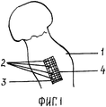
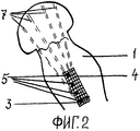
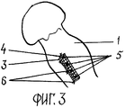
Сущность технического решения поясняется чертежами. На фиг.1 изображен проксимальный конец бедренной кости с переломом шейки, с намеченными по одной линии точками введения стержневых фиксаторов и уложенной на кортикальную поверхность кости сетку. На фиг.2 - то же, после введения стержневых фиксаторов в кость через сетку у одного ее края. На фиг.3 - то же, после укрытия свободным краем сетки опорных элементов (головок) стержневых фиксаторов и скрепления краев сетки между собой.The essence of the technical solution is illustrated by drawings. Figure 1 shows the proximal end of the femur with a fracture of the neck, with the points of introduction of the rod retainers and the mesh laid on the cortical surface of the bone, outlined in one line. Figure 2 - the same, after the introduction of rod retainers into the bone through the mesh at one of its edges. In Fig.3 - the same, after covering the free edge of the grid of the supporting elements (heads) of the rod clamps and fastening the edges of the grid to each other.
Конкретный пример осуществления способа. Например, при внутрисуставном переломе шейки бедренной кости после операционного разреза и обнажения кортикального слоя кости намечают по одной линии точки введения стержневых фиксаторов. Берут лист мягко-эластичной мелкоячеистой сетки с ячейками, пропускающими стержень фиксатора, но не пропускающими опорные элементы (головки) фиксаторов, и кладут его на намеченный участок поверхности кости. Сетка может быть выполнена из синтетического материала или из длительно самопроизвольно рассасывающегося в организме материала. Через ячейку у верхнего и одного бокового края сетки пропускают стержень фиксатора и вводят его в кость в намеченном направлении до упора головки фиксатора в сетку и кортикальный слой кости. Головка фиксатора остается над поверхностью кости. Затем через другую ячейку у нижнего того же бокового края листа сетки, после ее натяжения, пропускают стержень другого фиксатора и вводят его в костные отломки, оставляя его головку над поверхностью кости. Затем между головками установленных фиксаторов по одной с ними линии через другие ячейки у того же края листа сетки проводят стержни остальных фиксаторов в расходящемся направлении их рабочих концов. Далее свободный край сетки перегибают и поворачивают в сторону другого края сетки, закрепленного введенными фиксаторами на кости, плотно укрывают им опорные элементы (головки) фиксаторов и с натяжением скрепляют края сетки между собой лигатурами. Рану послойно ушивают.A specific example of the method. For example, in case of an intraarticular fracture of the femoral neck after the surgical incision and exposure of the cortical layer of the bone, the points of insertion of the rod fixators are plotted along one line. Take a sheet of soft-elastic fine-mesh with cells passing through the retainer rod, but not passing through the supporting elements (heads) of the retainers, and put it on the intended area of the bone surface. The mesh can be made of synthetic material or of a material that spontaneously absorbs in the body for a long time. The fixer rod is passed through a cell at the upper and one lateral edge of the mesh and inserted into the bone in the intended direction until the stop of the fixator head in the mesh and cortical bone layer. The retainer head remains above the bone surface. Then, through the other cell at the bottom of the same lateral edge of the grid sheet, after its tension, the rod of another fixator is passed and introduced into the bone fragments, leaving its head above the surface of the bone. Then, between the heads of the installed clamps along one line with them through the other cells at the same edge of the grid sheet, the rods of the remaining clamps are drawn in the diverging direction of their working ends. Next, the free edge of the mesh is bent and rotated towards the other edge of the mesh, fixed by the introduced fixators on the bones, tightly cover the supporting elements (heads) of the fixers and fasten the edges of the mesh together with ligatures. The wound is sutured in layers.
Второй конкретный пример осуществления способа. При вколоченном медиальном переломе шейки бедренной кости производят операционный разрез и обнажают площадку на кортикальной поверхности кости, на которой намечают по одной линии точки введения стержневых фиксаторов. К намечаемой площадке на обнаженной поверхности кости прикладывают сетку, сплетенную из металлической мягко-эластичной проволоки с ячейками, пропускающими стержень фиксатора, но не пропускающими опорные элементы (головки) фиксаторов. Через ячейку у верхнего и одного бокового края сетки пропускают стержень фиксатора и вводят его через намеченную точку в кость в заданном направлении до упора головки фиксатора в сетку и кортикальную поверхность кости. Головка фиксатора остается над поверхностью кости. Затем, слегка натягивая лист, закрепленный введенным фиксатором, через другую ячейку у нижнего того же бокового края листа сетки, пропускают стержень другого фиксатора и вводят его в костные отломки в другом направлении, оставляя головку фиксатора над поверхностью кости. Потом между головками установленных фиксаторов по одной с ними линии через другие ячейки у того же края листа сетки проводят стержни остальных фиксаторов в расходящемся направлении их рабочих концов. Далее свободный край сетки перегибают, поворачивают в сторону другого края сетки, закрепленного введенными фиксаторами на кости, плотно укрывают им опорные элементы (головки) фиксаторов и скрепляют с другим краем сетки, закрепленным фиксаторами. Рану ушивают.The second specific example of the method. When an implanted medial fracture of the femoral neck is made, an operative incision is made and the site on the cortical surface of the bone is exposed, on which the injection points of the rod fixators are drawn along one line. A mesh made of soft-elastic metal wire with cells passing through the retainer rod but not passing through the supporting elements (heads) of the fixators is applied to the intended site on the exposed surface of the bone. The fixer rod is passed through a cell at the upper and one lateral edge of the mesh and introduced through the target point into the bone in a predetermined direction until the stop of the fixator head in the mesh and cortical bone surface. The retainer head remains above the bone surface. Then, slightly pulling the sheet secured by the inserted fixative through another cell at the lower same lateral edge of the mesh sheet, the core of the other fixative is passed and inserted into the bone fragments in the other direction, leaving the fixative head above the bone surface. Then, between the heads of the installed clamps along the same line with them through the other cells at the same edge of the grid sheet, the rods of the remaining clamps are drawn in the diverging direction of their working ends. Next, the free edge of the mesh is bent, turned to the side of the other edge of the mesh, fixed with the introduced fixators on the bone, tightly cover the supporting elements (heads) of the fixers with it and fastened to the other edge of the mesh fixed with clamps. The wound is sutured.
Существенность отличия заявленного способа в следующем. Проведение через ячейку у одного края листа, выполненного из мягко-эластичной мелкоячеистой сетки, стержня фиксатора в кость до упора его опорного элемента обеспечивает удержание одного конца сетки на кости, освобождая руки ассистента на операции. Введение следующих стержней фиксаторов через другие ячейки у того же края листа сетки позволяет расположить головки фиксаторов по одной линии, что уменьшает площадь, необходимую для укрытия вторым краем листа, обеспечивает более плотное прилегание сетки ко всем головкам фиксаторов и лучшее скрепление всех фиксирующих элементов. Скрепленные сеткой головки фиксаторов представляют одну конструкцию со связанными между собой элементами. При расходящихся стержнях фиксаторов в кости такая конструкция делает вероятность миграции фиксаторов из тканей организма ничтожной. Поэтому введение рабочих концов стержней фиксаторов в расходящемся направлении при фиксированных сеткой головках повышает надежность удержания фиксаторов в кости, предупреждая их миграцию. Перегибание свободного бокового края листа сетки и укрывание им опорных элементов фиксаторов с последующим скреплением его с уже закрепленным на кости листом краем обеспечивает надежное удержание фиксаторов от миграции как внутрь, так и наружу. Выполнение размеров ячейки сетки больше толщины стержня, но меньше размера его опорного элемента исключает повреждение элементов сетки стержнями фиксаторов, предупреждает проваливание головок фиксаторов через ячейки в костную ткань, то есть миграцию фиксаторов внутрь. Сетка, сплетенная из металлической мягко-эластичной проволоки обладает высокой механической прочностью и может быть применена для повышения прочности фиксации головок фиксаторов. Сетка из эластичного синтетического материала не приводит к реакциям тканей организма на металл, и при необходимости ее проще удалять. Использование сетки, изготовленной из длительно рассасывающейся нити, не требует хирургического удаления сетки в дальнейшем. В случаях, когда не намечают в дальнейшем удаление фиксаторов из тканей организма при сращении перелома и самопроизвольном рассасывании сетки, снижается масса инородного тела в организме, что уменьшает вероятность возникновения отдаленных осложнений. Сетка из длительно самопроизвольно рассасывающегося в организме материала снижает операционную травму мягких тканей при необходимости удалении фиксаторов, например, у детей, так как не требуется удаление сетки.The significance of the differences of the claimed method in the following. Passing through the cell at one edge of the sheet made of soft-elastic fine-mesh mesh, the retainer rod into the bone to the stop of its supporting element ensures that one end of the mesh is held onto the bone, freeing the assistant's hands for operations. The introduction of the following clamp rods through other cells at the same edge of the grid sheet allows the clamp heads to be arranged in one line, which reduces the area required to cover the second edge of the sheet, provides a more snug fit of the grid to all clamp heads and better fastening of all locking elements. The mesh heads of the fasteners fastened by a mesh represent one design with elements interconnected. With divergent fixation rods in the bone, this design makes the probability of migration of fixatives from body tissues negligible. Therefore, the introduction of the working ends of the clamp rods in the diverging direction with mesh-fixed heads increases the reliability of retainer holding in the bones, preventing their migration. Bending the free lateral edge of the mesh sheet and covering it with the supporting elements of the retainers, followed by fastening it with the edge already fixed to the bone, ensures reliable retention of the retainers from migration both in and out. Performing mesh cell sizes is greater than the thickness of the rod, but less than the size of its supporting element, eliminates damage to the mesh elements by the retainer rods, prevents the heads of the retainers from falling through the cells into the bone tissue, that is, the migration of the retainers inward. The mesh woven from soft-elastic metal wire has high mechanical strength and can be used to increase the fixation strength of the clamp heads. A mesh of elastic synthetic material does not lead to reactions of body tissues to metal, and if necessary it is easier to remove. The use of a mesh made of a long absorbable thread does not require surgical removal of the mesh in the future. In cases where the removal of fixatives from the tissues of the body during the fusion of the fracture and spontaneous resorption of the mesh is not planned in the future, the foreign body mass in the body decreases, which reduces the likelihood of distant complications. A mesh made of a material that spontaneously absorbs in the body for a long time reduces the operative trauma of soft tissues if it is necessary to remove fixatives, for example, in children, since mesh removal is not required.
Таким образом, благодаря совокупности всех признаков заявленный способ обеспечивает повышение качества лечения и уменьшение осложнений путем более надежного удержания оставляемых фиксаторов в кости и снижения вероятности миграции их не только наружу, но и внутрь. Изобретенный способ хирургического лечения переломов кости может быть использован при различных видах переломов бедренной, плечевой и других костей. Улучшая качество хирургического лечения с сокращением сроков пребывания больных в стационаре, использование изобретения приведет к экономическому эффекту для пациентов и для учреждения.Thus, due to the combination of all the features, the claimed method provides an improvement in the quality of treatment and a reduction in complications by more reliable retention of retained retainers in the bone and a decrease in the probability of their migration not only outward, but also inward. The inventive method for the surgical treatment of bone fractures can be used for various types of fractures of the femur, humerus and other bones. Improving the quality of surgical treatment with a reduction in the length of hospital stay, the use of the invention will lead to an economic effect for patients and for the institution.
Применение способа возможно в детской и военно-полевой хирургии, а также в ветеринарии.The application of the method is possible in pediatric and field surgery, as well as in veterinary medicine.
Claims (5)
Priority Applications (1)
| Application Number | Priority Date | Filing Date | Title |
|---|---|---|---|
| RU2008116375/14A RU2367373C1 (en) | 2008-04-24 | 2008-04-24 | Osteosynthesis technique |
Applications Claiming Priority (1)
| Application Number | Priority Date | Filing Date | Title |
|---|---|---|---|
| RU2008116375/14A RU2367373C1 (en) | 2008-04-24 | 2008-04-24 | Osteosynthesis technique |
Publications (1)
| Publication Number | Publication Date |
|---|---|
| RU2367373C1 true RU2367373C1 (en) | 2009-09-20 |
Family
ID=41167724
Family Applications (1)
| Application Number | Title | Priority Date | Filing Date |
|---|---|---|---|
| RU2008116375/14A RU2367373C1 (en) | 2008-04-24 | 2008-04-24 | Osteosynthesis technique |
Country Status (1)
| Country | Link |
|---|---|
| RU (1) | RU2367373C1 (en) |
Citations (2)
| Publication number | Priority date | Publication date | Assignee | Title |
|---|---|---|---|---|
| RU2159591C1 (en) * | 1999-05-25 | 2000-11-27 | Кемеровская городская клиническая больница N 3 им. М.А. Подгорбунского | Method for carrying out osteosynthesis in the cases of lateral fracture of the femur neck |
| RU2181266C2 (en) * | 1998-06-09 | 2002-04-20 | Нижнекамская гор. больница № 2 | Method for carrying out femur neck fracture osteosynthesis |
-
2008
- 2008-04-24 RU RU2008116375/14A patent/RU2367373C1/en not_active IP Right Cessation
Patent Citations (2)
| Publication number | Priority date | Publication date | Assignee | Title |
|---|---|---|---|---|
| RU2181266C2 (en) * | 1998-06-09 | 2002-04-20 | Нижнекамская гор. больница № 2 | Method for carrying out femur neck fracture osteosynthesis |
| RU2159591C1 (en) * | 1999-05-25 | 2000-11-27 | Кемеровская городская клиническая больница N 3 им. М.А. Подгорбунского | Method for carrying out osteosynthesis in the cases of lateral fracture of the femur neck |
Non-Patent Citations (1)
| Title |
|---|
| Травматология, национальное руководство. ГЭОТАР-Медиа, подписано к печати 12.10.2007, 380-386. ЗВЕРЕВ Е.В. Функциональный внутрикостный остеосинтез шейки бедренной кости пучком спиц (Биомеханическое, анатомофизиологическое и клиническое обоснование). Ортопедия, травматология и протезирование, 1989, №11, с.6-9. RODRIGUEZ-MERCHAN E.C. In situ fixation of nondisplaced intracapsular fractures of the proximal femur. Clin Orthop Relat Res. 2002 Jun; (399):42-51 (Abstract). * |
Similar Documents
| Publication | Publication Date | Title |
|---|---|---|
| US9326807B2 (en) | Peri-prosthetic fixation implant and method | |
| RU2152763C1 (en) | Method for performing osteosynthesis using wires | |
| US10070904B2 (en) | Bone fixation implants | |
| US20170027617A1 (en) | Odontoid bullet | |
| RU2596089C1 (en) | Method for minimally invasive osteosynthesis floating fractures of ribs and device therefor | |
| RU2465844C2 (en) | Face and neck lifting device | |
| RU2367373C1 (en) | Osteosynthesis technique | |
| RU2405487C1 (en) | Method of surgical treatment of comminuted fracture of proximal part of humerus, clamp and implant for its realisation | |
| RU2634028C1 (en) | Method for surgical treatment of cranial vertebra rupture | |
| RU2210331C2 (en) | Method for surgical treatment of brachial collum fractures | |
| RU2157129C2 (en) | Method for substituting long bone defects | |
| RU2272592C1 (en) | Surgical intervention method for in the cases of humerus cervix fracture | |
| RU2718323C1 (en) | Method of humeral surgical neck fractures osteosynthesis | |
| RU2503424C1 (en) | Method and device for treating multiple ribs and breastbone fractures | |
| US10420543B2 (en) | Scapho-lunate and other ligament and bone repair/reconstruction | |
| RU2609058C1 (en) | Method of surgical treatment of fractures of proximal part of shoulder bone in children and teenagers | |
| SU1426557A1 (en) | Intraosseus fixative | |
| RU2468762C1 (en) | Method of surgical treatment of habitual shoulder dislocation | |
| CN107224319A (en) | A kind of reduction of the fracture free removal staple line internal fixation system | |
| RU2794407C1 (en) | Method for fixing an epidural catheter under the skin of the lumbar region | |
| RU2159591C1 (en) | Method for carrying out osteosynthesis in the cases of lateral fracture of the femur neck | |
| RU157220U1 (en) | BRACKET FOR FIXING THE TENDON | |
| RU2157130C1 (en) | Gear to treat ridge deformation of chest and process of its application | |
| RU2458648C1 (en) | Method of treating dislocation of acromial end of clavicle | |
| RU2816028C1 (en) | Method for surgical management of idiopathic thoracic scoliosis of iv degree by selective hybrid fixation |
Legal Events
| Date | Code | Title | Description |
|---|---|---|---|
| MM4A | The patent is invalid due to non-payment of fees |
Effective date: 20100425 |