KR102140055B1 - Organ mimic device with microchannels and methods of use and manufacturing thereof - Google Patents
Organ mimic device with microchannels and methods of use and manufacturing thereof Download PDFInfo
- Publication number
- KR102140055B1 KR102140055B1 KR1020197015134A KR20197015134A KR102140055B1 KR 102140055 B1 KR102140055 B1 KR 102140055B1 KR 1020197015134 A KR1020197015134 A KR 1020197015134A KR 20197015134 A KR20197015134 A KR 20197015134A KR 102140055 B1 KR102140055 B1 KR 102140055B1
- Authority
- KR
- South Korea
- Prior art keywords
- cells
- membrane
- delete delete
- cell
- fluid
- Prior art date
Links
Images


Classifications
-
- C—CHEMISTRY; METALLURGY
- C12—BIOCHEMISTRY; BEER; SPIRITS; WINE; VINEGAR; MICROBIOLOGY; ENZYMOLOGY; MUTATION OR GENETIC ENGINEERING
- C12M—APPARATUS FOR ENZYMOLOGY OR MICROBIOLOGY; APPARATUS FOR CULTURING MICROORGANISMS FOR PRODUCING BIOMASS, FOR GROWING CELLS OR FOR OBTAINING FERMENTATION OR METABOLIC PRODUCTS, i.e. BIOREACTORS OR FERMENTERS
- C12M3/00—Tissue, human, animal or plant cell, or virus culture apparatus
- C12M3/06—Tissue, human, animal or plant cell, or virus culture apparatus with filtration, ultrafiltration, inverse osmosis or dialysis means
-
- C—CHEMISTRY; METALLURGY
- C12—BIOCHEMISTRY; BEER; SPIRITS; WINE; VINEGAR; MICROBIOLOGY; ENZYMOLOGY; MUTATION OR GENETIC ENGINEERING
- C12M—APPARATUS FOR ENZYMOLOGY OR MICROBIOLOGY; APPARATUS FOR CULTURING MICROORGANISMS FOR PRODUCING BIOMASS, FOR GROWING CELLS OR FOR OBTAINING FERMENTATION OR METABOLIC PRODUCTS, i.e. BIOREACTORS OR FERMENTERS
- C12M23/00—Constructional details, e.g. recesses, hinges
- C12M23/02—Form or structure of the vessel
- C12M23/16—Microfluidic devices; Capillary tubes
-
- B—PERFORMING OPERATIONS; TRANSPORTING
- B01—PHYSICAL OR CHEMICAL PROCESSES OR APPARATUS IN GENERAL
- B01L—CHEMICAL OR PHYSICAL LABORATORY APPARATUS FOR GENERAL USE
- B01L3/00—Containers or dishes for laboratory use, e.g. laboratory glassware; Droppers
-
- B—PERFORMING OPERATIONS; TRANSPORTING
- B01—PHYSICAL OR CHEMICAL PROCESSES OR APPARATUS IN GENERAL
- B01L—CHEMICAL OR PHYSICAL LABORATORY APPARATUS FOR GENERAL USE
- B01L3/00—Containers or dishes for laboratory use, e.g. laboratory glassware; Droppers
- B01L3/50—Containers for the purpose of retaining a material to be analysed, e.g. test tubes
- B01L3/502—Containers for the purpose of retaining a material to be analysed, e.g. test tubes with fluid transport, e.g. in multi-compartment structures
- B01L3/5027—Containers for the purpose of retaining a material to be analysed, e.g. test tubes with fluid transport, e.g. in multi-compartment structures by integrated microfluidic structures, i.e. dimensions of channels and chambers are such that surface tension forces are important, e.g. lab-on-a-chip
- B01L3/50273—Containers for the purpose of retaining a material to be analysed, e.g. test tubes with fluid transport, e.g. in multi-compartment structures by integrated microfluidic structures, i.e. dimensions of channels and chambers are such that surface tension forces are important, e.g. lab-on-a-chip characterised by the means or forces applied to move the fluids
-
- C—CHEMISTRY; METALLURGY
- C12—BIOCHEMISTRY; BEER; SPIRITS; WINE; VINEGAR; MICROBIOLOGY; ENZYMOLOGY; MUTATION OR GENETIC ENGINEERING
- C12M—APPARATUS FOR ENZYMOLOGY OR MICROBIOLOGY; APPARATUS FOR CULTURING MICROORGANISMS FOR PRODUCING BIOMASS, FOR GROWING CELLS OR FOR OBTAINING FERMENTATION OR METABOLIC PRODUCTS, i.e. BIOREACTORS OR FERMENTERS
- C12M1/00—Apparatus for enzymology or microbiology
- C12M1/12—Apparatus for enzymology or microbiology with sterilisation, filtration or dialysis means
-
- C—CHEMISTRY; METALLURGY
- C12—BIOCHEMISTRY; BEER; SPIRITS; WINE; VINEGAR; MICROBIOLOGY; ENZYMOLOGY; MUTATION OR GENETIC ENGINEERING
- C12M—APPARATUS FOR ENZYMOLOGY OR MICROBIOLOGY; APPARATUS FOR CULTURING MICROORGANISMS FOR PRODUCING BIOMASS, FOR GROWING CELLS OR FOR OBTAINING FERMENTATION OR METABOLIC PRODUCTS, i.e. BIOREACTORS OR FERMENTERS
- C12M21/00—Bioreactors or fermenters specially adapted for specific uses
- C12M21/08—Bioreactors or fermenters specially adapted for specific uses for producing artificial tissue or for ex-vivo cultivation of tissue
-
- C—CHEMISTRY; METALLURGY
- C12—BIOCHEMISTRY; BEER; SPIRITS; WINE; VINEGAR; MICROBIOLOGY; ENZYMOLOGY; MUTATION OR GENETIC ENGINEERING
- C12M—APPARATUS FOR ENZYMOLOGY OR MICROBIOLOGY; APPARATUS FOR CULTURING MICROORGANISMS FOR PRODUCING BIOMASS, FOR GROWING CELLS OR FOR OBTAINING FERMENTATION OR METABOLIC PRODUCTS, i.e. BIOREACTORS OR FERMENTERS
- C12M23/00—Constructional details, e.g. recesses, hinges
- C12M23/58—Reaction vessels connected in series or in parallel
-
- C—CHEMISTRY; METALLURGY
- C12—BIOCHEMISTRY; BEER; SPIRITS; WINE; VINEGAR; MICROBIOLOGY; ENZYMOLOGY; MUTATION OR GENETIC ENGINEERING
- C12M—APPARATUS FOR ENZYMOLOGY OR MICROBIOLOGY; APPARATUS FOR CULTURING MICROORGANISMS FOR PRODUCING BIOMASS, FOR GROWING CELLS OR FOR OBTAINING FERMENTATION OR METABOLIC PRODUCTS, i.e. BIOREACTORS OR FERMENTERS
- C12M25/00—Means for supporting, enclosing or fixing the microorganisms, e.g. immunocoatings
- C12M25/02—Membranes; Filters
-
- C—CHEMISTRY; METALLURGY
- C12—BIOCHEMISTRY; BEER; SPIRITS; WINE; VINEGAR; MICROBIOLOGY; ENZYMOLOGY; MUTATION OR GENETIC ENGINEERING
- C12M—APPARATUS FOR ENZYMOLOGY OR MICROBIOLOGY; APPARATUS FOR CULTURING MICROORGANISMS FOR PRODUCING BIOMASS, FOR GROWING CELLS OR FOR OBTAINING FERMENTATION OR METABOLIC PRODUCTS, i.e. BIOREACTORS OR FERMENTERS
- C12M29/00—Means for introduction, extraction or recirculation of materials, e.g. pumps
- C12M29/10—Perfusion
-
- C—CHEMISTRY; METALLURGY
- C12—BIOCHEMISTRY; BEER; SPIRITS; WINE; VINEGAR; MICROBIOLOGY; ENZYMOLOGY; MUTATION OR GENETIC ENGINEERING
- C12M—APPARATUS FOR ENZYMOLOGY OR MICROBIOLOGY; APPARATUS FOR CULTURING MICROORGANISMS FOR PRODUCING BIOMASS, FOR GROWING CELLS OR FOR OBTAINING FERMENTATION OR METABOLIC PRODUCTS, i.e. BIOREACTORS OR FERMENTERS
- C12M35/00—Means for application of stress for stimulating the growth of microorganisms or the generation of fermentation or metabolic products; Means for electroporation or cell fusion
- C12M35/04—Mechanical means, e.g. sonic waves, stretching forces, pressure or shear stimuli
-
- C—CHEMISTRY; METALLURGY
- C12—BIOCHEMISTRY; BEER; SPIRITS; WINE; VINEGAR; MICROBIOLOGY; ENZYMOLOGY; MUTATION OR GENETIC ENGINEERING
- C12N—MICROORGANISMS OR ENZYMES; COMPOSITIONS THEREOF; PROPAGATING, PRESERVING, OR MAINTAINING MICROORGANISMS; MUTATION OR GENETIC ENGINEERING; CULTURE MEDIA
- C12N5/00—Undifferentiated human, animal or plant cells, e.g. cell lines; Tissues; Cultivation or maintenance thereof; Culture media therefor
- C12N5/06—Animal cells or tissues; Human cells or tissues
- C12N5/0602—Vertebrate cells
- C12N5/0603—Embryonic cells ; Embryoid bodies
- C12N5/0606—Pluripotent embryonic cells, e.g. embryonic stem cells [ES]
-
- C—CHEMISTRY; METALLURGY
- C12—BIOCHEMISTRY; BEER; SPIRITS; WINE; VINEGAR; MICROBIOLOGY; ENZYMOLOGY; MUTATION OR GENETIC ENGINEERING
- C12N—MICROORGANISMS OR ENZYMES; COMPOSITIONS THEREOF; PROPAGATING, PRESERVING, OR MAINTAINING MICROORGANISMS; MUTATION OR GENETIC ENGINEERING; CULTURE MEDIA
- C12N5/00—Undifferentiated human, animal or plant cells, e.g. cell lines; Tissues; Cultivation or maintenance thereof; Culture media therefor
- C12N5/06—Animal cells or tissues; Human cells or tissues
- C12N5/0602—Vertebrate cells
- C12N5/0608—Germ cells
- C12N5/061—Sperm cells, spermatogonia
-
- C—CHEMISTRY; METALLURGY
- C12—BIOCHEMISTRY; BEER; SPIRITS; WINE; VINEGAR; MICROBIOLOGY; ENZYMOLOGY; MUTATION OR GENETIC ENGINEERING
- C12N—MICROORGANISMS OR ENZYMES; COMPOSITIONS THEREOF; PROPAGATING, PRESERVING, OR MAINTAINING MICROORGANISMS; MUTATION OR GENETIC ENGINEERING; CULTURE MEDIA
- C12N5/00—Undifferentiated human, animal or plant cells, e.g. cell lines; Tissues; Cultivation or maintenance thereof; Culture media therefor
- C12N5/06—Animal cells or tissues; Human cells or tissues
- C12N5/0602—Vertebrate cells
- C12N5/0618—Cells of the nervous system
- C12N5/0623—Stem cells
-
- C—CHEMISTRY; METALLURGY
- C12—BIOCHEMISTRY; BEER; SPIRITS; WINE; VINEGAR; MICROBIOLOGY; ENZYMOLOGY; MUTATION OR GENETIC ENGINEERING
- C12N—MICROORGANISMS OR ENZYMES; COMPOSITIONS THEREOF; PROPAGATING, PRESERVING, OR MAINTAINING MICROORGANISMS; MUTATION OR GENETIC ENGINEERING; CULTURE MEDIA
- C12N5/00—Undifferentiated human, animal or plant cells, e.g. cell lines; Tissues; Cultivation or maintenance thereof; Culture media therefor
- C12N5/06—Animal cells or tissues; Human cells or tissues
- C12N5/0602—Vertebrate cells
- C12N5/0634—Cells from the blood or the immune system
- C12N5/0647—Haematopoietic stem cells; Uncommitted or multipotent progenitors
-
- C—CHEMISTRY; METALLURGY
- C12—BIOCHEMISTRY; BEER; SPIRITS; WINE; VINEGAR; MICROBIOLOGY; ENZYMOLOGY; MUTATION OR GENETIC ENGINEERING
- C12N—MICROORGANISMS OR ENZYMES; COMPOSITIONS THEREOF; PROPAGATING, PRESERVING, OR MAINTAINING MICROORGANISMS; MUTATION OR GENETIC ENGINEERING; CULTURE MEDIA
- C12N5/00—Undifferentiated human, animal or plant cells, e.g. cell lines; Tissues; Cultivation or maintenance thereof; Culture media therefor
- C12N5/06—Animal cells or tissues; Human cells or tissues
- C12N5/0602—Vertebrate cells
- C12N5/0652—Cells of skeletal and connective tissues; Mesenchyme
- C12N5/0654—Osteocytes, Osteoblasts, Odontocytes; Bones, Teeth
-
- C—CHEMISTRY; METALLURGY
- C12—BIOCHEMISTRY; BEER; SPIRITS; WINE; VINEGAR; MICROBIOLOGY; ENZYMOLOGY; MUTATION OR GENETIC ENGINEERING
- C12N—MICROORGANISMS OR ENZYMES; COMPOSITIONS THEREOF; PROPAGATING, PRESERVING, OR MAINTAINING MICROORGANISMS; MUTATION OR GENETIC ENGINEERING; CULTURE MEDIA
- C12N5/00—Undifferentiated human, animal or plant cells, e.g. cell lines; Tissues; Cultivation or maintenance thereof; Culture media therefor
- C12N5/06—Animal cells or tissues; Human cells or tissues
- C12N5/0602—Vertebrate cells
- C12N5/0652—Cells of skeletal and connective tissues; Mesenchyme
- C12N5/0662—Stem cells
-
- C—CHEMISTRY; METALLURGY
- C12—BIOCHEMISTRY; BEER; SPIRITS; WINE; VINEGAR; MICROBIOLOGY; ENZYMOLOGY; MUTATION OR GENETIC ENGINEERING
- C12N—MICROORGANISMS OR ENZYMES; COMPOSITIONS THEREOF; PROPAGATING, PRESERVING, OR MAINTAINING MICROORGANISMS; MUTATION OR GENETIC ENGINEERING; CULTURE MEDIA
- C12N5/00—Undifferentiated human, animal or plant cells, e.g. cell lines; Tissues; Cultivation or maintenance thereof; Culture media therefor
- C12N5/06—Animal cells or tissues; Human cells or tissues
- C12N5/0602—Vertebrate cells
- C12N5/067—Hepatocytes
- C12N5/0671—Three-dimensional culture, tissue culture or organ culture; Encapsulated cells
-
- C—CHEMISTRY; METALLURGY
- C12—BIOCHEMISTRY; BEER; SPIRITS; WINE; VINEGAR; MICROBIOLOGY; ENZYMOLOGY; MUTATION OR GENETIC ENGINEERING
- C12N—MICROORGANISMS OR ENZYMES; COMPOSITIONS THEREOF; PROPAGATING, PRESERVING, OR MAINTAINING MICROORGANISMS; MUTATION OR GENETIC ENGINEERING; CULTURE MEDIA
- C12N5/00—Undifferentiated human, animal or plant cells, e.g. cell lines; Tissues; Cultivation or maintenance thereof; Culture media therefor
- C12N5/06—Animal cells or tissues; Human cells or tissues
- C12N5/0602—Vertebrate cells
- C12N5/0688—Cells from the lungs or the respiratory tract
-
- C—CHEMISTRY; METALLURGY
- C12—BIOCHEMISTRY; BEER; SPIRITS; WINE; VINEGAR; MICROBIOLOGY; ENZYMOLOGY; MUTATION OR GENETIC ENGINEERING
- C12N—MICROORGANISMS OR ENZYMES; COMPOSITIONS THEREOF; PROPAGATING, PRESERVING, OR MAINTAINING MICROORGANISMS; MUTATION OR GENETIC ENGINEERING; CULTURE MEDIA
- C12N5/00—Undifferentiated human, animal or plant cells, e.g. cell lines; Tissues; Cultivation or maintenance thereof; Culture media therefor
- C12N5/06—Animal cells or tissues; Human cells or tissues
- C12N5/0602—Vertebrate cells
- C12N5/0696—Artificially induced pluripotent stem cells, e.g. iPS
-
- C—CHEMISTRY; METALLURGY
- C12—BIOCHEMISTRY; BEER; SPIRITS; WINE; VINEGAR; MICROBIOLOGY; ENZYMOLOGY; MUTATION OR GENETIC ENGINEERING
- C12N—MICROORGANISMS OR ENZYMES; COMPOSITIONS THEREOF; PROPAGATING, PRESERVING, OR MAINTAINING MICROORGANISMS; MUTATION OR GENETIC ENGINEERING; CULTURE MEDIA
- C12N5/00—Undifferentiated human, animal or plant cells, e.g. cell lines; Tissues; Cultivation or maintenance thereof; Culture media therefor
- C12N5/06—Animal cells or tissues; Human cells or tissues
- C12N5/0697—Artificial constructs associating cells of different lineages, e.g. tissue equivalents
-
- G—PHYSICS
- G01—MEASURING; TESTING
- G01N—INVESTIGATING OR ANALYSING MATERIALS BY DETERMINING THEIR CHEMICAL OR PHYSICAL PROPERTIES
- G01N33/00—Investigating or analysing materials by specific methods not covered by groups G01N1/00 - G01N31/00
- G01N33/48—Biological material, e.g. blood, urine; Haemocytometers
- G01N33/50—Chemical analysis of biological material, e.g. blood, urine; Testing involving biospecific ligand binding methods; Immunological testing
- G01N33/5005—Chemical analysis of biological material, e.g. blood, urine; Testing involving biospecific ligand binding methods; Immunological testing involving human or animal cells
- G01N33/5008—Chemical analysis of biological material, e.g. blood, urine; Testing involving biospecific ligand binding methods; Immunological testing involving human or animal cells for testing or evaluating the effect of chemical or biological compounds, e.g. drugs, cosmetics
- G01N33/5082—Supracellular entities, e.g. tissue, organisms
- G01N33/5088—Supracellular entities, e.g. tissue, organisms of vertebrates
-
- G—PHYSICS
- G01—MEASURING; TESTING
- G01N—INVESTIGATING OR ANALYSING MATERIALS BY DETERMINING THEIR CHEMICAL OR PHYSICAL PROPERTIES
- G01N33/00—Investigating or analysing materials by specific methods not covered by groups G01N1/00 - G01N31/00
- G01N33/48—Biological material, e.g. blood, urine; Haemocytometers
- G01N33/50—Chemical analysis of biological material, e.g. blood, urine; Testing involving biospecific ligand binding methods; Immunological testing
- G01N33/5005—Chemical analysis of biological material, e.g. blood, urine; Testing involving biospecific ligand binding methods; Immunological testing involving human or animal cells
- G01N33/5091—Chemical analysis of biological material, e.g. blood, urine; Testing involving biospecific ligand binding methods; Immunological testing involving human or animal cells for testing the pathological state of an organism
-
- B—PERFORMING OPERATIONS; TRANSPORTING
- B01—PHYSICAL OR CHEMICAL PROCESSES OR APPARATUS IN GENERAL
- B01L—CHEMICAL OR PHYSICAL LABORATORY APPARATUS FOR GENERAL USE
- B01L2200/00—Solutions for specific problems relating to chemical or physical laboratory apparatus
- B01L2200/06—Fluid handling related problems
- B01L2200/0647—Handling flowable solids, e.g. microscopic beads, cells, particles
- B01L2200/0663—Stretching or orienting elongated molecules or particles
-
- B—PERFORMING OPERATIONS; TRANSPORTING
- B01—PHYSICAL OR CHEMICAL PROCESSES OR APPARATUS IN GENERAL
- B01L—CHEMICAL OR PHYSICAL LABORATORY APPARATUS FOR GENERAL USE
- B01L2300/00—Additional constructional details
- B01L2300/08—Geometry, shape and general structure
- B01L2300/0848—Specific forms of parts of containers
- B01L2300/0854—Double walls
-
- B—PERFORMING OPERATIONS; TRANSPORTING
- B01—PHYSICAL OR CHEMICAL PROCESSES OR APPARATUS IN GENERAL
- B01L—CHEMICAL OR PHYSICAL LABORATORY APPARATUS FOR GENERAL USE
- B01L2300/00—Additional constructional details
- B01L2300/08—Geometry, shape and general structure
- B01L2300/0861—Configuration of multiple channels and/or chambers in a single devices
- B01L2300/0877—Flow chambers
-
- B—PERFORMING OPERATIONS; TRANSPORTING
- B01—PHYSICAL OR CHEMICAL PROCESSES OR APPARATUS IN GENERAL
- B01L—CHEMICAL OR PHYSICAL LABORATORY APPARATUS FOR GENERAL USE
- B01L2300/00—Additional constructional details
- B01L2300/08—Geometry, shape and general structure
- B01L2300/0887—Laminated structure
-
- B—PERFORMING OPERATIONS; TRANSPORTING
- B01—PHYSICAL OR CHEMICAL PROCESSES OR APPARATUS IN GENERAL
- B01L—CHEMICAL OR PHYSICAL LABORATORY APPARATUS FOR GENERAL USE
- B01L2300/00—Additional constructional details
- B01L2300/16—Surface properties and coatings
- B01L2300/161—Control and use of surface tension forces, e.g. hydrophobic, hydrophilic
- B01L2300/163—Biocompatibility
-
- B—PERFORMING OPERATIONS; TRANSPORTING
- B01—PHYSICAL OR CHEMICAL PROCESSES OR APPARATUS IN GENERAL
- B01L—CHEMICAL OR PHYSICAL LABORATORY APPARATUS FOR GENERAL USE
- B01L2400/00—Moving or stopping fluids
- B01L2400/04—Moving fluids with specific forces or mechanical means
- B01L2400/0403—Moving fluids with specific forces or mechanical means specific forces
- B01L2400/0472—Diffusion
-
- B—PERFORMING OPERATIONS; TRANSPORTING
- B01—PHYSICAL OR CHEMICAL PROCESSES OR APPARATUS IN GENERAL
- B01L—CHEMICAL OR PHYSICAL LABORATORY APPARATUS FOR GENERAL USE
- B01L2400/00—Moving or stopping fluids
- B01L2400/04—Moving fluids with specific forces or mechanical means
- B01L2400/0475—Moving fluids with specific forces or mechanical means specific mechanical means and fluid pressure
- B01L2400/0481—Moving fluids with specific forces or mechanical means specific mechanical means and fluid pressure squeezing of channels or chambers
-
- B—PERFORMING OPERATIONS; TRANSPORTING
- B01—PHYSICAL OR CHEMICAL PROCESSES OR APPARATUS IN GENERAL
- B01L—CHEMICAL OR PHYSICAL LABORATORY APPARATUS FOR GENERAL USE
- B01L2400/00—Moving or stopping fluids
- B01L2400/04—Moving fluids with specific forces or mechanical means
- B01L2400/0475—Moving fluids with specific forces or mechanical means specific mechanical means and fluid pressure
- B01L2400/0487—Moving fluids with specific forces or mechanical means specific mechanical means and fluid pressure fluid pressure, pneumatics
-
- B—PERFORMING OPERATIONS; TRANSPORTING
- B01—PHYSICAL OR CHEMICAL PROCESSES OR APPARATUS IN GENERAL
- B01L—CHEMICAL OR PHYSICAL LABORATORY APPARATUS FOR GENERAL USE
- B01L3/00—Containers or dishes for laboratory use, e.g. laboratory glassware; Droppers
- B01L3/50—Containers for the purpose of retaining a material to be analysed, e.g. test tubes
- B01L3/502—Containers for the purpose of retaining a material to be analysed, e.g. test tubes with fluid transport, e.g. in multi-compartment structures
- B01L3/5027—Containers for the purpose of retaining a material to be analysed, e.g. test tubes with fluid transport, e.g. in multi-compartment structures by integrated microfluidic structures, i.e. dimensions of channels and chambers are such that surface tension forces are important, e.g. lab-on-a-chip
Abstract
하나 이상의 다공성 막으로 분리된 마이크로채널. 이들 막은 중심 마이크로채널을 2개 이상의 평행한 중심 마이크로채널로 분할하도록 구성되며, 여기서 하나 이상의 제1 유체가 제1 중심 마이크로채널을 통해 적용되고, 하나 이상의 제2 유체가 제2 이상의 중심 마이크로채널을 통해 적용된다. 각각의 다공성 막의 표면은 세포 접착성 분자로 코팅되어 세포의 부착을 지지하고 막의 상면 및 하면 상의 조직 내로의 세포의 조직화를 촉진시킬 수 있다. 기공은 단지 기체 및 작은 화학물질의 교환을 허용하기만 하기에, 또는 큰 단백질 및 살아있는 세포 전체(whole living cell)의 이동 및 채널횡단 통과(transchannel passage)를 허용하기에 충분히 클 수 있다.A microchannel separated by one or more porous membranes. These membranes are configured to divide the central microchannel into two or more parallel central microchannels, where one or more first fluids are applied through the first central microchannels, and one or more second fluids cross the second or more central microchannels. Is applied through. The surface of each porous membrane can be coated with cell adhesive molecules to support cell adhesion and promote the organization of cells into tissue on the top and bottom surfaces of the membrane. The pores can only be large enough to allow for the exchange of gases and small chemicals, or to allow the transport and transchannel passage of large proteins and whole living cells.
Description
관련 출원과의 상호 참조Cross-reference to related applications
본 출원은 35 U. S. C. 119조(e) 하에서, 2008년 7월 16일자로 출원된 미국 가출원 제61/081,080호의 이득을 주장하며, 이 출원의 내용은 전체적으로 본 명세서에 참고로 포함된다.This application claims the benefit of U.S. Provisional Application No. 61/081,080 filed on July 16, 2008, under Article 119(e) of 35 U. S. C., the contents of which are incorporated herein by reference in their entirety.
*정부 지원 * Government support
본 발명은 미국국립보건원(National Institutes of Health)이 인정한 승인 번호: NIH ROl ES016665-01A1 하에서 정부 지원으로 이루어졌다. 정부는 본 발명에 대해 일정한 권리를 갖는다.The present invention was made with government support under the approval number: NIH ROl ES016665-01A1 recognized by the National Institutes of Health. The government has certain rights in this invention.
기술분야Technology field
본 발명은 일반적으로 마이크로채널을 갖는 기관 모방 장치 및 그 사용 및 제조 방법에 관한 것이다.The present invention generally relates to an apparatus for mimicking an organ having a microchannel and a method for using and manufacturing the same.
배경기술Background
역학적 힘 - 밀기, 당기기, 인장, 압축 - 은 세포 발생 및 거동의 중요한 조절인자이다. 텐세그리티(tensegrity)는 이들 물리적 힘이 세포 또는 조직 내부에서 어떻게 분포되는지, 그리고 이들 물리적 힘이 그들의 영향을 어떻게 그리고 어디에 주는지를 결정하는 구조를 제공한다.Mechanical forces-push, pull, tension, and compression-are important regulators of cell development and behavior. Tensegrity provides a structure that determines how these physical forces are distributed within a cell or tissue, and how and where these physical forces exert their influence.
인체에서, 대부분의 세포는 끊임없이 인장 또는 압축과 같은 역학적 힘을 받는다. 배양액 내의 세포에의 역학적 변형(mechanical strain)의 인가는 생체내(in vivo) 환경을 시뮬레이션하여, 세포 내에서 극적인 형태학적 변화 및 생체역학적 반응을 일으킨다. 세포가 배양액 내에 역학적으로 로딩될 때 일어나는 장기간 변화 및 단기간 변화 둘 모두가 있으며, 예를 들어 DNA 또는 RNA 합성 또는 분해, 단백질 발현 및 분비의 속도 및 양에 있어서의 변경, 세포 분열 및 정렬의 속도에 있어서의 변경, 에너지 대사에 있어서의 변화, 거대분자 합성 또는 분해의 속도에 있어서의 변화, 및 생화학 및 생체에너지학에 있어서의 다른 변화이다.In the human body, most cells are constantly subject to mechanical forces such as tension or compression. The application of mechanical strain to cells in the culture medium simulates the in vivo environment, resulting in dramatic morphological changes and biomechanical reactions in the cells. There are both long-term and short-term changes that occur when cells are dynamically loaded into the culture medium, for example, DNA or RNA synthesis or degradation, changes in the rate and amount of protein expression and secretion, and the rate of cell division and alignment. Changes in energy metabolism, changes in energy metabolism, changes in the rate of macromolecular synthesis or degradation, and other changes in biochemistry and bioenergetics.
모든 세포는 분자 "스트러트 및 와이어"로부터 형성되는 격자인, 내부 뼈대(internal scaffolding), 또는 세포골격을 갖는다. "와이어"는 미세섬유(microfilament)로 알려진 미세한 케이블들의 교차 네트워크(crisscrossing network)로, 이는 세포막으로부터 핵으로 신장하여 내향 당김(inward pull)을 나타낸다. 이러한 당김에 대항하는 것이 미세소관, 즉 세포골격의 보다 두꺼운 압축-견딤(compression-bearing) "스트러트", 및 세포를 세포들의 그룹을 함께 유지하는 섬유질 물질인 세포외 기질에 고정시키는, 세포의 외부 막 상의 특수화된 수용체 분자이다. 이러한 힘의 균형이 텐세그리티의 특징이다.All cells have an internal scaffolding, or cytoskeleton, which is a lattice formed from the molecules “struts and wires”. “Wire” is a crisscrossing network of microscopic cables known as microfilament, which extends from the cell membrane to the nucleus to indicate an inward pull. Against this pull is the microtubule, the thicker compression-bearing "strut" of the cytoskeleton, and the outside of the cell, which anchors the cell to the extracellular matrix, a fibrous substance that holds a group of cells together. It is a specialized receptor molecule on the membrane. This balance of power is a characteristic of Tensegrity.
조직은 "달걀 상자(egg carton)"에 놓여 있는 달걀들처럼, 세포외 기질의 세포들의 그룹으로부터 구성된다. 세포들을 기질에 고정시키는 수용체 분자 - 인테그린으로 알려짐 - 는 세포들을 더 넓은 세계에 연결시킨다. 조직에 대한 역학적 힘은 먼저 이들 고정점에서 인테그린에 의해 느껴지고, 이어서 세포골격에 의해 각각의 세포 내부의 심부로 운반되는데, 이 힘은 단백질 분자의 형상을 진동 또는 변화시켜 생화학적 반응을 유발시키거나, 또는 핵 내의 염색체를 잡아당겨 유전자를 활성화시킬 수 있을 것이다.Tissues are constructed from groups of cells of the extracellular matrix, such as eggs placed in "egg cartons". Receptor molecules that fix cells to the matrix-known as integrins-connect cells to a wider world. The mechanical force on the tissue is first felt by the integrins at these anchor points, and then transported by the cytoskeleton to the deep inside each cell, which vibrates or changes the shape of the protein molecule, causing a biochemical reaction or Alternatively, the gene may be activated by pulling a chromosome in the nucleus.
세포는 또한 근육처럼 "톤(tone)"을 갖는다고 할 수 있는데, 이는 세포골격 섬유의 일정한 당김 때문이다. 늘어난 바이올린 현이, 그 길이를 따라 상이한 지점에서 힘이 인가될 때, 상이한 소리를 만들어내는 것과 흡사하게, 세포는 그것이 얼마나 많이 변형되는지에 따라 상이하게 화학 신호를 처리한다.Cells can also be said to have “tones” like muscles, due to the constant pulling of the cytoskeletal fibers. The cell handles chemical signals differently depending on how much it deforms, as an elongated violin string produces different sounds when a force is applied at different points along its length.
성장 인자는 세포가 얼마나 많이 신장되는지에 따라 상이한 효과를 가질 것이다. 상처 표면에서의 것들과 같이 신장되고 편평해진 세포는 성장하고 증가하려는 경향이 있는 반면, 과도하게 밀집된 상태에 의해 위축된, 둥글게 된 세포는 "자살(suicide)" 프로그램을 스위치 온하고 죽는다. 대조적으로, 신장되지도 수축되지도 않은 세포는 그의 의도된 기능을 계속한다.Growth factors will have different effects depending on how many cells are stretched. Elongated and flattened cells, such as those on the wound surface, tend to grow and increase, while rounded cells, atrophied by an overcrowded state, switch on and kill the "suicide" program. In contrast, cells that are neither stretched nor contracted continue their intended function.
세포 텐세그리티의 또 다른 원칙(tenet)은 물리적 위치가 중요하다는 것이다. 조절 분자들이 세포 내부에서 마음대로 떠돌아다닐 때, 이들 분자의 활성은 전체로서 세포에 작용하는 역학적 힘에 거의 영향을 받지 않는다. 그러나, 그들이 세포골격에 부착될 때, 그들은 그 보다 큰 네트워크의 일부가 되며, 세포 의사 결정(cellular decision-making)에 영향을 주는 위치에 있게 된다. 많은 조절 및 신호전달 분자는 인테그린이 모이는 접착 부위로 알려진 지점(spot) 내의 세포의 표면 막에서 세포골격 상에 고정된다. 이들 최고의 장소는 컴퓨터 네트워크 상의 노드와 같은 핵심 신호-처리 센터이며, 여기서 이웃 분자들이 외부 세계로부터의 역학적 정보를 받아들이고 신호를 교환할 수 있다.Another principle of cellular tensegity is that physical location is important. When regulatory molecules wander freely inside the cell, the activity of these molecules is almost unaffected by the mechanical forces acting on the cell as a whole. However, when they attach to the cytoskeleton, they become part of a larger network and are in a position to influence cellular decision-making. Many regulatory and signaling molecules are immobilized on the cytoskeleton at the cell's surface membrane within a spot known as the adhesion site where integrins converge. These best places are key signal-processing centers, such as nodes on a computer network, where neighboring molecules can accept and exchange epidemiological information from the outside world.
따라서, 생리학적인 환경에서 약물, 약물 전달 비히클, 나노진단제 또는 치료제 또는 환경적 스트레스 인자, 예를 들어 입자, 기체, 및 독소의 최대 효과를 평가하는 것은 세포-세포 상호작용 및 세포-화학물질 상호작용에 관한 연구뿐만 아니라, 이들 상호작용이 건강한 조직 및 질환에 이환된 조직 둘 모두에서 생리학적인 역학적 힘에 의해 어떻게 영향을 받는지에 대한 연구도 필요로 한다.Thus, evaluating the maximal effects of a drug, drug delivery vehicle, nanodiagnostic agent or therapeutic agent or environmental stressors, such as particles, gases, and toxins in a physiological environment, is a cell-cell interaction and cell-chemical interaction. In addition to research on action, research into how these interactions are affected by physiological mechanical forces in both healthy and diseased tissues is also needed.
배양액 내에서 세포의 역학적 환경 또는 반응을 변경시키는 방법들은 단층(monolayer)을 긁어내거나, 자계 또는 전계를 인가하거나, 또는 스크류 장치, 수압, 또는 추를 사용하여 배양 세포에 직접 정적 또는 주기적 인장 또는 압축을 인가함으로써 세포를 손상시키는 것을 포함해 왔다. 세포를 유체 유동에 놓이게 함으로써 전단 응력이 또한 유도되어 왔다. 그러나, 이들 수법의 거의 대부분은 생리학적으로 관련된 조직-조직 상호작용을 유지하는 배양 미세환경 내에서 폭넓은 재현가능한 범위의 주기적 변형을 달성하도록 조절을 제공하지 않았거나 인가된 변형의 정량을 가능하게 하지 않았다.Methods for altering the dynamical environment or response of cells in the culture medium include scraping a monolayer, applying a magnetic or electric field, or directly or statically or periodically tensioning or compressing the cultured cells using a screw device, water pressure, or weight. It has been involved in damaging cells by applying. Shear stress has also been induced by placing cells in fluid flow. However, almost all of these techniques did not provide control to achieve a wide reproducible range of periodic modifications within a cultured microenvironment that maintains physiologically relevant tissue-tissue interactions or enable quantification of approved modifications. Did not do it.
생체 기관(living organ)은 근접하여 나란히 놓여진 2개 이상의 조직으로 구성된 3차원 혈관화된 구조물로, 이는 집합적으로 기능하고 유체 전단 및 역학적 변형과 같은 동적 역학적 힘의 존재 하에서 조직-조직 계면을 가로질러 물질, 세포 및 정보를 수송한다. 생리학적 조직-조직 계면을 재현하고 유체 유동 및 동적 역학적 변형을 가능하게 하는, 살아있는 세포를 함유하는 마이크로장치의 창조는 복잡한 기관 기능, 예를 들어 면역 세포 수송(trafficking), 영양소 흡수, 감염, 산소 및 이산화탄소 교환 등의 연구에 있어서, 그리고 약물 스크리닝, 독물학, 진단학 및 치료학에 있어서 큰 가치를 가질 것이다.A living organ is a three-dimensional vascularized structure composed of two or more adjacent tissues placed side by side, which collectively function and traverse the tissue-tissue interface in the presence of dynamic mechanical forces such as fluid shear and mechanical deformation. Barges transport material, cells and information. The creation of microdevices containing living cells, reproducing physiological tissue-tissue interfaces and enabling fluid flow and dynamic mechanical transformation, is a complex organ function, such as immune cell trafficking, nutrient uptake, infection, oxygen And carbon dioxide exchange, and drug screening, toxicology, diagnostics and therapeutics.
폐포-모세혈관 단위는 폐의 정상적인 생리학적 기능의 유지에 있어서뿐만 아니라 다양한 폐질환의 병인 및 진행에 있어서 중요한 역할을 한다. 폐의 복잡한 구조, 작은 크기의 폐포들 및 그들을 둘러싸는 미세혈관, 및 이 기관의 동적 역학적 운동으로 인해, 마이크로스케일로 이 구조를 연구하기란 어렵다. Alveolar-capillary units play an important role not only in maintaining normal physiological functions of the lung, but also in the pathogenesis and progression of various lung diseases. Due to the complex structure of the lungs, small-sized alveoli and the microvessels surrounding them, and the dynamic mechanical motion of the organ, it is difficult to study this structure in microscale.
폐는 기체 교환이 일어나는 현미경적 폐포 구획으로 및 이로부터 대류 기체 수송을 가능하게 하는 전도관(conducting tube)의 계층적 분지 네트워크를 갖는 해부학적으로 독특한 구조를 갖는다. 폐포는 정상적인 호흡을 위한 폐의 가장 중요한 기능적 단위이며, 그것은 혈액-기체 장벽 또는 계면일 뿐만 아니라, 계면활성제가 공기 유입을 가능하게 하도록 작용하고, 급성 호흡 곤란 증후군(acute respiratory distress syndrome, ARDS) 환자 또는 감염, 예를 들어 폐렴 환자에서 면역 세포, 병원체 및 유체가 축적되는 부위라는 점에서 가장 임상적으로 적절하다.The lungs have an anatomically unique structure with a hierarchical branching network of conducting tubes that allows for the transport of convective gases to and from the microscopic alveolar compartment where gas exchange takes place. Alveoli is the most important functional unit of the lungs for normal breathing, which is not only the blood-gas barrier or interface, but also acts to enable the air to enter the air, and suffers from acute respiratory distress syndrome (ARDS) Or, it is the most clinically appropriate in that it is the site of accumulation of immune cells, pathogens and fluids in patients with infections, eg pneumonia.
폐 모세혈관과 폐포 내강 사이의 혈액-기체 장벽 또는 조직-조직 계면은 배아에서 세포 및 분자의 자기-집합(self-assembly)을 통하여 형성되는 얇은 세포외 기질(extracellular matrix, ECM)에 의해 분리된 모세혈관 내피의 단일층에 근접하여 나란히 놓여진 폐포 상피의 단일층으로 구성된다. 사실상, 폐포-모세혈관 단위의 기능에 관한 모든 분석은 동물 전체(whole animal) 연구에서 수행되어 왔는데, 이는 그것이 시험관내에서(in vitro) 이 기관-수준 구조물을 재생하는 데 가능하지 않았기 때문이다.The blood-gas barrier or tissue-tissue interface between the lung capillaries and the alveolar lumen is separated by a thin extracellular matrix (ECM) that is formed through the self-assembly of cells and molecules in the embryo. It consists of a single layer of alveolar epithelium placed side by side close to a single layer of capillary endothelium. In fact, all analyzes of the function of alveolar-capillary units have been performed in whole animal studies because it was not possible to regenerate this organ-level construct in vitro.
폐포-모세혈관 단위의 핵심 구조 조직화, 생리학적 기능, 및 생리학적 또는 병리학적 역학적 활성을 나타내는 다세포 및 다조직 기관-유사 구조물의 집합을 촉진시킬 수 있는 실험 도구의 결여에 큰 난제가 놓여 있는데, 이러한 실험 도구는 보통 각 호흡 주기 동안 반복된 팽창 및 수축을 받는다. 이러한 제한은 그것이 시험관내에서 이 기관-수준 구조물을 재생할 수 있고 그의 생리학적인 역학적 미세환경을 재현(recapitulate)할 수 있다면 극복할 수 있을 것이다. 그러나, 이것은 충분히 달성되어 있지 않다.A major challenge lies in the lack of experimental tools that can facilitate the aggregation of multicellular and multi-tissue organ-like structures that exhibit the organization of key structures of the alveolar-capillary units, their physiological function, and physiological or pathological epidemiological activity. These experimental tools usually undergo repeated expansion and contraction during each breathing cycle. This limitation could be overcome if it could regenerate this organ-level construct in vitro and recapitulate its physiological mechanical microenvironment. However, this has not been sufficiently achieved.
필요한 것은 조직-조직 관련 기능을 수행하고, 또한 화학적 힘뿐만 아니라 역학적 힘에도 응하여 세포를 장치 내에 자연적으로 조직화할 수 있게 하고, 조직-조직 생리를 모방하는 막을 통한 세포 거동에 관한 연구를 가능하게 하는, 시험관내 또는 생체내에서 사용될 수 있는 기관 모방 장치이다.What is needed is to perform tissue-tissue-related functions, also to allow cells to naturally organize within the device in response to chemical as well as mechanical forces, and to study the behavior of cells through membranes that mimic tissue-tissue physiology. It is an organ-mimicking device that can be used in vitro or in vivo.
시스템 및 방법은 하나 이상의 다공성 막에 의해 분리된 중심 마이크로채널을 갖는 본체(body)를 포함한다. 이들 막은 중심 마이크로채널을 근접하여 나란히 배치된 2개 이상의 평행한 중심 마이크로채널로 분할하도록 구성되며, 여기서 하나 이상의 제1 유체가 제1 중심 마이크로채널을 통해 적용되고, 하나 이상의 제2 유체가 제2 또는 그 이상의 중심 마이크로채널을 통해 적용된다. 각각의 다공성 막의 표면은 세포 접착성 분자로 코팅되어 세포의 부착을 지지하고 각각의 막의 상면 및 하면 상의 조직 내로의 세포의 조직화를 촉진시킬 수 있으며, 그럼으로써 인접한 평행한 유체 채널들 사이의 다공성 막에 의해 분리된 하나 이상의 조직-조직 계면을 생성할 수 있다. 막은 다공성이거나, 연성이거나, 탄성이거나, 또는 이들의 조합일 수 있으며, 이때 기공은 단지 기체 및 작은 화학물질의 교환을 허용하기만 하기에 충분히 크거나, 또는 큰 단백질 및 살아있는 세포 전체(whole living cell)의 이동 및 채널횡단 통과(transchannel passage)를 허용하기에 충분히 크다. 원하는 유체 전단 응력을 하나 또는 둘 모두의 조직층에 가하기 위하여 유체 압력, 유동 특성 및 채널 기하형상이 또한 변화될 수 있다.The system and method includes a body having a central microchannel separated by one or more porous membranes. These membranes are configured to divide the central microchannel into two or more parallel central microchannels arranged in close proximity, where one or more first fluids are applied through the first central microchannel, and one or more second fluids are second. Or, it is applied through a central microchannel or more. The surface of each porous membrane can be coated with cell adhesive molecules to support cell adhesion and promote the organization of cells into tissue on the top and bottom surfaces of each membrane, whereby the porous membrane between adjacent parallel fluid channels Can separate one or more tissue-tissue interfaces. The membrane can be porous, ductile, elastic, or a combination thereof, where the pores are large or large enough to only allow the exchange of gases and small chemicals, or proteins and whole living cells ) Is large enough to allow movement and transchannel passage. Fluid pressure, flow characteristics and channel geometry can also be varied to apply the desired fluid shear stress to one or both tissue layers.
일 실시 형태에서, 중심 채널에 인접한 작동 채널들에는 양압 또는 음압이 인가되고, 이 양압 또는 음압은 압력차를 생성하고, 이 압력차는 압력에 응하여 막을 선택적으로 팽창 또는 수축시키고, 그럼으로써 추가로 살아있는 조직-조직 계면의 역학적 힘을 생리학적으로 시뮬레이션한다.In one embodiment, positive or negative pressure is applied to operating channels adjacent to the central channel, which positive or negative pressure creates a pressure differential, which selectively expands or contracts the membrane in response to pressure, thereby further living The mechanical force of the tissue-tissue interface is simulated physiologically.
다른 실시 형태에서, 3개 이상의 평행한 마이크로채널이 복수의 평행한 다공성 막에 의해 분리되는데, 이들 막은 중심 채널 내에서는 공통된 조직 유형에 의해, 그리고 2개의 외부 채널 내에서는 이들 막의 반대면 상에 2개의 상이한 조직 유형에 의해 라이닝(lining)된다. 일례가 암 모방 장치가 될 것이며, 여기서는 암 세포가 중심 마이크로채널 내 및 다공성 막 둘 모두의 내부 표면 상에서 성장하며, 한편 모세혈관 내피가 한 다공성 막의 반대 표면 상에서 성장하고, 림프관 내피가 제2 다공성 막의 반대 표면 상에서 성장한다. 이는 종양 마이크로구조를 재현하고, 혈관 도관을 통한 산소, 영양소, 약물 및 면역 세포의 전달뿐만 아니라 림프관 마이크로채널을 통한 종양 세포 방출 및 전이에 관한 연구를 가능하게 한다.In other embodiments, three or more parallel microchannels are separated by a plurality of parallel porous membranes, these membranes being of a common tissue type in the central channel, and on the opposite sides of these membranes in the two outer channels. Lined by different tissue types of dogs. An example would be a cancer imitation device, where cancer cells grow on the inner surface of both the central microchannel and the porous membrane, while the capillary endothelium grows on the opposite surface of one porous membrane, and the lymphatic endothelium of the second porous membrane. It grows on the opposite surface. This allows for the study of tumor microstructure regeneration, delivery of oxygen, nutrients, drugs and immune cells through vascular ducts, as well as tumor cell release and metastasis through lymphatic microchannels.
본 명세서 내로 포함되고 이의 일부를 구성하는 첨부된 도면은 실시 형태들의 하나 이상의 예를 도시하며, 예시적인 실시 형태들에 관한 설명과 함께, 이들 실시 형태의 원리 및 구현을 설명하는 역할을 한다. 이들 도면에서:
도 1은 일 실시 형태에 따라 예시적인 기관 모방 장치를 사용하는 시스템의 블록도를 도시한다.
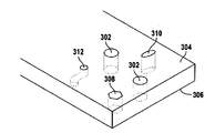
도 2a는 일 실시 형태에 따라 기관 모방 장치의 사시도를 도시한다.
도 2b는 일 실시 형태에 따라 기관 모방 장치의 분해도를 도시한다.
도 2c 내지 도 2d는 일 실시 형태에 따라 장치의 조직-조직 계면 영역의 사시도를 도시한다.
도 2e 내지 도2g는 하나 이상의 일시 형태에 따라 장치의 조직-조직 계면 영역의 탑다운형 단면도를 도시한다.
도 3a 내지 도 3b는 일 실시 형태에 따라 장치의 조직-조직 계면 영역의 사시도를 도시한다.
도 3c 내지 도 3e는 하나 이상의 실시 형태에 따라 막의 사시도를 도시한다.
도 4a 내지 도 4c는 일 실시 형태에 따라 2채널 장치의 막의 형성에 관한 사시도를 도시한다.
도 4d는 일 실시 형태에 따라 조직-조직 계면 장치의 막의 측면도를 도시한다.
도 5a 내지 도 5e는 일 실시 형태에 따라 기관 모방 장치의 형성에 관한 사시도를 도시한다.
도 6은 일 실시 형태에 따라 다수의 채널을 갖는 기관 모방 장치를 사용하는 계통도를 도시한다.
도 7a 내지 도 7b는 일 실시 형태에 따라 기관 모방 장치의 사시도 도시한다.
도 7c는 일 실시 형태에 따라 기관 모방 장치의 막의 측면도를 도시한다.
도 8 및 도 9는 본 발명의 장치를 사용하여 수행한 실험에 따라 시간 경과에 따른 ROS 생성을 도시한다.The accompanying drawings, which are incorporated into and constitute a part of this specification, illustrate one or more examples of embodiments, and serve to explain the principles and implementation of these embodiments, along with descriptions of example embodiments. In these drawings:
1 shows a block diagram of a system using an exemplary tracheal imitation device, according to one embodiment.
2A shows a perspective view of an organ imitation device according to one embodiment.
2B shows an exploded view of an organ mimic device, according to one embodiment.
2C-2D show perspective views of a tissue-tissue interface region of a device according to one embodiment.
2E-2G show top-down cross-sectional views of a tissue-tissue interface region of a device according to one or more temporal configurations.
3A-3B show perspective views of a tissue-tissue interface region of a device according to one embodiment.
3C-3E show perspective views of membranes in accordance with one or more embodiments.
4A to 4C show perspective views of the formation of a film of a two-channel device according to one embodiment.
4D shows a side view of a membrane of a tissue-tissue interface device, according to one embodiment.
5A-5E show perspective views of the formation of an organ mimic device according to one embodiment.
6 shows a schematic diagram using an organ mimic device having multiple channels, according to one embodiment.
7A to 7B show perspective views of an organ imitation device according to one embodiment.
7C shows a side view of the membrane of an organ mimic device, according to one embodiment.
8 and 9 illustrate ROS generation over time according to experiments performed using the apparatus of the present invention.
예시적인 실시 형태가 기관 시뮬레이션 장치 및 그 사용 및 제조 방법과 관련하여 본 명세서에 기재되어 있다. 당업자는 하기의 설명이 단지 예시적이며 어떠한 방식으로든 제한되는 것으로 의도되지 않음을 인식할 것이다. 다른 실시 형태는 본 발명의 이득을 갖는 그러한 숙련된 자에게 그 자체를 용이하게 제시할 것이다. 이제, 첨부된 도면에 도시된 예시적인 실시 형태의 구현에 대하여 상세하게 참조할 것이다. 동일한 도면 부호가 동일하거나 유사한 항목을 언급하기 위하여 도면 및 하기의 설명에 걸쳐 사용될 것이다. 어구 "일 일시 형태"는 하나 초과의 실시 형태를 포함하고, 따라서 간략함을 위한 단지 하나의 실시 형태로 제한되지 않음이 이해된다.Exemplary embodiments are described herein in connection with an engine simulation device and its use and manufacturing method. Those skilled in the art will recognize that the following description is illustrative only and is not intended to be limited in any way. Other embodiments will readily present themselves to such skilled persons having the benefit of the present invention. Reference will now be made in detail to the implementation of the exemplary embodiment shown in the accompanying drawings. The same reference numbers will be used throughout the drawings and the following description to refer to the same or similar items. It is understood that the phrase "one date and time form" includes more than one embodiment, and thus is not limited to only one embodiment for simplicity.
본 발명에 따르면, 기관 모방 장치("본 발명의 장치"로도 지칭됨)는 바람직하게는 센서, 컴퓨터, 디스플레이 및 소프트웨어, 데이터 구성요소, 프로세스 단계 및/또는 데이터 구조를 이용하는 다른 전산 장비를 포함하는 전체적인 시스템에 사용된다. 기관 모방 장치와 함께 사용되는 컴퓨터 시스템에 관하여 본 명세서에 기재된 구성요소, 프로세스 단계, 및/또는 데이터 구조는 다양한 유형의 작동 시스템, 컴퓨팅 플랫폼, 컴퓨터 프로그램, 및/또는 범용 기계를 사용하여 구현될 수 있다. 추가적으로, 당업자는 범용성이 보다 적은 장치, 예를 들어 하드와이어드(hardwired) 장치, 현장 프로그램 가능한 게이트 어레이(field programmable gate array, FPGA), 주문형 집적 회로(application specific integrated circuit, ASIC) 등이 또한 본 명세서에 개시된 발명 개념의 범주 및 사상으로부터 벗어남 없이 사용될 수 있음을 인식할 것이다.According to the present invention, an organ mimic device (also referred to as “the device of the present invention”) preferably comprises sensors, computers, displays and software, data components, other computerized equipment using process steps and/or data structures. It is used for the whole system. The components, process steps, and/or data structures described herein with respect to a computer system used with an organ mimic device may be implemented using various types of operating systems, computing platforms, computer programs, and/or general purpose machines. have. Additionally, those skilled in the art may also use less versatile devices, such as hardwired devices, field programmable gate arrays (FPGAs), application specific integrated circuits (ASICs), and the like. It will be appreciated that the invention can be used without departing from the scope and spirit of the disclosed concepts.
일련의 프로세스 단계를 포함하는 방법이, 컴퓨터 또는 기계를 하기에 기재되는 기관 모방 장치와 함께 사용함으로써 구현되고 이들 프로세스 단계가 기계에 의해 판독가능한 일련의 명령들로서 저장될 수 있는 경우, 그들은 유형 매체(tangible medium), 예를 들어 컴퓨터 메모리 장치(예: ROM(읽기 전용 메모리), PROM(프로그램 가능한 읽기 전용 메모리), EEPROM(전기적으로 소거가능한 프로그램 가능한 읽기 전용 메모리), 플래시 메모리, 점프 드라이브 등), 자기 저장 매체(예: 테이프, 자기 디스크 드라이브 등), 광학 저장 매체(예: CD-ROM, DVD-ROM, 페이퍼 카드, 페이퍼 테이프 등) 및 다른 유형의 프로그램 메모리에 저장될 수 있다.If a method comprising a series of process steps is implemented by using a computer or machine in conjunction with an organ imitation device described below and these process steps can be stored as a series of instructions readable by a machine, they are tangible media ( tangible medium (e.g., computer memory devices (e.g. ROM (read only memory), PROM (programmable read only memory), EEPROM (electrically erasable programmable read only memory), flash memory, jump drives, etc.), Magnetic storage media (eg, tape, magnetic disk drive, etc.), optical storage media (eg, CD-ROM, DVD-ROM, paper card, paper tape, etc.) and other types of program memory.
본 발명의 장치의 실시 형태는 기초생물과학, 생명과학 연구, 약물 발견 및 개발, 약물 안전성 시험, 화학적 및 생물학적 검정뿐만 아니라 조직 및 기관 공학을 비롯한 다수의 분야에 적용될 수 있다. 일 실시 형태에서, 기관 모방 장치는 심혈관 질환, 암 질환, 및 기관-특이적 질환 생태의 기초 연구를 위한 미세혈관 네트워크 구조물로서 사용될 수 있다. 더욱이, 장치의 하나 이상의 실시 형태는 간, 신장, 폐, 장, 골수, 및 다른 기관 및 조직을 위한 기관 보조 장치에서뿐만 아니라 기관 대체 구조물에서 응용을 찾는다.Embodiments of the device of the present invention can be applied to a number of fields including basic biological science, life science research, drug discovery and development, drug safety testing, chemical and biological assays, as well as tissue and organ engineering. In one embodiment, an organ mimic device can be used as a microvascular network structure for basic research in cardiovascular disease, cancer disease, and organ-specific disease ecology. Moreover, one or more embodiments of the device find application in organ replacement structures as well as in organ assistive devices for the liver, kidney, lung, intestine, bone marrow, and other organs and tissues.
다양한 환경적 신호에 대한 세포 반응이 본 발명의 장치와 조합될 수 있는 다양한 시스템을 사용하여 모니터링될 수 있다. 잘 알려진 센서를 사용하여 pH의 변화를 모니터링할 수 있다. 또한, 유전자 전사의 변화 또는 세포 생화학 또는 구조 조직화의 변화를 측정하기 위하여 연속적으로 또는 주기적으로 세포를 샘플링할 수 있다. 예를 들어, 또한 다공성 막 상에 성장된 "조직"에 현미경적 분석, 면역조직학적 분석, 동일계내 하이브리드화 분석, 또는 헤마톡실린 및 에로신 염색과 같은 염색을 이용한 통상적인 병리학적 분석을 행할 수 있다. 이들 분석을 위한 샘플은 실시간으로 수행되거나, 실험 후에 채취되거나 또는 연구 또는 실험 동안 상이한 스테이지에서 소량의 생검체(biopsy)를 채취함으로써 수행될 수 있다.Cell responses to various environmental signals can be monitored using various systems that can be combined with the devices of the present invention. Changes in pH can be monitored using well-known sensors. In addition, cells can be sampled continuously or periodically to measure changes in gene transcription or changes in cell biochemistry or structural organization. For example, also "pathology" grown on a porous membrane can be subjected to microscopic analysis, immunohistological analysis, in situ hybridization analysis, or conventional pathological analysis using staining such as hematoxylin and erosine staining. Can be. Samples for these analyzes can be performed in real time, after an experiment, or by taking a small amount of biopsy at different stages during a study or experiment.
막 상에 성장된 세포에 다른 세포, 예를 들어 면역계 세포 또는 세균성 세포를, 항체 또는 항체 매개성 세포를, 예를 들어 표적 특이적 세포 수용체를 가할 수 있다. 이들 세포를 바이러스 또는 다른 입자에 노출시킬 있다. 외부적으로 공급되는 물질, 예를 들어 세포, 바이러스, 입자 또는 단백질의 운동의 검출을 보조하기 위하여, 방사성 표지 또는 형광 표지와 같은 통상적인 수단을 사용하여 그들을 자연스럽게 표지화할 수 있다.Cells grown on the membrane can be added with other cells, such as immune system cells or bacterial cells, antibodies or antibody-mediated cells, such as target specific cell receptors. These cells can be exposed to viruses or other particles. To aid in the detection of movement of externally supplied substances, such as cells, viruses, particles or proteins, they can be naturally labeled using conventional means, such as radioactive or fluorescent labels.
세포는 1, 2, 3, 4, 5, 6, 또는 7일, 적어도 1 내지 2주 사이, 그리고 심지어 2주 넘게 본 발명의 장치를 사용하여 성장되고, 배양되고 분석될 수 있다. 예를 들어, 하기에 논의되는 바와 같이, 개시된 장치의 일 실시 형태에서 얇은 다공성 막 상에 폐포 상피 세포와 폐 미세혈관 내피 세포의 공배양(co-culture)이 세포의 생존력의 손실 없이 2주 넘게 성장될 수 있음이 밝혀졌다.Cells can be grown, cultured and analyzed using the device of the invention for 1, 2, 3, 4, 5, 6, or 7 days, at least between 1 and 2 weeks, and even over 2 weeks. For example, as discussed below, in one embodiment of the disclosed device, the co-culture of alveolar epithelial cells and lung microvascular endothelial cells on a thin porous membrane is more than two weeks without loss of cell viability. It has been found that it can be grown.
본 명세서에 개시된 기관 모방 장치는 많은 상이한 응용을 갖는데, 이러한 응용에는 제한 없이, 질환의 마커의 확인; 항암 치료제의 효능 평가; 유전자 치료 벡터의 시험; 약물 개발; 스크리닝; 세포, 특히 줄기 세포 및 골수 세포의 연구; 생체내 변환, 흡수, 청소, 대사, 및 생체 이물(xenobiotics)의 활성에 대한 연구; 상피층 또는 내피층을 가로질러 행해지는 화학적 또는 생물학적 에이전트의 수송 및 생체이용률에 대한 연구; 혈액-뇌 장벽을 가로질러 행해지는 생물학적 또는 화학적 에이전트의 수송에 대한 연구; 장 상피 장벽을 가로질러 행해지는 생물학적 또는 화학적 에이전트의 수송에 대한 연구; 화학적 에이전트의 급성 기초 독성에 대한 연구; 화학적 에이전트의 급성 국소 또는 급성 기관-특이적 독성에 대한 연구; 화학적 에이전트의 만성 기초 독성에 대한 연구; 화학적 에이전트의 만성 국소 또는 만성 기관-특이적 독성에 대한 연구; 화학적 에이전트의 최기형성에 대한 연구; 화학적 에이전트의 유전독성, 발암성, 및 변이원성에 대한 연구; 감염성 생물학적 에이전트 및 생물학적 무기의 검출; 유해한 화학적 에이전트 및 화학적 무기의 검출; 감염성 질환에 대한 연구; 질환을 치료하는 데 화학적 또는 생물학적 에이전트의 효능에 대한 연구; 질환을 치료하는 데 에이전트의 최적의 용량 범위에 대한 연구; 생물학적 에이전트에 대한 생체내에서의 기관의 반응의 예측; 화학적 또는 생물학적 에이전트의 약동학의 예측; 화학적 또는 생물학적 에이전트의 약동학의 예측; 에이전트에 대한 반응에 미치는 유전적 내용의 영향에 관한 연구; 화학적 또는 생물학적 에이전트에 응한 유전자 전사에 대한 연구; 화학적 또는 생물학적 에이전트에 응한 단백질 발현에 대한 연구; 화학적 또는 생물학적 에이전트에 응한 대사 변화에 대한 연구가 포함된다. 기관 모방 장치는 또한 (예를 들어, 약물의 조직 대사와 등가의 방법으로) 재료에 미치는 세포의 효과를 위하여, 세포에 대하여 스크리닝하는 데 사용될 수 있다.The organ mimic devices disclosed herein have many different applications, including, without limitation, identification of markers of disease; Evaluation of efficacy of anticancer drugs; Testing of gene therapy vectors; Drug development; Screening; Studies of cells, especially stem cells and bone marrow cells; Studies on in vivo transformation, absorption, clearance, metabolism, and activity of xenobiotics; Study of the transport and bioavailability of chemical or biological agents across the epithelial or endothelial layer; Studies of the transport of biological or chemical agents across the blood-brain barrier; Studies of the transport of biological or chemical agents across the intestinal epithelial barrier; Study of acute basic toxicity of chemical agents; Studies of acute local or acute organ-specific toxicity of chemical agents; Study of chronic basic toxicity of chemical agents; Studies of chronic local or chronic organ-specific toxicity of chemical agents; Study of teratogenicity of chemical agents; Studies of genotoxicity, carcinogenicity, and mutagenicity of chemical agents; Detection of infectious biological agents and biological weapons; Detection of harmful chemical agents and chemical weapons; Studies of infectious diseases; Studies of the efficacy of chemical or biological agents to treat disease; Study of the optimal dose range of agents for treating diseases; Prediction of organ response in vivo to biological agents; Prediction of pharmacokinetics of chemical or biological agents; Prediction of pharmacokinetics of chemical or biological agents; Study of the effects of genetic content on the response to agents; Studies of gene transcription in response to chemical or biological agents; Studies of protein expression in response to chemical or biological agents; Studies of metabolic changes in response to chemical or biological agents are included. Organ mimic devices can also be used to screen for cells, for the effect of cells on materials (e.g., in ways equivalent to tissue metabolism of drugs).
본 발명의 장치는 역학적으로 활성인 조직, 예를 들어 심장, 폐, 골격근, 뼈, 인대, 힘줄, 연골, 평활근 세포, 장, 신장, 내피 세포 및 다른 조직으로부터의 세포로부터 배양된 세포에 대하여, 걷기, 달리기, 숨쉬기, 연동, 유체 또는 소변의 유동, 또는 심장 박동의 역학적 하중 환경을 시뮬레이션하는 데 사용될 수 있다. 정적 환경에서의 세포의 생물학적 또는 생화학적 반응을 시험하기보다는, 연구자는 인장, 압축 및 전단을 비롯한 역학적 응력의 일정 범위의 빈도, 진폭 및 지속시간을 배양된 세포에 가할 수 있다.The device of the present invention is for cells cultured from dynamically active tissues, such as heart, lung, skeletal muscle, bone, ligament, tendon, cartilage, smooth muscle cells, intestine, kidney, endothelial cells and cells from other tissues It can be used to simulate a walking, running, breathing, peristaltic, fluid or urine flow, or dynamic loading environment of the heartbeat. Rather than testing the biological or biochemical response of cells in a static environment, researchers can apply a range of frequencies, amplitudes, and durations of mechanical stresses, including tension, compression, and shear, to cultured cells.
숙련된 기술자는 막의 표면 상에 다양한 유형의 세포를 이식할 수 있다. 세포는 선충류, 아메바류, 포유류(예를 들어, 사람)를 비롯한 다세포 구조물로부터의 임의의 세포 유형을 포함한다. 장치 상에 이식되는 세포 유형은 모방하기를 원하는 기관 또는 기관 기능의 유형, 및 그러한 기관을 포함하는 조직에 좌우된다. 본 발명의 장치의 막 상에 이식가능한 세포의 다양한 유형에 대한 보다 상세한 내용이 하기에 논의되어 있다.Skilled technicians can implant various types of cells on the surface of the membrane. Cells include any cell type from multicellular structures, including nematodes, amoebas, and mammals (eg, humans). The type of cell implanted on the device depends on the type of organ or organ function that you want to mimic, and the tissue that contains that organ. More details of the various types of cells implantable on the membrane of the device of the present invention are discussed below.
또한, 다양한 줄기 세포, 예를 들어 골수 세포, 유도된 성체 골수 세포, 배아 줄기 세포 또는 성체 조직으로부터 분리된 줄기 세포를 다공성 막의 어느 한면 또는 양면 상에 공배양할 수 있다. 각각의 세포층을 공급하는 챔버 내에서 상이한 배양 배지를 사용하여, 상이한 분화 신호(differentiation cue)가 줄기 세포층에 도달되게 하고, 그럼으로써 세포들을 상이한 세포 유형으로 분화시킬 수 있다. 또한, 막의 동일한 면 상에서 세포 유형을 혼합하여 막분리 없이 상이한 세포들의 공배양액을 생성할 수 있다.In addition, various stem cells, such as bone marrow cells, induced adult bone marrow cells, embryonic stem cells, or stem cells isolated from adult tissue, can be co-cultured on either or both sides of the porous membrane. Using different culture media in the chamber that feeds each cell layer, different differentiation cues can be reached to the stem cell layer, thereby differentiating cells into different cell types. In addition, cell types can be mixed on the same side of the membrane to produce a co-culture of different cells without membrane separation.
본 명세서에 개시된 기관 모방 장치를 사용하여, 생체내 변환, 흡수, 청소, 대사, 및 생체 이물의 활성화뿐만 아니라 약물 전달도 연구할 수 있다. 장에서와 같이 상피층, 혈관에서와 같은 내피층을 가로질러, 그리고 혈액-뇌 장벽을 가로질러 행해지는 화학적 및 생물학적 에이전트의 수송 및 생체이용률이 또한 연구될 수 있다. 화학적 에이전트의 급성 기초 독성, 급성 국소 독성 또는 급성 기관-특이적 독성, 최기형성, 유전독성, 발암성, 및 변이원성이 또한 연구될 수 있다. 감염성 생물학적 에이전트, 생물학적 무기, 유해한 화학적 에이전트 및 화학적 무기의 효과가 또한 검출되고 연구될 수 있다. 감염성 질환 및 이들 질환을 치료하는 데 화학적 및 생물학적 에이전트의 효능뿐만 아니라 이들 에이전트에 대한 최적 용량 범위가 연구될 수 있다. 화학적 및 생물학적 에이전트에 대한 생체내에서의 기관의 반응, 및 이들 에이전트의 약동학 및 약역학이 본 발명의 장치를 사용하여 검출되고 연구될 수 있다. 이들 에이전트에 대한 반응에 미치는 유전 내용물의 영향이 연구될 수 있다. 화학적 또는 생물학적 에이전트에 응한 단백질 및 유전 발현의 양이 측정될 수 있다. 화학적 또는 생물학적 에이전트에 응한 대사의 변화가 본 발명의 장치를 사용하여 역시 연구될 수 있다.The organ mimic devices disclosed herein can be used to study in vivo transformation, absorption, clearance, metabolism, and activation of biomaterials as well as drug delivery. The transport and bioavailability of chemical and biological agents, such as in the intestine, across the epithelial layer, the endothelial layer as in blood vessels, and across the blood-brain barrier can also be studied. Acute basic toxicity, acute local toxicity or acute organ-specific toxicity, teratogenicity, genotoxicity, carcinogenicity, and mutagenicity of chemical agents can also be studied. The effects of infectious biological agents, biological weapons, harmful chemical agents and chemical weapons can also be detected and studied. The efficacy of chemical and biological agents in treating infectious diseases and these diseases as well as the optimal dose range for these agents can be studied. Organ responses in vivo to chemical and biological agents, and the pharmacokinetics and pharmacodynamics of these agents can be detected and studied using the devices of the present invention. The effect of genetic content on the response to these agents can be studied. The amount of protein and genetic expression in response to a chemical or biological agent can be measured. Changes in metabolism in response to chemical or biological agents can also be studied using the device of the present invention.
종래의 세포 배양 또는 조직 배양과 반대되는 것으로서 기관 모방 장치의 이점은 무수하다. 예를 들어, 세포가 기관 모방 장치 내에 놓여질 때, 생체내 상황에 가까운 확정된 3차원 구조 조직-조직 관계를 재확립하는 섬유모세포, SMC(평활근 세포) 및 EC(내피 세포) 분화가 일어날 수 있으며, 약리학적 에이전트 또는 활성 물질 또는 생성물에 대한 세포 기능 및 반응이 조직 및 기관 수준에서 조사될 수 있다.The advantages of organ mimic devices as opposed to conventional cell culture or tissue culture are numerous. For example, when a cell is placed in an organ mimic device, fibroblasts, smooth muscle cells (SMC) and endothelial cell (EC) differentiation can occur that reestablish a defined three-dimensional structural tissue-tissue relationship close to the in vivo situation, , Cellular function and response to pharmacological agents or active substances or products can be investigated at the tissue and organ level.
추가적으로, 많은 세포 또는 조직 활성이 기관 모방 장치 내에서 검출하기에 적합하며, 이에는 제한 없이, 수송된 유동 채널 내에서 층상 조직 내로의 그리고 이를 통한 약물의 확산 속도; 상이한 층에서의 세포 형태학, 분화 및 분비 변화; 세포 이동, 성장, 아폽토시스 등이 포함된다. 또한, 시스템의 상이한 층에 위치된 상이한 유형의 세포들에 미치는 다양한 약물의 효과가 용이하게 평가될 수 있다.Additionally, many cell or tissue activities are suitable for detection in organ mimic devices, including, without limitation, the rate of drug diffusion into and through laminar tissue in a transported flow channel; Changes in cell morphology, differentiation and secretion in different layers; Cell migration, growth, apoptosis, and the like. In addition, the effects of various drugs on different types of cells located in different layers of the system can be readily assessed.
예를 들어, 약물 발견의 경우, 본 명세서에 개시된 기관 모방 장치를 사용함에 있어서 2가지 이점이 있을 수 있다: (1) 기관 모방 장치는 조직의 생체내 층상 구조물을 보다 우수하게 모방할 수 있으며, 따라서 세포 수준 및 조직 수준에 더하여 기관 수준에서 약물 효과를 연구할 수 있게 하고, (2) 기관 모방 장치는 약물 선택 및 독물학 연구를 위한 생체내 조직 모델의 사용 및 동물의 사용을 줄여준다.For example, in the case of drug discovery, there may be two advantages to using the organ imitation device disclosed herein: (1) The organ imitation device can better mimic the in vivo layered structure of tissue, Thus, in addition to cell level and tissue level, drug effects can be studied at the organ level, and (2) organ mimic devices reduce the use of in vivo tissue models and drug use for drug selection and toxicology studies.
약물 발견 및 개발에 더하여, 본 명세서에 개시된 기관 모방 장치는 또한 기초 및 임상 연구에 유용할 수 있다. 예를 들어, 기관 모방 장치는 종양발생(tumorigenesis)의 메커니즘을 연구하는 데 사용될 수 있다. 생체내 암의 진행이 간질 세포 및 세포외 기질(ECM)을 비롯한 숙주 및 종양 미세환경에 의해 조절됨이 잘 확립되어 있다. 예를 들어, 간질 세포가 종양 형성에 영향을 주는 것으로 확인되었다. 확정된 구조로 성장하는 세포가 단층 내의 세포보다 세포독성 에이전트에 대하여 더 많은 내성을 나타낸다는 증거가 증가되고 있다. 따라서, 기관 모방 장치는 암 세포의 원래의 성장 특성을 시뮬레이션하기 위한 보다 우수한 수단이며, 그럼으로써 생체내 종양의 실제의 약물의 감수성을 보다 우수하게 반영한다.In addition to drug discovery and development, the organ mimic devices disclosed herein may also be useful for basic and clinical research. For example, organ mimic devices can be used to study the mechanisms of tumorigenesis. It is well established that the progression of cancer in vivo is regulated by host and tumor microenvironments, including interstitial cells and extracellular matrix (ECM). For example, it has been found that interstitial cells influence tumor formation. There is increasing evidence that cells growing with a defined structure exhibit more resistance to cytotoxic agents than cells within a monolayer. Thus, organ mimic devices are a better means for simulating the original growth characteristics of cancer cells, thereby better reflecting the actual drug susceptibility of tumors in vivo.
기관 모방 장치는 다양한 조직을 가공(engineer)하는 데 사용될 수 있으며, 이러한 조직에는 제한 없이, 심혈관계, 폐, 장, 신장, 뇌, 골수, 골, 치아, 및 피부가 포함된다. 장치가 적합한 생체적합성 및/또는 생분해성 재료, 예를 들어 폴리-락티드-코-글리콜리드산(PLGA)으로 가공될 경우, 당해 기관 모방 장치가 생체내 이식(transplantation 또는 implantation)을 위하여 사용될 수 있다. 더욱이, 여러 세포 유형의 상호작용을 공간적으로 국재화하고 제어하는 능력은 계층적으로 가공하고, 보다 생리학적으로 올바른 조직 및 기관 유사체를 생성할 기회를 제공한다. 확정된 배열의 다수의 세포 유형의 배열은 세포 분화, 유지, 및 기능적 수명에 대하여 유익한 효과를 갖는다.Organ mimic devices can be used to engineer various tissues, including, but not limited to, cardiovascular, lung, intestinal, kidney, brain, bone marrow, bone, teeth, and skin. If the device is processed with a suitable biocompatible and/or biodegradable material, such as poly-lactide-co-glycolidic acid (PLGA), the organ mimic device can be used for in vivo implantation (transplantation or implantation). have. Moreover, the ability to spatially localize and control the interaction of different cell types provides the opportunity to process hierarchically and create more physiologically correct tissue and organ analogs. The arrangement of multiple cell types in a defined arrangement has a beneficial effect on cell differentiation, maintenance, and functional lifespan.
기관 모방 장치는 또한 상이한 성장 인자, 화학물질, 기체 및 영양소가 세포 및 생체내 그 존재의 필요성에 따라 상이한 세포 유형에 첨가되게 할 수 있다. 그러한 인자들 및 단백질의 위치의 제어는 특이적 세포 리모델링 및 기능화의 과정을 안내할 수 있으며, 또한 전체 시스템에 대하여 분자 신호를 제공할 수 있으며, 그 결과 신생조직(neotissue), 세포 리모델링, 향상된 분비 등과 같은 유익한 발전을 가져온다.Organ mimic devices can also allow different growth factors, chemicals, gases and nutrients to be added to different cell types depending on the needs of the cells and their presence in vivo. Control of the location of such factors and proteins can guide the process of specific cell remodeling and functionalization, and also can provide molecular signals for the entire system, resulting in neoissues, cell remodeling, and enhanced secretion. It brings beneficial developments, such as.
또 다른 태양에서, 기관 모방 장치는 다세포 유형의 세포 미세배열, 예를 들어 마이크로유체 장치로서 이용될 수 있다. 기관 모방 장치를 사용하여, 세포 배열의 패턴 완전성(pattern integrity)이 유지될 수 있다. 이들 세포 미세배열은 미래의 "랩온어칩(lab-on-a-chip)"을 구성할 수 있는데, 이는 특히 다중화되고 자동화될 때이다. 이들 소형화된 다세포 유형 배양은 보다 신속하고 소음은 보다 적은 검정으로 세포 동력학의 관찰을 용이하게 할 것이며, 세포가 배열에 대하여 생체내-유사 반응을 나타낼 수 있게 할 빌트-인 복잡성을 갖는다.In another aspect, the organ mimic device can be used as a multicellular cell microarray, eg, a microfluidic device. Using organ mimic devices, pattern integrity of the cell array can be maintained. These cell microarrays can constitute the future "lab-on-a-chip", especially when multiplexed and automated. These miniaturized multicellular type cultures will facilitate observation of cell dynamics with faster and less noise assays, and have built-in complexity that will enable cells to exhibit an in vivo-like response to the array.
또 다른 태양에서, 기관 모방 장치는 생물학적 센서로서 이용될 수 있다. 세포-기재 바이오센서는 다른 바이오센서보다 많은 정보를 제공할 수 있는데, 그 이유는 세포는 흔히 자극에 대한 다면화된 생리학적 반응뿐만 아니라 이들 반응을 증폭시키는 신규한 메커니즘을 갖기 때문이다. 기관 모방 장치 내의 모든 세포 유형은 분석물의 상이한 측면을 동시에 모니터링하는 데 사용될 수 있거나; 기관 모방 장치 내의 상이한 세포 유형은 상이한 분석물을 모니터링하는 데 사용될 수 있거나; 모니터링의 두 가지 유형의 혼합이 사용될 수 있다. 환경 모니터링, 독소 검출, 및 생리학적 모니터링에 있어서의 응용을 위한 센서로서 E. 콜라이로부터 포유류의 세포주까지의 세포가 사용되었다.In another aspect, an organ mimic device can be used as a biological sensor. Cell-based biosensors can provide more information than other biosensors, because cells often have multiplexed physiological responses to stimuli as well as novel mechanisms to amplify these responses. All cell types in organ mimic devices can be used to simultaneously monitor different aspects of analytes; Different cell types in organ mimic devices can be used to monitor different analytes; Mixing of the two types of monitoring can be used. Cells from E. coli to mammalian cell lines were used as sensors for applications in environmental monitoring, toxin detection, and physiological monitoring.
또 다른 태양에서, 기관 모방 장치는 세포 생리학 및 세포-ECM 상호작용의 기본 과정을 이해하는 데 사용될 수 있다. 생체내 리모델링 과정은 세포와 ECM 사이의 복잡하고 동적이며 상호적인 과정이다. 기관 모방 장치는 이들 생물계의 복잡성을 포착하여 이들 계가 조사 및 유익한 조작에 적합하게 되게 할 수 있을 것이다. 더욱이, 이미징 도구, 예를 들어 형광 현미경법, 마이크로형광법, 미세형광법 또는 광 간섭 단층촬영법(optical coherence tomography, OCT), 다층상 조직 내에서의 세포 거동의 실시간 분석과 결합되어 장치를 사용하는 것이 예상된다. 실시간 분석에 적합한 세포 및 조직 연구의 예에는 세포 분비 및 신호전달, 세포-세포 상호작용, 조직-조직 상호작용, 동적 가공 조직 작제(dynamic engineered tissue construction) 및 모니터링, 조직 공학에서 구조물-기능 조사, 및 시험관내에서 세포 리모델링 기질의 과정이 포함된다.In another aspect, organ mimic devices can be used to understand the basic processes of cell physiology and cell-ECM interaction. The in vivo remodeling process is a complex, dynamic and interactive process between cells and ECM. Organ imitation devices will be able to capture the complexity of these biological systems and make them suitable for investigation and beneficial manipulation. Moreover, it is expected to use the device in combination with imaging tools such as fluorescence microscopy, microfluorescence, microfluorescence or optical coherence tomography (OCT), real-time analysis of cell behavior within multi-layered tissue. do. Examples of cell and tissue studies suitable for real-time analysis include cell secretion and signaling, cell-cell interactions, tissue-tissue interactions, dynamic engineered tissue construction and monitoring, and structure-function investigations in tissue engineering, And the process of cell remodeling substrates in vitro.
이 장치의 용도의 다른 예는 조직-조직 계면을 유도하고 기관 구조물을 복잡화하여 장치 내에 형성하는 것인데, 이는 장치를 살아있는 동물의 체내에 생체내 이식하고, 세포 및 조직이 장치에 스며들고 정상 조직-조직 계면을 확립될 수 있게 함에 의해서이다. 이어서, 세포 생존에 필요한 매체 및 기체를 갖는 유체 채널들 중 하나 이상을 통하여 세포 및 조직이 함유된 전체 장치를 관류하면서, 세포 및 조직이 함유된 전체 장치가 외과적으로 제거된다. 이어서, 이 복잡한 기관 모방 장치는 연속 관류를 통하여 시험관내에서 생존 상태로 유지되고, 임의의 기존의 시험관내 모델 시스템을 사용해서는 불가능한 일정 수준의 복잡성으로 보통 3D 컨텍스트에서 매우 복잡한 세포 및 조직 기능을 연구하는 데 사용될 수 있다.Another example of the use of this device is to induce a tissue-tissue interface and complicate organ structures to form in the device, which implants the device into the body of a living animal in vivo, cells and tissues infiltrate the device and normal tissue- By allowing the tissue interface to be established. The entire device containing cells and tissues is then surgically removed while perfusioning the entire device containing cells and tissues through one or more of the fluid channels with the medium and gas necessary for cell survival. Subsequently, this complex organ-mimetic device remains viable in vitro through continuous perfusion and studies very complex cellular and tissue functions, usually in a 3D context, with a level of complexity that would not be possible using any existing in vitro model system. Can be used to
특히, 마이크로채널 장치는 동물에 생체내에서 피하내 이식될 수 있는데, 여기서 장치는 주위 조직 공간에 개방된 하나 이상의 대응하는 포트를 갖는 채널 내에 골-유도성 재료, 예를 들어 탈염된 골 분말 또는 골 형태형성 단백질(bone morphogenic protein, BMP)을 함유한다. 제2 채널은 그의 말단 포트를 폐쇄하거나 그것을 제거가능한 고체 재료, 예를 들어 고체봉으로 채움으로써 이식 동안 제2 채널은 닫힐 것이다. 상처 치유의 결과로서, 중간엽 줄기 세포 및 미소미세혈관을 함유하는 결합 조직이 장치의 개방된 채널 내로 성장할 것이며, 골-유도성 재료의 존재로 인해, 순환하는 조혈 전구 세포를 동원하는 공간을 갖는 뼈를 형성하여, 과거 연구에서 밝혀진 바와 같이 완전히 기능적인 골수를 형성할 것이다.In particular, the microchannel device can be implanted subcutaneously in vivo in an animal, where the device is a bone-inducing material, such as desalted bone powder, or in a channel having one or more corresponding ports open to the surrounding tissue space. Contains bone morphogenic protein (BMP). The second channel will be closed during implantation by closing its distal port or filling it with a removable solid material, for example a solid rod. As a result of wound healing, connective tissue containing mesenchymal stem cells and microtubules will grow into the open channels of the device and, due to the presence of bone-inducing materials, have space to mobilize circulating hematopoietic progenitor cells. By forming bone, it will form a fully functional bone marrow, as revealed in past studies.
일단 이 과정이 완료되면, 수술 부위가 재개봉될 것이며, 막대 또는 플러그를 제거함으로써 제2 채널이 재개봉될 것이며, 이어서 유체 저장소에 결합된 카테터와 연결되어 세포 생존에 필요한 영양소 및 기체를 함유하는 배양 배지가 제2 채널을 통하여 펌핑되고, 막의 기공을 통하여 형성된 골수를 함유하는 제2 채널 내로 통과할 수 있을 것이다. 이어서, 전체 마이크로채널 장치가 절단되어 주위 조직으로부터 자유로와질 수 있게 되고, 장치 내로 연속적으로 유동하는 매체와 함께 동물로부터 제거되고 조직 배양 인큐베이터로 이동되고 제2 채널을 통한 연속된 유체 유동을 갖는 배양액 내에 유지되고, 필요하다면 유입 포트 및 유출 포트에 연결함으로써, 추가적인 유동이 제1 채널에 역시 가해질 수 있다. 이어서, 마이크로채널 장치가 제어된 환경에서와 같이 시험관내에서 온전한 골수 기능을 연구하는 데 사용될 것이다.Once this process is complete, the surgical site will be reopened, the second channel will be reopened by removing the rod or plug, and then connected to the catheter bound to the fluid reservoir to contain the nutrients and gases necessary for cell survival. The culture medium will be pumped through the second channel and will be able to pass into the second channel containing bone marrow formed through the pores of the membrane. Subsequently, the entire microchannel device can be cut and freed from surrounding tissue, removed from the animal with a medium flowing continuously into the device, transferred to the tissue culture incubator, and cultured with continuous fluid flow through the second channel. Retaining within and connecting to the inlet and outlet ports if necessary, additional flow can also be applied to the first channel. The microchannel device will then be used to study intact bone marrow function in vitro, such as in a controlled environment.
본 발명의 장치의 생체내 이식을 용이하게 하기 위하여 생체적합성 재료 및 생분해성 재료 둘 모두가 본 발명의 장치에 사용될 수 있다. 또한, 생체적합성 코팅 및 생분해성 코팅을 사용할 수 있다. 예를 들어, 금속 기재(metallic substrate) 상에의 세라믹 코팅을 사용할 수 있다. 그러나, 임의의 유형의 코팅 재료 및 그 코팅이 상이한 유형의 재료, 즉 금속, 세라믹, 중합체, 하이드로겔 또는 임의의 이들 재료의 조합으로 만들어질 수 있다.Both biocompatible and biodegradable materials can be used in the device of the present invention to facilitate implantation of the device of the present invention in vivo. In addition, biocompatible coatings and biodegradable coatings can be used. For example, a ceramic coating on a metallic substrate can be used. However, any type of coating material and its coating can be made of different types of materials, such as metals, ceramics, polymers, hydrogels, or any combination of these materials.
생체적합성 재료에는 제한 없이, 산화물, 인산염, 탄산염, 질화물 또는 탄질화물이 포함된다. 산화물 중에서는 하기의 것들이 바람직하다: 산화탄탈, 산화알루미늄, 산화이리듐, 산화지르코늄 또는 산화티타늄. 일부 경우에, 코팅은 또한 시간이 지남에 따라 분해될 것이며 살아있는 조직에 의해 대체될 수 있는 생분해성 재료로 만들어질 수 있다. 기재는 금속, 세라믹, 중합체 또는 임의의 이들의 조합으로 만들어질 수 있다. 스테인리스 강, 니티놀(Nitinol), 티타늄, 티타늄 합금, 또는 알루미늄과 같은 금속 및 세라믹, 예를 들어 지르코니아, 알루미나, 또는 인산칼슘이 특히 관심 대상이다.Biocompatible materials include, without limitation, oxides, phosphates, carbonates, nitrides or carbonitrides. Among the oxides, the following are preferable: tantalum oxide, aluminum oxide, iridium oxide, zirconium oxide or titanium oxide. In some cases, the coating can also be made of a biodegradable material that will degrade over time and can be replaced by living tissue. The substrate can be made of metal, ceramic, polymer, or any combination thereof. Of particular interest are metals and ceramics such as stainless steel, Nitinol, titanium, titanium alloys, or aluminum, such as zirconia, alumina, or calcium phosphate.
생체적합성 재료는 또한 그것이 시간에 걸쳐 분해될 것이며 살아있는 조직에 의해 대체될 수 있다는 점에서 생분해성일 수 있다. 그러한 생분해성 재료에는 제한 없이, 폴리(락트산-코-글리콜산), 폴리락트산, 폴리글리콜산(PGA), 콜라겐 또는 다른 ECM 분자, 다른 결합 조직 단백질, 마그네슘 합금, 폴리카프로락톤, 히알루산, 접착 단백질, 생분해성 중합체, 합성, 생체적합성 및 생분해성 재료, 예를 들어 생체고분자, 생체유리, 생체세라믹, 황산칼슘, 인산칼슘, 예를 들어 인산일칼슘 1수화물, 인산일칼슘 무수물, 인산이칼슘 2무수물, 인산이칼슘 무수물, 인산삼칼슘, 오르토인산칼슘, 파이로인산칼슘, 알파-삼칼슘 인산염, 베타-삼칼슘 인산염, 아파타이트, 예를 들어 하이드록시아파타이트, 또는 중합체, 예를 들어 폴리(알파-하이드록시에스테르), 폴리(오르토 에스테르), 폴리(에테르 에스테르), 폴리안하이드라이드, 폴리(포스파젠), 폴리(프로필렌 푸마레이트), 폴리(에스테르 아미드), 폴리(에틸렌 푸마레이트), 폴리(아미노산), 다당류, 폴리펩티드, 폴리(하이드록시 부티레이트), 폴리(하이드록시 발레레이트), 폴리우레탄, 폴리(말산), 폴리락티드, 폴리글리콜리드, 폴리카프로락톤, 폴리(글리콜리드-코-트리메틸렌 카르보네이트), 폴리디옥사논, 또는 이들의 공중합체, 삼원중합체 또는 그러한 중합체들의 블렌드, 또는 생체적합성 재료와 생분해성 재료의 조합이 포함된다. 또한, 폴리글리콜리드, 폴리락티드 및/또는 글리콜리드/락티드 공중합체와 같은 부분적으로 결정질인 생체흡수성 중합체로부터 집합된, 생분해성 유리 및 생체활성 유리 자기-강화된(self-reinforced) 초고강도 생체흡수성인 복합재를 사용할 수 있다.The biocompatible material can also be biodegradable in that it will degrade over time and can be replaced by living tissue. Without limitation to such biodegradable materials, poly(lactic acid-co-glycolic acid), polylactic acid, polyglycolic acid (PGA), collagen or other ECM molecules, other connective tissue proteins, magnesium alloys, polycaprolactone, hyaluronic acid, adhesion Proteins, biodegradable polymers, synthetic, biocompatible and biodegradable materials such as biopolymers, bioglass, bioceramics, calcium sulfate, calcium phosphate, such as monocalcium phosphate monohydrate, monocalcium phosphate anhydride, dicalcium phosphate Dihydride, dicalcium phosphate anhydride, tricalcium phosphate, calcium orthophosphate, calcium pyrophosphate, alpha-tricalcium phosphate, beta-tricalcium phosphate, apatite, e.g. hydroxyapatite, or polymers such as poly( Alpha-hydroxyester), poly(ortho ester), poly(ether ester), polyanhydride, poly(phosphagen), poly(propylene fumarate), poly(ester amide), poly(ethylene fumarate), Poly(amino acid), polysaccharide, polypeptide, poly(hydroxy butyrate), poly(hydroxy valerate), polyurethane, poly(malic acid), polylactide, polyglycolide, polycaprolactone, poly(glycolide-co -Trimethylene carbonate), polydioxanone, or copolymers thereof, terpolymers or blends of such polymers, or combinations of biocompatible and biodegradable materials. In addition, biodegradable glass and bioactive glass self-reinforced ultra-high strength, aggregated from partially crystalline bioabsorbable polymers such as polyglycolide, polylactide and/or glycolide/lactide copolymers. Bioabsorbable composites can be used.
이들 재료는 바람직하게는 이식 기하형상에 따라, 생체내에서 4주부터 최대 1년까지 높은 초기 강도, 적절한 탄성률 및 강도 보유 시간을 갖는다. 보강 요소, 예를 들어 결정질 중합체의 섬유, 중합체 수지 중 탄소의 섬유, 및 미립자 충전제, 예를 들어 하이드록시아파타이트가 또한 생분해성 장치의 치수 안정성 및 기계적 특성을 제공하는 데 사용될 수 있다. 생분해성 재료 구성에 있어서의 상호침투 네트워크(interpenetrating network, IPN)의 사용은 기계적 강도를 개선하는 수단으로서 입증되었다. IPN-보강된 생분해성 재료의 기계적 특성을 추가로 개선하기 위하여, 본 발명의 장치는 상이한 가교결합제를 사용하여 폴리(락티드-코-글리콜리드) 85:15(PLGA) 또는 폴리(l-락티드-코-d,l-락티드) 70:30(PLA)의 호스트 매트릭스 내에서 가교결합된 폴리프로필렌 푸마레이트의 반상호침투 네트워크(semi-interpenetrating network, SIPN)로서 제조될 수 있다. 또한, 문헌[Charles-Hilaire Rivard et al. (Journal of Applied Biomaterials, Volume 6 Issue 1, Pages 65 - 68, 1 Sep 2004)]에 기재된 천연 폴리(하이드록시부티레이트-코-9% 하이드록시발레레이트) 코폴리에스테르 막을 사용할 수 있다. 숙련된 기술자는 또한 장치가 사용되는 응용에 따라 임의의 특정 목적 및 세포 및 조직 유형에 적합한 다른 생분해성 재료를 선택할 수 있을 것이다.These materials preferably have high initial strength, adequate modulus of elasticity and strength retention time in vivo from 4 weeks up to 1 year, depending on the implant geometry. Reinforcing elements, such as fibers of crystalline polymers, fibers of carbon in polymer resins, and particulate fillers, such as hydroxyapatite, can also be used to provide dimensional stability and mechanical properties of biodegradable devices. The use of interpenetrating networks (IPNs) in biodegradable material construction has been demonstrated as a means to improve mechanical strength. To further improve the mechanical properties of IPN-reinforced biodegradable materials, the device of the present invention uses poly(lactide-co-glycolide) 85:15 (PLGA) or poly(l-lock) using different crosslinkers. Tide-co-d,l-lactide) can be prepared as a semi-interpenetrating network (SIPN) of crosslinked polypropylene fumarate in a host matrix of 70:30 (PLA). See also Charles-Hilaire Rivard et al. (Journal of Applied Biomaterials,
개시된 장치는 또한 생체내에 위치될 때, 치료학적 장치로서 사용될 수 있다. 장치를, 예를 들어 동물 모델 내에 위치시켜 당해 장치가 살아있는 세포 및 조직에 의해 거주될 수 있게 하고, 이어서 매체를 갖는 혈관 채널을 관류하면서, 살아있는 세포와 함께 전체 장치를 제거함으로써 기관 모방체, 예를 들면 골수 또는 림프절을 재현할 수 있다. 이어서, 장치가 제거되고, 시험관내 또는 생체외 연구를 위하여 생체외에서(ex vivo) 살아있는 상태로 유지될 수 있다. 특히, 막은 시험관내에서 막의 적어도 한면 상에 하나 이상의 세포층으로 코팅될 수 있다. 이 실시 형태에서, 세포는 유기체 외부에서 플레이팅된다. 일 실시 형태에서, 막은 생체내에서 막의 적어도 한면 상에 하나 이상의 세포층으로 코팅된다. 시험관내에서 막의 한면을 처리하고 생체내에서 나머지 다른 한막을 처리할 수 있다. 또한, 시험관내에서 한 세포 유형으로 초기에 코팅된 한면 또는 양면을 가질 수 있으며, 이어서 장치를 이식하여 생체내에서 추가적인 세포층을 끌어당길 수 있다.The disclosed device can also be used as a therapeutic device when placed in vivo. Organ mimetics, e.g., by placing the device within an animal model, such that the device can be inhabited by living cells and tissues, and then removing the entire device with the living cells, while perfusioning the vascular channels with the medium. For example, bone marrow or lymph nodes can be reproduced. The device can then be removed and kept alive ex vivo for in vitro or ex vivo studies. In particular, the membrane may be coated with one or more cell layers on at least one side of the membrane in vitro. In this embodiment, the cells are plated outside the organism. In one embodiment, the membrane is coated with one or more cell layers on at least one side of the membrane in vivo. One side of the membrane can be treated in vitro and the other one can be treated in vivo. In addition, one cell type in vitro may have one or both sides initially coated, and then the device may be implanted to attract additional cell layers in vivo.
일반적으로, 본 발명은 장치 및 사용 방법에 관한 것이며, 여기서 장치는 하나 이상의 다공성 막에 의해 분리된 중심 마이크로채널을 갖는 본체를 포함한다. 이들 막(들)은 중심 마이크로채널을 근접하여 나란히 배치된 2개 이상의 평행한 중심 마이크로채널로 분할하도록 구성되며, 여기서 하나 이상의 제1 유체가 제1 중심 마이크로채널을 통해 적용되고, 하나 이상의 제2 유체가 제2 또는 그 이상의 중심 마이크로채널을 통해 적용된다. 각각의 다공성 막의 표면은 세포 접착성 분자로 코팅되어 세포의 부착을 지지하고 막의 상면 및 하면 상의 조직 내로의 세포의 조직화를 촉진시킬 수 있으며, 그럼으로써 인접한 평행한 유체 채널들 사이의 다공성 막에 의해 분리된 하나 이상의 조직-조직 계면을 생성할 수 있다. 막은 다공성이거나, 연성이거나, 탄성이거나, 또는 이들의 조합일 수 있으며, 이때 기공은 단지 기체 및 작은 화학물질의 교환을 허용하기만 하기에 충분히 크거나, 또는 큰 단백질 및 전 살아있는 세포의 이동 및 채널횡단 통과를 허용하기에 충분히 크다. 원하는 유체 전단 응력을 하나 또는 둘 모두의 조직층에 가하기 위하여 유체 압력, 유동 및 채널 기하형상이 또한 변화될 수 있다.In general, the present invention relates to a device and method of use, wherein the device comprises a body having a central microchannel separated by one or more porous membranes. These membrane(s) are configured to divide the central microchannel into two or more parallel central microchannels arranged in close proximity, where one or more first fluids are applied through the first central microchannel, and one or more second Fluid is applied through the second or more central microchannels. The surface of each porous membrane can be coated with cell adhesive molecules to support cell adhesion and promote the organization of cells into tissues on the top and bottom surfaces of the membrane, whereby by the porous membrane between adjacent parallel fluid channels. It is possible to create one or more separate tissue-tissue interfaces. The membrane may be porous, ductile, elastic, or a combination thereof, where the pores are large or large enough to only allow the exchange of gases and small chemicals, or the movement and channels of proteins and whole living cells It is large enough to allow crossing. Fluid pressure, flow and channel geometry can also be varied to apply the desired fluid shear stress to one or both tissue layers.
비제한적인 예시적인 실시 형태에서, 장치는 폐의 작동을 모방하도록 구성되며, 이렇게 함으로써 폐 상피 세포가 ECM 막의 한면 상에 자기 집합되고, 폐 모세혈관 내피 세포가 동일한 다공성 막의 반대면 상에 자기 집합된다. 그럼으로써, 장치는 생리학적인 역학적 변형에 노출될 수 있는 기능적인 폐포-모세혈관 단위의 구조 및 기능의 시뮬레이션하여 호흡을 시뮬레이션하거나 또는 공기계(air-borne) 및 혈액계(blood-borne) 둘 모두의 화학물질, 분자, 미립자 및 세포 자극에 대하여, 막의 기공을 통하여 조직-조직 계면을 가로질러 행해지는 화학물질, 분자, 및 세포의 교환을 조사할 수 있다. 장치는 생리학적 및 병리학적 조건 하에서 분석될 수 있는 기관-수준 반응을 모방하는 시험관내 폐 모델의 개발에 영향을 준다. 이 시스템은 여러 응용에 사용될 수 있으며, 이러한 응용에는 제한 없이, 약물 스크리닝, 약물 전달, 백신 전달, 생물검출, 독물학, 생리학 및 기관/조직 공학 응용이 포함된다.In a non-limiting exemplary embodiment, the device is configured to mimic the operation of the lung, whereby the lung epithelial cells self-assemble on one side of the ECM membrane, and the lung capillary endothelial cells self-assemble on the opposite side of the same porous membrane. do. By doing so, the device simulates breathing by simulating the structure and function of functional alveolar-capillary units that may be exposed to physiological mechanical deformation, or both air-borne and blood-borne. For stimulation of chemicals, molecules, particulates, and cells, the exchange of chemicals, molecules, and cells across the tissue-tissue interface through pores in the membrane can be investigated. The device influences the development of an in vitro lung model that mimics an organ-level response that can be analyzed under physiological and pathological conditions. The system can be used for a number of applications, including, without limitation, drug screening, drug delivery, vaccine delivery, biodetection, toxicology, physiology and organ/tissue engineering applications.
도 1은 일 실시 형태에 따라 본 발명의 장치를 사용하는 전체적인 시스템의 블록도를 도시한다. 도 1에 도시된 바와 같이, 시스템(100)은 기관 모방 장치(102), 장치(102)에 결합된 하나 이상의 유체 공급원(104, 104N), 유체 공급원(104) 및 장치(102)에 결합된 하나 이상의 선택적 펌프(106)를 포함한다. 하나 이상의 CPU(110)는 펌프(106)에 결합되며, 이는 바람직하게는 장치(102)의 내외에서 유체의 유동을 제어한다. CPU는 바람직하게는 하나 이상의 프로세서(112) 및 하나 이상의 로컬/리모트 저장 메모리(114)를 포함한다. 디스플레이(116)는 CPU(110)에 결합되고, 하나 이상의 압력 공급원(118)은 CPU(110) 및 장치(102)에 결합된다. CPU(110)는 바람직하게는 장치에 대한 가압 유체(pressurized fluid)의 유동 및 속도를 제어한다. 여기서는 하나의 계면 장치(102)가 도시되고 설명되어 있지만, 하기에 논의되는 바와 같이 복수의 계면 장치(102)가 시스템(100) 내에서 시험되고 분석될 수 있음이 고려된다는 것을 알아야 한다.1 shows a block diagram of an overall system using the apparatus of the present invention according to one embodiment. As shown in FIG. 1,
보다 상세히 논의하겠지만, 기관 모방 장치(102)는 바람직하게는 시스템의 외부 구성요소, 예를 들어 유체 공급원 및 압력 공급원과 연통(communication)하고 있는 장치(102)의 마이크로채널들을 위치시키는 2개 이상의 포트를 포함한다. 특히, 장치(102)는 하나 이상의 유체 공급원(104N)에 결합되는데, 여기서 유체 공급원은 공기, 혈액, 물, 세포, 화합물, 미립자, 및/또는 장치(102)에 전달될 임의의 다른 매체를 함유할 수 있다. 일 실시 형태에서, 유체 공급원(104)은 유체를 장치(102)의 하나 이상의 마이크로채널에 제공하며, 또한 바람직하게는 장치(102)를 빠져나가는 유체를 받아들인다. 장치(102)를 빠져나가는 유체는 추가적으로 또는 대안적으로 유체 공급원(104)과는 별개의 유체 수집기 또는 저장소(108) 내에 수집될 수 있음이 고려된다. 따라서, 별개의 유체 공급원(104, 104N)은 개별적으로 장치(102)로 유체를 제공하고 장치(102)로부터 유체를 제거하는 것이 가능하다.As will be discussed in more detail, the
일 실시 형태에서, 장치(102)를 빠져나간 유체는 재사용되어, 이 유체가 이전에 그를 통해 들어간 동일하거나 상이한 유입 포트 내로 재도입될 수 있다. 예를 들어, 장치(102)는 특정 중심 마이크로채널을 통과한 유체가 장치로 되돌아와서 재순환되고 동일한 중심 마이크로채널을 통해 다시 흐르도록 설정될 수 있다. 이는, 예를 들어, 유체가 장치로 재순환됨에 따라 유체에서의 분석물의 농도를 증가시키는 데 사용될 수 있을 것이다. 다른 예에서, 장치(102)는 장치를 통과한 유체가 장치 내로 되돌아와서 재순환되고, 이어서 계속하여 다른 중심 마이크로채널을 통해 흐르도록 설정될 수 있다. 이는 유체가 다른 마이크로채널을 통하여 순환됨에 따라 유체의 농도 또는 조성(makeup)을 변화시키는 데 사용될 수 있을 것이다.In one embodiment, the fluid exiting the
하나 이상의 펌프(106)는 바람직하게는 유체를 장치(102) 내로 펌핑하는 데 이용되지만, 펌프는 일반적으로 시스템에 대하여 선택사양이다. 유체 펌프는 당업계에 잘 알려져 있으며, 본 명세서에서는 상세하게 논의되지 않는다. 하기에 보다 상세히 논의되겠지만, 각각의 마이크로채널 부분은 바람직하게는 그 개별의 유입 포트 및/또는 유출 포트와 연통하고 있으며, 이렇게 함으로써 각각의 마이크로채널 부분은 그를 통하여 유체가 유동될 수 있게 한다.One or
장치 내의 각각의 마이크로채널은 바람직하게는 개별의 전용 유체 공급원 및/또는 유체 수집기에 연결된 전용 유입 포트 및 유출 포트를 가져서 매체의 유량, 유동 내용물, 압력, 온도 및 다른 특성이 각각의 중심 마이크로채널을 통하여 독립적으로 제어될 수 있게 한다. 따라서 또한, 원하는 세포 마커에 대하여 별개의 유체 채널들을 샘플링함으로써 세포층 또는 조직층 각각에 대한 다양한 자극의 효과, 예를 들어 RNA 또는 단백질 수준에서의 유전자 발현의 변화를 따로따로 모니터링할 수 있다.Each microchannel in the device preferably has its own dedicated fluid source and/or dedicated inlet and outlet ports connected to a fluid collector such that the flow rate, flow content, pressure, temperature and other properties of the media are used to establish each central microchannel. It can be controlled independently. Thus, it is also possible to separately monitor the effect of various stimuli on each of the cell layer or tissue layer, for example changes in gene expression at the RNA or protein level, by sampling separate fluid channels for the desired cell marker.
세포 주입기/제거기(108) 구성요소가 장치(102)와 연통된 상태로 도시되어 있는데, 이렇게 함으로써 주입기/제거기(108)는 유입 포트(들)(210, 218)를 통하여 장치 내로 도입된 세포와는 관계없이 장치(102) 내의 계면 막의 하나 이상의 표면 상에 세포 - 예를 들어 제한 없이, 상피 세포 및 내피 세포 - 를 주입, 제거 및/또는 조작하도록 구성된다. 예를 들어, 병원성 세포를 끌어당기는 혈액 함유 자성(magnetic) 입자가 별개의 장치 내에서 배양될 수 있으며, 이렇게 함으로써 이 혼합물이 나중에, 이 혼합물을 유체 공급원(104)을 통해 흐르게 하지 않고서도 원하는 시간에 주입기를 통하여 시스템 내로 도입될 수 있다. 일 실시 형태에서, 세포 주입기/제거기(108)는 독립적으로 제어되지만, 주입기/제거기(108)는 도 1에 도시된 바와 같이 CPU(110)에 의해 제어될 수도 있다. 세포 주입기/제거기(108)는 선택적 구성요소이며 반드시 필요한 것은 아니다.The cell injector/
필수요건은 아니지만, 압력차를 생성하여 장치(102) 내에서 역학적 운동을 일으키기 위하여 하나 이상의 압력 공급원(118)으로부터 압력이 인가될 수 있다. 장치와 함께 압력이 사용되는 실시 형태에서, 압력 공급원(118)은 CPU(110)에 의해 제어되어 장치 내에서 압력차를 인가하여, 인가된 압력차에 응하여 장치 내의 하나 이상의 막(도 3a 내지 도 3b)을 효과적으로 팽창 및/또는 수축시킨다. 일 실시 형태에서, 압력 공급원(118)에 의해 장치(100)에 인가된 압력은 장치의 구성 또는 응용에 따라 양압이다. 추가적으로 또는 대안적으로, 압력 공급원(118)에 의해 인가된 압력은 장치의 구성 또는 응용에 따라 음압, 예를 들어 진공 또는 흡인(suction)이다. 압력 공급원(118)은 바람직하게는 CPU(110)에 의해 제어되어 설정된 시간 간격 또는 빈도로 장치(102)에 압력을 인가하며, 이렇게 함으로써 시간 간격이 균일 또는 불균일하도록 설정될 수 있다. 압력 공급원(118)은 시간 간격으로 균일한 압력을 인가하도록 제어될 수도 있거나, 상이한 간격으로 상이한 압력을 인가할 수도 있다. 예를 들어, 압력 공급원(118)에 의해 인가되는 압력은 사람이 달리거나 경험하는 운동을 모방하기 위하여 큰 크기를 가질 수 있고/있거나 원하는 빈도로 설정될 수 있다. 압력 공급원(118)은 또한 사람의 수면을 시뮬레이션하는 것과 같은 느린 불규칙한 패턴을 인가할 수 있다. 일 실시 형태에서, CPU(110)는 압력 공급원(118)이 인가하는 압력의 간격을 랜덤하게 변화시켜 주기적 신장 패턴(cyclic stretching pattern)이 자연적인 호흡 동안 호흡률 및 1회 호흡량(tidal volume)의 불규칙성을 시뮬레이션하도록 작동한다.Although not required, pressure may be applied from one or
하나 이상의 센서(120)가 장치(102)에 결합되어 장치(102) 내의 하나 이상의 영역을 모니터링할 수 있으며, 이렇게 함으로써 센서(120)는 모니터링 데이터를 CPU(110)에 제공한다. 센서(120)의 한 유형은 바람직하게는 장치(102)의 하나 이상의 작동 마이크로채널 또는 중심 마이크로채널에 있어서의 압력의 양에 관한 데이터를 제공하는 압력 센서이다. 마이크로채널 벽들의 반대면으로부터의 압력 데이터는 작동 마이크로채널과 중심 마이크로채널 사이의 실시간 압력차 정보를 계산하는 데 사용될 수 있다. 모니터링 데이터는 CPU(110)에 의해 사용되어 장치의 선택적 조건에 관한 정보뿐만 아니라 세포가 실시간으로 특정 환경에서의 장치(102) 내에서 어떻게 거동하는지에 관한 정보도 제공할 것이다. 센서(120)는 전극일 수 있거나, 적외선 성능, 광학 성능(예를 들어, 카메라, LED), 또는 자기 성능을 가질 수 있거나, 또는 모니터링 데이터를 제공하기 위한 임의의 다른 적절한 기술 유형을 이용할 수 있다. 예를 들어, 센서는 하나 이상의 마이크로전극일 수 있는데, 이는 막을 가로질러 전기 특성(예를 들어, 전위차, 저항, 및 단락 회로 전류)을 분석하여 조직화된 장벽의 형성을 확인시켜 줄 뿐만 아니라, 막을 가로질러 조직화된 장벽의 유체/이온 수송 기능을 분석한다. 센서(120)는 장치(102)의 외부에 있을 수도 있거나 장치(102) 내에 일체화될 수 있음을 알아야 한다. CPU(110)가 센서(120)의 작동을 제어함이 고려되지만, 이는 반드시 필요한 것은 아니다. 데이터는 바람직하게는 디스플레이(116)에 보여진다.One or
도 2a는 일 실시 형태에 따라 조직 계면 장치의 사시도를 도시한다. 특히, 도 2a에 도시된 바와 같이, 장치(200)(도면 부호 102로도 지칭됨)는 바람직하게는 일 실시 형태에 따라 분지형 마이크로채널 디자인(203)을 갖는 본체(200)를 포함한다. 본체(202)는 연성 재료로 만들어질 수 있지만, 본체는 대안적으로 비연성 재료로 만들어짐이 고려된다. 마이크로채널 디자인(203)은 단지 예시적이며 도 2a에 도시된 구성으로 한정되지 않음을 알아야 한다. 본체(202)는 바람직하게는 연성 생체적합성 중합체로 만들어지며, 이러한 중합체에는 제한 없이, 폴리디메틸 실록산(PDMS) 또는 폴리이미드가 포함된다. 또한, 본체(202)는 유리, 규소, 경질 플라스틱 등과 같은 비연성 재료로 만들어질 수 있음이 고려된다. 계면 막은 본체(202)와 동일한 재료로 만들어지는 것이 바람직하지만, 계면 막은 장치의 본체와 상이한 재료로 만들어지는 것이 고려된다.2A shows a perspective view of a tissue interfacial device according to one embodiment. In particular, as shown in FIG. 2A, the device 200 (also referred to as reference numeral 102) preferably includes a
도 2a의 장치는 복수의 포트(205)를 포함하는데, 이에 대해서는 하기에 보다 상세히 설명될 것이다. 추가적으로, 분지형 구성(203)은 조직-조직 계면 시뮬레이션 영역(도2b에서 막(208))을 포함하는데, 여기서 세포 거동 및/또는 기체, 화학물질, 분자, 미립자 및 세포의 통과가 모니터링된다. 도 2b는 일 실시 형태에 따라 기관 모방 장치의 분해도를 도시한다. 특히, 장치(200)의 외부 본체(202)는 바람직하게는 제1 외부 본체 부분(204), 제2 외부 본체 부분(206) 및 제1 외부 본체 부분(204) 및 제2 외부 본체 부분(206)이 전체적인 본체를 형성하도록 서로 장착될 때, 제1 외부 본체 부분(204)과 제2 외부 본체 부분(206) 사이에 장착되도록 구성된 중간 다공성 막(208)으로 구성된다.The device of FIG. 2A includes a plurality of
도 2b는 일 실시 형태에 따라 장치의 분해도를 도시한다. 도 2b에 도시된 바와 같이, 제1 외부 본체 부분(204)은, 바람직하게는 본체(202)의 외부 표면 상에 위치된 하나 이상의 대응하는 유입 개구부(211)와 연통하고 있는 하나 이상의 유입 유체 포트(210)를 포함한다. 장치(100)는 바람직하게는 유입 개구부(211)를 통하여 유체 공급원(104)에 연결되는데, 여기서 유체가 유체 공급원(104)으로부터 유입 유체 포트(210)를 통하여 장치(100) 내로 이동한다.2B shows an exploded view of the device according to one embodiment. As shown in FIG. 2B, the first
추가적으로, 제1 외부 본체 부분(204)은, 바람직하게는 본체(202)의 외부 표면 상에 있는 하나 이상의 대응하는 유출 개구부(215)와 연통하고 있는 하나 이상의 유출 유체 포트(212)를 포함한다. 특히, 장치(100)를 통과하는 유체는 대응하는 유출 개구부(215)를 통하여 장치(100)를 빠져나가 유체 수집기(108) 또는 다른 적절한 구성요소로 간다. 장치(200)는 유체 포트(210)가 유출구이고 유체 포트(212)가 유입구가 되도록 설정될 수 있음을 알아야 한다. 유입 개구부(211) 및 유출 개구부(215)가 본체(202)의 상면 상에 도시되어 있지만, 이들 개구부의 하나 이상은 바다의 하나 이상의 측면 상에 위치될 수도 있다.Additionally, the first
일 실시 형태에서, 유입 유체 포트(210) 및 유출 유체 포트(212)는 유체가 제2 중심 마이크로채널(250B)(도 3a 참조)와는 관계없이 유입 유체 포트(210)로부터 제1 중심 마이크로채널(250A)을 통하여 유출 유체 포트(212)로 동적으로 이동할 수 있도록 제1 중심 마이크로채널(250A)(도 3a 참조)과 연통하고 있다.In one embodiment, the
또한, 유입 유체 포트와 유출 유체 포트 사이를 통과하는 유체가 중심 섹션(250A 및 250B)에서 공유될 수 있음이 고려된다. 어느 실시 형태이든, 중심 마이크로채널(250A)을 통과하는 유체 유동의 특성, 예를 들어 유량 등은 중심 마이크로채널(250B)을 통한 유체 유동 특성과 독립적으로 제어가능하며, 반대도 마찬가지이다.It is also contemplated that fluid passing between the inlet and outlet fluid ports can be shared in the
추가적으로, 제1 부분(204)은 하나 이상의 압력 유입 포트(214) 및 하나 이상의 압력 유출 포트(216)를 포함하며, 여기서 유입 포트(214)는 장치(100)의 외부 표면 상에 위치된 대응하는 개구부(217)와 연통하고 있다. 유입 개구부 및 유출 개구부가 본체(202)의 상면 상에 도시되어 있지만, 이들 개구부 중 하나 이상은 대안적으로 본체의 하나 이상의 측면 상에 위치될 수도 있다.Additionally, the
작동에 있어서, 압력 공급원(118)(도 1)에 연결된 하나 이상의 압력관(도시되지 않음)은 개구부(217)를 통하여 장치에 양압 또는 음압을 제공한다. 추가적으로, 압력관(도시되지 않음)은 장치(100)에 연결되어 유출 포트(216)로부터 개구부(223)를 통하여 가압 유체를 제거한다. 장치(200)는 압력 포트(214)가 유출구이고 압력 포트(216)가 유입구가 되도록 설정될 수 있음을 알아야 한다. 압력 개구부(217, 223)는 본체(202)의 상면 상에 도시되어 있지만, 압력 개구부(217, 223) 중 하나 이상은 본체(202)의 하나 이상의 측면 상에 위치될 수도 있음이 고려됨을 알아야 한다.In operation, one or more pressure tubes (not shown) connected to pressure source 118 (FIG. 1) provide positive or negative pressure to the device through
도 2b를 참고하면, 제2 외부 본체 부분(206)은 바람직하게는 하나 이상의 유입 유체 포트(218) 및 하나 이상의 유출 유체 포트(220)를 포함한다. 유입 유체 포트(218)는 개구부(219)와 연통하고 있고 유출 유체 포트(220)는 개구부(221)와 연통하고 있는 것이 바람직하며, 이렇게 함으로써 개구부(219 및 221)는 바람직하게는 제2 외부 본체 부분(206)의 외부 표면 상에 위치된다. 유입 개구부 및 유출 개구부가 본체(202)의 표면 상에 도시되어 있지만, 이들 개구부 중 하나 이상은 대안적으로 본체의 하나 이상의 측면 상에 위치될 수도 있다.Referring to FIG. 2B, the second
상기에 기재된 제1 외부 본체 부분(204)과 마찬가지로, 유체 공급원(104)(도 1)에 연결된 하나 이상의 유체관이 바람직하게는 개구부(219)와 결합되어 포트(218)를 통하여 유체를 장치(100)에 제공한다. 추가적으로, 유체는 유출 포트(220) 및 유출 개구부(221)를 통하여 장치(100)를 빠져나가 유체 저장소/수집기(108) 또는 다른 구성요소로 간다. 장치(200)는 유체 포트(218)가 유출구이고 유체 포트(220)가 유입구가 되도록 설정될 수 있음을 알아야 한다.As with the first
추가적으로, 제2 외부 본체 부분(206)은 하나 이상의 압력 유입 포트(222) 및 하나 이상의 압력 유출 포트(224)를 포함하는 것이 바람직하다. 특히, 압력 유입 포트(222)는 개구부(227)와 연통하고 있고 압력 유출 포트(224)는 개구부(229)와 연통하고 있는 것이 바람직하며, 이렇게 함으로써 개구부(227 및 229)는 바람직하게는 제2 부분(206)의 외부 표면 상에 위치된다. 유입 개구부 및 유출 개구부가 본체(202)의 저면 상에 도시되어 있지만, 이들 개구부 중 하나 이상은 대안적으로 본체의 하나 이상의 측면 상에 위치될 수도 있다. 압력 공급원(118)(도 1)에 연결된 압력관은 바람직하게는 대응하는 개구부(227 및 229)를 통하여 포트(222 및 224)와 맞물리게 된다. 장치(200)는 압력 포트(22)가 유출구가 되고 유체 포트(224)가 유입구가 되도록 설정될 수 있음을 알아야 한다.Additionally, it is preferred that the second
일 실시 형태에서, 막(208)은 제1 부분(204)과 제2 부분(206) 사이에 장착되며, 이렇게 함으로써 막(208)은 장치(200)의 본체(202) 내에 위치된다(도 5e 참조). 일 실시 형태에서, 막(208)은 그를 통과하는 복수의 기공 또는 개구를 갖는 재료로 만들어지며, 이렇게 함으로써 분자, 세포, 유체 또는 임의의 매체가 막(208) 내의 하나 이상의 기공을 통하여 막(208)을 통과할 수 있다. 하기에 보다 상세히 논의되는 바와 같이, 일 실시 형태에서 다공성 막(208)은 막(208)이 중심 마이크로채널(250A, 250B)과 작동 마이크로채널 사이에 존재하는 압력차에 응하여 응력 및/또는 변형을 일으킬 수 있게 하는 재료로 만들어질 수 있음이 고려된다. 대안적으로, 다공성 막(208)은 상대적으로 비탄성이며, 여기서 막(208)은 최소한으로 운동하거나 전혀 운동하지 않으며, 이 동안에 매체가 중심 마이크로채널(250A, 250B) 중 하나 이상을 통과하고 세포가 다공성 막을 통하여 중심 마이크로채널(250A, 250B) 사이에 조직화되고 이동한다.In one embodiment, the
도 2c를 참고하면, 이는 (도 2b로부터의) C-C 선에서 취해진 본체의 제1 외부 부분(204)의 조직-조직 계면 영역의 사시도를 도시한다. 도 2c에 도시된 바와 같이, 조직-조직 계면 영역(207A)의 상부는 제1 부분(204)의 본체 내에 있으며, 이는 중심 마이크로채널(230)의 상부 및 중심 마이크로채널(230)에 인접하여 위치된 측면 작동 마이크로채널(232)의 하나 이상의 상부를 포함한다. 마이크로채널 벽(234)은 바람직하게는 중심 마이크로채널(230)을 통한 유체 이동이 작동 마이크로채널(232) 내로 통과하지 않도록 중심 마이크로채널(230)을 작동 마이크로채널(232)과 분리시킨다. 마찬가지로, 채널 벽(234)은 작동 마이크로채널(232)을 따라 통과하는 가압 유체가 중심 마이크로채널(230)로 들어가는 것을 방지한다. 도 2c 및 도 3a에서 중심 마이크로채널(230)의 반대면들 상에 한 쌍의 작동 마이크로채널(232)이 도시되어 있지만, 장치는 2개 초과의 작동 마이크로채널(232)을 포함할 수 있음이 고려됨을 알아야 한다. 또한, 장치(200)는 중심 마이크로채널(230)에 인접한 단지 1개의 작동 마이크로채널(232)을 포함할 수 있음이 고려된다.Referring to FIG. 2C, this shows a perspective view of the tissue-tissue interface region of the first
도 2d는 본체의 제2 외부 부분(206)의 D-D 선에서 취해진 조직 계면 영역의 사시도를 도시한다. 도 2d에 도시된 바와 같이, 조직 계면 영역은 중심 마이크로채널(240)의 저부 및 중심 마이크로채널(240) 부분에 인접하여 위치된 한 작동 마이크로채널(242)의 적어도 2개의 저부를 포함한다. 한 쌍의 채널 벽(234)은 바람직하게는 중심 마이크로채널(230)을 통하여 이동하는 유체가 작동 마이크로채널(232) 내로 통과하지 않도록 중심 마이크로채널(240)을 작동 마이크로채널(232)과 분리시킨다. 마찬가지로, 채널 벽(234)은 작동 마이크로채널(232)를 따라 통과하는 가압 유체가 중심 마이크로채널(230)로 들어가는 것을 방지한다.2D shows a perspective view of a tissue interfacial region taken at the D-D line of the second
도 2c 및 도 2d에 도시된 바와 같이, 중심 마이크로채널의 상부(230) 및 저부(240)는 각각 폭 치수(B로 나타냄)의 범위가 50 내지 1000미크론, 그리고 바람직하게는 약 400미크론이다. 장치에서 모방되고 있는 생리학적 시스템의 유형에 따라 다른 폭 치수가 고려됨을 알아야 한다. 추가적으로, 작동 마이크로채널(232 및 242)의 상부 및 하부는 각각 폭 치수(A로 나타냄)가 25 내지 800미크론, 그리고 바람직하게는 약 200미크론이지만, 다른 폭 치수가 고려된다. 중심 마이크로채널 및/또는 작동 마이크로채널의 높이 치수는 50미크론 내지 수 센티미터, 그리고 바람직하게는 약 200미크론의 범위이다. 마이크로채널 벽(234, 244)은 바람직하게는 두께 범위가 5미크론 내지 50미크론이지만, 벽에 사용되는 재료, 장치가 사용되는 용도 등에 따라 다른 폭 치수가 고려된다. 2C and 2D, the upper and lower portions of the
도 3a는 일 실시 형태에 따라 본체 내의 조직 계면 영역의 사시도를 도시한다. 특히, 도 3a는 서로 짝지어진 제1 부분(207A) 및 제2 부분(207B)을 도시하며, 이렇게 함으로써 측벽(228 및 238)뿐만 아니라 채널 벽(234, 244)도 전체적인 중심 마이크로채널(250) 및 작동 마이크로채널(252)을 형성한다. 상기에 기재된 바와 같이, 중심 마이크로채널(250)과 작동 마이크로채널(252)은 유체가 이들 채널(250, 252) 사이를 통과할 수 없도록 벽(234, 244)에 의해 분리되는 것이 바람직하다.3A shows a perspective view of a tissue interface region within a body according to one embodiment. In particular, FIG. 3A shows the
막(208)은 바람직하게는 중심 마이크로채널(250)의 중심에 위치되고, 도 3a에 도시된 x-y 평면에 평행한 평면을 따라 배향된다. 하나의 막(208)이 중심 마이크로채널(250)에 도시되어 있지만, 하기에 보다 상세히 논의되는 바와 같이, 하나 초과의 막(208)이 중심 마이크로채널(250) 내에 구성될 수 있음을 알아야 한다. 중심 마이크로채널(250) 내에 위치되는 것에 더하여, 막(208)은 장치의 형성 동안 채널 벽(234, 244) 사이의 소정의 위치에 끼워진다.The
막(208)은 바람직하게는 전체적인 중심 마이크로채널(250)을 2개 이상의 구별되는 중심 마이크로채널(250A 및 250B)로 분리시킨다. 막(208)이 중심 마이크로채널(250)의 중간을 통과하여 도시되어 있지만, 막(208)은 대안적으로 중심 마이크로채널(250) 내에 중심을 벗어나 수직으로 위치되고, 이에 따라 중심 마이크로채널 섹션(250A, 250B) 중 하나를 부피 또는 단면적에 있어서 나머지 다른 한 마이크로채널 섹션보다 더 크게 만들 수 있음을 알아야 한다.The
하기에 보다 상세히 논의될 바와 같이, 막(208)은 적어도 일부가 다공성일 수 있어서 세포 또는 분자가 그것을 통과할 수 있게 할 수 있다. 추가적으로 또는 대안적으로, 막(208)의 적어도 일부는 탄성 또는 연성 특성을 가질 수 있는데, 이들 특성은 막(208)이 하나 이상의 평면축을 따라 팽창/수축되도록 조작될 수 있게 한다. 따라서, 막(208)의 한 부분 이상이 다공성이고 탄성이거나, 또는 다공성이지만 비탄성일 수 있음이 고려된다. As will be discussed in more detail below, the
다공성이고 탄성인 막에 관해서는, 장치 내에서 압력차가 인가되어 x-y 평면을 따라 막(208)의 상대적인 연속적 팽창 및 수축을 일으킬 수 있다. 특히, 상기에 기재된 바와 같이, 하나 이상의 압력 공급원은 바람직하게는 하나 이상의 작동 마이크로채널(252)을 따라 가압 유체(예를 들어, 공기)를 적용하고, 이렇게 함으로써 마이크로채널(252) 내의 가압 유체는 마이크로채널 벽(234, 244) 상에 압력차를 생성한다. 막(208)은 그것을 만드는 재료의 유형에 따라서는 탄성을 가질 수 있다. 막(208)이 하나 초과의 재료로 만들어진다면, 막을 형성하는 개별적인 재료들의 중량비는 탄성을 결정하는 데 있어서 인자가 된다. 예를 들어, 막(208)이 PDMS로 만들어진 실시 형태에서, 영률 값은 12kPa 내지 20MPa의 범위이지만, 다른 탄성 값이 고려된다.As for the porous and elastic membrane, a pressure difference may be applied within the device to cause relative continuous expansion and contraction of the
도 3a 및 도 3b에 도시된 실시 형태에서, 가압 유체는 진공력 또는 흡인력이며, 이들 힘은 단지 작동 마이크로채널(252)을 통해서만 인가된다. 마이크로채널 벽(234, 244)에 대한 흡인력에 의해 야기된 압력차는 벽(234, 244)을 장치의 측부(228, 238)를 향해 외측으로 굽혀지거나 불룩해지게 한다(도 3b 참조). 막(208)이 벽(234, 244)에 장착되고 이들 사이에 끼워지는 것을 고려하면, 그럼으로써 벽(234, 244)의 상대적인 운동은 막의 반대단들을 벽과 함께 이동시켜 막의 평면을 따라 신장시킨다(도 3b의 208'로서 나타냄). 이 신장은, 예를 들어 호흡 동안 폐포에서 조직-조직 계면이 경험하는 역학적 힘을 모방하며, 따라서 조직 구조물 내로의 세포 자기 집합 및 세포 거동에 대한 중요한 조절을 제공한다.In the embodiment shown in FIGS. 3A and 3B, the pressurized fluid is a vacuum force or a suction force, and these forces are applied only through the working
음압이 더 이상 인가되지 않을 때 (및/또는 양압이 작동 채널에 인가될 때), 작동 채널(252)과 중심 채널(250) 사이의 압력차는 감소하며, 채널 벽(234, 244)이 그들의 중립적 위치를 향해 탄성적으로 수축한다(도 3a에서와 같이). 작동 동안, 음압이 정해진 시간 간격으로 장치(200)에 교대로 인가되어 막의 평면을 따라 막(208)의 연속 팽창 및 수축을 일으키고, 그럼으로써 제어된 시험관내 환경 내에서의 생체 기관의 조직-조직 계면의 작동을 모방한다. 논의될 바와 같이, 제어된 환경 내에서의 이 모방된 기관 작동은 조직 내에서의 세포 거동뿐만 아니라, 막 및 관련된 제1 마이크로채널(250A) 및 제2 마이크로채널(250B)에 대하여 분자, 화학물질, 미립자 및 세포의 통과에 대한 모니터링을 가능하게 한다.When the negative pressure is no longer applied (and/or when positive pressure is applied to the working channel), the pressure difference between the working
본 명세서에서 용어 압력차는 중심 마이크로채널과 외부 작동 채널 사이의 특정 벽의 반대면들 상에서의 압력의 차에 관한 것임을 알아야 한다. 압력차는 막(208)의 팽창 및/또는 수축이라는 목적을 달성하기 위하여 다수의 방법으로 생성될 수 있음이 고려된다. 상기에 기재된 바와 같이, 음압(즉, 흡인 또는 진공)이 작동 채널(252) 중 하나 이상에 인가될 수 있다. 대안적으로, 도 3b에 도시된 바와 같이 벽(234, 244)이 이미 굽혀진 구성에 있도록, 막(208)이 디폴트로(by default) 팽창된 상태에 있게 사전-로딩되거나 사전-스트레싱됨이 고려된다. 이 실시 형태에서, 작동 채널(252)에 인가된 양압은 압력차를 생성할 것이며, 이 압력차는 벽(234, 244)을 중심 마이크로채널을 향해 내측으로 이동시켜(도 3a 참조) 막(208)을 수축시킬 것이다.It should be noted that the term pressure difference herein refers to the difference in pressure on opposite sides of a particular wall between the central microchannel and the external working channel. It is contemplated that the pressure difference can be created in a number of ways to achieve the goal of expansion and/or contraction of the
또한, 다른 실시 형태에서, 양압 및 음압의 조합이 하나 이상의 작동 마이크로채널(252)에 인가되어 중심 마이크로채널 내에서 막의 평면을 따라 막(208)의 운동을 일으키는 것이 고려된다. 상기의 임의의 실시 형태에서, 하나 이상의 작동 채널(252) 내의 유체의 압력은, 압력차가 사실상 중심 마이크로채널(들)(250A, 250B) 중 하나 이상의 채널 내의 유체(들)의 압력에 대하여 생성되어 막(208)의 상대적인 팽창/수축을 일으키게 하는 것이 바람직하다. 예를 들어, 소정의 압력을 가질 수 있는 유체가 상부 중심 마이크로채널(250A) 내에서 적용될 수 있으며, 이렇게 함으로써, 저부 중심 마이크로채널(250B) 내의 유체는 상이한 압력을 가질 수 있다. 이 예에서, 하나 이상의 작동 채널(252)에 인가되는 압력은 중심 마이크로채널(250A, 250B) 어느 하나 또는 둘 모두의 채널 내의 유체의 압력을 고려하여 막(208)의 원하는 팽창/수축을 확보해야 한다.Further, in other embodiments, it is contemplated that a combination of positive and negative pressure is applied to one or more working
일 실시 형태에서, 상부 마이크로채널(250A)과 하부 마이크로채널(250B) 사이에 존재하는 압력차에 의하여, 막(208)의 적어도 일부를 x-y 평면을 따른 팽창/수축에 더하여 z-방향으로 수직하게 팽창 및/또는 수축시키는 것이 가능하다.In one embodiment, due to the pressure difference present between the
일 실시 형태에서, 막(208)의 팽창 및 수축은 바람직하게는 역학적 힘을 접착 세포 및 ECM에 인가하는데, 이는 조직-조직 계면을 가로질러 행해지는 화학물질, 분자 미립자, 및/또는 유체 또는 기체의 수송에 영향을 주고, 세포 생리를 변경시킬 수 있는 생리학적인 역학적 신호(cue)를 모방한다. 장치 내에서 생성된 압력차가 막의 팽창/수축을 일으키기는 하지만, 마이크로모터 또는 액추에이터와 같은 기계적 수단이 압력차를 보조하거나 대체하여 막 상에서의 세포의 팽창/수축을 일으켜 세포 생리를 조절하는 데 사용될 수 있음이 고려됨을 알아야 한다.In one embodiment, the expansion and contraction of the
도 3e 및 도 4c는 일 실시 형태에 따라 막을 통하여 연장되는 복수의 개구(302)를 포함하는 막(208)의 사시도를 도시한다. 특히, 도 3e 및 도 4c에 도시된 막은 막(208)의 상면(304)과 저면(306) 사이에서 연장되는 일체화된 기공 또는 개구(302) 중 하나 이상을 포함한다.3E and 4C show a perspective view of a
막은 세포, 미립자, 화학물질 및/또는 매체가 막(208)을 통하여 중심 마이크로채널의 한 섹션으로부터 나머지 다른 한 섹션으로 또는 그 반대로 중심 마이크로채널 부분(250A, 250B) 사이를 이동할 수 있도록 구성된다. 기공 개구는 도 4a 내지 도 4에서 오각형 단면 형상을 갖는 것으로 도시되어 있지만, 임의의 다른 단면 형상이 고려되며, 이에는 제한 없이, 환형(302), 육각형(308), 사각형, 타원형(310) 등이 포함된다. 기공(302, 308, 310)(일반적으로 도면 부호 302로 지칭됨)은 바람직하게는 상면(304)과 하면(306) 사이에서 수직으로 연장되지만, 기공(312)과 마찬가지로 그들이 상면과 하면 사이에서 횡방향으로 역시 연장될 수 있음이 고려된다. 또한, 다공성 막은 추가적으로/대안적으로 막(208)의 적어도 일부를 따라 슬릿 또는 다른 형상화된 개구를 포함할 수 있음을 알아야 하는데, 이는 세포, 미립자, 화학물질 및/또는 유체가 막(208)을 통하여 중심 마이크로채널의 한 섹션으로부터 나머지 다른 한 섹션으로 통과할 수 있게 한다.The membrane is configured to allow cells, particulates, chemicals and/or media to move through the
기공의 폭 치수는 바람직하게는 0.5미크론 내지 20미크론의 범위이지만, 폭 치수는 약 10미크론인 것이 바람직하다. 그러나, 폭 치수가 상기에 제공된 범위 밖에 있는 것도 고려된다. 일부 실시 형태에서, 막(208)은 전통적인 분자/화학 여과 장치보다 더 큰 기공 또는 개구를 갖는데, 이는 막(208)을 가로질러 한 마이크로채널 섹션(예를 들어, 250A)으로부터 나머지 다른 한 마이크로채널 섹션(예를 들어, 250B)로 또는 그 반대로 세포뿐만 아니라 분자도 이동될 수 있게 한다. 이는 장치에 의해 제공되는 마이크로채널 환경 내에 있는 것에 응하여 상부 중심 채널 및 저부 중심 채널 내에서 극성화되는 세포를 배양할 때 유용할 수 있는데, 이렇게 함으로써 유체(들) 및 세포가 이들 마이크로채널(250A, 250B)을 연결하는 기공을 동적으로 통과한다.The width dimension of the pores is preferably in the range of 0.5 microns to 20 microns, but the width dimension is preferably about 10 microns. However, it is also contemplated that the width dimension is outside the range provided above. In some embodiments,
도 4b에 도시된 바와 같이, 막(208)의 두께는 70나노미터 내지 50미크론일 수 있지만, 두께의 바람직한 범위는 5 내지 15미크론일 것이다. 또한, 막(208)이 막(208) 내의 다른 영역들보다 더 작거나 더 큰 두께를 갖는 영역을 포함하도록 디자인되는 것이 고려된다. 도 3c에 도시된 바와 같이, 막(208)은 막(208)의 다른 영역들에 비하여 하나 이상의 감소된 두께 영역(209)을 갖는 것으로 나타나 있다. 감소된 두께 영역(들)(209)은 막(208)은 전체 길이 또는 폭을 따라 이어질 수도 있거나, 또는 대안적으로 단지 막(208)의 소정의 장소에만 위치될 수도 있다. 감소된 두께 영역(209)이 막(208)의 저면(즉, 마이크로채널(250B)을 대면함)을 따라 나타나 있지만, 감소된 두께 영역(들)(209)이 추가적으로/대안적으로 막(208)의 반대면(즉, 마이크로채널(250A)을 대면함) 상에 있을 수 있음이 고려됨을 알아야 한다. 또한, 도 3d에 도시된 바와 같이, 막(208)의 적어도 일부가 막의 나머지 부분에 비하여 하나 이상의 더 큰 두께 영역(209')을 가질 수 있으며, 상기에 기재된 감소된 두께 영역과 동일한 대안을 가질 수 있음을 알아야 한다.As shown in FIG. 4B, the thickness of the
일 실시 형태에서, 다공성 막(208)은 마이크로크기 및/또는 나노크기의 패턴, 예를 들어 홈(groove) 및 릿지(ridge)을 그 안에 포함하도록 디자인되거나 표면 패턴화될 수 있으며, 이렇게 함으로써 이들 패턴의 임의의 파라미터 또는 특성이 원하는 크기, 형상, 두께, 충전 재료 등으로 디자인될 수 있다.In one embodiment, the
일 실시 형태에서, 막(208)은 폴리디메틸실록산(PDMS) 또는 임의의 다른 중합체 화합물 또는 재료로 만들어지지만, 이는 반드시 필요한 것은 아니다. 예를 들어, 막(208)은 폴리이미드, 폴리에스테르, 폴리카르보네이트, 사이클로올레핀 공중합체, 폴리메틸메타크릴레이트, 나일론, 폴리이소프렌, 폴리부타디엔, 폴리클로로펜, 폴리이소부틸렌, 폴리(스티렌-부타디엔-스티렌), 니트릴, 폴리우레탄 및 폴리실리콘으로 만들어질 수 있다. GE RTV 615, 비닐-실란 가교결합(형) 실리콘 탄성중합체(패밀리)가 사용될 수 있다. 폴리디메틸실록산(PDMS) 막은 비스코 실리콘즈(Bisco Silicons)(엘크 그로브, 111.)로부터 HT-6135 및 HT-6240 막으로 입수가능하며, 이 막은 선택된 응용에서 유용하다. 재료의 선택은 통상적으로 수행되는 응용에 필요한 특정 재료 특성(예를 드어, 내용매성, 강성, 기체 투과성, 및/또는 온도 안정성)에 좌우된다. 마이크로유체 장치의 구성요소의 제조시에 사용될 수 있는 추가적인 탄성 재료가 Unger et al.(문헌[2000 Science 288:113-116])에 기재되어 있다. 본 발명의 장치의 일부 탄성중합체는 다이어프램으로서 사용되며, 그들의 신장 및 완화 특성에 더하여, 또한 그들의 다공성, 불투과성, 내화학성, 및 그들의 습윤 및 부동태화 특성에 의하여 선택된다. 다른 탄성중합체가 그들의 열 전도성에 의하여 선택된다. 마이크로닉스 파커 코머릭스 터마갭(Micronics Parker Chomerics Thermagap) 재료 61-02-0404-F574(0.020" 두께)는 1.6 WVm-℉의 열 전도성을 제공하는 데 단지 5 내지 10psi의 압력만을 필요로 하는 연질 탄성중합체(쇼어 A가 5 미만임)이다. 탄성이 결여된 변형가능한 필름(deformable film)이 또한 마이크로유체 장치에 사용될 수 있다. 또한, 기재된 것과 같은 장치 및 막을 제조하는 데 적합한 다른 재료로서 가교결합을 갖거나 갖지 않는, 실크, ECM 겔을 사용할 수도 있다.In one embodiment,
중심 마이크로 채널(250) 및 작동 마이크로채널(252)이 사실상 사각형 또는 직사각형 단면을 갖는 것으로 나타나 있지만, 환형, 계란형, 육각형 등과 같은 다른 단면 형상이 고려됨을 알아야 한다. 또한, 장치(200)는 일 실시 형태에 따라 2개보다 많거나 적은 작동 채널(252) 및 2개보다 많거나 적은 중심 마이크로채널(250A, 250B)을 가질 수 있음이 고려된다.It should be noted that although the
일 실시 형태에서, 중심 마이크로채널은 장치 내에서 그의 길이의 적어도 일부를 따라 불균일한 폭 치수 B를 가짐이 고려된다. 도 2e는 일 실시 형태에 따라 조직 계면 영역(400)의 탑다운형 단면도를 도시한다. 도 2e에 도시된 바와 같이, 계면(400)은 마이크로채널 벽(404)에 의해 분리된 인접한 작동 채널(406)과 함께 중심 마이크로채널(402)을 포함한다. 도 2e의 실시 형태에서, 중심 마이크로채널(402)은 폭 치수 C(단부(408)에서)로부터 폭 치수 D(단부(410)에서)로 점차로 증가하는 폭을 갖는 것으로 나타나 있다. 도 2e의 실시 형태에서, 작동 채널(406)은 각각 이에 대응하여 (단부(408)에서의 폭 치수 E로부터 단부(410)에서의 폭 치수 F로) 감소하는 폭 치수를 갖는다. 다른 실시 형태에서, 도 2f에 도시된 바와 같이, 작동 채널(406')은 단부(408 및 410)에서 사실상 균일한 폭 치수 F를 갖는다. 막(도시되지 않음)은 중심 마이크로채널(402) 위에 배치되고 벽(404)의 상면에 장착됨이 고려되는데, 이렇게 함으로써 막은 중심 마이크로채널(402)과 유사한 테이퍼링된 형상을 갖는다. 그럼으로써 테이퍼링된 막은 작동 마이크로채널(406)과 중심 마이크로채널(402) 사이에 압력차가 인가될 때, 화살표 방향으로 불균일한 신장을 겪게 될 것이다.In one embodiment, it is contemplated that the central microchannel has a non-uniform width dimension B along at least a portion of its length within the device. 2E shows a top-down cross-sectional view of a tissue
다른 실시 형태에서, 도 2g에 도시된 바와 같이, 중심 마이크로채널은 부분적으로 환형인 단면 형상을 갖는 부분을 가질 수 있다. 특히, 도 2g의 실시 형태에 대하여, 중심 마이크로채널(502)과 인접한 작동 마이크로채널(504) 사이에 생성된 압력차는 마이크로채널 벽(506)을 화살표로 나타낸 방향으로 이동시킬 것이다. 중심 마이크로채널(502)의 환형 부분(508)의 에 관해서는, 중심 부분(508)에서 (화살표로 나타낸 바와 같이) 벽의 등축 외향 운동(equiaxial outward movement)이 벽(506)의 상부에 장착된 막의 등축 신장(도시되지 않음)을 생성한다.In another embodiment, as shown in FIG. 2G, the central microchannel may have a portion having a partially annular cross-sectional shape. In particular, for the embodiment of FIG. 2G, the pressure difference created between the
본 명세서에서 개시된 장치(200)는 여러 응용에 대한 잠재성을 갖는다. 예를 들어, 일 응용에서, 막(208)은 막(208)의 주기적 신장에 의해 생성되는 생리학적인 역학적 변형 및/또는 생물학적 유체(예를 들어, 공기, 점액, 혈액)의 유동을 받아서 폐포 및 그 아래에 놓여 있는 폐 모세혈관의 본래의 미세환경을 반복할 수 있다. 일 실시 형태에서, 막(208) 상에의 세포의 배양 조건은 세포외 기질(ECM) 코팅, 배지 관류, 또는 주기적인 역학적 변형 하에서 최적화되어 공배양된 세포들의 형태학적 및 기능적 특성을 유지하고 막(208)을 가로질러 행해지는 그들의 직접적인 세포 상호작용을 가능하게 할 수 있다. 따라서, 장치(200)는 동시에 막 상에 성장된 폐 상피 또는 내피 세포의 인접한 단층들의 동적인 역학적 신장 및 장기간 세포 배양을 가능하게 할 것이다.The
폐에서의 폐포 상피와 폐 내피 사이의 조직-조직 계면을 시뮬레이션하는 데 있어서 막(208)을 이용함에 있어서, 한 가지 방법은 상피층을 모방하기 위하여 제I형 폐포 상피 세포를 제1 섹션(250A)에 대면하는 막(208)의 면(이하, 막의 상측면)에 적용하는 것일 수 있다. 그러나, 제I형-유사 및 제II형-유사 폐포 상피 세포를 약 7:13의 비로 혼합하여 폐포 상피의 생체내 세포 조성을 재구성하는 것이 가능할 수 있다. 이 예시적인 방법에서는, 폐 미세혈관 내피 세포가 제2 섹션(250B )과 대면하는, 막(208)의 반대면(이하, 막의 하측면) 상에 배양된다. 이 예시적인 방법에서는, 음압이 장치(200)에 주기적으로 인가되어 막(208)을 그의 평면을 따라 연속해서 팽창 및 수축시킨다.In using the
그러한 조작 동안, 생리학적 폐포-모세혈관 단위가 막(208) 상에 형성될 수 있는데, 이는 통상적인 결합 구조(junctional structure)가 막(207) 상에 형성되고 유체뿐만 아니라 이온이 막(208)을 가로질러 제1 섹션(250A)과 제2 섹션(250B) 사이로 수송될 수 있기 때문이다. 막(208) 상에의 밀착 결합(tight junction)의 형성은 ZO-I 및 오클루딘(occludin)과 같은 밀착 결합 단백질의 온-칩 면역조직화학 검출(on-chip immunohistochemical detection)을 사용하여 평가될 수 있다. 추가적으로 또는 대안적으로, 배양 조건을 변화시킴으로써 상피 막 장벽 형성을 최적화하고 막의 투과성을 측정하기 위하여 형광 표지된 큰 분자(예를 들어, 상이한 중량의 덱스트린)의 배제가 정량화될 수 있다. 추가적으로, 막(208) 상에 기능적 폐포-모세혈관 단위의 생체내 상대물(counterpart)의 핵심 구조 조직화를 재현하는 기능적 폐포-모세혈관 단위의 형성을 입증하기 위하여 조직학적, 생화학적, 및 마이크로형광측정 기법이 사용될 수 있다.During such manipulation, a physiological alveolar-capillary unit may be formed on the
일례에서, 각각 그들 자신의 산소 분압 및 혈압을 갖는 상이한 유체들을 개별의 제1 섹션(250A) 및 제2 섹션(250B) 내로 주입함으로써 막(208) 상에 자기 집합된 조직-조직 계면의 기체 교환 기능이 측정될 수 있는데, 이렇게 함으로써 제1 섹션(250A)은 폐포 구획으로서 작용하고, 제2 섹션(250B)은 미세혈관 구획으로서 작용한다. 장치로 혈액을 통과시키기 전과 후에 개별의 섹션(250A, 250B)에서 혈액 내의 산소 수준을 측정하기 위하여 혈액-기체 측정 장치가 바람직하게는 장치(200) 내에서 사용된다. 예를 들어, 공기가 상부 채널(250A) 내로 주입되고 있는 동안에, 혈액이 채널(250B)을 통하여 유동할 수 있는데, 이렇게 함으로써 빠져나가는 공기가 수집되고 측정되어 산소농도계를 사용하여 산소 수준을 측정한다. 산소농도계는 기존의 시스템과 일체화되거나, 또는 하나 이상의 중심 마이크로채널의 유출 포트에 연결된 별개의 유닛으로서 존재할 수 있다. 일 실시 형태에서, 약물 또는 미립자를 함유하는 에어로졸을 갖는 공기 또는 다른 매체가 장치를 통하여 유동될 수 있으며, 이렇게 함으로써 이어서 막을 통하여 혈액으로의 이들 약물 또는 미립자의 수송이 측정된다. 또한, 병원체 또는 사이토카인이 공기 또는 기상 매체 측에 첨가되고, 이어서 인근의 모세혈관 내피에의 면역 세포의 부착 및 부종액(edema fluid)과 함께 혈액 측으로부터 기도 측으로의 그들의 통과뿐만 아니라, 혈액 내로의 병원체 유입도 측정됨이 고려된다.In one example, gas exchange at the tissue-tissue interface self-assembled on the
상피의 기능성은 구성 세포의 극성화를 필요로 하므로, 투과 전자 현미경법, 면역조직세포화학, 공초점 현미경법, 또는 다른 적절한 수단을 사용하여 막의 구조를 가시화하여 막(208)의 폐포 상피 세포 측의 극성화를 모니터링할 수 있다. 폐 모방 실시 형태에서는, 막(208)에서 기도 상피에 의한 폐 계면활성제 생성을 측정하기 위하여 제1 마이크로채널(250A) 및/또는 제2 마이크로채널(250B)에 형광 염료가 적용될 수 있다. 특히, 막(208) 상의 폐 상피 세포는 폐 계면활성제의 세포내 저장을 특이적으로 표지하는 형광 염료(예를 들어, 퀴나크린)의 세포 흡수로부터 생성되는 형광을 측정함으로써 또는 특이 항체를 사용함으로써 모니터링될 수 있다.Since the function of the epithelium requires polarization of the constituent cells, alveolar epithelial cell side of the
조직 계면 장치(200)의 독특한 능력 중 하나는 기관 수준에서 폐의 염증 반응을 시뮬레이션하는 시험관내 모델의 개발을 가능하게 하여 면역 세포가 혈액으로부터 내피 세포를 통하여 그리고 폐포 구획 내로 어떻게 이동하는지에 대한 연구를 가능하게 한다. 이를 달성하는 한 가지 방법은 제1 섹션(250A)으로뿐만 아니라, 제2 섹션(250B) 내의 순환 면역 세포를 함유하는 사람 전혈(whole human blood) 유동 또는 매체로의 염증촉진 인자(예를 들어, IL-1β, TNF-α, IL-8, 실리카 마이크로입자 및 나노입자, 병원체)의 제어되고 프로그램 가능한 마이크로유체 전달에 의한다. 염증 반응 동안 폐포 공간 내로의 세포 통과 및 유체의 혈관 투과성, 혈관외 유출(extravasation)에 있어서의 변화를 연구하기 위하여 막을 가로지르는 전기 저항 및 단락 회로 전류가 모니터링될 수 있다. 혈관외 유출 반응 동안 동적 세포 운동성 거동을 가시화하기 위하여 형광 현미경법이 사용될 수 있다.One of the unique capabilities of the tissue
*조직 계면 장치(200)는 또한 나노재료가 폐 조직-조직 계면에 대하여 어떻게 거동하는지를 조사하는 데 사용될 수 있다. 특히, 나노재료(예를 들어, 실리카 나노입자, 초상자성 나노입자, 금 나노입자, 단일벽 탄소 나노튜브)를 막(208)의 기도 표면에 적용하여 막(208) 상에서 성장되는 기도 또는 내피 세포뿐만 아니라 기도 채널로부터 혈액 채널 내로의 그들의 통과에 미치는 나노재료의 잠재적인 독성 효과를 조사할 수 있다. 예를 들어, 막(208) 상에 형성된 조직 장벽을 통한 나노재료의 이행 및 기체 교환 및 유체/이온 수송과 같은 장벽 기능에 있어서의 나노재료-유도된 변화를 모니터링하기 위하여 센서(120)가 사용될 수 있다.* The
조직 계면 장치(200)는 폐 생물학 및 생리학의 다양한 중요한 영역의 직접 분석을 가능하게 하는데, 이러한 영역에는 제한 없이, 기체 교환, 유체/이온 수송, 감염, 염증, 부종/호흡 곤란 증후군, 암 및 전이 발생, 진균 감염, 약물 전달과 약물 스크리닝, 생물학적 검출, 및 폐 역학적 전이(pulmonary mechanotransduction)가 포함된다. 추가적으로, 장치(200)는 혈액-뇌 장벽, 장, 골수, 사구체, 및 암성 종양 미세환경과 같은 다른 생리학적 시스템 내에서 발견되는 생물학적 조직-조직 계면을 정확하게 모델링할 수 있게 한다. 상기에 기재된 바와 같이, 약물, 화학물질, 미립자, 독소, 병원체 및 약물, 독소 및 백신 스크리닝을 위한 다른 환경적 자극에 대한 세포 및 조직 반응의 고속대량 분석뿐만 아니라 독물학 및 생물학적 검출 응용을 제공하기 위하여 하나 초과의 조직 계면 장치(200)가 복합되고 자동화될 수 있다. 장치는 시험관내에서 복합 조직 및 기관 생리를 연구하기 위해서뿐만 아니라, 생체적합성 또는 생분해성 장치를 사용하여 생체내에서 조직 및 기관 공학을 연구하기 위하여 사용될 수 있다.The tissue
일 실시 형태에서, 조직 계면 장치(200)는 인공 조직층들을 그 안에 생성하는 데 사용될 수 있다. 이 실시 형태에서는, 2개 이상의 상이한 세포 유형이 막의 반대 표면들 상에 적용되고, 적절한 생리학적 환경을 모방하는 조건 하에서 성장된다. 추가적으로 또는 대안적으로, 중심 마이크채널과 작동 마이크로채널들 중 적어도 하나 사이에 압력차가 인가될 수 있으며, 이 압력차는 마이크로채널 벽을 이동시키고, 그에 따라 막(208)이 그의 평면을 따라 팽창/수축을 일으키게 된다.In one embodiment, the
다른 예에서, 장치(200)는 다공성 막(208)을 이용하며, 이렇게 함으로써 폐 세포가 막(208)의 한면 상에서 성장되고, 내피 세포가 막(208)의 나머지 다른 한면 상에 유지된다. 장치(200)의 작동 동안, 이들 2개의 세포층은 막(208) 상의 기공을 통한 화학적 및 분자적 신호의 통과를 통하여 서로 연통한다. 이 연통은 이들 세포가 생리학적인 역학적 시뮬레이션을 갖거나 갖지 않고서, 조직-조직 계면으로서 어떻게 상이하게 기능하는지를 이해하기 위하여 모니터링되고 분석될 수 있으며, 표준 조직 배양 시스템 내에서와 같이 격리되어 단일 조직 유형으로서 성장될 때와 비교될 수 있다. 세포 및 조직 생리에 있어서의 변화뿐만 아니라, 이 조직-조직 계면을 가로질러 행해지는 화학물질, 분자, 미립자 및 세포의 통과를 모니터링함으로써, 보다 효과적인 약물 또는 치료제를 생성하는 데 사용될 수 있는 정보가 얻어져 이전에 알려지지 않은 독성을 확인하게 되고, 이들 개발 공정의 기간을 상당히 단축시키게 된다. 특히, 그러한 제어된 환경에서의 세포의 거동은 연구자들이 종래의 시험관내 배양 기법을 사용해서는 재현될 수 없는, 상기에 언급된 시스템 내에서 일어나는 다양한 생리학적 현상을 연구할 수 있게 한다. 다시 말해서, 장치(200)는 환자의 신체 외부에서 그리고 제어가능한 환경 내에서 여전히 폐의 핵심 생리학적 기능 및 구조가 유지되는 모니터링가능한 인공 혈액-공기 장벽을 생성하도록 기능한다. 상기의 장치가 폐 기능의 모방의 관점에서 기재되어 있지만, 이 장치는 다른 생리학적 시스템, 예를 들어 살아있는 미생물 개체군을 함유하는 위장관 내에서의 연동 및 흡수, 신장 내에서의 관류 및 소변 생성, 혈액-뇌 장벽의 기능, 피부 노화에 미치는 역학적 변형의 효과, 조혈 줄기 세포 니치(niche)를 갖는 세포 골수-미세혈관 계면 등을 모방하도록 용이하게 구성될 수 있음을 알아야 한다.In another example, the
막 표면 처리 및 장치의 작동시에 막에 및/또는 중심 마이크로채널(250A, 250B)을 통하여 적용될 수 있는 배지의 유형에 대한 상세한 내용이 이제 논의될 것이다. 도 4d에 도시된 바와 같이, 다공성 막을 비롯한 막은 다양한 세포 접착 촉진 물질과 같은 물질 또는 피브로넥틴, 라미닌 또는 다양한 콜라겐 유형과 같은 ECM 단백질 또는 이들의 조합으로 코팅될 수 있다. 일반적으로, 도 4d에 도시된 바와 같이, 하나 이상의 물질(608)이 막(604)의 한 표면 상에 나타나는 한편, 또 하나의 다른 물질(610)이 막(604)의 반대 표면에 적용되거나, 또는 양쪽 표면이 동일한 물질로 코팅될 수 있다. 일부 실시 형태에서, ECM - 이는 1차 세포 또는 배아 줄기 세포와 같은 세포에 의해 생성된 ECM일 수 있음 -, 및 물질의 다른 조성물이 무혈청 환경 내에서 생성된다.Details of the type of media that can be applied to the membrane and/or through the
일 실시 형태에서, 세포 접착 인자 및 양으로 하전된 분자의 조합으로 막을 코팅하는데, 세포 접착 인자 및 양으로 하전된 분자는 막에 결합되어 세포 부착을 개선하고 세포 성장을 안정화한다. 양으로 하전된 분자는 폴리리신, 키토산, 폴리(에틸렌이민) 또는 아크릴아미드 또는 메타크릴아미드로부터 중합된 아크릴 및 1차, 2차 또는 3차 아민, 또는 4차 염의 형태의 양으로 하전된 기를 포함하는 것으로 이루어진 군으로부터 선택될 수 있다. 세포 접착 인자는 막에 가해질 수 있으며, 이는 바람직하게는 피브로넥틴, 라미닌, 콜라겐, 비트로넥틴 또는 테나신, 또는 그의 세포 결합 도메인을 갖는 단편 또는 유사체이다. 양으로 하전된 분자와 세포 접착 인자는 막에 공유 결합될 수 있다. 다른 실시 형태에서, 양으로 하전된 분자와 세포 부착 인자는 서로 공유 결합되고, 양으로 하전된 분자 또는 세포 부착 인자 중 어느 하나가 막에 공유 결합된다. 또한, 양으로 하전된 분자 또는 세포 부착 인자 또는 이들 둘 모두는 막에 비공유 결합된 안정한 코팅의 형태로 제공될 수 있다.In one embodiment, the membrane is coated with a combination of cell adhesion factor and positively charged molecules, wherein the cell adhesion factor and positively charged molecules bind to the membrane to improve cell adhesion and stabilize cell growth. Positively charged molecules include groups polymerized from polylysine, chitosan, poly(ethyleneimine) or acrylamide or methacrylamide and groups charged in the form of primary, secondary or tertiary amines, or quaternary salts It can be selected from the group consisting of. Cell adhesion factors can be applied to the membrane, which are preferably fibronectin, laminin, collagen, vitronectin or tenascin, or fragments or analogs having a cell binding domain thereof. Positively charged molecules and cell adhesion factors can be covalently bound to the membrane. In other embodiments, the positively charged molecule and the cell adhesion factor are covalently bound to each other, and either the positively charged molecule or the cell adhesion factor is covalently bound to the membrane. In addition, positively charged molecules or cell adhesion factors, or both, can be provided in the form of a stable coating that is non-covalently bound to the membrane.
일 실시 형태에서, 세포 부착-촉진 물질, 기질-형성 제형, 및 물질의 다른 조성물은 원하지 않는 오염을 방지하기 위하여 멸균된다. 멸균은, 예를 들어 자외선, 여과, 또는 가열에 의해 완수될 수 있다. 항생제가 또한, 특히 인큐베이션 동안 첨가되어 세균, 진균 및 다른 원하지 않는 미생물의 성장을 방지할 수 있다. 그러한 항생제에는, 비제한적인 예로서, 젠타미신, 스트렙토마이신, 페니실린, 암포테리신 및 시프로플록사신이 포함된다.In one embodiment, cell adhesion-promoting substances, matrix-forming formulations, and other compositions of substances are sterilized to prevent unwanted contamination. Sterilization can be accomplished, for example, by ultraviolet light, filtration, or heating. Antibiotics can also be added, particularly during incubation, to prevent the growth of bacteria, fungi, and other unwanted microorganisms. Such antibiotics include, by way of non-limiting example, gentamicin, streptomycin, penicillin, amphotericin and ciprofloxacin.
다른 실시 형태에서, 막은 세포 배양액으로 코팅되며, 이러한 세포 배양액은 제한 없이, 1차 세포 배양액, 수립 세포주, 또는 줄기 세포 배양액, 예를 들어 ESC를 포함하며, 피브로넥틴 또는 콜라겐을 포함하거나 이로 본질적으로 이루어진 ECM 물질에 부착된다.In other embodiments, the membrane is coated with a cell culture fluid, including, without limitation, a primary cell culture fluid, established cell line, or stem cell culture fluid, e.g. ESC, comprising or consisting essentially of fibronectin or collagen It is attached to the ECM material.
일 실시 형태에서, 막에 부착된 1차 세포 또는 세포주는 폐포 세포, 내피 세포, 장세포, 각질 세포 - 이에는 제한 없이, 사람 진피 각질 세포가 포함됨 -, 또는 본 명세서의 어딘가 다른 곳에서 열거되거나 당업자에게 잘 알려진 임의의 다른 유형의 세포일 수 있다. 다른 실시 형태에서, 1차 세포는 섬유모세포일 수 있으며, 이에는 제한 없이, 사람 태아 섬유모세포가 포함된다. 일부 실시 형태에서, 줄기 세포 배양액의 줄기 세포는 배아 줄기 세포이다. 배아 줄기 세포의 공급원에는 제한 없이, 포유류 - 비사람 영장류 및 사람을 포함함 - 가 포함될 수 있다. 사람 배아 줄기 세포의 비제한적인 예에는 주(line) BGOl, BG02, BG03, BGOIv, CHA-hES-1, CHA-hES-2, FCNCBSl, FCNCBS2, FCNCBS3, Hl, H7, H9, H13, H14, HSF-I, H9.1, H9.2, HES-I, HES-2, HES-3, HES-4, HES-5, HES-6, hES-1-2, hES-3-0, hES-4-0, hES-5-1, hES-8-1, hES-8-2, hES-9-1, hES-9-2, hES-101, MCM8, MCM9, MCM40, MCM41, MCM42, MCM43, HSF-6, HUES-I, HUES-2, HUES-3, HUES-4 HUES-5, HUES-6, HUES-7 HUES-8, HUES-9, HUES-10, HUES-Il, HUES-12, HUES-13, HUES-14, HUESS-15, HUES-16, HUES-17, 13, 14, 16, 13.2, 13.3, 16.2, J3, J3.2, MBOl, MB02, MB03, Miz-hESl, RCM-I, RLS ES 05, RLS ES 07, RLS ES 10, RLS ES 13, RLS ES 15, RLS ES 20, RLS ES 21, SAOl, SA02, 및 SA03이 포함된다. 일 실시 형태에서, 줄기 세포 배양액의 줄기 세포는 유도된 다능성 줄기 세포이다.In one embodiment, the primary cell or cell line attached to the membrane is alveolar cells, endothelial cells, enterocytes, keratinocytes, including but not limited to human dermal keratinocytes, or elsewhere herein, or Can be any other type of cell well known to those skilled in the art. In other embodiments, the primary cells may be fibroblasts, including but not limited to human fetal fibroblasts. In some embodiments, the stem cells of the stem cell culture are embryonic stem cells. Sources of embryonic stem cells may include, without limitation, mammals, including non-human primates and humans. Non-limiting examples of human embryonic stem cells include line BGOl, BG02, BG03, BGOIv, CHA-hES-1, CHA-hES-2, FCNCBSl, FCNCBS2, FCNCBS3, Hl, H7, H9, H13, H14, HSF-I, H9.1, H9.2, HES-I, HES-2, HES-3, HES-4, HES-5, HES-6, hES-1-2, hES-3-0, hES- 4-0, hES-5-1, hES-8-1, hES-8-2, hES-9-1, hES-9-2, hES-101, MCM8, MCM9, MCM40, MCM41, MCM42, MCM43, HSF-6, HUES-I, HUES-2, HUES-3, HUES-4 HUES-5, HUES-6, HUES-7 HUES-8, HUES-9, HUES-10, HUES-Il, HUES-12, HUES-13, HUES-14, HUESS-15, HUES-16, HUES-17, 13, 14, 16, 13.2, 13.3, 16.2, J3, J3.2, MBOl, MB02, MB03, Miz-hESl, RCM- I, RLS ES 05, RLS ES 07, RLS ES 10, RLS ES 13, RLS ES 15, RLS ES 20, RLS ES 21, SAOl, SA02, and SA03. In one embodiment, the stem cells of the stem cell culture are induced pluripotent stem cells.
일 실시 형태에서, 세포 배양액은 무혈청인 1차 세포 배양액 또는 줄기 세포 배양액과 같은 세포 배양액일 수 있다. 일부 이들 실시 형태에서는, 무혈청 1차 세포 ECM이 1차 세포 무혈청 배지(serum-free medium, SFM)와 함께 사용된다. 적합한 SFM에는 제한 없이, (a) 에피라이프® 확정 성장 보충제(EPILIFE® Defined Growth Supplement)로 보충된 에피라이프(EPILIFE)® 무혈청 배양 배지 및 (b) 확정 각질 세포-SFM 성장 보충제로 보충된 확정 각질 세포-SFM이 포함되며, 이들 모두는 기브코/인비트로겐(Gibco/Invitrogen)(미국 캘리포니아주 칼스배드 소재)으로부터 구매가능하다. 이들 실시 형태 중 일부에서는, 무혈청 줄기 세포 ECM이 줄기 세포 SFM과 함께 사용된다. 적합한 SFM에는 제한 없이, 염기성 섬유모세포 성장 인자 및 .베타.-메르캅토에탄올로 보충된 스템프로(STEMPRO)® hESC 무혈청 배지(SFM), 녹아웃(KNOCKOUT)™. 혈청 대체물(SR)로 보충된 녹아웃(KNOCKOUT)™. D-MEM, 스템프로®. MSC SFM 및 스템프로®. NSC SFM이 포함되며, 이들 모두는 기브코/인비트로겐(미국 캘리포니아주 칼스배드 소재)으로부터 구매가능하다.In one embodiment, the cell culture medium may be a serum-free primary cell culture medium or a cell culture medium such as stem cell culture medium. In some of these embodiments, serum-free primary cell ECM is used in conjunction with primary cell-free medium (SFM). Without limitation to suitable SFM, (a) Epilife® supplemented serum-free culture medium supplemented with EPILIFE® Defined Growth Supplement and (b) confirmation supplemented with defined keratinocyte-SFM growth supplement Keratinocyte-SFM is included, all of which are commercially available from Gibco/Invitrogen (Carlsbad, Calif.). In some of these embodiments, serum-free stem cell ECM is used in conjunction with stem cell SFM. STEMPRO® hESC serum-free medium (SFM), knockout (KNOCKOUT)™ supplemented with basic fibroblast growth factor and .beta.-mercaptoethanol, without limitation to suitable SFM. KNOCKOUT™ supplemented with serum replacement (SR). D-MEM, StampPro®. MSC SFM and StampPro®. NSC SFM is included, all of which are commercially available from Gibco/Invitrogen (Carlsbad, CA).
일 실시 형태에서, 이들 조성물은 또한 제노-프리(xeno-free)일 수 있다. 물질의 조성물이 세포가 유래되는 동물의 종 이외의 임의의 동물로부터의 물질이 전혀 없을 때, 이를 "제노-프리"라고 한다. 일 실시 형태에서, 1차 세포 배양액 또는 줄기 세포 배양액과 같은 세포 배양액일 수 있는 세포 배양액은 제노-프리이다. 이들 실시 형태에서, 제노-프리 ECM은 1차 세포 ECM 또는 줄기 세포 성장 또는 분화를 지지하도록 특수 설계된 ECM과 같은 ECM일 수 있다. 이들 기질은 제노-프리가 되도록 특수 설계될 수 있다.In one embodiment, these compositions may also be xeno-free. When the composition of matter is free of any material from any animal other than the species of the animal from which the cell is derived, it is referred to as "geno-free". In one embodiment, the cell culture fluid, which may be a primary cell culture fluid or a cell culture fluid such as a stem cell culture fluid, is geno-free. In these embodiments, the geno-free ECM can be a primary cell ECM or an ECM, such as an ECM specifically designed to support stem cell growth or differentiation. These substrates can be specially designed to be geno-free.
일 실시 형태에서, 세포 배양액은 컨디셔닝된 배양 배지 내에서 배양된 1차 세포 또는 줄기 세포이다. 다른 실시 형태에서, 배양 배지는 비컨디셔닝된 배양 배지이다.In one embodiment, the cell culture fluid is primary cells or stem cells cultured in conditioned culture medium. In other embodiments, the culture medium is an unconditioned culture medium.
일 실시 형태에서, 세포 배양 조건은 완전히 규정된다. 이들 실시 형태에서는, 유체 챔버 내에서 완전히 규정된 세포 배양 배지를 사용한다. 적합한 배지에는 제한 없이, 1차 세포에 대해서는, 에피라이프® 확정 성장 보충제로 보충된 에피라이프®. 무혈청 배양 배지, 그리고, 줄기 세포에 대해서는, 스펨프로®. hESC SFM이 포함되며, 이들 모두는 미국 캘리포니아주 칼스배드 소재의 기브코/인비트로겐으로부터 구매가능하다.In one embodiment, cell culture conditions are fully defined. In these embodiments, a cell culture medium that is completely defined in the fluid chamber is used. Epilife® supplemented with EpiLife® Definitive Growth Supplement for primary cells, without limitation in suitable media. Serum culture medium, and for stem cells, STEMPRO®. hESC SFM, all of which are commercially available from Gibco/Invitrogen, Carlsbad, California, USA.
약제, 환경적 스트레스 인자, 병원체, 독소 등의 효과를 연구하기 위하여, 채널 내의 다공성 막에 부착된 세포를 성장시키는 데 적합한 원하는 세포 배양 배지 내로 이들을 첨가할 수 있다. 그에 따라, 병원체, 예를 들어 세균, 바이러스, 에어로졸, 다양한 유형의 나노입자, 독소, 기상 물질 등을, 챔버 내에서 유동하는 배양 배지 내로 도입하여 세포에 공급할 수 있다.To study the effects of drugs, environmental stressors, pathogens, toxins, etc., they can be added into the desired cell culture medium suitable for growing cells attached to the porous membrane in the channel. Accordingly, pathogens such as bacteria, viruses, aerosols, various types of nanoparticles, toxins, gaseous substances, etc. can be introduced into the culture medium flowing in the chamber and supplied to cells.
숙련된 기술자는 또한 세포의 대사 활성에 따라 배지의 pH 균형을 제어하여 대상이 되는 임의의 세포 또는 조직 유형에 적합한 수준으로 pH를 유지할 수 있을 것이다. pH를 모니터링하고 조정하기 위한 모니터 및 조정 시스템이 장치 내로 삽입될 수 있다.The skilled artisan will also be able to maintain the pH at a level suitable for any cell or tissue type of interest by controlling the pH balance of the medium according to the metabolic activity of the cells. Monitors and adjustment systems for monitoring and adjusting the pH can be inserted into the device.
막은 바람직하게는 세포, 분자 또는 다른 물질로 한면 또는 양면 상에 코팅되며, 이렇게 함으로써 장치는 막을 통하여 마이크로채널들 사이에서 및/또는 이들 마이크로채널을 따라 세포 거동을 모니터링하도록 제어된 환경을 제공한다. 장치 내에는 다세포 유기체로부터의 임의의 세포를 사용할 수 있다. 예를 들어, 인체는 적어도 210개의 알려진 세포 유형을 포함한다. 숙련된 기술자는 장치 내에서 세포의 유용한 조합을 용이하게 작제할 수 있다. 장치에 사용될 수 있는 세포 유형(예를 들어, 사람)에는 제한 없이, 외피계(integumentary system)의 세포[이에는 제한 없이, 각화 상피 세포, 표피 각질 세포(분화 표피 세포), 표피 기저 세포(줄기 세포), 손톱 및 발톱의 각질 세포, 손발톱바닥 기저 세포(줄기 세포), 수질 모간 세포, 수질 모간 세포, 피질 모간 세포, 표피 모간 세포, 표피 모근 초세포, 헉슬리층의 모근 초세포, 헨레층의 모근 초세포, 외부 모근 초세포, 모기질 세포(줄기 세포)가 포함됨]; 습윤 중층 장벽 상피 세포, 예를 들어 각막, 혀, 구강, 식도, 항문관, 원위 요도 및 질의 중층 편평 상피의 표면 상피 세포, 각막, 혀, 구강, 식도, 항문관, 원위 요도 및 질의 상피의 기저 세포(줄기 세포), 요로 상피 세포(방광 및 요관의 내막을 형성함); 외분비 상피 세포, 예를 들어 타액선 점액 세포(다당류-풍부 분비), 타액선 장액 세포(당단백질 효소-풍부 분비), 혀의 본 에브너(Von Ebner) 선 세포(미뢰를 세척함), 유선 세포(젖 분비), 누선 세포(눈물 분비), 귀의 이도선 세포(왁스 분비), 에크린 한선 어두운 세포(당단백질 분비), 에크린 한선 투명 세포(소분자 분비), 아포크린 한선 세포(냄새 분비, 성호르몬 감수성), 안검의 몰(Moll) 선 세포(특수화된 한선), 피지선 세포(지질-풍부 피지 분비), 코의 바우만 선 세포(후각 상피를 세척함), 십이지장의 브루너 선 세포(효소 및 알칼리성 점액), 정낭 세포(수영하는 정자를 위한 프룩토오스를 비롯한 정액 성분을 분비함), 전립선 세포(정액 성분을 분비함), 구요도선 세포(점액 분비), 바톨린 선 세포(질액 분비), 리트레 선 세포(점액 분비), 자궁 내막 세포(탄수화물 분비), 기도 및 소화관의 분리된 배상 세포(점액 분비), 위 내막 점액 세포(점액 분비), 위선 효소원세포(펩시노겐 분비), 위선 산 분비 세포(염산 분비), 췌장 선방 세포(중탄산염 및 소화 효소 분비), 췌장 내분피 세포, 소장의 파네트 세포(리소짐 분비), 장 상피 세포, 폐의 I형 및 II형 폐포 세포(계면활성제 분비), 및/또는 클라라 세포가 포함된다.The membrane is preferably coated on one or both sides of the cell, molecule or other material, thereby providing a controlled environment through which the device monitors cell behavior between and/or along the microchannels. Any cell from a multicellular organism can be used in the device. For example, the human body contains at least 210 known cell types. Skilled technicians can easily construct useful combinations of cells within the device. Cells of the integumentary system (without limitation, keratinized epithelial cells, epidermal keratinocytes (differentiated epidermal cells), epidermal basal cells (stem) Cells), nail and toenail keratinocytes, nail bed base cells (stem cells), mesenchymal stem cells, mesenchymal stem cells, cortical stem cells, epidermal stem cells, epidermal hair root cells, Huxley's hair root cells, and Henley layer Hair root chondrocytes, external hair root chondrocytes, mosquito cells (stem cells); Wet membranous barrier epithelial cells, e.g., corneal, tongue, oral, esophageal, anal canal, distal urethra and vaginal surface epithelial cells of the mesial squamous epithelium; Stem cells), urinary tract epithelial cells (which form the lining of the bladder and ureters); Exocrine epithelial cells, such as salivary gland mucus cells (polysaccharide-rich secretion), salivary gland serous cells (glycoprotein enzyme-rich secretion), Von Ebner gland cells of the tongue (washing taste buds), mammary gland cells ( Milk secretion), glandular cells (tear secretion), ear canal cells (wax secretion), eccrine gland dark cells (glycoprotein secretion), eccrine gland transparent cells (small molecule secretion), apocrine gland cells (smell secretion, sex hormone sensitivity) ), molar gland cells of the eyelid (specialized sweat gland), sebaceous gland cells (lipid-rich sebum secretion), Baumann gland cells of the nose (washing olfactory epithelium), Brunner gland cells of the duodenum (enzyme and alkaline mucus) , Seminal vesicle cells (secreting semen components including fructose for sperm to swim), prostate cells (secreting sperm components), urinary tract cells (secreting mucus), bartoline gland cells (secreting vaginal fluid), retre Gland cells (mucus secretion), endometrial cells (carbohydrate secretion), isolated goblet cells in the airways and digestive tract (mucus secretion), gastric endometrial mucus cells (mucus secretion), gastrointestinal enzyme source cells (pepsinogen secretion), gastric acid secretion cells (Hydrochloric acid secretion), pancreatic glandular cells (bicarbonate and digestive enzyme secretion), pancreatic endothelial cells, small intestine paneth cells (lysozyme secretion), intestinal epithelial cells, type I and II alveolar cells in the lung (surface secretion) , And/or Clara cells.
또한, 막을 호르몬 분비 세포, 예를 들어 췌장의 랑게르한스섬의 내분비 세포, 뇌하수체 전엽 세포, 성장자극세포, 프로락틴 분비세포, 갑상선 자극세포, 성선자극세포, 부신피질 자극세포, 뇌하수체 중엽 세포, 멜라닌세포 분비-자극 호르몬; 및 옥시토신 또는 바소프레신을 분비하는 대세포성 신경분비 세포; 세로토닌, 엔도르핀, 소마토스타틴, 가스트린, 세크레틴, 콜레시스토키닌, 인슐린, 글루카곤, 봄베신을 분비하는 장관 및 기도 세포; 티로이드 선 세포, 예를 들어 티로이드 상피 세포, 소포방 세포, 파라티로이드 선 세포, 파라티로이드 주세포, 호산성 세포, 부신 세포, 스테로이드 호르몬(미네랄 코르티코이드 및 글루코 코르티코이드)을 분비하는 크롬 친화성 세포, 테스토스테론을 분비하는 고환의 라이디히 세포, 에스트로겐을 분비하는 난포의 속난포막 세포, 프로게스테론을 분비하는 파열된 난포의 황체 세포, 과립층 황체 세포, 난포막 황체 세포, 방사구체 세포(레닌 분비), 신장의 밀집 반세포, 신장의 극 주위 세포, 및/또는 신장의 혈관간 세포로 코팅할 수 있다. In addition, hormone-secreting cells, such as endocrine cells of the Langerhans Island of the pancreas, pituitary progenitor cells, growth stimulating cells, prolactin secreting cells, thyroid stimulating cells, gonadotropin cells, adrenal cortical stimulating cells, pituitary mesenchymal cells, melanocyte secretion- Stimulating hormone; And macrocellular neurosecretory cells that secrete oxytocin or vasopressin; Intestinal and airway cells that secrete serotonin, endorphin, somatostatin, gastrin, secretin, cholecystokinin, insulin, glucagon, bombesin; Chromium affinity to secrete thyroid gland cells, such as thyroid epithelial cells, vesicle cells, paratiroid gland cells, paratiroid main cells, eosinophilic cells, adrenal cells, steroid hormones (mineral corticoids and glucocorticoids) Cells, Leipzig cells of testicles that secrete testosterone, follicular membrane cells of follicles that secrete estrogen, ruptured follicle luteal cells that secrete progesterone, granulosa luteal cells, follicular membrane luteinocytes, and radial sphere cells (Lenin secretion) , Dense half cells of the kidney, cells around the poles of the kidney, and/or interstitial cells of the kidney.
추가적으로 또는 대안적으로, 대사 및 저장 세포, 예를 들어 간세포, 백색 지방 세포, 갈색 지방 세포, 간 지방 세포로 막의 적어도 한면을 처리할 수 있다. 또한, 장벽 기능 세포(폐, 장, 외분비선 및 비뇨생식기관) 또는 신장 세포, 예를 들어 신장 사구체 벽세포, 신장 사구체 족세포, 신장 근위 세관 솔 가장자리막, 헬레 루프 얇은 세그먼트 세포, 신장 원위 세관 세포, 및/또는 신장 집합관 세포를 사용할 수 있다.Additionally or alternatively, at least one side of the membrane can be treated with metabolic and storage cells, such as hepatocytes, white adipocytes, brown adipocytes, liver adipocytes. In addition, barrier function cells (lungs, intestines, exocrine glands and genitourinary organs) or kidney cells, such as renal glomerular wall cells, renal glomerular podocytes, renal proximal tubule brush marginal membrane, Helen loop thin segment cells, renal distal tubules Cells, and/or kidney duct cells can be used.
장치에 사용될 수 있는 다른 세포에는 I형 폐포 세포(폐의 공기 공간 내막을 형성함), 췌장관 세포(선방중심 세포), (한선, 타액선, 유선 등의) 민무늬 관 세포, 주세포, 개재 세포, (정낭, 전립선 등의) 관 세포, (미세융모를 갖는) 장 솔 가장자리 세포, 외분비선 횡문 관 세포, 담낭 상피 세포, 수출 소관 무섬포 세포, 부고환 주세포, 및/또는 부고환 기저 세포가 포함된다.Other cells that can be used in the device include type I alveolar cells (which form the lining of the air space of the lungs), pancreatic duct cells (axonal center cells), neural tube cells (such as the sweat glands, salivary glands, and mammary glands), main cells, intervening cells, Tubular cells (such as seminal vesicles, prostates, etc.), intestinal brush marginal cells (with microvilli), exocrine gland duct cells, gallbladder epithelial cells, export ductal axillary cells, epididymal main cells, and/or epididymal basal cells.
또한, 폐쇄 내부 체강의 내막을 형성하는 상피 세포, 예를 들어 혈관 및 림프관 내피 유창 세포, 혈관 및 림프관 내피 연속 세포, 혈관 및 림프관 내피 비장 세포, 활막 세포(관절강의 내막을 형성함, 히알루론산 분비), 장막 세포(복막강, 흉막강, 및 심막강의 내막을 형성함), 편평 세포(귀의 외림프 공간의 내막을 형성함), 편평 세포(귀의 내림프 공간의 내막을 형성함), 미세융모를 갖는 내림프낭의 원주 세포(귀의 내림프 공간의 내막을 형성함), 미세융모를 갖지 않는 내림프낭의 원주 세포(귀의 내림프 공간의 내막을 형성함), 어두운 세포(귀의 내림프 공간의 내막을 형성함), 전정막 세포(귀의 내림프 공간의 내막을 형성함), 혈관조 기저 세포(귀의 내림프 공간의 내막을 형성함), 혈관조 연변 세포(귀의 내림프 공간의 내막을 형성함), 클라우디우스의 세포(귀의 내림프 공간의 내막을 형성함), 보에처의 세포(귀의 내림프 공간의 내막을 형성함), 맥락막총 세포(뇌척수액 분비), 연막거리막 편평 세포, 눈의 색소 섬모 상피 세포, 눈의 비색소 섬모 상피 세포, 및/또는 각막 내피 세포를 사용할 수 있다. In addition, epithelial cells that form the lining of the closed internal body cavity, such as vascular and lymphatic endothelial fluent cells, vascular and lymphatic endothelial continuous cells, vascular and lymphatic endothelial splenocytes, synovial cells (forming the lining of the joint cavity, hyaluronic acid secretion) ), serous cells (which form the lining of the peritoneal cavity, pleural cavity, and pericardium), squamous cells (which form the inner membrane of the ear's outer lymph space), squamous cells (which form the inner membrane of the ear's inner lymph space), microvilli Circumferential cells of the endolymphoid with having (forming the inner membrane of the endolymph space of the ear), circumferential cells of the endolymphoid having no microvilli (forming the inner membrane of the endolymph space of the ear), dark cells (the endolymph space of the ear Of the inner membrane of the ear), vestibular cells (which form the inner membrane of the ear's inner lymphatic space), angiogenic basal cells (which form the inner membrane of the ear's inner lymphatic space), and angiogenic marginal cells (the inner membrane of the ear's inner lymphatic space) Forming), Claudius cells (which form the lining of the ear's endolymph space), Boecher's cells (which form the lining of the ear's lymph space), choroid plexus cells (secretion of the cerebrospinal fluid), and choriosquamous squamous cells, Pigmented ciliated epithelial cells of the eye, non-pigmented ciliated epithelial cells of the eye, and/or corneal endothelial cells can be used.
하기의 세포들을 배양 배지 내의 막의 표면에 가함으로써 이들이 장치에 사용될 수 있다. 이들 세포에는 추진 기능을 갖는 섬모 세포, 예를 들어 기도 섬모 세포, (여성의) 난관 섬모 세포, (여성의) 자궁 내막 섬모 세포, (남성의) 고환망 섬모 세포, (남성의) 수출 소관 섬모 세포, 및/또는 중추 신경계의 섬모 뇌실막 세포(뇌강의 내막을 형성함)와 같은 세포가 포함된다.These cells can be used in the device by adding the following cells to the surface of the membrane in the culture medium. These cells include ciliated cells with propelling functions, such as airway ciliary cells, (female) oviductal ciliary cells, (female) endometrial ciliary cells, (male) testicular network ciliary cells, (male) export ductial ciliary cells Cells, and/or cells such as ciliated ventricular cells of the central nervous system (which form the lining of the brain cavity).
또한, 특수화된 ECM을 분비하는 세포를 플레이팅할 수 있으며, 예를 들어 사기질 상피 세포(치아 에나멜 분비), 귀의 전정 기관의 반월 평면 상피 세포(프로테오글리칸 분비), 코르티 기관 치간 상피 세포(유모세포를 덮는 피개막을 분비함), 성긴 결합 조직 섬유모세포, 각막 섬유모세포(각막 각질 세포), 힘줄 섬유모세포, 골수 망상 조직 섬유모세포, 다른 비상피 섬유모세포, 외막 세포, 추간판의 수질핵 세포, 시멘트질 모세포/시멘트질 세포(치근 골유사 시멘트질 분비), 상아질 모세포/상아질 세포(치아 상아질 분비), 유리연골 연골세포, 섬유연골 연골세포, 탄력연골 연골세포, 골모세포/골세포, 골조상 세포(골모세포의 줄기 세포), 눈의 유리체의 유리체 세포, 귀의 외림프 공간의 성상 세포, 간 성상 세포(이토 세포), 및/또는 췌장 성상 세포이다.In addition, cells that secrete specialized ECMs can be plated, for example, enamel epithelial cells (tooth enamel secretion), meniscal planar epithelial cells of the vestibular organs (proteoglycan secretion), Corti organ interdental epithelial cells (hair cells) Secreting the covering mucosa), coarse connective tissue fibroblasts, corneal fibroblasts (corneal keratinocytes), tendon fibroblasts, myeloid reticular fibroblasts, other non-epithelial fibroblasts, outer membrane cells, medulla cells of the intervertebral discs, cementitious stem cells/ Cementitious cells (peritonal bone-like cementitious secretion), dentin parental/dentinal cells (dental dentin secretion), free cartilage chondrocytes, fibrocartilage chondrocytes, elastic cartilage chondrocytes, osteoblasts/osteocytes, osteoblast cells (stem of osteoblasts) Cells), vitreous cells of the vitreous of the eye, astrocytes of the outer lymph space of the ear, hepatic astrocytes (Eto cells), and/or pancreatic astrocytes.
추가적으로 또는 대안적으로, 수축 세포, 예를 들어 골격근 세포, 적색 골격근 세포(느림), 백색 골격근 세포(빠름), 중간 골격근 세포, 근방추의 핵낭 세포, 근방추의 핵사슬 세포, 위성 세포(줄기 세포), 정상 심장근 세포, 결절 심장근 세포, 푸르키네에 섬유 세포, 평활근 세포(다양한 종류), 홍채의 근상피 세포, 외분비선의 근상피 세포가 본 발명의 장치에 사용될 수 있다.Additionally or alternatively, contractile cells, such as skeletal muscle cells, red skeletal muscle cells (slow), white skeletal muscle cells (fast), intermediate skeletal muscle cells, myofibroblastic follicular cells, myofi spindle nuclear chain cells, satellite cells (stem cells) , Normal cardiomyocytes, nodular cardiomyocytes, Purkinje fiber cells, smooth muscle cells (various types), iris myoepithelial cells, exocrine myoepithelial cells can be used in the device of the present invention.
하기의 세포들이 또한 본 장치에 사용될 수 있다: 혈액 및 면역계 세포, 예를 들어 적혈구, 거핵구(혈소판 전구체), 단핵구, 결합 조직 대식세포(다양한 유형), 표피 랑게르한스 세포, (뼈의) 파골세포, (림프양 조직의) 수지상 세포, (중추 신경계의) 미세아교 세포, 호중성 과립구, 호산성 과립구, 호염기성 과립구, 비만 세포, 보조 T 세포, 억제 T 세포, 세포독성 T 세포, 자연 살해 T 세포, B 세포, 자연 살해 세포, 망상적혈구, 혈액 및 면역계를 위한 줄기 세포 및 수임 전구세포(다양한 유형). 조직 배양 시스템 내의 면역 세포의 효과를 측정하기 위하여 이들 세포를 단일 세포 유형으로서 또는 혼합물로 사용할 수 있다.The following cells can also be used in the present device: blood and immune system cells, such as red blood cells, megakaryocytes (platelet precursors), monocytes, connective tissue macrophages (various types), epidermal Langerhans cells, (bone) osteoclasts, Dendritic cells (of lymphoid tissue), microglia cells (of the central nervous system), neutrophil granulocytes, eosinophilic granulocytes, basophilic granulocytes, mast cells, auxiliary T cells, inhibitory T cells, cytotoxic T cells, natural killer T cells , B cells, natural killer cells, reticulocytes, stem cells and committed progenitor cells (various types) for the blood and immune system. These cells can be used as a single cell type or as a mixture to measure the effect of immune cells in a tissue culture system.
또한, 하나 이상의 신경계 세포, 감각 변환기 세포, 예를 들어 코르티 기관의 청각 내유모세포, 후각 상피의 기저 세포(후각 신경을 위한 줄기 세포), 냉-감수성 1차 감각 신경, 열-감수성 1차 감각 뉴런, 상피의 머켈 세포(촉각 센서), 후각 수용체 신경, 통증-감수성 1차 감각 뉴런(다양한 유형); 광수용체 간상 세포, 눈의 광수용체 청색-감수성 원추 세포, 눈의 광수용체 녹색-감수성 원추 세포, 눈의 광수용체 적색-감수성 원추 세포를 비롯한 눈의 망막의 광수용체 세포, 고유감각 1차 감각 뉴런(다양한 유형); 촉각-감수성 1차 감각 뉴런(다양한 유형); I형 경동맥 소체 세포(혈액 pH 센서); II형 경동맥 소체 세포(혈액 pH 센서); 귀의 전정 기관의 I형 유모세포(가속 및 중력); 귀의 전정 기관의 II형 유모세포(가속 및 중력); 및/또는 I형 미뢰 세포로 막을 처리할 수 있다.In addition, one or more nervous system cells, sensory transducer cells, such as auditory endothelial cells of the Corti organ, basal cells of the olfactory epithelium (stem cells for olfactory nerves), cold-sensitive primary sensory neurons, heat-sensitive primary sensory neurons , Merkel cells of the epithelium (tactile sensor), olfactory receptor neurons, pain-sensitive primary sensory neurons (various types); Photoreceptor cells in the retina of the eye, including photoreceptor rod cells, photoreceptor blue-sensitive cone cells in the eye, photoreceptor green-sensitive cone cells in the eye, and photoreceptor red-sensitive cone cells in the eye, intrinsic sensory primary sensory neurons (Various types); Tactile-sensitive primary sensory neurons (various types); Type I carotid artery bovine cells (blood pH sensor); Type II carotid artery bovine cells (blood pH sensor); Type I hair cells of the vestibular organs (acceleration and gravity); Type II hair cells of the vestibular organs (acceleration and gravity); And/or type I taste bud cells.
본 발명의 장치에 콜린성 신경 세포(다양한 유형), 아드레날린성 신경 세포(다양한 유형), 펩티드성 신경 세포(다양한 유형)와 같은 자율 신경 세포를 추가로 사용할 수 있다. 또한, 감각 기관 및 말초 신경 지지 세포가 또한 사용될 수 있다. 이들에는, 예를 들어, 코르티 기관의 내주 세포, 코르티 기관의 외주 세포, 코르티 기관의 내지상 세포, 코르티 기관의 외지상 세포, 코르티 기관의 경계 세포, 코르티 기관의 한센 세포, 전정 기관 지지 세포, I형 미뢰 지지 세포, 후각 상피 지지 세포, 슈반 세포, 위성 세포(말초 신경 세포체를 피막화함) 및 장 아교세포가 포함된다. 일부 실시 형태에서, 또한 중추 신경계 신경 및 아교세포, 예를 들어 별아교세포(다양한 유형), 신경 세포(매우 다양한 유형, 여전히 불충분하게 분류됨), 희소돌기아교세포, 및 방추 뉴런을 사용할 수 있다.Autonomous neurons such as cholinergic neurons (various types), adrenaline neurons (various types), and peptidic neurons (various types) can be further used in the device of the present invention. In addition, sensory organs and peripheral nerve support cells can also be used. These include, for example, the inner circumferential cells of the Corti organ, the outer cells of the Corti organ, the interphase cells of the Corti organ, the outer cells of the Corti organ, the border cells of the Corti organ, the Hansen cells of the Corti organ, the vestibular organ support cells, Type I taste bud support cells, olfactory epithelial support cells, Schwann cells, satellite cells (encapsulate peripheral nerve cell bodies) and glial cells. In some embodiments, it is also possible to use central nervous system neurons and glial cells, such as astrocytes (various types), neurons (very diverse types, still poorly classified), oligodendrocytes, and spindle neurons.
렌즈 세포, 예를 들어 전방 렌즈 상피 세포 및 결정-함유 렌즈 섬유 세포가 또한 본 발명의 장치에 사용될 수 있다. 추가적으로, 색소 세포, 예를 들어 멜라닌 세포 및 망막 색소 상피 세포; 및 배아 세포, 예를 들어 난조 세포/난모 세포, 정자 세포, 정모 세포, 정조 세포(정모 세포를 위한 줄기 세포), 및 정자를 사용할 수 있다.Lens cells, such as anterior lens epithelial cells and crystal-containing lens fiber cells, can also be used in the device of the present invention. Additionally, pigment cells such as melanocytes and retinal pigment epithelial cells; And embryonic cells such as oocyte/egg cells, sperm cells, sperm cells, sperm cells (stem cells for sperm cells), and sperm.
일부 실시 형태에서, 막에 보모(nurse) 세포들인 난포 세포, (조직의) 세르톨리 세포, 흉선 상피 세포를 가할 수 있다. 또한, 간질 세포, 예를 들어 간질 신장 세포를 사용할 수 있다.In some embodiments, membranes can be provided with nursing cells, follicular cells, (tissue) Sertoli cells, and thymic epithelial cells. In addition, interstitial cells, such as interstitial kidney cells, can be used.
일 실시 형태에서, 막의 적어도 한면을 상피 세포로 코팅할 수 있다. 상피는 신체 전체를 통하여 구조물의 강(cavity) 및 표면의 내막을 형성하는 세포로 구성된 조직이다. 많은 샘이 또한 상피 조직으로부터 형성된다. 그것은 결합 조직의 상부에 놓여지며, 기저막에 의해 2개의 층이 분리된다. 사람에서, 상피는 1차 체조직으로 분류되며, 나머지 다른 것들은 결합 조직, 근육 조직 및 신경 조직이다. 상피는 흔히 접착 분자 e-카드헤린(결합 조직의 신경 및 세포에 사용되는 n-카드헤린의 반대어로서)의 발현으로 정의된다.In one embodiment, at least one side of the membrane can be coated with epithelial cells. The epithelium is a tissue composed of cells that form the cavity of the structure and the inner membrane of the surface throughout the body. Many glands are also formed from epithelial tissue. It is placed on top of connective tissue, and the two layers are separated by a basement membrane. In humans, the epithelium is classified as primary body tissue, and the others are connective tissue, muscle tissue and nerve tissue. The epithelium is often defined as the expression of the adhesion molecule e-cadherin (as the opposite of n-cadherin used for nerves and cells in connective tissue).
상피 세포의 기능에는 분비, 선택적 흡수, 보호, 세포횡단 수송 및 감각의 검지가 포함되며, 그들은 일반적으로 결과로서 광범위한 정단-기저외측 극성(예를 들어, 발현된 상이한 막 단백질) 및 특수화를 나타낸다. 상피 세포의 예에는 얇고 편평한 혈소판의 외관을 갖는 편평 세포가 포함된다. 그들은 조직 내에 함께 꼭 맞게 끼워져; 평활한 저마찰 표면을 제공하며, 이 위로 유체가 용이하게 이동할 수 있다. 핵의 형상은 통상 세포 형태에 대응하며, 상피 유형을 확인하는 데 도움이 된다. 편평 세포는 세포의 얇고 편평한 형태로 인해 수평으로 편평한 타원형 핵을 갖는 경향이 있다. 고전적으로, 편평 상피는 단순 수동 확산을 이용하여 표면의 내막을 형성함이 확인되는데, 예를 들어 폐의 폐포 상피이다. 특수화된 편평 상피는 또한 혈관(내피) 및 심장(중피)과 같은 강 및 체내에서 발견되는 주요 강의 내막을 형성한다.The functions of epithelial cells include secretion, selective uptake, protection, transmembrane transport, and detection of sensations, and they generally exhibit extensive apical-basal extrapolar (eg, different membrane proteins expressed) and specialization as a result. Examples of epithelial cells include squamous cells with the appearance of thin, flat platelets. They fit snugly together within the tissue; It provides a smooth, low-friction surface, on which the fluid can easily move. The shape of the nucleus usually corresponds to the cell morphology and helps to identify the epithelial type. Squamous cells tend to have horizontally flat oval nuclei due to the thin, flat shape of the cells. Classically, squamous epithelium is confirmed to form a lining of the surface using simple passive diffusion, for example, the alveolar epithelium of the lung. Specialized squamous epithelium also forms the lining of the major cavities found in the body and in rivers such as blood vessels (endothelium) and heart (mesenchymal).
상피 세포의 다른 예는 입방 세포이다. 입방 세포는 대략 입방 형상이며, 단면이 사각형을 나타낸다. 각각의 세포는 중심에 구형 핵을 갖는다. 입방 상피는 일반적으로 분비 또는 흡수 조직, 예를 들어 (분비) 외분비선 및 췌장, 및 세뇨관의 (흡수) 내막에서뿐만 아니라, 이들 샘의 관에서도 발견된다. 그들은 또한 여성 난소 내에서 난세포, 그리고 남성 고환 내에서 정자 세포를 생성하는 배아 상피를 구성한다.Another example of epithelial cells is cubic cells. The cubic cells are approximately cubic in shape, and the cross section is square. Each cell has a spherical nucleus in the center. Cubic epithelium is commonly found in the secretory or absorbing tissues, such as the (secretory) exocrine gland and pancreas, and the (absorptive) lining of the tubule, as well as in the tubes of these glands. They also make up the embryonic epithelium that produces egg cells within the female ovary and sperm cells within the male testicle.
상피 세포의 또 다른 유형은 세장형이고 기둥 형상인 원주 상피 세포이다. 그들의 핵은 세장형이고 통상 세포의 기저 가까이에 위치된다. 원주 상피는 위 및 장의 내막을 형성한다. 일부 원주 세포가 코, 귀 및 혀의 미뢰 등에서의 감각 수용을 위하여 특수화된다. 배상 세포(단세포선)가 십이지장의 원주 상피 세포들 사이에서 발견된다. 그들은 윤활제로서 작용하는 점액을 분비한다.Another type of epithelial cells are elongated and columnar columnar epithelial cells. Their nuclei are elongated and are usually located near the base of the cell. The columnar epithelium forms the lining of the stomach and intestine. Some columnar cells are specialized for sensory reception in the taste buds of the nose, ears and tongue. Goblet cells (single cell line) are found among the circumferential epithelial cells of the duodenum. They secrete mucus that acts as a lubricant.
상피 세포의 또 다른 예는 거짓중층 세포이다. 이것은 단순 원추 상피 세포로, 이의 핵이 상이한 높이에서 나타나서, 이 세포의 단면을 관찰할 때 상피가 중층이라는 오해를 초래하는(따라서 "거짓") 느낌을 제공한다. 거짓중층 상피는 또한 섬모라 불리는 그의 정단(관강)막의 미세한 모-유사 확장(hair-like extension)을 가질 수 있다. 이 경우에, 상피는 "섬모(ciliated)" 거짓중층 상피로 표현된다. 섬모는 세포골격 미세소관의 상호작용을 통하여 소정의 방향으로 에너지 의존성 박동을 할 수 있거나, 구조 단백질 및 효소를 결합시킬 수 있다. 생성되는 퍼짐 효과(wafting effect)는 (병원체 및 입자를 윤활시키고 포획하기 위하여) 배상 세포에 의해 국소적으로 분비되는 점액을 그 방향으로(통상적으로 체외로) 흐를 수 있게 한다. 섬모 상피는 기도(코, 기관지)에서 발견되지만, 여성의 자궁 및 난관에서도 발견되는데, 여기서 섬모는 난자를 자궁으로 밀어낸다.Another example of epithelial cells is false membranous cells. It is a simple conical epithelial cell whose nuclei appear at different heights, giving a misleading (and thus "false") feeling that the epithelium is the middle layer when observing the cross section of the cell. The false middle layer epithelium may also have a fine hair-like extension of its apical (luminal) membrane called cilia. In this case, the epithelium is represented as a "ciliated" false middle layer epithelium. Cilia can perform energy-dependent beating in a predetermined direction through the interaction of cytoskeletal microtubules, or they can bind structural proteins and enzymes. The resulting spreading effect allows mucus secreted locally by goblet cells (to lubricate and capture pathogens and particles) to flow in that direction (usually out of the body). Cilia epithelium is found in the airways (nose, bronchi), but it is also found in the female uterus and fallopian tubes, where cilia push the egg into the uterus.
상피는 신체의 외부(피부) 및 내부 강과 루멘 둘 모두의 내막을 형성한다. 우리 피부의 최외층은 죽은 중층 편평 각질화 상피 세포로 구성된다.The epithelium forms the inner (both skin) and inner linings of both the inner cavity and the lumen. The outermost layer of our skin consists of dead, meso-stratum keratinized epithelial cells.
입, 식도 및 직장 일부의 내부의 내막을 형성하는 조직은 비각질화 중층 편평 상피로 구성된다. 체강을 외부 환경과 분리하는 다른 표면은 단순 편평 원주, 또는 거짓중층 상피 세포에 의해 내막이 형성된다. 다른 상피 세포는 폐, 위장관, 생식기관 및 요로 내부의 내막을 형성하며, 외분비선 및 내분비선을 구성한다. 각막의 외부 표면은 빠르게 성장하고 용이하게 재생되는 상피 세포로 덮여진다. 내피(혈관, 심장 및 림프관의 내막)는 외피의 특수화된 형태이다. 또 하나의 다른 유형인 중피는 심막강, 흉막강, 및 복막강의 벽을 형성한다.The tissues that form the inner lining of the mouth, esophagus, and parts of the rectum are composed of non-keratinized stratified squamous epithelium. The other surface separating the body cavity from the external environment is an inner membrane formed by simple squamous circumference, or false membranous epithelial cells. Other epithelial cells form the inner lining of the lungs, gastrointestinal tract, reproductive organs and urinary tract, and constitute the exocrine glands and endocrine glands. The outer surface of the cornea is covered with epithelial cells that grow quickly and regenerate easily. The endothelium (the inner membrane of blood vessels, heart and lymphatic vessels) is a specialized form of the envelope. Another type of mesothelioma forms the walls of the pericardium, pleural cavity, and peritoneal cavity.
따라서, 다공성 막 상에 적용가능한 세포 유형을 플레이팅하고 적용가능한 진공을 인가하여 세포 상에 생리학적 또는 인공적인 역학적 힘을 제공하여 피부 상에의 인장 또는 폐 상에의 역학적 변형과 같은 생리학적 힘을 모방함으로써, 개시된 세포 배양 장치 내에서 이들 조직 중 임의의 것을 재현할 수 있다. 일 실시 형태에서, 막의 한면은 상피 세포로 코팅되고, 나머지 다른 한면은 내피 세포로 코팅된다.Thus, physiological forces such as tension on the skin or mechanical deformation on the lungs are provided by plating the applicable cell types on a porous membrane and applying an applicable vacuum to provide physiological or artificial mechanical forces on the cells. By imitating, any of these tissues can be reproduced within the disclosed cell culture device. In one embodiment, one side of the membrane is coated with epithelial cells and the other side is coated with endothelial cells.
내피는 혈관의 내부 표면의 내막을 형성하는 세포들의 얇은 층으로, 루멘 내에서와 혈관벽의 나머지 부분 내에서 순환하는 혈액 사이의 계면을 형성한다. 내피 세포는 심장으로부터 가장 작은 모세혈관까지의 전체 순환계의 내막을 형성한다. 이들 세포는 혈액 유동의 와류를 감소시키며, 이는 혈액이 더 멀리 펌핑될 수 있게 한다. 내피 조직은 상피 조직의 특수화된 유형(동물의 생물학적 조직의 네 가지 유형 중 하나)이다. 보다 구체적으로, 그것은 단순 편평 상피이다.The endothelium is a thin layer of cells that forms the lining of the inner surface of a blood vessel, forming the interface between blood circulating in the lumen and in the rest of the vessel wall. Endothelial cells form the lining of the entire circulatory system from the heart to the smallest capillaries. These cells reduce the vortex of blood flow, which allows blood to be pumped further. Endothelial tissue is a specialized type of epithelial tissue (one of four types of animal biological tissue). More specifically, it is a simple squamous epithelium.
해부학의 기초 모델은 내포 세포와 외피 세포 사이를 그들이 생성되는 조직에 기초하여 구별하며, 케라틴 섬유라기보다는 비멘틴의 존재가 이들을 상피 세포와 분리함을 명시한다. 심실(heart chamber)의 내부 표면의 내피는 심내막이라 불린다. 혈액 모세혈관 및 림프 모세혈관 둘 모두는 단층이라 불리는 내피 세포의 단일층으로 구성된다. 내피 세포는 혈관 생물학의 많은 측면에 관여하며, 이러한 측면에는 혈관수축 및 혈관확장, 및 따라서 혈압의 제어; 혈액 응고(혈전 형성 및 피브린 용해); 죽상경화; 새로운 혈관의 형성(혈관 신생); 염증 및 장벽 기능 - 내피는 혈관 루멘과 주위 조직 사이의 선택적 장벽으로서 작용하여 혈류 내외로의 백혈구의 이행 및 물질의 통과를 제어함 - 이 포함된다. 만성 염증의 경우에서와 같이, 내피 단층의 투과성의 과도하거나 연장된 증가는 조직 부종/종창으로 이어질 수 있다. 일부 기관에서는, 특수화된 '여과' 기능을 수행하기 위한 매우 분화된 내피 세포가 있다. 그러한 독특한 내피 구조물의 예에는 신장 사구체 및 혈액-뇌 장벽이 포함된다.The basic model of anatomy distinguishes between endothelial cells and cortical cells based on the tissue in which they are produced, and indicates that the presence of non-mentin separates them from epithelial cells rather than keratinous fibers. The endothelium of the inner surface of the heart chamber is called the endocardium. Both blood capillaries and lymphatic capillaries consist of a single layer of endothelial cells called monolayers. Endothelial cells are involved in many aspects of vascular biology, including vasoconstriction and vasodilation, and thus control of blood pressure; Blood clotting (thrombus formation and fibrin lysis); Atherosclerosis; Formation of new blood vessels (angiogenesis); Inflammation and barrier function-the endothelium acts as a selective barrier between the vascular lumen and surrounding tissue to control the migration of white blood cells into and out of the bloodstream and the passage of substances. As in the case of chronic inflammation, an excessive or prolonged increase in the permeability of the endothelial monolayer can lead to tissue edema/swelling. In some organs, there are highly differentiated endothelial cells to perform specialized'filtration' functions. Examples of such unique endothelial structures include kidney glomeruli and blood-brain barrier.
일 실시 형태에서, 조직 수준에서 면역계 세포의 기능에 미치는 시험 에이전트의 효과를 연구하기 위하여, 배양된 내피 세포를 함유하는 막의 면이 다양한 시험 물질 및 또한 백혈구 또는 특이적 면역계 세포에 노출될 수 있다.In one embodiment, to study the effect of a test agent on the function of immune system cells at the tissue level, the surface of the membrane containing cultured endothelial cells can be exposed to various test agents and also to white blood cells or specific immune system cells.
조직 계면 장치(200)가 어떻게 형성되는지에 대한 상세한 내용이 일 실시 형태에 따라 이제 논의될 것이다. PDMS 막의 제작은 바람직하게는 단계적으로 조립되는 다수의 부품을 병행 가공하는 것을 포함한다. 도 4a는 일 실시 형태에 따라 궁극적으로 다공성 막(208)을 생성하는 데 사용되는 마스터(600)의 사시도를 도시한다. 도 4a에 도시된 바와 같이, 마스터(600)는 바람직하게는 규소 기재 상에 원하는 형상 및 크기로 포토레지스트를 패턴화함으로써 형성된다.Details of how the tissue
포스트(602)가 막(208)의 의도된 디자인에 따라 임의의 원하는 배열로 디자인될 수 있음을 알아야 한다. 예를 들어, 포스트(602)는 환형 패턴으로 배열되어, 이에 대응하여 막(208) 내에 기공의 환형 패턴화된 세트를 형성할 수 있다. 상기에 논의된 바와 같이 포스트(602)는 오각형 이외의 임의의 다른 단면 형상을 가져서, 막 내에 대응하는 기공을 만들 수 있음을 알아야 한다. 또한, 마스터(600)는 상이한 높이의 릿지를 함유하여 비평면 막을 생성할 수 있음을 알아야 한다.It should be appreciated that
이후, 도 4b에 도시된 바와 같이, 마스터(600)는 바람직하게는 PDMS로 스핀-코팅되어 스핀-코팅된 층(604)을 형성한다. 이후, 스핀-코팅된 층(604)이 설정 시간 및 온도(예를 들어, 15분에 110℃) 동안 경화되고, 마스터(600)로부터 박리되어, 도 4c에 도시된 바와 같이 오각형 쓰루-홀(606)의 배열을 갖는 얇은 PDMS 막(604)을 생성한다. 도시된 예는 10㎛ 두께 PDMS 막의 제작을 나타내지만, 다른 두께 값이 고려된다.Thereafter, as shown in FIG. 4B, the
다른 재료가 사용될 수 있지만, PDMS는 비독성이고 300 nm까지 광학적으로 투명한, 적당하게 강성인 탄성중합체(1MPa)라는 점에서 생물학에서 유용한 특성을 갖는다. PDMS는 본질적으로 매우 소수성이지만, 플라즈마에 의한 처리에 의해 친수성 형태로 전환될 수 있다. 막(604)은 일부 상기에 논의된 다양한 목적을 위하여 가공될 수 있다. 예를 들어, 막(604) 상의 기공(606)이 당업자에게 알려진 ECM 분자 또는 겔, 예를 들어 마트리겔(MATRIGEL), 라미닌, 콜라겐, 피브로넥틴, 피브린, 엘라스틴 등으로 코팅되거나 충전될 수 있다. 도 4d에 도시된 바와 같이, 조직-조직 계면은 막(604)의 각각의 면 상에 상이한 유형의 세포들을 배양함으로써 코팅될 수 있다. 특히, 도 4d에 도시된 바와 같이, 한 유형의 세포(608)가 막(604)의 한면 상에 코팅되고, 한편 다른 유형의 세포(610)가 막(604)의 반대면 상에 코팅된다.Although other materials can be used, PDMS has useful properties in biology in that it is a non-toxic, optically transparent up to 300 nm, moderately rigid elastomer (1 MPa). PDMS is essentially very hydrophobic, but can be converted to a hydrophilic form by treatment with plasma.
도 5a 및 도5b는 제1 외부 본체 부분(202), 제2 외부 본체 부분(204)이 일 실시 형태에 따라 어떻게 형성되는지 그 공정을 도시한다. 제1 외부 본체 부분(202) 및 제2 외부 본체 부분(204)은 바람직하게는 소프트 리소그래피 기법을 사용하여 형성되지만, 당업계에 잘 알려진 다른 기법이 고려된다. 일 실시 형태에서, 포토레지스트(도시되지 않음)가 기재 상에 형성되며, 여기서 포토레지스트는 제1 외부 본체 부분에서의 원하는 분지형 구성을 미러링하는 양각 부조 특징형상(positive relief feature)을 갖는다. 유사하게, 제2 포토레지스트(도시되지 않음)가 다른 기재 상에 형성되며, 여기서 제2 포토레지스트는 제2 외부 본체 부분(204)에서의 분지형 구성을 미러링하는, 대응하는 양각 부조 특징형상을 갖는다. 바람직하게는, 연통 포트 및 포트 개구부와 함께 마이크로채널은 바람직하게는 PDMS 또는 다른 적절한 재료를 각각의 마스터 상에 캐스팅함으로써 생성된다. 일단 제1 외부 본체 부분(202) 및 제2 외부 본체 부분(204)이 형성되면, 포트 개구부로서의 역할을 하는 쓰루-홀이, 바람직하게는 개구 형성 기구 또는 스탬프를 사용하여 PDMS 슬래브를 통하여 만들어진다.5A and 5B show how the first
도 5c에 도시된 바와 같이, 이어서 이미 형성된 PDMS 막(208)이 제1 외부 본체 부분(202)과 제2 외부 본체 부분(204) 사이에 끼워지며, 이렇게 함으로써 마이크로채널 벽(234, 244)뿐만 아니라 외부 벽(238, 248)도 적절한 제조 장비 및 기법을 사용하여 정렬된다. 이후, 바람직하게는, 마이크로채널 벽(234, 244) 및 외부 벽이 적절한 접착제 또는 에폭시를 사용하여 막(208)에 접합된다. 추가적으로, 이들 외부 본체 부분(202, 204)의 나머지 부분이 적절한 접착제 또는 에폭시를 사용하여 서로 영구적으로 접합되어 전체적인 장치를 형성한다.As shown in FIG. 5C, the already formed
이어서, 도 5d에 도시된 바와 같이, PDMS 에칭 용액이 작동 채널 내로 도입되어 작동 채널 내의 PDMS 막 단편을 에칭하여 제거한다. 이는 막이 없는 2개의 측면 작동 채널(252)을 생성하게 되지만, 막은 도 5e에 도시된 바와 같이 중심 마이크로채널 내에 유지된다. 장치는 바람직하게는 소프트 리소그래피 기법을 사용하여 형성되며, 이에 대한 상세한 내용은 문헌["Soft Lithography in Biology and Biochemistry," by Whitesides, et al., published Annual Review, Biomed Engineering, 3.335-3.373 (2001)]뿐만 아니라 문헌["An Ultra-Thin PDMS Membrane As A Bio/Micro-Nano Interface: Fabrication And Characterization", by Thangawng et al., Biomed Microdevices, vol. 9, num. 4, 2007, p. 587-95]에도 기재되어 있으며, 이들 둘 모두는 본 명세서에 참고로 포함된다.Then, as shown in FIG. 5D, a PDMS etching solution is introduced into the working channel to etch away the PDMS film fragments in the working channel. This will create two
도 6은 일 실시 형태에 따라 다수의 조직 계면 장치들을 갖는 시스템의 개략도를 도시한다. 특히, 도 6에 도시된 바와 같이, 시스템(700)은 하나 이상의 유체 공급원(704) 및 압력 공급원(도시되지 않음)에 결합된 하나 이상의 CPU(702)를 포함하며, 이렇게 함으로써 상기 것들은 3개의 도시된 조직 계면 장치(706A, 706B, 및 706C)에 결합된다. 3개의 장치(706)가 이 실시 형태에서 나타나 있지만, 3개보다 더 적거나 더 많은 장치(706)가 고려됨을 알아야 한다. 시스템(700)에서, 3개의 장치 중 2개(즉, 706A 및 706B)는 유체 공급원(704)에 대하여 병렬로 연결되고, 장치(706A)와 장치(706C)는 유체 공급원(704)에 대하여 직렬식으로 연결된다. 제시된 구성이 단지 일례이며 그 응용에 따라 임의의 다른 유형의 연결 패턴이 이용될 수 있음을 알아야 한다.6 shows a schematic diagram of a system with multiple tissue interfacial devices according to one embodiment. In particular, as shown in FIG. 6, the
제시된 일례에서, 유체 공급원(704)으로부터의 유체는 장치(706A 및 706B)의 유체 유입구로 직접 제공된다. 유체가 장치(706A)를 통과함에 따라, 그것은 장치(706B 및 706C)의 유체 유입 포트(fluid inlet port) 내로 직접 유출된다. 추가적으로, 장치(706B)로부터의 유체 유출은 장치(706A)로부터 장치(706C) 내로의 유출과 조합된다. 다수의 장치들이 작동하므로, 센서 데이터를 사용하여, 유체가 다른 제어된 환경을 통과한 후에, 유체 또는 막 내의 세포들이 어떻게 거동하는지를 모니터링하는 것이 가능하다. 따라서, 이 시스템은 다수의 독립적인 "스테이지"가 설정될 수 있게 하며, 여기서 각 단계에서의 세포 거동이 시뮬레이션된 생리학적 조건 하에서 모니터링되고 장치(706)를 사용하여 제어될 수 있다. 직렬로 연결된 하나 이상의 장치는 세포들 사이의 화학적 연통을 연구하는 데 제공할 수 있다. 예를 들어, 한 세포 유형은 특정 유체에의 노출에 응하여 단백질 A를 분비할 수 있으며, 이렇게 함으로써 분비된 단백질 A를 함유하는 유체가 한 장치를 빠져나오고, 이어서 다른 장치 내에서 구체적으로 패턴화된 다른 세포 유형에 노출되고, 이렇게 함으로써 단백질 A를 갖는 유체와 다른 장치 내의 다른 세포들의 상호작용이 모니터링될 수 있다(예를 들어, 주변분비(paracrine) 신호전달). 병렬 구성의 경우, 병렬로 연결된 하나 이상의 장치는 개개의 장치를 통하여 따로따로 세포 거동을 분석하는 대신에 한번에 다수의 장치들을 가로질러 세포 거동의 분석 효율을 증가시키는 데 유리할 수 있다.In the example presented, fluid from
도 7a는 일 실시 형태에 따라 2개의 다공성 막에 의해 분리된 3개의 평행한 마이크로채널을 함유하는 기관 모방 장치의 사시도를 도시한다. 도 7a에 도시된 바와 같이, 기관 모방 장치(800)는 작동 마이크로채널(802) 및 작동 마이크로채널(802) 사이에 위치된 전체적인 중심 마이크로채널(804)을 포함한다. 전체적인 중심 마이크로채널(804)은 마이크로채널(804)을 3개의 구별되는 중심 마이크로채널(804A, 804B 및 804C)로 분리하는, 개별의 평행한 x-y 평면을 따라 위치된 다수의 막(806A, 806B)을 포함한다. 막(806 A 및 806B)은 다공성이거나, 탄성이거나, 또는 이들의 조합일 수 있다. 양 및/또는 음의 가압 매체가 작동 채널(802)을 통하여 적용되어 압력차를 생성하여 그럼으로써 막(806A, 806B)을 그들 개별의 평면을 따라 병행하여 팽창 및 수축시킬 수 있다.7A shows a perspective view of an organ imitation device containing three parallel microchannels separated by two porous membranes according to one embodiment. As shown in FIG. 7A, the
도 7b는 일 실시 형태에 따라 기관 모방 장치의 사시도를 도시한다. 도 7b에 도시된 바와 같이, 조직 계면 장치(900)는 작동 마이크로채널(902A, 902B) 및 이들 마이크로채널(902) 사이에 위치된 중심 마이크로채널(904)을 포함한다. 중심 마이크로채널(904)은 개별의 평행한 x-y 평면을 따라 위치된 다수의 막(906A, 906B)을 포함한다. 추가적으로, 벽(910)이 중심 마이크로채널을, 개별의 단면을 갖는 2개의 구별되는 중심 마이크로채널로 분리시키며, 이렇게 함으로써 막(904A 및 904B)과 함께 벽(910)이 마이크로채널(904A, 904B, 904C 및 904D)을 획정한다. 막(906A 및 906B)은 적어도 부분적으로 다공성이거나, 탄성이거나 또는 이들의 조합이다.7B shows a perspective view of an organ imitation device according to one embodiment. As shown in FIG. 7B, the tissue
도 7b의 장치는 작동 마이크로채널(902A 및 902B)이 벽(908)에 의해 분리된다는 점에서 도 7a와 상이한데, 이렇게 함으로써 마이크로채널(902A 및 902B)에 인가되는 별개의 압력이 그들의 개별의 막(904A 및 904B)을 팽창 또는 수축시킬 수 있다. 특히, 양압 및/또는 음압이 작동 마이크로채널(902A)을 통하여 인가되어 막(906A)을 그의 평면을 따라 팽창 및 수축시킬 수 있으며, 한편 상이한 양압 및/또는 음압이 작동 마이크로채널(902B)을 통하여 인가되어 막(906B)을 상이한 빈도 및/또는 크기로 그의 평면을 따라 팽창 및 수축시킬 수 있다. 물론, 작동 마이크로채널의 하나의 세트는 압력을 경험할 수 있으며, 한편 나머지 한 세트는 압력을 경험하지 않으며, 그럼으로써 단지 하나의 막만을 작동시킬 수 있다. 2개의 막이 장치(800 및 900) 내에 나타나 있지만, 2개 초과의 막이 고려되고 장치 내에 구성될 수 있음을 알아야 한다.The device of FIG. 7B differs from FIG. 7A in that the working
도 7c에 도시된 일례에서, 도 7a에 도시된 3개의 채널을 함유하는 장치는 혈관화된 종양의 세포 거동을 측정하기 위하여 코팅된 2개의 막(806A 및 806B)을 갖는다. 특히, 막(806A)은 그의 상면(805A) 상에 림프관 내피가 코팅되고, 그의 하면 상에 간질 세포가 코팅되며, 간질 세포는 또한 제2 다공성 막(805B)의 상면 상에 코팅되고, 혈관 내피가 그의 저면(805C) 상에 코팅된다. 종양 세포는 단면(804B)의 상부 막 및 하부 막의 표면 상에 있는 간질 세포의 층들에 의해 상부 및 저부가 둘러싸인 중심 마이크로채널 내에 위치된다. 세포 배양 배지 또는 혈액과 같은 유체가 단면(804C)의 혈관 채널로 들어간다. 세포 배양 배지 또는 림프와 같은 유체는 단면(804A)의 림프관 채널로 들어간다. 장치(800)의 이 구성은 연구자가 암 전이 동안 종양 성장 및 혈관 및 림프관 내로의 침습을 모방하고 연구할 수 있게 한다. 이 예에서, 막(806A, 806B) 중 하나 이상은 작동 마이크로채널을 통한 압력에 응하여 팽창/수축시킬 수 있다. 추가적으로 또는 대안적으로, 막은 작동시킬 수 없고, 다공성이거나 홈을 가져서 세포가 막을 통과되게 할 수 있다.In the example shown in FIG. 7C, the device containing the three channels shown in FIG. 7A has two
본 발명의 장치의 독특한 능력은 생리학적인 역학적 힘에 의해 유도된 가공 나노재료의 급성 독성 및 폐외 전위(extrapulmonary translocation)에 대처하는 실험에서 모니터링되었다. 장치는 폐 염증을 모델링하기 위하여 사용되었는데, 여기서 그것은 폐포-모세혈관 장벽을 가로질러 이행하는 혈액계 면역 세포 및 사이토카인과 폐 조직의 복잡한 상호작용을 정확하게 재현하고 직접 가시화할 수 있다. 이 모델의 사용으로, 장치는 나노재료에 대한 모방된 폐의 상당한 염증 반응을 밝힌다. 마지막으로, 장치는 세균에 의한 폐 감염 및 호중구 동원 및 포식작용에 의한 그의 제거를 시뮬레이션하는 데 사용된다.The unique ability of the device of the present invention was monitored in experiments dealing with acute toxicity and extrapulmonary translocation of processed nanomaterials induced by physiological mechanical forces. The device was used to model lung inflammation, where it can accurately reproduce and directly visualize the complex interactions of blood system immune cells and cytokines with lung tissue that transverse the alveolar-capillary barrier. With the use of this model, the device reveals a significant inflammatory response of the mimic lung to nanomaterials. Finally, the device is used to simulate bacterial lung infection and neutrophil recruitment and its removal by phagocytosis.
장치는 생리학적인 역학적 힘이 폐에서 가공 나노재료의 독성을 유도하거나 악화시킬 수 있으며, 체순환 내로의 그들의 전위를 촉진시킬 수 있다는 발견으로 이어진 실험에 사용되었다. 더욱이, 폐 감염을 시뮬레이션하는 시험관내 모델이 개발되었는데, 이 모델은 염증을 일으킨 내피에의 순환하는 혈액계 면역 세포의 접착 및 폐포-모세혈관 장벽을 가로질러 행해지는 그들의 이행을 직접 관찰할 수 있게 한다. 이 모델에 기초하여, 가공 나노재료의 상당한 염증촉진 활성이 밝혀졌다. 이 증거에 기초하여, 폐 감염의 모델이 확립될 수 있으며, 폐포 내로의 호중구 침윤에 의해 매개된 세균에 대한 폐의 고유의 면역 반응 및 세균 포식작용의 재현이 행해질 수 있다.The device was used in experiments that led to the discovery that physiological mechanical forces can induce or exacerbate the toxicity of processed nanomaterials in the lungs and promote their dislocation into the systemic circulation. Moreover, an in vitro model has been developed that simulates lung infection, which allows direct observation of adhesion of circulating blood system immune cells to the inflammatory endothelial and their migration across the alveolar-capillary barrier. do. Based on this model, a significant inflammatory-promoting activity of the engineered nanomaterial was found. Based on this evidence, a model of lung infection can be established and reproduction of the lung's innate immune response and bacterial phagocytosis to bacteria mediated by neutrophil infiltration into the alveoli can be done.
본 발명의 장치는 여러 실험에 이용되었으며, 이렇게 함으로써 장치는 살아있는 폐를 모방하는 데 사용되었다. 본 발명의 장치에 의한 관찰 및 발견이 이후에 기재된다. 실제의 폐의 정상 흡식 동안, 횡격막의 수축 및 흉곽의 팽창으로 인해 흉강이 확대되고, 그 결과, 폐포 외부의 흉막강 내압이 감소된다. 폐포 벽을 가로질러 증가된 압력차는 폐포를 팽창시키고, 공기를 폐 내로 강제로 유입시키고, 그 결과 주위 모세혈관 내의 폐포 상피 및 내피가 신장된다. 폐포 상피 세포는 얇은 다공성 막 상에서 폐 미세혈관 내피 세포와 공배양되어 폐포 상피와 폐 내피 사이의 계면을 모방하는 2개의 대향하는 조직층을 생성한다. 구획화된 마이크로채널 구성은 독립적으로 상피 및 내피의 유체 환경을 조작하고, 생리학적인 역학적 변형을 용이하게 인가할 수 있게 한다.The device of the present invention has been used in several experiments, whereby the device has been used to imitate living lungs. Observations and discoveries by the device of the invention are described hereinafter. During normal gestation of the lungs, the thoracic cavity expands due to the contraction of the diaphragm and the expansion of the rib cage, resulting in a decrease in intrapleural pressure outside the alveoli. The increased pressure differential across the alveolar wall expands the alveoli, forces air into the lungs, and as a result the alveolar epithelium and endothelium in the surrounding capillaries stretch. Alveolar epithelial cells are co-cultured with lung microvascular endothelial cells on a thin porous membrane to create two opposing tissue layers that mimic the interface between the alveolar epithelium and the pulmonary endothelium. The compartmentalized microchannel configuration independently manipulates the epithelial and endothelium fluid environment, and allows easy application of physiological and mechanical modifications.
이 실험에서는, 생존력의 손실 없이 2주 넘게 사람 유래의 폐포 상피 세포와 1차 폐 미세혈관 내피 세포의 공배양이 발현되었다. 마이크로유체 배양은 상피층 및 내피층 둘 모두에 존재하는 통상적인 연접 복합체(junctional complex)에 의해 입증된 구조 완전성(structural integrity)을 갖는 조밀한 폐포-모세혈관 장벽의 생성으로 이어졌다. 마이크로유체 장치는 컴퓨터-제어된 진공과 일체화되어, 프로그램 가능한 방법으로 변형의 다양한 빈도 및 수준에서 주기적 막/세포 신장을 가능하게 할 수 있다. 인가된 진공이 넓은 중심 마이크로채널을 가로질러 균일한 단방향 인장을 발생시켰음이 관찰되었다. 동시에, 이 인장이 접착 세포들에 의해 인지되고 그들을 신장시켜 그들의 돌출된 표면 영역을 증가시켰음이 발견되었다. 또한, 신장-유도된 정렬 및 내피 세포의 과도 칼슘 응답을 보여줌으로써 세포에 대한 역학적 변형의 효과적인 인가가 확인되었다.In this experiment, co-culture of human-derived alveolar epithelial cells and primary lung microvascular endothelial cells was expressed for over 2 weeks without loss of viability. Microfluidic culture has led to the creation of a dense alveolar-capillary barrier with structural integrity as evidenced by the conventional junctional complex present in both the epithelial and endothelial layers. The microfluidic device can be integrated with a computer-controlled vacuum to enable periodic membrane/cell elongation at various frequencies and levels of deformation in a programmable way. It was observed that the applied vacuum generated uniform unidirectional tension across a wide central microchannel. At the same time, it was found that this tension was recognized by adhesive cells and stretched them to increase their protruding surface area. In addition, effective authorization of epidemiologic modifications to cells was confirmed by showing kidney-induced alignment and transient calcium response of endothelial cells.
폐 조직의 온-칩 생성 및 그들의 본래의 미세환경의 신뢰성 있는 반복에 의해 제공되는 독특한 능력에 기초하여, 장치는 나노재료의 잠재적인 부작용을 평가하는 데 사용되었다. 가공 나노입자의 폭넓은 사용에도 불구하고, 건강 및 환경에 대한 그들의 위험에 대하여 알아야 할 것들이 많이 남아 있다. 기존의 독성학 방법은 지나치게 간소화된 시험관내 모델 또는 장시간의 고가의 동물 시험에 의존하는데, 이들은 흔히 세포 수준에서 기계론적인 연구에 대한 난제를 제기한다. 세포 배양 연구와 동물 모델 사이의 갭을 메우기 위하여, 장치가 엄격하게 제어된 생체모방 미세환경 내에서 나노재료 독성의 보다 현실적이고 정확한 평가를 가능하게 하는 데 사용되었다.Based on the unique ability provided by on-chip production of lung tissue and reliable repetition of their native microenvironment, the device was used to assess potential side effects of nanomaterials. Despite the widespread use of processed nanoparticles, there remains much to know about their risks to health and the environment. Existing toxicology methods rely on oversimplified in vitro models or long-term expensive animal testing, which often poses challenges for mechanistic studies at the cellular level. To fill the gap between cell culture studies and animal models, the device was used to enable a more realistic and accurate assessment of nanomaterial toxicity in a strictly controlled biomimetic microenvironment.
이 실험에서는, 장치 내에 준비된 폐포 상피 조직이 다양한 나노재료에 노출되고, 마이크로형광법을 사용하여 반응성 산소종(reactive oxygen species, ROS)의 세포내 생성을 측정함으로써 산화적 스트레스가 조사되었다. 콜로이드성 실리카 나노입자 및 양자 도트의 시험을 통하여, 생리학적인 역학적 변형이 나노입자-생성된 산화적 스트레스를 극적으로 증가시키고, 조기에 폐 상피에서 독성 반응을 유도할 수 있음이 발견되었다. 예를 들어, 0.2Hz에서 10% 변형의 주기적 신장 - 이는 정상 호흡을 시뮬레이션함 - 과 조합하여 12nm 실리카 나노입자에 세포가 노출되었을 때, ROS 생성은 2시간 후에 5배를 초과하여 증가한 반면, 나노입자 또는 역학적 변형 단독은 실험의 지속시간에 걸쳐 측정가능한 반응을 일으키지 않았다(도 8 참조). 카르복실화 양자 도트로 처리된 세포의 반응은 유사한 경향을 보여주었다(도 9 참조). 도 9에 도시된 바와 같이, 실리카 나노입자 단독에 대하여 24시간의 오랜 노출 후에 유사한 수준의 ROS 증가가 달성됨을 알았다.In this experiment, the alveolar epithelial tissue prepared in the device was exposed to various nanomaterials, and oxidative stress was investigated by measuring the intracellular production of reactive oxygen species (ROS) using microfluorescence. Through testing of colloidal silica nanoparticles and quantum dots, it has been found that physiological mechanical modifications can dramatically increase nanoparticle-generated oxidative stress and induce toxic responses in the lung epithelium prematurely. For example, when cells were exposed to 12 nm silica nanoparticles in combination with a periodic elongation of 10% strain at 0.2 Hz-which simulates normal breathing-ROS production increased more than 5 fold after 2 hours, whereas nano Particle or mechanical modification alone did not cause a measurable response over the duration of the experiment (see Figure 8). The response of cells treated with carboxylated quantum dots showed a similar trend (see Figure 9). As shown in Figure 9, it was found that a similar level of ROS increase was achieved after a long 24 hour exposure to the silica nanoparticles alone.
또한, 주기적 변형 단독은 도 9에 도시된 바와 같이, 그의 지속시간에 관계없이 유의한 영향을 갖지 않는 것으로 밝혀졌다. 종합하면, 이들 관찰은 생리학적 힘이 나노입자와 함께 상승적으로 작용하여 폐에서 조기에 독성 효과를 나타내거나 나노입자 독성을 악화시킴을 제시한다. 나노재료에 대한 이 신장-유도된 ROS 반응은 변형의 수준 및 카스파아제 활성에 의해 검출된 상피 세포의 유도된 아폽토시스에 좌우되었다. 나노입자 노출 동안 임상적으로 사용되는 자유 라디칼 포착제, N-아세틸시스테인(NAC)으로 처리되었을 때, 증가된 세포내 글루타티온으로 이어지는 NAC의 항산화 활성으로 인해 세포는 아마도 산화적 스트레스로부터 완전히 구조되었을 것이다. 또한, 나노재료와 변형의 조합된 효과에 의해 생성된 산화적 스트레스가 나노재료의 유형에 따라 상당히 달랐음이 관찰되었다. 예를 들어, 동일한 조건 하에서의 50nm 초상자성 철 나노입자에의 노출은 산화적 스트레스에 있어서 일과성 증가를 가져왔을 뿐이다. 이 독특한 ROS 반응은 하기 표 1에 나타낸 바와 같이, 단일벽 탄소 나노튜브, 금 나노입자, 폴리스티렌 나노입자, 및 폴리에틸렌 글리콜로 코팅된 양자 도트를 비롯한 다른 나노재료의 시험에서는 관찰되지 않았다.In addition, it was found that the cyclic modification alone had no significant effect regardless of its duration, as shown in FIG. 9. Taken together, these observations suggest that physiological forces act synergistically with the nanoparticles to show toxic effects early in the lungs or worsen nanoparticle toxicity. This kidney-induced ROS response to nanomaterials depended on the level of modification and induced apoptosis of epithelial cells detected by caspase activity. When treated with N-acetylcysteine (NAC), a free radical scavenger used clinically during nanoparticle exposure, the cells may have been completely rescued from oxidative stress due to NAC's antioxidant activity leading to increased intracellular glutathione . In addition, it was observed that the oxidative stress generated by the combined effect of nanomaterials and deformation was significantly different depending on the type of nanomaterials. For example, exposure to 50 nm superparamagnetic iron nanoparticles under the same conditions only resulted in increased transients in oxidative stress. This unique ROS reaction was not observed in the testing of other nanomaterials, including single-walled carbon nanotubes, gold nanoparticles, polystyrene nanoparticles, and quantum dots coated with polyethylene glycol, as shown in Table 1 below.
(0% 변형)ROS reaction
(0% variation)
(10% 변형)ROS reaction
(10% variation)
카르복실기
아민기
카르복실기
카르복실기Carboxyl group
Carboxyl group
Amine group
Carboxyl group
Carboxyl group
200nm
200nm
100nm
20nm500nm
200nm
200nm
100nm
20nm
아니오
아니오
아니오
아니오no
no
no
no
no
아니오
아니오
아니오
아니오no
no
no
no
no
(Quantom dots)Quantum dots
(Quantom dots)
폴리에틸렌 글리콜Carboxyl group
Polyethylene glycol
13nm16nm
13nm
아니오no
no
아니오Yes
no
조직-나노재료 계면에 미치는 생리학적 힘의 영향을 이해하기 위하여, 공초점 현미경법을 사용하여 노출 1시간 후에 상피 세포 내로의 100nm 형광 나노입자의 내재화(internalization)를 분석하였다. 그러나, 세포내 구획 내에서 검출된 입자 또는 그들의 응집체의 개수는 역학적 변형의 존재 하에서 훨씬 더 많았으며, 세포의 80% 초과가 나노입자를 흡수한 것으로 확인된 반면, 나노입자 흡수의 정도는 변형의 부재 하에서 상당히 더 작았다. 이들 결과는 생리학적인 역학적 힘이 나노재료의 세포 흡수를 촉진시킬 수 있어, 나노재료가 세포 성분(subcellular component)과 상호작용할 수 있게 하고, 그럼으로써 나노재료를 잠재적으로 더 유해하게 함을 나타낸다. 더욱이, 장치는 폐포 공간으로부터 미세혈관계로의 나노재료의 폐외 전위를 조사할 기회를 제공한다. 생체내 증거의 증가는 폐포 내의 나노재료가 폐포-모세혈관 장벽을 가로질러 폐순환으로 들어갈 능력을 가져서, 잠재적으로 다른 기관에 영향을 줌을 제시한다. 이 상황을 조사하기 위하여, 상피 면 상에 20nm 형광 나노입자가 도입되었으며, 연속 유체 유동에 의해 하부의 혈관 채널로부터 운반된 입자의 개수를 카운팅함으로써 나노입자 전위가 모니터링되었다. 이 모델은 이완된 정적 조직 장벽을 가로질러 행해지는 수송과 비교할 때, 10% 주기적 변형을 갖는 생리학적 조건 하에서 혈관 구획 내로의 나노입자 이동의 속도가 현저하게 증가됨을 밝혔다. 이들 발견은 살아있는 폐의 고유의 역학적 활성이 폐포 공간으로부터 혈류 내로 나노재료가 전위될 수 있게 할 수 있다는 시험관내 증거를 제공한다. 실험으로부터의 데이터는 또한 동물 연구에서 관찰된 흡입된 나노재료의 전신 분포 및 축적을 뒷받침하며, 이 과정의 메커니즘을 묘사하는 데뿐만 아니라 이 반응을 연구하기 위한 대용 모델 시스템을 제공하는 데 잠재적으로 기여할 수 있다. To understand the effect of physiological forces on the tissue-nanomaterial interface, internalization of 100 nm fluorescent nanoparticles into epithelial cells 1 hour after exposure was analyzed using confocal microscopy. However, the number of particles or their aggregates detected in the intracellular compartment was much higher in the presence of mechanical modification, and it was confirmed that more than 80% of the cells absorbed nanoparticles, while the degree of nanoparticle absorption was It was significantly smaller in the absence. These results indicate that physiological mechanical forces can promote cell uptake of nanomaterials, allowing nanomaterials to interact with subcellular components, thereby potentially making the nanomaterials potentially more harmful. Moreover, the device provides an opportunity to investigate the extrapulmonary potential of nanomaterials from the alveolar space to the microvascular system. The increase in evidence in vivo suggests that nanomaterials in the alveoli have the ability to enter the pulmonary circulation across the alveolar-capillary barrier, potentially affecting other organs. To investigate this situation, 20 nm fluorescent nanoparticles were introduced on the epithelial surface, and the nanoparticle potential was monitored by counting the number of particles carried from the lower vascular channel by continuous fluid flow. This model revealed a significant increase in the rate of nanoparticle migration into the vascular compartment under physiological conditions with 10% cyclic strain, compared to transport across a relaxed static tissue barrier. These findings provide in vitro evidence that the intrinsic mechanical activity of living lungs can allow nanomaterials to displace from the alveolar space into the bloodstream. Data from the experiments also support the systemic distribution and accumulation of inhaled nanomaterials observed in animal studies, and potentially contribute to not only delineating the mechanism of this process, but also providing a surrogate model system for studying this response. Can.
폐에서의 통합된 기관-수준 반응을 재구성하는 장치의 능력을 추가로 입증하기 위하여, 순환하는 혈액계 면역 세포를 일체화시키고, 폐 감염의 핵심 단계를 재현한 보다 정교한 모델이 개발되었다. 일반적으로, 폐에서의 염증 반응은 상피 생성 및 조기 반응 사이토카인의 방출, 백혈구 접착 분자의 상향조절을 통한 혈관 내피의 활성화 및 이어지는 폐 미세순환으로부터 폐포 공간 내로의 백혈구 침윤이라는 매우 동조된 다단계 캐스케이드(coordinated multistep cascade)를 포함한다. 이 과정을 시뮬레이션하기 위하여, 폐포 상피의 정단 표면이 먼저 강력한 염증촉진 매개체인 종양 괴사 인자-α(TNF-α)로 자극되었으며, 세포내 접착 분자-1(ICAM-I)의 발현을 측정함으로써 내피 활성화가 조사되었다. 5시간 동안 폐포 조직의 TNF-α 자극에 응하여, 막의 반대면 상의 내피 세포들이 ICAM-I의 그들의 표면 발현을 극적으로 증가시켰다. 더욱이, 활성화된 내피는 혈관 마이크로채널 내에서 유동하는 사람 호중구(이는 사이토카인 노출의 부재 하에서는 접착하지 않음)의 포획 및 단단한 부착을 지지하였다. 저용량의 TNF-α에 의한 상피 세포의 처리는 내피의 약한 활성화를 가져왔으며, 이는 저지됨 없이 포획된 호중구를 유동 방향으로 연속적으로 롤링시켰다. 직접적인 현미경적 시각화는 접착 호중구가 편평해지고 단단한 접착 부위로부터 멀리 떨어진 장소까지 크롤링하게 되었음을 밝혔는데, 이 멀리 떨어진 장소에서 접착 호중구는 내피를 통하여 혈관외 유출되고, 수 분의 기간에 걸쳐 막 기공을 통하여 폐포-모세혈관 장벽을 가로질러 이행되었다. 이어서, 이행된 호중구는 우선적으로 세포주위 연접부를 통하여 폐포 상피의 정단 표면 상으로 이주하였으며, 유체 유동 및 주기적 신장에도 불구하고 상피층 상에 유지되었다. 이들 순차적 사건은 폐 염증의 특징인 미세혈관계로부터 폐포 구획으로의 호중구 동원의 전체 과정을 성공적으로 복제한다.To further demonstrate the device's ability to reconstruct an integrated organ-level response in the lungs, more sophisticated models have been developed that integrate circulating blood system immune cells and reproduce key steps in lung infection. In general, the inflammatory response in the lung is a highly coordinated multi-step cascade of epithelial production and premature release of cytokines, activation of vascular endothelium through upregulation of leukocyte adhesion molecules and subsequent leukocyte infiltration from the lung microcirculation into the alveolar space ( coordinated multistep cascade). To simulate this process, the apical surface of the alveolar epithelium was first stimulated with tumor necrosis factor-α (TNF-α), a potent inflammatory mediator, and it was measured by measuring the expression of intracellular adhesion molecule-1 (ICAM-I). Activation was investigated. In response to TNF-α stimulation of alveolar tissue for 5 hours, endothelial cells on the opposite side of the membrane dramatically increased their surface expression of ICAM-I. Moreover, the activated endothelium supported the capture and tight attachment of human neutrophils (which do not adhere in the absence of cytokine exposure) that flow within the vascular microchannels. Treatment of epithelial cells with low doses of TNF-α resulted in weak activation of the endothelium, which continuously rolled the captured neutrophils in the flow direction without being blocked. Direct microscopic visualization revealed that the adhesive neutrophils flattened and crawled away from the hard adhesive sites, where the adhesive neutrophils leaked extravascularly through the endothelium and through the membrane pores over a period of several minutes. Alveolar-capillary vascular barrier. Subsequently, the migrated neutrophils preferentially migrated through the pericellular junction onto the apical surface of the alveolar epithelium and remained on the epithelial layer despite fluid flow and periodic elongation. These sequential events successfully replicate the entire process of neutrophil mobilization from the microvascular to alveolar compartments characteristic of lung inflammation.
장치를 사용하여, 폐에 미치는 콜로이드성 실리카 나노입자의 염증촉진 효과가 조사되었다. 폐포 상피 세포가 5시간 동안 12nm 실리카 나노입자에 노출될 때, 미세혈관 내피가 활성화되고 고수준의 ICAM-I 발현을 나타내었다. 나노입자와 함께 10% 주기적 변형의 인가가 상승적으로 ICAM-I의 내피 발현을 상향조절했음을 알았다. 혈관 채널 내에서 순환하는 사람 호중구는 염증을 일으킨 내피에 단단히 부착되고, 조직 장벽을 가로질러 이행하고, 상피 표면 상에 축적되는 것으로 관찰되었다. 이들 관찰은 이들 실리카 나노입자의 상당한 염증촉진 활성을 입증하며, 이는 폐에서의 급성 염증을 유발시키는 생리학적 힘으로 인해 더 명백해질 수 있다.Using the device, the effect of promoting colloidal silica nanoparticles on the lungs was investigated. When alveolar epithelial cells were exposed to 12 nm silica nanoparticles for 5 hours, microvascular endothelial was activated and showed high levels of ICAM-I expression. It was found that application of 10% cyclic modification with nanoparticles synergistically upregulated endothelial expression of ICAM-I. It has been observed that human neutrophils circulating within the vascular channels adhere firmly to the inflamed endothelium, migrate across tissue barriers and accumulate on the epithelial surface. These observations demonstrate the significant stimulating activity of these silica nanoparticles, which can be made more evident due to the physiological forces causing acute inflammation in the lungs.
*실험에서, 본 발명의 장치는 세균 기원의 폐 감염에 대한 고유의 면역 반응을 모방하도록 구성되었다. 세균 감염으로 이환된 폐를 모방하기 위하여, 5시간 동안 녹색 형광 단백질(GFP)을 구성적으로 발현하는 에스케리키아 콜라이(Escherichia coli, E. coli)를 사용하여 폐포 상피 세포가 정단에서 자극되었다. 이어서 사람 호중구가 혈관 마이크로채널 내에서 유동될 수 있게 되었을 때, 사람 호중구는 내피 세포에 부착되고 조직층을 가로질러 혈구누출이 일어났으며, 이는 상피의 세균 자극이 내피 활성화를 일으킴을 나타낸다. 상피 표면에 도달했을 때, 호중구는 GFP-표지된 세균을 향한 방향성 운동을 보여주었으며, 형광 표지된 움직이는 호중구에 의해 포식된 세균의 검출에 의해 예증된 바와 같이 그들을 완전히 에워쌌다. 또한, 호중구가 단시간에 걸쳐 하나 초과의 세균을 삼킬 수 있으며, 관찰 영역으로부터 대부분의 세균이 제거될 때까지 그들의 식세포 활성이 계속됨이 관찰되었다. 이들 결과는 이 모델이 시험관내 3D 생리학적 기관 컨텍스트 내에서의 미생물 감염에 대한 통합된 면역 반응의 완전한 과정을 재현하는 능력을 명백히 입증한다.*In experiments, the device of the present invention was configured to mimic the innate immune response to lung infections of bacterial origin. To mimic the lung affected by a bacterial infection, alveolar epithelial cells were stimulated at the apical surface using Escherichia coli (E. coli), which constitutively expresses green fluorescent protein (GFP) for 5 hours. Subsequently, when human neutrophils were allowed to flow within the vascular microchannels, human neutrophils adhered to endothelial cells and blood leakage occurred across the tissue layer, indicating that bacterial stimulation of the epithelium causes endothelial activation. Upon reaching the epithelial surface, neutrophils showed directional movement towards GFP-labeled bacteria and completely surrounded them as exemplified by detection of bacteria predated by fluorescently labeled moving neutrophils. It has also been observed that neutrophils can swallow more than one bacterium over a short period of time, and their phagocytic activity continues until most of the bacteria are removed from the viewing area. These results clearly demonstrate the ability of this model to reproduce the complete process of the integrated immune response to microbial infections in the context of 3D physiological organs in vitro.
실시 형태 및 응용이 제시되고 기재되어 있지만, 본 명세서에 개시된 발명 개념으로부터 벗어남 없이 상기에 언급된 것보다 더욱 많은 변형이 가능함이 본 발명의 이득을 갖는 당업자에게 명백할 것이다. 따라서, 실시 형태(들)은 첨부된 특허 청구 범위의 사상을 제외하고는 제한되지 않아야 한다.While embodiments and applications have been presented and described, it will be apparent to those skilled in the art having the benefit of the present invention that many more modifications than those mentioned above are possible without departing from the inventive concept disclosed herein. Accordingly, the embodiment(s) should not be limited except for the spirit of the appended claims.
본 발명의 요지는 하기의 알파벳순 단락 중 임의의 것에서 정의될 수 있다:The subject matter of the present invention can be defined in any of the following alphabetical paragraphs:
[A] 중심 마이크로채널을 그 안에 갖는 본체와; [A] a main body having a central microchannel therein;
상기 중심 마이크로채널 내에 그리고 평면을 따라 위치된 적어도 부분적으로 다공성인 막을 포함하며, 상기 막은 상기 중심 마이크로채널을 분리하여 제1 중심 마이크로채널 및 제2 중심 마이크채널을 형성하도록 구성되며, 여기서 제1 유체가 상기 제1 중심 마이크로채널을 통해 적용되고, 제2 유체가 상기 제2 중심 마이크로채널을 통해 적용되며, 상기 막은 복수의 살아있는 세포의 접착을 지지하는 적어도 하나의 부착 분자(attachment molecule)로 코팅된, 기관 모방 장치(organomimetic device).And a membrane positioned at least partially within the central microchannel and along a plane, the membrane is configured to separate the central microchannel to form a first central microchannel and a second central microchannel, wherein the first fluid Is applied through the first central microchannel, a second fluid is applied through the second central microchannel, and the membrane is coated with at least one attachment molecule supporting adhesion of a plurality of living cells. , Organomimetic device.
[B] 상기 다공성 막은 적어도 부분적으로 연성이며, 상기 장치는 [B] The porous membrane is at least partially soft, and the device
상기 제1 중심 마이크로채널 및 상기 제2 중심 마이크로채널에 인접하여 위치된, 상기 본체의 제1 챔버 벽으로서, 상기 막이 장착되는 제1 챔버 벽과; A first chamber wall of the body positioned adjacent to the first central microchannel and the second central microchannel, the first chamber wall on which the membrane is mounted;
상기 제1 챔버 벽의 반대면 상에 있는, 상기 제1 중심 마이크로채널 및 상기 제2 중심 마이크로채널에 인접한 제1 작동 채널로서, 상기 제1 작동 채널과 상기 중심 마이크로채널 사이에 인가된 압력차가 상기 제1 챔버 벽을 원하는 제1 방향으로 굽혀 상기 제1 중심 마이크로채널 및 상기 제2 중심 마이크로채널 내의 평면을 따라 팽창 또는 수축시키는 제1 작동 채널을 추가로 포함하는, [A]의 장치.A first working channel adjacent to the first central microchannel and the second central microchannel on the opposite side of the first chamber wall, wherein a pressure difference applied between the first working channel and the central microchannel is The apparatus of [A], further comprising a first working channel that bends the first chamber wall in a desired first direction to expand or contract along a plane within the first central microchannel and the second central microchannel.
[C] 상기 제1 중심 마이크로채널 및 상기 제2 중심 마이크로채널에 인접하여 위치된, 상기 본체의 제2 챔버 벽으로서, 상기 막의 반대단이 장착되는 제2 챔버 벽과;[C] a second chamber wall of the main body located adjacent to the first central microchannel and the second central microchannel, the second chamber wall to which the opposite end of the membrane is mounted;
상기 제2 챔버 벽의 반대면 상에 있는, 상기 중심 마이크로채널에 인접하여 위치된 제2 작동 채널로서, 상기 제2 작동 채널과 상기 중심 마이크로채널 사이의 압력차가 상기 제2 챔버 벽을 원하는 제2 방향으로 굽혀 상기 제1 중심 마이크로채널 및 상기 제2 중심 마이크로채널 내의 상기 평면을 따라 팽창 또는 수축시키는 제2 작동 채널을 추가로 포함하는, [A] 또는 [B]의 장치.A second working channel located on the opposite side of the second chamber wall, adjacent to the central microchannel, wherein a pressure difference between the second working channel and the central microchannel is desired for the second chamber wall The device of [A] or [B], further comprising a second working channel that is bent in a direction to expand or contract along the plane in the first central microchannel and the second central microchannel.
[D] 상기 막 내의 적어도 하나의 기공 개구(pore aperture)는 폭 치수를 따라 0.5 내지 20미크론인, 임의의 또는 모든 상기 단락의 장치.[D] The device of any or all of the above paragraphs, wherein at least one pore aperture in the membrane is 0.5 to 20 microns along the width dimension.
[E] 상기 막은 상기 중심 마이크로채널 내에 위치된 제1 막 및 제2 막을 추가로 포함하며, 여기서 상기 제2 막은 상기 제1 막과 평행하게 배향되어 그 사이에 제3 중심 마이크로채널을 형성하는, 임의의 또는 모든 상기 단락의 장치.[E] the film further comprises a first film and a second film located within the central microchannel, wherein the second film is oriented parallel to the first film to form a third central microchannel therebetween, The device of any or all of the above paragraphs.
[F] 상기 막은 PDMS를 포함하는, 임의의 또는 모든 상기 단락의 장치.[F] The device of any or all of the above paragraphs, wherein the membrane comprises PDMS.
[G] 상기 막은 하나 이상의 세포층으로 코팅되며, 여기서 상기 하나 이상의 세포층은 상기 막의 표면에 적용되는, 임의의 또는 모든 상기 단락의 장치.[G] The device of any or all of the above paragraphs, wherein the membrane is coated with one or more cell layers, wherein the one or more cell layers are applied to the surface of the membrane.
[H] 상기 막의 한면 또는 양면이 하나 이상의 세포층으로 코팅되며, 여기서 상기 하나 이상의 세포층은 후생동물 세포, 포유류 세포, 및 사람 세포로 이루어진 군으로부터 선택되는 세포를 포함하는, 임의의 또는 모든 상기 단락의 장치. [H] Any or all of the above paragraphs, wherein one or both sides of the membrane are coated with one or more cell layers, wherein the one or more cell layers include cells selected from the group consisting of epigenetic cells, mammalian cells, and human cells. Device.
[I] 상기 세포는 상피 세포, 내피 세포, 간엽 세포, 근육 세포, 면역 세포, 신경 세포, 및 조혈 세포로 이루어진 군으로부터 선택되는, 임의의 또는 모든 상기 단락의 장치.[I] The device of any or all of the above paragraphs, wherein the cells are selected from the group consisting of epithelial cells, endothelial cells, mesenchymal cells, muscle cells, immune cells, neurons, and hematopoietic cells.
[J] 상기 막의 한면은 상피 세포로 코팅되고, 상기 막의 나머지 다른 한면은 내피 세포로 코팅되는, 임의의 또는 모든 상기 단락의 장치.[J] The device of any or all of the above paragraphs, wherein one side of the membrane is coated with epithelial cells and the other side of the membrane is coated with endothelial cells.
[K] 상기 장치의 본체 및 상기 막은 생체적합성 또는 생분해성 재료로 만들어지는, 임의의 또는 모든 상기 단락의 장치.[K] The device of any or all of the above paragraphs, wherein the body and the membrane of the device are made of biocompatible or biodegradable material.
[L] 살아있는 유기체 내에 추가로 이식되는, 임의의 또는 모든 상기 단락의 장치.[L] The device of any or all of the above paragraphs, further implanted in a living organism.
[M] 상기 살아있는 유기체는 사람인, 임의의 또는 모든 상기 단락의 장치.[M] The device of any or all of the above paragraphs, wherein the living organism is a human.
[N] 상기 막은 시험관내에서(in vitro) 상기 하나 이상의 세포층으로 코팅되는, 임의의 또는 모든 상기 단락의 장치.[N] The device of any or all of the above paragraphs, wherein the membrane is coated with the one or more cell layers in vitro.
[O] 상기 적어도 하나의 막은 생체내에서(in vivo) 상기 하나 이상의 세포층으로 코팅되는, 임의의 또는 모든 상기 단락의 장치.[O] The device of any or all of the above paragraphs, wherein the at least one membrane is coated with the one or more cell layers in vivo.
*[P] 상기 막은 상기 막 상에의 상기 적어도 하나의 세포층의 부착을 용이하게 하는 생체적합성 에이전트로 코팅되는, 임의의 또는 모든 상기 단락의 장치.* [P] The device of any or all of the above paragraphs, wherein the membrane is coated with a biocompatible agent that facilitates the attachment of the at least one cell layer onto the membrane.
[Q] 상기 생체적합성 에이전트는 콜라겐, 피브로넥틴 및/또는 라미닌을 포함하는 세포외 기질인, 임의의 또는 모든 상기 단락의 장치.[Q] The device of any or all of the above paragraphs, wherein the biocompatible agent is an extracellular matrix comprising collagen, fibronectin, and/or laminin.
[R] 상기 생체적합성 에이전트는 콜라겐, 라미닌, 프로테오글리칸, 비트로넥틴, 피브로넥틴, 폴리-D-리신 및 다당류로 이루어진 군으로부터 선택되는, 임의의 또는 모든 상기 단락의 장치.[R] The device of any or all of the above paragraphs, wherein the biocompatible agent is selected from the group consisting of collagen, laminin, proteoglycan, vitronectin, fibronectin, poly-D-lysine, and polysaccharides.
[S] 상기 제1 유체는 백혈구를 포함하는, 임의의 또는 모든 상기 단락의 장치.[S] The device of any or all of the above paragraphs, wherein the first fluid comprises leukocytes.
[T] 본체를 가지며, 상기 본체는 중심 마이크로채널 내에 평면을 따라 위치된 적어도 부분적으로 다공성인 막을 포함하여 상기 중심 마이크로채널을 제1 중심 마이크로채널 및 제2 중심 마이크로채널로 분할하며, 상기 막은 복수의 살아있는 세포의 접착을 지지하는 적어도 하나의 부착 분자로 코팅되는, 기관 모방 장치를 선택하는 단계;[T] having a main body, the main body comprising at least a partially porous membrane positioned along a plane within the central microchannel, dividing the central microchannel into a first central microchannel and a second central microchannel, the membrane comprising a plurality of Selecting an organ imitation device, which is coated with at least one adhesion molecule that supports adhesion of living cells;
제1 유체를 상기 제1 중심 마이크로채널을 통해 적용하는 단계;Applying a first fluid through the first central microchannel;
제2 유체를 상기 제2 중심 마이크로채널을 통해 적용하는 단계; 및Applying a second fluid through the second central microchannel; And
상기 제1 중심 마이크로채널과 상기 제2 중심 마이크로채널 사이의 상기 막에 대하여 세포의 거동을 모니터링하는 단계를 포함하는 방법.And monitoring the behavior of cells for the membrane between the first central microchannel and the second central microchannel.
[U] 상기 막은 적어도 부분적으로 탄성이고, 상기 본체는 상기 제1 중심 마이크로채널 및 상기 제2 중심 마이크로채널에 인접하여 위치된 적어도 하나의 작동 채널을 포함하며, 상기 방법은 [U] the membrane is at least partially elastic, and the body comprises at least one working channel positioned adjacent to the first central microchannel and the second central microchannel, the method comprising:
상기 중심 마이크로채널과 상기 적어도 하나의 작동 채널 사이의 압력차를 조정하는 단계를 추가로 포함하며, 여기서 상기 막은 상기 압력차에 응하여 상기 평면을 따라 신장되는, 임의의 또는 모든 상기 단락의 방법.The method of any or all of the above paragraphs further comprising adjusting a pressure difference between the central microchannel and the at least one working channel, wherein the membrane is stretched along the plane in response to the pressure difference.
[V] 상기 압력차를 조정하는 단계는, 상기 막의 일측 또는 양측이 상기 평면을 따라 원하는 방향으로 이동되도록 상기 압력차를 증가시키는 단계; 및[V] Adjusting the pressure difference may include increasing the pressure difference such that one or both sides of the membrane are moved in a desired direction along the plane; And
상기 막의 일측 또는 양측이 상기 평면을 따라 반대 방향으로 이동되도록 상기 압력차를 감소시키는 단계를 추가로 포함하는, 임의의 또는 모든 상기 단락의 방법.And further comprising reducing the pressure differential such that one or both sides of the membrane are moved in opposite directions along the plane.
[W] 상기 막 내의 적어도 하나의 기공 개구는 폭 치수를 따라 0.5 내지 20미크론인, 임의의 또는 모든 상기 단락의 방법.[W] The method of any or all of the above paragraphs, wherein at least one pore opening in the membrane is 0.5 to 20 microns along the width dimension.
[X] 상기 막을 하나 이상의 세포층으로 처리하는 단계를 추가로 포함하며, 여기서 상기 하나 이상의 세포층은 상기 막의 표면에 적용되는, 임의의 또는 모든 상기 단락의 방법.[X] The method of any or all of the above paragraphs, further comprising treating the membrane with one or more cell layers, wherein the one or more cell layers are applied to the surface of the membrane.
[Y] 하나 이상의 세포층을 상기 막의 한면 또는 양면 상에 적용하는 단계를 추가로 포함하며, 여기서 상기 하나 이상의 세포층은 후생동물 세포, 포유류 세포, 및 사람 세포로 이루어진 군으로부터 선택되는 세포를 포함하는, 임의의 또는 모든 상기 단락의 방법.[Y] further comprising applying one or more cell layers on one or both sides of the membrane, wherein the one or more cell layers comprise cells selected from the group consisting of epigenetic cells, mammalian cells, and human cells, Any or all of the above paragraph methods.
[Z] 상기 세포는 상피 세포, 내피 세포, 간엽 세포, 근육 세포, 면역 세포, 신경 세포, 및 조혈 세포로 이루어진 군으로부터 선택되는, 임의의 또는 모든 상기 단락의 방법.[Z] The method of any or all of the above paragraphs, wherein the cells are selected from the group consisting of epithelial cells, endothelial cells, mesenchymal cells, muscle cells, immune cells, neurons, and hematopoietic cells.
[AA] 상기 막의 한면은 상피 세포로 코팅되고, 상기 막의 나머지 다른 한면은 내피 세포로 코팅되는, 임의의 또는 모든 상기 단락의 방법.[AA] The method of any or all of the above paragraphs, wherein one side of the membrane is coated with epithelial cells and the other side of the membrane is coated with endothelial cells.
[BB] 상기 장치의 상기 본체 및 상기 막은 생체적합성 또는 생분해성 재료로 만들어지는, 임의의 또는 모든 상기 단락의 방법.[BB] The method of any or all of the above paragraphs, wherein the body and the membrane of the device are made of a biocompatible or biodegradable material.
[CC] 상기 장치는 살아있는 유기체 내에 추가로 이식되는, 임의의 또는 모든 상기 단락의 방법.[CC] The method of any or all of the above paragraphs, wherein the device is further implanted into a living organism.
[DD] 상기 살아있는 유기체는 사람인, 임의의 또는 모든 상기 단락의 방법.[DD] The method of any or all of the above paragraphs, wherein the living organism is a human.
[EE] 상기 막은 시험관내에서 상기 하나 이상의 세포층으로 코팅되는, 임의의 또는 모든 상기 단락의 방법.[EE] The method of any or all of the above paragraphs, wherein the membrane is coated with the one or more cell layers in vitro.
[FF] 상기 적어도 하나의 막은 생체내에서 상기 하나 이상의 세포층으로 코팅되는, 임의의 또는 모든 상기 단락의 방법.[FF] The method of any or all of the above paragraphs, wherein the at least one membrane is coated with the one or more cell layers in vivo.
*[GG] 상기 막은 상기 막 상에의 적어도 하나의 세포층의 부착을 용이하게 하는 생체적합성 에이전트로 코팅되는, 임의의 또는 모든 상기 단락의 방법.* [GG] The method of any or all of the above paragraphs, wherein the membrane is coated with a biocompatible agent that facilitates the attachment of at least one cell layer onto the membrane.
[HH] 상기 생체적합성 에이전트는 콜라겐, 피브로넥틴 및/또는 라미닌을 포함하는 세포외 기질인, 임의의 또는 모든 상기 단락의 방법.[HH] The method of any or all of the above paragraphs, wherein the biocompatible agent is an extracellular matrix comprising collagen, fibronectin, and/or laminin.
[II] 상기 생체적합성 에이전트는 콜라겐, 라미닌, 프로테오글리칸, 비트로넥틴, 피브로넥틴, 폴리-D-리신 및 다당류로 이루어진 군으로부터 선택되는, 임의의 또는 모든 상기 단락의 방법.[II] The method of any or all of the above paragraphs, wherein the biocompatible agent is selected from the group consisting of collagen, laminin, proteoglycan, vitronectin, fibronectin, poly-D-lysine, and polysaccharides.
[JJ] 상기 제1 유체는 백혈구를 포함하는, 임의의 또는 모든 상기 단락의 방법.[JJ] The method of any or all of the above paragraphs, wherein the first fluid comprises leukocytes.
[KK] 생리학적 또는 병리학적인 역학적 힘을 사용하여 조직계 내에서의 적어도 하나의 에이전트의 효과를 판정하는 방법으로서, [KK] A method for determining the effect of at least one agent in a tissue system using a physiological or pathological epidemiological force,
본체를 가지며, 상기 본체는 중심 마이크로채널 내에 평면을 따라 위치된 적어도 부분적으로 다공성인 막을 포함하여 상기 중심 마이크로채널을 제1 중심 마이크로채널 및 제2 중심 마이크로채널로 분할하는, 장치를 선택하는 단계;Selecting a device having a body, the body comprising at least a partially porous membrane positioned along a plane within the central microchannel, dividing the central microchannel into first and second central microchannels;
상기 막을 상기 막의 제1 면 상에서 적어도 하나의 세포층과, 그리고 상기 다공성 막의 제2 면 상에서 적어도 하나의 세포층과 접촉시키고, 그럼으로써 적어도 2개의 상이한 세포 유형을 포함하는 조직 구조물을 형성하는 단계;Contacting the membrane with at least one cell layer on the first side of the membrane, and at least one cell layer on the second side of the porous membrane, thereby forming a tissue structure comprising at least two different cell types;
상기 적어도 2개의 상이한 세포 유형을 포함하는 조직 구조물을, 적용가능한 세포 배양 배지 내에서 적어도 하나의 에이전트와 접촉시키는 단계;Contacting a tissue construct comprising said at least two different cell types with at least one agent in an applicable cell culture medium;
일정 기간 동안 상기 세포에 균일 또는 불균일한 힘을 인가하는 단계; 및Applying a uniform or non-uniform force to the cells for a period of time; And
상기 적어도 2개의 상이한 세포 유형을 포함하는 조직 구조물 내의 상기 세포의 반응을 측정하여 상기 적어도 하나의 에이전트가 상기 세포에 미치는 효과를 판정하는 단계를 포함하는 방법.Measuring a response of the cell in a tissue construct comprising the at least two different cell types to determine the effect of the at least one agent on the cell.
[LL] 상기 적용가능한 세포 배양 배지는 백혈구로 보충되는, 임의의 또는 모든 상기 단락의 방법.[LL] The method of any or all of the above paragraphs, wherein the applicable cell culture medium is supplemented with leukocytes.
*[MM] 상기 균일 또는 불균일한 힘은 진공을 사용하여 인가되는, 임의의 또는 모든 상기 단락의 방법.* [MM] The method of any or all of the above paragraphs, wherein the uniform or non-uniform force is applied using a vacuum.
[NN] 상기 적어도 2개의 상이한 세포 유형을 포함하는 조직 구조물은 상기 다공성 막의 상기 제1 면 상에 폐포 상피 세포를, 그리고 상기 다공성 막의 상기 제2 면 상에 폐 미세혈관 세포를 포함하는, 임의의 또는 모든 상기 단락의 방법.[NN] The tissue construct comprising the at least two different cell types comprises any alveolar epithelial cells on the first side of the porous membrane and lung microvascular cells on the second side of the porous membrane. Or any of the above paragraph methods.
[OO] 상기 에이전트는 나노입자, 환경 독소 또는 오염물, 담배 연기, 화장품에 사용되는 화학물질 또는 입자, 약물 또는 약물 후보, 에어로졸, 꽃가루를 비롯한 천연적으로 발생하는 입자, 화학 무기, 단본쇄 또는 이본쇄 핵산, 바이러스, 세균 및 단세포 유기체로 이루어진 군으로부터 선택되는, 임의의 또는 모든 상기 단락의 방법.[OO] The agents are nanoparticles, environmental toxins or contaminants, tobacco smoke, chemicals or particles used in cosmetics, drugs or drug candidates, naturally occurring particles including aerosols, pollen, chemical weapons, short chains or doubles The method of any or all of the above paragraphs, selected from the group consisting of chain nucleic acids, viruses, bacteria and single cell organisms.
[PP] 상기 반응을 측정하는 단계는 반응성 산소종의 발현을 측정함으로써 수행되는, 임의의 또는 모든 상기 단락의 방법.[PP] The step of measuring the reaction is carried out by measuring the expression of reactive oxygen species, any or all of the above methods.
[QQ] 상기 반응을 측정하는 단계는 조직 염색을 사용하여 수행되는, 임의의 또는 모든 상기 단락의 방법.[QQ] The step of measuring the reaction is performed using tissue staining, any or all of the above methods.
[RR] 상기 에이전트의 효과를 측정하는 단계 전에, 적어도 2개의 상이한 세포 유형을 포함하는 조직 구조물을 포함하는 상기 막의 생검체(biopsy)를 채취하는 단계를 추가로 포함하며, 여기서 생검체는 염색되는, 임의의 또는 모든 상기 단락의 방법.[RR] before the step of measuring the effect of the agent, further comprising taking a biopsy of the membrane comprising a tissue construct comprising at least two different cell types, wherein the biopsy is stained , Any or all of the above paragraph methods.
[SS] 상기 반응을 측정하는 단계는 적어도 2개의 상이한 세포 유형을 포함하는 막 형태 조직 구조물의 제1 면 또는 제2 면 또는 양면과 접촉된 상기 세포 배양 배지의 샘플로부터 수행되는, 임의의 또는 모든 상기 단락의 방법.[SS] The step of measuring the response is performed from a sample of the cell culture medium in contact with the first side or the second side or both sides of the membrane-like tissue structure comprising at least two different cell types, any or all The method of paragraph above.
[TT] 제2의 기관 모방 장치에서 상기 에이전트 대 다른 에이전트 또는 상기 에이전트가 없는 대조의 효과를 비교하는 단계를 추가로 포함하는, 임의의 또는 모든 상기 단락의 방법.[TT] The method of any or all of the above paragraphs, further comprising comparing the effect of the agent to the other agent or the agentless contrast in a second organ mimic device.
[UU] 상기 막을 적어도 2개의 에이전트와 접촉시키는 단계를 추가로 포함하며, 여기서 상기 제1 에이전트가 상기 적어도 2개의 상이한 세포 유형을 포함하는 조직 구조물에 효과를 일으키도록 먼저 접촉되고, 적어도 상기 제2 에이전트가 일정 기간 후에 접촉되어 상기 제1 에이전트에 영향을 받은 상기 적어도 2개의 상이한 세포 유형을 포함하는 조직 구조물에 미치는 상기 제2 에이전트의 효과를 시험하는, 임의의 또는 모든 상기 단락의 방법.[UU] further comprising contacting the membrane with at least two agents, wherein the first agent is first contacted to effect an tissue structure comprising the at least two different cell types, and at least the second. The method of any or all of the above paragraphs, wherein an agent is contacted after a period of time to test the effect of the second agent on tissue constructs comprising the at least two different cell types affected by the first agent.
[VV] 중심 마이크로채널을 갖는 본체와; [VV] a main body having a central microchannel;
상기 중심 마이크로채널 내에 평행한 평면들을 따라 위치된 복수의 막을 포함하며, 여기서 상기 복수의 막 중 적어도 하나는 적어도 부분적으로 다공성이고, 상기 복수의 막은 상기 중심 마이크로채널을 복수의 중심 마이크로채널로 분할하도록 구성되는, 장기 모방 장치.A plurality of membranes located along parallel planes within the central microchannel, wherein at least one of the plurality of membranes is at least partially porous, and the plurality of membranes divide the central microchannel into a plurality of central microchannels Organized, long-term imitation device.
Claims (48)
상기 제1 마이크로채널에 제1 세포 및 상기 제2 마이크로채널에 제2 세포를 도입하는 단계로, 상기 제1 세포는 상피 세포를 포함하고 상기 제2 세포는 내피 세포를 포함하고, 상기 제1 마이크로채널로 도입된 상기 상피 세포는 상기 막의 상기 제1 표면에 부착하고, 상기 제2 마이크로채널로 도입된 상기 내피 세포는 상기 막의 상기 제2 표면에 부착하는 단계; 및
상기 제1 세포 및 상기 제2 세포를 배양하기 위해 상기 제1 마이크로채널에서 제1 유체를 유동시키고, 상기 제2 마이크로채널에서 제2 유체를 유동시킴으로써 상기 제1 마이크로채널에서 제1 극성화된(polarized) 조직층이, 상기 제2 마이크로채널에서 제2 극성화된 조직층이 상기 막의 반대면 상에 형성되는 단계로, 상기 제1 유체 및 상기 제2 유체의 유동은 하나 또는 둘 모두의 조직층에 원하는 유체 전단 응력을 가하는 단계를 포함하고,
상기 제2 조직층은, 상기 제1 유체 및 상기 제2 유체 내의 상기 제1 세포 및 상기 제2 세포와 접촉하는 배지의 특성이 상기 제1 마이크로 채널 및 상기 제2 마이크로 채널을 통해 독립적으로 제어될 수 있도록 내피 장벽을 재현(recapitulate)하는, 세포 배양 방법.In a step of providing a microfluidic device comprising first and second microchannels, the microchannels are fluidly connected by a membrane comprising first and second surfaces, the surfaces being a plurality Comprising a pore of;
A step of introducing a first cell to the first microchannel and a second cell to the second microchannel, wherein the first cell comprises epithelial cells and the second cell comprises endothelial cells, and the first micro The epithelial cells introduced into the channel adhere to the first surface of the membrane, and the endothelial cells introduced into the second microchannel adhere to the second surface of the membrane; And
First polarized in the first microchannel by flowing a first fluid in the first microchannel and cultivating the second fluid in the second microchannel to culture the first cells and the second cells ( The polarized) tissue layer is a step in which a second polarized tissue layer is formed on the opposite side of the membrane in the second microchannel, wherein the flow of the first fluid and the second fluid is a desired fluid in one or both tissue layers. And applying a shear stress,
In the second tissue layer, characteristics of a medium in contact with the first cell and the second cell in the first fluid and the second fluid may be independently controlled through the first micro channel and the second micro channel. Cell culture method, so as to recapitulate the endothelial barrier.
상기 제1 유체는 세포 배양 배지인 방법.According to claim 1,
The first fluid is a cell culture medium.
상기 기공은 0.5미크론 내지 20미크론 범위의 폭을 갖는 방법.According to claim 1,
The pores have a width in the range of 0.5 microns to 20 microns.
상기 기공은 10미크론의 폭을 갖는 방법.According to claim 1,
The pore has a width of 10 microns.
상기 제1 마이크로채널에 도입된 상기 제1 세포는 폐 상피 세포인 방법.According to claim 1,
The method in which the first cells introduced into the first microchannel are lung epithelial cells.
상기 폐 상피 세포는 폐포 상피 세포인 방법.The method of claim 5,
The method of the lung epithelial cells are alveolar epithelial cells.
상기 기공은 세포외 기질(ECM)과 관련된 하나 이상의 분자로 코팅되거나 충전되는 방법.According to claim 1,
The pores are coated or filled with one or more molecules associated with the extracellular matrix (ECM).
상기 기공은 라미닌, 콜라겐, 피브로넥틴, 피브린, 및 엘라스틴 중 하나 이상으로 코팅되거나 충전되는 방법.According to claim 1,
The pores are coated or filled with one or more of laminin, collagen, fibronectin, fibrin, and elastin.
Priority Applications (1)
| Application Number | Priority Date | Filing Date | Title |
|---|---|---|---|
| KR1020207021806A KR102355820B1 (en) | 2008-07-16 | 2009-07-16 | Organ mimic device with microchannels and methods of use and manufacturing thereof |
Applications Claiming Priority (3)
| Application Number | Priority Date | Filing Date | Title |
|---|---|---|---|
| US8108008P | 2008-07-16 | 2008-07-16 | |
| US61/081,080 | 2008-07-16 | ||
| PCT/US2009/050830 WO2010009307A2 (en) | 2008-07-16 | 2009-07-16 | Organ mimic device with microchannels and methods of use and manufacturing thereof |
Related Parent Applications (1)
| Application Number | Title | Priority Date | Filing Date |
|---|---|---|---|
| KR1020177031172A Division KR20170124616A (en) | 2008-07-16 | 2009-07-16 | Organ mimic device with microchannels and methods of use and manufacturing thereof |
Related Child Applications (1)
| Application Number | Title | Priority Date | Filing Date |
|---|---|---|---|
| KR1020207021806A Division KR102355820B1 (en) | 2008-07-16 | 2009-07-16 | Organ mimic device with microchannels and methods of use and manufacturing thereof |
Publications (2)
| Publication Number | Publication Date |
|---|---|
| KR20190060894A KR20190060894A (en) | 2019-06-03 |
| KR102140055B1 true KR102140055B1 (en) | 2020-07-31 |
Family
ID=41551014
Family Applications (6)
| Application Number | Title | Priority Date | Filing Date |
|---|---|---|---|
| KR1020227002299A KR102481683B1 (en) | 2008-07-16 | 2009-07-16 | Organ mimic device with microchannels and methods of use and manufacturing thereof |
| KR1020197015134A KR102140055B1 (en) | 2008-07-16 | 2009-07-16 | Organ mimic device with microchannels and methods of use and manufacturing thereof |
| KR1020227043513A KR20230004903A (en) | 2008-07-16 | 2009-07-16 | Organ mimic device with microchannels and methods of use and manufacturing thereof |
| KR1020177031172A KR20170124616A (en) | 2008-07-16 | 2009-07-16 | Organ mimic device with microchannels and methods of use and manufacturing thereof |
| KR1020117003448A KR101793128B1 (en) | 2008-07-16 | 2009-07-16 | Organ mimic device with microchannels and methods of use and manufacturing thereof |
| KR1020207021806A KR102355820B1 (en) | 2008-07-16 | 2009-07-16 | Organ mimic device with microchannels and methods of use and manufacturing thereof |
Family Applications Before (1)
| Application Number | Title | Priority Date | Filing Date |
|---|---|---|---|
| KR1020227002299A KR102481683B1 (en) | 2008-07-16 | 2009-07-16 | Organ mimic device with microchannels and methods of use and manufacturing thereof |
Family Applications After (4)
| Application Number | Title | Priority Date | Filing Date |
|---|---|---|---|
| KR1020227043513A KR20230004903A (en) | 2008-07-16 | 2009-07-16 | Organ mimic device with microchannels and methods of use and manufacturing thereof |
| KR1020177031172A KR20170124616A (en) | 2008-07-16 | 2009-07-16 | Organ mimic device with microchannels and methods of use and manufacturing thereof |
| KR1020117003448A KR101793128B1 (en) | 2008-07-16 | 2009-07-16 | Organ mimic device with microchannels and methods of use and manufacturing thereof |
| KR1020207021806A KR102355820B1 (en) | 2008-07-16 | 2009-07-16 | Organ mimic device with microchannels and methods of use and manufacturing thereof |
Country Status (11)
| Country | Link |
|---|---|
| US (11) | US8647861B2 (en) |
| EP (4) | EP2313487B1 (en) |
| JP (8) | JP5415538B2 (en) |
| KR (6) | KR102481683B1 (en) |
| CN (3) | CN104328050B (en) |
| AU (1) | AU2009270821B2 (en) |
| CA (2) | CA3198597A1 (en) |
| DK (2) | DK2313487T3 (en) |
| ES (1) | ES2672201T3 (en) |
| HK (2) | HK1159158A1 (en) |
| WO (1) | WO2010009307A2 (en) |
Cited By (1)
| Publication number | Priority date | Publication date | Assignee | Title |
|---|---|---|---|---|
| WO2022221522A1 (en) * | 2021-04-14 | 2022-10-20 | Cornell University | Implantable cell encapsulation systems |
Families Citing this family (239)
| Publication number | Priority date | Publication date | Assignee | Title |
|---|---|---|---|---|
| US7960166B2 (en) | 2003-05-21 | 2011-06-14 | The General Hospital Corporation | Microfabricated compositions and processes for engineering tissues containing multiple cell types |
| US9157060B2 (en) | 2005-06-09 | 2015-10-13 | The United States Of America, As Represented By The Secretary Of The Navy | Micro blood vessels and tissue ducts |
| US8266791B2 (en) * | 2007-09-19 | 2012-09-18 | The Charles Stark Draper Laboratory, Inc. | Method of fabricating microfluidic structures for biomedical applications |
| JP5415538B2 (en) | 2008-07-16 | 2014-02-12 | チルドレンズ メディカル センター コーポレーション | Organ mimic device having microchannel and method of using and manufacturing the same |
| US20110004304A1 (en) * | 2009-03-20 | 2011-01-06 | Tao Sarah L | Culturing retinal cells and tissues |
| EP2284821A1 (en) * | 2009-08-07 | 2011-02-16 | Nederlandse Organisatie voor toegepast -natuurwetenschappelijk onderzoek TNO | Method, device and computer program product for assessing the disintegration of a dosage form in the gastrointestinal tract |
| US20110082563A1 (en) * | 2009-10-05 | 2011-04-07 | The Charles Stark Draper Laboratory, Inc. | Microscale multiple-fluid-stream bioreactor for cell culture |
| WO2012006320A1 (en) | 2010-07-06 | 2012-01-12 | President And Fellows Of Harvard College | Photosensitive cardiac rhythm modulation systems |
| WO2012009606A1 (en) * | 2010-07-16 | 2012-01-19 | University Of Utah Research Foundation | Electrophysiological recording system and methods of using same |
| WO2012017343A2 (en) * | 2010-08-05 | 2012-02-09 | Koninklijke Philips Electronics N.V. | Cardiomyocyte containing device, manufacturing method and measuring method |
| US9261496B2 (en) | 2010-09-29 | 2016-02-16 | Massachusetts Institute Of Technology | Device for high throughput investigations of multi-cellular interactions |
| EP3498818A1 (en) * | 2011-02-28 | 2019-06-19 | President and Fellows of Harvard College | Cell culture system |
| US20140158233A1 (en) * | 2011-05-09 | 2014-06-12 | President And Fellows Of Harvard College | Aerosol delivery to a microfluidic device |
| RU2631807C2 (en) | 2011-06-02 | 2017-09-26 | Президент Энд Феллоуз Ов Харвард Колледж | Methods and application of tissue culture systems ex vivo |
| WO2012174460A1 (en) | 2011-06-15 | 2012-12-20 | The Charles Stark Draper Laboratory, Inc. | Systems, methods, and devices relating to a biomimetic cellularized nephron unit |
| EP2721141B1 (en) * | 2011-06-15 | 2018-02-07 | The Charles Stark Draper Laboratory, Inc. | Device and method for culturing cells in a biomimetic environment |
| EP2560004A1 (en) * | 2011-08-19 | 2013-02-20 | DST Diagnostische Systeme & Technologien GmbH | New PoC test system and method |
| DE102011112638B4 (en) * | 2011-09-05 | 2013-11-14 | Karlsruher Institut für Technologie | Microfluidic chip with microfluidic channel system |
| JP2013111053A (en) * | 2011-11-30 | 2013-06-10 | Kyoto Univ | Method of searching biomarker, and method of detecting cancer, using biomarker found by the same |
| US20140335496A1 (en) * | 2011-12-05 | 2014-11-13 | Research Triangle Institute | Human conducting airway model comprising multiple fluidic pathways |
| US10087422B2 (en) | 2011-12-09 | 2018-10-02 | President And Fellows Of Harvard College | Organ chips and uses thereof |
| WO2013086486A1 (en) * | 2011-12-09 | 2013-06-13 | President And Fellows Of Harvard College | Integrated human organ-on-chip microphysiological systems |
| WO2013086512A2 (en) * | 2011-12-09 | 2013-06-13 | President And Fellows Of Harvard College | Muscle chips and methods of use thereof |
| TWI454636B (en) * | 2011-12-26 | 2014-10-01 | Ind Tech Res Inst | Device for driving air flow and light-emitting diode module |
| WO2013116834A2 (en) * | 2012-02-03 | 2013-08-08 | The Charles Stark Draper Laboratory, Inc. | Microfluidic device for generating neural cells to simulate post-stroke conditions |
| CA2865795A1 (en) * | 2012-03-02 | 2013-09-06 | The Charles Stark Draper Laboratory, Inc. | Porous structure with independently controlled surface patterns |
| US20150072413A1 (en) * | 2012-03-29 | 2015-03-12 | Arizona Board Of Regents On Behalf University Of Arizona | Cell culture apparatus and culture methods using same |
| US20150064141A1 (en) | 2012-04-05 | 2015-03-05 | The Regents Of The University Of California | Regenerative sera cells and mesenchymal stem cells |
| EP2844733B1 (en) * | 2012-04-18 | 2020-07-15 | Hemoshear, LLC | In vitro model for pathological or physiologic conditions |
| US9081003B2 (en) * | 2012-05-03 | 2015-07-14 | Purdue Research Foundation | Systems and methods for testing drugs and drug delivery systems |
| KR101480753B1 (en) | 2012-05-21 | 2015-01-09 | 한국과학기술원 | Particle Processing Device |
| CN102723039A (en) * | 2012-06-25 | 2012-10-10 | 关真民 | Model for demonstrating tumor vascular dissemination |
| US9962698B2 (en) * | 2012-09-05 | 2018-05-08 | President And Fellows Of Harvard College | Removing bubbles in microfluidic systems |
| TWI454291B (en) * | 2012-10-26 | 2014-10-01 | Univ Nat Taiwan | Implant and method for manufacturing the same |
| US20150283316A1 (en) * | 2012-11-16 | 2015-10-08 | Lightstat Llc | Micro-pillar array method and apparatus |
| WO2014133624A2 (en) | 2012-12-10 | 2014-09-04 | President And Fellows Of Harvard College | Membrane-based fluid-flow control devices |
| JP6429794B2 (en) * | 2013-01-03 | 2018-11-28 | ブリガム・アンド・ウイミンズ・ホスピタル・インコーポレイテッド | Biomimetic fluid process systems and methods |
| US9504780B2 (en) | 2013-01-30 | 2016-11-29 | The Charles Stark Draper Laboratory, Inc. | Extracorporeal clearance of organophosphates from blood on an acoustic separation device |
| US10012640B2 (en) | 2013-02-14 | 2018-07-03 | Cfd Research Corporation | Cell culture device with an array of microfluidic networks |
| EP2767831A1 (en) * | 2013-02-19 | 2014-08-20 | DST Diagnostische Systeme & Technologien GmbH | New PoC test system and method |
| WO2014138739A1 (en) * | 2013-03-08 | 2014-09-12 | The Charles Stark Draper Laboratory, Inc. | System and method for blood separation by microfluidic acoustic focusing |
| US20140308207A1 (en) * | 2013-04-16 | 2014-10-16 | Christopher Janetopoulos | Open microfluidic devices for chemotaxis, methods of using same, and applications of same |
| KR101663044B1 (en) * | 2013-05-28 | 2016-10-06 | 이화여자대학교 산학협력단 | Screening device for endocrine factor, and screening method of endocrine factor using the same |
| CA3209478A1 (en) | 2013-07-12 | 2015-01-15 | President And Fellows Of Harvard College | Systems and methods for cell culture device interconnection and fluidic device interconnection |
| WO2015013332A1 (en) | 2013-07-22 | 2015-01-29 | President And Fellows Of Harvard College | Microfluidic cartridge assembly |
| CN105593358B (en) * | 2013-09-05 | 2021-12-21 | 伯尔尼大学 | Device for in vitro modelling of an organ tissue in vivo |
| WO2015061372A1 (en) | 2013-10-21 | 2015-04-30 | Hemoshear, Llc | In vitro model for a tumor microenvironment |
| NL2011895C2 (en) * | 2013-12-04 | 2015-06-08 | Mikhail Alexandrovich Ponomarenko | Fluidic device and perfusion system for in vitro tissue reconstruction. |
| AU2014386209B2 (en) | 2013-12-20 | 2021-03-11 | President And Fellows Of Harvard College | Low shear microfluidic devices and methods of use and manufacturing thereof |
| EP2886644A1 (en) * | 2013-12-20 | 2015-06-24 | Kallistem | Process for implementing in vitro spermatogenesis and associated device |
| CA2944220C (en) * | 2013-12-20 | 2024-01-02 | President And Fellows Of Harvard College | Organomimetic devices and methods of use and manufacturing thereof |
| WO2015116523A1 (en) | 2014-01-31 | 2015-08-06 | The Charles Stark Draper Laboratory, Inc. | Multi-layered micro-channel electrode array with regenerative selectivity |
| WO2015120222A1 (en) | 2014-02-06 | 2015-08-13 | The Charles Stark Draper Laboratory, Inc. | Array of microelectrodes for interfacing to neurons within fascicles |
| WO2015178980A2 (en) | 2014-02-18 | 2015-11-26 | President And Fellows Of Harvard College | Anisotropic muscular tissue devices with integrated electrical force readouts |
| US9631167B2 (en) * | 2014-03-21 | 2017-04-25 | Iontox, Llc | Dynamic multi organ plate |
| US10590373B2 (en) | 2014-03-31 | 2020-03-17 | Brigham And Women's Hospital, Inc. | Systems and methods for biomimetic fluid processing |
| US10018620B2 (en) | 2014-04-16 | 2018-07-10 | The Charles Stark Draper Laboratory, Inc. | Microfluidic tissue model |
| WO2015176653A1 (en) * | 2014-05-22 | 2015-11-26 | 第三军医大学第三附属医院 | Bioreactor for three-dimensional tissue perfusion culture |
| CA2955172C (en) | 2014-07-14 | 2021-07-20 | President And Fellows Of Havard College | Systems and methods for improved performance of fluidic and microfluidic systems |
| US10099002B2 (en) | 2014-07-31 | 2018-10-16 | The Charles Stark Draper Laboratory, Inc. | Systems and methods for parallel channel microfluidic separation |
| US10946133B2 (en) | 2014-07-31 | 2021-03-16 | The Charles Stark Draper Laboratory, Inc. | Systems and methods for parallel channel microfluidic separation |
| US20170283756A1 (en) * | 2014-09-18 | 2017-10-05 | Association For The Advancement Of Tissue Engineering And Cell Based Technologies | Multi-chambers bioreactor, methods and uses |
| KR101687291B1 (en) * | 2014-09-19 | 2016-12-19 | (주)세포바이오 | 3d cell culture system and cell culture method using the same |
| GB2547567A (en) | 2014-09-24 | 2017-08-23 | Harvard College | Contractile function measuring devices, systems, and methods of use thereof |
| CA2964422C (en) * | 2014-10-27 | 2018-06-12 | The Governing Council Of The University Of Toronto | Microfluidic device for cell-based assays |
| EP3223831B1 (en) | 2014-11-25 | 2020-06-24 | President and Fellows of Harvard College | Methods for generation of podocytes from pluripotent stem cells and cells produced by the same |
| US20180355298A1 (en) * | 2014-12-15 | 2018-12-13 | The Regents Of The University Of California | Multi-organ cell culture system and methods of use thereof |
| CN104524652B (en) * | 2015-01-29 | 2017-06-16 | 中国科学技术大学 | A kind of external workbench of artificial liver |
| WO2016190939A2 (en) * | 2015-03-04 | 2016-12-01 | Daniel Levner | Fluidic device for quantifying the dynamic permeability and hydraulic conductivity of living tissue layers |
| CA2980287C (en) * | 2015-03-19 | 2022-08-16 | President And Fellows Of Harvard College | Incubating enclosure |
| GB2588733A (en) * | 2015-03-24 | 2021-05-05 | Univ Pennsylvania | Artificial placenta |
| GB2554818A (en) * | 2015-04-01 | 2018-04-11 | Harvard College | Respiration device for analysis of a response to shear stress and foreign agents on cells |
| EP3283610B1 (en) * | 2015-04-14 | 2020-12-23 | President and Fellows of Harvard College | Method of removing cells from seeding channels within a microfluidic device |
| CA2983821A1 (en) * | 2015-04-24 | 2017-01-05 | President And Fellows Of Harvard College | Devices for simulating a function of a tissue and methods of use and manufacturing thereof |
| WO2016174607A1 (en) * | 2015-04-29 | 2016-11-03 | Politecnico Di Milano | Microfluidic device and relative method for the generation and/or culture and/or maturation of three- dimensional cell and/or tissue constructs |
| CN104849439B (en) * | 2015-04-30 | 2017-01-18 | 四川农业大学 | Efficient nanometer particle renal toxicity detection method |
| CN104830683B (en) * | 2015-04-30 | 2017-06-30 | 大连医科大学附属第二医院 | A kind of bionical micro-fluidic chip for simulating interior tumor cell and its transfer microenvironment |
| WO2016183231A1 (en) * | 2015-05-12 | 2016-11-17 | Baker Group, LLP | Method and system for a bioartificial organ |
| GB2555319A (en) | 2015-05-22 | 2018-04-25 | Harvard College | Methods, systems, and compositions for determining blood clot formation, and uses thereof |
| KR101744663B1 (en) | 2015-06-25 | 2017-06-08 | 홍익대학교 산학협력단 | Biomimetic stomach-gut-liver microchip |
| WO2017004516A1 (en) * | 2015-07-01 | 2017-01-05 | The Johns Hopkins University | Device and method for analysis of tissue constructs |
| US11162859B2 (en) | 2015-07-20 | 2021-11-02 | Haemonetics Corporation | System and method for measuring pressure of fluid flow |
| US10202569B2 (en) | 2015-07-24 | 2019-02-12 | President And Fellows Of Harvard College | Radial microfluidic devices and methods of use |
| WO2017019796A1 (en) | 2015-07-27 | 2017-02-02 | The Trustees Of The University Of Pennsylvania | Fibrosis model on a chip |
| US10465155B2 (en) * | 2015-08-10 | 2019-11-05 | Board Of Regents, The University Of Texas System | Non-linear flow path devices and methods for cell culture |
| US11130133B2 (en) * | 2015-08-12 | 2021-09-28 | Meon Medical Solutions Gmbh & Co. Kg | Device and method for determining the migration ability of amoeboidally mobile cells |
| CN108138127A (en) * | 2015-08-14 | 2018-06-08 | 通用医疗公司 | For using the system and method for the biomimetic features that connection is provided in living tissue |
| KR102499711B1 (en) * | 2015-08-26 | 2023-02-13 | 에뮬레이트, 인크. | Perfusion manifold assembly |
| JP6859351B2 (en) * | 2015-09-08 | 2021-04-14 | ブリガム・アンド・ウイミンズ・ホスピタル・インコーポレイテッド | Systems and methods for producing platelets |
| KR101750416B1 (en) | 2015-10-16 | 2017-06-23 | 공주대학교 산학협력단 | Microfluidic chip for HET-CAM test |
| US20180057788A1 (en) | 2016-08-29 | 2018-03-01 | EMULATE, Inc. | Development of spinal cord on a microfluidic chip |
| AU2016341880B2 (en) | 2015-10-19 | 2021-01-21 | Cedars-Sinai Medical Center | Microfluidic model of the blood brain barrier |
| US11066633B2 (en) | 2015-10-22 | 2021-07-20 | The Trustees Of The University Of Pennsylvania | Systems and methods for producing micro-engineered models of the human cervix |
| US11384328B2 (en) | 2015-11-18 | 2022-07-12 | President And Fellows Of Harvard College | Cartridge-based system for long term culture of cell clusters |
| US10852298B2 (en) | 2015-11-20 | 2020-12-01 | Reza Nosrati | Devices and methods for quantification of male fertility |
| CN105420106A (en) * | 2015-11-23 | 2016-03-23 | 大连医科大学附属第一医院 | Bionic glomerulus chip and chipset composed of bionic glomerulus chips |
| GB2562396B (en) | 2015-12-04 | 2022-04-13 | Harvard College | Devices for simulating a function of a liver tissue and methods of use and manufacturing thereof |
| GB2600066B (en) | 2015-12-04 | 2022-11-02 | Emulate Inc | Open-Top Microfluidic Device With Structural Anchors |
| EP3998073A1 (en) | 2015-12-04 | 2022-05-18 | Emulate, Inc. | Devices and methods for simulating a function of a liver |
| WO2017096285A1 (en) | 2015-12-04 | 2017-06-08 | President And Fellows Of Harvard College | Open-top microfluidic devices and methods for simulating a function of a tissue |
| GB2561775A (en) * | 2015-12-16 | 2018-10-24 | Harvard College | Electrode integration into organs on chip devices |
| US20190018000A1 (en) * | 2016-01-12 | 2019-01-17 | Cedars-Sinai Medical Center | A method of non destructive monitoring of biological processes in microfluidic tissue culture systems |
| JP2019506861A (en) | 2016-02-01 | 2019-03-14 | シーダーズ−サイナイ メディカル センター | System and method for enterocyte growth in a microfluidic device |
| US11371022B2 (en) | 2016-02-16 | 2022-06-28 | President And Fellows Of Harvard College | Blood-brain barrier endothelial cells derived from pluripotent stem cells for blood-brain barrier models |
| CA3017259A1 (en) | 2016-03-07 | 2017-09-14 | The Regents Of The University Of California | Bioartificial ultrafiltration device and methods related thereto |
| US10488394B2 (en) | 2016-03-21 | 2019-11-26 | Ontera Inc. | Wafer-scale assembly of insulator-membrane-insulator devices for nanopore sensing |
| SG11201808524QA (en) * | 2016-03-30 | 2018-10-30 | Emulate Inc | Devices, systems and methods for inhibiting invasion and metastases of cancer |
| IL244922A0 (en) * | 2016-04-05 | 2016-07-31 | Omrix Biopharmaceuticals Ltd | Device and process for sample preparation |
| FR3051195B1 (en) | 2016-05-11 | 2018-05-18 | Universite Grenoble Alpes | CELL CULTURE CHIP AND METHOD OF MANUFACTURING THE SAME |
| FR3051252B1 (en) | 2016-05-11 | 2021-03-19 | Univ Grenoble Alpes | UROLOGIC CANCER DIAGNOSIS METHOD |
| CN105925480B (en) * | 2016-05-12 | 2018-07-13 | 大连理工大学 | Micro-fluidic chip and preparation method for blood-brain barrier drug permeability high flux screening |
| EP3463507A1 (en) * | 2016-05-27 | 2019-04-10 | The Charles Stark Draper Laboratory, Inc. | Biomimetic microfluidic device for high efficiency carbon dioxide removal from patients at low blood flow rates |
| RU2741806C2 (en) * | 2016-06-15 | 2021-01-28 | Миметас Б.В. | Device and methods for culturing cells |
| US11506652B2 (en) | 2016-07-12 | 2022-11-22 | EMULATE, Inc. | Compositions and methods of cell attachment |
| SG11201900081QA (en) | 2016-07-12 | 2019-02-27 | Emulate Inc | Removing bubbles in a microfluidic device |
| AU2017297384B2 (en) | 2016-07-12 | 2020-07-23 | EMULATE, Inc. | Additive channels |
| USD812766S1 (en) * | 2016-07-12 | 2018-03-13 | EMULATE, Inc. | Microfluidic chip for use with a fluid perfusion module |
| WO2018017605A1 (en) | 2016-07-18 | 2018-01-25 | President And Fellows Of Harvard College | Human lymphoid tissue-on-chip |
| USD842493S1 (en) * | 2016-09-07 | 2019-03-05 | EMULATE, Inc. | Microfluidic chip without pressure features for use with a fluid perfusion module |
| USD838864S1 (en) * | 2016-09-07 | 2019-01-22 | EMULATE, Inc. | Opaque microfluidic chip without pressure features for use with a fluid perfusion module |
| USD816861S1 (en) * | 2016-09-07 | 2018-05-01 | EMULATE, Inc. | Transparent microfluidic chip without pressure features for use with a fluid perfusion module |
| US20180106784A1 (en) | 2016-10-19 | 2018-04-19 | R.J. Reynolds Tobacco Company | Microfluidics Aerosol-Evaluation Apparatus |
| KR101774709B1 (en) | 2016-10-20 | 2017-09-04 | 연세대학교 산학협력단 | Cell stimulation apparatus |
| US11723558B2 (en) | 2016-11-03 | 2023-08-15 | Arizona Board Of Regents On Behalf Of The University Of Arizona | Encapsulation device systems with oxygen sensors with or without exogenous oxygen delivery |
| CN115322880A (en) | 2016-11-03 | 2022-11-11 | 代表亚利桑那大学的亚利桑那董事会 | Method and system for real-time assessment of cells in a packaged device before and after transplantation |
| WO2018094003A1 (en) * | 2016-11-16 | 2018-05-24 | President And Fellows Of Harvard College | Coupled organ-on-chip systems, devices, and methods of use for mimicking a de-coupled organ |
| CN106754354B (en) * | 2016-11-19 | 2019-04-05 | 大连医科大学附属第一医院 | Dissecting aneurysm of aorta wall shear stress, which induces vascular cell release inflammatory factor, influences the micro flow control chip device of pulmonary epithelial cells function |
| WO2018102202A1 (en) | 2016-12-02 | 2018-06-07 | EMULATE, Inc. | In vitro gastrointestinal model comprising lamina propria-derived cells |
| GB2573427B (en) | 2016-12-02 | 2020-12-09 | Emulate Inc | In vitro epithelial models comprising lamina propria-derived cells |
| KR101916567B1 (en) * | 2017-01-04 | 2019-01-30 | 고려대학교 산학협력단 | Intervertebral disc cell co-culture device for the study of intervertebral disc pain control mechanism |
| WO2018131829A1 (en) * | 2017-01-16 | 2018-07-19 | 한림대학교 산학협력단 | Stretchable skin-on-a-chip |
| KR101970125B1 (en) | 2017-01-16 | 2019-04-18 | 한림대학교 산학협력단 | Stretchable skin-on-a-chips |
| IT201700004017A1 (en) * | 2017-01-16 | 2018-07-16 | React4Life Srl | Bioreactor and method of use of said bioreactor |
| US11913022B2 (en) | 2017-01-25 | 2024-02-27 | Cedars-Sinai Medical Center | In vitro induction of mammary-like differentiation from human pluripotent stem cells |
| US11474103B2 (en) | 2017-02-17 | 2022-10-18 | Koniku Inc. | Systems for detection |
| WO2018155086A1 (en) * | 2017-02-22 | 2018-08-30 | アルプス電気株式会社 | Flow channel chip and method for manufacturing flow channel chip |
| US20200017812A1 (en) * | 2017-03-07 | 2020-01-16 | Platelet Biogenesis, Inc. | Recirculating Bioreactor |
| BR102017004855A2 (en) * | 2017-03-10 | 2018-10-30 | Soc Beneficente Israelita Brasileira Hospital Albert Einstein | dynamic cell culture system |
| US11767513B2 (en) | 2017-03-14 | 2023-09-26 | Cedars-Sinai Medical Center | Neuromuscular junction |
| WO2018176001A2 (en) | 2017-03-24 | 2018-09-27 | Cedars-Sinai Medical Center | Methods and compositions for production of fallopian tube epithelium |
| GB2608510B (en) * | 2017-03-24 | 2023-04-05 | Harvard College | Bone marrow microfluidic devices and methods for preparing and using the same |
| WO2018201098A2 (en) * | 2017-04-27 | 2018-11-01 | Stc.Unm | Enzymatically active high-flux selectively gas-permeable membranes for enhanced oil recovery and carbon capture |
| EP3398558A1 (en) * | 2017-05-02 | 2018-11-07 | Carlo Andretta | In body perfusion system |
| CA3064566A1 (en) | 2017-05-23 | 2018-11-29 | EMULATE, Inc. | Microfluidic systems and methods for modeling of diseases of the lung and small airway |
| US20180356398A1 (en) * | 2017-06-09 | 2018-12-13 | Fujifilm Corporation | Living tissue model device, vascular wall model, vascular wall model device and method of evaluating test substance |
| WO2018225836A1 (en) * | 2017-06-09 | 2018-12-13 | 富士フイルム株式会社 | Micro flow passage device |
| EP3635395A4 (en) * | 2017-06-09 | 2020-07-29 | FUJIFILM Corporation | Blood vessel model |
| CA3069742C (en) | 2017-07-12 | 2021-06-22 | EMULATE, Inc. | Surface functionalization |
| EP3431582A1 (en) * | 2017-07-18 | 2019-01-23 | Koninklijke Philips N.V. | Cell culturing materials |
| KR101985311B1 (en) * | 2017-07-26 | 2019-06-03 | 강원대학교산학협력단 | 3 Dimensional Cell Culture Microchip Containing Nano-porous Membrane And Manufacturing Method Thereof |
| US11649424B2 (en) | 2017-07-28 | 2023-05-16 | The Trustees Of Columbia University In The City Of New York | Smart micro bioreactor platform for high throughput mechanical stimulation of cardiac microtissue |
| US11826757B2 (en) * | 2017-08-02 | 2023-11-28 | Wake Forest University Health Sciences | Niches-on-a-chip |
| WO2019033096A1 (en) * | 2017-08-11 | 2019-02-14 | The Trustees Of The University Of Pennsylvania | Native extracellular matrix-derived membrane inserts for organs on chips, multilayer microfluidics microdevices, and three-dimensional cell culture systems |
| US11373552B2 (en) * | 2017-08-17 | 2022-06-28 | Virginia Commonwealth University | Anatomically accurate brain phantoms and methods for making and using the same |
| WO2019054287A1 (en) * | 2017-09-13 | 2019-03-21 | 国立研究開発法人産業技術総合研究所 | Cell culturing device and cell culturing method |
| WO2019060735A1 (en) | 2017-09-21 | 2019-03-28 | EMULATE, Inc. | Physiology and pathophysiology of human gut: intestine-on-chip |
| WO2019060680A1 (en) * | 2017-09-22 | 2019-03-28 | The Regents Of The University Of Colorado, A Body Corporate | Organ-on-chip microphysiological system |
| US11629318B2 (en) | 2017-10-20 | 2023-04-18 | President And Fellows Of Harvard College | Methods for producing mature adipocytes and methods of use thereof |
| US20210372995A1 (en) * | 2017-10-22 | 2021-12-02 | The Johns Hopkins University | Biomimetic platforms to model vascular pathophysiology, diagnostics, and therapy |
| US11124821B2 (en) | 2017-11-10 | 2021-09-21 | Reliant Immune Diagnostics, Inc. | Microfluidic testing system with cell capture/analysis regions for processing in a parallel and serial manner |
| US11200986B2 (en) | 2017-11-10 | 2021-12-14 | Reliant Immune Diagnostics, Inc. | Database and machine learning in response to parallel serial dual microfluidic chip |
| WO2019094848A1 (en) * | 2017-11-10 | 2019-05-16 | Reliant Immune Diagnostics, Inc. | Microfluidic testing system with cell capture/analysis regions for processing in a parallel and serial manner |
| US11041185B2 (en) | 2017-11-10 | 2021-06-22 | Reliant Immune Diagnostics, Inc. | Modular parallel/serial dual microfluidic chip |
| US10930381B2 (en) | 2017-11-10 | 2021-02-23 | Reliant Immune Diagnostics, Inc. | Microfluidic testing system for mobile veterinary applications |
| US11527324B2 (en) | 2017-11-10 | 2022-12-13 | Reliant Immune Diagnostics, Inc. | Artificial intelligence response system based on testing with parallel/serial dual microfluidic chip |
| US10930380B2 (en) | 2017-11-10 | 2021-02-23 | Reliant Immune Diagnostics, Inc. | Communication loop and record loop system for parallel/serial dual microfluidic chip |
| DE102017126629A1 (en) * | 2017-11-13 | 2019-05-16 | Fachhochschule Südwestfalen | Disposable housings with cells and / or co-cultures for in situ testing |
| WO2019108607A1 (en) * | 2017-11-28 | 2019-06-06 | Alan Kersey | Apparatus and method for assessment of cancer margin |
| US20200406263A1 (en) * | 2017-12-21 | 2020-12-31 | Nanobiose | Microfluidic chip, microfluidic lab-on-chip, fabrication method of one such chip and analysis method |
| CA3086392A1 (en) * | 2017-12-22 | 2019-06-27 | Tissuse Gmbh | Novel microfluidic devices implementing blood and urine circuit for emulating homeostatic microphysiological system |
| JP2021506319A (en) * | 2017-12-22 | 2021-02-22 | アンスティテュ・パストゥール | Hydrogel-type biofunctional chip microfluidic device |
| KR20190088354A (en) | 2018-01-18 | 2019-07-26 | 포항공과대학교 산학협력단 | Membrane made by Nano-fiber coated with Cytophilic Material and process for the same |
| KR20190088337A (en) | 2018-01-18 | 2019-07-26 | 포항공과대학교 산학협력단 | Process for formation of nano-fibr basilar membrane on surface of bio-mimetic tissue |
| GB2585148B (en) | 2018-02-07 | 2023-03-29 | Emulate Inc | Gas mixer and pressurizer apparatus |
| US11679546B2 (en) | 2018-02-09 | 2023-06-20 | The Regents Of The University Of Colorado, A Body Corporate | Bioprinter and methods of manufacturing an organomimetic device |
| WO2019165279A1 (en) | 2018-02-23 | 2019-08-29 | EMULATE, Inc. | Organs-on-chips as a platform for epigenetics discovery |
| WO2019163551A1 (en) * | 2018-02-26 | 2019-08-29 | 富士フイルム株式会社 | Management method of cell culture device, and management system of cell culture device |
| JP6892965B2 (en) * | 2018-02-26 | 2021-06-23 | 富士フイルム株式会社 | Channel device |
| TW201938263A (en) * | 2018-03-02 | 2019-10-01 | 日商富士軟片股份有限公司 | Microchannel device |
| GB2611641B (en) * | 2018-03-14 | 2023-06-28 | Emulate Inc | Innervated intestine on-chip |
| PL425107A1 (en) * | 2018-03-30 | 2019-10-07 | Bacteromic Spółka Z Ograniczoną Odpowiedzialnością | Incubation section |
| AU2019247650A1 (en) | 2018-04-02 | 2020-10-29 | President And Fellows Of Harvard College | Complex human gut microbiome cultured in an anaerobic human gut-on-a-chip |
| GB2609342B (en) * | 2018-04-03 | 2023-05-03 | Emulate Inc | Microfluidic control |
| KR102064769B1 (en) * | 2018-04-03 | 2020-01-13 | 한국생산기술연구원 | A cell culture plate equipped with a porous film and a micro-artificial blood vessel channel, a cell culture apparatus including the same and a cell culture method using the cell culture apparatus |
| US11443085B2 (en) * | 2018-04-11 | 2022-09-13 | University Of Central Florida Research Foundation, Inc. | Model for fluid and mass transport in a recirculating microfluidic system |
| CN210340927U (en) * | 2018-04-12 | 2020-04-17 | 苏州天际创新纳米技术有限公司 | Cell separator |
| CN111971384B (en) * | 2018-05-21 | 2022-06-03 | 深圳华大生命科学研究院 | Bionic intestine-liver organ chip and preparation method and application thereof |
| US20190359927A1 (en) * | 2018-05-22 | 2019-11-28 | The Charles Stark Draper Laboratory, Inc. | System and Method to Generate Progenitor Cells |
| US11480560B2 (en) | 2018-06-11 | 2022-10-25 | The Regents Of The University Of Colorado, A Body Corporate | Delivery of aerosolized respiratory pathogens |
| WO2020023904A1 (en) * | 2018-07-27 | 2020-01-30 | The Trustees Of Columbia University In The City Of New York | Human organ-on-chip models for predictive screening |
| WO2020026047A1 (en) * | 2018-07-30 | 2020-02-06 | Indian Institute Of Science | A device and method for enhanced poration of biological cells |
| KR102054610B1 (en) * | 2019-01-02 | 2019-12-10 | 인제대학교 산학협력단 | Apparatus for Measuring Velocity of Fluid with Constant Sensitivity to Changes in Pressure using Various Kinds of Ultra-Thin Films |
| CA3114197C (en) * | 2018-09-24 | 2023-10-03 | Procyon Technologies Llc | Methods and systems for implantable medical devices and vascularization membranes |
| US20210371786A1 (en) * | 2018-10-03 | 2021-12-02 | Oregon Health & Science University | Microfluidic devices for dental applications |
| EP3861097A4 (en) * | 2018-10-05 | 2022-07-27 | The Trustees of the University of Pennsylvania | Artificial human pulmonary airway and methods of preparation |
| TWI671399B (en) | 2018-10-22 | 2019-09-11 | 國立清華大學 | Cell culture device and cell culture system |
| US20200123484A1 (en) * | 2018-10-22 | 2020-04-23 | National Tsing Hua University | Integrated chip and method for sperm sorting, oocyte incubation, and in vitro fertilization |
| KR102233285B1 (en) * | 2018-11-08 | 2021-03-29 | 한국화학연구원 | Three-dimensional cell co-culture using microfluidic device |
| US10864483B2 (en) * | 2018-11-16 | 2020-12-15 | Integrated Protein Technologies, Snc. | Molecular weight filtration system and apparatus |
| NL2022085B1 (en) | 2018-11-28 | 2020-06-25 | Mimetas B V | Device for assessing mechanical strain induced in or by cells |
| CN109596837B (en) * | 2018-12-10 | 2022-02-08 | 中国农业科学院北京畜牧兽医研究所 | Bionic digestion determination method for protein digestibility of pig feed |
| CN111394245A (en) * | 2018-12-12 | 2020-07-10 | 中科芯瑞(苏州)生物科技有限公司 | Method and device for screening compounds having preventive and therapeutic activity against endothelial glycocalyx-related diseases |
| CN109580427B (en) * | 2019-01-14 | 2021-07-23 | 内蒙古工业大学 | Experimental method for simulating micro-channel blockage |
| US11725190B2 (en) | 2019-02-22 | 2023-08-15 | EMULATE, Inc. | Microfluidic proximal tubule kidney-on-chip |
| KR102247512B1 (en) * | 2019-02-28 | 2021-04-30 | 인제대학교 산학협력단 | Three dimension biomimetic structure with function for adjusting blood brain barrier opening and closing and apparatus of drug delivery test including the structure |
| CN109865540A (en) * | 2019-03-04 | 2019-06-11 | 中国科学院微电子研究所 | Micro-fluidic alveolar chip based on chemical modification |
| US20220276227A1 (en) * | 2019-04-30 | 2022-09-01 | Northeastern University | Microfluidic Chip for Single Cell Pairing |
| KR20220007669A (en) * | 2019-05-13 | 2022-01-18 | 뉴사우스 이노베이션즈 피티와이 리미티드 | Microfluidic Devices and Methods of Use for Cell Culture |
| CN110106081B (en) * | 2019-05-13 | 2022-10-14 | 大连医科大学附属第一医院 | Micro-fluidic chip for constructing brain function unit model and construction method |
| AU2020294779A1 (en) * | 2019-06-18 | 2021-12-23 | 3D Biolabs, Llc | Systems and methods for fabricating a microchannel vascular network device and seeding a microchannel |
| CA3140261A1 (en) * | 2019-06-27 | 2020-12-30 | EMULATE, Inc. | Compound distribution in microfluidic devices |
| KR102354778B1 (en) * | 2019-09-30 | 2022-01-24 | 서울대학교병원 | Microfluidic system mimicking lung tissue |
| TWI708842B (en) * | 2019-10-02 | 2020-11-01 | 中原大學 | Bionic organ device and method for making the same |
| WO2021079931A1 (en) * | 2019-10-25 | 2021-04-29 | 富士フイルム株式会社 | Cell-culturing substrate and cell-provided cell-culturing substrate |
| AU2020373099A1 (en) * | 2019-10-30 | 2022-05-19 | The General Hospital Corporation | High-throughput culture of iPSC-derived alveolar cells |
| CA3161116A1 (en) * | 2019-11-11 | 2021-05-20 | Case Western Reserve University | Microfluidic device and uses thereof |
| EP4058574A4 (en) * | 2019-11-15 | 2024-02-07 | Hope City | System, device and method for production of bioproduct including high density cell respirator for intensified production of adeno-associated viruses |
| TWI728680B (en) * | 2020-02-04 | 2021-05-21 | 國立陽明交通大學 | Biomimetic system |
| JP7469795B2 (en) * | 2020-03-27 | 2024-04-17 | 学校法人東海大学 | Cell Culture Equipment |
| GB202005154D0 (en) * | 2020-04-07 | 2020-05-20 | Univ Of Hertfordshire Higher Education Corporation | Method and model |
| US20230159896A1 (en) * | 2020-05-01 | 2023-05-25 | Cedars-Sinai Medical Center | Multi-lineage cardiovascular microfluidic organ-chip |
| CN111607516B (en) * | 2020-06-09 | 2021-07-09 | 苏州大学 | Early embryo oviduct-simulated environment in-vitro culture chip for breaking development retardation |
| KR102403774B1 (en) * | 2020-06-23 | 2022-05-31 | 의료법인 성광의료재단 | 3d biomimetic chip for replicating endometrium and method for replicating endometrium using the 3d biomimetic chip |
| KR102475225B1 (en) * | 2020-08-27 | 2022-12-07 | 성균관대학교산학협력단 | Fabrication of apparatus for real-time monitoring of organoid through R2R technology |
| US20220062895A1 (en) * | 2020-08-27 | 2022-03-03 | Wisconsin Alumni Research Foundation | Microfluidic Device And Method Of Assaying For Immune Cell Exhaustion Using Same |
| EP3974516A1 (en) * | 2020-09-24 | 2022-03-30 | T.J. Smith and Nephew, Limited | Physiological simulation device |
| CN112452367B (en) * | 2020-12-10 | 2022-02-22 | 深圳先进技术研究院 | Double-layer micropore chip, preparation method of double-layer micropore chip and biological device |
| US11912967B2 (en) * | 2020-12-22 | 2024-02-27 | Upside Foods, Inc. | Systems and methods for cultivating tissue on porous substrates |
| JPWO2022163827A1 (en) | 2021-01-29 | 2022-08-04 | ||
| IT202100004313A1 (en) * | 2021-02-24 | 2022-08-24 | Univ Degli Studi Genova | BIOCOMPATIBLE APPARATUS AND BIOCOMPATIBLE SYSTEM FOR THE CREATION OF IN VITRO CELL CULTURES |
| WO2022191222A1 (en) * | 2021-03-10 | 2022-09-15 | 国立大学法人京都大学 | Bile duct chip and use thereof |
| KR102515586B1 (en) * | 2021-03-25 | 2023-03-29 | 주식회사 바이오스페로 | Micro physiological systems capable of cell multi culture, mass culture and real time monitoring |
| WO2022250988A1 (en) * | 2021-05-24 | 2022-12-01 | Koniku Inc. | System and methods for detection of compounds and metabolites |
| EP4363113A1 (en) * | 2021-07-01 | 2024-05-08 | Ramot at Tel-Aviv University Ltd. | Multi-layered modular organ-on-a-chip platform |
| WO2023283363A1 (en) * | 2021-07-08 | 2023-01-12 | The Texas A&M University System | Microfluidic device mimicking tumor microenvironment |
| KR20230015682A (en) * | 2021-07-23 | 2023-01-31 | 한림대학교 산학협력단 | Aging skin model of full thickness and a producing method thereof |
| CN114292751B (en) * | 2021-11-12 | 2023-04-25 | 中山大学 | Organ chip and application thereof |
| EP4198119A1 (en) * | 2021-12-16 | 2023-06-21 | Institute of Solid State Physics, University of Latvia | A microfluidic device for simulating organ functions |
| CN115463698A (en) * | 2022-09-23 | 2022-12-13 | 浙江大学 | Microfluidic chip for detecting stem cell deformation performance and preparation method thereof |
Citations (3)
| Publication number | Priority date | Publication date | Assignee | Title |
|---|---|---|---|---|
| WO2003082145A2 (en) | 2002-03-25 | 2003-10-09 | The General Hospital Corporation | Fabrication of vascularized tissue using microfabricated two-dimensional molds |
| US20060154361A1 (en) * | 2002-08-27 | 2006-07-13 | Wikswo John P | Bioreactors with substance injection capacity |
| US20070281353A1 (en) | 2003-05-21 | 2007-12-06 | Vacanti Joseph P | Microfabricated Compositions and Processes for Engineering Tissues Containing Multiple Cell Types |
Family Cites Families (171)
| Publication number | Priority date | Publication date | Assignee | Title |
|---|---|---|---|---|
| ES16665A1 (en) | 1894-12-01 | 1895-02-01 | Faure Pierre Paulin | A new procedure and apparatus for the decortization of textile plants and leaves, especially ramie. |
| GB863707A (en) | 1957-08-23 | 1961-03-22 | Nat Res Dev | Apparatus for culture of biological cells and tissues |
| FR1301897A (en) | 1961-07-10 | 1962-08-24 | Selection process for emulsions intended for the preparation of cosmetic and dermatological products | |
| US3313290A (en) | 1963-08-14 | 1967-04-11 | Research Corp | Spectrofluorometer |
| FR1598245A (en) | 1968-11-29 | 1970-07-06 | ||
| US3722504A (en) | 1969-12-23 | 1973-03-27 | P Sawyer | Method of screening substances for use in the treatment of circulatory system diseases |
| US3941662A (en) | 1971-06-09 | 1976-03-02 | Max-Planck-Gesellschaft Zur Forderung Der Wissenschaften E.V. | Apparatus for culturing cells |
| US3948732A (en) | 1973-05-31 | 1976-04-06 | Instrumentation Laboratory, Inc. | Cell culture assembly |
| DE2431918A1 (en) | 1974-07-03 | 1976-01-22 | Bbc Brown Boveri & Cie | Determination and evaluation of toxicity for humans, of fluids - by continuously mixing the fluid with cells and monitoring a parameter of their living functions |
| DE2726313C3 (en) | 1977-06-10 | 1980-02-07 | Battelle-Institut E.V., 6000 Frankfurt | Process for the in vitro biosynthesis of hormones, in particular of insulin |
| CH651587A5 (en) | 1980-11-18 | 1985-09-30 | Chemap Ag | METHOD AND DEVICE FOR SUBMERSE CELL CULTURE. |
| SE448881B (en) | 1981-03-12 | 1987-03-23 | Erik Walum | APPARATUS FOR PERFUSION CULTIVATION OF ONE OR MULTIPLE CELL CULTURES AND PROCEDURE FOR PERFUSION CULTIVATION OF ONE OR MULTIPLE CELL CULTURES |
| US4436824A (en) | 1981-06-09 | 1984-03-13 | Ortho Diagnostic Systems, Inc. | Leukocyte migration through antigen containing agaroses for immunocompetence testing |
| US4629686A (en) | 1982-02-19 | 1986-12-16 | Endotronics, Inc. | Apparatus for delivering a controlled dosage of a chemical substance |
| US4537860A (en) | 1982-12-08 | 1985-08-27 | Monsanto Company | Static cell culture maintenance system |
| US4446229A (en) | 1982-12-30 | 1984-05-01 | Indech Robert B | Method of tissue growth |
| US4734372A (en) | 1983-02-04 | 1988-03-29 | Brown University Research Foundation | Cell culturing methods and apparatus |
| US4610878A (en) | 1983-06-16 | 1986-09-09 | Medical University Of South Carolina | Use of in vitro assay techniques to measure parameters related to clinical applications of transfer factor therapy |
| US4649118A (en) * | 1984-04-05 | 1987-03-10 | The Virtis Company, Inc. | Cell culturing apparatus with improved stirring and filter means |
| NL8401221A (en) | 1984-04-16 | 1985-11-18 | Innovi Nv | ANALYTICAL APPLICATION OF PHAGOCYT CELL LINES. |
| US4650766A (en) | 1984-10-09 | 1987-03-17 | Endotronics, Inc. | Culturing apparatus |
| US4720462A (en) | 1985-11-05 | 1988-01-19 | Robert Rosenson | Culture system for the culture of solid tissue masses and method of using the same |
| US4673650A (en) | 1986-03-28 | 1987-06-16 | Damon Biotech, Inc. | Protein harvesting system |
| US5160490A (en) | 1986-04-18 | 1992-11-03 | Marrow-Tech Incorporated | Three-dimensional cell and tissue culture apparatus |
| US5316905A (en) | 1986-09-29 | 1994-05-31 | Suzuki Shokan Co., Ltd. | Culture medium supplying method and culture system |
| US4835102A (en) | 1987-03-31 | 1989-05-30 | Eugene Bell | Tissue equivalent test systems |
| US4839280A (en) | 1987-05-04 | 1989-06-13 | Banes Albert J | Apparatus for applying stress to cell cultures |
| US5108926A (en) | 1987-09-08 | 1992-04-28 | Board Of Regents, The University Of Texas System | Apparatus for the precise positioning of cells |
| US4929542A (en) | 1987-10-30 | 1990-05-29 | Cornell Research Foundation, Inc. | In vitro screening test for mutagenicity and genotoxicity during spermatogenesis |
| US5043260A (en) | 1987-11-02 | 1991-08-27 | Rhode Island Hospital | Perfusion device with hepatocytes |
| US4851354A (en) | 1987-12-07 | 1989-07-25 | Trustees Of The University Of Pennsylvania | Apparatus for mechanically stimulating cells |
| US4940853A (en) | 1988-07-22 | 1990-07-10 | Vandenburgh Herman H | Method for growing tissue specimens in vitro |
| DE68922390T2 (en) | 1988-10-21 | 1995-10-05 | Molecular Devices Corp | METHOD AND APPARATUS FOR MEASURING THE EFFECTS OF CELL EFFECTIVE AGENTS ON LIVING CELLS. |
| US5002890A (en) | 1988-11-29 | 1991-03-26 | The United States Of America As Represented By The Administrator Of The National Aeronautics And Space Administration | Spiral vane bioreactor |
| DE69033032T2 (en) | 1989-08-04 | 1999-11-11 | Peter Grandics | INTEGRATED CELL CULTURE PROTEIN CLEANING SYSTEM FOR THE AUTOMATED MANUFACTURE AND CLEANING OF CELL CULTURE PRODUCTS |
| US5151362A (en) * | 1989-08-25 | 1992-09-29 | Hitachi, Ltd. | Apparatus containing a septum which impedes cell permeation for cell culture and method of use |
| AU6513790A (en) | 1989-09-18 | 1991-04-18 | Biostar Medical Products, Inc. | Apparatus for detection of an immobilized analyte |
| JP3117994B2 (en) | 1990-05-16 | 2000-12-18 | ベイラー・カレツジ・オブ・メデイシン | Permanent human hepatocyte cell line and its use in liver assist device (LAD) |
| US5217899A (en) | 1990-08-24 | 1993-06-08 | The General Hospital Corporation | Cell stretching apparatus |
| DE4116727C2 (en) | 1991-05-17 | 1995-09-07 | Uwe Dr Marx | Method and device for the simultaneous cultivation of different mammalian cells |
| EP0620939A4 (en) | 1991-11-25 | 1996-05-29 | Cornell Res Foundation Inc | Automated multicompartmental cell culture system. |
| US5612188A (en) | 1991-11-25 | 1997-03-18 | Cornell Research Foundation, Inc. | Automated, multicompartmental cell culture system |
| US5637469A (en) | 1992-05-01 | 1997-06-10 | Trustees Of The University Of Pennsylvania | Methods and apparatus for the detection of an analyte utilizing mesoscale flow systems |
| US5587128A (en) | 1992-05-01 | 1996-12-24 | The Trustees Of The University Of Pennsylvania | Mesoscale polynucleotide amplification devices |
| US5498392A (en) | 1992-05-01 | 1996-03-12 | Trustees Of The University Of Pennsylvania | Mesoscale polynucleotide amplification device and method |
| US5744366A (en) | 1992-05-01 | 1998-04-28 | Trustees Of The University Of Pennsylvania | Mesoscale devices and methods for analysis of motile cells |
| US5726026A (en) | 1992-05-01 | 1998-03-10 | Trustees Of The University Of Pennsylvania | Mesoscale sample preparation device and systems for determination and processing of analytes |
| DE69322774T2 (en) | 1992-05-01 | 1999-06-17 | Univ Pennsylvania | POLYNUCLEOTID AMPLIFICATION ANALYSIS WITH A MICROFABRICATED DEVICE |
| US5486335A (en) | 1992-05-01 | 1996-01-23 | Trustees Of The University Of Pennsylvania | Analysis based on flow restriction |
| PL310329A1 (en) * | 1993-02-11 | 1995-12-11 | Mark G Fontenot | Detection of body canals by means of ir radiation emitting catheter |
| US5900160A (en) | 1993-10-04 | 1999-05-04 | President And Fellows Of Harvard College | Methods of etching articles via microcontact printing |
| EP0804541A1 (en) * | 1994-07-14 | 1997-11-05 | Smithkline Beecham Corporation | Diffusion chamber system and method for transport studies |
| US5695996A (en) * | 1994-09-23 | 1997-12-09 | The United States Of America As Represented By The Department Of Health And Human Services | Artificial organ culture system |
| US7008634B2 (en) | 1995-03-03 | 2006-03-07 | Massachusetts Institute Of Technology | Cell growth substrates with tethered cell growth effector molecules |
| US5820769A (en) | 1995-05-24 | 1998-10-13 | Regents Of The University Of Minnesota | Method for making magnetic storage having discrete elements with quantized magnetic moments |
| US6087549A (en) * | 1997-09-22 | 2000-07-11 | Argentum International | Multilayer laminate wound dressing |
| CA2215835A1 (en) | 1996-01-24 | 1997-07-31 | Hirokazu Sugihara | A method for testing a chemical, microorganism, or virus on neural or muscle tissue samples |
| US6054277A (en) | 1996-05-08 | 2000-04-25 | Regents Of The University Of Minnesota | Integrated microchip genetic testing system |
| JP3163533B2 (en) * | 1996-12-02 | 2001-05-08 | 徹 武政 | Stretch stimulation load device for cultured cells using silicon belt |
| CZ297165B6 (en) | 1997-04-21 | 2006-09-13 | Randox Laboratories Ltd. A British Company Of Ardmore | Solid state device for performing multi-analyte assays |
| US6133030A (en) | 1997-05-14 | 2000-10-17 | The General Hospital Corporation | Co-cultivation of cells in a micropatterned configuration |
| ATE248615T1 (en) | 1997-10-31 | 2003-09-15 | Childrens Medical Center | BLADDER RECONSTRUCTION |
| US6048723A (en) | 1997-12-02 | 2000-04-11 | Flexcell International Corporation | Flexible bottom culture plate for applying mechanical load to cell cultures |
| CA2324208C (en) | 1998-03-18 | 2009-06-30 | Massachusetts Institute Of Technology | Vascularized perfused microtissue/micro-organ arrays |
| US6472202B1 (en) | 1998-05-08 | 2002-10-29 | Flexcell International Corporation | Loading station assembly and method for tissue engineering |
| AT407047B (en) | 1998-08-03 | 2000-11-27 | Pfaller Walter Dr | CELL CULTURE DEVICE |
| US6586235B1 (en) | 1998-12-04 | 2003-07-01 | Flexcell International Corporation | Apparatus for growing cells in culture under shear stress and/or strain |
| FR2786783B1 (en) | 1998-12-04 | 2002-12-06 | Rtm Rech S Et Tech Modernes | POCKET COMPRISING A POROUS BIOCOMPATIBLE TEXTILE FOR THREE-DIMENSIONAL IN VITRO EXPANSION OF CELLS, PARTICULARLY FOR THERAPEUTIC USE |
| US6645759B2 (en) | 1998-12-04 | 2003-11-11 | Flexcell International Corporation | Apparatus for growing cells in culture under shear stress and/or strain |
| US20030021792A1 (en) | 2001-06-08 | 2003-01-30 | Roben Paul W. | Tissue-specific endothelial membrane proteins |
| US6562616B1 (en) | 1999-06-21 | 2003-05-13 | The General Hospital Corporation | Methods and devices for cell culturing and organ assist systems |
| US6530370B1 (en) | 1999-09-16 | 2003-03-11 | Instrumentation Corp. | Nebulizer apparatus |
| GR1003489B (en) * | 1999-11-19 | 2000-11-30 | Method for the determination of biological and chemical compounds by measuring changes in the electrical properties of immobilized cells or cell components (bioelectric recognition assay) | |
| WO2001063241A2 (en) | 2000-02-23 | 2001-08-30 | Zyomyx, Inc. | Microfluidic devices and methods |
| US6645432B1 (en) | 2000-05-25 | 2003-11-11 | President & Fellows Of Harvard College | Microfluidic systems including three-dimensionally arrayed channel networks |
| AU1189702A (en) | 2000-10-13 | 2002-04-22 | Fluidigm Corp | Microfluidic device based sample injection system for analytical devices |
| US6653124B1 (en) | 2000-11-10 | 2003-11-25 | Cytoplex Biosciences Inc. | Array-based microenvironment for cell culturing, cell monitoring and drug-target validation |
| AU2002232543A1 (en) * | 2000-12-08 | 2002-06-18 | Flexcell International Corporation | Method and apparatus to grow and mechanically condition cell cultures |
| US6546927B2 (en) | 2001-03-13 | 2003-04-15 | Aerogen, Inc. | Methods and apparatus for controlling piezoelectric vibration |
| US7314718B1 (en) | 2001-04-03 | 2008-01-01 | Bioarray Solutions Ltd. | Method and apparatus for maintaining multiple planar fluid flows |
| US20040132166A1 (en) | 2001-04-10 | 2004-07-08 | Bioprocessors Corp. | Determination and/or control of reactor environmental conditions |
| US20070037277A1 (en) | 2001-04-25 | 2007-02-15 | Michael Shuler | Pharmacokinetic-based culture system with biological barriers |
| CA2445458C (en) | 2001-04-25 | 2016-12-13 | Cornell Research Foundation, Inc. | Devices and methods for pharmacokinetic-based cell culture system |
| US20070048727A1 (en) | 2001-04-25 | 2007-03-01 | Michael Shuler | Biliary barrier |
| EP1820846A1 (en) | 2001-04-25 | 2007-08-22 | Cornell Research Foundation, Inc. | Cell culture system |
| US20020173033A1 (en) | 2001-05-17 | 2002-11-21 | Kyle Hammerick | Device and method or three-dimensional spatial localization and functional interconnection of different types of cells |
| WO2003008102A1 (en) | 2001-07-18 | 2003-01-30 | The Regents Of The University Of Michigan | Microfluidic gravity pump with constant flow rate |
| TWI313609B (en) | 2001-08-21 | 2009-08-21 | Mitsubishi Tanabe Pharma Corp | Pharmaceutical composition for inhibiting the metastasis or preventing the recurrence of malignant tumor |
| US6630801B2 (en) | 2001-10-22 | 2003-10-07 | Lümileds USA | Method and apparatus for sensing the color point of an RGB LED white luminary using photodiodes |
| WO2003043675A1 (en) * | 2001-11-16 | 2003-05-30 | Children's Medical Center Corporation | Creation of tissue engineered female reproductive organs |
| US6921253B2 (en) | 2001-12-21 | 2005-07-26 | Cornell Research Foundation, Inc. | Dual chamber micropump having checkvalves |
| US20030175824A1 (en) | 2002-01-22 | 2003-09-18 | The Penn State Research Foundation | Drug candidate screening systems based on micropatterned hydrogels and microfluidic systems |
| AU2003251874A1 (en) | 2002-07-12 | 2004-02-02 | Dirk R. Albrecht | Three dimensional cell patterned bioploymer scaffolds and method of making the same |
| AU2003268201A1 (en) * | 2002-08-27 | 2004-03-19 | Vanderbilt University | Capillary perfused bioreactors with multiple chambers |
| WO2004026115A2 (en) | 2002-09-23 | 2004-04-01 | The General Hospital Corporation | Theree-dimensional construct for the design and fabrication of physiological fluidic networks |
| ATE439039T1 (en) | 2003-01-16 | 2009-08-15 | Gen Hospital Corp | USE OF THREE-DIMENSIONAL MICROMANUFACTURED TISSUE TECHNOLOGY SYSTEMS FOR PHARMACOLOGICAL APPLICATIONS |
| WO2004076608A2 (en) * | 2003-02-26 | 2004-09-10 | Georgia Tech Research Corporation | Bioreactor and methods for tissue growth and conditioning |
| US20060263336A1 (en) | 2003-03-24 | 2006-11-23 | Caplan Arnold I | Cell targeting methods and compositions |
| WO2004094586A2 (en) | 2003-04-18 | 2004-11-04 | Carnegie Mellon University | Three-dimentional, flexible cell growth substrate and related methods |
| WO2004101743A2 (en) | 2003-05-06 | 2004-11-25 | Bellbrook Labs, Llc | Three dimensional cell cultures in a microscale fluid handling system |
| JP4439010B2 (en) * | 2003-05-19 | 2010-03-24 | 独立行政法人科学技術振興機構 | Microchamber for cell culture |
| JP2005034069A (en) * | 2003-07-16 | 2005-02-10 | Fuji Photo Film Co Ltd | Bioreactor and method for culturing cell using the same |
| KR20060054294A (en) | 2003-07-16 | 2006-05-22 | 베링거 인겔하임 미크로파르 게엠베하 | A process for producing microfluidic arrangements from a plate-shaped composite structure |
| US20050032205A1 (en) | 2003-08-05 | 2005-02-10 | Smith Sidney T. | In vitro cell culture employing a fibrin network in a flexible gas permeable container |
| WO2005033263A1 (en) | 2003-10-01 | 2005-04-14 | Athena Capital Partners, Llc | Circulating flow device for assays of cell cultures, cellular components and cell products |
| EP1548031A1 (en) | 2003-12-22 | 2005-06-29 | Dubai Genetics FZ-LLC | Nature-identical erythropoietin |
| JP2007525667A (en) * | 2004-02-17 | 2007-09-06 | ヘンケル カーゲーアーアー | Device for microfluidic analysis |
| US20050227350A1 (en) | 2004-04-13 | 2005-10-13 | Agency For Science, Technology And Research | Device and method for studying cell migration and deformation |
| AU2004319758A1 (en) | 2004-04-14 | 2005-11-24 | Celgene Corporation | Methods of using and compositions comprising immunomodulatory compounds for the treatment and management of myelodysplastic syndromes |
| US8318479B2 (en) | 2004-05-19 | 2012-11-27 | Massachusetts Institute Of Technology | Perfused three-dimensional cell/tissue disease models |
| CA2500747A1 (en) | 2004-06-14 | 2005-12-14 | University Technologies International Inc. | Microfluidic device with electrode structures |
| EP1793865A4 (en) | 2004-08-05 | 2009-05-13 | Baylor Res Inst | Gene or drug delivery system |
| US20070243627A1 (en) * | 2004-09-30 | 2007-10-18 | Shuichi Takayama | Computerized control method and system for microfluidics and computer program product for use therein |
| US20080102438A1 (en) | 2004-10-27 | 2008-05-01 | Yannas Ioannis V | Novel Technique to Fabricate Molded Structures Having a Patterned Porosity |
| WO2006052223A1 (en) | 2004-11-11 | 2006-05-18 | Agency For Science, Technology And Research | Cell culture device |
| US20060252087A1 (en) * | 2005-01-18 | 2006-11-09 | Biocept, Inc. | Recovery of rare cells using a microchannel apparatus with patterned posts |
| JP2008539787A (en) | 2005-05-18 | 2008-11-20 | コーネル・リサーチ・ファンデーション・インコーポレイテッド | Pharmacokinetic-based culture system with biological barrier |
| UA94711C2 (en) | 2005-05-25 | 2011-06-10 | Аэроджен, Инк. | Vibration systems and methods of making a vibration system, methods of vibrating a plate, aerosol generating system and method of treating a patient |
| US9637715B2 (en) | 2005-07-07 | 2017-05-02 | Emd Millipore Corporation | Cell culture and invasion assay method and system |
| US9388374B2 (en) | 2005-07-07 | 2016-07-12 | Emd Millipore Corporation | Microfluidic cell culture systems |
| US8257964B2 (en) | 2006-01-04 | 2012-09-04 | Cell ASIC | Microwell cell-culture device and fabrication method |
| US7745209B2 (en) | 2005-07-26 | 2010-06-29 | Corning Incorporated | Multilayered cell culture apparatus |
| US7790028B1 (en) | 2005-09-28 | 2010-09-07 | The Charles Stark Draper Laboratory, Inc. | Systems, methods, and devices relating to a cellularized nephron unit |
| US9220831B2 (en) | 2005-10-06 | 2015-12-29 | Children's Medical Center Corporation | Device and method for combined microfluidic-micromagnetic separation of material in continuous flow |
| US20070090166A1 (en) | 2005-10-18 | 2007-04-26 | Shuichi Takayama | Microfluidic cell culture device |
| WO2007056355A2 (en) | 2005-11-07 | 2007-05-18 | Athena Capital Partners, Llc | Devices and methods for culturing, preserving, transporting, storing, reconstituting and assaying cellular materials |
| US7964078B2 (en) | 2005-11-07 | 2011-06-21 | The Regents Of The University Of California | Microfluidic device for cell and particle separation |
| US7763453B2 (en) * | 2005-11-30 | 2010-07-27 | Micronics, Inc. | Microfluidic mixing and analytic apparatus |
| RU2301677C1 (en) | 2005-12-09 | 2007-06-27 | ЗАО "РеМеТэкс" | Biotransplant for treatment of degenerative and traumatic disease of cartilage tissue and method for its preparing |
| US7802569B2 (en) | 2005-12-22 | 2010-09-28 | Kaer Biotherapeutics Corporation | Aerosol processing and inhalation method and system for high dose rate aerosol drug delivery |
| US7976795B2 (en) | 2006-01-19 | 2011-07-12 | Rheonix, Inc. | Microfluidic systems |
| KR20070077320A (en) | 2006-01-23 | 2007-07-26 | 삼성전자주식회사 | Cell culture chip and method for monitoring cell culture in real time |
| CA2641446C (en) | 2006-02-03 | 2016-06-07 | President And Fellows Of Harvard College | Engineered cell growth on polymeric films and biotechnological applications thereof |
| US8119394B2 (en) * | 2006-03-14 | 2012-02-21 | University Of Rochester | Cell culture devices having ultrathin porous membrane and uses thereof |
| US7622298B2 (en) | 2006-03-24 | 2009-11-24 | Norits, Inc. | Method for creating perfusable microvessel systems |
| US9023642B2 (en) | 2006-07-07 | 2015-05-05 | The University Of Houston System | Method and apparatus for a miniature bioreactor system for long-term cell culture |
| US20080064088A1 (en) | 2006-09-08 | 2008-03-13 | Michael Shuler | Devices and methods for pharmacokinetic-based cell culture system |
| FR2905690B1 (en) | 2006-09-12 | 2008-10-17 | Saint Gobain | METHOD FOR MANUFACTURING MICROFLUIDIC DEVICE |
| EP2069068A2 (en) | 2006-09-28 | 2009-06-17 | The University of Washington | 3d micro-scale engineered tissue model systems |
| WO2008086477A1 (en) | 2007-01-10 | 2008-07-17 | The Regents Of The University Of Michigan | Ultrafiltration membrane, device, bioartificial organ, and related methods |
| US8343740B2 (en) | 2007-03-29 | 2013-01-01 | The United States Of America As Represented By The Administrator Of The National Aeronautics And Space Administration | Micro-organ device |
| US7722628B2 (en) * | 2007-04-04 | 2010-05-25 | Ethicon Endo-Surgery, Inc. | Device for plicating and fastening gastric tissue |
| WO2008130618A1 (en) | 2007-04-19 | 2008-10-30 | The Charles Stark Draper Laboratory, Inc. | Method and apparatus for separating particles, cells, molecules and particulates |
| US20080318334A1 (en) | 2007-06-20 | 2008-12-25 | Robotti Karla M | Microfluidic devices comprising fluid flow paths having a monolithic chromatographic material |
| KR20090030084A (en) | 2007-09-19 | 2009-03-24 | 삼성전자주식회사 | Microfluidic device |
| EP3173787B1 (en) | 2008-01-03 | 2021-06-02 | EMD Millipore Corporation | Cell culture array system for automated assays and methods of operation and manufacture thereof |
| US20100041128A1 (en) | 2008-01-08 | 2010-02-18 | Medtrain Technologies, Llc | Microfluidic Device for Application of Shear Stress and Tensile Strain |
| JP5579620B2 (en) | 2008-01-10 | 2014-08-27 | ヒューレル コーポレーション | Immune system modeling device and method |
| US9595206B2 (en) | 2008-02-11 | 2017-03-14 | The General Hospital | System and method for in vitro blood vessel modeling |
| KR100952056B1 (en) | 2008-02-29 | 2010-04-07 | 아주대학교산학협력단 | Cell-chip and automatic controlled system capable of detecting conditions for optimizing differentiation of stem cell using mechanical stimuls |
| DE102008018170B4 (en) | 2008-04-03 | 2010-05-12 | NMI Naturwissenschaftliches und Medizinisches Institut an der Universität Tübingen | Microfluidic system and method for the construction and subsequent cultivation and subsequent investigation of complex cell arrays |
| JP5774477B2 (en) | 2008-06-04 | 2015-09-09 | ティスユーズ・ゲゼルシャフト・ミット・ベシュレンクテル・ハフツングTissUse GmbH | On-chip organ device |
| CA2728385C (en) | 2008-06-17 | 2017-05-09 | The Governing Council Of The University Of Toronto | Device for investigation of a flow conduit |
| JP5415538B2 (en) | 2008-07-16 | 2014-02-12 | チルドレンズ メディカル センター コーポレーション | Organ mimic device having microchannel and method of using and manufacturing the same |
| US20110269226A1 (en) | 2008-08-27 | 2011-11-03 | Agency For Science, Technology And Research | Microfluidic Continuous Flow Device for Culturing Biological Material |
| US20110183312A1 (en) | 2008-08-29 | 2011-07-28 | Peking University | Microfluidic chip for accurately controllable cell culture |
| US9243221B2 (en) | 2008-11-26 | 2016-01-26 | Hurel Corporation | Compositions and methods of functionally enhanced in vitro cell culture system |
| WO2010123594A2 (en) | 2009-01-15 | 2010-10-28 | Children's Medical Center Corporation | Device for filtration of fluids there through and accompanying method |
| US20120135446A1 (en) | 2009-03-04 | 2012-05-31 | Maine Institute For Human Genetics & Health, Inc | Microfluidic Device and Related Methods |
| US20100294986A1 (en) | 2009-05-19 | 2010-11-25 | Massachusetts Institute Of Technology | Supercritical fluid facilitated particle formation in microfluidic systems |
| US9180263B2 (en) | 2009-07-01 | 2015-11-10 | Microdose Therapeutx, Inc. | Laboratory animal pulmonary dosing device |
| US8748180B2 (en) | 2009-07-29 | 2014-06-10 | Cornell University | Microfluidic device for pharmacokinetic-pharmacodynamic study of drugs and uses thereof |
| CN101884573B (en) * | 2010-05-27 | 2012-05-02 | 清华大学 | Multi-nozzle injection device for complex organ prosoma three-dimensional controlled forming machine |
| WO2012016711A1 (en) | 2010-08-06 | 2012-02-09 | Technische Universität Berlin | Circulation system |
| KR20130100149A (en) | 2010-09-10 | 2013-09-09 | 솔리더스 바이오사이언스, 인코포레이티드 | Microarray cell chip |
| EP3498818A1 (en) | 2011-02-28 | 2019-06-19 | President and Fellows of Harvard College | Cell culture system |
| US20140158233A1 (en) | 2011-05-09 | 2014-06-12 | President And Fellows Of Harvard College | Aerosol delivery to a microfluidic device |
| US20140199764A1 (en) | 2011-05-09 | 2014-07-17 | President And Fellows Of Harvard College | Microfluidic module and uses thereof |
| WO2012174460A1 (en) | 2011-06-15 | 2012-12-20 | The Charles Stark Draper Laboratory, Inc. | Systems, methods, and devices relating to a biomimetic cellularized nephron unit |
| EP2721141B1 (en) | 2011-06-15 | 2018-02-07 | The Charles Stark Draper Laboratory, Inc. | Device and method for culturing cells in a biomimetic environment |
| EP2766473A4 (en) | 2011-10-12 | 2015-03-11 | Univ Pennsylvania | In vitro microphysiological system for high throughput 3d tissue organization and biological function |
-
2009
- 2009-07-16 JP JP2011518909A patent/JP5415538B2/en active Active
- 2009-07-16 DK DK09790529.3T patent/DK2313487T3/en active
- 2009-07-16 KR KR1020227002299A patent/KR102481683B1/en active Application Filing
- 2009-07-16 US US13/054,095 patent/US8647861B2/en active Active
- 2009-07-16 CN CN201410465085.0A patent/CN104328050B/en active Active
- 2009-07-16 CA CA3198597A patent/CA3198597A1/en active Pending
- 2009-07-16 AU AU2009270821A patent/AU2009270821B2/en active Active
- 2009-07-16 KR KR1020197015134A patent/KR102140055B1/en active IP Right Grant
- 2009-07-16 KR KR1020227043513A patent/KR20230004903A/en active IP Right Grant
- 2009-07-16 CN CN201711113999.0A patent/CN107988072B/en active Active
- 2009-07-16 ES ES09790529.3T patent/ES2672201T3/en active Active
- 2009-07-16 KR KR1020177031172A patent/KR20170124616A/en active Application Filing
- 2009-07-16 EP EP09790529.3A patent/EP2313487B1/en active Active
- 2009-07-16 CN CN200980132001.9A patent/CN102124096B/en active Active
- 2009-07-16 KR KR1020117003448A patent/KR101793128B1/en active IP Right Grant
- 2009-07-16 WO PCT/US2009/050830 patent/WO2010009307A2/en active Application Filing
- 2009-07-16 EP EP19214609.0A patent/EP3693454A1/en active Pending
- 2009-07-16 CA CA2730928A patent/CA2730928C/en active Active
- 2009-07-16 KR KR1020207021806A patent/KR102355820B1/en active IP Right Grant
- 2009-07-16 EP EP18163446.0A patent/EP3404093B1/en active Active
- 2009-07-16 DK DK18163446.0T patent/DK3404093T3/en active
- 2009-07-16 EP EP20192502.1A patent/EP3778858A1/en active Pending
-
2011
- 2011-12-20 HK HK11113740.3A patent/HK1159158A1/en unknown
-
2013
- 2013-11-13 JP JP2013235255A patent/JP5815643B2/en active Active
- 2013-12-06 US US14/099,113 patent/US20140093905A1/en active Pending
- 2013-12-06 US US14/099,247 patent/US20140093906A1/en active Pending
-
2014
- 2014-01-31 US US14/169,832 patent/US20140147880A1/en active Pending
-
2015
- 2015-08-04 HK HK15107475.2A patent/HK1206778A1/en unknown
- 2015-09-24 JP JP2015186465A patent/JP6254127B2/en active Active
- 2015-10-26 US US14/922,840 patent/US20160046897A1/en active Pending
- 2015-10-26 US US14/922,831 patent/US20160046896A1/en active Pending
-
2017
- 2017-08-15 US US15/677,224 patent/US20170349871A1/en active Pending
- 2017-11-29 JP JP2017229175A patent/JP6698619B2/en active Active
-
2019
- 2019-07-31 US US16/528,128 patent/US20190352590A1/en active Pending
- 2019-09-30 JP JP2019179247A patent/JP2019213562A/en not_active Withdrawn
-
2021
- 2021-10-21 JP JP2021172443A patent/JP7200328B2/en active Active
-
2022
- 2022-12-21 JP JP2022204021A patent/JP2023024611A/en active Pending
-
2023
- 2023-05-25 JP JP2023086394A patent/JP2023099780A/en active Pending
- 2023-06-30 US US18/346,194 patent/US20230357691A1/en active Pending
- 2023-09-07 US US18/463,249 patent/US20230416662A1/en active Pending
- 2023-09-07 US US18/463,248 patent/US20230416661A1/en active Pending
Patent Citations (3)
| Publication number | Priority date | Publication date | Assignee | Title |
|---|---|---|---|---|
| WO2003082145A2 (en) | 2002-03-25 | 2003-10-09 | The General Hospital Corporation | Fabrication of vascularized tissue using microfabricated two-dimensional molds |
| US20060154361A1 (en) * | 2002-08-27 | 2006-07-13 | Wikswo John P | Bioreactors with substance injection capacity |
| US20070281353A1 (en) | 2003-05-21 | 2007-12-06 | Vacanti Joseph P | Microfabricated Compositions and Processes for Engineering Tissues Containing Multiple Cell Types |
Non-Patent Citations (1)
| Title |
|---|
| Sensors and Actuators B, Vol. 128, pp. 108-116 (2007.06.14.) |
Cited By (1)
| Publication number | Priority date | Publication date | Assignee | Title |
|---|---|---|---|---|
| WO2022221522A1 (en) * | 2021-04-14 | 2022-10-20 | Cornell University | Implantable cell encapsulation systems |
Also Published As
Similar Documents
| Publication | Publication Date | Title |
|---|---|---|
| KR102140055B1 (en) | Organ mimic device with microchannels and methods of use and manufacturing thereof | |
| AU2019280027B2 (en) | Organ mimic device with microchannels and methods of use and manufacturing thereof |
Legal Events
| Date | Code | Title | Description |
|---|---|---|---|
| A107 | Divisional application of patent | ||
| E902 | Notification of reason for refusal | ||
| E701 | Decision to grant or registration of patent right | ||
| A107 | Divisional application of patent | ||
| GRNT | Written decision to grant |