JP6704390B2 - 血球計数 - Google Patents
血球計数 Download PDFInfo
- Publication number
- JP6704390B2 JP6704390B2 JP2017517029A JP2017517029A JP6704390B2 JP 6704390 B2 JP6704390 B2 JP 6704390B2 JP 2017517029 A JP2017517029 A JP 2017517029A JP 2017517029 A JP2017517029 A JP 2017517029A JP 6704390 B2 JP6704390 B2 JP 6704390B2
- Authority
- JP
- Japan
- Prior art keywords
- frame
- frames
- objects
- cell
- cells
- Prior art date
- Legal status (The legal status is an assumption and is not a legal conclusion. Google has not performed a legal analysis and makes no representation as to the accuracy of the status listed.)
- Active
Links
- 238000004820 blood count Methods 0.000 title description 61
- 210000004027 cell Anatomy 0.000 claims description 228
- 210000000265 leukocyte Anatomy 0.000 claims description 169
- 238000000034 method Methods 0.000 claims description 140
- 210000004369 blood Anatomy 0.000 claims description 95
- 239000008280 blood Substances 0.000 claims description 95
- 238000009826 distribution Methods 0.000 claims description 37
- 210000003979 eosinophil Anatomy 0.000 claims description 30
- 210000001616 monocyte Anatomy 0.000 claims description 26
- 210000004698 lymphocyte Anatomy 0.000 claims description 17
- 210000003651 basophil Anatomy 0.000 claims description 11
- 238000012935 Averaging Methods 0.000 claims description 10
- 210000000440 neutrophil Anatomy 0.000 claims description 9
- 210000000601 blood cell Anatomy 0.000 claims description 7
- 238000004590 computer program Methods 0.000 claims description 4
- 230000008859 change Effects 0.000 claims description 3
- 238000005315 distribution function Methods 0.000 claims description 3
- 239000007788 liquid Substances 0.000 description 71
- 210000004940 nucleus Anatomy 0.000 description 38
- 238000002360 preparation method Methods 0.000 description 35
- 239000003153 chemical reaction reagent Substances 0.000 description 32
- 210000000805 cytoplasm Anatomy 0.000 description 32
- 230000001086 cytosolic effect Effects 0.000 description 32
- 238000001514 detection method Methods 0.000 description 28
- 230000008569 process Effects 0.000 description 23
- 239000002699 waste material Substances 0.000 description 20
- 238000012545 processing Methods 0.000 description 19
- 238000010186 staining Methods 0.000 description 17
- 238000004422 calculation algorithm Methods 0.000 description 16
- 210000003743 erythrocyte Anatomy 0.000 description 16
- 230000011218 segmentation Effects 0.000 description 14
- 238000003556 assay Methods 0.000 description 13
- 238000003064 k means clustering Methods 0.000 description 12
- 230000007246 mechanism Effects 0.000 description 12
- 239000003795 chemical substances by application Substances 0.000 description 11
- 238000010586 diagram Methods 0.000 description 11
- 230000017531 blood circulation Effects 0.000 description 9
- 238000007619 statistical method Methods 0.000 description 9
- 241000288113 Gallirallus australis Species 0.000 description 8
- 238000006243 chemical reaction Methods 0.000 description 8
- 239000000203 mixture Substances 0.000 description 8
- 230000000877 morphologic effect Effects 0.000 description 8
- 238000005070 sampling Methods 0.000 description 7
- 230000009471 action Effects 0.000 description 6
- 238000003384 imaging method Methods 0.000 description 6
- 230000002934 lysing effect Effects 0.000 description 6
- 238000010801 machine learning Methods 0.000 description 6
- 239000002356 single layer Substances 0.000 description 6
- 230000000007 visual effect Effects 0.000 description 6
- 238000004458 analytical method Methods 0.000 description 5
- WZUVPPKBWHMQCE-UHFFFAOYSA-N Haematoxylin Chemical compound C12=CC(O)=C(O)C=C2CC2(O)C1C1=CC=C(O)C(O)=C1OC2 WZUVPPKBWHMQCE-UHFFFAOYSA-N 0.000 description 4
- 235000010627 Phaseolus vulgaris Nutrition 0.000 description 4
- 244000046052 Phaseolus vulgaris Species 0.000 description 4
- 239000012298 atmosphere Substances 0.000 description 4
- 210000003855 cell nucleus Anatomy 0.000 description 4
- 230000009089 cytolysis Effects 0.000 description 4
- 238000013500 data storage Methods 0.000 description 4
- 239000012530 fluid Substances 0.000 description 4
- 238000005194 fractionation Methods 0.000 description 4
- 230000006870 function Effects 0.000 description 4
- 238000002372 labelling Methods 0.000 description 4
- 238000005259 measurement Methods 0.000 description 4
- 230000003287 optical effect Effects 0.000 description 4
- YQGOJNYOYNNSMM-UHFFFAOYSA-N eosin Chemical compound [Na+].OC(=O)C1=CC=CC=C1C1=C2C=C(Br)C(=O)C(Br)=C2OC2=C(Br)C(O)=C(Br)C=C21 YQGOJNYOYNNSMM-UHFFFAOYSA-N 0.000 description 3
- 238000000684 flow cytometry Methods 0.000 description 3
- ZOMLUNRKXJYKPD-UHFFFAOYSA-N 1,3,3-trimethyl-2-[2-(2-methylindol-3-ylidene)ethylidene]indole;hydrochloride Chemical compound [Cl-].C1=CC=C2C(C)(C)C(/C=C/C=3C4=CC=CC=C4NC=3C)=[N+](C)C2=C1 ZOMLUNRKXJYKPD-UHFFFAOYSA-N 0.000 description 2
- RBTBFTRPCNLSDE-UHFFFAOYSA-N 3,7-bis(dimethylamino)phenothiazin-5-ium Chemical compound C1=CC(N(C)C)=CC2=[S+]C3=CC(N(C)C)=CC=C3N=C21 RBTBFTRPCNLSDE-UHFFFAOYSA-N 0.000 description 2
- 239000012790 adhesive layer Substances 0.000 description 2
- 238000013459 approach Methods 0.000 description 2
- 230000004888 barrier function Effects 0.000 description 2
- 230000006037 cell lysis Effects 0.000 description 2
- 230000001413 cellular effect Effects 0.000 description 2
- 238000013480 data collection Methods 0.000 description 2
- 230000007423 decrease Effects 0.000 description 2
- 229940079593 drug Drugs 0.000 description 2
- 239000003814 drug Substances 0.000 description 2
- 238000004043 dyeing Methods 0.000 description 2
- 239000008187 granular material Substances 0.000 description 2
- 239000003219 hemolytic agent Substances 0.000 description 2
- 238000003709 image segmentation Methods 0.000 description 2
- 230000006872 improvement Effects 0.000 description 2
- 230000003458 metachromatic effect Effects 0.000 description 2
- 229960000907 methylthioninium chloride Drugs 0.000 description 2
- 238000012986 modification Methods 0.000 description 2
- 230000004048 modification Effects 0.000 description 2
- 239000001397 quillaja saponaria molina bark Substances 0.000 description 2
- 230000000717 retained effect Effects 0.000 description 2
- 238000012552 review Methods 0.000 description 2
- 229930182490 saponin Natural products 0.000 description 2
- 150000007949 saponins Chemical class 0.000 description 2
- 239000007787 solid Substances 0.000 description 2
- 239000002904 solvent Substances 0.000 description 2
- 238000000638 solvent extraction Methods 0.000 description 2
- 238000012706 support-vector machine Methods 0.000 description 2
- 239000004094 surface-active agent Substances 0.000 description 2
- 238000012549 training Methods 0.000 description 2
- 230000007704 transition Effects 0.000 description 2
- 238000004364 calculation method Methods 0.000 description 1
- 238000005266 casting Methods 0.000 description 1
- 238000004163 cytometry Methods 0.000 description 1
- 230000001419 dependent effect Effects 0.000 description 1
- 239000012634 fragment Substances 0.000 description 1
- 238000007429 general method Methods 0.000 description 1
- 230000009191 jumping Effects 0.000 description 1
- 230000008520 organization Effects 0.000 description 1
Images


Classifications
-
- G—PHYSICS
- G01—MEASURING; TESTING
- G01N—INVESTIGATING OR ANALYSING MATERIALS BY DETERMINING THEIR CHEMICAL OR PHYSICAL PROPERTIES
- G01N15/00—Investigating characteristics of particles; Investigating permeability, pore-volume, or surface-area of porous materials
- G01N15/10—Investigating individual particles
- G01N15/14—Electro-optical investigation, e.g. flow cytometers
- G01N15/1468—Electro-optical investigation, e.g. flow cytometers with spatial resolution of the texture or inner structure of the particle
- G01N15/147—Electro-optical investigation, e.g. flow cytometers with spatial resolution of the texture or inner structure of the particle the analysis being performed on a sample stream
-
- G—PHYSICS
- G01—MEASURING; TESTING
- G01N—INVESTIGATING OR ANALYSING MATERIALS BY DETERMINING THEIR CHEMICAL OR PHYSICAL PROPERTIES
- G01N15/00—Investigating characteristics of particles; Investigating permeability, pore-volume, or surface-area of porous materials
- G01N15/02—Investigating particle size or size distribution
- G01N15/0205—Investigating particle size or size distribution by optical means, e.g. by light scattering, diffraction, holography or imaging
- G01N15/0227—Investigating particle size or size distribution by optical means, e.g. by light scattering, diffraction, holography or imaging using imaging, e.g. a projected image of suspension; using holography
-
- G—PHYSICS
- G01—MEASURING; TESTING
- G01N—INVESTIGATING OR ANALYSING MATERIALS BY DETERMINING THEIR CHEMICAL OR PHYSICAL PROPERTIES
- G01N15/00—Investigating characteristics of particles; Investigating permeability, pore-volume, or surface-area of porous materials
- G01N15/02—Investigating particle size or size distribution
- G01N15/0266—Investigating particle size or size distribution with electrical classification
-
- G—PHYSICS
- G01—MEASURING; TESTING
- G01N—INVESTIGATING OR ANALYSING MATERIALS BY DETERMINING THEIR CHEMICAL OR PHYSICAL PROPERTIES
- G01N15/00—Investigating characteristics of particles; Investigating permeability, pore-volume, or surface-area of porous materials
- G01N15/10—Investigating individual particles
- G01N15/14—Electro-optical investigation, e.g. flow cytometers
- G01N15/1434—Electro-optical investigation, e.g. flow cytometers using an analyser being characterised by its optical arrangement
- G01N15/1436—Electro-optical investigation, e.g. flow cytometers using an analyser being characterised by its optical arrangement the optical arrangement forming an integrated apparatus with the sample container, e.g. a flow cell
-
- G—PHYSICS
- G01—MEASURING; TESTING
- G01N—INVESTIGATING OR ANALYSING MATERIALS BY DETERMINING THEIR CHEMICAL OR PHYSICAL PROPERTIES
- G01N33/00—Investigating or analysing materials by specific methods not covered by groups G01N1/00 - G01N31/00
- G01N33/48—Biological material, e.g. blood, urine; Haemocytometers
- G01N33/483—Physical analysis of biological material
- G01N33/487—Physical analysis of biological material of liquid biological material
- G01N33/49—Blood
- G01N33/4915—Blood using flow cells
-
- G—PHYSICS
- G06—COMPUTING; CALCULATING OR COUNTING
- G06F—ELECTRIC DIGITAL DATA PROCESSING
- G06F18/00—Pattern recognition
- G06F18/20—Analysing
- G06F18/25—Fusion techniques
- G06F18/253—Fusion techniques of extracted features
-
- G—PHYSICS
- G06—COMPUTING; CALCULATING OR COUNTING
- G06V—IMAGE OR VIDEO RECOGNITION OR UNDERSTANDING
- G06V10/00—Arrangements for image or video recognition or understanding
- G06V10/70—Arrangements for image or video recognition or understanding using pattern recognition or machine learning
- G06V10/77—Processing image or video features in feature spaces; using data integration or data reduction, e.g. principal component analysis [PCA] or independent component analysis [ICA] or self-organising maps [SOM]; Blind source separation
- G06V10/80—Fusion, i.e. combining data from various sources at the sensor level, preprocessing level, feature extraction level or classification level
- G06V10/806—Fusion, i.e. combining data from various sources at the sensor level, preprocessing level, feature extraction level or classification level of extracted features
-
- G—PHYSICS
- G06—COMPUTING; CALCULATING OR COUNTING
- G06V—IMAGE OR VIDEO RECOGNITION OR UNDERSTANDING
- G06V20/00—Scenes; Scene-specific elements
- G06V20/60—Type of objects
- G06V20/69—Microscopic objects, e.g. biological cells or cellular parts
- G06V20/693—Acquisition
-
- G—PHYSICS
- G06—COMPUTING; CALCULATING OR COUNTING
- G06V—IMAGE OR VIDEO RECOGNITION OR UNDERSTANDING
- G06V20/00—Scenes; Scene-specific elements
- G06V20/60—Type of objects
- G06V20/69—Microscopic objects, e.g. biological cells or cellular parts
- G06V20/698—Matching; Classification
-
- B—PERFORMING OPERATIONS; TRANSPORTING
- B01—PHYSICAL OR CHEMICAL PROCESSES OR APPARATUS IN GENERAL
- B01L—CHEMICAL OR PHYSICAL LABORATORY APPARATUS FOR GENERAL USE
- B01L3/00—Containers or dishes for laboratory use, e.g. laboratory glassware; Droppers
- B01L3/50—Containers for the purpose of retaining a material to be analysed, e.g. test tubes
- B01L3/502—Containers for the purpose of retaining a material to be analysed, e.g. test tubes with fluid transport, e.g. in multi-compartment structures
- B01L3/5027—Containers for the purpose of retaining a material to be analysed, e.g. test tubes with fluid transport, e.g. in multi-compartment structures by integrated microfluidic structures, i.e. dimensions of channels and chambers are such that surface tension forces are important, e.g. lab-on-a-chip
- B01L3/502761—Containers for the purpose of retaining a material to be analysed, e.g. test tubes with fluid transport, e.g. in multi-compartment structures by integrated microfluidic structures, i.e. dimensions of channels and chambers are such that surface tension forces are important, e.g. lab-on-a-chip specially adapted for handling suspended solids or molecules independently from the bulk fluid flow, e.g. for trapping or sorting beads, for physically stretching molecules
-
- G01N15/1433—
-
- G—PHYSICS
- G01—MEASURING; TESTING
- G01N—INVESTIGATING OR ANALYSING MATERIALS BY DETERMINING THEIR CHEMICAL OR PHYSICAL PROPERTIES
- G01N15/00—Investigating characteristics of particles; Investigating permeability, pore-volume, or surface-area of porous materials
- G01N15/10—Investigating individual particles
- G01N15/14—Electro-optical investigation, e.g. flow cytometers
- G01N15/1484—Electro-optical investigation, e.g. flow cytometers microstructural devices
-
- G—PHYSICS
- G01—MEASURING; TESTING
- G01N—INVESTIGATING OR ANALYSING MATERIALS BY DETERMINING THEIR CHEMICAL OR PHYSICAL PROPERTIES
- G01N15/00—Investigating characteristics of particles; Investigating permeability, pore-volume, or surface-area of porous materials
- G01N15/10—Investigating individual particles
- G01N2015/1006—Investigating individual particles for cytology
-
- G01N2015/1029—
-
- G—PHYSICS
- G01—MEASURING; TESTING
- G01N—INVESTIGATING OR ANALYSING MATERIALS BY DETERMINING THEIR CHEMICAL OR PHYSICAL PROPERTIES
- G01N15/00—Investigating characteristics of particles; Investigating permeability, pore-volume, or surface-area of porous materials
- G01N15/10—Investigating individual particles
- G01N15/14—Electro-optical investigation, e.g. flow cytometers
- G01N2015/1486—Counting the particles
-
- G—PHYSICS
- G01—MEASURING; TESTING
- G01N—INVESTIGATING OR ANALYSING MATERIALS BY DETERMINING THEIR CHEMICAL OR PHYSICAL PROPERTIES
- G01N15/00—Investigating characteristics of particles; Investigating permeability, pore-volume, or surface-area of porous materials
- G01N15/10—Investigating individual particles
- G01N15/14—Electro-optical investigation, e.g. flow cytometers
- G01N2015/1488—Methods for deciding
-
- G—PHYSICS
- G01—MEASURING; TESTING
- G01N—INVESTIGATING OR ANALYSING MATERIALS BY DETERMINING THEIR CHEMICAL OR PHYSICAL PROPERTIES
- G01N35/00—Automatic analysis not limited to methods or materials provided for in any single one of groups G01N1/00 - G01N33/00; Handling materials therefor
- G01N35/00029—Automatic analysis not limited to methods or materials provided for in any single one of groups G01N1/00 - G01N33/00; Handling materials therefor provided with flat sample substrates, e.g. slides
- G01N2035/00099—Characterised by type of test elements
- G01N2035/00158—Elements containing microarrays, i.e. "biochip"
Landscapes
- Engineering & Computer Science (AREA)
- Health & Medical Sciences (AREA)
- Life Sciences & Earth Sciences (AREA)
- Physics & Mathematics (AREA)
- Chemical & Material Sciences (AREA)
- General Physics & Mathematics (AREA)
- General Health & Medical Sciences (AREA)
- Theoretical Computer Science (AREA)
- Biomedical Technology (AREA)
- Biochemistry (AREA)
- Immunology (AREA)
- Pathology (AREA)
- Analytical Chemistry (AREA)
- Molecular Biology (AREA)
- Dispersion Chemistry (AREA)
- Computer Vision & Pattern Recognition (AREA)
- Multimedia (AREA)
- Hematology (AREA)
- Evolutionary Computation (AREA)
- Data Mining & Analysis (AREA)
- Artificial Intelligence (AREA)
- Urology & Nephrology (AREA)
- Ecology (AREA)
- Medicinal Chemistry (AREA)
- Food Science & Technology (AREA)
- Biophysics (AREA)
- Computing Systems (AREA)
- Software Systems (AREA)
- Databases & Information Systems (AREA)
- Medical Informatics (AREA)
- Bioinformatics & Cheminformatics (AREA)
- Bioinformatics & Computational Biology (AREA)
- Evolutionary Biology (AREA)
- General Engineering & Computer Science (AREA)
- Investigating Or Analysing Biological Materials (AREA)
Description
−試料が画像取込装置の視野を流れるときに、画像取込装置で試料の一連のフレームを取込むこと、
−各フレームをオブジェクトおよびバックグラウンドにセグメント化すること、
−フレームの少なくとも一部にわたって各セグメント化オブジェクトを追跡すること、
−各追跡されたオブジェクトおよびフレームの個々の少なくとも一部の各々についての分類結果を得るために、フレームの個々の少なくとも一部の各々における追跡されたオブジェクトを分類すること、および
−各追跡されたオブジェクトを複数のオブジェクトの種類のうちの1つとして分類するために、フレームの少なくとも一部の各々から得られた個々の分類結果に基づいて、各追跡されたオブジェクトの全体的な分類結果を計算することであって、オブジェクトの種類は1つ以上の細胞の種類を含む、計算すること。
−複数のフレームを選択すること、
−各選択されたフレーム内の細胞数を計数すること、および
−それらの個数を組み合わせて推定細胞数を計算すること
によって推定される。
−試料が画像取込装置の視野を流れるときに、画像取込装置で試料の一連のフレームを取込むこと、
−各フレーム内の細胞を特定すること、
−一連のフレームのうちの複数のフレームを選択すること、
−各選択されたフレーム内の特定された細胞のフレーム細胞数を判定すること、および
−判定されたフレーム細胞数を組み合わせて推定細胞数を計算すること
を含む。
[概要]
本開示には、4つの主要な工程が記載されている。この工程を使用し、白血球が流れるときに画像化される試料のビデオフレームを取得することによって、2つの種類の白血球数のうちの1つを得る。その2つの種類とは、白血球分画と絶対的白血球数である。分画は、白血球の種類の各々が占める全白血球に対する割合として定義される。本件で検討する5種類の白血球は、単球、リンパ球、好酸球、好塩基球、および好中球である。所与の細胞の種類の絶対数は、血液の単位体積当たりのその種類の細胞の総数である。
(1)流れる血液試料の各フレーム内のオブジェクトの分類結果を得るために、フレーム毎にオブジェクトを分類する。
(2)分画を得るために、複数のフレームにわたって1つのフレームから次のフレームへとオブジェクトを追跡し、各追跡されたオブジェクトの全体的なオブジェクトの分類を得ることによって、分画を行う。
(3)取込まれたフレームの速度適合型試料採取または統計的分析を用いて、5つの白血球の種類のうちの1つに属するということを分類結果が示すすべてのオブジェクトから、絶対的白血球数を得ることによって、すべての白血球の種類について、絶対数を得る。
(4)5つの白血球の種類のそれぞれについての絶対的白血球数を得るために、工程(2)および(3)から得られた結果を使用することにより、各種白血球の絶対数の計数を行う。
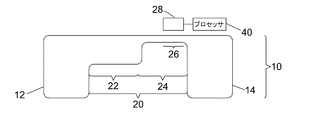
図1Aおよび図1Bを参照すると、血液試料内の白血球の分類および計数のための装置2が記載されている。装置2は、微小流体的液体処理構造10を含む。図1Aおよび1Bは、微小流体的液体処理構造10を含む、装置2の相互に垂直な断面を示す。図1Cは、装置2を上方から見た図を示す。
フレーム内のオブジェクトを分類するために使用される方法を、図3を参照してこれから説明する。ステップ42において、いくつかの実施形態で閾値処理、例えばOtsuの方法を使用して、各フレームを最初にオブジェクトおよびバックグラウンドピクセルにセグメント化し、ステップ44で、フレーム内のオブジェクトを、画像処理の技術分野で周知のように特定する。ステップ46で、各オブジェクトに当該のオブジェクトおよびフレームに固有の識別子が与えられ、ステップ48で、オブジェクトの識別子およびそれらに関連するオブジェクトの位置のリストが取得される。ステップ50において、オブジェクトの位置を中心とする画像パッチが抽出される。次に、ステップ52において、第1の画像パッチについて、画像パッチが別個のオブジェクト領域にセグメント化される。これらの領域は、概観するとバックグラウンド、細胞の核、細胞質である。
1)細胞(暗めのピクセル)とバックグラウンド(明るめのピクセル)を分離するためのk=2におけるk−平均クラスタ化法;
2)バックグラウンドのピクセルをバックグラウンドとマスクに標識化する(事前に定義されている色、例えば緑に設定);
3)核(最も暗い)、細胞質(明るめ)、そして今や、例えば緑のバックグラウンドを得るための、k=3でのk−平均クラスタ化法;
4)細胞質のピクセルを細胞質領域1として標識化し、細胞質のピクセルを所定の色に設定することにより、バックグラウンドに加えて細胞質をマスクする;
5)核のピクセル(最も暗い)、細胞質領域2、および今やマスクされたバックグラウンド(今や以前の細胞質領域1の領域を含む)を得るためのk=3でのk−平均クラスタ化法;
6)核のピクセルを核と標識化し、細胞質領域2のピクセルを細胞質領域2と標識化する。
・関連するピクセルについて平均を求めた画像の彩度とその標準偏差
・関連するピクセルについて平均を求めた強度とその標準偏差
・関連するピクセルについて平均を求めた色彩とその標準偏差
・オブジェクトの真円度
・例えば、

・オブジェクトの領域、および
・画像のフォーカスの質。
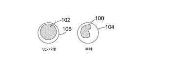
・リンパ球
・単球
・好中球
・好塩基球
・好酸球
・「無染色細胞」
・「細胞のクラスタ」
・「バックグラウンドのオブジェクト」
・「大きなデブリ」(例えば、血小板の凝集体、溶解していない染色液)
・「焦点の合っていない細胞」
・「赤血球」
・「有核赤血球」
・「範囲外」
・「その他」
・「クラスタ内の好酸球」
分画を得る方法を、これから図6を参照して説明する。ステップ60において、上述の方法に従って、血液試料を多数のフレームを含むビデオに取込む。ステップ62では、各フレーム内のオブジェクトが特定され、クラス所属スコアが単純ロジスティックモデルを使用して取得される。この工程は、図3を参照して上述されている。ステップ64で、各オブジェクトは、図6を参照してこれから説明するように、できるだけ多くのフレームにわたって追跡される。試料が視野26を横切って流れるとき、試料中の各オブジェクトは、典型的には、1〜2秒間、画像取込装置28の視野26内にとどまる。したがって、所与のオブジェクトは約15〜30フレーム表示される。次いで、オブジェクトは、1つのフレームから次のフレームまで追跡される。この追跡工程を、図7および図8を参照してこれから説明する。
[絶対数の方法1−統計的分析]
上記のように、判定すべき別の有用な量は絶対的白血球数である。これは、血液の単位体積当たりの推定白血球数である。アッセイで使用される血液試料の全体積は正確には分かっていないが、任意の一時点での画像取込装置28の視野にある血液の体積は、視野の面積と検出部24の深さの積である。この事実は、絶対数の推定に使用される。
視野26に障害物が検出されない場合、VOBS=0および
別の実施形態では、二重計数の可能性を低減するために、血液試料のビデオのフレームのサブセット内に含まれる球数の平均を直接求めることによって、血液試料の絶対数が推定されている。これを行う方法について、これから説明する。
いくつかの実施形態では、白血球の分画の値および絶対的白血球数の値を組み合わせて、5つの白血球の種類のそれぞれの絶対数を得る。白血球各種の絶対数を得る方法を、これから図15を参照して説明する。ステップ156で、絶対的白血球数が上記方法のいずれかに従って得られる。ステップ158において、白血球各種の分画が上記の方法に従って得られる。ステップ160において、各白血球の絶対数は、それぞれの分画に従った絶対的白血球の総数の割合として得られる。例えば、白血球の種類毎の各分画は、この形式でなくてもすでに、0と1との間の割合として書き換えられており、絶対的白血球数と乗算させて、白血球各種に対する絶対数の推定値が得られる。
上述した方法ステップの様々な組み合わせが実行でき、それらが実行される順序は、球数の推定に影響を及ぼすことなくある程度変更できることが理解されるであろう。
装置の特定の実施形態について上述したが、様々な実施形態において多くの変形形態が可能であることが理解されるであろう。
[装置の構造と動作]
図16Aおよび図16Bを参照すると、血液試料内の白血球の分類および計数のための装置2が記載されている。装置2は、微小流体的液体処理構造10を含む。図16Aおよび図16Bは、微小流体的液体処理構造10を含む、装置2の互いに垂直な断面を示す。図16Cは、装置2を上方から見た図である。
フレーム内のオブジェクトを分類するために使用される方法を、図18を参照してこれから説明する。ステップ42において、いくつかの実施形態で閾値処理、例えばOtsuの方法を使用して、各フレームを最初にオブジェクトおよびバックグラウンドピクセルにセグメント化し、ステップ44で、フレーム内のオブジェクトを、画像処理の技術分野で周知のように特定する。ステップ46で、各オブジェクトに当該のオブジェクトおよびフレームに固有の識別子が与えられ、ステップ48で、オブジェクトの識別子およびそれらに関連するオブジェクトの位置のリストが取得される。ステップ50において、オブジェクトの位置を中心とする画像パッチが抽出される。次に、ステップ52において、第1の画像パッチについて、画像パッチが別個のオブジェクト領域にセグメント化される。これらの領域は、概観するとバックグラウンド、細胞の核、細胞質である。
1)細胞(暗めのピクセル)とバックグラウンド(明るめのピクセル)を分離するためのk=2におけるk−平均クラスタ化法;
2)バックグラウンドのピクセルをバックグラウンドとマスクに標識化する(所定の色、例えば緑に設定);
3)核(最も暗い)、細胞質(明るめ)、そして今や、例えば緑のバックグラウンドを得るための、k=3でのk−平均クラスタ化法;
4)細胞質のピクセルを細胞質領域1として標識化し、細胞質のピクセルを所定の色に設定することにより、バックグラウンドに加えて細胞質をマスクする;
5)核のピクセル(最も暗い)、細胞質領域2、および今やマスクされたバックグラウンド(今や以前の細胞質領域1の領域を含む)を得るためのk=3でのk−平均クラスタ化法;
6)核のピクセルを核と標識化し、細胞質領域2のピクセルを細胞質領域2と標識化する。
・関連するピクセルについて平均を求めた画像の彩度とその標準偏差
・関連するピクセルについて平均を求めた強度とその標準偏差
・関連するピクセルについて平均を求めた色彩とその標準偏差
・オブジェクトの真円度
・例えば、
・オブジェクトの領域、および
・画像のフォーカスの質。
・リンパ球
・単球
・好中球
・好塩基球
・好酸球
・「無染色細胞」
・「細胞のクラスタ」
・「バックグラウンドのオブジェクト」
・「大きなデブリ」(例えば、血小板の凝集体、溶解していない染色液)
・「焦点の合っていない細胞」
・「赤血球」
・「有核赤血球」
・「範囲外」
・「その他」
・「クラスタ内の好酸球」
上述したように、いくつかの実施形態では、血液試料の絶対数の推定は、細胞速度の推定に基づいて取込まれたフレームのサブセットを選択することによって、判定することができる。細胞速度を推定する1つの方法は、1つのフレームから次のフレームまで細胞を追跡することである。この工程を図20を参照してこれから説明する。
上述したように、絶対的白血球数は、血液の単位体積当たりの推定白血球数である。アッセイで使用される血液試料の全体積は正確には分かっていないが、任意の一時点での画像取込装置28の視野にある血液の体積は、視野の面積と検出部24の深さの積である。この事実は、絶対数の推定に使用される。
視野26に障害物が検出されない場合、VOBS=0および
別の実施形態では、二重計数の可能性を低減するために、血液試料のビデオのフレームのサブセット内に含まれる球数の平均を直接求めることによって、血液試料の絶対数が推定されている。これを行う方法について、これから説明する。
上述した方法ステップの様々な組み合わせが実行でき、それらが実行される順序は、球数の推定に影響を及ぼすことなくある程度変更できることが理解されるであろう。速度適合型試料採取法を使用して絶対的白血球数が判定される実施形態では、サブセット内に1回しか現れない試料において各オブジェクトの可能性が増加するように、フレームのサブセットが選択される。そのため、フレームのサブセット内のオブジェクトの分類結果を取得することが必要であるだけである。したがって、一実施形態では、フレームのサブセットは、各フレーム内のオブジェクトの分類結果が得られる前に選択され、そのため関連するオブジェクトに対して行われるのみである。これにより、計算能力をより効率的に使用することができる。しかし、サブセットを選択する前に、ビデオ全体の各フレーム内のオブジェクトの分類結果を得ることも同様に可能である。分類結果が他の工程でも使用されている場合、これはより効率的である可能性がある。
装置の特定の実施形態について上述したが、様々な実施形態において多くの変形形態が可能であることが理解されるであろう。
Claims (30)
- 血液試料中の血球を分類する方法であって、
前記試料が画像取込装置の視野を流れるときに、前記画像取込装置で前記試料の一連のフレームを取込むこと、
各フレームをオブジェクトおよびバックグラウンドにセグメント化すること、
前記フレームの少なくとも一部にわたって各セグメント化オブジェクトを追跡すること、
各追跡されたオブジェクトおよび前記フレームの少なくとも一部の各々についての分類結果を得るために、前記フレームの少なくとも一部の各々における前記追跡されたオブジェクトを分類すること、および
各追跡されたオブジェクトを複数のオブジェクトの種類のうちの1つとして分類するために、前記フレームの前記少なくとも一部の各々から得られた個々の分類結果に基づいて、各追跡されたオブジェクトの全体的な分類結果を計算することであって前記オブジェクトの種類は1つ以上の細胞の種類を含む計算すること
を含む方法。 - 全体的な分類結果を計算することが、フレームにわたる前記個々の分類結果の平均値を求めること、または中央値を計算することを含む請求項1に記載の方法。
- 全体的な分類結果を計算することが、各オブジェクトの前記フレームの少なくとも一部から計算された個々の一連の分類結果に、1つ以上のルールを適用することを含む請求項1または請求項2に記載の方法。
- 各追跡されたオブジェクトおよび前記フレームの少なくとも一部の各々の前記分類結果が、前記オブジェクトの種類の各々に属する個々の可能性を示す請求項1〜3のいずれか一項に記載の方法。
- 前記複数のオブジェクトの種類のうちのあるオブジェクトの種類が非等方性の特徴を有し、前記フレームの少なくとも一部の予め設定された数で当該のオブジェクトの種類に属する当該の追跡されたオブジェクトの分類結果が閾値を超えるか、前記フレームの少なくとも一部の間で当該のオブジェクトの種類に属する当該の追跡されたオブジェクトの分類結果の変化が閾値を超える場合に、当該のオブジェクトの種類に追跡されたオブジェクトを割り当てることを含む請求項4に記載の方法。
- 前記1つまたは複数の細胞の種類のうちのいずれか1つとして分類されたオブジェクト数から、各々の細胞の種類として分類されたオブジェクト数の個々の割合を計算することを含む請求項1〜5のいずれか一項に記載の方法。
- 前記1つまたは複数の細胞の種類のいずれか1つのオブジェクトの数を推定すること、および前記1つまたは複数の細胞の種類のうちのいずれか1つのオブジェクトの推定数および前記個々の割合に基づいて、前記1つまたは複数の細胞の種類の各々のオブジェクト数を計算することを含む請求項6に記載の方法。
- 個数を推定することが、各フレームの前記1つまたは複数の細胞の種類のいずれかのオブジェクトを特定すること、
前記一連のフレームの複数のフレームを選択して各選択されたフレームの特定されたオブジェクトのフレーム細胞数を判定すること、および
前記判定されたフレーム細胞数を組み合わせて前記1つまたは複数の細胞の種類のいずれか1つのオブジェクトの推定数を計算すること
を含む請求項7に記載の方法。 - 前記判定されたフレーム細胞数を結合することが、前記判定されたフレーム細胞数に分布関数を適合させ、前記適合された分布に基づいて推定細胞数を計算することを含む請求項8に記載の方法。
- フレームが、1つまたは複数のフレームにおける細胞速度の推定に基づいて選択される請求項8に記載の方法。
- 前記1つまたは複数の細胞の種類が、白血球または白血球の種類である請求項1〜10のいずれか一項に記載の方法。
- 前記1つまたは複数の細胞の種類が、好中球、好塩基球、好酸球、リンパ球、および単球のうちの1つまたは複数を含む請求項11に記載の方法。
- 視野を有する画像取込装置と、画像プロセッサとを備える、血液試料中の細胞数を推定するためのシステムであって、前記画像プロセッサは、
前記試料が前記画像取込装置の視野を流れるときに、前記画像取込装置で前記試料の一連のフレームを取込み、
各フレームをオブジェクトおよびバックグラウンドにセグメント化し、
前記フレームの少なくとも一部にわたって各セグメント化オブジェクトを追跡し、
各追跡されたオブジェクトおよび前記フレームの少なくとも一部の各々についての分類結果を得るために、前記フレームの少なくとも一部の各々における前記追跡されたオブジェクトを分類し、
各追跡されたオブジェクトを複数のオブジェクトの種類のうちの1つとして分類するために、フレームの前記少なくとも一部の各々から得られた個々の分類結果に基づいて、各追跡されたオブジェクトの全体的な分類結果を計算するように構成され、
前記オブジェクトの種類は1つ以上の細胞の種類を含む、
システム。 - 全体的な分類結果を計算することが、フレームにわたる前記個々の分類結果の平均値を求めること、または中央値を計算することを含む請求項13に記載のシステム。
- 全体的な分類結果を計算することが、各オブジェクトの前記フレームの少なくとも一部から計算された個々の一連の分類結果に、1つ以上のルールを適用することを含む請求項13または請求項14に記載のシステム。
- 各追跡されたオブジェクトおよび前記フレームの少なくとも一部の各々の前記分類結果が、前記オブジェクトの種類の各々に属する個々の可能性を示す請求項13〜15のいずれか一項に記載のシステム。
- 前記複数のオブジェクトの種類のうちのあるオブジェクトの種類が非等方性の特徴を有し、前記画像プロセッサが、前記フレームの少なくとも一部の予め設定された数で当該のオブジェクトの種類に属する当該の追跡されたオブジェクトの分類結果が閾値を超えるか、または前記フレームの少なくとも一部の間で当該のオブジェクトの種類に属する当該の追跡されたオブジェクトの分類結果の変化が閾値を超える場合に、当該のオブジェクトの種類に追跡されたオブジェクトを割り当てるように構成された請求項16に記載のシステム。
- 前記プロセッサが、前記1つまたは複数の細胞の種類のうちのいずれか1つとして分類されたオブジェクト数から、各細胞の種類として分類された前記オブジェクト数の個々の割合を計算するように構成される請求項13〜17のいずれか一項に記載のシステム。
- 前記プロセッサが、前記1つまたは複数の細胞の種類のうちのいずれか1つのオブジェクトの数を推定し、前記1つまたは複数の細胞の種類のうちいずれか1つのオブジェクトの推定数および前記個々の割合に基づいて、前記1つまたは複数の細胞の種類の各々のオブジェクト数を計算するように構成されている請求項18に記載のシステム。
- 個数を推定することが、各フレームの前記1つまたは複数の細胞の種類のいずれかのオブジェクトを特定すること、
前記一連のフレームの複数のフレームを選択して各選択されたフレームの特定したオブジェクトのフレーム細胞数を判定すること、および
前記判定されたフレーム細胞数を組み合わせて前記1つまたは複数の細胞の種類のいずれか1つのオブジェクトの推定数を計算すること
を含む請求項19に記載のシステム。 - 前記1つまたは複数の細胞の種類が、白血球または白血球の種類である請求項13〜20のいずれか一項に記載のシステム。
- 前記1つまたは複数の細胞の種類が、好中球、好塩基球、好酸球、リンパ球、および単球のうちの1つまたは複数を含む請求項21に記載のシステム。
- 推定細胞数および前記視野に関連する流量に基づいて、体積当たりの個数を計算することを含む請求項1〜12のいずれか一項に記載の方法。
- 流れの障害物が存在する前記視野内の閉塞した領域を検出することと、前記閉塞した領域に関連する体積のために前記視野に関連付けられた閉塞していない体積を補正することによって、前記流量を判定することとを含む請求項23に記載の方法。
- 閉塞した領域を検出することが、前記視野を候補領域に分割することと、当該の候補領域内の前記一連のフレームにわたって閾値量未満の細胞が特定された場合、候補領域を閉塞した領域としてマークすることとを含む請求項24に記載の方法。
- 前記画像プロセッサが、推定細胞数および前記視野に関連する流量に基づいて、体積当たりの個数を計算するよう構成されている請求項13〜22のいずれか一項に記載のシステム。
- 前記画像プロセッサが、流れの障害物が存在する前記視野内の閉塞した領域を検出することと、前記閉塞した領域に関連する体積のために前記視野に関連付けられた閉塞していない体積を補正することとによって、前記流量を判定するように構成される請求項26に記載のシステム。
- 閉塞した領域を検出することが、前記視野を候補領域に分割することと、当該の候補領域内の前記一連のフレームにわたって閾値量未満の細胞が特定された場合、候補領域を閉塞した領域としてマークすることとを含む請求項27に記載のシステム。
- プロセッサで実行するときに、請求項1〜12、23〜25のいずれか一項に記載の方法を実装する、コード化された命令を含むコンピュータプログラム製品。
- 請求項29に記載のコンピュータプログラム製品を含むコンピュータ可読記憶媒体。
Applications Claiming Priority (5)
| Application Number | Priority Date | Filing Date | Title |
|---|---|---|---|
| PT107931T | 2014-09-29 | ||
| PT10793114 | 2014-09-29 | ||
| GB201417178A GB201417178D0 (en) | 2014-09-29 | 2014-09-29 | Cell counting |
| GB1417178.9 | 2014-09-29 | ||
| PCT/EP2015/072392 WO2016050755A2 (en) | 2014-09-29 | 2015-09-29 | Cell counting |
Publications (3)
| Publication Number | Publication Date |
|---|---|
| JP2017534858A JP2017534858A (ja) | 2017-11-24 |
| JP2017534858A5 JP2017534858A5 (ja) | 2018-11-08 |
| JP6704390B2 true JP6704390B2 (ja) | 2020-06-03 |
Family
ID=54199248
Family Applications (1)
| Application Number | Title | Priority Date | Filing Date |
|---|---|---|---|
| JP2017517029A Active JP6704390B2 (ja) | 2014-09-29 | 2015-09-29 | 血球計数 |
Country Status (4)
| Country | Link |
|---|---|
| US (2) | US10684206B2 (ja) |
| EP (2) | EP3200917B1 (ja) |
| JP (1) | JP6704390B2 (ja) |
| WO (1) | WO2016050755A2 (ja) |
Families Citing this family (11)
| Publication number | Priority date | Publication date | Assignee | Title |
|---|---|---|---|---|
| US10362238B2 (en) | 2016-04-06 | 2019-07-23 | Biosurfit, S.A. | Method and system for capturing images of a liquid sample |
| US10788413B2 (en) * | 2017-01-05 | 2020-09-29 | Massachusetts Institute Of Technology | Method to distinguish and analyze white blood cells in the presence of red blood cells |
| US10321160B2 (en) * | 2017-07-13 | 2019-06-11 | International Business Machines Corporation | Compressing multiple video files using localized camera meta data |
| CA3082097A1 (en) * | 2017-11-14 | 2019-05-23 | miDiagnostics NV | Classification of a population of objects by convolutional dictionary learning with class proportion data |
| JP2019100988A (ja) * | 2017-12-08 | 2019-06-24 | 株式会社島津製作所 | 粒子画像解析装置及び粒子画像解析方法 |
| WO2019198094A1 (en) * | 2018-04-09 | 2019-10-17 | Sigtuple Technologies Private Limited | Method and system for estimating total count of blood cells in a blood smear |
| JP2019195304A (ja) | 2018-05-10 | 2019-11-14 | 学校法人順天堂 | 画像解析方法、装置、コンピュータプログラム、及び深層学習アルゴリズムの生成方法 |
| JP7381003B2 (ja) * | 2019-04-26 | 2023-11-15 | 学校法人順天堂 | 疾患解析を支援する方法、装置、及びコンピュータプログラム、並びにコンピュータアルゴリズムを訓練する方法、装置、及びプログラム |
| US11959908B2 (en) * | 2019-12-17 | 2024-04-16 | Arkray, Inc. | Measurement device and measurement method |
| CN111274949B (zh) * | 2020-01-19 | 2023-05-30 | 重庆医科大学附属第一医院 | 一种基于结构分析的血液病白细胞散点图相似度分析方法 |
| EP4172591A1 (en) * | 2020-06-03 | 2023-05-03 | Case Western Reserve University | Classification of blood cells |
Family Cites Families (29)
| Publication number | Priority date | Publication date | Assignee | Title |
|---|---|---|---|---|
| US4075462A (en) * | 1975-01-08 | 1978-02-21 | William Guy Rowe | Particle analyzer apparatus employing light-sensitive electronic detector array |
| JP3111706B2 (ja) * | 1992-02-18 | 2000-11-27 | 株式会社日立製作所 | 粒子分析装置及び粒子分析方法 |
| US5760950A (en) * | 1996-07-25 | 1998-06-02 | Advanced Scanning, Ltd. | Scanning confocal microscope |
| WO2002017219A1 (en) | 2000-08-25 | 2002-02-28 | Amnis Corporation | Measuring the velocity of small moving objects such as cells |
| US6778263B2 (en) * | 2000-08-25 | 2004-08-17 | Amnis Corporation | Methods of calibrating an imaging system using calibration beads |
| KR20040105717A (ko) * | 2002-02-14 | 2004-12-16 | 이뮤니베스트 코포레이션 | 저비용 세포 분석기에서의 세포 계산 방법 및 알고리즘 |
| WO2004003160A2 (en) * | 2002-06-27 | 2004-01-08 | University Of Washington | Use of adhesion molecules as bond stress-enhanced nanoscale binding switches |
| US7517453B2 (en) * | 2003-03-01 | 2009-04-14 | The Trustees Of Boston University | Microvascular network device |
| AU2003902589A0 (en) * | 2003-05-26 | 2003-06-12 | Commonwealth Scientific And Industrial Research Organisation | A method for identifying a subset of components of a system |
| US7295309B1 (en) | 2003-12-09 | 2007-11-13 | United States Of America As Represented By The Administrator Of The National Aeronautics And Space Administration | Microparticle analysis system and method |
| GB0503629D0 (en) | 2005-02-22 | 2005-03-30 | Durand Technology Ltd | Method and apparatus for automated analysis of biological specimen |
| WO2008064423A1 (en) * | 2006-12-01 | 2008-06-05 | The Walter And Eliza Hall Institute Of Medical Research | A cell population system and process |
| DE102007013971B4 (de) | 2007-03-23 | 2011-07-14 | Fraunhofer-Gesellschaft zur Förderung der angewandten Forschung e.V., 80686 | Verfahren und Vorrichtung zur Ermittlung einer Zellkontur einer Zelle |
| US8159670B2 (en) * | 2007-11-05 | 2012-04-17 | Abbott Laboratories | Method and apparatus for rapidly counting and identifying biological particles in a flow stream |
| DE102008030874A1 (de) | 2008-06-30 | 2010-01-07 | Fraunhofer-Gesellschaft zur Förderung der angewandten Forschung e.V. | Verfahren und Vorrichtung zum Ermitteln einer Kontur und eines Mittelpunktes eines Objekts |
| JP5438962B2 (ja) * | 2008-12-25 | 2014-03-12 | シスメックス株式会社 | 細胞画像表示装置 |
| US20140152801A1 (en) * | 2009-10-28 | 2014-06-05 | Alentic Microscience Inc. | Detecting and Using Light Representative of a Sample |
| US8774488B2 (en) | 2010-03-11 | 2014-07-08 | Cellscape Corporation | Method and device for identification of nucleated red blood cells from a maternal blood sample |
| CN103189883A (zh) * | 2010-09-07 | 2013-07-03 | 里兰斯坦福初级大学理事会 | 医学评分系统及方法 |
| JP5320510B2 (ja) * | 2010-11-01 | 2013-10-23 | 公益財団法人神奈川科学技術アカデミー | 細胞分析装置 |
| JP2013015357A (ja) | 2011-07-01 | 2013-01-24 | Shimadzu Corp | フローサイトメータ |
| TW201433296A (zh) * | 2013-02-21 | 2014-09-01 | Univ Nat Taiwan | 一種血球之觀察鑑測方法 |
| WO2013135713A1 (en) | 2012-03-12 | 2013-09-19 | Biosurfit S.A. | Liquid sample imaging device and method |
| EP2852461B1 (en) * | 2012-05-22 | 2022-06-15 | Hemanext Inc. | Capillary network devices and methods of use |
| US20140002617A1 (en) | 2012-06-27 | 2014-01-02 | The Board Of Trustees Of The University Of Illinois | Particle tracking system and method |
| WO2014040184A1 (en) * | 2012-09-13 | 2014-03-20 | The Governing Council Of The University Of Toronto | System and method for fetal and maternal red blood cell counting |
| JP6278519B2 (ja) * | 2012-11-28 | 2018-02-21 | 国立研究開発法人科学技術振興機構 | 細胞観察装置、細胞観察方法及びそのプログラム |
| EP2972208B1 (en) * | 2013-03-15 | 2018-06-13 | Iris International, Inc. | Method and composition for staining and sample processing |
| US20170209864A1 (en) * | 2013-03-15 | 2017-07-27 | Gpb Scientific, Llc | Methods and systems for processing particles |
-
2015
- 2015-09-29 EP EP15770923.9A patent/EP3200917B1/en active Active
- 2015-09-29 US US15/515,529 patent/US10684206B2/en active Active
- 2015-09-29 EP EP20157751.7A patent/EP3708254A1/en not_active Withdrawn
- 2015-09-29 JP JP2017517029A patent/JP6704390B2/ja active Active
- 2015-09-29 WO PCT/EP2015/072392 patent/WO2016050755A2/en active Application Filing
-
2018
- 2018-03-29 US US15/940,330 patent/US10684207B2/en active Active
Also Published As
| Publication number | Publication date |
|---|---|
| JP2017534858A (ja) | 2017-11-24 |
| EP3708254A1 (en) | 2020-09-16 |
| EP3200917B1 (en) | 2020-02-19 |
| US10684206B2 (en) | 2020-06-16 |
| WO2016050755A2 (en) | 2016-04-07 |
| WO2016050755A3 (en) | 2016-06-09 |
| US10684207B2 (en) | 2020-06-16 |
| US20180217043A1 (en) | 2018-08-02 |
| US20170212028A1 (en) | 2017-07-27 |
| EP3200917A2 (en) | 2017-08-09 |
Similar Documents
| Publication | Publication Date | Title |
|---|---|---|
| JP6704390B2 (ja) | 血球計数 | |
| CN105143849B (zh) | 用于血液样品中粒子分析的动态范围扩展系统和方法 | |
| Savkare et al. | Automatic detection of malaria parasites for estimating parasitemia | |
| Wang et al. | Label-free detection of rare circulating tumor cells by image analysis and machine learning | |
| Hegde et al. | Peripheral blood smear analysis using image processing approach for diagnostic purposes: A review | |
| JP2023532483A (ja) | 循環異常細胞を検出する方法及び装置 | |
| WO2015153691A2 (en) | Computer-implemented methods, computer-readable media, and systems for tracking a plurality of spermatozoa | |
| Lei et al. | Automatic detection and counting of urediniospores of Puccinia striiformis f. sp. tritici using spore traps and image processing | |
| Monteiro et al. | A comparative study between methodologies based on the Hough transform and watershed transform on the blood cell count | |
| CN109844494B (zh) | 动态聚焦系统和方法 | |
| US20230186659A1 (en) | Machine learning models for cell localization and classification learned using repel coding | |
| Ciurte et al. | Automatic detection of circulating tumor cells in darkfield microscopic images of unstained blood using boosting techniques | |
| Lin et al. | Digital pathology and artificial intelligence as the next chapter in diagnostic hematopathology | |
| CN110226083B (zh) | 红细胞碎片识别方法和装置、血液细胞分析仪及分析方法 | |
| CN112912923A (zh) | 基于距离的组织状态确定 | |
| Soldati et al. | Microfluidic droplets content classification and analysis through convolutional neural networks in a liquid biopsy workflow | |
| EP3789914A1 (en) | Methods and systems for automated assessment of respiratory cytology specimens | |
| Angay et al. | Image‐based modeling and scoring of Howell–Jolly Bodies in human erythrocytes | |
| Wei et al. | Automatic counting method for complex overlapping erythrocytes based on seed prediction in microscopic imaging | |
| CN116385334A (zh) | 一种用于尿沉渣检查的高效yolo检测方法 | |
| Ossandon et al. | A computational streak mode cytometry biosensor for rare cell analysis | |
| Tomari et al. | An empirical framework for automatic red blood cell morphology identification and counting | |
| Mukherjee et al. | Application of biomedical image processing in blood cell counting using hough transform | |
| Panpatte et al. | Application of image processing for blood group detection | |
| Dong et al. | ABO blood group detection based on image processing technology |
Legal Events
| Date | Code | Title | Description |
|---|---|---|---|
| A521 | Request for written amendment filed |
Free format text: JAPANESE INTERMEDIATE CODE: A821 Effective date: 20170609 |
|
| A521 | Request for written amendment filed |
Free format text: JAPANESE INTERMEDIATE CODE: A523 Effective date: 20170609 |
|
| A521 | Request for written amendment filed |
Free format text: JAPANESE INTERMEDIATE CODE: A523 Effective date: 20180925 |
|
| A621 | Written request for application examination |
Free format text: JAPANESE INTERMEDIATE CODE: A621 Effective date: 20180925 |
|
| A977 | Report on retrieval |
Free format text: JAPANESE INTERMEDIATE CODE: A971007 Effective date: 20190628 |
|
| A131 | Notification of reasons for refusal |
Free format text: JAPANESE INTERMEDIATE CODE: A131 Effective date: 20190716 |
|
| A601 | Written request for extension of time |
Free format text: JAPANESE INTERMEDIATE CODE: A601 Effective date: 20191015 |
|
| TRDD | Decision of grant or rejection written | ||
| A01 | Written decision to grant a patent or to grant a registration (utility model) |
Free format text: JAPANESE INTERMEDIATE CODE: A01 Effective date: 20200421 |
|
| A61 | First payment of annual fees (during grant procedure) |
Free format text: JAPANESE INTERMEDIATE CODE: A61 Effective date: 20200512 |
|
| R150 | Certificate of patent or registration of utility model |
Ref document number: 6704390 Country of ref document: JP Free format text: JAPANESE INTERMEDIATE CODE: R150 |
|
| R250 | Receipt of annual fees |
Free format text: JAPANESE INTERMEDIATE CODE: R250 |