JP6704390B2 - Blood cell count - Google Patents
Blood cell count Download PDFInfo
- Publication number
- JP6704390B2 JP6704390B2 JP2017517029A JP2017517029A JP6704390B2 JP 6704390 B2 JP6704390 B2 JP 6704390B2 JP 2017517029 A JP2017517029 A JP 2017517029A JP 2017517029 A JP2017517029 A JP 2017517029A JP 6704390 B2 JP6704390 B2 JP 6704390B2
- Authority
- JP
- Japan
- Prior art keywords
- frame
- frames
- objects
- cell
- cells
- Prior art date
- Legal status (The legal status is an assumption and is not a legal conclusion. Google has not performed a legal analysis and makes no representation as to the accuracy of the status listed.)
- Active
Links
- 238000004820 blood count Methods 0.000 title description 61
- 210000004027 cell Anatomy 0.000 claims description 228
- 210000000265 leukocyte Anatomy 0.000 claims description 169
- 238000000034 method Methods 0.000 claims description 140
- 210000004369 blood Anatomy 0.000 claims description 95
- 239000008280 blood Substances 0.000 claims description 95
- 238000009826 distribution Methods 0.000 claims description 37
- 210000003979 eosinophil Anatomy 0.000 claims description 30
- 210000001616 monocyte Anatomy 0.000 claims description 26
- 210000004698 lymphocyte Anatomy 0.000 claims description 17
- 210000003651 basophil Anatomy 0.000 claims description 11
- 238000012935 Averaging Methods 0.000 claims description 10
- 210000000440 neutrophil Anatomy 0.000 claims description 9
- 210000000601 blood cell Anatomy 0.000 claims description 7
- 238000004590 computer program Methods 0.000 claims description 4
- 230000008859 change Effects 0.000 claims description 3
- 238000005315 distribution function Methods 0.000 claims description 3
- 239000007788 liquid Substances 0.000 description 71
- 210000004940 nucleus Anatomy 0.000 description 38
- 238000002360 preparation method Methods 0.000 description 35
- 239000003153 chemical reaction reagent Substances 0.000 description 32
- 210000000805 cytoplasm Anatomy 0.000 description 32
- 230000001086 cytosolic effect Effects 0.000 description 32
- 238000001514 detection method Methods 0.000 description 28
- 230000008569 process Effects 0.000 description 23
- 239000002699 waste material Substances 0.000 description 20
- 238000012545 processing Methods 0.000 description 19
- 238000010186 staining Methods 0.000 description 17
- 238000004422 calculation algorithm Methods 0.000 description 16
- 210000003743 erythrocyte Anatomy 0.000 description 16
- 230000011218 segmentation Effects 0.000 description 14
- 238000003556 assay Methods 0.000 description 13
- 238000003064 k means clustering Methods 0.000 description 12
- 230000007246 mechanism Effects 0.000 description 12
- 239000003795 chemical substances by application Substances 0.000 description 11
- 238000010586 diagram Methods 0.000 description 11
- 230000017531 blood circulation Effects 0.000 description 9
- 238000007619 statistical method Methods 0.000 description 9
- 241000288113 Gallirallus australis Species 0.000 description 8
- 238000006243 chemical reaction Methods 0.000 description 8
- 239000000203 mixture Substances 0.000 description 8
- 230000000877 morphologic effect Effects 0.000 description 8
- 238000005070 sampling Methods 0.000 description 7
- 230000009471 action Effects 0.000 description 6
- 238000003384 imaging method Methods 0.000 description 6
- 230000002934 lysing effect Effects 0.000 description 6
- 238000010801 machine learning Methods 0.000 description 6
- 239000002356 single layer Substances 0.000 description 6
- 230000000007 visual effect Effects 0.000 description 6
- 238000004458 analytical method Methods 0.000 description 5
- WZUVPPKBWHMQCE-UHFFFAOYSA-N Haematoxylin Chemical compound C12=CC(O)=C(O)C=C2CC2(O)C1C1=CC=C(O)C(O)=C1OC2 WZUVPPKBWHMQCE-UHFFFAOYSA-N 0.000 description 4
- 235000010627 Phaseolus vulgaris Nutrition 0.000 description 4
- 244000046052 Phaseolus vulgaris Species 0.000 description 4
- 239000012298 atmosphere Substances 0.000 description 4
- 210000003855 cell nucleus Anatomy 0.000 description 4
- 230000009089 cytolysis Effects 0.000 description 4
- 238000013500 data storage Methods 0.000 description 4
- 239000012530 fluid Substances 0.000 description 4
- 238000005194 fractionation Methods 0.000 description 4
- 230000006870 function Effects 0.000 description 4
- 238000002372 labelling Methods 0.000 description 4
- 238000005259 measurement Methods 0.000 description 4
- 230000003287 optical effect Effects 0.000 description 4
- YQGOJNYOYNNSMM-UHFFFAOYSA-N eosin Chemical compound [Na+].OC(=O)C1=CC=CC=C1C1=C2C=C(Br)C(=O)C(Br)=C2OC2=C(Br)C(O)=C(Br)C=C21 YQGOJNYOYNNSMM-UHFFFAOYSA-N 0.000 description 3
- 238000000684 flow cytometry Methods 0.000 description 3
- ZOMLUNRKXJYKPD-UHFFFAOYSA-N 1,3,3-trimethyl-2-[2-(2-methylindol-3-ylidene)ethylidene]indole;hydrochloride Chemical compound [Cl-].C1=CC=C2C(C)(C)C(/C=C/C=3C4=CC=CC=C4NC=3C)=[N+](C)C2=C1 ZOMLUNRKXJYKPD-UHFFFAOYSA-N 0.000 description 2
- RBTBFTRPCNLSDE-UHFFFAOYSA-N 3,7-bis(dimethylamino)phenothiazin-5-ium Chemical compound C1=CC(N(C)C)=CC2=[S+]C3=CC(N(C)C)=CC=C3N=C21 RBTBFTRPCNLSDE-UHFFFAOYSA-N 0.000 description 2
- 239000012790 adhesive layer Substances 0.000 description 2
- 238000013459 approach Methods 0.000 description 2
- 230000004888 barrier function Effects 0.000 description 2
- 230000006037 cell lysis Effects 0.000 description 2
- 230000001413 cellular effect Effects 0.000 description 2
- 238000013480 data collection Methods 0.000 description 2
- 230000007423 decrease Effects 0.000 description 2
- 229940079593 drug Drugs 0.000 description 2
- 239000003814 drug Substances 0.000 description 2
- 238000004043 dyeing Methods 0.000 description 2
- 239000008187 granular material Substances 0.000 description 2
- 239000003219 hemolytic agent Substances 0.000 description 2
- 238000003709 image segmentation Methods 0.000 description 2
- 230000006872 improvement Effects 0.000 description 2
- 230000003458 metachromatic effect Effects 0.000 description 2
- 229960000907 methylthioninium chloride Drugs 0.000 description 2
- 238000012986 modification Methods 0.000 description 2
- 230000004048 modification Effects 0.000 description 2
- 239000001397 quillaja saponaria molina bark Substances 0.000 description 2
- 230000000717 retained effect Effects 0.000 description 2
- 238000012552 review Methods 0.000 description 2
- 229930182490 saponin Natural products 0.000 description 2
- 150000007949 saponins Chemical class 0.000 description 2
- 239000007787 solid Substances 0.000 description 2
- 239000002904 solvent Substances 0.000 description 2
- 238000000638 solvent extraction Methods 0.000 description 2
- 238000012706 support-vector machine Methods 0.000 description 2
- 239000004094 surface-active agent Substances 0.000 description 2
- 238000012549 training Methods 0.000 description 2
- 230000007704 transition Effects 0.000 description 2
- 238000004364 calculation method Methods 0.000 description 1
- 238000005266 casting Methods 0.000 description 1
- 238000004163 cytometry Methods 0.000 description 1
- 230000001419 dependent effect Effects 0.000 description 1
- 239000012634 fragment Substances 0.000 description 1
- 238000007429 general method Methods 0.000 description 1
- 230000009191 jumping Effects 0.000 description 1
- 230000008520 organization Effects 0.000 description 1
Images


Classifications
-
- G—PHYSICS
- G01—MEASURING; TESTING
- G01N—INVESTIGATING OR ANALYSING MATERIALS BY DETERMINING THEIR CHEMICAL OR PHYSICAL PROPERTIES
- G01N15/00—Investigating characteristics of particles; Investigating permeability, pore-volume, or surface-area of porous materials
- G01N15/10—Investigating individual particles
- G01N15/14—Electro-optical investigation, e.g. flow cytometers
- G01N15/1468—Electro-optical investigation, e.g. flow cytometers with spatial resolution of the texture or inner structure of the particle
- G01N15/147—Electro-optical investigation, e.g. flow cytometers with spatial resolution of the texture or inner structure of the particle the analysis being performed on a sample stream
-
- G—PHYSICS
- G01—MEASURING; TESTING
- G01N—INVESTIGATING OR ANALYSING MATERIALS BY DETERMINING THEIR CHEMICAL OR PHYSICAL PROPERTIES
- G01N15/00—Investigating characteristics of particles; Investigating permeability, pore-volume, or surface-area of porous materials
- G01N15/02—Investigating particle size or size distribution
- G01N15/0205—Investigating particle size or size distribution by optical means, e.g. by light scattering, diffraction, holography or imaging
- G01N15/0227—Investigating particle size or size distribution by optical means, e.g. by light scattering, diffraction, holography or imaging using imaging, e.g. a projected image of suspension; using holography
-
- G—PHYSICS
- G01—MEASURING; TESTING
- G01N—INVESTIGATING OR ANALYSING MATERIALS BY DETERMINING THEIR CHEMICAL OR PHYSICAL PROPERTIES
- G01N15/00—Investigating characteristics of particles; Investigating permeability, pore-volume, or surface-area of porous materials
- G01N15/02—Investigating particle size or size distribution
- G01N15/0266—Investigating particle size or size distribution with electrical classification
-
- G—PHYSICS
- G01—MEASURING; TESTING
- G01N—INVESTIGATING OR ANALYSING MATERIALS BY DETERMINING THEIR CHEMICAL OR PHYSICAL PROPERTIES
- G01N15/00—Investigating characteristics of particles; Investigating permeability, pore-volume, or surface-area of porous materials
- G01N15/10—Investigating individual particles
- G01N15/14—Electro-optical investigation, e.g. flow cytometers
- G01N15/1434—Electro-optical investigation, e.g. flow cytometers using an analyser being characterised by its optical arrangement
- G01N15/1436—Electro-optical investigation, e.g. flow cytometers using an analyser being characterised by its optical arrangement the optical arrangement forming an integrated apparatus with the sample container, e.g. a flow cell
-
- G—PHYSICS
- G01—MEASURING; TESTING
- G01N—INVESTIGATING OR ANALYSING MATERIALS BY DETERMINING THEIR CHEMICAL OR PHYSICAL PROPERTIES
- G01N33/00—Investigating or analysing materials by specific methods not covered by groups G01N1/00 - G01N31/00
- G01N33/48—Biological material, e.g. blood, urine; Haemocytometers
- G01N33/483—Physical analysis of biological material
- G01N33/487—Physical analysis of biological material of liquid biological material
- G01N33/49—Blood
- G01N33/4915—Blood using flow cells
-
- G—PHYSICS
- G06—COMPUTING; CALCULATING OR COUNTING
- G06F—ELECTRIC DIGITAL DATA PROCESSING
- G06F18/00—Pattern recognition
- G06F18/20—Analysing
- G06F18/25—Fusion techniques
- G06F18/253—Fusion techniques of extracted features
-
- G—PHYSICS
- G06—COMPUTING; CALCULATING OR COUNTING
- G06V—IMAGE OR VIDEO RECOGNITION OR UNDERSTANDING
- G06V10/00—Arrangements for image or video recognition or understanding
- G06V10/70—Arrangements for image or video recognition or understanding using pattern recognition or machine learning
- G06V10/77—Processing image or video features in feature spaces; using data integration or data reduction, e.g. principal component analysis [PCA] or independent component analysis [ICA] or self-organising maps [SOM]; Blind source separation
- G06V10/80—Fusion, i.e. combining data from various sources at the sensor level, preprocessing level, feature extraction level or classification level
- G06V10/806—Fusion, i.e. combining data from various sources at the sensor level, preprocessing level, feature extraction level or classification level of extracted features
-
- G—PHYSICS
- G06—COMPUTING; CALCULATING OR COUNTING
- G06V—IMAGE OR VIDEO RECOGNITION OR UNDERSTANDING
- G06V20/00—Scenes; Scene-specific elements
- G06V20/60—Type of objects
- G06V20/69—Microscopic objects, e.g. biological cells or cellular parts
- G06V20/693—Acquisition
-
- G—PHYSICS
- G06—COMPUTING; CALCULATING OR COUNTING
- G06V—IMAGE OR VIDEO RECOGNITION OR UNDERSTANDING
- G06V20/00—Scenes; Scene-specific elements
- G06V20/60—Type of objects
- G06V20/69—Microscopic objects, e.g. biological cells or cellular parts
- G06V20/698—Matching; Classification
-
- B—PERFORMING OPERATIONS; TRANSPORTING
- B01—PHYSICAL OR CHEMICAL PROCESSES OR APPARATUS IN GENERAL
- B01L—CHEMICAL OR PHYSICAL LABORATORY APPARATUS FOR GENERAL USE
- B01L3/00—Containers or dishes for laboratory use, e.g. laboratory glassware; Droppers
- B01L3/50—Containers for the purpose of retaining a material to be analysed, e.g. test tubes
- B01L3/502—Containers for the purpose of retaining a material to be analysed, e.g. test tubes with fluid transport, e.g. in multi-compartment structures
- B01L3/5027—Containers for the purpose of retaining a material to be analysed, e.g. test tubes with fluid transport, e.g. in multi-compartment structures by integrated microfluidic structures, i.e. dimensions of channels and chambers are such that surface tension forces are important, e.g. lab-on-a-chip
- B01L3/502761—Containers for the purpose of retaining a material to be analysed, e.g. test tubes with fluid transport, e.g. in multi-compartment structures by integrated microfluidic structures, i.e. dimensions of channels and chambers are such that surface tension forces are important, e.g. lab-on-a-chip specially adapted for handling suspended solids or molecules independently from the bulk fluid flow, e.g. for trapping or sorting beads, for physically stretching molecules
-
- G01N15/1433—
-
- G—PHYSICS
- G01—MEASURING; TESTING
- G01N—INVESTIGATING OR ANALYSING MATERIALS BY DETERMINING THEIR CHEMICAL OR PHYSICAL PROPERTIES
- G01N15/00—Investigating characteristics of particles; Investigating permeability, pore-volume, or surface-area of porous materials
- G01N15/10—Investigating individual particles
- G01N15/14—Electro-optical investigation, e.g. flow cytometers
- G01N15/1484—Electro-optical investigation, e.g. flow cytometers microstructural devices
-
- G—PHYSICS
- G01—MEASURING; TESTING
- G01N—INVESTIGATING OR ANALYSING MATERIALS BY DETERMINING THEIR CHEMICAL OR PHYSICAL PROPERTIES
- G01N15/00—Investigating characteristics of particles; Investigating permeability, pore-volume, or surface-area of porous materials
- G01N15/10—Investigating individual particles
- G01N2015/1006—Investigating individual particles for cytology
-
- G01N2015/1029—
-
- G—PHYSICS
- G01—MEASURING; TESTING
- G01N—INVESTIGATING OR ANALYSING MATERIALS BY DETERMINING THEIR CHEMICAL OR PHYSICAL PROPERTIES
- G01N15/00—Investigating characteristics of particles; Investigating permeability, pore-volume, or surface-area of porous materials
- G01N15/10—Investigating individual particles
- G01N15/14—Electro-optical investigation, e.g. flow cytometers
- G01N2015/1486—Counting the particles
-
- G—PHYSICS
- G01—MEASURING; TESTING
- G01N—INVESTIGATING OR ANALYSING MATERIALS BY DETERMINING THEIR CHEMICAL OR PHYSICAL PROPERTIES
- G01N15/00—Investigating characteristics of particles; Investigating permeability, pore-volume, or surface-area of porous materials
- G01N15/10—Investigating individual particles
- G01N15/14—Electro-optical investigation, e.g. flow cytometers
- G01N2015/1488—Methods for deciding
-
- G—PHYSICS
- G01—MEASURING; TESTING
- G01N—INVESTIGATING OR ANALYSING MATERIALS BY DETERMINING THEIR CHEMICAL OR PHYSICAL PROPERTIES
- G01N35/00—Automatic analysis not limited to methods or materials provided for in any single one of groups G01N1/00 - G01N33/00; Handling materials therefor
- G01N35/00029—Automatic analysis not limited to methods or materials provided for in any single one of groups G01N1/00 - G01N33/00; Handling materials therefor provided with flat sample substrates, e.g. slides
- G01N2035/00099—Characterised by type of test elements
- G01N2035/00158—Elements containing microarrays, i.e. "biochip"
Description
本発明は、血液試料中の細胞の計数に関する。 The present invention relates to counting cells in blood samples.
血液試料の細胞の計数は重要な診断ツールである。 Cell counting of blood samples is an important diagnostic tool.
血液試料の細胞を計数する既知の手法の例は、フローサイトメトリーである。フローサイトメトリーでは、血液試料が毛細管を通って流れ、細胞が光源および検出装置の前を1つずつ通過する。反射光および/または透過光を検出し、分析する。検出された光の特性は、試料中の細胞の特徴およびそれらがいかに染色されているかに依存しており、このようにして細胞を分類し、計数する。しかし、フローサイトメトリーを用いても細胞の形態学的情報は得られない。形態学的分析は、2枚のスライドの間に試料を挟み、顕微鏡下で試料を調べることによって、血液試料に対して行うことができる。予め準備されたスライドのカートリッジを使用する血球計数用の自動システムが知られている。このようなスライドは、分析されるべき各試料について手動で準備する必要がある。これは、一部自動化されているにもかかわらず、時間を要する工程である。 An example of a known technique for counting cells in blood samples is flow cytometry. In flow cytometry, a blood sample flows through a capillary and cells pass one in front of a light source and a detector. The reflected and/or transmitted light is detected and analyzed. The characteristics of the light detected depend on the characteristics of the cells in the sample and how they are stained, thus sorting and counting the cells. However, flow cytometry does not provide cell morphological information. Morphological analysis can be performed on blood samples by sandwiching the sample between two slides and examining the sample under a microscope. Automated systems for blood cell counting using a cartridge of pre-prepared slides are known. Such slides need to be manually prepared for each sample to be analyzed. This is a time consuming process, albeit partially automated.
細胞の形態学的情報が失われず、それどころか血球計数工程の精度を向上させるために使用され、さらにスライドの準備を必要としない血球計数用のシステムを有することは、望ましいであろう。 It would be desirable to have a system for hemocytometry that does not lose cell morphological information, is used to improve the accuracy of the hemocytometry process, and does not require slide preparation.
本発明の態様は、独立請求項に記載されている。さらに、本発明の随意による特徴は、従属請求項に記載されている。 Aspects of the invention are set out in the independent claims. Further optional features of the invention are set out in the dependent claims.
いくつかの実施形態で、血液試料中の血球を分類する方法を提供する。方法は、以下を含む:
−試料が画像取込装置の視野を流れるときに、画像取込装置で試料の一連のフレームを取込むこと、
−各フレームをオブジェクトおよびバックグラウンドにセグメント化すること、
−フレームの少なくとも一部にわたって各セグメント化オブジェクトを追跡すること、
−各追跡されたオブジェクトおよびフレームの個々の少なくとも一部の各々についての分類結果を得るために、フレームの個々の少なくとも一部の各々における追跡されたオブジェクトを分類すること、および
−各追跡されたオブジェクトを複数のオブジェクトの種類のうちの1つとして分類するために、フレームの少なくとも一部の各々から得られた個々の分類結果に基づいて、各追跡されたオブジェクトの全体的な分類結果を計算することであって、オブジェクトの種類は1つ以上の細胞の種類を含む、計算すること。
In some embodiments, a method of classifying blood cells in a blood sample is provided. The method includes:
Capturing a series of frames of the sample with the image capture device as the sample flows through the field of view of the image capture device;
Segmenting each frame into an object and background,
Tracking each segmented object over at least part of the frame,
-Classifying the tracked objects in each individual at least part of the frame to obtain a classification result for each tracked object and each individual at least part of the frame; and-each tracked object Calculate an overall classification result for each tracked object based on the individual classification results obtained from each of at least some of the frames to classify the object as one of a plurality of object types Calculating the object type includes one or more cell types.
いくつかの実施形態で、視野を有する画像取込装置と、画像プロセッサとを備える、血液試料中の血球を分類するためのシステムが提供されている。画像プロセッサは、上記の方法を実行するように構成されている。 In some embodiments, a system for classifying blood cells in a blood sample is provided that includes an image capture device having a field of view and an image processor. The image processor is configured to perform the above method.
好都合なことに、2つ以上の取込まれたフレームのオブジェクトの分類結果を得ることによって、血球計数の精度が向上する。そうすることで、試料が視野を横切って流れるとき、複数の独立した観察が得られ、各オブジェクトを異なる角度から見ることができ、豊富な形態学的データのセットを作成することができる。細胞のサイトメトリーと比較すると、形態学的情報が失われず、公知の形態学的方法と比較すると、手動のスライドの準備が必要ではない。 Advantageously, by obtaining a classification result for objects of more than one captured frame, the accuracy of blood cell counting is improved. By doing so, as the sample flows across the field of view, multiple independent observations are obtained, each object can be viewed from a different angle, creating a rich morphological data set. No morphological information is lost when compared to cytometry of cells and no manual slide preparation is required when compared to known morphological methods.
いくつかの実施形態では、各追跡オブジェクトおよび個々のフレームの各々についての分類結果は、単純ロジスティックモデルを使用して取得することができる。他の実施形態では、関連分野の当業者に知られている他の分類子を使用する場合がある。例については、S.B.Kotsiantis、 「Supervised Machine Learning:A Review of Classification Techniques」、Informatica,vol.31,number 3,October 2007,pages 249−268を参照されたい。 In some embodiments, the classification results for each tracking object and each individual frame can be obtained using a simple logistic model. Other embodiments may use other classifiers known to those of ordinary skill in the relevant art. For an example, see S. B. Kotsiantis, "Supervised Machine Learning: A Review of Classification Techniques," Informatica, vol. 31, number 3, October 2007, pages 249-268.
いくつかの実施形態では、全体的な分類結果を計算することは、フレームにわたる個々の分類結果の平均値を求めること、または中央値を計算することを含む。他の実施形態では、全体的な分類結果を計算することが、オブジェクトが追跡される各個々のフレームから得られた分類結果に、1つ以上のルールを適用することを含む。これにより、平均を求める工程で失われる、各フレームから得られるいずれかの分類結果を、全体的な分類工程を判定する際に、考慮することが可能になる。例えば、このようなルールを設定することにより、1つまたは複数のフレームで検出されるあるオブジェクトの種類の任意の識別特性が、比較的少数のフレームでしか検出されない場合であっても、オブジェクトの全体的な分類で考慮されることが可能になる。例えば、これは、いくつかの方向から見た場合にのみ核が高度に特徴的な構成である細胞を検出することについて、改善することができる。 In some embodiments, calculating the overall classification result comprises averaging the individual classification results over the frame, or calculating the median. In other embodiments, calculating the overall classification result includes applying one or more rules to the classification result obtained from each individual frame in which the object is tracked. This allows any classification result from each frame that is lost in the averaging step to be taken into account when determining the overall classification step. For example, by setting such a rule, an object's arbitrary distinguishing characteristic detected in one or more frames, even if detected in a relatively small number of frames, is Allows to be considered in the overall classification. For example, this can be improved with respect to detecting cells whose nucleus is a highly characteristic organization only when viewed from several directions.
いくつかの実施形態では、当該のオブジェクトが追跡される各フレームに関する各追跡されたオブジェクトの分類結果は、オブジェクトの種類のそれぞれに属する当該のオブジェクトのクラス所属スコアのセット(例えば、クラスに所属尤度または確率のセット)または分類、つまりクラス所属のバイナリ表示、または割り当てられたオブジェクトの種類の識別子を含む。同様に、全体的な分類結果は、オブジェクトの種類のそれぞれに属する当該のオブジェクトのクラス所属スコアのセット(例えば、クラスに所属尤度または確率のセット)または分類、つまりクラス所属のバイナリ表示または割り当てられたオブジェクトの種類であってよい。クラス所属スコアのセットは、さらなる計算で使用することができる。 In some embodiments, a classification result for each tracked object for each frame in which the object is tracked is a set of class membership scores for the object of interest belonging to each of the object types (eg, likelihood of belonging to a class). A set of degrees or probabilities) or a classification, ie a binary representation of class membership, or an identifier of the assigned object type. Similarly, the overall classification result is a set (eg, a set of likelihoods or probabilities belonging to a class) or classification of the relevant objects belonging to each of the object types, ie, a binary representation or assignment of class belongings. It may be the type of object created. The set of class affiliation scores can be used in further calculations.
他の実施形態では、所与のオブジェクトの全体的な分類結果を得る際に、当該のオブジェクトが現れる各々の個々のフレームが票を投じている。そして、これらの投票に基づいて全体的な分類が得られる。いくつかの実施形態では、オブジェクトは、最も多くの票を有するオブジェクトの種類として分類される。他の実施形態では、1つまたは複数のルールを実行して、残りのフレームからの票を、オブジェクトの種類の所定の選択のために、所定の数の票によって無効にすることができるようにする。いくつかの実施形態では、オブジェクトを全体的に分類する際に、各オブジェクトの種類の投票数を考慮するが、上で概説したような1つまたは複数のルールも実行する。 In another embodiment, in obtaining the overall classification result for a given object, each individual frame in which that object appears is casting a vote. An overall classification is then obtained based on these votes. In some embodiments, objects are classified as the type of object that has the most votes. In another embodiment, one or more rules may be executed to allow votes from the remaining frames to be overridden by a predetermined number of votes for a given selection of object type. To do. In some embodiments, when classifying objects globally, the number of votes for each object type is considered, but also one or more rules as outlined above are also enforced.
追跡される各オブジェクトおよびフレームの分類結果は、少なくともいくつかのフレーム内のすべてのオブジェクトについて、追跡する前に計算してもよく、オブジェクトを追跡したら、(フレーム毎の)分類結果が直ちに利用可能となるようにする。あるいは、(フレーム毎の)分類結果は、追跡後に計算してもよく、その場合、追跡されたオブジェクトについてのみ、また各追跡されるオブジェクトを追跡した個々のフレームにおいてのみ、計算してもよい。 The classification result for each tracked object and frame may be calculated before tracking for all objects in at least some frames, and once the object is tracked, the classification result (per frame) is immediately available. So that Alternatively, the classification result (per frame) may be calculated after tracking, in which case it may be calculated only for tracked objects and only in the individual frames in which each tracked object was tracked.
いくつかの実施形態では、オブジェクトの種類が非等方性の特徴を有し、方法が、当該のオブジェクトの種類に属する当該のオブジェクトのクラス所属スコアが1つまたは複数のフレームについて閾値を超える場合に、当該のオブジェクトの種類に追跡されたオブジェクトを割り当てることを含む。これにより、1つまたは複数のフレームについて、所与のオブジェクトの種類に対するオブジェクトのクラス所属スコアが十分に高い場合、他の各フレームから得られた他の分類結果を「無効にすること」が可能になる。オブジェクトの種類が非等方性の特徴を有する他の実施形態では、方法は、2つのフレーム間のそのオブジェクトの種類のクラス所属スコアの変化が閾値を超える場合、追跡されたオブジェクトをそのオブジェクトの種類に割り当てることを含む。つまり、所与のオブジェクトの種類のオブジェクトのスコアが十分に「ジャンプ」している場合、そのオブジェクトはそのオブジェクトの種類に属するものとして分類される。各フレームの分類結果が平均化された場合、この情報は失われる可能性がある。 In some embodiments, if the object type has anisotropic characteristics and the method has a class affiliation score of the object of interest belonging to the object type that exceeds a threshold for one or more frames. Assigning the tracked object to the object type of interest. This allows other classification results from each other frame to be "invalidated" if the class membership score of the object for a given object type is high enough for one or more frames. become. In another embodiment, in which the object type has anisotropic characteristics, the method determines that the tracked object is classified as a tracked object if the change in the class membership score for that object type between two frames exceeds a threshold. Including assigning to types. That is, if the score for an object of a given object type is "jumping" sufficiently, then the object is classified as belonging to that object type. This information can be lost if the classification results for each frame are averaged.
上述したように、オブジェクトの種類は、1つまたは複数の細胞の種類を含む。いくつかの実施形態では、これらの細胞の種類は白血球である。いくつかの実施形態では、これらの細胞の種類は、好中球、好塩基球、好酸球、リンパ球、および単球を含む。いくつかの実施形態では、方法は細胞の種類のうちのいずれか1つとして分類されたオブジェクトの総数から、各細胞の種類として分類されたオブジェクト数の割合を計算することを含む。これらの割合は、細胞分画として知られている。いくつかの実施形態では、オブジェクトの種類は、赤血球、赤血球断片、細胞凝集体などの非白血球のタイプも含む。 As mentioned above, the object type includes one or more cell types. In some embodiments, these cell types are white blood cells. In some embodiments, these cell types include neutrophils, basophils, eosinophils, lymphocytes, and monocytes. In some embodiments, the method includes calculating a percentage of the number of objects classified as each cell type from the total number of objects classified as any one of the cell types. These proportions are known as cell fractions. In some embodiments, object types also include non-white blood cell types such as red blood cells, red blood cell fragments, and cell aggregates.
いくつかの実施形態では、この方法は、白血球の種類のいずれか1つに属するオブジェクト(試料の単位体積当たりのオブジェクト)の絶対数を推定することを含む。これは、次いで、白血球の種類のいずれか1つに属するオブジェクトの総数の推定および白血球各種に属するオブジェクトの数の割合に基づいて、白血球各種に属するオブジェクトの絶対数を計算するために使用される。 In some embodiments, the method includes estimating an absolute number of objects (objects per unit volume of sample) belonging to any one of the leukocyte types. This is then used to calculate the absolute number of objects belonging to each type of white blood cell based on an estimate of the total number of objects belonging to any one of the types of white blood cells and the proportion of the number of objects belonging to each type of white blood cells. ..
いくつかの実施形態では、白血球の絶対数を推定することは、各フレーム中の白血球の種類のいずれか1つに属するオブジェクトを特定することを含む。複数のフレームを選択し、各選択されたフレームにおけるこれらの特定された白血球の球数を判定する。次いで、これらのフレームの数を組み合わせて、白血球の種類のいずれか1つに属する白血球の推定合計数を計算する。 In some embodiments, estimating the absolute white blood cell count comprises identifying an object that belongs to any one of the white blood cell types in each frame. Multiple frames are selected and the number of spheres of these identified white blood cells in each selected frame is determined. Then, the numbers of these frames are combined to calculate the estimated total number of white blood cells belonging to any one of the types of white blood cells.
いくつかの実施形態では、絶対的細胞数は、
−複数のフレームを選択すること、
−各選択されたフレーム内の細胞数を計数すること、および
−それらの個数を組み合わせて推定細胞数を計算すること
によって推定される。
In some embodiments, the absolute cell number is
-Selecting multiple frames,
-Estimated by counting the number of cells in each selected frame, and-combining those numbers to calculate an estimated number of cells.
個数を組み合わせることは、フレーム細胞数に分布関数を適合させ、適合させた分布に基づいて推定細胞数を計算することを含むことができる。推定細胞数は、適合させた分布の1つまたは複数のパラメータを使用して導出し得る。適合させる分布は、ガンマ分布でもポアソン分布でもよい。 Combining the numbers can include fitting a distribution function to the frame cell number and calculating an estimated cell number based on the fitted distribution. Estimated cell numbers can be derived using one or more parameters of the fitted distribution. The fitted distribution may be a gamma distribution or a Poisson distribution.
推定細胞数を計算することは、一連のフレームをサブセットのブロックに分割し、それぞれの分布を各ブロックに適合させることによって、個々の推定細胞数を計算することに関与し得る。次いで、推定された個々の細胞数のうちの1つを利用して、結果として、一連のフレームにおけるサブセットのうちのブロックの1つ以上の位置、個々の推定数の大きさ、および適合度の指標に基づいて選択することによって、記憶(またはリターン)させることができる。いくつかの実施形態では、いずれかのブロックに対する適合度の指標が十分に良好でない場合、すなわち、それが所定の閾値よりも低い場合、そのブロックのそれぞれの推定細胞数は棄却される。 Calculating the estimated cell number may involve calculating the individual estimated cell number by dividing the series of frames into blocks of subsets and fitting each distribution to each block. One of the estimated individual cell numbers is then utilized to result in one or more positions of the blocks of the subset in the series of frames, the size of the individual estimated number, and the goodness of fit. It can be stored (or returned) by selecting based on the index. In some embodiments, if the goodness-of-fit measure for any of the blocks is not good enough, that is, it is below a predetermined threshold, then the respective estimated cell number of the block is rejected.
あるいは、フレーム細胞数を組み合わせることは、フレーム細胞数の平均または中央値を計算することを含むことができる。 Alternatively, combining frame cell numbers can include calculating an average or median frame cell number.
代替の実施形態では、フレームは、細胞速度の推定に基づいて選択される。細胞速度を考慮することによって、選択されたフレームのサブセット内の次のフレームは、前のフレーム内のオブジェクトが視野を離れた可能性が高くなるように選択することができる。これを行うのに、前のフレーム内のオブジェクトが視野を離れた可能性があるときよりも前に、必要なフレーム数を計算することが伴う場合がある。これにより、二重計数の可能性が低下する可能性がある。 In an alternative embodiment, the frame is selected based on an estimate of cell velocity. By considering cell velocities, the next frame in the selected subset of frames can be selected such that objects in the previous frame are more likely to have left the field of view. Doing this may involve calculating the required number of frames before the object in the previous frame may have left the field of view. This can reduce the likelihood of double counting.
両方のタイプの実施形態(分布の適合、速度に基づく選択)において、フレームの選択は、カットオフ位置を設定することを含むことができ、この場合、カットオフ位置を超えてビデオを構成する一連のフレーム内の位置を有するフレームは選択されない。 In both types of embodiments (distribution fit, velocity-based selection), frame selection can include setting a cutoff position, in which case a series of videos that are configured to cross the cutoff position. A frame having a position within the frame is not selected.
いくつかの実施形態では、推定細胞数および画像取込装置の視野内に含まれる試料の体積に基づいて絶対的細胞数を推定することができる。いくつかの実施形態では、視野内に含まれる試料の体積は、視野内で見出される流れに対する任意の障害物に対し、補正をすることができる。これは、最初に障害物を検出し、次いでその体積を推定することによって行われ得る。次いで、視野に関連付けられた閉塞されていない体積から障害物の推定の体積を差し引くことができる。閉塞した領域を検出することは、視野を候補領域に分割することと、その候補領域内の一連のフレームにわたって閾値量未満の細胞が特定された場合、候補領域を閉塞された領域としてマークすることとを含むことができる。 In some embodiments, the absolute cell number can be estimated based on the estimated cell number and the volume of the sample contained within the field of view of the image capture device. In some embodiments, the volume of sample contained within the field of view can be corrected for any obstacles to the flow found within the field of view. This can be done by first detecting the obstacle and then estimating its volume. The estimated volume of obstacles can then be subtracted from the unoccluded volume associated with the field of view. Detecting an occluded region is dividing the field of view into candidate regions and marking the candidate region as an occluded region if cells below a threshold amount are identified over a series of frames within the candidate region. Can be included.
いくつかの実施形態では、血液試料中の細胞数を推定するためのシステムが提供され、このシステムは、視野を有する画像取込装置と画像プロセッサとを含み、画像プロセッサは、上述の方法ステップのいずれかまたはすべて、またはそれらの組み合わせを実行するように構成される。 In some embodiments, a system is provided for estimating the number of cells in a blood sample, the system including an image capture device having a field of view and an image processor, the image processor comprising: Configured to perform any or all, or a combination thereof.
いくつかの実施形態では、血液試料中の細胞数を評価するための上記システムと共に使用するための装置が提供される。この装置は、入口ポートと、接続導管と、廃棄チャンバとを有する装填チャンバを備える、微小流体的液体処理構造を備える。装填チャンバは、接続導管を介して廃棄チャンバに接続される。接続導管は、準備部と検出部とを含む。準備部は、装置が動作しているときに、試料と反応して赤血球の溶解および白血球の示差的(differntial)染色を引き起こす1つ以上の試薬を含む。いくつかの実施形態では、これらの試薬は、試料による再懸濁のために、乾燥形態で準備部に提供し得る。あるいは、いくつかの実施形態では、試薬は、微小流体的液体処理構造の別の領域に保持されてもよいが、依然として画像取込装置の視野に入る前に試料の溶解および染色が起こるようにする。他の実施形態では、試料は、微小流体的液体処理構造に導入される前に、微小流体的液体処理構造の外部の試薬と混合する場合がある。 In some embodiments, an apparatus is provided for use with the above system for assessing cell number in a blood sample. The device comprises a microfluidic liquid treatment structure comprising a loading chamber having an inlet port, a connecting conduit and a waste chamber. The loading chamber is connected to the waste chamber via a connecting conduit. The connection conduit includes a preparation unit and a detection unit. The preparation includes one or more reagents that react with the sample to cause lysis of red blood cells and differential staining of white blood cells when the device is in operation. In some embodiments, these reagents may be provided to the preparation in dry form for resuspension by the sample. Alternatively, in some embodiments, the reagent may be retained in another region of the microfluidic liquid handling structure, yet lysis and staining of the sample occurs before entering the field of view of the image capture device. To do. In other embodiments, the sample may be mixed with reagents external to the microfluidic liquid treatment structure before being introduced into the microfluidic liquid treatment structure.
検出部は、システムが動作しているときに、画像取込装置によって試料が画像化できるように構成されている。いくつかの実施形態では、検出部は、画像取込装置の視野に垂直な深さを有し、画像取込装置の視野の深さ内に、視野の試料を制約する。検出部の深さは、0.030mm未満であってもよく、0.020mm未満であってもよい。いくつかの実施形態では、検出部は、幅が少なくとも深さと同じである。 The detector is configured to allow the sample to be imaged by the image capture device when the system is operating. In some embodiments, the detector has a depth perpendicular to the field of view of the image capture device, constraining the sample of the field of view within the depth of field of view of the image capture device. The depth of the detection part may be less than 0.030 mm or less than 0.020 mm. In some embodiments, the detector is at least as wide as it is deep.
上記のことは、検出部の深さが、検出部を流れるすべての細胞が画像取込装置によって画像化し得るようにすることを意味する。これは、深さが白血球の大きさのオーダである場合、例えば、最大細胞寸法の2倍未満の場合である。 The above means that the depth of the detector allows all cells flowing through the detector to be imaged by the image capture device. This is the case when the depth is on the order of the size of white blood cells, eg less than twice the maximum cell size.
上述したように、細胞速度は、1つのフレームから次のフレームまで細胞を追跡することによって推定することができる。いくつかの実施形態では、追加的または代替的に、微小流体的液体処理構造の寸法および構成は、血液試料が最初に検出部に到達してから一定期間、例えば3秒間が経過した後に検出部を通る試料の流速が実質的に一定と考えられるように設計することができる。次いで、この一定の流速は、いくつかの実施形態では、試料中のオブジェクト、例えば細胞の速度であると考えられる。 As mentioned above, cell velocity can be estimated by tracking cells from one frame to the next. In some embodiments, in addition or in the alternative, the size and configuration of the microfluidic liquid treatment structure is such that the blood sample first reaches the detector and then the detector is detected after a period of time, eg, 3 seconds. It can be designed so that the flow rate of the sample through the is considered to be substantially constant. This constant flow rate is then considered, in some embodiments, the velocity of the object, eg, cell, in the sample.
いくつかの実施形態では、装置は、画像取込装置に対してカートリッジを保持するための保持構造に挿入するように構成された、微小流体的液体処理装置を提供するカートリッジを含むことができる。いくつかの実施形態では、カートリッジは、実質的に円盤形状であってもよく、いくつかの実施形態では、カートリッジは回転軸を中心に回転するよう構成させてもよく、回転軸は、装置を回転させるための駆動機構と係合するための装置の特徴によって画定される。いくつかの実施形態では、カートリッジは、カートリッジの別の部分の同じ試料または異なる試料を処理するために、回転軸を中心に回転する。 In some embodiments, the device can include a cartridge that provides a microfluidic liquid handling device configured to be inserted into a retaining structure for retaining the cartridge with respect to an image capture device. In some embodiments, the cartridge may be substantially disk-shaped, and in some embodiments, the cartridge may be configured to rotate about an axis of rotation, the axis of rotation of the device Defined by the features of the device for engaging the drive mechanism for rotating. In some embodiments, the cartridge rotates about an axis of rotation to process the same or different samples in different parts of the cartridge.
いくつかの実施形態では、画像取込装置は、接続導管の検出部の上方に配置されてもよく、他の実施形態では、検出部の下方に配置されてもよい。 In some embodiments, the image capture device may be located above the detector of the connecting conduit, and in other embodiments below the detector.
動作中、試料は、入口ポートから微小流体的液体処理構造に導入し、試料を装填チャンバに流入させる。次いで、試料は、毛細管現象によって接続導管内に引き込まれ、毛細管現象の力の作用下で導管を流れる。いくつかの実施形態では、微小流体的液体処理構造を通る試料の流れは、圧力差によって、または関連分野の当業者に周知のその他の手段によって駆動され得る。 During operation, the sample is introduced into the microfluidic liquid treatment structure from the inlet port, causing the sample to flow into the loading chamber. The sample is then drawn into the connecting conduit by capillarity and flows through the conduit under the action of capillarity forces. In some embodiments, the flow of sample through the microfluidic liquid treatment structure can be driven by a pressure differential or by other means known to those of ordinary skill in the relevant art.
上記のように、試料は、画像取込装置の視野に入る前に、溶解剤および染色剤と反応し得る。試料が視野に入ると、画像取込装置は一緒にビデオを形成する試料のフレームの取込みを開始する。いくつかの実施形態では、フレームを取得する速度は、特定のアッセイのパラメータに従って、例えば、視野にわたる試料の流速に基づいて適合させることができる。追加または代替で、取得されたフレームの総数をアッセイのパラメータに適合させることができる。 As mentioned above, the sample may react with the lysing agent and the staining agent before entering the field of view of the image capture device. When the sample is in view, the image capture device begins capturing a frame of the sample that together forms the video. In some embodiments, the rate of frame acquisition can be adapted according to the parameters of the particular assay, eg, based on the flow rate of the sample across the field of view. Additionally or alternatively, the total number of frames acquired can be adapted to the parameters of the assay.
試料のフレームを取込むと、画像プロセッサは、上で概説したように、フレームに対していくつかの処理ステップを実行する。いくつかの実施形態では、フレームはオブジェクトとバックグラウンドにセグメント化され、セグメント化されたオブジェクトはできるだけ多くのフレームにわたって追跡される。いくつかの実施形態では、この追跡は、第1のフレームのオブジェクトが、第1のフレームのオブジェクトの位置に最も近い位置の次のフレームのオブジェクトと同じオブジェクトであるという仮定に基づいている。いくつかの実施形態では、第2のフレーム内のオブジェクトから所定の「安全距離」内にオブジェクトがない場合、オブジェクトは第1のフレームから次のフレームまで追跡されるだけである。 Upon capturing the frame of sample, the image processor performs a number of processing steps on the frame, as outlined above. In some embodiments, frames are segmented into objects and backgrounds, and the segmented objects are tracked over as many frames as possible. In some embodiments, this tracking is based on the assumption that the object in the first frame is the same object as the object in the next frame closest to the position of the object in the first frame. In some embodiments, the object is only tracked from the first frame to the next frame if the object is not within a predetermined "safe distance" from the object in the second frame.
本発明のさらなる態様では、プロセッサで実行されると、上記の方法ステップのいずれかまたはすべて、またはそれらの任意の組み合わせを実行する、コード化された命令を記憶するコンピュータ可読媒体(複数可)が提供されている。このコンピュータ可読媒体(複数可)は、任意の種類のデータ記憶媒体、例えば磁気ディスクまたは光ディスク、ハードディスク、ソリッドステートまたは他のデータ記憶装置、例えばRAM、ROM、EPROM、EEPROMまたはフラッシュのうちの1つまたは複数を含むことができる。 In a further aspect of the invention, computer readable medium(s) storing coded instructions that, when executed on a processor, perform any or all of the above method steps, or any combination thereof. It is provided. The computer-readable medium(s) is one of any type of data storage medium, such as a magnetic or optical disk, hard disk, solid state or other data storage device, such as RAM, ROM, EPROM, EEPROM or flash. Or it may include more than one.
本発明のさらに別の態様では、血球数を得る方法が提供され、該方法は、血液試料が画像取込装置を流れ過ぎるときに、血液試料を画像化して一連のフレームを取込むこと、各フレームをオブジェクトおよびバックグラウンドにセグメント化すること、複数のオブジェクトの種類のうちの1つに属するものとして各フレームのオブジェクトを分類することであって、オブジェクトの種類は1つまたは複数の細胞の種類を含む分類すること、および分類に基づいて血球数を得ることであって、血球数を得ることは、1つまたは複数の細胞の種類のうちの1つに属するものとして分類されたオブジェクトの計数を含む得ることを備える。 In yet another aspect of the invention, a method of obtaining a blood cell count is provided, the method comprising imaging a blood sample to capture a series of frames as the blood sample flows past an image capture device, Segmenting a frame into objects and backgrounds, classifying objects in each frame as belonging to one of multiple object types, where the object type is one or more cell types , And obtaining a blood cell count based on the classification, wherein obtaining the blood cell count is a count of objects classified as belonging to one of one or more cell types. Is included.
上に記載した態様および実施形態に従って、細胞を計数して分画または絶対数を得ることができる。 Cells can be counted to obtain fractions or absolute numbers according to the aspects and embodiments described above.
本発明の態様は、画像取込装置と、このさらに別の態様によるこのような方法を実行するように構成されたプロセッサとを備えるシステムにも及ぶ。記載された方法ステップのいずれかは、いくつかの実施形態では、プロセッサの制御によって、またはプロセッサの制御下で実行されてもよい。 Aspects of the invention also extend to a system comprising an image capture device and a processor configured to perform such a method according to this further aspect. Any of the described method steps may, in some embodiments, be performed under or under the control of a processor.
本発明の態様は、以下に示す区分化方法の様々な実施形態に記載されているような画像区分化方法、ならびにこのような方法を実行するための手段を含むシステム、例えばこのような方法を実行するように構成されたプロセッサを含むシステムにさらに及び、またそのような方法を実行するためのコード化された命令を有するコンピュータプログラム製品およびコンピュータ可読媒体にも及ぶ。 Aspects of the invention include image segmentation methods as described in various embodiments of segmentation methods set forth below, as well as systems including means for performing such methods, eg, such methods. It further extends to a system that includes a processor configured to execute, and also to a computer program product and a computer-readable medium having coded instructions for performing such a method.
説明の大部分は、白血球、より具体的には種々の健常な白血球集団に対応する細胞の種類を分類するという観点からなされているが、本発明はこれに限定されず、他の細胞の種類、例えば病んだ白血球または癌の白血球または赤血球を分類および計数し得る。 Most of the explanation is made from the viewpoint of classifying leukocytes, more specifically, the cell types corresponding to various healthy leukocyte populations, but the present invention is not limited to this, and other cell types , For example, diseased or cancerous leukocytes or erythrocytes can be classified and counted.
いくつかの実施形態では、血液試料中の細胞数を推定する方法が提供される。この方法は、
−試料が画像取込装置の視野を流れるときに、画像取込装置で試料の一連のフレームを取込むこと、
−各フレーム内の細胞を特定すること、
−一連のフレームのうちの複数のフレームを選択すること、
−各選択されたフレーム内の特定された細胞のフレーム細胞数を判定すること、および
−判定されたフレーム細胞数を組み合わせて推定細胞数を計算すること
を含む。
In some embodiments, a method of estimating the number of cells in a blood sample is provided. This method
Capturing a series of frames of the sample with the image capture device as the sample flows through the field of view of the image capture device;
-Identifying the cells within each frame,
Selecting a plurality of frames in a series of frames,
-Determining the frame cell number of the identified cells in each selected frame, and-combining the determined frame cell number to calculate an estimated cell number.
いくつかの実施形態では、各フレーム内の細胞を特定することは、各フレーム内のオブジェクトを分類することを含む。各オブジェクトのパラメータはフレームから抽出され、いくつかの実施形態では、各オブジェクトの分類結果は、単純ロジスティックモデルを使用して取得することができる。他の実施形態では、関連分野の当業者に知られている他の分類子を使用する場合がある。例については、S.B.Kotsiantis、 「Supervised Machine Learning:A Review of Classification Techniques」、Informatica,vol.31,number 3,October 2007,pages 249−268を参照されたい。フレーム内の各オブジェクトを分類すると、フレーム内の細胞の数を計数する。このようにして、フレーム細胞数が各フレームについて見出される。 In some embodiments, identifying the cells within each frame comprises classifying the objects within each frame. The parameters for each object are extracted from the frame, and in some embodiments the classification result for each object can be obtained using a simple logistic model. Other embodiments may use other classifiers known to those of ordinary skill in the relevant art. For an example, see S. B. Kotsiantis, "Supervised Machine Learning: A Review of Classification Techniques," Informatica, vol. 31, number 3, October 2007, pages 249-268. Classifying each object in the frame counts the number of cells in the frame. In this way, the frame cell number is found for each frame.
いくつかの実施形態では、個数を組み合わせることは、分布関数をフレーム細胞数に適合させ、適合させた分布に基づいて推定細胞数を計算することを含むことができる。推定細胞数は、適合させた分布の1つまたは複数のパラメータを使用して導出し得る。適合させる分布は、ガンマ分布でもポアソン分布でもよい。 In some embodiments, combining the numbers can include fitting a distribution function to the frame cell number and calculating an estimated cell number based on the fitted distribution. Estimated cell numbers can be derived using one or more parameters of the fitted distribution. The fitted distribution may be a gamma distribution or a Poisson distribution.
推定細胞数を計算することは、いくつかの実施形態では、一連のフレームをサブセットのブロックに分割し、個々の分布を各ブロックに適合させることによって、個々の推定細胞数を計算することを伴うことができる。次いで、推定された個々の細胞数のうちの1つを利用して、結果として、一連のフレームにおけるサブセットのうちのブロックの1つ以上の位置、個々の推定数の大きさ、および適合度の指標に基づいて選択することによって、記憶(またはリターン)させることができる。いくつかの実施形態では、いずれかのブロックに対する適合度の指標が十分に良好でない場合、すなわち、それが所定の閾値よりも低い場合、そのブロックのそれぞれの推定細胞数は棄却される。 Calculating the estimated cell number involves, in some embodiments, calculating the individual estimated cell number by dividing the series of frames into subsets of blocks and fitting the individual distributions to each block. be able to. One of the estimated individual cell numbers is then utilized to result in one or more positions of the blocks of the subset in the series of frames, the size of the individual estimated number, and the goodness of fit. It can be stored (or returned) by selecting based on the index. In some embodiments, if the goodness-of-fit measure for any of the blocks is not good enough, that is, it is below a predetermined threshold, then the respective estimated cell number of the block is rejected.
あるいは、フレーム細胞数を組み合わせることは、フレーム細胞数の平均または中央値を計算することを含むことができる。 Alternatively, combining frame cell numbers can include calculating an average or median frame cell number.
代替の実施形態では、フレームは、細胞速度の推定に基づいて選択される。細胞速度を考慮することによって、選択されたフレームのサブセット内の次のフレームは、前のフレーム内のオブジェクトが視野を離れた可能性が高くなるように選択することができる。これを行うのに、前のフレーム内のオブジェクトが視野を離れた可能性があるときよりも前に、必要なフレーム数を計算することが伴う場合がある。これにより、二重計数の可能性が低下する可能性がある。 In an alternative embodiment, the frame is selected based on an estimate of cell velocity. By considering cell velocities, the next frame in the selected subset of frames can be selected such that objects in the previous frame are more likely to have left the field of view. Doing this may involve calculating the required number of frames before the object in the previous frame may have left the field of view. This can reduce the likelihood of double counting.
いくつかの実施形態では、細胞速度は、1つのフレームから次のフレームまでオブジェクトを追跡することによって推定される。この工程について、細胞速度を推定する別の方法と共に、以下に記載する。 In some embodiments, cell velocities are estimated by tracking the object from one frame to the next. This step is described below, along with another method of estimating cell velocity.
両方のタイプの実施形態(分布の適合、速度に基づく選択)において、フレームの選択は、カットオフ位置を設定することを含むことができ、この場合、カットオフ位置を超えてビデオを構成する一連のフレーム内の位置を有するフレームは選択されない。 In both types of embodiments (distribution fit, velocity-based selection), frame selection can include setting a cutoff position, in which case a series of videos that are configured to cross the cutoff position. A frame having a position within the frame is not selected.
いくつかの実施形態では、推定細胞数および画像取込装置の視野内に含まれる試料の体積に基づいて絶対的細胞数を推定することができる。いくつかの実施形態では、視野内に含まれる試料の体積は、視野内で見出される流れに対する任意の障害物に対し、補正をすることができる。これは、最初に障害物を検出し、次いでその体積を推定することによって行われ得る。次いで、視野に関連付けられた閉塞されていない体積から障害物の推定の体積を差し引くことができる。閉塞した領域を検出することは、視野を候補領域に分割することと、その候補領域内の一連のフレームにわたって閾値量未満の細胞が特定された場合、候補領域を閉塞された領域としてマークすることとを含むことができる。 In some embodiments, the absolute cell number can be estimated based on the estimated cell number and the volume of the sample contained within the field of view of the image capture device. In some embodiments, the volume of sample contained within the field of view can be corrected for any obstacles to the flow found within the field of view. This can be done by first detecting the obstacle and then estimating its volume. The estimated volume of obstacles can then be subtracted from the unoccluded volume associated with the field of view. Detecting an occluded region is dividing the field of view into candidate regions and marking the candidate region as an occluded region if cells below a threshold amount are identified over a series of frames within the candidate region. Can be included.
いくつかの実施形態では、血液試料中の細胞数を推定するためのシステムが提供され、このシステムは、視野を有する画像取込装置と画像プロセッサとを含み、画像プロセッサは、上述の方法ステップのいずれかまたはすべて、またはそれらの組み合わせを実行するように構成される。 In some embodiments, a system is provided for estimating the number of cells in a blood sample, the system including an image capture device having a field of view and an image processor, the image processor comprising: Configured to perform any or all, or a combination thereof.
いくつかの実施形態では、血液試料中の細胞数を評価するための上記システムと共に使用するための装置が提供される。この装置は、入口ポートと、接続導管と、廃棄チャンバとを有する装填チャンバを備える、微小流体的液体処理構造を備える。装填チャンバは、接続導管を介して廃棄チャンバに接続される。接続導管は、準備部と検出部とを含む。準備部は、装置が動作しているときに、試料と反応して赤血球の溶解および白血球の示差的染色を引き起こす1つ以上の試薬を含む。いくつかの実施形態では、これらの試薬は、試料による再懸濁のために、乾燥形態で準備部に提供することができる。あるいは、いくつかの実施形態では、試薬は、微小流体的液体処理構造の別の領域に保持されてもよいが、依然として画像取込装置の視野に入る前に試料の溶解および染色が起こるようにする。他の実施形態では、試料は、微小流体的液体処理構造に導入される前に、微小流体的液体処理構造の外部の試薬と混合する場合がある。 In some embodiments, an apparatus is provided for use with the above system for assessing cell number in a blood sample. The device comprises a microfluidic liquid treatment structure comprising a loading chamber having an inlet port, a connecting conduit and a waste chamber. The loading chamber is connected to the waste chamber via a connecting conduit. The connection conduit includes a preparation unit and a detection unit. The preparation section includes one or more reagents that react with the sample to cause lysis of red blood cells and differential staining of white blood cells when the device is in operation. In some embodiments, these reagents can be provided to the preparation in dry form for resuspension with the sample. Alternatively, in some embodiments, the reagent may be retained in another region of the microfluidic liquid handling structure, yet lysis and staining of the sample occurs before entering the field of view of the image capture device. To do. In other embodiments, the sample may be mixed with reagents external to the microfluidic liquid treatment structure before being introduced into the microfluidic liquid treatment structure.
検出部は、システムが動作しているときに、画像取込装置によって試料が画像化できるように構成されている。いくつかの実施形態では、検出部は、画像取込装置の視野に垂直な深さを有し、画像取込装置の視野の深さ内に、視野の試料を制約する。検出部の深さは、0.030mm未満であってもよく、0.020mm未満であってもよい。いくつかの実施形態では、検出部は、幅が少なくとも深さと同じである。 The detector is configured to allow the sample to be imaged by the image capture device when the system is operating. In some embodiments, the detector has a depth perpendicular to the field of view of the image capture device, constraining the sample of the field of view within the depth of field of view of the image capture device. The depth of the detection part may be less than 0.030 mm or less than 0.020 mm. In some embodiments, the detector is at least as wide as it is deep.
上記のことは、検出部の深さが、検出部を流れるすべての細胞が画像取込装置によって画像化し得るようにすることを意味する。これは、深さが白血球の大きさのオーダである場合、例えば、最大細胞寸法の2倍未満の場合である。 The above means that the depth of the detector allows all cells flowing through the detector to be imaged by the image capture device. This is the case when the depth is on the order of the size of white blood cells, eg less than twice the maximum cell size.
上述したように、細胞速度は、1つのフレームから次のフレームまで細胞を追跡することによって推定することができる。いくつかの実施形態では、追加的または代替的に、微小流体的液体処理構造の寸法および構成は、血液試料が最初に検出部に到達してから一定期間、例えば3秒間が経過した後に検出部を通る試料の流速が実質的に一定と考えられるように設計することができる。次いで、この一定の流速は、いくつかの実施形態では、試料中のオブジェクト、例えば細胞の速度であると考えられる。 As mentioned above, cell velocity can be estimated by tracking cells from one frame to the next. In some embodiments, in addition or in the alternative, the size and configuration of the microfluidic liquid treatment structure is such that the blood sample first reaches the detector and then the detector is detected after a period of time, eg, 3 seconds. It can be designed so that the flow rate of the sample through the is considered to be substantially constant. This constant flow rate is then considered, in some embodiments, the velocity of the object, eg, cell, in the sample.
いくつかの実施形態では、装置は、画像取込装置に対してカートリッジを保持するための保持構造に挿入するように構成された、微小流体的液体処理装置を提供するカートリッジを含むことができる。いくつかの実施形態では、カートリッジは、実質的に円盤形状であってもよく、いくつかの実施形態では、カートリッジは回転軸を中心に回転するよう構成させてもよく、回転軸は、装置を回転させるための駆動機構と係合するための装置の特徴によって画定される。いくつかの実施形態では、カートリッジは、カートリッジの別の部分の同じ試料または異なる試料を処理するために、回転軸を中心に回転する。 In some embodiments, the device can include a cartridge that provides a microfluidic liquid handling device configured to be inserted into a retaining structure for retaining the cartridge with respect to an image capture device. In some embodiments, the cartridge may be substantially disk-shaped, and in some embodiments, the cartridge may be configured to rotate about an axis of rotation, the axis of rotation of the device Defined by the features of the device for engaging the drive mechanism for rotating. In some embodiments, the cartridge rotates about an axis of rotation to process the same or different samples in different parts of the cartridge.
いくつかの実施形態では、画像取込装置は、接続導管の検出部の上方に配置されてもよく、他の実施形態では、検出部の下方に配置されてもよい。 In some embodiments, the image capture device may be located above the detector of the connecting conduit, and in other embodiments below the detector.
動作中、試料は、入口ポートから微小流体的液体処理構造に導入し、試料を装填チャンバに流入させる。次いで、試料は、毛細管現象によって接続導管内に引き込まれ、毛細管現象の力の作用下で導管を流れる。いくつかの実施形態では、微小流体的液体処理構造を通る試料の流れは、圧力差によって、または関連分野の当業者に周知のその他の手段によって駆動され得る。 During operation, the sample is introduced into the microfluidic liquid treatment structure from the inlet port, causing the sample to flow into the loading chamber. The sample is then drawn into the connecting conduit by capillarity and flows through the conduit under the action of capillarity forces. In some embodiments, the flow of sample through the microfluidic liquid treatment structure can be driven by a pressure differential or by other means known to those of ordinary skill in the relevant art.
上記のように、試料は、画像取込装置の視野に入る前に、溶解剤および染色剤と反応し得る。試料が視野に入ると、画像取込装置は一緒にビデオを形成する試料のフレームの取込みを開始する。いくつかの実施形態では、フレームを取得する速度は、特定のアッセイのパラメータに従って、例えば、視野にわたる試料の流速に基づいて適合させることができる。追加または代替で、取得されたフレームの総数をアッセイのパラメータに適合させることができる。 As mentioned above, the sample may react with the lysing agent and the staining agent before entering the field of view of the image capture device. When the sample is in view, the image capture device begins capturing a frame of the sample that together forms the video. In some embodiments, the rate of frame acquisition can be adapted according to the parameters of the particular assay, eg, based on the flow rate of the sample across the field of view. Additionally or alternatively, the total number of frames acquired can be adapted to the parameters of the assay.
試料のフレームを取込むと、画像プロセッサは、上で概説したように、フレームに対していくつかの処理ステップを実行する。いくつかの実施形態では、フレームはオブジェクトとバックグラウンドにセグメント化され、セグメント化されたオブジェクトはできるだけ多くのフレームにわたって追跡される。いくつかの実施形態では、この追跡は、第1のフレームのオブジェクトが、第1のフレームのオブジェクトの位置に最も近い位置の次のフレームのオブジェクトと同じオブジェクトであるという仮定に基づいている。いくつかの実施形態では、第2のフレーム内のオブジェクトから所定の「安全距離」内にオブジェクトがない場合、オブジェクトは第1のフレームから次のフレームまで追跡されるだけである。 Upon capturing the frame of sample, the image processor performs a number of processing steps on the frame, as outlined above. In some embodiments, frames are segmented into objects and backgrounds, and the segmented objects are tracked over as many frames as possible. In some embodiments, this tracking is based on the assumption that the object in the first frame is the same object as the object in the next frame closest to the position of the object in the first frame. In some embodiments, the object is only tracked from the first frame to the next frame if the object is not within a predetermined "safe distance" from the object in the second frame.
本発明のさらなる態様では、プロセッサで実行されると、上記の方法ステップのいずれかまたはすべて、またはそれらの任意の組み合わせを実行する、コード化された命令を記憶するコンピュータ可読媒体(複数可)が提供されている。このコンピュータ可読媒体(複数可)は、任意の種類のデータ記憶媒体、例えば磁気ディスクまたは光ディスク、ハードディスク、ソリッドステートまたは他のデータ記憶装置、例えばRAM、ROM、EPROM、EEPROMまたはフラッシュのうちの1つまたは複数を含むことができる。 In a further aspect of the invention, computer readable medium(s) storing coded instructions that, when executed on a processor, perform any or all of the above method steps, or any combination thereof. It is provided. The computer-readable medium(s) is one of any type of data storage medium, such as a magnetic or optical disk, hard disk, solid state or other data storage device, such as RAM, ROM, EPROM, EEPROM or flash. Or it may include more than one.
本発明のさらに別の態様では、血球数を得る方法が提供され、該方法は、血液試料が画像取込装置を流れ過ぎるときに、血液試料を画像化して一連のフレームを取込むこと、各フレームをオブジェクトおよびバックグラウンドにセグメント化すること、複数のオブジェクトの種類のうちの1つに属するものとして各フレームのオブジェクトを分類することであって、オブジェクトの種類は1つまたは複数の細胞の種類を含む分類すること、および分類に基づいて血球数を得ることであって、血球数を得ることは、1つまたは複数の細胞の種類のうちの1つに属するものとして分類されたオブジェクトの計数を含む得ることを備える。 In yet another aspect of the invention, a method of obtaining a blood cell count is provided, the method comprising imaging a blood sample to capture a series of frames as the blood sample flows past an image capture device, Segmenting a frame into objects and backgrounds, classifying objects in each frame as belonging to one of multiple object types, where the object type is one or more cell types , And obtaining a blood cell count based on the classification, wherein obtaining the blood cell count is a count of objects classified as belonging to one of one or more cell types. Is included.
細胞は、上述の態様および実施形態に従って、絶対数を得るために計数することができる。白血球の絶対数は、血液の単位体積当たりの推定白血球数である。 Cells can be counted to obtain absolute numbers according to the aspects and embodiments described above. The absolute white blood cell count is the estimated white blood cell count per unit volume of blood.
本発明の態様は、画像取込装置と、このさらに別の態様によるこのような方法を実行するように構成されたプロセッサとを備えるシステムにも及ぶ。記載された方法ステップのいずれかは、いくつかの実施形態では、プロセッサの制御によって、またはプロセッサの制御下で実行されてもよい。 Aspects of the invention also extend to a system comprising an image capture device and a processor configured to perform such a method according to this further aspect. Any of the described method steps may, in some embodiments, be performed under or under the control of a processor.
本発明の態様は、以下に示す区分化方法の様々な実施形態に記載されているような画像区分化方法、ならびにこのような方法を実行するための手段を含むシステム、例えばこのような方法を実行するように構成されたプロセッサを含むシステムにさらに及び、またそのような方法を実行するためのコード化された命令を有するコンピュータプログラム製品およびコンピュータ可読媒体にも及ぶ。 Aspects of the invention include image segmentation methods as described in various embodiments of segmentation methods set forth below, as well as systems including means for performing such methods, eg, such methods. It further extends to a system that includes a processor configured to execute, and also to a computer program product and a computer-readable medium having coded instructions for performing such a method.
説明の大部分は、白血球、より具体的には種々の健常な白血球集団に対応する細胞の種類を分類するという観点からなされているが、本発明はこれに限定されず、他の細胞の種類、例えば病んだ白血球または癌の白血球または赤血球を分類および計数し得る。 Most of the explanation is made from the viewpoint of classifying leukocytes, more specifically, the cell types corresponding to various healthy leukocyte populations, but the present invention is not limited to this, and other cell types , For example, diseased or cancerous leukocytes or erythrocytes can be classified and counted.
これから、添付の図面を参照して、実施形態を例により詳細に説明する。
図面は、縮尺通りではなく、さらに、明確にするために正確な比率で描かれてはいない。 The drawings are not drawn to scale and, for clarity, are not drawn to scale.
[第I部]
[概要]
本開示には、4つの主要な工程が記載されている。この工程を使用し、白血球が流れるときに画像化される試料のビデオフレームを取得することによって、2つの種類の白血球数のうちの1つを得る。その2つの種類とは、白血球分画と絶対的白血球数である。分画は、白血球の種類の各々が占める全白血球に対する割合として定義される。本件で検討する5種類の白血球は、単球、リンパ球、好酸球、好塩基球、および好中球である。所与の細胞の種類の絶対数は、血液の単位体積当たりのその種類の細胞の総数である。
[Part I]
[Overview]
This disclosure describes four major steps. Using this process, one of two types of white blood cell counts is obtained by taking a video frame of the sample that is imaged as the white blood cells flow. The two types are the white blood cell fraction and the absolute white blood cell count. Fractionation is defined as the proportion of each type of white blood cell to total white blood cells. The five types of white blood cells considered in this case are monocytes, lymphocytes, eosinophils, basophils, and neutrophils. The absolute number of a given cell type is the total number of cells of that type per unit volume of blood.
4つの主要な工程は、以下のように要約できる:
(1)流れる血液試料の各フレーム内のオブジェクトの分類結果を得るために、フレーム毎にオブジェクトを分類する。
(2)分画を得るために、複数のフレームにわたって1つのフレームから次のフレームへとオブジェクトを追跡し、各追跡されたオブジェクトの全体的なオブジェクトの分類を得ることによって、分画を行う。
(3)取込まれたフレームの速度適合型試料採取または統計的分析を用いて、5つの白血球の種類のうちの1つに属するということを分類結果が示すすべてのオブジェクトから、絶対的白血球数を得ることによって、すべての白血球の種類について、絶対数を得る。
(4)5つの白血球の種類のそれぞれについての絶対的白血球数を得るために、工程(2)および(3)から得られた結果を使用することにより、各種白血球の絶対数の計数を行う。
The four main steps can be summarized as follows:
(1) Objects are classified for each frame in order to obtain a classification result of the objects in each frame of the flowing blood sample.
(2) Fractionation is performed by tracking an object from one frame to the next over a plurality of frames to obtain a fraction and obtaining an overall object classification of each tracked object.
(3) Absolute white blood cell counts from all objects whose classification results indicate that they belong to one of the five white blood cell types using rate-adaptive sampling or statistical analysis of the captured frames. By obtaining an absolute number for all leukocyte types.
(4) Count the absolute numbers of various leukocytes by using the results obtained from steps (2) and (3) to obtain the absolute white blood cell numbers for each of the five leukocyte types.
これから、工程(1)〜(4)を実施するための装置の実施形態の構造および動作について説明する。 The structure and operation of the embodiment of the apparatus for performing steps (1) to (4) will now be described.
[装置の構造と動作]
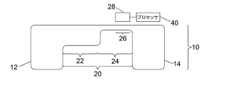
図1Aおよび図1Bを参照すると、血液試料内の白血球の分類および計数のための装置2が記載されている。装置2は、微小流体的液体処理構造10を含む。図1Aおよび1Bは、微小流体的液体処理構造10を含む、装置2の相互に垂直な断面を示す。図1Cは、装置2を上方から見た図を示す。
[Device structure and operation]
With reference to FIGS. 1A and 1B, an
図1Cを参照すると、微小流体的液体処理構造10は、以下の主な要素、すなわち装填チャンバ12、接続導管20、および廃棄チャンバ14を含む。装填チャンバ12は、血液試料を微小流体的液体処理構造10に導入することができる試料の入口16を有する。入口16は、封止されたときに、いったん試料が微小流体的液体処理構造10内にくると、試料が出て行くことを防ぐように、封止可能である。廃棄チャンバ14および装填チャンバ12は、チャンバが大気に開放されるようにするため、通気孔17、18を有する。これによって、血液試料は、毛細管作用により、装填チャンバ12から導管20を通って廃棄チャンバ14に流れることができる。微小流体的液体処理構造は、画像取込装置28に対してカートリッジを保持するための保持構造の中に挿入できる、カートリッジの一部を形成している。いくつかの実施形態で、カートリッジは、微小流体的液体処理構造へ適用するのに適切な体積の血液を計量するためのカスタムの計量器具、例えばピペットを備えていてもよい。
Referring to FIG. 1C, a microfluidic
いくつかの実施形態では、入口16が封止され、微小流体的液体処理構造が、装填チャンバ12と廃棄チャンバ14とを接続する空気回路を含む空気チャネルのネットワークを含むとき、微小流体的液体処理構造10は大気から封止される。空気回路によって、血液試料が装填チャンバ12から流れて廃棄チャンバ14を満たすとき、廃棄チャンバ14から装填チャンバ12に空気が出ることが可能になる。空気回路は、毛細管現象により試料が空気回路に入るのを防止するために、1つ以上の流動障壁バルブを含む。その例として、空気回路の突然の膨張がある。
In some embodiments, the microfluidic liquid treatment is accomplished when the
微小流体的液体処理構造10内の鋭角部は、試料の流れに対する障害を減らし、廃棄チャンバ14が血液試料で充填されている間、接続導管20内に気泡を取込むのを防止することを避けることが好ましい。接続導管20は、準備部22と検出部24という2つの部分を含む。準備部22は、赤血球の溶解および白血球の染色のために構成されている。試料が準備部22を通って移動すると、一連の乾燥試薬32、34、36のパッチに遭遇する。図1Aでは、3つの乾燥試薬のパッチが提供されているが、任意の数(1つまたは複数)のパッチを準備部22内に提供することができることは理解されるであろう。血液試料が1つまたは複数のパッチにわたって流れると、試薬(複数可)は溶解し、血液(blood volume)を経て徐々に拡散し、化学反応を促す。そのような反応の動態は、主に、血流の速度や乾燥試薬のパッチ(複数可)の長さ(複数可)に依存する。準備部22に貯蔵された乾燥試薬の内容物およびそれが血液中どの程度容易に溶解するかということも、動態に影響を与える。乾燥試薬は、例えば、赤血球を選択的に溶解するための溶血剤を含み、染色剤は、白血球と染色剤の示差的染色をするためのものである。ヘマトキシリンおよびエオシン(H&E)染色におけるファミリーの染色剤、ロマーノスキー染色、異染性の染色、またはそれらの任意の組合せを使用することができる。細胞の細胞質および核の粒状性、大きさ、形状のような形態学的特徴に色の情報を組み合わせてから、検討中の亜集団のそれぞれについて、独自のサインのセットを得ることが可能である。試薬を使用することに関するさらなる考察は、参照により本明細書に組み込まれる国際公開第2013135713号パンフレットという出願に見出すことができる。
Sharp edges within the microfluidic
1つの具体的な実施形態では、準備部22は、界面活性剤と溶解剤(それぞれサーフィノールとサポニン)との混合物を含む長さ10mmの第1の反応部位と、染色用混合物を含む10mmの長さの反応部位とを含む2つの一連の連続的な試薬を含有する。この実施形態では、染色用の混合物は、エオシン、メチレンブルーおよび塩基性オレンジ21の混合物を含み、白血球の5つの分類のため示差的な色に導く。リンパ球は青い色に染色し、単球は青/紫色に染色し、好中球は青い色の核と淡黄色の細胞質を示し、好酸球は青い色の核と濃い黄色の顆粒を示し、好塩基球は明るいピンク色に染色する。
In one particular embodiment, the
接続導管20の検出部24は、動作中に細胞を画像化し、計数する場所である。検出部24が、画像取込装置28の視野26と位置合わせされているので、検出部24を流れる血液は、画像取込装置28の視野26を横切って流れる。検出部24の幅の約2/3は、画像取込装置28の視野26内にある。画像取込装置28は、検出部24の上方に配置されている。画像取込装置28は、検出部24にあるときに、溶解および染色された試料を画像化するために、検出部24に流入した血液試料に焦点を合わせるレンズ装置および集束機構を含む。
The
検出部24は、画像取込装置28の視野26を横切って移動する単一層のオブジェクトを収容するように、準備部22よりも浅く構成される。各オブジェクトが計数される機会を増やし、オブジェクトの分類を容易にするために、単一層のオブジェクトが画像取込装置28の視野を横切って移動するのを確実にすることが好ましい。複数の層のオブジェクトが視野内に提供された場合、一部のオブジェクトが完全に視野から遮られる可能性があり、他にも、別のオブジェクトによって部分的に隠される可能性がある。また、単一層のオブジェクトがあると、取込まれるオブジェクト(細胞を含む)の特徴を任意に定めるのが容易になる。準備部22は、検出部24よりも深く、試薬32、34、36の処理を容易にし、その表面積を増大させる。準備部22から検出部24まで深さが減少することから、検出部24までの毛細管現象による試料の流れが促される。さらに、準備部の深さを増加することにより、血球の溶解および染色の均質性が促される。
The
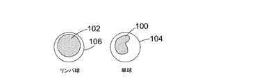
いくつかの実施形態では、検出部24は、検出される任意のオブジェクトの推定最大寸法の少なくとも2倍の幅であり、その深さはこの最大寸法の2倍未満である。一実施形態では、検出部24は、長さ15mm、幅0.06mm、および深さ0.02mmである。この構成により、0.018μLという検出部24の体積が定まる。図2は、そのような検出部でいくつかの染色された白血球37、38、39について得られる画像の種類を概略的に示す。
In some embodiments, the
装置2には、細胞数を得るために必要なデータ処理を実行するように構成されたプロセッサ40が関連付けられている。処理のステップを以下に説明する。
Associated with the
これから装置2の動作について説明する。試料を、入口16から微小流体的液体処理構造10に導入し、その後、入口を封止する。試料は装填チャンバ12に入り、毛細管作用によって準備部22に引き込まれ、続いて検出部24に引き込まれる。したがって、血液試料が検出部24に入ると、それは赤血球溶解剤および白血球用の示差的染色剤とすでに反応している。
The operation of the
画像取込装置28は、最初は焦点が合っていなくてもよいが、試料が検出部24に入り、続いて画像取込装置28の視野に入ると、焦点機構により画像取込装置28が試料に焦点を合わせ、画像取込装置28はビデオのフレームとして画像の取込みを開始する。ソフトウェアおよび/またはハードウェアで実行されるフォーカシング方法は、一般に周知である。装置2と使用するように適合された特定の例は、英国特許第1417170.6号明細書に開示されており、これは参照により本明細書に組み込まれている。1つの具体的な実施形態では、ビデオは毎秒15フレームの速度で4分間撮影され、その結果、各細胞について約15〜30フレームが取込まれる。フレーム取得期間および速度は、その後の分析のため、細胞当たり十分な数のフレームを取込むべく、微小流体的液体処理構造10内の特定の流れに適合される。期間および時間は、状況に応じてデータ収集を最適化するために、当業者が容易に適合させる。アッセイの間、約1500個の細胞が、健常な試料について、一例では画像取込装置28の視野26を横切って移動する。ビデオから得られたデータは、一連のフレーム(約4000)からなり、各フレームは、1つまたは複数の細胞、および細胞のクラスタや血小板などの他のオブジェクトを含む画像を含む。一部のフレームにはオブジェクトがまったく含まれていない場合があり、それらのフレームは棄却される。
The
試料中の細胞を計数するために、細胞(および画像取込装置28の視野を横切って移動する任意の他のオブジェクト)を分類しなければならない。画像取込装置28は、細胞が視野を横切って移動するときにフレームを取込むので、視野26内の異なる位置にある細胞のいくつかの画像が得られる。また、細胞が視野26を横切って移動するとき、細胞は典型的には回転もする。細胞が非等方性である場合、すなわち、すべての方向から同じようには見えない場合、細胞が回転するときに細胞に取込まれるフレームは異なるものになる。いくつかの細胞の種類が非等方性であることが知られており、以下に説明するように、細胞および他のオブジェクトを分類および計数するために、採取されたフレームが異なるという事実を本発明では利用する。また、フレームは、試料を画像化するために使用される不完全な光学系のために、異なっている。
In order to count the cells in the sample, the cells (and any other object moving across the field of view of the image capture device 28) must be sorted. The
上で簡単に紹介した様々な工程で使用される装置を説明したので、そのような工程をここで詳細に説明する。 Having described the apparatus used in the various steps briefly introduced above, such steps will now be described in detail.
[オブジェクトの分類]
フレーム内のオブジェクトを分類するために使用される方法を、図3を参照してこれから説明する。ステップ42において、いくつかの実施形態で閾値処理、例えばOtsuの方法を使用して、各フレームを最初にオブジェクトおよびバックグラウンドピクセルにセグメント化し、ステップ44で、フレーム内のオブジェクトを、画像処理の技術分野で周知のように特定する。ステップ46で、各オブジェクトに当該のオブジェクトおよびフレームに固有の識別子が与えられ、ステップ48で、オブジェクトの識別子およびそれらに関連するオブジェクトの位置のリストが取得される。ステップ50において、オブジェクトの位置を中心とする画像パッチが抽出される。次に、ステップ52において、第1の画像パッチについて、画像パッチが別個のオブジェクト領域にセグメント化される。これらの領域は、概観するとバックグラウンド、細胞の核、細胞質である。
Object classification
The method used to classify the objects in the frame will now be described with reference to FIG. At
いくつかの実施形態では、色彩のRGB値に基づくk−平均クラスタ化法のアルゴリズムが区分化に使用される。各画像パッチのピクセルは、3つのクラスタを定義するためにピクセルのRGB値に対してk−平均(または他の任意の適切なクラスタ化アルゴリズム)を使用してクラスタ化される。これにより、各ピクセルについてクラスタ識別子(クラスタ1、クラスタ2、クラスタ3)のマスクが得られる。最も暗い(例えば平均)ピクセル強度または色彩を有するクラスタは核として標識化され、最も明るい(例えば平均)ピクセル強度または色彩を有するクラスタはバックグラウンドとして標識化され、残りのクラスタは細胞質として標識化される。したがって、区分化アルゴリズムは、各領域の強度または色彩に従って、バックグラウンド、核、および細胞質として特定される3つの領域を出力する。
In some embodiments, a k-means clustering algorithm based on RGB values of color is used for segmentation. The pixels of each image patch are clustered using k-means (or any other suitable clustering algorithm) on the RGB values of the pixels to define three clusters. This gives a mask of cluster identifiers (
いくつかの実施形態では、改良として、k−平均クラスタ化法を再度利用し、以前に核として特定されたピクセルを分割して、これらのピクセルを2つのサブピクセル、つまり核と「細胞質領域2」にクラスタ化する。これは、核と細胞質の境界をより正確に画定するのに役立つ。このステップでは、核として標識化されたピクセルは「核」として残されるか、「細胞質領域2」として再分類される。
In some embodiments, as an improvement, the k-means clustering method is reused to divide the pixels previously identified as nuclei into two subpixels, the nuclei and the “
いくつかの実施形態では、代替的な区分化手法を適用することができ、好都合なことに画像の多様性に対してより堅牢であり得る。このアプローチは、各画像パッチに対して以下のステップを伴う。
1)細胞(暗めのピクセル)とバックグラウンド(明るめのピクセル)を分離するためのk=2におけるk−平均クラスタ化法;
2)バックグラウンドのピクセルをバックグラウンドとマスクに標識化する(事前に定義されている色、例えば緑に設定);
3)核(最も暗い)、細胞質(明るめ)、そして今や、例えば緑のバックグラウンドを得るための、k=3でのk−平均クラスタ化法;
4)細胞質のピクセルを細胞質領域1として標識化し、細胞質のピクセルを所定の色に設定することにより、バックグラウンドに加えて細胞質をマスクする;
5)核のピクセル(最も暗い)、細胞質領域2、および今やマスクされたバックグラウンド(今や以前の細胞質領域1の領域を含む)を得るためのk=3でのk−平均クラスタ化法;
6)核のピクセルを核と標識化し、細胞質領域2のピクセルを細胞質領域2と標識化する。
In some embodiments, alternative segmentation techniques may be applied and may conveniently be more robust to image diversity. This approach involves the following steps for each image patch.
1) k-means clustering method at k=2 to separate cells (dark pixels) and background (light pixels);
2) Label background pixels with background and mask (set to a predefined color, eg green);
3) k-means clustering method at k=3 to obtain nuclei (darkest), cytoplasm (bright), and now eg green background;
4) Mask cytoplasm in addition to background by labeling cytoplasmic pixels as
5) k-means clustering method at k=3 to obtain nuclear pixels (darkest),
6) Label the pixels of the nucleus with the nucleus and the pixels of the
1つの細胞質領域のみがセグメント化される実施形態では、クラスタ化は、核および細胞質のピクセルをそれ相応に標識化してステップ3の後に停止する。 In embodiments where only one cytoplasmic region is segmented, clustering is stopped after step 3 with corresponding labeling of nuclear and cytoplasmic pixels.
いくつかの実施形態では、「細胞質領域2」の領域は、第1のパス(「細胞質領域1」)で特定された細胞質に溶け込み、さらなる分析/細胞の分類のため全体の細胞質として標識化される。他の実施形態では、「細胞質領域1」は細胞質の代表とみなされる。「細胞質領域2」が、細胞の核と細胞質との間の移行領域であることに留意しながら、いくつかの実施形態では、この領域のオブジェクトのパラメータを全体の細胞質に加えてオブジェクトの分類に用いる。
In some embodiments, a region of "
全体として、各オブジェクトは、実施形態に応じて、3つまたは4つの領域、つまり核、バックグラウンド、細胞質、細胞質領域2、または核、バックグラウンド、細胞質に分割される。
Overall, each object is divided into three or four regions, nucleus, background, cytoplasm,
血球を特定するための他のクラスタリングに基づくアルゴリズムの例は、Congcong Zhang et alによる「White Blood Cell Segmentation by Color−Space−Based K−Means Clustering」、Sensors 2014,14,16128−16147; doi:10.3390 / s140916128、およびHerbert Ramoser et alによる「Leukocyte segmentation and classification in blood−smear images」、Proceedings of the 2005 IEEE Engineering in Medicine and Biology 27th Annual Conference,Shanghai,China,September 1−4,2005で見出すことができ、そのすべては、参照により本明細書に組み込まれ、いくつかの実施形態でオブジェクトの区分化に使用することがある。いくつかの実施形態では、クラスタ化などの技術を使用して、監督されたまたは監督されない他の任意の適切な区分化方法が使用され得ることが理解される。具体的には、上記の実施形態のいずれかにおいて、k−平均以外の任意の適切な代替クラスタ化アルゴリズムを使用することがある。
An example of another clustering-based algorithm for identifying blood cells is "White Blood Cell Segmentation by Color-Space-Based K-Means Clustering" by Congcong Zhang et al.,
細胞ではなく、したがって核も細胞質も有しないオブジェクトについては、k−平均アルゴリズムは依然としてオブジェクトを複数の領域に分割するが、これらの領域は意味を持たず、k−平均アルゴリズムの収束は、非細胞のオブジェクトにとっては、細胞のオブジェクトほど良好ではない。非細胞オブジェクトは、以下に説明する分類子で特定される。 For objects that are not cells and thus have neither a nucleus nor a cytoplasm, the k-means algorithm still divides the object into regions, but these regions have no meaning and the convergence of the k-means algorithm is non-cellular. Not as good for cell objects as for cell objects. Non-cellular objects are identified by the classifiers described below.
ステップ54において、個々のオブジェクトのそれぞれについて、各画像パッチから、特定された細胞領域について、オブジェクトのパラメータが抽出される。これらのパラメータには次のものが含まれる:
・関連するピクセルについて平均を求めた画像の彩度とその標準偏差
・関連するピクセルについて平均を求めた強度とその標準偏差
・関連するピクセルについて平均を求めた色彩とその標準偏差
・オブジェクトの真円度
・例えば、

・オブジェクトの領域、および
・画像のフォーカスの質。
In
・Saturation of the image averaged for the related pixels and its standard deviation ・Intensity calculated for the related pixels and its standard deviation ・Color averaged for the related pixels and its standard deviation ・True circle of the object Degrees ・For example,
図4は、オブジェクトが4つの領域に分割される実施形態において、個々のオブジェクトのそれぞれについて抽出され得るオブジェクトのパラメータのリストを示す。 FIG. 4 shows a list of object parameters that can be extracted for each individual object in an embodiment where the object is divided into four regions.
いくつかのパラメータの値、例えば、オブジェクトの真円度とオブジェクトの周長などは、全体としてのオブジェクトのために抽出される。マスクを使用して、各領域を順番に分離するために、他のパラメータの値、例えば強度および色彩、およびそれらの標準偏差が各オブジェクトの領域で抽出される。バックグラウンドの領域の値は較正で使用される。例えば、それらは、視野26の1つの領域で別の領域よりも多くの染色を占めるために使用される。画像のフォーカスの質があまりにも劣悪な場合、例えば所定の閾値未満である場合、その画像は棄却される。
The values of some parameters, such as the roundness of the object and the perimeter of the object, are extracted for the object as a whole. Using the mask, the values of other parameters, such as intensity and color, and their standard deviations, are extracted in each object area to separate each area in turn. The values in the background area are used in the calibration. For example, they are used to occupy more stain in one area of the field of
次いでステップ56で、抽出されたパラメータの値は、分類結果を得るためにロジスティック・モデル・ツリー(Logistic Model Tree)分類子で使用されるが、その議論は、N.Landwehr、M.Hall and E.Frankによる、「Logistic Model Trees」、Machine Learning,vol.59,pages 161−205,2005で見出すことができ、これらは参照により本明細書に組み込まれている。分類子は、標識化されたエキスパートの訓練データを用いて、分類するために使用する前に、訓練される。ロジスティック・モデル・ツリー分類子は、各オブジェクトのクラス所属スコア、例えばオブジェクト各種に属する各オブジェクトの確率を得るために使用される。いくつかの実施形態では、機械学習アルゴリズムのWekaコレクションによって提供されるSimpleLogisticのクラスを利用している(http://weka.sourceforge.net/doc.dev/weka/classifiers/functions/SimpleLogistic.htmlと、http://www.cs.waikato.ac.nz/ml/weka/とを参照されたい。両方とも参照により本明細書に組み込まれている)。当業者には明らかであるように、任意の適切な分類器、例えばSupport Vector Machineを使用することができる。ステップ52〜56は、フレームの各オブジェクトに対して繰り返される。
Then, in
オブジェクトの種類には、白血球の5つの種類、計数される白血球以外のオブジェクトで使用される9つの他のオブジェクトの種類、および「細胞のクラスタ」という分類が含まれる。オブジェクトの種類には、以下のものがある。
・リンパ球
・単球
・好中球
・好塩基球
・好酸球
・「無染色細胞」
・「細胞のクラスタ」
・「バックグラウンドのオブジェクト」
・「大きなデブリ」(例えば、血小板の凝集体、溶解していない染色液)
・「焦点の合っていない細胞」
・「赤血球」
・「有核赤血球」
・「範囲外」
・「その他」
・「クラスタ内の好酸球」
Object types include five types of white blood cells, nine other object types used in non-white blood cell objects to be counted, and a classification of "cell clusters". There are the following types of objects.
・Lymphocytes ・Monocytes ・Neutrophils ・Basophils ・Eosinophils ・"Stainless cells"
・"Cellclusters"
・"Backgroundobjects"
・"Largedebris" (eg platelet aggregates, undissolved stain)
・"Out of focus cells"
・"Red blood cells"
・"Nucleated red blood cells"
·"Out of range"
・"Other"
・"Eosinophils in a cluster"
上記のオブジェクトの種類またはクラスは、大部分が自明である。「範囲外」は、フレーム内に完全には存在していないオブジェクトを示し、そのため、オブジェクトの一部のみが見える。「その他」とは、他の分類の1つに該当しないいかなるデブリをも示す。 Most of the above object types or classes are self-explanatory. "Out of range" refers to objects that are not completely within the frame, so only part of the object is visible. "Other" refers to any debris that does not fall into one of the other categories.
血液試料に非白血球(すなわち、最初の5つ以外)という分類のうちのいずれか1つのオブジェクトがあまりにも多く含まれている場合(例えば、「その他」の種類として分類されたオブジェクトの割合が閾値を超えた場合)、アッセイの結果は棄却してもよい。 If the blood sample contains too many objects in any one of the categories of non-white blood cells (ie, other than the first five) (eg, the percentage of objects classified as “other” type is the threshold The results of the assay may be rejected.
好酸球がすべての白血球に占める割合が非常に少ないので、「クラスタ内の好酸球」という分類は有用である。細胞のクラスタに含まれるそうした好酸球が無視され、計数されない場合、所与のアッセイでは、好酸球は極めてわずかにしか計数されないか、またはまったく計数されない。 The classification of "eosinophils in a cluster" is useful because eosinophils make up a very small percentage of all white blood cells. If such eosinophils contained in a cluster of cells are ignored and not counted, then, in a given assay, eosinophils will be counted very little or not at all.
典型的には、細胞のクラスタは、例えば500ピクセル以上の大きなオブジェクト領域によって特徴付けられる。したがって、オブジェクトの領域は細胞のクラスタを区別する。好酸球には明確な色のサインがあり、そのため、全体としてのクラスタの色の平均(または中央値または他の全体的な尺度)がシフトする。この色のシフトを特定することにより、クラスタ内に好酸球が存在していることが検出される。他のオブジェクトを検出するのとは対照的に、クラスタ内の好酸球を検出することは比較的稀な事象であり、いくつかの実施形態では、クラスタの色のシフトが検出された場合、クラスタには1つの好酸球が含まれると推定される。特定されたクラスタの好酸球を計数するさらなる処理が、他の実施形態で使用される。上記の方法ステップのいずれも、プロセッサの制御によって、またはプロセッサの制御下で実行することができる。 Typically, a cluster of cells is characterized by a large object area, for example 500 pixels or more. Thus, the area of the object distinguishes the cluster of cells. Eosinophils have a distinct color signature, which shifts the average (or median or other overall measure) of cluster color as a whole. By identifying this color shift, the presence of eosinophils within the cluster is detected. Detecting eosinophils within a cluster, as opposed to detecting other objects, is a relatively rare event, and in some embodiments, when a color shift of the cluster is detected, It is estimated that the cluster contains one eosinophil. Further processing of counting eosinophils in the identified clusters is used in other embodiments. Any of the above method steps may be performed under or under the control of the processor.
細胞数を推定する際にクラスタ内に細胞を含めることは、単独で、または他の開示された特徴およびステップと組み合わせて実行することができる。この技術は、他の細胞の種類、例えば好塩基球にも適用することができる。 Inclusion of cells within the cluster in estimating cell number can be performed alone or in combination with other disclosed features and steps. This technique can also be applied to other cell types, such as basophils.
図3を参照して説明した工程は、必要なだけ多くのフレームに対して繰り返される。所与のオブジェクトに対して得られたデータの例を図5に示す。 The process described with reference to FIG. 3 is repeated for as many frames as needed. An example of the data obtained for a given object is shown in FIG.
[分画]
分画を得る方法を、これから図6を参照して説明する。ステップ60において、上述の方法に従って、血液試料を多数のフレームを含むビデオに取込む。ステップ62では、各フレーム内のオブジェクトが特定され、クラス所属スコアが単純ロジスティックモデルを使用して取得される。この工程は、図3を参照して上述されている。ステップ64で、各オブジェクトは、図6を参照してこれから説明するように、できるだけ多くのフレームにわたって追跡される。試料が視野26を横切って流れるとき、試料中の各オブジェクトは、典型的には、1〜2秒間、画像取込装置28の視野26内にとどまる。したがって、所与のオブジェクトは約15〜30フレーム表示される。次いで、オブジェクトは、1つのフレームから次のフレームまで追跡される。この追跡工程を、図7および図8を参照してこれから説明する。
[Fractionation]
The method of obtaining the fraction will now be described with reference to FIG. In
図7を参照すると、上述したように、オブジェクトをフレーム毎に分類する間にオブジェクトの位置のリストを取得してから、ステップ74で、フレーム1のオブジェクトの位置のリストとフレーム2のオブジェクトの位置のリストを比較する。第1のフレームの位置(x、y)の第1のオブジェクト88(図8参照)に対して、位置(x、y)に最も近い位置である第2のフレームのオブジェクト94が特定される。ステップ78では、任意の他のオブジェクトが、予め規定されている「安全距離」に等しい半径である、オブジェクト94に中心を置いた円の中にあるかどうかを判定するためのチェックが実行される。この「安全距離」は、2つのオブジェクトが近過ぎて、1つのフレームから次のフレームまで互いに信頼性を備えて区別することができない距離として定義される。ステップ80において、オブジェクト96がオブジェクト94から安全な距離以内にある場合、オブジェクト88は第2のフレームで特定することができず、オブジェクト88の追跡は停止する。ステップ80において、オブジェクト94から安全な距離以内にはオブジェクトが見つからない場合、オブジェクト94は、前のフレームのオブジェクト88と同じオブジェクトであると特定される。ステップ76〜84は、第1のフレームのすべてのオブジェクトに対して繰り返され、その結果各オブジェクトはフレーム1からフレーム2まで追跡されるか、フレーム2において十分に確実に特定されない場合がある。後者の場合(安全な距離以内でオブジェクトが競合する)、これらのオブジェクトの追跡は停止する。次いで、工程はステップ86で次のフレームに移り、フレーム2と3の位置のリストを比較する。ステップ74〜86は、最後にフレームn−1およびnのオブジェクトの位置のリストが比較されるまで、データセット内のすべてのフレームについて繰り返される。ここで、データセットはn個のフレームからなる。
Referring to FIG. 7, as described above, after the object position list is obtained while classifying the objects by frame, in
いくつかの実施形態では、追跡を改善するためにオブジェクト速度が使用されている。ステップ76で、フレーム1のオブジェクト88に最も近いフレーム2のオブジェクト94を選択するのではなく、フレーム1および1つ以上の前のフレームから判定されたオブジェクト88の速度に基づいて、オブジェクト88の予測位置に最も近いオブジェクトが選択され、安全距離を減らすことが可能になる。なぜなら、より多くの情報が追跡に使用されるからである。いくつかの実施形態では、これは、第2フレームから第3フレームまで、およびその後のフレームのオブジェクトの追跡において実行されるが、第1フレームから第2フレームへの追跡では実行されない。なぜなら、オブジェクトの速度は、1つ超のフレームを分析することで推定されるからである。
In some embodiments, object velocity is used to improve tracking. In
オブジェクトの追跡が画像取込装置28の視野26を離れる前に停止する場合、またはステップ80において「衝突」が検出される場合、任意の所与のオブジェクトが15〜30のフレームで表示されるが、フレームのすべてを使用してオブジェクトを分類することはできないことが理解されるであろう。
If tracking of the object stops before leaving the field of
オブジェクトを追跡する際に、追跡された各オブジェクトの画像パッチのセットが取得される。言い換えれば、図3を参照して上述した方法で得られたオブジェクト(画像パッチ)は、同じオブジェクトに対応する他のオブジェクト(画像パッチ)と「マッチアップ」される。 When tracking an object, a set of image patches for each tracked object is obtained. In other words, the object (image patch) obtained by the method described above with reference to FIG. 3 is “matched up” with another object (image patch) corresponding to the same object.
図6に戻ると、各個々のフレームに関する各々追跡されたオブジェクトのクラス所属スコアを取得した(ステップ62および64)ので、ステップ66で、各細胞の全体的な分類が判定される。これは、以下で説明するいくつかの方法で行うことができる。
Returning to FIG. 6, since the class membership scores of each tracked object for each individual frame have been obtained (
いくつかの実施形態では、クラス所属スコアは、所与のオブジェクトについてすべてのフレームにわたる平均が求められる。最高の点数のクラスは、そのオブジェクトが属するクラス、つまり第1の種類、例えば、「リンパ球」であると受け取られ、「リンパ球」のオブジェクトの種類に対するオブジェクトについての点数は、全フレームにわたる平均が求められる。同じことが「単球」、「好酸球」...などのすべてのオブジェクトの種類で行われる。その場合、オブジェクトは、最高平均点を有する種類に属すると判定される。 In some embodiments, class affiliation scores are averaged over all frames for a given object. The class with the highest score is taken to be the class to which the object belongs, ie the first type, eg "lymphocyte", and the score for the object for the "lymphocyte" object type is the average over all frames. Is required. The same applies to "monocytes" and "eosinophils". . . Is done on all object types such as. In that case, the object is determined to belong to the type with the highest average score.
他の実施形態では、上述の平均化を実行するが、平均化の結果が1つまたは複数の事前設定された事象によって「無効」にされ得るように、ルールが設定される。図9を参照すると、単球が、典型的には、「豆の形の」核100を有するのに対し、リンパ球の核102は、より円形に近い。しかし、ある角度(例えば、核の「豆」の形が特定できない所)から見た単球104は、外観がリンパ球106と類似している可能性がある。例えば、画像取込装置28の視野を横切って移動する単球も回転しているので、単球は、大部分のフレームにおいて、その核の「豆」の形が見えない角度で取込まれ得る。単球が回転するとき、核の「豆」の形が1つまたは2つのフレームで見えることがあるが、単球が回転を続けると、再びリンパ球のように見える場合がある。したがって、そのオブジェクトが実際に単球である場合、12フレームのうち10は、オブジェクトがリンパ球であることを示し得るが、2つだけが単球であることを示している。上記の平均化法を用いた場合、細胞は全体的にリンパ球として誤って分類される可能性がある。しかし、この分類を無効にするようにルールが設定されている場合、例えば、少なくとも1つのフレームが、オブジェクトが単球であることを示している場合に、細胞は単球として正しく分類される。この理由のために、いくつかの実施形態では、少なくとも1つのフレーム(または少なくとも2つの、または少なくとも別の所定の数のフレーム)のオブジェクトの種類が「単球」である細胞のクラス所属スコアが、閾値、例えば90%を超えていると、他のフレームおよび細胞の種類についての点数の値を問わず、細胞は単球であると判定される。
In other embodiments, the averaging described above is performed, but the rules are set such that the results of the averaging can be "invalidated" by one or more preset events. Referring to Figure 9, monocytes typically have "bean-shaped"
他の実施形態では、「投票」工程を採用することができる。この実施形態では、各フレームは票が与えられ、各フレームの票は、そのフレームのクラス所属スコアが最も高いオブジェクトの種類に対応する。次に、オブジェクトは、最も多くの票を有するオブジェクトの種類に属すると判定される。 In other embodiments, a "voting" process can be employed. In this embodiment, each frame is given a vote, and the vote of each frame corresponds to the type of object with the highest class affiliation score for that frame. The object is then determined to belong to the type of object that has the most votes.
さらなる実施形態では、前の実施形態の「投票」システムを採用することができるが、特定のオブジェクトの種類は、無効化する票を有する。少なくとも1つのフレーム、または予め設定されたフレーム数より多い数のフレームが例えば「単球」に投票する場合、そのオブジェクトは、他のフレームからの投票を問わず、全体で単球として分類される。 In a further embodiment, the "voting" system of the previous embodiment can be employed, but certain object types have votes to invalidate. If at least one frame, or more than a preset number of frames, vote for a "monocyte", for example, the object is classified as a whole monocyte, regardless of votes from other frames. ..
さらなる実施形態では、平均化は実行されず、細胞の全体的な分類は、上記のようなルールのみに基づいて判定してもよい。 In a further embodiment, no averaging is performed and the overall classification of cells may be determined based only on the rules as described above.
図10を参照すると、いくつかの実施形態では全体的な分類を判定するために移動平均を計算する。例えば、3つのフレームのウィンドウが、フレームのセットの開始に配置され、クラス所属スコアは、これらの3つのフレームにわたる平均が求められる。次に、ウィンドウが1フレームだけ沿ってシフトされ、次の平均が計算される。この工程は、セット内のすべてのフレームに及ぶまで繰り返される。このような移動平均を計算することは、オブジェクトが画像取込装置28の視野を横切るその動きと比較して比較的ゆっくりと回転する可能性があるため、有利であり得る。言い換えれば、オブジェクトが1つのフレーム内で1つの向きにあり、次のフレームでは突然完全に異なる向きに切り替わる可能性は低い。図9を参照すると、型「A」の特定のオブジェクトに対するクラス所属スコアが領域108で落ちると同時に、型「B」のクラス所属スコアが領域110で上がる。この型のシグネチャは、オブジェクトが画像取込装置28の視野26を横切って移動するときにオブジェクトが回転し、単球の核の「豆」の形状のような、オブジェクトの種類の識別特性が検出可能になるときに生じ得る。したがって、いくつかの実施形態では、上に記したもののような特徴のパターンを使用して、全体的な分類を導出することができる。
Referring to FIG. 10, some embodiments calculate a moving average to determine an overall classification. For example, a window of three frames is placed at the beginning of a set of frames and the class affiliation score is averaged over these three frames. Then the window is shifted along one frame and the next average is calculated. This process is repeated until all the frames in the set have been reached. Calculating such a moving average may be advantageous because the object may rotate relatively slowly compared to its movement across the field of view of the
さらに別の実施形態では、全体的なオブジェクトの分類を判定するために加重平均を使用することができる。各フレームには、フレームの光学的性質およびフレームのセット内のフレームの位置に依存する重みが与えられる。一実施形態では、フレームに与えられる重みは、そのフレームの光学的性質と共に増加し、フレームセット内の中央フレームからのその距離に反比例して変化する。したがって、セット内の最初および最後のフレームは、介在フレームよりも軽い重みが与えられる。 In yet another embodiment, a weighted average can be used to determine the overall object classification. Each frame is given a weight that depends on the optical properties of the frame and the position of the frame within the set of frames. In one embodiment, the weight given to a frame increases with the optical properties of that frame and varies inversely with its distance from the central frame within the frameset. Therefore, the first and last frames in the set are given less weight than the intervening frames.
オブジェクトの全体的な分類を判定するための上記の方法のいずれかの結果が決定的でない場合、そのオブジェクトは「未知」として分類され得る。これは、例えば、所与のオブジェクトのフレームの半分が、そのオブジェクトが1つのオブジェクトの種類に属することを示し、残りの半分がそのオブジェクトが別のオブジェクトの種類に属することを示すときに発生し得る。この場合、オブジェクトがどのクラスに属しているかを確実に判断することができない可能性があるため、オブジェクトは「未知」として分類される。多くのオブジェクトが「未知」として分類されるアッセイは棄却することができ、その結果は信頼性がないとみなされる。これは、例えば、「未知」として分類されたオブジェクトの割合が、予め設定された閾値を超える場合に行われる。 If the result of any of the above methods for determining the overall classification of an object is inconclusive, then the object may be classified as "unknown". This occurs, for example, when half of a given object's frame indicates that the object belongs to one object type and the other half indicates that the object belongs to another object type. obtain. In this case, the object may be classified as "unknown" because it may not be possible to reliably determine which class the object belongs to. Assays in which many objects are classified as "unknown" can be rejected and the results are considered unreliable. This is done, for example, if the proportion of objects classified as "unknown" exceeds a preset threshold.
細胞分画は、各々の細胞の種類として分類された細胞の総数に基づいて計算される。白血球分画を得るために、5つの白血球クラス、すなわちリンパ球、単球、好酸球、好中球、および好塩基球のそれぞれの細胞数を合計することにより、白血球の総数を最初に計算する。所与の細胞の種類の細胞数を、検出された白血球の総数で割ることによって、各クラスが構成する白血球の割合が見出される。白血球分画の典型的な値を以下の表に示す。
いったん細胞各種が占める細胞の割合が分かると、試料の総量が分かっている場合、細胞各種の絶対数を判定することができる。しかし、試料の総体積が正確には分かっていない可能性がある。試料の体積は、接続導管20を通る血流速度、接続導管20の寸法、およびフレーム取得中の流れの時間について知っている事柄に基づいて判定できるが、流速は可変であり、微小流体的液体処理構造の気泡などの要因により、試料の体積を正確に判定することが困難になる。総体積の情報が不足する可能性があることに対応するために、いくつかの実施形態で、絶対的細胞数を得るために異なる方法を用いることがある。これらの方法のいくつかを後述する。まず(すべての種類の)全白血球の絶対数を得る方法に話を移す。
Once the proportion of cells occupied by each cell type is known, the absolute number of each cell type can be determined if the total amount of sample is known. However, the total sample volume may not be known exactly. The sample volume can be determined based on what is known about the blood flow velocity through the connecting
[絶対数]
[絶対数の方法1−統計的分析]
上記のように、判定すべき別の有用な量は絶対的白血球数である。これは、血液の単位体積当たりの推定白血球数である。アッセイで使用される血液試料の全体積は正確には分かっていないが、任意の一時点での画像取込装置28の視野にある血液の体積は、視野の面積と検出部24の深さの積である。この事実は、絶対数の推定に使用される。
[Absolute number]
[Absolute Number Method 1-Statistical Analysis]
As mentioned above, another useful quantity to determine is absolute white blood cell count. This is the estimated white blood cell count per unit volume of blood. Although the total volume of the blood sample used in the assay is not known exactly, the volume of blood in the field of view of the
上記のことは、検出部の深さは、検出部を流れるすべてのオブジェクトを画像取込装置で画像化できるようにするということを意味する。これは、深さが白血球の大きさのオーダである場合、例えば、最大細胞寸法の2倍未満の場合である。検出部がそれよりも深い場合、一部の実施形態では、検出部の深さが、画像取込装置の焦点の深さによって置き換えられ、その全体にわたって、対応する断面を流れるすべてのオブジェクトを画像化することができ、それは任意の一時点で画像取込装置28の視野にある血液の体積を計算することを目的とする。
What has been said above is that the depth of the detector allows all objects flowing in the detector to be imaged by an image capture device. This is the case when the depth is on the order of the size of white blood cells, eg less than twice the maximum cell size. If the detector is deeper than that, in some embodiments, the depth of the detector is replaced by the depth of focus of the image capture device to image all objects flowing through the corresponding cross-section throughout. , Which aims at calculating the volume of blood in the field of view of the
実施形態では、統計的分析を使用して絶対的白血球数を推定する。絶対的白血球数を推定するこの工程を図11を参照して説明する。 In an embodiment, statistical analysis is used to estimate absolute white blood cell count. This process of estimating absolute white blood cell count will be described with reference to FIG.
ステップ112において、血液試料のビデオを撮影する際に得られたフレームのセットの最初の400フレームが棄却される。これは、これらの最初の400フレームには、典型的には、血小板のような、かなりの量のデブリが含まれているからである。
In
ステップ114において、各フレーム内の白血球が特定され、各フレームにおいて計数される。上記の方法で、すでにオブジェクトのクラス所属スコアを取得しているので、5種類の白血球のうちの1つについて最高のクラス所属スコアを有する場合、オブジェクトは白血球として計数される。あるいは、すべての白血球クラス所属スコアを合計し、その合計が非白血球のタイプのオブジェクトの点数の合計よりも大きい場合(または、固定閾値、例えば点数が確率であれば0.5)に分類を行うことによって、オブジェクトは計数目的で白血球として分類される。
In
ステップ116において、残りのフレームのセットはブロックに分割され、各ブロックは約1200フレームを含む。ブロック1は最初の1200個のフレームを含み、ブロック2は2番目の1200個のフレームを含み、以下同様である。ステップ118において、各ブロックについて、パラメータαおよびβを有するガンマ分布が、そのブロック内の各フレームの白血球数の分布(ヒストグラム)に適合される。各適合の適合度の測定値が得られる。次いで、ステップ120において、各ブロック内のフレーム当たりの平均細胞数μは、
![]()
![]()
別の実施形態では、パラメータλ(分布下で見込まれるフレーム当たりの計数に対応する)を有するポアソン分布は、各ブロックのフレームにわたる白血球数の分布(ヒストグラム)に適合され、以前と同様に、適合度の測定値が得られる。各ブロック内のフレーム当たりの平均細胞数は、ポアソン分布のパラメータλとみなされる。 In another embodiment, the Poisson distribution with the parameter λ (corresponding to the counts per frame expected under the distribution) is fitted to the distribution of white blood cell counts (histogram) over the frames of each block, and as before. A degree measurement is obtained. The average number of cells per frame in each block is considered as the Poisson distribution parameter λ.
ステップ122において、ブロックのうちの1つについて判定された平均値が、絶対的白血球数の全体的な推定を判定するために使用されるように選択される。平均値そのものの値と対応する分布の適合度を、使用する値を選択する際に考慮する。開始点として、フレームの第1のブロックの平均値を考慮する。後続のブロックの方が適合度の測定値が高く、そのブロックの平均値が第1のブロックの平均値よりも大幅には低くない(例えば、第1のブロックの値の10%以内)場合、その次のブロックから得た平均値を使用して、絶対的白血球数の全体的な推定値を判定する。例えば、第2のブロックの値が第1のブロックに対応する値と類似している場合、例えば、これはわずかに低いが第2の値の方が適合度の測定値が高い場合、第2のブロックの値が使用される。平均値は、例えば、その値の10%以内であれば、別の値と「類似」していると判定することができる。適合度の測定値がより高くて平均が類似しているこのような後続ブロックが存在しない場合、第1のブロックに由来する平均値を使用する。このようにしてフレームのブロックに適合する分布からの平均細胞数の推定値を得るには、血液試料の流速が実質的に一定であるフレームに分布を適合させることが有効である。流速が実質的に一定であることは、すべてのオブジェクトが概ね同じ数のフレームに現れる可能性が高いことを意味する。逆に、流速が可変であることにより、多くのフレームに遅い動きの細胞が現れ、比較的少数のフレームに速く動く細胞が現れる結果となる。分布が適合されるフレームにわたって流れが実質的に一定である可能性を高めるために、いくつかの実施形態では、閾値を下回る平均オブジェクト速度のフレームを棄却する。例えば、上述のように、オブジェクトは、フレームにわたって追跡され、各フレームのオブジェクト速度の平均が評価される。次いで、オブジェクト速度の平均が所定の閾値を下回るいかなるフレームも棄却し、フレーム当たりの平均白血球数の推定には考慮しない。
In
いくつかの実施形態では、追加または代替で、微小流体的構造の寸法および構成は、血液試料が最初検出部に到達してから一定期間、例えば3秒間が経過した後に、検出部を通る血液試料の流速が実質的に一定と考えられるように設計することができる。次いで、この時間間隔が経過した後のフレームのみを含む、フレーム当たりの平均細胞数を推定する目的で、フレームのブロックを選択することができる。さらに別の実施形態では、流れをモニタし、そうでなければ、流れがあまりに遅いフレーム、または閾値を超える流速において流れの変動があるフレームのブロックを棄却する。 In some embodiments, in addition or in the alternative, the size and configuration of the microfluidic structure is such that the blood sample passes through the detector after the blood sample first reaches the detector after a period of time, eg, 3 seconds. Can be designed so that the flow rate of the is considered to be substantially constant. A block of frames can then be selected for the purpose of estimating the average number of cells per frame, including only the frames after this time interval has elapsed. In yet another embodiment, the flow is monitored and otherwise blocks of frames where the flow is too slow or have flow fluctuations at flow velocity above a threshold are rejected.
1フレーム当たりの平均細胞数の推定値を得た後、絶対的な白血球総数の推定値が得られる。絶対数は、平均(推定)数と、計数に関連する血液の体積との両方に基づく。単一のフレーム内に「取込まれた」血液の体積、すなわちVFOVと呼ばれる画像取込装置28の視野内に含まれる血液の体積は、式(2)
![]()
![]()
いくつかの実施形態では、VFOVの値が上記のように、検出部の寸法に基づいてオンラインで計算される。他の実施形態では、VFOVに関連する情報は、例えば、微小流体的液体処理構造10が設けられているカートリッジの微小流体的液体処理構造に付随する較正データのセットの一部であってもよいし、微小流体的液体処理構造10を含むカートリッジまたはパッケージに取り付けられている装置2により、読取りが可能なタグに付随するものでもよい。いくつかの実施形態では、VFOVの判定の精度は、視野内の任意の障害物のチェックを行うことによって確認される。図12を参照すると、そのような障害物の例は、血小板の凝集体または細胞または気泡であり得る。視野26内に体積VOBSを有するそのような障害物124は、視野に関連する所の体積が血液で完全に満たされてはいないことを意味する。これは、血液試料の体積の過大評価につながり、したがって、絶対的白血球数の過小評価につながるであろう。
In some embodiments, the value of V FOV is calculated online based on the dimensions of the detector, as described above. In other embodiments, the information associated with V FOV may be, for example, part of a set of calibration data associated with the microfluidic liquid handling structure of the cartridge in which microfluidic
この問題を克服するために、いかなるこのような障害物についてもチェックが行われる。これは、視野を領域126に分割することによって行われる。所定の時間内に各領域を通過するオブジェクトの平均数を記録する。任意の領域の平均オブジェクト数が、ある閾値よりも小さい場合、その領域に障害物があると判定される。その後、その領域は、各々が少数のピクセルを含むさらなるサブ領域128に分割される。障害物がある体積の所のピクセルには、通過するオブジェクトがないので、通過するオブジェクトがゼロであるこれらのピクセルを特定することによって、障害物によって占められた視野の領域が判定される。次いで、この領域に検出部24の深さを掛けて、障害物の体積VOBSを得る。ここで、障害物が検出部24の深さ全体に及ぶと仮定する。次いで、視野26に関連付けられた補正された体積VFOV,Cを得るために、視野VFOVに関連付けられた閉塞していない体積から、この体積VOBSを引く。
To overcome this problem, checks are made for any such obstacles. This is done by dividing the field of view into
上記の実施形態は、効率的な測定時間と障害物のある領域の良好な解像度とを組み合わせるために、個々のグリッドの寸法で2つのステップ(最初は粗く、次が細かい)を使用する。他の実施形態では、解像度と処理時間との間のトレードオフとして、粗いステップか細かいステップの1つのステップで、単一のグリッドの大きさが使用される。
視野26に障害物が検出されない場合、VOBS=0および
![]()
![]()
If no obstacle is detected in the field of
![]()
![]()
絶対的白血球数の推定は、次いで図11のステップ130で、単位体積当たりの球数を得るために推定された球数をスケーリングして、例えば、フレーム毎の推定細胞数を、単一のフレーム内に含まれる血液の体積VFOV,Cで割ることによって、判定される。
The absolute white blood cell count is then estimated in
[絶対数の方法2−速度適合型試料採取]
別の実施形態では、二重計数の可能性を低減するために、血液試料のビデオのフレームのサブセット内に含まれる球数の平均を直接求めることによって、血液試料の絶対数が推定されている。これを行う方法について、これから説明する。
[Absolute number method 2-speed-adaptive sampling]
In another embodiment, the absolute number of blood samples is estimated by directly averaging the number of spheres contained within the subset of frames of the blood sample video to reduce the likelihood of double counting. .. The method of doing this will now be described.
血液試料から採取された各フレーム内に、少数の白血球および他のオブジェクト、例えば細胞のクラスタまたは血小板がある。通常、各フレームには1〜10個のオブジェクトがあるが、フレームは20〜30個のオブジェクトを含むことがある。上述のように、画像取込装置28は、1秒当たり約17フレームとなるように設定され、所与のオブジェクトは、画像取込装置28の視野を横切って移動するのに通常1〜2秒かかる。したがって、各オブジェクトは複数のフレームに表示される。
Within each frame taken from a blood sample, there are a few white blood cells and other objects, such as clusters of cells or platelets. Typically, each frame has 1-10 objects, but a frame may contain 20-30 objects. As mentioned above, the
いくつかの実施形態では、絶対的白血球数は、フレームのサブセットを選択することによって判定し、試料中の各オブジェクトがフレームのサブセット全体に一度出現するだけにするか、または少なくとも二重計数の可能性が低減されるようにする。図13を参照すると、オブジェクト132は画像取込装置28の視野26を横切って移動し、フレーム134〜140において取込まれる。フレーム134において、オブジェクト132は、画像取込装置28の視野26にちょうど入ったところであり、フレーム142において、オブジェクト132は、視野からちょうど離れたところである。オブジェクト132の速度は、推定されて、サブセット内のどのフレームがフレーム142に対応するか、すなわち、フレーム134において取込まれたオブジェクトのすべてが視野を離れた可能性が高く、したがって再び現れない一連のフレーム内の最初のフレームを判定するために、使用される。
In some embodiments, the absolute white blood cell count is determined by selecting a subset of frames such that each object in the sample only appears once over the entire subset of frames, or at least double counting possible. Sex is reduced. Referring to FIG. 13, the
具体的には、いくつかの実施形態では、フレームnにおける視野26を横切って移動するすべてのオブジェクト速度のうちの最も速い速度が、サブセットにおける次のフレーム、つまりフレームn+1を選択するために使用されている。
Specifically, in some embodiments, the fastest of all object velocities moving across the field of
ここで、絶対数を推定する工程を、図14を参照して説明する。ステップ144において、フレームmで、フレームmの中で最も速く動く細胞を特定し、その速度を推定する。これは、現在のフレーム、前のフレームまたは後のフレームの2つ以上から得られたオブジェクトの位置に基づいて行われる。ステップ146において、この速度は、すべてのオブジェクトがフレームmを離れた可能性が高い次のフレームを特定するために使用される。この次のフレームは、サブセットにおける次のフレームとして選択される。
Here, the process of estimating the absolute number will be described with reference to FIG. In
フレームmの最も速いオブジェクト速度vについて、フレームのサブセットにおける次のフレーム、つまりフレームm+1は、フレームmとフレームm+1との間の時間間隔を計算することによって判定される。時間間隔tは、以下の式(3)を用いて計算される。
![]()
![]()
いくつかの実施形態では、最も遅く動くオブジェクトの速度が、サブセットにおける次のフレームを選択するために使用される。この場合、式(3)の「w」は、血流の方向に沿った視野の範囲である。他の実施形態では、平均、例えばフレームにおけるすべてのオブジェクトの平均速度または速度の中央値が、サブセットにおける次のフレームを選択するために使用される。この場合、式(3)の「w」は、視野の対角範囲を表す。他の実施形態では、各フレームにおける最も速く動くオブジェクトの速度の平均が推定され、サブセットにおける次のフレームを選択するために使用される。 In some embodiments, the velocity of the slowest moving object is used to select the next frame in the subset. In this case, “w” in equation (3) is the range of the visual field along the direction of blood flow. In other embodiments, an average, eg, the average velocity or median velocity of all objects in a frame, is used to select the next frame in the subset. In this case, “w” in Expression (3) represents the diagonal range of the visual field. In another embodiment, the average velocity of the fastest moving objects in each frame is estimated and used to select the next frame in the subset.
いくつかの実施形態では、例えば、全フレームにわたる平均速度と固定の下位試料採取レートとに基づいて、または対応する可変の下位試料採取レートでフレームの速度プロファイルを使用して、フレームを下位試料採取する方法を決定するために、他の速度の尺度を利用する。同様に、最も速い速度と最も遅い速度を様々な長さのスケール(スケールの方向は対角の方向)と組み合わせることができる。 In some embodiments, a frame is subsampled, for example, based on an average rate over a frame and a fixed subsampling rate, or using a velocity profile of the frame with a corresponding variable subsampling rate. Other speed measures are used to determine how to do so. Similarly, the fastest and slowest speeds can be combined with scales of varying lengths (scale directions are diagonal directions).
ステップ148で、ビデオに取込まれたオブジェクトの大部分または実質的にすべてがフレームのサブセットに現れると判定されるまで、ステップ144〜148は繰り返される。ある実施形態で、カットオフポイント、例えば、サブセットでいかなるさらなるフレームを選択することをも停止するフレーム数が存在し得る。いくつかの実施形態では、フレームの選択は、セットの最後のフレームに達するか、または選択される次のフレームが存在しなくなるまで続く。いくつかの実施形態では、フレーム数4300(1秒当たり17フレーム)を超えるか、または流速が一定の時間で閾値を下回る場合か、または流れが完全に停止した場合は、さらなるフレームが選択されない。
Steps 144-148 are repeated until it is determined in
ステップ150では、各フレームにおける白血球が上記のように特定され(ステップ112)、フレームのサブセットにおけるすべてのフレームにわたって計数される。
In
ステップ152では、サブセットにおけるすべてのフレームにわたる白血球数の平均を求める(または、いくつかの実施形態では、中央値を計算する)。次に、この推定された球数は、ステップ154で、血液の単位体積当たりの推定白血球数を得るためにスケーリングされる。これが絶対的白血球数である。
At
[白血球各種の絶対数]
いくつかの実施形態では、白血球の分画の値および絶対的白血球数の値を組み合わせて、5つの白血球の種類のそれぞれの絶対数を得る。白血球各種の絶対数を得る方法を、これから図15を参照して説明する。ステップ156で、絶対的白血球数が上記方法のいずれかに従って得られる。ステップ158において、白血球各種の分画が上記の方法に従って得られる。ステップ160において、各白血球の絶対数は、それぞれの分画に従った絶対的白血球の総数の割合として得られる。例えば、白血球の種類毎の各分画は、この形式でなくてもすでに、0と1との間の割合として書き換えられており、絶対的白血球数と乗算させて、白血球各種に対する絶対数の推定値が得られる。
[Absolute number of various white blood cells]
In some embodiments, the white blood cell fraction values and the absolute white blood cell count values are combined to obtain absolute numbers for each of the five white blood cell types. A method for obtaining the absolute numbers of various white blood cells will be described with reference to FIG. At
[工程のステップの順序]
上述した方法ステップの様々な組み合わせが実行でき、それらが実行される順序は、球数の推定に影響を及ぼすことなくある程度変更できることが理解されるであろう。
[Order of process steps]
It will be appreciated that various combinations of the method steps described above can be performed and the order in which they are performed can be modified to some extent without affecting the estimation of sphere count.
特に、工程(1)〜(4)の様々な組み合わせを実施することができることが理解されるであろう。一実施形態では、工程(1)および(2)は、血液試料の分画のみを得るために実行される。分画と絶対的白血球数の両方を得るために、工程(1)、(2)および(3)を実施することができる。上記のように、5種類の白血球のそれぞれについての絶対数を得るために、行為(4)において、分画および絶対数を組み合わせることができる。 In particular, it will be appreciated that various combinations of steps (1)-(4) can be performed. In one embodiment, steps (1) and (2) are performed to obtain only a fraction of the blood sample. Steps (1), (2) and (3) can be carried out in order to obtain both the fraction and the absolute white blood cell count. As mentioned above, fractions and absolute numbers can be combined in act (4) to obtain absolute numbers for each of the five types of white blood cells.
また、上述の工程のステップは、必ずしも特定の順序で実行する必要がないことも理解されるであろう。工程のステップは、1つまたは複数の種類の細胞数の1つまたは複数のそれぞれの推定に影響を与えることなく、ある程度再構成し得る。例えば、分画のみが得られる一実施形態では、各フレーム内のオブジェクトの分類結果が取得され、その後、複数のフレームにわたってオブジェクトが追跡される。別のそのような実施形態では、オブジェクトは最初に複数のフレームにわたって追跡され、次に分類結果が得られる。例えば、各オブジェクトの分類結果は、それが追跡されるフレームで得られるのみである。いったんそれらが別のオブジェクトに接近し過ぎると、いくつかのオブジェクトの追跡が停止するので、分画を判定する目的で、追跡される各フレーム内のオブジェクトの分類結果を取得するのみであることは、有用である。そのため、後者のオプション、すなわちオブジェクトを追跡してから分類結果を得ることは、関連するフレーム内のオブジェクトについて分類結果が得られるのみなので、計算能力をより効率的に使用することが可能になる。 It will also be appreciated that the steps of the processes described above need not necessarily be performed in any particular order. The steps of the process may be reconstructed to some extent without affecting one or more respective estimates of one or more cell numbers. For example, in one embodiment where only fractions are obtained, the classification results for the objects within each frame are obtained and then the objects are tracked across multiple frames. In another such embodiment, the objects are first tracked over multiple frames and then the classification results are obtained. For example, the classification result for each object is only available in the frame in which it is tracked. Because they stop tracking some objects once they are too close to another object, it is only possible to get the classification result of the objects in each tracked frame for the purpose of determining the fraction. Useful. Therefore, the latter option, namely tracking objects and then obtaining classification results, allows more efficient use of computing power, since classification results are only obtained for objects in the relevant frames.
同様に、速度適合型試料採取法を使用して絶対的白血球数が判定される実施形態では、サブセット内に1回しか現れない試料において各オブジェクトの可能性が増加するように、フレームのサブセットが選択される。そのため、フレームのサブセット内のオブジェクトの分類結果を取得することが必要であるだけである。したがって、一実施形態では、フレームのサブセットは、各フレーム内のオブジェクトの分類結果が得られる前に選択され、そのため関連するオブジェクトに対して行われるのみである。これにより、計算能力をより効率的に使用することができる。しかし、サブセットを選択する前に、ビデオ全体の各フレーム内のオブジェクトの分類結果を得ることも同様に可能である。分類結果が他の工程でも使用されている場合、これはより効率的である可能性がある。 Similarly, in embodiments in which absolute white blood cell counts are determined using a rate-adaptive sampling method, a subset of frames may be added such that the likelihood of each object increases in samples that appear only once in the subset. To be selected. Therefore, it is only necessary to obtain the classification result of the objects in the subset of frames. Therefore, in one embodiment, the subset of frames is selected before the classification result of the objects in each frame is obtained and is therefore only performed on the relevant objects. This allows more efficient use of computing power. However, it is likewise possible to obtain a classification result of the objects within each frame of the entire video before selecting the subset. This may be more efficient if the classification results are also used in other steps.
各フレーム内のオブジェクトの各々についての分類結果を得ることは、白血球数を判定する統計的方法にとって有用である。この工程では、すべてのオブジェクトについて結果を得ることができ、それが現れるすべてのフレームにおいてオブジェクトを計数することができる。 Obtaining a classification result for each of the objects within each frame is useful for statistical methods of determining white blood cell count. In this step, the result can be obtained for every object and the object can be counted in every frame in which it appears.
また、絶対的白血球数を求めるいずれかの方法を用いると、オブジェクトは「白血球」または「非白血球」に分類する必要があるだけだが、分画を得る方法では、各白血球の種類を判定しなければならない。絶対的白血球数のみを判定する際に、(「白血球」または「非白血球」の)より単純な分類子をいくつかの実施形態で使用することができる。オブジェクトは、そのオブジェクトに関するすべての白血球のクラス所属スコアを合計し、合計が閾値(例えば、クラス所属スコアが確率である場合は0.5)より大きい場合に「白血球」の分類を行うことによって、「白血球」または「非白血球」のいずれかに分類することができる。 Also, using either method to determine the absolute white blood cell count, the object only needs to be classified as “white blood cell” or “non-white blood cell”, but in the method of obtaining the fraction, the type of each white blood cell must be determined. I have to. Simpler classifiers (“white blood cells” or “non-white blood cells”) may be used in some embodiments in determining only absolute white blood cell counts. An object sums the class affiliation scores of all white blood cells for that object, and if the sum is greater than a threshold (eg, 0.5 if the class affiliation score is probability), then classifies “white blood cells” to It can be classified as either "white blood cells" or "non-white blood cells".
追跡されたオブジェクトの全体的な分類を判定するために(分画またはその他の状況において)オブジェクトがフレームにわたって追跡されるいくつかの実施形態では、これらの全体的な分類を絶対数の推定に使用することができる。オブジェクトを追跡し、そのオブジェクトの全体的な分類を判定する際に、追跡する異なるフレームにおいてそのオブジェクトに対応するオブジェクト識別子のリスト(図3のステップ46で割り当てられる)が得られる。これらのオブジェクト識別子は同じオブジェクトに対応するため、全体的な分類は同じである。絶対数を推定することになると、所与のフレーム内の白血球は、分画の状況で上記のように得られたクラス所属スコアを、調べることによって、すなわち追跡することによって、特定することができる。他の実施形態では、オブジェクトは、白血球の種類のいずれか1つの全体的な分類(追跡されたフレームにわたる分類または分類スコアの組み合わせの結果として)を有する場合、白血球として分類され得る。全体的な分類は、追跡されるオブジェクトのクラス所属スコアを見る代わり用いることができるのみである、つまり、全体的な分類が追跡によって判定されたオブジェクトのためだけのものであることが理解されるであろう。可能であるときに全体的な分類を使用することは、複数のフレームから得られたクラス所属スコアを使用して全体的な分類が判定される場合にのみ、クラス所属スコアを見ることよりも正確である可能性がある。
In some embodiments where objects are tracked over a frame (in a fractional or other context) to determine a global classification of tracked objects, these global classifications are used to estimate absolute numbers. can do. In tracking the object and determining the overall classification of the object, a list of object identifiers (assigned in
いくつかの実施形態では、上記の追跡方法を使用して、少なくとも一部の細胞の全体的な分類を判定することができる。次にこれらの分類を使用して、絶対数のみ、分画のみ、またはその両方を推定することができる。 In some embodiments, the tracking methods described above can be used to determine an overall classification of at least some cells. These classifications can then be used to estimate absolute numbers only, fractions only, or both.
[さらなる装置の実施形態]
装置の特定の実施形態について上述したが、様々な実施形態において多くの変形形態が可能であることが理解されるであろう。
[Additional Device Embodiment]
While particular embodiments of the device have been described above, it will be appreciated that many variations in various embodiments are possible.
上記の実施形態では、試料は、毛細管現象によって接続導管20を通って流れる。しかし、接続導管20を通る試料の流れは、圧力差によって、または他の任意の手段によって等しく駆動してもよい。
In the above embodiments, the sample flows through the connecting
上述のような微小流体的液体処理構造は、回転軸の周りを回転するように設計された「ラボ・オン・ディスク」カートリッジ上に実装することができる。いったん試料が微小流体的液体処理構造10に導入されると、試料の入口16を通り、試料の入口が封止され、装置は回転機構を含む器具に配置される。この実施形態では、画像取込装置は、カートリッジが配置される器具と一体であってもよい。回転機構により、カートリッジの角度位置決めを制御することが可能になる。位置決めセンサを使用して、画像取込装置28の視野26と検出部24の精密な位置合わせを補助することができる。また、カートリッジは、個別の位置決めの目的のために固定化するか、ゆっくりと回転させることができ、すべての流体機能は、さらなる干渉/補助なしに、血液試料の細管ベースの処理によって達成し得る。試料中の細胞が上述のような実施形態に従って分類されると、カートリッジは、装置内の同じ試料または別の試料のさらなる処理のために、回転軸を中心に回転させることができる。カートリッジを回転軸の周りで回転させると、カートリッジ内の液体試料は遠心力を受ける。そのようなカートリッジの例は、参照により本明細書に組み込まれる、国際公開第2013135713号パンフレットという出願に見出すことができる。
Microfluidic liquid handling structures such as those described above can be implemented on "lab-on-disc" cartridges designed to rotate about an axis of rotation. Once the sample is introduced into the microfluidic
いくつかの実施形態では、各カートリッジに関連するのは、上記の方法で必要な場合に使用するための、その特定のカートリッジのVFOVの値である。例えば、VFOVは、カートリッジの機械可読要素に記録してもよく、または他の場所に記憶させてもよい。 Associated with each cartridge, in some embodiments, is the value of V FOV for that particular cartridge for use when required by the above method. For example, the V FOV may be recorded on the machine-readable element of the cartridge or may be stored elsewhere.
本明細書に記載された流体構造のすべては、前述の回転軸に対して極座標で設計されてもよい。その結果、すべての構造は、回転軸に対して半径方向および角度方向の次元および位置決めによって特徴付けることができる。 All of the fluid structures described herein may be designed in polar coordinates with respect to the axis of rotation described above. As a result, all structures can be characterized by radial and angular dimensions and positioning with respect to the axis of rotation.
上述のように、いくつかの実施形態では、微小流体的液体処理構造は、カートリッジに設けられる。いくつかの実施形態では、カートリッジ、特に回転機構を使用するカートリッジは、中間の接着層によって一緒にされた2つの透明で平坦な円形の半分によって構成されるCD/DVD構成に類似している。この半分には、好ましくは、上述した微小流体的構造および外部への開口部が刻み込まれる。微小流体的構造の正確な位置合わせにより、3つの部分を組み立てて結合して自己完結型カートリッジを形成することができる。 As mentioned above, in some embodiments, the microfluidic liquid treatment structure is provided in the cartridge. In some embodiments, the cartridge, particularly a cartridge that uses a rotating mechanism, is similar to the CD/DVD configuration constituted by two transparent, flat, circular halves held together by an intermediate adhesive layer. This half is preferably engraved with the microfluidic structure described above and an opening to the outside. The precise alignment of the microfluidic structure allows the three parts to be assembled and joined to form a self-contained cartridge.
準備部と検出部を含む接続導管20の寸法、ならびに装填チャンバ12および廃棄チャンバ14の寸法を調整できることは理解されるであろう。
It will be appreciated that the dimensions of the connecting
上述の実施形態は、血液試料の処理で適合されるが、上記の実施形態の少なくとも一部は、任意の液体試料、例えば、オブジェクトが計数される画像化前に1つ以上の試薬と反応する任意の液体を処理するのに適している。 Although the embodiments described above are adapted for processing blood samples, at least some of the above embodiments react with one or more reagents prior to imaging any liquid sample, eg, object, is counted. Suitable for treating any liquid.
接続導管20の形状および長さは、例えば、蛇行形状を使用して、微小流体的液体処理構造内の省スペースを可能にするように構成してもよい。接続導管20の軌道は、微小流体的液体処理構造の単一平面内に設けてもよい。直線的な接続導管20が図1Aに描かれているが、微小流体的液体処理構造内の血流の実質的に平面の軌道を提供するのに適した任意の形状を提供できることが理解されるであろう。
The shape and length of the connecting
他の既知の試薬およびそれらの組み合わせを装置に使用し得ることも理解されるであろう。試薬は、準備部22において異なる寸法を有するように構成してもよいことも理解されるであろう。
It will also be appreciated that other known reagents and combinations thereof may be used in the device. It will also be appreciated that the reagents may be configured in the
記載された工程は、任意の適切なコンピューティングプラットフォームまたはプロセッサなどの任意の適切なスタンドアロンまたは分散コンピューティング環境を使用して実装することができる。例えば自己完結型またはシステムの他の構成要素と組み合わせた集積回路、システムの他の構成要素と組み合わせた適切なカードを収容する専用のコンピューティング装置、さもなければ、パーソナルコンピュータ、タブレットコンピュータまたは携帯電話などのスタンドアロンコンピューティング装置、またはネットワーク接続を介してデータを少なくとも部分的に遠隔で交換するために必要な工程を実行するサーバが挙げられる。この処理は、クライアント−サーバアーキテクチャで実施されてもよく、処理はクライアントとサーバとの間で任意の適切な方法で分散される。クライアントは、上記の装置のいずれかを含むことができる。 The steps described may be implemented using any suitable standalone or distributed computing environment, such as any suitable computing platform or processor. For example, an integrated circuit that is self-contained or combined with other components of the system, a dedicated computing device containing a suitable card combined with other components of the system, or otherwise a personal computer, tablet computer or cell phone. A stand-alone computing device, or a server that performs the steps necessary to at least partially exchange data remotely over a network connection. This process may be implemented in a client-server architecture, where the process is distributed between the client and server in any suitable way. The client can include any of the above devices.
説明のために特定の実施形態を説明してきたが、特に上述した特定の特徴に加えて、特定の特徴の多くの変形形態、変更形態および並置形態が、当業者には説明を読む際に思い浮かぶであろう。また、それらは本開示の範囲内にある。本発明の範囲は、添付の特許請求の範囲によって規定される。 Although particular embodiments have been described for purposes of explanation, many variations, modifications, and juxtapositions of particular features, in addition to the particular features specifically described above, will occur to those of skill in the art upon reading the description. Will float. Also, they are within the scope of the present disclosure. The scope of the invention is defined by the appended claims.
[第II部]
[装置の構造と動作]
図16Aおよび図16Bを参照すると、血液試料内の白血球の分類および計数のための装置2が記載されている。装置2は、微小流体的液体処理構造10を含む。図16Aおよび図16Bは、微小流体的液体処理構造10を含む、装置2の互いに垂直な断面を示す。図16Cは、装置2を上方から見た図である。
[Part II]
[Device structure and operation]
With reference to Figures 16A and 16B, a
図16Cを参照すると、微小流体的液体処理構造10は、以下の主な要素、すなわち装填チャンバ12、接続導管20、および廃棄チャンバ14を含む。装填チャンバ12は、血液試料を微小流体的液体処理構造10に導入することができる試料の入口16を有する。入口16は、封止されたときに、いったん試料が微小流体的液体処理構造10内にくると、試料が出て行くことを防ぐように、封止可能である。廃棄チャンバ14および装填チャンバ12は、チャンバが大気に開放されるようにするため、通気孔17、18を有する。これにより、血液試料は、毛細管作用によって装填チャンバから導管20を通って廃棄チャンバ14に流れることができる。微小流体的液体処理構造は、画像取込装置28に対してカートリッジを保持するための保持構造の中に挿入できる、カートリッジの一部を形成している。いくつかの実施形態で、カートリッジは、微小流体的液体処理構造へ適用するのに適切な体積の血液を計量するためのカスタムの計量器具、例えばピペットを備えていてもよい。
Referring to FIG. 16C, the microfluidic
いくつかの実施形態では、入口16が封止され、微小流体的液体処理構造が、装填チャンバ12と廃棄チャンバ14とを接続する空気回路を含む空気チャネルのネットワークを含むとき、微小流体的液体処理構造10は大気から封止される。空気回路によって、血液試料が装填チャンバ12から流れて廃棄チャンバ14を満たすとき、廃棄チャンバ14から装填チャンバ12に空気が出ることが可能になる。空気回路は、毛細管現象により試料が空気回路に入るのを防止するために、1つ以上の流動障壁バルブを含む。その例として、空気回路の突然の膨張がある。
In some embodiments, the microfluidic liquid treatment is accomplished when the
微小流体的液体処理構造10内の鋭角部は、試料の流れに対する障害を減らし、廃棄チャンバ14が血液試料で満たされている間、接続導管20内に気泡を取込むのを防止することを避けることが好ましい。接続導管20は、準備部22と検出部24という2つの部分を含む。準備部22は、赤血球の溶解および白血球の染色のために構成されている。試料が準備部22を通って移動すると、一連の乾燥試薬32、34、36のパッチに遭遇する。図16Aでは、3つの乾燥試薬のパッチが提供されているが、任意の数(1つまたは複数)のパッチを準備部22内に提供することができることは理解されるであろう。血液試料が1つまたは複数のパッチにわたって流れると、試薬(複数可)は溶解し、血液(blood volume)を経て徐々に拡散し、化学反応を促す。そのような反応の動態は、主に、血流の速度や乾燥試薬のパッチ(複数可)の長さ(複数可)に依存する。準備部22に貯蔵された乾燥試薬の内容物およびそれが血液中どの程度容易に溶解するかということも、動態に影響を与える。乾燥試薬は、例えば、赤血球を選択的に溶解するための溶血剤、および白血球の示差的染色をするための染色剤を含む。ヘマトキシリンおよびコサイン(H&E)染色におけるファミリーの染色剤、ロマーノスキー染色、異染性の染色、またはそれらの任意の組合せを使用することができる。細胞の細胞質および核の粒状性、大きさ、形状のような形態学的特徴に色の情報を組み合わせてから、検討中の亜集団のそれぞれについて、独自のサインのセットを得ることが可能である。試薬を使用することに関するさらなる考察は、参照により本明細書に組み込まれる国際公開第2013135713号パンフレットという出願に見出すことができる。
Sharp edges within the microfluidic
1つの具体的な実施形態では、準備部22は、界面活性剤と溶解剤(それぞれサーフィノールとサポニン)との混合物を含む長さ10mmの第1の反応部位と、染色用混合物を含む10mmの長さの反応部位とを含む2つの一連の連続的な試薬を含有する。この実施形態では、染色用の混合物は、エオシン、メチレンブルーおよび塩基性オレンジ21の混合物を含み、白血球の5つの分類のため示差的な色に導く。リンパ球は青い色に染色し、単球は青/紫色に染色し、好中球は青い色の核と淡黄色の細胞質を示し、好酸球は青い色の核と濃い黄色の顆粒を示し、好塩基球は明るいピンク色に染色する。
In one particular embodiment, the
接続導管20の検出部24は、動作中に細胞を画像化し、計数する場所である。検出部24が、画像取込装置28の視野26と位置合わせされているので、検出部24を流れる血液は、画像取込装置28の視野26を横切って流れる。検出部24の幅の約2/3は、画像取込装置28の視野26内にある。画像取込装置28は、検出部24の上方に配置されている。画像取込装置28は、検出部24にあるときに、溶解および染色された試料を画像化するために、検出部24に流入した血液試料に焦点を合わせるレンズ装置および集束機構を含む。
The
検出部24は、画像取込装置28の視野26を横切って移動する単一層のオブジェクトを収容するように、準備部22よりも浅く構成される。各オブジェクトが計数される機会を増やし、オブジェクトの分類を容易にするために、単一層のオブジェクトが画像取込装置28の視野を横切って移動するのを確実にすることが好ましい。複数の層のオブジェクトが視野内に提供された場合、一部のオブジェクトが完全に視野から遮られる可能性があり、他にも、別のオブジェクトによって部分的に隠される可能性がある。また、単一層のオブジェクトがあると、取込まれるオブジェクト(細胞を含む)の特徴を任意に定めるのが容易になる。準備部22は、検出部24よりも深く、試薬32、34、36の処理を容易にし、その表面積を増大させる。準備部22から検出部24まで深さが減少することから、準備部22および検出部24までの毛細管現象による試料の流れが促される。さらに、準備部の深さにより、血球の溶解および染色の均質性が促される。
The
いくつかの実施形態では、検出部24は、検出される任意のオブジェクトの推定最大寸法の少なくとも2倍の幅であり、その深さはこの最大寸法の2倍未満である。一実施形態では、検出部24は、長さ15mm、幅0.06mm、および深さ0.02mmである。この構成により、0.018μLという検出部24の体積が定まる。図17は、そのような検出部のいくつかの染色された白血球37、38、39について得られる画像の種類を概略的に示す。
In some embodiments, the
装置2には、細胞数を得るために必要なデータ処理を実行するように構成されたプロセッサ40が関連付けられている。処理のステップを以下に説明する。
Associated with the
これから装置2の動作について説明する。試料を、入口16から微小流体的液体処理構造10に導入し、その後、入口を封止する。試料は装填チャンバ12に入り、毛細管作用によって準備部22に引き込まれ、続いて検出部24に引き込まれる。したがって、血液試料が検出部24に入ると、それは赤血球溶解剤および白血球用の示差的染色剤とすでに反応している。
The operation of the
画像取込装置28は、最初は焦点が合っていなくてもよいが、試料が検出部24に入り、続いて画像取込装置28の視野に入ると、焦点機構により画像取込装置28が試料に焦点を合わせ、画像取込装置28はビデオのフレームとして画像の取込みを開始する。ソフトウェアおよび/またはハードウェアで実行されるフォーカシング方法は、一般に周知である。装置2と使用するように適合された特定の例は、英国特許第1417170.6号明細書に開示されており、これは参照により本明細書に組み込まれている。1つの具体的な実施形態では、ビデオは毎秒15フレームの速度で4分間撮影され、その結果、各細胞について約15〜30フレームが取込まれる。フレーム取得期間および速度は、その後の分析のため、細胞当たり十分な数のフレームを取込むべく、微小流体的液体処理構造10内の特定の流れに適合される。期間および時間は、状況に応じてデータ収集を最適化するために、当業者が容易に適合させる。アッセイの間、約1500個の細胞が、健常な試料について、一例では画像取込装置28の視野26を横切って移動する。ビデオから得られたデータは、一連のフレーム(約4000)からなり、各フレームは、1つまたは複数の細胞、および細胞のクラスタや血小板などの他のオブジェクトを含む画像を含む。一部のフレームにはオブジェクトがまったく含まれていない場合があり、それらのフレームは棄却される。
The
様々な血球計数工程で使用される装置を説明したので、そのような工程をここで詳細に説明する。 Having described the apparatus used in various blood cell counting steps, such steps are now described in detail.
[オブジェクトの分類]
フレーム内のオブジェクトを分類するために使用される方法を、図18を参照してこれから説明する。ステップ42において、いくつかの実施形態で閾値処理、例えばOtsuの方法を使用して、各フレームを最初にオブジェクトおよびバックグラウンドピクセルにセグメント化し、ステップ44で、フレーム内のオブジェクトを、画像処理の技術分野で周知のように特定する。ステップ46で、各オブジェクトに当該のオブジェクトおよびフレームに固有の識別子が与えられ、ステップ48で、オブジェクトの識別子およびそれらに関連するオブジェクトの位置のリストが取得される。ステップ50において、オブジェクトの位置を中心とする画像パッチが抽出される。次に、ステップ52において、第1の画像パッチについて、画像パッチが別個のオブジェクト領域にセグメント化される。これらの領域は、概観するとバックグラウンド、細胞の核、細胞質である。
Object classification
The method used to classify objects in a frame will now be described with reference to FIG. At
いくつかの実施形態では、色彩のRGB値に基づくk−平均クラスタ化法のアルゴリズムが区分化に使用される。各画像パッチのピクセルは、3つのクラスタを定義するためにピクセルのRGB値に対してk−平均(または他の任意の適切なクラスタ化アルゴリズム)を使用してクラスタ化される。これにより、各ピクセルについてクラスタ識別子(クラスタ1、クラスタ2、クラスタ3)のマスクが得られる。最も暗い(例えば平均)ピクセル強度または色彩を有するクラスタは核として標識化され、最も明るい(例えば平均)ピクセル強度または色彩を有するクラスタはバックグラウンドとして標識化され、残りのクラスタは細胞質として標識化される。したがって、区分化アルゴリズムは、各領域の強度または色彩に従って、バックグラウンド、核、および細胞質として特定される3つの領域を出力する。
In some embodiments, a k-means clustering algorithm based on RGB values of color is used for segmentation. The pixels of each image patch are clustered using k-means (or any other suitable clustering algorithm) on the RGB values of the pixels to define three clusters. This gives a mask of cluster identifiers (
いくつかの実施形態では、改良として、k−平均クラスタ化法を再度利用し、以前に核として特定されたピクセルを分割して、これらのピクセルを2つのサブピクセル、つまり核と「細胞質領域2」にクラスタ化する。これは、核と細胞質の境界をより正確に画定するのに役立つ。このステップでは、核として標識化されたピクセルは「核」として残されるか、「細胞質領域2」として再分類される。
In some embodiments, as an improvement, the k-means clustering method is reused to divide the pixels previously identified as nuclei into two subpixels, the nuclei and the “
いくつかの実施形態では、代替的な区分化手法を適用することができ、好都合なことに画像の多様性に対してより堅牢であり得る。このアプローチは、各画像パッチに対して以下のステップを伴う。
1)細胞(暗めのピクセル)とバックグラウンド(明るめのピクセル)を分離するためのk=2におけるk−平均クラスタ化法;
2)バックグラウンドのピクセルをバックグラウンドとマスクに標識化する(所定の色、例えば緑に設定);
3)核(最も暗い)、細胞質(明るめ)、そして今や、例えば緑のバックグラウンドを得るための、k=3でのk−平均クラスタ化法;
4)細胞質のピクセルを細胞質領域1として標識化し、細胞質のピクセルを所定の色に設定することにより、バックグラウンドに加えて細胞質をマスクする;
5)核のピクセル(最も暗い)、細胞質領域2、および今やマスクされたバックグラウンド(今や以前の細胞質領域1の領域を含む)を得るためのk=3でのk−平均クラスタ化法;
6)核のピクセルを核と標識化し、細胞質領域2のピクセルを細胞質領域2と標識化する。
In some embodiments, alternative segmentation techniques may be applied and may conveniently be more robust to image diversity. This approach involves the following steps for each image patch.
1) k-means clustering method at k=2 to separate cells (dark pixels) and background (light pixels);
2) Label background pixels with background and mask (set to a given color, eg green);
3) k-means clustering method at k=3 to obtain nuclei (darkest), cytoplasm (bright), and now eg green background;
4) Mask cytoplasm in addition to background by labeling cytoplasmic pixels as
5) k-means clustering method at k=3 to obtain nuclear pixels (darkest),
6) Label the pixels of the nucleus with the nucleus and the pixels of the
1つの細胞質領域のみセグメント化される実施形態では、クラスタ化は、核および細胞質のピクセルをそれ相応に標識化してステップ3の後に停止する。 In embodiments where only one cytoplasmic region is segmented, clustering stops after step 3 with corresponding labeling of nuclear and cytoplasmic pixels.
いくつかの実施形態では。「細胞質領域2」の領域は、第1のパス(「細胞質領域1」)で特定された細胞質に溶け込み、さらなる分析/細胞の分類のため全体の細胞質として標識化される。他の実施形態では、「細胞質領域1」は細胞質の代表とみなされる。「細胞質領域2」が、細胞の核と細胞質との間の移行領域であることに留意しながら、いくつかの実施形態では、この領域のオブジェクトのパラメータを全体の細胞質に加えてオブジェクトの分類に用いる。
In some embodiments. The region of "
全体として、各オブジェクトは、実施形態に応じて、3つまたは4つの領域、つまり核、バックグラウンド、細胞質、細胞質領域2、または核、バックグラウンド、細胞質に分割される。
Overall, each object is divided into three or four regions, nucleus, background, cytoplasm,
血球を特定するための他のクラスタリングに基づくアルゴリズムの例は、Congcong Zhang et alによる「White Blood Cell Segmentation by Color−Space−Based K−Means Clustering」、Sensors 2014,14,16128−16147; doi:10.3390 / s140916128、およびHerbert Ramoser et alによる「Leukocyte segmentation and classification in blood−smear images」、Proceedings of the 2005 IEEE Engineering in Medicine and Biology 27th Annual Conference,Shanghai,China,September 1−4,2005で見出すことができ、そのすべては、参照により本明細書に組み込まれ、いくつかの実施形態でオブジェクトの区分化に使用することがある。いくつかの実施形態では、クラスタ化などの技術を使用して、監督されたまたは監督されない他の任意の適切な区分化方法が使用され得ることが理解される。具体的には、上記の実施形態のいずれかにおいて、k−平均以外の任意の適切な代替クラスタ化アルゴリズムを使用することがある。
An example of another clustering-based algorithm for identifying blood cells is "White Blood Cell Segmentation by Color-Space-Based K-Means Clustering" by Congcong Zhang et al.,
細胞ではなく、したがって核も細胞質も有しないオブジェクトについては、k−平均アルゴリズムは依然としてオブジェクトを複数の領域に分割するが、これらの領域は意味を持たず、k−平均アルゴリズムの収束は、非細胞のオブジェクトにとっては、細胞のオブジェクトほど良好ではない。非細胞オブジェクトは、以下に説明する分類子で特定される。 For objects that are not cells and thus have neither a nucleus nor a cytoplasm, the k-means algorithm still divides the object into regions, but these regions have no meaning and the convergence of the k-means algorithm is non-cellular. Not as good for cell objects as for cell objects. Non-cellular objects are identified by the classifiers described below.
ステップ54では、オブジェクトのパラメータが、個々のオブジェクトの各々について各画像パッチから抽出される。これらのパラメータには次のものが含まれる:
・関連するピクセルについて平均を求めた画像の彩度とその標準偏差
・関連するピクセルについて平均を求めた強度とその標準偏差
・関連するピクセルについて平均を求めた色彩とその標準偏差
・オブジェクトの真円度
・例えば、
・オブジェクトの領域、および
・画像のフォーカスの質。
In
・Saturation of the image averaged for the related pixels and its standard deviation ・Intensity calculated for the related pixels and its standard deviation ・Color averaged for the related pixels and its standard deviation ・True circle of the object Degrees ・For example,
図19は、個々のオブジェクトのそれぞれについて抽出され得るオブジェクトのパラメータのリストを示す。 FIG. 19 shows a list of object parameters that can be extracted for each individual object.
いくつかのパラメータの値、例えば、オブジェクトの真円度とオブジェクトの周長などは、全体としてのオブジェクトのために抽出される。マスクを使用して、各領域を順番に分離するために、他のパラメータの値、例えば強度および色彩、およびそれらの標準偏差が各オブジェクトの領域で抽出される。バックグラウンドの領域の値は較正で使用される。例えば、それらは、視野26の1つの領域で別の領域よりも多くの染色を占めるために使用される。画像のフォーカスの質があまりにも劣悪な場合、例えば所定の閾値未満である場合、その画像は棄却される。
The values of some parameters, such as the roundness of the object and the perimeter of the object, are extracted for the object as a whole. Using the mask, the values of other parameters, such as intensity and color, and their standard deviations, are extracted in each object area to separate each area in turn. The values in the background area are used in the calibration. For example, they are used to occupy more stain in one area of the field of
次いでステップ56で、抽出されたパラメータの値は、分類結果を得るためにロジスティック・モデル・ツリー分類子で使用されるが、その議論は、N.Landwehr、M.Hall and E.Frankによる、「Logistical Model Trees」、Machine Learning,vol.59,pages 161−205,2005で見出すことができ、これらは参照により本明細書に組み込まれている。ロジスティック・モデル・ツリーは、エキスパートの標識化した訓練データを使用した分類を使用する前に、訓練されている。ロジスティック・モデル・ツリーは、各オブジェクトのクラス所属スコア、例えばオブジェクト各種に属する各オブジェクトの確率を得るために使用される。いくつかの実施形態では、機械学習アルゴリズムのWekaコレクションによって提供されるSimpleLogisticのクラスを利用している(http://weka.sourceforge.net/doc.dev/weka/classifiers/functions/SimpleLogistic.htmlと、http://www.cs.waikato.ac.nz/ml/weka/とを参照されたい。両方とも参照により本明細書に組み込まれている)。当業者には明らかであるように、任意の適切な分類器、例えばSupport Vector Machineを使用することができる。ステップ52〜56は、フレームの各オブジェクトに対して繰り返される。
Then, in
オブジェクトの種類には、白血球の5つの種類、計数される白血球以外のオブジェクトで使用される9つの他のオブジェクトの種類、および「細胞のクラスタ」という分類が含まれる。オブジェクトの種類には、以下のものがある。
・リンパ球
・単球
・好中球
・好塩基球
・好酸球
・「無染色細胞」
・「細胞のクラスタ」
・「バックグラウンドのオブジェクト」
・「大きなデブリ」(例えば、血小板の凝集体、溶解していない染色液)
・「焦点の合っていない細胞」
・「赤血球」
・「有核赤血球」
・「範囲外」
・「その他」
・「クラスタ内の好酸球」
Object types include five types of white blood cells, nine other object types used in non-white blood cell objects to be counted, and a classification of "cell clusters". There are the following types of objects.
・Lymphocytes ・Monocytes ・Neutrophils ・Basophils ・Eosinophils ・"Stainless cells"
・"Cellclusters"
・"Backgroundobjects"
・"Largedebris" (eg platelet aggregates, undissolved stain)
・"Out of focus cells"
・"Red blood cells"
・"Nucleated red blood cells"
·"Out of range"
・"Other"
・"Eosinophils in a cluster"
上記のオブジェクトの種類またはクラスは、大部分が自明である。「範囲外」は、フレーム内に完全には存在していないオブジェクトを示し、そのため、オブジェクトの一部のみが見える。「その他」とは、他の分類の1つに該当しないいかなるデブリをも示す。 Most of the above object types or classes are self-explanatory. "Out of range" refers to objects that are not completely within the frame, so only part of the object is visible. "Other" refers to any debris that does not fall into one of the other categories.
血液試料に非白血球(すなわち、最初の5つ以外)という分類のうちのいずれか1つのオブジェクトがあまりにも多く含まれている場合(例えば、「その他」の種類として分類されたオブジェクトの割合が閾値を超えた場合)、アッセイの結果は棄却してもよい。 If the blood sample contains too many objects in any one of the categories of non-white blood cells (ie, other than the first five) (eg, the percentage of objects classified as “other” type is the threshold The results of the assay may be rejected.
好酸球がすべての白血球に占める割合が非常に少ないので、「クラスタ内の好酸球」という分類は有用である。細胞のクラスタに含まれるそうした好酸球が無視され、計数されない場合、所与のアッセイでは、好酸球は極めてわずかにしか計数されないか、またはまったく計数されない。 The classification of "eosinophils in a cluster" is useful because eosinophils make up a very small percentage of all white blood cells. If such eosinophils contained in a cluster of cells are ignored and not counted, then, in a given assay, eosinophils will be counted very little or not at all.
上記のように、細胞のクラスタは分類子によって特定される。典型的には、細胞のクラスタは、例えば500ピクセル以上の大きなオブジェクト領域によって特徴付けられる。したがって、オブジェクトの領域は細胞のクラスタを区別する。好酸球には明確な色のサインがあり、そのため、全体としてのクラスタの色の平均がシフトする。この色のシフトを特定することにより、クラスタ内に好酸球が存在していることが検出される。他のオブジェクトを検出するのとは対照的に、クラスタ内の好酸球を検出することは比較的稀な事象であり、いくつかの実施形態では、クラスタの色のシフトが検出された場合、クラスタには1つの好酸球が含まれると推定される。特定されたクラスタの好酸球を計数するさらなる処理が、他の実施形態で使用される。上記の方法ステップのいずれも、プロセッサの制御によって、またはプロセッサの制御下で実行することができる。 As mentioned above, clusters of cells are identified by a classifier. Typically, a cluster of cells is characterized by a large object area, for example 500 pixels or more. Thus, the area of the object distinguishes the cluster of cells. Eosinophils have a distinct color signature, which shifts the average cluster color as a whole. By identifying this color shift, the presence of eosinophils within the cluster is detected. Detecting eosinophils within a cluster, as opposed to detecting other objects, is a relatively rare event, and in some embodiments, when a color shift of the cluster is detected, It is estimated that the cluster contains one eosinophil. Further processing of counting eosinophils in the identified clusters is used in other embodiments. Any of the above method steps may be performed under or under the control of the processor.
細胞数を推定する際にクラスタ内に細胞を含めることは、単独で、または他の開示された特徴およびステップと組み合わせて実行することができる。この技術は、他の細胞の種類、例えば好塩基球にも適用することができる。 Inclusion of cells within the cluster in estimating cell number can be performed alone or in combination with other disclosed features and steps. This technique can also be applied to other cell types, such as basophils.
いくつかの実施形態では、オブジェクトは、オブジェクトが最も高いクラス所属スコアを有するクラスとして分類される。いくつかの実施形態では、最も高いクラス所属スコアが閾値未満である場合、そのオブジェクトは「未知」として分類される。 In some embodiments, the object is classified as the class in which the object has the highest class affiliation score. In some embodiments, an object is classified as "unknown" if the highest class affiliation score is below a threshold.
図18を参照して説明した工程は、必要な数のフレームに対して繰り返される。 The process described with reference to FIG. 18 is repeated for the required number of frames.
[細胞の追跡]
上述したように、いくつかの実施形態では、血液試料の絶対数の推定は、細胞速度の推定に基づいて取込まれたフレームのサブセットを選択することによって、判定することができる。細胞速度を推定する1つの方法は、1つのフレームから次のフレームまで細胞を追跡することである。この工程を図20を参照してこれから説明する。
[Cell tracking]
As mentioned above, in some embodiments, an estimate of the absolute number of blood samples can be determined by selecting a subset of captured frames based on the estimation of cell velocities. One way to estimate cell velocity is to track cells from one frame to the next. This step will be described with reference to FIG.
上述したように、オブジェクトを分類する間にオブジェクトの位置のリストを取得してから、ステップ58で、フレーム1のオブジェクトの位置のリストとフレーム2のオブジェクトの位置のリストを比較する。第1のフレームの位置(x、y)の第1のオブジェクト72(図21参照)に対して、位置(x、y)に最も近い位置である第2のフレームのオブジェクト78が特定される。ステップ62では、その他のオブジェクトが、予め規定されている「安全距離」に等しい半径である、オブジェクト78に中心を置いた円の中にあるかどうかを判定するためのチェックが実行される。この「安全距離」は、2つのオブジェクトが近過ぎて、1つのフレームから次のフレームまで互いに信頼性を備えて区別することができない距離として定義される。ステップ64において、オブジェクト80がオブジェクト78から安全な距離以内にある場合、オブジェクト72はフレーム2で特定することができず、オブジェクト72の追跡は停止する。ステップ64において、オブジェクト78から安全な距離以内にオブジェクトが見つからない場合、オブジェクト78は、前のフレームのオブジェクト72と同じオブジェクトであると特定される。ステップ60〜68は、第1のフレームのすべてのオブジェクトに対して繰り返され、その結果各オブジェクトはフレーム1からフレーム2まで追跡されるか、またはフレーム2において十分に確実に特定されない場合がある。後者の場合(安全な距離以内でオブジェクトが競合する)、これらのオブジェクトの追跡は停止する。次いで、工程はステップ70で次のフレームに移り、フレーム2と3の位置のリストを比較する。ステップ58〜70は、最後にフレームn−1およびnのオブジェクトの位置のリストが比較されるまで、データセット内のすべてのフレームについて繰り返される。ここで、データセットはn個のフレームからなる。
As described above, a list of object positions is obtained while classifying the objects, and then, in
いくつかの実施形態では、追跡を改善するためにオブジェクト速度が使用されている。例えば、ステップ60で、フレーム1のオブジェクト72に最も近いフレーム2のオブジェクト78を選択するのではなく、フレーム1および1つ以上の前のフレームから判定されたオブジェクト72の速度に基づいて、オブジェクト72の予測位置に最も近いオブジェクトが選択され、安全距離を減らすことが可能になる。なぜなら、より多くの情報が追跡に使用されるからである。いくつかの実施形態では、これは、第2フレームから第3フレームまで、およびその後のフレームのオブジェクトの追跡において実行されるが、第1フレームから第2フレームへの追跡では実行されない。なぜなら、オブジェクトの速度は、1つ超のフレームを分析することで推定されるからである。
In some embodiments, object velocity is used to improve tracking. For example, instead of selecting the
オブジェクトを追跡する際に、追跡された各オブジェクトの画像パッチのセットが取得される。言い換えれば、図18を参照して上述した方法で得られたオブジェクト(画像パッチ)は、同じオブジェクトに対応する他のオブジェクト(画像パッチ)と「マッチアップ」される。 When tracking an object, a set of image patches for each tracked object is obtained. In other words, the object (image patch) obtained by the method described above with reference to FIG. 18 is “matched up” with another object (image patch) corresponding to the same object.
画像取込装置の視野の寸法は公知であり、これらの寸法に基づいて、各オブジェクトがフレーム間を移動する距離が推定される。フレーム間の時間も公知である。このことと、フレーム間にオブジェクトが移動した距離の推定を使用して、オブジェクトの速度の推定を判定する。 The dimensions of the field of view of the image capture device are known and based on these dimensions, the distance each object travels between frames is estimated. The time between frames is also known. Using this and an estimate of the distance the object has moved between frames, an estimate of the velocity of the object is determined.
[絶対数の方法1−統計的分析]
上述したように、絶対的白血球数は、血液の単位体積当たりの推定白血球数である。アッセイで使用される血液試料の全体積は正確には分かっていないが、任意の一時点での画像取込装置28の視野にある血液の体積は、視野の面積と検出部24の深さの積である。この事実は、絶対数の推定に使用される。
[Absolute Number Method 1-Statistical Analysis]
As described above, the absolute white blood cell count is the estimated white blood cell count per unit volume of blood. Although the total volume of the blood sample used in the assay is not known exactly, the volume of blood in the field of view of the
上記のことは、検出部の深さは、検出部を流れるすべてのオブジェクトを画像取込装置で画像化できるようにするということを意味する。これは、深さが白血球の大きさのオーダである場合、例えば、最大細胞寸法の2倍未満の場合である。検出部がそれよりも深い場合、一部の実施形態では、検出部の深さが、画像取込装置の焦点の深さによって置き換えられ、その全体にわたって、対応する断面を流れるすべてのオブジェクトを画像化することができ、それは任意の一時点で画像取込装置28の視野にある血液の体積を計算することを目的とする。
What has been said above is that the depth of the detector allows all objects flowing in the detector to be imaged by an image capture device. This is the case when the depth is on the order of the size of white blood cells, eg less than twice the maximum cell size. If the detector is deeper than that, in some embodiments, the depth of the detector is replaced by the depth of focus of the image capture device to image all objects flowing through the corresponding cross-section throughout. , Which aims at calculating the volume of blood in the field of view of the
実施形態では、統計的分析を使用して絶対的白血球数を推定する。絶対的白血球数を推定するこの工程を図22を参照して説明する。 In an embodiment, statistical analysis is used to estimate absolute white blood cell count. This step of estimating the absolute white blood cell count will be described with reference to FIG.
ステップ84において、血液試料のビデオを撮影する際に得られたフレームのセットの最初の400フレームが棄却される。これは、これらの最初の400フレームには、典型的には、血小板のような、かなりの量のデブリが含まれているからである。
At
ステップ86において、各フレーム内の白血球が特定され、各フレームにおいて計数される。オブジェクトを分類する際には、上記の方法に従ってオブジェクトのクラス所属スコアを取得する。5種類の白血球のうちの1つについて最高のクラス所属スコアを有する場合、オブジェクトは白血球として計数される。あるいは、すべての白血球クラス所属スコアを合計し、その合計が非白血球のタイプのオブジェクトの点数の合計よりも大きい場合(または、固定閾値、例えば点数が確率であれば0.5)に分類を行うことによって、オブジェクトは計数目的で白血球として分類される。
In
ステップ88において、残りのフレームのセットはブロックに分割され、各ブロックは約1200フレームを含む。ブロック1は最初の1200個のフレームを含み、ブロック2は2番目の1200個のフレームを含み、以下同様である。ステップ90において、各ブロックについて、パラメータαおよびβを有するガンマ分布が、そのブロック内の各フレームの白血球数の分布(ヒストグラム)に適合される。各適合の適合度の測定値が得られる。次いで、ステップ92において、各ブロック内のフレーム当たりの平均細胞数μは、
![]()
![]()
別の実施形態では、パラメータλ(分布下で見込まれるフレーム当たりの計数に対応する)を有するポアソン分布は、各ブロックのフレームにわたる白血球数の分布(ヒストグラム)に適合され、以前と同様に、適合度の測定値が得られる。各ブロック内のフレーム当たりの平均細胞数は、ポアソン分布のパラメータλとみなされる。 In another embodiment, the Poisson distribution with the parameter λ (corresponding to the counts per frame expected under the distribution) is fitted to the distribution of white blood cell counts (histogram) over the frames of each block, and as before. A degree measurement is obtained. The average number of cells per frame in each block is considered as the Poisson distribution parameter λ.
ステップ94において、ブロックのうちの1つについて判定された平均値が、絶対的白血球数の全体的な推定を判定するために使用されるように選択される。平均値そのものの値と対応する分布の適合度を、使用する値を選択する際に考慮する。開始点として、フレームの第1のブロックの平均値を考慮する。後続のブロックの方が適合度の測定値が高く、そのブロックの平均値が第1のブロックの平均値よりも大幅には低くない(例えば、第1のブロックの値の10%以内)場合、その次のブロックから得た平均値を使用して、絶対的白血球数の全体的な推定値を判定する。例えば、第2のブロックの値が第1のブロックに対応する値と類似している場合、例えば、これはわずかに低いが第2の値の方が適合度の測定値が高い場合、第2のブロックの値が使用される。平均値は、例えば、その値の10%以内であれば、別の値と「類似」していると判定することができる。適合度の測定値がより高くて平均が類似しているこのような後続ブロックが存在しない場合、第1のブロックに由来する平均値を使用する。このようにしてフレームのブロックに適合する分布からの平均細胞数の推定値を得るには、血液試料の流速が実質的に一定であるフレームに分布を適合させることが有効である。流速が実質的に一定であることは、すべてのオブジェクトが概ね同じ数のフレームに現れる可能性が高いことを意味する。逆に、流速が可変であることにより、多くのフレームに遅い動きの細胞が現れ、比較的少数のフレームに速く動く細胞が現れる結果となる。分布が適合されるフレームにわたって流れが実質的に一定である可能性を高めるために、いくつかの実施形態では、閾値を下回る平均オブジェクト速度のフレームを棄却する。例えば、上述のように、オブジェクトは、フレームにわたって追跡され、各フレームのオブジェクト速度の平均が評価される。次いで、オブジェクト速度の平均が所定の閾値を下回るいかなるフレームも棄却し、フレーム当たりの平均白血球数の推定には考慮しない。
In
いくつかの実施形態では、追加または代替で、微小流体的構造の寸法および構成は、血液試料が最初検出部に到達してから一定期間、例えば3秒間が経過した後に、検出部を通る血液試料の流速が実質的に一定と考えられるように設計することができる。次いで、この時間間隔が経過した後のフレームのみを含む、フレーム当たりの平均細胞数を推定する目的で、フレームのブロックを選択することができる。さらに別の実施形態では、流れをモニタし、そうでなければ、流れがあまりに遅いフレーム、または閾値を超える流速において流れの変動があるフレームのブロックを棄却する。 In some embodiments, in addition or in the alternative, the size and configuration of the microfluidic structure is such that the blood sample passes through the detector after the blood sample first reaches the detector after a period of time, eg, 3 seconds. Can be designed so that the flow rate of the is considered to be substantially constant. A block of frames can then be selected for the purpose of estimating the average number of cells per frame, including only the frames after this time interval has elapsed. In yet another embodiment, the flow is monitored and otherwise blocks of frames where the flow is too slow or have flow fluctuations at flow velocity above a threshold are rejected.
1フレーム当たりの平均細胞数の推定値を得た後、絶対的な白血球総数の推定値が得られる。絶対数は、平均(推定)数と、計数に関連する血液の体積との両方に基づく。単一のフレーム内に「取込まれた」血液の体積、すなわちVFOVと呼ばれる画像取込装置28の視野内に含まれる血液の体積は、式(2)
![]()
![]()
いくつかの実施形態では、VFOVの値が上記のように、検出部の寸法に基づいてオンラインで計算される。他の実施形態では、VFOVに関連する情報は、例えば、微小流体的液体処理構造が設けられているカートリッジにある微小流体的液体処理構造10に付随する較正データのセットの一部であってもよいし、微小流体的液体処理構造10を含むカートリッジまたはパッケージに取り付けられている装置2により、読取りが可能なタグに付随するものでもよい。いくつかの実施形態では、VFOVの判定の精度は、視野内の任意の障害物のチェックを行うことによって確認される。図23を参照すると、そのような障害物の例は、血小板の凝集体または細胞または気泡であり得る。視野26内の体積VOBSを有するそのような障害物98は、視野に関連する所の体積が血液で完全に満たされてはいないことを意味する。これは、血液試料の体積の過大評価につながり、したがって、絶対的白血球数の過小評価につながるであろう。
In some embodiments, the value of V FOV is calculated online based on the dimensions of the detector, as described above. In other embodiments, the information associated with the V FOV is, for example, part of a set of calibration data associated with the microfluidic
この問題を克服するために、いかなるこのような障害物についてもチェックが行われる。これは、視野を領域100に分割することによって行われる。所定の時間内に各領域を通過するオブジェクトの平均数を記録する。任意の領域の平均オブジェクト数が、ある閾値よりも小さい場合、その領域に障害物があると判定される。その後、その領域は、それぞれが少数のピクセルを含むさらなるサブ領域102に分割される。障害物がある体積の所のピクセルには、通過するオブジェクトがないので、通過するオブジェクトがゼロであるこれらのピクセルを特定することによって、障害物によって占められた視野の領域が判定される。次いで、この領域に検出部24の深さを掛けて、障害物の体積VOBSを得る。ここで、障害物が検出部24の深さ全体に及ぶと仮定する。次いで、視野26に関連付けられた補正された体積VFOV,Cを得るために、視野VFOVに関連付けられた閉塞していない体積から、この体積VOBSを引く。
To overcome this problem, checks are made for any such obstacles. This is done by dividing the field of view into
上記の実施形態は、効率的な測定時間と障害物のある領域の良好な解像度とを組み合わせるために、個々のグリッドの寸法で2つのステップ(最初は粗く、次が細かい)を使用する。他の実施形態では、解像度と処理時間との間のトレードオフとして、粗いステップか細かいステップの1つのステップで、単一のグリッドの大きさが使用される。
視野26に障害物が検出されない場合、VOBS=0および
![]()
![]()
If no obstacle is detected in the field of
![]()
![]()
絶対的白血球数の推定は、次いで図22のステップ96で、単位体積当たりの球数を得るために推定された球数をスケーリングして、例えば、フレーム毎の推定細胞数を、単一のフレーム内に含まれる血液量VFOV,Cで割ることによって、判定される。
The absolute white blood cell count is then estimated in
[絶対数の方法2−速度適合型試料採取]
別の実施形態では、二重計数の可能性を低減するために、血液試料のビデオのフレームのサブセット内に含まれる球数の平均を直接求めることによって、血液試料の絶対数が推定されている。これを行う方法について、これから説明する。
[Absolute number method 2-speed-adaptive sampling]
In another embodiment, the absolute number of blood samples is estimated by directly averaging the number of spheres contained within the subset of frames of the blood sample video to reduce the likelihood of double counting. .. The method of doing this will now be described.
血液試料から採取された各フレーム内に、少数の白血球および他のオブジェクト、例えば細胞のクラスタまたは血小板がある。通常、各フレームには1〜10個のオブジェクトがあるが、フレームは20〜30個のオブジェクトを含むことがある。上述のように、画像取込装置28は、1秒当たり約17フレームとなるように設定され、所与のオブジェクトは、画像取込装置28の視野を横切って移動するのに通常1〜2秒かかる。したがって、各オブジェクトは複数のフレームに表示される。
Within each frame taken from a blood sample, there are a few white blood cells and other objects, such as clusters of cells or platelets. Typically, each frame has 1-10 objects, but a frame may contain 20-30 objects. As mentioned above, the
いくつかの実施形態では、絶対的白血球数は、フレームのサブセットを選択することによって判定し、試料中の各オブジェクトがフレームのサブセット全体に一度出現するだけにするか、または少なくとも二重計数の可能性が低減されるようにする。図24を参照すると、オブジェクト104は画像取込装置28の視野26を横切って移動し、フレーム106〜112において取込まれる。フレーム106において、オブジェクト104は、画像取込装置28の視野26にちょうど入ったところであり、フレーム114において、オブジェクト104は、視野からちょうど離れたところである。オブジェクト104の速度は、推定されて、サブセット内のどのフレームがフレーム114に対応するか、すなわち、フレーム106において取込まれたオブジェクトのすべてが視野を離れた可能性が高く、したがって再び現れない一連のフレーム内の最初のフレームを判定するために、使用される。
In some embodiments, the absolute white blood cell count is determined by selecting a subset of frames such that each object in the sample only appears once over the entire subset of frames, or at least double counting possible. Sex is reduced. Referring to FIG. 24, the
具体的には、いくつかの実施形態では、フレームnにおける視野26を横切って移動するすべてのオブジェクト速度のうちの最も速い速度が、サブセットにおける次のフレーム、つまりフレームn+1を選択するために使用されるが、複数のフレームにわたってオブジェクトを追跡することか、または細胞速度が同じであり、検出部を通る血液試料の速度と等しいと仮定することによって、オブジェクト速度の推定が得られている。
Specifically, in some embodiments, the fastest of all object velocities moving across the field of
ここで、絶対数を推定する工程を、図25を参照して説明する。ステップ116において、フレームmについて、フレームmにおける最も速く動く細胞を特定し、その速度を推定する。これは、現在のフレーム、前のフレームまたは後のフレームの2つ以上から得られたオブジェクトの位置に基づいて行われる。ステップ118において、この速度は、すべてのオブジェクトがフレームmを離れた可能性が高い次のフレームを特定するために使用される。この次のフレームは、サブセットにおける次のフレームとして選択される。
Here, the step of estimating the absolute number will be described with reference to FIG. In
フレームmの最も速いオブジェクト速度vについて、フレームのサブセットにおける次のフレーム、つまりフレームm+1は、フレームmとフレームm+1との間の時間間隔を計算することによって判定される。時間間隔tは、以下の式(3)を用いて計算される。
![]()
![]()
いくつかの実施形態では、最も遅く動くオブジェクトの速度が、サブセットにおける次のフレームを選択するために使用される。この場合、式(3)の「w」は、血流の方向に沿った視野の範囲である。他の実施形態では、平均、例えばフレームにおけるすべてのオブジェクトの平均速度または速度の中央値が、サブセットにおける次のフレームを選択するために使用される。この場合、式(3)の「w」は、視野の対角範囲を表す。他の実施形態では、各フレームにおける最も速く動くオブジェクトの速度の平均が推定され、サブセットにおける次のフレームを選択するために使用される。 In some embodiments, the velocity of the slowest moving object is used to select the next frame in the subset. In this case, “w” in equation (3) is the range of the visual field along the direction of blood flow. In other embodiments, an average, eg, the average velocity or median velocity of all objects in a frame, is used to select the next frame in the subset. In this case, “w” in Expression (3) represents the diagonal range of the visual field. In another embodiment, the average velocity of the fastest moving objects in each frame is estimated and used to select the next frame in the subset.
いくつかの実施形態では、例えば、全フレームにわたる平均速度と固定の下位試料採取レートとに基づいて、または対応する可変の下位試料採取レートでフレームの速度プロファイルを使用して、フレームを下位試料採取する方法を決定するために、他の速度の尺度を利用する。同様に、最も速い速度と最も遅い速度を様々な長さのスケール(スケールの方向は対角の方向)と組み合わせることができる。 In some embodiments, a frame is subsampled, for example, based on an average rate over a frame and a fixed subsampling rate, or using a velocity profile of the frame with a corresponding variable subsampling rate. Other speed measures are used to determine how to do so. Similarly, the fastest and slowest speeds can be combined with scales of varying lengths (scale directions are diagonal directions).
ステップ120で、ビデオに取込まれたオブジェクトの大部分または実質的にすべてがフレームのサブセットに現れると判定されるまで、ステップ116〜120は繰り返される。ある実施形態で、カットオフポイント、例えば、サブセットでいかなるさらなるフレームを選択することをも停止するフレーム数が存在し得る。いくつかの実施形態では、フレームの選択は、セットの最後のフレームに達するか、または選択される次のフレームが存在しなくなるまで続く。いくつかの実施形態では、フレーム数4300(1秒当たり17フレーム)を超えるか、または流速が一定の時間で閾値を下回る場合か、または流れが完全に停止した場合、さらなるフレームは選択されない。
Steps 116-120 are repeated until it is determined in
ステップ122では、各フレームにおける白血球が上記のように特定され、フレームのサブセットにおけるすべてのフレームにわたって計数される。
In
ステップ124では、サブセット内のすべてのフレームにわたり、白血球数の平均を求める(または、いくつかの実施形態では、中央値が計算される)。次に、この推定数は、ステップ126において、血液の単位体積当たりの推定白血球数を得るためにスケーリングされる。これが絶対的白血球数である。
At
[工程のステップの順序]
上述した方法ステップの様々な組み合わせが実行でき、それらが実行される順序は、球数の推定に影響を及ぼすことなくある程度変更できることが理解されるであろう。速度適合型試料採取法を使用して絶対的白血球数が判定される実施形態では、サブセット内に1回しか現れない試料において各オブジェクトの可能性が増加するように、フレームのサブセットが選択される。そのため、フレームのサブセット内のオブジェクトの分類結果を取得することが必要であるだけである。したがって、一実施形態では、フレームのサブセットは、各フレーム内のオブジェクトの分類結果が得られる前に選択され、そのため関連するオブジェクトに対して行われるのみである。これにより、計算能力をより効率的に使用することができる。しかし、サブセットを選択する前に、ビデオ全体の各フレーム内のオブジェクトの分類結果を得ることも同様に可能である。分類結果が他の工程でも使用されている場合、これはより効率的である可能性がある。
[Order of process steps]
It will be appreciated that various combinations of the method steps described above can be performed and the order in which they are performed can be modified to some extent without affecting the estimation of sphere count. In an embodiment where the absolute white blood cell count is determined using a rate-adaptive sampling method, a subset of frames is selected to increase the likelihood of each object in a sample that appears only once in the subset. .. Therefore, it is only necessary to obtain the classification result of the objects in the subset of frames. Therefore, in one embodiment, the subset of frames is selected before the classification result of the objects in each frame is obtained and is therefore only performed on the relevant objects. This allows more efficient use of computing power. However, it is likewise possible to obtain a classification result of the objects within each frame of the entire video before selecting the subset. This may be more efficient if the classification results are also used in other steps.
各フレーム内のオブジェクトの各々についての分類結果を得ることは、白血球数を判定する統計的方法にとって有用である。この工程では、すべてのオブジェクトについて結果を得ることができ、それが現れるすべてのフレームにおいてオブジェクトを計数することができる。 Obtaining a classification result for each of the objects within each frame is useful for statistical methods of determining white blood cell count. In this step, the result can be obtained for every object and the object can be counted in every frame in which it appears.
さらに、絶対的白血球数を判定する際に、オブジェクトは「白血球」または「非白血球」と分類することが必要なだけである。したがって、(「白血球」または「非白血球」の)より単純な分類子をいくつかの実施形態で使用することができる。オブジェクトは、そのオブジェクトに関するすべての白血球のクラス所属スコアを合計し、合計が閾値(例えば、クラス所属スコアが確率である場合は0.5)より大きい場合に「白血球」の分類を行うことによって、「白血球」または「非白血球」のいずれかに分類することができる。 Moreover, in determining the absolute white blood cell count, the object only needs to be classified as "white blood cells" or "non-white blood cells". Thus, simpler classifiers (“white blood cells” or “non-white blood cells”) can be used in some embodiments. An object sums the class affiliation scores of all white blood cells for that object, and if the sum is greater than a threshold (eg, 0.5 if the class affiliation score is probability), then classifies “white blood cells” to It can be classified as either "white blood cells" or "non-white blood cells".
[さらなる装置の実施形態]
装置の特定の実施形態について上述したが、様々な実施形態において多くの変形形態が可能であることが理解されるであろう。
[Additional Device Embodiment]
While particular embodiments of the device have been described above, it will be appreciated that many variations in various embodiments are possible.
上記の実施形態では、試料は、毛細管現象によって接続導管20を通って流れる。しかし、接続導管20を通る試料の流れは、圧力差によって、または他の任意の手段によって等しく駆動してもよい。
In the above embodiments, the sample flows through the connecting
上述のような微小流体的液体処理構造は、回転軸の周りを回転するように設計された「ラボ・オン・ディスク」カートリッジ上に実装することができる。いったん試料が微小流体的液体処理構造10に導入されると、試料の入口16を通り、試料の入口が封止され、装置は回転機構を含む器具に配置される。この実施形態では、画像取込装置は、カートリッジが配置される器具と一体であってもよい。回転機構により、カートリッジの角度位置決めを制御することが可能になる。位置決めセンサを使用して、画像取込装置28の視野26と検出部24の精密な位置合わせを補助することができる。また、カートリッジは、個別の位置決めの目的のために固定化するか、ゆっくりと回転させることができ、すべての流体機能は、さらなる干渉/補助なしに、血液試料の細管ベースの処理によって達成し得る。試料中の細胞が上述のような実施形態に従って分類されると、カートリッジは、装置内の同じ試料または別の試料のさらなる処理のために、回転軸を中心に回転させることができる。カートリッジを回転軸の周りで回転させると、カートリッジ内の液体試料は遠心力を受ける。そのようなカートリッジの例は、参照により本明細書に組み込まれる、国際公開第2013135713号パンフレットという出願に見出すことができる。
Microfluidic liquid handling structures such as those described above can be implemented on "lab-on-disc" cartridges designed to rotate about an axis of rotation. Once the sample is introduced into the microfluidic
いくつかの実施形態では、各カートリッジに関連するのは、上記の方法で必要な場合に使用するための、その特定のカートリッジのVFOVの値である。例えば、VFOVは、カートリッジの機械可読要素に記録してもよく、または他の場所に記憶させてもよい。 Associated with each cartridge, in some embodiments, is the value of V FOV for that particular cartridge for use when required by the above method. For example, the V FOV may be recorded on the machine-readable element of the cartridge or may be stored elsewhere.
本明細書に記載された流体構造のすべては、前述の回転軸に対して極座標で設計されてもよい。その結果、すべての構造は、回転軸に対して半径方向および角度方向の次元および位置決めによって特徴付けることができる。 All of the fluid structures described herein may be designed in polar coordinates with respect to the axis of rotation described above. As a result, all structures can be characterized by radial and angular dimensions and positioning with respect to the axis of rotation.
上述のように、いくつかの実施形態では、微小流体的液体処理構造は、カートリッジに設けられる。いくつかの実施形態では、カートリッジ、特に回転機構を使用するカートリッジは、中間の接着層によって一緒にされた2つの透明で平坦な円形の半分によって構成されるCD/DVD構成に類似している。この半分には、好ましくは、上述した微小流体的構造および外部への開口部が刻み込まれる。微小流体的構造の正確な位置合わせにより、3つの部分を組み立てて結合して自己完結型カートリッジを形成することができる。 As mentioned above, in some embodiments, the microfluidic liquid treatment structure is provided in the cartridge. In some embodiments, the cartridge, particularly a cartridge that uses a rotating mechanism, is similar to the CD/DVD configuration constituted by two transparent, flat, circular halves held together by an intermediate adhesive layer. This half is preferably engraved with the microfluidic structure described above and an opening to the outside. The precise alignment of the microfluidic structure allows the three parts to be assembled and joined to form a self-contained cartridge.
準備部と検出部を含む接続導管20の寸法、ならびに装填チャンバ12および廃棄チャンバ14の寸法を調整できることは理解されるであろう。
It will be appreciated that the dimensions of the connecting
上述の実施形態は、血液試料の処理で適合されるが、上記の実施形態の少なくとも一部は、任意の液体試料、例えば、オブジェクトが計数される画像化前に1つ以上の試薬と反応する任意の液体を処理するのに適している。 Although the embodiments described above are adapted for processing blood samples, at least some of the above embodiments react with one or more reagents prior to imaging any liquid sample, eg, object, is counted. Suitable for treating any liquid.
接続導管20の形状および長さは、例えば、蛇行形状を使用して、微小流体的液体処理構造内の省スペースを可能にするように構成してもよい。接続導管20の軌道は、微小流体的液体処理構造の単一平面内に設けてもよい。直線的な接続導管20が図16Aに描かれているが、微小流体的液体処理構造内の血流の実質的に平面の軌道を提供するのに適した任意の形状を提供できることが理解されるであろう。
The shape and length of the connecting
他の既知の試薬およびそれらの組み合わせを装置に使用し得ることも理解されるであろう。試薬は、準備部22において異なる寸法を有するように構成してもよいことも理解されるであろう。
It will also be appreciated that other known reagents and combinations thereof may be used in the device. It will also be appreciated that the reagents may be configured in the
記載された工程は、任意の適切なコンピューティングプラットフォームまたはプロセッサなどの任意の適切なスタンドアロンまたは分散コンピューティング環境を使用して実装することができる。例えば自己完結型またはシステムの他の構成要素と組み合わせた集積回路、システムの他の構成要素と組み合わせた適切なカードを収容する専用のコンピューティング装置、さもなければ、パーソナルコンピュータ、タブレットコンピュータまたは携帯電話などのスタンドアロンコンピューティング装置、またはネットワーク接続を介してデータを少なくとも部分的に遠隔で交換するために必要な工程を実行するサーバが挙げられる。この処理は、クライアント−サーバアーキテクチャで実施されてもよく、処理はクライアントとサーバとの間で任意の適切な方法で分散される。クライアントは、上記の装置のいずれかを含むことができる。 The steps described may be implemented using any suitable standalone or distributed computing environment, such as any suitable computing platform or processor. For example, an integrated circuit that is self-contained or combined with other components of the system, a dedicated computing device containing a suitable card combined with other components of the system, or otherwise a personal computer, tablet computer or cell phone. A stand-alone computing device, or a server that performs the steps necessary to at least partially exchange data remotely over a network connection. This process may be implemented in a client-server architecture, where the process is distributed between the client and server in any suitable way. The client can include any of the above devices.
説明のために特定の実施形態を説明してきたが、特に上述した特定の特徴に加えて、特定の特徴の多くの変形形態、変更形態および並置形態が、当業者には説明を読む際に思い浮かぶであろう。また、それらは本開示の範囲内にある。本発明の範囲は、添付の特許請求の範囲によって規定される。 Although particular embodiments have been described for purposes of explanation, many variations, modifications, and juxtapositions of particular features, in addition to the particular features specifically described above, will occur to those of skill in the art upon reading the description. Will float. Also, they are within the scope of the present disclosure. The scope of the invention is defined by the appended claims.
Claims (30)
前記試料が画像取込装置の視野を流れるときに、前記画像取込装置で前記試料の一連のフレームを取込むこと、
各フレームをオブジェクトおよびバックグラウンドにセグメント化すること、
前記フレームの少なくとも一部にわたって各セグメント化オブジェクトを追跡すること、
各追跡されたオブジェクトおよび前記フレームの少なくとも一部の各々についての分類結果を得るために、前記フレームの少なくとも一部の各々における前記追跡されたオブジェクトを分類すること、および
各追跡されたオブジェクトを複数のオブジェクトの種類のうちの1つとして分類するために、前記フレームの前記少なくとも一部の各々から得られた個々の分類結果に基づいて、各追跡されたオブジェクトの全体的な分類結果を計算することであって前記オブジェクトの種類は1つ以上の細胞の種類を含む計算すること
を含む方法。 A method for classifying blood cells in a blood sample, the method comprising:
Capturing a series of frames of the sample with the image capture device as the sample flows through the field of view of the image capture device;
Segmenting each frame into an object and a background,
Tracking each segmented object over at least a portion of the frame;
Classifying the tracked objects in each of the at least some of the frames to obtain a classification result for each of the tracked objects and at least some of the frames; An overall classification result for each tracked object based on the individual classification results obtained from each of the at least some of the frames to classify it as one of Wherein the object type comprises calculating including one or more cell types.
前記一連のフレームの複数のフレームを選択して各選択されたフレームの特定されたオブジェクトのフレーム細胞数を判定すること、および
前記判定されたフレーム細胞数を組み合わせて前記1つまたは複数の細胞の種類のいずれか1つのオブジェクトの推定数を計算すること
を含む請求項7に記載の方法。 Estimating the number identifies an object of any of the one or more cell types in each frame,
Selecting a plurality of frames of the series of frames to determine a frame cell number of a specified object in each selected frame; and combining the determined frame cell numbers of the one or more cells. The method of claim 7, comprising calculating an estimated number of objects of any one of the types.
前記試料が前記画像取込装置の視野を流れるときに、前記画像取込装置で前記試料の一連のフレームを取込み、
各フレームをオブジェクトおよびバックグラウンドにセグメント化し、
前記フレームの少なくとも一部にわたって各セグメント化オブジェクトを追跡し、
各追跡されたオブジェクトおよび前記フレームの少なくとも一部の各々についての分類結果を得るために、前記フレームの少なくとも一部の各々における前記追跡されたオブジェクトを分類し、
各追跡されたオブジェクトを複数のオブジェクトの種類のうちの1つとして分類するために、フレームの前記少なくとも一部の各々から得られた個々の分類結果に基づいて、各追跡されたオブジェクトの全体的な分類結果を計算するように構成され、
前記オブジェクトの種類は1つ以上の細胞の種類を含む、
システム。 A system for estimating the number of cells in a blood sample comprising an image capture device having a field of view and an image processor, the image processor comprising:
Capturing a series of frames of the sample with the image capture device as the sample flows through the field of view of the image capture device;
Segment each frame into an object and a background,
Tracking each segmented object over at least a portion of the frame,
Classifying the tracked objects in each of the at least some of the frames to obtain a classification result for each of the tracked objects and at least some of the frames;
A global classification for each tracked object based on the individual classification results obtained from each of said at least some of the frames to classify each tracked object as one of a plurality of object types. Is configured to calculate various classification results,
The object type includes one or more cell types,
system.
前記一連のフレームの複数のフレームを選択して各選択されたフレームの特定したオブジェクトのフレーム細胞数を判定すること、および
前記判定されたフレーム細胞数を組み合わせて前記1つまたは複数の細胞の種類のいずれか1つのオブジェクトの推定数を計算すること
を含む請求項19に記載のシステム。 Estimating the number identifies an object of any of the one or more cell types in each frame,
Selecting a plurality of frames of the series of frames to determine the number of frame cells of the specified object in each selected frame, and combining the determined number of frame cells to the one or more cell types 20. The system of claim 19, comprising calculating an estimated number of any one of the objects.
Applications Claiming Priority (5)
| Application Number | Priority Date | Filing Date | Title |
|---|---|---|---|
| GB1417178.9 | 2014-09-29 | ||
| GB201417178A GB201417178D0 (en) | 2014-09-29 | 2014-09-29 | Cell counting |
| PT107931T | 2014-09-29 | ||
| PT10793114 | 2014-09-29 | ||
| PCT/EP2015/072392 WO2016050755A2 (en) | 2014-09-29 | 2015-09-29 | Cell counting |
Publications (3)
| Publication Number | Publication Date |
|---|---|
| JP2017534858A JP2017534858A (en) | 2017-11-24 |
| JP2017534858A5 JP2017534858A5 (en) | 2018-11-08 |
| JP6704390B2 true JP6704390B2 (en) | 2020-06-03 |
Family
ID=54199248
Family Applications (1)
| Application Number | Title | Priority Date | Filing Date |
|---|---|---|---|
| JP2017517029A Active JP6704390B2 (en) | 2014-09-29 | 2015-09-29 | Blood cell count |
Country Status (4)
| Country | Link |
|---|---|
| US (2) | US10684206B2 (en) |
| EP (2) | EP3200917B1 (en) |
| JP (1) | JP6704390B2 (en) |
| WO (1) | WO2016050755A2 (en) |
Families Citing this family (10)
| Publication number | Priority date | Publication date | Assignee | Title |
|---|---|---|---|---|
| EP3440830A1 (en) | 2016-04-06 | 2019-02-13 | Biosurfit, S.A. | Method and system for capturing images of a liquid sample |
| US10788413B2 (en) | 2017-01-05 | 2020-09-29 | Massachusetts Institute Of Technology | Method to distinguish and analyze white blood cells in the presence of red blood cells |
| US10321160B2 (en) * | 2017-07-13 | 2019-06-11 | International Business Machines Corporation | Compressing multiple video files using localized camera meta data |
| AU2018369869B2 (en) * | 2017-11-14 | 2021-04-08 | miDiagnostics NV | Classification of a population of objects by convolutional dictionary learning with class proportion data |
| JP2019100988A (en) * | 2017-12-08 | 2019-06-24 | 株式会社島津製作所 | Particle image analysis device, and particle image analysis method |
| WO2019198094A1 (en) * | 2018-04-09 | 2019-10-17 | Sigtuple Technologies Private Limited | Method and system for estimating total count of blood cells in a blood smear |
| JP2019195304A (en) | 2018-05-10 | 2019-11-14 | 学校法人順天堂 | Image analysis method, device, computer program, and generation method of deep learning algorithm |
| JP7381003B2 (en) * | 2019-04-26 | 2023-11-15 | 学校法人順天堂 | METHODS, APPARATUS AND COMPUTER PROGRAMS TO ASSIST DISEASE ANALYSIS AND METHODS, APPARATUS AND PROGRAMS FOR TRAINING COMPUTER ALGORITHM |
| CN111274949B (en) * | 2020-01-19 | 2023-05-30 | 重庆医科大学附属第一医院 | Blood disease white blood cell scatter diagram similarity analysis method based on structural analysis |
| CA3181222A1 (en) * | 2020-06-03 | 2021-12-09 | Niksa PRALJAK | Classification of blood cells |
Family Cites Families (29)
| Publication number | Priority date | Publication date | Assignee | Title |
|---|---|---|---|---|
| US4075462A (en) * | 1975-01-08 | 1978-02-21 | William Guy Rowe | Particle analyzer apparatus employing light-sensitive electronic detector array |
| JP3111706B2 (en) * | 1992-02-18 | 2000-11-27 | 株式会社日立製作所 | Particle analyzer and particle analysis method |
| US5760950A (en) * | 1996-07-25 | 1998-06-02 | Advanced Scanning, Ltd. | Scanning confocal microscope |
| US6778263B2 (en) * | 2000-08-25 | 2004-08-17 | Amnis Corporation | Methods of calibrating an imaging system using calibration beads |
| JP4994560B2 (en) | 2000-08-25 | 2012-08-08 | アムニス コーポレイション | Speed measurement of small moving objects such as cells |
| JP4568499B2 (en) * | 2002-02-14 | 2010-10-27 | ベリデックス・リミテッド・ライアビリティ・カンパニー | Method and algorithm for cell counting at low cost |
| AU2003256329A1 (en) * | 2002-06-27 | 2004-01-19 | University Of Washington | Use of adhesion molecules as bond stress-enhanced nanoscale binding switches |
| US7517453B2 (en) * | 2003-03-01 | 2009-04-14 | The Trustees Of Boston University | Microvascular network device |
| AU2003902589A0 (en) * | 2003-05-26 | 2003-06-12 | Commonwealth Scientific And Industrial Research Organisation | A method for identifying a subset of components of a system |
| US7295309B1 (en) | 2003-12-09 | 2007-11-13 | United States Of America As Represented By The Administrator Of The National Aeronautics And Space Administration | Microparticle analysis system and method |
| GB0503629D0 (en) | 2005-02-22 | 2005-03-30 | Durand Technology Ltd | Method and apparatus for automated analysis of biological specimen |
| WO2008064423A1 (en) * | 2006-12-01 | 2008-06-05 | The Walter And Eliza Hall Institute Of Medical Research | A cell population system and process |
| DE102007013971B4 (en) | 2007-03-23 | 2011-07-14 | Fraunhofer-Gesellschaft zur Förderung der angewandten Forschung e.V., 80686 | Method and device for determining a cell contour of a cell |
| US8159670B2 (en) * | 2007-11-05 | 2012-04-17 | Abbott Laboratories | Method and apparatus for rapidly counting and identifying biological particles in a flow stream |
| DE102008030874A1 (en) | 2008-06-30 | 2010-01-07 | Fraunhofer-Gesellschaft zur Förderung der angewandten Forschung e.V. | Method and device for determining a contour and a center point of an object |
| JP5438962B2 (en) * | 2008-12-25 | 2014-03-12 | シスメックス株式会社 | Cell image display device |
| US20140152801A1 (en) * | 2009-10-28 | 2014-06-05 | Alentic Microscience Inc. | Detecting and Using Light Representative of a Sample |
| US8774488B2 (en) | 2010-03-11 | 2014-07-08 | Cellscape Corporation | Method and device for identification of nucleated red blood cells from a maternal blood sample |
| WO2012033771A2 (en) * | 2010-09-07 | 2012-03-15 | The Board Of Trustees Of The Leland Stanford Junior University | Medical scoring systems and methods |
| EP2637015A4 (en) * | 2010-11-01 | 2016-09-07 | Kanagawa Kagaku Gijutsu Akad | Cell analyzer |
| JP2013015357A (en) * | 2011-07-01 | 2013-01-24 | Shimadzu Corp | Flow cytometer |
| TW201433296A (en) * | 2013-02-21 | 2014-09-01 | Univ Nat Taiwan | A method for the observation, identification, and detection of blood cells |
| EP2825881B1 (en) | 2012-03-12 | 2018-05-30 | Biosurfit, S.A. | Liquid sample imaging device and method |
| WO2013177339A1 (en) * | 2012-05-22 | 2013-11-28 | New Health Sciences, Inc. | Capillary network devices and methods of use |
| US20140002617A1 (en) | 2012-06-27 | 2014-01-02 | The Board Of Trustees Of The University Of Illinois | Particle tracking system and method |
| US9588035B2 (en) * | 2012-09-13 | 2017-03-07 | The Governing Council Of The University Of Toronto | System and method for fetal and maternal red blood cell counting |
| AU2013353154B2 (en) * | 2012-11-28 | 2016-06-23 | Japan Science And Technology Agency | Cell monitoring device, cell monitoring method and program thereof. |
| US20170209864A1 (en) * | 2013-03-15 | 2017-07-27 | Gpb Scientific, Llc | Methods and systems for processing particles |
| JP6404317B2 (en) | 2013-03-15 | 2018-10-10 | アイリス インターナショナル, インコーポレイテッド | Flow cell system and method for analysis of particles in blood samples |
-
2015
- 2015-09-29 EP EP15770923.9A patent/EP3200917B1/en active Active
- 2015-09-29 EP EP20157751.7A patent/EP3708254A1/en not_active Withdrawn
- 2015-09-29 US US15/515,529 patent/US10684206B2/en active Active
- 2015-09-29 WO PCT/EP2015/072392 patent/WO2016050755A2/en active Application Filing
- 2015-09-29 JP JP2017517029A patent/JP6704390B2/en active Active
-
2018
- 2018-03-29 US US15/940,330 patent/US10684207B2/en active Active
Also Published As
| Publication number | Publication date |
|---|---|
| JP2017534858A (en) | 2017-11-24 |
| EP3708254A1 (en) | 2020-09-16 |
| US20180217043A1 (en) | 2018-08-02 |
| US20170212028A1 (en) | 2017-07-27 |
| US10684207B2 (en) | 2020-06-16 |
| US10684206B2 (en) | 2020-06-16 |
| WO2016050755A3 (en) | 2016-06-09 |
| EP3200917B1 (en) | 2020-02-19 |
| EP3200917A2 (en) | 2017-08-09 |
| WO2016050755A2 (en) | 2016-04-07 |
Similar Documents
| Publication | Publication Date | Title |
|---|---|---|
| JP6704390B2 (en) | Blood cell count | |
| CN105143849B (en) | Automatic dynamic range expansion system and method for particle analysis in blood sample | |
| Savkare et al. | Automatic detection of malaria parasites for estimating parasitemia | |
| Wang et al. | Label-free detection of rare circulating tumor cells by image analysis and machine learning | |
| Hegde et al. | Peripheral blood smear analysis using image processing approach for diagnostic purposes: A review | |
| JP2023532483A (en) | Method and apparatus for detecting circulating abnormal cells | |
| WO2015153691A2 (en) | Computer-implemented methods, computer-readable media, and systems for tracking a plurality of spermatozoa | |
| Lei et al. | Automatic detection and counting of urediniospores of Puccinia striiformis f. sp. tritici using spore traps and image processing | |
| Monteiro et al. | A comparative study between methodologies based on the Hough transform and watershed transform on the blood cell count | |
| CN109844494B (en) | Dynamic focusing system and method | |
| US20230186659A1 (en) | Machine learning models for cell localization and classification learned using repel coding | |
| Ciurte et al. | Automatic detection of circulating tumor cells in darkfield microscopic images of unstained blood using boosting techniques | |
| Lin et al. | Digital pathology and artificial intelligence as the next chapter in diagnostic hematopathology | |
| CN110226083B (en) | Erythrocyte fragment recognition method and device, blood cell analyzer and analysis method | |
| CN112912923A (en) | Distance-based tissue state determination | |
| Soldati et al. | Microfluidic droplets content classification and analysis through convolutional neural networks in a liquid biopsy workflow | |
| EP3789914A1 (en) | Methods and systems for automated assessment of respiratory cytology specimens | |
| Angay et al. | Image‐based modeling and scoring of Howell–Jolly Bodies in human erythrocytes | |
| CN116385334A (en) | High-efficiency YOLO detection method for urinary sediment inspection | |
| Gezahegn et al. | Malaria detection and classification using machine learning algorithms | |
| Ossandon et al. | A computational streak mode cytometry biosensor for rare cell analysis | |
| Tomari et al. | An empirical framework for automatic red blood cell morphology identification and counting | |
| Mukherjee et al. | Application of biomedical image processing in blood cell counting using hough transform | |
| Panpatte et al. | Application of image processing for blood group detection | |
| Dong et al. | ABO blood group detection based on image processing technology |
Legal Events
| Date | Code | Title | Description |
|---|---|---|---|
| A521 | Request for written amendment filed |
Free format text: JAPANESE INTERMEDIATE CODE: A821 Effective date: 20170609 |
|
| A521 | Request for written amendment filed |
Free format text: JAPANESE INTERMEDIATE CODE: A523 Effective date: 20170609 |
|
| A521 | Request for written amendment filed |
Free format text: JAPANESE INTERMEDIATE CODE: A523 Effective date: 20180925 |
|
| A621 | Written request for application examination |
Free format text: JAPANESE INTERMEDIATE CODE: A621 Effective date: 20180925 |
|
| A977 | Report on retrieval |
Free format text: JAPANESE INTERMEDIATE CODE: A971007 Effective date: 20190628 |
|
| A131 | Notification of reasons for refusal |
Free format text: JAPANESE INTERMEDIATE CODE: A131 Effective date: 20190716 |
|
| A601 | Written request for extension of time |
Free format text: JAPANESE INTERMEDIATE CODE: A601 Effective date: 20191015 |
|
| TRDD | Decision of grant or rejection written | ||
| A01 | Written decision to grant a patent or to grant a registration (utility model) |
Free format text: JAPANESE INTERMEDIATE CODE: A01 Effective date: 20200421 |
|
| A61 | First payment of annual fees (during grant procedure) |
Free format text: JAPANESE INTERMEDIATE CODE: A61 Effective date: 20200512 |
|
| R150 | Certificate of patent or registration of utility model |
Ref document number: 6704390 Country of ref document: JP Free format text: JAPANESE INTERMEDIATE CODE: R150 |
|
| R250 | Receipt of annual fees |
Free format text: JAPANESE INTERMEDIATE CODE: R250 |