EP3200917B1 - Cell counting - Google Patents
Cell counting Download PDFInfo
- Publication number
- EP3200917B1 EP3200917B1 EP15770923.9A EP15770923A EP3200917B1 EP 3200917 B1 EP3200917 B1 EP 3200917B1 EP 15770923 A EP15770923 A EP 15770923A EP 3200917 B1 EP3200917 B1 EP 3200917B1
- Authority
- EP
- European Patent Office
- Prior art keywords
- frame
- frames
- field
- view
- cell
- Prior art date
- Legal status (The legal status is an assumption and is not a legal conclusion. Google has not performed a legal analysis and makes no representation as to the accuracy of the status listed.)
- Active
Links
- 238000000034 method Methods 0.000 claims description 42
- 210000004369 blood Anatomy 0.000 claims description 32
- 239000008280 blood Substances 0.000 claims description 32
- 238000000638 solvent extraction Methods 0.000 claims description 2
- 238000004590 computer program Methods 0.000 claims 2
- 210000004027 cell Anatomy 0.000 description 69
- 210000000265 leukocyte Anatomy 0.000 description 35
- 210000000805 cytoplasm Anatomy 0.000 description 32
- 238000001514 detection method Methods 0.000 description 24
- 239000007788 liquid Substances 0.000 description 22
- 210000004940 nucleus Anatomy 0.000 description 19
- 238000002360 preparation method Methods 0.000 description 15
- 230000008569 process Effects 0.000 description 13
- 239000003153 chemical reaction reagent Substances 0.000 description 12
- 210000003979 eosinophil Anatomy 0.000 description 12
- 238000012545 processing Methods 0.000 description 10
- 239000002699 waste material Substances 0.000 description 8
- 238000004820 blood count Methods 0.000 description 7
- 230000011218 segmentation Effects 0.000 description 7
- 210000003743 erythrocyte Anatomy 0.000 description 6
- 238000003064 k means clustering Methods 0.000 description 6
- 238000010186 staining Methods 0.000 description 6
- 238000003556 assay Methods 0.000 description 5
- 239000003795 chemical substances by application Substances 0.000 description 5
- 230000007246 mechanism Effects 0.000 description 5
- 238000005070 sampling Methods 0.000 description 5
- 241000288113 Gallirallus australis Species 0.000 description 4
- 238000004458 analytical method Methods 0.000 description 4
- 238000006243 chemical reaction Methods 0.000 description 4
- 239000000203 mixture Substances 0.000 description 4
- 230000000877 morphologic effect Effects 0.000 description 4
- 238000013459 approach Methods 0.000 description 3
- 210000003651 basophil Anatomy 0.000 description 3
- 230000017531 blood circulation Effects 0.000 description 3
- 238000000684 flow cytometry Methods 0.000 description 3
- 238000010801 machine learning Methods 0.000 description 3
- 239000002245 particle Substances 0.000 description 3
- 239000002356 single layer Substances 0.000 description 3
- WZUVPPKBWHMQCE-UHFFFAOYSA-N Haematoxylin Chemical compound C12=CC(O)=C(O)C=C2CC2(O)C1C1=CC=C(O)C(O)=C1OC2 WZUVPPKBWHMQCE-UHFFFAOYSA-N 0.000 description 2
- 230000009471 action Effects 0.000 description 2
- 239000012298 atmosphere Substances 0.000 description 2
- 210000000601 blood cell Anatomy 0.000 description 2
- 238000013500 data storage Methods 0.000 description 2
- 230000006870 function Effects 0.000 description 2
- 210000004698 lymphocyte Anatomy 0.000 description 2
- 230000002934 lysing effect Effects 0.000 description 2
- 230000002101 lytic effect Effects 0.000 description 2
- 210000001616 monocyte Anatomy 0.000 description 2
- 210000000440 neutrophil Anatomy 0.000 description 2
- 230000009467 reduction Effects 0.000 description 2
- ZOMLUNRKXJYKPD-UHFFFAOYSA-N 1,3,3-trimethyl-2-[2-(2-methylindol-3-ylidene)ethylidene]indole;hydrochloride Chemical compound [Cl-].C1=CC=C2C(C)(C)C(/C=C/C=3C4=CC=CC=C4NC=3C)=[N+](C)C2=C1 ZOMLUNRKXJYKPD-UHFFFAOYSA-N 0.000 description 1
- NECRQCBKTGZNMH-UHFFFAOYSA-N 3,5-dimethylhex-1-yn-3-ol Chemical compound CC(C)CC(C)(O)C#C NECRQCBKTGZNMH-UHFFFAOYSA-N 0.000 description 1
- RBTBFTRPCNLSDE-UHFFFAOYSA-N 3,7-bis(dimethylamino)phenothiazin-5-ium Chemical compound C1=CC(N(C)C)=CC2=[S+]C3=CC(N(C)C)=CC=C3N=C21 RBTBFTRPCNLSDE-UHFFFAOYSA-N 0.000 description 1
- 238000012935 Averaging Methods 0.000 description 1
- 230000003044 adaptive effect Effects 0.000 description 1
- 239000012790 adhesive layer Substances 0.000 description 1
- 230000004075 alteration Effects 0.000 description 1
- 230000004888 barrier function Effects 0.000 description 1
- 210000003855 cell nucleus Anatomy 0.000 description 1
- 239000003086 colorant Substances 0.000 description 1
- 230000009089 cytolysis Effects 0.000 description 1
- 238000013480 data collection Methods 0.000 description 1
- 230000001419 dependent effect Effects 0.000 description 1
- 238000010586 diagram Methods 0.000 description 1
- 238000005315 distribution function Methods 0.000 description 1
- 229940079593 drug Drugs 0.000 description 1
- 239000003814 drug Substances 0.000 description 1
- 230000000694 effects Effects 0.000 description 1
- YQGOJNYOYNNSMM-UHFFFAOYSA-N eosin Chemical compound [Na+].OC(=O)C1=CC=CC=C1C1=C2C=C(Br)C(=O)C(Br)=C2OC2=C(Br)C(O)=C(Br)C=C21 YQGOJNYOYNNSMM-UHFFFAOYSA-N 0.000 description 1
- 239000012530 fluid Substances 0.000 description 1
- 239000008187 granular material Substances 0.000 description 1
- 230000002949 hemolytic effect Effects 0.000 description 1
- 238000003384 imaging method Methods 0.000 description 1
- 238000002372 labelling Methods 0.000 description 1
- 229960000907 methylthioninium chloride Drugs 0.000 description 1
- SYXUBXTYGFJFEH-UHFFFAOYSA-N oat triterpenoid saponin Chemical compound CNC1=CC=CC=C1C(=O)OC1C(C=O)(C)CC2C3(C(O3)CC3C4(CCC5C(C)(CO)C(OC6C(C(O)C(OC7C(C(O)C(O)C(CO)O7)O)CO6)OC6C(C(O)C(O)C(CO)O6)O)CCC53C)C)C4(C)CC(O)C2(C)C1 SYXUBXTYGFJFEH-UHFFFAOYSA-N 0.000 description 1
- 230000003287 optical effect Effects 0.000 description 1
- 238000012552 review Methods 0.000 description 1
- 239000007787 solid Substances 0.000 description 1
- 238000007619 statistical method Methods 0.000 description 1
- 238000012706 support-vector machine Methods 0.000 description 1
- 239000004094 surface-active agent Substances 0.000 description 1
- 238000012549 training Methods 0.000 description 1
- 230000007704 transition Effects 0.000 description 1
Images


Classifications
-
- G—PHYSICS
- G01—MEASURING; TESTING
- G01N—INVESTIGATING OR ANALYSING MATERIALS BY DETERMINING THEIR CHEMICAL OR PHYSICAL PROPERTIES
- G01N15/00—Investigating characteristics of particles; Investigating permeability, pore-volume, or surface-area of porous materials
- G01N15/10—Investigating individual particles
- G01N15/14—Electro-optical investigation, e.g. flow cytometers
- G01N15/1468—Electro-optical investigation, e.g. flow cytometers with spatial resolution of the texture or inner structure of the particle
- G01N15/147—Electro-optical investigation, e.g. flow cytometers with spatial resolution of the texture or inner structure of the particle the analysis being performed on a sample stream
-
- G—PHYSICS
- G01—MEASURING; TESTING
- G01N—INVESTIGATING OR ANALYSING MATERIALS BY DETERMINING THEIR CHEMICAL OR PHYSICAL PROPERTIES
- G01N15/00—Investigating characteristics of particles; Investigating permeability, pore-volume, or surface-area of porous materials
- G01N15/02—Investigating particle size or size distribution
- G01N15/0205—Investigating particle size or size distribution by optical means, e.g. by light scattering, diffraction, holography or imaging
- G01N15/0227—Investigating particle size or size distribution by optical means, e.g. by light scattering, diffraction, holography or imaging using imaging, e.g. a projected image of suspension; using holography
-
- G—PHYSICS
- G01—MEASURING; TESTING
- G01N—INVESTIGATING OR ANALYSING MATERIALS BY DETERMINING THEIR CHEMICAL OR PHYSICAL PROPERTIES
- G01N15/00—Investigating characteristics of particles; Investigating permeability, pore-volume, or surface-area of porous materials
- G01N15/02—Investigating particle size or size distribution
- G01N15/0266—Investigating particle size or size distribution with electrical classification
-
- G—PHYSICS
- G01—MEASURING; TESTING
- G01N—INVESTIGATING OR ANALYSING MATERIALS BY DETERMINING THEIR CHEMICAL OR PHYSICAL PROPERTIES
- G01N15/00—Investigating characteristics of particles; Investigating permeability, pore-volume, or surface-area of porous materials
- G01N15/10—Investigating individual particles
- G01N15/14—Electro-optical investigation, e.g. flow cytometers
- G01N15/1434—Electro-optical investigation, e.g. flow cytometers using an analyser being characterised by its optical arrangement
- G01N15/1436—Electro-optical investigation, e.g. flow cytometers using an analyser being characterised by its optical arrangement the optical arrangement forming an integrated apparatus with the sample container, e.g. a flow cell
-
- G—PHYSICS
- G01—MEASURING; TESTING
- G01N—INVESTIGATING OR ANALYSING MATERIALS BY DETERMINING THEIR CHEMICAL OR PHYSICAL PROPERTIES
- G01N33/00—Investigating or analysing materials by specific methods not covered by groups G01N1/00 - G01N31/00
- G01N33/48—Biological material, e.g. blood, urine; Haemocytometers
- G01N33/483—Physical analysis of biological material
- G01N33/487—Physical analysis of biological material of liquid biological material
- G01N33/49—Blood
- G01N33/4915—Blood using flow cells
-
- G—PHYSICS
- G06—COMPUTING; CALCULATING OR COUNTING
- G06F—ELECTRIC DIGITAL DATA PROCESSING
- G06F18/00—Pattern recognition
- G06F18/20—Analysing
- G06F18/25—Fusion techniques
- G06F18/253—Fusion techniques of extracted features
-
- G—PHYSICS
- G06—COMPUTING; CALCULATING OR COUNTING
- G06V—IMAGE OR VIDEO RECOGNITION OR UNDERSTANDING
- G06V10/00—Arrangements for image or video recognition or understanding
- G06V10/70—Arrangements for image or video recognition or understanding using pattern recognition or machine learning
- G06V10/77—Processing image or video features in feature spaces; using data integration or data reduction, e.g. principal component analysis [PCA] or independent component analysis [ICA] or self-organising maps [SOM]; Blind source separation
- G06V10/80—Fusion, i.e. combining data from various sources at the sensor level, preprocessing level, feature extraction level or classification level
- G06V10/806—Fusion, i.e. combining data from various sources at the sensor level, preprocessing level, feature extraction level or classification level of extracted features
-
- G—PHYSICS
- G06—COMPUTING; CALCULATING OR COUNTING
- G06V—IMAGE OR VIDEO RECOGNITION OR UNDERSTANDING
- G06V20/00—Scenes; Scene-specific elements
- G06V20/60—Type of objects
- G06V20/69—Microscopic objects, e.g. biological cells or cellular parts
- G06V20/693—Acquisition
-
- G—PHYSICS
- G06—COMPUTING; CALCULATING OR COUNTING
- G06V—IMAGE OR VIDEO RECOGNITION OR UNDERSTANDING
- G06V20/00—Scenes; Scene-specific elements
- G06V20/60—Type of objects
- G06V20/69—Microscopic objects, e.g. biological cells or cellular parts
- G06V20/698—Matching; Classification
-
- B—PERFORMING OPERATIONS; TRANSPORTING
- B01—PHYSICAL OR CHEMICAL PROCESSES OR APPARATUS IN GENERAL
- B01L—CHEMICAL OR PHYSICAL LABORATORY APPARATUS FOR GENERAL USE
- B01L3/00—Containers or dishes for laboratory use, e.g. laboratory glassware; Droppers
- B01L3/50—Containers for the purpose of retaining a material to be analysed, e.g. test tubes
- B01L3/502—Containers for the purpose of retaining a material to be analysed, e.g. test tubes with fluid transport, e.g. in multi-compartment structures
- B01L3/5027—Containers for the purpose of retaining a material to be analysed, e.g. test tubes with fluid transport, e.g. in multi-compartment structures by integrated microfluidic structures, i.e. dimensions of channels and chambers are such that surface tension forces are important, e.g. lab-on-a-chip
- B01L3/502761—Containers for the purpose of retaining a material to be analysed, e.g. test tubes with fluid transport, e.g. in multi-compartment structures by integrated microfluidic structures, i.e. dimensions of channels and chambers are such that surface tension forces are important, e.g. lab-on-a-chip specially adapted for handling suspended solids or molecules independently from the bulk fluid flow, e.g. for trapping or sorting beads, for physically stretching molecules
-
- G01N15/1433—
-
- G—PHYSICS
- G01—MEASURING; TESTING
- G01N—INVESTIGATING OR ANALYSING MATERIALS BY DETERMINING THEIR CHEMICAL OR PHYSICAL PROPERTIES
- G01N15/00—Investigating characteristics of particles; Investigating permeability, pore-volume, or surface-area of porous materials
- G01N15/10—Investigating individual particles
- G01N15/14—Electro-optical investigation, e.g. flow cytometers
- G01N15/1484—Electro-optical investigation, e.g. flow cytometers microstructural devices
-
- G—PHYSICS
- G01—MEASURING; TESTING
- G01N—INVESTIGATING OR ANALYSING MATERIALS BY DETERMINING THEIR CHEMICAL OR PHYSICAL PROPERTIES
- G01N15/00—Investigating characteristics of particles; Investigating permeability, pore-volume, or surface-area of porous materials
- G01N15/10—Investigating individual particles
- G01N2015/1006—Investigating individual particles for cytology
-
- G01N2015/1029—
-
- G—PHYSICS
- G01—MEASURING; TESTING
- G01N—INVESTIGATING OR ANALYSING MATERIALS BY DETERMINING THEIR CHEMICAL OR PHYSICAL PROPERTIES
- G01N15/00—Investigating characteristics of particles; Investigating permeability, pore-volume, or surface-area of porous materials
- G01N15/10—Investigating individual particles
- G01N15/14—Electro-optical investigation, e.g. flow cytometers
- G01N2015/1486—Counting the particles
-
- G—PHYSICS
- G01—MEASURING; TESTING
- G01N—INVESTIGATING OR ANALYSING MATERIALS BY DETERMINING THEIR CHEMICAL OR PHYSICAL PROPERTIES
- G01N15/00—Investigating characteristics of particles; Investigating permeability, pore-volume, or surface-area of porous materials
- G01N15/10—Investigating individual particles
- G01N15/14—Electro-optical investigation, e.g. flow cytometers
- G01N2015/1488—Methods for deciding
-
- G—PHYSICS
- G01—MEASURING; TESTING
- G01N—INVESTIGATING OR ANALYSING MATERIALS BY DETERMINING THEIR CHEMICAL OR PHYSICAL PROPERTIES
- G01N35/00—Automatic analysis not limited to methods or materials provided for in any single one of groups G01N1/00 - G01N33/00; Handling materials therefor
- G01N35/00029—Automatic analysis not limited to methods or materials provided for in any single one of groups G01N1/00 - G01N33/00; Handling materials therefor provided with flat sample substrates, e.g. slides
- G01N2035/00099—Characterised by type of test elements
- G01N2035/00158—Elements containing microarrays, i.e. "biochip"
Definitions
- the present invention relates to the counting of cells in a blood sample.
- the counting of cells in a blood sample is an important diagnostic tool.
- flow cytometry An example of a known approach to counting cells in a blood sample is flow cytometry.
- flow cytometry a blood sample flows through a capillary such that cells pass one by one in front of a light source and detection arrangement. The reflected and/or transmitted light is detected and analysed. The characteristics of the detected light depend on characteristics of the cells in the sample and how they are stained and, in this way, cells are classified and counted.
- no morphological information for the cells is obtained using flow cytometry.
- Morphological analysis can be carried out on a blood sample by sandwiching the sample between two slides and investigating the sample under a microscope.
- Automated systems for cell counting are known which use cartridges of pre-prepared slides. Such slides need to be prepared manually for each sample to be analysed. This is a time-consuming process, in spite of the partial automation.
- US4075462 discloses a particle analyser apparatus employing a light-sensitive electronic detector array.
- US5880835 discloses an apparatus for investigating particles in a fluid and a method of operation thereof.
- WO2014/146061 discloses dynamic range extension systems and methods for particle analysis in blood samples.
- identifying cells in each frame includes classifying objects in each frame. Parameters of each object are extracted from the frame and, in some embodiments, the classification result for each object may be obtained using a Simple Logistic Model. In other embodiments, other classifiers, as will be known to a person skilled in the relevant field, may be used. For examples, refer to S.B. Kotsiantis, 'Supervised Machine Learning: A Review of Classification Techniques', Informatica, vol. 31, number 3, October 2007, pages 249-268 . Once each object in a frame has been classified, the number of cells within the frame is counted. As such, a frame cell count is found for each frame.
- Combining the counts may comprise fitting a distribution function to the frame cell counts and computing an estimated cell count based on the fitted distribution.
- the estimated cell count may be derived using one or more parameters of the fitted distribution.
- the fitted distribution may be a Gamma or a Poisson distribution.
- Computing the estimated cell count may, in some embodiments, involve splitting the sequence of frames into subset blocks and computing a respective estimated cell count by fitting a respective distribution to each block.
- One of the estimated respective cell counts can then be used as the result to be stored (or returned) by making a selection based on one or more of the position of the subset block in the sequence, the magnitude of the respective estimated counts and a goodness of fit indicator.
- the goodness of fit indicator for any block is not good enough, i.e. if it is lower than a predetermined threshold, then the respective estimated cell count for that block is discarded.
- Combining the frame cell counts includes computing a mean or median of the frame cell counts.
- Frames are selected based on an estimate of cell velocity.
- the next frame in the subset of selected frames can be selected such that the objects in the previous frame are likely to have left the field of view. Doing so may involve computing the number of frames needed before the objects in the previous frame are likely to have left the field of view. This may reduce the chances of double counting.
- cell velocities are estimated by tracking objects from one frame to the next. This process, along with alternative methods of estimating cell velocities, will be described below.
- the selecting of frames may include setting a cut-off position, wherein frames with a position in the sequence of frames that make up the video that is beyond the cut-off position are not selected.
- an absolute cell count may be estimated based on an estimated cell count and the volume of sample contained within the field of view of the image capture device.
- the volume of sample contained within the field of view may be corrected for any obstructions to the flow that are found within the field of view. This may be done by first detecting an obstruction and then estimating its volume. The estimated volume of the obstruction may then be subtracted from the unobstructed volume associated with the field of view. Detecting an obstructed area may include partitioning the field of view into candidate areas and marking a candidate area as obstructed if less than a threshold amount of cells have been identified over the sequence of frames in that candidate area.
- the image capture device When the sample enters the field of view, the image capture device begins capturing frames of the sample which together form a video.
- the rate at which frames are taken can be adapted according to the parameters of a specific assay, for example based on the rate of flow of the sample across the field of view. Additionally or alternatively, the total number of frames taken can be adapted to the parameters of the assay.
- the image processor carries out a number of processing steps on the frames, as outlined above.
- the frames are segmented into objects and background and the segmented objects are tracked over as many frames as possible. In some embodiments, this tracking is based on the assumption that an object in a first frame is that same object as the object in the next frame with the position closest to that of the object in the first frame. In some embodiments, an object is only tracked from a first frame to the next if there are no objects within a pre-determined 'safe-distance' from the object in the second frame.
- This computer readable medium or media can comprise any kind of data storage medium, for example one or more of a magnetic or optical disc, a hard disc, solid state or other data storage device, including RAM, ROM, EPROM, EEPROM or Flash.
- the invention is not so limited and other cell types can be classified and counted, for example diseased or cancerous white blood cells or red blood cells.
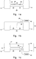
- FIG. 1A and 1B a device 2, for the classification and counting of white blood cells within a blood sample is described.
- the device 2 includes a microfluidic liquid handling structure 10.
- Figures 1A and 1B illustrate mutually perpendicular cross-sections of the device 2, including the microfluidic liquid handling structure 10.
- Figure 1C illustrates the device 2 as viewed from above.
- the microfluidic liquid handling structure 10 comprises the following main elements: a loading chamber 12, a connection conduit 20 and a waste chamber 14.
- the loading chamber 12 has a sample inlet 16 through which a blood sample can be introduced into the microfluidic liquid handling structure 10.
- the inlet 16 is sealable in order to, when sealed, prevent the sample escaping once it is in the microfluidic liquid handling structure 10.
- the waste chamber 14 and the loading chamber 12 have air vents 17, 18 such that the chambers are open to the atmosphere. This allows the blood sample to flow from the loading chamber, through the conduit 20 and into the waste chamber 14 by capillary action.
- the microfluidic liquid handling structure forms part of a cartridge insertable into a holding structure for holding the cartridge relative to an image capture device 28.
- the cartridge may come with a custom metering implement for metering an appropriate volume of blood for application to the microfluidic liquid handling structure, for example a pipette.
- the microfluidic liquid handling structure 10 is sealed from the atmosphere when the inlet 16 is sealed and the microfluidic liquid handling structure includes a network of air channels including an air circuit which connects the loading chamber 12 and the waste chamber 14.
- the air circuit allows air to escape from the waste chamber 14 into the loading chamber 12 as the blood sample flows from the loading chamber 12 and fills the waste chamber 14.
- the air circuit includes one or more flow barrier valves, for example a sudden expansion of the air circuit, to prevent the sample from entering the air circuit by capillarity.
- connection conduit 20 comprises two portions: a preparation portion 22 and a detection portion 24.
- the preparation portion 22 is arranged for the lysing of red blood cells and staining of white blood cells. As the sample moves through the preparation portion 22, it encounters a series of patches of dry reagents 32, 34, 36. Although in Figure 1A three patches of dry reagents are provided, it will be appreciated that any number (one or more) patches may be provided within the preparation portion 22.
- the blood sample As the blood sample flows over the one or more patches, it will dissolve the reagent(s) which will gradually diffuse through the blood volume and prompt a chemical reaction.
- the dynamics of such reactions depends mainly on the blood flow rate and the length(s) of the patch(es) of dry reagent.
- the content of dry reagent stored in the preparation portion 22 and how easily it dissolves in blood will also have an effect on the dynamics.
- the dry reagents comprise, for example, a haemolytic agent for the selective lysing of red blood cells and a staining agent for differential staining of white blood cells..
- a staining agent from the family of hematoxylin and cosin (H&E) stains, Romanowsky stains, methacromatic stains or any combination thereof can be used. From combinations of colour information with morphological features like granularity, size, shape of the cell cytoplasm and nucleus, it is then possible to obtain a set of distinct signatures for each of the sub-populations under study. Further discussion of the use of reagents can be found in application WO2013135713
- the preparation portion 22 contains a series of 2 sequential reagents, comprising a first reaction site which is 10 mm long comprising a mixture of surfactant and lytic agent (Surfynol and saponine, respectively) and a 10 mm long reaction site with a mixture of stains.
- the mixture of stains includes a mixture of eosin, methylene blue and basic orange 21 leading to differential colours for a 5-part classification of white blood cells: lymphocytes stain blue, monocytes stain blue/purple, neutrophils exhibit blue nucleus and pale yellow cytoplasm, eosinophils exhibit blue nucleus and dark yellow granules, basophils stain bright pink.
- the detection portion 24 of the connection conduit 20 is where the cells are, in operation, imaged and counted.
- the detection portion 24 is aligned with a field of view 26 of an image capture device 28, such that the blood flowing through the detection portion 24 flows across the field of view 26 of the image capture device 28. Approximately two thirds of the width of the detection portion 24 is within the field of view 26 of the image capture device 28.
- the image capture device 28 is positioned above the detection portion 24.
- the image capture device 28 includes a lens arrangement and a focusing mechanism for focusing on the blood sample flowing into the detection portion 24 to image the lysed and stained sample when it is in the detection portion 24.
- the detection portion 24 is arranged to be shallower than preparation portion 22, such that it accommodates a single layer of objects which move across the field of view 26 of the image capture device 28. It is preferable to ensure that a single layer of objects move across the field of view of the image capture device 28 to increase the chance that each object is counted and to facilitate the classification of the objects. If multiple layers of objects were provided in the field of view then some objects could be blocked from view completely and others could be partially obscured by other objects. Having a single layer of objects also facilitates any defining characteristics of objects (including cells) being captured.
- the preparation portion 22 is deeper than the detection portion 24 to facilitate the arrangement of the reagents 32, 34, 36 and to increase their surface areas. The reduction in depth from the preparation portion 22 to the detection portion 24 facilitates sample flow by capillarity through the preparation portion 22 and the detection portion 24. Further, the depth of the preparation portion facilitates homogeneity of lysis and staining of the blood cells.
- the detection portion 24 is at least twice as wide as the estimated largest dimension of any object to be detected and its depth is less than twice this largest dimension. In one embodiment, the detection portion 24 is 15 mm long, 0.06 mm wide and 0.02 mm deep. This configuration defines a volume for the detection portion 24 of 0.018 ⁇ L.
- Figure 2 schematically illustrates the type of image obtained with several stained white blood cells 37, 38, 39 in such a detection portion.
- a processor 40 configured to carry out the data processing required to obtain the cell counts. The processing steps will be described below.
- the operation of the device 2 will now be described.
- the sample is introduced into the microfluidic liquid handling structure 10 through the inlet 16 and the inlet is then sealed.
- the sample enters the loading chamber 12 and is drawn by capillary action into the preparation portion 22, and, subsequently, into the detection portion 24. Therefore, once the blood sample enters the detection portion 24, it has already reacted with the red blood cell lytic agent and differential stains for white blood cells.
- the image capture device 28 may initially be out of focus, but when the sample enters the detection portion 24 and subsequently the field of view of the image capture device 28, a focusing mechanism focuses the image capture device 28 on the sample and the image capture device 28 begins capturing images as frames of a video. Focusing methods, implemented in software and/or hardware are generally well known. A specific example adapted for use with the device 2 is disclosed in UK application number 1417170.6 . In one specific implementation, the video is taken for four minutes at a rate of 15 frames per second, resulting in approximately 15-30 frames being captured of each cell. Frame acquisition durations and rates are adapted to the specific flow in the microfluidic liquid handling structure 10 to capture a sufficient number of frames per cell for subsequent analysis.
- the data obtained from the video consists of a series of frames (approximately 4000), wherein each frame comprises an image containing one or more cells and other objects, such as clusters of cells or platelets. Some frames may contain no objects at all and these frames are discarded.
- Each frame is first segmented into object and background pixels at step 42 using thresholding in some embodiments, for example Otsu's method, and the objects in the frame are identified at step 44, as is well known in the art of image processing.
- each object is given an identifier which is unique to that object and frame and at step 48, a list of the object identifiers and their associated object positions is obtained.
- Image patches centred on the object positions are extracted at step 50.
- the image patch is segmented into separate object regions at step 52. These regions are, in overview: background; nucleus of the cell; cytoplasm .
- a k-means clustering algorithm based on RGB colour values is used for segmentation.
- the pixels of each image patch are clustered using k-means (or any other suitable clustering algorithm) on the RGB values of the pixels to define three clusters.
- the cluster with the darkest (e.g. average) pixel intensity or colour is labelled as the nucleus
- the cluster with the lightest (e.g. average) pixel intensity or colour is labelled as background
- the remaining cluster is labelled as cytoplasm.
- the segmentation algorithm therefore outputs three regions, which are identified as background, nucleus and cytoplasm according to the intensity or colour of each region.
- k-means clustering used again to split pixels previously identified as the nucleus, to cluster these pixels into two sub-regions: nucleus and 'cytoplasm region 2'. This may help to more accurately define the boundary between the nucleus and cytoplasm.
- pixels labelled as nucleus are either left as 'nucleus', or are reclassified as 'cytoplasm region 2'.
- an alternative segmentation approach can be applied, which advantageously may be more robust against image variations.
- the approach involves the following steps for each image patch:
- the clustering stops after step 3 with labelling the nucleus and cytoplasm pixels accordingly.
- the 'cytoplasm region 2' region is merged with the cytoplasm identified in the first pass (cytoplasm region 1') and labelled as an overall cytoplasm for the further analysis / cell classification.
- 'cytoplasm region 1' is taken as representative of the cytoplasm.
- object parameters for this region are used in classifying objects in addition to the overall cytoplasm, in some embodiments.
- each object is split into three or four regions, depending on the embodiment: nucleus; background; cytoplasm; cytoplasm region 2, or: nucleus; background; cytoplasm.
- Non-cell objects are identified by the classifier discussed below.
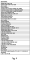
- object parameters are extracted from each image patch for each of the respective objects. These parameters include:
- Figure 4 shows a list of object parameters that may be extracted for each of the respective objects.
- the values of some parameters are extracted for the object as a whole, for example the object roundness and the object perimeter.
- the values of other parameters are extracted for each object region, for example the intensity and colour and their standard deviations, using masks to isolate each region in turn.
- the values for the background region are used for calibration. For example, they are used to account for more staining in one region of the field of view 26 than another. If the focus quality of an image is too poor, for example if it is below a predetermined threshold, then that image is discarded.
- the values of the extracted parameters are then used with a Logistic Model Tree classifier to obtain classification results, a discussion of which may be found in N. Landwehr, M. Hall and E. Frank, 'Logistical Model Trees', Machine Learning, vol. 59, pages 161-205, 2005 , incorporated herein by reference.
- the Logistic Model Tree is trained prior to its use for classification using expert labelled training data.
- the Logistic Model Tree is used to obtain class-belonging scores for each object, e.g. probabilities of each object belonging to each object type.
- the SimpleLogistic class provided by the Weka collection of machine learning algorithms is used (see http://weka.sourceforge.net/doc.dev/weka/classifiers/functions/SimpleLogistic.html , http://www.cs.waikato.ac.nz/ml/weka/ .
- Any suitable classifier for example a Support Vector Machine can be used, as will be apparent to a person skilled in the art. Steps 52-56 are repeated for each object in the frame.
- the object types include five white blood cell types, nine other object types, used for objects other than white blood cells to be counted, and a 'cell cluster' classification.
- the object types are:
- a blood sample contains too many objects in any one of the non-white blood cell (i.e. other than the first five) classifications (for example if the proportion of objects classified as an 'other' type exceeds a threshold) then the results of the assay may be discarded.
- the 'eosinophil within a cluster' classification is useful because eosinophils make up a very small proportion of all white blood cells. If those eosinophils contained in a cluster of cells were ignored and not counted, then very few or even no eosinophils would be counted in a given assay.
- Clusters of cells are identified by the classifier, as above.
- a cluster of cells is characterised by a large object area, for example 500 pixels or more. Therefore, object area is discriminative of clusters of cells.
- An eosinophil has a distinct colour signature and as such, the mean colour of the cluster as a whole is shifted. By identifying this shift in colour, the presence of an eosinophil within a cluster is detected.
- detecting an eosinophil within the cluster is a relatively rare event, in contrast to detecting other objects, it is assumed that if the shift in colour of the cluster is detected, that the cluster contains one eosinophil, in some embodiments. Further processing to count eosinophils in identified clusters is used in other embodiments. Any of the above method steps may be carried out by or under the control of a processor.
- Including a cell within a cluster in estimating a cell count can be implemented alone or in combination with other disclosed features and steps. This technique may also be applied to other cell types, for example Basophils.
- the object is classified as the class for which the object has the highest class-belonging score. In some embodiments, if the highest class-belonging score is below a threshold, the object is classified as 'unknown'.
- an estimate of the absolute count for a blood sample may be determined by selecting a subset of the frames captured based on an estimate of cell velocity.
- One way to estimate cell velocities is by tracking cells from one frame to the next. This process will now be described with reference to Figure 5 .
- the list of object positions for frame 1 is compared to the list of object positions for frame 2 at step 58.
- an object 78 in a second frame which has the position closest to position (x, y) is identified.
- a check is carried out to determine whether any other objects lie within a circle centred on object 78, with radius equal to a predefined 'safe distance'. This 'safe distance' is defined as a distance at which two objects are too close to be distinguishable with confidence from each other from one frame to the next.
- step 64 if an object 80 is within the safe distance from object 78 then object 72 cannot be identified in frame 2 and the tracking of object 72 stops. If at step 64, no objects within the safe distance from object 78 are found, object 78 is identified as being the same object as object 72 in the previous frame. Steps 60-68 are repeated for all objects in the first frame such that each object is either tracked from frame 1 to frame 2, or cannot be identified with enough certainty in frame 2. In the latter case (conflicting object within safe distance), the tracking of these objects stops. The process then moves onto the next frame, at step 70, and the lists of positions for frames 2 & 3 are compared. Steps 58-70 are repeated for all frames in the data set until lastly, the lists of object positions for frames n-1 and n are compared, where the data set consists of n frames.
- object velocity is used to improve tracking. For example, rather than picking the object 78 in frame 2 that is closest to object 72 in frame 1 at step 60, the object closest to the predicted position of object 72, based on the velocity of object 72 determined from frame 1 and one or more previous frames, is picked, which enables a reduction of the safe distance, since more information is used in the tracking. This is carried out, in some embodiments, in the tracking of an object from a second to a third frame and for subsequent frames thereafter, but not from a first to a second frame, because the velocity of the object is estimated by analysing more than one frame.
- a set of image patches for each tracked object is obtained.
- the objects (image patches) obtained in the method described above with reference to Figure 3 are 'matched up' with other objects (image patches) which correspond to the same object.
- the dimensions of the field of view of the image capture device are known and based on these dimensions, the distance travelled by each object between frames is estimated.
- the time between frames is also known. Using this and the estimate of the distance travelled by an object between frames, an estimate of an object's velocity is determined.
- the absolute count of a blood sample is estimated by the direct averaging of counts contained within a subset of frames of the video of the blood sample to reduce the likelihood of double counting. A method of doing so is now described.
- each frame taken of the blood sample is a small number of white blood cells and other objects, such as clusters of cells or platelets. Typically, there are between 1 and 10 objects in each frame, but frames can include 20-30 objects.
- the image capture device 28 is set up to take approximately 17 frames per second and a given object will typically take 1-2 seconds to move across the field of view of the image capture device 28. Therefore, each object will appear in multiple frames.
- the absolute white blood cell count is determined by selecting a subset of frames such that each object in the sample only appears once throughout the subset of frames, or at least such that the likelihood of double counting is reduced.
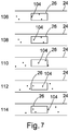
- an object 104 moves across the field of view 26 of the image capture device 28 and is captured in frames 106-112.
- object 104 has just entered the field of view 26 of the image capture device 28 and in frame 114 object 104 has just left the field of view.
- the speed of an object 104 is estimated and used to determine which frame in the subset corresponds to frame 114, i.e. the first frame in the series of frames in which all of the objects captured in frame 106 are likely to have left the field of view and hence do not appear again.
- the fastest speed of all object speeds moving across the field of view 26 in a frame n is used to select the next frame in the subset, frame, n+1, having obtained estimates of object velocities either by tracking objects over multiple frames or by assuming that cell velocities are the same and equal to the velocity of the blood sample through the detection portion.
- step 116 for a frame m, the fastest-moving cell in frame m is identified and its speed is estimated. This is done based on object positions from two or more of the current frame, previous or subsequent frames.
- this speed is used to identify the next frame in which all of the objects are likely to have left the frame m. This next frame is selected as the next frame in the subset.
- v For a fastest object speed, v, in frame m, the next frame in the subset of frames, frame m+1, is determined by calculating the time interval between frames m and m+1.
- the speed of the slowest-moving object is used to select the next frame in the subset.
- 'w' in equation (3) is the extent of the field of view along the direction of blood flow.
- the average, for example the mean, or the median speed of all the objects in the frame is used to select the next frame in the subset.
- 'w' in equation (3) represents the diagonal extent of the field of view.
- the average of the speeds of the fastest-moving object in each frame is estimated and is used to select the next frame in the subset.
- Some embodiments make use of other speed measures to decide how to sub-sample the frames, for example based on an average speed across all frames and fixed sub-sampling rate or using a speed profile of frames with a corresponding variable sub-sampling rate. Equally, fastest and slowest speeds can be combined with various length scales (direction of scales, diagonal).
- Steps 116-120 are repeated until, at step 120, it is determined that most or substantially all of the objects captured in the video appear in the subset of frames.
- the selection of frames continues until the final frame in the set is reached, or the next frame that would be selected does not exist.
- no further frames are selected beyond frame number 4300 (at 17 frames per second) or if flow speed falls below a certain threshold for a certain period of time or if flow stops completely.
- the white blood cells in each frame are identified as described above and counted across all frames in the subset of frames.
- the white blood cell counts are averaged (or, in some embodiments, the median is calculated) across all frames in the subset. This estimated count is then scaled to obtain the estimated number of white blood cells per unit volume of blood at step 126. This is the absolute white blood cell count.
- a subset of frames is selected such that the likelihood of each object in the sample appearing only once in the subset is increased. It is therefore only necessary to obtain classification results for the objects in the subset of frames. In one embodiment, the subset of frames is therefore selected before classification results for the objects in each frame are obtained so this is only done for the relevant objects. This allows for a more efficient use of computing power. It is equally possible however, to obtain classification results for the objects in each of the frames in the whole video before selecting the subset. This may be more efficient if the classification results are also used in other processes.
- results for each of the objects in each of the frames is useful for the statistical method of determining the white blood cell count. In this process, results may be obtained for every object and an object may be counted in all frames it appears in.
- an object may be classified as either 'white blood cell' or 'non-white blood cell' by summing all white blood cell class belonging scores for that object and making the classification of 'white blood cell' if the sum is greater than a threshold (for example 0.5 if the class belonging scores are probabilities).
- a threshold for example 0.5 if the class belonging scores are probabilities.
- the sample flows through the connecting conduit 20 by capillarity.
- the flow of the sample through the connecting conduit 20 may equally be driven by a pressure difference or by any other means.
- the microfluidic liquid handling structure as described above may be implemented on a 'lab on a disc' cartridge designed for rotation about an axis of rotation.
- the sample inlet is sealed and the device is placed in an instrument comprising a rotation mechanism.
- the image capture device may be integral to the instrument into which the cartridge is placed.
- the rotation mechanism allows angular positioning of the cartridge to be controlled.
- a positioning sensor may be used to assist the precise alignment of the detection portion 24 with the field of view 26 of the image capture device 28.
- the cartridge may also be immobilized or slowly rotating for discrete positioning purposes and all fluidic functions may be accomplished by capillary based handling of the blood sample without further interference/assistance.
- the cartridge may be rotated about an axis of rotation for the further processing of the same or a different sample within the device.
- a liquid sample in the cartridge experiences a centrifugal force.
- An example of such a cartridge can be found in application WO2013135713 .
- V FOV associated with each cartridge is the value of V FOV for that particular cartridge for use where necessary in the methods described above.
- V FOV may be recorded on a machine readable element on the cartridge or may be stored elsewhere.
- All of the fluidic structures described herein may be designed in polar coordinates relative to said axis of rotation. Consequently, all structures may be characterized by their radial and angular dimensions and positioning in respect of an axis of rotation.
- the microfluidic liquid handling structure is provided on a cartridge.
- the cartridge in some examples, in particular those using a rotational mechanism, resembles a CD/DVD configuration constituted by two transparent and planar circular halves brought together by an intermediate adhesive layer.
- the halves are preferably engraved with the microfluidic structures and openings to the exterior described above. With precise alignment of the microfluidic structures, the three parts may be assembled and bonded to form a self-contained cartridge.
- connection conduit 20 including its preparation and detection portions, and those of the loading 12 and waste 14 chambers may be adjusted.
- the embodiments described above are adapted for the processing of a blood sample, at least some of the above embodiments are suitable for processing any liquid sample, for example any liquid to be reacted with one or more reagents prior to imaging, in which objects are to be counted.
- connection conduit 20 may be arranged in such a manner that enables space saving within the microfluidic liquid handling structure, for example using a meandering configuration.
- the trajectory of the connection conduit 20 may be provided within a single plane of the microfluidic liquid handling structure. It will be appreciated that although a straight connection conduit 20 is depicted in Figure 1A , any shape suitable for providing a substantially planar trajectory of the blood flow within the microfluidic liquid handling structure could be provided.
- reagents may be used in the device. It will also be appreciated that the reagents may be arranged to have different dimensions in the preparation portion 22.
- the described processes can be implemented using any suitable stand-alone or distributed computing environment using any suitable computing platform or processor, for example an integrated circuit, self-contained or in combination with other components of the system, a dedicated computing device housed on an appropriate card together with the other components of the system or otherwise, a standalone computing device such as a personal computer, tablet computer or mobile phone or a server which performs the necessary processes at least in part remotely exchanging data over a network connection.
- the processing may be implemented in a client-server architecture, with the processing being distributed between the client and the server in any appropriate manner.
- the client may include any of the above mentioned devices.
Description
- The present invention relates to the counting of cells in a blood sample.
- The counting of cells in a blood sample is an important diagnostic tool.
- An example of a known approach to counting cells in a blood sample is flow cytometry. In flow cytometry, a blood sample flows through a capillary such that cells pass one by one in front of a light source and detection arrangement. The reflected and/or transmitted light is detected and analysed. The characteristics of the detected light depend on characteristics of the cells in the sample and how they are stained and, in this way, cells are classified and counted. However, no morphological information for the cells is obtained using flow cytometry. Morphological analysis can be carried out on a blood sample by sandwiching the sample between two slides and investigating the sample under a microscope. Automated systems for cell counting are known which use cartridges of pre-prepared slides. Such slides need to be prepared manually for each sample to be analysed. This is a time-consuming process, in spite of the partial automation.
- It would be desirable to have a system for cell counting in which the morphological information of cells is not lost but instead is used to improve the accuracy of the cell counting process and which, further, does not require the preparation of slides.
-
US4075462 discloses a particle analyser apparatus employing a light-sensitive electronic detector array.US5880835 discloses an apparatus for investigating particles in a fluid and a method of operation thereof.WO2014/146061 discloses dynamic range extension systems and methods for particle analysis in blood samples. - Aspects of the invention are set out in the independent claims. Further, optional, features of the invention are set out in the dependent claims.
- There is provided a method of estimating a cell count in a blood sample according to
claim 1. - In some embodiments, identifying cells in each frame includes classifying objects in each frame. Parameters of each object are extracted from the frame and, in some embodiments, the classification result for each object may be obtained using a Simple Logistic Model. In other embodiments, other classifiers, as will be known to a person skilled in the relevant field, may be used. For examples, refer to S.B. Kotsiantis, 'Supervised Machine Learning: A Review of Classification Techniques', Informatica, vol. 31, number 3, October 2007, pages 249-268. Once each object in a frame has been classified, the number of cells within the frame is counted. As such, a frame cell count is found for each frame.
- Combining the counts may comprise fitting a distribution function to the frame cell counts and computing an estimated cell count based on the fitted distribution. The estimated cell count may be derived using one or more parameters of the fitted distribution. The fitted distribution may be a Gamma or a Poisson distribution.
- Computing the estimated cell count may, in some embodiments, involve splitting the sequence of frames into subset blocks and computing a respective estimated cell count by fitting a respective distribution to each block. One of the estimated respective cell counts can then be used as the result to be stored (or returned) by making a selection based on one or more of the position of the subset block in the sequence, the magnitude of the respective estimated counts and a goodness of fit indicator. In some embodiments, if the goodness of fit indicator for any block is not good enough, i.e. if it is lower than a predetermined threshold, then the respective estimated cell count for that block is discarded.
- Combining the frame cell counts includes computing a mean or median of the frame cell counts.
- Frames are selected based on an estimate of cell velocity. By considering cell velocity, the next frame in the subset of selected frames can be selected such that the objects in the previous frame are likely to have left the field of view. Doing so may involve computing the number of frames needed before the objects in the previous frame are likely to have left the field of view. This may reduce the chances of double counting.
- In some embodiments, cell velocities are estimated by tracking objects from one frame to the next. This process, along with alternative methods of estimating cell velocities, will be described below.
- The selecting of frames may include setting a cut-off position, wherein frames with a position in the sequence of frames that make up the video that is beyond the cut-off position are not selected.
- In some embodiments, an absolute cell count may be estimated based on an estimated cell count and the volume of sample contained within the field of view of the image capture device. In some embodiments, the volume of sample contained within the field of view may be corrected for any obstructions to the flow that are found within the field of view. This may be done by first detecting an obstruction and then estimating its volume. The estimated volume of the obstruction may then be subtracted from the unobstructed volume associated with the field of view. Detecting an obstructed area may include partitioning the field of view into candidate areas and marking a candidate area as obstructed if less than a threshold amount of cells have been identified over the sequence of frames in that candidate area.
- There is provided a system for estimating a cell count in a blood sample according to claim 4.
- When the sample enters the field of view, the image capture device begins capturing frames of the sample which together form a video. In some embodiments, the rate at which frames are taken can be adapted according to the parameters of a specific assay, for example based on the rate of flow of the sample across the field of view. Additionally or alternatively, the total number of frames taken can be adapted to the parameters of the assay.
- Once the frames of the sample have been captured, the image processor carries out a number of processing steps on the frames, as outlined above. In some embodiments, the frames are segmented into objects and background and the segmented objects are tracked over as many frames as possible. In some embodiments, this tracking is based on the assumption that an object in a first frame is that same object as the object in the next frame with the position closest to that of the object in the first frame. In some embodiments, an object is only tracked from a first frame to the next if there are no objects within a pre-determined 'safe-distance' from the object in the second frame.
- There is provided a computer readable medium or computer readable media storing coded instructions that, when executed on a processor, implement any or all of the method steps as described above or any combination thereof. This computer readable medium or media can comprise any kind of data storage medium, for example one or more of a magnetic or optical disc, a hard disc, solid state or other data storage device, including RAM, ROM, EPROM, EEPROM or Flash.
- While most of the description is made in terms of classifying white blood cells (leukocytes) and more specifically cell types corresponding to different healthy leukocyte populations, the invention is not so limited and other cell types can be classified and counted, for example diseased or cancerous white blood cells or red blood cells.
- Examples are now described in detail, with reference to the accompanying drawings, in which:
-
Figures 1a-c illustrate views of a microfluidic liquid handling structure; -
Figure 2 illustrates a schematic view of white blood cells in the detection portion; -
Figure 3 illustrates a flow diagram representing a method for obtaining classification results for objects within a frame; -
Figure 4 provides a list of object parameters that may be used in classifying objects; -
Figure 5 illustrates a method for tracking objects across multiple frames; -
Figure 6 illustrates two consecutive frames of the blood sample; -
Figure 7 shows a schematic view of a set of consecutive frames; and -
Figure 8 illustrates a method for estimating the absolute white blood cell count by speed-adaptive frame sampling. - The figures are not to scale and further, are not drawn in exact proportion for the purpose of clarity.
- Referring to
Figures 1A and 1B , adevice 2, for the classification and counting of white blood cells within a blood sample is described. Thedevice 2 includes a microfluidicliquid handling structure 10.Figures 1A and 1B illustrate mutually perpendicular cross-sections of thedevice 2, including the microfluidicliquid handling structure 10.Figure 1C illustrates thedevice 2 as viewed from above. - With reference to
Figure 1C , the microfluidicliquid handling structure 10 comprises the following main elements: aloading chamber 12, aconnection conduit 20 and awaste chamber 14. Theloading chamber 12 has asample inlet 16 through which a blood sample can be introduced into the microfluidicliquid handling structure 10. Theinlet 16 is sealable in order to, when sealed, prevent the sample escaping once it is in the microfluidicliquid handling structure 10. Thewaste chamber 14 and theloading chamber 12 haveair vents conduit 20 and into thewaste chamber 14 by capillary action. The microfluidic liquid handling structure forms part of a cartridge insertable into a holding structure for holding the cartridge relative to animage capture device 28. In some embodiments, the cartridge may come with a custom metering implement for metering an appropriate volume of blood for application to the microfluidic liquid handling structure, for example a pipette. - In some embodiments, the microfluidic
liquid handling structure 10 is sealed from the atmosphere when theinlet 16 is sealed and the microfluidic liquid handling structure includes a network of air channels including an air circuit which connects theloading chamber 12 and thewaste chamber 14. The air circuit allows air to escape from thewaste chamber 14 into theloading chamber 12 as the blood sample flows from theloading chamber 12 and fills thewaste chamber 14. The air circuit includes one or more flow barrier valves, for example a sudden expansion of the air circuit, to prevent the sample from entering the air circuit by capillarity. - Sharp angles within the microfluidic
liquid handling structure 10 are preferably avoided to reduce impediments to sample flow and to prevent trapping of air bubbles inside theconnection conduit 20 whilst thewaste chamber 14 is filling with the blood sample. Theconnection conduit 20 comprises two portions: apreparation portion 22 and adetection portion 24. Thepreparation portion 22 is arranged for the lysing of red blood cells and staining of white blood cells. As the sample moves through thepreparation portion 22, it encounters a series of patches ofdry reagents Figure 1A three patches of dry reagents are provided, it will be appreciated that any number (one or more) patches may be provided within thepreparation portion 22. As the blood sample flows over the one or more patches, it will dissolve the reagent(s) which will gradually diffuse through the blood volume and prompt a chemical reaction. The dynamics of such reactions depends mainly on the blood flow rate and the length(s) of the patch(es) of dry reagent. The content of dry reagent stored in thepreparation portion 22 and how easily it dissolves in blood will also have an effect on the dynamics. The dry reagents comprise, for example, a haemolytic agent for the selective lysing of red blood cells and a staining agent for differential staining of white blood cells.. A staining agent from the family of hematoxylin and cosin (H&E) stains, Romanowsky stains, methacromatic stains or any combination thereof can be used. From combinations of colour information with morphological features like granularity, size, shape of the cell cytoplasm and nucleus, it is then possible to obtain a set of distinct signatures for each of the sub-populations under study. Further discussion of the use of reagents can be found in applicationWO2013135713 - In one specific embodiment, the
preparation portion 22 contains a series of 2 sequential reagents, comprising a first reaction site which is 10 mm long comprising a mixture of surfactant and lytic agent (Surfynol and saponine, respectively) and a 10 mm long reaction site with a mixture of stains. In this embodiment, the mixture of stains includes a mixture of eosin, methylene blue and basic orange 21 leading to differential colours for a 5-part classification of white blood cells: lymphocytes stain blue, monocytes stain blue/purple, neutrophils exhibit blue nucleus and pale yellow cytoplasm, eosinophils exhibit blue nucleus and dark yellow granules, basophils stain bright pink. - The
detection portion 24 of theconnection conduit 20 is where the cells are, in operation, imaged and counted. Thedetection portion 24 is aligned with a field ofview 26 of animage capture device 28, such that the blood flowing through thedetection portion 24 flows across the field ofview 26 of theimage capture device 28. Approximately two thirds of the width of thedetection portion 24 is within the field ofview 26 of theimage capture device 28. Theimage capture device 28 is positioned above thedetection portion 24. Theimage capture device 28 includes a lens arrangement and a focusing mechanism for focusing on the blood sample flowing into thedetection portion 24 to image the lysed and stained sample when it is in thedetection portion 24. - The
detection portion 24 is arranged to be shallower thanpreparation portion 22, such that it accommodates a single layer of objects which move across the field ofview 26 of theimage capture device 28. It is preferable to ensure that a single layer of objects move across the field of view of theimage capture device 28 to increase the chance that each object is counted and to facilitate the classification of the objects. If multiple layers of objects were provided in the field of view then some objects could be blocked from view completely and others could be partially obscured by other objects. Having a single layer of objects also facilitates any defining characteristics of objects (including cells) being captured. Thepreparation portion 22 is deeper than thedetection portion 24 to facilitate the arrangement of thereagents preparation portion 22 to thedetection portion 24 facilitates sample flow by capillarity through thepreparation portion 22 and thedetection portion 24. Further, the depth of the preparation portion facilitates homogeneity of lysis and staining of the blood cells. - In some embodiments, the
detection portion 24 is at least twice as wide as the estimated largest dimension of any object to be detected and its depth is less than twice this largest dimension. In one embodiment, thedetection portion 24 is 15 mm long, 0.06 mm wide and 0.02 mm deep. This configuration defines a volume for thedetection portion 24 of 0.018 µL.Figure 2 schematically illustrates the type of image obtained with several stainedwhite blood cells - Associated with the
device 2 is aprocessor 40 configured to carry out the data processing required to obtain the cell counts. The processing steps will be described below. - The operation of the
device 2 will now be described. The sample is introduced into the microfluidicliquid handling structure 10 through theinlet 16 and the inlet is then sealed. The sample enters theloading chamber 12 and is drawn by capillary action into thepreparation portion 22, and, subsequently, into thedetection portion 24. Therefore, once the blood sample enters thedetection portion 24, it has already reacted with the red blood cell lytic agent and differential stains for white blood cells. - The
image capture device 28 may initially be out of focus, but when the sample enters thedetection portion 24 and subsequently the field of view of theimage capture device 28, a focusing mechanism focuses theimage capture device 28 on the sample and theimage capture device 28 begins capturing images as frames of a video. Focusing methods, implemented in software and/or hardware are generally well known. A specific example adapted for use with thedevice 2 is disclosed in UK application number1417170.6 liquid handling structure 10 to capture a sufficient number of frames per cell for subsequent analysis. Durations and times are readily adapted by a skilled person to optimise data collection according to the circumstances. During the assay, approximately 1500 cells move across the field ofview 26 of theimage capture device 28 in one example, for a healthy sample. The data obtained from the video consists of a series of frames (approximately 4000), wherein each frame comprises an image containing one or more cells and other objects, such as clusters of cells or platelets. Some frames may contain no objects at all and these frames are discarded. - Having described a device used for various cell counting processes, such processes are now described in detail.
- A method used to classify the objects in a frame is now described with reference to
Figure 3 . Each frame is first segmented into object and background pixels atstep 42 using thresholding in some embodiments, for example Otsu's method, and the objects in the frame are identified atstep 44, as is well known in the art of image processing. Atstep 46, each object is given an identifier which is unique to that object and frame and atstep 48, a list of the object identifiers and their associated object positions is obtained. Image patches centred on the object positions are extracted atstep 50. Then, for a first image patch, the image patch is segmented into separate object regions atstep 52. These regions are, in overview: background; nucleus of the cell; cytoplasm . - In some embodiments, a k-means clustering algorithm based on RGB colour values is used for segmentation. The pixels of each image patch are clustered using k-means (or any other suitable clustering algorithm) on the RGB values of the pixels to define three clusters. This results in a mask with a cluster identifier (
cluster 1,cluster 2, cluster 3) for each pixel. The cluster with the darkest (e.g. average) pixel intensity or colour is labelled as the nucleus, the cluster with the lightest (e.g. average) pixel intensity or colour is labelled as background and the remaining cluster is labelled as cytoplasm. The segmentation algorithm therefore outputs three regions, which are identified as background, nucleus and cytoplasm according to the intensity or colour of each region. - In some embodiments, as a refinement, k-means clustering used again to split pixels previously identified as the nucleus, to cluster these pixels into two sub-regions: nucleus and 'cytoplasm region 2'. This may help to more accurately define the boundary between the nucleus and cytoplasm. In this step, pixels labelled as nucleus are either left as 'nucleus', or are reclassified as 'cytoplasm region 2'.
- In some embodiments, an alternative segmentation approach can be applied, which advantageously may be more robust against image variations. The approach involves the following steps for each image patch:
- 1) k-means clustering with k=2 to separate cell (darker pixels) from background (lighter pixels);
- 2) label background pixels as background and mask (set to a predefined colour, say green) ;
- 3) k-means clustering with k=3 to get nucleus (darkest), cytoplasm (lighter) and the now, say, green background;
- 4) label cytoplasm pixels as
cytoplasm region 1 and mask cytoplasm in addition to the background by setting cytoplasm pixels to the predefined colour; - 5) k-means clustering with k=3 to get nucleus pixels (darkest),
cytoplasm region 2 and the now masked background (which now includes theprevious cytoplasm region 1 area); - 6) label nucleus pixels as nucleus and
cytoplasm region 2 pixels ascytoplasm region 2. - In embodiments where only one cytoplasm region is segmented, the clustering stops after step 3 with labelling the nucleus and cytoplasm pixels accordingly.
- In some embodiments. the 'cytoplasm region 2' region is merged with the cytoplasm identified in the first pass (cytoplasm region 1') and labelled as an overall cytoplasm for the further analysis / cell classification. In other embodiments, 'cytoplasm region 1' is taken as representative of the cytoplasm. Noting that 'cytoplasm region 2' is a transition region between the nucleus and cytoplasm of the cell, object parameters for this region are used in classifying objects in addition to the overall cytoplasm, in some embodiments.
- Overall, each object is split into three or four regions, depending on the embodiment: nucleus; background; cytoplasm;
cytoplasm region 2, or: nucleus; background; cytoplasm. - Examples of other clustering based algorithms for identifying blood cells can be found in 'White Blood Cell Segmentation by Color-Space-Based K-Means Clustering', Congcong Zhang et al, ; Leukocyte segmentation and classification in blood-smear images', Herbert Ramoser et al, Proceedings of the 2005 IEEE Engineering in Medicine and Biology 27th Annual Conference, Shanghai, China, September 1-4, 2005.
and may be used for object segmentation in some embodiments. It will be understood that any other suitable segmentation method, supervised or unsupervised , using clustering or other techniques, may be used in some embodiments. Specifically, any suitable alternative clustering algorithm other than k-means may be used in any of the above embodiments. - For objects which are not cells and hence do not have a nucleus or cytoplasm, the k-means algorithm still splits the object up into multiple regions but these regions are not meaningful and the convergence of the k-means algorithm is not as good for non-cell object as that for cells. Non-cell objects are identified by the classifier discussed below.
- At
step 54, object parameters are extracted from each image patch for each of the respective objects. These parameters include: - image saturation averaged over the relevant pixels and its standard deviation
- intensity averaged over the relevant pixels and its standard deviation,
- colour averaged over the relevant pixels and its standard deviation,
- object roundness,
-

-

- object perimeter,
- object area and
- focus quality of the image.
-
Figure 4 shows a list of object parameters that may be extracted for each of the respective objects. - The values of some parameters are extracted for the object as a whole, for example the object roundness and the object perimeter. The values of other parameters are extracted for each object region, for example the intensity and colour and their standard deviations, using masks to isolate each region in turn. The values for the background region are used for calibration. For example, they are used to account for more staining in one region of the field of
view 26 than another. If the focus quality of an image is too poor, for example if it is below a predetermined threshold, then that image is discarded. - At
step 56, the values of the extracted parameters are then used with a Logistic Model Tree classifier to obtain classification results, a discussion of which may be found in N. Landwehr, M. Hall and E. Frank, 'Logistical Model Trees', Machine Learning, vol. 59, pages 161-205, 2005, incorporated herein by reference. The Logistic Model Tree is trained prior to its use for classification using expert labelled training data. The Logistic Model Tree is used to obtain class-belonging scores for each object, e.g. probabilities of each object belonging to each object type. In some embodiments, the SimpleLogistic class provided by the Weka collection of machine learning algorithms is used (see http://weka.sourceforge.net/doc.dev/weka/classifiers/functions/SimpleLogistic.html, http://www.cs.waikato.ac.nz/ml/weka/. Any suitable classifier, for example a Support Vector Machine can be used, as will be apparent to a person skilled in the art. Steps 52-56 are repeated for each object in the frame. - The object types include five white blood cell types, nine other object types, used for objects other than white blood cells to be counted, and a 'cell cluster' classification. The object types are:
- lymphocyte
- monocyte
- neutrophil
- basophil
- eosinophil
- unstained cell'
- cluster of cells'
- background object'
- 'large debris' (e.g. aggregate of platelets, undissolved stain)
- 'out-of-focus cell'
- 'red blood cell'
- 'nucleated red blood cell'
- 'out of range'
- 'other'
- 'eosinophil within a cluster'
- The above object types or classes are largely self-explanatory. 'Out of range' refers to objects that do not lie entirely within the frame and hence only part of the object can be seen. 'Other' refers to any debris that does not fall into one of the other classifications.
- If a blood sample contains too many objects in any one of the non-white blood cell (i.e. other than the first five) classifications (for example if the proportion of objects classified as an 'other' type exceeds a threshold) then the results of the assay may be discarded.
- The 'eosinophil within a cluster' classification is useful because eosinophils make up a very small proportion of all white blood cells. If those eosinophils contained in a cluster of cells were ignored and not counted, then very few or even no eosinophils would be counted in a given assay.
- Clusters of cells are identified by the classifier, as above. Typically, a cluster of cells is characterised by a large object area, for example 500 pixels or more. Therefore, object area is discriminative of clusters of cells. An eosinophil has a distinct colour signature and as such, the mean colour of the cluster as a whole is shifted. By identifying this shift in colour, the presence of an eosinophil within a cluster is detected. As detecting an eosinophil within the cluster is a relatively rare event, in contrast to detecting other objects, it is assumed that if the shift in colour of the cluster is detected, that the cluster contains one eosinophil, in some embodiments. Further processing to count eosinophils in identified clusters is used in other embodiments. Any of the above method steps may be carried out by or under the control of a processor.
- Including a cell within a cluster in estimating a cell count can be implemented alone or in combination with other disclosed features and steps. This technique may also be applied to other cell types, for example Basophils.
- In some embodiments, the object is classified as the class for which the object has the highest class-belonging score. In some embodiments, if the highest class-belonging score is below a threshold, the object is classified as 'unknown'.
- The process described with reference to
Figure 3 is repeated for as many frames as necessary. - As mentioned above, in some embodiments, an estimate of the absolute count for a blood sample may be determined by selecting a subset of the frames captured based on an estimate of cell velocity. One way to estimate cell velocities is by tracking cells from one frame to the next. This process will now be described with reference to
Figure 5 . - Having obtained a list of object positions during the classification of the objects, as described above, the list of object positions for
frame 1 is compared to the list of object positions forframe 2 atstep 58. For a first object 72 (seeFigure 6 ) at position (x, y) in the first frame, anobject 78 in a second frame which has the position closest to position (x, y) is identified. Atstep 62, a check is carried out to determine whether any other objects lie within a circle centred onobject 78, with radius equal to a predefined 'safe distance'. This 'safe distance' is defined as a distance at which two objects are too close to be distinguishable with confidence from each other from one frame to the next. Atstep 64, if anobject 80 is within the safe distance fromobject 78 then object 72 cannot be identified inframe 2 and the tracking ofobject 72 stops. If atstep 64, no objects within the safe distance fromobject 78 are found,object 78 is identified as being the same object asobject 72 in the previous frame. Steps 60-68 are repeated for all objects in the first frame such that each object is either tracked fromframe 1 toframe 2, or cannot be identified with enough certainty inframe 2. In the latter case (conflicting object within safe distance), the tracking of these objects stops. The process then moves onto the next frame, atstep 70, and the lists of positions forframes 2 & 3 are compared. Steps 58-70 are repeated for all frames in the data set until lastly, the lists of object positions for frames n-1 and n are compared, where the data set consists of n frames. - In some embodiments, object velocity is used to improve tracking. For example, rather than picking the
object 78 inframe 2 that is closest to object 72 inframe 1 atstep 60, the object closest to the predicted position ofobject 72, based on the velocity ofobject 72 determined fromframe 1 and one or more previous frames, is picked, which enables a reduction of the safe distance, since more information is used in the tracking. This is carried out, in some embodiments, in the tracking of an object from a second to a third frame and for subsequent frames thereafter, but not from a first to a second frame, because the velocity of the object is estimated by analysing more than one frame. - In tracking the objects, a set of image patches for each tracked object is obtained. In other words, the objects (image patches) obtained in the method described above with reference to
Figure 3 are 'matched up' with other objects (image patches) which correspond to the same object. - The dimensions of the field of view of the image capture device are known and based on these dimensions, the distance travelled by each object between frames is estimated. The time between frames is also known. Using this and the estimate of the distance travelled by an object between frames, an estimate of an object's velocity is determined.
- In another embodiment, the absolute count of a blood sample is estimated by the direct averaging of counts contained within a subset of frames of the video of the blood sample to reduce the likelihood of double counting. A method of doing so is now described.
- Within each frame taken of the blood sample is a small number of white blood cells and other objects, such as clusters of cells or platelets. Typically, there are between 1 and 10 objects in each frame, but frames can include 20-30 objects. As mentioned above, the
image capture device 28 is set up to take approximately 17 frames per second and a given object will typically take 1-2 seconds to move across the field of view of theimage capture device 28. Therefore, each object will appear in multiple frames. - In some embodiments, the absolute white blood cell count is determined by selecting a subset of frames such that each object in the sample only appears once throughout the subset of frames, or at least such that the likelihood of double counting is reduced. With reference to
Figure 7 anobject 104 moves across the field ofview 26 of theimage capture device 28 and is captured in frames 106-112. Inframe 106,object 104 has just entered the field ofview 26 of theimage capture device 28 and inframe 114object 104 has just left the field of view. The speed of anobject 104 is estimated and used to determine which frame in the subset corresponds to frame 114, i.e. the first frame in the series of frames in which all of the objects captured inframe 106 are likely to have left the field of view and hence do not appear again. - Specifically, in some embodiments, the fastest speed of all object speeds moving across the field of
view 26 in a frame n is used to select the next frame in the subset, frame, n+1, having obtained estimates of object velocities either by tracking objects over multiple frames or by assuming that cell velocities are the same and equal to the velocity of the blood sample through the detection portion. - The process of estimating the absolute count is now described with reference to Figure 10. At
step 116, for a frame m, the fastest-moving cell in frame m is identified and its speed is estimated. This is done based on object positions from two or more of the current frame, previous or subsequent frames. Atstep 118, this speed is used to identify the next frame in which all of the objects are likely to have left the frame m. This next frame is selected as the next frame in the subset. - For a fastest object speed, v, in frame m, the next frame in the subset of frames, frame m+1, is determined by calculating the time interval between frames m and m+1. The time interval, t, is calculated using equation (3), below:

- In some embodiments, the speed of the slowest-moving object is used to select the next frame in the subset. In this case, 'w' in equation (3) is the extent of the field of view along the direction of blood flow. In other embodiments, the average, for example the mean, or the median speed of all the objects in the frame is used to select the next frame in the subset. In this case, 'w' in equation (3) represents the diagonal extent of the field of view. In other embodiments, the average of the speeds of the fastest-moving object in each frame is estimated and is used to select the next frame in the subset.
- Some embodiments make use of other speed measures to decide how to sub-sample the frames, for example based on an average speed across all frames and fixed sub-sampling rate or using a speed profile of frames with a corresponding variable sub-sampling rate. Equally, fastest and slowest speeds can be combined with various length scales (direction of scales, diagonal).
- Steps 116-120 are repeated until, at
step 120, it is determined that most or substantially all of the objects captured in the video appear in the subset of frames. In an embodiment, there may be a cut-off point, a frame number, for example, at which the selection of any further frames for the subset stops. In some embodiments, the selection of frames continues until the final frame in the set is reached, or the next frame that would be selected does not exist. In some embodiments, no further frames are selected beyond frame number 4300 (at 17 frames per second) or if flow speed falls below a certain threshold for a certain period of time or if flow stops completely. - At
step 122, the white blood cells in each frame are identified as described above and counted across all frames in the subset of frames. - At
step 124, the white blood cell counts are averaged (or, in some embodiments, the median is calculated) across all frames in the subset. This estimated count is then scaled to obtain the estimated number of white blood cells per unit volume of blood atstep 126. This is the absolute white blood cell count. - It will be appreciated that various combinations of the method steps as described above may be carried out and the order in which they are carried out may be altered to some extent without affecting the estimate of the counts. In an embodiment where the absolute white blood cell count is determined using the speed-adaptive sampling method, a subset of frames is selected such that the likelihood of each object in the sample appearing only once in the subset is increased. It is therefore only necessary to obtain classification results for the objects in the subset of frames. In one embodiment, the subset of frames is therefore selected before classification results for the objects in each frame are obtained so this is only done for the relevant objects. This allows for a more efficient use of computing power. It is equally possible however, to obtain classification results for the objects in each of the frames in the whole video before selecting the subset. This may be more efficient if the classification results are also used in other processes.
- Obtaining classification results for each of the objects in each of the frames is useful for the statistical method of determining the white blood cell count. In this process, results may be obtained for every object and an object may be counted in all frames it appears in.
- Further, in determining the absolute white blood cell count, the objects only need to be classified as 'white blood cell' or 'non-white blood cell'. Therefore, a simpler classifier (of 'white blood cell' or 'non-white blood cell') can be used in some embodiments. An object may be classified as either 'white blood cell' or 'non-white blood cell' by summing all white blood cell class belonging scores for that object and making the classification of 'white blood cell' if the sum is greater than a threshold (for example 0.5 if the class belonging scores are probabilities).
- In the above examples, the sample flows through the connecting
conduit 20 by capillarity. However, the flow of the sample through the connectingconduit 20 may equally be driven by a pressure difference or by any other means. - The microfluidic liquid handling structure as described above may be implemented on a 'lab on a disc' cartridge designed for rotation about an axis of rotation. Once the sample has been introduced into the microfluidic
liquid handling structure 10, through thesample inlet 16, the sample inlet is sealed and the device is placed in an instrument comprising a rotation mechanism. In this embodiment, the image capture device may be integral to the instrument into which the cartridge is placed. The rotation mechanism allows angular positioning of the cartridge to be controlled. A positioning sensor may be used to assist the precise alignment of thedetection portion 24 with the field ofview 26 of theimage capture device 28. The cartridge may also be immobilized or slowly rotating for discrete positioning purposes and all fluidic functions may be accomplished by capillary based handling of the blood sample without further interference/assistance. Once the cells in the sample have been classified according to an embodiment as described above, the cartridge may be rotated about an axis of rotation for the further processing of the same or a different sample within the device. Upon rotation of the cartridge about the axis of rotation, a liquid sample in the cartridge experiences a centrifugal force. An example of such a cartridge can be found in applicationWO2013135713 . - In some examples, associated with each cartridge is the value of VFOV for that particular cartridge for use where necessary in the methods described above. For example, VFOV, may be recorded on a machine readable element on the cartridge or may be stored elsewhere.
- All of the fluidic structures described herein may be designed in polar coordinates relative to said axis of rotation. Consequently, all structures may be characterized by their radial and angular dimensions and positioning in respect of an axis of rotation.
- As mentioned above, in some examples, the microfluidic liquid handling structure is provided on a cartridge. The cartridge, in some examples, in particular those using a rotational mechanism, resembles a CD/DVD configuration constituted by two transparent and planar circular halves brought together by an intermediate adhesive layer. The halves are preferably engraved with the microfluidic structures and openings to the exterior described above. With precise alignment of the microfluidic structures, the three parts may be assembled and bonded to form a self-contained cartridge.
- It will be appreciated that the dimensions of the
connection conduit 20, including its preparation and detection portions, and those of theloading 12 and waste 14 chambers may be adjusted. - Although the embodiments described above are adapted for the processing of a blood sample, at least some of the above embodiments are suitable for processing any liquid sample, for example any liquid to be reacted with one or more reagents prior to imaging, in which objects are to be counted.
- The shape and length of the
connection conduit 20 may be arranged in such a manner that enables space saving within the microfluidic liquid handling structure, for example using a meandering configuration. The trajectory of theconnection conduit 20 may be provided within a single plane of the microfluidic liquid handling structure. It will be appreciated that although astraight connection conduit 20 is depicted inFigure 1A , any shape suitable for providing a substantially planar trajectory of the blood flow within the microfluidic liquid handling structure could be provided. - It will be appreciated that other known reagents and combinations thereof may be used in the device. It will also be appreciated that the reagents may be arranged to have different dimensions in the
preparation portion 22. - The described processes can be implemented using any suitable stand-alone or distributed computing environment using any suitable computing platform or processor, for example an integrated circuit, self-contained or in combination with other components of the system, a dedicated computing device housed on an appropriate card together with the other components of the system or otherwise, a standalone computing device such as a personal computer, tablet computer or mobile phone or a server which performs the necessary processes at least in part remotely exchanging data over a network connection. The processing may be implemented in a client-server architecture, with the processing being distributed between the client and the server in any appropriate manner. The client may include any of the above mentioned devices.
- While specific embodiments have been described above for illustration, many variations, alterations and juxtapositions of the specific features in addition to those specifically mentioned above, will occur to the skilled reader upon reading the description and are within the scope of the present disclosure. The scope of the invention is defined by the appended claims.
Claims (6)
- A method of estimating a cell count in a blood sample, the method comprising
capturing a sequence of frames of the sample with an image capture device (28) as the sample flows through a field of view (26) of the image capture device (28);
identifying cells in each frame;
selecting a plurality of frames of the sequence of frames based on an estimate of cell velocity in one or more frames, wherein each frame is selected based on an estimate of the number of frames needed for all cells identified in the previous selected frame to leave the field of view, and determining a frame cell count of identified cells in each selected frame; and
combining the determined frame cell counts to compute an estimated cell count, preferably wherein combining the frame cell counts and computing an estimated cell counts includes determining a mean or median value of the frame cell counts. - A method as claimed in claim 1, the method comprising:computing a count per volume based on the estimated cell count and a flow volume associated with the field of view (26);detecting an obstructed area in the field of view (26) in which there is a flow obstruction; anddetermining the flow volume by correcting an unobstructed volume associated with the field for a volume associated with the obstructed area.
- A method as claimed 2, wherein detecting an obstructed area includes partitioning the field of view (26) into candidate areas and marking a candidate area as obstructed if less than a threshold amount of cells have been identified over the sequence of frames in that candidate area.
- A system for estimating a cell count in a blood sample, the system comprising an image capture device (28) having a field of view (26) and an image processor (40), wherein the image processor (40) is configured to:capture a sequence of frames of the sample with the image capture device (28) as the sample flows through the field of view (26) of the image capture device (28);identify cells in each frame;select a plurality of frames of the sequence of frames based on an estimate of cell velocity in one or more frames, wherein each frame is selected based on an estimate of the number of frames needed for all cells identified in the previous selected frame to leave the field of view, and determine a frame cell count of identified cells in each selected frame; andcombine the determined frame cell counts to compute an estimated cell count in a volume corresponding to the field of view (26), preferably wherein combining the frame cell counts and computing an estimated cell counts includes determining a mean or median value of the frame cell counts.
- A computer program product comprising coded instructions which, when run on a processor, implement a method as claimed in any one of claims 1 to 3.
- A computer readable storage medium or media comprising a computer program product as claimed in claim 5.
Priority Applications (1)
| Application Number | Priority Date | Filing Date | Title |
|---|---|---|---|
| EP20157751.7A EP3708254A1 (en) | 2014-09-29 | 2015-09-29 | Cell counting |
Applications Claiming Priority (3)
| Application Number | Priority Date | Filing Date | Title |
|---|---|---|---|
| GB201417178A GB201417178D0 (en) | 2014-09-29 | 2014-09-29 | Cell counting |
| PT10793114 | 2014-09-29 | ||
| PCT/EP2015/072392 WO2016050755A2 (en) | 2014-09-29 | 2015-09-29 | Cell counting |
Related Child Applications (1)
| Application Number | Title | Priority Date | Filing Date |
|---|---|---|---|
| EP20157751.7A Division EP3708254A1 (en) | 2014-09-29 | 2015-09-29 | Cell counting |
Publications (2)
| Publication Number | Publication Date |
|---|---|
| EP3200917A2 EP3200917A2 (en) | 2017-08-09 |
| EP3200917B1 true EP3200917B1 (en) | 2020-02-19 |
Family
ID=54199248
Family Applications (2)
| Application Number | Title | Priority Date | Filing Date |
|---|---|---|---|
| EP15770923.9A Active EP3200917B1 (en) | 2014-09-29 | 2015-09-29 | Cell counting |
| EP20157751.7A Withdrawn EP3708254A1 (en) | 2014-09-29 | 2015-09-29 | Cell counting |
Family Applications After (1)
| Application Number | Title | Priority Date | Filing Date |
|---|---|---|---|
| EP20157751.7A Withdrawn EP3708254A1 (en) | 2014-09-29 | 2015-09-29 | Cell counting |
Country Status (4)
| Country | Link |
|---|---|
| US (2) | US10684206B2 (en) |
| EP (2) | EP3200917B1 (en) |
| JP (1) | JP6704390B2 (en) |
| WO (1) | WO2016050755A2 (en) |
Families Citing this family (10)
| Publication number | Priority date | Publication date | Assignee | Title |
|---|---|---|---|---|
| EP3440830A1 (en) | 2016-04-06 | 2019-02-13 | Biosurfit, S.A. | Method and system for capturing images of a liquid sample |
| US10788413B2 (en) | 2017-01-05 | 2020-09-29 | Massachusetts Institute Of Technology | Method to distinguish and analyze white blood cells in the presence of red blood cells |
| US10321160B2 (en) * | 2017-07-13 | 2019-06-11 | International Business Machines Corporation | Compressing multiple video files using localized camera meta data |
| AU2018369869B2 (en) * | 2017-11-14 | 2021-04-08 | miDiagnostics NV | Classification of a population of objects by convolutional dictionary learning with class proportion data |
| JP2019100988A (en) * | 2017-12-08 | 2019-06-24 | 株式会社島津製作所 | Particle image analysis device, and particle image analysis method |
| WO2019198094A1 (en) * | 2018-04-09 | 2019-10-17 | Sigtuple Technologies Private Limited | Method and system for estimating total count of blood cells in a blood smear |
| JP2019195304A (en) | 2018-05-10 | 2019-11-14 | 学校法人順天堂 | Image analysis method, device, computer program, and generation method of deep learning algorithm |
| JP7381003B2 (en) * | 2019-04-26 | 2023-11-15 | 学校法人順天堂 | METHODS, APPARATUS AND COMPUTER PROGRAMS TO ASSIST DISEASE ANALYSIS AND METHODS, APPARATUS AND PROGRAMS FOR TRAINING COMPUTER ALGORITHM |
| CN111274949B (en) * | 2020-01-19 | 2023-05-30 | 重庆医科大学附属第一医院 | Blood disease white blood cell scatter diagram similarity analysis method based on structural analysis |
| CA3181222A1 (en) * | 2020-06-03 | 2021-12-09 | Niksa PRALJAK | Classification of blood cells |
Citations (3)
| Publication number | Priority date | Publication date | Assignee | Title |
|---|---|---|---|---|
| US4075462A (en) * | 1975-01-08 | 1978-02-21 | William Guy Rowe | Particle analyzer apparatus employing light-sensitive electronic detector array |
| US5880835A (en) * | 1992-02-18 | 1999-03-09 | Hitachi, Ltd. | Apparatus for investigating particles in a fluid, and a method of operation thereof |
| WO2014146061A1 (en) * | 2013-03-15 | 2014-09-18 | Iris International, Inc. | Dynamic range extension systems and methods for particle analysis in blood samples |
Family Cites Families (26)
| Publication number | Priority date | Publication date | Assignee | Title |
|---|---|---|---|---|
| US5760950A (en) * | 1996-07-25 | 1998-06-02 | Advanced Scanning, Ltd. | Scanning confocal microscope |
| US6778263B2 (en) * | 2000-08-25 | 2004-08-17 | Amnis Corporation | Methods of calibrating an imaging system using calibration beads |
| JP4994560B2 (en) | 2000-08-25 | 2012-08-08 | アムニス コーポレイション | Speed measurement of small moving objects such as cells |
| JP4568499B2 (en) * | 2002-02-14 | 2010-10-27 | ベリデックス・リミテッド・ライアビリティ・カンパニー | Method and algorithm for cell counting at low cost |
| AU2003256329A1 (en) * | 2002-06-27 | 2004-01-19 | University Of Washington | Use of adhesion molecules as bond stress-enhanced nanoscale binding switches |
| US7517453B2 (en) * | 2003-03-01 | 2009-04-14 | The Trustees Of Boston University | Microvascular network device |
| AU2003902589A0 (en) * | 2003-05-26 | 2003-06-12 | Commonwealth Scientific And Industrial Research Organisation | A method for identifying a subset of components of a system |
| US7295309B1 (en) | 2003-12-09 | 2007-11-13 | United States Of America As Represented By The Administrator Of The National Aeronautics And Space Administration | Microparticle analysis system and method |
| GB0503629D0 (en) | 2005-02-22 | 2005-03-30 | Durand Technology Ltd | Method and apparatus for automated analysis of biological specimen |
| WO2008064423A1 (en) * | 2006-12-01 | 2008-06-05 | The Walter And Eliza Hall Institute Of Medical Research | A cell population system and process |
| DE102007013971B4 (en) | 2007-03-23 | 2011-07-14 | Fraunhofer-Gesellschaft zur Förderung der angewandten Forschung e.V., 80686 | Method and device for determining a cell contour of a cell |
| US8159670B2 (en) * | 2007-11-05 | 2012-04-17 | Abbott Laboratories | Method and apparatus for rapidly counting and identifying biological particles in a flow stream |
| DE102008030874A1 (en) | 2008-06-30 | 2010-01-07 | Fraunhofer-Gesellschaft zur Förderung der angewandten Forschung e.V. | Method and device for determining a contour and a center point of an object |
| JP5438962B2 (en) * | 2008-12-25 | 2014-03-12 | シスメックス株式会社 | Cell image display device |
| US20140152801A1 (en) * | 2009-10-28 | 2014-06-05 | Alentic Microscience Inc. | Detecting and Using Light Representative of a Sample |
| US8774488B2 (en) | 2010-03-11 | 2014-07-08 | Cellscape Corporation | Method and device for identification of nucleated red blood cells from a maternal blood sample |
| WO2012033771A2 (en) * | 2010-09-07 | 2012-03-15 | The Board Of Trustees Of The Leland Stanford Junior University | Medical scoring systems and methods |
| EP2637015A4 (en) * | 2010-11-01 | 2016-09-07 | Kanagawa Kagaku Gijutsu Akad | Cell analyzer |
| JP2013015357A (en) * | 2011-07-01 | 2013-01-24 | Shimadzu Corp | Flow cytometer |
| TW201433296A (en) * | 2013-02-21 | 2014-09-01 | Univ Nat Taiwan | A method for the observation, identification, and detection of blood cells |
| EP2825881B1 (en) | 2012-03-12 | 2018-05-30 | Biosurfit, S.A. | Liquid sample imaging device and method |
| WO2013177339A1 (en) * | 2012-05-22 | 2013-11-28 | New Health Sciences, Inc. | Capillary network devices and methods of use |
| US20140002617A1 (en) | 2012-06-27 | 2014-01-02 | The Board Of Trustees Of The University Of Illinois | Particle tracking system and method |
| US9588035B2 (en) * | 2012-09-13 | 2017-03-07 | The Governing Council Of The University Of Toronto | System and method for fetal and maternal red blood cell counting |
| AU2013353154B2 (en) * | 2012-11-28 | 2016-06-23 | Japan Science And Technology Agency | Cell monitoring device, cell monitoring method and program thereof. |
| US20170209864A1 (en) * | 2013-03-15 | 2017-07-27 | Gpb Scientific, Llc | Methods and systems for processing particles |
-
2015
- 2015-09-29 EP EP15770923.9A patent/EP3200917B1/en active Active
- 2015-09-29 EP EP20157751.7A patent/EP3708254A1/en not_active Withdrawn
- 2015-09-29 US US15/515,529 patent/US10684206B2/en active Active
- 2015-09-29 WO PCT/EP2015/072392 patent/WO2016050755A2/en active Application Filing
- 2015-09-29 JP JP2017517029A patent/JP6704390B2/en active Active
-
2018
- 2018-03-29 US US15/940,330 patent/US10684207B2/en active Active
Patent Citations (3)
| Publication number | Priority date | Publication date | Assignee | Title |
|---|---|---|---|---|
| US4075462A (en) * | 1975-01-08 | 1978-02-21 | William Guy Rowe | Particle analyzer apparatus employing light-sensitive electronic detector array |
| US5880835A (en) * | 1992-02-18 | 1999-03-09 | Hitachi, Ltd. | Apparatus for investigating particles in a fluid, and a method of operation thereof |
| WO2014146061A1 (en) * | 2013-03-15 | 2014-09-18 | Iris International, Inc. | Dynamic range extension systems and methods for particle analysis in blood samples |
Also Published As
| Publication number | Publication date |
|---|---|
| JP2017534858A (en) | 2017-11-24 |
| EP3708254A1 (en) | 2020-09-16 |
| US20180217043A1 (en) | 2018-08-02 |
| JP6704390B2 (en) | 2020-06-03 |
| US20170212028A1 (en) | 2017-07-27 |
| US10684207B2 (en) | 2020-06-16 |
| US10684206B2 (en) | 2020-06-16 |
| WO2016050755A3 (en) | 2016-06-09 |
| EP3200917A2 (en) | 2017-08-09 |
| WO2016050755A2 (en) | 2016-04-07 |
Similar Documents
| Publication | Publication Date | Title |
|---|---|---|
| EP3200917B1 (en) | Cell counting | |
| AU2017263807B2 (en) | Performing optical measurements on a sample | |
| CA2859269C (en) | System for detecting genes in tissue samples | |
| EP2798352B1 (en) | Method and apparatus for automated platelet identification within a whole blood sample from microscopy images | |
| US20200256856A1 (en) | System and methods of image-based assay using crof and machine learning | |
| US20180003634A1 (en) | Nucleated red blood cell warning method and device, and flow cytometer using the same | |
| Rees et al. | Imaging flow cytometry | |
| KR102122068B1 (en) | Image analyzing system and method thereof | |
| US11226280B2 (en) | Automated slide assessments and tracking in digital microscopy | |
| EP3006551B1 (en) | Image processing device, image processing method, program, and storage medium | |
| US20230186659A1 (en) | Machine learning models for cell localization and classification learned using repel coding | |
| Monteiro et al. | A comparative study between methodologies based on the Hough transform and watershed transform on the blood cell count | |
| WO2012109571A1 (en) | Systems and methods for object identification | |
| CN108229473A (en) | Vehicle annual inspection label detection method and device | |
| Saeki et al. | Digital cell counting device integrated with a single-cell array | |
| KR101836830B1 (en) | Optical method for identifying cell | |
| CN110226083A (en) | Red cell debris recognition methods and device, blood cell analyzer and analysis method | |
| CN107367456B (en) | Washing-free image class flow type fluorescence detection method and system | |
| CN115656134B (en) | Cell detection method, cell detection equipment, storage medium and cell detection device based on microfluidic chip | |
| CN110650790B (en) | Method and system for sample analysis | |
| Ossandon et al. | A computational streak mode cytometry biosensor for rare cell analysis | |
| Ertürk | Automatic cell counting from microchannel images | |
| CN115050024A (en) | Interpretable granulocyte intelligent real-time identification method and system | |
| Moradi et al. | Comprehensive quantitative analysis of erythrocytes and leukocytes using trace volume of human blood using microfluidic-image cytometry and machine learning | |
| Ayazi et al. | Real-time image processor for detection of rare cells and particles in flow at 37 MHz line scans per second |
Legal Events
| Date | Code | Title | Description |
|---|---|---|---|
| STAA | Information on the status of an ep patent application or granted ep patent |
Free format text: STATUS: THE INTERNATIONAL PUBLICATION HAS BEEN MADE |
|
| PUAI | Public reference made under article 153(3) epc to a published international application that has entered the european phase |
Free format text: ORIGINAL CODE: 0009012 |
|
| STAA | Information on the status of an ep patent application or granted ep patent |
Free format text: STATUS: REQUEST FOR EXAMINATION WAS MADE |
|
| 111Z | Information provided on other rights and legal means of execution |
Free format text: AL AT BE BG CH CY CZ DE DK EE ES FI FR GB GR HR HU IE IS IT LT LU LV MC MK MT NL NO PL PT RO RS SE SI SK SM TR Effective date: 20170510 |
|
| 17P | Request for examination filed |
Effective date: 20170428 |
|
| AK | Designated contracting states |
Kind code of ref document: A2 Designated state(s): AL AT BE BG CH CY CZ DE DK EE ES FI FR GB GR HR HU IE IS IT LI LT LU LV MC MK MT NL NO PL PT RO RS SE SI SK SM TR |
|
| AX | Request for extension of the european patent |
Extension state: BA ME |
|
| RAP1 | Party data changed (applicant data changed or rights of an application transferred) |
Owner name: BIOSURFIT, S.A. |
|
| DAV | Request for validation of the european patent (deleted) | ||
| DAX | Request for extension of the european patent (deleted) | ||
| PUAG | Search results despatched under rule 164(2) epc together with communication from examining division |
Free format text: ORIGINAL CODE: 0009017 |
|
| STAA | Information on the status of an ep patent application or granted ep patent |
Free format text: STATUS: EXAMINATION IS IN PROGRESS |
|
| 17Q | First examination report despatched |
Effective date: 20180514 |
|
| B565 | Issuance of search results under rule 164(2) epc |
Effective date: 20180514 |
|
| RIC1 | Information provided on ipc code assigned before grant |
Ipc: B01L 3/00 20060101AFI20180508BHEP Ipc: G06K 9/00 20060101ALI20180508BHEP Ipc: G01N 33/49 20060101ALI20180508BHEP Ipc: G06K 9/62 20060101ALI20180508BHEP Ipc: G01N 15/10 20060101ALI20180508BHEP Ipc: G01N 15/14 20060101ALI20180508BHEP Ipc: G01N 35/00 20060101ALI20180508BHEP |
|
| RIC1 | Information provided on ipc code assigned before grant |
Ipc: G01N 33/49 20060101ALI20181211BHEP Ipc: G01N 35/00 20060101ALI20181211BHEP Ipc: G01N 15/14 20060101ALI20181211BHEP Ipc: G06K 9/62 20060101ALI20181211BHEP Ipc: G01N 15/10 20060101ALI20181211BHEP Ipc: G06K 9/00 20060101ALI20181211BHEP Ipc: B01L 3/00 20060101AFI20181211BHEP |
|
| GRAP | Despatch of communication of intention to grant a patent |
Free format text: ORIGINAL CODE: EPIDOSNIGR1 |
|
| STAA | Information on the status of an ep patent application or granted ep patent |
Free format text: STATUS: GRANT OF PATENT IS INTENDED |
|
| INTG | Intention to grant announced |
Effective date: 20190125 |
|
| GRAJ | Information related to disapproval of communication of intention to grant by the applicant or resumption of examination proceedings by the epo deleted |
Free format text: ORIGINAL CODE: EPIDOSDIGR1 |
|
| STAA | Information on the status of an ep patent application or granted ep patent |
Free format text: STATUS: EXAMINATION IS IN PROGRESS |
|
| INTC | Intention to grant announced (deleted) | ||
| GRAP | Despatch of communication of intention to grant a patent |
Free format text: ORIGINAL CODE: EPIDOSNIGR1 |
|
| STAA | Information on the status of an ep patent application or granted ep patent |
Free format text: STATUS: GRANT OF PATENT IS INTENDED |
|
| INTG | Intention to grant announced |
Effective date: 20190911 |
|
| GRAS | Grant fee paid |
Free format text: ORIGINAL CODE: EPIDOSNIGR3 |
|
| GRAA | (expected) grant |
Free format text: ORIGINAL CODE: 0009210 |
|
| STAA | Information on the status of an ep patent application or granted ep patent |
Free format text: STATUS: THE PATENT HAS BEEN GRANTED |
|
| AK | Designated contracting states |
Kind code of ref document: B1 Designated state(s): AL AT BE BG CH CY CZ DE DK EE ES FI FR GB GR HR HU IE IS IT LI LT LU LV MC MK MT NL NO PL PT RO RS SE SI SK SM TR |
|
| REG | Reference to a national code |
Ref country code: CH Ref legal event code: EP |
|
| REG | Reference to a national code |
Ref country code: DE Ref legal event code: R096 Ref document number: 602015047350 Country of ref document: DE |
|
| REG | Reference to a national code |
Ref country code: AT Ref legal event code: REF Ref document number: 1234322 Country of ref document: AT Kind code of ref document: T Effective date: 20200315 |
|
| REG | Reference to a national code |
Ref country code: IE Ref legal event code: FG4D |
|
| REG | Reference to a national code |
Ref country code: SE Ref legal event code: TRGR |
|
| REG | Reference to a national code |
Ref country code: NL Ref legal event code: MP Effective date: 20200219 |
|
| PG25 | Lapsed in a contracting state [announced via postgrant information from national office to epo] |
Ref country code: FI Free format text: LAPSE BECAUSE OF FAILURE TO SUBMIT A TRANSLATION OF THE DESCRIPTION OR TO PAY THE FEE WITHIN THE PRESCRIBED TIME-LIMIT Effective date: 20200219 Ref country code: NO Free format text: LAPSE BECAUSE OF FAILURE TO SUBMIT A TRANSLATION OF THE DESCRIPTION OR TO PAY THE FEE WITHIN THE PRESCRIBED TIME-LIMIT Effective date: 20200519 Ref country code: RS Free format text: LAPSE BECAUSE OF FAILURE TO SUBMIT A TRANSLATION OF THE DESCRIPTION OR TO PAY THE FEE WITHIN THE PRESCRIBED TIME-LIMIT Effective date: 20200219 |
|
| REG | Reference to a national code |
Ref country code: LT Ref legal event code: MG4D |
|
| PG25 | Lapsed in a contracting state [announced via postgrant information from national office to epo] |
Ref country code: GR Free format text: LAPSE BECAUSE OF FAILURE TO SUBMIT A TRANSLATION OF THE DESCRIPTION OR TO PAY THE FEE WITHIN THE PRESCRIBED TIME-LIMIT Effective date: 20200520 Ref country code: BG Free format text: LAPSE BECAUSE OF FAILURE TO SUBMIT A TRANSLATION OF THE DESCRIPTION OR TO PAY THE FEE WITHIN THE PRESCRIBED TIME-LIMIT Effective date: 20200519 Ref country code: LV Free format text: LAPSE BECAUSE OF FAILURE TO SUBMIT A TRANSLATION OF THE DESCRIPTION OR TO PAY THE FEE WITHIN THE PRESCRIBED TIME-LIMIT Effective date: 20200219 Ref country code: IS Free format text: LAPSE BECAUSE OF FAILURE TO SUBMIT A TRANSLATION OF THE DESCRIPTION OR TO PAY THE FEE WITHIN THE PRESCRIBED TIME-LIMIT Effective date: 20200619 Ref country code: HR Free format text: LAPSE BECAUSE OF FAILURE TO SUBMIT A TRANSLATION OF THE DESCRIPTION OR TO PAY THE FEE WITHIN THE PRESCRIBED TIME-LIMIT Effective date: 20200219 |
|
| PG25 | Lapsed in a contracting state [announced via postgrant information from national office to epo] |
Ref country code: NL Free format text: LAPSE BECAUSE OF FAILURE TO SUBMIT A TRANSLATION OF THE DESCRIPTION OR TO PAY THE FEE WITHIN THE PRESCRIBED TIME-LIMIT Effective date: 20200219 |
|
| PG25 | Lapsed in a contracting state [announced via postgrant information from national office to epo] |
Ref country code: DK Free format text: LAPSE BECAUSE OF FAILURE TO SUBMIT A TRANSLATION OF THE DESCRIPTION OR TO PAY THE FEE WITHIN THE PRESCRIBED TIME-LIMIT Effective date: 20200219 Ref country code: LT Free format text: LAPSE BECAUSE OF FAILURE TO SUBMIT A TRANSLATION OF THE DESCRIPTION OR TO PAY THE FEE WITHIN THE PRESCRIBED TIME-LIMIT Effective date: 20200219 Ref country code: ES Free format text: LAPSE BECAUSE OF FAILURE TO SUBMIT A TRANSLATION OF THE DESCRIPTION OR TO PAY THE FEE WITHIN THE PRESCRIBED TIME-LIMIT Effective date: 20200219 Ref country code: CZ Free format text: LAPSE BECAUSE OF FAILURE TO SUBMIT A TRANSLATION OF THE DESCRIPTION OR TO PAY THE FEE WITHIN THE PRESCRIBED TIME-LIMIT Effective date: 20200219 Ref country code: SK Free format text: LAPSE BECAUSE OF FAILURE TO SUBMIT A TRANSLATION OF THE DESCRIPTION OR TO PAY THE FEE WITHIN THE PRESCRIBED TIME-LIMIT Effective date: 20200219 Ref country code: RO Free format text: LAPSE BECAUSE OF FAILURE TO SUBMIT A TRANSLATION OF THE DESCRIPTION OR TO PAY THE FEE WITHIN THE PRESCRIBED TIME-LIMIT Effective date: 20200219 Ref country code: PT Free format text: LAPSE BECAUSE OF FAILURE TO SUBMIT A TRANSLATION OF THE DESCRIPTION OR TO PAY THE FEE WITHIN THE PRESCRIBED TIME-LIMIT Effective date: 20200712 Ref country code: EE Free format text: LAPSE BECAUSE OF FAILURE TO SUBMIT A TRANSLATION OF THE DESCRIPTION OR TO PAY THE FEE WITHIN THE PRESCRIBED TIME-LIMIT Effective date: 20200219 Ref country code: SM Free format text: LAPSE BECAUSE OF FAILURE TO SUBMIT A TRANSLATION OF THE DESCRIPTION OR TO PAY THE FEE WITHIN THE PRESCRIBED TIME-LIMIT Effective date: 20200219 |
|
| REG | Reference to a national code |
Ref country code: AT Ref legal event code: MK05 Ref document number: 1234322 Country of ref document: AT Kind code of ref document: T Effective date: 20200219 |
|
| REG | Reference to a national code |
Ref country code: DE Ref legal event code: R097 Ref document number: 602015047350 Country of ref document: DE |
|
| PLBE | No opposition filed within time limit |
Free format text: ORIGINAL CODE: 0009261 |
|
| STAA | Information on the status of an ep patent application or granted ep patent |
Free format text: STATUS: NO OPPOSITION FILED WITHIN TIME LIMIT |
|
| 26N | No opposition filed |
Effective date: 20201120 |
|
| PG25 | Lapsed in a contracting state [announced via postgrant information from national office to epo] |
Ref country code: IT Free format text: LAPSE BECAUSE OF FAILURE TO SUBMIT A TRANSLATION OF THE DESCRIPTION OR TO PAY THE FEE WITHIN THE PRESCRIBED TIME-LIMIT Effective date: 20200219 Ref country code: AT Free format text: LAPSE BECAUSE OF FAILURE TO SUBMIT A TRANSLATION OF THE DESCRIPTION OR TO PAY THE FEE WITHIN THE PRESCRIBED TIME-LIMIT Effective date: 20200219 |
|
| PG25 | Lapsed in a contracting state [announced via postgrant information from national office to epo] |
Ref country code: SI Free format text: LAPSE BECAUSE OF FAILURE TO SUBMIT A TRANSLATION OF THE DESCRIPTION OR TO PAY THE FEE WITHIN THE PRESCRIBED TIME-LIMIT Effective date: 20200219 Ref country code: PL Free format text: LAPSE BECAUSE OF FAILURE TO SUBMIT A TRANSLATION OF THE DESCRIPTION OR TO PAY THE FEE WITHIN THE PRESCRIBED TIME-LIMIT Effective date: 20200219 |
|
| PG25 | Lapsed in a contracting state [announced via postgrant information from national office to epo] |
Ref country code: MC Free format text: LAPSE BECAUSE OF FAILURE TO SUBMIT A TRANSLATION OF THE DESCRIPTION OR TO PAY THE FEE WITHIN THE PRESCRIBED TIME-LIMIT Effective date: 20200219 |
|
| REG | Reference to a national code |
Ref country code: CH Ref legal event code: PL |
|
| REG | Reference to a national code |
Ref country code: BE Ref legal event code: MM Effective date: 20200930 |
|
| PG25 | Lapsed in a contracting state [announced via postgrant information from national office to epo] |
Ref country code: LU Free format text: LAPSE BECAUSE OF NON-PAYMENT OF DUE FEES Effective date: 20200929 |
|
| PG25 | Lapsed in a contracting state [announced via postgrant information from national office to epo] |
Ref country code: BE Free format text: LAPSE BECAUSE OF NON-PAYMENT OF DUE FEES Effective date: 20200930 Ref country code: CH Free format text: LAPSE BECAUSE OF NON-PAYMENT OF DUE FEES Effective date: 20200930 Ref country code: SE Free format text: LAPSE BECAUSE OF NON-PAYMENT OF DUE FEES Effective date: 20200930 Ref country code: IE Free format text: LAPSE BECAUSE OF NON-PAYMENT OF DUE FEES Effective date: 20200929 Ref country code: LI Free format text: LAPSE BECAUSE OF NON-PAYMENT OF DUE FEES Effective date: 20200930 |
|
| REG | Reference to a national code |
Ref country code: SE Ref legal event code: EUG |
|
| PG25 | Lapsed in a contracting state [announced via postgrant information from national office to epo] |
Ref country code: TR Free format text: LAPSE BECAUSE OF FAILURE TO SUBMIT A TRANSLATION OF THE DESCRIPTION OR TO PAY THE FEE WITHIN THE PRESCRIBED TIME-LIMIT Effective date: 20200219 Ref country code: MT Free format text: LAPSE BECAUSE OF FAILURE TO SUBMIT A TRANSLATION OF THE DESCRIPTION OR TO PAY THE FEE WITHIN THE PRESCRIBED TIME-LIMIT Effective date: 20200219 Ref country code: CY Free format text: LAPSE BECAUSE OF FAILURE TO SUBMIT A TRANSLATION OF THE DESCRIPTION OR TO PAY THE FEE WITHIN THE PRESCRIBED TIME-LIMIT Effective date: 20200219 |
|
| PG25 | Lapsed in a contracting state [announced via postgrant information from national office to epo] |
Ref country code: MK Free format text: LAPSE BECAUSE OF FAILURE TO SUBMIT A TRANSLATION OF THE DESCRIPTION OR TO PAY THE FEE WITHIN THE PRESCRIBED TIME-LIMIT Effective date: 20200219 Ref country code: AL Free format text: LAPSE BECAUSE OF FAILURE TO SUBMIT A TRANSLATION OF THE DESCRIPTION OR TO PAY THE FEE WITHIN THE PRESCRIBED TIME-LIMIT Effective date: 20200219 |
|
| PGFP | Annual fee paid to national office [announced via postgrant information from national office to epo] |
Ref country code: GB Payment date: 20230927 Year of fee payment: 9 |
|
| PGFP | Annual fee paid to national office [announced via postgrant information from national office to epo] |
Ref country code: FR Payment date: 20230925 Year of fee payment: 9 Ref country code: DE Payment date: 20230927 Year of fee payment: 9 |