JP6243453B2 - Multimodal segmentation in intravascular images - Google Patents
Multimodal segmentation in intravascular images Download PDFInfo
- Publication number
- JP6243453B2 JP6243453B2 JP2015561702A JP2015561702A JP6243453B2 JP 6243453 B2 JP6243453 B2 JP 6243453B2 JP 2015561702 A JP2015561702 A JP 2015561702A JP 2015561702 A JP2015561702 A JP 2015561702A JP 6243453 B2 JP6243453 B2 JP 6243453B2
- Authority
- JP
- Japan
- Prior art keywords
- interest
- feature
- imaging modality
- image
- data
- Prior art date
- Legal status (The legal status is an assumption and is not a legal conclusion. Google has not performed a legal analysis and makes no representation as to the accuracy of the status listed.)
- Active
Links
- 230000011218 segmentation Effects 0.000 title description 5
- 238000003384 imaging method Methods 0.000 claims description 99
- 238000002608 intravascular ultrasound Methods 0.000 claims description 63
- 238000000034 method Methods 0.000 claims description 59
- 238000004422 calculation algorithm Methods 0.000 claims description 35
- 210000004204 blood vessel Anatomy 0.000 claims description 25
- 238000012014 optical coherence tomography Methods 0.000 claims description 23
- 238000002583 angiography Methods 0.000 claims description 18
- 238000001514 detection method Methods 0.000 claims description 14
- 238000002594 fluoroscopy Methods 0.000 claims description 12
- 238000012549 training Methods 0.000 claims description 11
- 230000002792 vascular Effects 0.000 claims description 11
- 238000002604 ultrasonography Methods 0.000 claims description 4
- 230000002962 histologic effect Effects 0.000 claims description 3
- 230000001131 transforming effect Effects 0.000 claims description 2
- 238000002595 magnetic resonance imaging Methods 0.000 claims 3
- 238000011017 operating method Methods 0.000 claims 2
- 238000002560 therapeutic procedure Methods 0.000 claims 1
- 238000004458 analytical method Methods 0.000 description 18
- 238000010845 search algorithm Methods 0.000 description 18
- 239000000523 sample Substances 0.000 description 16
- 238000012545 processing Methods 0.000 description 14
- 239000003550 marker Substances 0.000 description 13
- 230000006870 function Effects 0.000 description 11
- 239000013598 vector Substances 0.000 description 9
- 238000012706 support-vector machine Methods 0.000 description 8
- 238000007637 random forest analysis Methods 0.000 description 7
- 238000013459 approach Methods 0.000 description 6
- 230000008569 process Effects 0.000 description 6
- 210000005166 vasculature Anatomy 0.000 description 6
- 230000008901 benefit Effects 0.000 description 5
- 230000017531 blood circulation Effects 0.000 description 5
- 230000000004 hemodynamic effect Effects 0.000 description 5
- 230000005855 radiation Effects 0.000 description 5
- 208000024172 Cardiovascular disease Diseases 0.000 description 4
- 238000000540 analysis of variance Methods 0.000 description 4
- 239000008280 blood Substances 0.000 description 4
- 210000004369 blood Anatomy 0.000 description 4
- 238000007635 classification algorithm Methods 0.000 description 4
- 238000002360 preparation method Methods 0.000 description 4
- 230000000153 supplemental effect Effects 0.000 description 4
- 238000012360 testing method Methods 0.000 description 4
- 239000000090 biomarker Substances 0.000 description 3
- 230000000295 complement effect Effects 0.000 description 3
- 238000003066 decision tree Methods 0.000 description 3
- 230000014509 gene expression Effects 0.000 description 3
- 238000003709 image segmentation Methods 0.000 description 3
- 238000007477 logistic regression Methods 0.000 description 3
- 238000005259 measurement Methods 0.000 description 3
- 230000007246 mechanism Effects 0.000 description 3
- 238000000513 principal component analysis Methods 0.000 description 3
- 238000000926 separation method Methods 0.000 description 3
- 238000000638 solvent extraction Methods 0.000 description 3
- 244000208734 Pisonia aculeata Species 0.000 description 2
- 230000001413 cellular effect Effects 0.000 description 2
- 230000008859 change Effects 0.000 description 2
- 238000013145 classification model Methods 0.000 description 2
- 238000004891 communication Methods 0.000 description 2
- 238000002591 computed tomography Methods 0.000 description 2
- 239000002872 contrast media Substances 0.000 description 2
- 238000010348 incorporation Methods 0.000 description 2
- 230000004083 survival effect Effects 0.000 description 2
- 230000009466 transformation Effects 0.000 description 2
- 238000010200 validation analysis Methods 0.000 description 2
- 238000012795 verification Methods 0.000 description 2
- 208000030507 AIDS Diseases 0.000 description 1
- 201000001320 Atherosclerosis Diseases 0.000 description 1
- 241000274177 Juniperus sabina Species 0.000 description 1
- 206010028980 Neoplasm Diseases 0.000 description 1
- 238000009825 accumulation Methods 0.000 description 1
- 238000013528 artificial neural network Methods 0.000 description 1
- 230000003143 atherosclerotic effect Effects 0.000 description 1
- 238000013476 bayesian approach Methods 0.000 description 1
- 230000005540 biological transmission Effects 0.000 description 1
- 230000036772 blood pressure Effects 0.000 description 1
- 239000000872 buffer Substances 0.000 description 1
- 230000007211 cardiovascular event Effects 0.000 description 1
- 230000004087 circulation Effects 0.000 description 1
- 239000003086 colorant Substances 0.000 description 1
- 238000010968 computed tomography angiography Methods 0.000 description 1
- 238000004590 computer program Methods 0.000 description 1
- 230000008602 contraction Effects 0.000 description 1
- 208000029078 coronary artery disease Diseases 0.000 description 1
- 210000004351 coronary vessel Anatomy 0.000 description 1
- 238000012937 correction Methods 0.000 description 1
- 230000001186 cumulative effect Effects 0.000 description 1
- 238000013500 data storage Methods 0.000 description 1
- 238000011161 development Methods 0.000 description 1
- 230000018109 developmental process Effects 0.000 description 1
- 206010012601 diabetes mellitus Diseases 0.000 description 1
- 238000003745 diagnosis Methods 0.000 description 1
- 238000010586 diagram Methods 0.000 description 1
- 238000002592 echocardiography Methods 0.000 description 1
- 230000002526 effect on cardiovascular system Effects 0.000 description 1
- 238000001914 filtration Methods 0.000 description 1
- 230000007274 generation of a signal involved in cell-cell signaling Effects 0.000 description 1
- 238000007689 inspection Methods 0.000 description 1
- 238000012417 linear regression Methods 0.000 description 1
- 239000004973 liquid crystal related substance Substances 0.000 description 1
- 230000007774 longterm Effects 0.000 description 1
- 238000010801 machine learning Methods 0.000 description 1
- 239000000463 material Substances 0.000 description 1
- 238000012067 mathematical method Methods 0.000 description 1
- 238000002493 microarray Methods 0.000 description 1
- 239000000203 mixture Substances 0.000 description 1
- 238000012986 modification Methods 0.000 description 1
- 230000004048 modification Effects 0.000 description 1
- 238000000491 multivariate analysis Methods 0.000 description 1
- 208000010125 myocardial infarction Diseases 0.000 description 1
- 210000005036 nerve Anatomy 0.000 description 1
- 238000005192 partition Methods 0.000 description 1
- 238000003909 pattern recognition Methods 0.000 description 1
- 239000013641 positive control Substances 0.000 description 1
- 108090000623 proteins and genes Proteins 0.000 description 1
- 238000012552 review Methods 0.000 description 1
- 238000012216 screening Methods 0.000 description 1
- 230000003068 static effect Effects 0.000 description 1
- 238000007619 statistical method Methods 0.000 description 1
- 238000013179 statistical model Methods 0.000 description 1
- 238000000844 transformation Methods 0.000 description 1
Images


Classifications
-
- A—HUMAN NECESSITIES
- A61—MEDICAL OR VETERINARY SCIENCE; HYGIENE
- A61B—DIAGNOSIS; SURGERY; IDENTIFICATION
- A61B5/00—Measuring for diagnostic purposes; Identification of persons
- A61B5/48—Other medical applications
- A61B5/4887—Locating particular structures in or on the body
- A61B5/489—Blood vessels
-
- A—HUMAN NECESSITIES
- A61—MEDICAL OR VETERINARY SCIENCE; HYGIENE
- A61B—DIAGNOSIS; SURGERY; IDENTIFICATION
- A61B5/00—Measuring for diagnostic purposes; Identification of persons
- A61B5/0033—Features or image-related aspects of imaging apparatus classified in A61B5/00, e.g. for MRI, optical tomography or impedance tomography apparatus; arrangements of imaging apparatus in a room
- A61B5/0035—Features or image-related aspects of imaging apparatus classified in A61B5/00, e.g. for MRI, optical tomography or impedance tomography apparatus; arrangements of imaging apparatus in a room adapted for acquisition of images from more than one imaging mode, e.g. combining MRI and optical tomography
-
- A—HUMAN NECESSITIES
- A61—MEDICAL OR VETERINARY SCIENCE; HYGIENE
- A61B—DIAGNOSIS; SURGERY; IDENTIFICATION
- A61B5/00—Measuring for diagnostic purposes; Identification of persons
- A61B5/0033—Features or image-related aspects of imaging apparatus classified in A61B5/00, e.g. for MRI, optical tomography or impedance tomography apparatus; arrangements of imaging apparatus in a room
- A61B5/0037—Performing a preliminary scan, e.g. a prescan for identifying a region of interest
-
- A—HUMAN NECESSITIES
- A61—MEDICAL OR VETERINARY SCIENCE; HYGIENE
- A61B—DIAGNOSIS; SURGERY; IDENTIFICATION
- A61B5/00—Measuring for diagnostic purposes; Identification of persons
- A61B5/06—Devices, other than using radiation, for detecting or locating foreign bodies ; determining position of probes within or on the body of the patient
- A61B5/061—Determining position of a probe within the body employing means separate from the probe, e.g. sensing internal probe position employing impedance electrodes on the surface of the body
-
- A—HUMAN NECESSITIES
- A61—MEDICAL OR VETERINARY SCIENCE; HYGIENE
- A61B—DIAGNOSIS; SURGERY; IDENTIFICATION
- A61B8/00—Diagnosis using ultrasonic, sonic or infrasonic waves
- A61B8/44—Constructional features of the ultrasonic, sonic or infrasonic diagnostic device
- A61B8/4416—Constructional features of the ultrasonic, sonic or infrasonic diagnostic device related to combined acquisition of different diagnostic modalities, e.g. combination of ultrasound and X-ray acquisitions
-
- G—PHYSICS
- G06—COMPUTING; CALCULATING OR COUNTING
- G06F—ELECTRIC DIGITAL DATA PROCESSING
- G06F18/00—Pattern recognition
- G06F18/20—Analysing
- G06F18/25—Fusion techniques
- G06F18/253—Fusion techniques of extracted features
-
- A—HUMAN NECESSITIES
- A61—MEDICAL OR VETERINARY SCIENCE; HYGIENE
- A61B—DIAGNOSIS; SURGERY; IDENTIFICATION
- A61B90/00—Instruments, implements or accessories specially adapted for surgery or diagnosis and not covered by any of the groups A61B1/00 - A61B50/00, e.g. for luxation treatment or for protecting wound edges
- A61B90/36—Image-producing devices or illumination devices not otherwise provided for
- A61B2090/364—Correlation of different images or relation of image positions in respect to the body
-
- A—HUMAN NECESSITIES
- A61—MEDICAL OR VETERINARY SCIENCE; HYGIENE
- A61B—DIAGNOSIS; SURGERY; IDENTIFICATION
- A61B90/00—Instruments, implements or accessories specially adapted for surgery or diagnosis and not covered by any of the groups A61B1/00 - A61B50/00, e.g. for luxation treatment or for protecting wound edges
- A61B90/39—Markers, e.g. radio-opaque or breast lesions markers
- A61B2090/3966—Radiopaque markers visible in an X-ray image
-
- A—HUMAN NECESSITIES
- A61—MEDICAL OR VETERINARY SCIENCE; HYGIENE
- A61B—DIAGNOSIS; SURGERY; IDENTIFICATION
- A61B8/00—Diagnosis using ultrasonic, sonic or infrasonic waves
- A61B8/44—Constructional features of the ultrasonic, sonic or infrasonic diagnostic device
- A61B8/4444—Constructional features of the ultrasonic, sonic or infrasonic diagnostic device related to the probe
- A61B8/445—Details of catheter construction
-
- G—PHYSICS
- G06—COMPUTING; CALCULATING OR COUNTING
- G06V—IMAGE OR VIDEO RECOGNITION OR UNDERSTANDING
- G06V2201/00—Indexing scheme relating to image or video recognition or understanding
- G06V2201/03—Recognition of patterns in medical or anatomical images
Description
関連出願の相互参照
本願は、2013年3月7日出願の米国仮特許出願第61/774,154号に基づく利益及び優先権を主張するものであり、参照により全内容を本願に援用する。
CROSS REFERENCE TO RELATED APPLICATIONS This application claims benefit and priority based on US Provisional Patent Application No. 61 / 774,154, filed March 7, 2013, the entire contents of which are incorporated herein by reference.
本願は、一般に、血管画像における関心のある特徴を検出することに関する。 The present application relates generally to detecting features of interest in blood vessel images.
循環器疾患は、血管内腔、特に冠状動脈の動脈内腔及び他の脈管構造の内壁上のアテローム沈着物の蓄積から生じ、粥状動脈硬化として既知の状態をもたらすことが頻繁にある。これらの沈着物は、広範囲にわたる特性を有することができ、一部の沈着物は比較的柔らかく、他の沈着物は、線維状であり、及び/又は、石灰化している。後者の場合、この沈着物は、プラークと呼ばれることが頻繁にある。これらの沈着物は、血流を制限し、より重症な場合には心筋梗塞をもたらす恐れがある。 Cardiovascular disease often results from the accumulation of atherosclerotic deposits on vascular lumens, particularly the arterial lumen of coronary arteries and the inner walls of other vasculature, often resulting in a condition known as atherosclerosis. These deposits can have a wide range of properties, with some deposits being relatively soft and other deposits being fibrous and / or calcified. In the latter case, this deposit is often called a plaque. These deposits limit blood flow and, in more severe cases, can lead to myocardial infarction.
循環器疾患の評価及び後の治療は、種々のイメージングモダリティを利用して、脈管構造の内部を画像化することが多くある。これらのイメージングモダリティは、数ある中でも、蛍光透視画像処理、光干渉断層撮影(OCT)画像処理、血管内超音波(IVUS)画像処理及びバーチャルヒストロジー血管内超音波(VH−IVUS)画像処理を含み得る。蛍光透視法は、X線を使用して、構造又は対象のリアルタイムの動画像を得る。OCTは反射光を使用して、深さ分解画像を作成する。IVUSは、超音波エコーを利用して、血管及び周囲の領域の画像を取得する。VH−IVUSは、動脈血管の色分けされたマップを生成する画像処理技術であり、異なる組織学的成分には、異なる色が割り当てられる。 Cardiovascular disease assessment and subsequent treatment often uses various imaging modalities to image the interior of the vasculature. These imaging modalities include, among others, fluoroscopic image processing, optical coherence tomography (OCT) image processing, intravascular ultrasound (IVUS) image processing, and virtual histologic intravascular ultrasound (VH-IVUS) image processing. May be included. Fluoroscopy uses X-rays to obtain real-time moving images of structures or objects. OCT uses reflected light to create a depth-resolved image. IVUS uses ultrasonic echoes to acquire images of blood vessels and surrounding areas. VH-IVUS is an image processing technique that generates a color-coded map of arterial blood vessels, and different histological components are assigned different colors.
これらのモダリティの全てがその独自の方法で有用であるけれども、特に、関心のある特定の特徴を検出する場合に、その制限も有している。例えば、従来のグレースケールIVUSを使用して、相当な苦労を有することなく血管の内側に置かれたステントを画像化することはできない。加えて、従来のIVUSは、血管内の血液の存在により、管腔の境界を容易に画像化することもできない。 Although all of these modalities are useful in their own way, they also have their limitations, especially when detecting specific features of interest. For example, conventional gray scale IVUS cannot be used to image a stent placed inside a blood vessel without considerable effort. In addition, conventional IVUS cannot easily image lumen boundaries due to the presence of blood in the blood vessels.
これらのイメージングモダリティの制限は、循環器疾患を適切に診断する及び治療する努力を妨げ得る。 These imaging modality limitations may hinder efforts to properly diagnose and treat cardiovascular disease.
本発明は、多数のイメージングモダリティから得られるコレジスタされたデータのセットに基づき、血管画像における関心のある特徴を検出する方法を提供する。1つのイメージングモダリティのみに依存して関心のある特徴を検出する従来の画像処理技術と違って、本発明は、多数のイメージングモダリティからの潜在的に補足的な情報を使用し、さらに、抽出された情報を組み合わせて、関心のある特徴を検出することを促進する。本発明は、次に、コレジスタされた画像処理データのセットを分析目的で使用する。例えば、関心のある特徴は、従来のグレースケールIVUSを使用して検出するのが困難である心血管ステントであってもよい。この例において、本発明は、従来のグレースケールIVUSを使用してステントを含み且つステントを取り囲む領域の画像を得ることを含んでもよい。情報が、次に、画像から抽出され、さらに、位置データ(すなわち、座標のセット)に変換される。同じ領域が、次に、VH−IVUS等の第2のイメージングモダリティを用いて画像化される。グレースケールIVUSにおけるステントは検出するのが困難であるけれども、VH−IVUSにおけるステントは明瞭な形を有し、容易に同定可能である。VH分析によって、VH−IVUS及びグレースケールIVUSのデータセットは空間的にコレジスタされ、その結果、コレジスタされた位置データの組み合わされたセットを提供する。VH−IVUSから抽出された特徴を、グレースケールIVUSのデータから抽出された特徴と組み合わせて、次に、サーチアルゴリズムを訓練することができ、そのサーチアルゴリズムは、従来のグレースケールIVUSに戻ってステントを同定するために使用することができる。このマルチモーダル検出アプローチの主要な利益は、補足的なイメージングモダリティから得られるさらなる情報を利用することである。このさらなる情報は、血管のセットにおける関心のある特徴の検出を改善又は促進することができる。 The present invention provides a method for detecting features of interest in vascular images based on co-registered data sets obtained from multiple imaging modalities. Unlike traditional image processing techniques that rely on only one imaging modality to detect features of interest, the present invention uses potentially complementary information from multiple imaging modalities and is further extracted. Information to facilitate the detection of features of interest. The present invention then uses the co-registered set of image processing data for analysis purposes. For example, the feature of interest may be a cardiovascular stent that is difficult to detect using conventional grayscale IVUS. In this example, the present invention may include obtaining an image of a region that includes and surrounds the stent using conventional gray scale IVUS. Information is then extracted from the image and further converted into position data (ie, a set of coordinates). The same region is then imaged using a second imaging modality such as VH-IVUS. Although stents in grayscale IVUS are difficult to detect, stents in VH-IVUS have a well-defined shape and are easily identifiable. With VH analysis, VH-IVUS and grayscale IVUS data sets are spatially co-registered, thus providing a combined set of co-registered location data. The features extracted from VH-IVUS can be combined with features extracted from grayscale IVUS data, and then the search algorithm can be trained, and the search algorithm can return to the conventional grayscale IVUS and stent. Can be used to identify The main benefit of this multimodal detection approach is to take advantage of additional information obtained from supplemental imaging modalities. This additional information can improve or facilitate detection of features of interest in the set of blood vessels.
グレースケールIVUS、VH−IVUS、OCT、MRI、X線血管造影法及び光音響画像処理を含むいかなるイメージングモダリティも、本発明を実行するのに有用である。さらに、本発明を適用し、上記のイメージングモダリティを使用して関心のあるいかなる特徴の検出も促進することができる。関心のある特徴は、血管の境界又は壁等、生物学的であってもよい。関心のある特徴はまた、非生物学的であってもよい。非生物学的な関心のある特徴は、ステント、バルーン又はカテーテル等、体管腔(bodily lumen)内に挿入された医療装置を含んでもよい。 Any imaging modality including grayscale IVUS, VH-IVUS, OCT, MRI, X-ray angiography and photoacoustic imaging is useful for practicing the present invention. Furthermore, the present invention can be applied to facilitate the detection of any feature of interest using the imaging modalities described above. The feature of interest may be biological, such as a blood vessel boundary or wall. The feature of interest may also be non-biological. The non-biological feature of interest may include a medical device inserted into a body lumen, such as a stent, balloon or catheter.
本発明は、上記の方法を実行するシステムも包含する。本発明の特定の態様が、種々のイメージングモダリティからの情報の受け取り及び変換、並びに、多数のモダリティからの位置データの、組み合わされたデータセットへの整列等、特に、コンピュータによる実現に対して受け入れられる。従って、本発明のシステムは、本発明の方法を実行するコンピュータ及びプロセッサを含んでもよい。 The present invention also includes a system for performing the above method. Certain aspects of the present invention are particularly amenable to computer implementation, such as receiving and transforming information from various imaging modalities, and aligning position data from multiple modalities into a combined data set, etc. It is done. Accordingly, the system of the present invention may include a computer and a processor that perform the method of the present invention.
本発明は、多数のイメージングモダリティから得られるコレジスタされたデータのセットに基づき、血管画像における関心のある特徴を検出する方法を提供する。本発明は、多数のイメージングモダリティからの潜在的に補足的な情報にてこ入れし、さらに、モダリティから抽出された情報を組み合わせて、所望の関心のある特徴を検出することを促進する。特定の態様において、本発明は、第1のイメージングモダリティから情報を受け取ること、及び、第1のモダリティからの情報を第1の座標空間、すなわち、位置データ又は座標のセットに変換することを含んでもよい。本発明は、第2のイメージングモダリティから情報を受け取ること、及び、第2のモダリティからの情報を第2の座標空間に変換することも含んでよい。本発明は、第1の座標空間と第2の座標空間とを整列させ、その結果、第1のモダリティからの情報と第2のモダリティからの情報とを組み合わせて、組み合わされたデータセットにすることをさらに含んでもよい。本発明は、次に、組み合わされたデータセットからの情報を適用して、選択されたモダリティにおいて関心のある特徴を探す。例えば、その情報を使用して、関心のある特徴を検出するためのサーチアルゴリズムを訓練してもよい。一般的に言えば、本発明は、コレジスタされたデータセットから得られた情報を利用して、関心のある特徴を検出することを促進する。 The present invention provides a method for detecting features of interest in vascular images based on co-registered data sets obtained from multiple imaging modalities. The present invention leverages potentially complementary information from a number of imaging modalities, and further combines information extracted from the modalities to facilitate detecting desired features of interest. In certain aspects, the present invention includes receiving information from a first imaging modality and converting the information from the first modality into a first coordinate space, i.e., position data or a set of coordinates. But you can. The present invention may also include receiving information from the second imaging modality and converting the information from the second modality into a second coordinate space. The present invention aligns the first coordinate space and the second coordinate space, so that the information from the first modality and the information from the second modality are combined into a combined data set. It may further include. The present invention then applies information from the combined data set to look for features of interest in the selected modality. For example, the information may be used to train a search algorithm for detecting features of interest. Generally speaking, the present invention utilizes information obtained from a co-registered data set to facilitate detecting features of interest.
多数のイメージングモダリティからの位置データの整列は、典型的には、コレジストレーションと呼ばれる。コレジストレーションは、一般的に、画像を再び並べる、特に、異なるモダリティからの画像を整列させるか又はオーバーレイするいかなる方法も意味する。コレジストレーションは、構造的及び機能的画像をオーバーレイするため、並びに、機能的走査を解剖学的走査に関連づけるために使用されることが多くある。多数のイメージングモダリティからの画像及び位置データのコレジストレーションは、当技術分野において既知である。画像のコレジストレーションに関する詳細は、例えば、参照により全内容を本願に援用する特許文献1、特許文献2及び特許文献3において見つけることができる。
The alignment of position data from multiple imaging modalities is typically referred to as co-registration. Co-registration generally refers to any method of reordering images, in particular aligning or overlaying images from different modalities. Co-registration is often used to overlay structural and functional images, and to associate functional scans with anatomical scans. Co-registration of image and position data from a number of imaging modalities is known in the art. Details regarding image co-registration can be found, for example, in
次に、X線蛍光透視及び血管内超音波を使用して、コレジスタされた血管内データセットを得るコレジストレーションの例証的な方法が記載される。しかし、本発明は、血管内超音波(IVUS)、光干渉断層撮影(OCT)、X線血管造影、コンピュータ断層撮影(CT)血管造影及び磁気共鳴(MR)血管造影を含むがそれらの限定されないいかなる及び全てのイメージングモダリティを包含する。 Next, an exemplary method of co-registration using X-ray fluoroscopy and intravascular ultrasound to obtain a co-registered intravascular data set is described. However, the present invention includes, but is not limited to, intravascular ultrasound (IVUS), optical coherence tomography (OCT), X-ray angiography, computed tomography (CT) angiography and magnetic resonance (MR) angiography. Includes any and all imaging modalities.
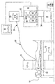
図1は、血管造影図又は蛍光透視画像及び血管内超音波画像のコレジストレーションに有用な本発明のシステムを示している。放射線及び超音波画像データ取得サブシステムは、一般的に、当技術分野においてよく知られている。放射線画像データに関して、患者10が、血管造影テーブル12の上に置かれる。血管造影テーブル12は、テーブル12上の患者10に関して操作位置での血管造影/蛍光透視ユニットCアーム14の位置決めに対して十分な空間を提供するように構成される。血管造影/蛍光透視Cアーム14によって取得される放射線画像データは、伝送ケーブル16を介して血管造影/蛍光透視プロセッサ18に渡る。血管造影/蛍光透視プロセッサ18は、ケーブル16を介して受け取った放射線画像データを、血管造影/蛍光透視画像データに変える。血管造影/蛍光透視(「放射線」)画像データは、最初に、プロセッサ18内に格納される。
FIG. 1 illustrates the system of the present invention useful for co-registration of angiograms or fluoroscopic images and intravascular ultrasound images. Radiation and ultrasound image data acquisition subsystems are generally well known in the art. With respect to the radiographic image data, a
当該システムのうち、超音波画像データの取得に関連する部分に関して、イメージングカテーテル20、特にIVUSカテーテルが、診断プローブ22(特にIVUSプローブ)を含むその遠位端が所望の血管の画像処理位置の近くにあるように患者10内に挿入される。図1において具体的に同定されていないけれども、プローブ22の近くに置かれる放射線不透過性材料が、放射線画像においてプローブ22の現在の位置のしるしを提供する。例として、診断プローブ22は、超音波を生成し、診断プローブ22に近接した領域を表す超音波エコーを受け取り、さらに、超音波エコーを対応する電気信号に変える。対応する電気信号は、イメージングカテーテル20の長手方向に沿って、近位のコネクタ24まで送られる。プローブ22のIVUSバージョンは、単一及び多数のトランスデューサ要素の構成を含む種々の構成で手に入る。多数のトランスデューサ要素の構成の場合、トランスデューサのアレイが潜在的に構成され:イメージングカテーテル20の縦方向の軸に沿って直線的に、カテーテル20の縦方向の軸を中心として曲線をなして、縦方向の軸を中心として周囲に等、構成される。
With respect to the portion of the system related to the acquisition of ultrasound image data, the
カテーテル20の近位のコネクタ24は、カテーテル画像プロセッサ26に通信可能に接続される。カテーテル画像プロセッサ26は、近位のコネクタ24を介して受け取った信号を、例えば、血管セグメントの断面画像に変える。さらに、カテーテル画像プロセッサ26は、血管の長手方向に沿って撮られた血管のスライスに対応する縦断面画像を生成する。カテーテル画像プロセッサ26によって与えられるIVUS画像データは、最初に、プロセッサ26内に格納される。
A
診断プローブ22によって取得され、さらに、カテーテル画像プロセッサ26によって処理される診断画像処理データのタイプは、本発明の代わりとなる実施形態に従って変わる。特定の代わりとなる実施形態に従って、診断プローブ22には、機能流れ測定値(functional flow measurement)とも呼ばれる(例えば血流速度及び圧力等の)血行動態情報を提供するために(例えばドップラー及び/又は圧力等)1つ又は複数のセンサが備えられる。そのような代わりとなる実施形態において、機能流れ測定値は、カテーテル画像プロセッサ26によって処理される。従って、「画像」という用語は、血圧、血流速度/体積、血管断面組成、血液中の剪断応力、血液/血管壁の接触部分での剪断応力等を含む血管情報を表す種々の方法を包含するように広く解釈されるとして意図されるということに留意されたい。血管の特定部分に対する血行動態データを取得する場合、効果的な診断は、脈管構造内の診断プローブ22の現在の位置を、循環器疾患を示す機能流れ測定基準を同時に観察しながら可視化する能力次第である。血行動態及び放射線画像のコレジストレーションは、病気の血管の正確な治療を促進する。或いは、カテーテルが乗せられたセンサの代わりに、ガイドワイヤ、例えば0.018’’以下の直径を有するガイドワイヤの上にセンサを乗せることができる。このように、本発明の実施形態に従って、種々のプローブタイプが使用されるだけでなく、そのようなプローブが遠位端にて乗せられる種々の可撓性の細長い部材(例えばカテーテル、ガイドワイヤ等)も使用される。
The type of diagnostic image processing data acquired by the
コレジストレーションプロセッサ30は、ライン32を介してカテーテル画像プロセッサ26からIVUS画像データを受け取り、さらに、ライン34を介して放射線画像プロセッサ18から放射線画像データを受け取る。或いは、センサ及びプロセッサ間の通信は、ワイヤレスメディアを介して実行される。コレジストレーションプロセッサ30は、受け取った画像データから得られる放射線画像のフレームもIVUS画像のフレームも含むコレジストレーション画像を与える。本発明の一実施形態に従って、しるし(例えば放射線不透過性マーカーのアーチファクト等)が、同時に表示されたIVUS画像データに対応するある位置の放射線画像上に提供される。コレジストレーションプロセッサ30は、最初に、放射線画像プロセッサ18からライン34を介して受け取った血管造影図の画像データを、画像データメモリ40の第1の部分36へバッファーリングする。その後、カテーテル処置手順の過程の間に、ライン32及び34を介して受け取ったIVUS及び放射線不透過性マーカーの画像データが、画像データメモリ40の第2の部分38及び第3の部分42内にそれぞれ格納される。個々に与えられる格納された画像データのフレームは、(例えばタイムスタンプ、シーケンス番号等)適切にタグ付けされて、IVUS画像フレーム及び対応する放射線(放射線不透過性マーカー)画像データのフレームを関連づける。IVUSデータではなく血行動態データが取得される一実施形態において、血行動態データは、第2の部分38内に格納される。
加えて、さらなるマーカーを、血管造影図/蛍光透視画像処理装置の視野内で患者の表面上又は患者の近くに置くことができる。従って、これらのマーカーの位置は、正確な位置において血管造影画像上の放射線不透過性マーカーのアーチファクトを位置決めするために使用される。 In addition, additional markers can be placed on or near the patient's surface within the field of view of the angiogram / fluoroscopic imaging device. Thus, the location of these markers is used to locate the radiopaque marker artifact on the angiographic image at the exact location.
コレジストレーションプロセッサ30は、画像データメモリ40の第1の部分36、第2の部分38及び第3の部分42内に以前に格納されたデータからコレジストレーション画像を与える。例として、特定のIVUS画像のフレーム/スライスが、第2の部分38から選択される。コレジストレーションプロセッサ30は、第2の部分38から選択されたIVUS画像データに対応する第3の部分42内の蛍光透視画像データを同定する。その後、コレジストレーションプロセッサ30は、第3の部分42からの蛍光透視画像データを、第1の部分36から検索された血管造影画像のフレームの上に重ねる。その後、コレジスタされた放射線画像のフレーム及びIVUS画像のフレームが、互いに並んで、グラフィックディスプレイ装置50の上に同時に表示される。ディスプレイ装置50を駆動するコレジスタされた画像データフレームも、画像データメモリ40において格納された放射線画像データ及びIVUS画像データを取得した手順とは別のセッションにおける後の再調査のために、長期記憶装置60上に格納される。
図1において示されていないけれども、制御された/測定された様式にて患者からカテーテル20を引く引戻装置が組み込まれる。そのような装置は、当技術分野においてよく知られている。そのような装置の組み入れは、蛍光透視が活動中ではない時点での視野内のプローブ22の現在の位置を計算することを促進する。
Although not shown in FIG. 1, a pullback device is incorporated that pulls the
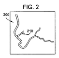
図2は、血管造影/蛍光透視プロセッサ18によってキャプチャされた所望の投影(患者/血管の定位)及び拡大における血管造影「ロードマップ」画像200を示している。例として、画像200は、最初に、患者の脈管構造内の関心のある領域までのIVUSカテーテルの追跡に先立ち行われる血管造影手順によってキャプチャされる。血管内のカテーテル20なしで血管造影手順を行うことによって、最大の造影剤の流れ、より優れた血管の充填、従って、より優れた全体的な血管造影画像が提供される。従って、側枝210等の側枝及び他の脈管構造の目印を、グラフィックディスプレイ装置50の上に表示されるコレジスタされた画像の放射線画像部分の上に表示する及び明確に見ることができる。
FIG. 2 shows an angiographic “roadmap”
図3は、蛍光透視画像において可視のカテーテルの放射線不透過性マーカー300を示している。カテーテル20は、その開始位置(例えばIVUS引戻手順が始まる位置)まで追跡される。典型的には、カテーテル20は、以前に進められたガイドワイヤ(図示せず)にわたって追跡される。その後、蛍光透視画像が得られる。図3に示されている画像において、カテーテルの放射線不透過性マーカー300は可視化されているが、血管腔は、造影剤の流れがないため可視化されていない。しかし、血管造影図においても蛍光透視画像においても存在する位置決定マーカーのセットは、以前に得た血管造影図の画像内でのマーカー画像の適切な位置決め(重ね合わせ)を可能にする。血管造影図の画像の視野内で放射線不透過性マーカー画像を適切に位置決めする他の方法は、本明細書における教示を考慮して当業者には既知になる。さらに、マーカーのアーチファクトを、重ね合わされた画像のフレーム上で(サイズも位置も)自動的に調整して、トランスデューサのおおよその位置に一致させることができる。
FIG. 3 shows a
図4は、血管造影図の画像上への放射線不透過性マーカーのアーチファクトのオーバーレイ又は重ね合わせから生じる例証的なコレジストレーション画像を示している。例証的なコレジストレーションディスプレイ401(関連づけられた放射線画像及びIVUS画像を含む)は、選択された血管の断面IVUS画像400を描いている。放射線画像410が、ディスプレイ50の上でIVUS画像400と並んで同時に表示されている。放射線画像410は、蛍光透視画像のフレームによって与えられた放射線画像データから生成され、メモリ40の第1の部分36から与えられた血管造影図のバックグラウンド上に重ね合わされたマーカーのアーチファクト420を含む。蛍光透視画像のフレームは、観察下で血管内の診断プローブ22の現在の位置に対応している。血管造影図の画像においても蛍光透視画像においても表された視野の正確なマッチング(すなわち、2つの画像の正確な投影及び拡大)は、図4において表示されているコレジスタされた画像の右側の区画における表示されたIVUS画像400に対応するIVUSプローブの現在の位置の同定を可能にする。画像のコレジストレーションの考察は、参照により全内容を本願に援用する特許文献4において見つけることができる。
FIG. 4 shows an exemplary co-registration image resulting from overlay or overlay of radiopaque marker artifacts on the angiogram image. An exemplary co-registration display 401 (including associated radiographic and IVUS images) depicts a
例えば上記の方法を使用することによって、コレジスタされたデータのセットが得られると、次に、コレジスタされたデータのセットを適用して、所与のモダリティにおいて関心のある特徴を検出することを促進することができる。特定の実施形態において、これは、コレジスタされたデータセットを使用して、所与のモダリティにおいて関心のある特徴を検出するためのサーチアルゴリズムを訓練することを含み得る。例えば、関心のある特徴はステントであり得る。ステント及び周囲の脈管構造は、IVUS及びVH−IVUS等の2つのイメージングモダリティを用いて画像化することができる。コレジスタされたデータセットを得るために、特徴が双方から抽出され且つ整列させられる。次に、この組み合わされたデータセットを使用して、例えば、ステントの検出が問題を有することが多くある従来のグレースケールIVUSにおいてステントを検出するためのサーチアルゴリズムを訓練することができる。サーチアルゴリズムを訓練するための適した方法が次に記載される。 Once a co-registered set of data is obtained, for example by using the method described above, the co-registered set of data is then applied to help detect features of interest in a given modality. can do. In certain embodiments, this may include training a search algorithm to detect features of interest in a given modality using the co-registered data set. For example, the feature of interest can be a stent. The stent and surrounding vasculature can be imaged using two imaging modalities such as IVUS and VH-IVUS. Features are extracted from both and aligned to obtain a co-registered data set. This combined data set can then be used, for example, to train a search algorithm for detecting stents in conventional gray scale IVUS, where stent detection is often problematic. A suitable method for training the search algorithm will now be described.
特定の態様において、関心のある特徴が、コレジスタされた血管内データセットで訓練されたサーチアルゴリズムの使用を介してイメージングモダリティにおいて同定される。上記のように、コレジスタされた血管内データセットは、複数のイメージングモダリティからの、血管壁等の関心のある特徴に関する情報を含む。組み合わされたデータセットにおいて提供されるその情報に基づき、サーチアルゴリズムは、所与のイメージングモダリティにおいて関心のある特徴を同定することができる。アルゴリズムは、特定のファクター又はパラメータに焦点を当てて、総合的な評価を行い、さらに、多数のイメージングモダリティから蓄積された位置及び他のデータに基づき関心のある特徴を同定する。一般的に言えば、そのプロセスは、第1のイメージングモダリティを用いて画像を得ること、画像から関心のある特徴を抽出すること、及び、その特徴を位置データ(すなわち座標のセット)に変換することを含む。そのプロセスは、少なくとも、第2のイメージングモダリティを用いて第2の領域の画像を得ること、関心のある特徴を抽出すること、及び、ここでもその特徴を位置データに変換することをさらに含む。第1及び第2のイメージングモダリティからの位置データは、次に、コレジストレーションを介して1つのデータセットに組み合わされ、さらに、組み合わされたデータセットを使用して、所与のイメージングモダリティにおいて関心のある特徴を検出するように構成されたサーチアルゴリズムを訓練する。 In certain aspects, features of interest are identified in an imaging modality through the use of a search algorithm trained on a co-registered intravascular data set. As described above, the co-registered intravascular data set includes information about features of interest, such as vessel walls, from multiple imaging modalities. Based on that information provided in the combined data set, the search algorithm can identify features of interest in a given imaging modality. The algorithm focuses on specific factors or parameters to make a comprehensive assessment and further identify features of interest based on location and other data accumulated from multiple imaging modalities. Generally speaking, the process involves obtaining an image using a first imaging modality, extracting features of interest from the image, and converting the features into location data (ie, a set of coordinates). Including that. The process further includes at least obtaining an image of the second region using the second imaging modality, extracting features of interest, and again converting the features into position data. The position data from the first and second imaging modalities are then combined into one data set via co-registration and further used in the given imaging modality using the combined data set. Train a search algorithm configured to detect certain features.
アルゴリズムの訓練は、一連の反復ステップを含んでもよく、各連続的なステップは、分析に対して入力されたか又は提示された全てのテストデータ(すなわち位置データ)が包括的に評価されるまで、以前のサイクル(前のイメージングモダリティ)のステップにおいて提示された全てのデータ、及び、関心のある特徴についての関連している情報と組み合わせて、新たなデータのそれぞれ(すなわち、さらなるイメージングモダリティから提供されたもの)を評価する。最後のステップの完了後、分析機能は終結し、さらに、検索結果が、分析機能の完了後に形成される。本発明は、イメージングモダリティから受け取った新たな情報に基づく位置データの修正又は更新も熟考し、そのようなデータは検索の精度を改善するために利用可能になり且つ役立つため、アルゴリズムの一部として含まれる。 Algorithm training may include a series of iterative steps, each successive step until all test data (or position data) entered or presented for analysis has been comprehensively evaluated. All of the data presented in the previous cycle (previous imaging modality) step and each new data (ie, provided by the additional imaging modality) combined with relevant information about the feature of interest Evaluate). After completion of the last step, the analysis function is terminated, and further, search results are formed after the analysis function is completed. The present invention also contemplates correction or update of location data based on new information received from the imaging modality, and as such data becomes available and useful to improve the accuracy of searches, as part of the algorithm. included.
本発明のサーチアルゴリズムは、その使用を促進することができるコンピュータプログラム又はコード等、いかなる適したアプリケーションにおいて具体化されてもよい。その適用を具体化するアルゴリズム又はアプリケーションは、コンピュータの内部若しくは外部のハードドライブ、ポータブルドライブ若しくはディスク、サーバー、一時若しくは固定のメモリ装置、又は、アルゴリズム及び/又はその使用から得られる結果の使用を促進することができるいかなる他の記憶手段内に格納されてもよい。アルゴリズム又はアプリケーションは、好ましくは、例えばコンピュータ又はネットワークプロセッサ等、予測分析を促進する少なくとも1つの処理装置と通信している。アルゴリズム又は関連するアプリケーションは、(例えば単一の若しくはネットワーク化されたコンピュータ上で)局所的に、又は、(例えば、インターネットを介した若しくはイントラネットを介したウェブベースのネットワーク等)離れてアクセスされてもよい。 The search algorithm of the present invention may be embodied in any suitable application, such as a computer program or code that can facilitate its use. Algorithms or applications that embody the application facilitate the use of hard drives internal or external to the computer, portable drives or disks, servers, temporary or fixed memory devices, or algorithms and / or results obtained from their use. It may be stored in any other storage means that can. The algorithm or application is preferably in communication with at least one processing device that facilitates predictive analysis, such as a computer or network processor. Algorithms or related applications can be accessed locally (eg, on a single or networked computer) or remotely (eg, over the Internet or via an intranet, etc.) Also good.
このアルゴリズム又はアプリケーションへのアクセスは、コンピュータ、インターネットアプライアンス、電話装置、ワイヤレス装置等を含むがそれらに限定されないいかなる適した機器の使用を介して促進されてもよい。アルゴリズム、アルゴリズム又はアルゴリズムの使用から得られた結果を具体化するアプリケーションへのアクセスは、一般的なアクセスから、或いは、パスワード、暗号化、生体計測による若しくは音声による駆動、又は、他の適した保護手段を介した使用から守られるか若しくは制限されてもよい。 Access to this algorithm or application may be facilitated through the use of any suitable equipment, including but not limited to computers, internet appliances, telephone devices, wireless devices, and the like. Access to the application that embodies the algorithm, the algorithm or the results obtained from the use of the algorithm, from general access, or from passwords, encryption, biometric or voice driven, or other suitable protection It may be protected or restricted from use through means.
本発明によって熟考されるように、上記の機能及び実施形態は、ソフトウェア、ハードウェア、ファームウエア、ハード配線、又は、これらのいかなる組合せも使用して行うことができる。機能を行う機構は、種々の位置にて物理的に置くこともでき、機能の一部分が異なる物理的位置にて行われるように分配されることを含む。 As contemplated by the present invention, the functions and embodiments described above can be performed using software, hardware, firmware, hard wiring, or any combination thereof. The mechanism that performs the function can be physically located at various locations, including being distributed such that a portion of the function is performed at different physical locations.
本発明の方法の遂行に対して必要であるか又は最も適しているとして当業者は認識するように、本発明のコンピュータシステム又は機械は、バスを介して互いに通信する1つ又は複数のプロセッサ(例えば、中央処理装置(CPU)、グラフィック処理装置(GPU)又はその両方等)、メインメモリ及びスタティックメモリを含む。本発明のシステムは、コンピュータ及びプロセッサだけでなく、コンピュータ可読記憶媒体の命令を含んでもよく、コンピュータ可読記憶媒体の命令は、該命令が実行された場合、コンピュータに第1のイメージングモダリティから情報を受け取らせ、さらに、その情報を第1の座標空間に変換させ、第2のイメージングモダリティからの情報を受け取らせ、その情報を第2の座標空間に変換させ、さらに、第1の座標空間を第2の座標空間と整列させ、その結果、第1のモダリティからの情報と第2のモダリティからの情報とを組み合わせて、組み合わされたデータセットにする。この命令は、さらにコンピュータに、組み合わされたデータセットに基づき、第3のイメージングモダリティにおいて関心のある特徴を検出させる。 As those skilled in the art will recognize as necessary or most suitable for performing the methods of the present invention, the computer system or machine of the present invention may include one or more processors (one or more) that communicate with each other via a bus. For example, a central processing unit (CPU), a graphics processing unit (GPU) or both), main memory and static memory. The system of the present invention may include instructions of a computer readable storage medium as well as a computer and a processor, and the instructions of the computer readable storage medium may cause the computer to receive information from the first imaging modality when the instructions are executed. And receiving the information from the second imaging modality, converting the information to the second coordinate space, and further converting the first coordinate space to the first coordinate space. Align with the two coordinate spaces, so that the information from the first modality and the information from the second modality are combined into a combined data set. This instruction further causes the computer to detect features of interest in the third imaging modality based on the combined data set.
図5は、本発明の実施形態によるシステム100を図で示している。システム100は、好ましくは、コンピュータ249(例えばラップトップ、デスクトップ、タブレット又はスマートフォン等)を含む。コンピュータ249は、ネットワーク109を横断して通信するように構成されてもよい。コンピュータ249は、1つ又は複数のプロセッサ159及びメモリ163、並びに、入力/出力機構154を含む。本発明の方法が、クライアント/サーバーアーキテクチャーを利用する場合、本発明の方法のステップは、サーバー113を使用して行われてもよく、サーバー113は、プロセッサ121及びメモリ129のうち1つ又は複数を含み、インターフェースモジュール125を介してデータ、命令等を得る、若しくは、結果を提供するか、又は、ファイル117として結果を提供する能力を有する。サーバー113は、ネットワーク109を超えて、コンピュータ249若しくはターミナル267まで結合されてもよく、又は、サーバー113は、1つ又は複数のプロセッサ175及びメモリ179、並びに、入力/出力機構171を含むターミナル167に直接接続されてもよい。
FIG. 5 schematically illustrates a
本発明によるシステム100又は機械は、I/O154又は171のいずれに対しても、表示装置(例えば液晶ディスプレイ(LCD)又は陰極線管(CRT)等)をさらに含んでもよい。本発明によるコンピュータシステム又は機械は、英数字入力装置(例えばキーボード等)、カーソル制御装置(例えばマウス等)、ディスクドライブ装置、信号生成装置、(例えばスピーカー等)、タッチスクリーン、加速度計、マイクロフォン、セルラー無線周波数アンテナ、及び、例えばネットワークインターフェースカード(NIC)、Wi−Fiカード又はセルラーモデムであり得るネットワークインターフェース装置も含み得る。
The
本発明によるメモリ163、179又は129は機械可読媒体を含むことができ、該媒体上には、本明細書において記載される方法論又は機能のうちいずれか1つ又は複数を具体化する1つ又は複数の命令のセット(例えばソフトウェア等)が格納される。好ましい実施形態において、本発明のコンピュータシステムは、有形の非一時的メモリである1つ又は複数のメモリ装置を含む。ソフトウェアは、コンピュータシステムによるその実行の間、メインメモリ内及び/又はプロセッサ内に、完全に又は少なくとも部分的に存在してもよく、メインメモリ及びプロセッサも、機械可読媒体を構成している。ソフトウェアは、さらに、ネットワークインターフェース装置を介してネットワークを渡って送られるか又は受け取られてもよい。 The memory 163, 179 or 129 according to the present invention may include a machine-readable medium on which one or more of the methodologies or functions described herein may be implemented. A set of a plurality of instructions (for example, software) is stored. In a preferred embodiment, the computer system of the present invention includes one or more memory devices that are tangible non-transitory memory. The software may be wholly or at least partially resident in the main memory and / or processor during its execution by the computer system, and the main memory and processor also constitute a machine-readable medium. The software may also be sent or received across the network via a network interface device.
図6は、本発明の方法のステップを示している。本明細書において記載される方法だけでなく、本明細書において開示されるシステム及び方法のいかなる部分も、上記の装置を含むコンピュータによって行うことができるということが理解されることになる。好ましくは、各ステップは、プロセッサ又は接続された医用画像処理装置によって行われる。画像は、関心のある特徴が同定を要求する所与のモダリティを使用して得られる(201)。モダリティは、例えば、グレースケールIVUSであってもよく、さらに、関心のある特徴は、グレースケールIVUSでは検出するのが困難であるステントである。関心のある特徴に関する情報は、イメージングモダリティから抽出される。その特徴は、関心のある特徴に関する位置データを含む特徴ベクトルに変えられる。このデータは、次に、コンピュータの中央処理装置(CPU)に入力される(202)。CPUは、サーチアルゴリズム等、本発明の方法を行うための命令を格納する記憶装置又はメモリに結合されている。命令は、CPUによって実行される場合、CPUに、イメージングモダリティにおいて選択された関心のある特徴を同定させる。CPUは、イメージングモダリティからのデータを、既知である関心のある特徴に関する位置データに対して複数のイメージングモダリティから得られるコレジスタされたデータのセットで訓練されたアルゴリズムに入力することによって、この決定を提供する(203)。コレジスタされたデータのセットは、コンピュータメモリ内等、コンピュータ内で局所的に格納されてもよい。或いは、コレジスタされたデータセットは、サーバー等、コンピュータから離れた位置において格納されてもよい。この例において、コンピュータは、ネットワークを横断して通信して、コレジスタされたデータセットにアクセスする。CPUは、次に、アルゴリズムに入力されたデータに基づきイメージングモダリティにおいて関心のある特徴を同定する(204)。 FIG. 6 shows the steps of the method of the invention. It will be understood that any portion of the systems and methods disclosed herein, as well as the methods described herein, can be performed by a computer including the apparatus described above. Preferably, each step is performed by a processor or a connected medical image processing apparatus. An image is obtained 201 using a given modality for which the feature of interest requires identification. The modality may be, for example, grayscale IVUS, and the feature of interest is a stent that is difficult to detect with grayscale IVUS. Information about the features of interest is extracted from the imaging modality. The feature is converted into a feature vector that contains position data about the feature of interest. This data is then input to a central processing unit (CPU) of the computer (202). The CPU is coupled to a storage device or memory that stores instructions for performing the method of the present invention, such as a search algorithm. The instructions, when executed by the CPU, cause the CPU to identify the feature of interest selected in the imaging modality. The CPU makes this determination by inputting data from the imaging modality into an algorithm trained on a set of co-registered data obtained from multiple imaging modalities for position data relating to a known feature of interest. Provide (203). The set of co-registered data may be stored locally in the computer, such as in computer memory. Alternatively, the co-registered data set may be stored at a location remote from the computer, such as a server. In this example, the computer communicates across the network to access the co-registered data set. The CPU then identifies (204) the features of interest in the imaging modality based on the data entered into the algorithm.
複数のイメージングモダリティから得られるコレジスタされた位置データのセットを調製した後で、相互相関、主成分分析(PCA)、因子回転、ロジスティック回帰(LogReg)、線形判別分析(LDA)、固有遺伝子(Eigengene)線形判別分析(ELDA)、サポートベクトルマシン(SVM)、ランダムフォレスト(RF)、再帰分割木(RPART)、関連する決定木分類技術、収縮重心(SC)、StepAIC、K−近傍法、ブースティング、決定木、神経回路網、ベイズのネットワーク、サポートベクトルマシン、及び、隠れマルコフモデル、線形回帰若しくは分類アルゴリズム、非線形回帰若しくは分類アルゴリズム、分散分析(ANOVA)、階層分析若しくはクラスタ化アルゴリズム、決定木を使用する階層アルゴリズム、カーネル部分最小二乗アルゴリズム、カーネルマッチング追跡アルゴリズム、カーネルフィッシャー判別分析アルゴリズム若しくはカーネル主成分分析アルゴリズム等のカーネルベースのマシンアルゴリズム、又は、他の数学的及び統計学的方法等、よく知られた技術を使用して、サーチアルゴリズムを開発することができる。複数のイメージングモダリティから得られるコレジスタされた位置データのセットは、そのアルゴリズムを訓練するために使用される。イメージングモダリティにおいて選択された関心のある特徴を同定するために、関心のある特徴に関する位置データが、複数のイメージングモダリティから得られ、さらに、コレジスタされた位置データのセットに組み合わされ、次に、コレジスタされた位置データのセットは、入力データ(選択された個々の集団から得られるコレジスタされた位置データに適合されるサーチアルゴリズムへの入力)として使用される。いかなる式又はアルゴリズムも、種々のイメージングモダリティから抽出される関心のある特徴に基づき位置データを組み合わせるために使用することができる。 After preparing a set of co-registered location data from multiple imaging modalities, cross-correlation, principal component analysis (PCA), factor rotation, logistic regression (LogReg), linear discriminant analysis (LDA), eigengene (Eigengene) ) Linear discriminant analysis (ELDA), support vector machine (SVM), random forest (RF), recursive partition tree (RPART), related decision tree classification techniques, contraction centroid (SC), StepAIC, K-neighbor method, boosting Decision trees, neural networks, Bayesian networks, support vector machines, and hidden Markov models, linear regression or classification algorithms, nonlinear regression or classification algorithms, analysis of variance (ANOVA), hierarchical analysis or clustering algorithms, decision trees use Well known, such as layer algorithms, kernel partial least squares algorithms, kernel matching tracking algorithms, kernel-based machine algorithms such as kernel Fisher discriminant analysis algorithms or kernel principal component analysis algorithms, or other mathematical and statistical methods Techniques can be used to develop search algorithms. A set of co-registered position data obtained from multiple imaging modalities is used to train the algorithm. In order to identify the feature of interest selected in the imaging modality, location data for the feature of interest is obtained from the plurality of imaging modalities and further combined into a coregistered set of location data, and then coregister The set of location data is used as input data (input to a search algorithm adapted to co-registered location data obtained from selected individual populations). Any equation or algorithm can be used to combine position data based on features of interest extracted from various imaging modalities.
種々の好ましい式がここで記載されるけれども、ここで記載される及び上記のものの他にいくつか他のモデル及び式のタイプが、当業者にはよく知られている。使用される実際のモデルタイプ又は式は、それ自体が、訓練集団(training population)における能力及びその結果の診断精度の特徴に基づき可能なモデルの分野から選択されてもよい。式の詳述自体は、一般的に、関連している訓練集団における選択されたパラメータの結果から得ることができる。数ある用途の中でも、そのような式は、1つ又は複数の選択されたパラメータの入力から得られる特徴空間をサブジェクトクラスのセットに変換する、ベイズのアプローチを使用してリスクの確率関数の推定を得る、又は、クラス条件確率を推定し、次に、前のようにベイズのルールを使用してクラス確率関数を生成するよう意図され得る。 Although various preferred formulas are described herein, several other models and formula types are well known to those skilled in the art in addition to those described herein and above. The actual model type or formula used may itself be selected from the field of possible models based on the capabilities in the training population and the resulting diagnostic accuracy characteristics. The formula detail itself can generally be derived from the results of selected parameters in the associated training population. Among other applications, such an expression is an estimate of the probability function of risk using a Bayesian approach that transforms the feature space resulting from the input of one or more selected parameters into a set of subject classes. Or estimate the class condition probabilities and then use Bayes rules as before to generate a class probability function.
好ましい式は、広範なクラスの統計学的分類アルゴリズムを含み、特に、判別分析の使用を含む。この判別分析の目的は、以前に同定された特徴のセットから集合の要素を予想することである。線形判別分析(LDA)の場合、一部の基準によって群の中からの分離を最大にする特徴の線形結合が同定される。特徴は、異なる閾値を用いる固有遺伝子(eigengene)ベースのアプローチ(ELDA)、又は、多変量分散分析(MANOVA)に基づくステッピングアルゴリズムを使用して、LDAに対して同定することができる。Hotelling−Lawleyの統計量に基づき分離なしの確率を最小限にするフォーワード、バックワード及びステップワイズアルゴリズムを行うことができる。 Preferred formulas include a broad class of statistical classification algorithms, especially the use of discriminant analysis. The purpose of this discriminant analysis is to predict the elements of a set from a previously identified set of features. For linear discriminant analysis (LDA), some criteria identify a linear combination of features that maximizes separation from the group. Features can be identified for LDA using an eigengene based approach (ELDA) with different thresholds or a stepping algorithm based on multivariate analysis of variance (MANOVA). Forward, backward and stepwise algorithms can be performed that minimize the probability of no separation based on the Hotelling-Lawley statistic.
固有遺伝子に基づく線形判別分析(ELDA)は、非特許文献1において開発且つ報告された特徴選択技術である。式は、修正された固有値分析を使用して多変量フレームワークにおいて特徴(例えばパラメータ等)を選択し、最も重要な固有ベクトルに関連する特徴を同定する。「重要」は、ある閾値と比較して分類されるよう試みるサンプル間の差における最大の分散を説明する固有ベクトルとして定義される。
Linear discriminant analysis (ELDA) based on a unique gene is a feature selection technique developed and reported in
サポートベクトルマシン(SVM)は、2つのクラスを分離する超平面を見つけることを試みる分類式である。この超平面は、まさしく超平面からのマージン距離であるサポートベクトル、データポイントを含有している。現在のデータの次元において分離超平面が存在しないあり得る事象において、次元数は、元の変数の非線形関数をとることによりデータをより高い次元に投影することによって大きく拡張される(非特許文献2)。要求はされないけれども、SVMに対する特徴のフィルタリングによって、予測が改善されることが多くある。特徴(例えばパラメータ/バイオマーカー等)を、ノンパラメトリックなKruskal−Wallis(KW)テストを使用してサポートベクトルマシンに対して同定して、最も優れた単変量特徴を選択することができる。ランダムフォレスト又は再帰分割(RPART,非特許文献3)も、最も重要なパラメータの組合せを同定するために、別々に又は組み合わせて使用することができる。ランダムフォレスト及び再帰分割は、参照により全内容を本願に援用する非特許文献4、非特許文献5、非特許文献6、特許文献5及び特許文献6において考察されている。KWもRFも、多くの特徴が全特徴から選択されるということを要求する。RPARTは、利用可能なパラメータのサブセットを使用して単一の分類木を作製する。 A support vector machine (SVM) is a classification formula that attempts to find a hyperplane that separates two classes. This hyperplane contains support vectors, data points that are exactly the margin distance from the hyperplane. In a possible event where there is no separation hyperplane in the current data dimension, the number of dimensions is greatly expanded by projecting the data to a higher dimension by taking a nonlinear function of the original variable (2). ). Although not required, feature filtering for SVMs often improves prediction. Features (eg, parameters / biomarkers, etc.) can be identified to a support vector machine using a non-parametric Kruskal-Wallis (KW) test to select the best univariate features. Random forest or recursive partitioning (RPART, Non-Patent Document 3) can also be used separately or in combination to identify the most important parameter combinations. Random forest and recursive partitioning are discussed in Non-Patent Document 4, Non-Patent Document 5, Non-Patent Document 6, Patent Document 5 and Patent Document 6, the entire contents of which are incorporated herein by reference. Both KW and RF require that many features be selected from all features. RPART creates a single classification tree using a subset of available parameters.
他の式を使用して、個々の選択されたパラメータの測定値の結果を、予測式に対するその提示に先立ち、より価値のある情報の形に前処理してもよい。中でも注目すべきは、集団の平均値に関して、正規分布又は他の分布の位置として、対数又はロジスティック関数等の一般的な数学的変換を使用したパラメータ結果の標準化等は、当業者にはよく知られている。 Other formulas may be used to pre-process the results of individual selected parameter measurements prior to their presentation to the prediction formula in the form of more valuable information. It should be noted that standardization of parameter results using a general mathematical transformation such as logarithm or logistic function as the position of normal distribution or other distribution with respect to the mean value of the population is well known to those skilled in the art. It has been.
1つの対象の個々のパラメータの値が潜在的に標準化されることに加えて、全ての対象又はいかなる既知のクラスの対象に対する総合的な予測式も、それ自体を、非特許文献7において概説されている技術、又は、他の類似の標準化及び再校正技術に従って、集団の予測有病率及び平均パラメータ値に対する調整に基づき再校正するか、又さもなければ、調整することができる。そのような調整統計量は、機械可読であってもよいモデルに対して提示された過去のデータのレジストリを介して、又さもなければ、若しくは時折、そのようなパラメータ及び統計量の史的研究に対する格納されたサンプル若しくは参照の遡及クエリ(retrospective query)を介して連続的に獲得、確認、改善及び更新することができる。式の再校正又は他の調整の対象であってもよいさらなる例は、オッズ比の制限に対する研究において使用される統計量(非特許文献8を参照)、及び、リスク予測における受信者特性(ROC)曲線(非特許文献9、非特許文献10及び非特許文献11を参照)を含む。さらに、分類子の式の数値結果自体が、選択された関心のある特徴がすでに同定且つ確認されている実際の正の制御に対するその参照によって後処理変換されてもよい。
In addition to the potential standardization of individual parameter values for one object, a comprehensive prediction formula for all objects or any known class of objects is itself outlined in Non-Patent Document 7. Can be recalibrated or otherwise adjusted based on adjustments to the population's predicted prevalence and average parameter values, according to existing techniques, or other similar standardization and recalibration techniques. Such adjusted statistics can be obtained through a registry of historical data presented for models that may be machine-readable, or otherwise, sometimes historical studies of such parameters and statistics. Can be obtained, verified, refined and updated continuously via stored sample or reference retrospective queries. Further examples that may be subject to equation recalibration or other adjustments include statistics used in studies on odds ratio limitations (see Non-Patent Document 8), and receiver characteristics (ROC) in risk prediction. ) Curve (see Non-Patent Document 9,
図7は、関心のある特徴を探すために使用することができるモデル開発300に対する例証的な方法を表す流れ図である。方法300は、図5の例となるコンピューティングシステム環境100を使用して行われてもよく、さらに、環境100の作動を説明するために使用されることになる。しかし、方法300は、コンピューティングシステム環境100とは異なるシステムによって行うことができるということが認識されるべきである。ブロック301にて、選択された関心のある特徴に関するコレジスタされた位置データのセットが、本明細書において記載されてきたように、システムのメモリ129、内部若しくは外部のデータベース、又は、他のコンピュータ記憶媒体等のデータ記憶装置から得られる。コレジスタされたデータセットは、最初に、画像において関心のある特徴をキャプチャする種々のイメージングモダリティを含む種々の手段を介して得られてもよく、特徴は、そのイメージングモダリティから抽出され、さらに、位置データ(すなわち、座標のセット)に変えられる。
FIG. 7 is a flow diagram representing an exemplary method for
ブロック302にて、コレジスタされたデータセットが、以下に記載されるように、サーチアルゴリズムを訓練するために使用されることになるモデル又は分析の要求を満たすように必要に応じて調製される。例えば、データセットの調製は、各イメージングモダリティから位置データ又はその選ばれたサブセットを調製することを含んでもよい。必要に応じて、(例えば、最近傍補間又は他のパターン認識等の)間隔閉塞(gap fill)技術、品質検査、(例えば統計学的分類アルゴリズム等の)種々の式を使用するデータの組合せ、(例えば10進法、自然対数等の)モデルの要求を満たすようにデータの分布を変える対数関数等の標準化及び/又は変換等、種々のデータ調製方法が、アルゴリズムを訓練することに先立ちデータを調製するために使用されてもよい。ここでも、特定のデータ調製手順は、コレジスタされたデータセットを使用して訓練されることになる1つ又は複数のモデル次第である。種々の異なるモデルタイプに対する特定のデータ調製技術は既知であり、さらに記載する必要はない。
At
ブロック303にて、特定の抽出された特徴が、パラメータを選択するために使用される位置データに変換され、パラメータは、所与のイメージングモダリティにおいて関心のある特徴を同定するために使用されるサーチモデルの訓練において後に使用される。コレジスタされた位置データの使用は、最も再現性のある結果を提供するコレジスタされたセットからの位置データのみを利用することを含んでもよい。データセット確認の例は、クロス確認及びブートストラッピングを含んでもよいが、それらに限定されない。パラメータ選択から、所与のイメージングモダリティにおいて関心のある特徴を同定するための最適なサーチモデルが決定且つ選択されてもよい。しかし、全てのモデルが、同じデータセットを用いて同じ結果を提供するわけではないということに留意されたい。例えば、モデルによって、利用する位置データの態様は異なり、さらに、もたらされる結果は異なってもよく、その結果、最適化されたモデルにおける選択される位置データに対する有意性を加える。従って、多数の選択モデルが、所与のモダリティにおいて関心のある特徴を同定するための最適なサーチモデルを同定するために、コレジスタされたデータセット又はコレジスタされたデータセットのサブセットを用いて選ばれ且つ利用されてもよい。コレジスタされたデータセットから位置データを選択するために使用することができる統計学的モデル、アルゴリズム等を含む特定のモデルの例は先に記載されている。
At
データセット又はそのサブセットを用いて使用される選択モデル毎に、位置データが、モデルにおけるその統計学的有意性に基づき選択される。各モデル内に入力される場合、位置データは、統計学的有意性に対する種々の基準に基づき選択され、さらに、累積投票及び重みづけを含んでもよい。統計学的有意性に対するテストは、出口テスト(exit−test)及び分散分析(ANOVA)を含んでもよい。モデルは、(例えばLDA、ロジスティック回帰、SVM、RF、木モデル等の)分類モデル、及び、(例えばコックス等の)生存モデルを含んでもよく、その多くの例が先に記載されている。 For each selected model used with the data set or a subset thereof, location data is selected based on its statistical significance in the model. When entered within each model, location data is selected based on various criteria for statistical significance and may further include cumulative voting and weighting. Tests for statistical significance may include exit-test and analysis of variance (ANOVA). Models may include classification models (eg, LDA, logistic regression, SVM, RF, tree models, etc.) and survival models (eg, Cox), many examples of which have been described earlier.
ブロック304にて、関心のある特徴を同定するために使用されることになるサーチモデルが、選択、訓練及び確認される。特に、主要な候補モデルが、1つ又は複数の性能基準に基づき選択されてもよく、その例は、先に記載されている。例えば、データセット又はデータのサブセットを使用することから、種々のモデルでは、モデルだけが、統計学的に有意なパラメータを決定するために使用されるのではなく、結果も、そのパラメータと共に最適なモデルを選択するために使用されてもよい。そのようなものとして、リスクを評価するために使用される評価モデルは、分類モデル及び生存モデルを含む選択モデルとして使用されるモデルのうちの1つを含んでもよい。マーカーのサブセットを含む、モデルマーカーの組合せが、サブセット及び個々のデータのセットにおいて比較及び確認されてもよい。その比較及び確認は、モデルを訓練及び確認するため、並びに、適切なモデルを選ぶために何度も繰り返されてもよく、そのモデルは、次に、糖尿状態のリスクを評価するための評価モデルとして使用される。
At
次に、特定の筋書きにおける本発明の適用が参照される。これらの実施形態は限定的ではないということが理解されることになる。以下の例において例示されているように、本発明は、多数のデータセット又はイメージングモダリティから得られる血管画像における、血管の境界及びステントストラット等の関心のある特徴の検出に関する。本発明の方法は、多数のイメージングモダリティからの潜在的に補足的な情報にてこ入れし、さらに、各画像から抽出された情報を組み合わせて、標的特徴を検出する。 Reference is now made to the application of the invention in a specific scenario. It will be understood that these embodiments are not limiting. As illustrated in the examples below, the present invention relates to the detection of features of interest such as vessel boundaries and stent struts in vascular images obtained from multiple data sets or imaging modalities. The method of the present invention leverages potentially complementary information from multiple imaging modalities, and further combines information extracted from each image to detect target features.
画像セグメンテーションは、デジタル画像を多数のセグメント(スーパーピクセルとしても既知のピクセルのセット)に分割するプロセスである。セグメンテーションの目的は、画像の表示を、より有意義で分析するのが容易な何かに単純化する及び/又は変えることである。画像セグメンテーションは、典型的には、画像内の対象及び境界(線、曲線等)の位置を突きとめるために使用される。セグメンテーションに対する1つのアプローチは、画像データから特徴をコンピュータ計算及び抽出することである。特徴は、(例えば、位置データに変換する等)組み合わせて特徴ベクトルにすることができ、次に、既知の特徴ベクトル(すなわち至適規準情報)と比べる、又は、訓練された分類子(例えば神経回路網又は他のアルゴリズム等)にかけて、画像データを所定のクラスに分類することができる。 Image segmentation is the process of dividing a digital image into a number of segments (a set of pixels, also known as superpixels). The purpose of segmentation is to simplify and / or change the display of the image to something more meaningful and easy to analyze. Image segmentation is typically used to locate objects and boundaries (lines, curves, etc.) within an image. One approach to segmentation is to compute and extract features from image data. Features can be combined into feature vectors (eg, converted to location data, etc.) and then compared to known feature vectors (ie, optimal criteria information) or trained classifiers (eg, nerves). The image data can be classified into a predetermined class through a network or other algorithm.
図8は、デジタル画像を多数のセグメントに分割するプロセスを図で表している。図8において示されているように、画像が得られ(801)、さらに、画像から特徴が抽出される(802)。次に、特徴が、(例えば位置データに変換される等)組み合わされて特徴ベクトルになり(803)、次に、特徴ベクトルを使用してサーチアルゴリズムを訓練し(804)、所与のモダリティにおいて選択された関心のある特徴を同定することができる(805)。しかし、多数のイメージングモダリティから得られる空間的にコレジスタされた血管内画像のセットが与えられると、特徴を、多数の画像からコンピュータ計算及び抽出し、さらに、組み合わせて単一の特徴ベクトルにすることができる。マルチモーダルソースの場合、多数のイメージングモダリティに基づくコレジスタされたデータセットは、補足的な情報を提供することができ、結果として生じる特徴ベクトルは、シングルソースから抽出される場合よりも多くの情報を含有する。従って、このコレジスタされたデータのセットで訓練されたサーチアルゴリズムは、より正確な結果を提供するはずである。 FIG. 8 illustrates the process of dividing a digital image into a number of segments. As shown in FIG. 8, an image is obtained (801) and features are extracted from the image (802). The features are then combined into feature vectors (eg, converted to location data, etc.) (803), and then the search algorithm is trained using the feature vectors (804) at a given modality. Selected features of interest can be identified (805). However, given a set of spatially co-registered intravascular images derived from multiple imaging modalities, features are computed and extracted from multiple images and combined into a single feature vector Can do. For multimodal sources, a co-registered data set based on multiple imaging modalities can provide supplemental information, and the resulting feature vector can contain more information than if extracted from a single source. contains. Thus, a search algorithm trained on this co-registered data set should provide more accurate results.
図9は、多数のイメージングモダリティによってキャプチャされた画像を例示し(901)、さらに、画像セグメンテーションプロセスの一例を与えている。上記のように、いかなるイメージングモダリティも、本発明に従って使用することができる。例えば、図9において描かれているイメージングモダリティは、IVUS、X線血管造影及びOCTを含み得る。画像が得られた(901)後、画像から特徴が抽出され(902)、さらに、位置データに変換される(903)。位置データは、次に、組み合わされ且つ整列させられて、コレジスタされたデータのセットにされる(904)。このコレジスタされたデータのセット(904)は、次に、サーチアルゴリズムを訓練するために使用され(905)、サーチアルゴリズムは、所与のモダリティにおいて関心のある特徴を同定するために使用することができる(906)。本発明の方法は、以下の非限定的な筋書きにおいて容易に適用することができる。 FIG. 9 illustrates (901) an image captured by a number of imaging modalities and further provides an example of an image segmentation process. As noted above, any imaging modality can be used in accordance with the present invention. For example, the imaging modalities depicted in FIG. 9 may include IVUS, X-ray angiography and OCT. After the image is obtained (901), features are extracted from the image (902) and further converted into position data (903). The position data is then combined and aligned into a co-registered set of data (904). This co-registered set of data (904) is then used to train a search algorithm (905), which can be used to identify features of interest in a given modality. Yes (906). The method of the present invention can be readily applied in the following non-limiting scenario.
VH−IVUS(バーチャルヒストロジー血管内超音波)及びグレースケールIVUS(血管内超音波):ステントは、従来のグレースケールIVUSを使用して検出するのが困難であると既知である。しかし、本発明の方法を使用して、補足的なIVUS及びVH−IVUSのイメージングモダリティを組み合わせてより多くの情報を提供することによって、非生物学的な関心のある特徴を検出することができる。VH分析によって、VH−IVUS及びグレースケールIVUSのデータは、すでに空間的にコレジスタされる。VH−IVUSにおけるステントは、IVUS画像において容易に同定することができる明瞭な形を有する。この情報は、グレースケールIVUSのデータセットにおけるステント検出に対する検索方法を初期化するために使用するか、又は、ステント検出アルゴリズムの一部としてグレースケールIVUSのデータセットから抽出された特徴と組み合わせることができる。 VH-IVUS (Virtual Histology Intravascular Ultrasound) and Grayscale IVUS (Intravascular Ultrasound): Stents are known to be difficult to detect using conventional grayscale IVUS. However, the method of the present invention can be used to detect features of nonbiological interest by combining supplemental IVUS and VH-IVUS imaging modalities to provide more information. . By VH analysis, VH-IVUS and grayscale IVUS data are already spatially co-registered. Stents in VH-IVUS have a well-defined shape that can be easily identified in IVUS images. This information can be used to initialize a search method for stent detection in the grayscale IVUS data set or combined with features extracted from the grayscale IVUS data set as part of the stent detection algorithm. it can.
グレースケールIVUS及びOCTのステント検出:上記のように、ステントは、グレースケールIVUSのみを使用して検出するのが困難である。この例において、本発明の方法は、コレジスタされたIVUS及びOCTデータのセットを利用して、ステント検出に寄与する。空間的にコレジスタされたグレースケールIVUS及びOCTデータセットのセットが与えられると、特徴を各モダリティから抽出し、さらに、組み合わせて、上記の単一の特徴ベクトルにすることができる。この例は、VH−IVUS、グレースケールIVUS及びOCTを包含するアプローチまで拡張することができる。 Grayscale IVUS and OCT stent detection: As noted above, stents are difficult to detect using only grayscale IVUS. In this example, the method of the present invention utilizes a co-registered IVUS and OCT data set to contribute to stent detection. Given a set of spatially co-registered grayscale IVUS and OCT data sets, features can be extracted from each modality and further combined into the single feature vector described above. This example can be extended to approaches that include VH-IVUS, grayscale IVUS and OCT.
グレースケールIVUS及びOCTの血管及び管腔の境界検出:この例は上記と同じであるが、関心の特徴は、ここでは非生物学的ではなく生物学的である。この特定のイメージングモダリティの組合せは、管腔境界の検出として有意に有利であり得、グレースケールIVUSのみでは、血液の存在により、極度にチャレンジングなものであり得る。一方、OCTは、血液が流れる血管において画像データを取得し、管腔境界を画像化することを促進する。関心のある特徴が血管境界である場合、血管境界は常に管腔の外側にあり、さらに、OCTデータは管腔を同定することにより適しているという事実を除いては、OCTデータはより有意ではなくなる。 Grayscale IVUS and OCT vessel and lumen boundary detection: This example is the same as above, but the feature of interest here is biological rather than non-biological. This particular combination of imaging modalities can be significantly advantageous for detection of luminal boundaries, and grayscale IVUS alone can be extremely challenging due to the presence of blood. On the other hand, OCT facilitates acquiring image data in a blood vessel through which blood flows and imaging a lumen boundary. If the feature of interest is a vascular boundary, the vascular boundary is always outside the lumen, and the OCT data is more significant except for the fact that the OCT data is more suitable for identifying the lumen. Disappear.
これらの例は限定的ではないということ、並びに、本発明は全てのイメージングモダリティ及び全ての関心のある特徴を、生物学的であろうと非生物学的であろうと、含むということが理解されることになる。例えば、この血管マルチモーダルアプローチは、IVUS及び/又はOCTデータセットとコレジスタされるX線血管造影及び/又はCT血管造影のデータに適用することもできる。さらに、本発明は、唯一の血管内イメージングモダリティとしてIVUS及びOCTに限定されない。他の適した血管内イメージングモダリティは、MRI及び光音響画像処理を含む。本明細書において記載されるマルチモーダルアプローチの主要な利益は、利用可能な場合は補足的なイメージングモダリティから得られるさらなる情報を利用することである。このさらなる情報は、提供される方法に従って使用される場合に、血管データセットにおける関心のある特徴の検出を改善又は促進する。 It will be understood that these examples are not limiting, and that the present invention includes all imaging modalities and all features of interest, whether biological or non-biological. It will be. For example, this vascular multimodal approach can also be applied to x-ray angiography and / or CT angiography data co-registered with IVUS and / or OCT data sets. Furthermore, the present invention is not limited to IVUS and OCT as the only intravascular imaging modalities. Other suitable intravascular imaging modalities include MRI and photoacoustic image processing. A major benefit of the multimodal approach described herein is to take advantage of additional information obtained from supplemental imaging modalities when available. This additional information improves or facilitates detection of features of interest in the vascular dataset when used in accordance with the provided methods.
参照による援用
特許、特許出願、特許公開、ジャーナル、本、論文、ウェブコンテンツ等、他の文書が、本開示を通して参照及び引用される。そのような文書は全て、全内容が全ての目的のため参照により本願に援用される。
INCORPORATION BY REFERENCE Other documents such as patents, patent applications, patent publications, journals, books, papers, web content, etc. are referenced and cited throughout this disclosure. All such documents are hereby incorporated by reference in their entirety for all purposes.
同等物
本発明は、その真意又は必須の特徴から逸脱することなく、他の特定の形で具体化されてもよい。上記の実施形態は、従って、あらゆる点で、本明細書において記載される本発明に対して限定的ではなく例示的であると考慮されることになる。本発明の範囲は、従って、上記の説明によってではなく、付随の特許請求の範囲によって示されており、従って、特許請求の範囲の同等物の意味及び範囲にある全ての変更が、その中に含まれると意図される。
Equivalents The present invention may be embodied in other specific forms without departing from its true or essential characteristics. The above embodiments are therefore to be considered in all respects illustrative rather than limiting on the invention described herein. The scope of the invention is, therefore, indicated by the appended claims rather than by the foregoing description, and thus, all modifications within the meaning and scope of the equivalents of the claims are included therein. Intended to be included.
Claims (19)
前記プロセッサが、第1のイメージングモダリティから情報を受け取り、さらに、前記第1のイメージングモダリティからの情報を第1の座標空間に変換するステップであり、前記第1のイメージングモダリティは、患者の血管内に置かれる血管内装置に付随した血管内イメージングモダリティを含む、ステップ、
前記プロセッサが、第2のイメージングモダリティから情報を受け取り、さらに、前記第2のイメージングモダリティからの情報を第2の座標空間に変換するステップであり、前記第2のイメージングモダリティは、超音波、光音響イメージング、バーチャルヒストロジー血管内超音波(VH−IVUS)、光干渉断層撮影(OCT)、X線血管造影、蛍光透視又は磁気共鳴画像処理(MRI)のうち少なくとも1つを含む、ステップ、
前記プロセッサが、前記第1の座標空間と前記第2の座標空間とを整列させ、その結果、前記第1のイメージングモダリティからの情報と前記第2のイメージングモダリティからの情報とを組み合わせて、組み合わされたデータセットにするステップ、及び、
前記プロセッサが、前記組み合わされたデータセットに基づき、血管画像における関心のある特徴を検出するステップであり、関心のある特徴の検出は、前記組み合わされたデータセットに基づき前記関心のある特徴を検出するためにアルゴリズムを訓練すること、及び、前記組み合わされたデータセットに基づき前記関心のある特徴に対する検索を初期化することを含む、ステップ、
を含む作動方法。 A method of operating a system for detecting features of interest in a blood vessel image , including a processor, comprising :
The processor receives information from a first imaging modality and further converts the information from the first imaging modality into a first coordinate space, wherein the first imaging modality Including an intravascular imaging modality associated with an intravascular device placed in
The processor receiving information from a second imaging modality and further converting the information from the second imaging modality into a second coordinate space, the second imaging modality comprising ultrasound, light Including at least one of acoustic imaging, virtual histologic intravascular ultrasound (VH-IVUS), optical coherence tomography (OCT), x-ray angiography, fluoroscopy or magnetic resonance imaging (MRI),
The processor aligns the first coordinate space and the second coordinate space so that information from the first imaging modality and information from the second imaging modality are combined and combined. The resulting data set, and
The processor detecting features of interest in a vascular image based on the combined data set, wherein detecting features of interest detects the features of interest based on the combined data set; Training an algorithm to: and initializing a search for the feature of interest based on the combined data set;
Operation method including.
プロセッサと、
命令を含むコンピュータ可読記憶媒体と、
を含み、
前記命令は、実行された場合に、当該システムに、
患者の血管内に置かれる血管内装置に付随した血管内イメージングモダリティを含む第1のイメージングモダリティから情報を受け取らせ、さらに、前記第1のイメージングモダリティからの情報を第1の座標空間に変換させ、
超音波、光音響イメージング、バーチャルヒストロジー血管内超音波(VH−IVUS)、光干渉断層撮影(OCT)、X線血管造影、蛍光透視又は磁気共鳴画像処理(MRI)のうち少なくとも1つを含む第2のイメージングモダリティから情報を受け取らせ、さらに、前記第2のイメージングモダリティからの情報を第2の座標空間に変換させ、
前記第1の座標空間と前記第2の座標空間とを整列させ、その結果、前記第1のイメージングモダリティからの情報と前記第2のイメージングモダリティからの情報とを組み合わせて、組み合わされたデータセットにさせ、さらに、
前記組み合わされたデータセットに基づき、血管画像における関心のある特徴を検出させ、関心のある特徴の検出は、前記組み合わされたデータセットに基づき前記関心のある特徴を検出するためにアルゴリズムを訓練すること、及び、前記組み合わされたデータセットに基づき前記関心のある特徴に対する検索を初期化することを含む、システム。 A system for determining the degree of improvement after a therapeutic procedure,
A processor;
A computer readable storage medium containing instructions;
Including
When the instructions are executed, the system
Receiving information from a first imaging modality, including an intravascular imaging modality associated with an intravascular device placed in a patient's blood vessel, and further converting the information from the first imaging modality to a first coordinate space ,
Includes at least one of ultrasound, photoacoustic imaging, virtual histology intravascular ultrasound (VH-IVUS), optical coherence tomography (OCT), X-ray angiography, fluoroscopy or magnetic resonance imaging (MRI) Receiving information from a second imaging modality, further transforming information from the second imaging modality into a second coordinate space;
Aligning the first coordinate space and the second coordinate space, resulting in a combination of information from the first imaging modality and information from the second imaging modality; And then
Based on the combined data set, a feature of interest in a blood vessel image is detected, and the detection of the feature of interest trains an algorithm to detect the feature of interest based on the combined data set. And initializing a search for the feature of interest based on the combined data set.
前記第1のイメージングモダリティを用いて第1の画像を取得させる、
前記第1の画像内の前記関心のある特徴を同定させる、
前記第1の画像内の関心のある特徴に関連する位置データを取得させる、
前記第2のイメージングモダリティを用いて第2の画像を取得させる、
前記第2の画像内の前記関心のある特徴を同定させる、
前記第2の画像内の関心のある特徴に関連する位置データを取得させる、
前記第1の画像からの前記位置データと前記第2の画像からの前記位置データとを組み合させる、さらに、
結果として生じるデータセットを使用させて、前記第1のイメージングモダリティ及び前記第2のイメージングモダリティのうち少なくとも1つにおいて前記関心のある特徴を前記アルゴリズムが検出するのを可能にする、
命令を含む、請求項8に記載のシステム。 The computer readable storage medium further includes instructions operable to cause the system to train the algorithm;
Acquiring a first image using the first imaging modality;
Identifying the feature of interest in the first image;
Obtaining position data associated with a feature of interest in the first image;
Obtaining a second image using the second imaging modality;
Identifying the feature of interest in the second image;
Obtaining position data associated with a feature of interest in the second image;
Combining the position data from the first image and the position data from the second image;
Using the resulting data set to enable the algorithm to detect the feature of interest in at least one of the first imaging modality and the second imaging modality;
The system of claim 8, comprising instructions.
プロセッサと、
命令を含むコンピュータ可読記憶媒体と、
を含み、
前記命令は、実行された場合に、当該システムに、
患者の血管内に置かれる血管内装置に付随した血管内イメージングモダリティを含む第1のイメージングモダリティから位置データを受け取らせ、さらに、前記位置データを第1の座標のセットに変換させ、
前記第1のイメージングモダリティとは異なる第2のイメージングモダリティから位置データを受け取らせ、さらに、前記位置データを第2の座標のセットに変換させ、
前記第1の座標のセットと前記第2の座標のセットとを整列させることによって、組み合わされたデータセットを作成させ、さらに、
前記組み合わされたデータセットに基づき、血管画像における関心のある特徴を検出させる、
システム。 A system for detecting features of interest in blood vessel images,
A processor;
A computer readable storage medium containing instructions;
Including
When the instructions are executed, the system
Receiving position data from a first imaging modality, including an intravascular imaging modality associated with an intravascular device placed in a patient's blood vessel, and converting the position data to a first set of coordinates;
Receiving position data from a second imaging modality different from the first imaging modality, further converting the position data to a second set of coordinates;
Creating a combined data set by aligning the first set of coordinates and the second set of coordinates; and
Based on the combined data set, the feature of interest in the blood vessel image is detected.
system.
Applications Claiming Priority (3)
| Application Number | Priority Date | Filing Date | Title |
|---|---|---|---|
| US201361774154P | 2013-03-07 | 2013-03-07 | |
| US61/774,154 | 2013-03-07 | ||
| PCT/US2014/021659 WO2014138555A1 (en) | 2013-03-07 | 2014-03-07 | Multimodal segmentation in intravascular images |
Publications (3)
| Publication Number | Publication Date |
|---|---|
| JP2016525893A JP2016525893A (en) | 2016-09-01 |
| JP2016525893A5 JP2016525893A5 (en) | 2017-06-22 |
| JP6243453B2 true JP6243453B2 (en) | 2017-12-06 |
Family
ID=51487895
Family Applications (1)
| Application Number | Title | Priority Date | Filing Date |
|---|---|---|---|
| JP2015561702A Active JP6243453B2 (en) | 2013-03-07 | 2014-03-07 | Multimodal segmentation in intravascular images |
Country Status (5)
| Country | Link |
|---|---|
| US (1) | US9770172B2 (en) |
| EP (1) | EP2965263B1 (en) |
| JP (1) | JP6243453B2 (en) |
| CN (2) | CN113705586A (en) |
| WO (1) | WO2014138555A1 (en) |
Families Citing this family (46)
| Publication number | Priority date | Publication date | Assignee | Title |
|---|---|---|---|---|
| JP6125615B2 (en) * | 2013-04-05 | 2017-05-10 | テルモ株式会社 | Diagnostic imaging apparatus and program |
| US9087370B2 (en) * | 2013-05-22 | 2015-07-21 | Siemens Aktiengesellschaft | Flow diverter detection in medical imaging |
| US10780298B2 (en) | 2013-08-22 | 2020-09-22 | The Regents Of The University Of Michigan | Histotripsy using very short monopolar ultrasound pulses |
| US10317189B2 (en) * | 2014-01-23 | 2019-06-11 | Kabushiki Kaisha Topcon | Detection of missampled interferograms in frequency domain OCT with a k-clock |
| DE102014213408B4 (en) * | 2014-07-10 | 2018-10-25 | Siemens Healthcare Gmbh | Method for determining a three-dimensional model data set of a blood vessel system comprising at least one vessel segment |
| KR102328269B1 (en) * | 2014-10-23 | 2021-11-19 | 삼성전자주식회사 | Ultrasound imaging apparatus and control method for the same |
| CN108139320B (en) * | 2015-02-17 | 2023-12-22 | 西门子医疗保健诊断公司 | Model-based method and device for classifying interferents in a sample |
| US9972093B2 (en) * | 2015-03-30 | 2018-05-15 | Siemens Healthcare Gmbh | Automated region of interest detection using machine learning and extended Hough transform |
| WO2017040155A1 (en) * | 2015-09-01 | 2017-03-09 | The Government Of The United States Of America, As Represented By The Secretary Of The Navy | Miniature acoustic leaky-wave antenna for ultrasonic imaging |
| US9471836B1 (en) * | 2016-04-01 | 2016-10-18 | Stradvision Korea, Inc. | Method for learning rejector by forming classification tree in use of training images and detecting object in test images, and rejector using the same |
| WO2017197527A1 (en) | 2016-05-20 | 2017-11-23 | Perimeter Medical Imaging, Inc. | Method and system for combining microscopic imaging with x-ray |
| US20180012359A1 (en) * | 2016-07-06 | 2018-01-11 | Marinko Venci Sarunic | Systems and Methods for Automated Image Classification and Segmentation |
| GB201615051D0 (en) * | 2016-09-05 | 2016-10-19 | Kheiron Medical Tech Ltd | Multi-modal medical image procesing |
| US11617560B2 (en) * | 2016-09-16 | 2023-04-04 | Inserm (Institut National De La Sante Et De La Recherche Medicale) | Method for imaging a sample with blood and associated devices |
| JP7203724B2 (en) * | 2016-11-11 | 2023-01-13 | インテュイティブ サージカル オペレーションズ, インコーポレイテッド | Surgical system with multi-modality image display |
| CN107194933A (en) * | 2017-04-24 | 2017-09-22 | 天津大学 | With reference to convolutional neural networks and the brain tumor dividing method and device of fuzzy reasoning |
| EP3664705A4 (en) | 2017-08-09 | 2021-09-29 | Allen Institute | Systems, devices, and methods for image processing to generate an image having predictive tagging |
| EP3467771A1 (en) * | 2017-10-05 | 2019-04-10 | Koninklijke Philips N.V. | Image feature annotation in diagnostic imaging |
| US10762637B2 (en) * | 2017-10-27 | 2020-09-01 | Siemens Healthcare Gmbh | Vascular segmentation using fully convolutional and recurrent neural networks |
| GB2569103A (en) * | 2017-11-16 | 2019-06-12 | Univ Oslo Hf | Histological image analysis |
| CN109091167A (en) * | 2018-06-29 | 2018-12-28 | 东南大学 | The prediction technique that Coronary Atherosclerotic Plaque increases |
| US11690551B2 (en) | 2018-07-30 | 2023-07-04 | Biosense Webster (Israel) Ltd. | Left atrium shape reconstruction from sparse location measurements using neural networks |
| JP2022510654A (en) | 2018-11-28 | 2022-01-27 | ヒストソニックス,インコーポレーテッド | Tissue disruption system and method |
| JP7164423B2 (en) | 2018-12-12 | 2022-11-01 | キヤノンメディカルシステムズ株式会社 | MEDICAL IMAGE PROCESSING APPARATUS, X-RAY CT APPARATUS, AND MEDICAL IMAGE PROCESSING METHOD |
| DE102018222606A1 (en) * | 2018-12-20 | 2020-06-25 | Siemens Healthcare Gmbh | Method and device for the detection of an anatomical feature of a blood vessel section |
| CN113544737A (en) * | 2019-01-13 | 2021-10-22 | 光实验成像公司 | System and method for classification of arterial image regions and features thereof |
| JP7246952B2 (en) | 2019-02-12 | 2023-03-28 | キヤノンメディカルシステムズ株式会社 | Medical information processing device, X-ray diagnostic device and program |
| CN111627529A (en) * | 2019-02-28 | 2020-09-04 | 未艾医疗技术(深圳)有限公司 | VRDS4D medical image processing method and product |
| CN109961490A (en) * | 2019-04-08 | 2019-07-02 | 深圳大学 | Outline imaging method and device |
| CN110232691A (en) * | 2019-04-18 | 2019-09-13 | 浙江大学山东工业技术研究院 | A kind of dividing method of multi-modal CT images |
| CN110364256A (en) * | 2019-06-21 | 2019-10-22 | 平安科技(深圳)有限公司 | A kind of disease forecasting system and method for the blood-vessel image identification based on big data |
| EP4030996A4 (en) * | 2019-09-20 | 2023-10-25 | Canon U.S.A. Inc. | Artificial intelligence coregistration and marker detection, including machine learning and using results thereof |
| US11931107B1 (en) * | 2019-10-03 | 2024-03-19 | Smith & Nephew, Inc. | Intraoperative three-dimensional bone model generation |
| AU2021213168A1 (en) | 2020-01-28 | 2022-09-01 | The Regents Of The University Of Michigan | Systems and methods for histotripsy immunosensitization |
| WO2021188477A1 (en) * | 2020-03-16 | 2021-09-23 | University Of Pittsburgh-Of The Commonwealth System Of Higher Education | Scalable and high precision context-guided segmentation of histological structures including ducts/glands and lumen, cluster of ducts/glands, and individual nuclei in whole slide images of tissue samples from spatial multi-parameter cellular and sub-cellular imaging platforms |
| EP4138672B1 (en) | 2020-04-21 | 2023-11-22 | Philips Image Guided Therapy Corporation | Automated control of intraluminal data acquisition and associated devices, systems, and methods |
| EP4178446A4 (en) * | 2020-08-10 | 2023-06-07 | Shanghai United Imaging Healthcare Co., Ltd. | Imaging systems and methods |
| WO2022069303A2 (en) * | 2020-09-29 | 2022-04-07 | Philips Image Guided Therapy Corporation | Mapping between computed tomography and angiography for co-registration of intravascular data and blood vessel metrics with computed tomography-based three-dimensional model |
| EP4222706A2 (en) * | 2020-09-29 | 2023-08-09 | Philips Image Guided Therapy Corporation | Computed tomography-based pathway for co-registration of intravascular data and blood vessel metrics with computed tomography-based three-dimensional model |
| CN112294260B (en) * | 2020-10-10 | 2022-04-05 | 浙江大学 | Magnetic compatible optical brain function imaging method and device |
| CN112370078B (en) * | 2020-11-10 | 2024-01-26 | 安徽理工大学 | Image detection method based on ultrasonic imaging and Bayesian optimization |
| CN113397579A (en) * | 2021-07-23 | 2021-09-17 | 上海友脉科技有限责任公司 | Hemodynamics analysis device, method, medium, and electronic device |
| WO2023023748A1 (en) * | 2021-08-26 | 2023-03-02 | 3P Healthcare Pty Ltd | System and method for cardiovascular health assessment and risk management |
| US20230210381A1 (en) * | 2021-12-31 | 2023-07-06 | Boston Scientific Scimed, Inc. | Systems and methods for vascular image co-registration |
| US20230214881A1 (en) * | 2021-12-31 | 2023-07-06 | Synamedia Limited | Methods, Devices, and Systems for Dynamic Targeted Content Processing |
| CN116863146B (en) * | 2023-06-09 | 2024-03-08 | 强联智创(北京)科技有限公司 | Method, apparatus and storage medium for extracting hemangio features |
Family Cites Families (957)
| Publication number | Priority date | Publication date | Assignee | Title |
|---|---|---|---|---|
| US3301258A (en) | 1963-10-03 | 1967-01-31 | Medtronic Inc | Method and apparatus for treating varicose veins |
| US3617880A (en) | 1970-05-15 | 1971-11-02 | Northern Electric Co | Time domain reflectometer |
| US3789841A (en) | 1971-09-15 | 1974-02-05 | Becton Dickinson Co | Disposable guide wire |
| JPS584481Y2 (en) | 1973-06-23 | 1983-01-26 | オリンパス光学工業株式会社 | Naishikiyoushiyahenkankogakkei |
| US3841308A (en) | 1973-10-15 | 1974-10-15 | Medical Evaluation Devices & I | Distally valved catheter device |
| JPS5921495B2 (en) | 1977-12-15 | 1984-05-21 | 株式会社豊田中央研究所 | Capillary pressure gauge |
| US4344438A (en) | 1978-08-02 | 1982-08-17 | The United States Of America As Represented By The Department Of Health, Education And Welfare | Optical sensor of plasma constituents |
| US4398791A (en) | 1981-02-09 | 1983-08-16 | Litton Systems, Inc. | Single channel optical slip ring |
| US4432370A (en) | 1981-10-14 | 1984-02-21 | The Board Of Trustees Of The Leland Stanford Junior University | Method and means for minimally invasive angiography using mono-chromatized synchrotron radiation |
| US5041108A (en) | 1981-12-11 | 1991-08-20 | Pillco Limited Partnership | Method for laser treatment of body lumens |
| US4816567A (en) | 1983-04-08 | 1989-03-28 | Genentech, Inc. | Recombinant immunoglobin preparations |
| US4864578A (en) | 1983-04-12 | 1989-09-05 | Coherent, Inc. | Scannable laser with integral wavemeter |
| US4577543A (en) | 1983-08-18 | 1986-03-25 | American Hospital Supply Corporation | Construction of a monolithic reinforced catheter with flexible portions |
| US4552554A (en) | 1984-06-25 | 1985-11-12 | Medi-Tech Incorporated | Introducing catheter |
| GB8417911D0 (en) | 1984-07-13 | 1984-08-15 | British Telecomm | Connecting waveguides |
| DE3442736A1 (en) | 1984-11-23 | 1986-06-05 | Tassilo Dr.med. 7800 Freiburg Bonzel | DILATATION CATHETER |
| US5188632A (en) | 1984-12-07 | 1993-02-23 | Advanced Interventional Systems, Inc. | Guidance and delivery system for high-energy pulsed laser light |
| US4682895A (en) | 1985-08-06 | 1987-07-28 | Texas A&M University | Fiber optic probe for quantification of colorimetric reactions |
| US4676980A (en) | 1985-09-23 | 1987-06-30 | The United States Of America As Represented By The Secretary Of The Department Of Health And Human Services | Target specific cross-linked heteroantibodies |
| US4733665C2 (en) | 1985-11-07 | 2002-01-29 | Expandable Grafts Partnership | Expandable intraluminal graft and method and apparatus for implanting an expandable intraluminal graft |
| US4834093A (en) | 1986-02-03 | 1989-05-30 | Littleford Phillip O | Dilation catheter and method |
| US4803639A (en) | 1986-02-25 | 1989-02-07 | General Electric Company | X-ray inspection system |
| US4771774A (en) | 1986-02-28 | 1988-09-20 | Devices For Vascular Intervention, Inc. | Motor drive unit |
| US4794931A (en) | 1986-02-28 | 1989-01-03 | Cardiovascular Imaging Systems, Inc. | Catheter apparatus, system and method for intravascular two-dimensional ultrasonography |
| US5000185A (en) | 1986-02-28 | 1991-03-19 | Cardiovascular Imaging Systems, Inc. | Method for intravascular two-dimensional ultrasonography and recanalization |
| US5040548A (en) | 1989-06-01 | 1991-08-20 | Yock Paul G | Angioplasty mehtod |
| US4821731A (en) | 1986-04-25 | 1989-04-18 | Intra-Sonix, Inc. | Acoustic image system and method |
| US4766386A (en) | 1986-05-23 | 1988-08-23 | Cabletron | Time domain reflectometer for measuring impedance discontinuities on a powered transmission line |
| US4800886A (en) | 1986-07-14 | 1989-01-31 | C. R. Bard, Inc. | Sensor for measuring the concentration of a gaseous component in a fluid by absorption |
| US4887606A (en) | 1986-09-18 | 1989-12-19 | Yock Paul G | Apparatus for use in cannulation of blood vessels |
| GB8629871D0 (en) | 1986-12-15 | 1987-01-28 | British Telecomm | Optical switch |
| US5174295A (en) | 1987-04-10 | 1992-12-29 | Cardiometrics, Inc. | Apparatus, system and method for measuring spatial average velocity and/or volumetric flow of blood in a vessel and screw joint for use therewith |
| US5163445A (en) | 1987-04-10 | 1992-11-17 | Cardiometrics, Inc. | Apparatus, system and method for measuring spatial average velocity and/or volumetric flow of blood in a vessel and screw joint for use therewith |
| US4824435A (en) | 1987-05-18 | 1989-04-25 | Thomas J. Fogarty | Instrument guidance system |
| JP2697822B2 (en) | 1987-05-25 | 1998-01-14 | オリンパス光学工業株式会社 | Endoscope objective lens |
| US4841977A (en) | 1987-05-26 | 1989-06-27 | Inter Therapy, Inc. | Ultra-thin acoustic transducer and balloon catheter using same in imaging array subassembly |
| US4917097A (en) | 1987-10-27 | 1990-04-17 | Endosonics Corporation | Apparatus and method for imaging small cavities |
| US4819740A (en) | 1987-11-16 | 1989-04-11 | Vulcan Iron Works Inc. | Vibratory hammer/extractor |
| US4830023A (en) | 1987-11-27 | 1989-05-16 | Medi-Tech, Incorporated | Medical guidewire |
| US4917085A (en) | 1987-12-14 | 1990-04-17 | Cordis Corporation | Drive cutting catheter having new and improved drive motor |
| US4948229A (en) | 1988-03-18 | 1990-08-14 | The United States Of America As Represented By The Secretary Of The Air Force | Optical switches using ferroelectric liquid crystals |
| US4932419A (en) | 1988-03-21 | 1990-06-12 | Boston Scientific Corporation | Multi-filar, cross-wound coil for medical devices |
| US4951677A (en) | 1988-03-21 | 1990-08-28 | Prutech Research And Development Partnership Ii | Acoustic imaging catheter and the like |
| US5372138A (en) | 1988-03-21 | 1994-12-13 | Boston Scientific Corporation | Acousting imaging catheters and the like |
| US4998972A (en) | 1988-04-28 | 1991-03-12 | Thomas J. Fogarty | Real time angioscopy imaging system |
| US4987412A (en) | 1988-08-25 | 1991-01-22 | The United States Of America As Represented By The United States Department Of Energy | Method and apparatus for the simultaneous display and correlation of independently generated images |
| US5178159A (en) | 1988-11-02 | 1993-01-12 | Cardiometrics, Inc. | Torqueable guide wire assembly with electrical functions, male and female connectors rotatable with respect to one another |
| US5240437A (en) | 1988-11-02 | 1993-08-31 | Cardiometrics, Inc. | Torqueable guide wire assembly with electrical functions, male and female connectors for use therewith and system and apparatus for utilizing the same |
| US5065769A (en) | 1988-11-23 | 1991-11-19 | Boston Scientific Corporation | Small diameter guidewires of multi-filar, cross-wound coils |
| US5431673A (en) | 1989-02-17 | 1995-07-11 | American Biomed, Inc. | Distal atherectomy catheter |
| US4932413A (en) | 1989-03-13 | 1990-06-12 | Schneider (Usa), Inc. | Guidewire exchange catheter |
| US4928693A (en) | 1989-03-13 | 1990-05-29 | Schneider (Usa), Inc. | Pressure monitor catheter |
| US5203779A (en) | 1989-03-17 | 1993-04-20 | Schott Glaswerke | Catheter system for vessel recanalization in the human body |
| US5120308A (en) | 1989-05-03 | 1992-06-09 | Progressive Angioplasty Systems, Inc. | Catheter with high tactile guide wire |
| US4969742A (en) | 1989-06-27 | 1990-11-13 | The Boeing Company | Integrated optic wavemeter |
| JP3045306B2 (en) | 1989-07-20 | 2000-05-29 | ディバイシズ フォー バスキュラー インターベンション,インコーポレイティド | Vascular catheter |
| US4993412A (en) | 1989-08-02 | 1991-02-19 | Eclipse Surgical Technologies, Inc. | Method and apparatus for removal of obstructive substance from body channels |
| US5226909A (en) | 1989-09-12 | 1993-07-13 | Devices For Vascular Intervention, Inc. | Atherectomy device having helical blade and blade guide |
| NL8902559A (en) | 1989-10-16 | 1991-05-16 | Du Med Bv | INTRA-LUMINAL DEVICE. |
| US5240003A (en) | 1989-10-16 | 1993-08-31 | Du-Med B.V. | Ultrasonic instrument with a micro motor having stator coils on a flexible circuit board |
| US5024234A (en) | 1989-10-17 | 1991-06-18 | Cardiovascular Imaging Systems, Inc. | Ultrasonic imaging catheter with guidewire channel |
| US5133035A (en) | 1989-11-14 | 1992-07-21 | Hicks John W | Multifiber endoscope with multiple scanning modes to produce an image free of fixed pattern noise |
| US5025445A (en) | 1989-11-22 | 1991-06-18 | Cymer Laser Technologies | System for, and method of, regulating the wavelength of a light beam |
| US5155439A (en) | 1989-12-12 | 1992-10-13 | Tektronix, Inc. | Method of detecting and characterizing anomalies in a propagative medium |
| US5135516A (en) | 1989-12-15 | 1992-08-04 | Boston Scientific Corporation | Lubricious antithrombogenic catheters, guidewires and coatings |
| US5032123A (en) | 1989-12-28 | 1991-07-16 | Cordis Corporation | Laser catheter with radially divergent treatment beam |
| US5313957A (en) | 1990-01-05 | 1994-05-24 | Medamicus, Inc. | Guide wire mounted pressure transducer |
| US5358478A (en) | 1990-02-02 | 1994-10-25 | Ep Technologies, Inc. | Catheter steering assembly providing asymmetric left and right curve configurations |
| DE59102110D1 (en) | 1990-02-09 | 1994-08-11 | Heidenhain Gmbh Dr Johannes | Interferometer. |
| US5037169A (en) | 1990-02-20 | 1991-08-06 | Unisys Corporation | High speed low loss optical switch for optical communication systems |
| DE9016985U1 (en) | 1990-03-05 | 1991-03-07 | Schneider (Europe) Ag, Zuerich, Ch | |
| US5039193A (en) | 1990-04-03 | 1991-08-13 | Focal Technologies Incorporated | Fibre optic single mode rotary joint |
| US5158548A (en) | 1990-04-25 | 1992-10-27 | Advanced Cardiovascular Systems, Inc. | Method and system for stent delivery |
| US5095911A (en) | 1990-05-18 | 1992-03-17 | Cardiovascular Imaging Systems, Inc. | Guidewire with imaging capability |
| US5100424A (en) | 1990-05-21 | 1992-03-31 | Cardiovascular Imaging Systems, Inc. | Intravascular catheter having combined imaging abrasion head |
| US5674232A (en) | 1990-06-05 | 1997-10-07 | Halliburton; Alexander George | Catheter and method of use thereof |
| US5085221A (en) | 1990-06-14 | 1992-02-04 | Interspec, Inc. | Ultrasonic imaging probe |
| US5437778A (en) | 1990-07-10 | 1995-08-01 | Telic Technologies Corporation | Slotted cylindrical hollow cathode/magnetron sputtering device |
| US5520189A (en) | 1990-07-13 | 1996-05-28 | Coraje, Inc. | Intravascular ultrasound imaging guidewire |
| US5065010A (en) | 1990-08-30 | 1991-11-12 | Camino Laboratories | Fiber optic measurement system having a reference conductor for controlling the energy level of the light source |
| US5135486A (en) | 1990-08-31 | 1992-08-04 | Endosonics Corporation | Self-venting balloon dilitation catheter |
| US5125137A (en) | 1990-09-06 | 1992-06-30 | Cardiometrics, Inc. | Method for providing a miniature ultrasound high efficiency transducer assembly |
| US5266302A (en) | 1990-10-03 | 1993-11-30 | Peyman Gholam A | Method of performing angiography |
| US5242460A (en) | 1990-10-25 | 1993-09-07 | Devices For Vascular Intervention, Inc. | Atherectomy catheter having axially-disposed cutting edge |
| US5202745A (en) | 1990-11-07 | 1993-04-13 | Hewlett-Packard Company | Polarization independent optical coherence-domain reflectometry |
| US5054492A (en) | 1990-12-17 | 1991-10-08 | Cardiovascular Imaging Systems, Inc. | Ultrasonic imaging catheter having rotational image correlation |
| JP3367666B2 (en) | 1990-12-17 | 2003-01-14 | カーディオヴァスキュラー イメイジング システムズ インコーポレイテッド | Vascular catheter with low profile distal end |
| US5167233A (en) | 1991-01-07 | 1992-12-01 | Endosonics Corporation | Dilating and imaging apparatus |
| US5267954A (en) | 1991-01-11 | 1993-12-07 | Baxter International Inc. | Ultra-sound catheter for removing obstructions from tubular anatomical structures such as blood vessels |
| US5353798A (en) | 1991-03-13 | 1994-10-11 | Scimed Life Systems, Incorporated | Intravascular imaging apparatus and methods for use and manufacture |
| US5243988A (en) | 1991-03-13 | 1993-09-14 | Scimed Life Systems, Inc. | Intravascular imaging apparatus and methods for use and manufacture |
| US5201316A (en) | 1991-03-18 | 1993-04-13 | Cardiovascular Imaging Systems, Inc. | Guide wire receptacle for catheters having rigid housings |
| US5454788A (en) | 1991-04-24 | 1995-10-03 | Baxter International Inc. | Exchangeable integrated-wire balloon catheter |
| US6111645A (en) | 1991-04-29 | 2000-08-29 | Massachusetts Institute Of Technology | Grating based phase control optical delay line |
| US6134003A (en) | 1991-04-29 | 2000-10-17 | Massachusetts Institute Of Technology | Method and apparatus for performing optical measurements using a fiber optic imaging guidewire, catheter or endoscope |
| EP0581871B2 (en) | 1991-04-29 | 2009-08-12 | Massachusetts Institute Of Technology | Apparatus for optical imaging and measurement |
| US6501551B1 (en) | 1991-04-29 | 2002-12-31 | Massachusetts Institute Of Technology | Fiber optic imaging endoscope interferometer with at least one faraday rotator |
| DE4116789A1 (en) | 1991-05-23 | 1992-11-26 | Standard Elektrik Lorenz Ag | OPTICAL SWITCH |
| US5219335A (en) | 1991-05-23 | 1993-06-15 | Scimed Life Systems, Inc. | Intravascular device such as introducer sheath or balloon catheter or the like and methods for use thereof |
| US5183048A (en) | 1991-06-24 | 1993-02-02 | Endosonics Corporation | Method and apparatus for removing artifacts from an ultrasonically generated image of a small cavity |
| US5630806A (en) | 1991-08-13 | 1997-05-20 | Hudson International Conductors | Spiral wrapped medical tubing |
| CA2117088A1 (en) | 1991-09-05 | 1993-03-18 | David R. Holmes | Flexible tubular device for use in medical applications |
| US5377682A (en) | 1991-09-05 | 1995-01-03 | Matsushita Electric Industrial Co., Ltd. | Ultrasonic probe for transmission and reception of ultrasonic wave and ultrasonic diagnostic apparatus including ultrasonic probe |
| US5312361A (en) | 1991-09-13 | 1994-05-17 | Zadini Filiberto P | Automatic cannulation device |
| WO1993006213A1 (en) | 1991-09-23 | 1993-04-01 | Medical Research Council | Production of chimeric antibodies - a combinatorial approach |
| US5565332A (en) | 1991-09-23 | 1996-10-15 | Medical Research Council | Production of chimeric antibodies - a combinatorial approach |
| US5500013A (en) | 1991-10-04 | 1996-03-19 | Scimed Life Systems, Inc. | Biodegradable drug delivery vascular stent |
| WO1993008829A1 (en) | 1991-11-04 | 1993-05-13 | The Regents Of The University Of California | Compositions that mediate killing of hiv-infected cells |
| US5596079A (en) | 1991-12-16 | 1997-01-21 | Smith; James R. | Mimetics of senescent cell derived inhibitors of DNA synthesis |
| US5631973A (en) | 1994-05-05 | 1997-05-20 | Sri International | Method for telemanipulation with telepresence |
| US5301001A (en) | 1992-02-12 | 1994-04-05 | Center For Innovative Technology | Extrinsic fiber optic displacement sensors and displacement sensing systems |
| DE69328423T2 (en) | 1992-02-21 | 2000-10-19 | Boston Scient Ltd | GUIDE DEVICE FOR ULTRASONIC IMAGING DEVICE |
| US5405377A (en) | 1992-02-21 | 1995-04-11 | Endotech Ltd. | Intraluminal stent |
| US5220922A (en) | 1992-03-05 | 1993-06-22 | Barany Laszlo P | Ultrasonic non-contact motion monitoring system |
| US5226421A (en) | 1992-03-06 | 1993-07-13 | Cardiometrics, Inc. | Doppler elongate flexible member having an inflatable balloon mounted thereon |
| US5224953A (en) | 1992-05-01 | 1993-07-06 | The Beth Israel Hospital Association | Method for treatment of obstructive portions of urinary passageways |
| US5713848A (en) | 1993-05-19 | 1998-02-03 | Dubrul; Will R. | Vibrating catheter |
| US5373845A (en) | 1992-05-22 | 1994-12-20 | Echo Cath, Ltd. | Apparatus and method for forward looking volume imaging |
| EP0597195B1 (en) | 1992-08-18 | 1999-07-21 | The Spectranetics Corporation | Fiber optic guide wire |
| US5257974A (en) | 1992-08-19 | 1993-11-02 | Scimed Life Systems, Inc. | Performance enhancement adaptor for intravascular balloon catheter |
| AU4926193A (en) | 1992-09-21 | 1994-04-12 | Vitaphore Corporation | Embolization plugs for blood vessels |
| US6086581A (en) | 1992-09-29 | 2000-07-11 | Ep Technologies, Inc. | Large surface cardiac ablation catheter that assumes a low profile during introduction into the heart |
| US5336178A (en) | 1992-11-02 | 1994-08-09 | Localmed, Inc. | Intravascular catheter with infusion array |
| US5383853A (en) | 1992-11-12 | 1995-01-24 | Medtronic, Inc. | Rapid exchange catheter |
| US5373849A (en) | 1993-01-19 | 1994-12-20 | Cardiovascular Imaging Systems, Inc. | Forward viewing imaging catheter |
| US5348017A (en) | 1993-01-19 | 1994-09-20 | Cardiovascular Imaging Systems, Inc. | Drive shaft for an intravascular catheter system |
| US5453575A (en) | 1993-02-01 | 1995-09-26 | Endosonics Corporation | Apparatus and method for detecting blood flow in intravascular ultrasonic imaging |
| US5368037A (en) | 1993-02-01 | 1994-11-29 | Endosonics Corporation | Ultrasound catheter |
| CA2114988A1 (en) | 1993-02-05 | 1994-08-06 | Matthew O'boyle | Ultrasonic angioplasty balloon catheter |
| US5325198A (en) | 1993-03-31 | 1994-06-28 | General Electric Company | Unitary transform methods of identifying defects in imaging devices |
| US5873835A (en) | 1993-04-29 | 1999-02-23 | Scimed Life Systems, Inc. | Intravascular pressure and flow sensor |
| JPH0719965A (en) | 1993-06-30 | 1995-01-20 | Ando Electric Co Ltd | Light wavemeter |
| US20020197456A1 (en) | 1993-06-30 | 2002-12-26 | Pope Edward J. A. | Integrated electro-luminescent biochip |
| US5581638A (en) | 1993-07-26 | 1996-12-03 | E-Systems, Inc. | Method for autonomous image registration |
| US5358409A (en) | 1993-08-31 | 1994-10-25 | Cardiometrics, Inc. | Rotary connector for flexible elongate member having electrical properties |
| US5348481A (en) | 1993-09-29 | 1994-09-20 | Cardiometrics, Inc. | Rotary connector for use with small diameter flexible elongate member having electrical capabilities |
| US5423806A (en) | 1993-10-01 | 1995-06-13 | Medtronic, Inc. | Laser extractor for an implanted object |
| US5427118A (en) | 1993-10-04 | 1995-06-27 | Baxter International Inc. | Ultrasonic guidewire |
| GB9320500D0 (en) | 1993-10-05 | 1993-11-24 | Rensihaw Plc | Interferometric distance measuring apparatus |
| JPH09511078A (en) | 1993-12-12 | 1997-11-04 | エーエスピー ソルーション ユーエスエイ インコーポレイション | Signal processing method and apparatus |
| US5496997A (en) | 1994-01-03 | 1996-03-05 | Pope; Edward J. A. | Sensor incorporating an optical fiber and a solid porous inorganic microsphere |
| US5538510A (en) | 1994-01-31 | 1996-07-23 | Cordis Corporation | Catheter having coextruded tubing |
| US5439139A (en) | 1994-01-31 | 1995-08-08 | Lanard Toys Limited | Toy water gun |
| US5387193A (en) | 1994-02-09 | 1995-02-07 | Baxter International Inc. | Balloon dilation catheter with hypotube |
| US5411016A (en) | 1994-02-22 | 1995-05-02 | Scimed Life Systems, Inc. | Intravascular balloon catheter for use in combination with an angioscope |
| US5443457A (en) | 1994-02-24 | 1995-08-22 | Cardiovascular Imaging Systems, Incorporated | Tracking tip for a short lumen rapid exchange catheter |
| DE4408108A1 (en) | 1994-03-10 | 1995-09-14 | Bavaria Med Tech | Catheter for injecting a fluid or a drug |
| US5546717A (en) | 1994-04-20 | 1996-08-20 | Walker Systems, Inc. | Access floor trench raceway |
| US6139510A (en) | 1994-05-11 | 2000-10-31 | Target Therapeutics Inc. | Super elastic alloy guidewire |
| US5436759A (en) | 1994-06-14 | 1995-07-25 | The Regents Of The University Of California | Cross-talk free, low-noise optical amplifier |
| US5586054A (en) | 1994-07-08 | 1996-12-17 | Fluke Corporation | time-domain reflectometer for testing coaxial cables |
| US5397355A (en) | 1994-07-19 | 1995-03-14 | Stentco, Inc. | Intraluminal stent |
| EP1658808A1 (en) | 1994-09-02 | 2006-05-24 | Volcano Corporation | Microminiature pressure sensor and guidewire using the same |
| US8025661B2 (en) | 1994-09-09 | 2011-09-27 | Cardiofocus, Inc. | Coaxial catheter instruments for ablation with radiant energy |
| US5606975A (en) | 1994-09-19 | 1997-03-04 | The Board Of Trustees Of The Leland Stanford Junior University | Forward viewing ultrasonic imaging catheter |
| US5667499A (en) | 1994-10-04 | 1997-09-16 | Scimed Life Systems, Inc. | Guide catheter unibody |
| US5512044A (en) | 1994-10-11 | 1996-04-30 | Duer; Edward Y. | Embolic cutting catheter |
| US5507761A (en) | 1994-10-11 | 1996-04-16 | Duer; Edward Y. | Embolic cutting catheter |
| US5492125A (en) | 1995-02-10 | 1996-02-20 | University Of Washington | Ultrasound signal processing apparatus |
| US6176842B1 (en) | 1995-03-08 | 2001-01-23 | Ekos Corporation | Ultrasound assembly for use with light activated drugs |
| US5485845A (en) | 1995-05-04 | 1996-01-23 | Hewlett Packard Company | Rotary encoder for intravascular ultrasound catheter |
| US5702892A (en) | 1995-05-09 | 1997-12-30 | The United States Of America As Represented By The Department Of Health And Human Services | Phage-display of immunoglobulin heavy chain libraries |
| US5592939A (en) | 1995-06-14 | 1997-01-14 | Martinelli; Michael A. | Method and system for navigating a catheter probe |
| US6059738A (en) | 1995-06-30 | 2000-05-09 | Meadox Medicals, Inc. | Guidewire having a coated tip |
| US5882722A (en) | 1995-07-12 | 1999-03-16 | Partnerships Limited, Inc. | Electrical conductors formed from mixtures of metal powders and metallo-organic decompositions compounds |
| US5598844A (en) | 1995-08-03 | 1997-02-04 | Cordis Corporation | Device for flushing a guidewire receiving lumen of a monorail or rapid exchange catheter |
| ATE515237T1 (en) | 1995-10-13 | 2011-07-15 | Medtronic Vascular Inc | DEVICE AND SYSTEM FOR AN INTERSTITIAL TRANSVASCULAR PROCEDURE |
| WO1997013463A1 (en) | 1995-10-13 | 1997-04-17 | Transvascular, Inc. | Methods and apparatus for bypassing arterial obstructions and/or performing other transvascular procedures |
| US6375615B1 (en) | 1995-10-13 | 2002-04-23 | Transvascular, Inc. | Tissue penetrating catheters having integral imaging transducers and their methods of use |
| US6283951B1 (en) | 1996-10-11 | 2001-09-04 | Transvascular, Inc. | Systems and methods for delivering drugs to selected locations within the body |
| US6726677B1 (en) | 1995-10-13 | 2004-04-27 | Transvascular, Inc. | Stabilized tissue penetrating catheters |
| US5780958A (en) | 1995-11-03 | 1998-07-14 | Aura Systems, Inc. | Piezoelectric vibrating device |
| US5803083A (en) | 1995-11-09 | 1998-09-08 | Cordis Corporation | Guiding catheter with ultrasound imaging capability |
| US5749848A (en) | 1995-11-13 | 1998-05-12 | Cardiovascular Imaging Systems, Inc. | Catheter system having imaging, balloon angioplasty, and stent deployment capabilities, and method of use for guided stent deployment |
| US7226417B1 (en) | 1995-12-26 | 2007-06-05 | Volcano Corporation | High resolution intravascular ultrasound transducer assembly having a flexible substrate |
| US5690642A (en) | 1996-01-18 | 1997-11-25 | Cook Incorporated | Rapid exchange stent delivery balloon catheter |
| US6031071A (en) | 1996-01-24 | 2000-02-29 | Biophage, Inc. | Methods of generating novel peptides |
| US6709444B1 (en) | 1996-02-02 | 2004-03-23 | Transvascular, Inc. | Methods for bypassing total or near-total obstructions in arteries or other anatomical conduits |
| CA2244066A1 (en) | 1996-02-02 | 1997-08-07 | Transvascular, Inc. | A device, system and method for interstitial transvascular intervention |
| US5771895A (en) | 1996-02-12 | 1998-06-30 | Slager; Cornelis J. | Catheter for obtaining three-dimensional reconstruction of a vascular lumen and wall |
| US5798521A (en) | 1996-02-27 | 1998-08-25 | The United States Of America As Represented By The Administrator Of The National Aeronautics And Space Administration | Apparatus and method for measuring strain in bragg gratings |
| US5830224A (en) | 1996-03-15 | 1998-11-03 | Beth Israel Deaconess Medical Center | Catheter apparatus and methodology for generating a fistula on-demand between closely associated blood vessels at a pre-chosen anatomic site in-vivo |
| US5672877A (en) | 1996-03-27 | 1997-09-30 | Adac Laboratories | Coregistration of multi-modality data in a medical imaging system |
| DE19615456A1 (en) | 1996-04-19 | 1997-10-23 | Philips Patentverwaltung | Process for the detection and correction of image distortions in computer tomography |
| GB9609866D0 (en) | 1996-05-11 | 1996-07-17 | Morgan John M | Ablation catheter |
| US5851464A (en) | 1996-05-13 | 1998-12-22 | Cordis Corporation | Method of making a fuseless soft tip catheter |
| US5951586A (en) | 1996-05-15 | 1999-09-14 | Medtronic, Inc. | Intraluminal stent |
| US6068623A (en) | 1997-03-06 | 2000-05-30 | Percusurge, Inc. | Hollow medical wires and methods of constructing same |
| US5916194A (en) | 1996-05-24 | 1999-06-29 | Sarcos, Inc. | Catheter/guide wire steering apparatus and method |
| GB2315020A (en) | 1996-07-11 | 1998-01-21 | Intravascular Res Ltd | Ultrasonic visualisation catheters |
| US5745634A (en) | 1996-07-24 | 1998-04-28 | Jds Fitel Inc. | Voltage controlled attenuator |
| WO1998012667A2 (en) | 1996-08-29 | 1998-03-26 | Johnson Steven A | Wavefield imaging using inverse scattering techniques |
| JPH1090603A (en) | 1996-09-18 | 1998-04-10 | Olympus Optical Co Ltd | Endscopic optical system |
| US5827313A (en) | 1996-09-27 | 1998-10-27 | Boston Scientific Corporation | Device for controlled longitudinal movement of an operative element within a catheter sheath and method |
| RU2132635C1 (en) | 1996-09-30 | 1999-07-10 | Алексеев Сергей Григорьевич | Method and device for diagnosing oncological diseases |
| US5800450A (en) | 1996-10-03 | 1998-09-01 | Interventional Technologies Inc. | Neovascularization catheter |
| US20050182297A1 (en) | 1996-10-04 | 2005-08-18 | Dietrich Gravenstein | Imaging scope |
| EP0835673A3 (en) | 1996-10-10 | 1998-09-23 | Schneider (Usa) Inc. | Catheter for tissue dilatation and drug delivery |
| US5848121A (en) | 1996-10-28 | 1998-12-08 | General Electric Company | Method and apparatus for digital subtraction angiography |
| US7591846B2 (en) | 1996-11-04 | 2009-09-22 | Boston Scientific Scimed, Inc. | Methods for deploying stents in bifurcations |
| US5872879A (en) | 1996-11-25 | 1999-02-16 | Boston Scientific Corporation | Rotatable connecting optical fibers |
| US5779731A (en) | 1996-12-20 | 1998-07-14 | Cordis Corporation | Balloon catheter having dual markers and method |
| US5857974A (en) | 1997-01-08 | 1999-01-12 | Endosonics Corporation | High resolution intravascular ultrasound transducer assembly having a flexible substrate |
| US6141089A (en) | 1997-01-16 | 2000-10-31 | Hewlett-Packard Company | Optical time domain reflectometer for measurements in optical networks with currently applied traffic signals |
| US6480285B1 (en) | 1997-01-28 | 2002-11-12 | Zetetic Institute | Multiple layer confocal interference microscopy using wavenumber domain reflectometry and background amplitude reduction and compensation |
| US5760901A (en) | 1997-01-28 | 1998-06-02 | Zetetic Institute | Method and apparatus for confocal interference microscopy with background amplitude reduction and compensation |
| EP0971626A1 (en) | 1997-03-06 | 2000-01-19 | Massachusetts Institute Of Technology | Instrument for optically scanning of living tissue |
| US5921931A (en) | 1997-04-08 | 1999-07-13 | Endosonics Corporation | Method and apparatus for creating a color blood flow image based upon ultrasonic echo signals received by an intravascular ultrasound imaging probe |
| DE69706827T2 (en) | 1997-05-02 | 2002-03-28 | Agilent Technologies Inc | Wavelength measuring device and a device for regulating the wavelength of a light source |
| US5976120A (en) | 1997-05-05 | 1999-11-02 | Micro Therapeutics, Inc. | Single segment microcatheter |
| EP1007901B1 (en) | 1997-06-02 | 2009-04-29 | Joseph A. Izatt | Doppler flow imaging using optical coherence tomography |
| DE19731346C2 (en) | 1997-06-06 | 2003-09-25 | Lpkf Laser & Electronics Ag | Conductor structures and a method for their production |
| US6208415B1 (en) | 1997-06-12 | 2001-03-27 | The Regents Of The University Of California | Birefringence imaging in biological tissue using polarization sensitive optical coherent tomography |
| US6102938A (en) | 1997-06-17 | 2000-08-15 | Medtronic Inc. | Endoluminal prosthetic bifurcation shunt |
| US6050944A (en) | 1997-06-17 | 2000-04-18 | Acuson Corporation | Method and apparatus for frequency control of an ultrasound system |
| US6095976A (en) | 1997-06-19 | 2000-08-01 | Medinol Ltd. | Method for enhancing an image derived from reflected ultrasound signals produced by an ultrasound transmitter and detector inserted in a bodily lumen |
| US5925055A (en) | 1997-06-23 | 1999-07-20 | Medelex, Inc | Multimodal rotary abrasion and acoustic ablation catheter |
| US5978391A (en) | 1997-07-18 | 1999-11-02 | Cymer, Inc. | Wavelength reference for excimer laser |
| US6256090B1 (en) | 1997-07-31 | 2001-07-03 | University Of Maryland | Method and apparatus for determining the shape of a flexible body |
| ES2227880T3 (en) | 1997-08-09 | 2005-04-01 | Roche Diagnostics Gmbh | ANALYSIS DEVICE FOR PERFORMING LIVE ANALYSIS IN A PATIENT'S BODY. |
| US6148095A (en) | 1997-09-08 | 2000-11-14 | University Of Iowa Research Foundation | Apparatus and method for determining three-dimensional representations of tortuous vessels |
| US6050949A (en) | 1997-09-22 | 2000-04-18 | Scimed Life Systems, Inc. | Catheher system having connectable distal and proximal portions |
| US6179809B1 (en) | 1997-09-24 | 2001-01-30 | Eclipse Surgical Technologies, Inc. | Drug delivery catheter with tip alignment |
| US6078831A (en) | 1997-09-29 | 2000-06-20 | Scimed Life Systems, Inc. | Intravascular imaging guidewire |
| DE69832408T2 (en) | 1997-09-29 | 2006-09-28 | Boston Scientific Ltd., St. Michael | GUIDANCE CATHETER FOR INTRAVASCULAR PICTURE GENERATION |
| US5951480A (en) | 1997-09-29 | 1999-09-14 | Boston Scientific Corporation | Ultrasound imaging guidewire with static central core and tip |
| US6343168B1 (en) | 1997-10-02 | 2002-01-29 | Luna Innovations, Inc. | Optical sensor arrangement |
| US6021240A (en) | 1997-10-02 | 2000-02-01 | F&S, Inc. | Optical sensor activation device |
| US6842639B1 (en) | 1997-10-03 | 2005-01-11 | Intraluminal Therapeutics, Inc. | Method and apparatus for determining neovascular flow through tissue in a vessel |
| US6099471A (en) | 1997-10-07 | 2000-08-08 | General Electric Company | Method and apparatus for real-time calculation and display of strain in ultrasound imaging |
| US6097755A (en) | 1997-10-20 | 2000-08-01 | Tektronix, Inc. | Time domain reflectometer having optimal interrogating pulses |
| US6183432B1 (en) | 1997-11-13 | 2001-02-06 | Lumend, Inc. | Guidewire and catheter with rotating and reciprocating symmetrical or asymmetrical distal tip |
| US5876344A (en) | 1997-12-09 | 1999-03-02 | Endosonics Corporation | Modular imaging/treatment catheter assembly and method |
| US6140740A (en) | 1997-12-30 | 2000-10-31 | Remon Medical Technologies, Ltd. | Piezoelectric transducer |
| US6085004A (en) | 1998-02-03 | 2000-07-04 | 3M Innovative Properties Company | Optical fiber connector using photocurable adhesive |
| US20060074442A1 (en) | 2000-04-06 | 2006-04-06 | Revascular Therapeutics, Inc. | Guidewire for crossing occlusions or stenoses |
| EP1059878B1 (en) | 1998-03-05 | 2005-11-09 | Gil M. Vardi | Optical-acoustic imaging device |
| US6099497A (en) | 1998-03-05 | 2000-08-08 | Scimed Life Systems, Inc. | Dilatation and stent delivery system for bifurcation lesions |
| US6210332B1 (en) | 1998-03-31 | 2001-04-03 | General Electric Company | Method and apparatus for flow imaging using coded excitation |
| US6200266B1 (en) | 1998-03-31 | 2001-03-13 | Case Western Reserve University | Method and apparatus for ultrasound imaging using acoustic impedance reconstruction |
| JP4184602B2 (en) | 1998-03-31 | 2008-11-19 | メドトロニック バスキュラー インコーポレイテッド | Catheter, system and method for percutaneous in situ arterio-venous bypass |
| US6186949B1 (en) | 1998-03-31 | 2001-02-13 | General Electric Company | Method and apparatus for three-dimensional flow imaging using coded excitation |
| EP1067882A1 (en) | 1998-03-31 | 2001-01-17 | Salviac Limited | A delivery catheter |
| US6312384B1 (en) | 1998-03-31 | 2001-11-06 | General Electric Company | Method and apparatus for flow imaging using golay codes |
| US6094591A (en) | 1998-04-10 | 2000-07-25 | Sunnybrook Health Science Centre | Measurement of coronary flow reserve with MR oximetry |
| US6249076B1 (en) | 1998-04-14 | 2001-06-19 | Massachusetts Institute Of Technology | Conducting polymer actuator |
| US6428498B2 (en) | 1998-04-14 | 2002-08-06 | Renan Uflacker | Suction catheter for rapidly debriding abscesses |
| EP0905490B1 (en) | 1998-04-21 | 2001-01-17 | Hewlett-Packard Company | Optical component tester |
| US6231518B1 (en) | 1998-05-26 | 2001-05-15 | Comedicus Incorporated | Intrapericardial electrophysiological procedures |
| US6740113B2 (en) | 1998-05-29 | 2004-05-25 | Scimed Life Systems, Inc. | Balloon expandable stent with a self-expanding portion |
| NL1009551C2 (en) | 1998-07-03 | 2000-01-07 | Cordis Europ | Vena cava filter with improvements for controlled ejection. |
| JP2000037355A (en) | 1998-07-24 | 2000-02-08 | Fuji Photo Film Co Ltd | Method for measuring glucose concentration and apparatus therefor |
| US6212308B1 (en) | 1998-08-03 | 2001-04-03 | Agilent Technologies Inc. | Thermal optical switches for light |
| ATE251763T1 (en) | 1998-08-19 | 2003-10-15 | Boston Scient Ltd | OPTICAL SCANNING AND IMAGING SYSTEM |
| US6419644B1 (en) | 1998-09-08 | 2002-07-16 | Scimed Life Systems, Inc. | System and method for intraluminal imaging |
| US6626852B2 (en) | 1998-09-08 | 2003-09-30 | Scimed Life Systems, Inc. | System for intraluminal imaging |
| US6426796B1 (en) | 1998-09-28 | 2002-07-30 | Luna Innovations, Inc. | Fiber optic wall shear stress sensor |
| US6102862A (en) | 1998-10-02 | 2000-08-15 | Scimed Life Systems, Inc. | Adaptive cancellation of ring-down artifact in IVUS imaging |
| US6120445A (en) | 1998-10-02 | 2000-09-19 | Scimed Life Systems, Inc. | Method and apparatus for adaptive cross-sectional area computation of IVUS objects using their statistical signatures |
| US6937696B1 (en) | 1998-10-23 | 2005-08-30 | Varian Medical Systems Technologies, Inc. | Method and system for predictive physiological gating |
| US6459844B1 (en) | 1998-10-30 | 2002-10-01 | Jds Uniphase Corporation | Tunable fiber optic filter |
| US6701176B1 (en) | 1998-11-04 | 2004-03-02 | Johns Hopkins University School Of Medicine | Magnetic-resonance-guided imaging, electrophysiology, and ablation |
| US6275628B1 (en) | 1998-12-10 | 2001-08-14 | Luna Innovations, Inc. | Single-ended long period grating optical device |
| US6152877A (en) | 1998-12-16 | 2000-11-28 | Scimed Life Systems, Inc. | Multimode video controller for ultrasound and X-ray video exchange system |
| US6373970B1 (en) | 1998-12-29 | 2002-04-16 | General Electric Company | Image registration using fourier phase matching |
| GB2345543A (en) | 1999-01-06 | 2000-07-12 | Intravascular Res Ltd | Ultrasonic visualisation system with remote components |
| US7194294B2 (en) | 1999-01-06 | 2007-03-20 | Scimed Life Systems, Inc. | Multi-functional medical catheter and methods of use |
| CA2359115C (en) | 1999-01-22 | 2011-06-21 | Elan Pharmaceuticals, Inc. | Acyl derivatives which treat vla-4 related disorders |
| US6398777B1 (en) | 1999-02-01 | 2002-06-04 | Luis Navarro | Endovascular laser device and treatment of varicose veins |
| US6544217B1 (en) | 1999-02-01 | 2003-04-08 | Micro Therapeutics, Inc. | Guidewire-occluded balloon catheter |
| US6615072B1 (en) | 1999-02-04 | 2003-09-02 | Olympus Optical Co., Ltd. | Optical imaging device |
| US6203537B1 (en) | 1999-02-04 | 2001-03-20 | Sorin Adrian | Laser-driven acoustic ablation catheter |
| US6146328A (en) | 1999-02-23 | 2000-11-14 | General Electric Company | Method and apparatus for ultrasonic beamforming using golay-coded excitation |
| US6210339B1 (en) | 1999-03-03 | 2001-04-03 | Endosonics Corporation | Flexible elongate member having one or more electrical contacts |
| US6366722B1 (en) | 1999-03-04 | 2002-04-02 | Luna Innovations, Inc. | Optical waveguide sensors having high refractive index sensitivity |
| US20030032886A1 (en) | 1999-03-09 | 2003-02-13 | Elhanan Dgany | System for determining coronary flow reserve (CFR) value for a stenosed blood vessel, CFR processor therefor, and method therefor |
| US6545760B1 (en) | 1999-03-25 | 2003-04-08 | The United States Of America As Represented By The Administrator Of The National Aeronautics And Space Administration | Apparatus and method for measuring strain in optical fibers using rayleigh scatter |
| US6566648B1 (en) | 1999-03-25 | 2003-05-20 | The United States Of America As Represented By The United States National Aeronautics And Space Administration | Edge triggered apparatus and method for measuring strain in bragg gratings |
| JP2000283841A (en) | 1999-03-30 | 2000-10-13 | Ando Electric Co Ltd | Method and device for wavelength calibration of light spectrum analyzer |
| JP3991497B2 (en) | 1999-03-31 | 2007-10-17 | 横河電機株式会社 | Wavelength tracking method of optical spectrum analyzer and tunable light source |
| US6325797B1 (en) | 1999-04-05 | 2001-12-04 | Medtronic, Inc. | Ablation catheter and method for isolating a pulmonary vein |
| US6285897B1 (en) | 1999-04-07 | 2001-09-04 | Endonetics, Inc. | Remote physiological monitoring system |
| US7666204B2 (en) | 1999-04-09 | 2010-02-23 | Evalve, Inc. | Multi-catheter steerable guiding system and methods of use |
| US6396976B1 (en) | 1999-04-15 | 2002-05-28 | Solus Micro Technologies, Inc. | 2D optical switch |
| US6370217B1 (en) | 1999-05-07 | 2002-04-09 | General Electric Company | Volumetric computed tomography system for cardiac imaging |
| US6712836B1 (en) | 1999-05-13 | 2004-03-30 | St. Jude Medical Atg, Inc. | Apparatus and methods for closing septal defects and occluding blood flow |
| JP2000329534A (en) | 1999-05-18 | 2000-11-30 | Olympus Optical Co Ltd | Optical imaging apparatus |
| US7778688B2 (en) | 1999-05-18 | 2010-08-17 | MediGuide, Ltd. | System and method for delivering a stent to a selected position within a lumen |
| ATE248728T1 (en) | 1999-05-21 | 2003-09-15 | Volkswagen Ag | AIR BAG DEVICE FOR A MOTOR VEHICLE |
| US6645152B1 (en) | 1999-06-02 | 2003-11-11 | Matthew T. Jung | Apparatus for the intravascular ultrasound-guided placement of a vena cava filter |
| US7628803B2 (en) | 2001-02-05 | 2009-12-08 | Cook Incorporated | Implantable vascular device |
| US6440077B1 (en) | 1999-06-02 | 2002-08-27 | Matthew T. Jung | Apparatus and method for the intravascular ultrasound-guided placement of a vena cava filter |
| US6884258B2 (en) | 1999-06-04 | 2005-04-26 | Advanced Stent Technologies, Inc. | Bifurcation lesion stent delivery using multiple guidewires |
| US20010007940A1 (en) | 1999-06-21 | 2001-07-12 | Hosheng Tu | Medical device having ultrasound imaging and therapeutic means |
| US6398792B1 (en) | 1999-06-21 | 2002-06-04 | O'connor Lawrence | Angioplasty catheter with transducer using balloon for focusing of ultrasonic energy and method for use |
| US6546272B1 (en) | 1999-06-24 | 2003-04-08 | Mackinnon Nicholas B. | Apparatus for in vivo imaging of the respiratory tract and other internal organs |
| US6381350B1 (en) | 1999-07-02 | 2002-04-30 | The Cleveland Clinic Foundation | Intravascular ultrasonic analysis using active contour method and system |
| JP3447984B2 (en) | 1999-07-21 | 2003-09-16 | 朝日インテック株式会社 | Medical guidewire |
| US6445939B1 (en) | 1999-08-09 | 2002-09-03 | Lightlab Imaging, Llc | Ultra-small optical probes, imaging optics, and methods for using same |
| US6611720B2 (en) | 1999-08-12 | 2003-08-26 | Irvine Biomedical Inc. | High torque catheter possessing multi-directional deflectability and methods thereof |
| US6725073B1 (en) | 1999-08-17 | 2004-04-20 | Board Of Regents, The University Of Texas System | Methods for noninvasive analyte sensing |
| US6299622B1 (en) | 1999-08-19 | 2001-10-09 | Fox Hollow Technologies, Inc. | Atherectomy catheter with aligned imager |
| US6295308B1 (en) | 1999-08-31 | 2001-09-25 | Corning Incorporated | Wavelength-locked external cavity lasers with an integrated modulator |
| JP3770527B2 (en) | 1999-09-06 | 2006-04-26 | アンリツ株式会社 | Optical pulse test equipment |
| US6200268B1 (en) | 1999-09-10 | 2001-03-13 | The Cleveland Clinic Foundation | Vascular plaque characterization |
| US6376830B1 (en) | 1999-09-14 | 2002-04-23 | The United States Of America As Represented By The Administrator Of The National Aeronautics And Space Administration | System and method for measuring the transfer function of a guided wave device |
| US6660013B2 (en) | 1999-10-05 | 2003-12-09 | Omnisonics Medical Technologies, Inc. | Apparatus for removing plaque from blood vessels using ultrasonic energy |
| US6490476B1 (en) | 1999-10-14 | 2002-12-03 | Cti Pet Systems, Inc. | Combined PET and X-ray CT tomograph and method for using same |
| JP2001125009A (en) | 1999-10-28 | 2001-05-11 | Asahi Optical Co Ltd | Endoscope |
| US6367984B1 (en) | 1999-11-10 | 2002-04-09 | Lucent Technologies, Inc. | Optical fiber adapter |
| US6537310B1 (en) | 1999-11-19 | 2003-03-25 | Advanced Bio Prosthetic Surfaces, Ltd. | Endoluminal implantable devices and method of making same |
| US6738144B1 (en) | 1999-12-17 | 2004-05-18 | University Of Central Florida | Non-invasive method and low-coherence apparatus system analysis and process control |
| US6417948B1 (en) | 1999-12-24 | 2002-07-09 | Corning Incorporated | Variable delay device for an optical component such as a polarization mode dispersion compensator |
| US6440125B1 (en) | 2000-01-04 | 2002-08-27 | Peter Rentrop | Excimer laser catheter |
| JP3930216B2 (en) | 2000-01-07 | 2007-06-13 | ジーイー・メディカル・システムズ・グローバル・テクノロジー・カンパニー・エルエルシー | Angiography / CT system |
| US6429421B1 (en) | 2000-01-21 | 2002-08-06 | Luna Innovations, Inc. | Flexible fiber optic microbend device, with interlocking flexible fibers, sensors, and method use |
| US6375618B1 (en) | 2000-01-31 | 2002-04-23 | General Electric Company | Enhanced tissue-generated harmonic imaging using coded excitation |
| US6491631B2 (en) | 2001-01-11 | 2002-12-10 | General Electric Company | Harmonic golay-coded excitation with differential pulsing for diagnostic ultrasound imaging |
| KR100362000B1 (en) | 2000-02-01 | 2002-11-22 | 주식회사 메디슨 | Ultrasound imaging method and apparatus based on pulse compression technique using modified golay code |
| WO2001057568A1 (en) | 2000-02-01 | 2001-08-09 | Soquel Technology, Inc. | SINGLE CHANNEL M x N OPTICAL FIBER SWITCH |
| US6457365B1 (en) | 2000-02-09 | 2002-10-01 | Endosonics Corporation | Method and apparatus for ultrasonic imaging |
| US6508824B1 (en) | 2000-02-18 | 2003-01-21 | Transvascular, Inc. | Catheter-based methods for enlarging blood vessels to facilitate the formation of penetration tracts, fistulas and/or blood flow channels |
| DE60138935D1 (en) | 2000-02-23 | 2009-07-23 | Fujitsu Ltd | Optical amplifier |
| US6612992B1 (en) | 2000-03-02 | 2003-09-02 | Acuson Corp | Medical diagnostic ultrasound catheter and method for position determination |
| US6719717B1 (en) | 2000-03-17 | 2004-04-13 | Advanced Research & Technology Institute, Inc. | Thrombectomy treatment system and method |
| JP2001272331A (en) | 2000-03-24 | 2001-10-05 | Japan Science & Technology Corp | Spatial delay type fizeau interferometer |
| US6454799B1 (en) | 2000-04-06 | 2002-09-24 | Edwards Lifesciences Corporation | Minimally-invasive heart valves and methods of use |
| US6671055B1 (en) | 2000-04-13 | 2003-12-30 | Luna Innovations, Inc. | Interferometric sensors utilizing bulk sensing mediums extrinsic to the input/output optical fiber |
| US6592612B1 (en) | 2000-05-04 | 2003-07-15 | Cardeon Corporation | Method and apparatus for providing heat exchange within a catheter body |
| AU2001266579A1 (en) | 2000-05-16 | 2001-11-26 | Dario B. Crosetto | Method and apparatus for anatomical and functional medical imaging |
| DE10025371A1 (en) | 2000-05-23 | 2001-11-29 | Hilti Ag | Hand tool with electromagnetic striking mechanism |
| US6468290B1 (en) | 2000-06-05 | 2002-10-22 | Scimed Life Systems, Inc. | Two-planar vena cava filter with self-centering capabilities |
| WO2001093745A2 (en) * | 2000-06-06 | 2001-12-13 | The Research Foundation Of State University Of New York | Computer aided visualization, fusion and treatment planning |
| US6611339B1 (en) | 2000-06-09 | 2003-08-26 | Massachusetts Institute Of Technology | Phase dispersive tomography |
| US6443901B1 (en) | 2000-06-15 | 2002-09-03 | Koninklijke Philips Electronics N.V. | Capacitive micromachined ultrasonic transducers |
| GB2365127A (en) | 2000-07-20 | 2002-02-13 | Jomed Imaging Ltd | Catheter |
| EP1099943B1 (en) | 2000-08-16 | 2003-01-08 | Agilent Technologies, Inc. (a Delaware corporation) | Wavemeter comprising coarse and fine measuring units |
| GB2365994B (en) | 2000-08-18 | 2002-10-30 | Marconi Comm Ltd | Tunable optical filter |
| US20020186818A1 (en) | 2000-08-29 | 2002-12-12 | Osteonet, Inc. | System and method for building and manipulating a centralized measurement value database |
| JP4177955B2 (en) | 2000-08-30 | 2008-11-05 | 株式会社日立メディコ | Ultrasonic diagnostic apparatus and ultrasonic signal processing method |
| US6450964B1 (en) | 2000-09-05 | 2002-09-17 | Advanced Cardiovascular Systems, Inc. | Imaging apparatus and method |
| US7775981B1 (en) | 2000-09-06 | 2010-08-17 | Siemens Medical Solutions Usa, Inc. | Contrast imaging beam sequences for medical diagnostic ultrasound |
| GB0021976D0 (en) | 2000-09-07 | 2000-10-25 | Optomed As | Multi-parameter fiber optic probes |
| JP4641340B2 (en) | 2000-09-20 | 2011-03-02 | 日本製紙クレシア株式会社 | Wiper base fabric and manufacturing method thereof |
| US7031504B1 (en) | 2000-09-26 | 2006-04-18 | Vital Images, Inc. | Image data based retrospective temporal selection of medical images |
| US7027743B1 (en) | 2000-10-05 | 2006-04-11 | Agilent Technologies, Inc. | System and method for optical heterodyne detection of an optical signal including optical pre-selection that is adjusted to accurately track a local oscillator signal |
| GB2368123A (en) | 2000-10-14 | 2002-04-24 | Jomed Imaging Ltd | Electrostrictive ultrasonic transducer array suitable for catheter |
| SE0003852D0 (en) | 2000-10-24 | 2000-10-24 | St Jude Medical | Pressure sensor |
| US6514237B1 (en) | 2000-11-06 | 2003-02-04 | Cordis Corporation | Controllable intralumen medical device |
| US6343178B1 (en) | 2000-11-07 | 2002-01-29 | Integrated Micromachines, Inc. | Micromachined voltage controlled optical attenuator |
| EP1263536A2 (en) | 2000-11-15 | 2002-12-11 | Koninklijke Philips Electronics N.V. | Multidimensional ultrasonic transducer arrays |
| US6832024B2 (en) | 2000-11-20 | 2004-12-14 | David C. Gerstenberger | Method and apparatus for fiber bragg grating production |
| US6491636B2 (en) | 2000-12-07 | 2002-12-10 | Koninklijke Philips Electronics N.V. | Automated border detection in ultrasonic diagnostic images |
| US20020069676A1 (en) | 2000-12-12 | 2002-06-13 | Kopp Victor Il?Apos;Ich | Apparatus and method of manufacturing chiral fiber bragg gratings |
| US6856400B1 (en) | 2000-12-14 | 2005-02-15 | Luna Technologies | Apparatus and method for the complete characterization of optical devices including loss, birefringence and dispersion effects |
| ES2436668T3 (en) | 2000-12-20 | 2014-01-03 | Covidien Lp | Catheter to remove atheromatous or thrombotic occlusive material |
| US7927784B2 (en) | 2000-12-20 | 2011-04-19 | Ev3 | Vascular lumen debulking catheters and methods |
| US6511471B2 (en) | 2000-12-22 | 2003-01-28 | Biocardia, Inc. | Drug delivery catheters that attach to tissue and methods for their use |
| US6714021B2 (en) | 2001-01-11 | 2004-03-30 | Sun Microsystems, Inc. | Integrated time domain reflectometry (TDR) tester |
| US7177491B2 (en) | 2001-01-12 | 2007-02-13 | Board Of Regents The University Of Texas System | Fiber-based optical low coherence tomography |
| EP1362252A4 (en) | 2001-01-12 | 2006-02-01 | Univ Texas | Method and apparatus for differential phase optical coherence tomography |
| US6602241B2 (en) | 2001-01-17 | 2003-08-05 | Transvascular, Inc. | Methods and apparatus for acute or chronic delivery of substances or apparatus to extravascular treatment sites |
| US6830559B2 (en) | 2001-01-17 | 2004-12-14 | Datascope Investment Corp. | Intra-aortic balloon catheter having a collapsible variable diameter inner tube |
| US7826059B2 (en) | 2001-01-22 | 2010-11-02 | Roth Jonathan E | Method and apparatus for polarization-sensitive optical coherence tomography |
| US7068852B2 (en) | 2001-01-23 | 2006-06-27 | Zoran Corporation | Edge detection and sharpening process for an image |
| US6570894B2 (en) | 2001-01-30 | 2003-05-27 | Tektronix, Inc. | Real-time wavelength calibration for swept lasers |
| US6985234B2 (en) | 2001-01-30 | 2006-01-10 | Thorlabs, Inc. | Swept wavelength meter |
| US7283975B2 (en) | 2001-02-05 | 2007-10-16 | Broughton W Curtis | System and method for tracking and managing construction projects |
| EP1233255A1 (en) | 2001-02-15 | 2002-08-21 | Ando Electric Co., Ltd. | Wavelength measurement apparatus |
| US6594448B2 (en) | 2001-02-24 | 2003-07-15 | Eyesee360, Inc. | Radially-oriented planar surfaces for flare reduction in panoramic cameras |
| US6856472B2 (en) | 2001-02-24 | 2005-02-15 | Eyesee360, Inc. | Panoramic mirror and system for producing enhanced panoramic images |
| US6952603B2 (en) | 2001-03-16 | 2005-10-04 | Roche Diagnostics Operations, Inc. | Subcutaneous analyte sensor |
| US6551250B2 (en) | 2001-03-29 | 2003-04-22 | Hassan Khalil | Transit time thermodilution guidewire system for measuring coronary flow velocity |
| US20050013778A1 (en) | 2001-04-03 | 2005-01-20 | Theseus Imaging Corporation | Methods and compositions for predicting the response to a therapeutic regimen in a subject having a disease associated with cell death |
| JP2002306477A (en) | 2001-04-11 | 2002-10-22 | Ge Medical Systems Global Technology Co Llc | Method and apparatus for transmitting and receiving ultrasonic waves, and method and apparatus for ultrasonic photographing using the same |
| EP1172637B1 (en) | 2001-04-12 | 2003-01-15 | Agilent Technologies, Inc. (a Delaware corporation) | Device for calibrating a wavelength measuring unit |
| KR100393370B1 (en) | 2001-04-25 | 2003-07-31 | 주식회사 메디슨 | Ultrasound imaging method and apparatus using orthogonal golay codes |
| EP2333523B1 (en) | 2001-04-30 | 2020-04-08 | The General Hospital Corporation | Method and apparatus for improving image clarity and sensitivity in optical coherence tomography using dynamic feedback to control focal properties and coherence gating |
| US6535764B2 (en) | 2001-05-01 | 2003-03-18 | Intrapace, Inc. | Gastric treatment and diagnosis device and method |
| US6615062B2 (en) | 2001-05-31 | 2003-09-02 | Infraredx, Inc. | Referencing optical catheters |
| US7329223B1 (en) | 2001-05-31 | 2008-02-12 | Abbott Cardiovascular Systems Inc. | Catheter with optical fiber sensor |
| JP2002374034A (en) | 2001-06-14 | 2002-12-26 | Ando Electric Co Ltd | Variable wavelength light source device |
| JP2003010178A (en) | 2001-07-03 | 2003-01-14 | Toshiba Corp | Ultrasonograph |
| US20030069723A1 (en) | 2001-07-03 | 2003-04-10 | Datacube, Inc. | System to integrate FPGA functions into a pipeline processing environment |
| DE10132092A1 (en) | 2001-07-05 | 2003-01-23 | Lpkf Laser & Electronics Ag | Track structures and processes for their manufacture |
| US20030016604A1 (en) | 2001-07-20 | 2003-01-23 | Hanes David H. | System and method for detecting the border of recorded video data |
| JP4751534B2 (en) | 2001-07-24 | 2011-08-17 | 大日本印刷株式会社 | Optical system and apparatus using the same |
| AU2002324605A1 (en) | 2001-08-03 | 2003-02-17 | Joseph A Izatt | Real-time imaging system and method |
| US6701044B2 (en) | 2001-08-10 | 2004-03-02 | Lightwave Electronics | Solid state laser generating UV radiation for writing fiber bragg gratings |
| DE60103482T2 (en) | 2001-08-17 | 2005-06-02 | Agilent Technologies, Inc. (n.d.Ges.d.Staates Delaware), Palo Alto | light interference |
| US7249357B2 (en) | 2001-08-20 | 2007-07-24 | Silicon Graphics, Inc. | Transparent distribution and execution of data in a multiprocessor environment |
| US6730107B2 (en) | 2001-08-23 | 2004-05-04 | Scimed Life Systems, Inc. | Single lumen rapid-exchange catheter |
| US7139440B2 (en) | 2001-08-25 | 2006-11-21 | Eyesee360, Inc. | Method and apparatus for encoding photographic images |
| US6895106B2 (en) | 2001-09-11 | 2005-05-17 | Eastman Kodak Company | Method for stitching partial radiation images to reconstruct a full image |
| US7004963B2 (en) | 2001-09-14 | 2006-02-28 | Scimed Life Systems, Inc. | Conformable balloons |
| US6860867B2 (en) | 2001-09-20 | 2005-03-01 | The Regents Of The University Of California | Method of interventional surgery |
| US6917899B2 (en) | 2001-09-21 | 2005-07-12 | Microsoft Corporation | System and methods for providing histogram computation in a high precision rasterization data pipeline |
| US6475149B1 (en) | 2001-09-21 | 2002-11-05 | Acuson Corporation | Border detection method and system |
| US6621562B2 (en) | 2001-09-26 | 2003-09-16 | Tempo Research Corporation | Time domain reflectometer with wideband dual balanced duplexer line coupling circuit |
| US7123777B2 (en) | 2001-09-27 | 2006-10-17 | Eyesee360, Inc. | System and method for panoramic imaging |
| JP3607231B2 (en) | 2001-09-28 | 2005-01-05 | 有限会社日本エレクテル | High frequency heating balloon catheter |
| US6961123B1 (en) | 2001-09-28 | 2005-11-01 | The Texas A&M University System | Method and apparatus for obtaining information from polarization-sensitive optical coherence tomography |
| DE10150282A1 (en) | 2001-10-12 | 2003-04-30 | Basf Ag | Hair cosmetic preparations |
| US7006231B2 (en) | 2001-10-18 | 2006-02-28 | Scimed Life Systems, Inc. | Diffraction grating based interferometric systems and methods |
| DE60100469T2 (en) | 2001-10-22 | 2004-06-09 | Agilent Technologies, Inc. (n.d.Ges.d.Staates Delaware), Palo Alto | Wavelength meter with increased accuracy in a wide range of wavelengths |
| US6646745B2 (en) | 2001-10-22 | 2003-11-11 | Triquint Technology Holding Co. | Method and apparatus for locking the transmission wavelength in optical communication laser packages |
| US6749344B2 (en) | 2001-10-24 | 2004-06-15 | Scimed Life Systems, Inc. | Connection apparatus for optical coherence tomography catheters |
| US7058239B2 (en) | 2001-10-29 | 2006-06-06 | Eyesee360, Inc. | System and method for panoramic imaging |
| JP2003143783A (en) | 2001-10-31 | 2003-05-16 | Mitsumi Electric Co Ltd | Small-sized motor |
| US6954737B2 (en) | 2001-11-05 | 2005-10-11 | Johnsondiversey, Inc. | Method and apparatus for work management for facility maintenance |
| US6652508B2 (en) | 2001-11-09 | 2003-11-25 | Scimed Life Systems, Inc. | Intravascular microcatheter having hypotube proximal shaft with transition |
| US20030092995A1 (en) | 2001-11-13 | 2003-05-15 | Medtronic, Inc. | System and method of positioning implantable medical devices |
| TW506537U (en) | 2001-11-21 | 2002-10-11 | Hon Hai Prec Ind Co Ltd | Optical fiber collimator |
| US7488313B2 (en) | 2001-11-29 | 2009-02-10 | Boston Scientific Scimed, Inc. | Mechanical apparatus and method for dilating and delivering a therapeutic agent to a site of treatment |
| WO2007136946A2 (en) | 2001-12-03 | 2007-11-29 | Xtent, Inc. | Delivery catheter having active engagement mechanism for prosthesis |
| JP3869257B2 (en) | 2001-12-07 | 2007-01-17 | オリンパス株式会社 | Optical imaging device |
| US7557929B2 (en) | 2001-12-18 | 2009-07-07 | Massachusetts Institute Of Technology | Systems and methods for phase measurements |
| AU2002359814A1 (en) | 2001-12-19 | 2003-07-09 | Ran Yaron | Miniature refrigeration system for cryothermal ablation catheter |
| US6947787B2 (en) | 2001-12-21 | 2005-09-20 | Advanced Cardiovascular Systems, Inc. | System and methods for imaging within a body lumen |
| US7294124B2 (en) | 2001-12-28 | 2007-11-13 | Boston Scientific Scimed, Inc. | Hypotube with improved strain relief |
| US7493156B2 (en) | 2002-01-07 | 2009-02-17 | Cardiac Pacemakers, Inc. | Steerable guide catheter with pre-shaped rotatable shaft |
| US7218811B2 (en) | 2002-01-10 | 2007-05-15 | The Furukawa Electric Co., Ltd. | Optical module, and multi-core optical collimator and lens housing therefor |
| US6855115B2 (en) | 2002-01-22 | 2005-02-15 | Cardiomems, Inc. | Implantable wireless sensor for pressure measurement within the heart |
| JP2005516187A (en) | 2002-01-24 | 2005-06-02 | ザ ジェネラル ホスピタル コーポレーション | Apparatus and method for ranging with parallel detection of spectral bands and noise reduction of low coherence interferometry (LCI) and optical coherence tomography (OCT) signals |
| US7355716B2 (en) | 2002-01-24 | 2008-04-08 | The General Hospital Corporation | Apparatus and method for ranging and noise reduction of low coherence interferometry LCI and optical coherence tomography OCT signals by parallel detection of spectral bands |
| US7024025B2 (en) | 2002-02-05 | 2006-04-04 | Scimed Life Systems, Inc. | Nonuniform Rotational Distortion (NURD) reduction |
| US7050618B2 (en) | 2002-02-08 | 2006-05-23 | Eastman Kodak Company | Method for antiscatter stationary grid artifacts detection and attenuation in digital radiographic images |
| US6689144B2 (en) | 2002-02-08 | 2004-02-10 | Scimed Life Systems, Inc. | Rapid exchange catheter and methods for delivery of vaso-occlusive devices |
| US7363927B2 (en) | 2002-02-26 | 2008-04-29 | Arvik Enterprises, Llc | Removable blood vessel occlusion device |
| JP4032776B2 (en) | 2002-03-04 | 2008-01-16 | ソニー株式会社 | Mixed reality display apparatus and method, storage medium, and computer program |
| US8043287B2 (en) | 2002-03-05 | 2011-10-25 | Kimberly-Clark Inc. | Method of treating biological tissue |
| ITBS20020029U1 (en) | 2002-03-05 | 2003-09-05 | Fogazzi Di Venturelli Andrea & | FIBER OPTIC CATHETER FOR THERMAL-ABLATION |
| US6682570B2 (en) | 2002-03-15 | 2004-01-27 | Arko Development Limited | Bubble generating assembly |
| US6943939B1 (en) | 2002-03-19 | 2005-09-13 | Finisar Corporation | Optical amplifier with damped relaxation oscillation |
| EP1347279A3 (en) | 2002-03-19 | 2005-05-25 | Fuji Photo Film Co., Ltd. | Ultrasonic receiving apparatus and ultrasonic imaging apparatus |
| US20030204248A1 (en) * | 2002-03-25 | 2003-10-30 | Murphy Kieran P. | Device viewable under an imaging beam |
| US20030187369A1 (en) | 2002-03-28 | 2003-10-02 | Lewis Stephen B. | Optical pullback sensor for measuring linear displacement of a catheter or other elongate member |
| JP2003287534A (en) | 2002-03-28 | 2003-10-10 | Fuji Photo Film Co Ltd | Unit and apparatus for testing humor |
| WO2003091701A2 (en) | 2002-03-29 | 2003-11-06 | Board Of Regents Of The University Of Texas System | Implantable biosensor from stratified nanostructured membranes |
| US7016048B2 (en) | 2002-04-09 | 2006-03-21 | The Regents Of The University Of California | Phase-resolved functional optical coherence tomography: simultaneous imaging of the stokes vectors, structure, blood flow velocity, standard deviation and birefringence in biological samples |
| JP3954888B2 (en) | 2002-04-11 | 2007-08-08 | テルモ株式会社 | Ultrasound catheter |
| US7035484B2 (en) | 2002-04-12 | 2006-04-25 | Xtellus, Inc. | Tunable optical filter |
| US20050140981A1 (en) | 2002-04-18 | 2005-06-30 | Rudolf Waelti | Measurement of optical properties |
| US6916329B1 (en) | 2002-04-30 | 2005-07-12 | Ruan Jin Zhao | Optical/electrical acupuncture needle and system |
| US7251379B2 (en) | 2002-05-10 | 2007-07-31 | 976076 Alberta Inc. | Distributed vector processing of the S transform for medical applications |
| US7134994B2 (en) | 2002-05-20 | 2006-11-14 | Volcano Corporation | Multipurpose host system for invasive cardiovascular diagnostic measurement acquisition and display |
| US6845193B2 (en) | 2002-05-21 | 2005-01-18 | Trimedyne, Inc. | Laser channeling devices |
| AU2003241129A1 (en) | 2002-06-13 | 2003-12-31 | Existent, Inc. | Guidewire system |
| US7080556B2 (en) | 2002-06-14 | 2006-07-25 | National Research Council Of Canada | Ultrasonic apparatus and methods for the monitoring of melting, mixing and chemical reaction processes |
| US6679836B2 (en) | 2002-06-21 | 2004-01-20 | Scimed Life Systems, Inc. | Universal programmable guide catheter |
| KR100486972B1 (en) | 2002-07-09 | 2005-05-03 | 신용준 | Processing method for reflected wave of time-frequency domain |
| US7033347B2 (en) | 2002-12-11 | 2006-04-25 | Angiodynamics, Inc. | Endovascular laser treatment device |
| US7458967B2 (en) | 2003-10-31 | 2008-12-02 | Angiodynamics, Inc. | Endovascular treatment apparatus and method |
| US6856138B2 (en) | 2002-07-23 | 2005-02-15 | Fluke Corporation | Time-domain reflectometer for testing terminated network cable |
| US6891984B2 (en) | 2002-07-25 | 2005-05-10 | Lightlab Imaging, Llc | Scanning miniature optical probes with optical distortion correction and rotational control |
| JP2005534409A (en) | 2002-08-05 | 2005-11-17 | ミラヴァント メディカル テクノロジーズ,インコーポレーテッド | Light transmission catheter |
| US6969395B2 (en) | 2002-08-07 | 2005-11-29 | Boston Scientific Scimed, Inc. | Electroactive polymer actuated medical devices |
| US6822798B2 (en) | 2002-08-09 | 2004-11-23 | Optron Systems, Inc. | Tunable optical filter |
| US6947147B2 (en) | 2002-08-21 | 2005-09-20 | Agilent Technologies, Inc. | De-embedment of optical component characteristics and calibration of optical receivers using rayleigh backscatter |
| US7359554B2 (en) | 2002-08-26 | 2008-04-15 | Cleveland Clinic Foundation | System and method for identifying a vascular border |
| US7074188B2 (en) | 2002-08-26 | 2006-07-11 | The Cleveland Clinic Foundation | System and method of characterizing vascular tissue |
| US6966891B2 (en) | 2002-08-27 | 2005-11-22 | Terumo Kabushiki Kaisha | Catheter |
| US20040054287A1 (en) | 2002-08-29 | 2004-03-18 | Stephens Douglas Neil | Ultrasonic imaging devices and methods of fabrication |
| AU2003267091A1 (en) | 2002-09-11 | 2004-04-30 | University Of Maryland, Baltimore | Optical coherence tomography probe |
| US20070167804A1 (en) | 2002-09-18 | 2007-07-19 | Byong-Ho Park | Tubular compliant mechanisms for ultrasonic imaging systems and intravascular interventional devices |
| US7063679B2 (en) | 2002-09-20 | 2006-06-20 | Flowmedica, Inc. | Intra-aortic renal delivery catheter |
| JP2006515186A (en) | 2002-09-26 | 2006-05-25 | アンジオテック インターナショナル アクツィエン ゲゼルシャフト | Perivascular wrap |
| US20040068161A1 (en) | 2002-10-02 | 2004-04-08 | Couvillon Lucien Alfred | Thrombolysis catheter |
| US7245789B2 (en) | 2002-10-07 | 2007-07-17 | Vascular Imaging Corporation | Systems and methods for minimally-invasive optical-acoustic imaging |
| AU2003269460A1 (en) | 2002-10-18 | 2004-05-04 | Arieh Sher | Atherectomy system with imaging guidewire |
| US20040082947A1 (en) | 2002-10-25 | 2004-04-29 | The Regents Of The University Of Michigan | Ablation catheters |
| JP4009519B2 (en) | 2002-10-25 | 2007-11-14 | オリンパス株式会社 | Endoscope |
| US7921854B2 (en) | 2002-10-31 | 2011-04-12 | Cooltouch Incorporated | Endovenous laser treatment for varicose veins |
| EP1573495B1 (en) * | 2002-11-04 | 2009-11-04 | Spectrum Dynamics LLC | Apparatus and methods for imaging and attenuation correction |
| EP1420238B1 (en) | 2002-11-15 | 2008-02-13 | Agilent Technologies, Inc. | Determining an optical property by using superimposed delayed signals |
| US7599730B2 (en) | 2002-11-19 | 2009-10-06 | Medtronic Navigation, Inc. | Navigation system for cardiac therapies |
| US6847449B2 (en) | 2002-11-27 | 2005-01-25 | The United States Of America As Represented By The Secretary Of The Navy | Method and apparatus for reducing speckle in optical coherence tomography images |
| US7272664B2 (en) | 2002-12-05 | 2007-09-18 | International Business Machines Corporation | Cross partition sharing of state information |
| US7547304B2 (en) | 2002-12-19 | 2009-06-16 | Gore Enterprise Holdings, Inc. | Guidewire-centering catheter tip |
| US7300460B2 (en) | 2002-12-31 | 2007-11-27 | Counter Clockwise, Inc. | Bifurcated guidewire and methods of use |
| US7075658B2 (en) | 2003-01-24 | 2006-07-11 | Duke University | Method for optical coherence tomography imaging with molecular contrast |
| US7175597B2 (en) | 2003-02-03 | 2007-02-13 | Cleveland Clinic Foundation | Non-invasive tissue characterization system and method |
| US6922498B2 (en) | 2003-02-05 | 2005-07-26 | Mvm Electronics, Inc. | Fiber-optic matrix switch using phased array acousto-optic device |
| US7534251B2 (en) | 2003-02-11 | 2009-05-19 | Boston Scientific Scimed, Inc. | Retrievable IVC filter |
| US7474407B2 (en) | 2003-02-20 | 2009-01-06 | Applied Science Innovations | Optical coherence tomography with 3d coherence scanning |
| US7153299B1 (en) | 2003-02-24 | 2006-12-26 | Maxwell Sensors Inc. | Optical apparatus for detecting and treating vulnerable plaque |
| US7949385B2 (en) | 2003-03-11 | 2011-05-24 | Siemens Medical Solutions Usa, Inc. | System and method for reconstruction of the human ear canal from optical coherence tomography scans |
| EP1605987B1 (en) | 2003-03-13 | 2008-02-20 | Medtronic Vascular, Inc. | Optically guided penetration catheters and their methods of use |
| US6898337B2 (en) | 2003-03-19 | 2005-05-24 | Luna Innovations, Incorporated | Fiber-optic apparatus and method for making simultaneous multiple parameter measurements |
| US7620220B2 (en) | 2003-03-21 | 2009-11-17 | Boston Scientific Scimed, Inc. | Scan conversion of medical imaging data from polar format to cartesian format |
| WO2004090507A2 (en) | 2003-04-02 | 2004-10-21 | Luna Technologies, Inc. | Apparatus and method for correcting errors generated by a laser with non-ideal tuning characteristics |
| US7134999B2 (en) | 2003-04-04 | 2006-11-14 | Dexcom, Inc. | Optimized sensor geometry for an implantable glucose sensor |
| US20040242990A1 (en) | 2003-04-22 | 2004-12-02 | Medtronic Vascular, Inc. | Device, system, and method for detecting vulnerable plaque in a blood vessel |
| CA2521144A1 (en) | 2003-04-28 | 2004-11-11 | Board Of Regents, The University Of Texas System | Catheter imaging probe and method |
| US8226700B2 (en) | 2003-04-30 | 2012-07-24 | Medtronic Vascular, Inc. | Dual guide-wire medical device and method |
| US7450165B2 (en) | 2003-05-02 | 2008-11-11 | Grandeye, Ltd. | Multiple-view processing in wide-angle video camera |
| US20040225220A1 (en) | 2003-05-06 | 2004-11-11 | Rich Collin A. | Ultrasound system including a handheld probe |
| WO2004111929A2 (en) | 2003-05-28 | 2004-12-23 | Duke University | Improved system for fourier domain optical coherence tomography |
| US6943881B2 (en) | 2003-06-04 | 2005-09-13 | Tomophase Corporation | Measurements of optical inhomogeneity and other properties in substances using propagation modes of light |
| DE10325550B4 (en) | 2003-06-05 | 2007-02-01 | Novar Gmbh | Electrical contacting method |
| US7292715B2 (en) | 2003-06-09 | 2007-11-06 | Infraredx, Inc. | Display of diagnostic data |
| US7248771B2 (en) | 2003-06-16 | 2007-07-24 | Brigham Young University | Integrated sensor with electrical and optical single molecule sensitivity |
| US8114102B2 (en) | 2003-06-16 | 2012-02-14 | St. Jude Medical Atg, Inc. | Temporary hemostatic plug apparatus and method of use |
| US7399095B2 (en) | 2003-07-09 | 2008-07-15 | Eyesee360, Inc. | Apparatus for mounting a panoramic mirror |
| JP4222141B2 (en) | 2003-07-25 | 2009-02-12 | 沖電気工業株式会社 | Manufacturing method and manufacturing apparatus for superstructure fiber Bragg grating |
| US7591801B2 (en) | 2004-02-26 | 2009-09-22 | Dexcom, Inc. | Integrated delivery device for continuous glucose sensor |
| US20050031176A1 (en) | 2003-08-08 | 2005-02-10 | Hertel Sarah R. | Method and apparatus of multi-modality image fusion |
| US7398116B2 (en) | 2003-08-11 | 2008-07-08 | Veran Medical Technologies, Inc. | Methods, apparatuses, and systems useful in conducting image guided interventions |
| DE10337932B4 (en) | 2003-08-18 | 2009-02-05 | Siemens Ag | Apparatus and method for minimizing stripe artifacts in radial or helical k-space sampling in magnetic resonance imaging |
| EP1659950A1 (en) | 2003-08-20 | 2006-05-31 | Hansen Medical, Inc. | System and method for 3-d imaging |
| US7236812B1 (en) | 2003-09-02 | 2007-06-26 | Biotex, Inc. | System, device and method for determining the concentration of an analyte |
| DE202004021946U1 (en) | 2003-09-12 | 2013-05-29 | Vessix Vascular, Inc. | Selectable eccentric remodeling and / or ablation of atherosclerotic material |
| US7559894B2 (en) | 2003-09-18 | 2009-07-14 | New Paradigm Concepts, LLC | Multiparameter whole blood monitor and method |
| DE10343808B4 (en) | 2003-09-22 | 2017-06-01 | Siemens Healthcare Gmbh | Medical examination and / or treatment system |
| US7645229B2 (en) | 2003-09-26 | 2010-01-12 | Armstrong David N | Instrument and method for endoscopic visualization and treatment of anorectal fistula |
| JP4362631B2 (en) | 2003-09-26 | 2009-11-11 | 日本電信電話株式会社 | Variable wavelength light generator |
| AU2004277760B2 (en) | 2003-10-03 | 2011-09-29 | Academisch Medisch Centrum | System and method for imaging the reflectance of a substrate |
| US20050078317A1 (en) | 2003-10-14 | 2005-04-14 | Law Joanne Y. | Synchronizing the filter wavelength of an optical filter with the wavelength of a swept local oscillator signal |
| KR101321413B1 (en) | 2003-10-27 | 2013-10-22 | 더 제너럴 하스피탈 코포레이션 | Method and apparatus for performing optical imaging using frequency-domain interferometry |
| US7536044B2 (en) | 2003-11-19 | 2009-05-19 | Siemens Medical Solutions Usa, Inc. | System and method for detecting and matching anatomical structures using appearance and shape |
| US7095493B2 (en) | 2003-11-24 | 2006-08-22 | The Boeing Company | Optical time domain reflectometer and method of using the same |
| US7119810B2 (en) | 2003-12-05 | 2006-10-10 | Siemens Medical Solutions Usa, Inc. | Graphics processing unit for simulation or medical diagnostic imaging |
| US7027211B1 (en) | 2003-12-08 | 2006-04-11 | The United States Of America As Represented By The Secretary Of The Navy | Fiber optic switch employing optical amplifiers |
| US7359062B2 (en) | 2003-12-09 | 2008-04-15 | The Regents Of The University Of California | High speed spectral domain functional optical coherence tomography and optical doppler tomography for in vivo blood flow dynamics and tissue structure |
| US7575568B2 (en) | 2003-12-10 | 2009-08-18 | Boston Scientific Scimed, Inc. | Catheter distal tip |
| KR100499102B1 (en) | 2003-12-15 | 2005-07-01 | 엘지전자 주식회사 | Apparatus and Method of Driving Plasma Display Panel |
| US7415145B2 (en) | 2003-12-30 | 2008-08-19 | General Electric Company | Methods and apparatus for artifact reduction |
| US7145661B2 (en) | 2003-12-31 | 2006-12-05 | Carl Zeiss Meditec, Inc. | Efficient optical coherence tomography (OCT) system and method for rapid imaging in three dimensions |
| US7610074B2 (en) | 2004-01-08 | 2009-10-27 | The Board Of Trustees Of The University Of Illinois | Multi-functional plasmon-resonant contrast agents for optical coherence tomography |
| WO2005068973A1 (en) | 2004-01-13 | 2005-07-28 | Glucon Inc. | Photoacoustic sensor |
| US20080051660A1 (en) | 2004-01-16 | 2008-02-28 | The University Of Houston System | Methods and apparatuses for medical imaging |
| US20050159731A1 (en) | 2004-01-16 | 2005-07-21 | Lee Don W. | Intravascular catheter |
| US20080281205A1 (en) | 2004-01-16 | 2008-11-13 | Morteza Naghavi | Methods and Apparatuses For Medical Imaging |
| US8398693B2 (en) | 2004-01-23 | 2013-03-19 | Boston Scientific Scimed, Inc. | Electrically actuated medical devices |
| CA2551831A1 (en) | 2004-01-29 | 2005-08-11 | Ekos Corporation | Small vessel ultrasound catheter |
| JP4691512B2 (en) | 2004-02-18 | 2011-06-01 | コーニンクレッカ フィリップス エレクトロニクス エヌ ヴィ | Apparatus and method for determining the position of a catheter in the vascular system |
| US8046049B2 (en) | 2004-02-23 | 2011-10-25 | Biosense Webster, Inc. | Robotically guided catheter |
| US7440087B2 (en) | 2004-02-24 | 2008-10-21 | Luna Innovations Incorporated | Identifying optical fiber segments and determining characteristics of an optical device under test based on fiber segment scatter pattern data |
| US20060195266A1 (en) | 2005-02-25 | 2006-08-31 | Yeatman Timothy J | Methods for predicting cancer outcome and gene signatures for use therein |
| US7215802B2 (en) | 2004-03-04 | 2007-05-08 | The Cleveland Clinic Foundation | System and method for vascular border detection |
| US7972298B2 (en) | 2004-03-05 | 2011-07-05 | Hansen Medical, Inc. | Robotic catheter system |
| US20050197585A1 (en) | 2004-03-06 | 2005-09-08 | Transoma Medical, Inc. | Vascular blood pressure monitoring system with transdermal catheter and telemetry capability |
| DE102004011154B3 (en) | 2004-03-08 | 2005-11-24 | Siemens Ag | A method of registering a sequence of 2D image data of a lumen device with 3D image data of the lumen device |
| JP4556463B2 (en) | 2004-03-25 | 2010-10-06 | 有限会社グローバルファイバオプティックス | Birefringence measuring device |
| US7126693B2 (en) | 2004-03-29 | 2006-10-24 | Carl Zeiss Meditec, Inc. | Simple high efficiency optical coherence domain reflectometer design |
| WO2006031258A2 (en) | 2004-04-13 | 2006-03-23 | The Trustees Of Columbia University In The City Of New York | Digital signal processor-based detection system, method, and apparatus for optical tomography |
| US9178784B2 (en) | 2004-04-15 | 2015-11-03 | Raytheon Company | System and method for cluster management based on HPC architecture |
| JP4898986B2 (en) | 2004-04-16 | 2012-03-21 | クック メディカル テクノロジーズ エルエルシー | Removable vena cava filter with anchor shape to reduce damage |
| JP5036534B2 (en) | 2004-04-26 | 2012-09-26 | ヤンケレヴィッツ,デヴィット,エフ. | Medical imaging system for precise measurement and evaluation of changes in target lesions |
| US20070055224A1 (en) | 2004-04-29 | 2007-03-08 | Lee Fred T Jr | Intralumenal microwave device |
| US7397935B2 (en) | 2004-05-10 | 2008-07-08 | Mediguide Ltd. | Method for segmentation of IVUS image sequences |
| US7190464B2 (en) | 2004-05-14 | 2007-03-13 | Medeikon Corporation | Low coherence interferometry for detecting and characterizing plaques |
| US20050254059A1 (en) | 2004-05-14 | 2005-11-17 | Alphonse Gerard A | Low coherence interferometric system for optical metrology |
| US7122034B2 (en) | 2004-05-27 | 2006-10-17 | St. Jude Medical, Atrial Fibrillation Division, Inc. | Curved ablation catheter |
| US7623028B2 (en) | 2004-05-27 | 2009-11-24 | Lawrence Kates | System and method for high-sensitivity sensor |
| US7193721B2 (en) | 2004-05-28 | 2007-03-20 | Agilent Technologies, Inc. | Systems using polarization-manipulating retroreflectors |
| EP1769390B1 (en) | 2004-06-04 | 2014-12-03 | Stereotaxis, Inc. | User interface for remote control of medical devices |
| US20060036167A1 (en) | 2004-07-03 | 2006-02-16 | Shina Systems Ltd. | Vascular image processing |
| US20060013523A1 (en) | 2004-07-16 | 2006-01-19 | Luna Innovations Incorporated | Fiber optic position and shape sensing device and method relating thereto |
| US7781724B2 (en) | 2004-07-16 | 2010-08-24 | Luna Innovations Incorporated | Fiber optic position and shape sensing device and method relating thereto |
| EP1771756B1 (en) | 2004-07-23 | 2015-05-06 | The Regents of The University of California | Metamaterials |
| JP4746291B2 (en) | 2004-08-05 | 2011-08-10 | オリンパス株式会社 | Capacitive ultrasonic transducer and manufacturing method thereof |
| US8081316B2 (en) | 2004-08-06 | 2011-12-20 | The General Hospital Corporation | Process, system and software arrangement for determining at least one location in a sample using an optical coherence tomography |
| US20060029634A1 (en) | 2004-08-06 | 2006-02-09 | Berg Michael C | Porous structures |
| JP4505807B2 (en) | 2004-08-09 | 2010-07-21 | 国立大学法人 筑波大学 | Multiplexed spectral interferometric optical coherence tomography |
| WO2006018841A2 (en) | 2004-08-16 | 2006-02-23 | Navicath Ltd. | Image-guided navigation for catheter-based interventions |
| US7154082B2 (en) | 2004-08-20 | 2006-12-26 | Pgs Americas, Inc. | Frequency division and/or wavelength division multiplexed recursive fiber optic telemetry scheme for an optical sensor array |
| US7335161B2 (en) | 2004-08-20 | 2008-02-26 | Cardiac Pacemakers, Inc. | Techniques for blood pressure measurement by implantable device |
| EP2272420B1 (en) | 2004-08-24 | 2013-06-19 | The General Hospital Corporation | Apparatus for imaging of vessel segments |
| JP4043463B2 (en) | 2004-09-02 | 2008-02-06 | 沖電気工業株式会社 | Light switch |
| JP5215664B2 (en) | 2004-09-10 | 2013-06-19 | ザ ジェネラル ホスピタル コーポレイション | System and method for optical coherence imaging |
| US8309428B2 (en) | 2004-09-15 | 2012-11-13 | Sonetics Ultrasound, Inc. | Capacitive micromachined ultrasonic transducer |
| US20080280814A1 (en) | 2004-09-17 | 2008-11-13 | Novo Nordisk A/S | Pharmaceutical Compositions Containing Insulin and Insulinotropic Peptide |
| ATE429857T1 (en) | 2004-09-19 | 2009-05-15 | Bioscan Ltd | INTRAVASCULAR ULTRASONIC DISPLAY DEVICE |
| US7862508B2 (en) | 2004-09-20 | 2011-01-04 | Innervision Medical Technologies Inc. | Systems and methods for ultrasound imaging |
| US20060064009A1 (en) | 2004-09-21 | 2006-03-23 | Webler William E | Vessel imaging devices and methods |
| WO2006036842A2 (en) | 2004-09-24 | 2006-04-06 | The University Of North Carolina At Chapel Hill | Methods, systems, and computer program products for hierarchical registration between a blood vessel and tissue surface model for a subject and blood vessel and tissue surface image for the subject |
| US8277386B2 (en) | 2004-09-27 | 2012-10-02 | Volcano Corporation | Combination sensor guidewire and methods of use |
| EP1804638B1 (en) | 2004-09-29 | 2012-12-19 | The General Hospital Corporation | System and method for optical coherence imaging |
| ATE404951T1 (en) | 2004-10-01 | 2008-08-15 | Medcom Ges Fuer Medizinische B | REGISTRATION OF AN ULTRASONIC IMAGE WITH AN IMAGE FROM A 3D SCAN, FOR EXAMPLE FROM A COMPUTER TOMOGRAPHY (CT) OR MAGNETIC SPINTOMOGRAPHY (MR) |
| US20060089637A1 (en) | 2004-10-14 | 2006-04-27 | Werneth Randell L | Ablation catheter |
| JP5623692B2 (en) | 2004-11-02 | 2014-11-12 | ザ ジェネラル ホスピタル コーポレイション | Optical fiber rotator, optical system and method for sample imaging |
| ES2778851T3 (en) | 2004-11-05 | 2020-08-12 | Genomic Health Inc | Prediction of response to chemotherapy using gene expression markers |
| US8617152B2 (en) | 2004-11-15 | 2013-12-31 | Medtronic Ablation Frontiers Llc | Ablation system with feedback |
| DE102005045071A1 (en) | 2005-09-21 | 2007-04-12 | Siemens Ag | Catheter device with a position sensor system for the treatment of a partial and / or complete vascular occlusion under image monitoring |
| EP2278266A3 (en) | 2004-11-24 | 2011-06-29 | The General Hospital Corporation | Common-Path Interferometer for Endoscopic OCT |
| DE102004057308A1 (en) | 2004-11-26 | 2006-07-13 | Siemens Ag | Angiographic X-ray diagnostic device for rotational angiography |
| US20080085041A1 (en) | 2004-11-29 | 2008-04-10 | Koninklijke Philips Electronics, N.V. | Method Of Geometrical Distortion Correction In 3D Images |
| WO2006061829A1 (en) | 2004-12-06 | 2006-06-15 | Glucon Inc. | Photoacoustic intravascular probe |
| EP1827547A1 (en) | 2004-12-07 | 2007-09-05 | Wilson-Cook Medical Inc. | Catheter aperture with related structures and method |
| US7621874B2 (en) | 2004-12-14 | 2009-11-24 | Scimed Life Systems, Inc. | Systems and methods for improved three-dimensional imaging of a body lumen |
| EP2397813B1 (en) | 2004-12-14 | 2013-07-24 | Luna Innovations Incorporated | Compensating for time varying phase changes in interferometric measurements |
| US8983582B2 (en) | 2004-12-20 | 2015-03-17 | Advanced Cardiovascular Systems, Inc. | Methods and apparatuses for positioning within an internal channel |
| US20060142733A1 (en) | 2004-12-23 | 2006-06-29 | Andrew Forsberg | Catheter tip and method of attaching a catheter tip to a catheter shaft |
| US20080147111A1 (en) | 2005-01-03 | 2008-06-19 | Eric Johnson | Endoluminal Filter With Fixation |
| JP5345782B2 (en) | 2005-01-11 | 2013-11-20 | ヴォルケイノウ・コーポレーション | Blood vessel information acquisition device |
| CA2533279C (en) | 2005-01-18 | 2011-09-20 | Ryan Eccles | System and method for processing map data |
| EP2838167A1 (en) | 2005-01-20 | 2015-02-18 | Massachusetts Institute of Technology | Mode locking methods and apparatus |
| US8315282B2 (en) | 2005-01-20 | 2012-11-20 | Massachusetts Institute Of Technology | Fourier domain mode locking: method and apparatus for control and improved performance |
| WO2006078839A2 (en) | 2005-01-20 | 2006-07-27 | Duke University | Methods, systems and computer program products for characterizing structures based on interferometric phase data |
| US7860555B2 (en) | 2005-02-02 | 2010-12-28 | Voyage Medical, Inc. | Tissue visualization and manipulation system |
| US8007440B2 (en) | 2005-02-08 | 2011-08-30 | Volcano Corporation | Apparatus and methods for low-cost intravascular ultrasound imaging and for crossing severe vascular occlusions |
| US7613886B2 (en) | 2005-02-08 | 2009-11-03 | Sony Computer Entertainment Inc. | Methods and apparatus for synchronizing data access to a local memory in a multi-processor system |
| EP1850735A2 (en) | 2005-02-10 | 2007-11-07 | Lightlab Imaging, Inc. | Optical coherence tomography apparatus and methods |
| US7565021B2 (en) | 2005-03-01 | 2009-07-21 | Microsoft Corporation | Efficient implementation of block-based transform on graphics processing unit |
| US7449821B2 (en) | 2005-03-02 | 2008-11-11 | Research Triangle Institute | Piezoelectric micromachined ultrasonic transducer with air-backed cavities |
| JP2006266797A (en) | 2005-03-23 | 2006-10-05 | Anritsu Corp | Apparatus for optical heterodyne interference |
| ES2565342T3 (en) | 2005-03-28 | 2016-04-04 | Vessix Vascular, Inc. | Intraluminal electrical characterization of tissue and regulated RF energy for selective treatment of atheroma and other target tissues |
| GB0507756D0 (en) | 2005-04-16 | 2005-05-25 | Ridley Peter J | New filter press cell |
| US20060239312A1 (en) | 2005-04-23 | 2006-10-26 | Telaris Inc. | Semiconductor Lasers in Optical Phase-Locked Loops |
| EP1902331B1 (en) | 2005-05-04 | 2012-07-11 | Volcano Corporation | Miniature actuator mechanism for intravascular imaging |
| WO2006119314A2 (en) | 2005-05-04 | 2006-11-09 | Uab Research Foundation | System for determining oxygen saturation of hemoglobin, hematocrit of blood and/or macular degeneration |
| US7914458B2 (en) | 2005-05-05 | 2011-03-29 | Volcano Corporation | Capacitive microfabricated ultrasound transducer-based intravascular ultrasound probes |
| DE102005021061B4 (en) | 2005-05-06 | 2011-12-15 | Siemens Ag | Method for tomographic imaging of a cavity by optical coherence tomography (OCT) and an OCT device for carrying out the method |
| DE102005022120B4 (en) | 2005-05-12 | 2009-04-09 | Siemens Ag | Catheter, catheter device and diagnostic imaging device |
| DE102005035700A1 (en) | 2005-05-13 | 2006-11-16 | Leica Microsystems Semiconductor Gmbh | Measuring equipment determines relative position of positioning table, movable in coordinate directions, which incorporates laser light operated interferometric measuring devices |
| WO2006123300A2 (en) | 2005-05-18 | 2006-11-23 | Kolo Technologies, Inc. | Micro-electro-mechanical transducers |
| EP1883375B1 (en) | 2005-05-24 | 2016-12-07 | Edwards Lifesciences Corporation | Rapid deployment prosthetic heart valve |
| WO2006128167A2 (en) | 2005-05-27 | 2006-11-30 | Board Of Regents, The University Of Texas System | Optical coherence tomographic detection of cells and compositions |
| US8036732B2 (en) | 2006-10-18 | 2011-10-11 | Board Of Regents, The University Of Texas System | Hemoglobin contrast in magneto-motive optical doppler tomography, optical coherence tomography, and ultrasound imaging methods and apparatus |
| US8355776B2 (en) | 2005-05-27 | 2013-01-15 | Board Of Regents, The University Of Texas System | Hemoglobin contrast in magneto-motive optical doppler tomography, optical coherence tomography, and ultrasound imaging methods and apparatus |
| US7801590B2 (en) | 2005-05-27 | 2010-09-21 | Board Of Regents, The University Of Texas System | Optical coherence tomographic detection of cells and killing of the same |
| US20060270976A1 (en) | 2005-05-31 | 2006-11-30 | Prorhythm, Inc. | Steerable catheter |
| JP5702049B2 (en) | 2005-06-01 | 2015-04-15 | ザ ジェネラル ホスピタル コーポレイション | Apparatus, method and system for performing phase resolved optical frequency domain imaging |
| US7783337B2 (en) | 2005-06-06 | 2010-08-24 | Board Of Regents, The University Of Texas System | OCT using spectrally resolved bandwidth |
| DE102005027951A1 (en) | 2005-06-16 | 2007-01-04 | Siemens Ag | Medical system for introducing a catheter into a vessel |
| DE102005028746B4 (en) | 2005-06-21 | 2018-02-22 | Siemens Healthcare Gmbh | Method for determining the position and orientation of an object, in particular a catheter, from two-dimensional x-ray images |
| US8104479B2 (en) | 2005-06-23 | 2012-01-31 | Volcano Corporation | Pleated bag for interventional pullback systems |
| EP1903944B1 (en) | 2005-06-24 | 2017-04-19 | Volcano Corporation | Co-registration of graphical image data representing three-dimensional vascular features |
| DE102005029897A1 (en) | 2005-06-27 | 2007-01-04 | Siemens Ag | Picture giving procedure with optical coherence tomography catheter for visualizing molecular functional processes in vulnerable plaques of a blood vessel of a patient, comprises producing tomography picture of contrast agent-marked plaque |
| US7391520B2 (en) | 2005-07-01 | 2008-06-24 | Carl Zeiss Meditec, Inc. | Fourier domain optical coherence tomography employing a swept multi-wavelength laser and a multi-channel receiver |
| EP1906858B1 (en) | 2005-07-01 | 2016-11-16 | Hansen Medical, Inc. | Robotic catheter system |
| EP1741469A1 (en) | 2005-07-08 | 2007-01-10 | Engineers & Doctors Wallstén Medical A/S | Method of guiding an irradiation equipment |
| DE102005032755B4 (en) | 2005-07-13 | 2014-09-04 | Siemens Aktiengesellschaft | System for performing and monitoring minimally invasive procedures |
| CH705337B1 (en) | 2005-07-14 | 2013-02-15 | Brugg Ag Kabelwerke | Electro-optical communications and power cables. |
| US20070016029A1 (en) | 2005-07-15 | 2007-01-18 | General Electric Company | Physiology workstation with real-time fluoroscopy and ultrasound imaging |
| US7569015B2 (en) | 2005-07-15 | 2009-08-04 | General Electric Company | Integrated physiology and imaging workstation |
| US8644910B2 (en) | 2005-07-19 | 2014-02-04 | Biosensors International Group, Ltd. | Imaging protocols |
| CA2616760C (en) | 2005-07-26 | 2014-10-07 | Rox Medical, Inc. | Devices, systems, and methods for peripheral arteriovenous fistula creation |
| US8043232B2 (en) | 2005-08-05 | 2011-10-25 | Cook Medical Technologies Llc | High performance wire guide |
| DE102005037427A1 (en) | 2005-08-08 | 2007-02-15 | Siemens Ag | Method for recording and evaluating vascular examination data |
| CA2616700A1 (en) | 2005-08-09 | 2007-02-15 | Gil Zwirn | High resolution radio frequency medical imaging and therapy system |
| US7831081B2 (en) | 2005-08-15 | 2010-11-09 | Boston Scientific Scimed, Inc. | Border detection in medical image analysis |
| US20070043596A1 (en) | 2005-08-16 | 2007-02-22 | General Electric Company | Physiology network and workstation for use therewith |
| US7583857B2 (en) | 2005-08-24 | 2009-09-01 | Siemens Medical Solutions Usa, Inc. | System and method for salient region feature based 3D multi modality registration of medical images |
| WO2007025230A2 (en) | 2005-08-25 | 2007-03-01 | Fluid Medical, Inc. | Tubular compliant mechanisms for ultrasonic imaging systems and intravascular interventional devices |
| US9042974B2 (en) | 2005-09-12 | 2015-05-26 | New York University | Apparatus and method for monitoring and treatment of brain disorders |
| DE102005045088B4 (en) | 2005-09-21 | 2007-05-16 | Siemens Ag | Optical coherence tomography system |
| DE102005045373A1 (en) | 2005-09-22 | 2007-04-05 | Siemens Ag | catheter device |
| CN101360447B (en) | 2005-09-29 | 2012-12-05 | 通用医疗公司 | Method and apparatus for optical imaging via spectral encoding |
| JP5442993B2 (en) | 2005-10-11 | 2014-03-19 | コーニンクレッカ フィリップス エヌ ヴェ | 3D instrument path planning, simulation and control system |
| WO2007047457A2 (en) | 2005-10-13 | 2007-04-26 | Volcano Corporation | Component-based catheter lab intravascular ultrasound system |
| EP1933715A4 (en) | 2005-10-14 | 2012-08-29 | Cleveland Clinic Foundation | System and method for characterizing vascular tissue |
| EP1951358A1 (en) | 2005-10-14 | 2008-08-06 | Cytori Therapeutics, Inc. | Cell delivery catheters with distal tip high fidelity sensors |
| US7378649B2 (en) | 2005-10-17 | 2008-05-27 | Varian, Inc. | Simplex optimization methods for instrumentation tuning |
| US8167932B2 (en) | 2005-10-18 | 2012-05-01 | Edwards Lifesciences Corporation | Heart valve delivery system with valve catheter |
| WO2007047974A2 (en) | 2005-10-20 | 2007-04-26 | Board Of Regents, The University Of Texas System | Rotating optical catheter tip for optical coherence tomography |
| FR2892423B1 (en) | 2005-10-21 | 2012-08-24 | Proskelia Sas | RECOMBINANT CELL LINES FOR THE STABLE AND HIGH-LEVEL PRODUCTION OF BIOLOGICALLY ACTIVE PROTEINS |
| US8047996B2 (en) | 2005-10-31 | 2011-11-01 | Volcano Corporation | System and method for reducing angular geometric distortion in an imaging device |
| WO2007058895A2 (en) | 2005-11-11 | 2007-05-24 | Visualsonics Inc. | Overlay image contrast enhancement |
| US7823782B2 (en) | 2005-11-22 | 2010-11-02 | Shofu Inc. | Dental optical coherence tomograph |
| US7801351B2 (en) | 2005-11-22 | 2010-09-21 | General Electric Company | Method and system to manage digital medical images |
| US7801343B2 (en) | 2005-11-29 | 2010-09-21 | Siemens Medical Solutions Usa, Inc. | Method and apparatus for inner wall extraction and stent strut detection using intravascular optical coherence tomography imaging |
| US7358921B2 (en) | 2005-12-01 | 2008-04-15 | Harris Corporation | Dual polarization antenna and associated methods |
| US8303505B2 (en) | 2005-12-02 | 2012-11-06 | Abbott Cardiovascular Systems Inc. | Methods and apparatuses for image guided medical procedures |
| JP5149196B2 (en) | 2005-12-06 | 2013-02-20 | カール ツァイス メディテック アクチエンゲゼルシャフト | Sample measurement by interferometry |
| DE102005059261B4 (en) | 2005-12-12 | 2013-09-05 | Siemens Aktiengesellschaft | Catheter device for the treatment of a partial and / or complete vascular occlusion and X-ray device |
| US7547277B2 (en) | 2005-12-15 | 2009-06-16 | Microvision, Inc. | Method and apparatus for calibrating an endoscope system |
| WO2007111669A2 (en) | 2005-12-22 | 2007-10-04 | Visen Medical, Inc. | Combined x-ray and optical tomographic imaging system |
| US20080108867A1 (en) | 2005-12-22 | 2008-05-08 | Gan Zhou | Devices and Methods for Ultrasonic Imaging and Ablation |
| CA2633855A1 (en) | 2005-12-30 | 2007-07-12 | C.R. Bard Inc. | Embolus blood clot filter delivery system |
| US7930065B2 (en) | 2005-12-30 | 2011-04-19 | Intuitive Surgical Operations, Inc. | Robotic surgery system including position sensors using fiber bragg gratings |
| US7684991B2 (en) | 2006-01-05 | 2010-03-23 | Alpine Electronics, Inc. | Digital audio file search method and apparatus using text-to-speech processing |
| US9066847B2 (en) | 2007-01-05 | 2015-06-30 | Aceirx Pharmaceuticals, Inc. | Storage and dispensing devices for administration of oral transmucosal dosage forms |
| US20070161963A1 (en) | 2006-01-09 | 2007-07-12 | Smalling Medical Ventures, Llc | Aspiration thrombectomy catheter system, and associated methods |
| US20070162860A1 (en) | 2006-01-11 | 2007-07-12 | General Electric Company | Remote console for observing multiple workstations |
| US20070225590A1 (en) | 2006-01-13 | 2007-09-27 | Boston Scientific Scimed, Inc. | Control panel for a medical imaging system |
| WO2007084995A2 (en) | 2006-01-19 | 2007-07-26 | The General Hospital Corporation | Methods and systems for optical imaging of epithelial luminal organs by beam scanning thereof |
| US8605975B2 (en) | 2006-02-13 | 2013-12-10 | The University Of Chicago | Image reconstruction from limited or incomplete data |
| US8184367B2 (en) | 2006-02-15 | 2012-05-22 | University Of Central Florida Research Foundation | Dynamically focused optical instrument |
| US20070191682A1 (en) | 2006-02-15 | 2007-08-16 | Jannick Rolland | Optical probes for imaging narrow vessels or lumens |
| US7792342B2 (en) | 2006-02-16 | 2010-09-07 | Siemens Medical Solutions Usa, Inc. | System and method for detecting and tracking a guidewire in a fluoroscopic image sequence |
| US8414632B2 (en) | 2006-03-06 | 2013-04-09 | Boston Scientific Scimed, Inc. | Adjustable catheter tip |
| US20070232872A1 (en) | 2006-03-16 | 2007-10-04 | The Board Of Regents Of The University Of Texas System | Continuous noninvasive glucose monitoring in diabetic, non-diabetic, and critically ill patients with oct |
| US8604073B2 (en) | 2006-03-27 | 2013-12-10 | Ethicon, Inc. | Antimicrobial composition |
| JP4795830B2 (en) | 2006-03-30 | 2011-10-19 | テルモ株式会社 | Diagnostic imaging apparatus and processing method thereof |
| US7785286B2 (en) | 2006-03-30 | 2010-08-31 | Volcano Corporation | Method and system for imaging, diagnosing, and/or treating an area of interest in a patient's body |
| JP4838029B2 (en) | 2006-03-30 | 2011-12-14 | テルモ株式会社 | Diagnostic imaging apparatus and processing method thereof |
| JP2007268133A (en) | 2006-03-31 | 2007-10-18 | Terumo Corp | Catheter device |
| JP4768494B2 (en) | 2006-03-31 | 2011-09-07 | テルモ株式会社 | Diagnostic imaging apparatus and processing method thereof |
| US20070287914A1 (en) | 2006-04-11 | 2007-12-13 | Microfabrica Inc. | Forward-Looking Intravascular Ultrasound Devices and Methods for Making |
| US20090203991A1 (en) | 2006-04-21 | 2009-08-13 | Cedars-Sinai Medical Center | Multiple imaging and/or spectroscopic modality probe |
| US7951186B2 (en) | 2006-04-25 | 2011-05-31 | Boston Scientific Scimed, Inc. | Embedded electroactive polymer structures for use in medical devices |
| US7766896B2 (en) | 2006-04-25 | 2010-08-03 | Boston Scientific Scimed, Inc. | Variable stiffness catheter assembly |
| WO2007127395A2 (en) | 2006-04-28 | 2007-11-08 | Bioptigen, Inc. | Methods, systems and computer program products for optical coherence tomography (oct) using automatic dispersion compensation |
| EP2012653B1 (en) | 2006-05-01 | 2012-12-12 | Physical Sciences, Inc. | Hybrid spectral domain optical coherence tomography line scanning laser ophthalmoscope |
| GB2437714A (en) | 2006-05-05 | 2007-11-07 | Sun Chemical Ltd | Printing on optical discs |
| WO2007130537A1 (en) | 2006-05-05 | 2007-11-15 | Children's Medical Center Corporation | Transcatheter heart valve prostheses |
| US7792400B1 (en) | 2006-05-11 | 2010-09-07 | Princetel Inc. | Off-axis fiber optic slip ring |
| US7612773B2 (en) | 2006-05-22 | 2009-11-03 | Magnin Paul A | Apparatus and method for rendering for display forward-looking image data |
| US8379297B2 (en) | 2006-05-30 | 2013-02-19 | Weatherford/Lamb, Inc. | Wavelength swept light source and filter based on sweep function, and its method of operation |
| US7488930B2 (en) | 2006-06-02 | 2009-02-10 | Medeikon Corporation | Multi-channel low coherence interferometer |
| US8125648B2 (en) | 2006-06-05 | 2012-02-28 | Board Of Regents, The University Of Texas System | Polarization-sensitive spectral interferometry |
| US7535797B2 (en) | 2006-06-20 | 2009-05-19 | Rehabtek | High-resolution ultrasound displacement measurement apparatus and method |
| US8162836B2 (en) | 2006-06-23 | 2012-04-24 | Volcano Corporation | System and method for characterizing tissue based upon split spectrum analysis of backscattered ultrasound |
| US20080043024A1 (en) | 2006-06-26 | 2008-02-21 | Siemens Corporate Research, Inc. | Method for reconstructing an object subject to a cone beam using a graphic processor unit (gpu) |
| WO2008010996A2 (en) | 2006-07-17 | 2008-01-24 | Bioptigen, Inc. | Methods, systems and computer program products for removing undesired artifacts in fourier domain optical coherence tomography (fdoct) systems using continuous phase modulation and related phase modulators |
| EP3338735A1 (en) | 2006-07-19 | 2018-06-27 | Novate Medical Limited | A vascular filter |
| US20080045842A1 (en) | 2006-07-21 | 2008-02-21 | Prescient Medical, Inc. | Conformable tissue contact catheter |
| WO2008013705A2 (en) | 2006-07-26 | 2008-01-31 | Luna Innovations Incorporated | High resolution interferometric optical frequency domain reflectometry ( ofdr) beyond the laser coherence length |
| US7777399B2 (en) | 2006-07-31 | 2010-08-17 | Boston Scientific Scimed, Inc. | Medical balloon incorporating electroactive polymer and methods of making and using the same |
| US7909844B2 (en) | 2006-07-31 | 2011-03-22 | Boston Scientific Scimed, Inc. | Catheters having actuatable lumen assemblies |
| US8251825B2 (en) | 2006-08-14 | 2012-08-28 | Wms Gaming Inc. | Applying graphical characteristics to graphical objects in a wagering game machine |
| US8609016B2 (en) | 2006-08-28 | 2013-12-17 | Boston Scientific Scimed, Inc. | Refoldable balloon and method of making and using the same |
| US9314214B2 (en) | 2006-09-13 | 2016-04-19 | Brainlab Ltd. | Calibration of radiographic images |
| US8289284B2 (en) | 2007-08-09 | 2012-10-16 | Volcano Corporation | Controller user interface for a catheter lab intravascular ultrasound system |
| US7996060B2 (en) | 2006-10-09 | 2011-08-09 | Biosense Webster, Inc. | Apparatus, method, and computer software product for registration of images of an organ using anatomical features outside the organ |
| JP5559539B2 (en) | 2006-10-18 | 2014-07-23 | べシックス・バスキュラー・インコーポレイテッド | System that induces desirable temperature effects on body tissue |
| US20080095465A1 (en) | 2006-10-18 | 2008-04-24 | General Electric Company | Image registration system and method |
| EP2954868A1 (en) | 2006-10-18 | 2015-12-16 | Vessix Vascular, Inc. | Tuned rf energy and electrical tissue characterization for selective treatment of target tissues |
| US8126239B2 (en) | 2006-10-20 | 2012-02-28 | Siemens Aktiengesellschaft | Registering 2D and 3D data using 3D ultrasound data |
| US8108030B2 (en) | 2006-10-20 | 2012-01-31 | Board Of Regents, The University Of Texas System | Method and apparatus to identify vulnerable plaques with thermal wave imaging of heated nanoparticles |
| US7860283B2 (en) | 2006-10-25 | 2010-12-28 | Rcadia Medical Imaging Ltd. | Method and system for the presentation of blood vessel structures and identified pathologies |
| US7672790B2 (en) | 2006-11-02 | 2010-03-02 | Siemens Medical Solutions Usa, Inc. | System and method for stochastic DT-MRI connectivity mapping on the GPU |
| US8206429B2 (en) | 2006-11-02 | 2012-06-26 | Boston Scientific Scimed, Inc. | Adjustable bifurcation catheter incorporating electroactive polymer and methods of making and using the same |
| JP5266243B2 (en) | 2006-11-03 | 2013-08-21 | ヴォルカノ コーポレイション | Method and apparatus for sensing specimen |
| US10413284B2 (en) | 2006-11-07 | 2019-09-17 | Corvia Medical, Inc. | Atrial pressure regulation with control, sensing, monitoring and therapy delivery |
| EP2628443B1 (en) | 2006-11-08 | 2021-05-12 | Lightlab Imaging, Inc. | Opto-acoustic imaging device |
| WO2008061903A1 (en) | 2006-11-22 | 2008-05-29 | Agfa Healthcate Inc. | Method and system for client / server distributed image processing |
| EP2100267A4 (en) | 2006-11-28 | 2012-05-16 | Calgary Scient Inc | Texture-based multi-dimensional medical image registration |
| WO2008069991A1 (en) | 2006-12-01 | 2008-06-12 | E. I. Du Pont De Nemours And Company | Method for making 2-butanol |
| US20080146941A1 (en) | 2006-12-13 | 2008-06-19 | Ep Medsystems, Inc. | Catheter Position Tracking for Intracardiac Catheters |
| EP2954876B1 (en) | 2006-12-19 | 2018-12-19 | St. Jude Medical, Inc. | Method of making a prosthetic heart valve including stent structure and tissue leaflets |
| US7981041B2 (en) | 2007-01-17 | 2011-07-19 | The Regents Of The University Of California | Sonographically guided transvaginal or transrectal pelvic abscess drainage using trocar method and biopsy guide attachment |
| CA2675617C (en) | 2007-01-19 | 2016-11-01 | Sunnybrook Health Sciences Centre | Imaging probe with combined ultrasound and optical means of imaging |
| US8460195B2 (en) | 2007-01-19 | 2013-06-11 | Sunnybrook Health Sciences Centre | Scanning mechanisms for imaging probe |
| US8401257B2 (en) | 2007-01-19 | 2013-03-19 | Bioptigen, Inc. | Methods, systems and computer program products for processing images generated using Fourier domain optical coherence tomography (FDOCT) |
| US7936462B2 (en) | 2007-01-19 | 2011-05-03 | Thorlabs, Inc. | Optical coherence tomography imaging system and method |
| WO2008091961A2 (en) | 2007-01-23 | 2008-07-31 | Volcano Corporation | Optical coherence tomography implementation |
| US8238624B2 (en) | 2007-01-30 | 2012-08-07 | International Business Machines Corporation | Hybrid medical image processing |
| US8036440B2 (en) | 2007-02-05 | 2011-10-11 | Siemens Medical Solutions Usa, Inc. | System and method for computer aided detection of pulmonary embolism in tobogganing in CT angiography |
| EP2114507A1 (en) | 2007-03-06 | 2009-11-11 | Cook Incorporated | Therapeutic agent delivery system |
| US10716528B2 (en) | 2007-03-08 | 2020-07-21 | Sync-Rx, Ltd. | Automatic display of previously-acquired endoluminal images |
| US8781193B2 (en) | 2007-03-08 | 2014-07-15 | Sync-Rx, Ltd. | Automatic quantitative vessel analysis |
| EP2129284A4 (en) | 2007-03-08 | 2012-11-28 | Sync Rx Ltd | Imaging and tools for use with moving organs |
| WO2008115745A2 (en) | 2007-03-19 | 2008-09-25 | University Of Virginia Patent Foundation | Access needle pressure sensor device and method of use |
| WO2008122048A2 (en) | 2007-04-02 | 2008-10-09 | University Of Pittsburgh - Of The Commonwealth System Of Higher Education | Removal of contrast agents from blood |
| US7936379B2 (en) | 2007-04-02 | 2011-05-03 | Research In Motion Limited | Camera with automatic fluorescent lighting mode |
| US8187267B2 (en) | 2007-05-23 | 2012-05-29 | St. Jude Medical, Atrial Fibrillation Division, Inc. | Ablation catheter with flexible tip and methods of making the same |
| US7549975B2 (en) | 2007-04-20 | 2009-06-23 | Abbott Cardiovascular Systems, Inc. | Catheter having a readily bondable multilayer soft tip |
| US8050523B2 (en) | 2007-04-20 | 2011-11-01 | Koninklijke Philips Electronics N.V. | Optical fiber shape sensing systems |
| US8496653B2 (en) | 2007-04-23 | 2013-07-30 | Boston Scientific Scimed, Inc. | Thrombus removal |
| US8311611B2 (en) | 2007-04-24 | 2012-11-13 | Medtronic, Inc. | Method for performing multiple registrations in a navigated procedure |
| DE102007021769B4 (en) | 2007-05-09 | 2015-06-25 | Siemens Aktiengesellschaft | Angiography apparatus and associated recording method with a mechansimus for collision avoidance |
| DE102007024256A1 (en) | 2007-05-16 | 2008-11-20 | Gelita Ag | vascular stent |
| WO2008148040A1 (en) | 2007-05-24 | 2008-12-04 | Lifewave, Inc. | System and method for non-invasive instantaneous and continuous measurement of cardiac chamber volume |
| JP5305616B2 (en) | 2007-06-07 | 2013-10-02 | 株式会社東芝 | Inspection data processing apparatus and inspection system |
| EP2160217A1 (en) | 2007-06-08 | 2010-03-10 | Prescient Medical, Inc. | Optical catheter configurations combining raman spectroscopy with optical fiber-based low coherence reflectometry |
| US8172757B2 (en) | 2007-06-18 | 2012-05-08 | Sunnybrook Health Sciences Centre | Methods and devices for image-guided manipulation or sensing or anatomic structures |
| WO2009003138A1 (en) | 2007-06-26 | 2008-12-31 | Vasonova, Inc. | Apparatus and method for endovascular device guiding and positioning using physiological parameters |
| US8057394B2 (en) | 2007-06-30 | 2011-11-15 | St. Jude Medical, Atrial Fibrillation Division, Inc. | Ultrasound image processing to render three-dimensional images from two-dimensional images |
| JP2009017059A (en) | 2007-07-02 | 2009-01-22 | Canon Inc | Image processor and image processing method |
| EP2171396B1 (en) | 2007-07-12 | 2020-05-13 | Volcano Corporation | Apparatus and methods for uniform frequency sample clocking |
| WO2009009802A1 (en) | 2007-07-12 | 2009-01-15 | Volcano Corporation | Oct-ivus catheter for concurrent luminal imaging |
| US8395781B2 (en) | 2007-07-12 | 2013-03-12 | Volcano Corporation | Automatic calibration systems and methods of use |
| US9596993B2 (en) | 2007-07-12 | 2017-03-21 | Volcano Corporation | Automatic calibration systems and methods of use |
| US9622706B2 (en) | 2007-07-12 | 2017-04-18 | Volcano Corporation | Catheter for in vivo imaging |
| MX341954B (en) | 2007-07-17 | 2016-09-08 | Metabolon Inc | Biomarkers for pre-diabetes, cardiovascular diseases, and other metabolic-syndrome related disorders and methods using the same. |
| US8160322B2 (en) | 2007-08-02 | 2012-04-17 | Siemens Medical Solutions Usa, Inc. | Joint detection and localization of multiple anatomical landmarks through learning |
| US8323201B2 (en) | 2007-08-06 | 2012-12-04 | Orison Corporation | System and method for three-dimensional ultrasound imaging |
| JP5608556B2 (en) | 2007-08-10 | 2014-10-15 | ボード・オブ・リージエンツ,ザ・ユニバーシテイ・オブ・テキサス・システム | Forward imaging optical coherence tomography (OCT) system and probe |
| DE102007038381A1 (en) | 2007-08-14 | 2009-02-26 | Siemens Ag | Method for marking and visualizing an implant by an X-ray phase-contrast tomography examination and an implant |
| US20090069843A1 (en) | 2007-09-10 | 2009-03-12 | Agnew Charles W | Fistula plugs including a hydration resistant component |
| US7680247B2 (en) | 2007-09-25 | 2010-03-16 | Siemens Aktiengesellschaft | Combined image processing computer for medical diagnostics in the fields of radiography and fluoroscopy |
| AU2008305600B2 (en) | 2007-09-26 | 2013-07-04 | St. Jude Medical, Inc. | Collapsible prosthetic heart valves |
| US9283034B2 (en) | 2007-09-26 | 2016-03-15 | Retrovascular, Inc. | Recanalization system using radiofrequency energy |
| US9289137B2 (en) | 2007-09-28 | 2016-03-22 | Volcano Corporation | Intravascular pressure devices incorporating sensors manufactured using deep reactive ion etching |
| US8454686B2 (en) | 2007-09-28 | 2013-06-04 | St. Jude Medical, Inc. | Two-stage collapsible/expandable prosthetic heart valves and anchoring systems |
| JP2009085684A (en) | 2007-09-28 | 2009-04-23 | Yokogawa Electric Corp | Optical pulse tester |
| US9347765B2 (en) | 2007-10-05 | 2016-05-24 | Volcano Corporation | Real time SD-OCT with distributed acquisition and processing |
| CA2702672C (en) | 2007-10-15 | 2016-03-15 | Edwards Lifesciences Corporation | Transcatheter heart valve with micro-anchors |
| US7787127B2 (en) | 2007-10-15 | 2010-08-31 | Michael Galle | System and method to determine chromatic dispersion in short lengths of waveguides using a common path interferometer |
| WO2009055767A2 (en) | 2007-10-26 | 2009-04-30 | Trs Technologies, Inc. | Micromachined piezoelectric ultrasound transducer arrays |
| WO2009061389A2 (en) | 2007-11-05 | 2009-05-14 | St. Jude Medical, Inc. | Collapsible/expandable prosthetic heart valves with non-expanding stent posts and retrieval features |
| US7813609B2 (en) | 2007-11-12 | 2010-10-12 | Lightlab Imaging, Inc. | Imaging catheter with integrated reference reflector |
| US20090131831A1 (en) | 2007-11-15 | 2009-05-21 | Wright-Ahn Technologies, Llc | Variable Stiffness Guidewire Systems |
| US8062226B2 (en) | 2007-12-17 | 2011-11-22 | Silicon Valley Medical Instruments, Inc. | Telescope for an imaging catheter |
| US8280484B2 (en) | 2007-12-18 | 2012-10-02 | The Invention Science Fund I, Llc | System, devices, and methods for detecting occlusions in a biological subject |
| US8369593B2 (en) | 2007-12-21 | 2013-02-05 | Siemens Medical Solutions Usa, Inc. | Systems and methods for robust learning based annotation of medical radiographs |
| FR2925896B1 (en) | 2007-12-28 | 2010-02-05 | Messier Dowty Sa | PROCESS FOR MANUFACTURING A CERAMIC FIBER REINFORCED METAL PIECE |
| JP4986296B2 (en) | 2008-01-08 | 2012-07-25 | 富士フイルム株式会社 | Optical tomographic imaging system |
| JP2009201969A (en) | 2008-02-01 | 2009-09-10 | Fujifilm Corp | Oct optical probe and optical tomography imaging apparatus |
| CN101527047B (en) | 2008-03-05 | 2013-02-13 | 深圳迈瑞生物医疗电子股份有限公司 | Method and device for detecting tissue boundaries by use of ultrasonic images |
| GB2458653B (en) | 2008-03-25 | 2012-11-21 | Radiodetection Ltd | Time-domain reflectometer |
| JP2009233001A (en) | 2008-03-26 | 2009-10-15 | Newgin Co Ltd | Game machine |
| US20120022360A1 (en) | 2008-03-28 | 2012-01-26 | Volcano Corporation | Methods for intravascular imaging and flushing |
| WO2009121067A1 (en) | 2008-03-28 | 2009-10-01 | Volcano Corporation | Method and apparatus for simultaneous hemoglobin reflectivity measurement |
| US20110190586A1 (en) | 2008-03-28 | 2011-08-04 | Volcano Corporation | Methods and systems for intravascular imaging and flushing |
| US9125562B2 (en) | 2009-07-01 | 2015-09-08 | Avinger, Inc. | Catheter-based off-axis optical coherence tomography imaging system |
| US8398591B2 (en) | 2008-04-23 | 2013-03-19 | Medtronic Vascular, Inc. | Aspiration catheter having variable volume distal suction chamber |
| US8062316B2 (en) | 2008-04-23 | 2011-11-22 | Avinger, Inc. | Catheter system and method for boring through blocked vascular passages |
| US7942852B2 (en) | 2008-04-23 | 2011-05-17 | Medtronic Vascular, Inc. | Aspiration catheter having an internal vacuum accumulator |
| US7947012B2 (en) | 2008-04-24 | 2011-05-24 | Medtronic Vascular, Inc. | Aspiration catheter having selectively deformable tip |
| EP2279441B1 (en) | 2008-04-25 | 2016-01-27 | 3M Innovative Properties Company | Field terminable lc format optical connector with splice element |
| US7743189B2 (en) | 2008-05-05 | 2010-06-22 | International Business Machines Corporation | PCI function south-side data management |
| JP2011519689A (en) | 2008-05-07 | 2011-07-14 | インフラレデックス, インコーポレイテッド | Multimodal catheter system for intravascular analysis |
| EP2278915A4 (en) | 2008-05-07 | 2012-11-14 | Volcano Corp | Optical imaging catheter for aberration balancing |
| US20090284332A1 (en) | 2008-05-15 | 2009-11-19 | Silicon Valley Medical Instruments, Inc. | Ivus system with rotary capacitive coupling |
| US8564783B2 (en) | 2008-05-15 | 2013-10-22 | Axsun Technologies, Inc. | Optical coherence tomography laser with integrated clock |
| WO2009140617A2 (en) | 2008-05-15 | 2009-11-19 | Axsun Technologies, Inc. | Oct combining probes and integrated systems |
| US7705689B2 (en) | 2008-05-19 | 2010-04-27 | Texas Instruments Incorporated | Synchronously stackable double-edge modulated pulse width modulation generators |
| US8492464B2 (en) | 2008-05-23 | 2013-07-23 | Sabic Innovative Plastics Ip B.V. | Flame retardant laser direct structuring materials |
| US7663105B2 (en) | 2008-05-30 | 2010-02-16 | Morpho Detection, Inc. | Apparatus and method for image reconstruction for a synthetic aperture gamma ray imager |
| US8105237B2 (en) | 2008-05-30 | 2012-01-31 | Volcano Corporation | System and method for characterizing tissue based upon homomorphic deconvolution of backscattered ultrasound |
| EP3476367B1 (en) | 2008-06-06 | 2019-12-25 | Edwards Lifesciences Corporation | Low profile transcatheter heart valve |
| US20110066073A1 (en) | 2008-06-12 | 2011-03-17 | Stein Kuiper | Biopsy device with acoustic element |
| US8222906B2 (en) | 2008-06-19 | 2012-07-17 | Paul Francis Wyar | Adaptive pulse width time domain reflectometer |
| US7720322B2 (en) | 2008-06-30 | 2010-05-18 | Intuitive Surgical, Inc. | Fiber optic shape sensor |
| US8133199B2 (en) | 2008-08-27 | 2012-03-13 | Boston Scientific Scimed, Inc. | Electroactive polymer activation system for a medical device |
| US8177809B2 (en) | 2008-09-04 | 2012-05-15 | Curaseal Inc. | Inflatable device for enteric fistula treatment |
| US20100063400A1 (en) | 2008-09-05 | 2010-03-11 | Anne Lindsay Hall | Method and apparatus for catheter guidance using a combination of ultrasound and x-ray imaging |
| US8386560B2 (en) | 2008-09-08 | 2013-02-26 | Microsoft Corporation | Pipeline for network based server-side 3D image rendering |
| US20100061611A1 (en) | 2008-09-11 | 2010-03-11 | Siemens Corporate Research, Inc. | Co-registration of coronary artery computed tomography and fluoroscopic sequence |
| US8298156B2 (en) | 2008-09-11 | 2012-10-30 | Acist Medical Systems, Inc. | Physiological sensor delivery device and method |
| EP2418211B1 (en) | 2008-09-19 | 2016-03-16 | Concert Pharmaceuticals Inc. | Deuterated morphinane compounds |
| WO2010039950A1 (en) | 2008-10-02 | 2010-04-08 | Eberle Michael J | Optical ultrasound receiver |
| CA2728662C (en) | 2008-10-14 | 2020-06-16 | Lightlab Imaging, Inc. | Methods for stent strut detection and related measurement and display using optical coherence tomography |
| DE102008054297A1 (en) | 2008-11-03 | 2010-05-06 | Siemens Aktiengesellschaft | A catheter assembly for insertion into a blood vessel, medical examination and treatment device comprising such a catheter assembly and method for minimally invasive intervention on a blood vessel in the brain |
| WO2010056238A1 (en) | 2008-11-13 | 2010-05-20 | Hewlett-Packard Development Company, L.P. | Systems and methods for edge detection during an imaging operation |
| US20100125238A1 (en) | 2008-11-14 | 2010-05-20 | Therawire, Inc. | Iontophoretic Therapeutic Agent Delivery System |
| US8140708B2 (en) | 2008-11-17 | 2012-03-20 | Xrfiles, Inc. | System and method for the serving of extended bit depth high resolution images |
| EP2355737B1 (en) | 2008-11-17 | 2021-08-11 | Boston Scientific Scimed, Inc. | Selective accumulation of energy without knowledge of tissue topography |
| US9974509B2 (en) | 2008-11-18 | 2018-05-22 | Sync-Rx Ltd. | Image super enhancement |
| US8239938B2 (en) | 2008-12-08 | 2012-08-07 | Nvidia Corporation | Centralized device virtualization layer for heterogeneous processing units |
| US8702773B2 (en) | 2008-12-17 | 2014-04-22 | The Spectranetics Corporation | Eccentric balloon laser catheter |
| US8308798B2 (en) | 2008-12-19 | 2012-11-13 | Edwards Lifesciences Corporation | Quick-connect prosthetic heart valve and methods |
| US8465686B2 (en) | 2008-12-19 | 2013-06-18 | Volcano Corporation | Method of manufacturing a rotational intravascular ultrasound probe |
| US7853104B2 (en) | 2009-01-05 | 2010-12-14 | Yokogawa Electric Corporation | Bidirectional optical module and optical time domain reflectometer |
| US8187191B2 (en) | 2009-01-08 | 2012-05-29 | Volcano Corporation | System and method for equalizing received intravascular ultrasound echo signals |
| US8317713B2 (en) | 2009-01-09 | 2012-11-27 | Volcano Corporation | Ultrasound catheter with rotatable transducer |
| FR2941541B1 (en) | 2009-01-27 | 2011-02-25 | Draka Comteq France | OPTICAL FIBER MONOMODE |
| US8403856B2 (en) | 2009-03-11 | 2013-03-26 | Volcano Corporation | Rotational intravascular ultrasound probe with an active spinning element |
| US8021420B2 (en) | 2009-03-12 | 2011-09-20 | Medtronic Vascular, Inc. | Prosthetic valve delivery system |
| US8298149B2 (en) | 2009-03-31 | 2012-10-30 | Boston Scientific Scimed, Inc. | Systems and methods for making and using a motor distally-positioned within a catheter of an intravascular ultrasound imaging system |
| WO2010111785A1 (en) | 2009-03-31 | 2010-10-07 | Sunnybrook Health Science Centre | Medical device with means to improve transmission of torque along a rotational drive shaft |
| CN102484353B (en) | 2009-04-03 | 2015-10-07 | 艾克瑟劳斯股份公司 | Light source and optical coherence tomography module |
| US8139226B2 (en) | 2009-04-28 | 2012-03-20 | Axsun Technologies, Inc. | Soft clock delay for OCT system and method therefor |
| US8693745B2 (en) | 2009-05-04 | 2014-04-08 | Duke University | Methods and computer program products for quantitative three-dimensional image correction and clinical parameter computation in optical coherence tomography |
| JP5444840B2 (en) | 2009-05-21 | 2014-03-19 | 東レ株式会社 | Ablation catheter with balloon and ablation catheter system with balloon |
| EP2441005A2 (en) | 2009-06-09 | 2012-04-18 | Martin Vorbach | System and method for a cache in a multi-core processor |
| BR112012001001A2 (en) | 2009-07-14 | 2016-11-16 | Basf Se | azole compounds of formulas i and ii, compounds of formulas i and i, compounds of formula ix, agricultural composition, use of a pharmaceutical compound, method for treating cancer or virus infections to combat zoopathogenic or humanopathogenic fungi |
| US11490826B2 (en) | 2009-07-14 | 2022-11-08 | The General Hospital Corporation | Apparatus, systems and methods for measuring flow and pressure within a vessel |
| US8439970B2 (en) | 2009-07-14 | 2013-05-14 | Edwards Lifesciences Corporation | Transapical delivery system for heart valves |
| WO2011012274A1 (en) | 2009-07-27 | 2011-02-03 | Helmholtz Zentrum München Deutsches Forschungszentrum Für Gesundheit Und Umwelt (Gmbh) | Imaging device and method for optoacoustic imaging of small animals |
| GB0913314D0 (en) | 2009-07-31 | 2009-09-02 | Siemens Medical Solutions | Facilitated percist evaluation |
| US9019349B2 (en) | 2009-07-31 | 2015-04-28 | Naturalpoint, Inc. | Automated collective camera calibration for motion capture |
| US8909323B2 (en) | 2009-08-06 | 2014-12-09 | Siemens Medical Solutions Usa, Inc. | System for processing angiography and ultrasound image data |
| GB0913930D0 (en) | 2009-08-07 | 2009-09-16 | Ucl Business Plc | Apparatus and method for registering two medical images |
| BR112012002884A2 (en) * | 2009-08-12 | 2017-12-19 | Koninl Philips Electronics Nv | medical imaging system for generating data of an object of characteristics of a region of interest of an object, method for generating data of an object of characteristics of a region of interest of an object, computer program element for controlling a machine and a half that can be read by computer |
| JP5107322B2 (en) | 2009-09-10 | 2012-12-26 | 東芝テック株式会社 | Printing device |
| US8418117B2 (en) | 2009-09-18 | 2013-04-09 | Taiwan Semiconductor Manufacturing Company, Ltd. | Chip-level ECO shrink |
| ES2660147T3 (en) | 2009-09-23 | 2018-03-21 | Lightlab Imaging, Inc. | Blood purification systems in vivo in a light |
| US20110071401A1 (en) | 2009-09-24 | 2011-03-24 | Boston Scientific Scimed, Inc. | Systems and methods for making and using a stepper motor for an intravascular ultrasound imaging system |
| US8665450B2 (en) | 2009-10-02 | 2014-03-04 | Axsun Technologies, Inc. | Integrated dual swept source for OCT medical imaging |
| US9808222B2 (en) | 2009-10-12 | 2017-11-07 | Acist Medical Systems, Inc. | Intravascular ultrasound system for co-registered imaging |
| US8932223B2 (en) | 2009-11-02 | 2015-01-13 | Board Of Regents, The University Of Texas System | Catheter for intravascular ultrasound and photoacoustic imaging |
| US8594757B2 (en) | 2009-11-18 | 2013-11-26 | The Board Of Trustees Of The University Of Illinois | Apparatus for biomedical imaging |
| US8329053B2 (en) | 2009-11-23 | 2012-12-11 | Avago Technologies Wireless Ip (Singapore) Pte. Ltd. | Micromachined transducers and method of fabrication |
| US8734406B2 (en) | 2009-11-24 | 2014-05-27 | Regents Of The University Of Minnesota | Methods and systems for chemical ablation |
| US20110144502A1 (en) | 2009-12-15 | 2011-06-16 | Tea Time Partners, L.P. | Imaging guidewire |
| US20110150328A1 (en) * | 2009-12-21 | 2011-06-23 | Electronics And Telecommunications Research Institute | Apparatus and method for blockiing objectionable image on basis of multimodal and multiscale features |
| US20110160586A1 (en) | 2009-12-29 | 2011-06-30 | Boston Scientific Scimed, Inc. | Systems and methods for multi-frequency imaging of patient tissue using intravascular ultrasound imaging systems |
| US20110157597A1 (en) | 2009-12-30 | 2011-06-30 | Industrial Technology Research Institute | Swept source optical coherence tomography (ss-oct) system and method for processing optical imaging data |
| US10069668B2 (en) | 2009-12-31 | 2018-09-04 | Mediguide Ltd. | Compensation of motion in a moving organ using an internal position reference sensor |
| US8478384B2 (en) | 2010-01-19 | 2013-07-02 | Lightlab Imaging, Inc. | Intravascular optical coherence tomography system with pressure monitoring interface and accessories |
| KR20130014501A (en) | 2010-01-29 | 2013-02-07 | 리써치 트라이앵글 인스티튜트 | Methods for forming piezoelectric ultrasonic transducers, and associated apparatuses |
| EP4183441A1 (en) | 2010-02-26 | 2023-05-24 | The Board of Trustees of the Leland Stanford Junior University | Systems for endoluminal valve creation |
| US8949316B2 (en) | 2010-03-09 | 2015-02-03 | Avistar Communications Corp. | Scalable high-performance interactive real-time media architectures for virtual desktop environments |
| KR20110101967A (en) | 2010-03-10 | 2011-09-16 | 삼성전자주식회사 | Semiconductor device and method of fabricating the same |
| JP5399301B2 (en) | 2010-03-12 | 2014-01-29 | テルモ株式会社 | catheter |
| US8961420B2 (en) | 2010-04-01 | 2015-02-24 | Siemens Medical Solutions Usa, Inc. | System for cardiac condition detection and characterization |
| US20110257545A1 (en) | 2010-04-20 | 2011-10-20 | Suri Jasjit S | Imaging based symptomatic classification and cardiovascular stroke risk score estimation |
| TWI407773B (en) | 2010-04-13 | 2013-09-01 | Nat Univ Tsing Hua | Method and system for providing three dimensional stereo image |
| US8401265B2 (en) | 2010-05-10 | 2013-03-19 | Canon Kabushiki Kaisha | Processing of medical image data |
| US20110282334A1 (en) | 2010-05-11 | 2011-11-17 | Ceramoptec Industries Inc. | Device and method for fistula treatment |
| US8357981B2 (en) | 2010-05-28 | 2013-01-22 | Avago Technologies Wireless Ip (Singapore) Pte. Ltd. | Transducer devices having different frequencies based on layer thicknesses and method of fabricating the same |
| US20110301684A1 (en) | 2010-06-08 | 2011-12-08 | Svelte Medical Systems, Inc. | System and method for performing angiography and stenting |
| US8862237B2 (en) | 2010-06-14 | 2014-10-14 | Boston Scientific Neuromodulation Corporation | Programming interface for spinal cord neuromodulation |
| AU2011267862B2 (en) | 2010-06-14 | 2013-11-07 | Covidien Lp | Material removal device |
| US8565859B2 (en) | 2010-06-29 | 2013-10-22 | Siemens Aktiengesellschaft | Method and system for image based device tracking for co-registration of angiography and intravascular ultrasound images |
| EP2588025B1 (en) | 2010-06-30 | 2018-03-07 | Muffin Incorporated | Percutaneous, ultrasound-guided introduction of medical devices |
| JP5777936B2 (en) | 2010-07-16 | 2015-09-09 | テルモ株式会社 | Suction catheter |
| US8750615B2 (en) | 2010-08-02 | 2014-06-10 | Case Western Reserve University | Segmentation and quantification for intravascular optical coherence tomography images |
| US8336643B2 (en) | 2010-08-13 | 2012-12-25 | Ronald Harleman | Vibratory drilling apparatus |
| US20120071823A1 (en) | 2010-09-22 | 2012-03-22 | Boston Scientific Scimed, Inc. | Medical balloon having improved stability and strength |
| US20120071838A1 (en) | 2010-09-22 | 2012-03-22 | Control Medical Technology, Llc | Rapid exchange aspiration catheters with lumens configured for optimized flow |
| JP2012075702A (en) | 2010-10-01 | 2012-04-19 | Fujifilm Corp | Apparatus, method, and program for reconstructing intra-tubular-structure image |
| US20120262720A1 (en) | 2010-10-06 | 2012-10-18 | Brown William J | Optical coherence tomography imaging system |
| US8568326B2 (en) | 2010-10-13 | 2013-10-29 | Volcano Corporation | Intravascular ultrasound pigtail catheter |
| US8696581B2 (en) | 2010-10-18 | 2014-04-15 | CardioSonic Ltd. | Ultrasound transducer and uses thereof |
| KR101629342B1 (en) | 2010-10-25 | 2016-06-13 | 사빅 글로벌 테크놀러지스 비.브이. | Improved electroless plating performance of laser direct structuring materials |
| KR20180038065A (en) | 2010-10-26 | 2018-04-13 | 사빅 글로벌 테크놀러지스 비.브이. | Laser direct structuring materials with all color capability |
| US8654321B2 (en) | 2010-11-09 | 2014-02-18 | Adc Telecommunications, Inc. | Testing of optical cable using optical time domain reflectometry |
| US20130218032A1 (en) | 2010-11-09 | 2013-08-22 | Opsens Inc. | Guidewire with internal pressure sensor |
| US20130030410A1 (en) | 2010-11-23 | 2013-01-31 | William Joseph Drasler | Venous heated ablation catheter |
| JP5944917B2 (en) * | 2010-11-24 | 2016-07-05 | ボストン サイエンティフィック サイムド,インコーポレイテッドBoston Scientific Scimed,Inc. | Computer readable medium for detecting and displaying body lumen bifurcation and system including the same |
| AU2011332014B2 (en) | 2010-11-27 | 2016-12-22 | Securus Medical Group, Inc. | Ablation and temperature measurement devices |
| US11141063B2 (en) | 2010-12-23 | 2021-10-12 | Philips Image Guided Therapy Corporation | Integrated system architectures and methods of use |
| US20120172698A1 (en) | 2010-12-30 | 2012-07-05 | Boston Scientific Scimed, Inc. | Imaging system |
| US20120226155A1 (en) | 2010-12-31 | 2012-09-06 | Volcano Corporation | Therapeutic Ablation Devices, Systems, and Methods for Multiple Sclerosis, Deep Vein Thrombosis, and Pulmonary Embolism |
| US8761469B2 (en) | 2011-01-03 | 2014-06-24 | Volcano Corporation | Artifact management in rotational imaging |
| US20120283758A1 (en) | 2011-01-11 | 2012-11-08 | Arnold Miller | Method and apparatus for treating varicose veins |
| US20120184859A1 (en) | 2011-01-14 | 2012-07-19 | Pacesetter, Inc. | Systems and methods for corroborating impedance-based left atrial pressure (lap) estimates for use by an implantable medical device |
| US20120184977A1 (en) | 2011-01-18 | 2012-07-19 | Safeback Re-Entry Medical Ltd. | Device and Method for Crossing Occlusions |
| US20130120757A1 (en) | 2011-01-21 | 2013-05-16 | Carl Zeiss Meditec, Inc. | Methods, systems and applications of variable imaging depth in fourier domain optical coherence tomography |
| US20120244043A1 (en) | 2011-01-28 | 2012-09-27 | Sean Leblanc | Elastomeric gasket for fluid interface to a microfluidic chip |
| US10610203B2 (en) | 2011-02-11 | 2020-04-07 | The Arizona Board Of Regents On Behalf Of Arizona State University | Methods, systems, and media for determining carotid intima-media thickness |
| US10391277B2 (en) | 2011-02-18 | 2019-08-27 | Voxel Rad, Ltd. | Systems and methods for 3D stereoscopic angiovision, angionavigation and angiotherapeutics |
| US9824302B2 (en) * | 2011-03-09 | 2017-11-21 | Siemens Healthcare Gmbh | Method and system for model-based fusion of multi-modal volumetric images |
| US9011381B2 (en) | 2011-03-17 | 2015-04-21 | Terumo Medical Corporation | Microaccess kit comprising a tapered needle |
| US8660164B2 (en) | 2011-03-24 | 2014-02-25 | Axsun Technologies, Inc. | Method and system for avoiding package induced failure in swept semiconductor source |
| WO2012145133A2 (en) | 2011-03-28 | 2012-10-26 | Avinger, Inc. | Occlusion-crossing devices, imaging, and atherectomy devices |
| EP2692110B1 (en) | 2011-03-29 | 2016-05-11 | Brainlab AG | Processing of digital data, in particular medical data by a virtual machine |
| US9164240B2 (en) | 2011-03-31 | 2015-10-20 | Lightlab Imaging, Inc. | Optical buffering methods, apparatus, and systems for increasing the repetition rate of tunable light sources |
| EP2695137A4 (en) | 2011-04-08 | 2014-08-27 | Volcano Corp | Distributed medical sensing system and method |
| US8600917B1 (en) | 2011-04-18 | 2013-12-03 | The Boeing Company | Coupling time evolution model with empirical regression model to estimate mechanical wear |
| US20120289987A1 (en) | 2011-04-20 | 2012-11-15 | The Board Of Trustees Of The Leland Stanford Junior University | Systems and methods for endoluminal valve creation |
| US8873900B2 (en) | 2011-04-21 | 2014-10-28 | Medtronic Vascular, Inc. | Balloon catheter with integrated optical sensor for determining balloon diameter |
| US8951219B2 (en) | 2011-04-29 | 2015-02-10 | Medtronic, Inc. | Fluid volume monitoring for patients with renal disease |
| US20120274338A1 (en) | 2011-04-29 | 2012-11-01 | International Business Machines Corporation | High performance time domain reflectometry |
| US9259161B2 (en) | 2011-05-11 | 2016-02-16 | Acist Medical Systems, Inc. | Intravascular sensing method and system |
| EP2706913A4 (en) | 2011-05-12 | 2015-03-18 | Beaumont Hospital William | Catheter placement detection system and method for surgical procedures |
| US9144494B2 (en) | 2011-05-12 | 2015-09-29 | Medtronic, Inc. | Delivery catheter system with micro and macro movement control |
| US9687204B2 (en) * | 2011-05-20 | 2017-06-27 | Siemens Healthcare Gmbh | Method and system for registration of ultrasound and physiological models to X-ray fluoroscopic images |
| CN103959043B (en) | 2011-05-31 | 2016-11-02 | 光学实验室成像公司 | Multi-mode imaging system, equipment and method |
| JP6127042B2 (en) | 2011-06-17 | 2017-05-10 | キュラシール インコーポレイテッド | Device and method for fistula treatment |
| KR20130012500A (en) | 2011-07-25 | 2013-02-04 | 삼성전자주식회사 | Chip package structure and method of manufacturing the same |
| WO2013033592A1 (en) | 2011-08-31 | 2013-03-07 | Volcano Corporation | Optical-electrical rotary joint and methods of use |
| WO2013033490A1 (en) | 2011-08-31 | 2013-03-07 | Volcano Corporation | Rotational imaging systems with stabilizers |
| WO2013033414A1 (en) | 2011-08-31 | 2013-03-07 | Volcano Corporation | Variable scan conversion systems and methods of use |
| JP2014529471A (en) | 2011-08-31 | 2014-11-13 | ヴォルカノ コーポレイションVolcano Corporation | Embedded system architecture |
| KR101141663B1 (en) | 2011-10-04 | 2012-05-15 | 코아글림 주식회사 | Using visible light underwater communication device and method |
| US8849375B2 (en) | 2011-11-01 | 2014-09-30 | Siemens Medical Solutions Usa, Inc. | System for detecting rotation angle of a catheter in an X-ray image |
| EP2785252B1 (en) | 2011-11-28 | 2018-05-16 | Acist Medical Systems, Inc. | Catheters for imaging and ablating tissue |
| CA2856426A1 (en) | 2011-12-08 | 2013-06-13 | Volcano Corporation | Devices, systems, and methods for visualizing an occluded vessel |
| US9504458B2 (en) | 2012-02-17 | 2016-11-29 | Cook Biotech Incorporated | Methods and systems for treating complex fistulae |
| US10180547B2 (en) | 2012-02-23 | 2019-01-15 | Taiwan Semiconductor Manufacturing Company, Ltd. | Optical bench on substrate |
| US20150238167A1 (en) * | 2012-03-22 | 2015-08-27 | Gamma Medical Technologies, Llc | Dual modality endocavity biopsy imaging system and method |
| WO2013170143A1 (en) | 2012-05-11 | 2013-11-14 | Volcano Corporation | Device and system for imaging and blood flow velocity measurement |
| WO2013170207A1 (en) | 2012-05-11 | 2013-11-14 | Volcano Corporation | Ultrasound catheter for imaging and blood flow measurement |
| CN102750692B (en) * | 2012-05-31 | 2015-05-27 | 东南大学 | Method for image registration by combining multi-modal contrast agents |
| US9883941B2 (en) | 2012-06-19 | 2018-02-06 | Boston Scientific Scimed, Inc. | Replacement heart valve |
| DE102012213456A1 (en) | 2012-07-31 | 2014-02-06 | Siemens Aktiengesellschaft | Ultrasound sensor catheter and method of generating a volume graphic by means of the catheter |
| JP6404829B2 (en) | 2012-12-20 | 2018-10-17 | ボルケーノ コーポレイション | Implant delivery system and implant |
| CA2895821A1 (en) | 2012-12-21 | 2014-06-26 | Volcano Corporation | Focused rotational ivus transducer using single crystal composite material |
| US20140200438A1 (en) | 2012-12-21 | 2014-07-17 | Volcano Corporation | Intraluminal imaging system |
| CA2896505A1 (en) | 2013-01-08 | 2014-07-17 | Volcano Corporation | Method for focused acoustic computed tomography (fact) |
-
2014
- 2014-03-07 EP EP14761138.8A patent/EP2965263B1/en active Active
- 2014-03-07 WO PCT/US2014/021659 patent/WO2014138555A1/en active Application Filing
- 2014-03-07 US US14/200,512 patent/US9770172B2/en active Active
- 2014-03-07 JP JP2015561702A patent/JP6243453B2/en active Active
- 2014-03-07 CN CN202111141023.0A patent/CN113705586A/en active Pending
- 2014-03-07 CN CN201480012790.3A patent/CN105103163A/en active Pending
Also Published As
| Publication number | Publication date |
|---|---|
| CN105103163A (en) | 2015-11-25 |
| WO2014138555A1 (en) | 2014-09-12 |
| EP2965263B1 (en) | 2022-07-20 |
| US20140254900A1 (en) | 2014-09-11 |
| JP2016525893A (en) | 2016-09-01 |
| US9770172B2 (en) | 2017-09-26 |
| EP2965263A4 (en) | 2016-10-19 |
| EP2965263A1 (en) | 2016-01-13 |
| CN113705586A (en) | 2021-11-26 |
Similar Documents
| Publication | Publication Date | Title |
|---|---|---|
| JP6243453B2 (en) | Multimodal segmentation in intravascular images | |
| US11816836B2 (en) | Method and system for assessing vessel obstruction based on machine learning | |
| JP5944917B2 (en) | Computer readable medium for detecting and displaying body lumen bifurcation and system including the same | |
| US8565859B2 (en) | Method and system for image based device tracking for co-registration of angiography and intravascular ultrasound images | |
| Balocco et al. | Standardized evaluation methodology and reference database for evaluating IVUS image segmentation | |
| Katouzian et al. | A state-of-the-art review on segmentation algorithms in intravascular ultrasound (IVUS) images | |
| US8548213B2 (en) | Method and system for guiding catheter detection in fluoroscopic images | |
| US20200226752A1 (en) | Apparatus and method for processing medical image | |
| CN107464231B (en) | System and method for determining optimal operating parameters for medical imaging | |
| US9084531B2 (en) | Providing real-time marker detection for a stent in medical imaging | |
| US10685438B2 (en) | Automated measurement based on deep learning | |
| US20130108133A1 (en) | Medical image processing apparatus, method and program | |
| US9198603B2 (en) | Device, method and program for searching for the shortest path in a tubular structure | |
| US20150287243A1 (en) | Virtual endoscopic image display apparatus, method and program | |
| JP2010167042A (en) | Medical diagnostic support apparatus and control method of the same and program | |
| Bajaj et al. | Advanced deep learning methodology for accurate, real-time segmentation of high-resolution intravascular ultrasound images | |
| Ciompi et al. | Computer‐aided detection of intracoronary stent in intravascular ultrasound sequences | |
| Yue et al. | Speckle tracking in intracardiac echocardiography for the assessment of myocardial deformation | |
| US20230394654A1 (en) | Method and system for assessing functionally significant vessel obstruction based on machine learning | |
| Balocco et al. | Assessment of intracoronary stent location and extension in intravascular ultrasound sequences | |
| US20230274424A1 (en) | Appartus and method for quantifying lesion in biometric image | |
| Rigas et al. | Methodology for micro-CT data inflation using intravascular ultrasound images | |
| Kakadiaris et al. | Cardiovascular informatics | |
| CN116630386A (en) | CTA scanning image processing method and system thereof | |
| Jørgensen et al. | Automatic detection of coronary artery anastomoses in epicardial ultrasound images |
Legal Events
| Date | Code | Title | Description |
|---|---|---|---|
| A711 | Notification of change in applicant |
Free format text: JAPANESE INTERMEDIATE CODE: A711 Effective date: 20160607 |
|
| A521 | Request for written amendment filed |
Free format text: JAPANESE INTERMEDIATE CODE: A821 Effective date: 20160607 |
|
| A621 | Written request for application examination |
Free format text: JAPANESE INTERMEDIATE CODE: A621 Effective date: 20170303 |
|
| A521 | Request for written amendment filed |
Free format text: JAPANESE INTERMEDIATE CODE: A523 Effective date: 20170512 |
|
| A871 | Explanation of circumstances concerning accelerated examination |
Free format text: JAPANESE INTERMEDIATE CODE: A871 Effective date: 20170512 |
|
| A131 | Notification of reasons for refusal |
Free format text: JAPANESE INTERMEDIATE CODE: A131 Effective date: 20170613 |
|
| A975 | Report on accelerated examination |
Free format text: JAPANESE INTERMEDIATE CODE: A971005 Effective date: 20170605 |
|
| A521 | Request for written amendment filed |
Free format text: JAPANESE INTERMEDIATE CODE: A523 Effective date: 20170911 |
|
| TRDD | Decision of grant or rejection written | ||
| A01 | Written decision to grant a patent or to grant a registration (utility model) |
Free format text: JAPANESE INTERMEDIATE CODE: A01 Effective date: 20171017 |
|
| A61 | First payment of annual fees (during grant procedure) |
Free format text: JAPANESE INTERMEDIATE CODE: A61 Effective date: 20171109 |
|
| R150 | Certificate of patent or registration of utility model |
Ref document number: 6243453 Country of ref document: JP Free format text: JAPANESE INTERMEDIATE CODE: R150 |
|
| R250 | Receipt of annual fees |
Free format text: JAPANESE INTERMEDIATE CODE: R250 |
|
| R250 | Receipt of annual fees |
Free format text: JAPANESE INTERMEDIATE CODE: R250 |
|
| R250 | Receipt of annual fees |
Free format text: JAPANESE INTERMEDIATE CODE: R250 |
|
| R250 | Receipt of annual fees |
Free format text: JAPANESE INTERMEDIATE CODE: R250 |
|
| S531 | Written request for registration of change of domicile |
Free format text: JAPANESE INTERMEDIATE CODE: R313531 |
|
| S533 | Written request for registration of change of name |
Free format text: JAPANESE INTERMEDIATE CODE: R313533 |