JP5469293B2 - ハイブリッド型コンピュータ断層撮影検出器を使用して画像再構成を行うための方法 - Google Patents
ハイブリッド型コンピュータ断層撮影検出器を使用して画像再構成を行うための方法 Download PDFInfo
- Publication number
- JP5469293B2 JP5469293B2 JP2006343734A JP2006343734A JP5469293B2 JP 5469293 B2 JP5469293 B2 JP 5469293B2 JP 2006343734 A JP2006343734 A JP 2006343734A JP 2006343734 A JP2006343734 A JP 2006343734A JP 5469293 B2 JP5469293 B2 JP 5469293B2
- Authority
- JP
- Japan
- Prior art keywords
- image
- measurement data
- data
- component
- reconstruction
- Prior art date
- Legal status (The legal status is an assumption and is not a legal conclusion. Google has not performed a legal analysis and makes no representation as to the accuracy of the status listed.)
- Expired - Fee Related
Links
Images


Classifications
-
- G—PHYSICS
- G01—MEASURING; TESTING
- G01N—INVESTIGATING OR ANALYSING MATERIALS BY DETERMINING THEIR CHEMICAL OR PHYSICAL PROPERTIES
- G01N23/00—Investigating or analysing materials by the use of wave or particle radiation, e.g. X-rays or neutrons, not covered by groups G01N3/00 – G01N17/00, G01N21/00 or G01N22/00
- G01N23/02—Investigating or analysing materials by the use of wave or particle radiation, e.g. X-rays or neutrons, not covered by groups G01N3/00 – G01N17/00, G01N21/00 or G01N22/00 by transmitting the radiation through the material
- G01N23/04—Investigating or analysing materials by the use of wave or particle radiation, e.g. X-rays or neutrons, not covered by groups G01N3/00 – G01N17/00, G01N21/00 or G01N22/00 by transmitting the radiation through the material and forming images of the material
- G01N23/046—Investigating or analysing materials by the use of wave or particle radiation, e.g. X-rays or neutrons, not covered by groups G01N3/00 – G01N17/00, G01N21/00 or G01N22/00 by transmitting the radiation through the material and forming images of the material using tomography, e.g. computed tomography [CT]
-
- A—HUMAN NECESSITIES
- A61—MEDICAL OR VETERINARY SCIENCE; HYGIENE
- A61B—DIAGNOSIS; SURGERY; IDENTIFICATION
- A61B6/00—Apparatus or devices for radiation diagnosis; Apparatus or devices for radiation diagnosis combined with radiation therapy equipment
- A61B6/02—Arrangements for diagnosis sequentially in different planes; Stereoscopic radiation diagnosis
- A61B6/03—Computed tomography [CT]
- A61B6/032—Transmission computed tomography [CT]
-
- A—HUMAN NECESSITIES
- A61—MEDICAL OR VETERINARY SCIENCE; HYGIENE
- A61B—DIAGNOSIS; SURGERY; IDENTIFICATION
- A61B6/00—Apparatus or devices for radiation diagnosis; Apparatus or devices for radiation diagnosis combined with radiation therapy equipment
- A61B6/42—Arrangements for detecting radiation specially adapted for radiation diagnosis
- A61B6/4208—Arrangements for detecting radiation specially adapted for radiation diagnosis characterised by using a particular type of detector
- A61B6/4241—Arrangements for detecting radiation specially adapted for radiation diagnosis characterised by using a particular type of detector using energy resolving detectors, e.g. photon counting
-
- A—HUMAN NECESSITIES
- A61—MEDICAL OR VETERINARY SCIENCE; HYGIENE
- A61B—DIAGNOSIS; SURGERY; IDENTIFICATION
- A61B6/00—Apparatus or devices for radiation diagnosis; Apparatus or devices for radiation diagnosis combined with radiation therapy equipment
- A61B6/48—Diagnostic techniques
- A61B6/482—Diagnostic techniques involving multiple energy imaging
-
- G—PHYSICS
- G01—MEASURING; TESTING
- G01N—INVESTIGATING OR ANALYSING MATERIALS BY DETERMINING THEIR CHEMICAL OR PHYSICAL PROPERTIES
- G01N2223/00—Investigating materials by wave or particle radiation
- G01N2223/40—Imaging
- G01N2223/419—Imaging computed tomograph
-
- Y—GENERAL TAGGING OF NEW TECHNOLOGICAL DEVELOPMENTS; GENERAL TAGGING OF CROSS-SECTIONAL TECHNOLOGIES SPANNING OVER SEVERAL SECTIONS OF THE IPC; TECHNICAL SUBJECTS COVERED BY FORMER USPC CROSS-REFERENCE ART COLLECTIONS [XRACs] AND DIGESTS
- Y10—TECHNICAL SUBJECTS COVERED BY FORMER USPC
- Y10S—TECHNICAL SUBJECTS COVERED BY FORMER USPC CROSS-REFERENCE ART COLLECTIONS [XRACs] AND DIGESTS
- Y10S378/00—X-ray or gamma ray systems or devices
- Y10S378/901—Computer tomography program or processor
Landscapes
- Health & Medical Sciences (AREA)
- Life Sciences & Earth Sciences (AREA)
- Engineering & Computer Science (AREA)
- Medical Informatics (AREA)
- Pathology (AREA)
- General Health & Medical Sciences (AREA)
- Physics & Mathematics (AREA)
- Nuclear Medicine, Radiotherapy & Molecular Imaging (AREA)
- Radiology & Medical Imaging (AREA)
- Heart & Thoracic Surgery (AREA)
- Veterinary Medicine (AREA)
- Biomedical Technology (AREA)
- High Energy & Nuclear Physics (AREA)
- Molecular Biology (AREA)
- Surgery (AREA)
- Animal Behavior & Ethology (AREA)
- Biophysics (AREA)
- Public Health (AREA)
- Optics & Photonics (AREA)
- Pulmonology (AREA)
- Theoretical Computer Science (AREA)
- Chemical & Material Sciences (AREA)
- Analytical Chemistry (AREA)
- Biochemistry (AREA)
- General Physics & Mathematics (AREA)
- Immunology (AREA)
- Apparatus For Radiation Diagnosis (AREA)
Description
各物質の減弱度が2つの基底関数Φ(E)及びΘ(E)の一次結合として表すことができるので、それらのφ及びθが一次独立である任意の2つの物質を、新しい一組の基底関数を定義するために選ぶことができる。このような物質の典型的な例は、それらに限定されないが、水と骨、又は骨とヨウ素である。例えば、水と骨の分解は、以下に示されているように式(2)によって表すことができる。
ここで、wは水を表し、bは骨を表す。式(2)は下式を代入することによって、式(1)に変換することができる(その逆も可能である)。
B(E)=c3・Φ(E)+c4・Θ(E) (4)
ここで、c1、c2、c3及びc4は経験的に定められた係数である。この代わりに、理想的な物質、例えば、光電吸収を生じないでコンプトン相互作用のみを持つ第1の理想的物質とコンプトン相互作用を持たずに光電吸収のみを生じる第2の理想的な物質とを使用することができる。次いで、任意の実際の物質を、これらの2つの理想的な物質の一次結合として表すことができる。
12 X線放射線源
14 コリメータ
16 放射線の流れ
18 患者
20 放射線
22 検出器アレイ
24 システム制御装置
26 線形位置決めサブシステム
28 回転サブシステム
50、55、57、58、59、60、62 ハイブリッド型検出器のセル構成
52 EI検出器セル
54 ED検出器セル
56 減衰器
63 第1の放射線源
64 第2の放射線源
65 円弧状検出器
66 第1の側方翼状部
67 第2の側方翼状部
68 中央部
69 比較的大きい関心のある領域
70 比較的小さい関心のある領域
Claims (10)
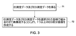
- エネルギ積算(EI)及びエネルギ弁別(ED)データ測定値を含む画像データセットを取得する方法であって、
取得サイクル中にEI測定データ及びED測定データを得る段階と、
選択された態様で前記EI測定データと前記ED測定データとを組み合わせる段階と
を含み、前記組み合わせる段階が、
前記EI測定データに基づいて第1の再構成を行って、EI画像を得る段階と、
前記ED測定データに基づいて第2の再構成を行って、少なくとも1つのED成分画像を得る段階と、
前記EI画像と前記少なくとも1つのED成分画像とを組み合わせて、前記EI画像または前記ED成分画像の情報の欠陥またはアーティファクトを補償した、更新ED成分画像およびED・EI組合せ画像の内うちの少なくとも1つを得る段階と、
を含む、方法。 - 更に、前記ED成分画像を処理して、線減弱係数画像、CTナンバー画像又は単一物質画像の内の少なくとも1つを作成する段階を含む、請求項1に記載の方法。
- 前記組み合わせる段階は、前記ED測定データ及び前記EI測定データについて逐次近似再構成を行って、前記少なくとも1つのED成分画像を作成する段階を含む、請求項1に記載の方法。
- 前記ED成分画像は、コンプトン散乱による減弱又は光電効果による減弱の内の少なくとも一方を表している、請求項3に記載の方法。
- エネルギ積算(EI)及びエネルギ弁別(ED)データ測定値を含む画像データセットを取得する方法であって、
取得サイクル中にEI測定データ及びED測定データを得る段階と、
前記EI測定データ及び前記ED測定データを選択的に組み合わせる段階と、
前記組み合わせたEI測定データ及びED測定データに基づいてEIデータセット及び1つ以上のED成分データセットの内の少なくとも1つを作成する段階と、
前記EIデータセット及び前記1つ以上のED成分データセットに基づいて再構成を行って、前記EIデータセットまたは前記ED成分データセットの情報の欠陥またはアーティファクトを補償した、EI再構成画像及び1つ以上のED成分画像の内の少なくとも1つを作成する段階と、
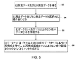
を含む、方法。 - エネルギ積算(EI)及びエネルギ弁別(ED)データ測定値を含む画像データセットを取得する方法であって、
取得サイクル中にEI測定データ及びED測定データを得る段階と、
前記EI測定データに基づいて第1の再構成を行って、EI画像を作成する段階と、
前記ED測定データ及び前記EI画像に基づいて第2の再構成を行って、前記ED測定データの情報の欠陥またはアーティファクトを補償した、1つ以上のED成分画像を作成する段階と、
を含む、方法。 - 前記第2の再構成は、1つ以上のED成分画像を作成するために逐次近似再構成を行う段階を含む、請求項6に記載の方法。
- 前記第2の再構成は、前記EI画像を拡縮する段階、前記ED成分画像の逐次近似再構成における最初の推定値として前記EI画像を使用する段階、又は前記ED成分画像の逐次近似再構成における事前情報として前記EI画像を使用する段階の内の少なくとも1つを含む、請求項7に記載の方法。
- エネルギ積算(EI)及びエネルギ弁別(ED)データ測定値を含む画像データセットを取得する方法であって、
取得サイクル中にEI測定データ及びED測定データを得る段階と、
前記EI測定データに基づいて第1の再構成を行って、EI画像を作成する段階と、
前記EI画像について区分化アルゴリズムを適用して、区分された画像を作成する段階と、
前記ED測定データ及び前記区分された画像に基づいて第2の再構成を行って、前記ED測定データの情報の欠陥またはアーティファクトを補償した、1つ以上のED成分画像を作成する段階と、
を含む、方法。 - 前記区分化アルゴリズムは、前記EI画像を、骨領域、軟組織又はヨウ素領域の内の少なくとも1つにセグメント化する段階を含む、請求項9に記載の方法。
Applications Claiming Priority (2)
| Application Number | Priority Date | Filing Date | Title |
|---|---|---|---|
| US11/317,841 | 2005-12-22 | ||
| US11/317,841 US7372934B2 (en) | 2005-12-22 | 2005-12-22 | Method for performing image reconstruction using hybrid computed tomography detectors |
Publications (2)
| Publication Number | Publication Date |
|---|---|
| JP2007167663A JP2007167663A (ja) | 2007-07-05 |
| JP5469293B2 true JP5469293B2 (ja) | 2014-04-16 |
Family
ID=38135998
Family Applications (1)
| Application Number | Title | Priority Date | Filing Date |
|---|---|---|---|
| JP2006343734A Expired - Fee Related JP5469293B2 (ja) | 2005-12-22 | 2006-12-21 | ハイブリッド型コンピュータ断層撮影検出器を使用して画像再構成を行うための方法 |
Country Status (4)
| Country | Link |
|---|---|
| US (1) | US7372934B2 (ja) |
| JP (1) | JP5469293B2 (ja) |
| CN (1) | CN1985765B (ja) |
| DE (1) | DE102006060493A1 (ja) |
Families Citing this family (57)
| Publication number | Priority date | Publication date | Assignee | Title |
|---|---|---|---|---|
| US8451974B2 (en) | 2003-04-25 | 2013-05-28 | Rapiscan Systems, Inc. | X-ray tomographic inspection system for the identification of specific target items |
| US8243876B2 (en) | 2003-04-25 | 2012-08-14 | Rapiscan Systems, Inc. | X-ray scanners |
| GB0525593D0 (en) | 2005-12-16 | 2006-01-25 | Cxr Ltd | X-ray tomography inspection systems |
| US9113839B2 (en) | 2003-04-25 | 2015-08-25 | Rapiscon Systems, Inc. | X-ray inspection system and method |
| US7949101B2 (en) | 2005-12-16 | 2011-05-24 | Rapiscan Systems, Inc. | X-ray scanners and X-ray sources therefor |
| US8837669B2 (en) | 2003-04-25 | 2014-09-16 | Rapiscan Systems, Inc. | X-ray scanning system |
| US8223919B2 (en) | 2003-04-25 | 2012-07-17 | Rapiscan Systems, Inc. | X-ray tomographic inspection systems for the identification of specific target items |
| US7983462B2 (en) * | 2005-11-22 | 2011-07-19 | Purdue Research Foundation | Methods and systems for improving quality of an image |
| US20070205367A1 (en) * | 2006-03-01 | 2007-09-06 | General Electric Company | Apparatus and method for hybrid computed tomography imaging |
| WO2008135897A2 (en) * | 2007-05-04 | 2008-11-13 | Koninklijke Philips Electronics N.V. | Detection device for detecting radiation and imaging system for imaging a region of interest |
| US7433443B1 (en) * | 2007-08-29 | 2008-10-07 | General Electric Company | System and method of CT imaging with second tube/detector patching |
| US9070181B2 (en) * | 2007-12-21 | 2015-06-30 | General Electric Company | System and method for extracting features of interest from an image |
| DE102008013413B4 (de) * | 2008-03-10 | 2010-08-19 | Siemens Aktiengesellschaft | Röntgendetektor in Schichtbauweise sowie Verfahren zur Erzeugung eines Röntgenbilds |
| JP5179268B2 (ja) * | 2008-06-17 | 2013-04-10 | ジーイー・メディカル・システムズ・グローバル・テクノロジー・カンパニー・エルエルシー | X線ct装置 |
| JP5715052B2 (ja) * | 2008-06-30 | 2015-05-07 | コーニンクレッカ フィリップス エヌ ヴェ | 画像化システム及び画像化方法 |
| US8111803B2 (en) * | 2009-04-29 | 2012-02-07 | General Electric Company | Method for energy sensitive computed tomography using checkerboard filtering |
| US8160206B2 (en) * | 2009-12-23 | 2012-04-17 | General Electric Company | Dual-energy imaging at reduced sample rates |
| DE102010006774A1 (de) * | 2010-02-04 | 2011-08-04 | Siemens Aktiengesellschaft, 80333 | CT-Messung mit Mehrfachröntgenquellen |
| US20110211667A1 (en) * | 2010-02-26 | 2011-09-01 | Abdelaziz Ikhlef | De-populated detector for computed tomography and method of making same |
| US8160200B2 (en) * | 2010-03-30 | 2012-04-17 | General Electric Company | Method and system for image data acquisition |
| GB2501661B (en) * | 2011-02-22 | 2017-04-12 | Rapiscan Systems Inc | X-ray inspection system and method |
| US20120236987A1 (en) * | 2011-03-18 | 2012-09-20 | David Ruimi | Multiple energy ct scanner |
| DE102011076346B4 (de) * | 2011-05-24 | 2016-07-14 | Siemens Healthcare Gmbh | Verfahren und Computertomographiesystem zur Erzeugung tomographischer Bilddatensätze |
| US9119589B2 (en) * | 2012-03-22 | 2015-09-01 | Kabushiki Kaisha Toshiba | Method and system for spectral computed tomography (CT) with sparse photon counting detectors |
| US9237874B2 (en) | 2012-04-30 | 2016-01-19 | General Electric Company | Method and system for non-invasive imaging of a target region |
| WO2014055066A1 (en) * | 2012-10-02 | 2014-04-10 | Analogic Corporation | Detector array comprising energy integrating and photon counting cells |
| CN103913472B (zh) * | 2012-12-31 | 2016-04-20 | 同方威视技术股份有限公司 | Ct成像系统和方法 |
| US9861324B2 (en) | 2013-04-23 | 2018-01-09 | Virginia Tech Intellectual Properties, Inc. | Hybrid detector modules and dynamic thresholding for spectral CT |
| DE102013104193A1 (de) * | 2013-04-25 | 2014-10-30 | Smiths Heimann Gmbh | CT-Röntgenprüfanlage, insbesondere zur Inspektion von Objekten |
| US9724056B2 (en) * | 2013-11-28 | 2017-08-08 | Toshiba Medical Systems Corporation | Method and system for spectral computed tomography (CT) with inner ring geometry |
| WO2015011587A1 (en) * | 2013-07-23 | 2015-01-29 | Koninklijke Philips N.V. | Hybrid (spectral/non-spectral) imaging detector array and corresponding processing electronics |
| US9437016B2 (en) * | 2013-08-07 | 2016-09-06 | Toshiba Medical Systems Corporation | Image domain pansharpening method and system for spectral CT with large pixel energy discriminating detectors |
| US10176603B2 (en) * | 2013-08-07 | 2019-01-08 | The University Of Chicago | Sinogram (data) domain pansharpening method and system for spectral CT |
| DE102013217528A1 (de) | 2013-09-03 | 2015-03-05 | Siemens Aktiengesellschaft | Röntgenstrahlungsdetektor |
| CN105122085B (zh) | 2013-10-09 | 2019-01-11 | 皇家飞利浦有限公司 | 利用调整的能量阈值用于生成能量分辨x射线图像的方法和设备 |
| US9274066B2 (en) * | 2013-10-25 | 2016-03-01 | Kabushiki Kaisha Toshiba | Method for spectral CT local tomography |
| US9226723B2 (en) * | 2014-02-25 | 2016-01-05 | Kabushiki Kaisha Toshiba | Ordered subset scheme in spectral CT |
| US9449385B2 (en) | 2014-06-02 | 2016-09-20 | Toshiba Medical Systems Corporation | Reconstruction of computed tomography images using combined third-generation and fourth-generation projection data |
| US10143434B2 (en) * | 2014-06-04 | 2018-12-04 | Koninklijke Philips N.V. | Imaging system for generating an image of an object |
| JP6313168B2 (ja) * | 2014-09-02 | 2018-04-18 | キヤノンメディカルシステムズ株式会社 | X線ct装置、画像処理装置及び画像処理プログラム |
| CN107072626B (zh) * | 2014-09-19 | 2020-10-27 | 皇家飞利浦有限公司 | 谱投影扩展 |
| WO2016063170A1 (en) * | 2014-10-20 | 2016-04-28 | Koninklijke Philips N.V. | Start image for spectral image iterative reconstruction |
| JP6625428B2 (ja) * | 2015-02-23 | 2019-12-25 | キヤノンメディカルシステムズ株式会社 | X線ct装置、および画像処理装置 |
| US10646176B2 (en) * | 2015-09-30 | 2020-05-12 | General Electric Company | Layered radiation detector |
| US10219775B2 (en) | 2015-11-02 | 2019-03-05 | Toshiba Medical Systems Corporation | Photon-counting X-ray CT apparatus and image processing apparatus |
| DE102015226489B4 (de) * | 2015-12-22 | 2024-05-16 | Siemens Healthineers Ag | Röntgensystem und Verfahren zur Bildrekonstruktion |
| US10646186B2 (en) | 2016-01-18 | 2020-05-12 | Canon Medical Systems Corporation | X-ray CT apparatus, information processing device and information processing method |
| CN107019518B (zh) * | 2016-02-01 | 2020-07-28 | 通用电气公司 | 用于计算机断层扫描中的散射校正的信号处理方法及成像系统 |
| CN109196957B (zh) * | 2016-05-31 | 2023-07-04 | 皇家飞利浦有限公司 | 用于生成x射线的装置 |
| JP6894928B2 (ja) * | 2016-06-07 | 2021-06-30 | コーニンクレッカ フィリップス エヌ ヴェKoninklijke Philips N.V. | スペクトル及び非スペクトル物質分解を組み合わせることによる定量的撮像の精度及び分解能の向上 |
| CN114224386B (zh) * | 2016-09-30 | 2025-01-24 | 深圳迈瑞生物医疗电子股份有限公司 | 一种成像方法和系统 |
| JP6956626B2 (ja) * | 2017-01-24 | 2021-11-02 | キヤノンメディカルシステムズ株式会社 | X線ct装置及び再構成処理装置 |
| US10585206B2 (en) | 2017-09-06 | 2020-03-10 | Rapiscan Systems, Inc. | Method and system for a multi-view scanner |
| US11212902B2 (en) | 2020-02-25 | 2021-12-28 | Rapiscan Systems, Inc. | Multiplexed drive systems and methods for a multi-emitter X-ray source |
| US11517274B2 (en) | 2020-06-02 | 2022-12-06 | Wisconsin Alumni Research Foundation | Hybrid detection systems and methods for C-arm interventional x-ray systems |
| US11593926B2 (en) * | 2021-02-09 | 2023-02-28 | Elucid Bioimaging Inc. | Systems and methods for improving soft tissue contrast, multiscale modeling and spectral CT |
| US12419596B2 (en) | 2022-07-26 | 2025-09-23 | GE Precision Healthcare LLC | Systems and methods for computed tomography |
Family Cites Families (21)
| Publication number | Priority date | Publication date | Assignee | Title |
|---|---|---|---|---|
| JPS5591338A (en) * | 1978-12-28 | 1980-07-10 | Shimadzu Corp | Xxray tomography device |
| JPS58206726A (ja) * | 1982-05-28 | 1983-12-02 | 株式会社日立製作所 | 画像処理装置 |
| JPS6075036A (ja) * | 1983-09-30 | 1985-04-27 | 株式会社東芝 | マルチカラ−x線ct装置 |
| JPS6236704U (ja) * | 1985-08-24 | 1987-03-04 | ||
| US5376795A (en) * | 1990-07-09 | 1994-12-27 | Regents Of The University Of California | Emission-transmission imaging system using single energy and dual energy transmission and radionuclide emission data |
| JP3449561B2 (ja) * | 1993-04-19 | 2003-09-22 | 東芝医用システムエンジニアリング株式会社 | X線ct装置 |
| JPH09108207A (ja) * | 1995-10-20 | 1997-04-28 | Hitachi Ltd | X線検出方法 |
| JPH09149901A (ja) * | 1995-11-30 | 1997-06-10 | Toshiba Corp | 画像生成装置及び画像生成方法 |
| US6236050B1 (en) * | 1996-02-02 | 2001-05-22 | TüMER TüMAY O. | Method and apparatus for radiation detection |
| US5742660A (en) * | 1997-01-10 | 1998-04-21 | Southeastern Universities Research Association, Inc. | Dual energy scanning beam laminographic x-radiography |
| JP2002077132A (ja) * | 2000-08-29 | 2002-03-15 | Dainippon Printing Co Ltd | 情報提供システム、サーバコンピュータ、携帯端末、及び記録媒体 |
| US6904118B2 (en) | 2002-07-23 | 2005-06-07 | General Electric Company | Method and apparatus for generating a density map using dual-energy CT |
| JP4114717B2 (ja) * | 2002-08-09 | 2008-07-09 | 浜松ホトニクス株式会社 | Ct装置 |
| US6819738B2 (en) | 2002-08-15 | 2004-11-16 | Ge Medical Systems Global Technology Company, Llc | Hybrid scintillator/photo sensor & direct conversion detector |
| JP2004129812A (ja) * | 2002-10-10 | 2004-04-30 | Ge Medical Systems Global Technology Co Llc | Ct撮影方法、ct画像生成方法およびx線ct装置 |
| US6898263B2 (en) * | 2002-11-27 | 2005-05-24 | Ge Medical Systems Global Technology Company, Llc | Method and apparatus for soft-tissue volume visualization |
| US6999549B2 (en) * | 2002-11-27 | 2006-02-14 | Ge Medical Systems Global Technology, Llc | Method and apparatus for quantifying tissue fat content |
| US7272429B2 (en) * | 2002-11-27 | 2007-09-18 | Ge Medical Systems Global Technology Company, Llc | Methods and apparatus for facilitating a reduction in artifacts |
| US7129498B2 (en) * | 2003-09-23 | 2006-10-31 | General Electric Company | Compact structural CT detector module |
| EP1676126A1 (en) * | 2003-10-14 | 2006-07-05 | Philips Intellectual Property & Standards GmbH | Fan-beam coherent-scatter computed tomography |
| US7105828B2 (en) * | 2004-02-10 | 2006-09-12 | Ge Medical Systems Global Technology Company, Llc | Hybrid x-ray detector |
-
2005
- 2005-12-22 US US11/317,841 patent/US7372934B2/en not_active Expired - Lifetime
-
2006
- 2006-12-19 DE DE102006060493A patent/DE102006060493A1/de not_active Withdrawn
- 2006-12-21 JP JP2006343734A patent/JP5469293B2/ja not_active Expired - Fee Related
- 2006-12-22 CN CN2006101701540A patent/CN1985765B/zh not_active Expired - Fee Related
Also Published As
| Publication number | Publication date |
|---|---|
| US20070147574A1 (en) | 2007-06-28 |
| CN1985765B (zh) | 2011-03-09 |
| US7372934B2 (en) | 2008-05-13 |
| CN1985765A (zh) | 2007-06-27 |
| JP2007167663A (ja) | 2007-07-05 |
| DE102006060493A1 (de) | 2007-07-05 |
Similar Documents
| Publication | Publication Date | Title |
|---|---|---|
| JP5469293B2 (ja) | ハイブリッド型コンピュータ断層撮影検出器を使用して画像再構成を行うための方法 | |
| US7760848B2 (en) | Method and system for generating a multi-spectral image of an object | |
| US6904118B2 (en) | Method and apparatus for generating a density map using dual-energy CT | |
| US8160206B2 (en) | Dual-energy imaging at reduced sample rates | |
| US9025815B2 (en) | System and method for multi-material correction of image data | |
| CN106233335B (zh) | X射线光谱成像方法与系统 | |
| US7396162B1 (en) | Scatter correction for CT method and apparatus | |
| US8396273B2 (en) | Noise reduction method for dual-energy imaging | |
| JP6513431B2 (ja) | X線ct装置及びその制御方法 | |
| JP2008062035A5 (ja) | ||
| US20090129539A1 (en) | Computed tomography method and system | |
| US8885910B2 (en) | Systems and methods for X-ray imaging | |
| US7643866B2 (en) | Method for producing a computed tomography display of tissue structures by applying a contrast medium | |
| US20100310036A1 (en) | Computed tomography method and apparatus | |
| US20160134852A1 (en) | System and method for multi-material correction of image data | |
| US9943279B2 (en) | Methods and systems for task-based data generation and weighting for CT spectral imaging | |
| JP7199399B2 (ja) | デュアルエネルギー撮像における自動管電位選択のためのシステムおよび方法 | |
| US10383589B2 (en) | Direct monochromatic image generation for spectral computed tomography | |
| US11270477B2 (en) | Systems and methods for tailored image texture in iterative image reconstruction | |
| Gonzales et al. | Rectangular computed tomography using a stationary array of CNT emitters: initial experimental results | |
| CN107212898B (zh) | 图像重建方法 | |
| Cierniak | Technical concepts of X-ray computed tomography scanners |
Legal Events
| Date | Code | Title | Description |
|---|---|---|---|
| A621 | Written request for application examination |
Free format text: JAPANESE INTERMEDIATE CODE: A621 Effective date: 20091214 |
|
| RD04 | Notification of resignation of power of attorney |
Free format text: JAPANESE INTERMEDIATE CODE: A7424 Effective date: 20091214 |
|
| A521 | Request for written amendment filed |
Free format text: JAPANESE INTERMEDIATE CODE: A523 Effective date: 20101213 |
|
| A977 | Report on retrieval |
Free format text: JAPANESE INTERMEDIATE CODE: A971007 Effective date: 20120301 |
|
| A131 | Notification of reasons for refusal |
Free format text: JAPANESE INTERMEDIATE CODE: A131 Effective date: 20120313 |
|
| A521 | Request for written amendment filed |
Free format text: JAPANESE INTERMEDIATE CODE: A523 Effective date: 20120528 |
|
| A131 | Notification of reasons for refusal |
Free format text: JAPANESE INTERMEDIATE CODE: A131 Effective date: 20130108 |
|
| A521 | Request for written amendment filed |
Free format text: JAPANESE INTERMEDIATE CODE: A523 Effective date: 20130405 |
|
| TRDD | Decision of grant or rejection written | ||
| A01 | Written decision to grant a patent or to grant a registration (utility model) |
Free format text: JAPANESE INTERMEDIATE CODE: A01 Effective date: 20140108 |
|
| A61 | First payment of annual fees (during grant procedure) |
Free format text: JAPANESE INTERMEDIATE CODE: A61 Effective date: 20140131 |
|
| R150 | Certificate of patent or registration of utility model |
Ref document number: 5469293 Country of ref document: JP Free format text: JAPANESE INTERMEDIATE CODE: R150 Free format text: JAPANESE INTERMEDIATE CODE: R150 |
|
| R250 | Receipt of annual fees |
Free format text: JAPANESE INTERMEDIATE CODE: R250 |
|
| R250 | Receipt of annual fees |
Free format text: JAPANESE INTERMEDIATE CODE: R250 |
|
| R250 | Receipt of annual fees |
Free format text: JAPANESE INTERMEDIATE CODE: R250 |
|
| R250 | Receipt of annual fees |
Free format text: JAPANESE INTERMEDIATE CODE: R250 |
|
| R250 | Receipt of annual fees |
Free format text: JAPANESE INTERMEDIATE CODE: R250 |
|
| R250 | Receipt of annual fees |
Free format text: JAPANESE INTERMEDIATE CODE: R250 |
|
| R250 | Receipt of annual fees |
Free format text: JAPANESE INTERMEDIATE CODE: R250 |
|
| R250 | Receipt of annual fees |
Free format text: JAPANESE INTERMEDIATE CODE: R250 |
|
| LAPS | Cancellation because of no payment of annual fees |