JP5143471B2 - イメージング装置 - Google Patents
イメージング装置 Download PDFInfo
- Publication number
- JP5143471B2 JP5143471B2 JP2007123709A JP2007123709A JP5143471B2 JP 5143471 B2 JP5143471 B2 JP 5143471B2 JP 2007123709 A JP2007123709 A JP 2007123709A JP 2007123709 A JP2007123709 A JP 2007123709A JP 5143471 B2 JP5143471 B2 JP 5143471B2
- Authority
- JP
- Japan
- Prior art keywords
- light
- subject
- detector
- signal
- imaging
- Prior art date
- Legal status (The legal status is an assumption and is not a legal conclusion. Google has not performed a legal analysis and makes no representation as to the accuracy of the status listed.)
- Expired - Fee Related
Links
- 238000003384 imaging method Methods 0.000 title claims description 169
- 238000012545 processing Methods 0.000 claims description 26
- 238000001514 detection method Methods 0.000 claims description 23
- 238000010521 absorption reaction Methods 0.000 claims description 17
- 238000009826 distribution Methods 0.000 claims description 15
- 239000000463 material Substances 0.000 claims description 13
- 238000005259 measurement Methods 0.000 claims description 10
- 230000002194 synthesizing effect Effects 0.000 claims description 6
- 238000004148 unit process Methods 0.000 claims 1
- 230000003287 optical effect Effects 0.000 description 64
- 238000000034 method Methods 0.000 description 51
- 238000012634 optical imaging Methods 0.000 description 47
- 239000003068 molecular probe Substances 0.000 description 30
- 238000010586 diagram Methods 0.000 description 25
- 230000003902 lesion Effects 0.000 description 21
- 230000005855 radiation Effects 0.000 description 21
- 230000006870 function Effects 0.000 description 19
- 230000005284 excitation Effects 0.000 description 17
- 210000000481 breast Anatomy 0.000 description 16
- 230000006835 compression Effects 0.000 description 16
- 238000007906 compression Methods 0.000 description 16
- 238000009543 diffuse optical tomography Methods 0.000 description 13
- 230000002829 reductive effect Effects 0.000 description 10
- 239000002131 composite material Substances 0.000 description 9
- 238000000799 fluorescence microscopy Methods 0.000 description 9
- 230000035945 sensitivity Effects 0.000 description 9
- 206010028980 Neoplasm Diseases 0.000 description 8
- 238000012937 correction Methods 0.000 description 7
- 238000009607 mammography Methods 0.000 description 7
- 238000002601 radiography Methods 0.000 description 6
- 239000000758 substrate Substances 0.000 description 6
- 201000011510 cancer Diseases 0.000 description 5
- 238000009792 diffusion process Methods 0.000 description 5
- 239000011669 selenium Substances 0.000 description 5
- 206010006187 Breast cancer Diseases 0.000 description 4
- 208000026310 Breast neoplasm Diseases 0.000 description 4
- 230000008901 benefit Effects 0.000 description 4
- 210000004204 blood vessel Anatomy 0.000 description 4
- 238000004364 calculation method Methods 0.000 description 4
- 230000007423 decrease Effects 0.000 description 4
- 239000011159 matrix material Substances 0.000 description 4
- 230000008569 process Effects 0.000 description 4
- 230000009467 reduction Effects 0.000 description 4
- 230000000903 blocking effect Effects 0.000 description 3
- 239000008280 blood Substances 0.000 description 3
- 210000004369 blood Anatomy 0.000 description 3
- 239000003990 capacitor Substances 0.000 description 3
- 239000011521 glass Substances 0.000 description 3
- 238000007689 inspection Methods 0.000 description 3
- 230000031700 light absorption Effects 0.000 description 3
- 230000007246 mechanism Effects 0.000 description 3
- 241001465754 Metazoa Species 0.000 description 2
- 238000002835 absorbance Methods 0.000 description 2
- NIXOWILDQLNWCW-UHFFFAOYSA-N acrylic acid group Chemical group C(C=C)(=O)O NIXOWILDQLNWCW-UHFFFAOYSA-N 0.000 description 2
- 229910021417 amorphous silicon Inorganic materials 0.000 description 2
- VJBCNMFKFZIXHC-UHFFFAOYSA-N azanium;2-(4-methyl-5-oxo-4-propan-2-yl-1h-imidazol-2-yl)quinoline-3-carboxylate Chemical compound N.N1C(=O)C(C(C)C)(C)N=C1C1=NC2=CC=CC=C2C=C1C(O)=O VJBCNMFKFZIXHC-UHFFFAOYSA-N 0.000 description 2
- 230000015572 biosynthetic process Effects 0.000 description 2
- 238000006243 chemical reaction Methods 0.000 description 2
- 238000002347 injection Methods 0.000 description 2
- 239000007924 injection Substances 0.000 description 2
- 230000000670 limiting effect Effects 0.000 description 2
- 239000004973 liquid crystal related substance Substances 0.000 description 2
- 238000004020 luminiscence type Methods 0.000 description 2
- 235000015097 nutrients Nutrition 0.000 description 2
- 230000001681 protective effect Effects 0.000 description 2
- 238000003786 synthesis reaction Methods 0.000 description 2
- 229910004611 CdZnTe Inorganic materials 0.000 description 1
- 235000008733 Citrus aurantifolia Nutrition 0.000 description 1
- RYGMFSIKBFXOCR-UHFFFAOYSA-N Copper Chemical compound [Cu] RYGMFSIKBFXOCR-UHFFFAOYSA-N 0.000 description 1
- 240000006829 Ficus sundaica Species 0.000 description 1
- 108060001084 Luciferase Proteins 0.000 description 1
- 239000005089 Luciferase Substances 0.000 description 1
- BUGBHKTXTAQXES-UHFFFAOYSA-N Selenium Chemical compound [Se] BUGBHKTXTAQXES-UHFFFAOYSA-N 0.000 description 1
- 235000011941 Tilia x europaea Nutrition 0.000 description 1
- 238000000149 argon plasma sintering Methods 0.000 description 1
- 238000012984 biological imaging Methods 0.000 description 1
- 230000005540 biological transmission Effects 0.000 description 1
- UHYPYGJEEGLRJD-UHFFFAOYSA-N cadmium(2+);selenium(2-) Chemical compound [Se-2].[Cd+2] UHYPYGJEEGLRJD-UHFFFAOYSA-N 0.000 description 1
- 230000002308 calcification Effects 0.000 description 1
- 239000003086 colorant Substances 0.000 description 1
- 238000004040 coloring Methods 0.000 description 1
- 239000002872 contrast media Substances 0.000 description 1
- 239000010949 copper Substances 0.000 description 1
- 229910052802 copper Inorganic materials 0.000 description 1
- 230000003247 decreasing effect Effects 0.000 description 1
- 238000000151 deposition Methods 0.000 description 1
- 238000011161 development Methods 0.000 description 1
- 230000018109 developmental process Effects 0.000 description 1
- 201000010099 disease Diseases 0.000 description 1
- 208000037265 diseases, disorders, signs and symptoms Diseases 0.000 description 1
- 238000013399 early diagnosis Methods 0.000 description 1
- 230000005684 electric field Effects 0.000 description 1
- 238000002795 fluorescence method Methods 0.000 description 1
- 238000012632 fluorescent imaging Methods 0.000 description 1
- 108091006047 fluorescent proteins Proteins 0.000 description 1
- 102000034287 fluorescent proteins Human genes 0.000 description 1
- 230000010354 integration Effects 0.000 description 1
- 230000001678 irradiating effect Effects 0.000 description 1
- 239000004571 lime Substances 0.000 description 1
- 238000004519 manufacturing process Methods 0.000 description 1
- 239000002105 nanoparticle Substances 0.000 description 1
- 230000009826 neoplastic cell growth Effects 0.000 description 1
- 239000013307 optical fiber Substances 0.000 description 1
- 230000035699 permeability Effects 0.000 description 1
- 230000002441 reversible effect Effects 0.000 description 1
- 238000012216 screening Methods 0.000 description 1
- 229910052711 selenium Inorganic materials 0.000 description 1
- 239000010409 thin film Substances 0.000 description 1
- 239000012780 transparent material Substances 0.000 description 1
- 230000000007 visual effect Effects 0.000 description 1
- 208000008918 voyeurism Diseases 0.000 description 1
- 229910052724 xenon Inorganic materials 0.000 description 1
- FHNFHKCVQCLJFQ-UHFFFAOYSA-N xenon atom Chemical compound [Xe] FHNFHKCVQCLJFQ-UHFFFAOYSA-N 0.000 description 1
Images


Classifications
-
- A—HUMAN NECESSITIES
- A61—MEDICAL OR VETERINARY SCIENCE; HYGIENE
- A61B—DIAGNOSIS; SURGERY; IDENTIFICATION
- A61B6/00—Apparatus for radiation diagnosis, e.g. combined with radiation therapy equipment
- A61B6/48—Diagnostic techniques
- A61B6/485—Diagnostic techniques involving fluorescence X-ray imaging
-
- A—HUMAN NECESSITIES
- A61—MEDICAL OR VETERINARY SCIENCE; HYGIENE
- A61B—DIAGNOSIS; SURGERY; IDENTIFICATION
- A61B5/00—Measuring for diagnostic purposes; Identification of persons
- A61B5/0059—Measuring for diagnostic purposes; Identification of persons using light, e.g. diagnosis by transillumination, diascopy, fluorescence
- A61B5/0082—Measuring for diagnostic purposes; Identification of persons using light, e.g. diagnosis by transillumination, diascopy, fluorescence adapted for particular medical purposes
- A61B5/0091—Measuring for diagnostic purposes; Identification of persons using light, e.g. diagnosis by transillumination, diascopy, fluorescence adapted for particular medical purposes for mammography
-
- A—HUMAN NECESSITIES
- A61—MEDICAL OR VETERINARY SCIENCE; HYGIENE
- A61B—DIAGNOSIS; SURGERY; IDENTIFICATION
- A61B5/00—Measuring for diagnostic purposes; Identification of persons
- A61B5/43—Detecting, measuring or recording for evaluating the reproductive systems
- A61B5/4306—Detecting, measuring or recording for evaluating the reproductive systems for evaluating the female reproductive systems, e.g. gynaecological evaluations
- A61B5/4312—Breast evaluation or disorder diagnosis
-
- A—HUMAN NECESSITIES
- A61—MEDICAL OR VETERINARY SCIENCE; HYGIENE
- A61B—DIAGNOSIS; SURGERY; IDENTIFICATION
- A61B6/00—Apparatus for radiation diagnosis, e.g. combined with radiation therapy equipment
- A61B6/44—Constructional features of apparatus for radiation diagnosis
- A61B6/4417—Constructional features of apparatus for radiation diagnosis related to combined acquisition of different diagnostic modalities
-
- A—HUMAN NECESSITIES
- A61—MEDICAL OR VETERINARY SCIENCE; HYGIENE
- A61B—DIAGNOSIS; SURGERY; IDENTIFICATION
- A61B6/00—Apparatus for radiation diagnosis, e.g. combined with radiation therapy equipment
- A61B6/50—Clinical applications
- A61B6/502—Clinical applications involving diagnosis of breast, i.e. mammography
-
- A—HUMAN NECESSITIES
- A61—MEDICAL OR VETERINARY SCIENCE; HYGIENE
- A61B—DIAGNOSIS; SURGERY; IDENTIFICATION
- A61B6/00—Apparatus for radiation diagnosis, e.g. combined with radiation therapy equipment
- A61B6/52—Devices using data or image processing specially adapted for radiation diagnosis
- A61B6/5211—Devices using data or image processing specially adapted for radiation diagnosis involving processing of medical diagnostic data
- A61B6/5229—Devices using data or image processing specially adapted for radiation diagnosis involving processing of medical diagnostic data combining image data of a patient, e.g. combining a functional image with an anatomical image
- A61B6/5247—Devices using data or image processing specially adapted for radiation diagnosis involving processing of medical diagnostic data combining image data of a patient, e.g. combining a functional image with an anatomical image combining images from an ionising-radiation diagnostic technique and a non-ionising radiation diagnostic technique, e.g. X-ray and ultrasound
-
- A—HUMAN NECESSITIES
- A61—MEDICAL OR VETERINARY SCIENCE; HYGIENE
- A61B—DIAGNOSIS; SURGERY; IDENTIFICATION
- A61B6/00—Apparatus for radiation diagnosis, e.g. combined with radiation therapy equipment
- A61B6/04—Positioning of patients; Tiltable beds or the like
- A61B6/0407—Supports, e.g. tables or beds, for the body or parts of the body
- A61B6/0414—Supports, e.g. tables or beds, for the body or parts of the body with compression means
-
- A—HUMAN NECESSITIES
- A61—MEDICAL OR VETERINARY SCIENCE; HYGIENE
- A61B—DIAGNOSIS; SURGERY; IDENTIFICATION
- A61B6/00—Apparatus for radiation diagnosis, e.g. combined with radiation therapy equipment
- A61B6/42—Apparatus for radiation diagnosis, e.g. combined with radiation therapy equipment with arrangements for detecting radiation specially adapted for radiation diagnosis
- A61B6/4291—Apparatus for radiation diagnosis, e.g. combined with radiation therapy equipment with arrangements for detecting radiation specially adapted for radiation diagnosis the detector being combined with a grid or grating
-
- A—HUMAN NECESSITIES
- A61—MEDICAL OR VETERINARY SCIENCE; HYGIENE
- A61B—DIAGNOSIS; SURGERY; IDENTIFICATION
- A61B6/00—Apparatus for radiation diagnosis, e.g. combined with radiation therapy equipment
- A61B6/50—Clinical applications
- A61B6/508—Clinical applications for non-human patients
Description
(手段1)
イメージング装置において、X線を被検体に照射するX線源と、被検体を介してX線源と対面しX線及び光を検出する第1検出器と、第1検出器の検出結果を処理する処理部とを有し、処理部はX線源がX線を照射する照射期間に第1検出器が検出するX線の信号を第1信号とし、X線照射期間以外の期間に第1検出器が検出する光の信号を第2信号として処理することを特徴とする。これにより、X線イメージングと発光性分子プローブを用いた光イメージングを一度の検査で行える。乳房を検査対象とした場合、空間分解能の高いマンモグラフィ画像と病変検出感度の高い光画像が一度の検査で得られる。このため両者の利点の組み合わせることで、病変の見落としや読影負担を軽減して、診断能を向上できる。
手段1に記載のイメージング装置において、第1検出器の受光部がX線以上赤外線以下の範囲の波長を有する光に感度を有する光導電体材料で形成されていることを特徴とする。これにより、1つの検出器にてX線と光の両方を検出できる。
手段1に記載のイメージング装置において、第1検出器の受光部がシンチレータ材料で形成されており、シンチレータ材料は可視光線以上赤外線以下の範囲の波長を有する光の一部を透過することを特徴とする。シンチレータにてX線信号を光信号に変換することで、光センサによるX線イメージングが可能となる。また被検体から出力された光はシンチレータを透過して光センサで検出できるため、1つの検出器にてX線と光の両方を検出できる。
手段1に記載のイメージング装置において、第1検出器は、X線に感度を有するX線検出層と可視光線以上赤外線以下の範囲の波長を有する光に感度を有する光検出層との2層で構成されることを特徴とする。被検体から出力された光を光検出層で検出すると共に、被検体から出力されたX線は光検出層を透過した後にX線検出層で検出される。これにより、1つの検出器にてX線と光の両方を検出できる。
手段1に記載のイメージング装置において、被検体の外表面より出力された光を第1検出器の受光面に導く導光部を有することを特徴とする。これにより、被検体表面と第1検出器の間の空間で発生する光の拡散に起因するボケを防ぎ、光イメージングにおける空間分解能の低下を防止できる。
手段5に記載のイメージング装置において、導光部は被検体の内部で散乱したX線の一部を除去する機能を有することを特徴とする。これにより、X線イメージングと光イメージングの両方において、空間分解能の低下を防止できる。
手段1に記載のイメージング装置において、第1信号と第2信号を同一画像上に合成する機能を処理部が有することを特徴とする。これにより、X線画像と光画像を同一の画像上で確認できるため、両者の位置関係が明確になり、病変の見落としや読影負担を軽減できる。
手段1に記載のイメージング装置において、紫外線以上赤外線以下の範囲の波長を有する光を被検体に照射する光源を有することを特徴とする。これにより、X線イメージングに加えて光による血管イメージングや蛍光性分子プローブを用いた光イメージングを一度の検査で行える。
手段8に記載のイメージング装置において、被検体に接触した状態で被検体を支持する支持部を有し、支持部が光源より照射された光を被検体に導く導光機能を有することを特徴とする。これにより、被検体表面における光の反射を低減して被検体内部への光の入射強度の低下を防止できる。
手段1に記載のイメージング装置において、X線源と被検体の間に配置され光を検出する第2検出器を有し、処理部は、X線源がX線を照射する照射期間に第1検出器が検出する光の信号を第1信号とし、照射期間以外の期間に第1検出器及び第2検出器が検出する信号をそれぞれ第2信号及び第3信号として処理することを特徴とする。これにより、第1検出器が配置された側と反対側の被検体表面から出力される光を第2検出器で検出できるため、光イメージングにおける撮影範囲を拡大して、感度を向上できる。
イメージング装置において、X線を被検体に照射するX線源と、被検体を介してX線源と対面しX線を検出する第1検出器と、X線源と被検体の間に配置され光を検出する第2検出器と、被検体と第1検出器の間に配置され光を検出する第3検出器と、第1〜3検出器の検出結果を処理する処理部とを有し、処理部は、X線源がX線を照射する照射期間に第1検出器が検出するX線の信号を第1信号とし、照射期間以外の期間に第3検出器及び第2検出器が検出する光の信号をそれぞれ第2信号及び第3信号として処理することを特徴とする。これにより、第1検出器にてX線画像を計測すると共に、第1検出器が配置された側、及びその反対側の被検体表面より出力される光をそれぞれ第3、第2検出器で検出できる。このため、X線画像と2つの光画像を組み合わせが可能となり、病変の見落としや読影負担を軽減して、診断能を向上できる。
手段10及び11に記載のイメージング装置において、第1信号と第2信号と第3信号とを同一画像上に合成する機能を処理部が有することを特徴とする。これにより、X線画像と2つの光画像を同一の画像上で確認できるため、両者の位置関係が明確になり、病変の見落としや読影負担を軽減できる。
手段11に記載のイメージング装置において、X線源と被検体の間に配置される反射鏡と、反射鏡により反射される光を集光するレンズと、レンズにより集光された光を検出する第2検出器とを有し、処理部は、X線源がX線を照射する照射期間に第1検出器が検出するX線の信号を第1信号とし、X線照射期間以外の期間に第1検出器及び第2検出器が検出する光の信号をそれぞれ第2信号及び第3信号として処理することを特徴とする。これにより、第1検出器が配置された側と反対側の被検体表面から出力される光を第2検出器で検出できるため、光イメージングにおける撮影範囲を拡大して、感度を向上できる。
イメージング装置において、X線を被検体に照射するX線源と、被検体を介してX線源と対面しX線を検出する第1検出器と、X線源と被検体の間に配置される第1反射鏡と、第1反射鏡により反射される光を集光する第1レンズと、第1レンズにより集光された光を検出する第2検出器と、被検体と第1検出器の間に配置される第2反射鏡と、第2反射鏡により反射される光を集光する第2レンズと、第2レンズにより集光された光を検出する第3検出器と、第1〜3検出器の検出結果を処理する処理部とを有し、処理部は、X線源がX線を照射する照射期間に第1検出器が検出するX線の信号を第1信号とし、X線照射期間以外の期間に第3検出器及び第2検出器が検出する光の信号をそれぞれ第2信号及び第3信号として処理することを特徴とする。これにより、第1検出器にてX線画像を計測すると共に、第1検出器が配置された側、及びその反対側の被検体表面より出力される光をそれぞれ第3、第2検出器で検出できる。このため、X線画像と2つの光画像を組み合わせが可能となり、病変の見落としや読影負担を軽減して、診断能を向上できる。
手段13及び14に記載のイメージング装置において、被検体のサイズを測定するサイズ測定手段と、サイズ測定手段の測定結果に応じてレンズの配置を変更してピントを調整するピント調整部とを有することを特徴とする。これにより、被検体のサイズの違いに起因するピントのボケを防ぎ、光イメージングにおける空間分解能の低下を防止できる。
手段13及び14に記載のイメージング装置において、第1信号と第2信号と第3信号を同一画像上に合成する機能を処理部が有することを特徴とする。これにより、X線画像と2つの光画像を同一の画像上で確認できるため、両者の位置関係が明確になり、病変の見落としや読影負担を軽減できる。
手段16に記載のイメージング装置において、被検体のサイズを測定するサイズ測定手段と、サイズ測定手段の測定結果に応じて第1信号と第3信号の合成位置を調整する機能を処理部が有することを特徴とする。これにより、X線画像と光画像を合成する際に、被検体のサイズの違いに起因する両者の位置ズレを防止して、位置精度を向上できる。
手段10、11、13及び14に記載のイメージング装置において、第2信号と第3信号に基づいて被検体内部における発光または吸光強度分布を計算する機能を処理部が有することを特徴とする。これにより、X線画像に加えて被検体内部における発光または吸光強度分布を一度の検査で共に取得できる。被写体内部における病変位置の推定が可能となり、診断能を向上できる。
手段10、11、13及び14に記載のイメージング装置において、被検体内部におけるX線のビームパス上で発光または吸光強度分布を積分した仮想信号を計算する機能と、第1信号と仮想信号を同一画像上に合成する機能とを処理部が有することを特徴とする。これにより、発光強度分布または吸光強度分布を仮想的なX線源に対して投影した仮想画像が作成できると共に、これをX線画像と重ね合わせることができるため、両者の位置ズレを防止すると共に、光画像の空間分解能を向上できる。
[実施の形態1]
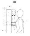
図1は、本発明の実施の形態1に係るイメージング装置の模式図である。なお以下では、図1の紙面に対して水平方向、垂直方向、及び上下方向をそれぞれX方向、Y方向、Z方向とする。本実施の形態1に係るイメージング装置は、X線管1、コリメータ2、光源3、光拡散板4、圧迫板5、光学フィルタ7、導光板8、散乱線除去グリッド9、検出器10、撮影制御装置101、コンソール102、メモリ103、演算装置104、モニタ105等から構成される。なお、本実施の形態1に記載のイメージング装置が対象とする被写体6は乳房である。
図14は、本発明の実施の形態2に係るイメージング装置の模式図である。なお本実施の形態2に係るイメージング装置の構成及び動作方法は、実施の形態1と共通する箇所が多いため、以下では共通箇所の説明を省略し、相違する箇所のみを説明する。
X1= X3 * Z1 / (Z1 - Z2) …(1)
ただし、Z1はX線発生点Sと検出器10の入力面との距離、Z2は導光板8の下面と検出器10の入力面との距離とする。また同様に位置P1及び位置P3のY方向の位置をそれぞれY1、Y3とすると、Y1とY3には次式の関係が成り立つ。
Y1= Y3 * Z1 / (Z1 - Z2) …(2)
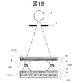
図19は、本発明の実施の形態3に係るイメージング装置の模式図である。本実施の形態3に係るイメージング装置の機能は、実施の形態2に示したイメージング装置と同一であるが、一部の構成が異なる。相違点は、検出器1900がX線専用検出器である点、導光板8aと散乱線除去グリッド9の間に光専用検出器1901aが配置されている点、導光板8bの上面に光専用検出器1901bが配置されている点である。
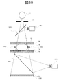
図20は、本発明の実施の形態4に係るイメージング装置の模式図である。本実施の形態4に係るイメージング装置の機能は実施の形態2に示したイメージング装置と同一であるが、一部の構成が異なる。相違点は、検出器1900がX線専用検出器である点、導光板8aと散乱線除去グリッド9の間に反射鏡2000が配置されている点、反射鏡2000に映った被検体6の像を撮影するためのCCDカメラ2001が配置されている点である。
2…コリメータ
3…光源
4…光拡散板
5…圧迫板
6…被検体
7…光学フィルタ
8…導光板
9…散乱線除去グリッド
10…検出器
1400…反射鏡
1401…CCDカメラ
Claims (7)
- X線を被検体に照射するX線源と、
被検体を介して前記X線源と対面する受光部を有し、被検体を介して入射するX線及び光に感度を有する光導電体材料で構成される前記受光部を用いてX線及び光を検出する第1検出器と、
前記第1検出器の検出結果を処理する処理部とを有し、
前記処理部は、前記X線源がX線を照射する照射期間に前記第1検出器が検出したX線の信号を第1信号とし、前記照射期間以外の期間に前記第1検出器が検出した光の信号を第2信号として処理し、前記第1信号と前記第2信号を同一画像上に合成する機能を有することを特徴とするイメージング装置。 - 請求項1に記載のイメージング装置において、被検体の外表面より放出された光を前記第1検出器の前記受光部に導く導光部を有し、前記導光部は被検体の内部で散乱したX線の一部を除去する機能を有することを特徴とするイメージング装置。
- 請求項1に記載のイメージング装置において、
光を被検体に照射する光源と、
被検体に接触した状態で被検体を支持すると共に、前記光源より照射された光を被検体に導く支持部と
を有することを特徴とするイメージング装置。 - X線を被検体に照射するX線源と、
被検体を介して前記X線源と対面する受光部を有し、被検体を介して入射するX線及び光に感度を有する光導電体材料で構成される前記受光部を用いてX線及び光を検出する第1検出器と、
前記X線源と被検体の間に配置され光を検出する第2検出器と、
前記第1及び第2検出器の検出結果を処理する処理部とを有し、
前記処理部は、前記X線源がX線を照射する照射期間に前記第1検出器が検出したX線の信号を第1信号とし、前記照射期間以外の期間に前記第1検出器が検出した光の信号を第2信号として処理し、
前記処理部は、前記X線源がX線を照射する照射期間以外の期間に前記第2検出器が検出する信号を第3信号として処理し、前記第1信号と第2信号と第3信号を同一画像上に合成する機能を有することを特徴とするイメージング装置。 - 請求項4に記載のイメージング装置において、前記X線源と被検体の間に配置される反射鏡と、前記反射鏡により反射される光を集光するレンズとを有し、前記第2検出器は前記レンズにより集光された光を検出することを特徴とするイメージング装置。
- 請求項4に記載のイメージング装置において、前記被検体のサイズを測定するサイズ測定手段を有し、前記処理部は、前記サイズ測定手段の測定結果に応じて前記第1信号と第3信号の合成位置を調整する機能を有することを特徴とするイメージング装置。
- 請求項4に記載のイメージング装置において、前記処理部は、前記第2信号と前記第3信号に基づいて被検体内部における発光または吸光強度分布を計算する機能を有することを特徴とするイメージング装置。
Priority Applications (3)
| Application Number | Priority Date | Filing Date | Title |
|---|---|---|---|
| JP2007123709A JP5143471B2 (ja) | 2007-05-08 | 2007-05-08 | イメージング装置 |
| US12/030,930 US7742561B2 (en) | 2007-05-08 | 2008-02-14 | Imaging apparatus |
| CN2008100058670A CN101301222B (zh) | 2007-05-08 | 2008-02-15 | 成像装置 |
Applications Claiming Priority (1)
| Application Number | Priority Date | Filing Date | Title |
|---|---|---|---|
| JP2007123709A JP5143471B2 (ja) | 2007-05-08 | 2007-05-08 | イメージング装置 |
Publications (3)
| Publication Number | Publication Date |
|---|---|
| JP2008278955A JP2008278955A (ja) | 2008-11-20 |
| JP2008278955A5 JP2008278955A5 (ja) | 2010-04-02 |
| JP5143471B2 true JP5143471B2 (ja) | 2013-02-13 |
Family
ID=39969524
Family Applications (1)
| Application Number | Title | Priority Date | Filing Date |
|---|---|---|---|
| JP2007123709A Expired - Fee Related JP5143471B2 (ja) | 2007-05-08 | 2007-05-08 | イメージング装置 |
Country Status (3)
| Country | Link |
|---|---|
| US (1) | US7742561B2 (ja) |
| JP (1) | JP5143471B2 (ja) |
| CN (1) | CN101301222B (ja) |
Families Citing this family (32)
| Publication number | Priority date | Publication date | Assignee | Title |
|---|---|---|---|---|
| FR2878424B1 (fr) * | 2004-11-26 | 2008-02-01 | Oreal | Procede d'observation d'un tissu biologique, notamment de la peau humaine |
| JP5361336B2 (ja) | 2008-11-06 | 2013-12-04 | キヤノン株式会社 | X線乳房撮影装置 |
| US20110216880A1 (en) * | 2010-03-05 | 2011-09-08 | General Electric Company | System and method for molecular breast imaging |
| WO2011149707A2 (en) * | 2010-05-25 | 2011-12-01 | American Science And Engineering, Inc. | Low-cost position-sensitive x-ray detector |
| US8515006B2 (en) * | 2010-06-15 | 2013-08-20 | Image Mining, Inc. | Fiducial systems for mammography |
| JP5427719B2 (ja) * | 2010-07-21 | 2014-02-26 | 日立コンシューマエレクトロニクス株式会社 | 投写型映像表示装置 |
| JP5776159B2 (ja) * | 2010-09-28 | 2015-09-09 | 富士ゼロックス株式会社 | 搬送装置、および画像形成装置 |
| US8610076B2 (en) | 2010-11-26 | 2013-12-17 | General Electric Company | System and method for molecular breast imaging |
| JP5950538B2 (ja) * | 2011-10-26 | 2016-07-13 | キヤノン株式会社 | 被検体情報取得装置 |
| RU2618912C2 (ru) * | 2011-11-23 | 2017-05-11 | Конинклейке Филипс Н.В. | Способ и устройство для визуализации мягких тканей тела с использованием проекции рентгеновского излучения и оптической томографии |
| JP5879285B2 (ja) | 2012-02-29 | 2016-03-08 | 富士フイルム株式会社 | 音響波検出用プローブおよび光音響計測装置 |
| JP6112773B2 (ja) * | 2012-04-17 | 2017-04-12 | キヤノン株式会社 | 放射線撮影装置、その制御方法及びプログラム |
| JP2014018543A (ja) * | 2012-07-23 | 2014-02-03 | Canon Inc | 放射線発生装置及び放射線撮影システム |
| JP6045365B2 (ja) * | 2013-01-22 | 2016-12-14 | 日本電子株式会社 | 撮像装置 |
| US9535016B2 (en) * | 2013-02-28 | 2017-01-03 | William Beaumont Hospital | Compton coincident volumetric imaging |
| KR20150001184A (ko) * | 2013-06-26 | 2015-01-06 | 삼성전자주식회사 | 엑스선 촬영 장치 및 그 동작 방법 |
| US9216004B2 (en) | 2013-09-12 | 2015-12-22 | Jesse Talant | Adam and ease mammography device |
| US9620256B2 (en) * | 2013-09-26 | 2017-04-11 | Varian Medical Systems, Inc. | X-ray imaging device including anti-scatter grid |
| US20150103975A1 (en) * | 2013-10-11 | 2015-04-16 | National Chiao Tung University | X-ray image sensor and x-ray image sensor system using the same |
| JP6429881B2 (ja) * | 2013-12-27 | 2018-11-28 | ビューワークス カンパニー リミテッド | 動物用映像装置 |
| JP6200839B2 (ja) * | 2014-03-19 | 2017-09-20 | 富士フイルム株式会社 | 乳房厚測定装置、乳房厚測定方法及び放射線撮影システム |
| WO2016083483A1 (en) * | 2014-11-27 | 2016-06-02 | Koninklijke Philips N.V. | Imaging device and method for generating an image of a patient |
| CN104458772A (zh) * | 2014-12-02 | 2015-03-25 | 成都发动机(集团)有限公司 | 一种筒体焊缝x射线检测实时成像检测系统 |
| US10539682B2 (en) * | 2015-02-17 | 2020-01-21 | Koninklijke Philips N.V. | Medical imaging detector |
| WO2016163177A1 (ja) * | 2015-04-09 | 2016-10-13 | 株式会社島津製作所 | X線撮影装置 |
| EP3205261B1 (en) * | 2016-02-10 | 2018-03-28 | Nokia Technologies Oy | Intra-oral imaging |
| EP3210539B1 (en) | 2016-02-24 | 2019-09-11 | Nokia Technologies Oy | Intra-oral x-ray detection |
| JP6707048B2 (ja) * | 2017-03-22 | 2020-06-10 | 富士フイルム株式会社 | マンモグラフィ装置 |
| CN107582029A (zh) * | 2017-10-13 | 2018-01-16 | 徐晖 | 一种妇科检查给药一体装置 |
| EP3527138B1 (en) * | 2018-01-30 | 2022-06-29 | Globus Medical, Inc. | Portable medical imaging system with beam scanning |
| EP3660542A1 (en) * | 2018-11-29 | 2020-06-03 | Koninklijke Philips N.V. | Hybrid x-ray and optical detector |
| US11139088B2 (en) | 2019-06-12 | 2021-10-05 | alephFS—Systems for Imaging | Grid for X-ray imaging |
Family Cites Families (20)
| Publication number | Priority date | Publication date | Assignee | Title |
|---|---|---|---|---|
| US4212306A (en) * | 1978-05-18 | 1980-07-15 | Khalid Mahmud | Breast examination device and method |
| JP3112025B2 (ja) * | 1990-10-26 | 2000-11-27 | 株式会社日立製作所 | 生体計測装置 |
| US5453611A (en) * | 1993-01-01 | 1995-09-26 | Canon Kabushiki Kaisha | Solid-state image pickup device with a plurality of photoelectric conversion elements on a common semiconductor chip |
| US5803082A (en) * | 1993-11-09 | 1998-09-08 | Staplevision Inc. | Omnispectramammography |
| US5712890A (en) * | 1994-11-23 | 1998-01-27 | Thermotrex Corp. | Full breast digital mammography device |
| JP3461236B2 (ja) * | 1996-01-19 | 2003-10-27 | キヤノン株式会社 | 放射線撮影装置並びに画像処理方法及び装置 |
| EP0897282A2 (en) | 1996-12-03 | 1999-02-24 | Koninklijke Philips Electronics N.V. | Method and apparatus for imaging an interior of a turbid medium |
| US5812629A (en) * | 1997-04-30 | 1998-09-22 | Clauser; John F. | Ultrahigh resolution interferometric x-ray imaging |
| US5929434A (en) * | 1997-08-13 | 1999-07-27 | Rockwell Science Center, Llc | Ultra-low noise high bandwidth interface circuit for single-photon readout of photodetectors |
| JP4092037B2 (ja) * | 1999-03-05 | 2008-05-28 | 株式会社堀場製作所 | 物質同定装置 |
| DE10156627A1 (de) * | 2001-11-17 | 2003-05-28 | Philips Corp Intellectual Pty | Anordnung mit elektrischen Elementen |
| JP4558716B2 (ja) * | 2003-03-07 | 2010-10-06 | コーニンクレッカ フィリップス エレクトロニクス エヌ ヴィ | X線蛍光マーカの空間分布をイメージングする方法及びイメージングシステム |
| US7198404B2 (en) * | 2003-04-03 | 2007-04-03 | Siemens Medical Solutions Usa, Inc. | Real-time acquisition of co-registered X-ray and optical images |
| CN2635020Y (zh) * | 2003-07-11 | 2004-08-25 | 貊大卫 | 一种x射线数字成像乳腺扫描装置 |
| FR2864731B1 (fr) * | 2003-12-30 | 2006-02-03 | Commissariat Energie Atomique | Systeme de detection de rayonnements permettant un meilleur comptage d'evenements |
| JP2006026016A (ja) * | 2004-07-14 | 2006-02-02 | Fuji Photo Film Co Ltd | 乳房蛍光画像取得装置 |
| US7403354B2 (en) * | 2005-02-28 | 2008-07-22 | Seagate Technology Llc | Two layer writer heater using writer as one current lead |
| JP2006254969A (ja) * | 2005-03-15 | 2006-09-28 | Konica Minolta Medical & Graphic Inc | 放射線画像取得装置及び放射線画像取得方法 |
| DE102005022540B4 (de) * | 2005-05-17 | 2007-07-05 | Siemens Ag | Verfahren zur Minimierung von Bildartefakten und medizinisches Bildgebungssystem |
| DE102006001850B4 (de) * | 2006-01-13 | 2015-03-26 | Siemens Aktiengesellschaft | Bildgebendes medizintechnisches Gerät und Verfahren |
-
2007
- 2007-05-08 JP JP2007123709A patent/JP5143471B2/ja not_active Expired - Fee Related
-
2008
- 2008-02-14 US US12/030,930 patent/US7742561B2/en not_active Expired - Fee Related
- 2008-02-15 CN CN2008100058670A patent/CN101301222B/zh not_active Expired - Fee Related
Also Published As
| Publication number | Publication date |
|---|---|
| US20080279330A1 (en) | 2008-11-13 |
| JP2008278955A (ja) | 2008-11-20 |
| CN101301222B (zh) | 2011-04-27 |
| CN101301222A (zh) | 2008-11-12 |
| US7742561B2 (en) | 2010-06-22 |
Similar Documents
| Publication | Publication Date | Title |
|---|---|---|
| JP5143471B2 (ja) | イメージング装置 | |
| JP5268633B2 (ja) | スペクトルctのための検出器アレイ | |
| CA2218127C (en) | A system for quantitative radiographic imaging | |
| US5864146A (en) | System for quantitative radiographic imaging | |
| JP3647440B2 (ja) | X線撮影装置 | |
| US7486766B1 (en) | Micro CT scanners incorporating internal gain charge-coupled devices | |
| US5465284A (en) | System for quantitative radiographic imaging | |
| JP5361336B2 (ja) | X線乳房撮影装置 | |
| JP6753708B2 (ja) | 医用画像診断装置 | |
| US20120106702A1 (en) | Apparatus and method for multi-modal imaging using multiple x-ray sources | |
| US7550728B2 (en) | Diagnosis device and diagnosis method for radiographic and nuclear medical examinations | |
| KR101215917B1 (ko) | X선 피폭을 감소시킨 복합디지털x선 촬영장치 | |
| KR20160118922A (ko) | 복강경 수술중 감시림프절 절제술을 위한 삼중 융합 영상장치 | |
| JP5792569B2 (ja) | 放射線撮影システムおよび放射線撮影システムの長尺撮影方法 | |
| WO1997042877A1 (en) | A system for quantitative radiographic imaging | |
| JP5677539B2 (ja) | 検出装置 | |
| JP2013536412A (ja) | 電圧を用いて放射線検出器を補正する方法 | |
| JP2012120650A (ja) | 放射線撮影システム及び放射線位相コントラスト画像生成方法 | |
| JP2011163966A (ja) | 医用画像診断装置及び放射線量算出用制御プログラム | |
| Hachadorian et al. | Cherenkov and scintillation imaging dosimetry | |
| CN117122345B (zh) | 一种基于γ射线的甲状腺功能评估智能检测系统 | |
| JP5697726B2 (ja) | 検出装置 | |
| JP2006026016A (ja) | 乳房蛍光画像取得装置 | |
| JP2014028274A (ja) | 検出装置 |
Legal Events
| Date | Code | Title | Description |
|---|---|---|---|
| A521 | Request for written amendment filed |
Free format text: JAPANESE INTERMEDIATE CODE: A523 Effective date: 20100217 |
|
| A621 | Written request for application examination |
Free format text: JAPANESE INTERMEDIATE CODE: A621 Effective date: 20100217 |
|
| A977 | Report on retrieval |
Free format text: JAPANESE INTERMEDIATE CODE: A971007 Effective date: 20111128 |
|
| A131 | Notification of reasons for refusal |
Free format text: JAPANESE INTERMEDIATE CODE: A131 Effective date: 20111206 |
|
| A521 | Request for written amendment filed |
Free format text: JAPANESE INTERMEDIATE CODE: A523 Effective date: 20120203 |
|
| TRDD | Decision of grant or rejection written | ||
| A01 | Written decision to grant a patent or to grant a registration (utility model) |
Free format text: JAPANESE INTERMEDIATE CODE: A01 Effective date: 20121023 |
|
| A01 | Written decision to grant a patent or to grant a registration (utility model) |
Free format text: JAPANESE INTERMEDIATE CODE: A01 |
|
| A61 | First payment of annual fees (during grant procedure) |
Free format text: JAPANESE INTERMEDIATE CODE: A61 Effective date: 20121121 |
|
| FPAY | Renewal fee payment (event date is renewal date of database) |
Free format text: PAYMENT UNTIL: 20151130 Year of fee payment: 3 |
|
| R151 | Written notification of patent or utility model registration |
Ref document number: 5143471 Country of ref document: JP Free format text: JAPANESE INTERMEDIATE CODE: R151 |
|
| LAPS | Cancellation because of no payment of annual fees |