JP4773361B2 - Soluble TCR molecules and methods of use thereof - Google Patents
Soluble TCR molecules and methods of use thereof Download PDFInfo
- Publication number
- JP4773361B2 JP4773361B2 JP2006539829A JP2006539829A JP4773361B2 JP 4773361 B2 JP4773361 B2 JP 4773361B2 JP 2006539829 A JP2006539829 A JP 2006539829A JP 2006539829 A JP2006539829 A JP 2006539829A JP 4773361 B2 JP4773361 B2 JP 4773361B2
- Authority
- JP
- Japan
- Prior art keywords
- cells
- cell
- 264sctcr
- fragment
- tcr
- Prior art date
- Legal status (The legal status is an assumption and is not a legal conclusion. Google has not performed a legal analysis and makes no representation as to the accuracy of the status listed.)
- Active
Links
Images


Classifications
-
- A—HUMAN NECESSITIES
- A61—MEDICAL OR VETERINARY SCIENCE; HYGIENE
- A61K—PREPARATIONS FOR MEDICAL, DENTAL OR TOILETRY PURPOSES
- A61K51/00—Preparations containing radioactive substances for use in therapy or testing in vivo
- A61K51/02—Preparations containing radioactive substances for use in therapy or testing in vivo characterised by the carrier, i.e. characterised by the agent or material covalently linked or complexing the radioactive nucleus
- A61K51/04—Organic compounds
- A61K51/08—Peptides, e.g. proteins, carriers being peptides, polyamino acids, proteins
- A61K51/10—Antibodies or immunoglobulins; Fragments thereof, the carrier being an antibody, an immunoglobulin or a fragment thereof, e.g. a camelised human single domain antibody or the Fc fragment of an antibody
- A61K51/1027—Antibodies or immunoglobulins; Fragments thereof, the carrier being an antibody, an immunoglobulin or a fragment thereof, e.g. a camelised human single domain antibody or the Fc fragment of an antibody against receptors, cell-surface antigens or cell-surface determinants
-
- G—PHYSICS
- G01—MEASURING; TESTING
- G01N—INVESTIGATING OR ANALYSING MATERIALS BY DETERMINING THEIR CHEMICAL OR PHYSICAL PROPERTIES
- G01N33/00—Investigating or analysing materials by specific methods not covered by groups G01N1/00 - G01N31/00
- G01N33/48—Biological material, e.g. blood, urine; Haemocytometers
- G01N33/50—Chemical analysis of biological material, e.g. blood, urine; Testing involving biospecific ligand binding methods; Immunological testing
- G01N33/53—Immunoassay; Biospecific binding assay; Materials therefor
-
- A—HUMAN NECESSITIES
- A61—MEDICAL OR VETERINARY SCIENCE; HYGIENE
- A61K—PREPARATIONS FOR MEDICAL, DENTAL OR TOILETRY PURPOSES
- A61K47/00—Medicinal preparations characterised by the non-active ingredients used, e.g. carriers or inert additives; Targeting or modifying agents chemically bound to the active ingredient
- A61K47/50—Medicinal preparations characterised by the non-active ingredients used, e.g. carriers or inert additives; Targeting or modifying agents chemically bound to the active ingredient the non-active ingredient being chemically bound to the active ingredient, e.g. polymer-drug conjugates
- A61K47/51—Medicinal preparations characterised by the non-active ingredients used, e.g. carriers or inert additives; Targeting or modifying agents chemically bound to the active ingredient the non-active ingredient being chemically bound to the active ingredient, e.g. polymer-drug conjugates the non-active ingredient being a modifying agent
- A61K47/62—Medicinal preparations characterised by the non-active ingredients used, e.g. carriers or inert additives; Targeting or modifying agents chemically bound to the active ingredient the non-active ingredient being chemically bound to the active ingredient, e.g. polymer-drug conjugates the non-active ingredient being a modifying agent the modifying agent being a protein, peptide or polyamino acid
- A61K47/64—Drug-peptide, drug-protein or drug-polyamino acid conjugates, i.e. the modifying agent being a peptide, protein or polyamino acid which is covalently bonded or complexed to a therapeutically active agent
- A61K47/6415—Toxins or lectins, e.g. clostridial toxins or Pseudomonas exotoxins
-
- A—HUMAN NECESSITIES
- A61—MEDICAL OR VETERINARY SCIENCE; HYGIENE
- A61K—PREPARATIONS FOR MEDICAL, DENTAL OR TOILETRY PURPOSES
- A61K47/00—Medicinal preparations characterised by the non-active ingredients used, e.g. carriers or inert additives; Targeting or modifying agents chemically bound to the active ingredient
- A61K47/50—Medicinal preparations characterised by the non-active ingredients used, e.g. carriers or inert additives; Targeting or modifying agents chemically bound to the active ingredient the non-active ingredient being chemically bound to the active ingredient, e.g. polymer-drug conjugates
- A61K47/51—Medicinal preparations characterised by the non-active ingredients used, e.g. carriers or inert additives; Targeting or modifying agents chemically bound to the active ingredient the non-active ingredient being chemically bound to the active ingredient, e.g. polymer-drug conjugates the non-active ingredient being a modifying agent
- A61K47/62—Medicinal preparations characterised by the non-active ingredients used, e.g. carriers or inert additives; Targeting or modifying agents chemically bound to the active ingredient the non-active ingredient being chemically bound to the active ingredient, e.g. polymer-drug conjugates the non-active ingredient being a modifying agent the modifying agent being a protein, peptide or polyamino acid
- A61K47/64—Drug-peptide, drug-protein or drug-polyamino acid conjugates, i.e. the modifying agent being a peptide, protein or polyamino acid which is covalently bonded or complexed to a therapeutically active agent
- A61K47/642—Drug-peptide, drug-protein or drug-polyamino acid conjugates, i.e. the modifying agent being a peptide, protein or polyamino acid which is covalently bonded or complexed to a therapeutically active agent the peptide or protein in the drug conjugate being a cytokine, e.g. IL2, chemokine, growth factors or interferons being the inactive part of the conjugate
-
- A—HUMAN NECESSITIES
- A61—MEDICAL OR VETERINARY SCIENCE; HYGIENE
- A61K—PREPARATIONS FOR MEDICAL, DENTAL OR TOILETRY PURPOSES
- A61K47/00—Medicinal preparations characterised by the non-active ingredients used, e.g. carriers or inert additives; Targeting or modifying agents chemically bound to the active ingredient
- A61K47/50—Medicinal preparations characterised by the non-active ingredients used, e.g. carriers or inert additives; Targeting or modifying agents chemically bound to the active ingredient the non-active ingredient being chemically bound to the active ingredient, e.g. polymer-drug conjugates
- A61K47/51—Medicinal preparations characterised by the non-active ingredients used, e.g. carriers or inert additives; Targeting or modifying agents chemically bound to the active ingredient the non-active ingredient being chemically bound to the active ingredient, e.g. polymer-drug conjugates the non-active ingredient being a modifying agent
- A61K47/62—Medicinal preparations characterised by the non-active ingredients used, e.g. carriers or inert additives; Targeting or modifying agents chemically bound to the active ingredient the non-active ingredient being chemically bound to the active ingredient, e.g. polymer-drug conjugates the non-active ingredient being a modifying agent the modifying agent being a protein, peptide or polyamino acid
- A61K47/64—Drug-peptide, drug-protein or drug-polyamino acid conjugates, i.e. the modifying agent being a peptide, protein or polyamino acid which is covalently bonded or complexed to a therapeutically active agent
- A61K47/6425—Drug-peptide, drug-protein or drug-polyamino acid conjugates, i.e. the modifying agent being a peptide, protein or polyamino acid which is covalently bonded or complexed to a therapeutically active agent the peptide or protein in the drug conjugate being a receptor, e.g. CD4, a cell surface antigen, i.e. not a peptide ligand targeting the antigen, or a cell surface determinant, i.e. a part of the surface of a cell
-
- A—HUMAN NECESSITIES
- A61—MEDICAL OR VETERINARY SCIENCE; HYGIENE
- A61K—PREPARATIONS FOR MEDICAL, DENTAL OR TOILETRY PURPOSES
- A61K47/00—Medicinal preparations characterised by the non-active ingredients used, e.g. carriers or inert additives; Targeting or modifying agents chemically bound to the active ingredient
- A61K47/50—Medicinal preparations characterised by the non-active ingredients used, e.g. carriers or inert additives; Targeting or modifying agents chemically bound to the active ingredient the non-active ingredient being chemically bound to the active ingredient, e.g. polymer-drug conjugates
- A61K47/51—Medicinal preparations characterised by the non-active ingredients used, e.g. carriers or inert additives; Targeting or modifying agents chemically bound to the active ingredient the non-active ingredient being chemically bound to the active ingredient, e.g. polymer-drug conjugates the non-active ingredient being a modifying agent
- A61K47/62—Medicinal preparations characterised by the non-active ingredients used, e.g. carriers or inert additives; Targeting or modifying agents chemically bound to the active ingredient the non-active ingredient being chemically bound to the active ingredient, e.g. polymer-drug conjugates the non-active ingredient being a modifying agent the modifying agent being a protein, peptide or polyamino acid
- A61K47/66—Medicinal preparations characterised by the non-active ingredients used, e.g. carriers or inert additives; Targeting or modifying agents chemically bound to the active ingredient the non-active ingredient being chemically bound to the active ingredient, e.g. polymer-drug conjugates the non-active ingredient being a modifying agent the modifying agent being a protein, peptide or polyamino acid the modifying agent being a pre-targeting system involving a peptide or protein for targeting specific cells
- A61K47/665—Medicinal preparations characterised by the non-active ingredients used, e.g. carriers or inert additives; Targeting or modifying agents chemically bound to the active ingredient the non-active ingredient being chemically bound to the active ingredient, e.g. polymer-drug conjugates the non-active ingredient being a modifying agent the modifying agent being a protein, peptide or polyamino acid the modifying agent being a pre-targeting system involving a peptide or protein for targeting specific cells the pre-targeting system, clearing therapy or rescue therapy involving biotin-(strept) avidin systems
-
- B—PERFORMING OPERATIONS; TRANSPORTING
- B82—NANOTECHNOLOGY
- B82Y—SPECIFIC USES OR APPLICATIONS OF NANOSTRUCTURES; MEASUREMENT OR ANALYSIS OF NANOSTRUCTURES; MANUFACTURE OR TREATMENT OF NANOSTRUCTURES
- B82Y5/00—Nanobiotechnology or nanomedicine, e.g. protein engineering or drug delivery
-
- C—CHEMISTRY; METALLURGY
- C07—ORGANIC CHEMISTRY
- C07K—PEPTIDES
- C07K14/00—Peptides having more than 20 amino acids; Gastrins; Somatostatins; Melanotropins; Derivatives thereof
- C07K14/435—Peptides having more than 20 amino acids; Gastrins; Somatostatins; Melanotropins; Derivatives thereof from animals; from humans
- C07K14/52—Cytokines; Lymphokines; Interferons
- C07K14/54—Interleukins [IL]
- C07K14/55—IL-2
-
- C—CHEMISTRY; METALLURGY
- C07—ORGANIC CHEMISTRY
- C07K—PEPTIDES
- C07K14/00—Peptides having more than 20 amino acids; Gastrins; Somatostatins; Melanotropins; Derivatives thereof
- C07K14/435—Peptides having more than 20 amino acids; Gastrins; Somatostatins; Melanotropins; Derivatives thereof from animals; from humans
- C07K14/705—Receptors; Cell surface antigens; Cell surface determinants
- C07K14/70503—Immunoglobulin superfamily
- C07K14/7051—T-cell receptor (TcR)-CD3 complex
-
- G—PHYSICS
- G01—MEASURING; TESTING
- G01N—INVESTIGATING OR ANALYSING MATERIALS BY DETERMINING THEIR CHEMICAL OR PHYSICAL PROPERTIES
- G01N33/00—Investigating or analysing materials by specific methods not covered by groups G01N1/00 - G01N31/00
- G01N33/48—Biological material, e.g. blood, urine; Haemocytometers
- G01N33/50—Chemical analysis of biological material, e.g. blood, urine; Testing involving biospecific ligand binding methods; Immunological testing
- G01N33/53—Immunoassay; Biospecific binding assay; Materials therefor
- G01N33/543—Immunoassay; Biospecific binding assay; Materials therefor with an insoluble carrier for immobilising immunochemicals
- G01N33/554—Immunoassay; Biospecific binding assay; Materials therefor with an insoluble carrier for immobilising immunochemicals the carrier being a biological cell or cell fragment, e.g. bacteria, yeast cells
-
- G—PHYSICS
- G01—MEASURING; TESTING
- G01N—INVESTIGATING OR ANALYSING MATERIALS BY DETERMINING THEIR CHEMICAL OR PHYSICAL PROPERTIES
- G01N33/00—Investigating or analysing materials by specific methods not covered by groups G01N1/00 - G01N31/00
- G01N33/48—Biological material, e.g. blood, urine; Haemocytometers
- G01N33/50—Chemical analysis of biological material, e.g. blood, urine; Testing involving biospecific ligand binding methods; Immunological testing
- G01N33/53—Immunoassay; Biospecific binding assay; Materials therefor
- G01N33/566—Immunoassay; Biospecific binding assay; Materials therefor using specific carrier or receptor proteins as ligand binding reagents where possible specific carrier or receptor proteins are classified with their target compounds
- G01N33/567—Immunoassay; Biospecific binding assay; Materials therefor using specific carrier or receptor proteins as ligand binding reagents where possible specific carrier or receptor proteins are classified with their target compounds utilising isolate of tissue or organ as binding agent
-
- G—PHYSICS
- G01—MEASURING; TESTING
- G01N—INVESTIGATING OR ANALYSING MATERIALS BY DETERMINING THEIR CHEMICAL OR PHYSICAL PROPERTIES
- G01N33/00—Investigating or analysing materials by specific methods not covered by groups G01N1/00 - G01N31/00
- G01N33/48—Biological material, e.g. blood, urine; Haemocytometers
- G01N33/50—Chemical analysis of biological material, e.g. blood, urine; Testing involving biospecific ligand binding methods; Immunological testing
- G01N33/53—Immunoassay; Biospecific binding assay; Materials therefor
- G01N33/574—Immunoassay; Biospecific binding assay; Materials therefor for cancer
- G01N33/57484—Immunoassay; Biospecific binding assay; Materials therefor for cancer involving compounds serving as markers for tumor, cancer, neoplasia, e.g. cellular determinants, receptors, heat shock/stress proteins, A-protein, oligosaccharides, metabolites
- G01N33/57492—Immunoassay; Biospecific binding assay; Materials therefor for cancer involving compounds serving as markers for tumor, cancer, neoplasia, e.g. cellular determinants, receptors, heat shock/stress proteins, A-protein, oligosaccharides, metabolites involving compounds localized on the membrane of tumor or cancer cells
-
- G—PHYSICS
- G01—MEASURING; TESTING
- G01N—INVESTIGATING OR ANALYSING MATERIALS BY DETERMINING THEIR CHEMICAL OR PHYSICAL PROPERTIES
- G01N33/00—Investigating or analysing materials by specific methods not covered by groups G01N1/00 - G01N31/00
- G01N33/48—Biological material, e.g. blood, urine; Haemocytometers
- G01N33/50—Chemical analysis of biological material, e.g. blood, urine; Testing involving biospecific ligand binding methods; Immunological testing
- G01N33/68—Chemical analysis of biological material, e.g. blood, urine; Testing involving biospecific ligand binding methods; Immunological testing involving proteins, peptides or amino acids
- G01N33/6875—Nucleoproteins
-
- C—CHEMISTRY; METALLURGY
- C07—ORGANIC CHEMISTRY
- C07K—PEPTIDES
- C07K2319/00—Fusion polypeptide
-
- C—CHEMISTRY; METALLURGY
- C07—ORGANIC CHEMISTRY
- C07K—PEPTIDES
- C07K2319/00—Fusion polypeptide
- C07K2319/20—Fusion polypeptide containing a tag with affinity for a non-protein ligand
-
- C—CHEMISTRY; METALLURGY
- C07—ORGANIC CHEMISTRY
- C07K—PEPTIDES
- C07K2319/00—Fusion polypeptide
- C07K2319/30—Non-immunoglobulin-derived peptide or protein having an immunoglobulin constant or Fc region, or a fragment thereof, attached thereto
-
- C—CHEMISTRY; METALLURGY
- C07—ORGANIC CHEMISTRY
- C07K—PEPTIDES
- C07K2319/00—Fusion polypeptide
- C07K2319/32—Fusion polypeptide fusions with soluble part of a cell surface receptor, "decoy receptors"
-
- G—PHYSICS
- G01—MEASURING; TESTING
- G01N—INVESTIGATING OR ANALYSING MATERIALS BY DETERMINING THEIR CHEMICAL OR PHYSICAL PROPERTIES
- G01N2333/00—Assays involving biological materials from specific organisms or of a specific nature
- G01N2333/435—Assays involving biological materials from specific organisms or of a specific nature from animals; from humans
- G01N2333/705—Assays involving receptors, cell surface antigens or cell surface determinants
- G01N2333/70503—Immunoglobulin superfamily, e.g. VCAMs, PECAM, LFA-3
- G01N2333/7051—T-cell receptor (TcR)-CD3 complex
-
- G—PHYSICS
- G01—MEASURING; TESTING
- G01N—INVESTIGATING OR ANALYSING MATERIALS BY DETERMINING THEIR CHEMICAL OR PHYSICAL PROPERTIES
- G01N2333/00—Assays involving biological materials from specific organisms or of a specific nature
- G01N2333/435—Assays involving biological materials from specific organisms or of a specific nature from animals; from humans
- G01N2333/705—Assays involving receptors, cell surface antigens or cell surface determinants
- G01N2333/70503—Immunoglobulin superfamily, e.g. VCAMs, PECAM, LFA-3
- G01N2333/70539—MHC-molecules, e.g. HLA-molecules
-
- G—PHYSICS
- G01—MEASURING; TESTING
- G01N—INVESTIGATING OR ANALYSING MATERIALS BY DETERMINING THEIR CHEMICAL OR PHYSICAL PROPERTIES
- G01N2800/00—Detection or diagnosis of diseases
- G01N2800/70—Mechanisms involved in disease identification
- G01N2800/7023—(Hyper)proliferation
- G01N2800/7028—Cancer
Description
(関連出願)
本出願は、「可溶性TCR分子とその使用方法」の名称で2003年11月10日に出願された米国仮特許出願第60/518,790号の優先権を主張するものであり、その全てを本明細書取り込む。
(Related application)
This application claims priority from US Provisional Patent Application No. 60 / 518,790 filed Nov. 10, 2003 under the name "Soluble TCR Molecules and Methods of Use", all of which are incorporated herein by reference. Incorporated herein.
(技術分野)
本発明は、MHC又はHLA複合体として存在するペプチド抗原を含む細胞又は組織を検出するための組成物及び方法を特徴とする。本発明は、がん細胞を検出するための感度の高い方法を提供することを含む広範な適用を有する。
(Technical field)
The invention features compositions and methods for detecting cells or tissues containing peptide antigens present as MHC or HLA complexes. The present invention has a wide range of applications including providing a sensitive method for detecting cancer cells.
(米国政府の権利に関する記載)
本発明は、米国立衛生研究所のグラント第1R43CA88615−01号及び第1R43CA105816−01号に基づき、一部米国政府の助成を受けてなされたものであり、米国政府は本発明に関して所定の権利を有する。
(Statement regarding US government rights)
This invention was made in part with grants from the United States Government under grants 1R43CA88615-01 and 1R43CA105816-01 of the National Institutes of Health. Have.
免疫療法は、がん治療のための有望な方法であるとの認識が増しており、インターロイキン2(IL−2)のようなサイトカインでの治療を含む様々な方法が提案されている。IL−2は、T細胞、B細胞、単球、マクロファージ、リンフォカイン活性化キラー細胞(LAK)及びNK細胞を含む様々な免疫細胞種に影響を与えるものである。[文献10及び40参照] There is increasing recognition that immunotherapy is a promising method for cancer treatment, and various methods have been proposed including treatment with cytokines such as interleukin 2 (IL-2). IL-2 affects a variety of immune cell types including T cells, B cells, monocytes, macrophages, lymphokine activated killer cells (LAK) and NK cells. [Ref. 10 and 40]
効力を上げるために腫瘍部位にサイトカインを濃縮させることが提案されてきた。一般的な方法としては、サイトカイン又はそれをコードする遺伝子を腫瘍内へ直接注入すること、又はサイトカインを腫瘍抗原に特異的な抗体に融合させることによる標的化送達[文献20参照]があるが、これらの方法には問題がある。 It has been proposed to concentrate cytokines at the tumor site to increase efficacy. Common methods include targeted delivery by injecting a cytokine or gene encoding it directly into the tumor, or by fusing the cytokine to an antibody specific for a tumor antigen [see reference 20] There are problems with these methods.
例えば、多くの直接注入法は、腫瘍が概して小さい(微小転移巣)特にがんの初期段階で使用するのが困難である。さらにこの方法は、治療の成功の見込みが殆どないにもかかわらず多大な労力を要するものである。当該方法による治療は、多くの患者にとって非実用的であり、費用の嵩むものである。 For example, many direct injection methods are difficult to use, especially in the early stages of cancer, where the tumor is generally small (micrometastasis). Furthermore, this method is labor intensive despite little prospect of successful treatment. Treatment by this method is impractical and costly for many patients.
がんを治療する一つの方法として抗体とサイトカインの融合物が用いられてきたが、抗体の結合範囲には限りがあるため、当該方法も限られたものとなっている。すなわち、抗体は特定の細胞表面の抗原を認識することしかできないからである。残念乍ら、腫瘍抗原の多くは、抗体認識に都合よく提示されるわけではないので、抗体に基づく方法の可能性については限界がある。さらに、腫瘍に特異的な抗原の多くは、細胞型に特異的なタンパク質の異常発現に由来するとの報告があるが、これらは少数の腫瘍型にのみ存在するものである。当該問題は、抗体に基づいた治療の可能性をさらに限定するものである。 An antibody-cytokine fusion has been used as one method for treating cancer, but the method is also limited because of the limited range of antibody binding. That is, an antibody can only recognize a specific cell surface antigen. Unfortunately, many of the tumor antigens are not conveniently presented for antibody recognition, so the potential for antibody-based methods is limited. In addition, many tumor-specific antigens have been reported to be derived from abnormal expression of cell-type specific proteins, but these are only present in a few tumor types. The problem further limits the possibility of antibody-based therapy.
p53タンパク質は、細胞周期のG1/S期で異常細胞を拘束することにより作用することが報告されている、細胞内の腫瘍抑制因子である。そのタンパク質の過剰発現は、多くのヒト悪性腫瘍に対する重要な腫瘍マーカーであると信じられており、広い標的範囲を有する腫瘍免疫療法の良い標的であると認識されている。p53タンパク質は、通常、主要組織適合性複合体タンパク質(MHC)として、細胞表面に提示される。このタンパク質複合体は、T細胞受容体(TCRs)の結合標的であることが知られている。[文献49参照] The p53 protein is an intracellular tumor suppressor that has been reported to act by binding abnormal cells in the G1 / S phase of the cell cycle. Overexpression of the protein is believed to be an important tumor marker for many human malignancies and is recognized as a good target for tumor immunotherapy with a broad target range. The p53 protein is usually presented on the cell surface as a major histocompatibility complex protein (MHC). This protein complex is known to be a binding target for T cell receptors (TCRs). [Ref. 49]
ペプチドを含むMHC/ペプチド複合体を検出するために、ある種のTCRsを用いる試みがなされてきた(Epelら, 2002; Hollerら, 2003; Lebowitzら, 1999; Plaksinら, 1997; Watayaら, 2001; O'Herronら, 1997)が、これら、また、関連の方法には、重大な欠点がある。 Attempts have been made to use certain TCRs to detect MHC / peptide complexes containing peptides (Epel et al., 2002; Holler et al., 2003; Lebowitz et al., 1999; Plaksin et al., 1997; Wataya et al., 2001). O'Herron et al., 1997), but these and related methods have significant drawbacks.
例えば多くの方法において、おそらくは、ペプチド抗原のペプチド抗原に対する結合を人為的に促進するために、TCR構築物を複合体化させる(すなわち、多様なTCRコピーを持つようにデザインされる)ことが必要とされている。標的(抗原提示)細胞は一般に、比較的大量のペプチド抗原を発現するように操作されているが、ペプチド抗原の密度は、場合によっては、細胞当たりの複合体数が104〜105個と高くなる(Watayaら, 2001)。ペプチド抗原の密度が高いと、TCRでの結合及び検出が容易になると考えられているが、ペプチド抗原のこのようなレベルは人為的なものであり、腫瘍関連抗原(TAAs)が存在するMHC/ペプチド複合体の通常のレベルよりは遙かに高くなっている。TAAsによっては、HLA/ペプチド複合体が一つの細胞当たり約50未満しか存在しないものもある(Pascoloら, 2001; Schirleら, 2000)。従って、従来の方法は、全てではないにしても大部分のTAAsを検出するには感度が十分であるとは言えない、との認識がある。 For example, in many methods, it may be necessary to complex the TCR construct (ie, designed to have diverse TCR copies), possibly to artificially promote the binding of the peptide antigen to the peptide antigen. Has been. Target (antigen presenting) cells are typically engineered to express relatively large amounts of peptide antigens, but the density of peptide antigens can be as high as 10 4 to 10 5 complexes per cell in some cases. It becomes higher (Wataya et al., 2001). Although high density of peptide antigens are believed to facilitate TCR binding and detection, such levels of peptide antigens are artificial and include MHC / with tumor associated antigens (TAAs). It is much higher than the normal level of peptide complex. Some TAAs have less than about 50 HLA / peptide complexes per cell (Pascolo et al., 2001; Schirle et al., 2000). Accordingly, it is recognized that the conventional method is not sufficiently sensitive to detect most, if not all, of the TAAs.
ある種のTCRsを用いて特定のペプチド抗原を発現している細胞を検出する試みがなされてきたが、抗体に基づいた多くの方法と同様、これらの取り組みはペプチド抗原を検出するには感度が十分ではなかったか、又は抗原を完全には検出することができなかった。 While attempts have been made to detect cells expressing specific peptide antigens using certain TCRs, as with many antibody-based methods, these approaches are not sensitive to detecting peptide antigens. It was not enough or the antigen could not be detected completely.
例えば、Hollerら(2003)は、MHC/ペプチド複合体と反応することが報告されているある種の可溶性TCRsの開発を報告した。そのTCRsは、人為的に抗原を「収容した(loaded)」細胞に於いては抗原を検出できたが、腫瘍細胞上の内因性の抗原を検出することはできなかった。Hollerらは、抗原が細胞当たり600コピー未満の低い密度でしか存在しない場合には、TCRに基づいた方法は、抗原を検出する感度や信頼性が十分ではないと結論付けた。 For example, Holler et al. (2003) reported the development of certain soluble TCRs that have been reported to react with MHC / peptide complexes. The TCRs were able to detect antigen in cells that were artificially “loaded” with antigen, but were unable to detect endogenous antigen on tumor cells. Holler et al. Concluded that TCR-based methods are not sensitive enough or reliable to detect antigens when antigens are present at a low density of less than 600 copies per cell.
TCRに基づいた特定の方法は、MHC分子としてのウィルスペプチドを検出するために用いられてきた(Stromingerらの国際公開第9618105号公報)が、これら及び関連の方法には問題がある。例えば、ウィルス感染はしばしば例外的に、一つの細胞に通常1000〜105個といった高い密度でMHC/ペプチド複合体を産生するものと一般的に認識されている。Herbertsら(2001)及びvan Elsら(2000)を参照されたい。他の多くのペプチド抗原の検出方法と同様に、これまでのTCRに基づいたウィルス抗原を検出するための方法には、比較的多数の標的抗原が用いられてきた。 Certain methods based on TCR have been used to detect viral peptides as MHC molecules (Strominger et al., WO 9618105), but there are problems with these and related methods. For example, viral infection often Exceptionally, one cell has been recognized normally 1000 to five such high producing an MHC / peptide complexes by the density and general. See Herberts et al. (2001) and van Els et al. (2000). As with many other peptide antigen detection methods, a relatively large number of target antigens have been used in previous methods for detecting viral antigens based on TCR.
いくつかのTCRに基づいた方法は比較的大量のペプチド抗原を検出するために用いられてきたが、サイトカイン、IgG1のような免疫グロビン(immunoglobin)ドメイン、ビオチン、又はストレプトアビジンのようなその他の分子にTCRが結合した場合に、その方法が機能するかどうかは、あまり確かではない。すなわち、特に低密度のTAAを分析する必要がある場合に、その融合分子がTCRペプチド結合溝に対してどのように影響するのかは明らかではない。TCRペプチド結合溝の小さな歪曲は、比較的多くのペプチド抗原を分析する際には必ずしも問題とはならないが、TAA結合の特異性及び選択性を減少させている。TCRペプチド結合溝の機能に於けるわずかな変化でさえも、低密度のTAAしか発現していないがん細胞の検出を危うくする可能性がある。 Some TCR-based methods have been used to detect relatively large amounts of peptide antigens, but other molecules such as cytokines, immunoglobin domains such as IgG1, biotin, or streptavidin It is not very sure whether the method will work if a TCR is attached to the. That is, it is not clear how the fusion molecule affects the TCR peptide binding groove, especially when it is necessary to analyze low density TAAs. Small distortions in the TCR peptide binding groove are not necessarily a problem when analyzing relatively many peptide antigens, but reduce the specificity and selectivity of TAA binding. Even minor changes in the function of the TCR peptide binding groove can jeopardize the detection of cancer cells expressing only low density TAAs.
特に、ペプチド抗原が低密度にしか発現していない場合に、高感度で、選択性があり、再現性のあるTAAsを検出できる方法があれば、有用である。そのような方法がもし、検出可能な標識又はサイトカインに結合した分子を含む多様な可溶性TCRsを用いるものであれば、特に有用であろう。 In particular, if peptide antigens are expressed only at low density, it would be useful if there was a method that could detect TAAs with high sensitivity, selectivity, and reproducibility. Such a method would be particularly useful if it used a variety of soluble TCRs, including molecules attached to a detectable label or cytokine.
本発明は、一般に、MHC又はHLA複合体として細胞又は組織に存在しているペプチド抗原を含む細胞又は組織を検出するための方法に関する。ある態様に於いては、本発明は、以下の:
a)細胞又は組織を、存在しているペプチド抗原と可溶性TCR分子又はその断片が特異的な結合複合体を形成する条件下で、少なくとも一つの可溶性TCR分子又はその機能性断片に接触させる、
b)存在しているペプチド抗原に結合しなかったすべての可溶性TCR分子又はその断片を取り除くのに適した条件下で、細胞又は組織を洗浄する、及び
c)存在しているペプチド抗原を含む細胞又は組織の指標としての特異的な結合複合体を検出する、
工程のうち少なくとも一つ、好ましくはその全てを含む。
The present invention relates generally to methods for detecting cells or tissues containing peptide antigens present in the cells or tissues as MHC or HLA complexes. In certain embodiments, the present invention provides the following:
a) contacting a cell or tissue with at least one soluble TCR molecule or functional fragment thereof under conditions such that the existing peptide antigen and soluble TCR molecule or fragment thereof form a specific binding complex;
b) washing the cell or tissue under conditions suitable to remove all soluble TCR molecules or fragments thereof that did not bind to the present peptide antigen, and c) cells containing the present peptide antigen. Or detect specific binding complexes as an indicator of tissue,
At least one, preferably all of the steps are included.
好ましい実施に於いて、本発明は、細胞又は組織上のペプチド抗原の量が、約100000コピーより少ない、好ましくは約100から800コピーのような1000コピーより少ないものを検出するために利用される。 In a preferred implementation, the present invention is utilized to detect that the amount of peptide antigen on a cell or tissue is less than about 100,000 copies, preferably less than 1000 copies, such as about 100 to 800 copies. .
本発明の使用にはいくつかの利点があるが、例えば、本発明は、きわめて感度が高いので、内因性ペプチド、より特化すれば、操作されていない腫瘍細胞に存在する腫瘍関連ペプチド抗原を含む非常に低密度のMHC/ペプチド複合体を検出及び場合によっては定量するために利用される。一方、MHC/ペプチド複合体を検出するための従来の方法は、相対的により高密度の複合体を検出できることが報告されている。 Although there are several advantages to the use of the present invention, for example, because the present invention is extremely sensitive, endogenous peptides, and more specifically, tumor-associated peptide antigens present in unengineered tumor cells, It is used to detect and optionally quantify very low density MHC / peptide complexes containing. On the other hand, conventional methods for detecting MHC / peptide complexes have been reported to be able to detect relatively higher density complexes.
さらに、本発明は、例えば腫瘍組織アレイのような組織アレイに通常みられるような固定された細胞及び組織を検出及び場合によっては定量するために利用される。MHC/ペプチド複合体を検出する能力(「染色」とも言う)は、特に患者から採取された細胞、組織又は他の生物学的試料の固定化が通常行われている臨床又はその他の医学的環境に於いて有用である。一方、非共有結合ペプチドは通常、組織を処理する工程で失われてしまうので、従来のTCRに基づいた検出方法の多くは固定化された組織には適用できない。 In addition, the present invention is utilized to detect and optionally quantify fixed cells and tissues such as are commonly found in tissue arrays such as tumor tissue arrays. The ability to detect MHC / peptide complexes (also referred to as “staining”) is particularly useful in clinical or other medical environments where immobilization of cells, tissues or other biological samples taken from patients is common. It is useful in On the other hand, since non-covalent peptides are usually lost in the process of treating tissues, many conventional detection methods based on TCR cannot be applied to immobilized tissues.
本発明は、更なる利点を提供する。例えば、本発明の方法は、単量体及び/又は多量体の可溶性TCR分子を使用する場合にも柔軟に適応されるものとなっている。従来の方法は残念乍ら、主に柔軟性と感度が限られている多量体のTCRsを使用するものであった。多量体のTCRsは特に、分解又は凝集能力、標的部位への到達性を欠くこと、免疫原性の増加びクリアランスのため、インビボ画像化に使用するのは難しい。 The present invention provides further advantages. For example, the method of the present invention is flexibly adapted when using monomeric and / or multimeric soluble TCR molecules. Unfortunately, the conventional methods have mainly used multimeric TCRs with limited flexibility and sensitivity. Multimeric TCRs are particularly difficult to use for in vivo imaging due to their ability to degrade or aggregate, lack of reach to target sites, increased immunogenicity and clearance.
本発明の態様は、細胞の表面にMHC/ペプチド複合体として提示される内因性ペプチド抗原を検出する能力を提供することによって、当該分野で長らく待ち望まれてきたニーズに応えるものである。本発明の方法は、細胞の活性、病理、及び感染をモニターする助けとなる様々な重要な使用を有する。例えば、本発明による細胞又は組織の内因性腫瘍関連のペプチド抗原の検出は、がんの存在/存在範囲を検出及び及び場合によっては定量する方法を提供する。従来の態様に於いては、がん細胞の表面にあるタンパク質性抗原を検出する診断ツールとして抗体を利用してきたが、抗体は通常、細胞膜タンパク質の検出に限られたものとなっている。さらに、抗体による検出は、抗原が遊離する(shedding)又は抗原性タンパク質が循環系に分泌されるといった障害がある。抗体は又、標的の認識に限界がある。本発明の態様は、標的ペプチド抗原を検出するために可溶性TCRs及びその断片を使用する、高感度で信頼性のある検出方法を提供することによって、これら及び他の問題を回避するものである。 Aspects of the present invention address the long-awaited need in the art by providing the ability to detect endogenous peptide antigens presented as MHC / peptide complexes on the surface of cells. The methods of the invention have a variety of important uses that help monitor cell activity, pathology, and infection. For example, detection of endogenous tumor-related peptide antigens in cells or tissues according to the present invention provides a method of detecting and optionally quantifying the presence / extent of cancer. Conventionally, antibodies have been used as diagnostic tools for detecting proteinaceous antigens on the surface of cancer cells, but antibodies are usually limited to detection of cell membrane proteins. In addition, detection with antibodies suffers from problems such as antigen shedding or antigenic proteins being secreted into the circulatory system. Antibodies also have limited target recognition. Aspects of the present invention avoid these and other problems by providing a sensitive and reliable detection method that uses soluble TCRs and fragments thereof to detect target peptide antigens.
本発明のそのような使用及び利点は、インビボ(例えば、画像化又は診断法のような)又はインビトロ(例えば、組織アレイ又はFACS分析のような)を含む様々な状況に於いて、ペプチド抗原を検出するために用いることが可能である。 Such uses and advantages of the present invention include peptide antigens in a variety of situations, including in vivo (such as imaging or diagnostic methods) or in vitro (such as tissue arrays or FACS analysis). It can be used to detect.
本発明のその他の態様は、下記考察の通りである。 Other aspects of the invention are as discussed below.
(発明の詳細な説明)
上記考察のように、本発明は一般に、MHC複合体として細胞又は組織に提示されるペプチド抗原を含む細胞又は組織を検出するための方法を含む。ある態様では、本発明は、存在しているペプチド抗原と可溶性TCR又はその断片が特異的に結合する複合体を形成する条件下で、少なくとも一つの可溶性TCR分子又はその機能的断片と細胞又は組織を接触させること、存在しているペプチド抗原に結合しなかったすべての可溶性TCR分子又はその断片を取り除くのに適した条件下で、細胞又は組織を洗浄すること、及び存在しているペプチド抗原を含む細胞又は組織の指標としての特異的な結合複合体を検出すること、を含む。
(Detailed description of the invention)
As discussed above, the present invention generally includes a method for detecting a cell or tissue comprising a peptide antigen that is presented to the cell or tissue as an MHC complex. In one aspect, the present invention relates to at least one soluble TCR molecule or functional fragment thereof and a cell or tissue under conditions that form a complex in which an existing peptide antigen and soluble TCR or fragment thereof specifically bind. Contacting cells, washing cells or tissues under conditions suitable to remove any soluble TCR molecules or fragments thereof that did not bind to existing peptide antigens, and Detecting a specific binding complex as an indicator of the cell or tissue comprising.
一般に、本可溶性TCRsは、本明細書に開示されている手順及び認識されているDNA組み換え技術を用いての調製される。例えば、プラスミドDNAの調製、制限酵素によるDNA切断、DNAの結紮、DNAの細胞内への導入、細胞の培養、並びに発現タンパク質の単離及び精製は、公知の技術である。一般的には、Sambrookらの「Molecular Cloning: A Laboratory Manual (2d ed. 1989)」;及びAusubelらの「Current Protocols in Molecular Biology, John Wiley&Sons, New York(1989)」を参照されたい。 In general, the soluble TCRs are prepared using the procedures disclosed herein and recognized DNA recombination techniques. For example, preparation of plasmid DNA, DNA cleavage with restriction enzymes, DNA ligation, introduction of DNA into cells, cell culture, and isolation and purification of expressed proteins are known techniques. See generally Sambrook et al., “Molecular Cloning: A Laboratory Manual (2d ed. 1989)”; Ausubel et al., “Current Protocols in Molecular Biology, John Wiley & Sons, New York (1989)”.
様々な可溶性TCR構築物の一般的な構造並びに同一物の作製及び使用の方法は、係属中の米国特許出願第08/813,781号及び第08/943,086号に開示されている。
The general structure of various soluble TCR constructs and methods of making and using the same are disclosed in pending
例えば、特定の可溶性TCRは、少なくとも片方、好ましくは両方のV鎖に於いて膜貫通配列が除去されている異種二量体である。しかしながら便宜上、係属中の出願第08/813,781号及び第08/943,086号に報告されているように、一本鎖の(「sc−」)構築物を使うことが好まれる場合が多い。
For example, a particular soluble TCR is a heterodimer from which transmembrane sequences have been removed on at least one, preferably both V chains. However, for convenience, it is often preferred to use single stranded ("sc-") constructs, as reported in pending
簡潔に述べると、一本鎖の(「sc−」)TCR分子は、適切なペプチドリンカー配列を介して共有結合しているV−α及びV−β鎖を含む。例えば、V−α鎖は、V−α鎖のC末端とV−β鎖のN末端に結合した適切なペプチドリンカー配列を介して、V−β鎖に共有結合できる。sc−TCR融合タンパク質のV−α及びV−β鎖は、一般にアミノ酸長が約200から400であり、好ましくはアミノ酸長が約300から350であり、天然に存在するTCRのV−α及びV−β鎖と、少なくとも90%、好ましくは100%同一である。「同一(identical)」という用語は、V−α又はV−β鎖のアミノ酸は、対応する天然に存在するTCRのV−β又はV−α鎖と100%相同(homologous)であることを意味する。 Briefly, single-stranded (“sc-”) TCR molecules comprise V-α and V-β chains covalently linked through suitable peptide linker sequences. For example, the V-α chain can be covalently attached to the V-β chain via an appropriate peptide linker sequence attached to the C-terminus of the V-α chain and the N-terminus of the V-β chain. The V-α and V-β chains of sc-TCR fusion proteins are generally about 200 to 400 amino acids in length, preferably about 300 to 350 amino acids in length, and naturally occurring TCR V-α and V-chains. -Is at least 90%, preferably 100% identical to the β chain. The term “identical” means that the amino acids of the V-α or V-β chain are 100% homologous with the corresponding naturally occurring TCR V-β or V-α chain. To do.
第08/943,086号出願に開示されているように、sc−TCR分子のV−α鎖はさらに、V−β鎖のC末端に結合したC−β鎖又はその断片を含むことができる。さらに、V−α鎖は、V−α鎖のC末端及びペプチドリンカー配列のN末端に、結合したC−α鎖又はその断片を含むことができる。一般に、C−β鎖断片を含む融合タンパク質に於いて、その断片はアミノ酸長が約50から130であり、通常C−β鎖の最後のシステイン残基(マウスに於いては127位、ヒトに於いては131位)を含まない。C−α鎖を含む融合タンパク質では、その長さは、約1から90アミノ酸(すなわち、最後のシステイン残基を含まないがそこまでのC−α鎖)の間で変化してもよい。例えば、ある態様に於いては、融合タンパク質は、1番目のアミノ酸から始まり72番目までのアミノ酸長約1〜72のC−α鎖断片を含む。別の態様に於いては、C−α鎖断片は、最初のアミノ酸から始まり22番目(ロイシン)までのアミノ酸長約1〜22である。C−α鎖断片は、通常、二つのシステイン残基を含んだCα90変異体及び一つのシステイン残基を含んだCα72変異体以外は、如何なるシステイン残基も含まない。Cα及びCβ鎖長は、選択された特定のV鎖及び可溶性融合分子の使用目的を含むいくつかのパラメータを使用して選択される場合が多い。 As disclosed in the 08 / 943,086 application, the V-α chain of the sc-TCR molecule may further comprise a C-β chain or fragment thereof linked to the C-terminus of the V-β chain. . Further, the V-α chain can include a C-α chain or a fragment thereof linked to the C-terminus of the V-α chain and the N-terminus of the peptide linker sequence. In general, in fusion proteins containing a C-β chain fragment, the fragment is about 50 to 130 amino acids in length, usually the last cysteine residue of the C-β chain (position 127 in mice, human). (No. 131) is not included. For fusion proteins containing a C-α chain, the length may vary between about 1 and 90 amino acids (ie, the C-α chain up to but not including the last cysteine residue). For example, in one embodiment, the fusion protein comprises a C-α chain fragment of about 1 to 72 amino acids long starting at the first amino acid and ending at the 72nd amino acid. In another embodiment, the C-α chain fragment is about 1-22 amino acids long starting at the first amino acid and ending at position 22 (leucine). C-α chain fragments usually do not contain any cysteine residues other than a Cα90 variant containing two cysteine residues and a Cα72 variant containing one cysteine residue. Cα and Cβ chain lengths are often selected using a number of parameters including the intended use of the particular V chain and soluble fusion molecule selected.
さらに第08/943,086号出願に開示されるように、本発明の追加のsc−TCRタンパク質は、V−α鎖のC末端とV−β鎖のN末端の間に最初のペプチドリンカー配列が結合する場合には、例えば、二つのペプチドリンカー配列を含む。V−β鎖のC末端は、C−β鎖断片のN末端に結合でき、そして2番目のペプチドリンカーは、V−β鎖又はC−β鎖断片のC末端又は、必要に応じて、以下に説明されるような標識分子に結合する。別の説明的な態様に於いては、sc−TCRタンパク質は、V−β鎖又はそのC−β鎖断片のC末端と、V−α鎖のN末端を共有結合でつなぐ、適当なペプチドリンカーを介してV−β鎖をV−α鎖に結合することによって作製することができる。 As further disclosed in the 08 / 943,086 application, the additional sc-TCR protein of the present invention comprises an initial peptide linker sequence between the C-terminus of the V-α chain and the N-terminus of the V-β chain. Include, for example, two peptide linker sequences. The C-terminus of the V-β chain can be attached to the N-terminus of the C-β chain fragment, and the second peptide linker can be the C-terminus of the V-β chain or C-β chain fragment, or To a labeled molecule as described in. In another illustrative embodiment, the sc-TCR protein comprises a suitable peptide linker that covalently connects the C-terminus of the V-β chain or C-β chain fragment thereof to the N-terminus of the V-α chain. It can be produced by binding the V-β chain to the V-α chain via
本発明の可溶性TCRタンパク質は、一つ又はそれ以上の融合したタンパク質タグ(protain tag)を含むことができる。そのようなタグが「検出可能」である態様に於いて、可溶性TCRは、「検出可能に標識された」と言える。例えば、可溶性の融合タンパク質に関して、タンパク質タグは、sc−TCRのV−β鎖(又はC−β鎖断片)のC末端に結合可能である。必要に応じて、このような可溶性TCRタンパク質は、係属中の第08/943,086号出願によって報告されているような、また、以下の実施例に於いてさらに説明されるような、免疫グロビン鎖に結合可能である。 The soluble TCR protein of the present invention can include one or more fused protein tags. In embodiments where such a tag is “detectable”, the soluble TCR can be said to be “detectably labeled”. For example, for soluble fusion proteins, the protein tag can be attached to the C-terminus of the sc-TCR V-β chain (or C-β chain fragment). Optionally, such a soluble TCR protein is an immunoglobin as reported by the pending 08 / 943,086 application and as further described in the examples below. Can bind to a chain.
本発明で使用される好ましい可溶性融合タンパク質は、完全に機能的で可溶性である。「完全に機能的」という用語又は同様の用語は、融合タンパク質が特異的にリガンドに結合することを意味する。このような特異的な結合を検出するためのアッセイは、本明細書に開示されており、また、ウェスタンブロットのような標準的なイムノブロット技術を含む。このような可溶性TCRsの機能的断片は、ウェスタンブロットや表面プラズマ共鳴分析により決定される親和性で、対応する全長TCRの少なくとも70%、好ましくは約80から90%、又はそれ以上で抗原に結合することができる。 Preferred soluble fusion proteins used in the present invention are fully functional and soluble. The term “fully functional” or similar term means that the fusion protein specifically binds to the ligand. Assays for detecting such specific binding are disclosed herein and include standard immunoblot techniques such as Western blot. Such functional fragments of soluble TCRs bind antigens with an affinity determined by Western blot or surface plasma resonance analysis, at least 70%, preferably about 80-90% or more of the corresponding full-length TCR. can do.
適したTCR鎖の核酸及びタンパク質配列は、公開されている。例えば、「Fundamental Immunology,(1993)3rd Edi. W. Paul. Ed. Rsen Press Ltd. New York」;及びKabat,E.A.らの「Sequences of Proteins of Immunological Interest (5th Ed.) Public Health Services, National Institutes of Health(1991)」を参照されたい。又以下の実施例同様、係属中の第08/813,781号及び第08/943,086号出願を参照されたい。 Suitable TCR chain nucleic acid and protein sequences are publicly available. For example, "Fundamental Immunology, (1993) 3 rd Edi W. Paul Ed Rsen Press Ltd. New York... "; And Kabat, E. A. La See "Sequences of Proteins of Immunological Interest (5 th Ed.) Public Health Services, National Institutes of Health (1991) ". See also pending 08 / 813,781 and 08 / 943,086 applications as well as the following examples.
本発明の特定の態様に於いて、本発明の方法はさらに細胞又は組織を少なくとも一つのブロッキング試薬に接触させることを含む。接触させる工程は、可溶性のTCR又はその断片と細胞との非特異的結合を減少させるため、工程a)の前、間、又は後、を含むどの時点で実施してもよい。本発明では、過酸化物、血清蛋白質、抗体又はその抗原結合断片のような、ほとんど全ての標準的ブロッキング試薬も利用できる。 In certain embodiments of the invention, the method of the invention further comprises contacting the cell or tissue with at least one blocking reagent. The contacting step may be performed at any time, including before, during, or after step a) to reduce non-specific binding of the soluble TCR or fragment thereof to the cell. In the present invention, almost all standard blocking reagents such as peroxides, serum proteins, antibodies or antigen-binding fragments thereof can be used.
ある態様に於いては、検出される細胞又は組織上のMHC複合体に対するTCRの結合特異性を確定ことが有用である。その場合、本発明はさらに、複合体に結合できる可溶性TCR又は断片に競合的及び特異的に結合する条件下で、特定の複合体(細胞又は組織上に存在する可溶性TCRとMHC複合体間に形成される)が、競合するMHC(又はHLA)分子若しくはその断片と接触させることを含む。様々な適応するMHC分子が公開されている。 In certain embodiments, it is useful to determine the binding specificity of the TCR for the MHC complex on the cell or tissue to be detected. In that case, the present invention further contemplates that certain complexes (between soluble TCR and MHC complexes present on the cell or tissue) under conditions that competitively and specifically bind soluble TCRs or fragments capable of binding to the complex. Forming) comprises contacting with a competing MHC (or HLA) molecule or fragment thereof. Various adaptive MHC molecules have been published.
本発明の方法の一つの態様では、競合MHC分子又はその断片の添加により、可溶性TCR又はその断片が競合MHC分子又はその断片に結合して競合複合体を形成し、可溶性TCR又はその断片の特異的な結合が減少、若しくは実質的に解消される。本発明の方法のある特定の態様に於いては、競合MHC分子は、可溶性TCRに対する分子比率で0.01〜1000倍の濃度範囲、好ましくは1〜100倍で添加される。他の態様では、競合MHC分子は、可溶性TCRの特異的結合を減少させるのに十分な単一濃度(つまり、可溶性TCRに対する分子比率で1倍、10倍、又は100倍)で添加される。必要に応じて慣用的な方法の一つ又は何個かを組み合わせることにより、競合複合体を検出し、MHC分子又は可溶性TCRの結合特異性を決定することができる。特定のMHC分子又はその断片は一本鎖であるが、多くの場合、米国特許第5,869,270号;第6,309,645号;及び係属中の出願第09/848,164号に開示されているような、可溶性の異種二量体分子である。以下の実施例と同様、さらなる開示として国際特許出願PCT/US95/09816号を参照されたい。典型的なMHC分子又はその断片は、ペプチド抗原を収容している。
In one embodiment of the method of the present invention, the addition of a competing MHC molecule or fragment thereof, the soluble TCR or fragment thereof binds to the competing MHC molecule or fragment thereof to form a competing complex, and the soluble TCR or fragment thereof is specific. Coupling is reduced or substantially eliminated. In one particular embodiment of the method of the invention, competing MHC molecules are added in a concentration range of 0.01 to 1000 times, preferably 1 to 100 times, in molecular ratio to soluble TCR. In other embodiments, competing MHC molecules are added at a single concentration sufficient to reduce specific binding of soluble TCR (ie, 1 fold, 10 fold, or 100 fold in molecular ratio to soluble TCR). By combining one or several of the conventional methods as necessary, competitive complexes can be detected and the binding specificity of MHC molecules or soluble TCRs can be determined. Certain MHC molecules or fragments thereof are single stranded, but are often described in US Pat. Nos. 5,869,270; 6,309,645; and pending
本発明の実施に用いられるその他の可溶性TCR及びMHC分子に関連する開示として、次の公開された米国特許出願「第20020198144号;第20020091079号;第20020034513号;第20030171552号;第20030144474号;第20030082719号;及びそこに引用された参照文献」を参照されたい。 Disclosures relating to other soluble TCR and MHC molecules used in the practice of the present invention include the following published U.S. patent applications “2002020198144; 20020091079; 20020034513; 20030115552; 20030144444; 20030082719; and references cited therein.
結合特異性を確定することが望まれる典型的な方法に於いて、TCR分子又はその断片は、一つ又はそれ以上の標識で検出可能に標識される。適切な標識は、商業的に入手可能なモノクローナル抗体に特異的に結合されるEE又はmycエピトープを含む。一般に、抗体、例えばモノクローナル抗体、によって特異的に結合できる様々なエピトープが、タンパク質タグとして供給される。他の適当な合成マトリクスは、その分子を特異的に結合できる結合抗体を有するものを含む。さらなる標識は、エンテロキナーゼ、ファクターXa、蛇毒(スネークヴェノム)又はトロンビン切断部位を有するものを含む。公開された国際特許出願WO96/13593を参照されたい。 In typical methods where it is desired to determine binding specificity, the TCR molecule or fragment thereof is detectably labeled with one or more labels. Suitable labels include EE or myc epitopes that are specifically bound to commercially available monoclonal antibodies. In general, various epitopes that can be specifically bound by antibodies, such as monoclonal antibodies, are supplied as protein tags. Other suitable synthetic matrices include those having binding antibodies that can specifically bind the molecule. Additional labels include those with enterokinase, factor Xa, snake venom or thrombin cleavage site. See published international patent application WO 96/13593.
TCR分子又はその断片を検出可能に標識するための他の適当な標識は、ビオチン、ストレプトアビジン、例えばジフテリア毒素(DT)、志賀毒素、アブリン、コレラ毒素、リシン、サポリン、シュードモナス外毒素(PE)、ポークウィード抗ウィルスタンパク質、又はゲロニンのような、例えば植物又は微生物を起源とする細胞毒を含む。そのような毒素の生物学的に活性な断片は当業者によく知られており、例えばDTのA鎖やリシンのA鎖を含む。さらに毒素は、例えばフォスフォリパーゼ酵素(例えばフォスフォリパーゼC)のような細胞表面で活性な試薬である。エフェクター又は標識を含むタンパク質を製造及び使用することに関連する公開として、Moskaugらの「J. Biol. Chem. 264, 15709 (1989)」; Pastan,I.らの「Cell 47, 641, 1986」; Pastanらの「Recombinant Toxins as Novel Therapeutic Agents, Ann. Rev. Biochem. 61, 331, (1992)」;「"Chimeric Toxins" Olsnes and Phil, Pharmac. Ther., 25, 355 (1982)」、「公開された国際特許出願WO 94/29350」;「公開された国際特許出願WO 94/04689」;及び「米国特許第5,620,939号」を参照されたい。ビオチン受容体の機能を有する標識の例は、Beckett,D.らの「Protein Sci. 1999 Apr; 8(4):921-9」に記載されているように、BirA標識である。以下の実施例にさらに述べられているように、BirA標識の配列は、タンパク質のビオチン化を促進するためにTCR分子に含まれうる。さらに標識は、例えばビンデシン、ビンクリスチン、ビンブラスチン、メトトレキセート、アドリアマイシン、ブレオマイシン、又はシスプラチンのような化学療法剤でありうる。 Other suitable labels for detectably labeling the TCR molecule or fragment thereof are biotin, streptavidin, such as diphtheria toxin (DT), Shiga toxin, abrin, cholera toxin, ricin, saporin, Pseudomonas exotoxin (PE). For example, a porkweed antiviral protein, or a cytotoxin originating from a plant or microorganism, such as gelonin. Biologically active fragments of such toxins are well known to those skilled in the art and include, for example, the DT A chain and the ricin A chain. In addition, toxins are cell surface active reagents such as phospholipase enzymes (eg, phospholipase C). Publications relating to the production and use of proteins containing effectors or labels include Moskaug et al., “J. Biol. Chem. 264, 15709 (1989)”; "Cell 47, 641, 1986"; Pastan et al., "Recombinant Toxins as Novel Therapeutic Agents, Ann. Rev. Biochem. 61, 331, (1992)"; "" Chimeric Toxins "Olsnes and Phil, Pharmac. Ther. , 25, 355 (1982) ”,“ published international patent application WO 94/29350 ”;“ published international patent application WO 94/04689 ”; and“ US Pat. No. 5,620,939 ”. I want. Examples of labels having the function of a biotin receptor are described in Beckett, D. et al. It is a BirA label, as described in “Protein Sci. 1999 Apr; 8 (4): 921-9”. As further described in the Examples below, a BirA-labeled sequence can be included in the TCR molecule to facilitate protein biotinylation. Further, the label can be a chemotherapeutic agent such as vindesine, vincristine, vinblastine, methotrexate, adriamycin, bleomycin, or cisplatin.
さらに標識は、診断又は画像化研究に適した、ヨウ素131、イットリウム90、レニウム188、ヨウ素123、インジウム111、テクネチウム99m、ガリウム67、タリウム201、又はビスマス212のような、放射性核種又はキレートであり得る。使用される放射性核種の中で、ガンマ放出体、陽電子放出体、X線放出体及び蛍光放出体は、局在のために適している、一方ベータ放出体やアルファ放出体も使用することができる。本発明の方法のための他の適当な放射性同位元素は、カドミウム109、アクチニウム225、アクチニウム227、アスタチン211、ヨウ素125、ヨウ素126、ヨウ素133、ジスプロジウム165、ジスプロジウム166、ビスマス212、ビスマス213、臭素77、インジウム113m、ガリウム67、ガリウム68、ルテニウム95、ルテニウム97、ルテニウム101、ルテニウム103、ルテニウム105、水銀107、水銀203、レニウム186、レニウム188、テルリウム99m、テルリウム121m、テルリウム122m、テルリウム125m、ツリウム165、ツリウム167、ツリウム168、フッ素18、銀111、白金197、パラジウム109、銅67、リン32、リン33、イットリウム90、スカンジウム47、サマリウム153、ルテニウム177、ロジウム105、プラセオジウム142、プラセオジウム143、プロメチウム149、テルビウム161、ホルミウム166、金198、金199、コバルト57、コバルト58、クロム51、鉄59、セレン75、及びイッテルビウム169を含むが、これらに限定されるものではない。好ましくは、放射性同位元素は、10から5000kevの範囲、より好ましくは、50から1500kev、最も好ましい場合は50から500kevで放射するであろう。
In addition, the label is a radionuclide or chelate, such as iodine 131, yttrium 90, rhenium 188, iodine 123, indium 111, technetium 99m, gallium 67, thallium 201, or bismuth 212, suitable for diagnostic or imaging studies. obtain. Among the radionuclides used, gamma emitters, positron emitters, X-ray emitters and fluorescent emitters are suitable for localization, while beta emitters and alpha emitters can also be used. . Other suitable radioisotopes for the method of the present invention are
適した陽電子放射体及び他の有用な放射性核種は、11C、13N、15O、18F、51Mn、52Fe、55Co、60Cu、61Cu、62Cu、64Cu、62Zn、63Zn、70As、71As、72As、76Br、82Rb、86Y、89Zr、94mTc、110In、120I、124I、122Xe、128Ba、131Ba、7Be、204Bi、205Bi、206Bi、14C、36C、48Cr、51Cr、155Eu、153Gd、66Ga、72Ga、3H、115mIn、189Ir、191mIr、192Ir、194Ir、55Fe、59Fe、119mOs、42K、226Ra、186Re、188Re、82mRb、46Sc、47Sc、72Se、105Ag、22Na、24Na、89Sr、35S、38S、177Ta、96Tc、201Tl、202Tl、113Sn、117mSn、121Sn、166Yb、174Yb、88Y、90Y、62Zn及び65Znを含むが、これらに限定されるものではない。 Suitable positron emitters and other useful radionuclides are 11 C, 13 N, 15 O, 18 F, 51 Mn, 52 Fe, 55 Co, 60 Cu, 61 Cu, 62 Cu, 64 Cu, 62 Zn, 63 Zn, 70 As, 71 As, 72 As, 76 Br, 82 Rb, 86 Y, 89 Zr, 94 m Tc, 110 In, 120 I, 124 I, 122 Xe, 128 Ba, 131 Ba, 7 Be, 204 Bi , 205 Bi, 206 Bi, 14 C, 36 C, 48 Cr, 51 Cr, 155 Eu, 153 Gd, 66 Ga, 72 Ga, 3 H, 115 m In, 189 Ir, 191 m Ir, 192 Ir, 194 Ir, 55 Fe, 59 Fe, 119 m Os, 42 K, 226 Ra, 186 Re, 188 Re, 82 m R b, 46 Sc, 47 Sc, 72 Se, 105 Ag, 22 Na, 24 Na, 89 Sr, 35 S, 38 S, 177 Ta, 96 Tc, 201 Tl, 202 Tl, 113 Sn, 117 m Sn, 121 Sn 166 Yb, 174 Yb, 88 Y, 90 Y, 62 Zn, and 65 Zn.
適したキレートは、ジエチレントリアミン五酢酸(DTPA)、1,4,7,10−テトラアザシクロテトラデカン−1,4,7,10−四酢酸(DOTA)、1−置換1,4,7−トリカルボキシメチル−1,4,7,10−テトラアザシクロドデカン三酢酸(DO3A)、エチレンジアミン四酢酸(EDTA)、及び1,4,8,11−テトラアザシクロテトラデカン−1,4,8,11−四酢酸(TETA)を含むが、これらに限定されるものではない。更なるキレート配位子は、エチレンビス−(2−ヒドロキシ−フェニルグリシン)(EHPG)、及び5−C1−EHPG、5Br−EHPG、5−Me−EHPG、5t−Bu−EHPG、及び5sec−Bu−EHPGを含むその誘導体;ベンゾジエチレントリアミン五酢酸(ベンゾ−DTPA)及びジベンゾ−DTPA、フェニル−DTPA、ジフェニル−DTPA、ベンジル−DTPA、及びジベンジル−DTPAを含むその誘導体;ビス−2(ヒドロキシベンジル)−エチレンジアミン二酢酸(HBED)及びその誘導体;少なくとも3個の炭素原子、より好ましくは少なくとも6個、及び少なくとも2個のヘテロ原子(O及び/又はN)を含むマクロ環状化合物の種類であり、そのマクロ環状化合物は、単一環、又はヘテロ環要素とともに結合した、2個か3個の環、例えばベンゾ−DOTA、ジベンゾ−DOTA、及びベンゾ−NOTA(ここに於いて、NOTAは、1,4,7−トリアザシクロノナンN,N’,N”−三酢酸である)、ベンゾ−TETA、ベンゾ−DOTMA(ここに於いて、DOTMAは1,4,7,10−テトラアザシクロテトラデカン−1,4,7,10−テトラ(メチル四酢酸)である)、及びベンゾ−TETMA(ここに於いて、TETMAは、1,4,8,11−テトラアザシクロテトラデカン−1,4,8,11−(メチル四酢酸)である);1,3−プロピレンジアミン四酢酸(PDTA)及びトリエチレンテトラアミン六酢酸(TTHA)の誘導体;1,5,10−N,N’,N”−トリス(2,3−ジヒドロキシベンゾイル)−トリカテコレート(LICAM)及び1,3,5−N,N’,N”−トリス(2,3−ジヒドロキシベンゾイル)アミノメチルベンゼン(MECAM)の誘導体を含むことができる;である。 Suitable chelates are diethylenetriaminepentaacetic acid (DTPA), 1,4,7,10-tetraazacyclotetradecane-1,4,7,10-tetraacetic acid (DOTA), 1-substituted 1,4,7-tricarboxyl. Methyl-1,4,7,10-tetraazacyclododecane triacetic acid (DO3A), ethylenediaminetetraacetic acid (EDTA), and 1,4,8,11-tetraazacyclotetradecane-1,4,8,11-4 Including but not limited to acetic acid (TETA). Further chelating ligands include ethylene bis- (2-hydroxy-phenylglycine) (EHPG), and 5-C1-EHPG, 5Br-EHPG, 5-Me-EHPG, 5t-Bu-EHPG, and 5 sec-Bu. Derivatives thereof including EHPG; benzodiethylenetriaminepentaacetic acid (benzo-DTPA) and its derivatives including dibenzo-DTPA, phenyl-DTPA, diphenyl-DTPA, benzyl-DTPA, and dibenzyl-DTPA; bis-2 (hydroxybenzyl)- Ethylenediaminediacetic acid (HBED) and its derivatives; a class of macrocyclic compounds containing at least 3 carbon atoms, more preferably at least 6 and at least 2 heteroatoms (O and / or N) A cyclic compound is a single ring or a heterocyclic ring. 2 or 3 rings bonded together, such as benzo-DOTA, dibenzo-DOTA, and benzo-NOTA (where NOTA is 1,4,7-triazacyclononane N, N ′, N -Triacetic acid), benzo-TETA, benzo-DOTMA (where DOTMA is 1,4,7,10-tetraazacyclotetradecane-1,4,7,10-tetra (methyltetraacetic acid) And benzo-TETMA, where TETMA is 1,4,8,11-tetraazacyclotetradecane-1,4,8,11- (methyltetraacetic acid); 1,3 -Derivatives of propylenediaminetetraacetic acid (PDTA) and triethylenetetraaminehexaacetic acid (TTHA); 1,5,10-N, N ', N "-tris (2,3-dihydroxybenzoyl) - tri catecholate (LICAM) and 1,3,5-N, N ', N "- may include derivatives of tris (2,3-dihydroxybenzoyl) aminomethyl benzene (MECAM); a.
他の適した標識は、ポリヒスチジン、蛍光標識、化学発光標識、核磁気共鳴活性標識、発色団標識、陽電子放射性断層撮影法(「PET」)による検出可能な陽電子放射性同位元素、β−ガラクトシダーゼや西洋わさびペルオキシダーゼを含むペルオキシダーゼのような酵素マーカー、ナノ粒子、常磁性金属イオン、造影剤、又は抗原性標識を含む。 Other suitable labels include polyhistidine, fluorescent labels, chemiluminescent labels, nuclear magnetic resonance active labels, chromophore labels, positron emitting isotopes detectable by positron emission tomography (“PET”), β-galactosidase, Includes enzyme markers such as peroxidase, including horseradish peroxidase, nanoparticles, paramagnetic metal ions, contrast agents, or antigenic labels.
適した蛍光標識は、152Eu標識、蛍光標識、イソチオシアネート標識、ローダミン標識、フィコエリスリン標識、フィコシアニン標識、アロフィコシアニン標識、オルトフタルアルデヒド標識、テキサスレッド標識、フルオレサミン標識、ランタナイドリン標識、例えば緑色蛍光タンパク質(GFP)標識のような蛍光タンパク質標識、又は量子ドット標識、を含みうるが、それらに限定されるものではない。化学発光標識の例は、ルミナル標識、イソルミナル標識、芳香性アクリジニウムエステル標識、イミダゾール標識、アクリジニウム塩標識、シュウ酸エステル標識、ルシフェリン標識、ルシフェラーゼ標識、エクオリン標識、などを含む。 Suitable fluorescent labels include: 152 Eu label, fluorescent label, isothiocyanate label, rhodamine label, phycoerythrin label, phycocyanin label, allophycocyanin label, orthophthalaldehyde label, Texas red label, fluorescamine label, lanthanideline label, such as It can include, but is not limited to, a fluorescent protein label, such as a green fluorescent protein (GFP) label, or a quantum dot label. Examples of chemiluminescent labels include luminal labels, isoluminal labels, aromatic acridinium ester labels, imidazole labels, acridinium salt labels, oxalate ester labels, luciferin labels, luciferase labels, aequorin labels, and the like.
適した常磁性金属イオンは、Mn2+、Cu2+、Fe2+、Co2+、Ni2+、Gd3+、Eu3+、Dy3+、Pr3+、Cr3+、Co3+、Fe3+、Ti3+、Tb3+、Nd3+、Sm3+、Ho3+、Er3+、Pa4+、及びEu2+を含むが、それらに限定されるものではない。 Suitable paramagnetic metal ions are Mn 2+ , Cu 2+ , Fe 2+ , Co 2+ , Ni 2+ , Gd 3+ , Eu 3+ , Dy 3+ , Pr 3+ , Cr 3+ , Co 3+ , Fe 3+ , Ti 3+ , Tb 3+ , Including but not limited to Nd 3+ , Sm 3+ , Ho 3+ , Er 3+ , Pa 4+ , and Eu 2+ .
使用できる酵素マーカーは、あらゆる容易に検出できる酵素活性又は酵素の基質を含む。そのような酵素は、リンゴ酸デヒドロゲナーゼ、ブドウ球菌のヌクレアーゼ、デルタ−5−ステロイド異性化酵素、アルコールデヒドロゲナーゼ、グリセリンリン酸デヒドロゲナーゼ、トリオースリン酸異性化酵素、ペルオキシダーゼ、アルカリホスファターゼ、アスパラギナーゼ、グルコース酸化酵素、βガラクトシダーゼ、リボヌクレアーゼ、ウレアーゼ、カタラーゼ、グルコース−6−リン酸デヒドロゲナーゼ、グルコアミラーゼ、アセチルコリンエステラーゼ、ルシフェラーゼ、及びDNAポリメラーゼを含む。 Enzymatic markers that can be used include any easily detectable enzymatic activity or enzyme substrate. Such enzymes include malate dehydrogenase, staphylococcal nuclease, delta-5-steroid isomerase, alcohol dehydrogenase, glycerol phosphate dehydrogenase, triose phosphate isomerase, peroxidase, alkaline phosphatase, asparaginase, glucose oxidase, β Includes galactosidase, ribonuclease, urease, catalase, glucose-6-phosphate dehydrogenase, glucoamylase, acetylcholinesterase, luciferase, and DNA polymerase.
適したナノ粒子は、固体のコロイド状粒子、デンドリマー、リポソーム、ミセル、セラミック粒子、アルミナカプセル、乳化蝋又はBrij72粒子、強磁性体粒子、金又は銀粒子、ポリ(ラクティック−コ−グリコリック)酸、ポリグリコリック酸、ポリD−又はL−乳酸、ポリカプロラクトン、又は血清アルブミンを含む生分解性粒子、ポリ(ビニルピロリドン)、ポリスチレン、ポリアクリルアミド、又はポリ(ブチルシアノアクリレート)又はその誘導体を含む粒子、を含むが、それらに限定されるものではない。本発明のある出願に於いては、ポリエチレングリコール、多糖類、ポリペプチド、脂質、シリカ、などのような試薬で覆われたナノ粒子が、用いられうる。そのような覆われたナノ粒子は、吸収、バイオアベイラビリティ、組織分布、組織交差反応性、毒性、薬物動態/作用、又は腫瘍局在性を改善することができる。標的リガンドをナノ粒子に付着させる方法は、可溶性TCRをベースとした試薬に適用されうることが、記載されている(例えば、Nobらの「J Pharm Sci.93:1980-92(2004)」を参照されたい)。 Suitable nanoparticles are solid colloidal particles, dendrimers, liposomes, micelles, ceramic particles, alumina capsules, emulsified wax or Brij 72 particles, ferromagnetic particles, gold or silver particles, poly (lactic-co-glycolic). Biodegradable particles including acid, polyglycolic acid, poly D- or L-lactic acid, polycaprolactone, or serum albumin, poly (vinyl pyrrolidone), polystyrene, polyacrylamide, or poly (butyl cyanoacrylate) or derivatives thereof Particles including, but not limited to. In certain applications of the present invention, nanoparticles covered with reagents such as polyethylene glycol, polysaccharides, polypeptides, lipids, silica, etc. can be used. Such covered nanoparticles can improve absorption, bioavailability, tissue distribution, tissue cross-reactivity, toxicity, pharmacokinetics / action, or tumor localization. It has been described that methods for attaching targeting ligands to nanoparticles can be applied to soluble TCR-based reagents (see, eg, Nob et al., “J Pharm Sci. 93: 1980-92 (2004)”). See).
本発明の可溶性TCRsは、単量体及び多量体TCRsを含む。多量体TCR分子は、TCRタンパク質が多量化を誘発するポリペプチドドメイン又はタグに融合されているものを含む。そのようなドメインは、タンパク質の二量化を促進する、免疫グロビン、ロイシンジッパー、ヘリックス・ターン・ヘリックス、及びバレル・バレルモチーフを含む。このようなタグは、抗体結合エピトープ、ストレプトアビジン結合ペプチド、6xHisモチーフ、ビオチンリガーゼ標的モチーフ、及びその類似物を含む。多量体TCR分子は又、化学的にクロスリンクしている反応性アミノ酸又は多糖類を介して産生されるものを含む。このようなアミノ酸(多糖類)は、TCR分子の中に本来備わっていることもあるし、遺伝子改変によって加えられることもある。多量体TCRsは又、ここに記載されるような検出可能な標識を含んでも含まなくてもよい、他の分子(分子群)に付着することで産生されるものを含む。そのような接着分子は、ここに記載されるストレプトアビジン、ビオチン、抗体、プロテインA、又は、タンパク質、脂質及び多糖で覆われるても覆われていなくてもよいビーズ、ナノ粒子、固相表面、アレイ、マトリックスを含む足場、を含む。例えば、検出可能な標識がビオチンである様々な実施例に於いては、さらに、方法はTCR分子を多量体化するためにTCR分子とストレプトアビジンと結合することを含む。 The soluble TCRs of the present invention include monomeric and multimeric TCRs. Multimeric TCR molecules include those in which a TCR protein is fused to a polypeptide domain or tag that induces multimerization. Such domains include immunoglobin, leucine zipper, helix-turn-helix, and barrel-barrel motifs that promote protein dimerization. Such tags include antibody binding epitopes, streptavidin binding peptides, 6xHis motifs, biotin ligase target motifs, and the like. Multimeric TCR molecules also include those produced via chemically cross-linked reactive amino acids or polysaccharides. Such amino acids (polysaccharides) may be inherent in the TCR molecule or may be added by genetic modification. Multimeric TCRs also include those produced by attaching to other molecules (groups of molecules) that may or may not include a detectable label as described herein. Such adhesion molecules include streptavidin, biotin, antibody, protein A, or beads, nanoparticles, solid phase surfaces, which may or may not be covered with proteins, lipids and polysaccharides, as described herein. Including an array, a scaffold comprising a matrix. For example, in various embodiments where the detectable label is biotin, the method further includes combining the TCR molecule with streptavidin to multimerize the TCR molecule.
本発明で開示されたいずれの標識も、特に目的のペプチド抗原が発現している細胞又は組織を検出するために、本発明の方法で用いられる可溶性TCRを検出可能に標識するために用いられうることは、高く評価されるであろう。 Any label disclosed in the present invention can be used to detectably label the soluble TCR used in the methods of the present invention, particularly to detect cells or tissues in which the peptide antigen of interest is expressed. That would be appreciated.
細胞や組織を「固定」するために、十分な変性剤に接触させた細胞や組織を使用した、ペプチド抗原の検出方法を提供することは、本発明の目的である。そのような試薬の例は、当該技術分野では公知であるが、ホルムアルデヒド(ホルマリン)、グルタルアルデヒド、メタノール、プロパノール等のようなアルコール、ベンゼン及びキシレンのような有機溶剤を含む。考察されるように、本発明の方法は、細胞や組織が固定されている場合に於いても、細胞上のMHC分子と同起源のペプチド抗原との相互作用を実質的に阻害しないことが見出された。このように本発明は、固定された細胞又は組織上で使用でき、このことより、構造的な完全性を保存し、本発明の方法の信頼性を高めることに役立っている。 It is an object of the present invention to provide a method for detecting peptide antigens using cells and tissues that have been contacted with sufficient denaturing agents to “fix” the cells and tissues. Examples of such reagents are known in the art but include alcohols such as formaldehyde (formalin), glutaraldehyde, methanol, propanol, etc., and organic solvents such as benzene and xylene. As discussed, the method of the present invention does not substantially inhibit the interaction of the MHC molecule on the cell with the cognate peptide antigen even when the cell or tissue is fixed. It was issued. Thus, the present invention can be used on fixed cells or tissues, which helps to preserve structural integrity and increase the reliability of the methods of the present invention.
従って、ある態様に於いて、本発明はさらに、細胞又は組織を少なくとも一つの変性剤に接触させることを含む。このような接触は、工程a)の前や細胞又は組織を変性(固定)させるときを含むほとんどあらゆる時期に実施することができる。 Thus, in certain embodiments, the present invention further comprises contacting the cell or tissue with at least one denaturing agent. Such contact can be performed almost any time, including before step a) and when cells or tissues are denatured (fixed).
また考察されるように、本発明は、当該技術分野で組織アレイと呼ばれるもののようなアレイに於いて、細胞や組織を使用することに適している。すなわち本発明は、反復的な方法に於いて(臨床で直面するような)細胞や組織試料を選別するために必要な感度と信頼性を有している。そのようなアレイの多くは、米国特許第6,466,690号;第4,384,193号;第6,602,661号;第6,594,432号;第6,566,063号;第6,406,840号;第6,246,785号及びその参照文献に記載されているように、当該技術分野公知である。 As also discussed, the present invention is suitable for use with cells and tissues in arrays such as those referred to in the art as tissue arrays. That is, the present invention has the sensitivity and reliability necessary to sort cells and tissue samples (as faced in the clinic) in an iterative manner. Many such arrays are described in US Pat. Nos. 6,466,690; 4,384,193; 6,602,661; 6,594,432; 6,566,063; Known in the art, as described in 6,406,840; 6,246,785 and references therein.
従って、ある態様に於いて、本発明の方法はさらに、アレイに複数の細胞や組織を搭載することを含む。好ましくは、そのような細胞又は組織は、腫瘍細胞を含む(又は、からなる)ことが知られているか又は想定されている。本発明の方法は、細胞又は組織を含むアレイのそれぞれの要素に於いて実施される。必要ならば、本発明の方法は、アレイのそれぞれの要素について実質的に同時に実施される。ある態様に於いては、本発明の方法の工程c)はさらに、アレイをスキャンすること、及び特異的な結合複合体の存在を示す画像情報を発生させることを含む。必要ならば、その工程はさらに、その情報をリアルタイムで利用者に出力すること及び画像情報の保存画像に必要に応じて見出しをつけてもよい、を含む。 Accordingly, in certain embodiments, the methods of the present invention further comprise mounting a plurality of cells or tissues in the array. Preferably, such cells or tissues are known or assumed to comprise (or consist of) tumor cells. The method of the invention is performed on each element of an array containing cells or tissues. If necessary, the method of the invention is performed substantially simultaneously on each element of the array. In some embodiments, step c) of the method of the invention further includes scanning the array and generating image information indicating the presence of specific binding complexes. If necessary, the process further includes outputting the information to the user in real time, and optionally adding headings to the stored images of the image information.
本発明は、腫瘍関連ペプチド抗原又はTAAsと呼ばれるものを含む幅広い様々なペプチド抗原を検出するために用いられる。細胞又は組織は、本発明の方法に従って、懸濁、半懸濁又は固定することができる。 The present invention is used to detect a wide variety of peptide antigens including those referred to as tumor associated peptide antigens or TAAs. Cells or tissues can be suspended, semi-suspended or fixed according to the methods of the present invention.
考察されるように、可溶性TCR分子又はその断片は、少なくとも一つの単一鎖TCRを含むことができる、又は膜貫通ドメインを取り除くように組み換え操作されたもののようなヘテロ二量体構築物であってもよい。以下の実施例と同様、係属中の出願第08/813,781号及び第08/943,086号を参照されたい。このような可溶性TCR分子又はその断片は、ビオチン、ストレプトアビジン、酵素又はその触媒活性を有する断片、放射性核種、又は蛍光、燐光、若しくは化学発光分子で標識することを含む、本発明の概略のような一つ又はいくつかの組み合わせからなる方法によって、検出可能に標識することができる。公知の緑色(又は赤色)の蛍光タンパク質又はその断片も、その例に含まれる。
As discussed, the soluble TCR molecule or fragment thereof may comprise at least one single chain TCR or a heterodimeric construct such as one that has been engineered to remove the transmembrane domain. Also good. See pending
ある態様に於いて、可溶性TCRは、その分子が少なくとも一つのサイトカインに共有結合している単一鎖のTCRである。そのようなサイトカインの例は、IL−2、GM−CSFのようなコロニー刺激因子、IFNγ、IFN−α及び類似物を含むが、それらに限定されるものではない。例として、可溶性TCR分子又はその断片は、少なくとも一つ及び好ましくはサイトカイン又はその断片に共有結合したものを含む単一鎖TCRである。 In some embodiments, the soluble TCR is a single chain TCR whose molecule is covalently linked to at least one cytokine. Examples of such cytokines include, but are not limited to, colony stimulating factors such as IL-2, GM-CSF, IFNγ, IFN-α and the like. By way of example, the soluble TCR molecule or fragment thereof is a single chain TCR comprising at least one and preferably covalently linked to a cytokine or fragment thereof.
その他の異形に於いて、可溶性TCRは、少なくとも一つの免疫グロブリン(immunoglobulin)ドメイン又はその断片に共有結合したものを含む、単一鎖TCR又はその断片である。ある態様に於いては、単一鎖TCR又はその断片は、IgG1ドメイン又はその断片を含む配列に結合している。
さらに他の態様に於いて、MHC複合体はHLA−A2限定性(restricted)である。
In other variants, the soluble TCR is a single chain TCR or fragment thereof comprising a covalent bond to at least one immunoglobulin domain or fragment thereof. In some embodiments, the single chain TCR or fragment thereof is linked to a sequence comprising an IgG1 domain or fragment thereof.
In yet other embodiments, the MHC complex is HLA-A2 restricted.
例えば、可溶性TCR又はその断片とペプチド抗原を含まない細胞との如何なる結合をも検出するためには、通常、対照を用いる方法が有用である。
本発明で使用される特定のペプチド抗原は、p53(aa149〜157)又はp53(aa264〜272)を含む。
本発明の方法は、インビボ、エキソビボ、又はインビトロで実施することができる。
For example, in order to detect any binding between a soluble TCR or a fragment thereof and cells that do not contain peptide antigens, a method using a control is usually useful.
Specific peptide antigens used in the present invention include p53 (aa 149-157) or p53 (aa 264-272).
The methods of the invention can be performed in vivo, ex vivo, or in vitro.
例えば、HLAタイピング(例えば、A.K.Abbasの「Cellular and Molecular Immunology, page 328(W.B.Saunders Co.1991)」を参照されたい)は、本発明で実施可能である。インビボ画像化に適用するために、可溶性TCRは、望ましくはTCR若しくはその断片の結合に対して、公知の方法によりスキャンされる哺乳動物及び患者に投与できる、放射性核種(例えば、125I、32P、99Tc)又は他の検出可能な標識を含む。そのような哺乳動物の分析は、例えば、免疫系の疾患及びがんを伴う望ましくないAPCの発現を含む多くの疾患の診断及び治療に於いて助けとなるであろう。 For example, HLA typing (see, for example, AK Abbas, “Cellular and Molecular Immunology, page 328 (W.B. Saunders Co. 1991)”) can be performed with the present invention. For application to in vivo imaging, soluble TCRs are preferably radionuclides (eg, 125I, 32P, 99Tc) that can be administered to mammals and patients scanned by known methods for binding of TCR or fragments thereof. ) Or other detectable label. Analysis of such mammals will aid in the diagnosis and treatment of many diseases, including, for example, the development of unwanted APCs associated with diseases of the immune system and cancer.
本発明は又、このような腫瘍を有している又はその疑いのある患者に於いて、腫瘍関連ペプチド抗原を保持している腫瘍のインビボ画像化のために用いることができる。本発明の方法の実施に於いて、腫瘍上でペプチド/MHC複合体として腫瘍関連ペプチド抗原に特異的に結合する、検出可能に標識された可溶性TCR分子又はその断片を含有する組成物が、患者に投与される。組成物は、腫瘍部位へ蓄積するのに十分な時間、インビボで投与される。蓄積された組成物は、その後、腫瘍を画像化するために検出される。 The present invention can also be used for in vivo imaging of tumors carrying tumor associated peptide antigens in patients with or suspected of such tumors. In practicing the method of the present invention, a composition comprising a detectably labeled soluble TCR molecule or fragment thereof that specifically binds to a tumor associated peptide antigen as a peptide / MHC complex on a tumor is provided by a patient. To be administered. The composition is administered in vivo for a time sufficient to accumulate at the tumor site. The accumulated composition is then detected to image the tumor.
TCRからなる組成物は、非経口(経静脈的に、経筋肉的に、皮下に、腫瘍内に、など)でその部位に、及び/又は、目的の組織、器官又は細胞に接近する経路で投与される。他の適用では、TCRを含む組成物は、鼻腔内に、経口的に、又は経皮的に投与される。 A composition comprising a TCR may be administered parenterally (intravenously, transmuscularly, subcutaneously, intratumorally, etc.) to the site and / or via a route approaching the tissue, organ or cell of interest. Be administered. In other applications, the composition comprising the TCR is administered intranasally, orally, or transdermally.
可溶性TCRの蓄積した組成物は、様々な方法によって検出される。これらは、在来型のシンチレーションカメラ、ガンマカメラ、直線スキャナー、PETスキャナー、SPECTスキャナー、MRIスキャナー、NMRスキャナー、超音波機器、X線機器、発光画像化システム、及び蛍光画像化システムから、なる群から選択された検出器によって検出することを含む。 The accumulated composition of soluble TCR is detected by various methods. These are the group consisting of conventional scintillation cameras, gamma cameras, linear scanners, PET scanners, SPECT scanners, MRI scanners, NMR scanners, ultrasound equipment, X-ray equipment, luminescence imaging systems, and fluorescence imaging systems. Detecting with a detector selected from:
本発明の画像化の方法はさらに、ある適用に於いて、腫瘍細胞又は腫瘍組織の検出を改良する可能性のある、前もって標的化する方法を包含する。この方法は、マルチステップ・プロトコールを使用する。例えば、標的のTCRはアビジン又はビオチンのどちらかに結合され、その後、例えば、目的の腫瘍に局在化するような部位への注入により、投与される。その後、ビオチン又はアビジンのいずれかは(標的化の抗体に結合しているものに依存する)、標識を保持しながら、注入され、アビジン又はビオチンそれぞれに結合することにより、一次抗体の部位へ局在化する。代わりに他の相互作用する分子のペアで、ビオチン/ストレプトアビジン分子を置換することができる。いくつかの前もって標的化する方法は、TCRに基づいた試薬を前もって標的化するために用いられる抗体(Changらの「Mol. Cancer Therap.1:553−563(2002)」を参照されたい)を開発してきた。 The imaging methods of the present invention further include pre-targeting methods that may improve the detection of tumor cells or tumor tissue in certain applications. This method uses a multi-step protocol. For example, the target TCR is bound to either avidin or biotin and then administered, for example, by injection at a site that localizes to the tumor of interest. Subsequently, either biotin or avidin (depending on what is bound to the targeting antibody) is injected while retaining the label and bound to the primary antibody site by binding to avidin or biotin, respectively. It becomes natural. Alternatively, biotin / streptavidin molecules can be replaced with other interacting molecule pairs. Some pre-targeting methods include antibodies used to pre-target TCR-based reagents (see Chang et al., “Mol. Cancer Therap. 1: 553-563 (2002)”). Have been developing.
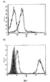
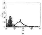
本発明は又、蛍光活性化細胞選別法(FACS)を含む適用で、実施可能である。FACSは、可溶性TCRs又はその断片と標的細胞の相互作用を検出するために用いられる。例えば、可溶性TCRは、標準的方法に従って、ビオチン化され、例えば、標識されたsc−TCR四量体を形成するために、ストレプトアビジン−フィコエリスリン(PE)と結合される。しかしながら、前述したように、多量体化はあまり必要とされない。FACSは、可溶性TCRと、T2細胞及び腫瘍細胞株のような適当な標的細胞との相互作用を定性的に測定するために用いられる。 The present invention can also be practiced with applications involving fluorescence activated cell sorting (FACS). FACS is used to detect the interaction of soluble TCRs or fragments thereof with target cells. For example, soluble TCR is biotinylated according to standard methods, eg, combined with streptavidin-phycoerythrin (PE) to form a labeled sc-TCR tetramer. However, as described above, multimerization is not so necessary. FACS is used to qualitatively measure the interaction of soluble TCRs with appropriate target cells such as T2 cells and tumor cell lines.
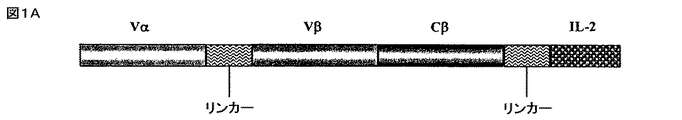
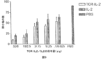
以下の実施例は、遺伝子的にヒトIL−2に結合する、p53アミノ酸残基264〜272番にわたる未変異のp53ペプチドを認識する、一本鎖の可溶性HLA−A2.1限定性のTCRを含む新規融合タンパク質の構築及び特徴を示す。分子のIL−2部分のIL−2受容体特異的な結合能や生物活性と同様に、ペプチドが収容されているHLA−A2と、分子のTCR部分のペプチド特異的な結合が検討された。実施例は、これらのタイプのTCRベースの融合タンパク質が、抗体ベースの標的化腫瘍療法に代わるものとして、又は抗体ベースの免疫サイトカインのようなその他の標的化腫瘍療法への付加的なものとして提供されることを示す。腫瘍を標的化するための分離及び識別方法が、相加的又は相乗的な抗腫瘍効果を示す可能性がある。 The following example shows a single-chain soluble HLA-A2.1-restricted TCR that recognizes an unmutated p53 peptide spanning p53 amino acid residues 264-272 that binds to human IL-2 genetically. The construction and characteristics of the novel fusion protein containing are shown. Similar to the IL-2 receptor-specific binding ability and biological activity of the IL-2 portion of the molecule, peptide-specific binding of HLA-A2 containing the peptide to the TCR portion of the molecule was examined. Examples provide these types of TCR-based fusion proteins as an alternative to antibody-based targeted tumor therapy or as an addition to other targeted tumor therapies such as antibody-based immune cytokines Indicates that Separation and identification methods for targeting tumors may show additive or synergistic anti-tumor effects.
実施例はさらに、HLA−A2.1としてヒトp53ペプチド(aa264〜272)を認識する、3つのドメインからなる可溶性のマウスscTCRの構築及び発現を示す。3つのドメインからなるscTCRは、ヒトIL−2に結合して、哺乳動物の細胞で高発現され分泌される可溶性264scTCR/IL−2融合タンパク質を産生する。264scTCR/IL−2融合タンパク質のTCR部分は、MHC限定性であり、ペプチド特異的抗原を結合する特性があり、IL−2部分がIL−2受容体に結合し、そして、生物学的に活性である。その上、実施例はさらに、この融合タンパク質が、標的細胞とエフェクター細胞を結合させ、マウス内で好ましい薬物動態を示し、標的腫瘍細胞に結合可能であり、抗腫瘍効果を持つことを示す。それゆえ、可溶性scTCR融合タンパク質は、抗体ベースの免疫療法では認識不可能であった、標的化免疫療法に向けての抗原のその他のレパートリーへのアクセスを提供するものである。TCRベースの治療は、抗体ベースの治療に変わるものとして、又はその他の標的化腫瘍療法に対する有用な付加的な療法となる。 The examples further show the construction and expression of a soluble murine scTCR consisting of three domains that recognize human p53 peptide (aa 264-272) as HLA-A2.1. A three-domain scTCR binds to human IL-2 to produce a soluble 264scTCR / IL-2 fusion protein that is highly expressed and secreted in mammalian cells. The TCR portion of the H.264 scTCR / IL-2 fusion protein is MHC restricted, has the property of binding peptide specific antigens, the IL-2 portion binds to the IL-2 receptor, and is biologically active It is. Moreover, the examples further show that this fusion protein binds target and effector cells, exhibits favorable pharmacokinetics in mice, can bind to target tumor cells, and has an anti-tumor effect. Soluble scTCR fusion proteins therefore provide access to other repertoires of antigens for targeted immunotherapy that were unrecognizable with antibody-based immunotherapy. TCR-based therapy is a useful additional therapy as an alternative to antibody-based therapy or for other targeted tumor therapies.
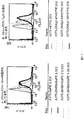
本発明には、可溶性TCRが、ペプチド抗原を良好に検出できる十分な親和性を有していることが開示されている。特に、264scTCRの親和性は、遺伝子操作されていない腫瘍細胞に結合する、また、標的細胞とエフェクター細胞を効果的に結合させるのに十分な親和性を有している。 In the present invention, it is disclosed that the soluble TCR has sufficient affinity to allow good detection of peptide antigens. In particular, the affinity of 264scTCR has sufficient affinity to bind to non-genetically engineered tumor cells and to effectively bind target cells and effector cells.
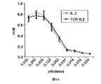
腫瘍を治療するためのサイトカインの全身性投与をめぐって報告された問題は、血清の半減期が短いことと、これらタンパク質の毒性である。重要なのは、本発明の264scTCR/IL−2融合タンパク質は、約3時間の見かけ上の血清半減期を有し、血液中に原型のまま残存するということである。このように264scTCR/IL−2融合タンパク質は、効果的にIL−2の半減期を増大させ、血液中に原型のまま残存させる。このことは、当該タンパク質が免疫修飾がん療法のための新しい試薬であることを示唆している。実施例で用いられたより高濃度の投与量で、本発明の融合タンパク質の血清の半減期は、増大し[文献3、25、37及び38参照]、それによりさらにその分子の腫瘍に対する効果が改善される。 Problems reported over the systemic administration of cytokines to treat tumors are the short half-life of serum and the toxicity of these proteins. Importantly, the 264scTCR / IL-2 fusion protein of the present invention has an apparent serum half-life of about 3 hours and remains intact in the blood. Thus, the 264scTCR / IL-2 fusion protein effectively increases the half-life of IL-2 and remains intact in the blood. This suggests that the protein is a new reagent for immunomodulating cancer therapy. At higher doses used in the examples, the serum half-life of the fusion protein of the present invention is increased [see refs. 3, 25, 37 and 38], thereby further improving the effect of the molecule on tumors. Is done.
腫瘍部位に濃縮されたIL−2は、他のIL−2反応性細胞と同様に、局所T細胞を活性化し、それによりエフェクター細胞を腫瘍部位へ集めるとの認識がある。このように、腫瘍部位にIL−2を濃縮させることにより、本発明のTCR融合分子は、生来の免疫系のNK細胞又は他のメンバーの活性化と同様に、様々なT細胞クローンの活性化と増殖を含む明確な免疫応答を増強する可能性がある。そのような多面的な抗腫瘍応答は、遠隔転移と同様に初期の腫瘍の撲滅により効果的であろう。 There is recognition that IL-2 concentrated at the tumor site, like other IL-2 responsive cells, activates local T cells, thereby collecting effector cells at the tumor site. Thus, by concentrating IL-2 at the tumor site, the TCR fusion molecule of the present invention activates various T cell clones as well as the activation of NK cells or other members of the innate immune system. And may enhance a clear immune response including proliferation. Such a multifaceted anti-tumor response would be more effective at eradicating early tumors as well as distant metastases.
データは、TCR及びサイトカインを含む生物学的に活性な2機能性分子を構築することが可能であることを示す。この融合タンパク質は、腫瘍細胞に結合でき、標的細胞とエフェクター細胞の結合を仲介し、妥当な薬物動態特性を持つ。p53とは別に、アップレギュレーションされ、腫瘍又はウィルス感染細胞上にMHCとして提示されるその他の遺伝子産物も、標的として本発明のTCRベースの免疫療法に用いることができる。さらに、GM−CSF、IFNγ、又はIFNαのようなその他の免疫修飾分子は、TCRに結合され、抗腫瘍又は抗ウィルス応答のためにその他のエフェクター細胞を活性化する。これらの新規TCR融合物は、がんやウィルス感染の治療のための免疫療法の新しい分類を形成する。 The data show that it is possible to construct biologically active bifunctional molecules including TCR and cytokines. This fusion protein can bind to tumor cells, mediates the binding of target cells and effector cells, and has reasonable pharmacokinetic properties. Apart from p53, other gene products that are up-regulated and presented as MHC on tumor or virus-infected cells can also be used as targets in the TCR-based immunotherapy of the present invention. In addition, other immunomodulating molecules such as GM-CSF, IFNγ, or IFNα are bound to the TCR and activate other effector cells for anti-tumor or anti-viral responses. These novel TCR fusions form a new class of immunotherapy for the treatment of cancer and viral infections.
「特異的結合」という用語又は類似の用語は、本明細書に開示された分子が他の分子に結合し、それにより特異的な結合対を形成すること、を意味する。しかしながら、この分子は、例えばウェスタンブロッティング、ELISA、RIA、移動度シフトアッセイ、酵素免疫アッセイ、競合アッセイ、飽和アッセイ、又は他の当該技術部分野公知のタンパク質結合アッセイによって決定されるようなその他の分子を認識、結合しない。一般的に、例えば分子間の特異的結合を検出するための方法については、上記のAusubelらの文献、Harlow及びLaneの「Antibodies: A Laboratory Manual (1988)」及びそこに引用された参考文献を参照されたい。 The term “specific binding” or similar terms means that a molecule disclosed herein binds to another molecule, thereby forming a specific binding pair. However, this molecule may be other molecules as determined, for example, by Western blotting, ELISA, RIA, mobility shift assay, enzyme immunoassay, competition assay, saturation assay, or other protein binding assays known in the art. Recognize and do not combine. In general, see, for example, Ausubel et al., Harlow and Lane, “Antibodies: A Laboratory Manual (1988)” and references cited therein for methods for detecting specific binding between molecules. Please refer.
「完全に可溶性の」という用語又はその類似の用語は、TCRについて述べれば、例えば細胞の培地といった水溶性緩衝液で、低Gでの遠心分離によってで容易に沈殿しないことを意味する。さらに、sc−TCR融合タンパク質は、低濃度のアニオン性又は非イオン性の界面活性剤の存在下及び非存在下、約5〜37℃より高い温度かつ中性付近のpHで、水溶液のままでいるならば、可溶性である。これらの条件下、可溶性タンパク質はしばしば、例えば約10〜50スベドベリ単位より小さい低い沈降価を有する。本明細書で言及される水溶液は、通常pH約5〜9を確立する緩衝化合物を含み、イオン強度の範囲が約2mM〜500mMである。プロテアーゼ阻害剤又は穏やかな非イオン性界面活性剤が加えられてもよく、必要であれば、牛血清アルブミン(BSA)のようなキャリアタンパク質が、数mg/mLまで加えられてもよい。水性緩衝液の例としては、標準的なリン酸緩衝液生理食塩水、トリス緩衝液生理食塩水、又はその他の公知の緩衝液及び細胞の培地製剤を含む。
以下の実施例は、本発明を説明するものであり、何ら本発明を限定するものではない。
The term “fully soluble” or similar terms, when referring to TCR, means that it is an aqueous buffer, such as cell culture medium, that does not precipitate easily by centrifugation at low G. Furthermore, the sc-TCR fusion protein remains in aqueous solution at temperatures above about 5-37 ° C. and near neutral pH in the presence and absence of low concentrations of anionic or nonionic surfactants. If so, it is soluble. Under these conditions, soluble proteins often have a low sedimentation number, for example, less than about 10-50 Svedberg units. The aqueous solutions referred to herein contain buffer compounds that usually establish a pH of about 5-9 and have an ionic strength range of about 2 mM to 500 mM. Protease inhibitors or mild nonionic surfactants may be added, and carrier proteins such as bovine serum albumin (BSA) may be added up to several mg / mL if necessary. Examples of aqueous buffers include standard phosphate buffer saline, Tris buffer saline, or other known buffers and cell culture media.
The following examples illustrate the invention and do not limit the invention in any way.
TCR融合タンパク質構築物の産生
ヒトIL−2に融合したp53ペプチド抗原に特異的な、3つのドメインをもつ、HLA−A2.1限定性マウスTCRを含む融合タンパク質を産生した。TCR融合タンパク質構築物として、ヒトp53ペプチド(aa264〜272)に特異的なTCRsを産生するマウスT細胞株から単離したRNAのRT−PCRによりVα及びVβ/Cβ領域(region)を産生した。TCRα鎖(Vα3)可変領域のカルボキシル末端は、TCRの抗原結合部分を産生するために、柔軟なリンカー(G4S)4[文献21参照]を介して、Vβ(Vβ3)のN末端に結合した。Vβドメインに直接結合するCβドメインは、一本鎖の可溶性TCR分子を産生するために、最終システイン直前のアミノ酸残基で切断して膜貫通及び細胞質ドメインを除去した(図1A及び1B)。ヒトIL−2を、短いリンカー(アミノ酸配列 VNAKTTAPSVYPLAPV;配列番号1)を介してTCR部分に融合した。EE標識(アミノ酸配列EEEEYMPME;配列番号2)[文献11参照]は、必要であれば、TCR/IL−2融合タンパク質を抗EE標識mAbによって検出可能にするために、融合分子のIL−2部分のすぐ下流に挿入した。CMVプロモータによって哺乳動物細胞の発現を駆動し、抗体の軽鎖のリーダー配列によって分泌を方向付け、G418耐性にて選別した。
Production of TCR fusion protein constructs A fusion protein was produced comprising an HLA-A2.1 restricted mouse TCR with three domains specific for p53 peptide antigen fused to human IL-2. As a TCR fusion protein construct, Vα and Vβ / Cβ regions were produced by RT-PCR of RNA isolated from a mouse T cell line that produces TCRs specific for human p53 peptides (aa264-272). The carboxyl terminus of the TCR α chain (Vα3) variable region binds to the N terminus of Vβ (Vβ3) via a flexible linker (G 4 S) 4 [see reference 21] to produce the antigen binding portion of TCR. did. The Cβ domain, which binds directly to the Vβ domain, was cleaved at the amino acid residue just before the final cysteine to remove the transmembrane and cytoplasmic domain to produce a single-chain soluble TCR molecule (Figures 1A and 1B). Human IL-2 was fused to the TCR moiety via a short linker (amino acid sequence VNAKTTAPPSYPLAPV; SEQ ID NO: 1). The EE tag (amino acid sequence EEEEYMPME; SEQ ID NO: 2) [Ref 11] is used to make the IL-2 portion of the fusion molecule, if necessary, in order to make the TCR / IL-2 fusion protein detectable by anti-EE labeled mAb It was inserted immediately downstream. The expression of mammalian cells was driven by the CMV promoter, the secretion was directed by the antibody light chain leader sequence, and selected for G418 resistance.
図1を、以下に、詳細に説明する。
図1Aは、264scTCR/IL−2融合タンパク質のドメイン構造を模式的に示すものである。
図1Bは、264scTCR/IL−2融合タンパク質のアミノ酸配列である。
融合タンパク質のそれぞれのドメインのアミノ酸番号を図中に示す。
FIG. 1 is described in detail below.
FIG. 1A schematically shows the domain structure of the 264scTCR / IL-2 fusion protein.
FIG. 1B is the amino acid sequence of the 264scTCR / IL-2 fusion protein.
The amino acid number of each domain of the fusion protein is shown in the figure.
哺乳動物細胞に於けるTCR/IL−2融合タンパク質の発現
264scTCR/IL−2融合タンパク質の性状を検討するために、264scTCR/IL−2構築物を、CHO−K1細胞に安定に形質転換した。264scTCR/IL−2融合タンパク質を分泌する安定な形質転換体は、材料と方法の欄に記載されるようにELISAアッセイを用いて選択した。これらのELISAに於ける陽性シグナルは、形質転換した細胞が、抗マウスTCR及び抗ヒトIL−2抗体両方ともに認識される264scTCR/IL−2融合タンパク質を分泌していることを示し、更に分泌された264scTCR/IL−2が形質転換した細胞内で、適切に集合し、折りたたまれていること、及び細胞から分泌されたときに原型のままで存在すること、を示唆している。
Expression of TCR / IL-2 fusion protein in mammalian cells To examine the properties of the 264scTCR / IL-2 fusion protein, the 264scTCR / IL-2 construct was stably transformed into CHO-K1 cells. Stable transformants secreting 264scTCR / IL-2 fusion protein were selected using an ELISA assay as described in the Materials and Methods section. A positive signal in these ELISAs indicates that the transformed cells secrete a 264scTCR / IL-2 fusion protein that is recognized by both anti-mouse TCR and anti-human IL-2 antibodies and is also secreted. This suggests that 264scTCR / IL-2 is properly assembled and folded in transformed cells and remains intact when secreted from the cells.
264scTCR/IL−2融合タンパク質は、免疫親和性クロマトグラフィで上清1リットル当たり約1.8mgの収量で細胞上清から精製した。精製し融合タンパク質は、SDS−PAGE及びクーマシー染色した。還元又は非還元条件下、主として存在する染色バンドは、このタンパク質の予想分子量に一致する約60kDa(図2)に移動し、このことは融合タンパク質が細胞から分泌されるときに、予期されない分子内ジスルフィド結合を形成することなく原型のままでいることを示している。非還元ゲルに於ける大きなバンドは、融合タンパク質の2量体である。この結論は、大きなバンドは見かけ上、融合タンパク質の分子量の約2倍の分子量を有していること、そしてこのバンドは還元条件下では融合タンパク質の大きさに減少するという観察に基づいている。データは、形質転換されたCHO細胞は、予想された分子量の264scTCR/IL−2融合タンパク質を産生すること、そして適切に折りたたまれ、集合し、可溶性融合タンパク質として分泌されることを示している。 The 264scTCR / IL-2 fusion protein was purified from cell supernatant by immunoaffinity chromatography at a yield of approximately 1.8 mg per liter of supernatant. The purified fusion protein was stained with SDS-PAGE and Coomassie. Under reducing or non-reducing conditions, the predominantly present staining band migrates to approximately 60 kDa (FIG. 2), consistent with the expected molecular weight of this protein, which is an unexpected intramolecular when the fusion protein is secreted from the cell. It shows that it remains intact without forming disulfide bonds. The large band in the non-reducing gel is a dimer of the fusion protein. This conclusion is based on the observation that the large band apparently has a molecular weight approximately twice that of the fusion protein and that this band decreases to the size of the fusion protein under reducing conditions. The data show that transformed CHO cells produce the expected molecular weight of 264scTCR / IL-2 fusion protein and are properly folded, assembled and secreted as a soluble fusion protein.
図2を、以下に、詳細に説明する。
CHO細胞を、264scTCR/IL−2発現ベクターで安定に形質転換した。分泌された融合タンパク質は、免疫親和性クロマトグラフィで精製し、図の上部に示されるように還元又は非還元条件下で、SDS−PAGE処理した。SDS−PAGEゲルは、クーマシーブリリアントブルーで染色した。
FIG. 2 is described in detail below.
CHO cells were stably transformed with the 264scTCR / IL-2 expression vector. The secreted fusion protein was purified by immunoaffinity chromatography and subjected to SDS-PAGE under reducing or non-reducing conditions as shown at the top of the figure. The SDS-PAGE gel was stained with Coomassie brilliant blue.
264scTCR/IL−2融合タンパク質のTCR部分のMHC/ペプチド結合能
264scTCR/IL−2融合タンパク質のペプチドを収容したMHCへの結合能をフローサイトメトリーで測定した。p53(aa264〜272)又はp53(aa149〜157)(対照)ペプチドを収容したT2細胞を、264scTCR/IL−2融合タンパク質で染色した。p53(aa264〜272)を収容した細胞は、抗TCR CβmAb又は抗IL−2検出抗体のいずれかでの検出で、264scTCR/IL−2により陽性に染色された。p53(aa149〜157)対照ペプチドを収容した細胞は、抗TCR CβmAb又は抗IL−2検出抗体のいずれによっても染色されなかった。p53(aa149〜157)を収容したT2細胞が染色されないのは、p53(aa149〜157)ペプチドがHLA−A2に対し結合不能であるためではないことを証明するために、ペプチドを収容していないT2細胞、p53(aa149〜157)ペプチドを収容したT2細胞又はp53(aa264〜272)ペプチドを収容したT2細胞をそれぞれBB7.2αHLA−A2モノクローナル抗体で染色した。何れのp53ペプチドを収容した細胞も、ペプチドを収容しなかった細胞に比べて強く染色された。このことは、両ペプチドはHLA−A2分子に結合可能であることを示した(図3C)。また、T2細胞をIL−2受容体について調べるために染色したが、IL−2受容体を発現していないことが分かった。これらのデータは、264scTCR/IL−2融合タンパク質の結合が、融合タンパク質のTCR部分によって仲介されていることを示している。T2細胞が対照ペプチドを収容している場合に融合タンパク質によって染色されなかったこともまた、染色がTCR部分を介してなされること、及び、染色が適切なペプチドに特異的であることを示している。これらのデータは、264scTCR/IL−2融合タンパク質のTCR部分がその特異的なペプチドをHLA−A2として認識することが可能であることを示している。
MHC / peptide binding ability of the TCR portion of the H.264 scTCR / IL-2 fusion protein The ability of the 264scTCR / IL-2 fusion protein to bind to MHC containing the peptide was measured by flow cytometry. T2 cells containing p53 (aa 264-272) or p53 (aa 149-157) (control) peptide were stained with 264scTCR / IL-2 fusion protein. Cells containing p53 (aa 264-272) stained positive with 264scTCR / IL-2 upon detection with either anti-TCR Cβ mAb or anti-IL-2 detection antibody. Cells containing the p53 (aa 149-157) control peptide were not stained with either anti-TCR Cβ mAb or anti-IL-2 detection antibody. T2 cells harboring p53 (aa 149-157) do not stain because the p53 (aa 149-157) peptide does not contain the peptide to demonstrate that it cannot bind HLA-A2. T2 cells, T2 cells containing p53 (
図3を、以下に、詳細に説明する。p53(aa264〜272)ペプチド(灰色の線)又はp53(aa149〜157)ペプチド(黒い線)を収容したT2細胞は、
図3A)264scTCR/IL−2融合タンパク質及び抗TCR CβmAbで染色されているか、又は
図3B)264scTCR/IL−2融合タンパク質及び抗IL−2mAbで染色されているかの何れかである。
図3C)p53(aa264〜272)ペプチド(暗い灰色の線)を収容したT2細胞、p53(aa149〜157)ペプチド(明るい灰色の線)を収容したT2細胞、又はペプチドを収容していない(黒い線)T2細胞は、抗HLA−A2 BB7.2mAbで、次いでFITC標識ヤギ抗マウスIgGで染色した。影をつけたピークは、染色されていないT2細胞である。
FIG. 3 is described in detail below. T2 cells containing the p53 (aa264-272) peptide (gray line) or the p53 (aa149-157) peptide (black line) are:
FIG. 3A) either stained with 264scTCR / IL-2 fusion protein and anti-TCR Cβ mAb, or FIG. 3B) stained with 264scTCR / IL-2 fusion protein and anti-IL-2 mAb.
FIG. 3C) T2 cells containing p53 (aa 264-272) peptide (dark gray line), T2 cells containing p53 (aa 149-157) peptide (light gray line), or no peptide (black) Line) T2 cells were stained with anti-HLA-A2 BB7.2 mAb followed by FITC-labeled goat anti-mouse IgG. The shaded peak is unstained T2 cells.
264scTCR/IL−2融合タンパク質のIL−2部分のIL−2受容体結合能
264scTCR/IL−2融合タンパク質のIL−2部分のIL−2受容体結合能は、フローサイトメトリーによって検討された。マウス初代脾臓細胞を単離し、T細胞芽細胞を産生するために、rIL−2及び抗CD3で刺激した。IL−2受容体を発現する刺激した脾臓細胞は、p53(aa264〜272)を収容したHLA−A2四量体で、264scTCR/IL−2融合タンパク質の存在下でのみ陽性に染色された(図4A)。同様に、CTLL−2マウス細胞傷害性のTリンパ細胞は、恒常的にIL−2受容体を発現しているが、264scTCR/IL−2融合タンパク質により、陽性に染色されたが、264scTCR/κ融合タンパク質では染色されなかった(図4B)。CTLL−2細胞をα−ヒトCD25ブロッキング抗体又はアイソタイプの対照抗体、次いで264scTCR/IL−2とインキュベートすると、細胞をブロッキング抗体とインキュベートしたときには染色は実質的に減少したが、アイソタイプの対照抗体では減少しなかった。264scTCR/マウスκ鎖融合タンパク質又はIL−2受容体ブロッキング抗体とインキュベートしたCTLL−2細胞からシグナルが欠如したことは、これらの細胞の染色が264scTCR/IL−2融合タンパク質のIL−2部分によって仲介されていることを示している。これらのデータは、264scTCR/IL−2融合タンパク質のIL−2部分が、IL−2受容体に結合できることを示唆している。
IL-2 receptor binding ability of IL-2 part of 264scTCR / IL-2 fusion protein The IL-2 receptor binding ability of IL-2 part of 264scTCR / IL-2 fusion protein was examined by flow cytometry. Mouse primary spleen cells were isolated and stimulated with rIL-2 and anti-CD3 to produce T cell blasts. Stimulated spleen cells expressing IL-2 receptor were positively stained with HLA-A2 tetramer containing p53 (aa 264-272) only in the presence of 264scTCR / IL-2 fusion protein (FIG. 4A). Similarly, CTLL-2 mouse cytotoxic T lymphocytes constitutively express the IL-2 receptor but stained positively with the 264scTCR / IL-2 fusion protein, but 264scTCR / κ. It was not stained with the fusion protein (FIG. 4B). When CTLL-2 cells were incubated with α-human CD25 blocking antibody or an isotype control antibody followed by 264scTCR / IL-2, staining was substantially reduced when the cells were incubated with the blocking antibody, but decreased with an isotype control antibody. I did not. Lack of signal from CTLL-2 cells incubated with 264scTCR / mouse kappa chain fusion protein or IL-2 receptor blocking antibody, staining of these cells was mediated by the IL-2 portion of the 264scTCR / IL-2 fusion protein It has been shown. These data suggest that the IL-2 portion of the 264scTCR / IL-2 fusion protein can bind to the IL-2 receptor.
図4を、以下に、詳細に説明する。
図4A)マウス脾臓細胞を、IL−2及び抗CD3εmAbで刺激し、その後、264scTCR/IL−2融合タンパク質の存在下(灰色の線)又は非存在下(黒い線)でインキュベートした。結合した融合タンパク質は、PE標識したHLA−A2p53(aa264〜272)四量体で検出された。
図4B)CTLL−2細胞を、α−ヒトCD25ブロッキング抗体又はアイソタイプの対照抗体、次いで264scTCR/IL−2又は264scTCR/κ融合タンパク質とインキュベートした。結合した融合タンパク質は、PE標識したα−TCR−Vβ3抗体で検出した。影のついたピークは、染色されていないCTLL−2細胞である。
黒い線:264scTCR/IL−2だけで染色したCTLL−2細胞。
灰色の点線:対照抗体に次いで264scTCR/IL−2で染色したCTLL−2細胞。
明るい灰色の線:α−ヒトCD25ブロッキング抗体、次いで264scTCR/IL−2でインキュベートしたCTLL−2細胞。
暗い灰色の線:264scTCR/κ融合タンパク質で染色したCTLL−2細胞。
黒いダッシュの線:α−TCR-Vβで染色したCTLL−2細胞。
FIG. 4 is described in detail below.
FIG. 4A) Mouse spleen cells were stimulated with IL-2 and anti-CD3ε mAb and then incubated in the presence (grey line) or absence (black line) of 264scTCR / IL-2 fusion protein. Bound fusion protein was detected with PE-labeled HLA-A2p53 (aa 264-272) tetramer.
FIG. 4B) CTLL-2 cells were incubated with an α-human CD25 blocking antibody or an isotype control antibody followed by a 264scTCR / IL-2 or 264scTCR / κ fusion protein. Bound fusion protein was detected with PE-labeled α-TCR-Vβ3 antibody. The shaded peak is unstained CTLL-2 cells.
Black line: CTLL-2 cells stained with 264scTCR / IL-2 alone.
Gray dotted line: CTLL-2 cells stained with 264scTCR / IL-2 following control antibody.
Light gray line: CTLL-2 cells incubated with α-human CD25 blocking antibody followed by 264scTCR / IL-2.
Dark gray line: CTLL-2 cells stained with 264scTCR / κ fusion protein.
Black dash line: CTLL-2 cells stained with α-TCR-Vβ.
264scTCR/IL−2融合タンパク質の生物学的活性
264scTCR/IL−2融合タンパク質のIL−2部分の生物学的活性を実証するため、IL−2依存性のCTLL−2細胞を、様々な濃度の264scTCR/IL−2又は組み換えIL−2のいずれかと共に培養し、細胞の生存率をWST−1を用いて評価した。図5Aに示されるように、rIL−2又は264scTCR/IL−2のCTLL−2細胞の増殖を支援する能力は、投与量依存性であり、ここに於いて、組み換えIL−2又は264scTCR/IL−2のいずれかをより高濃度にしたときに、より多くの細胞増殖がみられた。さらに、組み換えIL−2又は264scTCR/IL−2のいずれかを同等モル量を使用したとき、細胞の増殖は同様のレベルであった。特異性に対する更なる対照として、CTLL−2細胞を、α−ヒトCD25ブロッキング抗体又はアイソタイプの対照とともに264scTCR/IL−2とインキュベートした。培養物にブロッキング抗体が含まれた場合、増殖はブロッキング抗体の濃度の減少とともに実質的に減少したが、CTLL−2細胞の増殖は、いずれの濃度の対照抗体の影響も受けなかった(図5B)。このデータは、264scTCR/IL−2のIL−2部分がインビトロで組み換えIL−2と同様な生物学的活性を有すること、及び融合タンパク質の増殖活性がその分子のIL−2部分に依存していること、を示している。
Biological activity of the H.264 scTCR / IL-2 fusion protein To demonstrate the biological activity of the IL-2 portion of the 264scTCR / IL-2 fusion protein, IL-2 dependent CTLL-2 cells were Cells were cultured with either H.264scTCR / IL-2 or recombinant IL-2 and cell viability was assessed using WST-1. As shown in FIG. 5A, the ability of rIL-2 or 264scTCR / IL-2 to support the growth of CTLL-2 cells is dose dependent, where recombinant IL-2 or 264scTCR / IL More cell proliferation was seen when either of -2 was made higher. Furthermore, cell growth was at similar levels when equivalent molar amounts of either recombinant IL-2 or 264scTCR / IL-2 were used. As a further control for specificity, CTLL-2 cells were incubated with 264scTCR / IL-2 with an α-human CD25 blocking antibody or isotype control. When the blocking antibody was included in the culture, proliferation decreased substantially with decreasing blocking antibody concentration, but CTLL-2 cell proliferation was not affected by any concentration of control antibody (FIG. 5B). ). This data indicates that the IL-2 portion of 264scTCR / IL-2 has biological activity similar to recombinant IL-2 in vitro, and the growth activity of the fusion protein depends on the IL-2 portion of the molecule. It shows that it is.
264scTCR/IL−2の同起源のMHC/ペプチドに対する解離定数は、表面プラズモン共鳴検出法によって生理的な条件下で、約10−7Mであることが分かった。 The dissociation constant for H.264scTCR / IL-2 cognate MHC / peptide was found to be about 10 −7 M under physiological conditions by surface plasmon resonance detection.
図5を、以下に、詳細に説明する。
図5A)CTLL−2細胞を、264scTCR/IL−2(実線)又は組み換えIL−2(点線)と共に、図の下段に示されるような様々な濃度で培養した。
図5B)CTLL−2細胞を、図の下段に示されるような264scTCR/IL−2及びα−ヒトCD25ブロッキング抗体又はアイソタイプの対照抗体と共に、インキュベートした。細胞の生存率は、WST−1と共にインキュベートして測定し、吸光度は450nmで測定した。
Cab+5:5μg対照抗体;Cab+50:50μg対照抗体;Bab+5:5μgブロッキング抗体;Bab+50:50μgブロッキング抗体
FIG. 5 is described in detail below.
FIG. 5A) CTLL-2 cells were cultured with 264scTCR / IL-2 (solid line) or recombinant IL-2 (dotted line) at various concentrations as shown in the bottom of the figure.
FIG. 5B) CTLL-2 cells were incubated with 264scTCR / IL-2 and α-human CD25 blocking antibody or isotype control antibody as shown in the bottom of the figure. Cell viability was measured by incubation with WST-1 and absorbance was measured at 450 nm.
Cab + 5: 5 μg control antibody; Cab + 50: 50 μg control antibody; Bab + 5: 5 μg blocking antibody; Bab + 50: 50 μg blocking antibody
264scTCR/IL−2融合タンパク質により仲介される細胞の結合
264scTCR/IL−2融合タンパク質の有用な特性は、そのTCR及びサイトカイン部分の各々により、標的細胞及びエフェクター細胞を結合させる能力にある。264scTCR/IL−2融合タンパク質が細胞を効果的に結合させることができることを実証するために、T2細胞を、p53(aa264〜272)又はp53(aa149〜157)ペプチドのいずれかで収容し、その後ジヒドロエチジウム(HE)で標識した。CTLL−2細胞を、カルセインAMで標識し、二つの標識された細胞集団を、264scTCR/IL−2融合タンパク質の存在下又は非存在下で混合し、インキュベートした。試料を、フローサイトメトリーで分析した。二つの細胞集団を264scTCR/IL−2融合タンパク質の非存在下でインキュベートした時(図6A及び6B)、又はT2細胞に対照ペプチドを収容し、CTLL−2細胞と264scTCR/IL−2融合タンパク質の存在下でインキュベートした時(図6C)、フローサイトメトリーヒストグラム上では、細胞群は二つの異なる集団に含まれたが、総集団の約45%が各々(図6A、6B、及び6Cの領域1及び3に)示されており、総集団の約0.46%のみが、二重染色されたセルウィンドウ(図6A、6B、及び6Cの領域2)に示された。しかしながら、T2細胞をp53(aa264〜272)ペプチドを収容し、264scTCR/IL−2融合タンパク質の存在下で、CTLL−2細胞とインキュベートした場合(図6D)、2重染色された細胞集団は、全集団の4.1%(図6Dの領域2、結合細胞)であり、このことは、T2細胞が264scTCR/IL−2融合タンパク質を介してCTLL−2細胞に結合していることを示唆している。
Cellular Binding Mediated by the H.264scTCR / IL-2 Fusion Protein A useful property of the 264scTCR / IL-2 fusion protein is its ability to bind target cells and effector cells with each of its TCR and cytokine moieties. To demonstrate that the H.264 scTCR / IL-2 fusion protein can effectively bind cells, T2 cells were housed with either p53 (aa 264-272) or p53 (aa 149-157) peptides, and then Labeled with dihydroethidium (HE). CTLL-2 cells were labeled with calcein AM and the two labeled cell populations were mixed and incubated in the presence or absence of 264scTCR / IL-2 fusion protein. Samples were analyzed by flow cytometry. When the two cell populations were incubated in the absence of 264scTCR / IL-2 fusion protein (FIGS. 6A and 6B), or T2 cells contained control peptides, and CTLL-2 cells and 264scTCR / IL-2 fusion protein When incubated in the presence (FIG. 6C), on the flow cytometry histogram, the cell population was included in two different populations, but about 45% of the total population was each (
図6を、以下に、詳細に説明する。
T2細胞を、p53(aa264〜272)(図6B及び6D)又はp53(aa149〜157)(対照)ペプチド(図6A及び6C)のいずれかで収容し、その後HEで標識した。CTLL−2細胞を、カルセインAMで標識した。標識された細胞を、264scTCR/IL−2融合タンパク質の存在下(図6C及び6D)又は非存在下(図6A及び6B)で混合して、インキュベートし、次いで試料をフローサイトメトリーで分析した。収容に使用されたペプチド及び融合タンパク質の存在又は非存在といったアッセイ条件を、各ヒストグラムの下に示す。一重染色領域は、1及び3の番号で示され、2重染色された細胞集団は2番で示されている。
FIG. 6 will be described in detail below.
T2 cells were housed with either p53 (aa 264-272) (FIGS. 6B and 6D) or p53 (aa 149-157) (control) peptide (FIGS. 6A and 6C) and then labeled with HE. CTLL-2 cells were labeled with calcein AM. Labeled cells were mixed and incubated in the presence (FIGS. 6C and 6D) or absence (FIGS. 6A and 6B) of 264scTCR / IL-2 fusion protein, and the samples were then analyzed by flow cytometry. Assay conditions such as the presence or absence of peptides and fusion proteins used for housing are shown below each histogram. Single-stained areas are indicated by
マウスに於ける264scTCR/IL−2の薬物速度論
264scTCR/IL−2融合タンパク質の薬物速度論は、BLAB/cマウスを用いて測定した。薬物をマウスに、経静脈的に投与し、様々な経過時点での血清試料を回収した。264scTCR/IL−2融合タンパク質の血清レベルは、ELISAを用いて測定した。ELISA検出は、融合タンパク質がインビボで修飾されているか、又は切断されているかを決定するために、抗TCRmAb捕獲/抗IL−2Ab検出(図7A)、抗TCRmAb捕獲/抗TCRmAb検出(図7B)、又は抗IL−2mAb捕獲/抗IL−2ポリクローナルAb検出(図7C)を用いて実施した。264scTCR/IL−2融合タンパク質を注入されたマウスは、毒性の兆候をまったく示さなかった。これらのアッセイに於いて、使用したELISA形式により、見かけ上の血清中半減期が1.6〜3.0時間で、最大濃度0.75〜2.5μg/mLの264scTCR/IL−2が検出された(図7)。報告されているフリーのIL−2の血清中半減期は、約5分であるので[文献6参照]、これらのデータは、融合タンパク質がインビボで切断されておらず、その代わり血液中で比較的長期間にわたり原型のまま残存することを示している。これらの検討で測定された264scTCR/IL−2の半減期に於ける小さなぶれは、多分に、ELISAアッセイの感度の違いによるものである。
Pharmacokinetics of 264scTCR / IL-2 in mice The pharmacokinetics of the 264scTCR / IL-2 fusion protein were measured using BLAB / c mice. Drugs were administered intravenously to mice and serum samples were collected at various time points. Serum levels of 264scTCR / IL-2 fusion protein were measured using an ELISA. ELISA detection is anti-TCR mAb capture / anti-IL-2 Ab detection (FIG. 7A), anti-TCR mAb capture / anti-TCR mAb detection (FIG. 7B) to determine if the fusion protein is modified or cleaved in vivo. Or anti-IL-2 mAb capture / anti-IL-2 polyclonal Ab detection (FIG. 7C). Mice injected with 264scTCR / IL-2 fusion protein showed no signs of toxicity. In these assays, the ELISA format used detected 264scTCR / IL-2 at a maximum concentration of 0.75-2.5 μg / mL with an apparent serum half-life of 1.6-3.0 hours. (FIG. 7). Since the reported serum half-life of free IL-2 is approximately 5 minutes [see reference 6], these data indicate that the fusion protein has not been cleaved in vivo and instead compared in blood. This shows that it remains in its original form for a very long time. The small fluctuations in the half-life of 264scTCR / IL-2 measured in these studies are probably due to the difference in sensitivity of the ELISA assay.
図7を、以下に、詳細に説明する。
BALB/cマウスに、264scTCR/IL−2融合タンパク質を注入し、血清試料を、注入後15、30分、1、4、8、24時間後に集めた。264scTCR/IL−2の血清濃度は、以下の形式を用いて、ELISAにより測定した。
7A)抗TCRmAb捕獲/抗IL−2Ab検出;
7B)抗TCRmAb捕獲/抗TCRmAb検出;及び
7C)抗IL−2mAb捕獲/抗IL−2Ab検出
FIG. 7 will be described in detail below.
BALB / c mice were injected with 264scTCR / IL-2 fusion protein and serum samples were collected at 15, 30 minutes, 1, 4, 8, and 24 hours after injection. The serum concentration of 264scTCR / IL-2 was measured by ELISA using the following format.
7A) Anti-TCR mAb capture / anti-IL-2 Ab detection;
7B) Anti-TCR mAb capture / anti-TCR mAb detection; and 7C) Anti-IL-2 mAb capture / anti-IL-2 Ab detection.
264scTCR/IL−2による腫瘍細胞の染色
264scTCR/IL−2融合タンパク質が、標的の腫瘍細胞を認識し、結合できれば、有用である。264scTCR/IL−2が腫瘍細胞に結合することができるかどうかを試験するため、HLA−A2.1及びp53の両方を発現している、A375ヒトメラノーマ細胞を、264scTCR/IL−2又は無関係なTCR/IL−2融合タンパク質である3C8、のいずれかで染色した。融合タンパク質とインキュベートしていない細胞及び3C8とインキュベートした細胞は、H57−597検出抗体により陽性に染色されないが、264scTCR/IL−2とインキュベートした細胞は検出抗体で陽性に染色された(図8)。この結果から、264scTCR/IL−2融合タンパク質が、標的の腫瘍細胞を認識し、結合できること、そしてインビボ抗がん治療として役に立つことが示唆される。
Staining of tumor cells with 264scTCR / IL-2 It would be useful if the 264scTCR / IL-2 fusion protein could recognize and bind to the target tumor cells. To test whether 264scTCR / IL-2 can bind to tumor cells, A375 human melanoma cells expressing both HLA-A2.1 and p53 were tested using 264scTCR / IL-2 or irrelevant Stained with either 3C8, a TCR / IL-2 fusion protein. Cells not incubated with the fusion protein and cells incubated with 3C8 did not stain positively with the H57-597 detection antibody, whereas cells incubated with 264scTCR / IL-2 stained positive with the detection antibody (FIG. 8). . This result suggests that the 264scTCR / IL-2 fusion protein can recognize and bind to target tumor cells and is useful as an in vivo anticancer therapy.
図8を、以下に、詳細に説明する。
A375ヒトメラノーマ細胞を、融合タンパク質なし(黒いダッシュの線)、5μgの3C8TCR/IL−2融合たんぱく質(対照)(点線)、又は5μgの264scTCR/IL−2融合タンパク質(黒い実線)とインキュベートし、次いで、H57−597mAbで染色した。染色されなかった細胞は、影のついた領域で示す。
FIG. 8 will be described in detail below.
A375 human melanoma cells were incubated with no fusion protein (black dashed line), 5 μg 3C8TCR / IL-2 fusion protein (control) (dotted line), or 5 μg 264scTCR / IL-2 fusion protein (black solid line); It was then stained with H57-597 mAb. Unstained cells are indicated by shaded areas.
264scTCR/IL−2融合タンパク質の抗腫瘍効果
264scTCR/IL−2融合タンパク質がインビボで抗腫瘍効果を持つかどうかを決定するために、実験的転移アッセイを実施した。メスの胸腺欠損ヌードマウスに、高転移性A375ヒトメラノーマサブクローンであるA375−C15Nを注入し、様々な投与量の264scTCR/IL−2又は組み換えIL−2のいずれかで処置した。腫瘍細胞注入後42日で、肺の小塊をカウントした。264scTCR/IL−2と組み換えIL−2の両方とも、投与量依存的に肺転移を減少させた(図9)が、264scTCR/IL−2融合タンパク質の方が、全ての投与量に於いて、肺転移を大きく減少させた。このことは、サイトカインを腫瘍に標的化することは、がん治療として大きな効果をもたらすものであることが示唆される。
Anti-tumor effect of 264scTCR / IL-2 fusion protein An experimental metastasis assay was performed to determine whether the 264scTCR / IL-2 fusion protein has anti-tumor effect in vivo. Female athymic nude mice were injected with the highly metastatic A375 human melanoma subclone, A375-C15N, and treated with either various doses of 264scTCR / IL-2 or recombinant IL-2. At 42 days after tumor cell injection, lung nodules were counted. Both H.264scTCR / IL-2 and recombinant IL-2 reduced lung metastasis in a dose-dependent manner (FIG. 9), whereas the H.264scTCR / IL-2 fusion protein at all doses, Lung metastasis was greatly reduced. This suggests that targeting a cytokine to a tumor has a great effect as a cancer treatment.
264scTCR/IL−2又は組み換えIL−2のいずれかで処理されたマウスは、目立った毒性の兆候を示さなかった。両治療はともに、結果的に肺転移を減少させたが、264scTCR/IL−2による治療は、全ての投与量に於いて、組み換えIL−2による治療に較べより効果的であった。 Mice treated with either 264scTCR / IL-2 or recombinant IL-2 showed no significant signs of toxicity. Both treatments resulted in reduced lung metastases, but treatment with 264scTCR / IL-2 was more effective than treatment with recombinant IL-2 at all doses.
図9を、以下に、詳細に説明する。
メスの胸腺欠損ヌードマウスに、高度に転移性のA375−C15N細胞を注入し、264scTCR/IL−2、組み換えIL−2、又はPBSで処理した。腫瘍細胞の注入の42日後、肺を摘出し、肺の小塊をカウントし、次いでPBS処理した対照群に対応する肺の小塊数の平均をプロットした。
FIG. 9 is described in detail below.
Female athymic nude mice were injected with highly metastatic A375-C15N cells and treated with 264scTCR / IL-2, recombinant IL-2, or PBS. Forty-two days after tumor cell injection, lungs were removed, lung nodules were counted, and then the average number of lung nodules corresponding to the PBS-treated control group was plotted.
単量体及び多量体の264scTCR融合タンパク質による、ペプチドを収容したT2細胞の染色についてのフローサイトメトリー分析
様々な264scTCR融合タンパク質の単量体や多量体を調製し、それらのT2細胞に対する結合挙動を、以下のセクション11及び12に記載される方法で、フローサイトメトリーにより分析した。その結果、図10に示されるように、264scTCR融合タンパク質は、p53(aa264〜273)を収容したT2細胞を、p53(aa149〜157)を収容した細胞より強く染色した(図10A、10B)。図中では、染色されていないT2細胞は、T2 149非染色001と表示されたヒストグラム中に示されている;第2の試薬(H57−PE)で染色した、p53(aa149〜157)及びp53(aa264〜273)を収容したT2細胞は、それぞれ「T2 149 H57.002」及び「T2 264 H57.009」と表示されたヒストグラム中に示されている;多量体の264scTCR/IgG1に次いでH57−PEで染色した、p53(aa149〜157)及びp53(aa264〜273)を収容したT2細胞は、それぞれ「T2 149 IgG H57.003」及び「T2 264 IgG H57.010」と表示されたヒストグラム中に示されている;264scTCR/IL−2に次いでH57−PEで染色したp53(aa149〜157)及びp53(aa264〜273)を収容したT2細胞は、それぞれ「T2 149 IL2 H57.004」及び「T2 264 IL2 H57.011」と表示されたヒストグラム中に示されている;単量体の264scTCR/trunIgG1に次いでH57−PEで染色したp53(aa149〜157)及びp53(aa264〜273)を収容したT2細胞は、それぞれ「T2 149 trun H57.005」及び「T2 264 trun H57.012」と表示されたヒストグラム中に示されている;そして単量体の264scTCR/BirAに次いでH57−PEで染色したp53(aa149〜157)及びp53(aa264〜273)を収容したT2細胞は、それぞれ「T2 149 birA H57.006」及び「T2 264 birA H57.013」と表示されたヒストグラム中に示されている。この結果は、観察した染色がペプチド特異的であることを確証づけた。
Flow cytometric analysis of staining of peptide-containing T2 cells with monomeric and multimeric 264scTCR fusion proteins Prepare monomers and multimers of various 264scTCR fusion proteins and determine their binding behavior to T2 cells , And analyzed by flow cytometry as described in Sections 11 and 12 below. As a result, as shown in FIG. 10, the 264scTCR fusion protein stained T2 cells containing p53 (
単量体型264scTCRは、ある程まで度染色することができた。例えば、264scTCR/trunIgG型で染色したときの平均チャネル蛍光(MCF)は、p53(aa149〜157)を収容した細胞では10.95であるのに対し、p53(aa264〜273)を収容した細胞では55.34と増大した。同様に、264scTCR/BirA型に対するMCFは、p53(aa149〜157)を収容した細胞では13.41であったのに対し、p53(aa264〜273)を保持した細胞では95.14と増大した。多量体の264scTCRは、より顕著にペプチドを収容したT2細胞を特異的に染色できた。例えば、264scTCR/IgG1型で染色したときのMCFは、p53(aa149〜157)を収容した細胞では119であったのに対し、p53(aa264〜273)を収容した細胞では863と増大した。
The monomeric 264scTCR could be stained to some degree. For example, the mean channel fluorescence (MCF) when stained with 264scTCR / runIgG type is 10.95 in cells containing p53 (aa149-157), whereas in cells containing p53 (aa264-273) It increased to 55.34. Similarly, the MCF for 264scTCR / BirA type was 13.41 in cells containing p53 (
264scTCR融合タンパク質による腫瘍細胞の染色
264scTCR試薬が腫瘍細胞を染色する能力もまた試験された。培養したA375細胞を、10mMのEDTAを添加したPBS(pH7.4)で接着を剥がし、洗浄用緩衝液で2回洗浄した。細胞染色は、4μgの264scTCR/IgG1融合タンパク質を用いて、23℃にて45分間実行された。細胞を一回洗浄し、3μgのFITC結合のヤギ抗ヒトIgGFcのF(ab’)2断片(抗IgG−FITC)で染色した。2回洗浄した後、染色細胞を、再び縣濁し、次いでFACScanで分析した。抗IgG−FITC抗体のみで染色したA375細胞を、対照として用いた
。
Staining of tumor cells with 264scTCR fusion protein The ability of the 264scTCR reagent to stain tumor cells was also tested. The cultured A375 cells were peeled off with PBS (pH 7.4) supplemented with 10 mM EDTA, and washed twice with a washing buffer. Cell staining was performed with 4 μg of 264scTCR / IgG1 fusion protein at 23 ° C. for 45 minutes. Cells were washed once and stained with 3 μg of FITC-conjugated goat anti-human IgG Fc F (ab ′) 2 fragment (anti-IgG-FITC). After washing twice, the stained cells were resuspended and then analyzed by FACScan. A375 cells stained with anti-IgG-FITC antibody alone were used as a control.
図11Aに示されるように、この分析の結果は、A375腫瘍細胞が264scTCR/IgG1融合タンパク質で染色できることを示している。このパネル(図11A)に於いて、抗IgG−FITCのみで、又は抗IgG−FITC/IgG1に次いで抗IgG−FITCで染色したA375細胞は、それぞれ「375−FITC.005」及び「A375−264.FITC.006」と表示されてヒストグラム内に示されている。A375腫瘍細胞を用いた付加的な実験は、さらに最適な染色条件を特徴付けるために実施された。例えば、PE結合の抗ヒトIgG抗体(抗IgG−PE)(図11B)又はPE結合のH57mAb(図11D)は、FITC結合抗体の代わりに二次試薬として用いられた。図11Bに於いて、抗IgG−PEのみ、又は264scTCR/IgG1に次いで抗IgG−PEで染色されたA375細胞は、それぞれ「A375−PE.007」及び「A375−264.PE.008」と表示されてヒストグラム内に示されている。図11Dに於いて、H57−PEのみ、又は264scTCR/IgG1に次いでH57−PEで染色されたA375細胞は、それぞれ「A375−H57PE.009」及び「A375−264.H57PE.010」と表示されてヒストグラム内に示されている。それぞれの場合に於いて、264scTCR/IgG1は、A375腫瘍細胞を染色した。ストレプトアビジン−PE(SA−PE)を用いて多量体化されたビオチン化264scTCR/BirAは又、A375細胞を染色するために用いられ(図11C)、そしてストレプトアビジン−PEのみで染色された細胞と比べて、増大した染色を示した。図11Cでは、SA−PEのみ又はSA−PEで複合体化されたビオチン化264scTCR/BirAで染色されたA375細胞は、それぞれ「A375−SAPE.001」及び「A375−264BtnSaPE.002」と表示されてヒストグラム内に示されている。 As shown in FIG. 11A, the results of this analysis indicate that A375 tumor cells can be stained with 264scTCR / IgG1 fusion protein. In this panel (FIG. 11A), A375 cells stained with anti-IgG-FITC alone, or anti-IgG-FITC / IgG1 followed by anti-IgG-FITC are labeled “375-FITC.005” and “A375-264”, respectively. .FITC.006 "is shown in the histogram. Additional experiments with A375 tumor cells were performed to further characterize optimal staining conditions. For example, PE-conjugated anti-human IgG antibody (anti-IgG-PE) (FIG. 11B) or PE-conjugated H57 mAb (FIG. 11D) was used as a secondary reagent in place of the FITC-conjugated antibody. In FIG. 11B, A375 cells stained with anti-IgG-PE alone or 264scTCR / IgG1 followed by anti-IgG-PE are labeled “A375-PE.007” and “A375-264.PE.008”, respectively. And is shown in the histogram. In FIG. 11D, A375 cells stained with H57-PE alone or 264scTCR / IgG1 followed by H57-PE are labeled “A375-H57PE.009” and “A375-264.H57PE.010”, respectively. Shown in the histogram. In each case, 264scTCR / IgG1 stained A375 tumor cells. Biotinylated 264scTCR / BirA multimerized with streptavidin-PE (SA-PE) was also used to stain A375 cells (FIG. 11C) and cells stained only with streptavidin-PE. Increased staining compared to. In FIG. 11C, A375 cells stained with SA-PE alone or biotinylated 264scTCR / BirA complexed with SA-PE are labeled “A375-SAPE.001” and “A375-264BtnSaPE.002”, respectively. Is shown in the histogram.
フローサイトメトリーで検出される264scTCR融合タンパク質により固定された細胞の染色
考察のように、保存又は「固定された」試料のMHC/ペプチド複合体を検出することは、特に患者から取得した細胞、組織、又は他の生物学的試料を固定するのが通常である臨床又は他の医学的環境に於いて、有用性がある。しかしながら、MHC/ペプチド複合体は3つの分離したペプチド鎖からなる細胞表面抗原であるため、通常の固定化手順の後の可溶性TCRによる検出の際に、MHC/ペプチド複合体の完全な構造が十分原型をとどめていられるかどうか定かではない。可溶性TCRの染色が、固定された細胞上で実施できたかどうかを評価するために、ペプチドを収容したT2細胞及び遺伝子操作がなされていないA375細胞を、フローサイトメトリーで分析した。培養細胞A375は、10mMのEDTAを含むPBS(pH7.4)で接着を剥がし、洗浄用緩衝液で2回洗浄した。T2細胞は、50μMのp53(aa264〜273)で3時間インキュベートし、その後洗浄用緩衝液で2回洗浄した。両方の細胞は、3.7%のホルムアルデヒドで5分間固定し、2回洗浄した。細胞染色は、4μgの264scTCR/IgG1又はCMVscTCR/IgG1融合タンパク質を用いて、20μgのHLA−A2.1/p53(aa264〜272)四量体の存在下又は非存在下、23℃で45分間実施した。細胞を一回洗浄し、3μgのFITC結合ヤギ抗ヒトIgGFcのF(ab’)2断片で染色した。染色された細胞は、2回洗浄した後、再縣濁して、FACScanで分析した。
Detecting MHC / peptide complexes in a stored or “fixed” sample, particularly as obtained from staining of cells fixed by a 264scTCR fusion protein detected by flow cytometry, is particularly useful for cells, tissues obtained from patients Or in other clinical settings where it is usual to fix other biological samples. However, since the MHC / peptide complex is a cell surface antigen consisting of three separate peptide chains, the complete structure of the MHC / peptide complex is sufficient for detection by soluble TCR following normal immobilization procedures. I'm not sure if I can keep the prototype. To assess whether soluble TCR staining could be performed on fixed cells, T2 cells containing the peptide and A375 cells that had not been genetically engineered were analyzed by flow cytometry. The cultured cells A375 were peeled off with PBS (pH 7.4) containing 10 mM EDTA and washed twice with a washing buffer. T2 cells were incubated with 50 μM p53 (aa 264-273) for 3 hours and then washed twice with washing buffer. Both cells were fixed with 3.7% formaldehyde for 5 minutes and washed twice. Cell staining was performed with 4 μg 264scTCR / IgG1 or CMVscTCR / IgG1 fusion protein in the presence or absence of 20 μg HLA-A2.1 / p53 (aa264-272) tetramer at 23 ° C. for 45 minutes. did. Cells were washed once and stained with 3 μg FTC-conjugated goat anti-human IgG Fc F (ab ′) 2 fragment. Stained cells were washed twice, resuspended and analyzed by FACScan.
図12Aに示されるように、その結果では、264scTCR/IgG1融合タンパク質が、ホルムアルデヒド固定A375細胞を陽性に染色した(ヒストグラムは「A375F−264.006」と表示されている)のに対して、CMVscTCR/IgG1による染色はバックグラウンドを超えて検出されない(ヒストグラムは「A375F−CMV.005」と表示されている)ことを、示していた。CMVペプチドはA375細胞上に存在しないので、CMVscTCR/IgG1対照試薬の使用で、TCR又はIgG1ドメインと腫瘍細胞間のあらゆる非特異的相互作用が測定できる。この方法は、264scTCR/IgG1融合タンパク質とCMVscTCR/IgG1対照との腫瘍細胞染色に於ける違いを測定することにより、固定された腫瘍細胞試料の表面に存在する腫瘍抗原のレベルを直接測定可能にする。 As shown in FIG. 12A, the results showed that the 264scTCR / IgG1 fusion protein stained formaldehyde-fixed A375 cells positively (histogram displayed as “A375F-264.006”), whereas CMVscTCR Staining with / IgG1 was not detected beyond the background (histogram displayed as “A375F-CMV.005”). Since CMV peptide is not present on A375 cells, any non-specific interaction between TCR or IgG1 domain and tumor cells can be measured using CMVscTCR / IgG1 control reagent. This method allows direct measurement of the level of tumor antigen present on the surface of a fixed tumor cell sample by measuring the difference in tumor cell staining between the 264scTCR / IgG1 fusion protein and the CMVscTCR / IgG1 control .
264scTCR/IgG1融合タンパク質によるA375細胞染色がTCR特異的であることを確認するため、HLA−A2.1/p53(aa264〜272)四量体をブロッキング試薬として用いた。264scTCR/IgG1によるA375細胞の染色度は、HLA−A2.1/p53(aa264〜272)四量体ブロッキング試薬の添加により低減し(ヒストグラムは「A375F−264TET.264.008」と表示されている)、さらに264scTCR/IgG1が特異的に腫瘍細胞に結合できることを示した。予想されたように、264scTCR/IgG1で染色したA375細胞にHLA−A2.1/pCMV四量体試薬を添加しても、264scTCR/IgG1試薬の特異的な染色には何の影響も及ぼさなかった(ヒストグラムは「A375F−264TET.CMV.007」と表示されている)。同様の結果は、ペプチドを収容したT2細胞に於いても見られた(図12B)。 To confirm that A375 cell staining with 264scTCR / IgG1 fusion protein is TCR specific, HLA-A2.1 / p53 (aa 264-272) tetramer was used as a blocking reagent. The degree of staining of A375 cells with H.264scTCR / IgG1 was reduced by the addition of HLA-A2.1 / p53 (aa 264-272) tetramer blocking reagent (histogram displayed as “A375F-264TET.264.008”) ), And further showed that 264scTCR / IgG1 can specifically bind to tumor cells. As expected, addition of HLA-A2.1 / pCMV tetramer reagent to A375 cells stained with 264scTCR / IgG1 had no effect on the specific staining of 264scTCR / IgG1 reagent. (The histogram is displayed as “A375F-264TET.CMV.007”). Similar results were seen in T2 cells containing the peptide (FIG. 12B).
これらの結果は、単量体及び多量体の可溶性TCR試薬が、MHC複合体としてペプチドを提示している細胞を特異的に染色できることを示している。さらに、可溶性TCR試薬は、MHC複合体として腫瘍抗原を提示している、固定されていない及び固定されている腫瘍細胞を特異的に染色できる。加えて、可溶性TCR試薬による細胞の特異的な染色度は、可溶性TCR試薬に結合する完全なMHC分子の添加によって低減される。対照染色反応に於いて、完全なMHC分子を、可溶性TCR試薬に対し1〜100倍モルの過剰量にて添加することは、可溶性TCR染色の非特異的な結合の構成に対して特異的な結合の構成(すなわちペプチドMHCに対する結合)を区別するのに、特に有用である。これにより、異なる細胞及び組織の染色を比較する場合に、非特異的及び特異的な可溶性TCR結合の異なった程度を示すことができる。例えば、異なる試料(細胞又は組織)間に於ける可溶性TCRの非特異的な結合のばらつきは、対照染色反応に於いて適切な完全なMHC分子を使用しないで、特異的な可溶性TCR結合の程度を決定することを極めて困難なものとする。 These results indicate that monomeric and multimeric soluble TCR reagents can specifically stain cells presenting peptides as MHC complexes. In addition, soluble TCR reagents can specifically stain tumor cells that are presenting as a MHC complex, non-fixed and fixed. In addition, the specific staining degree of the cells with the soluble TCR reagent is reduced by the addition of complete MHC molecules that bind to the soluble TCR reagent. In the control staining reaction, the addition of complete MHC molecules in a 1-100 fold molar excess relative to the soluble TCR reagent is specific for the non-specific binding configuration of soluble TCR staining. It is particularly useful in distinguishing the configuration of binding (ie binding to peptide MHC). This can show different degrees of non-specific and specific soluble TCR binding when comparing staining of different cells and tissues. For example, non-specific binding variability of soluble TCR between different samples (cells or tissues) can be attributed to the extent of specific soluble TCR binding without using the appropriate complete MHC molecule in the control staining reaction. Is extremely difficult to determine.
免疫蛍光顕微鏡により検出される264scTCR融合タンパク質による固定された細胞の染色
HLA−A2及びp53発現に於いて異なるいくつかの細胞株(すなわち、A375、HT29及びSaos2)を、分析のために選び、264scTCR/IgG1融合タンパク質又は対照の融合タンパク質CMVscTCR/IgG1のいずれかで染色した。細胞を、カバースリップ上で24時間培養し、その後3.7%のホルムアルデヒドで5分間固定し、次いで洗浄緩衝液(0.5%のBSA及び0.1%のアジ化ナトリウム入りPBS)で2回洗浄した。BSAは、非特異的なタンパク質結合を低減させるブロッキング試薬である。細胞を、5%の正常ヤギ血清(NGS)を含む200μLのPBS中の10μgの264scTCR/IgG1又はCMVscTCR/IgG1融合タンパク質で、23℃にて45分間染色した。NGSは、非特異的結合を低減させるブロッキング試薬である。細胞を2回洗浄し、3μgのFITC結合のヤギ抗ヒトIgGFcのF(ab’)2断片(Jackson ImmunoResearch, West Grove, PA)で染色した。細胞を、2回洗浄し、その後平衡化緩衝液(Molecular Probe, Eugene, OR)で1回洗浄した。カバースリップは、スライドガラス上に、グリセロール緩衝液中の色あせ防止試薬(Molecular Probe, Eugene, OR)でマウントし、ネイルオイルで密閉した。スライドを、SPOT RTカメラを有したニコン社製のエピ蛍光顕微鏡(Nikon, Tokyo, Japan)及びSPOT RTソフトウェアv3.2(Diagnostic Instrument, Sterling Heights, MI)を用いて、記録した。
Staining of fixed cells with 264scTCR fusion protein detected by immunofluorescence microscopy Several cell lines that differ in HLA-A2 and p53 expression (ie A375, HT29 and Saos2) were selected for analysis, 264scTCR / IgG1 fusion protein or control fusion protein CMVscTCR / IgG1. Cells were cultured for 24 hours on coverslips, then fixed with 3.7% formaldehyde for 5 minutes, then washed with wash buffer (PBS with 0.5% BSA and 0.1% sodium azide). Washed twice. BSA is a blocking reagent that reduces non-specific protein binding. Cells were stained with 10 μg 264scTCR / IgG1 or CMVscTCR / IgG1 fusion protein in 200 μL PBS containing 5% normal goat serum (NGS) for 45 minutes at 23 ° C. NGS is a blocking reagent that reduces non-specific binding. Cells were washed twice and stained with 3 μg of FITC-conjugated goat anti-human IgG Fc F (ab ′) 2 fragment (Jackson ImmunoResearch, West Grove, Pa.). Cells were washed twice and then washed once with equilibration buffer (Molecular Probe, Eugene, OR). Cover slips were mounted on slide glass with anti-fading reagent (Molecular Probe, Eugene, OR) in glycerol buffer and sealed with nail oil. Slides were recorded using a Nikon epifluorescence microscope (Nikon, Tokyo, Japan) with a SPOT RT camera and SPOT RT software v3.2 (Diagnostic Instrument, Sterling Heights, MI).
HLA−A2染色用に、固定した細胞を、10μgのBB7.2、マウス抗ヒトHLA−A2抗体により、5%の正常ヤギ血清(NGS)を含む200μLのPBS中で、23℃にて45分間染色した。細胞を2回洗浄し、4μgのFITC結合ヤギ抗マウスIgGFcのF(ab’)2断片(Jackson ImmunoResearch, West Grove, PA)で染色した。細胞を、2回洗浄し、その後平衡化緩衝液(Molecular Probe, Eugene, OR)で1回洗浄した。カバースリップはマウントされ、上記に述べられたように、記録された。 For HLA-A2 staining, the fixed cells were treated with 10 μg BB7.2, mouse anti-human HLA-A2 antibody in 200 μL PBS containing 5% normal goat serum (NGS) at 23 ° C. for 45 minutes. Stained. The cells were washed twice and stained with 4 μg FTC-conjugated goat anti-mouse IgG Fc F (ab ′) 2 fragment (Jackson ImmunoResearch, West Grove, Pa.). Cells were washed twice and then washed once with equilibration buffer (Molecular Probe, Eugene, OR). Cover slips were mounted and recorded as described above.
p53染色用に、固定した細胞を、20分間0.2%のトリトンX−100で透過性にし、その後、10μgのPAb122、マウス抗p53抗体により、5%の正常ヤギ血清(NGS)を含む200μLのPBS中で、23℃にて45分間染色した。細胞を2回洗浄し、4μgのFITC結合ヤギ抗マウスIgGFcのF(ab’)2断片(Jackson ImmunoResearch, West Grove, PA)で染色した。細胞を、2回洗浄し、その後平衡化緩衝液(Molecular Probe, Eugene, OR)で1回洗浄した。カバースリップはマウントされ、上記に述べられたように、記録された。 For p53 staining, fixed cells are permeabilized with 0.2% Triton X-100 for 20 minutes and then 200 μL containing 5% normal goat serum (NGS) with 10 μg PAb122, mouse anti-p53 antibody. In PBS for 45 minutes at 23 ° C. The cells were washed twice and stained with 4 μg FTC-conjugated goat anti-mouse IgG Fc F (ab ′) 2 fragment (Jackson ImmunoResearch, West Grove, Pa.). Cells were washed twice and then washed once with equilibration buffer (Molecular Probe, Eugene, OR). Cover slips were mounted and recorded as described above.
図13に示すように、A375細胞をHLA−A2及びp53用に陽性に染色、HT29をHLA−A2用ではなくp53用に陽性に染色、及びSaos2細胞をp53用ではなくHLA−A2用に陽性に染色した。264scTCR/IgG1を用いた免疫蛍光染色では、A375細胞だけが検出され、CMVscTCR/IgG1では、一つの細胞も陽性に染色されなかった。これらの結果は、264scTCR試薬による陽性染色にはHLA−A2及びp53抗原の存在が必要であることを確認するものである。どの腫瘍細胞株に於いても、非特異的なCMVscTCR試薬では、バックグラウンド染色は見られず、又、HLA−A2及びp53抗原が発現していない場合は、264scTCR試薬にても、バックグラウンド染色は見られなかった。 As shown in FIG. 13, A375 cells stained positive for HLA-A2 and p53, HT29 stained positive for p53 instead of HLA-A2, and Saos2 cells positive for HLA-A2 but not for p53 Stained. In immunofluorescent staining using 264scTCR / IgG1, only A375 cells were detected, and in CMVscTCR / IgG1, no single cell was stained positively. These results confirm that the positive staining with the 264scTCR reagent requires the presence of HLA-A2 and p53 antigen. In any tumor cell line, non-specific CMVscTCR reagent shows no background staining, and if HLA-A2 and p53 antigens are not expressed, 264scTCR reagent also shows background staining. Was not seen.
264scTCR融合タンパク質を用いた定量的染色
ペプチドを収容したT2細胞に結合できる264scTCR複合体の数が、測定された。T2細胞を、様々な量のp53(aa264〜273)と共に3時間インキュベートし、その後、洗浄用緩衝液で2回洗浄した。3.7μgの264scTCR/BirA−ストレプトアビジン−PE四量体を用いて23℃にて45分間、細胞染色を実施した。2回洗浄後、染色された細胞を、再縣濁し、FACScanで分析した。一方、3.76μgの264scTCR/IgG1融合タンパク質を用いて23℃にて45分間、細胞染色を実施した。細胞を1回洗浄し、3μgのPE結合抗ヒトIgG抗体で染色した。2回洗浄後、染色された細胞を、再縣濁し、FACScanで分析した。
The number of 264scTCR complexes capable of binding to T2 cells containing quantitatively stained peptides using 264scTCR fusion protein was determined . T2 cells were incubated with various amounts of p53 (aa 264-273) for 3 hours and then washed twice with wash buffer. Cell staining was performed with 3.7 μg 264scTCR / BirA-streptavidin-PE tetramer at 23 ° C. for 45 minutes. After washing twice, the stained cells were resuspended and analyzed by FACScan. On the other hand, cell staining was carried out using 3.76 μg of 264scTCR / IgG1 fusion protein at 23 ° C. for 45 minutes. Cells were washed once and stained with 3 μg PE-conjugated anti-human IgG antibody. After washing twice, the stained cells were resuspended and analyzed by FACScan.
この分析結果は、264scTCR/BirA四量体については図14に、264scTCR/IgG1については図15に示されている。264scTCR/BirA四量体及び264scTCR/IgG1融合物については共に、p53ペプチドの量が増大するに連れて、染色レベルの増大が観察された。細胞を染色する複合体の数を定量するために、染色細胞の蛍光強度のレベルを、ビーズ当たり既知量のPE分子を持つ検量線用ビーズ(QuantiBRITE PEビーズ;BD Biosciences)の蛍光強度と比較した。このようにフローサイトメーターを使用した、PE−染色細胞を定量する方法が提供された。 The results of this analysis are shown in FIG. 14 for 264scTCR / BirA tetramer and in FIG. 15 for 264scTCR / IgG1. For both 264scTCR / BirA tetramer and 264scTCR / IgG1 fusion, an increase in staining level was observed as the amount of p53 peptide increased. To quantify the number of complexes that stain the cells, the level of fluorescence intensity of the stained cells was compared to the fluorescence intensity of a calibration curve bead (QuantiBRITE PE beads; BD Biosciences) with a known amount of PE molecules per bead. . Thus, a method for quantifying PE-stained cells using a flow cytometer was provided.
264scTCR/BirA四量体及び264scTCR/IgG1融合物について様々な濃度のペプチドとの複合体/細胞を算出した数が、図16にプロットされている。その結果は、400scTCR複合体と同じ位少ない結合が染色細胞上に検出できることを示している。加えて、264scTCR/IgG1融合物に次いでPE結合抗ヒトIgG抗体を用いた染色は、264scTCR/BirA四量体を用いたときに見られる染色に比べて、約4〜10倍強い染色度を示した。この増加はおそらく、同じ264scTCR/IgG1融合物と反応する抗体及び/又は多様性抗体群に対するPEの結合がより高いレベルであることの結果である。 The calculated numbers of complexes / cells with various concentrations of peptide for 264scTCR / BirA tetramer and 264scTCR / IgG1 fusion are plotted in FIG. The results indicate that as little binding as the 400 scTCR complex can be detected on stained cells. In addition, staining with 264scTCR / IgG1 fusion followed by PE-conjugated anti-human IgG antibody showed a staining intensity about 4-10 times stronger than that seen with 264scTCR / BirA tetramer. It was. This increase is probably the result of higher levels of PE binding to antibodies and / or diverse antibody groups that react with the same 264scTCR / IgG1 fusion.
これまでの考察から、定量的にTCR結合を検出するための上記方法は、珍しい抗原の検出を最適化するのに有用である。その方法は、腫瘍細胞に結合する264scTCR試薬を検出することに適用された。細胞を記載のように調製し、様々な量の264scTCR/BirA−ストレプトアビジン−PE四量体で、23℃にて45分間染色した。2回洗浄後、染色された細胞を、再縣濁し、次いでFACScanで分析した。一方、細胞染色は、様々な量の264scTCR/IgG1融合タンパク質を用いて、23℃にて45分間実施した。細胞を1回洗浄し、2.5μgのPE結合H57抗体で染色した。2回洗浄後、染色された細胞を、再縣濁し、次いでFACScanで分析した。 From the discussion so far, the above method for quantitatively detecting TCR binding is useful for optimizing the detection of rare antigens. The method was applied to detect 264scTCR reagent that binds to tumor cells. Cells were prepared as described and stained with various amounts of 264scTCR / BirA-streptavidin-PE tetramer for 45 minutes at 23 ° C. After washing twice, the stained cells were resuspended and then analyzed by FACScan. On the other hand, cell staining was performed for 45 minutes at 23 ° C. using various amounts of 264scTCR / IgG1 fusion protein. Cells were washed once and stained with 2.5 μg PE-conjugated H57 antibody. After washing twice, the stained cells were resuspended and then analyzed by FACScan.
各場合に於いて、染色された細胞上の蛍光強度のレベルを、ビーズ当たり既知量のPE分子を持つ検量線用ビーズの蛍光強度と比較することにより、細胞を染色する複合体の数を測定した。図17A及び17Bは、264scTCR試薬量を増加したときのA375腫瘍細胞の染色度を示す。図18及び19はそれぞれ、3つの腫瘍細胞株(A375、HT29及びSaos2)に於いて、264scTCR/BirA及び264scTCR/IgG1試薬量を増加したときに観察された、定量的な染色度を示す。HLA−A2/p53陽性のA375腫瘍細胞株は、両試薬で染色され、HT29(HLA−A2陰性)及びSaoa2(p53陰性)細胞株に比べて、264scTCR試薬に対し2.5倍多く結合した。さらに、264scTCR試薬の量が増えるにつれて、A375細胞の特異的な染色度が増大した。500程度に少ない染色複合体の差異の検出は、A375細胞の染色を他の腫瘍細胞株のそれと比較することによって測定した。定量的染色についての検討の結果は、264scTCR試薬の細胞当たり300〜500程度に少ないHLA−A2/ペプチド複合体に対する特異的な結合を容易に検出できることを示している。加えて、これらの染色反応の感度は、異なるTCR及び二次試薬の使用により増強され、最適化される。 In each case, measure the number of complexes that stain the cells by comparing the level of fluorescence intensity on the stained cells with the fluorescence intensity of a calibration curve bead with a known amount of PE molecules per bead did. 17A and 17B show the degree of staining of A375 tumor cells when the amount of 264scTCR reagent is increased. Figures 18 and 19 show the quantitative staining observed when increasing the amount of 264scTCR / BirA and 264scTCR / IgG1 in three tumor cell lines (A375, HT29 and Saos2), respectively. The HLA-A2 / p53 positive A375 tumor cell line was stained with both reagents and bound 2.5 times more to the 264scTCR reagent than the HT29 (HLA-A2 negative) and Saoa2 (p53 negative) cell lines. Furthermore, the specific staining degree of A375 cells increased as the amount of 264scTCR reagent increased. Detection of as little as 500 staining complex differences was measured by comparing staining of A375 cells with that of other tumor cell lines. The results of the examination on quantitative staining indicate that specific binding to the HLA-A2 / peptide complex of 264scTCR reagent as few as 300 to 500 per cell can be easily detected. In addition, the sensitivity of these staining reactions is enhanced and optimized through the use of different TCRs and secondary reagents.
264scTCR融合タンパク質による操作されていない腫瘍組織の免疫組織化学染色
皮下腫瘍を産生するために、A375ヒトメラノーマ細胞(1x10⁶)を、ヌードマウスの左肩に皮下注入した。腫瘍を、500mm3まで成長させ、マウスは人道的に犠牲にした。腫瘍を、覆っている皮膚とともに摘出し、中性緩衝液ホルマリン中で一晩固定させた。転移性の肺小塊を産生するために、MDA−MB−231細胞(1x10⁶)を、ヌードマウスの側部尾静脈に注入し、転移性の肺小塊を発達させた。18日後、マウスを人道的に犠牲にして、肺を取り出し、次いで中性緩衝液ホルマリン中で固定した。固定された組織を、70%、90%、95%、100%(2回)エタノール中で連続的に30分のインキュベーションし、次いでキシレン中で30分間のインキュベーションを2回行い、脱水した。その後組織を、パラフィン中に包埋して、5μm切片を調製し、そして顕微鏡スライド上にマウントした。
A375 human melanoma cells (1 × 10 6 ) were injected subcutaneously into the left shoulder of nude mice to produce immunohistochemically stained subcutaneous tumors of unengineered tumor tissue with 264scTCR fusion protein . Tumors were grown to 500 mm 3 and mice were humanely sacrificed. Tumors were removed with overlying skin and fixed overnight in neutral buffered formalin. In order to produce metastatic lung nodules, MDA-MB-231 cells (1 × 10 6 ) were injected into the lateral tail vein of nude mice to develop metastatic lung nodules. After 18 days, the mice were sacrificed humanely and the lungs were removed and then fixed in neutral buffered formalin. Fixed tissue was dehydrated by 30 minutes of continuous incubation in 70%, 90%, 95%, 100% (twice) ethanol, followed by two 30 minute incubations in xylene. The tissue was then embedded in paraffin to prepare 5 μm sections and mounted on a microscope slide.
免疫組織化学染色のために、切片をキシレン中でそれぞれ5分間のリンスを2回行い、次いで100%(2回)、95%及び85%のエタノール中でそれぞれ2分間、連続的にインキュベートし、再度水和した。PBSで5分間の洗浄を2回及び蒸留水による5分間の洗浄を1回行い、スライドを内因性のペルオキシダーゼを失活させるために3%の過酸化水素で5分間インキュベートし、その後蒸留水で5分間のインキュベートを1回行った。スライドを、抗原回復溶液(Dako)中に置き、97℃にて20分間加熱した。スライドを、抗原回復溶液中で20分間、室温になるまで冷やし、その後、PBSで5分間の洗浄を2回行った。 For immunohistochemical staining, sections were rinsed twice for 5 minutes each in xylene and then incubated continuously for 2 minutes each in 100% (twice), 95% and 85% ethanol, Rehydrated again. Two 5 minute washes with PBS and one 5 minute wash with distilled water and the slides are incubated with 3% hydrogen peroxide for 5 minutes to inactivate endogenous peroxidase and then with distilled water. One incubation for 5 minutes was performed. Slides were placed in antigen retrieval solution (Dako) and heated at 97 ° C. for 20 minutes. Slides were chilled in antigen retrieval solution for 20 minutes to room temperature and then washed twice with PBS for 5 minutes.
非HRPで標識された二次試薬を用いる場合は、スライドをアビジン/ビオチンブロッキング溶液中でインキュベートし(それぞれの溶液で10分)、次いでPBS中で5分間の洗浄を2回行う。スライドを、1%正常ヤギ血清(NGS)を含むPBS中で室温にて30分間ブロッキングした。このブロッキング工程は、二次ヤギ抗体試薬の非特異的な相互作用によるバックグラウンド染色度を低減するために必要である。スライドはその後、10μg(1%NGS中100μL当たり)の264scTCR/IgG1融合タンパク質又は対照のCMVscTCR/IgG1融合タンパク質の存在下、又は非存在下で、室温にて45分間インキュベートした。PBS中で5分間の洗浄を2回行った後、スライドを、1.6μg(1%NGS中200μL当たり)のHRP標識されたヤギ抗ヒトIgGFcγのF(ab’)2断片にて、室温で45分間インキュベートした。スライドを、それぞれ5分間PBSで2回洗浄した。スライドを、DAB溶液(Dako)中でうすいバックグラウンドが現れるまで、インキュベートした。スライドを水道水でリンスし、ヘマトキシリンで15秒間対比染色した。スライドを、水道水で洗浄後、100%エタノールバスで3回、キシレンバスで3回、それぞれ3分間リンスし、その後パーマウント(Fisher)でマウントした。組織染色度のレベルを、光学顕微鏡で評価し、SPOT RTカメラ及びSPOT RTソフトウェアv3.2(Diagnostic Instrument, Sterling Heights, MI)で記録した。 If non-HRP labeled secondary reagents are used, slides are incubated in avidin / biotin blocking solution (10 minutes with each solution) followed by two 5 minute washes in PBS. Slides were blocked for 30 minutes at room temperature in PBS containing 1% normal goat serum (NGS). This blocking step is necessary to reduce background staining due to non-specific interactions of secondary goat antibody reagents. Slides were then incubated for 45 minutes at room temperature in the presence or absence of 10 μg (per 100 μL in 1% NGS) 264scTCR / IgG1 fusion protein or control CMVscTCR / IgG1 fusion protein. After two 5 minute washes in PBS, slides were washed with 1.6 μg (per 200 μL in 1% NGS) of HRP-labeled goat anti-human IgG Fcγ F (ab ′) 2 fragment at room temperature. Incubated for 45 minutes. The slides were washed twice with PBS for 5 minutes each. Slides were incubated in DAB solution (Dako) until a light background appeared. The slides were rinsed with tap water and counterstained with hematoxylin for 15 seconds. The slide was rinsed with tap water, rinsed 3 times with a 100% ethanol bath and 3 times with a xylene bath for 3 minutes each, and then mounted with a permount (Fisher). The level of tissue staining was evaluated with a light microscope and recorded with a SPOT RT camera and SPOT RT software v3.2 (Diagnostic Instrument, Sterling Heights, MI).
A375腫瘍切片を用いた典型的な免疫組織化学分析結果は、図20及び21に示される。その結果では、CMVscTCR/IgG1融合タンパク質又は二次抗体のみと比較して、264scTCR/IgG1融合タンパク質でインキュベートしたとき、A375組織切片は、より強く染色される(すなわち、より濃い色がついて見える)ことが示された。CMVscTCR/IgG1融合タンパク質を用いて観察されたバックグラウンド染色は、ヒトIgG1抗体に次いでHRP標識された抗ヒトIgG抗体でインキュベートされたときに見えるものと同程度であり、このことはバックグラウンド染色がIgG1ドメインの組織切片との相互作用によるものらしいことを示している。さらに、264scTCR/IgG1融合タンパク質によるマウス間質組織の染色は、A375腫瘍組織の同じ切片に見られる染色よりかなり弱かった。これらの結果は、264scTCR試薬が、ヒト腫瘍試料を特定化するために通常使用される免疫組織化学的方法で、固定されたヒト組織切片を、特異的に染色できることを示している。 Typical immunohistochemical analysis results using A375 tumor sections are shown in FIGS. The results show that A375 tissue sections are more intensely stained (ie, appear darker) when incubated with 264scTCR / IgG1 fusion protein compared to CMVscTCR / IgG1 fusion protein or secondary antibody alone. It has been shown. The background staining observed with the CMVscTCR / IgG1 fusion protein is similar to that seen when incubated with human IgG1 antibody followed by HRP-labeled anti-human IgG antibody, indicating that background staining is It shows that it seems to be due to the interaction with the tissue section of the IgG1 domain. Furthermore, staining of mouse stromal tissue with 264scTCR / IgG1 fusion protein was significantly weaker than staining seen in the same section of A375 tumor tissue. These results indicate that the 264scTCR reagent can specifically stain fixed human tissue sections with immunohistochemical methods commonly used to identify human tumor samples.
264scTCR融合タンパク質による腫瘍組織アレイの免疫組織化学的染色
ヒト腫瘍組織アレイは、商業的な供給元又はティッシュアレイリサーチプログラム(Tissue Array Research Program:NCI)から得られる。染色のために、組織アレイのスライドを、それぞれキシレン中で5分間のリンスを2回行い、次いで100%(2回)、95%、及び85%エタノールでそれぞれ2分間、連続的なインキュベーションにより、再度水和した。PBSによる5分間2回及び蒸留水による5分間1回の洗浄後、内因性のペルオキシダーゼを失活させるためにスライドを、3%の過酸化水素中で5分間インキュベートし、次いで蒸留水で5分間の洗浄を1回行った。スライドを、抗原回復溶液(Dako)中に置き、97℃にて20分間加熱した。スライドを、抗原回復溶液中で20分間室温まで冷却し、その後、PBSで5分間の洗浄を2回行った。非HRPで標識された二次試薬を用いる場合は、スライドをアビジン/ビオチンブロッキング溶液中でインキュベートし(それぞれの溶液で10分)、次いでPBS中で5分間の洗浄を2回する。スライドを、1%の正常ヤギ血清(NGS)を含むPBS中で室温にて30分間ブロッキングし、その後、264scTCR/IgG1融合タンパク質又はCMVscTCR/IgG1融合タンパク質(又は他の非結合scTCR試薬)の存在下、又は非存在下で室温にて45分間インキュベートする。PBS中で5分間の洗浄を2回行った後、スライドを、二次試薬(HRP標識されたヤギ抗ヒトIgG又はビオチン化された抗TCRCβ抗体のいずれか)中、室温にて45分間インキュベートする。スライドは、それぞれPBSで5分間の洗浄を2回行う。
Immunohistochemical staining of tumor tissue arrays with H.264scTCR fusion protein Human tumor tissue arrays are obtained from commercial sources or from the Tissue Array Research Program (NCI). For staining, tissue array slides were rinsed twice for 5 minutes each in xylene, followed by continuous incubation for 2 minutes each with 100% (twice), 95%, and 85% ethanol, Rehydrated again. After washing twice for 5 minutes with PBS and once for 5 minutes with distilled water, the slides are incubated for 5 minutes in 3% hydrogen peroxide to inactivate endogenous peroxidase and then for 5 minutes with distilled water. Was washed once. Slides were placed in antigen retrieval solution (Dako) and heated at 97 ° C. for 20 minutes. Slides were cooled to room temperature in antigen retrieval solution for 20 minutes, followed by two 5 minute washes with PBS. If non-HRP labeled secondary reagents are used, slides are incubated in avidin / biotin blocking solution (10 minutes with each solution) followed by two 5 minute washes in PBS. Slides are blocked in PBS containing 1% normal goat serum (NGS) for 30 minutes at room temperature and then in the presence of 264scTCR / IgG1 fusion protein or CMVscTCR / IgG1 fusion protein (or other unbound scTCR reagent) Or incubate for 45 minutes at room temperature in the absence. After 2 washes for 5 minutes in PBS, the slides are incubated for 45 minutes at room temperature in a secondary reagent (either HRP-labeled goat anti-human IgG or biotinylated anti-TCRCβ antibody) . Slides are each washed twice for 5 minutes with PBS.
非HRP二次試薬を用いる場合は、スライドをストレプトアビジン・ペルオキシダーゼ溶液中で室温にて15分間インキュベートし、次いでPBS中で5分間の洗浄を2回行う。一方、scTCR/BirA−ストレプトアビジン・ペルオキシダーゼ試薬を、上記の試薬の代わりに染色試薬として用いてもよい。 If non-HRP secondary reagents are used, slides are incubated in streptavidin peroxidase solution for 15 minutes at room temperature, followed by two 5 minute washes in PBS. On the other hand, scTCR / BirA-streptavidin / peroxidase reagent may be used as a staining reagent instead of the above reagent.
スライドは、DAB溶液(Dako)中でうすいバックグラウンドが現れるまで、インキュベートする。スライドは、水道水でリンスし、ヘマトキシリンで15秒間、対比染色する。水道水で洗浄後、スライドを100%エタノールで3回、キシレンで3回リンスし、その後パーマウント(Fisher)でマウントする。組織染色のレベルは、光学顕微鏡で評価し、例えばSPOT RTカメラ及びSPOT RTソフトウェアv3.2(Diagnostic Instrument, Sterling Heights, MI)で写真撮影する。 Slides are incubated in DAB solution (Dako) until a light background appears. The slides are rinsed with tap water and counterstained with hematoxylin for 15 seconds. After washing with tap water, the slides are rinsed 3 times with 100% ethanol and 3 times with xylene and then mounted with a permount (Fisher). The level of tissue staining is evaluated with an optical microscope and photographed with, for example, a SPOT RT camera and SPOT RT software v3.2 (Diagnostic Instrument, Sterling Heights, MI).
HLA−A2及びp53を発現する腫瘍は、CMVscTCR融合タンパク質と比較して、264scTCR融合タンパク質でインキュベートされた場合、異なって染色されることが期待できる。組織アレイを融合タンパク質無しでインキュベートする場合、ほとんど又は全く染色されないことが予期される。さらに、HLA−A2及び/又はp53に対して陰性の腫瘍組織は、HLA−A2/p53陽性の腫瘍組織と比べて、264scTCR融合タンパク質により低減した染色度を示すことが期待される。これは、264scTCR融合タンパク質により認識される腫瘍の型、その相対的な比率、についての有用な情報を与え、264scTCRベースの治療により所定の型の腫瘍を治療する妥当性を決定する助けとなる。 Tumors expressing HLA-A2 and p53 can be expected to stain differently when incubated with 264scTCR fusion protein compared to CMVscTCR fusion protein. If the tissue array is incubated without the fusion protein, little or no staining is expected. Furthermore, tumor tissues that are negative for HLA-A2 and / or p53 are expected to show a degree of staining reduced by the 264scTCR fusion protein compared to tumor tissues that are positive for HLA-A2 / p53. This provides useful information about the types of tumors recognized by the 264scTCR fusion protein, their relative proportions, and helps determine the appropriateness of treating a given type of tumor with 264scTCR-based therapy.
蛍光TCR試薬によるインビボ腫瘍の画像化
発現ベクターは、GFP(緑色蛍光タンパク質)又はLuc(ホタル・ルシフェラーゼ)に融合した264scTCRを産生するために構築される。これらのベクターは、本明細書記載の264scTCR/IgG1発現ベクターから、IgG1遺伝子断片をGFP又はLucをコードする配列に置き換えることにより、産生できる。これらのコード配列は、商業的に入手可能である(例えば、GFP遺伝子に対してはpEGFP−C1(Clontech)、Luc遺伝子に対してはpSP−Luc(Promega))。ベクターは、標準的なPCR法により適切なDNA配列を単離するための鋳型として用いられる。対照TCR(すなわちCMVscTCR)のGFP及びLucへの融合物の発現ベクターは、同じ方法で産生可能である。ある適用では、これらの発現ベクターは、CHO細胞のような形質転換細胞に使用できる。そして結果として発現するタンパク質は、本明細書に記載されているようにして精製される。
In vivo tumor imaging expression vectors with fluorescent TCR reagents are constructed to produce 264scTCR fused to GFP (green fluorescent protein) or Luc (firefly luciferase). These vectors can be produced from the 264scTCR / IgG1 expression vector described herein by replacing the IgG1 gene fragment with a sequence encoding GFP or Luc. These coding sequences are commercially available (eg, pEGFP-C1 (Clontech) for the GFP gene and pSP-Luc (Promega) for the Luc gene). The vector is used as a template for isolating the appropriate DNA sequence by standard PCR methods. Expression vectors for fusions of control TCR (ie CMVscTCR) to GFP and Luc can be produced in the same manner. For certain applications, these expression vectors can be used in transformed cells such as CHO cells. The resulting expressed protein is then purified as described herein.
これらの精製タンパク質は、インビボに於ける腫瘍の画像化に用いられる。HLA−A2及びp53発現に関して様々であるヒトの腫瘍細胞を、皮下又は静脈内のいずれかに注入し、腫瘍又は転移性の肺小塊は、上記実施例15に記載されるように発達させる。scTCR/Luc融合物に関しては、マウスに、scTCR/Luc融合タンパク質の増加した量を経静脈的に注入する。融合タンパク質が全身に循環するのに必要な時間が経過した後に、マウスに、腹腔内にルシフェラーゼの基質であるD−ルシフェリン2.0 mg(100μLのPBS溶液)を注入し、その後、体重20g当たりキシラジン(3mg/mL)及びケタミン(7mg/mL)のPBS溶液120μLで麻酔する。scTCR/GFP融合のために、マウスに、経静脈的にscTCR/Luc融合タンパク質の増加した量を注入する。融合タンパク質が全身に循環するのに必要な時間が経過した後に、マウスを、上記のようにキシラジン(3mg/mL)及びケタミン(7mg/mL)のPBS溶液で麻酔する。 These purified proteins are used for tumor imaging in vivo. Human tumor cells that vary with respect to HLA-A2 and p53 expression are injected either subcutaneously or intravenously, and tumors or metastatic lung nodules are developed as described in Example 15 above. For scTCR / Luc fusions, mice are injected intravenously with increased amounts of scTCR / Luc fusion protein. After the time necessary for the fusion protein to circulate throughout the body, mice were injected intraperitoneally with 2.0 mg of D-luciferin, which is a substrate for luciferase (100 μL in PBS), and thereafter per 20 g body weight. Anesthetize with 120 μL of xylazine (3 mg / mL) and ketamine (7 mg / mL) in PBS. For scTCR / GFP fusion, mice are injected intravenously with an increased amount of scTCR / Luc fusion protein. After the time necessary for the fusion protein to circulate throughout the body, the mice are anesthetized with xylazine (3 mg / mL) and ketamine (7 mg / mL) in PBS as described above.
インビボでの腫瘍の検出に関して、麻酔されたマウスは、NightOwl LB981 Molecular Light Imagerの内に置く。画像化は、2段階の工程手順とWinLightソフトウェア(Berthold Technologies, Oak Ridge, TN)を使用して行う。はじめに、白黒の写真画像を、15msの照射で得、次いで5分間のフォトン積算期のバックグラウンドの差し引きをして発光画像を入手する。発光画像は、発光強度を色づけするためにソフトウェアにより加工し、その後、提示用に白黒の写真画像の上に重ねる。マウスを犠牲にし、病理学的な評価を、腫瘍の大きさ、場所及び性質(すなわち、抗原陽性又は陰性)を決定するために実施する場合もある。
HLA−A2/p53陽性の腫瘍細胞を保持する腫瘍部位に於ける264scTCR/Luc又は264scTCR/GFP試薬の、他の組織と比較して異なる検出を明らかにする画像化検討から得られる結果は、scTCR試薬が、インビボに於いて腫瘍を特異的に検出できることを示している。
For detection of tumors in vivo, anesthetized mice are placed in a NightOwl LB981 Molecular Light Imager. Imaging is performed using a two-step process procedure and WinLight software (Berthold Technologies, Oak Ridge, TN). First, a black and white photographic image is obtained by irradiation for 15 ms, and then a luminescent image is obtained by subtracting the background of the photon integration period of 5 minutes. The luminescent image is processed by software to color the luminescence intensity and then overlaid on a black and white photographic image for presentation. In some cases, mice are sacrificed and pathological assessments are performed to determine tumor size, location, and nature (ie, antigen positive or negative).
Results from imaging studies that reveal different detection of 264scTCR / Luc or 264scTCR / GFP reagent at tumor sites bearing HLA-A2 / p53 positive tumor cells compared to other tissues are It shows that the reagent can specifically detect tumors in vivo.
さらに、HLA−A2/p53陽性の腫瘍細胞を保持する腫瘍部位に於ける264scTCR/Luc又は264scTCR/GFPの、CMVscTCR/Luc又はCMVscTCR/GFP(対照)試薬の検出と比較して、異なる検出を明らかにする結果は、それらのscTCR試薬がインビボに於いて腫瘍をより特異的に検出できることを示している。HLA−A2/p53陽性の腫瘍細胞を保持する腫瘍部位に於ける264scTCR/Luc又は264scTCR/GFPの、HLA−A2陰性又はp53陰性の腫瘍細胞を保持している腫瘍部位の結果と比較して、差別化された検出を明らかにする画像化の結果は、さらにそれらのscTCR試薬がインビボに於いて腫瘍を特異的に検出できることを示している。 In addition, 264scTCR / Luc or 264scTCR / GFP at tumor sites bearing HLA-A2 / p53 positive tumor cells reveals a different detection compared to detection of CMVscTCR / Luc or CMVscTCR / GFP (control) reagents The results show that these scTCR reagents can detect tumors more specifically in vivo. 264scTCR / Luc or 264scTCR / GFP at a tumor site carrying HLA-A2 / p53 positive tumor cells, compared to the results at a tumor site carrying HLA-A2 negative or p53 negative tumor cells, Imaging results that reveal differentiated detection further indicate that these scTCR reagents can specifically detect tumors in vivo.
放射標識TCRによるインビボ腫瘍の画像化
別の態様に於いて、264scTCR融合タンパク質は、例えば、131Iによる直接のヨード化によって放射線標識される。ヨード化は、標準的な方法により実施される。HLA−A2及びp53発現に関して様々であるヒトの腫瘍細胞を、皮下又は静脈内のいずれかに注入し、腫瘍又は転移性の肺小塊を、記載されたように発達させる。マウスに放射線標識された264scTCR融合タンパク質を経静脈又は腹腔内投与し、放射線標識された264scTCR融合タンパク質を注入して、例えば、1、2、4、8、及び12時間後、そして1から14日後に、画像化する。全身スキャンするために、マウスを、ペントバルビタールナトリウム(100mg/kg)で麻酔して、例えばマイクロコンピュータと連結した、4mmピンホールのコリメーターを有した大きな視野のSopha DSXカメラで、画像化する。腫瘍部位に於ける放射線核種で標識された264scTCR試薬の、他の組織との比較により、差別化された検出を実践する画像化の検討から得られた結果は、それらの放射線標識されたscTCR試薬がインビボに於いて腫瘍を特異的に検出するために有用であることを示唆するものである。
以下の材料及び方法は、実施例に概説された実験を実施するために必要に応じて用いられた。
In another embodiment, imaging of in vivo tumors with radiolabeled TCR , a 264scTCR fusion protein is radiolabeled, for example by direct iodination with 131 I. Iodination is carried out by standard methods. Human tumor cells that vary in terms of HLA-A2 and p53 expression are injected either subcutaneously or intravenously, and tumors or metastatic lung nodules are developed as described. Mice are administered intravenously or intraperitoneally with radiolabeled 264scTCR fusion protein, injected with radiolabeled 264scTCR fusion protein, eg, 1, 2, 4, 8, and 12 hours later, and 1 to 14 days Later, it will be imaged. For full body scanning, mice are anesthetized with sodium pentobarbital (100 mg / kg) and imaged with a large field of view Sopha DSX camera with a 4 mm pinhole collimator connected to a microcomputer, for example. Results from imaging studies that practice differentiated detection of 264scTCR reagents labeled with radionuclides at the tumor site by comparison with other tissues show that their radiolabeled scTCR reagents Suggests that is useful for the specific detection of tumors in vivo.
The following materials and methods were used as needed to perform the experiments outlined in the examples.
1.材料
A2.1 264CTLクローン#5は、HLA−A2.1トランスジェニックマウス[文献49参照]に於いて産生されたヒトp53 264〜272ペプチドに特異的なCTLラインから限界希釈法[文献50参照]により得られた。CHO.K1チャイニーズハムスター卵巣、JurkatヒトTリンパ球、CTLL−2マウス細胞傷害性Tリンパ球、T2ヒトリンパ芽球、A375ヒトメラノーマ、H57−597ハイブリドーマ、及びBB7.2ハイブリドーマ細胞株は、アメリカンタイプカルチャーコレクション(ATCC:Rockville, MD)から得られた。T2ヒトリンパ芽球細胞は、HLA−A2.1に対し陽性であるが、外来性ペプチドを収容することができる空のMHC分子を提示するための、TAP1及び2タンパク質が欠損している[文献2参照]。A375ヒトメラノーマ細胞株は、本発明者らの研究室でHLA−A2.1及びp53に関して試験されて、両方の抗原に関して陽性であることが見出された。H57−597ハイブリドーマは、マウスのTCRβ定常領域に於けるエピトープを認識するモノクローナル抗体を産生し、BB7.2ハイブリドーマは、HLA−A2のα2ドメイン上のエピトープを特異的に認識するBB7.2モノクローナル抗体を産生する。ヒトメラノーマ細胞株A375の高度に転移性のサブクローン、A375−C15Nは、インビボに於ける転移性検討にしか使われないものであるが、以前に報告されたように維持された[文献53参照]。薬物速度論検討に於けるELISAに使用される、組み換えヒトIL−2及びビオチン化抗ヒトIL−2ポリクローナル抗体は、R&D Systems,Inc.(Minneapolis, MN)から購入された。抗TCR CβmAbH57−597、抗マウスTCR Vβ3mAb、抗マウスCD3εmAb、抗ヒトIL−2mAb、抗ヒトCD25ブロッキング抗体及びアイソタイプの対照抗体、及びFITC標識されたヤギ抗マウスIgGは、Pharmingen(San Diego, CA)から得られた。すべての細胞培養培地及び添加物は、CellGro(Herndon, VA)から購入され、他に記されない限り、すべての細胞培養材料は、Nunc(Rochester, NY)から購入された。すべてのマウスは、Harlan Labs(Indianapolis, IN)から購入された。
1. Materials A2.1
2.細胞培養
すべての細胞株は、10%熱不活性化FBS、2mMのL−グルタミン、及び1 mg/mLのG418(形質転換されたCHO細胞についてのみ)を添加されたIMDMを含む完全な培養培地中で、37℃にて5%のCO2下で維持された。CTLL−2細胞は、9U/mLの組み換えヒトIL−2を添加した同じ培地中で維持された。A375−C15N細胞は、10%熱不活性化FBS、ペニシリン及びストレプトマイシン(Life Technologies)を含むRPMI−1640中で維持された。
2. Cell Culture All cell lines are complete culture medium containing IMDM supplemented with 10% heat inactivated FBS, 2 mM L-glutamine, and 1 mg / mL G418 (only for transformed CHO cells). In, maintained at 37 ° C. under 5% CO 2 . CTLL-2 cells were maintained in the same medium supplemented with 9 U / mL recombinant human IL-2. A375-C15N cells were maintained in RPMI-1640 containing 10% heat inactivated FBS, penicillin and streptomycin (Life Technologies).
マウス脾臓細胞は、BLAB/cマウスから無菌的に解剖された脾臓を押しつぶして、ナイロンメッシュのスクリーンを通し、そして培養培地で洗浄することにより単離された。赤血球はGey溶液で2分間溶解され、次いで溶解を停止するための培養培地が添加された。単一細胞のペレットは2回洗浄されて、培養培地中のmL当たり2.5x106細胞に再縣濁され、そして50μMの2−ME、100IU/mLの組み換えヒトIL−2、及び50ng/mLの抗マウスCD3εmAbを含む完全な培養培地で培養された。 Mouse spleen cells were isolated by crushing aseptically dissected spleens from BLAB / c mice, passing through a nylon mesh screen and washing with culture medium. Red blood cells were lysed with Gey's solution for 2 minutes, and then culture medium was added to stop lysis. Single cell pellets are washed twice, resuspended to 2.5 × 10 6 cells per mL in culture medium, and 50 μM 2-ME, 100 IU / mL recombinant human IL-2, and 50 ng / mL Cultured in complete culture medium containing the anti-mouse CD3ε mAb.
3.構築物
プライマー−オリゴヌクレオチドプライマーは、マウスT細胞受容体及びヒトIL−2遺伝子に適合する又は相補する配列から合成された:
KC228:5’−GAGGTGGCCCAGCCGGCCATGGCCCAGTCAGTGACGCAGC−3’;(配列番号3)
KC229:5’−GAGGTGACTAGTGTCTGGCTTTATAATTAG−3’;(配列番号4)
PRIB4:5’−GGGGGGCTCGAGCAATTCAAAAGTCATTCAGACTC−3’;(配列番号5)
KC176:5’−GAGGTGGAGCCCGGGGTCTGCTCGGCCCCAGGC−3’;(配列番号6)
ET−TCRF1:5’−CCCACCGGTCAGTCAGTGACGCAGCCC−3’;(配列番号7)
KC−170:5’−GTGGAGTTCGAAAAGGTGACTTACGTTTGTCTGCTCGGCCCCAG−3’;(配列番号8)
KC231:5’CGATAAGTGTACTTACGTTTTCATTATTCCATCGGCATGTACTCTTCTTCCTCTCG−3’;(配列番号9)
KC208:5’GTGGAGATCGATAAGTGTACTTACGTTTTCATTATCGCGATCCGGAGTTAACGTCTGCTCGGCCCCAG−3’;(配列番号10)
KC327B:5’−TAGGTGTCCGGAGCACCTACTTCAAGTTCTAC−3’;(配列番号11)
KC328B:5’−TAGGTGTCGCGAAGTTAGTGTTGAGATGATG−3’;(配列番号12)
AP2:5’−ACTCACTATAGGGCTCGAGCGGC−3’;(配列番号13)
CαHYB:5’GCTGTCCTGAGACCGAGGATCTTTTAACTG3’;(配列番号14)
CβHYB:5’−TTGTTTGTTTGCAATCTGTGCTTTTGATGG−3’;(配列番号15)
3. Constructs Primer-oligonucleotide primers were synthesized from sequences that match or complement the mouse T cell receptor and the human IL-2 gene:
KC228: 5′-GAGGTGGCCCAGCCGGCCATGGCCAGAGCAGTGACGCAGC-3 ′; (SEQ ID NO: 3)
KC229: 5′-GAGGGTGATAGTAGGTCTGGCTTTAATAATTAG-3 ′; (SEQ ID NO: 4)
PRIB4: 5′-GGGGGGCTCGAGCAATTCAAAAGTCATTCAGACTC-3 ′; (SEQ ID NO: 5)
KC176: 5′-GAGGTGGAGCCCGGGGTCTGCTCGGCCCAGGC-3 ′; (SEQ ID NO: 6)
ET-TCRF1: 5′-CCCACCGGTCAGTCCAGTGACGCAGCCC-3 ′; (SEQ ID NO: 7)
KC-170: 5′-GTGGAGTTCGAAAAGGGTGACTTACGTTTGCTCCCTGGCCCCCAG-3 ′; (SEQ ID NO: 8)
KC231: 5 'CGATAAGGTTACTACTGTTTTTCATTTATTCATCATGGCATGTACTCTTTCTTCCTCTCG-3'; (SEQ ID NO: 9)
KC208: 5 ′ GTGGAGATCGATAAGTGTACTTACGTTTTCATTACGCGGATCCGGAGTTAACGTCTGCTCCGCCCCAG-3 ′; (SEQ ID NO: 10)
KC327B: 5'-TAGGTGTCCGGAGCACCTACTTCAAGTTCTAC-3 '; (SEQ ID NO: 11)
KC328B: 5′-TAGGTGTCGGCGAAGGTTAGGTTTGAGATGATG-3 ′; (SEQ ID NO: 12)
AP2: 5′-ACTCACTATAGGGGCTCGAGCGGC-3 ′; (SEQ ID NO: 13)
CαHYB: 5′GCTGTCCTGAGACCGAGGATCTTTTAACTG3 ′; (SEQ ID NO: 14)
CβHYB: 5′-TTGTTTGTTGCAATCTTGCTTTTGATGG-3 ′; (SEQ ID NO: 15)
TCR遺伝子は、T細胞クローンA2.1 264#5からクローン化された。本発明者らは、このT細胞クローン264scTCRから得られた一本鎖のTCRを指定する。ポリ(A)+RNAは、MicroFast Trackキット(Invitrogen, Carlsbad, CA)を用いて細胞から抽出され、二本鎖cDNAが調製され、Marathon cDNA Amplificationキット(Clontech, Palo Alto, CA)を用いて二本鎖のアダプターオリゴヌクレオチドに結合された。Vα及びVβセグメント(segment)を同定するために、A2.1 264#5 cDNA調製物、上記リストのAP2プライマー(アダプターDNAに特異的)、及びCαHYB(α鎖の定常ドメインに特異的)又はCβHYB(β鎖の定常ドメインに特異的)を用いて、5’−RACE PCRが実施された。PCR断片は、TAクローニングキット(Invitrogen)を用いて、pCR2.1ベクターにクローン化され、その配列は、M13フォワード及びリバースプライマーを用いて決定された。T細胞受容体Vα鎖は、SfiI/SpeI断片を産生するためにKC228及びKC229プライマーを用いて増幅され、VβCβ鎖は、XhoI/XmaI断片を産生するためにPRIB4及びKC176プライマーを用いて増幅された。Cβ鎖は、全長Cβ鎖の127番目のアミノ酸残基システインの直前で切断された。SfiI/SpeI Vα鎖断片は、無関係なTCRをコードするE.Coli発現ベクターであるpKC60のSfiI/SpeIで消化されたものに、元来のTCRインサートを置き換えることにより、サブクローン化された。XhoI/XmaI VβCβ断片は、その後、このベクターのXhoI/XmaI消化物に結合され、可溶性の3つのドメインの264scTCRをコードするベクターを産生した。この構築物由来の3ドメインT細胞受容体は、ET−TCRF1及びKC170プライマーを用いてAgeI/ClaI DNA断片を産生するために増幅され、その断片はその後AgeI/HpaI断片を産生するために、KC231及びKC208プライマーを用いたPCRの鋳型として用いられた。 The TCR gene was cloned from T cell clone A2.1 264 # 5. We specify a single-stranded TCR obtained from this T cell clone 264scTCR. Poly (A) + RNA is extracted from cells using the MicroFast Track kit (Invitrogen, Carlsbad, Calif.), Double-stranded cDNA is prepared, and double-stranded cDNA is prepared using the Marathon cDNA Amplification kit (Clontech, Palo Alto, Calif.). Bound to a double-stranded adapter oligonucleotide. To identify Vα and Vβ segments, A2.1 264 # 5 cDNA preparation, AP2 primer listed above (specific for adapter DNA), and CαHYB (specific for constant domain of α chain) or CβHYB 5'-RACE PCR was performed using (specific for the constant domain of the β chain). The PCR fragment was cloned into the pCR2.1 vector using the TA cloning kit (Invitrogen) and the sequence was determined using M13 forward and reverse primers. The T cell receptor Vα chain was amplified using KC228 and KC229 primers to produce SfiI / SpeI fragments, and the VβCβ chain was amplified using PRIB4 and KC176 primers to produce XhoI / XmaI fragments. . The Cβ chain was cleaved just before the 127th amino acid residue cysteine of the full length Cβ chain. The SfiI / SpeI Vα chain fragment is an E. coli encoding an irrelevant TCR. It was subcloned by replacing the original TCR insert with the SfiI / SpeI digest of pKC60, a Coli expression vector. The XhoI / XmaI VβCβ fragment was then ligated to the XhoI / XmaI digest of this vector to produce a vector encoding a soluble three-domain 264scTCR. A three domain T cell receptor from this construct was amplified to produce an AgeI / ClaI DNA fragment using ET-TCRF1 and KC170 primers, which fragment was then used to produce an AgeI / HpaI fragment to produce KC231 and It was used as a template for PCR using the KC208 primer.
ヒトIL−2をコードする配列は、Mini Total RNAキット(Qiagen, Valencia CA)及びQiashredder(Qiagen, Valencia CA)を用いて、Jurkat細胞から単離された全RNAから、RT−PCRによりクローン化された。逆転写は、KC328Bプライマーを用いて行われ、PCRはBspEI/NruIヒトIL−2断片を産生するために、KC327B及びKC328Bプライマーを用いて実施された。BspEI/NruI IL−2断片は、無関係なTCR/抗体融合タンパク質をコードするクローニングベクターであるBspEI/NruIで消化されたp149B1SPに、融合タンパク質の抗体部分を置き換えた。IL−2修飾ベクターは、AgeI及びHpaIにより消化され、上述のAgeI/HpaI264scTCR断片はそれに結合された。最後に、AgeI/ClaI264scTCR/IL−2融合タンパク質断片は、AgeI/BstBIで消化された、scTCR/マウスκ融合ベクターであるpSUN27内にクローン化され、元来クローン化されていた無関係なTCRを置き換え、264scTCR/IL−2融合タンパク質発現ベクターであるpSUN38を産生した。フローサイトメトリー分析のいくつかに関して、陰性対照として用いられた264scTCR/κ融合物は、AgeI/BstBI264scTCR断片をAgeI/BstBIで消化されたpSUN27にクローン化して、元来のTCRを置き換えることで産生された。 The sequence encoding human IL-2 was cloned by RT-PCR from total RNA isolated from Jurkat cells using the Mini Total RNA kit (Qiagen, Valencia CA) and Qiashredder (Qiagen, Valencia CA). It was. Reverse transcription was performed using the KC328B primer and PCR was performed using the KC327B and KC328B primers to produce a BspEI / NruI human IL-2 fragment. The BspEI / NruI IL-2 fragment replaced the antibody portion of the fusion protein with p149B1SP digested with BspEI / NruI, a cloning vector encoding an irrelevant TCR / antibody fusion protein. The IL-2 modified vector was digested with AgeI and HpaI and the above AgeI / HpaI264scTCR fragment was ligated to it. Finally, the AgeI / ClaI264 scTCR / IL-2 fusion protein fragment was cloned into pSUN27, a scTCR / mouse kappa fusion vector digested with AgeI / BstBI, replacing the unrelated originally cloned TCR. 264scTCR / IL-2 fusion protein expression vector pSUN38 was produced. For some of the flow cytometry analyses, the 264scTCR / κ fusion used as a negative control was produced by cloning the AgeI / BstBI264scTCR fragment into pSUN27 digested with AgeI / BstBI to replace the original TCR. It was.
哺乳動物細胞で融合タンパク質を産生することに関して、CHO.K1細胞は、Bio−Rad・Gene・Pulserを用いてエレクトロポレーションされ、次いで限界希釈法によりクローン化されて、1mg/mLのG418を含む培地中で選択された。 With respect to producing fusion proteins in mammalian cells, CHO. K1 cells were electroporated using Bio-Rad Gene Pulser, then cloned by limiting dilution and selected in medium containing 1 mg / mL G418.
4.タンパク質精製
264scTCR/IL−2は、抗マウスTCRモノクローナル抗体H57−597を用いた免疫親和性クロマトグラフィにより、細胞培養物の上清液から精製された。なお、この抗マウスTCRモノクローナル抗体H57−597は、TCRβ鎖の不変領域にあるエピトープを認識して、セファロース4Bカラム(Amersham Pharmacia, Piscataway, NJ)に結合している。精製された試料は、その後、30kDaの分子量でカットオフする膜(Millipore, Bedford, MA)を有したUltrafree−15遠心性フィルターを用いて、濃縮され、PBSに緩衝液交換された。TCR融合タンパク質試料は、2〜8℃(短期間)又は−80℃(長期間)で生化学的及び機能的分析のために保存された。SDS−PAGEが、還元条件又は非還元条件のいずれかで、4〜12%のNu−PAGEポリアクリルアミドゲル(Novex, San Diego, CA)及びNovex EX−Cell IIシステムを用いて実施された。SDS−PAGEゲルは、クーマシーブルーで染色された。
4). Protein Purification 264scTCR / IL-2 was purified from cell culture supernatants by immunoaffinity chromatography using anti-mouse TCR monoclonal antibody H57-597. This anti-mouse TCR monoclonal antibody H57-597 recognizes an epitope in the constant region of the TCR β chain and binds to a Sepharose 4B column (Amersham Pharmacia, Piscataway, NJ). The purified sample was then concentrated and buffer exchanged into PBS using an Ultrafree-15 centrifugal filter with a membrane (Millipore, Bedford, MA) that cut off at a molecular weight of 30 kDa. TCR fusion protein samples were stored for biochemical and functional analysis at 2-8 ° C (short term) or -80 ° C (long term). SDS-PAGE was performed using 4-12% Nu-PAGE polyacrylamide gel (Novex, San Diego, Calif.) And Novex EX-Cell II system in either reducing or non-reducing conditions. The SDS-PAGE gel was stained with Coomassie blue.
5.ELISA
すべてのELISAは、100〜200ng/wellの抗ヒトIL−2mAb又は抗マウスTCR Vβ3mAbでコートされたMaxisorb96ウェルプレート(Nunc, Rochester, NY)を用いて行われた。融合タンパク質は、ビオチン化された抗マウスTCRH57mAb、抗マウスTCRVβ3mAb、又は抗IL−2ポリクローナル抗体、次いでストレプトアビジン−HRP(Kirkegaard and Perry Laboratories, Gaithersburg, MD)、TMB基質、及び反応を停止させるための0.18MのH2SO4(BioFx, Owings Mills, MD)で検出された。吸光度は、96ウェルプレートリーダー(Bio-Tek Instruments, Inc., Winooski, VT)を用いて、450nmで測定された。
5. ELISA
All ELISAs were performed using Maxisorb 96 well plates (Nunc, Rochester, NY) coated with 100-200 ng / well anti-human IL-2 mAb or anti-mouse TCR Vβ3 mAb. The fusion protein is a biotinylated anti-mouse TCRH57 mAb, anti-mouse TCRVβ3 mAb, or anti-IL-2 polyclonal antibody, followed by streptavidin-HRP (Kirkegaard and Perry Laboratories, Gaithersburg, MD), TMB substrate, and to stop the reaction Detected with 0.18M H 2 SO 4 (BioFx, Owings Mills, MD). Absorbance was measured at 450 nm using a 96 well plate reader (Bio-Tek Instruments, Inc., Winooski, VT).
6.TCR融合タンパク質による細胞染色
p53(aa149〜157)又はp53(aa264〜272)ペプチドのいずれかでパルスされたT2細胞は、1%のFBSを含むPBS中で、0.5μgの264scTCR/IL−2融合タンパク質と室温にて30分間インキュベートされた。細胞はその後、0.5μgの抗IL−2Ab又は0.5μgのビオチン化抗TCRH57−597mAbと、室温にて30分間インキュベートされ、次いで1μgの抗マウスκPE又は5ngのストレプトアビジン−PE(両方とも、Becton Dickenson, Franklin Lakes, NJ)とそれぞれインキュベートされた。試料は、FACScan分析(Becton Dickenson, Franklin Lakes, NJ)の前に1%のFBSを含むPBS中で、洗浄された。両p53ペプチドがHLA−A2に同様に結合するかどうかを決定するため、ペプチドを収容した細胞は、BB7.2を用いて室温にて30分間染色され、次いでFITCで標識されたヤギ抗マウスIgGで染色され、そしてFACScan機器で分析された。
6). Cell staining with TCR fusion protein T2 cells pulsed with either p53 (aa 149-157) or p53 (aa 264-272) peptides were 0.5 μg 264scTCR / IL-2 in PBS containing 1% FBS. Incubated with the fusion protein for 30 minutes at room temperature. The cells are then incubated with 0.5 μg anti-IL-2 Ab or 0.5 μg biotinylated anti-TCRH57-597 mAb for 30 minutes at room temperature and then 1 μg anti-mouse κPE or 5 ng streptavidin-PE (both Becton Dickenson, Franklin Lakes, NJ). Samples were washed in PBS containing 1% FBS prior to FACScan analysis (Becton Dickenson, Franklin Lakes, NJ). To determine whether both p53 peptides bind to HLA-A2 similarly, cells containing the peptides were stained with BB7.2 for 30 minutes at room temperature and then FITC-labeled goat anti-mouse IgG And analyzed on a FACScan instrument.
CTLL−2細胞は、0.5μgの融合タンパク質と室温にて30分間インキュベートされた。結合した融合タンパク質を検出するために、0.5μgのビオチン化抗TCR Vβ3mAbが添加され、室温にて30分間インキュベートされ、次いで、5ngのストレプトアビジン−PEでインキュベートされた、又は0.5μgのPEで標識されたHLA−A2.1 p53(aa264〜272)四量体を30分間用いることにより、そのタンパク質は検出された。p53ペプチドで収容され結合したHLA−A2四量体は、以前に述べられたように、産生された[文献1参照]。試料は、FACScan分析の前に1%のFBSを含むPBS中で洗浄された。IL−2受容体ブロッキング実験に関しては、CTLL−2細胞は、264scTCR/IL−2又は264scTCR/κ融合タンパク質とインキュベートされる前に、αヒトCD25ブロッキング抗体又はアイソタイプの対照抗体と30分間インキュベートされた。BALB/cマウスの脾臓細胞染色に関しては、結合した融合タンパク質を検出するために、HLA−A2.1 p53(aa264〜272)四量体を用いて、CTLL−2細胞に関して記載されたように染色を実施した。 CTLL-2 cells were incubated with 0.5 μg of fusion protein for 30 minutes at room temperature. To detect bound fusion protein, 0.5 μg biotinylated anti-TCR Vβ3 mAb was added and incubated for 30 minutes at room temperature, then incubated with 5 ng streptavidin-PE, or 0.5 μg PE The protein was detected by using HLA-A2.1 p53 (aa 264-272) tetramer labeled with for 30 minutes. The HLA-A2 tetramer accommodated and bound by the p53 peptide was produced as previously described [Ref. 1]. Samples were washed in PBS containing 1% FBS prior to FACScan analysis. For IL-2 receptor blocking experiments, CTLL-2 cells were incubated with α-human CD25 blocking antibody or isotype control antibody for 30 minutes before being incubated with 264scTCR / IL-2 or 264scTCR / κ fusion protein. . For BALB / c mouse spleen cell staining, HLA-A2.1 p53 (aa264-272) tetramer was used to detect bound fusion protein and stained as described for CTLL-2 cells. Carried out.
A375細胞は、無酵素細胞解離緩衝液(Sigma, St. Louis, Mo)で回収された。5x10⁵細胞の試料は、1%のFBSを含むPBS中で洗浄され、融合タンパク質無し、5μgの3C8(無関係なTCR/IL−2融合タンパク質)、又は5μgの264scTCR/ IL−2と共に室温にて30分間インキュベートされ、次いで、1μgのビオチン化H57−597mAbでインキュベートされた。細胞はその後、PE標識されたストレプトアビジンと室温にて15分間インキュベートされ、洗浄され、そしてFACScanにより分析された。 A375 cells were harvested with enzyme-free cell dissociation buffer (Sigma, St. Louis, Mo). Samples of 5 × 10 5 cells were washed in PBS containing 1% FBS and without fusion protein, 5 μg 3C8 (irrelevant TCR / IL-2 fusion protein), or 5 μg 264scTCR / IL-2 at room temperature Incubated for 30 minutes and then incubated with 1 μg of biotinylated H57-597 mAb. The cells were then incubated with PE-labeled streptavidin for 15 minutes at room temperature, washed and analyzed by FACScan.
7.細胞結合
p53(aa264〜272)ペプチド又はp53(aa149〜157)ペプチドのいずれかでパルスされたT2細胞は、7.88ng/mlのジヒドロエチジウム(HE)(Molecular Probes, Inc., Eugene, OR)で標識され、CTLL−2細胞は、50ng/mlのカルセインAM(Molecular Probes, Inc., Eugene, OR)で標識された。洗浄後、標識された細胞の二つの集団は、1対1の比率で、2μgの264scTCR/IL−2融合タンパク質の存在又は非存在下で、室温にて20分間一緒に混合された。細胞はその後、FACScanにより分析された。
7). Cell binding T2 cells pulsed with either p53 (aa 264-272) peptide or p53 (aa 149-157) peptide were 7.88 ng / ml dihydroethidium (HE) (Molecular Probes, Inc., Eugene, OR). And CTLL-2 cells were labeled with 50 ng / ml calcein AM (Molecular Probes, Inc., Eugene, OR). After washing, the two populations of labeled cells were mixed together at room temperature in the presence or absence of 2 μg of 264scTCR / IL-2 fusion protein for 20 minutes at room temperature. The cells were then analyzed by FACScan.
8.バイオアッセイ
CTLL−2細胞は、様々な濃度の組み換えIL−2又は264scTCR/IL−2のいずれかを含む100μLの培養培地中に、1ウェル当たり4x103細胞で撒かれ、37℃にて21時間、5%CO2下でインキュベートされた。特異性についての対照として、CTLL−2細胞は、5又は50μgの抗ヒトCD25ブロッキング抗体又はアイソタイプの対照抗体の存在又は非存在下で、264scTCR/IL−2と37℃にて21時間、5%CO2下でインキュベートされた。細胞増殖試薬WST−1(Roche Inc., Indianapolis, IN)が、1ウェル当たり20μL添加され、37℃にて4時間、5%CO2下でインキュベートされた。吸光度は、96ウェルプレートリーダー上450nmで測定された。
8). Bioassay CTLL-2 cells were seeded at 4 × 10 3 cells per well in 100 μL of culture medium containing either varying concentrations of recombinant IL-2 or 264scTCR / IL-2 for 21 hours at 37 ° C. It was incubated under 5% CO 2. As a control for specificity, CTLL-2 cells were treated with 264scTCR / IL-2 for 21 hours at 37 ° C. in the presence or absence of 5 or 50 μg of anti-human CD25 blocking antibody or isotype control antibody, 5% CO 2 were incubated under. Cell proliferation reagent WST-1 (Roche Inc., Indianapolis, IN) was added at 20 μL per well and incubated at 37 ° C. for 4 hours under 5% CO 2 . Absorbance was measured at 450 nm on a 96 well plate reader.
9.マウスに於ける薬物速度論
動物に関するすべての実験に於いて、適用可能なところでは、特定の国内法令と同様、研究室に於ける動物配慮の原則(NIH発行 No.85〜23,1985年改訂)に従った。メスのBALB/cマウスは、経静脈的に側部尾静脈を介して、100μLになるようにPBSで希釈した、32μgの264scTCR/IL−2融合タンパク質を注入した。血清は、バックグラウンドレベルを確立するために、264scTCR/IL−2を注入されていない一つのマウス集団から採取された。血清は、注入された集団から、15分、30分、1、2、4、8、及び24時間後に、尾の出血で採取された。血液試料は、14,000xgにて、4℃で10分間遠心され、血清は回収され、そして使用されるまで−80℃に保存された。264scTCR/IL−2濃度は、捕獲のため抗TCRVβ3又は抗IL−2モノクローナル抗体、及びビオチン化抗TCRH57モノクローナル又は抗IL−2モノクローナル抗体のいずれか、次いで、検出のためにストレプトアビジンHRPを用いて、ELISAにより決定された。
9. Pharmacokinetics in mice Where applicable in all animal experiments, the principles of animal care in laboratories (NIH publication No. 85-23, revised in 1985), as well as specific national legislation. ). Female BALB / c mice were injected intravenously via the lateral tail vein with 32 μg of 264scTCR / IL-2 fusion protein diluted in PBS to 100 μL. Serum was collected from one mouse population that had not been injected with 264scTCR / IL-2 to establish background levels. Serum was collected from the injected population at 15 minutes, 30 minutes, 1, 2, 4, 8, and 24 hours with tail bleeding. Blood samples were centrifuged at 14,000 × g for 10 minutes at 4 ° C., serum was collected and stored at −80 ° C. until use. H.264scTCR / IL-2 concentration is determined using either anti-TCRVβ3 or anti-IL-2 monoclonal antibody for capture and biotinylated anti-TCRH57 monoclonal or anti-IL-2 monoclonal antibody, then streptavidin HRP for detection , Determined by ELISA.
10.インビボ研究
メスの胸腺欠損ヌードマウス(nu/nu)は、5.0x105のA375−C15N細胞を側部尾静脈を介して、注入された。動物は、様々な濃度の264scTCR/IL−2(全量100μlに32、10、3、1、又は0.1μg)又は組み換えヒトIL−2(全量100μlに8、2.5、0.75、0.25、又は0.025μg)を、腫瘍細胞投与後1、2、3、4、7、10、14、17、21、28、及び35日に注入された。腫瘍細胞注入後42日に、すべての動物は人道的に犠牲にされ、肺が取り出され、Bouin溶液中で固定され、肺表面腫瘍小塊が数えられた。それぞれの肺の腫瘍小塊は、二人の観察者によって数えられ、平均数が記録された。
10. In Vivo Studies Female athymic nude mice (nu / nu) were injected with 5.0 × 10 5 A375-C15N cells via the lateral tail vein. Animals were treated with various concentrations of 264scTCR / IL-2 (32, 10, 3, 1, or 0.1 μg in a total volume of 100 μl) or recombinant human IL-2 (8, 2.5, 0.75, 0 in a total volume of 100 μl). .25, or 0.025 μg) was injected at 1, 2, 3, 4, 7, 10, 14, 17, 21, 28, and 35 days after tumor cell administration. Forty-two days after tumor cell injection, all animals were humanely sacrificed, lungs were removed, fixed in Bouin's solution, and lung surface tumor nodules were counted. Each lung tumor nodule was counted by two observers and the average number was recorded.
11.IgG及びBirA標識配列を含むTCR構築物及び融合タンパク質
TCR遺伝子は、記載されたようにT細胞クローンA2.1 264#5からクローン化された。このT細胞クローンから得られた一本鎖のTCRは、264scTCRと指定された。3ドメインの一本鎖264scTCRは、264scTCR/IL−2融合タンパク質構築物を鋳型として用いて、増幅された。264scTCR/IgG1発現構築物を産生するために、一本鎖のTCR断片は、抗体重鎖発現ベクターに結紮されて、抗体の可変領域を置き換え、一本鎖のTCRをヒトIgG1重鎖領域に結合させた。264scTCR/trunIgG1を産生するために、ジスルフィド結合を形成するヒンジ領域の前で切り取られたIgG1重鎖ドメインを含む発現ベクターに、TCR断片が結紮された。
11. TCR constructs and fusion proteins containing IgG and BirA tag sequences The TCR gene was cloned from T cell clone A2.1 264 # 5 as described. The single-stranded TCR obtained from this T cell clone was designated 264scTCR. The three domain single chain 264scTCR was amplified using the 264scTCR / IL-2 fusion protein construct as a template. To produce the H.264 scTCR / IgG1 expression construct, the single chain TCR fragment is ligated to an antibody heavy chain expression vector to replace the variable region of the antibody and allow the single chain TCR to bind to the human IgG1 heavy chain region. It was. To produce 264scTCR / runIgG1, the TCR fragment was ligated into an expression vector containing an IgG1 heavy chain domain truncated in front of the hinge region forming the disulfide bond.
264scTCR/BirA発現構築物を産生するために、標識配列が264scTCR分子のC末端での構造に発現されるような、BirA標識配列(Beckett, D.ら Protein Sci. 1999 April; 8(4):921-9)を含む発現ベクターに、一本鎖のTCR断片が結紮された。 To produce a H.264 scTCR / BirA expression construct, a BirA tag sequence (Beckett, D. et al. Protein Sci. 1999 April; 8 (4): 921 The single-stranded TCR fragment was ligated to the expression vector containing -9).
サイトメガロウィルス一本鎖のTCR(CMVscTCR)が、HLA−A2限定性CMV−pp65ペプチドで刺激されたCTLsからクローン化された。IgG1断片は、CMVscTCR/IgG1構築物を作るために、264scTCR/IgG1 DNAから増幅された。 Cytomegalovirus single-stranded TCR (CMVscTCR) was cloned from CTLs stimulated with HLA-A2 restricted CMV-pp65 peptide. The IgG1 fragment was amplified from 264scTCR / IgG1 DNA to make a CMVscTCR / IgG1 construct.
哺乳動物細胞に於ける融合タンパク質の産生のため、CHO.K1細胞は、Bio−Rad Gene Pulserにより、エレクトロポレーションされ、次いで、限界希釈法によりクローン化され、そして1mg/mlのG418を含む培養培地中で選択された。 For production of fusion proteins in mammalian cells, CHO. K1 cells were electroporated by Bio-Rad Gene Pulser, then cloned by limiting dilution method and selected in culture medium containing 1 mg / ml G418.
タンパク質精製は、以下のように実施された。264scTCR/IgG1、264scTCR/BirA及び264scTCR/trunIgG1は、セファロース4Bカラム(Amersham Pharmacia, Piscataway, NJ)に結合されたH57−597モノクローナル抗体を用いた、免疫親和性クロマトグラフィにより、細胞培養上清から精製された。CMVscTCR/IgG1は、セファロース4Bカラム(Amersham Pharmacia, Piscataway, NJ)に結合されたBF1モノクローナル抗体を用いた、免疫親和性クロマトグラフィにより、細胞培養上清から精製された。264scTCR/BirAは、業者により推奨された条件下で、ビオチン−タンパク質リガーゼ(Avidity)によりビオチン化された。 Protein purification was performed as follows. H.264scTCR / IgG1, 264scTCR / BirA and 264scTCR / trunIgG1 were purified from cell culture supernatant by immunoaffinity chromatography using H57-597 monoclonal antibody coupled to Sepharose 4B column (Amersham Pharmacia, Piscataway, NJ). It was. CMVscTCR / IgG1 was purified from cell culture supernatant by immunoaffinity chromatography using a BF1 monoclonal antibody coupled to a Sepharose 4B column (Amersham Pharmacia, Piscataway, NJ). H.264scTCR / BirA was biotinylated with biotin-protein ligase (Avidity) under conditions recommended by the manufacturer.
12.フローサイトメトリーを用いた264scTCR試薬による細胞染色の検出
264scTCR試薬が固定された及び固定されていない細胞を染色する能力は、いくつかの検討で特徴付けられた。細胞染色方法は、様々な検出可能ドメインを保持している264scTCR融合物の利用、及び様々な蛍光標識プローブでこれらの融合物の細胞レベルの相互作用を検出すること、を含む。いくつかの対照は、特異的な染色を評価するために用いられた。対照は、p53(aa264〜273)抗原を欠損している細胞を264scTCR試薬で染色すること、p53陽性細胞をCMVscTCR試薬で染色すること、p53陽性細胞を二次染色試薬だけで染色すること、及びp53陽性細胞を可溶性HLA−A2/p53多量体のような競合的ブロッキング試薬の存在下及び非存在下に於いて264scTCR試薬で染色すること、を含んでいる。
12 Detection of cell staining with 264scTCR reagent using flow cytometry The ability of 264scTCR reagent to stain fixed and non-fixed cells has been characterized in several studies. Cell staining methods include the use of 264scTCR fusions carrying various detectable domains, and detecting the cellular level interactions of these fusions with various fluorescently labeled probes. Several controls were used to evaluate specific staining. Controls include staining cells lacking the p53 (aa 264-273) antigen with 264scTCR reagent, staining p53 positive cells with CMVscTCR reagent, staining p53 positive cells with secondary staining reagent only, and staining p53 positive cells with 264scTCR reagent in the presence and absence of competitive blocking reagents such as soluble HLA-A2 / p53 multimers.
単量体又は多量体型264scTCRの細胞は、特異的に染色する能力について試験された。T2細胞は、37℃、2.5時間で100μg/mlのp53(aa264〜273)又はp53(aa149〜157)を収容した。過剰なペプチドを取り除く洗浄工程後、細胞は125pMの264scTCR/IL−2、264scTCR/IgG1、264scTCR/trIgG1又は264scTCR/BirA(ビオチン化無しで)と、30〜45分間インキュベートされた。還元及び非還元試料のSDS−PAGE分析は、264scTCR/trunIgG1及び264scTCR/BirAタンパク質が単量体であること、及び264scTCR/IgG1タンパク質が二量体であることを示していた。更に一回の洗浄工程の後、細胞は2.5μgのPE結合H57mAb(H57−PE)と共に30分間インキュベートされた。細胞は洗浄され、FACScanフローサイトメトリー装置(BD Sciences, San Jose, CA)で、CellQuestソフトウェア(BD Sciences, San Jose, CA)を用いて分析された。非染色及びH57−PEで染色されたT2細胞はまた、バックグラウンド染色を確認するために分析された。
Monomeric or multimeric 264scTCR cells were tested for the ability to specifically stain. T2 cells housed 100 μg / ml of p53 (
参考文献
次の文献は、本発明の開示の全体を通して、(以下に示される番号によって)参照される。その各々は、参照して本明細書に取り込む。
1. Altman J D, Moss PA, Goulder PJ, Barouch DH, McHeyzer-Williams MG, Bell JI, McMichael AJ, Davis MM (1996) Phenotypic analysis of antigen-specific T lymphocytes. Science 274: 94
2. Anderson KS, Alexander J, Wei M, Cresswell P (1993) Intracellular transport of class I MHC molecules in antigen processing mutant cell lines. J Immunol 151: 3407
3. Bauer RJ, Dedrick RL, White ML, Murray MJ, Garovoy MR (1999) Population pharmacokinetics and pharmacodynamics of the anti-CD11a antibody hu1124 in human subjects with psoriasis. J Pharmacokinet Biopharm 27: 397
4. Becker JC, Varki N, Gillies SD, Furukawa K, Reisfeld RA (1996) Long-lived and transferable tumor immunity in mice after targeted interleukin-2 therapy. J Clin Invest 98: 2801
5. Chung S, Wucherpfennig KW, Friedman SM, Hafler DA, Strominger JL (1994) Functional three-domain single-chain T-cell receptors. Proc Natl Acad Sci USA 91: 12654
6. Donohue JH, Rosenberg SA (1983) The fate of interleukin-2 after in vivo administration. J Immunol 130: 2203
7. Dummer R, Gore ME, Hancock BW, Guillou PJ, Grobben HC, Becker JC, Oskam R, Dieleman JP, Burg G (1995) A multicenter phase II clinical trial using dacarbazine and continuous infusion interleukin-2 for metastatic melanoma. Clinical data and immunomonitoring. Cancer 75: 1038
8. Engel I, Ottenhoff TH, Klausner RD (1992) High-efficiency expression and solubilization of functional T cell antigen receptor heterodimers. Science 256: 1318
9. Gregoire C, Rebai N, Schweisguth F, Necker A, Mazza G, Auphan N, Millward A, Schmitt-Verhulst A M, Malissen B (1991) Engineered secreted T-cell receptor alpha beta heterodimers. Proc Natl Acad Sci USA 88: 8077
10. Grimm EA, Mazumder A, Zhang HZ, Rosenberg SA (1982) Lymphokine-activated killer cell phenomenon. Lysis of natural killer-resistant fresh solid tumor cells by interleukin 2-activated autologous human peripheral blood lymphocytes. J. Exp Med 155: 1823
11. Grussenmeyer T, Scheidtmann KH, Hutchinson MA, Eckhart W, Walter G (1985) Complexes of polyoma virus medium T antigen and cellular proteins. Proc Natl Acad Sci USA 82: 7952
12. Hank J A, Albertini MR, Schiller J, Sondel PM (1993) Activation of multiple effector mechanisms to enhance tumor immunotherapy. J. Immunother 14: 329
13. Hank JA, Robinson RR, Surfus J, Mueller BM, Reisfeld RA, Cheung NK, Sondel PM (1990a) Augmentation of antibody dependent cell mediated cytotoxicity following in vivo therapy with recombinant interleukin 2. Cancer Res 50: 5234
14. Hank JA, Sosman JA, Kohler PC, Bechhofer R, Storer B, Sondel PM (1990b) Depressed in vitro T cell responses concomitant with augmented interleukin-2 responses by lymphocytes from cancer patients following in vivo treatment with interleukin-2. J. Biol Response Mod 9: 5
15. Hank JA, Surfus J, Gan J, Chew TL, Hong R, Tans K, Reisfeld R, Seeger RC, Reynolds CP, Bauer M, et al. (1994) Treatment of neuroblastoma patients with antiganglioside GD2 antibody plus interleukin-2 induces antibody-dependent cellular cytotoxicity against neuroblastoma detected in vitro. J. Immunother 15: 29
16. Harvill ET, Fleming JM, Morrison SL (1996) In vivo properties of an IgG3-IL-2 fusion protein. A general strategy for immune potentiation. J Immunol 157: 3165
17. Harvill ET, Morrison SL (1995) An IgG3-IL2 fusion protein activates complement, binds Fc gamma R1, generates LAK activity and shows enhanced binding to the high affinity IL-2R. Immunotechnology 1: 95
18. Hilyard KL, Reyburn H, Chung S, Bell JI, Strominger JL (1994) Binding of soluble natural ligands to a soluble human T-cell receptor fragment produced in Escherichia coli. Proc Natl Acad Sci USA 91: 9057
19. Hinds PW, Finlay CA, Quartin RS, Baker SJ, Fearon ER., Vogelstein B, Levine A J (1990) Mutant p53 DNA clones from human colon carcinomas cooperate with ras in transforming primary rat cells: a comparison of the "hot spot" mutant phenotypes. Cell Growth Differ 1: 571
20. Hurford RK Jr, Dranoff G, Mulligan RC, Tepper RI (1995) Gene therapy of metastatic cancer by in vivo retroviral gene targeting. Nat Genet 10: 430
21. Huston JS, Levinson D, Mudgett-Hunter M, Tai MS, Novotny J, Margolies M N, Ridge RJ, Bruccoleri RE, Haber E, Crea R, et al (1988) Protein engineering of antibody binding sites: recovery of specific activity in an anti-digoxin single-chain Fv analogue produced in Escherichia coli. Proc Natl Acad Sci USA 85: 587
22. Iggo R, Gatter K, Bartek J, Lane D, Harris AL (1990) Increased expression of mutant forms of p53 oncogene in primary lung cancer. Lancet 335: 675
23. Kendra K, Gan J, Ricci M, Surfus J, Shaker A, Super M, Frost J D, Rakhmilevich A, Hank JA, Gillies SD, Sondel PM (1999) Pharmacokinetics and stability of the ch14.18-interleukin-2 fusion protein in mice. Cancer Immunol Immunother 48: 219
24. Klausner RD, Lippincott-Schwartz J, Bonifacino JS (1990) The T cell antigen receptor: insights into organelle biology. Annu Rev Cell Biol 6: 403
25. Lewis LD, Cole BF, Wallace PK, Fisher JL, Waugh M, Guyre PM, Fanger MW, Curnow RT, Kaufman PA, Ernstoff MS (2001) Pharmacokinetic-pharmacodynamic relationships of the bispecific antibody MDX-H210 when administered in combination with interferon gamma: a multiple-dose phase-I study in patients with advanced cancer which overexpresses HER-2/neu. J. Immunol Methods 248: 149
26. Lin AY, Devaux B, Green A, Sagerstrom C, Elliott JF, Davis MM (1990) Expression of T cell antigen receptor heterodimers in a lipid-linked form. Science 249: 677
27. Lode HN, Xiang R, Dreier T, Varki NM, Gillies SD, Reisfeld RA (1998) Natural killer cell-mediated eradication of neuroblastoma metastases to bone marrow by targeted interleukin-2 therapy. Blood 91: 1706
28. Lode HN, Xiang R, Varki NM, Dolman CS, Gillies SD, Reisfeld RA (1997) Targeted interleukin-2 therapy for spontaneous neuroblastoma metastases to bone marrow. J. Natl Cancer Inst 89: 1586
29. Lustgarten J, Marks J, Sherman LA (1999) Redirecting effector T cells through their IL-2 receptors. J. Immunol 162: 359
30. McLaughlin R, O'Hanlon D, McHale T, Connolly CE, Given HF (2001) Prognostic implications of p53 and bcl-2 expression in 108 women with stage two breast cancer. Ir J Med Sci 170: 11
31. Motzer RJ, Rakhit A, Ginsberg M, Rittweger K, Vuky J, Yu R, Fettner S, Hooftman L (2001) Phase I trial of 40-kd branched pegylated interferon alfa-2a for patients with advanced renal cell carcinoma. J. Clin Oncol 19: 1312
32. Motzer RJ, Rakhit A, Schwartz LH, Olencki T, Malone TM, Sandstrom K, Nadeau R, Parmar H, Bukowski R (1998) Phase I trial of subcutaneous recombinant human interleukin-12 in patients with advanced renal cell carcinoma. Clin Cancer Res 4: 1183
33. Nastala CL, Edington HD, McKinney TG, Tahara H, Nalesnik MA, Brunda MJ, Gately MK, Wolf SF, Schreiber RD, Storkus WJ, et al. (1994) Recombinant IL-12 administration induces tumor regression in association with IFN-gamma production. J. Immunol 153: 1697
34. Pardoll DM (1995) Paracrine cytokine adjuvants in cancer immunotherapy. Annu Rev Immunol 13: 399
35. Peng LS, Penichet ML, Morrison SL (1999) A single-chain IL-12 IgG3 antibody fusion protein retains antibody specificity and IL-12 bioactivity and demonstrates antitumor activity. J. Immunol 163: 250
36. Penichet ML, Harvill ET, Morrison SL (1997) Antibody-IL-2 fusion proteins: a novel strategy for immune protection. Hum Antibodies 8: 106
37. Posey JA, Raspet R, Verma U, Deo YM, Keller T, Marshall JL, Hodgson J, Mazumder A, Hawkins MJ (1999) A pilot trial of GM-CSF and MDX-H210 in patients with erbB-2-positive advanced malignancies. J. Immunother 22: 371
38. Pullarkat V, Deo Y, Link J, Spears L, Marty V, Curnow R, Groshen S, Gee C, Weber JS (1999) A phase I study of a HER2/neu bispecific antibody with granulocyte-colony-stimulating factor in patients with metastatic breast cancer that overexpresses HER2/neu. Cancer Immunol Immunother 48: 9
39. Reddy KR, Wright TL, Pockros PJ, Shiffman M, Everson G, Reindollar R, Fried MW, Purdum PP 3rd, Jensen D, Smith C, et al. (2001) Efficacy and safety of pegylated (40-kd) interferon alpha-2a compared with interferon alpha-2a in noncirrhotic patients with chronic hepatitis C. Hepatology 33: 433
40. Rosenberg SA, Lotze MT, Muul LM, Chang AE, Avis FP, Leitman S, Linehan WM, Robertson CN, Lee RE, Rubin JT, et al (1987) A progress report on the treatment of 157 patients with advanced cancer using lymphokine-activated killer cells and interleukin-2 or high-dose interleukin-2 alone. N Engl J. Med 316: 889
41. Rosenberg SA, Lotze MT, Yang JC, Aebersold PM, Linehan WM, Seipp CA, White D E (1989) Experience with the use of high-dose interleukin-2 in the treatment of 652 cancer patients. Ann Surg 210: 474
42. Rosenberg SA, Spiess PJ, Schwarz S (1983) In vivo administration of Interleukin-2 enhances specific alloimmune responses. Transplantation 35: 631
43. Rosenberg SA, Yang JC, White DE, Steinberg SM (1998) Durability of complete responses in patients with metastatic cancer treated with high-dose interleukin-2: identification of the antigens mediating response. Ann Surg 228: 307-
44. Royal RE, Steinberg SM, Krouse RS, Heywood G, White DE, Hwu P, Marincola FM, Parkinson DR, Schwartzentruber DJ, Topalian SL, et al. (1996) Correlates of Response to IL-2 Therapy in Patients Treated for Metastatic Renal Cancer and Melanoma. Cancer J. Sci Am 2: 91
45. Sherman LA, Hesse SV, Irwin MJ, La Face D, Peterson P (1992) Selecting T cell receptors with high affinity for self-MHC by decreasing the contribution of CD8. Science 258: 815
46. Sondel PM, Kohler PC, Hank JA, Moore KH, Rosenthal NS, Sosman JA, Bechhofer R, Storer B (1988) Clinical and immunological effects of recombinant interleukin 2 given by repetitive weekly cycles to patients with cancer. Cancer Res 48: 2561
47. Sosman JA, Hank JA, Moore KH, Borchert A, Schell K, Kohler PC, Goldstein D, Bechhofer R, Storer B, Albertini MR, et al. (1991) Prolonged interleukin-2 (IL-2) treatment can augment immune activation without enhancing antitumor activity in renal cell carcinoma. Cancer Invest 9: 35
48. Temmim L, Baker H, Sinowatz F (2001) Immunohistochemical detection of p53 protein expression in breast cancer in young Kuwaiti women. Anticancer Res 21: 743
49. Theobald M, Biggs J, Dittmer D, Levine AJ, Sherman LA (1995) Targeting p53 as a general tumor antigen. Proc Natl Acad Sci USA 92: 11993
50. Theobald M, Biggs J, Hernandez J, Lustgarten J, Labadie C, Sherman LA (1997) Tolerance to p53 by A2.1-restricted cytotoxic T lymphocytes. J. Exp Med 185: 833
51. thor Straten P, Guldberg P, Schrama D, Andersen MA, Moerch U, Seremet T, Siedel C, Reisfeld RA, Becker JC (2001) In situ cytokine therapy: redistribution of clonally expanded T cells. Eur J. Immunol 31: 250
52. Tsung K, Meko JB, Peplinski GR, Tsung YL, Norton JA (1997) IL-12 induces T helper 1-directed antitumor response. J. Immunol 158: 3359
53. van Golen KL, Risin S, Staroselsky A, Berger D, Tainsky MA, Pathak S, Price JE (1996) Predominance of the metastatic phenotype in hybrids formed by fusion of mouse and human melanoma clones. Clin Exp Metastasis. 14: 95
54. Weber S, Traunecker A, Oliveri F, Gerhard W, Karjalainen K (1992) Specific low-affinity recognition of major histocompatibility complex plus peptide by soluble T-cell receptor. Nature 356: 793
55. Weil-Hillman G, Voss SD, Fisch P, Schell K, Hank JA, Sosman JA, Sugamura K, Sondel P M (1990) Natural killer cells activated by interleukin 2 treatment in vivo respond to interleukin 2 primarily through the p75 receptor and maintain the p55 (TAC) negative phenotype. Cancer Res 50: 2683
56. Wiebke EA, Rosenberg SA, Lotze MT (1988) Acute immunologic effects of interleukin-2 therapy in cancer patients: decreased delayed type hypersensitivity response and decreased proliferative response to soluble antigens. J. Clin Oncol 6: 1440.
57. O'Herron SM, Lebowitz MS, Bieler JG, al-Ramadi BK, Utz U, Bothwell ALM and Schneck JP (1997) Analysis of the expression of peptide-major histocompatibility complexes using high affinity soluble divalent T cell receptors. J. Exp Med 186: 1333.
References The following references are referred to (by the numbers shown below) throughout the disclosure of the present invention. Each of which is incorporated herein by reference.
1. Altman JD, Moss PA, Goulder PJ, Barouch DH, McHeyzer-Williams MG, Bell JI, McMichael AJ, Davis MM (1996) Phenotypic analysis of antigen-specific T lymphocytes. Science 274: 94
2. Anderson KS, Alexander J, Wei M, Cresswell P (1993) Intracellular transport of class I MHC molecules in antigen processing mutant cell lines.J Immunol 151: 3407
3. Bauer RJ, Dedrick RL, White ML, Murray MJ, Garovoy MR (1999) Population pharmacokinetics and pharmacodynamics of the anti-CD11a antibody hu1124 in human subjects with psoriasis.J Pharmacokinet Biopharm 27: 397
4). Becker JC, Varki N, Gillies SD, Furukawa K, Reisfeld RA (1996) Long-lived and transferable tumor immunity in mice after targeted interleukin-2 therapy.J Clin Invest 98: 2801
5. Chung S, Wucherpfennig KW, Friedman SM, Hafler DA, Strominger JL (1994) Functional three-domain single-chain T-cell receptors.Proc Natl Acad Sci USA 91: 12654
6). Donohue JH, Rosenberg SA (1983) The fate of interleukin-2 after in vivo administration. J Immunol 130: 2203
7). Dummer R, Gore ME, Hancock BW, Guillou PJ, Grobben HC, Becker JC, Oskam R, Dieleman JP, Burg G (1995) A multicenter phase II clinical trial using dacarbazine and continuous infusion interleukin-2 for metastatic melanoma. immunomonitoring. Cancer 75: 1038
8). Engel I, Ottenhoff TH, Klausner RD (1992) High-efficiency expression and solubilization of functional T cell antigen receptor heterodimers. Science 256: 1318
9. Gregoire C, Rebai N, Schweisguth F, Necker A, Mazza G, Auphan N, Millward A, Schmitt-Verhulst AM, Malissen B (1991) Engineered secreted T-cell receptor alpha beta heterodimers.Proc Natl Acad Sci USA 88: 8077
10. Grimm EA, Mazumder A, Zhang HZ, Rosenberg SA (1982) Lymphokine-activated killer cell phenomenon. Lysis of natural killer-resistant fresh solid tumor cells by interleukin 2-activated autologous human peripheral blood lymphocytes.J. Exp Med 155: 1823
11. Grussenmeyer T, Scheidtmann KH, Hutchinson MA, Eckhart W, Walter G (1985) Complexes of polyoma virus medium T antigen and cellular proteins.Proc Natl Acad Sci USA 82: 7952
12 Hank JA, Albertini MR, Schiller J, Sondel PM (1993) Activation of multiple effector mechanisms to enhance tumor immunotherapy. J. Immunother 14: 329
13. Hank JA, Robinson RR, Surfus J, Mueller BM, Reisfeld RA, Cheung NK, Sondel PM (1990a) Augmentation of antibody dependent cell mediated cytotoxicity following in vivo therapy with
14 Hank JA, Sosman JA, Kohler PC, Bechhofer R, Storer B, Sondel PM (1990b) Depressed in vitro T cell responses concomitant with augmented interleukin-2 responses by lymphocytes from cancer patients following in vivo treatment with interleukin-2.J. Biol Response Mod 9: 5
15. Hank JA, Surfus J, Gan J, Chew TL, Hong R, Tans K, Reisfeld R, Seeger RC, Reynolds CP, Bauer M, et al. (1994) Treatment of neuroblastoma patients with antiganglioside GD2 antibody plus interleukin-2 induces antibody -dependent cellular cytotoxicity against neuroblastoma detected in vitro. J. Immunother 15: 29
16. Harvill ET, Fleming JM, Morrison SL (1996) In vivo properties of an IgG3-IL-2 fusion protein.A general strategy for immune potentiation.J Immunol 157: 3165
17. Harvill ET, Morrison SL (1995) An IgG3-IL2 fusion protein activates complement, binds Fc gamma R1, generates LAK activity and shows enhanced binding to the high affinity IL-2R.Immunotechnology 1: 95
18. Hilyard KL, Reyburn H, Chung S, Bell JI, Strominger JL (1994) Binding of soluble natural ligands to a soluble human T-cell receptor fragment produced in Escherichia coli.Proc Natl Acad Sci USA 91: 9057
19. Hinds PW, Finlay CA, Quartin RS, Baker SJ, Fearon ER., Vogelstein B, Levine AJ (1990) Mutant p53 DNA clones from human colon carcinomas cooperate with ras in transforming primary rat cells: a comparison of the "hot spot" mutant phenotypes. Cell Growth Differ 1: 571
20. Hurford RK Jr, Dranoff G, Mulligan RC, Tepper RI (1995) Gene therapy of metastatic cancer by in vivo retroviral gene targeting. Nat Genet 10: 430
21. Huston JS, Levinson D, Mudgett-Hunter M, Tai MS, Novotny J, Margolies MN, Ridge RJ, Bruccoleri RE, Haber E, Crea R, et al (1988) Protein engineering of antibody binding sites: recovery of specific activity in an anti-digoxin single-chain Fv analogue produced in Escherichia coli.Proc Natl Acad Sci USA 85: 587
22. Iggo R, Gatter K, Bartek J, Lane D, Harris AL (1990) Increased expression of mutant forms of p53 oncogene in primary lung cancer.Lancet 335: 675
23. Kendra K, Gan J, Ricci M, Surfus J, Shaker A, Super M, Frost JD, Rakhmilevich A, Hank JA, Gillies SD, Sondel PM (1999) Pharmacokinetics and stability of the ch14.18-interleukin-2 fusion protein in mice. Cancer Immunol Immunother 48: 219
24. Klausner RD, Lippincott-Schwartz J, Bonifacino JS (1990) The T cell antigen receptor: insights into organelle biology. Annu Rev Cell Biol 6: 403
25. Lewis LD, Cole BF, Wallace PK, Fisher JL, Waugh M, Guyre PM, Fanger MW, Curnow RT, Kaufman PA, Ernstoff MS (2001) Pharmacokinetic-pharmacodynamic relationships of the bispecific antibody MDX-H210 when administered in combination with interferon gamma : a multiple-dose phase-I study in patients with advanced cancer which overexpresses HER-2 / neu. J. Immunol Methods 248: 149
26. Lin AY, Devaux B, Green A, Sagerstrom C, Elliott JF, Davis MM (1990) Expression of T cell antigen receptor heterodimers in a lipid-linked form.Science 249: 677
27. Lode HN, Xiang R, Dreier T, Varki NM, Gillies SD, Reisfeld RA (1998) Natural killer cell-mediated eradication of neuroblastoma metastases to bone marrow by targeted interleukin-2 therapy. Blood 91: 1706
28. Lode HN, Xiang R, Varki NM, Dolman CS, Gillies SD, Reisfeld RA (1997) Targeted interleukin-2 therapy for spontaneous neuroblastoma metastases to bone marrow. J. Natl Cancer Inst 89: 1586
29. Lustgarten J, Marks J, Sherman LA (1999) Redirecting effector T cells through their IL-2 receptors.J. Immunol 162: 359
30. McLaughlin R, O'Hanlon D, McHale T, Connolly CE, Given HF (2001) Prognostic implications of p53 and bcl-2 expression in 108 women with stage two breast cancer.Ir J Med Sci 170: 11
31. Motzer RJ, Rakhit A, Ginsberg M, Rittweger K, Vuky J, Yu R, Fettner S, Hooftman L (2001) Phase I trial of 40-kd branched pegylated interferon alfa-2a for patients with advanced renal cell carcinoma.J. Clin Oncol 19: 1312
32. Motzer RJ, Rakhit A, Schwartz LH, Olencki T, Malone TM, Sandstrom K, Nadeau R, Parmar H, Bukowski R (1998) Phase I trial of subcutaneous recombinant human interleukin-12 in patients with advanced renal cell carcinoma.Clin Cancer Res 4: 1183
33. Nastala CL, Edington HD, McKinney TG, Tahara H, Nalesnik MA, Brunda MJ, Gately MK, Wolf SF, Schreiber RD, Storkus WJ, et al. (1994) Recombinant IL-12 administration induces tumor regression in association with IFN-gamma production. J. Immunol 153: 1697
34. Pardoll DM (1995) Paracrine cytokine adjuvants in cancer immunotherapy. Annu Rev Immunol 13: 399
35. Peng LS, Penichet ML, Morrison SL (1999) A single-chain IL-12 IgG3 antibody fusion protein retains antibody specificity and IL-12 bioactivity and demonstrates antitumor activity.J. Immunol 163: 250
36. Penichet ML, Harvill ET, Morrison SL (1997) Antibody-IL-2 fusion proteins: a novel strategy for immune protection. Hum Antibodies 8: 106
37. Posey JA, Raspet R, Verma U, Deo YM, Keller T, Marshall JL, Hodgson J, Mazumder A, Hawkins MJ (1999) A pilot trial of GM-CSF and MDX-H210 in patients with erbB-2-positive advanced malignancies J. Immunother 22: 371
38. Pullarkat V, Deo Y, Link J, Spears L, Marty V, Curnow R, Groshen S, Gee C, Weber JS (1999) A phase I study of a HER2 / neu bispecific antibody with granulocyte-colony-stimulating factor in patients with metastatic breast cancer that overexpresses HER2 / neu. Cancer Immunol Immunother 48: 9
39. Reddy KR, Wright TL, Pockros PJ, Shiffman M, Everson G, Reindollar R, Fried MW, Purdum PP 3rd, Jensen D, Smith C, et al. (2001) Efficacy and safety of pegylated (40-kd) interferon alpha- 2a compared with interferon alpha-2a in noncirrhotic patients with chronic hepatitis C. Hepatology 33: 433
40. Rosenberg SA, Lotze MT, Muul LM, Chang AE, Avis FP, Leitman S, Linehan WM, Robertson CN, Lee RE, Rubin JT, et al (1987) A progress report on the treatment of 157 patients with advanced cancer using lymphokine- activated killer cells and interleukin-2 or high-dose interleukin-2 alone.N Engl J. Med 316: 889
41. Rosenberg SA, Lotze MT, Yang JC, Aebersold PM, Linehan WM, Seipp CA, White DE (1989) Experience with the use of high-dose interleukin-2 in the treatment of 652 cancer patients. Ann Surg 210: 474
42. Rosenberg SA, Spiess PJ, Schwarz S (1983) In vivo administration of Interleukin-2 enhances specific alloimmune responses.Transplantation 35: 631
43. Rosenberg SA, Yang JC, White DE, Steinberg SM (1998) Durability of complete responses in patients with metastatic cancer treated with high-dose interleukin-2: identification of the antigens mediating response. Ann Surg 228: 307-
44. Royal RE, Steinberg SM, Krouse RS, Heywood G, White DE, Hwu P, Marincola FM, Parkinson DR, Schwartzentruber DJ, Topalian SL, et al. (1996) Correlates of Response to IL-2 Therapy in Patients Treated for Metastatic Renal Cancer and Melanoma. Cancer J. Sci Am 2: 91
45. Sherman LA, Hesse SV, Irwin MJ, La Face D, Peterson P (1992) Selecting T cell receptors with high affinity for self-MHC by decreasing the contribution of CD8. Science 258: 815
46. Sondel PM, Kohler PC, Hank JA, Moore KH, Rosenthal NS, Sosman JA, Bechhofer R, Storer B (1988) Clinical and immunological effects of
47. Sosman JA, Hank JA, Moore KH, Borchert A, Schell K, Kohler PC, Goldstein D, Bechhofer R, Storer B, Albertini MR, et al. (1991) Prolonged interleukin-2 (IL-2) treatment can augment immune activation without enhancing antitumor activity in renal cell carcinoma.Cancer Invest 9: 35
48. Temmim L, Baker H, Sinowatz F (2001) Immunohistochemical detection of p53 protein expression in breast cancer in young Kuwaiti women.Anticancer Res 21: 743
49. Theobald M, Biggs J, Dittmer D, Levine AJ, Sherman LA (1995) Targeting p53 as a general tumor antigen.Proc Natl Acad Sci USA 92: 11993
50. Theobald M, Biggs J, Hernandez J, Lustgarten J, Labadie C, Sherman LA (1997) Tolerance to p53 by A2.1-restricted cytotoxic T lymphocytes.J. Exp Med 185: 833
51. thor Straten P, Guldberg P, Schrama D, Andersen MA, Moerch U, Seremet T, Siedel C, Reisfeld RA, Becker JC (2001) In situ cytokine therapy: redistribution of clonally expanded T cells. Eur J. Immunol 31: 250
52. Tsung K, Meko JB, Peplinski GR, Tsung YL, Norton JA (1997) IL-12 induces T helper 1-directed antitumor response. J. Immunol 158: 3359
53. van Golen KL, Risin S, Staroselsky A, Berger D, Tainsky MA, Pathak S, Price JE (1996) Predominance of the metastatic phenotype in hybrids formed by fusion of mouse and human melanoma clones.Clin Exp Metastasis. 14: 95
54. Weber S, Traunecker A, Oliveri F, Gerhard W, Karjalainen K (1992) Specific low-affinity recognition of major histocompatibility complex plus peptide by soluble T-cell receptor.Nature 356: 793
55. Weil-Hillman G, Voss SD, Fisch P, Schell K, Hank JA, Sosman JA, Sugamura K, Sondel PM (1990) Natural killer cells activated by
56. Wiebke EA, Rosenberg SA, Lotze MT (1988) Acute immunologic effects of interleukin-2 therapy in cancer patients: decreased delayed type hypersensitivity response and decreased proliferative response to soluble antigens.J. Clin Oncol 6: 1440.
57. O'Herron SM, Lebowitz MS, Bieler JG, al-Ramadi BK, Utz U, Bothwell ALM and Schneck JP (1997) Analysis of the expression of peptide-major histocompatibility complexes using high affinity soluble divalent T cell receptors.J. Exp Med 186: 1333.
ここに開示されたすべての参考文献は、参照して本明細書に取り込む。本発明は、それらのより好ましい態様を参照して記載するものである。しかしながら、当業者であれば、本開示を考慮の上、本発明の精神と範囲内で、修飾及び改良を加えることができるものと理解されたい。 All references disclosed herein are incorporated herein by reference. The present invention will be described with reference to these more preferred embodiments. However, one of ordinary skill in the art appreciates that modifications and improvements can be made within the spirit and scope of the invention in light of the present disclosure.
Claims (35)
b)前記ペプチド抗原に結合しなかったすべての可溶性TCR分子又はその断片を取り除くのに適した条件下で、細胞又は組織を洗浄する、及び
c)前記ペプチド抗原を含む細胞又は組織の指標としての特異的な結合複合体を検出する、
工程を含む、MHC複合体として細胞又は組織上に提示される前記ペプチド抗原を含む細胞又は組織を検出するための方法であって、
前記可溶性TCR分子又はその断片が、V−α鎖、V−β鎖及びC−β鎖を含む一本鎖TCR分子であり、サイトカイン又は免疫グロブリンドメインと共有結合していることを特徴とする方法。a) contacting a cell or tissue presented with an endogenous peptide antigen with at least one soluble TCR molecule or fragment thereof under conditions such that the soluble TCR molecule or fragment thereof forms a specific binding complex;
b) washing the cell or tissue under conditions suitable to remove all soluble TCR molecules or fragments thereof that did not bind to the peptide antigen; and c) as an indicator of the cell or tissue containing the peptide antigen. Detect specific binding complexes,
A method for detecting a cell or tissue comprising said peptide antigen presented on a cell or tissue as an MHC complex comprising the steps of:
Wherein the soluble TCR molecule or fragment thereof, Ri Oh a single chain TCR molecules containing V-alpha chain, V-beta chain and C-beta chain, and wherein the covalently bound cytokine or immunoglobulin domains Method.
前記可溶性TCR分子又はその断片が、V−α鎖、V−β鎖及びC−β鎖を含む一本鎖TCR分子であり、サイトカイン又は免疫グロブリンドメインと共有結合していることを特徴とする検出用組成物。A detection composition for detecting an endogenous peptide antigen presented on a cell or tissue as an MHC complex in the cell or tissue comprising a soluble TCR molecule or fragment thereof,
Wherein the soluble TCR molecule or fragment thereof, Ri Oh a single chain TCR molecules containing V-alpha chain, V-beta chain and C-beta chain, and wherein the covalently bound cytokine or immunoglobulin domains Detection composition.
前記可溶性TCR分子又はその断片が、V−α鎖、V−β鎖及びC−β鎖を含む一本鎖TCR分子であり、サイトカイン又は免疫グロブリンドメインと共有結合していることを特徴とする検出用組成物。A detection composition for detecting an endogenous tumor-associated peptide antigen presented on a cell or tissue as an MHC complex in a tumor cell or tumor tissue, comprising a soluble TCR molecule or a fragment thereof. And
Wherein the soluble TCR molecule or fragment thereof, Ri Oh a single chain TCR molecules containing V-alpha chain, V-beta chain and C-beta chain, and wherein the covalently bound cytokine or immunoglobulin domains Detection composition.
Applications Claiming Priority (3)
| Application Number | Priority Date | Filing Date | Title |
|---|---|---|---|
| US51879003P | 2003-11-10 | 2003-11-10 | |
| US60/518,790 | 2003-11-10 | ||
| PCT/US2004/037569 WO2005046449A2 (en) | 2003-11-10 | 2004-11-10 | Soluble tcr molecules and methods of use |
Publications (3)
| Publication Number | Publication Date |
|---|---|
| JP2007513326A JP2007513326A (en) | 2007-05-24 |
| JP2007513326A5 JP2007513326A5 (en) | 2007-12-27 |
| JP4773361B2 true JP4773361B2 (en) | 2011-09-14 |
Family
ID=34590303
Family Applications (1)
| Application Number | Title | Priority Date | Filing Date |
|---|---|---|---|
| JP2006539829A Active JP4773361B2 (en) | 2003-11-10 | 2004-11-10 | Soluble TCR molecules and methods of use thereof |
Country Status (8)
| Country | Link |
|---|---|
| US (4) | US8772451B2 (en) |
| EP (1) | EP1691661B1 (en) |
| JP (1) | JP4773361B2 (en) |
| KR (1) | KR101131297B1 (en) |
| CN (2) | CN105021814B (en) |
| AU (1) | AU2004289316B2 (en) |
| CA (1) | CA2544483C (en) |
| WO (1) | WO2005046449A2 (en) |
Families Citing this family (27)
| Publication number | Priority date | Publication date | Assignee | Title |
|---|---|---|---|---|
| WO2007085266A1 (en) * | 2006-01-30 | 2007-08-02 | Dako Denmark A/S | High-speed quantification of antigen specific t-cells in whole blood by flow cytometry |
| WO2007097377A1 (en) | 2006-02-24 | 2007-08-30 | The Furukawa Electric Co., Ltd. | System for quantifying biomolecules by flow cytometry, the quantification method, system for detecting and sampling cells, the detection and sampling method, fluorescent silica particle to be used therein and kit comprising multiple silica particles combined together |
| EP2361930A3 (en) | 2007-03-26 | 2011-10-26 | Dako Denmark A/S | Multimers of MHC-peptide complexes and uses thereof in Borrelia infectious diseases |
| CA2690825C (en) | 2007-05-11 | 2019-02-12 | Altor Bioscience Corporation | Fusion molecules and il-15 variants |
| WO2009003493A2 (en) * | 2007-07-03 | 2009-01-08 | Dako Denmark A/S | Mhc multimers, methods for their generation, labeling and use |
| WO2009039854A2 (en) | 2007-09-27 | 2009-04-02 | Dako Denmark A/S | Mhc multimers in tuberculosis diagnostics, vaccine and therapeutics |
| DK2254592T3 (en) | 2008-02-28 | 2019-09-09 | Dako Denmark As | MHC multimers for Borrelia diagnostics and disease |
| US20110070191A1 (en) * | 2008-03-19 | 2011-03-24 | Wong Hing C | T cell receptor fusions and conjugates and methods of use there of |
| US10722562B2 (en) * | 2008-07-23 | 2020-07-28 | Immudex Aps | Combinatorial analysis and repair |
| GB0817244D0 (en) * | 2008-09-20 | 2008-10-29 | Univ Cardiff | Use of a protein kinase inhibitor to detect immune cells, such as T cells |
| US10369204B2 (en) | 2008-10-02 | 2019-08-06 | Dako Denmark A/S | Molecular vaccines for infectious disease |
| EP2258719A1 (en) * | 2009-05-19 | 2010-12-08 | Max-Delbrück-Centrum für Molekulare Medizin (MDC) | Multiple target T cell receptor |
| DK2918607T3 (en) | 2010-09-21 | 2018-01-08 | Altor Bioscience Corp | MULTIMERIC IL-15 SOLUBLE MUSCLE MOLECULES AND PROCEDURES FOR PRODUCING AND USING THEREOF |
| US11053299B2 (en) | 2010-09-21 | 2021-07-06 | Immunity Bio, Inc. | Superkine |
| WO2012058627A2 (en) * | 2010-10-29 | 2012-05-03 | Miqin Zhang | Pre-targeted nanoparticle system and method for labeling biological particles |
| EP3137100B1 (en) | 2014-04-15 | 2023-12-20 | University Of Virginia Patent Foundation | Isolated t cell receptors and methods of use therefor |
| ES2899890T3 (en) | 2014-06-30 | 2022-03-15 | Altor Bioscience Corp | IL-15-based molecules and methods of using the same |
| CN117362450A (en) | 2016-05-27 | 2024-01-09 | 阿尔托生物科学有限公司 | Construction and characterization of multimeric IL-15-based molecules with CD3 binding domains |
| US11325961B2 (en) | 2016-08-05 | 2022-05-10 | Tohoku University | Natural killer cell function enhancer |
| AU2017345791B2 (en) | 2016-10-21 | 2020-10-22 | Altor Bioscience Corporation | Multimeric IL-15-based molecules |
| WO2018165208A1 (en) | 2017-03-06 | 2018-09-13 | Altor Bioscience Corporation | Il-15-based fusions to il-12 and il-18 |
| JP2020509745A (en) * | 2017-03-08 | 2020-04-02 | エージェンシー フォー サイエンス,テクノロジー アンド リサーチ | T cell receptor-like antibodies that bind to p53-MHC class I complex |
| US20190048055A1 (en) | 2017-03-31 | 2019-02-14 | Altor Bioscience Corporation | Alt-803 in combination with anti-cd38 antibody for cancer therapies |
| KR20200090742A (en) | 2017-08-28 | 2020-07-29 | 알토 바이오사이언스 엘엘씨 | IL-15 based fusion to IL-7 and IL-21 |
| WO2019070435A1 (en) * | 2017-10-05 | 2019-04-11 | The United States Of America, As Represented By The Secretary, Department Of Health And Human Services | Methods for selectively expanding cells expressing a tcr with a murine constant region |
| TW202003011A (en) | 2018-03-26 | 2020-01-16 | 美商艾爾特生物科技責任有限公司 | Anti-PDL1, IL-15 and TGF-beta receptor combination molecules |
| US20220275043A1 (en) | 2018-07-17 | 2022-09-01 | Massachusetts Institute Of Technology | Soluble multimeric immunoglobulin-scaffold based fusion proteins and uses thereof |
Citations (4)
| Publication number | Priority date | Publication date | Assignee | Title |
|---|---|---|---|---|
| WO2001090747A2 (en) * | 2000-05-25 | 2001-11-29 | Sunol Molecular Corporation | Modulation of t-cell receptor interactions |
| JP2002515243A (en) * | 1998-05-19 | 2002-05-28 | アヴィデックス リミテッド | Multivalent T cell receptor complex |
| JP2003518377A (en) * | 1999-12-06 | 2003-06-10 | ボード オブ トラスティーズ オブ ザ ユニバーシティ オブ イリノイ | High affinity TCR proteins and methods |
| US20030144474A1 (en) * | 2000-06-05 | 2003-07-31 | Sunol Molecular Corporation | T cell receptor fusions and conjugates and methods of use thereof |
Family Cites Families (29)
| Publication number | Priority date | Publication date | Assignee | Title |
|---|---|---|---|---|
| US253632A (en) * | 1882-02-14 | Heney j | ||
| US171552A (en) * | 1875-12-28 | Improvement in wash-boards | ||
| US198144A (en) * | 1877-12-11 | Improvement in devices for decorating vases | ||
| US34513A (en) * | 1862-02-25 | Improvement in machines for stacking hay | ||
| US82719A (en) * | 1868-10-06 | Improved mode of preserving fruits, meats, vegetables | ||
| US91079A (en) * | 1869-06-08 | Improvement in device for operating throttle-valves | ||
| US144474A (en) * | 1873-11-11 | Improvement in the means for propelling vessels | ||
| US4384193A (en) | 1981-06-09 | 1983-05-17 | Immulok, Inc. | Incubating device for specimen mounted on glass slides in immunoassays |
| ATE120761T1 (en) * | 1987-05-21 | 1995-04-15 | Creative Biomolecules Inc | MULTIFUNCTIONAL PROTEINS WITH PREDEFINED TARGET. |
| FR2698880B1 (en) * | 1992-11-25 | 1995-02-24 | Inst Nat Sante Rech Med | Process for the production of soluble T receptors, products thus obtained and their use in diagnostic or therapeutic compositions. |
| US7074904B2 (en) | 1994-07-29 | 2006-07-11 | Altor Bioscience Corporation | MHC complexes and uses thereof |
| WO1996018105A1 (en) | 1994-12-06 | 1996-06-13 | The President And Fellows Of Harvard College | Single chain t-cell receptor |
| US5869270A (en) | 1996-01-31 | 1999-02-09 | Sunol Molecular Corporation | Single chain MHC complexes and uses thereof |
| DE19616997A1 (en) | 1996-04-27 | 1997-10-30 | Boehringer Mannheim Gmbh | Process for automated microscope-assisted examination of tissue or body fluid samples |
| EP1019439B1 (en) * | 1997-10-02 | 2011-11-16 | Altor BioScience Corporation | Soluble single-chain t-cell receptor proteins |
| US6232445B1 (en) | 1997-10-29 | 2001-05-15 | Sunol Molecular Corporation | Soluble MHC complexes and methods of use thereof |
| US6534633B1 (en) * | 1998-10-21 | 2003-03-18 | Altor Bioscience Corporation | Polyspecific binding molecules and uses thereof |
| JP2002543854A (en) | 1999-05-14 | 2002-12-24 | カイロン コーポレイション | Expression of ETS domain protein in cancer |
| US6406840B1 (en) | 1999-12-17 | 2002-06-18 | Biomosaic Systems, Inc. | Cell arrays and the uses thereof |
| EP1118661A1 (en) | 2000-01-13 | 2001-07-25 | Het Nederlands Kanker Instituut | T cell receptor libraries |
| US6594432B2 (en) | 2000-02-22 | 2003-07-15 | Genospectra, Inc. | Microarray fabrication techniques and apparatus |
| US20020058235A1 (en) * | 2000-02-29 | 2002-05-16 | Dinnerstein Mitchell Elliot | Jack switch talking block |
| US6466690C1 (en) | 2000-12-19 | 2008-11-18 | Bacus Res Lab Inc | Method and apparatus for processing an image of a tissue sample microarray |
| CN1692124B (en) * | 2001-06-05 | 2012-05-30 | 阿尔特生物科学公司 | P53 binding T cell receptor molecules and uses thereof |
| US6642661B2 (en) | 2001-08-28 | 2003-11-04 | Tokyo Electron Limited | Method to affect spatial distribution of harmonic generation in a capacitive discharge reactor |
| EP1567553A2 (en) * | 2002-12-03 | 2005-08-31 | Avidex Ltd. | Complexes of receptors |
| DE502005001151D1 (en) * | 2005-12-13 | 2007-09-13 | Nexans S A | An electrical feedthrough for connecting a superconducting device to a room temperature device |
| US20110070191A1 (en) * | 2008-03-19 | 2011-03-24 | Wong Hing C | T cell receptor fusions and conjugates and methods of use there of |
| TWM523243U (en) | 2015-04-01 | 2016-06-01 | 嚴介宏 | Mobile phone adapting device and system |
-
2004
- 2004-11-10 JP JP2006539829A patent/JP4773361B2/en active Active
- 2004-11-10 KR KR1020067011529A patent/KR101131297B1/en active IP Right Grant
- 2004-11-10 WO PCT/US2004/037569 patent/WO2005046449A2/en active Application Filing
- 2004-11-10 CA CA2544483A patent/CA2544483C/en active Active
- 2004-11-10 CN CN201510413443.8A patent/CN105021814B/en active Active
- 2004-11-10 CN CNA2004800401834A patent/CN1901839A/en active Pending
- 2004-11-10 US US10/985,271 patent/US8772451B2/en active Active
- 2004-11-10 EP EP04818691.0A patent/EP1691661B1/en active Active
- 2004-11-10 AU AU2004289316A patent/AU2004289316B2/en active Active
-
2014
- 2014-02-18 US US14/182,888 patent/US9290560B2/en active Active
-
2016
- 2016-02-10 US US15/040,724 patent/US20160199523A1/en not_active Abandoned
-
2019
- 2019-04-29 US US16/398,226 patent/US20190269807A1/en not_active Abandoned
Patent Citations (4)
| Publication number | Priority date | Publication date | Assignee | Title |
|---|---|---|---|---|
| JP2002515243A (en) * | 1998-05-19 | 2002-05-28 | アヴィデックス リミテッド | Multivalent T cell receptor complex |
| JP2003518377A (en) * | 1999-12-06 | 2003-06-10 | ボード オブ トラスティーズ オブ ザ ユニバーシティ オブ イリノイ | High affinity TCR proteins and methods |
| WO2001090747A2 (en) * | 2000-05-25 | 2001-11-29 | Sunol Molecular Corporation | Modulation of t-cell receptor interactions |
| US20030144474A1 (en) * | 2000-06-05 | 2003-07-31 | Sunol Molecular Corporation | T cell receptor fusions and conjugates and methods of use thereof |
Also Published As
| Publication number | Publication date |
|---|---|
| CN105021814B (en) | 2018-02-16 |
| US8772451B2 (en) | 2014-07-08 |
| US20160199523A1 (en) | 2016-07-14 |
| EP1691661A4 (en) | 2008-02-20 |
| AU2004289316B2 (en) | 2011-03-31 |
| CA2544483C (en) | 2013-09-10 |
| KR20070001909A (en) | 2007-01-04 |
| US20140378656A1 (en) | 2014-12-25 |
| US9290560B2 (en) | 2016-03-22 |
| CN105021814A (en) | 2015-11-04 |
| US20050214284A1 (en) | 2005-09-29 |
| KR101131297B1 (en) | 2012-03-30 |
| EP1691661B1 (en) | 2014-01-08 |
| EP1691661A2 (en) | 2006-08-23 |
| AU2004289316A1 (en) | 2005-05-26 |
| WO2005046449A3 (en) | 2006-01-12 |
| WO2005046449A2 (en) | 2005-05-26 |
| CA2544483A1 (en) | 2005-05-26 |
| JP2007513326A (en) | 2007-05-24 |
| CN1901839A (en) | 2007-01-24 |
| US20190269807A1 (en) | 2019-09-05 |
Similar Documents
| Publication | Publication Date | Title |
|---|---|---|
| JP4773361B2 (en) | Soluble TCR molecules and methods of use thereof | |
| US11479614B2 (en) | Methods for treating cancer or modulating T cells or NK cells in a subject with cancer by administering cells comprising chimeric cytokine receptors | |
| Card et al. | A soluble single-chain T-cell receptor IL-2 fusion protein retains MHC-restricted peptide specificity and IL-2 bioactivity | |
| WO2008146911A9 (en) | Antibody directed against il13ra2, and diagnostic/therapeutic agent comprising the antibody | |
| JP2002508183A (en) | Cell surface glycoproteins associated with human B-cell lymphoma-ULBP, DNA and polypeptide | |
| TW202126683A (en) | T-cell modulatory polypeptides and methods of use thereof | |
| US20220289820A1 (en) | Chimeric cytokine receptor | |
| WO2023073352A1 (en) | Chimeric cytokine receptor | |
| NZ739942B2 (en) | Chimeric cytokine receptor |
Legal Events
| Date | Code | Title | Description |
|---|---|---|---|
| A521 | Request for written amendment filed |
Free format text: JAPANESE INTERMEDIATE CODE: A523 Effective date: 20071112 |
|
| A621 | Written request for application examination |
Free format text: JAPANESE INTERMEDIATE CODE: A621 Effective date: 20071112 |
|
| A977 | Report on retrieval |
Free format text: JAPANESE INTERMEDIATE CODE: A971007 Effective date: 20100219 |
|
| A131 | Notification of reasons for refusal |
Free format text: JAPANESE INTERMEDIATE CODE: A131 Effective date: 20100316 |
|
| A601 | Written request for extension of time |
Free format text: JAPANESE INTERMEDIATE CODE: A601 Effective date: 20100611 |
|
| A602 | Written permission of extension of time |
Free format text: JAPANESE INTERMEDIATE CODE: A602 Effective date: 20100618 |
|
| A601 | Written request for extension of time |
Free format text: JAPANESE INTERMEDIATE CODE: A601 Effective date: 20100713 |
|
| A602 | Written permission of extension of time |
Free format text: JAPANESE INTERMEDIATE CODE: A602 Effective date: 20100721 |
|
| A601 | Written request for extension of time |
Free format text: JAPANESE INTERMEDIATE CODE: A601 Effective date: 20100803 |
|
| A602 | Written permission of extension of time |
Free format text: JAPANESE INTERMEDIATE CODE: A602 Effective date: 20100810 |
|
| A521 | Request for written amendment filed |
Free format text: JAPANESE INTERMEDIATE CODE: A523 Effective date: 20100915 |
|
| A131 | Notification of reasons for refusal |
Free format text: JAPANESE INTERMEDIATE CODE: A131 Effective date: 20101214 |
|
| A601 | Written request for extension of time |
Free format text: JAPANESE INTERMEDIATE CODE: A601 Effective date: 20110308 |
|
| A602 | Written permission of extension of time |
Free format text: JAPANESE INTERMEDIATE CODE: A602 Effective date: 20110315 |
|
| A521 | Request for written amendment filed |
Free format text: JAPANESE INTERMEDIATE CODE: A523 Effective date: 20110414 |
|
| TRDD | Decision of grant or rejection written | ||
| A01 | Written decision to grant a patent or to grant a registration (utility model) |
Free format text: JAPANESE INTERMEDIATE CODE: A01 Effective date: 20110531 |
|
| A01 | Written decision to grant a patent or to grant a registration (utility model) |
Free format text: JAPANESE INTERMEDIATE CODE: A01 |
|
| A61 | First payment of annual fees (during grant procedure) |
Free format text: JAPANESE INTERMEDIATE CODE: A61 Effective date: 20110623 |
|
| FPAY | Renewal fee payment (event date is renewal date of database) |
Free format text: PAYMENT UNTIL: 20140701 Year of fee payment: 3 |
|
| R150 | Certificate of patent or registration of utility model |
Free format text: JAPANESE INTERMEDIATE CODE: R150 Ref document number: 4773361 Country of ref document: JP Free format text: JAPANESE INTERMEDIATE CODE: R150 |
|
| R250 | Receipt of annual fees |
Free format text: JAPANESE INTERMEDIATE CODE: R250 |
|
| R250 | Receipt of annual fees |
Free format text: JAPANESE INTERMEDIATE CODE: R250 |
|
| R250 | Receipt of annual fees |
Free format text: JAPANESE INTERMEDIATE CODE: R250 |
|
| R250 | Receipt of annual fees |
Free format text: JAPANESE INTERMEDIATE CODE: R250 |
|
| R250 | Receipt of annual fees |
Free format text: JAPANESE INTERMEDIATE CODE: R250 |
|
| R250 | Receipt of annual fees |
Free format text: JAPANESE INTERMEDIATE CODE: R250 |
|
| R250 | Receipt of annual fees |
Free format text: JAPANESE INTERMEDIATE CODE: R250 |
|
| R250 | Receipt of annual fees |
Free format text: JAPANESE INTERMEDIATE CODE: R250 |
|
| R250 | Receipt of annual fees |
Free format text: JAPANESE INTERMEDIATE CODE: R250 |
|
| R250 | Receipt of annual fees |
Free format text: JAPANESE INTERMEDIATE CODE: R250 |