KR20200090742A - IL-15 based fusion to IL-7 and IL-21 - Google Patents
IL-15 based fusion to IL-7 and IL-21 Download PDFInfo
- Publication number
- KR20200090742A KR20200090742A KR1020207009046A KR20207009046A KR20200090742A KR 20200090742 A KR20200090742 A KR 20200090742A KR 1020207009046 A KR1020207009046 A KR 1020207009046A KR 20207009046 A KR20207009046 A KR 20207009046A KR 20200090742 A KR20200090742 A KR 20200090742A
- Authority
- KR
- South Korea
- Prior art keywords
- cells
- fusion protein
- protein complex
- soluble
- cell
- Prior art date
Links
Images


Classifications
-
- C—CHEMISTRY; METALLURGY
- C07—ORGANIC CHEMISTRY
- C07K—PEPTIDES
- C07K14/00—Peptides having more than 20 amino acids; Gastrins; Somatostatins; Melanotropins; Derivatives thereof
- C07K14/435—Peptides having more than 20 amino acids; Gastrins; Somatostatins; Melanotropins; Derivatives thereof from animals; from humans
- C07K14/52—Cytokines; Lymphokines; Interferons
- C07K14/54—Interleukins [IL]
- C07K14/5443—IL-15
-
- A—HUMAN NECESSITIES
- A21—BAKING; EDIBLE DOUGHS
- A21D—TREATMENT, e.g. PRESERVATION, OF FLOUR OR DOUGH, e.g. BY ADDITION OF MATERIALS; BAKING; BAKERY PRODUCTS; PRESERVATION THEREOF
- A21D8/00—Methods for preparing or baking dough
- A21D8/02—Methods for preparing dough; Treating dough prior to baking
- A21D8/04—Methods for preparing dough; Treating dough prior to baking treating dough with microorganisms or enzymes
- A21D8/047—Methods for preparing dough; Treating dough prior to baking treating dough with microorganisms or enzymes with yeasts
-
- A—HUMAN NECESSITIES
- A61—MEDICAL OR VETERINARY SCIENCE; HYGIENE
- A61K—PREPARATIONS FOR MEDICAL, DENTAL OR TOILETRY PURPOSES
- A61K38/00—Medicinal preparations containing peptides
-
- A—HUMAN NECESSITIES
- A61—MEDICAL OR VETERINARY SCIENCE; HYGIENE
- A61K—PREPARATIONS FOR MEDICAL, DENTAL OR TOILETRY PURPOSES
- A61K38/00—Medicinal preparations containing peptides
- A61K38/16—Peptides having more than 20 amino acids; Gastrins; Somatostatins; Melanotropins; Derivatives thereof
- A61K38/17—Peptides having more than 20 amino acids; Gastrins; Somatostatins; Melanotropins; Derivatives thereof from animals; from humans
- A61K38/19—Cytokines; Lymphokines; Interferons
- A61K38/20—Interleukins [IL]
- A61K38/2086—IL-13 to IL-16
-
- A—HUMAN NECESSITIES
- A61—MEDICAL OR VETERINARY SCIENCE; HYGIENE
- A61P—SPECIFIC THERAPEUTIC ACTIVITY OF CHEMICAL COMPOUNDS OR MEDICINAL PREPARATIONS
- A61P37/00—Drugs for immunological or allergic disorders
- A61P37/02—Immunomodulators
- A61P37/04—Immunostimulants
-
- C—CHEMISTRY; METALLURGY
- C07—ORGANIC CHEMISTRY
- C07K—PEPTIDES
- C07K14/00—Peptides having more than 20 amino acids; Gastrins; Somatostatins; Melanotropins; Derivatives thereof
- C07K14/435—Peptides having more than 20 amino acids; Gastrins; Somatostatins; Melanotropins; Derivatives thereof from animals; from humans
- C07K14/52—Cytokines; Lymphokines; Interferons
- C07K14/54—Interleukins [IL]
- C07K14/5418—IL-7
-
- C—CHEMISTRY; METALLURGY
- C07—ORGANIC CHEMISTRY
- C07K—PEPTIDES
- C07K14/00—Peptides having more than 20 amino acids; Gastrins; Somatostatins; Melanotropins; Derivatives thereof
- C07K14/435—Peptides having more than 20 amino acids; Gastrins; Somatostatins; Melanotropins; Derivatives thereof from animals; from humans
- C07K14/705—Receptors; Cell surface antigens; Cell surface determinants
- C07K14/715—Receptors; Cell surface antigens; Cell surface determinants for cytokines; for lymphokines; for interferons
- C07K14/7155—Receptors; Cell surface antigens; Cell surface determinants for cytokines; for lymphokines; for interferons for interleukins [IL]
-
- C—CHEMISTRY; METALLURGY
- C12—BIOCHEMISTRY; BEER; SPIRITS; WINE; VINEGAR; MICROBIOLOGY; ENZYMOLOGY; MUTATION OR GENETIC ENGINEERING
- C12N—MICROORGANISMS OR ENZYMES; COMPOSITIONS THEREOF; PROPAGATING, PRESERVING, OR MAINTAINING MICROORGANISMS; MUTATION OR GENETIC ENGINEERING; CULTURE MEDIA
- C12N15/00—Mutation or genetic engineering; DNA or RNA concerning genetic engineering, vectors, e.g. plasmids, or their isolation, preparation or purification; Use of hosts therefor
- C12N15/09—Recombinant DNA-technology
- C12N15/63—Introduction of foreign genetic material using vectors; Vectors; Use of hosts therefor; Regulation of expression
- C12N15/79—Vectors or expression systems specially adapted for eukaryotic hosts
- C12N15/80—Vectors or expression systems specially adapted for eukaryotic hosts for fungi
- C12N15/81—Vectors or expression systems specially adapted for eukaryotic hosts for fungi for yeasts
-
- C—CHEMISTRY; METALLURGY
- C07—ORGANIC CHEMISTRY
- C07K—PEPTIDES
- C07K2319/00—Fusion polypeptide
-
- C—CHEMISTRY; METALLURGY
- C07—ORGANIC CHEMISTRY
- C07K—PEPTIDES
- C07K2319/00—Fusion polypeptide
- C07K2319/01—Fusion polypeptide containing a localisation/targetting motif
- C07K2319/02—Fusion polypeptide containing a localisation/targetting motif containing a signal sequence
-
- C—CHEMISTRY; METALLURGY
- C07—ORGANIC CHEMISTRY
- C07K—PEPTIDES
- C07K2319/00—Fusion polypeptide
- C07K2319/30—Non-immunoglobulin-derived peptide or protein having an immunoglobulin constant or Fc region, or a fragment thereof, attached thereto
-
- C—CHEMISTRY; METALLURGY
- C12—BIOCHEMISTRY; BEER; SPIRITS; WINE; VINEGAR; MICROBIOLOGY; ENZYMOLOGY; MUTATION OR GENETIC ENGINEERING
- C12Y—ENZYMES
- C12Y302/00—Hydrolases acting on glycosyl compounds, i.e. glycosylases (3.2)
- C12Y302/01—Glycosidases, i.e. enzymes hydrolysing O- and S-glycosyl compounds (3.2.1)
- C12Y302/01002—Beta-amylase (3.2.1.2)
-
- C—CHEMISTRY; METALLURGY
- C12—BIOCHEMISTRY; BEER; SPIRITS; WINE; VINEGAR; MICROBIOLOGY; ENZYMOLOGY; MUTATION OR GENETIC ENGINEERING
- C12Y—ENZYMES
- C12Y302/00—Hydrolases acting on glycosyl compounds, i.e. glycosylases (3.2)
- C12Y302/01—Glycosidases, i.e. enzymes hydrolysing O- and S-glycosyl compounds (3.2.1)
- C12Y302/01033—Amylo-alpha-1,6-glucosidase (3.2.1.33)
-
- C—CHEMISTRY; METALLURGY
- C12—BIOCHEMISTRY; BEER; SPIRITS; WINE; VINEGAR; MICROBIOLOGY; ENZYMOLOGY; MUTATION OR GENETIC ENGINEERING
- C12Y—ENZYMES
- C12Y302/00—Hydrolases acting on glycosyl compounds, i.e. glycosylases (3.2)
- C12Y302/01—Glycosidases, i.e. enzymes hydrolysing O- and S-glycosyl compounds (3.2.1)
- C12Y302/0106—Glucan 1,4-alpha-maltotetraohydrolase (3.2.1.60)
Abstract
본 발명은 IL-15 또는 기능적 변이체를 포함하는 하나의 도메인 및 IL-7 또는 IL-21에 특이적인 결합 도메인을 갖는 다중 특이적 융합 단백질 복합체를 특징으로 한다.The present invention features a multi-specific fusion protein complex having one domain comprising IL-15 or functional variants and a binding domain specific for IL-7 or IL-21.
Description
관련 출원과의 상호 참조Cross-reference to related applications
본 출원은 2017년 8월 28일에 출원된 미국 가명세서 특허출원 제62/551,218호의 우선권을 주장하며, 그 전체 내용은 전문이 본원에 참조로 포함된다.This application claims the priority of United States Provisional Patent Application No. 62/551,218 filed on August 28, 2017, the entire contents of which are incorporated herein by reference in their entirety.
본 발명은 일반적으로 다량체 융합 분자 분야에 관한 것이다.The present invention relates generally to the field of multimeric fusion molecules.
본원에 기술된 발명이 있기 전까지만 해도 비특이적 면역 활성과 연관된 부작용이 일어나지 않으면서 다양한 효과기 분자를 질환이 발병한 부위에 표적화하여 치료적 이익을 제공하고자 하는 신규 전략 개발에 대한 필요가 절실하였다.Prior to the invention described herein, there was an urgent need to develop new strategies to target various effector molecules to the site of disease and provide therapeutic benefits without causing side effects associated with non-specific immune activity.
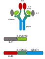
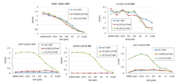
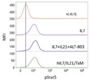
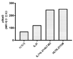
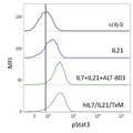
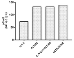
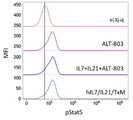
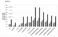
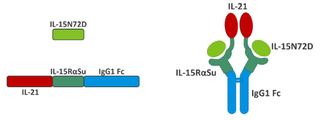
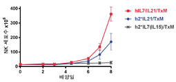
본 발명은 다중 특이적 인터류킨-15(IL-15) 기반 단백질 복합체가 면역 세포의 자극을 강화시키고 질환 세포에 대한 이들의 활성을 촉진시켜 결과적으로 질환을 감소 또는 예방할 수 있다는 놀라운 발견에 적어도 부분적으로 기초한다. 이러한 IL-15 기반 단백질 복합체는 또한 질환 및 표적 항원에 결합할 수 있다. 본원에는 IL-7 및 IL-21 결합 도메인을 포함하는 다중 특이적 IL-15 기반 단백질 복합체가 제공된다(도 1). 구체적으로, IL-7 및 IL-21 결합 도메인에 융합된 IL-15N72D:IL-15RαSu-Ig Fc 스캐폴드를 포함하는 단백질 복합체가 본원에 기재되어 있다. 하기에 상세히 기술되는 바와 같이, 인간 면역 세포를 사용하여 특성화 될 때, 이들 복합체는 각각의 IL-15, IL-7 및 IL-21 시토카인의 결합 및 생물학적 활성을 나타낸다. 또한, 이들 복합체는 IFN-γ의 생산 증가로 T 세포 및 자연살해 (NK) 세포 둘 다의 증식 및 활성화를 유도한다. 놀랍게도, 이들 복합체는 개별 시토카인 단독으로 또는 조합하여 관찰된 것보다 더 큰 정도로 면역 반응을 유도할 수 있었다.The present invention is at least in part upon the surprising discovery that multispecific interleukin-15 (IL-15) based protein complexes can enhance stimulation of immune cells and promote their activity against diseased cells, resulting in reduced or preventable disease. It is based. These IL-15 based protein complexes are also capable of binding disease and target antigens. Provided herein are multispecific IL-15 based protein complexes comprising IL-7 and IL-21 binding domains (FIG. 1 ). Specifically, a protein complex comprising an IL-15N72D:IL-15RαSu-Ig Fc scaffold fused to IL-7 and IL-21 binding domains is described herein. As described in detail below, when characterized using human immune cells, these complexes exhibit the binding and biological activity of the respective IL-15, IL-7 and IL-21 cytokines. In addition, these complexes lead to proliferation and activation of both T cells and natural killer (NK) cells with increased production of IFN-γ. Surprisingly, these complexes were able to elicit an immune response to a greater extent than observed with individual cytokines alone or in combination.
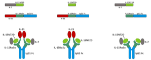
이와 같이, 단일 분자로서의 복합체는 NK 및 T 세포상의 다수의 시토카인 수용체에 결합하여 이를 통해 신호를 전달하여 다수의 시토카인의 조합으로만 이전에 관찰된 반응을 제공한다. 또한, 이들 복합체는 Ig 분자의 Fc 영역을 포함하며, 이는 이량체를 형성하여 가용성 다중 폴리펩티드 복합체를 제공하고, 정제를 목적으로 단백질 A에 결합하고, NK 세포 및 대식세포상의 Fcγ 수용체와 상호 작용함으로써, 개별 시토카인의 조합에서는 존재하지 않는 이점을 복합체에 제공할 수 있다. 이들 복합체를 임상 등급 물질의 대규모 생산에 적합하게 만드는 포유 동물 세포 발현 기반의 방법이 본원에 기재되어 있다. 본 발명의 단백질 복합체에 의한 유도 후 증식하는 NK 및 T 세포를 만들고 사용하는 추가의 방법이 또한 제공된다.As such, the complex as a single molecule binds to multiple cytokine receptors on NK and T cells and transmits a signal through it to provide a response previously observed only with a combination of multiple cytokines. In addition, these complexes contain the Fc region of the Ig molecule, which forms a dimer to provide a soluble multi-polypeptide complex, binds to protein A for purification purposes, and interacts with Fcγ receptors on NK cells and macrophages. , The combination of individual cytokines can provide the complex with advantages that do not exist. Described herein are methods based on mammalian cell expression that render these complexes suitable for large scale production of clinical grade materials. Additional methods of making and using NK and T cells that proliferate after induction by the protein complexes of the invention are also provided.
따라서, 적어도 2개의 가용성 단백질을 포함하는 단리된 가용성 융합 단백질 복합체가 제공된다. 예를 들어, 제1 단백질은 IL-15 폴리펩티드, 예를 들어 N72D 돌연변이 (IL-15N72D)를 포함하는 변이체 IL-15 폴리펩티드를 포함한다. 제2 단백질은 면역 글로불린 Fc 도메인 (IL-15RαSu/Fc)에 융합된 가용성 IL-15 수용체 알파 스시 결합 도메인 (IL-15RαSu)을 포함한다. 단리된 가용성 융합 단백질 복합체의 제3 성분은 IL-7의 결합 도메인을 포함하고, 여기서 IL-7 결합 도메인은 IL-15N72D 또는 IL-15RαSu/Fc 단백질에 융합된다. 단리된 가용성 융합 단백질 복합체의 제4 성분은 IL-21의 결합 도메인을 포함하고, 여기서 IL-21 결합 도메인은 IL-15N72D 또는 IL-15RαSu/Fc 단백질에 융합된다. 일부 경우에, IL-7 및/또는 IL-21 결합 도메인은 IL-15N72D 및 IL 15RαSu/Fc 단백질 모두에 융합된다. 다른 경우에, IL-7 또는 IL-21 결합 도메인은 IL-15N72D 또는 IL-15RαSu/Fc 단백질에 융합되고 다른 결합 도메인은 다른 단백질에 융합된다. 예시적인 융합 단백질 복합체는 IL-15N72D에 공유 결합된 IL-7 폴리펩티드 및 IL-15RαSu/Fc 융합 단백질에 공유 결합된 IL-21 폴리펩티드를 포함한다 (도 1).Thus, an isolated soluble fusion protein complex comprising at least two soluble proteins is provided. For example, the first protein includes an IL-15 polypeptide, such as a variant IL-15 polypeptide comprising an N72D mutation (IL-15N72D). The second protein comprises a soluble IL-15 receptor alpha sushi binding domain (IL-15RαSu) fused to an immunoglobulin Fc domain (IL-15RαSu/Fc). The third component of the isolated soluble fusion protein complex comprises the binding domain of IL-7, where the IL-7 binding domain is fused to the IL-15N72D or IL-15RαSu/Fc protein. The fourth component of the isolated soluble fusion protein complex comprises the binding domain of IL-21, where the IL-21 binding domain is fused to the IL-15N72D or IL-15RαSu/Fc protein. In some cases, IL-7 and/or IL-21 binding domains are fused to both IL-15N72D and IL 15RαSu/Fc proteins. In other cases, the IL-7 or IL-21 binding domain is fused to an IL-15N72D or IL-15RαSu/Fc protein and the other binding domain is fused to another protein. Exemplary fusion protein complexes include IL-7 polypeptide covalently bound to IL-15N72D and IL-21 polypeptide covalently bound to IL-15RαSu/Fc fusion protein (FIG. 1 ).
예시적인 제1 단백질은 서열 번호 2 및 서열 번호 4에 제시된 아미노산 서열을 포함한다. 제1 단백질을 암호화하는 예시적인 핵산 서열은 서열 번호 1 및 서열 번호 3에 제시된 서열을 포함한다. 일 양태에서, 핵산 서열(들)은 융합 단백질을 암호화하는 서열에 작동 가능하게 결합된 촉진인자, 번역 개시 신호 및 리더 서열을 추가로 포함한다. 본원에 기재된 핵산 서열을 포함하는 DNA 벡터가 또한 제공된다. 예를 들어, 핵산 서열은 복제, 발현 또는 둘 모두를 위한 벡터에 존재한다.Exemplary first proteins include the amino acid sequences set forth in SEQ ID NO: 2 and SEQ ID NO: 4. Exemplary nucleic acid sequences encoding the first protein include the sequences set forth in SEQ ID NO: 1 and SEQ ID NO: 3. In one aspect, the nucleic acid sequence(s) further comprises a promoter, translation initiation signal and leader sequence operably linked to the sequence encoding the fusion protein. DNA vectors comprising the nucleic acid sequences described herein are also provided. For example, nucleic acid sequences are present in vectors for replication, expression, or both.
또한, 제2 가용성 융합 단백질 복합체에 공유 연결된 제1 가용성 융합 단백질 복합체를 포함하는 가용성 융합 단백질 복합체가 제공된다. 예를 들어, 본 발명의 가용성 융합 단백질 복합체는 다량체화, 예를 들어, 이량체화, 삼량체화 또는 그렇지 않고 다량체화된다 (예를 들어, 사량체, 오량체 등). 예를 들어, 다량체는 동종 다량체 또는 이종 다량체이다. 가용성 융합 단백질 복합체는 공유 결합, 예를 들어 이황화 결합, 화학적 가교제에 의해 연결된다. 일부 경우에, 하나의 가용성 융합 단백질은 제1 가용성 단백질의 Fc 도메인을 제2 가용성 단백질의 Fc 도메인에 연결하는 이황화 결합에 의해 다른 가용성 융합 단백질에 공유 결합된다.Also provided is a soluble fusion protein complex comprising a first soluble fusion protein complex covalently linked to a second soluble fusion protein complex. For example, the soluble fusion protein complexes of the invention are multimerized, eg, dimerized, trimerized or otherwise multimerized (eg, tetramers, pentamers, etc.). For example, a multimer is a homomultimer or a heteromultimer. Soluble fusion protein complexes are linked by covalent bonds, for example disulfide bonds, chemical crosslinkers. In some cases, one soluble fusion protein is covalently bound to another soluble fusion protein by a disulfide bond that connects the Fc domain of the first soluble protein to the Fc domain of the second soluble protein.
Fc 도메인 또는 이의 기능적 단편은 IgG Fc 도메인, 인간 IgG1 Fc 도메인, 인간 IgG2 Fc 도메인, 인간 IgG3 Fc 도메인, 인간 IgG4 Fc 도메인, IgA Fc 도메인, IgD Fc 도메인, IgE Fc 도메인 및 IgM Fc 도메인으로 이루어진 군으로부터 선택된 Fc 도메인; 마우스 IgG2A 도메인, 또는 이들의 임의의 조합을 포함한다. 선택적으로, Fc 도메인은 변경된 보체 또는 Fc 수용체 결합 특성 또는 변경된 이량체화 또는 당화 프로파일을 갖는 Fc 도메인을 생성하는 아미노산 변화를 포함한다. 변경된 보체 또는 Fc 수용체 결합 특성 또는 변경된 이량체화 또는 당화 프로파일을 갖는 Fc 도메인을 생성하기 위한 아미노산 변화는 당 업계에 공지되어 있다. 예를 들어, IgG1 CH2의 위치 234 및 235 (항체 컨센서스 서열에 기초한 넘버링)에서의 루신 잔기 (즉,… PELLGG…)를 알라닌 잔기 (즉,… PEAAGG…)로 치환하면 Fc 감마 수용체 결합의 손실을 가져오는 한편, IgG1 CH2의 위치 322 (항체 컨센서스 서열에 기초한 넘버링)에서 라이신 잔기를 알라닌 잔기 (즉,… KCASL…)로 치환하면 보체 활성화의 손실을 가져온다. 일부 예에서, 이러한 돌연변이는 조합된다.The Fc domain or functional fragment thereof is from the group consisting of IgG Fc domain, human IgG1 Fc domain, human IgG2 Fc domain, human IgG3 Fc domain, human IgG4 Fc domain, IgA Fc domain, IgD Fc domain, IgE Fc domain and IgM Fc domain Selected Fc domain; Mouse IgG2A domain, or any combination thereof. Optionally, the Fc domain comprises amino acid changes that produce an Fc domain with altered complement or Fc receptor binding properties or altered dimerization or glycosylation profiles. Amino acid changes to generate Fc domains with altered complement or Fc receptor binding properties or altered dimerization or glycosylation profiles are known in the art. For example, substitution of a leucine residue (ie… PELLGG…) at positions 234 and 235 of IgG1 CH2 (numbering based on antibody consensus sequence) with an alanine residue (ie… PEAAGG…) results in loss of Fc gamma receptor binding. On the other hand, replacing lysine residues with alanine residues (ie… KCASL…) at position 322 of IgG1 CH2 (numbering based on antibody consensus sequence) results in loss of complement activation. In some examples, these mutations are combined.
일부 양태에서, IL-7 또는 IL-21 결합 도메인은 폴리펩티드 링커 서열에 의해 IL-15 폴리펩티드 (또는 이의 기능적 단편)에 공유적으로 연결된다. 유사하게, IL 7 또는 IL-21 결합 도메인은 폴리펩티드 링커 서열에 의해 IL-15Rα 폴리펩티드 (또는 이의 기능적 단편)에 공유적으로 연결된다. 임의로, IL-15Rα 폴리펩티드 (또는 이의 기능적 단편)는 폴리펩티드 링커 서열에 의해 Fc 도메인 (또는 이의 기능적 단편)에 공유적으로 연결된다. 각 폴리펩티드 링커 서열은 독립적으로 선택 될 수 있다. 임의로, 폴리펩티드 링커 서열은 동일하다. 또는 이들은 서로 다르다.In some embodiments, an IL-7 or IL-21 binding domain is covalently linked to an IL-15 polypeptide (or functional fragment thereof) by a polypeptide linker sequence. Similarly, the
선택적으로, 가용성 융합 단백질 중 하나 이상이 하나 이상의 결합 도메인 또는 검출 가능한 라벨을 포함하는 본 발명의 가용성 융합 단백질 복합체가 제공된다. 이러한 결합 도메인은 항체, 가용성 T 세포 수용체, 리간드, 가용성 수용체 도메인 또는 이의 기능적 단편을 포함할 수 있다. 이러한 결합 도메인을 포함하는 IL-15 기반 융합 단백질 복합체는 본원에 참조로 포함된 미국 특허 번호 제8,492,118호에 이미 기술되어 있다. 검출 가능한 표지는 제한하는 것은 아니지만, 비오틴, 스트렙타비딘, 이의 효소 또는 촉매 활성 단편, 방사성 핵종, 나노 입자, 상자성 금속 이온, 또는 형광, 인광 또는 화학 발광 분자, 또는 이들의 임의의 조합을 포함한다.Optionally, a soluble fusion protein complex of the invention is provided wherein one or more of the soluble fusion proteins comprises one or more binding domains or detectable labels. Such binding domains can include antibodies, soluble T cell receptors, ligands, soluble receptor domains or functional fragments thereof. IL-15 based fusion protein complexes comprising such binding domains are already described in US Pat. No. 8,492,118, incorporated herein by reference. Detectable labels include, but are not limited to, biotin, streptavidin, enzymes or catalytically active fragments thereof, radionuclides, nanoparticles, paramagnetic metal ions, or fluorescent, phosphorescent or chemiluminescent molecules, or any combination thereof. .
본 발명은 본 발명의 가용성 융합 단백질 복합체를 제조하는 방법을 제공한다. 상기 방법은 a) 제1 단백질을 암호화하는 적절한 제어 서열을 갖는 DNA 벡터를 제1 숙주 세포에 도입하는 단계, b) 세포 또는 배지에서 제1 단백질을 발현시키기에 충분한 조건 하의 배지에서 제1 숙주 세포를 배양하는 단계; c) 숙주 세포 또는 배지로부터 제1 단백질을 정제하는 단계; d) 제2 단백질을 암호화하는 적절한 제어 서열을 갖는 DNA 벡터를 제2 숙주 세포에 도입하는 단계; e) 세포 또는 배지에서 제2 단백질을 발현하기에 충분한 조건 하의 배지에서 제2 숙주 세포를 배양하는 단계; f) 숙주 세포 또는 배지로부터 제2 단백질을 정제하는 단계; 및 g) 제1 단백질의 IL-15 도메인과 제2 단백질의 가용성 IL-15Rα 도메인 사이의 결합을 허용하기에 충분한 조건 하에서 제1 및 제2 단백질을 혼합하여 가용성 융합 단백질 복합체를 형성하는 단계를 포함한다.The present invention provides a method for preparing the soluble fusion protein complex of the present invention. The method comprises: a) introducing a DNA vector having an appropriate control sequence encoding a first protein into a first host cell, b) a first host cell in a medium under conditions sufficient to express the first protein in the cell or medium. Culturing; c) purifying the first protein from the host cell or medium; d) introducing into the second host cell a DNA vector having an appropriate control sequence encoding the second protein; e) culturing the second host cell in a medium under conditions sufficient to express the second protein in the cell or medium; f) purifying the second protein from the host cell or medium; And g) mixing the first and second proteins under conditions sufficient to allow binding between the IL-15 domain of the first protein and the soluble IL-15Rα domain of the second protein to form a soluble fusion protein complex. do.
일부 경우에, 방법은 발현 벡터로부터 발현된 폴리펩티드 사이에 이황화 결합의 형성을 허용하기에 충분한 조건 하에서 제1 및 제2 단백질을 혼합하는 단계를 추가로 포함한다.In some cases, the method further comprises mixing the first and second proteins under conditions sufficient to allow the formation of disulfide bonds between the polypeptides expressed from the expression vector.
대안적으로, 본 발명의 가용성 융합 단백질 복합체를 제조하는 방법은 a) 제1 단백질을 암호화하는 적절한 제어 서열을 갖는 DNA 벡터 및 제2 단백질을 암호화하는 적절한 제어 서열을 갖는 DNA 벡터를 숙주 세포에 도입하는 단계, b) 세포 또는 배지에서 단백질을 발현하고 제1 단백질의 IL-15 도메인과 제2 단백질의 가용성 IL-15Rα 도메인 사이의 회합을 허용하기에 충분한 조건 하의 배지에서 숙주 세포를 배양하여 가용성 융합 단백질 복합체를 형성하는 단계; 및 c) 숙주 세포 또는 배지로부터 가용성 융합 단백질 복합체를 정제하는 단계에 의해 실행된다.Alternatively, the method for preparing a soluble fusion protein complex of the present invention comprises: a) introducing a DNA vector having an appropriate control sequence encoding a first protein and a DNA vector having an appropriate control sequence encoding a second protein into a host cell. Soluble fusion by culturing the host cell in medium under conditions sufficient to express a protein in a cell or medium and allow association between the IL-15 domain of the first protein and the soluble IL-15Rα domain of the second protein Forming a protein complex; And c) purifying the soluble fusion protein complex from the host cell or medium.
일 양태에서, 상기 방법은 발현 벡터로부터 발현된 폴리펩티드 사이에 이황화 결합의 형성을 허용하기에 충분한 조건 하에서 제1 및 제2 단백질을 혼합하는 단계를 추가로 포함한다.In one aspect, the method further comprises mixing the first and second proteins under conditions sufficient to allow the formation of disulfide bonds between the polypeptides expressed from the expression vector.
또한 가용성 융합 단백질 복합체를 만드는 방법은 a) 제1 및 제2 단백질을 암호화하는 적절한 제어 서열을 갖는 DNA 벡터를 숙주 세포에 도입하는 단계, b) 세포 또는 배지에서 단백질을 발현하고 제1 단백질의 IL-15 도메인과 제2 단백질의 가용성 IL-15Rα 도메인 사이의 회합을 허용하여 가용성 융합 단백질 복합체를 형성하고, 폴리펩티드들 사이에 이황화 결합의 형성을 허용하기에 충분한 조건 하의 배지에서 숙주 세포를 배양하는 단계; 및 c) 숙주 세포 또는 배지로부터 가용성 융합 단백질 복합체를 정제하는 단계를 포함한다.Also, a method of making a soluble fusion protein complex includes a) introducing a DNA vector having appropriate control sequences encoding first and second proteins into a host cell, b) expressing the protein in a cell or medium and IL of the first protein Allowing the association between the -15 domain and the soluble IL-15Rα domain of the second protein to form a soluble fusion protein complex, and culturing the host cell in medium under conditions sufficient to allow the formation of disulfide bonds between the polypeptides. ; And c) purifying the soluble fusion protein complex from the host cell or medium.
선택적으로, 상기 방법은 발현 벡터로부터 발현된 폴리펩티드들 사이에 이황화 결합을 형성하기에 충분한 조건 하에서 제1 및 제2 단백질을 혼합하는 단계를 추가로 포함한다.Optionally, the method further comprises mixing the first and second proteins under conditions sufficient to form disulfide bonds between the polypeptides expressed from the expression vector.
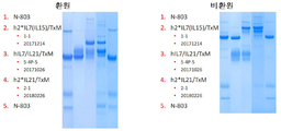
일부 경우에, 상기 방법은 임상 시약 또는 치료제로 사용하기에 적합한 순수 단백질 복합체를 생성하기에 충분한 단백질 A 친화성 크로마토그래피, 크기별 배제 크로마토그래피, 이온 교환 크로마토그래피 및/또는 다른 표준 방법 (바이러스 비활성화 및/또는 여과 포함)에 의한 복합체의 정제 단계를 더 포함한다.In some cases, the method is sufficient to produce a pure protein complex suitable for use as a clinical reagent or therapeutic agent, protein A affinity chromatography, size exclusion chromatography, ion exchange chromatography and/or other standard methods (viral inactivation and And/or purification of the complex by filtration).
본 발명의 가용성 융합 단백질 복합체에 관한 임의의 양태들에서, IL-15 폴리펩티드는 원산(native) IL-15 폴리펩티드의 아미노산 서열과 상이한 아미노산 서열을 가지는 IL-15 변이체이다. 인간 IL-15 폴리펩티드는 본원에서 huIL-15, hIL-15, huIL15, hIL15, IL-15 야생형(wt)이라 지칭되고, 이의 변이체는 원산 아미노산, 성숙 서열 중 이의 위치 및 변이체 아미노산을 이용하여 지칭된다. 예를 들어 huIL15N72D는, 72번 위치에 N → D의 치환을 포함하는 인간 IL-15를 지칭한다. 일 양태에서, IL-15 변이체는, 예컨대 원산 IL-15 폴리펩티드의 IL-15/IL-2βγC 수용체(IL-15R)와의 결합 활성보다 증가한, IL-15/IL-2βγC 수용체와의 결합 활성으로 입증되는 바와 같이, IL-15 효현제로서의 역할을 한다. 대안적으로 IL-15 변이체는, 예컨대 원산 IL-15 폴리펩티드의 IL-15R과의 결합 활성보다 감소한, IL-15R과의 결합 활성으로 입증되는 바와 같이, IL-15 길항제로서의 역할을 한다.In certain aspects of the soluble fusion protein complex of the invention, the IL-15 polypeptide is an IL-15 variant having an amino acid sequence different from the amino acid sequence of the native IL-15 polypeptide. Human IL-15 polypeptides are referred to herein as huIL-15, hIL-15, huIL15, hIL15, IL-15 wild type (wt), and variants thereof using native amino acids, their positions in the mature sequence and variant amino acids. . For example, huIL15N72D refers to human IL-15 which contains the substitution of N → D at
면역 기능을 향상시키는 방법은 a) 복수의 세포를 본 발명의 가용성 융합 단백질 복합체와 접촉시키는 단계 - 복수의 세포는 IL-15 도메인에 의해 인식되는 IL-15R 사슬, IL-7 도메인에 의해 인식된 IL-7R 사슬 또는 IL-21 도메인에 의해 인식되는 IL-21R 사슬을 갖는 면역 세포을 더 포함함 - ; 및 b) IL-15R, IL-7R 또는 IL-21R의 신호 전달을 통해 면역 세포의 증식 및 활성화를 유도하는 단계에 의해 실행된다. 일 양태에서, 면역 기능을 향상시키는 방법은 가용성 융합 단백질 복합체에 의한 IL-15R, IL-7R 및 IL-21R의 둘 이상의 조합의 신호 전달을 통해 면역 세포의 활성화를 추가로 포함한다. 면역 기능을 향상시키기 위한 예시적인 방법은 가용성 융합 단백질 복합체에 의한 IL-15R, IL-12R 및 IL-18R의 신호 전달을 통해 NK 및 T 세포의 증식 및 활성화를 유도하는 단계를 포함한다. 이러한 방법은 NK 및 T 세포의 증식 및 활성화로 인터페론 감마(IFN-γ) 생성 증가의 결과를 가져온다Methods for enhancing immune function include a) contacting a plurality of cells with a soluble fusion protein complex of the present invention, wherein the plurality of cells are recognized by the IL-15R chain, IL-7 domain recognized by the IL-15 domain. Further comprising immune cells having an IL-7R chain or an IL-21R chain recognized by the IL-21 domain; And b) inducing proliferation and activation of immune cells through signal transduction of IL-15R, IL-7R or IL-21R. In one aspect, the method of enhancing immune function further comprises activation of immune cells through signal transduction of two or more combinations of IL-15R, IL-7R and IL-21R by a soluble fusion protein complex. Exemplary methods for enhancing immune function include inducing proliferation and activation of NK and T cells through signal transduction of IL-15R, IL-12R and IL-18R by a soluble fusion protein complex. This method results in increased production of interferon gamma (IFN-γ) by proliferation and activation of NK and T cells.
표적 세포를 사멸시키는 방법은 a) 복수의 세포를 본 발명의 가용성 융합 단백질 복합체와 접촉시키는 단계 - 복수의 세포는 IL-15 도메인에 의해 인식되는 IL-15R 사슬, IL-7 도메인에 의해 인식된 IL-7R 사슬 또는 IL-21 도메인에 의해 인식되는 IL-21R 사슬을 갖는 면역 세포 및 표적 질환 세포를 추가로 포함함 - ; b) IL-15R, IL-7R 또는 IL 21R의 신호 전달을 통해 면역 세포의 증식 및 활성화를 유도하는 단계; 및 c) 활성화된 면역 세포에 의해 표적 질환 세포를 사멸시키는 단계에 의해 실행된다. 일 양태에서, 상기 방법은 가용성 융합 단백질 복합체에 의한 IL-15R, IL-7R 및 IL-21R의 둘 이상의 조합의 신호 전달을 통해 면역 세포의 증식 및 활성화를 유도하는 단계를 포함한다. 예시적인 방법은 가용성 융합 단백질 복합체에 의한 IL-15R, IL-7R 및 IL-21R의 신호 전달을 통해 NK 및 T 세포의 증식 및 활성화를 유도하는 단계를 포함한다. 이러한 방법은 NK 및 T 세포의 증식 및 활성화을 포함하므로 IFN-γ 생성의 증가의 결과를 가져온다.The method of killing a target cell comprises a) contacting a plurality of cells with a soluble fusion protein complex of the present invention-a plurality of cells recognized by the IL-15R chain, IL-7 domain recognized by the IL-15 domain Further includes immune cells and target disease cells having an IL-7R chain or an IL-21R chain recognized by the IL-21 domain; b) inducing proliferation and activation of immune cells through signal transduction of IL-15R, IL-7R or IL 21R; And c) killing target disease cells by activated immune cells. In one aspect, the method comprises inducing proliferation and activation of immune cells through signal transduction of IL-15R, IL-7R and IL-21R by a soluble fusion protein complex. Exemplary methods include inducing proliferation and activation of NK and T cells through signal transduction of IL-15R, IL-7R and IL-21R by a soluble fusion protein complex. This method involves the proliferation and activation of NK and T cells, resulting in an increase in IFN-γ production.
본 발명은 또한 환자의 질환을 예방 또는 치료하는 방법을 제공하며, 이 방법은 a) IL-15R 체인 또는 체크포인트를 갖는 면역 세포 또는 신호 전달 분자를 본 발명의 가용성 융합 단백질 복합체와 혼합하는 단계; b) 면역 세포의 증식과 활성화를 유도하는 단계; c) 환자에게 활성화된 면역 세포를 투여 (또는 입양 전달)하는 단계; 및 d) 환자의 질환을 예방 또는 치료하기에 충분한 활성화된 면역 세포를 통해 질환 세포를 훼손 또는 사멸시키는 단계를 포함한다. 일 양태에서, 상기 방법은 가용성 융합 단백질 복합체에 의한 IL-15R, IL-7R 및 IL-21R 중 적어도 둘 또는 모두의 조합의 신호 전달을 통한 면역 세포의 증식 및 활성화를 포함한다. 예시적인 방법은 가용성 융합 단백질 복합체에 의한 IL-15R, IL-7R 및 IL-21R의 신호 전달을 통해 NK 및 T 세포의 증식 및 활성화를 유도하는 것을 포함한다. 상기 방법의 일부 양태는 키메라 항원 수용체(CAR NK 및 CAR T 세포)를 발현하는 NK 및 T 세포의 사용을 포함한다. 본 발명의 일부 구현예에서, 환자는 입양 전달 세포의 생착 또는 생존을 용이하게 하기 위해 사전 치료 또는 전제조건화된다. 전제조건의 예는 시클로포스파미드 및 플루다라빈으로의 치료를 포함한다. 추가로, 환자는 세포 전 및/또는 세포후 입양 전달된 세포의 활성화, 생존 또는 지속성을 촉진하는 제제로 치료될 수 있다. 이러한 치료의 예는 IL-2, IL-15, ALT-803 (본 명세서에서 "N-803"으로도 상호 교환하여 지칭됨) 또는 다른 면역 자극제의 사용을 포함한다. 입양 세포 요법 분야 (즉, 제한하는 것은 아니지만, 동종, 자가, 동일 단배체, DLI, 줄기 세포, NK92 기반 및 CAR NK 요법을 포함)에서 공지된 다른 치료적 접근법이 본원의 방법에 사용될 수 있다.The invention also provides a method of preventing or treating a patient's disease, the method comprising: a) mixing an immune cell or signal transduction molecule having an IL-15R chain or checkpoint with a soluble fusion protein complex of the invention; b) inducing proliferation and activation of immune cells; c) administering (or adoptive delivery) activated immune cells to the patient; And d) damaging or killing diseased cells through activated immune cells sufficient to prevent or treat the patient's disease. In one aspect, the method comprises proliferation and activation of immune cells through signal transduction of a combination of at least two or both of IL-15R, IL-7R and IL-21R by a soluble fusion protein complex. Exemplary methods include inducing proliferation and activation of NK and T cells through signal transduction of IL-15R, IL-7R and IL-21R by soluble fusion protein complexes. Some embodiments of the method include the use of NK and T cells expressing chimeric antigen receptors (CAR NK and CAR T cells). In some embodiments of the invention, the patient is pre-treated or preconditioned to facilitate engraftment or survival of adoptive transfer cells. Examples of prerequisites include treatment with cyclophosphamide and fludarabine. Additionally, the patient can be treated with agents that promote activation, survival or persistence of the cells before and/or after adoptive transfer of the cells. Examples of such treatment include the use of IL-2, IL-15, ALT-803 (also referred to herein interchangeably as "N-803") or other immunostimulants. Other therapeutic approaches known in the field of adoptive cell therapy (ie, including but not limited to allogeneic, autologous, homoploid, DLI, stem cells, NK92 based and CAR NK therapy) can be used in the methods herein.
또한 환자의 질환을 예방하거나 치료하기 위한 방법이 제공되며, 이 방법은 a) 환자에게 본 발명의 가용성 융합 단백질 복합체를 투여하는 단계; b) 환자 내 면역 세포의 증식 및 활성화를 유도하는 단계; 및 c) 환자의 질환을 예방 또는 치료하기에 충분한 활성화된 면역 세포를 통해 질환 세포를 훼손 또는 사멸시키는 단계를 포함한다.Also provided is a method for preventing or treating a patient's disease, the method comprising: a) administering a soluble fusion protein complex of the invention to a patient; b) inducing proliferation and activation of immune cells in the patient; And c) damaging or killing the diseased cell through activated immune cells sufficient to prevent or treat the patient's disease.
본 발명의 융합 단백질 복합체의 투여는 대상체 내 면역 반응을 유도한다. 예를 들어, 본 발명의 융합 단백질 복합체의 투여는 신생물형성, 감염성 질환, 노화 세포 또는 연령 관련 질환 또는 자가 면역 질환과 관련된 세포에 대한 면역 반응을 유도한다. 일 양태에서, 본 발명의 융합 단백질 복합체는 면역 세포 증식, 활성화 마커, 표적 세포에 대한 세포 독성 및/또는 염증성 시토카인의 생성을 증가시킨다.Administration of the fusion protein complex of the invention induces an immune response in the subject. For example, administration of the fusion protein complex of the present invention induces an immune response against cells associated with neoplasia, infectious disease, aging cells or age-related diseases or autoimmune diseases. In one aspect, the fusion protein complexes of the invention increase immune cell proliferation, activation markers, cytotoxicity to target cells and/or production of inflammatory cytokines.
본 발명은 포유 동물에게 본 발명의 가용성 융합 단백질 복합체의 유효량을투여함으로써 포유 동물에서 면역 반응을 자극하는 방법을 제공한다. 본 발명은 또한 본 발명의 임의의 가용성 융합 단백질 복합체의 유효량을 포유 동물에게 투여함으로써 포유 동물의 면역 반응을 억제하는 방법을 제공한다.The present invention provides a method of stimulating an immune response in a mammal by administering an effective amount of the soluble fusion protein complex of the present invention to the mammal. The invention also provides a method of inhibiting the immune response of a mammal by administering to the mammal an effective amount of any soluble fusion protein complex of the invention.
이를 필요로 하는 대상체에서 신생물형성, 감염성 질환, 노화 세포 또는 연령 관련 질환 또는 자가 면역 질환을 치료하는 방법은 대상체에게 확장 및 활성화된 면역 세포의 유효량 또는 본원에 기재된 가용성 융합 단백질 복합체를 포함하는 약제학적 조성물을 투여함으로써 수행된다. 예를 들어,이를 필요로하는 대상체의 고형 또는 혈액학적 악성 종양을 치료하는 방법은 대상체에게 유효량의 NK 세포 및 T 세포, 및/또는 본 발명의 단백질 복합체에 의해 생체 외 확장된 CAR NK 및 CAR T 세포를 투여함으로써 실행되어, 악성 종양을 치료할 수 있다. 예시적인 가용성 융합 단백질 복합체는 서열 번호 2 및 서열 번호 4에 제시된 아미노산 서열을 포함한다.Methods for treating neoplastic, infectious diseases, aging cells or age-related diseases or autoimmune diseases in a subject in need thereof include an effective amount of expanded and activated immune cells in a subject or a medicament comprising a soluble fusion protein complex described herein It is carried out by administering a pharmaceutical composition. For example, a method of treating a solid or hematologic malignancy of a subject in need thereof includes an effective amount of NK cells and T cells in the subject, and/or CAR NK and CAR T expanded ex vivo by the protein complex of the invention This can be done by administering cells to treat malignant tumors. Exemplary soluble fusion protein complexes include the amino acid sequences set forth in SEQ ID NO: 2 and SEQ ID NO: 4.
본원에 기재된 방법에 의한 치료에 적절한 신생물형성은 교모세포종, 전립선암, 혈액암, B 세포 신생물 형성, 다발성 골수종, B 세포 림프종, B 세포 비호지킨 림프종, 호지킨 림프종, 만성 림프구성 백혈병, 급성 골수성 백혈병, 피부 T 세포 림프종, T 세포 림프종, 고형 종양, 요로상피/방광 암종, 흑색종, 폐암, 신세포 암종, 유방암, 위 및 식도 암, 전립선암, 췌장암, 결장 직장암, 난소암, 비 소세포 폐 암종, 및 편평 세포 두경부 암종을 포함한다.Neoplasias suitable for treatment by the methods described herein include glioblastoma, prostate cancer, blood cancer, B cell neoplasia, multiple myeloma, B cell lymphoma, B cell non-Hodgkin's lymphoma, Hodgkin's lymphoma, chronic lymphocytic leukemia, Acute myeloid leukemia, cutaneous T cell lymphoma, T cell lymphoma, solid tumor, urinary epithelial/bladder carcinoma, melanoma, lung cancer, renal cell carcinoma, breast cancer, gastric and esophageal cancer, prostate cancer, pancreatic cancer, colon rectal cancer, ovarian cancer, non Small cell lung carcinoma, and squamous cell head and neck carcinoma.
본원에 기재된 방법을 사용한 치료를 위한 예시적인 감염은 인간 면역 결핍 바이러스 (HIV) 또는 사이토메갈로 바이러스 (CMV)에 의한 감염을 포함한다. 본원에 기재된 방법은 또한 박테리아 감염 (예를 들어, 그람 양성 또는 그람 음성 박테리아)을 치료하는데 유용하다 (예를 들어, Oleksiewicz 등의 2012. Arch Biochem Biophys. 526:124-31 참조, 본원에서 참고로 포함).Exemplary infections for treatment using the methods described herein include infections with human immunodeficiency virus (HIV) or cytomegalovirus (CMV). The methods described herein are also useful for treating bacterial infections (eg, Gram positive or Gram negative bacteria) (see, eg, Oleksiewicz et al. 2012. Arch Biochem Biophys. 526:124-31, for reference herein) include).
본 발명의 세포 요법은 유효량의 확장 및 활성화된 면역 세포의 투여를 포함한다. 예를 들어, 유효량의 확장 및 활성화된 NK 또는 T 세포는 1 x 10⁴ cells/kg과 1 x 10¹⁰ cells/kg 사이, 예를 들어, 1 x 10⁴, 1 x 10⁵, 1 x 10⁶, 1 x 10⁷, 1 x 10⁸, 1 x 10⁹, and 1 x 10¹⁰ cells/kg, 또는 백혈구 단리에 의해 단리될 수 있는 양이다. 대안적으로, 확장된 면역 세포는 고정 용량으로 또는 체표면적 (즉, m2 당) 기준으로 투여된다. 세포는 생체 외 확장 후 투여되거나 해동 후 (및 필요에 따라 세척) 극저온 보존 및 투여될 수 있다.The cell therapy of the present invention includes an effective amount of expansion and administration of activated immune cells. For example, an effective amount of expanded and activated NK or T cells is between 1 x 10 4 cells/kg and 1 x 10 10 cells/kg, e.g. 1 x 10 4 , 1 x 10 5 , 1 x 10 6 , 1 x 10 7 , 1 x 10 8 , 1 x 10 9 , and 1 x 10 10 cells/kg, or an amount that can be isolated by leukocyte isolation. Alternatively, expanded immune cells are administered in fixed doses or on a body surface area (ie per m 2 ) basis. Cells can be administered after expansion in vitro or cryopreserved and administered after thawing (and washing as needed).
융합 단백질 복합체를 포함하는 약제 조성물이 유효량으로 투여된다. 예를 들어, 약제 조성물의 유효량은 약 1μg/kg와 100μg/kg 사이, 예를 들어, 1, 5, 10, 15, 20, 25, 30, 35, 40, 45, 50, 55, 60, 65, 70, 75, 80, 85, 90, 95 또는 100μg/kg이다. 대안적으로, TxM 복합체는 고정 용량으로 또는 체표면적 (즉, m2 당) 기준으로 투여된다.A pharmaceutical composition comprising a fusion protein complex is administered in an effective amount. For example, an effective amount of a pharmaceutical composition is between about 1 μg/kg and 100 μg/kg, e.g., 1, 5, 10, 15, 20, 25, 30, 35, 40, 45, 50, 55, 60, 65 , 70, 75, 80, 85, 90, 95 or 100 μg/kg. Alternatively, the TxM complex is administered at a fixed dose or on a body surface area (ie per m 2 ) basis.
융합 단백질 복합체를 포함하는 입양 전달된 면역 세포 또는 약학 조성물은 적어도 한 달에 한 번, 예를 들어 한 달에 두 번, 일주일에 한 번, 일주일에 두 번, 하루에 한 번, 하루에 두 번, 8 시간마다, 4 시간마다 시간, 2 시간마다 또는 1 시간마다 투여된다. 입양 전달된 면역 세포에 적합한 투여 방식은 전신 투여, 정맥 내 투여 또는 국소 투여를 포함한다. 약제 조성물의 적합한 투여 방식은 전신 투여, 정맥 내 투여, 국소 투여, 피하 투여, 근육 내 투여, 종양 내 투여, 흡입 및 복강 내 투여를 포함한다.An adoptively delivered immune cell or pharmaceutical composition comprising a fusion protein complex is at least once a month, for example twice a month, once a week, twice a week, once a day, twice a day. , Every 8 hours, every 4 hours, every 2 hours or every 1 hour. Modes of administration suitable for adoptively delivered immune cells include systemic administration, intravenous administration or topical administration. Suitable modes of administration of the pharmaceutical composition include systemic administration, intravenous administration, topical administration, subcutaneous administration, intramuscular administration, intratumoral administration, inhalation and intraperitoneal administration.
일 양태에서, 본 개시 내용은 2 개 이상의 가용성 단백질을 포함하는 단리된 가용성 융합 단백질 복합체를 제공하며, 여기서 제1 가용성 단백질은 인터루킨-15 (IL-15) 폴리펩티드 도메인을 포함하고 제2 가용성 단백질은 면역 글로불린 Fc 도메인에 융합된 가용성 IL-15 수용체알파 스시 결합 도메인 (IL-15RαSu)를 포함한다. 여기서 제1 또는 제2 가용성 단백질 중 하나는 IL-7 결합 도메인 또는 이의 기능적 단편을 추가로 포함하며, 여기서 제1 또는 제2 가용성 단백질 중 하나는 추가로 IL-21 결합 도메인 또는 이의 기능적 단편을 포함하고, 여기서 제1 가용성 단백질의 IL-15 도메인은 제2 가용성 단백질의 IL-15RαSu 도메인에 결합하여 가용성 융합 단백질 복합체를 형성한다.In one aspect, the present disclosure provides an isolated soluble fusion protein complex comprising two or more soluble proteins, wherein the first soluble protein comprises an interleukin-15 (IL-15) polypeptide domain and the second soluble protein is Soluble IL-15 receptor alpha sushi binding domain (IL-15RαSu) fused to an immunoglobulin Fc domain. Wherein one of the first or second soluble proteins further comprises an IL-7 binding domain or a functional fragment thereof, wherein one of the first or second soluble proteins further comprises an IL-21 binding domain or a functional fragment thereof Where the IL-15 domain of the first soluble protein binds to the IL-15RαSu domain of the second soluble protein to form a soluble fusion protein complex.
일 구현예에서, IL-15 폴리펩티드는 N72D 돌연변이 (IL-15N72D)를 포함하는 IL-15 변이체이다.In one embodiment, the IL-15 polypeptide is an IL-15 variant comprising an N72D mutation (IL-15N72D).
일 구현예에서, 제1 가용성 단백질은 서열 번호 2에 제시된 아미노산 서열을 포함한다.In one embodiment, the first soluble protein comprises the amino acid sequence set forth in SEQ ID NO: 2.
일 구현예에서, 제2 가용성 단백질은 서열 번호 4에 제시된 아미노산 서열을 포함한다.In one embodiment, the second soluble protein comprises the amino acid sequence set forth in SEQ ID NO: 4.
일 구현예에서, 제1 가용성 융합 단백질 복합체는 제2 가용성 융합 단백질 복합체에 공유 적으로 연결될 수 있다.In one embodiment, the first soluble fusion protein complex can be covalently linked to a second soluble fusion protein complex.
일 구현예에서, 제1 가용성 융합 단백질 복합체는 제1 가용성 융합 단백질 복합체의 Fc 도메인을 제2 가용성 융합 단백질 복합체의 Fc 도메인에 연결하는 이황화 결합에 의해 제2 가용성 융합 단백질 복합체에 공유 연결된다.In one embodiment, the first soluble fusion protein complex is covalently linked to the second soluble fusion protein complex by a disulfide bond connecting the Fc domain of the first soluble fusion protein complex to the Fc domain of the second soluble fusion protein complex.
일 구현예에서, 제1 또는 제2 가용성 단백질은 질환 항원을 인식하는 결합 도메인을 추가로 포함한다.In one embodiment, the first or second soluble protein further comprises a binding domain that recognizes the disease antigen.
일 구현예에서, 제1 또는 제2 가용성 단백질은 면역 체크포인트 또는 신호 전달 분자를 인식하는 결합 도메인을 추가로 포함한다.In one embodiment, the first or second soluble protein further comprises a binding domain that recognizes an immune checkpoint or signal transduction molecule.
일 구현예에서, 질환 항원은 신생물형성, 감염성 질환 또는 노화 세포 또는 연령 관련 질환과 관련된다.In one embodiment, the disease antigen is associated with neoplastic, infectious disease or aging cell or age related disease.
일 구현예에서, 제1 가용성 단백질은 서열 번호 1에 제시된 핵산 서열에 의해 암호화된다.In one embodiment, the first soluble protein is encoded by the nucleic acid sequence set forth in SEQ ID NO:1.
일 구현예에서, 핵산 서열은 가용성 단백질을 암호화하는 서열에 작동 가능하게 결합된 촉진인자, 번역 개시 신호 및 리더 서열을 추가로 포함한다.In one embodiment, the nucleic acid sequence further comprises a facilitator, translation initiation signal and leader sequence operably linked to a sequence encoding a soluble protein.
일 구현예에서, 제2 가용성 단백질은 서열 번호 3에 제시된 핵산 서열에 의해 암호화될 수 있다.In one embodiment, the second soluble protein can be encoded by the nucleic acid sequence set forth in SEQ ID NO: 3.
일 구현예에서, 핵산 서열은 가용성 단백질을 암호화하는 서열에 작동 가능하게 결합된 촉진인자, 번역 개시 신호 및 리더 서열을 추가로 포함한다.In one embodiment, the nucleic acid sequence further comprises a facilitator, translation initiation signal and leader sequence operably linked to a sequence encoding a soluble protein.
일 구현예에서, DNA 벡터는 상기 열거 된 임의의 핵산 서열 중 어느 하나를 포함할 수 있다.In one embodiment, the DNA vector can include any of the nucleic acid sequences listed above.
일 구현예에서, 면역 기능을 향상시키는 방법으로서, 이 방법은 a) 복수의 세포를 임의의 상기 가용성 융합 단백질 복합체와 접촉시키는 단계, - 복수의 세포는 IL-15 도메인, IL-7 도메인에 의해 인식되는 IL-7R 사슬 및/또는 IL-21 도메인에 의해 인식되는 IL-21R 사슬을 더 포함함 - , 및 b) IL-15R, IL-7R 및/또는 IL-21R의 신호 전달을 통해 면역 세포의 증식 및 활성화를 유도하는 단계를 포함한다.In one embodiment, a method for enhancing immune function, the method comprising: a) contacting a plurality of cells with any of the above soluble fusion protein complexes, wherein the plurality of cells are by an IL-15 domain, an IL-7 domain. Further comprising a recognized IL-7R chain and/or an IL-21R chain recognized by the IL-21 domain-, and b) immune cells through signal transduction of IL-15R, IL-7R and/or IL-21R Inducing the proliferation and activation of the.
일 양태에서, 본 개시 내용은 표적 세포를 사멸하는 방법을 제공하고, 이 방방법은 a) 복수의 세포를 임의의 상기 가용성 융합 단백질 복합체와 접촉시키는 단계 - 복수의 세포는 IL-15 도메인에 의해 인식되는 IL-15R 사슬, IL-7 도메인에 의해 인식되는 IL-7R 사슬 및/또는 IL-21 도메인에 의해 인식되는 IL-21R 사슬을 갖는 면역 세포를 더 포함함 -, b) IL-15R, IL-7R 및/또는 IL-21R의 신호 전달을 통해 면역 세포의 증식 및 활성화를 유도하는 단계, 및 c) 확장 및 활성화 된 면역 세포에 의해 표적 질환 세포를 사멸시키는 단계를 포함한다.In one aspect, the present disclosure provides a method of killing a target cell, the method comprising: a) contacting a plurality of cells with any of the above soluble fusion protein complexes, wherein the plurality of cells are by an IL-15 domain. Further comprising immune cells having an IL-15R chain recognized, an IL-7R chain recognized by the IL-7 domain and/or an IL-21R chain recognized by the IL-21 domain -, b) IL-15R, Inducing proliferation and activation of immune cells through signal transduction of IL-7R and/or IL-21R, and c) killing target disease cells by expanded and activated immune cells.
일 구현예에서, 표적 세포는 종양 세포 또는 감염된 세포이다.In one embodiment, the target cells are tumor cells or infected cells.
일 양태에서, 본 개시 내용은 대상체의 면역 반응을 증진시키는 방법을 제공하며, 이 방법은 a) 복수의 세포를 임의의 상기 가용성 융합 단백질 복합체와 접촉시키는 단계 - 복수의 세포는 IL-15 도메인에 의해 인식되는 IL-15R 사슬, IL-7 도메인에 의해 인식되는 IL-7R 사슬 및/또는 IL-21 도메인에 의해 인식되는 IL-21R 사슬을 갖는 면역 세포를 더 포함함 -, b) IL-15R, IL-7R 및/또는 IL-21R의 신호 전달을 통해 면역 세포의 증식 및 활성화를 유도하는 단계, c) 환자에게 확장 및 활성화 된 면역 세포를 투여 (또는 입양 전달)하는 단계; 및 d) 환자의 면역 반응을 강화하는 단계를 포함한다..In one aspect, the present disclosure provides a method of enhancing a subject's immune response, the method comprising: a) contacting a plurality of cells with any of the above soluble fusion protein complexes, wherein the plurality of cells are in an IL-15 domain. Further comprising immune cells having an IL-15R chain recognized by, an IL-7R chain recognized by an IL-7 domain and/or an IL-21R chain recognized by an IL-21 domain -, b) IL-15R , Inducing proliferation and activation of immune cells through signal transduction of IL-7R and/or IL-21R, c) administering (or adoptive delivery) expanded and activated immune cells to the patient; And d) enhancing the patient's immune response.
일 양태에서, 본 개시 내용은 환자의 질환을 예방 또는 치료하는 방법을 제공하고, 이 방법은 a) 복수의 세포를 본원에 구현된 가용성 융합 단백질 복합체와 접촉시키는 단계 - 복수의 세포는 IL- IL-15 도메인에 의해 인식되는 15R 사슬, IL-7 도메인에 의해 인식되는 IL-7R 사슬 및/또는 IL-21 도메인에 의해 인식되는 IL-21R 사슬을 갖는 면역 세포를 더 포함함 -, b) IL-15R, IL-7R 및/또는 IL-21R의신호 전달을 통해 면역 세포의 증식 및 활성화를 유도하는 단계, c) 유효량의 확장 및 활성화된 면역 세포를 환자에게 투여 (또는 입양 전달)하는 단계, 및 d) 환자의 질병 세포를 손상 또는 사멸시키기에 충분히 확장된 면역 세포를 통해 질환 세포를 손상 및 사멸시키는 단계를 포함한다.In one aspect, the present disclosure provides a method of preventing or treating a patient's disease, the method comprising: a) contacting a plurality of cells with a soluble fusion protein complex as implemented herein, wherein the plurality of cells are IL-IL -15 further comprising immune cells having a 15R chain recognized by the domain, an IL-7R chain recognized by the IL-7 domain and/or an IL-21R chain recognized by the IL-21 domain -, b) IL Inducing proliferation and activation of immune cells through signal transduction of -15R, IL-7R and/or IL-21R, c) administering (or adoptive delivery) an effective amount of expanded and activated immune cells to the patient, And d) damaging and killing diseased cells through immune cells that are sufficiently expanded to damage or kill the patient's diseased cells.
특정 구현예에서, 대상체 내 면역 반응을 자극하는 방법은, 면역 세포를 단리하는 단계; 면역 세포를 본원에 구현된 가용성 융합 단백질 복합체와 접촉시키는 단계; 면역 세포를 대상체에게 재주입하는 단계; 이에 의해, 대상체의 면역 반응을 자극하는 단계를 포함한다. 특정 구현예에서, 면역 세포는 자가, 일배체 동일성, 일배체 매칭 또는 이들의 조합을 포함한다. 특정 구현예에서, 면역 세포는 자가 또는 동종이계 줄기 세포로부터 유래된다. 특정 구현예에서, 면역 세포는 NK 세포, T 세포, 줄기 세포 기억 T 세포, 활성화된 NK (aNK) 세포, 키메라 항원 수용체 NK (CAR-NK) 세포, 키메라 항원 수용체 T (CAR-T) 세포, 또는 이들의 조합을 포함한다 특정 구현예에서, 하나 이상의 보조제는 임의로 본원에 구현된 가용성 융합 단백질 복합체와 함께 투여된다.In certain embodiments, a method of stimulating an immune response in a subject comprises isolating immune cells; Contacting the immune cells with a soluble fusion protein complex as implemented herein; Reinjecting the immune cells into the subject; Thereby, the step of stimulating the immune response of the subject. In certain embodiments, immune cells include autologous, haplotype identity, haplotype matching, or combinations thereof. In certain embodiments, immune cells are derived from autologous or allogeneic stem cells. In certain embodiments, the immune cells are NK cells, T cells, stem cell memory T cells, activated NK (aNK) cells, chimeric antigen receptor NK (CAR-NK) cells, chimeric antigen receptor T (CAR-T) cells, Or combinations thereof. In certain embodiments, one or more adjuvants are optionally administered with a soluble fusion protein complex as embodied herein.
일 구현예에서, 질환은 신생물형성, 감염성 질환, 또는 노화 세포 또는 연령관련 질환이다.In one embodiment, the disease is a neoplastic, infectious disease, or aging cell or age related disease.
일 양태에서, 본 개시는 유효량의 임의의 상기 가용성 융합 단백질 복합체를 대상체에게 투여하는 단계를 포함하는, 대상체 내 면역 반응을 향상시키는 방법을 제공한다.In one aspect, the present disclosure provides a method of enhancing an immune response in a subject comprising administering to the subject an effective amount of any such soluble fusion protein complex.
일 양태에서, 본 개시는 대상체의 신생물형성, 감염성 질환 또는 노화 세포 또는 노화 관련 질환을 치료하는 방법을 제공하며, 이 방법은 상기 가용성 융합 단백질 복합체 중 어느 것을 포함하는 유효량의 약제 조성물을 상기 대상체에게 투여하여 상기 신생물형성, 감염성 질환 또는 노화 세포 또는 노화 관련 질환을 치료하는 단계를 포함한다.In one aspect, the present disclosure provides a method of treating a neoplastic, infectious disease or aging cell or aging-related disease in a subject, the method comprising administering an effective amount of a pharmaceutical composition comprising any of the soluble fusion protein complexes to the subject. And treating the neoplastic, infectious disease or aging cell or aging-related disease.
다른 양태에서, 신생물형성, 감염성 질환 또는 노화 세포 또는 연령 관련 질환을 갖는 대상체를 치료하는 방법은 a) 면역 세포를 상기 가용성 융합 단백질 복합체와 접촉시켜 면역 세포의 증식 및 활성화를 유도하는 단계, b) 유효량의 활성화된 면역 세포를 대상체에게 투여 (또는 입양 전달)하는 단계, c) 대상체의 질환을 예방 또는 치료하기에 충분한 활성화된 면역 세포를 통해 질병 세포를 손상 또는 사멸시키는 단계를 포함한다..In another aspect, a method of treating a subject with neoplasia, infectious disease or aging cell or age related disease comprises: a) contacting immune cells with the soluble fusion protein complex to induce proliferation and activation of immune cells, b ) Administering (or adoptive delivery) an effective amount of activated immune cells to the subject, c) damaging or killing the diseased cells through activated immune cells sufficient to prevent or treat the subject's disease.
일 구현예에서, 신생물형성은 교모세포종, 전립선암, 혈액암, B 세포 신생물 형성, 다발성 골수종, B 세포 림프종, B 세포 비호지킨 림프종, 호지킨 림프종, 만성 림프구성 백혈병, 급성 골수성 백혈병, 피부 T 세포 림프종, T 세포 림프종, 고형 종양, 요로상피/방광 암종, 흑색종, 폐암, 신세포 암종, 유방암, 위 및 식도 암, 전립선암, 췌장암, 결장 직장암, 난소암, 비 소세포 폐 암종, 및 편평 세포 두경부 암종으로 이루어진 그룹에서 선택된다.In one embodiment, the neoplasia is glioblastoma, prostate cancer, blood cancer, B cell neoplasia, multiple myeloma, B cell lymphoma, B cell non-Hodgkin's lymphoma, Hodgkin's lymphoma, chronic lymphocytic leukemia, acute myeloid leukemia, Skin T cell lymphoma, T cell lymphoma, solid tumor, urinary tract epithelial/bladder carcinoma, melanoma, lung cancer, renal cell carcinoma, breast cancer, gastric and esophageal cancer, prostate cancer, pancreatic cancer, colon rectal cancer, ovarian cancer, non-small cell lung carcinoma, And squamous cell head and neck carcinoma.
다른 구현예에서, 노화 세포 또는 연령 관련 질환은 대사 (비만, 당뇨병), 신경계 (알츠하이머 및 파킨슨 병), 근육, 뼈 및 연골 관련 (사르코니아 질환 또는 장애, 골관절염, 후만증, 허리 디스크) 또는 조직 기능 장애 관련 (폐 폐기종, 심혈관 및 신장 질환 및 죽상 동맥 경화증) 질환으로 이루어진 그룹에서 선택된다.In other embodiments, the aging cell or age related disease is metabolic (obesity, diabetes), nervous system (Alzheimer's and Parkinson's disease), muscle, bone and cartilage related (Sarconia disease or disorder, osteoarthritis, kyphosis, back disc) or tissue function Disorder-related (pulmonary emphysema, cardiovascular and kidney disease and atherosclerosis) disease.
일 구현예에서, 면역 세포는 NK 세포 또는 시토카인 유도된 메모리 유사 (CIML) NK 세포이다.In one embodiment, the immune cells are NK cells or cytokine derived memory like (CIML) NK cells.
다른 구현예에서, 면역 세포는 T 세포 또는 기억 줄기 T 세포 (TSCM)이다.In other embodiments, the immune cells are T cells or memory stem T cells (T SCM ).
일 구현예에서, 확장 및 활성화된 면역 세포의 유효량은 1 x 10⁴ 세포/kg 내지 1 x 10¹⁰ 세포/kg이다.In one embodiment, the effective amount of expanded and activated immune cells is 1 x 10 4 cells/kg to 1 x 10 10 cells/kg.
일 구현예에서, 면역 세포는 주당 적어도 1 회 투여된다.In one embodiment, immune cells are administered at least once per week.
일 구현예에서, 유효량은 상기 융합 단백질 복합체의 약 1 내지 100 μg/kg이다.In one embodiment, the effective amount is about 1 to 100 μg/kg of the fusion protein complex.
일 구현예에서, 융합 단백질 복합체는 일주일에 적어도 1 회 투여된다.In one embodiment, the fusion protein complex is administered at least once a week.
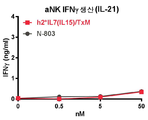
일 구현예에서, 융합 단백질 복합체는 IFN-γ를 포함하여, 면역 세포 증식, 활성화 마커, 표적 세포에 대한 세포 독성 및/또는 전염증성 시토카인의 생성을 증가시킨다.In one embodiment, the fusion protein complex comprises IFN-γ to increase immune cell proliferation, activation markers, cytotoxicity to target cells and/or production of pro-inflammatory cytokines.
바람직하게는, 융합 단백질 복합체는 인터페론 감마 (IFN-γ)의 혈청 수준을 증가시키고/시키거나 CD4+ 및 CD8+ T 세포 및 NK 세포를 자극하여 대상체의 질환 세포 또는 종양 세포를 사멸시킨다.Preferably, the fusion protein complex increases serum levels of interferon gamma (IFN-γ) and/or stimulates CD4 + and CD8 + T cells and NK cells to kill the subject's diseased cells or tumor cells.
정의Justice
달리 정의되지 않는 한, 본원에 사용된 모든 기술 및 과학 용어는 본 발명이 속하는 기술 분야의 당업자에 의해 일반적으로 이해되는 의미를 갖는다. 본 명세서에서 사용한 용어는 단지 특정한 구현예를 설명하기 위해 사용된 것으로, 본 발명을 한정하려는 것은 아니다.Unless defined otherwise, all technical and scientific terms used herein have the meanings commonly understood by one of ordinary skill in the art to which this invention belongs. The terms used herein are only used to describe specific embodiments, and are not intended to limit the present invention.
하기 참고 문헌은 당업자에게 본 발명에서 사용되는 많은 용어의 일반적인 정의를 제공한다: Singleton 등, Dictionary of Microbiology and Molecular Biology (2nd ed. 1994); 케임브리지 과학 기술 사전 (Walker ed., 1988); 유전학 용어집, 5th Ed., R. Rieger 등 (eds.), Springer Verlag (1991); and Hale & Marham, Harper Collins Dictionary of Biology (1991). 본 명세서에서 사용된 바와 같이, 다음의 용어는 달리 명시되지 않는 한 아래에 설명된 바와 같은 의미를 갖는다.The following references provide those skilled in the art with a general definition of many terms used in the present invention: Singleton et al., Dictionary of Microbiology and Molecular Biology (2nd ed. 1994); Cambridge Science and Technology Dictionary (Walker ed., 1988); Genetics Glossary, 5th Ed., R. Rieger et al. (eds.), Springer Verlag (1991); and Hale & Marham, Harper Collins Dictionary of Biology (1991). As used herein, the following terms have the meanings set forth below unless otherwise specified.
단수의 표현은 문맥 상 명백하게 다르게 뜻하지 않는 한, 복수의 표현을 포함한다. 또한, 용어 "갖는", "갖다", "구비하다", "구비한" 또는 이의 변형은 상세한 설명 및/또는 청구 범위에서 사용되는 한, 그러한 용어는 "포함하는"이라는 용어와 유사한 방식으로 포함하는 것으로 의도된다..Singular expressions include plural expressions unless the context clearly indicates otherwise. Also, the terms "having", "having", "having", "having", or variations thereof, are included in a manner similar to the term "comprising" as long as they are used in the description and/or claims. It is intended to..
문맥에서 구체적으로 언급되거나 명백하지 않은 한, 본원에 사용되는 용어 단수의 표현은 단수이거나 복수일 수도 있는 것으로 이해된다. 본 명세서 및 첨부된 청구 범위에 사용된 바와 같이, 용어 "또는"은 내용이 명확하게 달리 지시하지 않는 한 "및/또는"을 포함하는 의미로 사용된다.It is understood that, unless specifically stated or apparent in context, the term singular expression as used herein may be singular or plural. As used in this specification and the appended claims, the term “or” is used in its sense including “and/or” unless the content clearly dictates otherwise.
특별히 진술되거나, 내용으로부터 명백하지 않다면, 본원에 사용된 바와 같은 "약"이란 용어는, 당 분야에서 보통 관용되는 범위 내, 예를 들어 평균의 2 표준 편차 내에 속하는 것으로 이해된다. "약"은 진술된 값의 10%, 9%, 8%, 7%, 6%, 5%, 4%, 3%, 2%, 1%, 0.5%, 0.1%, 0.05% 또는 0.01% 이내인 것으로 이해될 수 있다. 내용으로부터 분명하지 않다면, 본원에 제공된 모든 수치 값은 용어 "약"에 의해 수식된다.The term "about" as used herein, unless specifically stated or apparent from the context, is understood to fall within the range commonly used in the art, for example within 2 standard deviations of the mean. “About” is within 10%, 9%, 8%, 7%, 6%, 5%, 4%, 3%, 2%, 1%, 0.5%, 0.1%, 0.05% or 0.01% of stated value It can be understood as. Unless apparent from the context, all numerical values provided herein are modified by the term “about”.
"제제"는 펩티드, 핵산 분자 또는 소량의 화합물을 의미한다."Formulation" means a peptide, nucleic acid molecule or a small amount of a compound.
"ALT-803"또는 "N-803"은 이량체 IL-15RαSu/Fc 융합 단백질과 비공유적으로 관련된 IL-15N72D를 포함하고 면역 자극 활성을 갖는 복합체를 의미한다. 이 복합체는 IL-15 SA라고도 한다. 일 구현예에서, IL-15N72D 및/또는 IL-15RαSu/Fc 융합 단백질은 참조 서열에 대한 하나, 둘, 셋, 넷 또는 그 이상의 아미노산 변이를 포함한다.“ALT-803” or “N-803” refers to a complex comprising IL-15N72D that is non-covalently associated with a dimeric IL-15RαSu/Fc fusion protein and having immune stimulating activity. This complex is also referred to as IL-15 SA. In one embodiment, the IL-15N72D and/or IL-15RαSu/Fc fusion protein comprises one, two, three, four or more amino acid variations to the reference sequence.
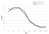
"TxM"은 결합 도메인에 결합된 IL-15N72D:IL-15RαSu/Fc 스캐폴드를 포함하는 복합체를 의미한다(도 1). 예시적인 TxM은 IL-7 및 IL-21 시토카인에 대한 융합을 포함하는 IL-15N72D:IL-15RαSu 복합체이다."TxM" refers to a complex comprising an IL-15N72D:IL-15RαSu/Fc scaffold bound to a binding domain (Figure 1). An exemplary TxM is the IL-15N72D:IL-15RαSu complex comprising fusion to IL-7 and IL-21 cytokines.
"완화하다"란, 질환의 발달 또는 진행을 늦추거나, 억제하거나, 경감시키거나, 감소시키거나, 정지시키거나 또는 안정화하는 것을 의미한다.“Relieving” means slowing, inhibiting, alleviating, reducing, halting, or stabilizing the development or progression of a disease.
"유사체"는 동일하지 않지만 유사한 기능적 또는 구조적 특징을 갖는 분자를 의미한다. 예를 들어, 폴리펩티드 유사체는 상응하는 자연 발생 폴리펩티드의 생물학적 활성을 유지하면서, 자연 발생 폴리펩티드에 비해 유사체의 기능을 향상시키는 특정 생화학적 변형을 갖는다. 이러한 생화학적 변형은 예를 들어 리간드 결합능을 변경시키지 않으면서 유사체의 프로테아제 내성, 막 투과성 또는 반감기를 증가시킬 수 있다. 유사체는 비 자연 발생 아미노산을 포함할 수 있다. 뉴클레오사이드와 관련하여 "유사체"는 예를 들어, 일반적으로 Scheit, Nucleotide Analogs, John Wiley, New York, 1980에 의해 기술된 변형된 염기 모이어티 및/또는 변형된 당 모이어티를 갖는 합성 뉴클레오사이드를 포함하며, 예를 들어, Scheit, Nucleotide Analogs, John Wiley, New York, 1980; Freier & Altmann, Nucl. Acid. Res., 1997, 25 (22), 4429-4443, Toulme, J.J., Nature Biotechnology 19:17-18 (2001); Manoharan M., Biochemica et Biophysica Acta 1489:117-139 (1999); Freier S.M., Nucleic Acid Research, 25:4429-4443 (1997), Uhlman, E., Drug Discovery & Development, 3:203-213 (2000), Herdewin P., Antisense & Nucleic Acid Drug Dev., 10:297 -310(2000)); 2'-O, 3'-C-연결된 [3.2.0] 비사이클로아라비노 뉴클레오사이드 (예를 들어, N.K. Christiensen 등, J. Am. Chem. Soc., 120:5458-5463 (1998) 참조). 이러한 유사체는 결합 특성, 예를 들어 이중 또는 삼중 안정성, 특이성 등을 향상시키도록 설계된 합성 뉴클레오사이드를 포함한다“Imulator” means a molecule that is not identical but has similar functional or structural characteristics. For example, polypeptide analogs have certain biochemical modifications that enhance the function of analogs compared to naturally occurring polypeptides, while maintaining the biological activity of the corresponding naturally occurring polypeptide. Such biochemical modifications can, for example, increase protease resistance, membrane permeability or half-life of analogs without altering the ligand binding capacity. Analogs can include non-naturally occurring amino acids. A “analogue” with reference to a nucleoside is a synthetic nucleoside having a modified base moiety and/or a modified sugar moiety, generally described by Scheit, Nucleotide Analogs, John Wiley, New York, 1980, for example. Side, including, for example, Scheit, Nucleotide Analogs, John Wiley, New York, 1980; Freier & Altmann, Nucl. Acid. Res., 1997, 25 (22), 4429-4443, Toulme, J.J., Nature Biotechnology 19:17-18 (2001); Manoharan M., Biochemica et Biophysica Acta 1489:117-139 (1999); Freier SM, Nucleic Acid Research, 25:4429-4443 (1997), Uhlman, E., Drug Discovery & Development, 3:203-213 (2000), Herdewin P., Antisense & Nucleic Acid Drug Dev., 10:297 -310 (2000)); See 2'-O, 3'-C-linked [3.2.0] bicycloarabino nucleosides (eg, NK Christiensen et al., J. Am. Chem. Soc., 120:5458-5463 (1998)) ). Such analogs include synthetic nucleosides designed to improve binding properties, such as double or triple stability, specificity, etc.
본 발명은 원하는 생물학적 활성을 나타내는 한, 항체나 이러한 항체의 단편을 포함한다. 또한 인간화 항체와 같은 키메라 항체가 본 발명에 포함된다. 일반적으로, 인간의 항체는 비인간 공급원으로부터 도입된 하나 이상의 아미노산 잔기를 갖는다. 인간화는 예를 들어 인간 항체의 상응하는 영역에 대한 설치류 상보성 결정 영역의 적어도 일부를 대체함으로써, 당 업계에 기재된 방법을 사용하여 수행 될 수 있다.The present invention includes antibodies or fragments of such antibodies as long as they exhibit the desired biological activity. Chimeric antibodies such as humanized antibodies are also included in the present invention. Generally, human antibodies have one or more amino acid residues introduced from a non-human source. Humanization can be performed using methods described in the art, for example, by replacing at least a portion of the rodent complementarity determining region for the corresponding region of the human antibody.
"항체" 또는 "면역글로불린"이란 용어는, 폴리클로날 항체와 모노클로날 항체 둘 다를 포함하도록 의도된다. 바람직한 항체는 항원과 반응성인 모노클로날 항체이다. "항체"란 용어는 또한, 항원과 반응성인 항체 하나 초과만큼의 혼합물(예컨대 항원과 반응성인 상이한 유형들의 모노클로날 항체 칵테일)을 포함한다. "항체"란 용어는, 전 항체, 이의 생물 기능성 단편, 단일 사슬 항체, 그리고 유전자 변경된 항체, 예컨대 하나를 초과하는 종으로부터 유래하는 부분들을 포함하는 키메라 항체, 이기능성 항체, 항체 접합체, 인간화 항체 및 인간 항체를 포함하도록 또한 의도된다. 추가로 사용될 수도 있는 생물 기능성 항체 단편으로서는 항원과의 결합에 충분한 항체 유래 펩티드 단편이 있다. 본원에 사용된 바와 같은 "항체"는, 관심 에피토프, 항원 또는 항원 단편과 결합할 수 있는 전체 항체뿐만 아니라, 임의의 항체 단편(예컨대 F(ab')2, Fab', Fab, Fv)을 포함하는 의미이다.The term "antibody" or "immunoglobulin" is intended to include both polyclonal and monoclonal antibodies. Preferred antibodies are monoclonal antibodies that are reactive with the antigen. The term “antibody” also includes more than one mixture of antibodies reactive with the antigen (eg cocktail of different types of monoclonal antibodies reactive with the antigen). The term "antibody" includes whole antibodies, biologically functional fragments thereof, single chain antibodies, and genetically modified antibodies, such as chimeric antibodies, bifunctional antibodies, antibody conjugates, humanized antibodies, and portions derived from more than one species, and It is also intended to include human antibodies. An antibody-derived peptide fragment sufficient for binding to an antigen is a bio-functional antibody fragment that may be further used. “Antibody” as used herein includes any antibody fragment (eg F(ab′) 2 , Fab′, Fab, Fv), as well as whole antibodies capable of binding an epitope, antigen or antigenic fragment of interest. It means.
본원에 사용된 용어 "연관된", "접합된", "연결된", "부착된" 및 "묶여진"은 2 개 이상의 모이어티와 관련하여 사용될 때, 모이어티가 구조가 사용되는 조건, 예를 들어 생리학적 상태에서 물리적으로 결합되도록 충분히 안정한 구조를 형성하기 위해서, 모이어티가 직접적으로 (예를 들어, 공유 결합) 또는 연결제로서 작용하는 하나 이상의 추가의 모이어티를 통해, 물리적으로 서로 연관되거나 연결되어 있음을 의미한다. 일부 구현예에서, 충분한 수의 약한 상호 작용은 모이어티가 다양한 상이한 조건 하에서 물리적으로 연관된 상태를 유지하기에 충분한 안정성을 제공할 수 있다.As used herein, the terms "associated", "conjugated", "connected", "attached" and "tied" when used in connection with two or more moieties, the conditions in which the moieties are used, for example To form a structure that is sufficiently stable to be physically bound in a physiological state, the moieties are physically associated with or linked to each other, either directly (eg, covalent bonds) or through one or more additional moieties that act as linking agents. It means. In some embodiments, a sufficient number of weak interactions can provide sufficient stability for the moiety to remain physically associated under a variety of different conditions.
분자에 "결합"한다는 것은 그 분자에 대한 물리 화학적인 친화성을 갖는 것을 의미한다."Bonding" to a molecule means having a physicochemical affinity for that molecule.
용어 "결합 도메인"은 항체, 단일 사슬 항체, Fab, Fv, T 세포 수용체 결합 도메인, 리간드 결합 도메인, 수용체 결합 도메인 또는 당 업계에 공지된 다른 항원-특이적 폴리펩티드를 포함하는 것으로 의도된다.The term “binding domain” is intended to include antibodies, single chain antibodies, Fab, Fv, T cell receptor binding domains, ligand binding domains, receptor binding domains or other antigen-specific polypeptides known in the art.
본원에 사용된 용어 "생물학적 활성 모이어티" 또는 "효과기 분자"는 핵산 서열, 단백질, 폴리펩티드 또는 펩티드와 같은 아미노산 서열; 당 또는 다당류; 본원에 논의된 바와 같이 원하는 효과를 생성할 수 있는 지질 또는 당지질, 당 단백질 또는 지단백질을 의미한다. 효과기 분자는 또한 화학 제제를 포함한다. 생물학적 활성 또는 효과기 단백질, 폴리펩티드 또는 펩티드를 암호화하는 효과기 분자 핵산이 또한 고려된다. 따라서, 적합한 분자는 조절 인자, 효소, 항체 또는 약물뿐만 아니라 DNA, RNA 및 올리고뉴클레오티드를 포함한다. 생물학적 활성 폴리펩타이드 또는 효과기 분자는 자연적으로 발생하거나 공지된 성분, 예를 들어 재조합 또는 화학적 합성에 의해 합성될 수 있으며 이종 성분을 포함할 수 있다. 생물학적 활성 폴리펩티드 또는 효과기 분자는 일반적으로 원심 단리 또는 SDS- 폴리아크릴 아미드 겔 전기 영동과 같은 표준 분자 사이징 기술에 의해 판단할 때 약 0.1 내지 100 KD 이상에서 약 1000 KD 사이, 바람직하게는 약 0.1, 0.2, 0.5, 1, 2, 5, 10, 20, 30 내지 50 KD이다. 본 발명의 바람직한 효과는 제한하는 것은 아니지만, 예를 들어 결합 활성이 증가된 본 발명의 융합 단백질 복합체를 형성하고, 표적 세포을 사멸하고, 세포 증식 또는 세포 사멸을 유도하고, 질환 예방 및 치료시 면역 반응을 개시하고, 진단 목적을 위한 검출 분자로서 작용하는 것등을 포함한다. 이러한 검출을 위해, 예를 들어 세포를 배양하여 이를 증식시키고, 세포를 본 발명의 융합 복합체와 접촉시킨 후 융합 복합체가 세포의 추가 발달을 저해하는지 여부를 평가하는 순차적 단계를 포함하는 검정이 사용될 수 있다.The term “biologically active moiety” or “effector molecule” as used herein refers to an amino acid sequence such as a nucleic acid sequence, protein, polypeptide or peptide; Sugar or polysaccharide; By lipid or glycolipid, sugar protein or lipoprotein capable of producing the desired effect as discussed herein. Effector molecules also include chemical agents. Also contemplated are effector molecular nucleic acids encoding biologically active or effector proteins, polypeptides or peptides. Thus, suitable molecules include DNA, RNA and oligonucleotides as well as regulatory factors, enzymes, antibodies or drugs. Biologically active polypeptides or effector molecules can occur naturally or can be synthesized by known components, such as recombinant or chemical synthesis, and can include heterologous components. The biologically active polypeptide or effector molecule is generally between about 0.1 and 100 KD and above about 1000 KD, preferably between about 0.1 and 0.2, as judged by standard molecular sizing techniques such as centrifugation or SDS-polyacrylamide gel electrophoresis. , 0.5, 1, 2, 5, 10, 20, 30 to 50 KD. The preferred effect of the present invention is not limited, for example, to form the fusion protein complex of the present invention with increased binding activity, kill target cells, induce cell proliferation or cell death, and immune response in disease prevention and treatment And acting as a detection molecule for diagnostic purposes. For such detection, an assay can be used that includes, for example, sequential steps of culturing cells to proliferate them, contacting the cells with the fusion complex of the invention, and then assessing whether the fusion complex inhibits further development of the cell. have.
본 발명에 따라 효과기 분자를 본 발명의 융합 단백질 복합체에 공유 결합하게 되면 많은 중요한 이점을 제공한다. 공지된 구조의 펩티드를 포함하는 단일 효과기 분자를 함유하는 본 발명의 융합 단백질 복합체가 제조될 수 있다. 추가로, 다양한 효과기 분자가 유사한 DNA 벡터에서 생성될 수 있다. 즉, 상이한 효과기 분자의 라이브러리는 감염 또는 질환 세포를 인식하기 위해 융합 단백질 복합체에 결합될 수 있다. 또한, 치료상의 적용을 위해, 본 발명의 융합 단백질 복합체를 대상체에게 투여하는 대신에, 융합 단백질 복합체를 암호화한 DNA 발현 벡터가 융합 단백질 복합체의 생체 내 발현을 위해 투여될 수 있다. 이러한 접근법에 의하면 통상적으로 재조합 단백질의 제조와 관련된 고가의 정제 단계와 통상적인 접근법과 관련된 항원 흡수 및 처리의 복잡성을 방지할 수 있다. Covalent binding of effector molecules to the fusion protein complex of the invention according to the invention provides many important advantages. Fusion proteins complexes of the invention containing single effector molecules comprising peptides of known structure can be prepared. Additionally, various effector molecules can be generated in similar DNA vectors. That is, a library of different effector molecules can be bound to a fusion protein complex to recognize infected or diseased cells. Further, for therapeutic application, instead of administering the fusion protein complex of the present invention to a subject, a DNA expression vector encoding the fusion protein complex can be administered for in vivo expression of the fusion protein complex. This approach avoids the expensive purification steps typically associated with the production of recombinant proteins and the complexity of antigen uptake and processing associated with conventional approaches.
언급한 바와 같이, 본원에 개시된 융합 단백질의 성분, 예를 들어 시토카인, 케모카인, 성장 인자, 단백질 독소, 면역 글로불린 도메인 또는 다른 생물 활성 분자 및 임의의 펩티드 링커와 같은 효과기 분자는 융합 단백질이 의도한 기능을 갖는다고 한다면 임의의 방식으로 조직될 수 있다. 특히, 융합 단백질의 각 성분은 원하는 경우 하나 이상의 적합한 펩티드 링커 서열에 의해 다른 성분과 이격될 수 있다. 추가로, 융합 단백질은 예를 들어 융합 단백질의 변형, 식별 및/또는 정제를 용이하게 하기 위해 태그를 포함할 수 있다. 보다 구체적인 융합 단백질은하기 기재된 예에서 볼 수 있다.As mentioned, the components of the fusion proteins disclosed herein, such as cytokines, chemokines, growth factors, protein toxins, immunoglobulin domains or other bioactive molecules and effector molecules such as any peptide linker, are intended for the function of the fusion protein. It can be organized in any way. In particular, each component of the fusion protein can be separated from other components by one or more suitable peptide linker sequences, if desired. Additionally, the fusion protein can include tags, for example, to facilitate modification, identification and/or purification of the fusion protein. More specific fusion proteins can be seen in the examples described below.
본원에 사용된 용어 "키메라 항원 수용체" 또는 "CAR"은 면역 세포를 활성화 또는 자극할 수 있는 세포 내 신호 전달 도메인에 융합된 항원 결합 도메인을 지칭하고, 특정 구현예에서, CAR은 또한 막 관통 도메인을 포함한다. 특정 구현예에서, CAR의 세포외 항원-결합 도메인은 뮤린 또는 인간화된 단일 클론 항체의 가변 중쇄 및 경쇄 영역을 융합시켜 유래된 단일쇄 가변 단편(scFv)으로 구성된다. 대안 적으로, (예를 들어, Fab 라이브러리로부터 얻은 항체 대신에) Fab로부터 유래된 scFv가 사용될 수 있다. 다양한 구현예에서, scFv는 막 관통 도메인에 융합된 후 세포 내 신호 전달 도메인에 융합된다. "제1 세대" CAR은 항원 결합시 CD3ζ 신호만을 제공하는 것을 포함하고, "제2 세대" CAR은 공동 자극 (예를 들어, CD28 또는 CD137) 및 활성화 (CD3ζ)를 모두 제공하는 것을 포함한다. "제3 세대" CAR은 다중 공동-자극 (예를 들어, CD28 및 CD137) 및 활성화 (CD3ζ)를 제공하는 것들을 포함한다. CAR의 제4 세대가 설명되었고, CAR 구축물을 함유하는 벡터가 시토카인 카세트를 보유하는 시토카인 사멸 (TRUCKS)을 위해 재전송된 CAR T 세포가 설명되었다. CAR이 결찰될 때, CAR T 세포는 전염증성 시토카인을 종양 병변에 침착시킨다. CAR-T 세포는 키메라 항원 수용체를 발현하는 T 세포이다. CAR-NK 세포는 키메라 항원 수용체를 발현하는 NK 세포이다. 키메라 항원 수용체 (CAR)는 세포 외 도메인에 결합된 항원-특이 적 세포 외 도메인을 가지며, 이는 세포 외 도메인에 항원의 결합시 세포가 특수화된 기능을 수행하도록 지시한다. 용어 "인공 T 세포 수용체", "키메라 T 세포 수용체" 및 "키메라 면역 수용체"는 각각 본원에서 용어 "키메라 항원 수용체"와 상호 교환적으로 사용될 수 있다. 키메라 항원 수용체는 MHC-비의존적 항원에 결합하고 그들의 세포 내 도메인을 통해 활성화 신호를 전달하는 능력에 의해 다른 항원 결합제와 구별된다.The term “chimeric antigen receptor” or “CAR” as used herein refers to an antigen binding domain fused to an intracellular signal transduction domain capable of activating or stimulating immune cells, and in certain embodiments, CAR also transmembrane domain It includes. In certain embodiments, the extracellular antigen-binding domain of the CAR consists of a single chain variable fragment (scFv) derived from fusing variable heavy and light chain regions of a murine or humanized monoclonal antibody. Alternatively, scFvs derived from Fabs (eg, instead of antibodies obtained from Fab libraries) can be used. In various embodiments, the scFv is fused to the transmembrane domain and then to the intracellular signal transduction domain. A “first generation” CAR includes providing only CD3ζ signals upon antigen binding, and a “second generation” CAR includes providing both co-stimulation (eg, CD28 or CD137) and activation (CD3ζ). “Third generation” CARs include those that provide multiple co-stimulation (eg, CD28 and CD137) and activation (CD3ζ). The fourth generation of CAR was described, and CAR T cells in which the vector containing the CAR construct was retransmitted for cytokine killing (TRUCKS) bearing the cytokine cassette were described. When CAR is ligated, CAR T cells deposit pro-inflammatory cytokines in tumor lesions. CAR-T cells are T cells that express chimeric antigen receptors. CAR-NK cells are NK cells that express chimeric antigen receptors. The chimeric antigen receptor (CAR) has an antigen-specific extracellular domain bound to the extracellular domain, which directs the cell to perform a specialized function upon binding of the antigen to the extracellular domain. The terms "artificial T cell receptor", "chimeric T cell receptor" and "chimeric immune receptor" can be used interchangeably with the term "chimeric antigen receptor" herein, respectively. Chimeric antigen receptors are distinguished from other antigen binding agents by their ability to bind MHC-independent antigens and transmit activation signals through their intracellular domains.
"검출하다"란, 검출될 피분석물의 존재, 부재 또는 그 양을 동정하는 것을 의미한다."Detect" means identifying the presence, absence or amount of analyte to be detected.
"질환"이란, 세포, 조직 또는 장기의 정상 기능을 손상시키거나 방해하는 임의의 병태나 장애를 의미한다. 질환의 예로서는 신생물형성, 자가면역성 질환 및 바이러스 감염을 포함한다.“Disease” means any condition or disorder that impairs or interferes with the normal functioning of a cell, tissue or organ. Examples of diseases include neoplasia, autoimmune diseases and viral infections.
제제 또는 제제 성분의 "유효량" 및 "치료적 유효량"이란 용어는, 해당 제제 또는 성분이 단독으로 또는 조합하여 원하는 효과를 제공하기 충분한 양을 의미한다. 예를 들어 "유효량"이란, 치료전인 환자에 있어서 어떤 질환의 증상들에 비해 해당 질환의 증상을 완화하는데 필요한, 화합물 단독의 양 또는 다른 성분과 합한 양을 의미한다. 어떤 질환의 치료적 처치를 위하여 본 발명을 수행하는데 사용되는 활성 화합물(들)의 유효량은, 투여 방식, 대상체의 나이, 체중 및 전반적 건강 상태에 따라 달라진다. 궁극적으로 참여 전문의나 수의사가 적당한 양과 투여 계획을 결정할 것이다. 이러한 양은 "유효"량이라 지칭된다.The terms “effective amount” and “therapeutically effective amount” of a formulation or formulation component refer to an amount sufficient to provide the desired effect, either alone or in combination, of the formulation or component. For example, "effective amount" means the amount of the compound alone or in combination with other ingredients, which is necessary to alleviate the symptoms of the disease in comparison to the symptoms of the disease in a patient before treatment. The effective amount of active compound(s) used to carry out the present invention for the therapeutic treatment of a disease depends on the mode of administration, the age of the subject, body weight and overall health. Ultimately, participating specialists or veterinarians will determine the appropriate amount and dosage regimen. This amount is referred to as the “effective” amount.
"단편"이란, 폴리펩티드 또는 핵산 분자의 일부를 의미한다. 이 일부는, 바람직하게 기준 핵산 분자 또는 폴리펩티드의 전체 길이의 적어도 10%, 20%, 30%, 40%, 50%, 60%, 70%, 80% 또는 90%를 함유한다. 예를 들어 단편은 10, 20, 30, 40, 50, 60, 70, 80, 90 또는 100, 200, 300, 400, 500, 600, 700, 800, 900, 또는 1000 개의 뉴클레오티드 또는 아미노산을 함유할 수 있다. 그러나, 본 발명은 또한 폴리펩티드 및 핵산 단편이 각각 전장 폴리펩티드 및 핵산의 원하는 생물 활성을 보이는 한, 이 폴리펩티드 및 핵산 단편도 포함한다. 거의 모든 길이를 가지는 핵산 단편이 사용된다. 예를 들어 총 길이가 약 10,000, 약 5,000, 약 3,000, 약 2,000, 약 1,000, 약 500, 약 200, 약 100, 약 50 개 염기쌍(이 중간 길이도 모두 포함)인 예시적 폴리뉴클레오티드 분절이 본 발명의 다수의 실시예에 포함된다. 이와 유사하게, 거의 모든 길이를 가지는 폴리펩티드 단편이 사용된다. 예를 들어 총 길이가 약 10,000, 약 5,000, 약 3,000, 약 2,000, 약 1,000, 약 5,000, 약 1,000, 약 500, 약 200, 약 100 또는 약 50 개 아미노산(이 중간 길이도 모두 포함)인 예시적 폴리펩티드 분절이 본 발명의 다수의 실시예에 포함된다.By "fragment" is meant a portion of a polypeptide or nucleic acid molecule. This portion preferably contains at least 10%, 20%, 30%, 40%, 50%, 60%, 70%, 80% or 90% of the total length of the reference nucleic acid molecule or polypeptide. For example, a fragment may contain 10, 20, 30, 40, 50, 60, 70, 80, 90 or 100, 200, 300, 400, 500, 600, 700, 800, 900, or 1000 nucleotides or amino acids Can. However, the present invention also includes polypeptides and nucleic acid fragments, as long as the polypeptides and nucleic acid fragments exhibit the desired biological activity of the full-length polypeptide and nucleic acid, respectively. Nucleic acid fragments of almost any length are used. For example, an exemplary polynucleotide segment with a total length of about 10,000, about 5,000, about 3,000, about 2,000, about 1,000, about 500, about 200, about 100, about 50 base pairs (including all of these intermediate lengths) is also seen. It is included in many embodiments of the invention. Similarly, polypeptide fragments of almost any length are used. For example, the total length is about 10,000, about 5,000, about 3,000, about 2,000, about 1,000, about 5,000, about 1,000, about 500, about 200, about 100, or about 50 amino acids (including all of these intermediate lengths). Red polypeptide segments are included in many embodiments of the invention.
본원에 사용된 용어 "면역 세포"는 일반적으로 골수 "면역 세포", 예를 들어 림프구 (T 세포, B 세포, 천연 킬러 (NK) 세포) 및 골수-유래 세포 (호중구, 호산구, 호염기구, 단핵구, 대식세포, 수지상 세포)에서 생성된 조혈 줄기 세포 (HSC)로부터 유래된 백혈 세포를 포함한다.The term "immune cells" as used herein generally refers to bone marrow "immune cells", such as lymphocytes (T cells, B cells, natural killer (NK) cells) and bone marrow-derived cells (neutrophils, eosinophils, basophils, monocytes). , Hematopoietic stem cells (HSC) produced from macrophages, dendritic cells).
본원에 사용된 용어 "면역 효과기 세포"는 면역 반응, 예를 들어 면역 효과기 반응의 촉진에 관여하는 세포를 지칭한다. 면역 효과기 세포의 예는 T 세포, 예를 들어 알파/베타 T 세포 및 감마/델타 T 세포, B 세포, 자연살해 (NK) 세포, 자연살해 T (NK-T) 세포, 비만 세포 및 골수 유도 포식 세포를 포함한다. 본원에 사용된 용어 "면역 효과기 기능 또는 면역 효과기 반응"은 표적 세포의 면역 공격을 강화 또는 촉진하는, 예를 들어 면역 효과기 세포의 기능 또는 반응을 지칭한다. 예를 들어, 면역 효과기 기능 또는 반응은 표적 세포의 사멸 또는 성장 또는 증식의 억제를 촉진하는 T 또는 NK 세포의 특성을 지칭한다. T 세포의 경우, 일차 자극 및 공동 자극은 면역 효과기 기능 또는 반응의 예이다.The term “immune effector cell” as used herein refers to a cell involved in the promotion of an immune response, eg, an immune effector response. Examples of immune effector cells include T cells, e.g. alpha/beta T cells and gamma/delta T cells, B cells, natural killer (NK) cells, natural killer T (NK-T) cells, mast cells and bone marrow-induced phagocytosis. Cells. As used herein, the term “immune effector function or immune effector response” refers to the function or response of, for example, immune effector cells, which enhances or promotes an immune attack of the target cell. For example, an immune effector function or response refers to the properties of T or NK cells that promote the death of target cells or inhibition of growth or proliferation. For T cells, primary and co-stimulation are examples of immune effector function or response.
"단리된", "정제된" 또는 "생물학적으로 순수한"이란 용어는, 어떤 물질이 자체의 원산 상태로서 발견될 때 보통 수반되는 성분들로부터 다양한 정도로 벗어난 상태를 지칭한다. "단리하다"란, 원래의 공급원이나 주위환경으로부터 어느 정도 단리되는 것을 나타낸다. "정제하다"란, 단리시보다 더 높은 정도로 단리되는 것을 나타낸다.The terms "isolated", "purified" or "biologically pure" refer to a state that deviates to a varying degree from the components usually involved when a substance is found as its native state. "Isolate" means that it is somewhat isolated from the original source or the surrounding environment. "Purify" means that it is isolated to a higher degree than when it is isolated.
"정제된" 또는 "생물학적으로 순수한" 단백질은, 어떠한 불순물도 해당 단백질의 생물 특성에 거의 영향을 미치지 않거나 여타의 부정적인 결과를 초래하지 않도록 다른 물질로부터 충분히 벗어나 있다. 즉 만일 본 발명의 핵산이나 펩티드가 재조합 DNA 기술에 의해 생산되었을 때 세포성 물질, 바이러스 물질 또는 배양 배지를 실질적으로 포함하지 않거나, 또는 만일 화학적으로 합성되었을 때 화학 전구체 또는 기타 화학물질을 실질적으로 포함하지 않는다면 해당 핵산이나 펩티드는 정제된 것이다. 순도와 균질성은 통상 분석 화학 기술, 예컨대 폴리아크릴아미드 겔 전기영동 또는 고성능 액체 크로마토그래피가 사용되어 측정된다. "정제된"이란 용어는, 전기영동 겔에서 핵산이나 단백질이 본질적으로 하나의 밴드를 보일 때를 가리킬 수 있다. 변형, 예를 들어 인산화 또는 당화가 일어날 수 있는 단백질의 경우, 상이한 변형은 별도로 정제될 수 있는 상이한 단리 단백질들을 생성할 수 있다.A “purified” or “biologically pure” protein is sufficiently free from other substances so that no impurities have little effect on the biological properties of the protein or cause other negative consequences. That is, if the nucleic acid or peptide of the present invention is produced by recombinant DNA technology, it does not substantially contain a cellular substance, a viral substance or a culture medium, or if it is chemically synthesized, a chemical precursor or other chemical substance is substantially contained. If not, the nucleic acid or peptide is purified. Purity and homogeneity are usually measured using analytical chemistry techniques such as polyacrylamide gel electrophoresis or high performance liquid chromatography. The term "purified" can refer to when a nucleic acid or protein essentially shows a band in an electrophoretic gel. In the case of proteins where modifications, for example phosphorylation or glycosylation, can occur, different modifications can result in different isolated proteins that can be purified separately.
이와 유사하게 "실질적으로 순수한"이란, 어떤 뉴클레오티드나 폴리펩티드가, 자연에서 이 뉴클레오티드나 폴리펩티드와 함께 발생하는 성분들로부터 단리된 경우를 의미한다. 통상적으로 뉴클레오티드 및 폴리펩티드가, 자연에서 이것과 결합되어 존재하는 단백질 및 자연 발생 유기 분자로부터 적어도 60 중량%, 70 중량%, 80 중량%, 90 중량%, 95 중량% 또는 심지어 99% 중량% 단리되어 있다면, 해당 뉴클레오티드 및 폴리펩티드는 실질적으로 순수한 것이다.Similarly, "substantially pure" refers to a case in which a nucleotide or polypeptide is isolated from components occurring in nature with the nucleotide or polypeptide. Typically, nucleotides and polypeptides are isolated from at least 60%, 70%, 80%, 90%, 95%, or even 99% by weight from naturally occurring organic molecules and proteins present in association with it in nature. If present, the nucleotides and polypeptides of interest are substantially pure.
"단리된 핵산"이란, 어떤 핵산의 기원이 된 유기체의 자연 발생 게놈 내 이 핵산에 측접하는 유전자로부터 벗어난 핵산을 의미한다. 이 용어는, 예를 들어 (a) 자연 발생 게놈 DNA 분자의 일부이긴 하지만, 유기체 게놈 내 자연 발생되는 DNA 분자의 해당 일부와 측접하는 핵산 서열이 양쪽으로 측접되어 있지 않은 DNA; (b) 생성될 분자가 임의의 자연 발생 벡터 또는 게놈 DNA와 동일하지 않게 되는 방식으로 원핵생물 또는 진핵생물 게놈 DNA 내 또는 벡터 내에 통합된 핵산; (c) 별도의 분자, 예컨대 cDNA, 게놈 단편, 중합효소 연쇄 반응(PCR)에 의해 생성된 단편, 또는 제한 단편; 그리고 (d) 하이브리드 유전자, 즉 융합 단백질을 암호화하는 유전자의 일부인 재조합 뉴클레오티드 서열을 아우른다. 본 발명에 따른 단리 핵산 분자는 합성에 의해 생산된 분자뿐만 아니라, 화학적으로 변경되었고/변경되었거나 변형 골격을 가지는 임의의 핵산들을 추가로 포함한다. 예를 들어 단리된 핵산은 정제 cDNA 또는 RNA 폴리뉴클레오티드이다. 단리된 핵산 분자는 또한 전령 리보핵산(mRNA) 분자를 포함하기도 한다.By "isolated nucleic acid" is meant a nucleic acid that deviates from the gene flanking this nucleic acid in the naturally occurring genome of the organism from which it originated. The term includes, for example, (a) DNA that is part of a naturally occurring genomic DNA molecule, but the nucleic acid sequence flanking the corresponding part of the naturally occurring DNA molecule in the organism's genome is not flanked by both; (b) nucleic acids incorporated into or within prokaryotic or eukaryotic genomic DNA in such a way that the molecule to be produced is not identical to any naturally occurring vector or genomic DNA; (c) separate molecules, such as cDNA, genomic fragments, fragments produced by polymerase chain reaction (PCR), or restriction fragments; And (d) a hybrid gene, ie, a recombinant nucleotide sequence that is part of a gene encoding a fusion protein. The isolated nucleic acid molecule according to the present invention further includes synthetically produced molecules, as well as any nucleic acids having chemically altered and/or modified backbones. For example, the isolated nucleic acid is a purified cDNA or RNA polynucleotide. Isolated nucleic acid molecules also include messenger ribonucleic acid (mRNA) molecules.
"단리된 폴리펩티드"란, 자연에서 함께 발생하는 성분들로부터 단리된 본 발명의 폴리펩티드를 의미한다. 통상적으로 본 폴리펩티드는, 그것이 자연에서 결합하고 있는 단백질 및 자연 발생 유기 분자로부터 적어도 60 중량% 벗어났을 때 단리된 것이다. 바람직하게 본 제제는 본 발명의 폴리펩티드 적어도 75 중량%, 더욱 바람직하게는 적어도 90 중량%, 그리고 가장 바람직하게는 적어도 99 중량%이다. 본 발명의 단리된 폴리펩티드는, 예를 들어 자연 공급원으로부터의 추출, 이러한 폴리펩티드를 암호화하는 재조합 핵산의 발현, 또는 단백질의 화학 합성에 의해 수득될 수 있다. 순도는 적당한 임의의 방법, 예를 들어 컬럼 크로마토그래피, 폴리아크릴아미드 겔 전기영동 또는 HPLC 분석에 의해 측정될 수 있다.“Isolated polypeptide” means a polypeptide of the invention isolated from components that occur together in nature. Typically, the present polypeptide is isolated when it deviates by at least 60% by weight from naturally occurring proteins and naturally occurring organic molecules. Preferably, the agent is at least 75% by weight of the polypeptide of the invention, more preferably at least 90% by weight, and most preferably at least 99% by weight. Isolated polypeptides of the invention can be obtained, for example, by extraction from natural sources, expression of recombinant nucleic acids encoding such polypeptides, or chemical synthesis of proteins. Purity can be measured by any suitable method, such as column chromatography, polyacrylamide gel electrophoresis or HPLC analysis.
"마커"란, 어떤 질환이나 장애와 연관된 발현 수준 또는 활성이 변경된 임의의 단백질 또는 폴리뉴클레오티드를 의미한다."Marker" means any protein or polynucleotide with altered expression levels or activity associated with a disease or disorder.
"신생물형성"이란, 과도한 증식 또는 감소한 세포자멸에 의해 특징지어지는 질환이나 장애를 의미한다. 본 발명이 사용될 수 있는 예시적 신생물형성으로서는 백혈병(예컨대 급성 백혈병, 급성 림프구성 백혈병, 급성 골수성 백혈병, 급성 골수아구성 백혈병, 급성 전골수성 백혈병, 급성 골수단핵구성 백혈병, 급성 단핵구성 백혈병, 급성 적백혈병, 만성 백혈병, 만성 골수성 백혈병, 만성 림프구성 백혈병), 진성 다혈구증, 림프종(호지킨 질환, 비호지킨 질환), 발덴스트롬 매크로글로불린혈증, 증쇄병 및 고형 종양, 예컨대 육종 및 암종(예컨대 섬유육종, 점액육종, 지방육종, 연골육종, 골원성 육종, 척색종, 혈관육종, 내피육종, 림프관육종, 림프관내피모세포종, 활막종, 중피종, 유잉 종양, 평활근육종, 횡문근육종, 결장 암종, 췌장암, 유방암, 난소암, 전립선암, 평평세포 암종, 기저세포 암종, 선암종, 한선암종, 피지선암종, 유두갑상선암종, 유두갑상선선암종, 낭샘암종, 수질암종, 기관지원성암종, 신세포암종, 간세포암, 담관암종, 융모막암종, 정상피종, 태생성암종, 빌름 종양, 자궁경부암, 자궁암, 고환암, 폐암종, 소세포 폐암종, 방광 암종, 상피암종, 신경교종, 다형성신경교종, 성상세포종, 수모세포종, 두개인두종, 상의세포종, 송과체부종양, 혈관모세포종, 청신경종, 핍지교종, 신경초종, 뇌수막종, 흑색종, 신경아세포종 및 망막모세포종)을 포함하나, 이에 한정되는 것은 아니다. 특정 구현예들에서, 신생물형성은 다발성 골수종, 베타 세포 림프종, 요로상피/방광 암종 또는 흑색종이다. 본원에 사용된 바와 같이 "제제를 수득하는 것"에서와 같이"수득하는 것"은, 해당 제제를 합성, 구매 또는 취득하는 것을 포함한다.By "neoplastic formation" is meant a disease or disorder characterized by excessive proliferation or reduced apoptosis. Exemplary neoplastic formations in which the present invention can be used include leukemia (e.g., acute leukemia, acute lymphocytic leukemia, acute myelogenous leukemia, acute myeloblastic leukemia, acute promyelocytic leukemia, acute osteomechanoblastic leukemia, acute mononuclear leukemia, acute Red leukemia, chronic leukemia, chronic myelogenous leukemia, chronic lymphocytic leukemia), polycythemia blastosis, lymphoma (Hodgkin's disease, non-Hodgkin's disease), Waldenstrom macroglobulinemia, retinopathy and solid tumors such as sarcoma and carcinoma (e.g. fibrosis Sarcoma, mucous sarcoma, liposarcoma, chondrosarcoma, osteogenic sarcoma, chordoma, angiosarcoma, endothelial sarcoma, lymphangiosarcoma, lymphangioendothelioma, synovioma, mesothelioma, Ewing tumor, smooth sarcoma, rhabdomyosarcoma, colon carcinoma, pancreatic cancer, Breast cancer, ovarian cancer, prostate cancer, squamous cell carcinoma, basal cell carcinoma, adenocarcinoma, adenocarcinoma, sebaceous carcinoma, papillary thyroid carcinoma, papillary thyroid carcinoma, cystic carcinoma, medulla carcinoma, organ supportive carcinoma, renal cell carcinoma, hepatocellular carcinoma, bile duct cancer Species, chorionic carcinoma, normal hematoma, birth carcinoma, Wilm's tumor, cervical cancer, uterine cancer, testicular cancer, lung carcinoma, small cell lung carcinoma, bladder carcinoma, epithelial carcinoma, glioma, polymorphic glioma, astrocytoma, medulloblastoma, craniocephaloma , Epithelial cell tumor, pineal edema tumor, hemangioblastoma, auditory glioma, oligodendroma, neuromastomy, meningioma, melanoma, neuroblastoma and retinoblastoma). In certain embodiments, the neoplasia is multiple myeloma, beta cell lymphoma, urinary tract epithelial/bladder carcinoma or melanoma. As used herein, “obtaining” as in “obtaining a formulation” includes synthesizing, purchasing, or obtaining the formulation.
본원에 사용된 용어 "핵산 서열", "폴리뉴클레오티드" 및 "유전자"는 명세서 전체에 걸쳐 상호 교환적으로 사용되며, 상보적 DNA (cDNA), 선형 또는 원형 올리고머 또는 천연 및/또는 변형된 단량체 또는 결합의 중합체, 데옥시리보뉴클레오 사이드, 리보뉴클레오사이드, 이의 치환 및 알파-아노머 형태, 펩타이드 핵산 (PNA), 잠금 핵산 (LNA), 포스포로티오에이트, 메틸포스포네이트 등을 포함한다. 폴리뉴클레오티드는 통상의 클로닝 기술 및 PCR ™ 등을 사용하여, 재조합 수단, 즉 재조합 라이브러리 또는 세포 게놈으로부터의 핵산 서열의 클로닝을 제한없이 포함하는, 관련 기술 분야에서 이용 가능한 임의의 수단에 의해 수득되는 모든 핵산 서열을 포함한다.As used herein, the terms “nucleic acid sequence”, “polynucleotide” and “gene” are used interchangeably throughout the specification and are complementary DNA (cDNA), linear or circular oligomers or natural and/or modified monomers or Polymers of binding, deoxyribonucleosides, ribonucleosides, substituted and alpha-anomer forms thereof, peptide nucleic acids (PNA), locked nucleic acids (LNA), phosphorothioates, methylphosphonates and the like. . Polynucleotides are obtained by any means available in the art, including, without limitation, recombinant means, ie, cloning of nucleic acid sequences from recombinant libraries or cellular genomes, using conventional cloning techniques and PCR™ and the like. Nucleic acid sequences.
핵산 서열은 "키메라", 즉 상이한 영역으로 구성될 수 있다. 본 발명의 맥락에서, "키메라" 화합물은 2개 이상의 화학적 영역, 예를 들어 DNA 영역(들), RNA 영역(들), PNA 영역(들) 등을 함유하는 올리고뉴클레오티드이다. 각 화학 영역은 하나 이상의 단량체 단위, 즉 뉴클레오티드로 이루어진다. 이들 서열은 전형적으로 하나 이상의 원하는 특성을 나타내도록 서열이 변형된 하나 이상의 영역을 포함한다.The nucleic acid sequence can be composed of “chimeras”, ie different regions. In the context of the present invention, a “chimeric” compound is an oligonucleotide containing two or more chemical regions, such as DNA region(s), RNA region(s), PNA region(s), and the like. Each chemical region consists of one or more monomer units, nucleotides. These sequences typically include one or more regions whose sequence has been modified to exhibit one or more desired properties.
본 발명의 방법에 유용한 핵산 분자는 본 발명의 폴리펩티드 또는 이의 단편을 암호화하는 임의의 핵산 분자를 포함한다. 이러한 핵산 분자는 내부 핵산 서열과 100% 동일할 필요는 없지만, 통상 실질적인 동일성을 보일 것이다. 내부 서열과의 "실질적 동일성"을 가지는 폴리뉴클레오티드는, 통상 이중 가닥 핵산 분자의 적어도 한 가닥과 잡종화될 수 있다. 본 발명의 방법에 유용한 핵산 분자는 본 발명의 폴리펩티드 또는 이의 단편을 암호화하는 임의의 핵산 분자를 포함한다. 이러한 핵산 분자는 내부 핵산 서열과 100% 동일할 필요는 없지만, 통상 실질적인 동일성을 보일 것이다. 내부 서열과 "실질적 동일성"을 가지는 폴리뉴클레오티드는, 통상 이중 가닥 핵산 분자의 적어도 한 가닥과 잡종화될 수 있다. "잡종화하다"란, 다양한 엄중도 조건(stringency condition) 하에 쌍을 이루어 상보성 폴리뉴클레오티드 서열들(예컨대 본원에 기술된 유전자) 또는 이의 일부들 간에 이중 가닥 분자를 형성하는 경우를 의미한다. 예를 들어 문헌(Wahl, G. M. and S. L. Berger (1987) Methods Enzymol. 152:399; Kimmel, A. R. (1987) Methods Enzymol. 152:507)을 참조한다.Nucleic acid molecules useful in the methods of the invention include any nucleic acid molecule that encodes a polypeptide or fragment thereof of the invention. These nucleic acid molecules need not be 100% identical to the internal nucleic acid sequence, but will usually exhibit substantial identity. Polynucleotides having "substantial identity" with an internal sequence can usually be hybridized with at least one strand of a double-stranded nucleic acid molecule. Nucleic acid molecules useful in the methods of the invention include any nucleic acid molecule that encodes a polypeptide or fragment thereof of the invention. These nucleic acid molecules need not be 100% identical to the internal nucleic acid sequence, but will usually exhibit substantial identity. Polynucleotides having “substantial identity” with an internal sequence can be hybridized, usually with at least one strand of a double-stranded nucleic acid molecule. By "hybridize" is meant the case of pairing under various stringency conditions to form a double-stranded molecule between complementary polynucleotide sequences (such as the genes described herein) or portions thereof. See, eg, Wahl, G. M. and S. L. Berger (1987) Methods Enzymol. 152:399; Kimmel, A. R. (1987) Methods Enzymol. 152:507.
예를 들어 엄중한 염 농도는 보통 약 750 mM 미만 NaCl 및 약 75 mM 미만의 시트르산삼나트륨, 바람직하게 약 500 mM 미만 NaCl 및 약 50 mM 미만의 시트르산삼나트륨, 그리고 더욱 바람직하게 약 250 mM 미만 NaCl 및 약 25 mM 미만의 시트르산삼나트륨일 것이다. 저 엄중도 잡종화는 유기 용매, 예컨대 포름아미드의 부재하에 이루어질 수 있는 반면, 고 엄중도 잡종화는 적어도 약 35%의 포름아미드, 더욱 바람직하게는 적어도 약 50%의 포름아미드의 존재 하에 이루어질 수 있다. 엄중한 온도 조건은 보통 적어도 약 30℃, 더욱 바람직하게는 적어도 약 37℃, 그리고 가장 바람직하게는 적어도 약 42℃의 온도를 포함할 것이다. 추가의 가변적 매개변수, 예컨대 잡종화 시간, 세제(예컨대 도데실황산나트륨(SDS)) 농도, 그리고 운반체 DNA의 포함 또는 배제는 당 업자들에게 널리 공지되어 있다. 다양한 엄중도 수준은 필요에 따라 다양한 조건들을 조합함으로써 달성된다. 바람직한 구현예에서, 잡종화는 30℃ 및 750 mM NaCl, 75 mM 시트르산삼나트륨 및 1% SDS 중에서 일어날 것이다. 더욱 바람직한 구현예에서, 잡종화는 37℃ 및 500 mM NaCl, 50 mM 시트르산삼나트륨 및 1% SDS, 35% 포름아미드 및 100 mu.g/ml 변성 연어 정자 DNA(ssDNA) 중에서 일어날 것이다. 가장 바람직한 구현예에서, 잡종화는 42℃ 및 250 mM NaCl, 25 mM 시트르산삼나트륨 및 1% SDS, 50% 포름아미드 및 200 μg/ml ssDNA 중에서 일어날 것이다. 이러한 조건들에 있어서 유용한 변동은 당 업자들이 용이하게 알 것이다.For example, stringent salt concentrations are usually less than about 750 mM NaCl and less than about 75 mM trisodium citrate, preferably less than about 500 mM NaCl and less than about 50 mM trisodium citrate, and more preferably less than about 250 mM NaCl. And less than about 25 mM trisodium citrate. The low stringency hybridization can be done in the absence of an organic solvent, such as formamide, while the high stringency hybridization can be done in the presence of at least about 35% formamide, more preferably at least about 50% formamide. Strict temperature conditions will usually include temperatures of at least about 30°C, more preferably at least about 37°C, and most preferably at least about 42°C. Additional variable parameters, such as hybridization time, detergent (such as sodium dodecyl sulfate (SDS)) concentration, and inclusion or exclusion of carrier DNA are well known to those skilled in the art. Various severity levels are achieved by combining various conditions as needed. In a preferred embodiment, hybridization will occur in 30° C. and 750 mM NaCl, 75 mM trisodium citrate and 1% SDS. In a more preferred embodiment, hybridization will occur in 37° C. and 500 mM NaCl, 50 mM trisodium citrate and 1% SDS, 35% formamide and 100 mu.g/ml denatured salmon sperm DNA (ssDNA). In the most preferred embodiment, hybridization will occur in 42° C. and 250 mM NaCl, 25 mM trisodium citrate and 1% SDS, 50% formamide and 200 μg/ml ssDNA. Useful variations in these conditions are readily known to those skilled in the art.
대부분의 응용에 있어서, 잡종화에 뒤이어 행해지는 세척 단계도 또한 엄중도가 다양할 것이다. 세척 엄중도 조건은 염의 농도와 온도에 의해 한정될 수 있다. 상기와 같이 세척 엄중도는, 염 농도를 낮추거나 온도를 높임으로써 증가할 수 있다. 예를 들어 세척 단계에 있어 엄중한 염 농도는, 바람직하게 약 30 mM 미만의 NaCl 및 약 3 mM 미만의 시트르산삼나트륨이고, 가장 바람직하게는 약 15 mM 미만의 NaCl 및 약 1.5 mM 미만의 시트르산삼나트륨일 것이다. 세척 단계에 있어 엄중한 온도 조건은, 보통 적어도 약 25℃, 더욱 바람직하게 적어도 약 42℃, 더욱더 바람직하게는 적어도 약 68℃를 포함할 것이다. 바람직한 구현예에서, 세척 단계는 25℃ 및 30 mM NaCl, 3 mM 시트르산삼나트륨 및 0.1% SDS 중에서 진행될 것이다. 더욱 바람직한 구현예에서, 세척 단계는 42℃ 및 15 mM NaCl, 1.5 mM 시트르산삼나트륨 및 0.1% SDS 중에서 진행될 것이다. 더욱 바람직한 구현예에서, 세척 단계는 68℃ 및 15 mM NaCl, 1.5 mM 시트르산삼나트륨 및 0.1% SDS 중에서 진행될 것이다. 이러한 조건에 대한 추가의 변동은 당 업자들이 용이하게 알 것이다. 잡종화 기술은 당 업자들에게 널리 공지되어 있으며, 예를 들어 문헌(Benton and Davis (Science 196:180, 1977); Grunstein and Hogness (Proc. Natl. Acad. Sci., USA 72:3961, 1975); Ausubel 등 (Current Protocols in Molecular Biology, Wiley Interscience, New York, 2001); Berger and Kimmel (Guide to Molecular Cloning Techniques, 1987, Academic Press, New York); and Sambrook 등, Molecular Cloning: A Laboratory Manual, Cold Spring Harbor Laboratory Press, New York)에 기술되어 있다.For most applications, the washing steps followed by hybridization will also vary in severity. Washing stringency conditions may be limited by salt concentration and temperature. As described above, the washing severity can be increased by lowering the salt concentration or increasing the temperature. For example, the stringent salt concentration in the washing step is preferably less than about 30 mM NaCl and less than about 3 mM trisodium citrate, most preferably less than about 15 mM NaCl and less than about 1.5 mM citrate. Sodium. Strict temperature conditions for the washing step will usually include at least about 25°C, more preferably at least about 42°C, even more preferably at least about 68°C. In a preferred embodiment, the washing step will proceed in 25° C. and 30 mM NaCl, 3 mM trisodium citrate and 0.1% SDS. In a more preferred embodiment, the washing step will proceed in 42° C. and 15 mM NaCl, 1.5 mM trisodium citrate and 0.1% SDS. In a more preferred embodiment, the washing step will proceed in 68° C. and 15 mM NaCl, 1.5 mM trisodium citrate and 0.1% SDS. Additional fluctuations in these conditions will be readily apparent to those skilled in the art. Hybridization techniques are well known to those skilled in the art, for example, Benton and Davis (Science 196:180, 1977); Grunstein and Hogness (Proc. Natl. Acad. Sci., USA 72:3961, 1975); Ausubel et al. (Current Protocols in Molecular Biology, Wiley Interscience, New York, 2001); Berger and Kimmel (Guide to Molecular Cloning Techniques, 1987, Academic Press, New York); and Sambrook et al., Molecular Cloning: A Laboratory Manual, Cold Spring Harbor Laboratory Press, New York).
본원에 사용된 "뉴클레오사이드"는 예를 들어 Kornberg and Baker, DNA Replication, 2nd Ed에 기재된 바와 같이 2'-데옥시 및 2'-하이드록실 형태를 포함하는 천연 뉴클레오사이드를 포함한다 (1992 년 샌프란시스코, 프리먼).“Nucleoside” as used herein includes natural nucleosides including 2′-deoxy and 2′-hydroxyl forms, eg, as described in Kornberg and Baker, DNA Replication, 2nd Ed (1992) San Francisco, Freeman).
"노화 세포 관련 질환" 또는 연령 관련 질환 "은 대사 (비만, 당뇨병), 신경 학적 (알츠하이머 및 파킨슨 병), 근육, 뼈 및 연골 관련 (사르코니아 질환 또는 장애, 골관절염, 후만증, 허리 디스크) 또는 조직 기능 장애 관련 (폐 폐기종, 심혈관 및 신장 질환 및 죽상 동맥 경화증) 질환으로 이루어진 그룹에서 선택된 질환이나 장애를 의미한다.“Aging cell-related disease” or age-related disease “metabolism (obesity, diabetes), neurological (Alzheimer's and Parkinson's disease), muscle, bone and cartilage related (Sarconia disease or disorder, osteoarthritis, kyphosis, back disc) or tissue Refers to a disease or disorder selected from the group consisting of dysfunction related (pulmonary emphysema, cardiovascular and kidney disease and atherosclerosis) disease.
"감소하다"란, 적어도 5%, 10%, 25%, 50%, 75% 또는 100% 만큼의 "-" 변경을 의미한다.“Reduce” means a “-” change of at least 5%, 10%, 25%, 50%, 75% or 100%.
"기준"이란, 표준 또는 대조군인 조건을 의미한다."Standard" means a condition that is a standard or a control.
"기준 서열"은, 서열 비교의 기초로서 사용되는 한정된 서열이다. 기준 서열은 지정 서열의 종속 서열 또는 전체 서열일 수 있는데; 예를 들어 전장 cDNA 또는 유전자 서열의 분절, 또는 전체 cDNA 또는 유전자 서열일 수 있다. 폴리펩티드에 있어서, 기준 폴리펩티드 서열의 길이는, 일반적으로 적어도 약 16개 아미노산, 바람직하게 적어도 약 20개 아미노산, 더욱 바람직하게 적어도 약 25개 아미노산, 그리고 더욱더 바람직하게 약 35개 아미노산, 약 50개 아미노산, 또는 약 100개 아미노산일 것이다. 핵산에 있어서, 기준 핵산 서열의 길이는, 일반적으로 적어도 약 50개 뉴클레오티드, 바람직하게 적어도 약 60개 뉴클레오티드, 더욱 바람직하게 적어도 약 75개 뉴클레오티드, 그리고 더욱더 바람직하게 약 100개 뉴클레오티드 또는 약 300개 뉴클레오티드, 또는 이 범위 정도나 이 범위 사이의 임의의 정수만큼의 개수의 뉴클레오티드일 것이다."Reference sequence" is a defined sequence used as a basis for sequence comparison. The reference sequence can be a dependent sequence or a complete sequence of designated sequences; For example, it may be a full-length cDNA or a segment of a gene sequence, or an entire cDNA or gene sequence. For polypeptides, the length of the reference polypeptide sequence is generally at least about 16 amino acids, preferably at least about 20 amino acids, more preferably at least about 25 amino acids, and even more preferably about 35 amino acids, about 50 amino acids, Or about 100 amino acids. For nucleic acids, the length of the reference nucleic acid sequence is generally at least about 50 nucleotides, preferably at least about 60 nucleotides, more preferably at least about 75 nucleotides, and even more preferably about 100 nucleotides or about 300 nucleotides, Or, the number of nucleotides may be as much as this range or any integer between the ranges.
"특이적으로 결합한다"는 본 발명의 폴리펩티드를 인식하고 결합하지만, 본 발명의 폴리펩티드를 자연적으로 포함하는 시료, 예를 들어 생물학적 시료에서 다른 분자를 실질적으로 인식하고 결합하지 않는 화합물 또는 항체를 의미한다 .“Specifically binds” refers to a compound or antibody that recognizes and binds a polypeptide of the invention, but does not substantially recognize and bind other molecules in a sample that naturally comprises the polypeptide of the invention, eg, a biological sample. do .
"실질적으로 동일한"이란, 어떤 폴리펩티드 또는 핵산 분자가 기준 아미노산 서열(예를 들어 본원에 기술된 아미노산 서열들 중 임의의 하나) 또는 핵산 서열(예를 들어 본원에 기술된 핵산 서열들 중 임의의 하나)에 대해 적어도 50%의 동일성을 보이는 경우를 의미한다. 바람직하게 이러한 서열은 비교에 사용된 서열과 아미노산 수준 또는 핵산 수준에서 적어도 60%, 더욱 바람직하게는 80% 또는 85%, 더욱더 바람직하게는 90%, 95% 또는 심지어 99% 동일하다.By “substantially identical”, a polypeptide or nucleic acid molecule is a reference amino acid sequence (eg, any one of the amino acid sequences described herein) or a nucleic acid sequence (eg, any one of the nucleic acid sequences described herein) ) For at least 50%. Preferably these sequences are at least 60%, more preferably 80% or 85%, even more preferably 90%, 95% or even 99% identical at the amino acid level or nucleic acid level to the sequences used for comparison.
서열 동일성은, 통상적으로 서열 분석 소프트웨어(예를 들어 Sequencher(Gene Codes Corporation, 775 Technology Drive, Ann Arbor, MI); Vector NTI(Life Technologies, 3175 Staley Rd. Grand Island, NY)가 사용되어 측정된다. 이러한 소프트웨어는 다양한 치환, 결실 및/또는 기타 변형에 상동성의 정도를 할당함으로써 동일하거나 유사한 서열을 매칭시킨다. 보존적 치환은, 통상적으로 하기 군들, 즉 글리신, 알라닌; 발린, 이소루신, 루신; 아스파르트산, 글루탐산, 아스파라긴, 글루타민; 세린, 트레오닌; 리신, 아르기닌; 및 페닐알라닌, 티로신 내에서의 치환을 포함한다. 동일성의 정도를 측정하기 위한 예시적 접근법에 있어서, BLAST 프로그램이 사용될 수 있는데, 이 프로그램에서 확률 스코어 e-3 내지 e-100이라 함은, 두 서열이 관련성이 매우 큰 서열임을 나타낸다.Sequence identity is typically measured using sequencing software (eg, Sequencher (Gene Codes Corporation, 775 Technology Drive, Ann Arbor, MI); Vector NTI (Life Technologies, 3175 Staley Rd. Grand Island, NY). This software matches identical or similar sequences by assigning a degree of homology to various substitutions, deletions and/or other modifications Conservative substitutions are typically of the following groups: glycine, alanine; valine, isoleucine, leucine; aspart Acid, glutamic acid, asparagine, glutamine; serine, threonine; lysine, arginine; and substitutions within phenylalanine, tyrosine.In an exemplary approach to measuring the degree of identity, the BLAST program can be used, which program The probability scores e -3 to e -100 in indicate that the two sequences are highly related sequences.
"대상체"란, 포유동물, 예컨대 인간 또는 인간 이외의 포유동물, 예컨대 소, 말, 개, 양 또는 고양이(이에 한정되는 것은 아님)를 의미한다. 대상체는, 바람직하게 B 세포 림프종 또는 이에 대한 소인의 치료를 필요로 하는 포유동물로서, 예를 들어 B 세포 림프종이라 진단되었거나 이에 대한 소인이 있는 것으로 진단된 대상체이다. 포유동물은 임의의 포유동물, 예컨대 인간, 영장류, 마우스, 래트, 개, 고양이, 말뿐만 아니라, 식용으로 기른 가축이나 동물, 예컨대 송아지, 양, 돼지, 닭 및 염소이다. 바람직한 구현예에서, 포유동물은 인간이다.“Subject” means a mammal, such as a human or non-human mammal, such as, but not limited to, a cow, horse, dog, sheep, or cat. The subject is preferably a mammal in need of treatment for B cell lymphoma or a predisposition thereto, for example a subject diagnosed with or predisposed to be B cell lymphoma. Mammals are any mammals, such as humans, primates, mice, rats, dogs, cats, horses, as well as edible livestock or animals such as calves, sheep, pigs, chickens, and goats. In a preferred embodiment, the mammal is a human.
본원에 제공된 범위들은 해당 범위 내의 모든 값들에 대한 약칭인 것으로 이해된다. 예를 들어 "1 내지 50의 범위"는 1, 2, 3, 4, 5, 6, 7, 8, 9, 10, 11, 12, 13, 14, 15, 16, 17, 18, 19, 20, 21, 22, 23, 24, 25, 26, 27, 28, 29, 30, 31, 32, 33, 34, 35, 36, 37, 38, 39, 40, 41, 42, 43, 44, 45, 46, 47, 48, 49 또는 50으로 이루어진 군에 속하는 임의의 수, 수들의 조합 또는 이 범위의 종속 범위를 포함하는 것으로 이해된다.It is understood that the ranges provided herein are shorthand for all values within that range. For example, "range from 1 to 50" is 1, 2, 3, 4, 5, 6, 7, 8, 9, 10, 11, 12, 13, 14, 15, 16, 17, 18, 19, 20 , 21, 22, 23, 24, 25, 26, 27, 28, 29, 30, 31, 32, 33, 34, 35, 36, 37, 38, 39, 40, 41, 42, 43, 44, 45 , 46, 47, 48, 49 or 50 is understood to include any number, combination of numbers or dependent ranges of this range.
본원에 사용된 바와 같은 "치료하는 것" 및 "치료"란 용어는, 어떤 제제(agent)나 제제(formulation)를, 불리한 병태, 장애 또는 질환이 걸린 개체로서, 임상 증상을 보이는 개체에 투여하여, 해당 증상의 중증도 및/또는 발생 빈도의 감소, 해당 증상 및/또는 이 증상의 기저 원인의 제거, 그리고/또는 손상의 개선 또는 교정의 가속화를 달성하는 것을 지칭한다. 어떤 장애나 병태를 치료하는 것은 해당 장애, 병태 또는 이러한 장애나 병태와 연관된 증상이 완전히 없어져야 하는 것은 아니지만, 그렇다고 이처럼 해당 장애, 병태 또는 이러한 장애나 병태와 연관된 증상이 완전히 없어져서는 안 될 것까지는 없음이 이해될 것이다. 치료에 이용되는 제제는 세포나 조직을 포함할 수 있다.As used herein, the terms "treating" and "treatment" refer to an agent or formulation that is administered to an individual who exhibits clinical symptoms as an individual with an adverse condition, disorder or disease. , Reducing the severity and/or frequency of occurrence of the symptom, elimination of the underlying cause of the symptom and/or the symptom, and/or achieving an improvement or accelerated correction of the damage. Treatment of a disorder or condition does not require that the disorder, condition, or symptoms associated with the disorder or condition be completely eliminated, but this does not mean that the disorder, condition, or symptoms associated with the disorder or condition must not be completely eliminated. This will make sense. Formulations used for treatment may include cells or tissues.
신생물형성을 갖는 환자의 치료는 다음 중 하나를 포함할 수 있다: 초기 치료 (예:수술)에 의해 공지된 종양이 제거된 후에 존재할 수 있는 잔류 종양 세포를 파괴하여, 가능한 아마 재발을 방지하는 보조 요법 (부가 요법 또는 부속 요법); 암을 수축시키기 위해 수술 절차 전에 제공된 신 보조 요법; 전형적으로 급성 백혈병에 대한 완화를 유도하는 유도 요법; 완화가 지속되면 완화를 지속하도록 하는 강화 요법 (증대 요법이라고도 함); 완화를 연장시키도록 하기 위해 더 적거나 덜 빈번한 복용량이 투여되는 투여 요법; 1 차 라인 요법 (표준 요법이라고도 함); 1 차 라인 요법 후에 질환이 반응하거나 재발하지 않으면 2 차 (또는 3 차, 4 차 등) 라인 요법 (인양 요법이라고도 함); 및 암을 현저하게 감소시키는 것은 기대하지 않고 증상 관리를 다루기 위한 일시적 처방 요법 (지원적 요법이라고도 함).Treatment of patients with neoplasia may include one of the following: destroying residual tumor cells that may exist after a known tumor is removed by initial treatment (eg surgery), possibly preventing recurrence Adjuvant therapy (additional therapy or adjunct therapy); Renal adjuvant therapy given before surgical procedures to contract cancer; Induction therapy, which typically induces relief against acute leukemia; Reinforcement therapy (also known as augmentation therapy) that allows the relief to continue if remission continues; Dosing regimens in which fewer or less frequent doses are administered to prolong relief; Primary line therapy (also called standard therapy); Secondary (or tertiary, quaternary, etc.) line therapy (also called salvage therapy) if the disease does not respond or recur after primary line therapy; And temporary prescription therapy (also called supportive therapy) to address symptom management without expecting a significant reduction in cancer.
용어 "예방하는 것" 및 "예방"은 특정한 불리한 상태, 장애 또는 질환에 취약하거나 이의 소인이 있는 무증상 개인에게 제제 또는 조성물을 투여하는 것을 말하며, 따라서 증상 발생 및/또는 해당 증상의 기저 원인을 예방하는 것에 관한 것이다.The terms “preventing” and “preventing” refer to the administration of a formulation or composition to an asymptomatic individual vulnerable to or predisposed to a particular adverse condition, disorder or disease, thus preventing the onset of symptoms and/or the underlying cause of the symptom. It's about doing.
폴리 뉴클레오티드 서열의 맥락에서 사용될 때 용어 "변이체"는 야생형 유전자와 관련된 폴리 뉴클레오티드 서열을 포함할 수 있다. 이 정의는 또한 예를 들어 "대립 유전자", "결합", "종"또는 "다형성" 변이체를 포함할 수 있다. 스플 라이스 변이체는 참조 분자와 상당한 동일성을 가질 수 있지만, 일반적으로 mRNA 처리 동안 엑손의 교 호적 스 플라이 싱으로 인해 더 많거나 적은 수의 폴리 뉴클레오티드를 가질 것이다. 상응하는 폴리펩티드는 추가의 기능성 도메인 또는 도메인의 부재를 가질 수 있다. 종 변이체는 종마다 다른 폴리 뉴클레오티드 서열이다. 본 발명에서 특히 유용한 것은 야생형 표적 유전자 생성물의 변이체이다. 변이체는 핵산 서열에서 적어도 하나의 돌연변이에 기인할 수 있고, 구조 또는 기능이 변경되거나 변경되지 않을 수 있는 변경된 mRNA 또는 폴리펩티드를 초래할 수 있다. 임의의 주어진 천연 또는 재조합 유전자는 하나, 또는 많은 대립 유전자 형태를 갖지 않을 수 있다. 변이를 일으키는 일반적인 돌연변이 변화는 일반적으로 뉴클레오티드의 자연적 결실, 첨가 또는 치환에 기인한다. 이러한 유형의 변화 각각은 주어진 순서로 단독으로 또는 다른 조합과 함께 발생할 수 있다. 생성된 폴리펩티드는 일반적으로 서로에 대해 상당한 아미노산 동일성을 가질 것이다. 다형성 변이체는 주어진 종의 개체들 사이에서 특정 유전자의 폴리 뉴클레오티드 서열의 변이이다. 다형성 변이체는 또한 "단일 뉴클레오티드 다형성"(SNP), 또는 폴리 뉴클레오티드 서열이 하나의 염기에 의해 변하는 단일 염기 돌연변이를 포함할 수 있다. SNP의 존재는 예를 들어 질환 상태에 대한 경향이있는 감수성 대 저항성 인 특정 집단을 나타낼 수 있다.The term “variant” as used in the context of a polynucleotide sequence can include polynucleotide sequences associated with wild-type genes. This definition can also include, for example, “allele”, “binding”, “species” or “polymorphic” variants. Splice variants may have substantial identity to the reference molecule, but will generally have more or fewer polynucleotides due to alternating splicing of exons during mRNA processing. Corresponding polypeptides can have additional functional domains or absence of domains. Species variants are polynucleotide sequences that vary from species to species. Particularly useful in the present invention are variants of wild type target gene products. Variants can be attributed to at least one mutation in the nucleic acid sequence and can result in altered mRNA or polypeptide with or without alteration in structure or function. Any given natural or recombinant gene may not have one, or many allelic forms. Common mutational changes that cause mutations are usually due to the natural deletion, addition or substitution of nucleotides. Each of these types of changes can occur alone or in combination with other combinations in a given order. The resulting polypeptides will generally have significant amino acid identity to each other. Polymorphic variants are variations of the polynucleotide sequence of a particular gene among individuals of a given species. Polymorphic variants may also include “single nucleotide polymorphism” (SNP), or single base mutations in which the polynucleotide sequence is changed by one base. The presence of SNPs may indicate a specific population that is sensitive vs. resistant, for example, prone to disease states.
본원에 사용된 폴리펩티드의 "변이체"는 하나 이상의 아미노산 잔기에 의해 변경된 아미노산 서열을 지칭한다. 변이체는 "보존적" 변화를 가질 수 있으며, 여기서 치환된 아미노산은 유사한 구조적 또는 화학적 특성 (예를 들어, 류신을 이소류신으로 대체)을 갖는다. 더 드물게, 변이체는 "비 보존적" 변화를 가질 수 있다 (예를 들어, 글리신을 트립토판으로 대체). 유사한 작은 변이는 또한 아미노산 결실 또는 삽입, 또는 둘 다를 포함할 수 있다. 생물학적 활성을 없애지 않고 치환, 삽입 또는 결실 될 수 있는 아미노산 잔기를 결정하는 지침은 당 업계에 잘 알려진 컴퓨터 프로그램, 예를 들어 LASERGENE 소프트웨어 (DNASTAR)를 사용하여 찾을 수 있다.As used herein, “variant” of a polypeptide refers to an amino acid sequence modified by one or more amino acid residues. Variants can have “conservative” changes, where the substituted amino acids have similar structural or chemical properties (eg, replacing leucine with isoleucine). More rarely, variants may have "non-conservative" changes (eg, glycine replaced by tryptophan). Similar minor variations may also include amino acid deletions or insertions, or both. Instructions for determining amino acid residues that can be substituted, inserted or deleted without destroying biological activity can be found using computer programs well known in the art, for example, LASERGENE software (DNASTAR).
본원의 변인에 관한 임의의 정의에 있어서 화학기의 나열에 대한 설명은 해당 변인의 정의를, 임의의 단일 군 또는 나열된 군들의 조합으로서 포함한다. 본원의 변인 또는 양태에 대한 한 구현예의 설명은, 해당 구현예를 임의의 구현예 하나로서, 또는 또 다른 임의의 구현예들이나 이의 일부와의 조합으로서 포함한다.Descriptions of the listing of chemical groups in any definition of a variable herein include the definition of that variable as any single group or combination of groups listed. Descriptions of one embodiment of the variations or aspects herein include that embodiment as any one embodiment, or in combination with any other embodiments or portions thereof.
본원에 제공된 임의의 조성물 또는 방법은, 본원에 제공된 방법 및 기타 조성물들 중 임의의 것 하나 이상과 조합될 수 있다.Any composition or method provided herein can be combined with one or more of any of the methods and other compositions provided herein.
"~를 포함하는(including)", "~를 함유하는" 또는 "~에 의해 특징지어지는"과 밀접한 전이적 용어 "~를 포함하는(comprising)"은 포괄적이거나 개방적인 용어로서, 언급되지 않은 추가의 요소나 방법의 단계들을 배제하지 않는다. 이와는 대조적으로, 전이적 어구 "~으로 이루어진"은, 청구항에 지정되지 않은 임의의 요소, 단계 또는 성분을 베지한다. 전이적 어구 "본질적으로 ~으로 이루어진"은, 지정된 물질이나 단계에 대한 특허청구범위와, 청구된 발명의 "기본적이고 신규한 특징(들)에 실질적으로 영향을 미치지 않는 물질이나 단계"를 한정하는 어구이다.The transitional terms "comprising" closely related to "including", "containing" or "characterized by" are inclusive or open terms, not mentioned. It does not exclude additional elements or method steps. In contrast, the transitional phrase “consisting of” refers to any element, step, or ingredient not specified in the claims. The transitional phrase “consisting essentially of” defines the claims for a specified substance or step and the “substance or step that does not materially affect the basic and novel feature(s)” of the claimed invention. It's a phrase.
본원에 개시된 임의의 유전자, 유전자 이름, 유전자 산물 또는 펩티드는 본원에 개시된 조성물 및 방법이 적용될 수 있는 임의의 종으로부터의 상 동체에 상응하도록 의도된다. 따라서, 용어는 인간 및 마우스로부터의 유전자 및 유전자 산물을 포함하지만 이에 제한되지는 않는다. 특정 종으로부터의 유전자 또는 유전자 산물이 개시될 때, 본 개시는 단지 예시적인 것으로 의도되며, 그것이 명백하게 나타나는 문맥이 나타내지 않는 한 제한으로서 해석되지 않아야한다는 것이 이해된다. 따라서, 예를 들어 본원에 개시된 유전자에 대해, 일부 구현예에서 포유 동물 핵산에 관한 것이고 아미노산 서열은 상 동성 및/또는 이종상 동성 유전자 및 다른 포유 동물을 포함하지만 이에 제한되지 않는 다른 동물로부터의 유전자 산물을 포함하도록 의도된다. 양서류, 파충류 및 조류. 바람직한 구현예에서, 유전자, 핵산 서열 또는 펩티드는 인간이다.Any gene, gene name, gene product or peptide disclosed herein is intended to correspond to a homologue from any species to which the compositions and methods disclosed herein can be applied. Thus, the term includes, but is not limited to, genes and gene products from humans and mice. It is understood that when a gene or gene product from a particular species is disclosed, the present disclosure is intended to be illustrative only, and should not be construed as a limitation unless the context in which it clearly appears. Thus, for example, for a gene disclosed herein, in some embodiments a mammalian nucleic acid and the amino acid sequence is a homologous and/or heterologous gene and a gene product from another animal, including but not limited to other mammals. It is intended to include. Amphibians, reptiles and algae. In a preferred embodiment, the gene, nucleic acid sequence or peptide is human.
본원에 언급된 승인 번호에 의해 표시되는 GenBank 및 NCBI 제출에 관한 사항은 본원에 참조로 인용되어 있다. 본원에 언급된 기타 공표된 모든 참고문헌, 서류, 논문 및 과학 문헌은 본원에 참조로 인용되어 있다. 상충될 경우, 용어의 정의를 포함하는 본 발명의 설명이 이를 조정할 것이다. 또한 물질, 방법 및 실시예는 오로지 예시적인 것일뿐, 본 발명을 제한하고자 하는 것은 아니다.The matters concerning GenBank and NCBI submissions indicated by the accession numbers mentioned herein are incorporated herein by reference. All other published references, documents, papers and scientific documents mentioned herein are incorporated herein by reference. In case of conflict, the present description, including definitions of terms, will control this. In addition, the materials, methods and examples are illustrative only and are not intended to limit the invention.
본 발명의 기타 특징 및 이점은 이하 본 발명의 바람직한 구현예들에 관한 설명과 특허청구범위로부터 명백할 것이다. 달리 정의되지 않는 한, 본원에 사용된 모든 기술 용어와 과학 용어는 본 발명이 속하는 분야의 당업자에 의해 보통 이해되는 바와 동일한 의미를 가진다. 비록 본원에 기술된 방법 및 물질과 유사하거나 균등한 방법 및 물질이 본 발명의 실시 또는 시험에 사용될 수 있지만, 적합한 방법과 물질이 이하에 기술되어 있다. 본원에 인용된 공개 외국 특허 및 특허출원 모두는 본원에 참조로 인용되어 있다.Other features and advantages of the invention will be apparent from the following description and claims of preferred embodiments of the invention. Unless defined otherwise, all technical and scientific terms used herein have the same meaning as commonly understood by one of ordinary skill in the art to which this invention belongs. Although methods and materials similar or equivalent to the methods and materials described herein can be used in the practice or testing of the present invention, suitable methods and materials are described below. All published foreign patents and patent applications cited herein are incorporated herein by reference.
도 1은 IL-7 및 IL-21 결합 도메인(IL7-IL15N72D:IL21-IL15RαSuFc)에 융합된 IL-15N72D:IL-15RαSu/Fc 스캐폴드를 포함하는 TxM 융합 단백질 복합체를 나타내는 개략도이다. 일부 경우에, 이량체 IL-15RαSu/Fc 융합 복합체는 하나 또는 두 개의 IL-15N72D 융합 단백질 단백질을 포함한다.
도 2a는 단백질 A 수지상의 결합 및 용리 후 hIL7/IL21/TxM 단백질 함유 세포 배양 상청액의 크로마토그래피 프로파일을 보여주는 선 그래프이다. 도 2b는 예비 크기별 배제 컬럼에서 용리 후 단백질 A-정제된 hIL7/IL21/TxM 융합 단백질 복합체의 크로마토그래피 프로파일을 보여주는 선 그래프이다. 도 2c는 단백질 응집체로부터 단량체 다중 단백질 hIL7/IL21/TxM 융합 단백질 복합체의 단리를 입증하는, 분석용 크기별 배제 컬럼에서 용리 후 단백질 A/SEC-정제된 hIL7/IL21/TxM 융합 단백질 복합체의 크로마토그래피 프로파일을 보여주는 선 그래프이다.
도 3a는 1) 인간 IgG Fc 도메인을 포함하는 단백질 밴드를 검출하기 위한 웨스턴 블롯 (왼쪽 패널) 및 2) 이황화 결합 환원 후 hIL7/IL21/TxM 융합 단백질 복합체의 쿠마시 블루(Coomassie Blue) 스테인드 나트륨 도데실 설페이트 폴리아크릴아미드겔 (4 내지 12 %) 전기 영동 (SDS-PAGE) 분석 (오른쪽 패널)의 사진을 보여준다. 도 3b는 1) 인간 IL-15 도메인을 함유하는 단백질 밴드를 검출하기 위한 웨스턴 블롯 (왼쪽 패널) 및 2) 이황화 결합 환원 후 hIL7/IL21/TxM 융합 단백질 복합체의 쿠마시 블루-나트륨 도데실 설페이트 폴리아크릴아미드겔 (4 내지 12 %) 전기 영동 (SDS-PAGE) 분석 (오른쪽 패널)의 사진을 보여준다. 각 유형의 분석에서, 레인 1은 Novex Sharp 단백질 표준, 레인 2 및 3은 정제된 hIL7/IL21/TxM Lot 20170714, 및 레인 4는 ALT-803 (IL-15N72D:IL-15RαSu/Fc 복합체) 대조군이다.
도 4는 이황화 결합 환원 후 ALT-803 (대조군) (레인 2 및 3) 및 hIL7/IL21/TxM 융합 단백질 복합체 (레인 4 및 5)의 쿠마시 블루 스테인드 SDS-PAGE 분석의 사진을 보여준다. 레인 3 및 5의 단백질 샘플은 제조사의 지시에 따라 단백질 탈당화 믹스 II (New England BioLabs)로 탈당화되었다. 레인 1은 Novex Sharp 단백질 표준, 레인 2는 ALT-803, 레인 3은 탈당화된 ALT-803, 레인 4는 hIL7/IL21/TxM Lot 20170714, 레인 5는 탈당화된 hIL7/IL21/TxM Lot 20170714 및 레인 6은 탈당화 반응 완충액 (탈당화 효소를 함유함).
도 5는 인간 IL-15 및 인간 IgG에 특이적인 항체에 대한 hIL7/IL21/TxM 융합 단백질 복합체의 결합 활성을 나타내는 선 그래프이다.
도 6은 인간 IgG (GAH) (왼쪽 위), IL15 (오른쪽 위), IL7 및 IL15 (왼쪽 아래), IL21 및 IL15 (중간 하단) 및 IL7 및 IL21 (오른쪽 하단)에 특이적인 항체에 대한 hIL7/IL21/TxM 융합 단백질 복합체의 결합 활성을 나타내는 일련의 선 그래프이다.
도 7은 ALT-803과 비교하여 hIL7/IL21/TxM 융합 단백질 복합체에 의해 매개되는 IL-15 의존적 32Dβ 세포의 증식을 나타내는 선 그래프이다.
도 8a는 유세포 분석법에 의해 마우스 2E8 세포에서 Stat5의 인산화를 측정함으로써 hIL7/IL21/TxM 융합 단백질 복합체, 재조합 IL-7, IL-21 및 ALT-803의 조합 (IL-7 + IL-21 + ALT-803), 및 재조합 LI-7 단독의 IL-7 생물학적 활성을 나타내는 막대 그래프이다. 데이터는 평균 형광 강도 (MFI) 배수 변화를 나타낸다. 도 8b는 유세포 분석법에 의해 마우스 2E8 세포에서 Stat5의 인산화를 측정함으로써 배지 대조와 비교하여 hIL7/IL21/TxM 융합 단백질 복합체, 재조합 IL-17, IL-21 및 ALT-803의 조합 (IL-7 + IL-21 + ALT-803) 및 재조합 IL-7 단독의 IL-7 생물학적 활성을 보여주는 막대 그래프이다. 도 8c는 유세포 분석에 의해 정제된 인간 T 세포에서 Stat3의 인산화를 측정함으로써 hIL7/IL21/TxM 융합 단백질 복합체, 재조합 IL-7, IL-21 및 ALT-803의 조합 (IL-7 + IL-21 + ALT-803) 및 재조합 IL-21 단독의 IL-21 생물학적 활성을 나타내는 막대 그래프이다. 데이터는 MFI 배수 변화를 나타낸다. 도 8d는 유세포 분석법에 의해 정제된 인간 T 세포에서 Stat3의 산화를 측정함으로써 배지 대조와 비교하여 hIL7/IL21/TxM 융합 단백질 복합체, 재조합 IL-17, IL-21 및 ALT-803의 조합 (IL-7 + IL-21 + ALT-803) 및 재조합 IL-21 단독의 IL-21 생물학적 활성을 보여주는 막대 그래프이다. 도 8e는 유동 세포 분석법에 의해 32D? 세포에서 Stat5의 인산화를 측정함으로써 hIL7/IL21/TxM 융합 단백질 복합체, 재조합 IL-7, IL-21 및 ALT-803의 조합 (IL-7 + IL-21 + ALT-803) 및 ALT-803 단독의 IL-15 생물학적 활성을 나타내는 막대 그래프이다. 데이터는 MFI 배수 변화를 나타낸다. 도 8f는 유세포 분석에 의해 32Dβ 세포에서 Stat5의 인산화에 의한 배지 대조군와 비교하여, hIL7/IL21/TxM 융합 단백질 복합체, 재조합 IL-17, IL-21 및 ALT-803의 조합 (IL-7 + IL-21 + ALT-803) 및 ALT-803 단독의 IL-15 생물학적 활성을 보여주는 막대 그래프이다.
도 9는 재조합 IL-7, IL-21 및 ALT-803의 조합 (IL + IL-21 + ALT-803)에 의한 자극과 비교하여 hIL7/IL21/TxM 융합 단백질 복합체에 의한 자극 후 정제된 인간 나이브 T 세포에 의한 IFN-γ 생산을 보여주는 막대 그래프이다. IFN-γ 생산은 ELISA에 의해 측정되었다.
도 10a 및 10b는 배지 대조군과 비교하여, hIL7/IL21/TxM 융합 단백질 복합체 또는 재조합 IL-7, IL-21 및 ALT-803의 조합(IL-7 + IL-21 + ALT-803)으로 다양한 시간 동안 자극 후 두 명의 공여자 (공여자 A, 도 10a; 공여자 B, 도 10b)로부터 정제된 인간 나이브 T 세포의 증식을 나타내는 막대 그래프이다. 증식은 Presto Blue Assay를 사용하여 측정되었다. 도 10c는 도 10a 및 도 10b에 도시된 데이터에 대한 72 시간 시점에서의 평균 인간 T 세포 증식을 나타내는 막대 그래프이다.
도 11은 배지 대조군과 비교하여 hIL7/IL21/TxM 융합 단백질 복합체 또는 재조합 IL-7, IL-21 및 ALT-803의 조합 (IL-7 + IL-21 + ALT-803)에 의한 자극 후 정제된 CSFE- 표지된 인간 T 세포의 증식을 보여주는 점도표이다. 세포 분열 후 CFSE 신호의 희석에 의해 증식을 평가하였다.
도 12는 IL-7 + IL-21 + ALT-803, ALT-803 단독 또는 대조군 배지와 비교하여, hIL7/IL21/TxM 융합 단백질 복합체를 함유하는 배지에서 항온처리한 후 정제된 인간 CD8+ T 세포의 확장을 나타내는 막대 그래프이다. hIL7/IL21/TxM의 항온 처리가 IL-7 + IL-21 + ALT-803 조합 처리로 관찰된 것보다 CD8+ T 세포가 더 확장된 결과를 가져왔다.
도 13은 IL-7 + IL-21 + ALT-803, ALT-803 단독 또는 대조군 배지와 비교하여 hIL7/IL21/TxM 융합 단백질 복합체를 함유하는 배지에서 항온처리한 후 정제된 인간 CD8+ T 세포의 증식 및 표현형을 나타내는 밀도 플롯이다. hIL7/IL21/TxM의 항온처리가 특히 IL-7 + IL-21 + ALT-803 조합 처리로 관찰된 것보다 CD8+ T 세포의 증식이 더 큰 결과를 가져왔다.
도 14는 대조군 배지 (US), IL-7 + IL-21, IL-7 + ALT-803, IL-21 + ALT-803 또는 IL-7 + IL-21 + ALT-803 조합과 비교하여, hIL7/IL21/TxM 융합 단백질 복합체를 함유하는 배지에서 항온처리한 후 정제된 인간 CD8+ T 세포 서브 세트의 확장을 나타내는 막대 그래프를 도시한다. hIL7/IL21/TxM과의 항온처리가 배지 대조군과 비교하여 나이브, 중앙 메모리, 효과기 메모리 및 메모리 스템 CD8+ T 세포 서브 세트의 확장을 가져왔고 다른 어떤 조합의 개별 시토카인 보다 중앙 메모리 및 효과기 메모리 CD8+ T 세포 서브세트가 더 나은 확장을 가져왔다.
도 15는 대조군 배지 (US), IL-7 + IL-21, IL-7 + ALT-803, IL-21 + ALT-803 또는 IL-7 + IL-21 + ALT-803 조합과 비교하여, hIL7/IL21/TxM 융합 단백질 복합체를 함유하는 배지에서 항온처리한 후 정제된 인간 CD8+ T 세포 서브세트의 증식을 나타내는 선 그래프 (유세포 측정 히스토그램)를 도시한다. hIL7/IL21/TxM의 항온처리가 배지 대조군과 비교하여 나이브, 중앙 기억, 효과기 기억 및 기억 줄기 CD8+ T 세포 서브 세트의 증식을 초래하였다. 중앙 기억 및 효과기 기억 CD8+ T 세포 서브 세트의 더 큰 증식은 개별 시토카인의 다른 조합보다 hIL7/IL21/TxM에서 관찰되었다.
도 16은 ALT-803과 비교하여 hIL7/IL21/TxM 융합 단백질 복합체에 의해 매개된 정제된 인간 NK 세포의 증식을 나타내는 선 그래프이다. 선은 상이한 공여자로부터 단리된 NK 세포를 나타낸다.
도 17은 IL-7 + IL-21 + ALT-803, ALT-803 단독 또는 hIL18/IL12/TxM 융합 단백질 복합체 함유 배지와 비교하여 hIL7/IL21/TxM 융합 단백질 복합체를 함유하는 배지에서 항온처리한 후 정제된 인간 NK 세포의 표현형을 나타내는 밀도 플롯이다.
도 18은 IL-7 + IL-21 + ALT-803, ALT-803 단독 또는 hIL18/IL12/TxM 융합 단백질 복합체 함유 배지와 비교하여 hIL7/IL21/TxM 융합 단백질 복합체를 함유하는 배지에서 항온처리한 후 정제된 인간 NK 세포에 의해 매개되는 K562 표적 세포 사멸 수준을 나타내는 막대 그래프이다.
도 19는 ALT-803 또는 대조군 배지와 비교하여, hIL7/IL21/TxM 융합 단백질 복합체에 의해 유도된 췌장 종양 세포 표적에 대한 인간 NK 세포 세포 독성 및 NK- 매개 항체 의존성 세포 세포 독성 (ADCC)의 수준을 나타내는 막대 그래프이다. 두 명의 상이한 공여자로부터 단리된 NK 세포의 데이터가 제시되어 있다.
도 20은 ALT-803 또는 대조군 배지와 비교하여 hIL7/IL21/TxM 융합 단백질 복합체에 반응하여 인간 NK 세포에 의해 방출된 IFNγ의 수준을 나타내는 막대 그래프이다. 두 명의 상이한 공여자로부터 단리된 NK 세포의 데이터가 제시되어 있다.
도 21은 ALT-803 또는 대조군 배지와 비교하여 hIL7/IL21/TxM 융합 단백질 복합체에 반응하여 인간 NK 세포에 의해 발현된 그랜자임 B의 수준을 나타내는 막대 그래프이다. 두 명의 상이한 공여자로부터 단리된 NK 세포의 데이터가 제시되어 있다.
도 22는 hIL7/IL21/TxM에서 IL-15, IL-7 및 IL-21 성분의 포획 및 검출을 나타내는 일련의 그래프를 보여준다.
도 23a-23c는 hIL7/IL21/TxM이 IL-7 (도 23a), IL-21 (도 23b) 및 IL-15 (도 23c) 수용체의 특이적 활성화를 유도한다는 것을 보여주는 일련의 그래프이다. 도 23a: IL-7 의존성 2E8 세포 (105)를 hIL7/IL21/TxM 또는 IL-7로 2일 동안 자극하고, 세포 증식을 PrestoBlue를 사용하여 평가하였다. hIL7/IL21/TxM에서 IL-7의 EC50은 14 pM이다. 2 실험에서 n = 4. 도 23b: 활성화된 자연살해 (aNK) 세포 aNK 세포 (2x10⁵)를 hIL7/IL21/TxM 또는 N-803으로 40 시간 동안 자극하고 IFNγ의 생산을 ELISA에 의해 측정하였다. 1 실험에서 n = 2. 도 23c: IL-2/15 의존적 32D-IL2/15Rβ 세포 (104)를 hIL7/IL21/TxM 또는 N-803으로 3 일 동안 자극하고 PrestoBlue를 사용하여 세포 증식을 평가하였다. hIL7/IL21/TxM에서 IL-15의 EC50은 530 pM이다. 2 실험에서 n = 4.
도 24는 hIL7/IL21/TxM에 의해 유도된 인간 NK 세포에서 그랜자임 B 발현의 향상을 입증하는 그래프이다. 사전 활성화된 인간 NK 세포에서의 그랜자임 B 발현 (16h, 50nM, n = 2).
도 25a 및 25b는 SW1990 췌장암 세포에 대한 hIL7/IL21/TxM 활성화 인간 NK 세포의 세포 독성 및 ADCC 활성을 나타내는 그래프이다. 신선한 NK 세포를 2:1의 E:T에서 40 시간 동안 SW1990 세포와 혼합 하였다. αTF = 0.1 nM. N-803 또는 hIL7/IL21/TxM = 50. 도 25a: NK 세포 세포 독성. 도 25b: NK 세포 세포 독성 관련 IFNγ.
도 26은 라모스 림프종 세포에 대한 hIL7/IL21/TxM- 활성화 인간 NK 세포의 세포 독성 및 ADCC 활성을 입증하는 그래프이다. 신선한 NK 세포를 1:1의 E:T에서 40 시간 동안 라모스 세포와 혼합하였다. αCD20 = 1 nM. N-803 또는 hIL7/IL21/TxM = 0.5 nM. n = 2로 표시.
도 27은 hIL7/IL21/TxM이 정제된 NK 세포의 확장에서 개별 시토카인보다 우수하다는 것을 입증하기 위해 사용된 방법의 구현예의 개략도 (왼쪽 패널)이다. 얻어진 결과는 그래프 (오른쪽 패널)로 표시된다.
도 28은 hIL7/IL21/TxM이 인간 공여자로부터 NK 세포를 확장시키는 데 있어서 개별 시토카인보다 우수함을 입증하는 일련의 그래프이다.
도 29a 내지 29c는 hIL7/IL21/TxM을 다른 생체 외 NK 확장 방법과 비교하는 일련의 그래프이다. 도 29a: 노출된 EBV 림프아세포 피더 세포 + IL-2 NK 확장. 도 29b: 막 결합된 IL-21 (mbIL21)을 갖는 K562 기반 활성화된 항원 제시 세포 (aAPC). 도 29c: hIL7/IL21/TxM 매개 NK 세포 확장.
도 30은 hIL7/IL21/TxM 확장 후 NK 세포 표현형을 보여주는 일련의 밀도 플롯이다.
도 31a 내지 도 31c는 NK 세포에서 hIL7/IL21/TxM이 IFNγ (도 31a), 그랜 자임 (도 31b) 및 퍼포린 (도 31c)을 유도함을 입증하는 그래프이다. hIL7/IL21/TxM-확장된 NK 세포는 밤새 별개의 시토카인 또는 TxM에 의해 자극되었다.
도 32는 hIL7/IL21/TxM- 확장 NK 세포의 직접 및 항체-매개 세포 독성을 나타내는 그래프: 9 일. 정제된 인간 NK 세포 (0.5 x 10⁶/ml)를 9 일 동안 20nM hIL7/IL21/TxM으로 확장하고, 1 회 세척한 후, 4 시간 동안 CD20+ Ramos Burkitt의 림프종 세포 (105)로 표지된 CellTrace Violet와 혼합한 후, 7-AAD 생존성 시약의 존재시 유세포 분석이 이어진다 .
도 33은 hIL7/IL21/TxM 처리 후 분류된 T 세포 집단의 확장을 나타내는 일련의 그래프를 도시한다. 분류된 CD8+ 나이브, 중심 기억, 효과기 기억 및 줄기 세포 기억 T 세포를 CFSE로 표지하고 총 부피 200ml, 96 well 평판 바닥 플레이트 37 ℃, 5 % CO2에서, 배지 단독 (US) 또는 IL-7/IL-21 (25ng), IL-7/N-803 (25ng/144ng), IL-21/N-803 (25ng/144ng), IL-7/IL-21/N-803 (25ng/25ng/144ng), TxM (1.4mg)으로 자극하였다.
도 34는 hIL7/IL21/TxM이 αCD3/CD28 비드에 짧은 노출 후에 분류된 CD8+ T 세포 집단을 효과적으로 확장한다는 것을 입증하는 일련의 그래프를 도시한다.
도 35는 h2 * IL21/TxM (IL15N72D:IL21-IL15RαSuFc)의 개략도이다.
도 36a 및 36b는 h2 * IL21/TxM이 IL-21 (도 36a) 및 IL-15 (도 36b) 수용체의 특이적 활성화를 유도함을 입증하는 그래프이다. 도 36a: aNK 세포 (2 x 10⁵)를 h2 * IL21/TxM 또는 N-803으로 40 시간 동안 자극하고 IFNγ의 생성을 ELISA로 측정하였다. 1 실험에서 n = 2. 도 36b: IL-2/15 의존적 32D-IL2/15Rβ 세포 (104)를 h2 * IL21/TxM 또는 N-803으로 3 일 동안 자극하고 PrestoBlue를 사용하여 세포 증식을 평가하였다. h2 * IL21/TxM에서 IL-15의 EC50은 56 pM이다. 2 실험에서 n = 4.
도 37은 h2 * IL7 (IL15)/TxM (IL7-IL15N72D:IL15RαSuFc)의 개략도이다.
도 38a 내지 38c는 h2 * IL7 (IL15)/TxM이 IL-7 (도 38a) 및 IL-15 (도 38c) 수용체의 특이적 활성화를 유도함을 보여주는 그래프이다. 도 38a: IL-7 의존적 2E8 세포 (105)를 h2 * IL7 (IL15)/TxM 또는 IL-7로 2 일 동안 자극하였고, 세포 증식을 PrestoBlue를 사용하여 평가하였다. h2 * IL7 (IL15)/TxM에서 IL-7의 EC50은 13.3 pM이다. 2 실험에서 n = 4. 도 38b: aNK 세포 (2x10⁵)를 h2 * IL7 (IL15)/TxM 또는 N-803으로 40 시간 동안 자극하고 IFNγ의 생성을 ELISA에 의해 측정하였다. 1 실험에서 n = 2. 도 38c: IL-2/15 의존적 32D-IL2/15Rβ 세포 (104)를 h2 * IL7 (IL15)/TxM 또는 N-803으로 3 일 동안 자극하였고 PrestoBlue를 사용하여 세포 증식을 평가하였다. h2 * IL7 (IL15)/TxM에서 IL-15의 EC50은 81.3 pM이다. 2 실험에서 n = 4.
도 39는 hIL7/IL21/TxM 대 h2 * IL21/TxM 대 h2 * IL7 (IL15)/TxM의 구조를 비교한 모식도이다.
도 40은 hIL7/IL21/TxM 대 h2 * IL21/TxM 대 h2 * IL7 (IL15)/TxM이 환원된 (왼쪽 겔) 및 비 환원된 (오른쪽 패널) 조건 하에서 겔상에서 진행하는 것을 나타낸다.
도 41은 hIL7/IL21/TxM 대 h2 * IL21/TxM 대 h2 * IL7 (IL15)/TxM으로 자극된 NK 세포 확장을 나타내는 그래프이다. 정제된 인간 NK 세포는 19.4nM hIL7/IL21/TxM, h2 * IL21/TxM, 또는 hIL7 (IL15)/TxM으로 자극되었고, 세포 수는 0.5-2 x 10⁶/ml 사이에서 유지되었다. Vi-CELL XR로 세포 수를 평가하였다. 1 실험에서 n = 2.
도 42는 수퍼카인 (hIL7IL15//IL21/TxM 융합 단백질 복합체의 존재 하에서 향상된 항원 특이적 CD8 T 세포 반응을 나타낸 그래프이다. T 세포는 비 부착성 PBMC로부터 선택되고, 이어서 TCR 자극 (CD2/3/28 효현제 Abs, 2d)으로 배양되고, TCR 자극으로 세정되고, 이어서 IL-7/15/21 수퍼카인으로 배양된다. T 세포 확장 후, T 세포를 자가 수지상 세포 (DC)에 첨가하였고, 이는 PBMC의 부착성 일부로 유도되었다. T/DC 배양에는 아무것도 추가하지 않거나, (CMV로부터) pp65를 발현하지 않는 BL21 대장균을 추가하거나 pp65를 발현하는 BL21을 추가하였다. 배양물을 밤새 자극한 다음, ELISPOT이 스팟 형성 세포의 수 (IFN-γ)를 나타내도록 개발되었다.1 is a schematic diagram showing a TxM fusion protein complex comprising an IL-15N72D:IL-15RαSu/Fc scaffold fused to IL-7 and IL-21 binding domains (IL7-IL15N72D:IL21-IL15RαSuFc). In some cases, the dimeric IL-15RαSu/Fc fusion complex comprises one or two IL-15N72D fusion protein proteins.
2A is a line graph showing the chromatographic profile of cell culture supernatant containing hIL7/IL21/TxM protein after binding and elution on Protein A dendritic. 2B is a line graph showing the chromatographic profile of the protein A-purified hIL7/IL21/TxM fusion protein complex after elution in pre-size exclusion columns. 2C is a chromatographic profile of protein A/SEC-purified hIL7/IL21/TxM fusion protein complex after elution in an analytical size-specific exclusion column, demonstrating isolation of the monomeric multi-protein hIL7/IL21/TxM fusion protein complex from protein aggregates. It is a line graph showing
Figure 3a is 1) Western blot to detect protein bands containing human IgG Fc domain (left panel) and 2) Coomassie Blue stained sodium dode of hIL7/IL21/TxM fusion protein complex after disulfide bond reduction Sil sulfate polyacrylamide gel (4-12%) Electrophoresis (SDS-PAGE) analysis (right panel) shows pictures. FIG. 3B shows 1) Coomassie blue-sodium dodecyl sulfate poly of hIL7/IL21/TxM fusion protein complex after 1) western blot (left panel) for detecting protein bands containing human IL-15 domain and 2) disulfide bond reduction. Photo of acrylamide gel (4-12%) electrophoresis (SDS-PAGE) analysis (right panel). In each type of analysis,
4 shows photographs of Coomassie blue stained SDS-PAGE analysis of ALT-803 (control) (
5 is a line graph showing the binding activity of hIL7/IL21/TxM fusion protein complexes to antibodies specific for human IL-15 and human IgG.
Figure 6 hIL7/ for antibodies specific for human IgG (GAH) (top left), IL15 (top right), IL7 and IL15 (bottom left), IL21 and IL15 (bottom middle) and IL7 and IL21 (bottom right) It is a series of line graphs showing the binding activity of IL21/TxM fusion protein complex.
7 is a line graph showing proliferation of IL-15 dependent 32Dβ cells mediated by hIL7/IL21/TxM fusion protein complex compared to ALT-803.
8A shows a combination of hIL7/IL21/TxM fusion protein complex, recombinant IL-7, IL-21 and ALT-803 (IL-7 + IL-21 + ALT) by measuring phosphorylation of Stat5 in mouse 2E8 cells by flow cytometry. -803), and a bar graph showing IL-7 biological activity of recombinant LI-7 alone. Data represent mean fluorescence intensity (MFI) fold change. FIG. 8B shows a combination of hIL7/IL21/TxM fusion protein complex, recombinant IL-17, IL-21 and ALT-803 (IL-7+) compared to media control by measuring phosphorylation of Stat5 in mouse 2E8 cells by flow cytometry. IL-21 + ALT-803) and a bar graph showing IL-7 biological activity of recombinant IL-7 alone. 8C shows a combination of hIL7/IL21/TxM fusion protein complex, recombinant IL-7, IL-21 and ALT-803 (IL-7 + IL-21) by measuring phosphorylation of Stat3 in human T cells purified by flow cytometry. + ALT-803) and a bar graph showing IL-21 biological activity of recombinant IL-21 alone. Data represent MFI fold change. 8D shows the combination of the hIL7/IL21/TxM fusion protein complex, recombinant IL-17, IL-21 and ALT-803 compared to the media control by measuring the oxidation of Stat3 in human T cells purified by flow cytometry (IL- 7 + IL-21 + ALT-803) and a bar graph showing IL-21 biological activity of recombinant IL-21 alone. 8E shows 32D by flow cytometry? HIL7/IL21/TxM fusion protein complex, a combination of recombinant IL-7, IL-21 and ALT-803 (IL-7 + IL-21 + ALT-803) and ALT-803 alone by measuring phosphorylation of Stat5 in cells Bar graph showing IL-15 biological activity. Data represent MFI fold change. Figure 8f is a combination of hIL7/IL21/TxM fusion protein complex, recombinant IL-17, IL-21 and ALT-803 (IL-7 + IL-) compared to media control by phosphorylation of Stat5 in 32Dβ cells by flow cytometry. 21 + ALT-803) and ALT-803 alone are bar graphs showing IL-15 biological activity.
9 shows purified human naive after stimulation with hIL7/IL21/TxM fusion protein complex compared to stimulation with a combination of recombinant IL-7, IL-21 and ALT-803 (IL + IL-21 + ALT-803) Bar graph showing IFN-γ production by T cells. IFN-γ production was measured by ELISA.
10A and 10B show various times with hIL7/IL21/TxM fusion protein complex or a combination of recombinant IL-7, IL-21 and ALT-803 (IL-7 + IL-21 + ALT-803) compared to the medium control. Bar graph showing proliferation of purified human naive T cells from two donors (donor A, FIG. 10A; donor B, FIG. 10B) after stimulation during. Proliferation was measured using Presto Blue Assay. 10C is a bar graph showing the average human T cell proliferation at the 72 hour time point for the data shown in FIGS. 10A and 10B.
11 is purified after stimulation by hIL7/IL21/TxM fusion protein complex or a combination of recombinant IL-7, IL-21 and ALT-803 (IL-7 + IL-21 + ALT-803) compared to the medium control. It is a viscosity plot showing the proliferation of CSFE-labeled human T cells. Proliferation was assessed by dilution of CFSE signal after cell division.
12 shows purified human CD8 + T cells after incubation in a medium containing hIL7/IL21/TxM fusion protein complex, compared to IL-7 + IL-21 + ALT-803, ALT-803 alone or control medium. It is a bar graph showing the expansion of. Incubation of hIL7/IL21/TxM resulted in more extended CD8 + T cells than observed with IL-7 + IL-21 + ALT-803 combination treatment.
13 shows purified human CD8 + T cells after incubation in a medium containing hIL7/IL21/TxM fusion protein complex compared to IL-7 + IL-21 + ALT-803, ALT-803 alone or control medium. Density plot showing proliferation and phenotype. Incubation of hIL7/IL21/TxM resulted in greater proliferation of CD8 + T cells than was observed with IL-7 + IL-21 + ALT-803 combination treatment in particular.
FIG. 14 shows hIL7 compared to control medium (US), IL-7 + IL-21, IL-7 + ALT-803, IL-21 + ALT-803 or IL-7 + IL-21 + ALT-803 combination Bar graph showing expansion of purified human CD8 + T cell subset after incubation in media containing /IL21/TxM fusion protein complex. Incubation with hIL7/IL21/TxM resulted in expansion of the naive, central memory, effector memory and memory stem CD8 + T cell subset compared to the media control and central memory and effector memory CD8 + than any other combination of individual cytokines. The T cell subset resulted in better expansion.
FIG. 15 shows hIL7 compared to control medium (US), IL-7 + IL-21, IL-7 + ALT-803, IL-21 + ALT-803 or IL-7 + IL-21 + ALT-803 combination A line graph (flow cytometry histogram) showing proliferation of purified human CD8 + T cell subsets after incubation in media containing /IL21/TxM fusion protein complex. Incubation of hIL7/IL21/TxM resulted in proliferation of naive, central memory, effector memory and memory stem CD8 + T cell subsets compared to media control. Greater proliferation of central memory and effector memory CD8 + T cell subsets was observed in hIL7/IL21/TxM than other combinations of individual cytokines.
16 is a line graph showing proliferation of purified human NK cells mediated by hIL7/IL21/TxM fusion protein complex compared to ALT-803. Lines represent NK cells isolated from different donors.
Figure 17 is IL-7 + IL-21 + ALT-803, ALT-803 alone or after incubation in a medium containing hIL7/IL21/TxM fusion protein complex compared to the medium containing hIL18/IL12/TxM fusion protein complex A density plot showing the phenotype of purified human NK cells.
Figure 18 after incubation in a medium containing hIL7/IL21/TxM fusion protein complex compared to IL-7 + IL-21 + ALT-803, ALT-803 alone or a medium containing hIL18/IL12/TxM fusion protein complex Bar graph showing K562 target cell killing levels mediated by purified human NK cells.
Figure 19 Levels of human NK cell cytotoxicity and NK-mediated antibody dependent cellular cytotoxicity (ADCC) against pancreatic tumor cell targets induced by hIL7/IL21/TxM fusion protein complex, compared to ALT-803 or control medium. It is a bar graph showing. Data from NK cells isolated from two different donors are shown.
20 is a bar graph showing the level of IFNγ released by human NK cells in response to hIL7/IL21/TxM fusion protein complex compared to ALT-803 or control medium. Data from NK cells isolated from two different donors are shown.
21 is a bar graph showing the level of Granzyme B expressed by human NK cells in response to hIL7/IL21/TxM fusion protein complex compared to ALT-803 or control medium. Data from NK cells isolated from two different donors are shown.
22 shows a series of graphs showing the capture and detection of IL-15, IL-7 and IL-21 components in hIL7/IL21/TxM.
23A-23C are a series of graphs showing that hIL7/IL21/TxM induces specific activation of IL-7 (FIG. 23A), IL-21 (FIG. 23B) and IL-15 (FIG. 23C) receptors. Figure 23a: IL-7 dependent 2E8 cells (10 5 ) were stimulated with hIL7/IL21/TxM or IL-7 for 2 days, and cell proliferation was assessed using PrestoBlue. The EC 50 of IL-7 at hIL7/IL21/TxM is 14 pM. N=4 in 2 experiments Figure 23b: Activated natural killer (aNK) cells aNK cells (2x10 5 ) were stimulated with hIL7/IL21/TxM or N-803 for 40 hours and the production of IFNγ was measured by ELISA. N=2 in 1 experiment Figure 23c: IL-2/15 dependent 32D-IL2/15Rβ cells (10 4 ) were stimulated with hIL7/IL21/TxM or N-803 for 3 days and cell proliferation was assessed using PrestoBlue Did. The EC 50 of IL-15 at hIL7/IL21/TxM is 530 pM. N = 4.
24 is a graph demonstrating improvement of Granzyme B expression in human NK cells induced by hIL7/IL21/TxM. Granzyme B expression in pre-activated human NK cells (16h, 50nM, n=2).
25A and 25B are graphs showing cytotoxicity and ADCC activity of hIL7/IL21/TxM activated human NK cells against SW1990 pancreatic cancer cells. Fresh NK cells were mixed with SW1990 cells for 40 hours at 2:1 E:T. αTF = 0.1 nM. N-803 or hIL7/IL21/TxM = 50. Figure 25A: NK cell cytotoxicity. 25B: IFNγ related to NK cell cytotoxicity.
26 is a graph demonstrating cytotoxicity and ADCC activity of hIL7/IL21/TxM-activated human NK cells against Ramos lymphoma cells. Fresh NK cells were mixed with Ramos cells for 40 hours at 1:1 E:T. αCD20 = 1 nM. N-803 or hIL7/IL21/TxM = 0.5 nM. n = 2.
FIG. 27 is a schematic (left panel) of an embodiment of the method used to demonstrate that hIL7/IL21/TxM is superior to individual cytokines in the expansion of purified NK cells. The results obtained are displayed in a graph (right panel).
28 is a series of graphs demonstrating that hIL7/IL21/TxM is superior to individual cytokines in expanding NK cells from human donors.
29A-29C are a series of graphs comparing hIL7/IL21/TxM with other in vitro NK expansion methods. Figure 29A: Exposed EBV lymphoblast feeder cells + IL-2 NK expansion. Figure 29B: K562 based activated antigen presenting cell (aAPC) with membrane bound IL-21 (mbIL21). Figure 29C: hIL7/IL21/TxM mediated NK cell expansion.
30 is a series of density plots showing the NK cell phenotype after hIL7/IL21/TxM expansion.
31A-31C are graphs demonstrating that hIL7/IL21/TxM induces IFNγ (FIG. 31A), granzyme (FIG. 31B) and perforin (FIG. 31C) in NK cells. hIL7/IL21/TxM-expanded NK cells were stimulated with distinct cytokines or TxM overnight.
FIG. 32 is a graph showing direct and antibody-mediated cytotoxicity of hIL7/IL21/TxM-expanded NK cells:
FIG. 33 depicts a series of graphs showing expansion of sorted T cell populations after hIL7/IL21/TxM treatment. Sorted CD8 + naive, central memory, effector memory and stem cell memory T cells are labeled with CFSE and
FIG. 34 depicts a series of graphs demonstrating that hIL7/IL21/TxM effectively expands the sorted CD8 + T cell population after short exposure to αCD3/CD28 beads.
35 is a schematic diagram of h2*IL21/TxM (IL15N72D:IL21-IL15RαSuFc).
36A and 36B are graphs demonstrating that h2*IL21/TxM induces specific activation of IL-21 (FIG. 36A) and IL-15 (FIG. 36B) receptors. Figure 36A: aNK cells (2 x 10 5 ) were stimulated with h2 * IL21/TxM or N-803 for 40 hours and the production of IFNγ was measured by ELISA. N=2 in 1 experiment Figure 36b: IL-2/15 dependent 32D-IL2/15Rβ cells (10 4 ) stimulated with h2*IL21/TxM or N-803 for 3 days and assess cell proliferation using PrestoBlue Did. The EC 50 of IL-15 at h2 * IL21/TxM is 56 pM. N = 4.
37 is a schematic of h2*IL7 (IL15)/TxM (IL7-IL15N72D:IL15RαSuFc).
38A-38C are graphs showing that h2*IL7 (IL15)/TxM induces specific activation of IL-7 (FIG. 38A) and IL-15 (FIG. 38C) receptors. Figure 38A: IL-7 dependent 2E8 cells (10 5 ) were stimulated with h2*IL7 (IL15)/TxM or IL-7 for 2 days, and cell proliferation was assessed using PrestoBlue. The EC 50 of IL-7 at h2 * IL7 (IL15)/TxM is 13.3 pM. N=4 in 2 experiments Figure 38b: aNK cells (2x10 5 ) were stimulated with h2 * IL7 (IL15)/TxM or N-803 for 40 hours and the production of IFNγ was measured by ELISA. N=2 in 1 experiment. Figure 38c: IL-2/15 dependent 32D-IL2/15Rβ cells (10 4 ) were stimulated with h2 * IL7 (IL15)/TxM or N-803 for 3 days and cells using PrestoBlue Proliferation was evaluated. The EC 50 of IL-15 at h2 * IL7 (IL15)/TxM is 81.3 pM. In 2 experiments, n = 4.
39 is a schematic diagram comparing the structure of hIL7/IL21/TxM vs. h2*IL21/TxM vs. h2*IL7 (IL15)/TxM.
40 shows hIL7/IL21/TxM vs h2 * IL21/TxM vs h2 * IL7 (IL15)/TxM proceeding on gel under reduced (left gel) and non-reduced (right panel) conditions.
41 is a graph showing NK cell expansion stimulated with hIL7/IL21/TxM vs h2 * IL21/TxM vs h2 * IL7 (IL15)/TxM. Purified human NK cells were stimulated with 19.4 nM hIL7/IL21/TxM, h2*IL21/TxM, or hIL7 (IL15)/TxM, and the cell number was maintained between 0.5-2
FIG. 42 is a graph showing enhanced antigen specific CD8 T cell responses in the presence of supercaine (hIL7IL15//IL21/TxM fusion protein complex. T cells were selected from non-adherent PBMC, followed by TCR stimulation (CD2/3/ 28 agonists Abs, 2d), washed with TCR stimulation, and then incubated with IL-7/15/21 supercaine.After T cell expansion, T cells were added to autologous dendritic cells (DC), which was PBMC. T/DC culture added nothing, added BL21 Escherichia coli expressing pp65 (from CMV) or added BL21 expressing pp65 After stimulating the culture overnight, ELISPOT was induced. It was developed to show the number of spot forming cells (IFN-γ).
자연살해(NK) 세포 및 T 세포를 사용하는 요법은 이들 세포가 병든 세포를 사멸시키고 전염증성 시토카인을 방출하는 능력으로 인해 암 및 바이러스 감염에 대한 잠재적 치료법으로 등장하였다 (예를 들어, Fehniger TA 및 Cooper MA. ㅆTrends Immunol. 2016; 37:877-888; 및 Cerwenka A 및 Lanier LL. Nat Rev Immunol. 2016 16:112-23 참조). 종양 특이적 면역 반응의 유도를 위해 키메라 항원 수용체 (CAR)를 발현하도록 유전자 조작된 T 세포의 입양 전달이 특히 중요하다. CAR T 세포의 표현형에 대한 시토카인의 효과는 이전에 기술되었다. IL-2, IL-7 및 IL-15에 의한 자극은 다른 시토카인이나 사이코타인이 존재하지 않는 경우보다 보다 더 나은 CAR T 세포의 생체 외 확장을 가져왔다. (Nayar S. 등, OncoImmunology, 2014; 4:e1002720; Golubovskaya V. and Wu, L. Cancers 2016; 8:236; Sabatino M. 등, Blood. 2016; 128:519-528; Xu Y 등, Blood. 2014:123:3750-3759; 및 Gomez-Eerland R, 등 Hum Humn Ther Methods. 2014; 25:277-287).Therapies using natural killer (NK) cells and T cells have emerged as potential treatments for cancer and viral infections due to their ability to kill diseased cells and release pro-inflammatory cytokines (e.g. Fehniger TA and Cooper MA. ㅆ Trends Immunol. 2016; 37:877-888; and Cerwenka A and Lanier LL. Nat Rev Immunol. 2016 16:112-23). Adoption delivery of T cells genetically engineered to express the chimeric antigen receptor (CAR) for the induction of a tumor-specific immune response is of particular importance. The effect of cytokines on the phenotype of CAR T cells has been described previously. Stimulation with IL-2, IL-7 and IL-15 resulted in better in vitro expansion of CAR T cells than in the absence of other cytokines or cytokines. (Nayar S. et al., OncoImmunology, 2014; 4:e1002720; Golubovskaya V. and Wu, L. Cancers 2016; 8:236; Sabatino M. et al., Blood. 2016; 128:519-528; Xu Y et al., Blood. 2014:123:3750-3759; and Gomez-Eerland R, et al. Hum Humn Ther Methods. 2014; 25:277-287).
최근의 임상 데이터는 덜 차별화된 T 세포, 특히 기억 줄기 T 세포 (TSCM)의 입양 전달이 심하고 내구성있는 종양 박멸을 유발할 수 있음을 시사한다 (예를 들어, Klebanoff CA 등, PNAS. 2005; 102 (27):9571-9576; 및 Sommermeyer D. 등, Leukemia. 2016:30 (2):492-500 참조). 순환하는 적은 수의 TSCM 세포로 인해, 입양 세포 요법을 위해 적절한 수의 임상 등급 TSCM 세포를 단리하여 생산하는 데 어려움이 있었다 (예를 들어, Gattinoni L. 등, Blood 2013; 121(4): 567-568 참조). 새로운 보고서에 따르면 전체 배양 기간에 걸쳐 CD3/CD28 공동자극 및 IL-7, IL-21 및 IL-15의 추가를 이용하여 TS3 세포의 생성 및 확장이 생체 외에서 달성될 수 있음이 밝혀졌다 (예:Alvarez-Fernabdez C 등의 J Trans Med. 2016; 14; 214; 및 Sabatino M 등의 Blood. 2016; 128(4):519-528 참조). IL-7은 CAR TSCM 세포의 증식을 증가시키는 데 가장 효과적인 것으로 나타났으며, IL-21은 줄기 세포-유사 표현형을 갖는 CAR T 세포의 확장을 지지하는 반면, IL-2는 더욱 분화된 CAR T 세포를 유도하였다. IL-2 및 IL-15 처리된 CAR T 세포는 전염증성 시토카인을 생성하고 시험관 내에서 증가된 항 종양 활성을 나타내었다. 또한, 체내 CAR T 세포로 IL-15 및 IL-21로 처리하면 종양 세포 용해 능력이 증가 하였다.Recent clinical data suggest that adoptive delivery of less differentiated T cells, especially memory stem T cells (T SCM ), can lead to severe and durable tumor eradication (eg, Klebanoff CA et al., PNAS. 2005; 102; (27):9571-9576; and Sommermeyer D. et al., Leukemia. 2016:30 (2):492-500). Due to the small number of circulating T SCM cells, it was difficult to isolate and produce an appropriate number of clinical grade T SCM cells for adoptive cell therapy (eg, Gattinoni L. et al., Blood 2013; 121(4) : 567-568). New reports have shown that the generation and expansion of TS3 cells can be achieved ex vivo using CD3/CD28 co-stimulation and the addition of IL-7, IL-21 and IL-15 over the entire culture period (e.g.: Alvarez-Fernabdez C et al. J Trans Med. 2016; 14; 214; and Sabatino M et al. Blood. 2016; 128(4):519-528). IL-7 has been shown to be most effective in increasing the proliferation of CAR T SCM cells, IL-21 supports the expansion of CAR T cells with a stem cell-like phenotype, while IL-2 is a more differentiated CAR T cells were induced. CAR T cells treated with IL-2 and IL-15 produced pro-inflammatory cytokines and showed increased anti-tumor activity in vitro. In addition, treatment with IL-15 and IL-21 with CAR T cells in the body increased tumor cell lysis capacity.
4 나선 공통 감마 사슬 시토카인의 일원인 IL-7, IL-15 및 IL-21은 자연살해 (NK) 세포의 분화, 발달, 성숙, 증식 및 활성화에 있어 중심이 된다 (Waldman T Nature Rev Immunology 2006; 6:595-601; Leonard WJ and Wan C.-K. F1000Research 2016; 5:244; Lin J. 등, Anticancer Research 2017; 37:936-968). NK 세포의 입양 전달은 암 및 감염원에 대한 유망한 면역 요법이다. NK 세포 요법의 주요 과제는 다수의 고 세포 독성 NK 세포가 요구된다는 것이다. 따라서, 생체 외 NK 세포 확장 접근법이 개발되고 있으며, 이들 배양 전략의 대부분은 최종 NK 세포 제품의 임상 적용 전에 제거되어야 하는 피더 또는 보조 세포의 사용에 기초한다 (Tong AA 등 OncoImmunology 2017; 6:e1303586; Denman CJ PLos One 7:e30264; Fujisaki H. 등, Cancer Research 2009; 69:4010-4017). 최근에, 확장 및 활성화를 위한 피더 세포의 부재 하에서 공통 감마 사슬 시토카인, 특히 IL-15 및 IL-21을 사용하는 방법이 연구되었다 (Wagner J. 등, Frontier in Immunology 2017; 8:676).IL-7, IL-15 and IL-21, members of the four-helix common gamma chain cytokines, are central to the differentiation, development, maturation, proliferation and activation of natural killer (NK) cells (Waldman T Nature Rev Immunology 2006; 6:595-601; Leonard WJ and Wan C.-K. F1000Research 2016; 5:244; Lin J. et al., Anticancer Research 2017; 37:936-968). Adoptive delivery of NK cells is a promising immunotherapy for cancer and infectious agents. The main challenge of NK cell therapy is that a large number of high cytotoxic NK cells are required. Thus, in vitro NK cell expansion approaches are being developed, and most of these culture strategies are based on the use of feeders or auxiliary cells that must be removed prior to clinical application of the final NK cell product (Tong AA et al. OncoImmunology 2017; 6:e1303586; Denman CJ PLos One 7:e30264; Fujisaki H. et al., Cancer Research 2009; 69:4010-4017). Recently, a method has been studied using common gamma chain cytokines, especially IL-15 and IL-21, in the absence of feeder cells for expansion and activation (Wagner J. et al., Frontier in Immunology 2017; 8:676).
본원에 기술된 본 발명에 앞서, TSCM 세포를 생성하고 확장하기 위한 최적의 방법은 완전히 밝혀지지 않았다. 전략은 재조합 인간 IL-7, 인간 IL-21 및 인간 IL-15를 사용하였으며, 이들은 포유 동물 세포 생산 시토카인과 비교하여 당화 및 잠재적으로 다른 전사 후 변형이 상이하다. 재조합 시토카인은 또한 상이한 순도 및 안정성을 가질 수 있고 일반적으로 임상 등급 물질로서 이용 가능하지 않다. 또한, 각각의 시토카인은 독특한 수용체 결합, 내재화 및 재활용 특성을 가질 것으로 예상된다.Prior to the invention described herein, the optimal method for generating and expanding T SCM cells is not fully understood. The strategy used recombinant human IL-7, human IL-21 and human IL-15, which differ in glycosylation and potentially other post-transcriptional modifications compared to mammalian cell production cytokines. Recombinant cytokines may also have different purity and stability and are generally not available as clinical grade materials. In addition, each cytokine is expected to have unique receptor binding, internalization and recycling properties.
따라서, IL-7 및 IL-21 결합 도메인을 포함하는 다중 특이적 IL-15 기반 단백질 복합체가 본원에 기술되어 있다 (도 1). 구체적으로, IL-7 및 IL-21 결합 도메인에 융합된 IL-15N72D:IL-15RαSu-Ig Fc 스캐폴드를 포함하는 단백질 복합체가 본원에 기재되어 있다. 인간 면역 세포를 사용하여 특성화될 때, 이들 복합체는 각각의 IL-15, IL-7 및 IL-21 시토카인의 결합 및 생물학적 활성을 나타낸다. 또한, 이들 복합체는 상승된 TSCM 세포 마커를 사용하여 T 세포의 증식 및 활성화를 유도하고 IFN-γ의 생산을 향상시키는 작용을 한다. 이들 복합체는 또한 생체 외 NK 세포를 확장시키고, 확장된 NK 세포는 증강된 세포 독성을 나타낸다. 따라서, 단일 분자로서의 복합체는 T 및 NK 세포상의 다수의 시토카인 수용체에 결합하고 이를 통해 전달 전달하여 신호를 다수의 시토카인의 조합으로 이미 관찰된 상승적 반응을 제공한다. 또한, 이들 복합체는 Ig 분자의 Fc 영역을 포함하며, 이는 이량체를 형성하여 가용성 다중-폴리펩티드 복합체를 제공하고, 정제를 목적으로 단백질 A에 결합하고, NK 세포상의 Fcγ 수용체 및 마크로파지와 상호 작용하여 형질 전환을 제공함으로써 개별 시토카인의 조합에 존재하지 않는 복합체에 이점을 제공할 수 있다. 포유 동물 세포 발현 기반 방법은 이들 복합체를 보다 우수한 활성 및/또는 안정성을 가질 수 있는 당화된 단백질로서 생성한다. 이들 방법은 또한 본원에 기재된 바와 같은 임상 등급 물질의 제조에 적합하다. NK 및 T 세포의 증식 및 활성화를 유도하고 본 발명의 단백질 복합체에 의해 유도된 TSCM 세포 및 CIML NK 세포를 생성하기 위한 추가의 방법이 또한 제공된다.Thus, multispecific IL-15 based protein complexes comprising IL-7 and IL-21 binding domains are described herein (FIG. 1 ). Specifically, a protein complex comprising an IL-15N72D:IL-15RαSu-Ig Fc scaffold fused to IL-7 and IL-21 binding domains is described herein. When characterized using human immune cells, these complexes exhibit the binding and biological activity of the respective IL-15, IL-7 and IL-21 cytokines. In addition, these complexes use elevated T SCM cell markers to induce proliferation and activation of T cells and enhance the production of IFN-γ. These complexes also expand in vitro NK cells, and expanded NK cells exhibit enhanced cytotoxicity. Thus, the complex as a single molecule binds to and transports multiple cytokine receptors on T and NK cells, providing a synergistic response already observed with a combination of multiple cytokines. In addition, these complexes contain the Fc region of the Ig molecule, which forms a dimer to provide a soluble multi-polypeptide complex, binds to protein A for purification purposes, and interacts with Fcγ receptors and macrophages on NK cells. Providing transformation can provide benefits to complexes that are not present in the combination of individual cytokines. Methods based on mammalian cell expression produce these complexes as glycated proteins that may have better activity and/or stability. These methods are also suitable for the manufacture of clinical grade materials as described herein. Additional methods are also provided for inducing proliferation and activation of NK and T cells and producing T SCM cells and CIML NK cells induced by the protein complex of the present invention.
인터루킨-15Interleukin-15
인터루킨-15(IL-15)는 효과기 NK 세포와 CD8+ 기억 T 세포의 발달, 증식 및 활성화에 중요한 시토카인이다. IL-15는 IL-15 수용체α(IL-15Rα)에 결합한 후, 이를 경유하여(in trans) 효과기 세포 상 IL-2/IL-15 수용체 β- 공통 γ 사슬(IL-15Rβγc) 복합체에 제시된다. IL-15 및 IL-2는 둘 다 IL-15Rβγc와 결합하고, STAT3 및 STAT5 경로들을 통해 신호를 전달한다. 그러나 IL-2와는 달리, IL-15는 CD4+CD25+FoxP3+ 조절 T(Treg) 세포의 유지를 지원하지 않거나, 또는 활성화된 CD8+ T 세포의 세포 사멸을 유도하지 않기 때문에, 이러한 특성들은 다발성 골수종에 대한 IL-2의 치료 활성을 제한할 수 있다. 뿐만 아니라 IL-15는 항 세포자멸 신호를 효과기 CD8+ T 세포에 제공하는 것으로 알려진 유일한 시토카인이다. 단독으로 투여되거나 또는 IL-15Rα와의 복합체로서 투여된 IL-15는 실험 동물 모델에서 잘 확립된 고형 종양에 강력한 항종양 활성을 보이므로, 잠재적으로 암을 치료할 수 있었던 면역치료 약물로서 가장 촉망받는 것들 중 하나인 것으로 확인되었다. Interleukin-15 (IL-15) is an important cytokine for the development, proliferation and activation of effector NK cells and CD8 + memory T cells. IL-15 binds to the IL-15 receptor α (IL-15Rα), and then trans (in trans) to the IL-2/IL-15 receptor β-common γ chain (IL-15Rβγ c ) complex on effector cells do. IL-15 and IL-2 both bind IL-15Rβγ c and transmit signals via STAT3 and STAT5 pathways. However, unlike IL-2, IL-15 does not support the maintenance of CD4 + CD25 + FoxP3 + regulatory T (Treg) cells, or does not induce apoptosis of activated CD8 + T cells, so these properties are multiple It may limit the therapeutic activity of IL-2 against myeloma. In addition, IL-15 is the only cytokine known to provide anti-apoptotic signals to effector CD8 + T cells. IL-15, administered alone or as a complex with IL-15Rα, has strong anti-tumor activity against well-established solid tumors in experimental animal models, making it the most promising immunotherapy drug that could potentially treat cancer. It was confirmed to be one of the.
IL-15 기반 암 치료제의 임상 개발을 가속화하기 위하여, IL-15에 비해 생물 활성이 증가한 IL-15 돌연변이체(IL-15N72D)가 동정되었다(Zhu 등, J Immunol, 183: 3598-3607, 2009). 이 IL-15 강력 효현제(IL-15N72D)의 약동학적 특성과 생물 활성은 IL-15N72D:IL-15Rα/Fc 융합 복합체(ALT-803)를 만듦으로써 더욱 향상되었으며, 그 결과 강력 효현성 복합체는 생체 내에서 원산 시토카인의 활성보다 적어도 25 배 큰 활성을 갖게 되었다(Han 등, Cytokine, 56: 804-810, 2011).To accelerate the clinical development of IL-15-based cancer therapeutics, IL-15 mutants (IL-15N72D) with increased biological activity compared to IL-15 have been identified (Zhu et al., J Immunol, 183: 3598-3607, 2009 ). The pharmacokinetic properties and biological activity of this IL-15 potent agonist (IL-15N72D) was further enhanced by making the IL-15N72D:IL-15Rα/Fc fusion complex (ALT-803), resulting in a potent agonist complex The activity of at least 25 times greater than that of the native cytokine was found (Han et al., Cytokine, 56: 804-810, 2011).
IL-15:IL-15Rα 복합체IL-15: IL-15Rα complex
전술한 바와 같이, IL-15:IL-15Rα 융합 단백질 복합체는 천연 IL-15Rα의 가용성 IL-15Rα 도메인에 비공유적으로 결합된 IL-15를 갖는 복합체를 지칭할 수 있다. 일부 경우에, 가용성 IL-15Rα는 생물학적 활성 폴리펩티드 및/또는 IgG Fc 도메인에 공유 결합된다. IL-15는 제2 생물학적 활성 폴리펩티드에 공유 결합된 IL-15 또는 IL-15일 수 있다. IL 15:IL 15Rα 복합체의 결정 구조는 본원에 참조로 포함된 Chirifu 등, 2007 Nat Immunol 8, 1001-1007에 제시되어 있다.As described above, the IL-15:IL-15Rα fusion protein complex can refer to a complex with IL-15 that is non-covalently bound to the soluble IL-15Rα domain of native IL-15Rα. In some cases, soluble IL-15Rα is covalently bound to a biologically active polypeptide and/or IgG Fc domain. IL-15 may be IL-15 or IL-15 covalently bound to the second biologically active polypeptide. The crystal structure of the IL 15:IL 15Rα complex is shown in Chirifu et al., 2007
본원에 기술된 상기 양태 또는 본 발명의 임의의 다른 양태의 다양한 구현예에서, IL-15Rα 융합 단백질은 가용성 IL-15Rα, 예를 들어 생물학적 활성 폴리펩티드에 공유 결합된 Il-15Rα (예를 들어, IgG의 중쇄 불변 도메인, IgG의 중쇄 불변 도메인의 Fc 도메인, 또는 시토카인)를 포함한다. 상기 양태의 본 발명의 다른 구현예에서, IL-15는 IL-15, 예를 들어 제2 생물학적 활성 폴리펩티드, 예를 들어 시토카인에 공유 연결된 IL-15를 포함한다. 다른 구현예에서, 숙주 세포 또는 배지로부터 IL-15:IL-15Rα 복합체를 정제하는 것은 IL-15:IL-15Rα 융합 단백질 복합체에 특이적으로 결합하는 친화성 시약상에서 IL-15:IL-15Rα 복합체를 포획하는 것을 포함한다. 다른 구현예에서, IL-15Rα 융합 단백질은 IL-15Rα/Fc 융합 단백질을 함유하고 친화성 시약은 Fc 도메인에 특이적으로 결합한다. 다른 구현예에서, 친화성 시약은 단백질 A 또는 단백질 G이다. 다른 구현예에서, 친화성 시약은 항체이다. 다른 구현예에서, 숙주 세포 또는 배지로부터 IL-15:IL-15Rα 복합체를 정제하는 것은 이온 교환 크로마토그래피를 포함한다. 다른 구현예에서, 숙주 세포 또는 배지로부터 IL-15:IL-15Rα 복합체를 정제하는 것은 크기별 배제 크로마토그래피를 포함한다.In various embodiments of the above described aspects or any other aspect of the invention, the IL-15Rα fusion protein is soluble IL-15Rα, such as Il-15Rα (e.g., IgG) covalently bound to a biologically active polypeptide. Heavy chain constant domain, Fc domain of the heavy chain constant domain of IgG, or cytokines). In another embodiment of the invention of this aspect, IL-15 comprises IL-15, eg a second biologically active polypeptide, eg IL-15 covalently linked to a cytokine. In another embodiment, purifying the IL-15:IL-15Rα complex from the host cell or medium is an IL-15:IL-15Rα complex on an affinity reagent that specifically binds the IL-15:IL-15Rα fusion protein complex. Includes capturing. In another embodiment, the IL-15Rα fusion protein contains the IL-15Rα/Fc fusion protein and the affinity reagent specifically binds the Fc domain. In other embodiments, the affinity reagent is Protein A or Protein G. In other embodiments, the affinity reagent is an antibody. In other embodiments, purifying the IL-15:IL-15Rα complex from the host cell or medium includes ion exchange chromatography. In other embodiments, purifying the IL-15:IL-15Rα complex from the host cell or medium includes exclusion chromatography by size.
다른 구현예에서, IL-15Rα는 IL-15RαSushi (IL-15RαSu)를 포함한다. 다른 구현예에서, IL-15는 변이체 IL-15 (예를 들어, IL-15N72D)이다. 다른 구현예에서, IL-15:IL-15Rα 복합체의 IL-15 결합 부위는 완전히 점유된다. 다른 구현예에서, IL-15:IL-15RαSu/Fc 복합체의 두 IL-15 결합 부위는 완전히 점유된다. 다른 구현예에서, IL-15:IL-15Rα 복합체는 복합체 전하 또는 크기 특성에 기초하여 정제된다. 다른 구현예에서, 완전히 점유된 IL-15N72D:IL-15RαSu/Fc 융합 단백질 복합체는 복합체 전하 특성에 기초한 음이온 교환 크로마토그래피에 의해 정제된다. 다른 구현예에서, 완전히 점유된 IL-15N72D:IL-15RαSu/Fc 융합 단백질 복합체는 낮은 이온 강도의 중성 pH 완충제를 사용하는 결합 조건 및 이온 강도를 증가시키는 완충제를 사용하는 용리 조건을 갖는 4기 아민계 수지를 사용하여 정제된다.In other embodiments, IL-15Rα comprises IL-15RαSushi (IL-15RαSu). In other embodiments, IL-15 is the variant IL-15 (eg, IL-15N72D). In other embodiments, the IL-15 binding site of the IL-15:IL-15Rα complex is fully occupied. In other embodiments, the two IL-15 binding sites of the IL-15:IL-15RαSu/Fc complex are fully occupied. In other embodiments, the IL-15:IL-15Rα complex is purified based on complex charge or size characteristics. In another embodiment, the fully occupied IL-15N72D:IL-15RαSu/Fc fusion protein complex is purified by anion exchange chromatography based on complex charge properties. In another embodiment, the fully occupied IL-15N72D:IL-15RαSu/Fc fusion protein complex is a quaternary amine having binding conditions using a neutral pH buffer of low ionic strength and elution conditions using a buffer to increase ionic strength. It is purified using a system resin.
본 발명의 가용성 융합 단백질 복합체의 특정 구현예에서, IL-15 폴리펩티드는 천연 IL-15 폴리펩티드와 상이한 아미노산 서열을 갖는 IL-15 변이체이다. 인간 IL-15 폴리펩티드는 본원에서 huIL-15, hIL-15, huIL15, hIL15, IL-15 야생형(wt)으로 지칭되고 이의 변이체는 천연 아미노산, 성숙 서열에서의 위치 및 변이체 아미노산을 이용하는 것을 지칭한다. 예를 들어, huIL15N72D는 위치 72에서 N을 D 로 치환한 것을 포함하는 인간 IL-15를 지칭한다. 특정 구현예에서, IL-15 변이체는 예를 들어 천연 IL-15 폴리펩티드와 비교하여 IL-15RβγC 수용체에 대한 결합 활성의 증가에 의해 증명되는 바와 같이 IL-15 효현제로서 기능한다. 특정 구현예에서, IL-15 변이체는 천연 IL-15 폴리펩티드와 비교하여 IL-15RβγC 수용체에 대한 결합 친화도의 감소로 증명되는 바와 같이 IL-15 효현제로 기능한다. 특정 구체예에서, IL-15 변이체는 천연 IL-15 폴리펩티드와 비교하여 IL-15RβγC 수용체에 대해 결합 친화도가 증가되거나 결합 활성이 감소된다. 특정 구현예에서, IL-15 변이체의 서열은 천연 IL-15 시퀀스과 비교하여 적어도 하나 (즉, 1, 2, 3, 4, 5, 6, 7, 8, 9, 10 이상)의 아미노산 변화를 갖는다. 아미노산 변화는 IL-15Rβ 및/또는 IL-15RγC와 상호 작용하는 IL-15의 도메인에서 하나 이상의 아미노산 치환 또는 결실을 포함할 수 있다. 특정 구현예에서, 아미노산 변화는 성숙한 인간 IL-15 서열의 위치 8, 61, 65, 72, 92, 101, 108 또는 111에서의 하나 이상의 아미노산 치환 또는 결실을 말한다. 예를 들어, 아미노산 변화는 성숙한 인간 IL-15 서열의 8번 위치에서 D가 N 또는 A로, 61번 위치에서 D를 A로, 65번 위치에서 N을 A로, 72번 위치를 N에서 R로, 또는 108번 위치에서 Q에서 A로 치환한 것이거나, 이들 치환의 임의의 조합한 것이다. 특정 구현예에서, 아미노산 변화는 성숙한 인간 IL-15 서열의 72번 위치에서 N이 D로 치환된 것이다.In a specific embodiment of the soluble fusion protein complex of the present invention, the IL-15 polypeptide is an IL-15 variant having a different amino acid sequence from the native IL-15 polypeptide. Human IL-15 polypeptides are referred to herein as huIL-15, hIL-15, huIL15, hIL15, IL-15 wild type (wt) and variants thereof refer to using natural amino acids, positions in mature sequences and variant amino acids. For example, huIL15N72D refers to human IL-15, including the substitution of N with D at
ALT-803 (N-803)ALT-803 (N-803)
ALT-803은 IL-2Rβγ에 결합하는 능력이 증가되고 생물학적 활성이 향상된 IL-15 돌연변이를 포함한다 (미국 특허 번호 8,507,222, 본원에 참고로 포함됨). IL-15의 이 강력 효현제 돌연변이는 공보 (Zu 등, 2009 J Immunol, 183:3598-3607, 본원에 참고로 포함됨)에 기재되어 있다. 가용성 IL-15α 수용체 융합 단백질 (IL-15RαSu/Fc)과 조합된 이 IL-15 강력 효현제는 시험관 내 및 생체 내에서 매우 강력한 IL-15 활성을 갖는 단백질 복합체를 가져온다 (Han 등, 2011, 시토카인, 56:804-810; Xu 등, 2013 Cancer Res. 73:3075-86, Wong 등, 2013, OncoImmunology 2:e26442). IL-15 강력 효현제 복합체 (IL-15N72D:IL-15RαSu/Fc)는 "ALT-803"으로 지칭된다.ALT-803 includes IL-15 mutations with increased ability to bind IL-2Rβγ and enhanced biological activity (US Pat. No. 8,507,222, incorporated herein by reference). This potent agonist mutation of IL-15 is described in the publication (Zu et al., 2009 J Immunol, 183:3598-3607, incorporated herein by reference). This IL-15 potent agonist in combination with a soluble IL-15α receptor fusion protein (IL-15RαSu/Fc) results in a protein complex with very potent IL-15 activity in vitro and in vivo (Han et al., 2011, cytokines, 56:804-810; Xu et al., 2013 Cancer Res. 73:3075-86, Wong et al., 2013, Onco Immunology 2:e26442). The IL-15 potent agonist complex (IL-15N72D:IL-15RαSu/Fc) is referred to as “ALT-803”.
약물 동력학 분석에 따르면, 복합체는 마우스에 정맥 내 투여된 이후 25 시간의 반감기를 갖는다. ALT-803은 면역 적격 마우스에서 공격적인 고형 및 혈액학적 종양 모델에 대한 항 종양 활성을 나타낸다. 주 2회 또는 주 1회 정맥 내 투여양을 사용한 단독 요법 또는 항체와의 복합 요법으로 투여될 수 있다. ALT-803 항 종양 반응 또한 뛰어나다. ALT-803 처치 후 치료된 종양 보유 마우스는 또한 동일한 종양 세포의 재투입에 대해 높은 내성을 보였는데, 이는 ALT-803이 재투입된 종양 세포에 대한 효과적인 면역 기억 반응을 유도함을 나타낸다.According to pharmacokinetic analysis, the complex has a half-life of 25 hours after intravenous administration to mice. ALT-803 exhibits antitumor activity against aggressive solid and hematologic tumor models in immune competent mice. It can be administered as a monotherapy with an intravenous dosage twice a week or once a week or in combination therapy with the antibody. The anti-tumor response to ALT-803 is also excellent. Tumor bearing mice treated after ALT-803 treatment also showed high resistance to re-infusion of the same tumor cells, indicating that ALT-803 induces an effective immune memory response against re-injected tumor cells.
IL-7IL-7
IL-7은 적응성 면역 세포 발달, 생존 및 증식에 필수적인 시토카인이다. IL-7은 주로 골수 및 흉선의 간질 세포에 의해 분비되지만, 수지상 세포 (DC)와 같은 다른 면역 세포도 IL-7을 생성할 수 있다. IL-7 수용체는 흉선 간질 림포포이에 틴 (TSLP)과 공유되는 IL-7Rα(CD127)와 IL-2, IL-4, IL-9, IL-15 및 IL-21와 공유되는 공통 γ 사슬(CD132)의 두 가지 사슬로 구성된 이종이량체이다. γ 사슬은 모든 조혈 세포 유형에서 발현되는 반면, IL-7Rα는 대부분 림프구에서 발현된다. IL-7Rα는 또한 선천성 림프구 세포 (ILC), 예컨대 림프 기관 발달 및 병원체에 대한 선천적 면역 반응에 중요한 장 관련 림프 조직 (GALT) 유래 LTi 세포 및 NK 세포에서 발견된다. IL-7은 또한 LTi 세포의 풀 (pool)을 제어함으로써 림프 조직 생성을 조절할 수 있다. 다른 유형의 IL-7 수용체는 가용성 IL-7R이며, 이는 세포 관련 IL-7R과 경쟁하여 IL-7R- 발현 세포에 의한 과도한 IL-7 소비를 감소시키고 시토카인이 제한될 때 IL-7의 생체 활성을 향상시킨다 (Gao J 등, Int J Mol Sci. 2015;16 (10267-10280)).IL-7 is a cytokine essential for adaptive immune cell development, survival and proliferation. IL-7 is primarily secreted by interstitial cells in the bone marrow and thymus, but other immune cells, such as dendritic cells (DC), can also produce IL-7. IL-7 receptors include IL-7Rα (CD127) shared with thymic interstitial lymphopoietin (TSLP) and a common γ chain shared with IL-2, IL-4, IL-9, IL-15 and IL-21 ( CD132) is a heterodimer composed of two chains. γ chains are expressed in all hematopoietic cell types, whereas IL-7Rα is mostly expressed in lymphocytes. IL-7Rα is also found in congenital lymphocyte cells (ILC), such as LTi cells and NK cells from intestinal related lymphoid tissue (GALT), which are important for lymphoid organ development and innate immune responses to pathogens. IL-7 can also regulate lymphoid tissue production by controlling the pool of LTi cells. Another type of IL-7 receptor is soluble IL-7R, which competes with cell-related IL-7R to reduce excessive IL-7 consumption by IL-7R-expressing cells and bioactivity of IL-7 when cytokines are restricted. (Gao J et al., Int J Mol Sci. 2015;16 (10267-10280)).
IL-7의 두 가지 주요 신호전달 경로는 Jak-Stat 및 PI3K-Akt이다. IL-7Rα는 단백질 티로신 키나아제 야누스 키나아제1 (Jak1)과 관련되고, γ 사슬의 세포질 꼬리는 Jak3과 관련된다. IL-7의 수용체에의 결합은 사이토졸에서 Jak의 활성화를 유도하여 Stat 단백질을 인산화시킨다. 이어서 이량체 인산화된 Stat 단백질은 핵으로 이동하고 유전자 발현을 유도한다. Jak3-Stat5 경로를 통해, IL-7은 항세포 자멸 유전자, Bcl-2 및 Mcl-1을 활성화시키고, Bax 및 Bak과 같은 항세포 자멸 단백질을 억제하여, 본래 및 기억 T 세포 생존을 이끈다. 이 기능은 용량 의존적이어서, IL-7의 높은 농도는 흉선 이민자 T 세포 증식을 유도하는 반면, 낮은 농도는 세포 생존을 유지한다. PI3K-Akt 경로를 활성화시킴으로써, IL-7은 세포주기 억제제 p27kip1을 하향 조절하여 세포 주기 진행을 위한 사이클린 D1의 발현을 유도한다. 또한, 그것은 세포 대사 및 크기를 긍정적으로 조절하기 위해 포도당 수송체 1 발현, 포도당 흡수 및 미토콘드리아 완전성을 촉진한다 (Gao J 등, Int J Mol Sci. 2015; 16 (10267-10280); 및 Jatiani SS 등, Genes Cancer. 2010; 1 (10):979-993).The two main signaling pathways of IL-7 are Jak-Stat and PI3K-Akt. IL-7Rα is associated with the protein tyrosine kinase Janus kinase 1 (Jak1), and the cytoplasmic tail of the γ chain is associated with Jak3. Binding of IL-7 to the receptor induces the activation of Jak in the cytosol and phosphorylates the Stat protein. The dimer phosphorylated Stat protein then migrates to the nucleus and induces gene expression. Through the Jak3-Stat5 pathway, IL-7 activates the anti-cell apoptosis genes, Bcl-2 and Mcl-1, and inhibits anti-cell apoptosis proteins such as Bax and Bak, leading to intact and memory T cell survival. This function is dose dependent, so a high concentration of IL-7 induces thymic immigrant T cell proliferation, while a low concentration maintains cell survival. By activating the PI3K-Akt pathway, IL-7 down regulates the cell cycle inhibitor p27kip1 to induce expression of cyclin D1 for cell cycle progression. In addition, it promotes
IL-21IL-21
IL-21은 전염증 및 항염증 활성을 모두 갖고 있으며 주로 활성화된 CD4+ 및 NK T 세포에 의해 생성되는 다항성 시토카인이다. 그것은 일반적인 γ-사슬 시토카인 패밀리에 속하며 림프구 활성화, 증식, 분화 및 생존에 관여한다. IL-21은 특이적 IL-21R 및 공통 γ-사슬 수용체로 이루어진 이종이량체 수용체 신호 전달을 통해 기능한다. IL-21는 Jak-Stat, PI3K 및 MAPK 경로를 통해 신호전달한다. IL-21은 T 세포 분화에 중요한 Stat3의 강력하고 지속적인 활성화를 유도한다 (Ouyang W, 등, Immunity. 2012; 28 (4) 454-467).IL-21 has both pro-inflammatory and anti-inflammatory activity, and is a polycyclic cytokine produced primarily by activated CD4 + and NK T cells. It belongs to the general γ-chain cytokine family and is involved in lymphocyte activation, proliferation, differentiation and survival. IL-21 functions through heterodimeric receptor signaling, consisting of specific IL-21R and a common γ-chain receptor. IL-21 signals through the Jak-Stat, PI3K and MAPK pathways. IL-21 induces potent and sustained activation of Stat3, which is important for T cell differentiation (Ouyang W, et al., Immunity. 2012; 28 (4) 454-467).
IL-15 및 IL-7과 함께, IL-21은 항원 특이적 CD8+ T 세포 수의 확장 및 효과기 기능을 촉진시켜 결과적으로 종양 퇴행을 가져온다. IL-21은 또한 초기 분화 표현형의 유도에 의해 본래 및 중앙 기억 T 세포 둘 다의 발달 및 생존에 중요한 역할을 하는 것으로 나타났다. 중요하게는, IL-21로 생성된 T 세포는 실험 모델에서 체내 우수한 항 종양 효과를 나타냈다.Together with IL-15 and IL-7, IL-21 promotes the expansion and effector function of antigen-specific CD8 + T cell numbers, resulting in tumor regression. IL-21 has also been shown to play an important role in the development and survival of both native and central memory T cells by induction of the early differentiation phenotype. Importantly, T cells produced with IL-21 showed excellent antitumor effect in the body in the experimental model.
IL-21은 또한 NK 세포를 생성함에 있어서 IL-2, IL-15 및 Flt-3L과 상승 작용을 하는 것으로 나타났다. 최근 IL-15가 NK 세포를 확장시키는 역할을 하는 반면, IL-21은 염증성 시토카인의 탈과립화 및 분비를 증가시켜 세포 독성 활성을 유도한다고 보고되었다 (Wagner J 등, Front Immunol. 2017; 8:676).IL-21 has also been shown to synergize with IL-2, IL-15 and Flt-3L in producing NK cells. Recently, IL-15 has been shown to expand NK cells, whereas IL-21 has been reported to induce cytotoxic activity by increasing degranulation and secretion of inflammatory cytokines (Wagner J et al., Front Immunol. 2017; 8:676 ).
항원 특이적 결합 도메인Antigen specific binding domain
항원 특이적 결합 도메인은 발병 세포상 표적들과 특이적으로 결합하는 폴리펩티드들로 이루어져 있다. 대안적으로 이러한 도메인은 병상(diseased state)을 지원하는 기타 세포상 표적, 예컨대 종양의 성장을 지원하는 기질 세포상 표적 또는 질환 매개성 면역억제를 지원하는 면역세포상 표적에 결합할 수 있다. 항원 특이적 결합 도메인은 당 분야에 공지된 항체, 단일 사슬 항체, Fab, Fv, T 세포 수용체 결합 도메인, 리간드 결합 도메인, 수용체 결합 도메인, 도메인 항체, 단일 도메인 항체, 미니바디, 나노바디, 펩티바디 또는 기타 다양한 항체 모의체(예컨대 아피머, 아피틴, 알파바디, 아트리머, CTLA4 기반 분자, 아드넥틴, 안티칼린, Kunitz 도메인 기반 단백질, 아비머, 노틴, 파이노머, 다르핀, 아피바디, 아필린, 모노바디 및 아르마딜로 반복 단백질 기반 단백질(Weidle, UH, 등, 2013. Cancer Genomics & Proteomics 10: 155-168))을 포함한다.The antigen-specific binding domain consists of polypeptides that specifically bind targets on the developing cell. Alternatively, these domains can bind other cellular targets that support the diseased state, such as stromal cell targets that support tumor growth or immunocytoplasmic targets that support disease-mediated immunosuppression. Antigen specific binding domains are antibodies, single chain antibodies, Fab, Fv, T cell receptor binding domains, ligand binding domains, receptor binding domains, domain antibodies, single domain antibodies, minibodies, nanobodies, peptibodies known in the art Or a variety of other antibody mimics (e.g., apimer, apitin, alphabody, atrimer, CTLA4-based molecule, adnectin, anticalin, Kunitz domain-based protein, avimer, nottin, pyronomer, darpin, apibody, ah Pilin, monobody and armadillo repeat protein based proteins (Weidle, UH, et al., 2013. Cancer Genomics & Proteomics 10: 155-168).
임의의 구현예들에서, 항원 특이적 결합 도메인에 대한 항원은 세포 표면 수용체 또는 리간드를 포함한다. 추가의 구현예에서, 항원은 CD 항원, 시토카인 또는 케모카인 수용체 또는 리간드, 성장 인자 수용체 또는 리간드, 조직 인자, 세포 부착 분자, MHC/MHC-유사 분자, Fc 수용체, Toll-유사 수용체, NK 수용체, TCR, BCR, 양성/음성 보조자극 수용체 또는 리간드, 사멸 수용체 또는 리간드, 종양 연관 항원, 또는 바이러스 암호화 항원을 포함한다.In certain embodiments, the antigen for an antigen specific binding domain comprises a cell surface receptor or ligand. In a further embodiment, the antigen is a CD antigen, cytokine or chemokine receptor or ligand, growth factor receptor or ligand, tissue factor, cell adhesion molecule, MHC/MHC-like molecule, Fc receptor, Toll-like receptor, NK receptor, TCR , BCR, positive/negative co-stimulatory receptor or ligand, death receptor or ligand, tumor associated antigen, or viral coding antigen.
바람직하게, 항원 특이적 결합 도메인은 종양 세포상 항원과 결합할 수 있다. 종양 특이적 결합 도메인은 암 환자 치료용으로 승인된 항체, 예컨대 리툭시맙, 오파투무맙 및 오비누투주맙(항 CD20 Ab); 트라스투주맙 및 퍼투주맙(항 HER2 Ab); 세툭시맙 및 파니투무맙(항 EGFR Ab); 그리고 알렘투주맙(항 CD52 Ab)로부터 유래할 수 있다. 이와 유사하게, 승인된 항체-효과기 분자 접합체로서, CD20에 특이적인 것(90Y-표지화 이브리투모맙 티욱세탄, 131I-표지화 토시투모맙), HER2에 특이적인 것(아도-트라스투주맙 엠탄신), CD30에 특이적인 것(브렌툭시맙 베도틴) 및 CD33에 특이적인 것(겜투주맙 오조가미신)으로부터 유래하는 결합 도메인이 사용될 수 있었다(Sliwkowski MX, Mellman I. 2013 Science 341:1192).Preferably, the antigen specific binding domain is capable of binding antigens on tumor cells. Tumor specific binding domains include antibodies approved for the treatment of cancer patients, such as rituximab, opatumumab and obinutuzumab (anti CD20 Ab); Trastuzumab and pertuzumab (anti HER2 Ab); Cetuximab and panitumumab (anti-EGFR Ab); And can be derived from alemtuzumab (anti CD52 Ab). Similarly, approved antibody-effector molecular conjugates that are specific for CD20 ( 90 Y-labeled Ibritumomab Thiuxetane, 131 I-labeled tositumomab), specific for HER2 (Ado-Trastuzumab Emtansine), binding domains derived from those specific for CD30 (Brentuximab vedotin) and those specific for CD33 (Gemtuzumab ozogamicin) could be used (Sliwkowski MX, Mellman I. 2013 Science 341 :1192).
또한, 본 발명의 바람직한 결합 도메인은 당 분야에 공지된 기타 다양한 종양 특이적 항체 도메인을 포함할 수 있다. 암 치료를 위한 항체와 이의 각각의 표적으로서는 니볼루맙(항 PD-1 Ab), TA99(항 gp75), 3F8(항 GD2), 8H9(항 B7-H3), 아바고보맙(항 CA-125(이미타티온)), 아데카투무맙(항 EpCAM), 아푸투주맙(항 CD20), 알라시주맙 페골(항 VEGFR2), 알투모맙 펜테테이트(항 CEA), 아마툭시맙(항 메소텔린), AME-133(항 CD20), 아나투모맙 마페나톡스(항 TAG-72), 아폴리주맙(항 HLA-DR), 아르시투모맙(항 CEA), 바비툭시맙(항 포스파티딜세린), 벡투모맙(항 CD22), 벨리무맙(항 BAFF), 베실레소맙(항 CEA-관련 항원), 베바시주맙(항 VEGF-A), 비바투주맙 메르탄신(항 CD44 v6), 블리나투모맙(항 CD19), BMS-663513(항 CD137), 브렌툭시맙 베도틴(항 CD30(TNFRSF8)), 칸투주맙 메르탄신(항 뮤신 CanAg), 칸투주맙 라브탄신(항 MUC1), 카프로맙 펜데타이드(항 전립선암종세포), 카를루맙(항 MCP-1), 카투막소맙(항 EpCAM, CD3), cBR96-독소루비신 면역접합체(항 루이-Y 항원), CC49(항 TAG-72), 세델리주맙(항 CD4), Ch.14.18(항 GD2), ch-TNT(항 DNA 연관 항원), 시타투주맙 보가톡스(항 EpCAM), 식수투무맙(항 IGF-1 수용체), 클리바투주맙 테트락세탄(항 MUC1), 코나투무맙(항 TRAIL-R2), CP-870893(항 CD40), 다세투주맙(항 CD40), 다클리주맙(항 CD25), 달로투주맙(항 인슐린 유사 성장 인자 I 수용체), 다라투무맙(항 CD38(사이클릭 ADP 리보스 하이드롤라아제)), 뎀시주맙(항 DLL4), 데투모맙(항 B-림프종 세포), 드로지투맙(항 DR5), 둘리고투맙(항 HER3), 두시기투맙(항 ILGF2), 에크로멕시맙(항 GD3 강글리오사이드), 에드레콜로맙(항 EpCAM), 엘로투주맙(항 SLAMF7), 엘시리모맙(항 IL-6), 에나바투주맙(항 TWEAK 수용체), 에노티쿠맙(항 DLL4), 엔시툭시맙(항 5AC), 에피투모맙 시툭세탄(항 에피시알린), 에프라투주맙(항 CD22), 에르투막소맙(항 HER2/neu, CD3), 에타라시주맙(항 인테그린 αvβ3), 파랄리모맙(항 인터페론 수용체), 파를레투주맙(항 엽산염 수용체 1), FBTA05(항 CD20), 피클라투주맙(항 HGF), 피기투무맙(항 IGF-1 수용체), 플란보투맙(항 TYRP1(당단백질 75)), 프레솔리무맙(항 TGFβ), 푸툭시맙(항 EGFR), 갈릭시맙(항 CD80), 가니투맙(항 IGF-I), 겜투주맙 오조가미신(항 CD33), 기렌툭시맙(항 탄산무수화효소 9(CA-IX)), 글렘바투무맙 베도틴(항 GPNMB), 구셀쿠맙(항 IL13), 이발리주맙(항 CD4), 이브리투모맙 티욱세탄(항 CD20), 이크루쿠맙(항 VEGFR-1), 이고보맙(항 CA-125), IMAB362(항 CLDN18.2), IMC-CS4(항 CSF1R), IMC-TR1(TGFβRII), 임가투주맙(항 EGFR), 인클라쿠맙(항 셀렉틴 P), 인다툭시맙 라브탄신(항 SDC1), 이노투주맙 오조가미신(항 CD22), 인테투무맙(항 CD51), 이필리무맙(항 CD152), 이라투무맙(항 CD30(TNFRSF8)), KM3065(항 CD20), KW-0761(항 CD194), LY2875358(항 MET) 라베투주맙(항 CEA), 람브롤리주맙(항 PDCD1), 렉사투무맙(항 TRAIL-R2), 린투주맙(항 CD33), 리릴루맙(항 KIR2D), 로르보투주맙 메르탄신(항 CD56), 루카투무맙(항 CD40), 루밀릭시맙(항 CD23(IgE 수용체)), 마파투무맙(항 TRAIL-R1), 마르게툭시맙(항 ch4D5), 마투주맙(항 EGFR), 마브릴리무맙(항 GMCSF 수용체 α-사슬), 밀라투주맙(항 CD74), 민레투모맙(항 TAG-72), 미투모맙(항 GD3 강글리오사이드), 모가물리주맙(항 CCR4), 목세투모맙 파수도톡스(항 CD22), 나콜로맙 타페나톡스(항 C242 항원), 납투모맙 에스타페나톡스(항 5T4), 나르나투맙(항 RON), 네시투무맙(항 EGFR), 네스바쿠맙(항 안지오포이에틴 2), 니모투주맙(항 EGFR), 니볼루맙(항 IgG4), 노페투모맙 메르펜탄, 오크렐리주맙(항 CD20), 오카라투주맙(항 CD20), 올라라투맙(항 PDGF-Rα), 오나르투주맙(항 c-MET), 온툭시주맙(항 TEM1), 오포르투주맙 모나톡스(항 EpCAM), 오레고보맙(항 CA-125), 오틀레르투주맙(항 CD37), 판코맙(MUC1의 항 종양 특이적 당화), 파르사투주맙(항 EGFL7), 파스콜리주맙(항 IL-4), 파트리투맙(항 HER3), 펨투모맙(항 MUC1), 페르투주맙(항 HER2/neu), 피딜리주맙(항 PD-1), 피나투주맙 베도틴(항 CD22), 핀투모맙(항 선암종 항원), 폴라투주맙 베도틴(항 CD79B), 프리투무맙(항 비멘틴), PRO131921(항 CD20), 퀼리주맙(항 IGHE), 라코투모맙(항 N-글리콜릴뉴라민산), 라드레투맙(항 피브로넥틴 여분 도메인-B), 라무시루맙(항 VEGFR2), 리로투무맙(항 HGF), 로바투무맙(항 IGF-1 수용체), 로레두맙(항 RHD), 로벨리주맙(항 CD11 및 CD18), 사말리주맙(항 CD200), 사투모맙 펜데타이드(항 TAG-72), 세리반투맙(항 ERBB3), SGN-CD19A(항 CD19), SGN-CD33A(항 CD33), 시브로투주맙(항 FAP), 실툭시맙(항 IL-6), 솔리토맙(항 EpCAM), 손투주맙(항 에피시알린), 타발루맙(항 BAFF), 타카투주맙 테트라제탄(항 알파-태아단백질), 타플리투모맙 팝톡스(항 CD19), 텔리모맙 아리톡스, 테나투모맙(항 테나신 C), 테네릭시맙(항 CD40), 테프로투무맙(항 CD221), TGN1412(항 CD28), 티실리무맙(항 CTLA-4), 티가투주맙(항 TRAIL-R2), TNX-650(항 IL-13), 토시투모맙(항 CS20), 토베투맙(항 CD140a), TRBS07(항 GD2), 트레갈리주맙(항 CD4), 트레멜리무맙(항 CTLA-4), TRU-016(항 CD37), 투코투주맙 셀모루킨(항 EpCAM), 우블리툭시맙(항 CD20), 우렐루맙(항 4-1BB), 반틱투맙(항 프리즐드(Frizzled) 수용체), 바팔릭시맙(항 AOC3(VAP-1)), 바텔리주맙(항 ITGA2), 벨투주맙(항 CD20), 베센쿠맙(항 NRP1), 비실리주맙(항 CD3), 볼로식시맙(항 인테그린 α5β1), 보르세투주맙 마포도틴(항 CD70), 보투무맙(항 종양 항원 CTAA16.88), 잘루투무맙(항 EGFR), 자놀리무맙(항 CD4), 자툭시맙(항 HER1), 지랄리무맙(항 CD147(바시긴)), RG7636(항 ETBR), RG7458(항 MUC16), RG7599(항 NaPi2b), MPDL3280A(항 PD-L1), RG7450(항 STEAP1) 및 GDC-0199(항 Bcl-2)를 포함하나, 이에 한정되는 것은 아니다.In addition, preferred binding domains of the present invention can include other various tumor specific antibody domains known in the art. Antibodies for the treatment of cancer and their respective targets are nivolumab (anti-PD-1 Ab), TA99 (anti-gp75), 3F8 (anti-GD2), 8H9 (anti-B7-H3), avagomab (anti-CA-125 (already) Tathion)), adecatumumab (anti-EpCAM), afutuzumab (anti-CD20), alazizumab pegol (anti-VEGFR2), altumomab pentetate (anti-CEA), amatuximab (anti-mesothelin), AME -133 (anti-CD20), anatumomab mapenatox (anti-TAG-72), apolizumab (anti-HLA-DR), arcitumomab (anti-CEA), vituximab (anti-phosphatidylserine), Beck Tumomab (anti-CD22), belimumab (anti-BAFF), besilezomab (anti-CEA-related antigen), bevacizumab (anti-VEGF-A), bibatuzumab mertansine (anti-CD44 v6), blinatumomob (Item CD19), BMS-663513 (Item CD137), Brentuximab vedotin (anti CD30 (TNFRSF8)), cantuzumab mertansine (anti mucin CanAg), cantuzumab rabtansine (anti MUC1), capromab pendetide (anti prostate carcinoma cells), karumab (anti MCP) -1), katumaxomab (anti EpCAM, CD3), cBR96-doxorubicin immunoconjugate (anti Louis-Y antigen), CC49 (anti TAG-72), cedelizumab (anti CD4), Ch.14.18 (anti GD2) , ch-TNT (anti-DNA-associated antigen), sitatuzumab bogatox (anti-EpCAM), drinking water tumumab (anti-IGF-1 receptor), clituzumab tetraxetane (anti-MUC1), conatumab (anti-TAIL- R2), CP-870893 (anti CD40), dacetuzumab (anti CD40), daclizumab (anti CD25), dalotuzumab (anti insulin-like growth factor I receptor), daratumab (anti CD38 (cyclic ADP) Ribose Hydrolase)), Dempizumab (anti DLL4), Detumomab (anti B-lymphoma cells), Drozitumab (anti DR5), Duligitumab (anti HER3), Ducitimab (anti ILGF2), Ecro Meximab (anti-GD3 ganglioside), edrecolomab (anti-EpCAM), erutuzumab (anti-SLAMF7), elcirimomab (anti-IL-6), enabatuzumab (anti-TWEAK receptor), enoticumab (anti-DLL4) ), encituximab (anti 5AC), epitumab situxetane (anti episialin), epratuzumab (anti CD22), ertumaxomab (anti HER2/neu, CD3), etarasizumab (anti integrin αvβ3) ), paralimomab (anti-interferon receptor), parletuzumab (anti-folate receptor 1), FBTA05 (anti-CD20), piclatuzumab (anti-HGF), pitutumumab (anti-IGF-1 receptor), flanbotumab (Anti TYRP1 (glycoprotein 75)), presolimumab (anti TGFβ), futuximab (anti EGFR), gallicimab (anti CD80), ganitumab (anti IGF-I), gemtuzumab ozogamicin (anti CD33), Girentuximab (Anti-Carbonic Anhydrase 9 (CA-IX)), Glembatumab Vedotin (Anti GPNMB), Guselcumab (Anti IL13), Ivalizumab (Anti CD4), Ibritumo Mab Tiukcetan (anti-CD20), Ike Lucumab (anti-VEGFR-1), igobomab (anti-CA-125), IMAB362 (anti-CLDN18.2), IMC-CS4 (anti-CSF1R), IMC-TR1 (TGFβRII), imgatuzumab (anti-EGFR), phosphorus Clacumab (anti-selectin P), indatuximab rabtansine (anti-SDC1), inotuzumab ozogamicin (anti-CD22), intetumumab (anti-CD51), ipilimumab (anti-CD152), ituratumumab ( Anti-CD30 (TNFRSF8)), KM3065 (anti-CD20), KW-0761 (anti-CD194), LY2875358 (anti-MET) Rabetuzumab (anti-CEA), Rambrolizumab (anti-PDCD1), Lexatumumab (anti-TRAIL-R2) ), lintuzumab (anti CD33), rilumab (anti KIR2D), lorbotuzumab mertansine (anti CD56), lucatumumab (anti CD40), lumilaximab (anti CD23 (IgE receptor)), mapatu Mumab (anti TRAIL-R1), margetuximab (anti ch4D5), matuzumab (anti EGFR), mabrilimumab (anti GMCSF receptor α-chain), milatuzumab (anti CD74), minletumumab (anti TAG) -72), mitumomab (anti-GD3 ganglioside), mogamulizumab (anti-CCR4), moxetumab pasudotox (anti-CD22), nacolomab tapenatox (anti-C242 antigen), leadtoumab estafenatox ( Anti 5T4), narnatumab (anti RON), necitumumab (anti EGFR), nesvacumab (anti angiopoietin 2), nimotuzumab (anti EGFR), nivolumab (anti IgG4), nofetumomab Merpentane, okrelizumab (anti-CD20), okaratuzumab (anti-CD20), olaratumab (anti-PDGF-Rα), onartuzumab (anti-c-MET), ontuxizumab (anti-TEM1), opportu Zumab monatox (anti EpCAM), oregovomab (anti CA-125), otteruzumab (anti CD37), pancomab (anti-tumor specific glycosylation of MUC1), parsatuzumab (anti EGFL7), pascolizumab (Anti IL-4), patritumab (anti HER3), femtumomab (anti MUC1), pertuzumab (anti HER2/neu), pidilizumab (anti PD-1), pinatuzumab bedotin ( Anti-CD22), pintumomab (anti-adenocarcinoma antigen), Paul Ratuzumab bedotin (anti-CD79B), pritumumab (anti-vimentin), PRO131921 (anti-CD20), quilizumab (anti-IGHE), lacomutomab (anti-N-glycolylneuraminic acid), radretumab (anti- Fibronectin spare domain-B), lamusirumab (anti VEGFR2), rimotumab (anti HGF), lovatumumab (anti IGF-1 receptor), loredumab (anti RHD), lovelizumab (anti CD11 and CD18) , Samalizumab (anti-CD200), Satumumab pendetide (anti-TAG-72), Seribantumab (anti-ERBB3), SGN-CD19A (anti-CD19), SGN-CD33A (anti-CD33), Sibrotuzumab (anti- FAP), siltuximab (anti IL-6), solitumab (anti EpCAM), sontuzumab (anti episialin), tavalumab (anti BAFF), taktuzumab tetrazetan (anti alpha-fetoprotein), Tapitumomab Poptox (anti-CD19), Telimomab Aritox, tenatumomab (anti-tenacin C), tenericimab (anti-CD40), teprotumumab (anti-CD221), TGN1412 (anti-CD28), Ticilimumab (anti CTLA-4), Tigatuzumab (anti TRAIL-R2), TNX-650 (anti IL-13), Tositumomab (anti CS20), Tobetumab (anti CD140a), TRBS07 (anti GD2), tregalizumab (anti-CD4), tremelimumab (anti-CTLA-4), TRU-016 (anti-CD37), tucotuzumab cellmorokine (anti-EpCAM), ublituximab (anti-CD20), Urelumab (anti 4-1BB), antiticumab (anti frizzled receptor), bapaliximab (anti AOC3 (VAP-1)), batelizumab (anti ITGA2), veltuzumab (anti CD20), Besencumab (anti-NRP1), bisilizumab (anti-CD3), bolosiximab (anti-integrin α5β1), borcetuzumab mapodotin (anti-CD70), botumab (anti-tumor antigen CTAA16.88), zalutumab (Anti EGFR), zanolimumab (anti CD4), zatuximab (anti HER1), ziralimumab (anti CD147 (Vasigin)), RG7636 (anti ETBR), RG7458 (anti MUC16), RG7599 (anti NaPi2b) , MPDL3280A (anti PD-L1), RG745 0 (anti STEAP1) and GDC-0199 (anti Bcl-2), but are not limited thereto.
본 발명에 유용한 기타 항체 도메인 또는 종양 결합 단백질(예컨대 TCR 도메인)로서는 하기 항원들과 결합하는 것들을 포함하나, 이에 한정되는 것은 아니다(주의; 명시된 암의 적응증들은 비 제한적 예를 나타냄): 아미노펩티다아제 N(CD13), 어넥신 Al, B7-H3(CD276, 다수의 암), CA125(난소암), CA15-3(암종), CA19-9(암종), L6(암종), 루이 Y(암종), 루이 X(암종), 알파 태아단백질(암종), CA242(결장직장암), 태반 알칼리성 포스파타아제(암종), 전립선 특이 항원(전립선), 전립선산 포스파타아제(전립선), 표피 성장 인자(암종), CD2(호지킨병, NHL 림프종, 다발성 골수종), CD3 엡실론(T 세포 림프종, 폐암, 유방암, 위암, 난소암, 자가면역성 질환, 악성 복수), CD19(B 세포 악성종양), CD20(비호지킨 림프종, B 세포 신생물형성, 자가면역성 질환), CD21(B 세포 림프종), CD22(백혈병, 림프종, 다발성 골수종, SLE), CD30(호지킨 림프종), CD33(백혈병, 자가면역성 질환), CD38(다발성 골수종), CD40(림프종, 다발성 골수종, 백혈병(CLL)), CD51(전이성 흑색종, 육종), CD52(백혈병), CD56(소세포 폐암, 난소암, Merkel 세포 암종, 및 액상 종양, 다발성 골수종), CD66e(암종), CD70(전이성 신세포 암종 및 비호지킨 림프종), CD74(다발성 골수종), CD80(림프종), CD98(암종), CD123(백혈병), 뮤신(암종), CD221(고형 종양), CD227(유방암, 난소암), CD262(NSCLC 및 기타 암), CD309(난소암), CD326(고형 종양), CEACAM3(결장직장암, 위암), CEACAM5(CEA, CD66e)(유방암, 결장직장암, 폐암), DLL4(A-유사-4), EGFR(다수의 암), CTLA4(흑색종), CXCR4(CD 184, 혈액종양, 고형 종양), 엔도글린(CD 105, 고형 종양), EPCAM(상피 세포 부착 분자, 방광, 두부, 경부, 결장, NHL 전립선, 및 난소의 암), ERBB2(폐, 유방 및 전립선의 암), FCGR1(자가면역성 질환), FOLR(엽산염 수용체, 난소암), FGFR(암종), GD2 강글리오사이드(암종), G-28(세포 표면 항원 당지질, 흑색종), GD3 이디오타입(암종), 열충격 단백질(암종), HER1(폐암, 위암), HER2(유방, 폐 및 난소의 암), HLA-DR10(NHL), HLA-DRB(NHL, B 세포 백혈병), 인간 융모막 성선자극호르몬(암종), IGF1R(고형 종양, 혈액암), IL-2 수용체(T 세포 백혈병 및 림프종), IL-6R(다발성 골수종, RA, 캐슬만씨병, IL6 의존성 종양), 인테그린 (다수의 암에 대한 ανβ3, α5β1, α6β4, α11β3, α5β5, ανβ5), MAGE-1(암종), MAGE-2(암종), MAGE-3(암종), MAGE 4(암종), 항 트랜스페린 수용체(암종), p97(흑색종), MS4A1(막 확장성 4-도메인 아과 A 일원 1, 비호지킨 B 세포 림프종, 백혈병), MUC1(유방, 난소, 자궁경부, 기관지 및 위장관의 암), MUC16(CA125)(난소암), CEA(결장직장암), gp100(흑색종), MARTI(흑색종), MPG(흑색종), MS4A1(막 확장성 4-도메인 아과 A, 소세포 폐암, NHL), 뉴클레올린, Neu 발암유전자 생성물(암종), P21(암종), 넥틴-4(암종), 항 (N-글리콜릴뉴라민산, 유방암, 흑색종)의 파라토프, PLAP-유사 고환 알칼리성 포스파타아제(난소암, 고환암), PSMA(전립선 종양), PSA(전립선), ROB04, TAG 72(종양 연관 당단백질 72, AML, 위암, 결장직장암, 난소암), T 세포 경막 단백질(암), Tie(CD202b), 조직 인자, TNFRSF10B(종양 괴사 인자 수용체 상과 일원 10B, 암종), TNFRSF13B(종양 괴사 인자 수용체 상과 일원 13B, 다발성 골수종, NHL, 기타 암, RA 및 SLE), TPBG(세포영양막 당단백질, 신세포 암종), TRAIL-R1(종양 괴사성 세포자멸 유도 리간드 수용체 1, 림프종, NHL, 결장직장암, 폐암), VCAM-1(CD106, 흑색종), VEGF, VEGF-A, VEGF-2(CD309)(다수의 암). 기타 몇몇 종양 연관 항원 표적에 관하여도 검토된 바 있다(Gerber 등, Mabs 2009 1:247-253; Novellino 등, Cancer Immunol Immunother. 2005 54:187-207, Franke, 등, Cancer Biother Radiopharm. 2000, 15:459-76, Guo, 등, Adv Cancer Res. 2013; 119: 421-475, Parmiani 등, J Immunol. 2007 178:1975-9). 이러한 항원의 예들로서는 분화클러스터들(CD4, CD5, CD6, CD7, CD8, CD9, CD10, CD11a, CD11b, CD11c, CD12w, CD14, CD15, CD16, CDwl7, CD18, CD21, CD23, CD24, CD25, CD26, CD27, CD28, CD29, CD31, CD32, CD34, CD35, CD36, CD37, CD41, CD42, CD43, CD44, CD45, CD46, CD47, CD48, CD49b, CD49c, CD53, CD54, CD55, CD58, CD59, CD61, CD62E, CD62L, CD62P, CD63, CD68, CD69, CD71, CD72, CD79, CD81, CD82, CD83, CD86, CD87, CD88, CD89, CD90, CD91, CD95, CD96, CD100, CD103, CD105, CD106, CD109, CD117, CD120, CD127, CD133, CD134, CD135, CD138, CD141, CD142, CD143, CD144, CD147, CD151, CD152, CD154, CD156, CD158, CD163, CD166, .CD168, CD184, CDw186, CD195, CD202(a, b), CD209, CD235a, CD271, CD303, CD304), 어넥신 Al, 뉴클레올린, 엔도글린(CD105), ROB04, 아미노-펩티다아제 N, -유사-4(DLL4), VEGFR-2(CD309), CXCR4(CD184), Tie2, B7-H3, WT1, MUC1, LMP2, HPV E6 E7, EGFRvIII, HER-2/neu, 이디오타입, MAGE A3, p53 비돌연변이체, NY-ESO-1, GD2, CEA, MelanA/MARTl, Ras 돌연변이체, gp100, p53 돌연변이체, 프로티나아제 3(PR1), bcr-abl, 티로시나아제, 서바이빈, hTERT, 육종 전좌 중단점(sarcoma translocation breakpoint), EphA2, PAP, ML-IAP, AFP, EpCAM, ERG(TMPRSS2 ETS 융합 유전자), NA17, PAX3, ALK, 안드로겐 수용체, 사이클린 B l, 폴리시알산, MYCN, RhoC, TRP-2, GD3, 푸코실 GMl, 메소텔린, PSCA, MAGE Al, sLe(a), CYPIB I, PLACl, GM3, BORIS, Tn, GloboH, ETV6-AML, NY-BR-1, RGS5, SART3, STn, 탄산무수화효소 IX, PAX5, OY-TES1, 정자 단백질 17, LCK, HMWMAA, AKAP-4, SSX2, XAGE 1, B7H3, 레규마인, Tie 2, Page4, VEGFR2, MAD-CT-1, FAP, PDGFR-β, MAD-CT-2 및 Fas-관련 항원 1을 포함한다.Other antibody domains or tumor binding proteins useful in the present invention (e.g., TCR domains) include, but are not limited to, those that bind to the following antigens (Note: indications of the indicated cancers represent non-limiting examples): aminopeptidase N (CD13), annexin Al, B7-H3 (CD276, multiple cancers), CA125 (ovarian cancer), CA15-3 (carcinoma), CA19-9 (carcinoma), L6 (carcinoma), Louis Y (carcinoma), Louis X (carcinoma), alpha fetal protein (carcinoma), CA242 (colorectal cancer), placental alkaline phosphatase (carcinoma), prostate specific antigen (prostate), prostate phosphatase (prostate), epidermal growth factor (carcinoma) , CD2 (Hodgkin's disease, NHL lymphoma, multiple myeloma), CD3 epsilon (T cell lymphoma, lung cancer, breast cancer, stomach cancer, ovarian cancer, autoimmune disease, malignant ascites), CD19 (B cell malignancy), CD20 (non-Hodgkin's) Lymphoma, B cell neoplasia, autoimmune disease), CD21 (B cell lymphoma), CD22 (leukemia, lymphoma, multiple myeloma, SLE), CD30 (Hodgkin's lymphoma), CD33 (leukemia, autoimmune disease), CD38 ( Multiple myeloma), CD40 (lymphoma, multiple myeloma, leukemia (CLL)), CD51 (metastatic melanoma, sarcoma), CD52 (leukemia), CD56 (small cell lung cancer, ovarian cancer, Merkel cell carcinoma, and liquid tumors, multiple myeloma) , CD66e (carcinoma), CD70 (metastatic renal cell carcinoma and non-Hodgkin's lymphoma), CD74 (multiple myeloma), CD80 (lymphoma), CD98 (carcinoma), CD123 (leukemia), mucin (carcinoma), CD221 (solid tumor), CD227 (breast cancer, ovarian cancer), CD262 (NSCLC and other cancers), CD309 (ovarian cancer), CD326 (solid tumor), CEACAM3 (colon cancer, gastric cancer), CEACAM5 (CEA, CD66e) (breast cancer, colorectal cancer, lung cancer) , DLL4 (A-like-4), EGFR (multiple cancers), CTLA4 (melanoma), CXCR4 (CD 184, hematoma, solid tumor), endoglin (CD 105, solid tumor), EPCAM (epithelial) Cell adhesion molecules, bladder, head, neck, colon, NHL prostate, and ovarian cancer), ERBB2 (cancer of lung, breast, and prostate), FCGR1 (autoimmune disease), FOLR (folate receptor, ovarian cancer), FGFR ( Carcinoma), GD2 ganglioside (carcinoma), G-28 (cell surface antigen glycolipid, melanoma), GD3 idiotype (carcinoma), heat shock protein (carcinoma), HER1 (lung cancer, gastric cancer), HER2 (breast, lung and ovary) Cancer), HLA-DR10 (NHL), HLA-DRB (NHL, B cell leukemia), human chorionic gonadotropin (carcinoma), IGF1R (solid tumor, hematologic cancer), IL-2 receptor (T cell leukemia and lymphoma) ), IL-6R (multiple myeloma, RA, Castleman's disease, IL6 dependent tumors), integrin (ανβ3, α5β1, α6β4, α11β3, α5β5, ανβ5 for multiple cancers), MAGE-1 (carcinoma), MAGE-2 (Carcinoma), MAGE-3 (carcinoma), MAGE 4 (carcinoma), anti-transferrin receptor (carcinoma), p97 (melanoma), MS4A1 (membrane-scaling 4-domain subfamily A member 1, non-Hodgkin's B cell lymphoma, leukemia) ), MUC1 (cancer, ovarian, cervical, bronchial and gastrointestinal cancer), MUC16 (CA125) (ovarian cancer), CEA (colonic cancer), gp100 (melanoma), MARTI (melanoma), MPG (melanoma) , MS4A1 (membrane-scalable 4-domain subfamily A, small cell lung cancer, NHL), nucleoline, Neu carcinogen product (carcinoma), P21 (carcinoma), nectin-4 (carcinoma), anti (N-glycolylneuraminic acid) , Paratope of breast cancer, melanoma), PLAP-like testicular alkaline phosphatase (ovarian cancer, testicular cancer), PSMA (prostate tumor), PSA (prostate), ROB04, TAG 72 (tumor associated glycoprotein 72, AML, gastric cancer , Colorectal cancer, ovarian cancer), T cell transmembrane protein (cancer), Tie (CD202b), tissue factor, TNFRSF10B (tumor necrosis factor receptor superfamily 10B, carcinoma), TNFRSF13B (tumor necrosis factor receptor superfamily 13B, multiple Myeloma, NHL, other cancers, RA and SLE), TPBG (cytotrophic glycoprotein, renal cell carcinoma), TRAIL-R1 (tumor necrotic apoptosis-inducing ligand receptor 1, lymphoma, NHL, colorectal cancer, lung cancer), VCAM-1 (CD106, melanoma), VEGF , VEGF-A, VEGF-2 (CD309) (multiple cancers). Several other tumor-associated antigen targets have also been reviewed (Gerber et al., Mabs 2009 1:247-253; Novellino et al., Cancer Immunol Immunother. 2005 54:187-207, Franke, et al., Cancer Biother Radiopharm. 2000, 15 :459-76, Guo, et al., Adv Cancer Res. 2013; 119: 421-475, Parmiani et al., J Immunol. 2007 178:1975-9). Examples of such antigens are differentiation clusters (CD4, CD5, CD6, CD7, CD8, CD9, CD10, CD11a, CD11b, CD11c, CD12w, CD14, CD15, CD16, CDwl7, CD18, CD21, CD23, CD24, CD25, CD26, CD27, CD28, CD29, CD31, CD32, CD34, CD35, CD36, CD37, CD41, CD42, CD43, CD44, CD45, CD46, CD47, CD48, CD49b, CD49c, CD53, CD54, CD55, CD58, CD59, CD61, CD62E, CD62L, CD62P, CD63, CD68, CD69, CD71, CD72, CD79, CD81, CD82, CD83, CD86, CD87, CD88, CD89, CD90, CD91, CD95, CD96, CD100, CD103, CD105, CD106, CD109, CD117, CD120, CD127, CD133, CD134, CD135, CD138, CD141, CD142, CD143, CD144, CD147, CD151, CD152, CD154, CD156, CD158, CD163, CD166, .CD168, CD184, CDw186, CD195, CD202 (a, b), CD209, CD235a, CD271, CD303, CD304), annexin Al, nucleolin, endoglin (CD105), ROB04, amino-peptidase N, -like-4 (DLL4), VEGFR-2( CD309), CXCR4 (CD184), Tie2, B7-H3, WT1, MUC1, LMP2, HPV E6 E7, EGFRvIII, HER-2/neu, Idiotype, MAGE A3, p53 non-mutant, NY-ESO-1, GD2, CEA, MelanA/MARTl, Ras mutant, gp100, p53 mutant, proteinase 3 (PR1), bcr-abl, tyrosinase, survivin, hTERT, sarcoma translocation breakpoi nt), EphA2, PAP, ML-IAP, AFP, EpCAM, ERG (TMPRSS2 ETS fusion gene), NA17, PAX3, ALK, androgen receptor, cyclin B l, polysialic acid, MYCN, RhoC, TRP-2, GD3, Foucault Thread GMl, Mesothelin, PSCA, MAGE Al, sLe(a), CYPIB I, PLACl, GM3, BORIS, Tn, GloboH, ETV6-AML, NY-BR-1, RGS5, SART3, STn, Carbonic Anhydrase IX , PAX5, OY-TES1, Sperm Protein 17, LCK, HMWMAA, AKAP-4, SSX2, XAGE 1, B7H3, Regumine, Tie 2, Page4, VEGFR2, MAD-CT-1, FAP, PDGFR-β, MAD- CT-2 and Fas-related antigen 1.
추가로 본 발명의 바람직한 결합 도메인으로서는, 당 분야에 공지된, 감염 세포 연관 에피토프 표적 및 항원 표적에 특이적인 것들을 포함한다. 이러한 표적들로서는 하기 관심 감염성 제제로부터 유래하는 것들을 포함하나, 이에 한정되는 것은 아니다: HIV 바이러스(구체적으로 HIV 외피 스파이크 유래 항원 및/또는 gp120 및 gp41 에피토프), 인유두종바이러스(HPV), 마이코박테리움 투베르큘로시스(Mycobacterium tuberculosis), 스트렙토코커스 아가락티아에(Streptococcus agalactiae), 메티실린 내성 스타필로코커스 아우레우스(Staphylococcus aureus), 레지오넬라 뉴모필리아(Legionella pneumophilia), 스트렙토코커스 피오제네스(Streptococcus pyogenes), 에스케리치아 콜라이(Escherichia coli), 네이세리아 고노로에아에(Neisseria gonorrhoeae), 네이세리아 메닌기티디스(Neisseria meningitidis), 뉴모코커스(Pneumococcus), 크립토코커스 네오포르만스(Cryptococcus neoformans), 히스토플라스마 캡슐라튬(Histoplasma capsulatum), - 인플루엔자 B, 트레포네마 팔리듐(Treponema pallidum), 라임병 스피로헤타, 슈도모나스 아에루기노사(Pseudomonas aeruginosa), 마이코박테리움 레프라에(Mycobacterium leprae), 브루셀라 아보르투스(Brucella abortus), 공수병 바이러스, 인플루엔자 바이러스, 거대세포바이러스, 헤르페스 심플렉스(herpes simplex) 바이러스 I, 헤르페스 심플렉스 바이러스 II, 인간 혈청 파르보 유사 바이러스, 호흡기세포융합 바이러스, 바리셀라-조스터(varicella-zoster) 바이러스, B형 간염 바이러스, C형 간염 바이러스, 홍역 바이러스, 아데노바이러스, 인간 T 세포 백혈병 바이러스, 엡스타인-바(Epstein-Barr) 바이러스, 마우스 백혈병 바이러스, 이하선염 바이러스, 수포성구내염 바이러스, 신드비스(sindbis) 바이러스, 림프구성 맥락수막염 바이러스, 사마귀 바이러스, 청설병 바이러스, 센다이(Sendai) 바이러스, 고양이 백혈병 바이러스, 레오바이러스, 소아마비 바이러스, 유인원 바이러스 40, 마우스 유방 종양 바이러스, 뎅기열 바이러스, 풍진 바이러스, 웨스트 나일(West Nile) 바이러스, 플라스모듐 팔시파륨(Plasmodium falciparum), 플라스모듐 비백스(Plasmodium vivax), 톡소플라스마 곤디이(Toxoplasma gondii), 트리파노소마 란겔리(Trypanosoma rangeli), 트리파노소마 크루지(Trypanosoma cruzi), 트리파노소마 로데시엔세이(Trypanosoma rhodesiensei), 트리파노소마 브루세이(Trypanosoma brucei), 쉬스토소마 만소니(Schistosoma mansoni), 쉬스토소마 자포니큠(Schistosoma japonicum), 바베시아 보비스(Babesia bovis), 엘메리아 테넬라(Elmeria tenella), 온코세르카 볼불루스(Onchocerca volvulus), 레이슈마니아 트로피카(Leishmania tropica), 트리키넬라 스피랄리스(Trichinella spiralis), 테일레리아 파르바(Theileria parva), 타에니아 히단티게나(Taenia hydatigena), 타에니아 오비스(Taenia ovis), 타에니아 사기나타(Taenia saginata), 에키노코커스 그래뉼로서스(Echinococcus granulosus), 메소세스토이데스 코르티(Mesocestoides corti), 마이코플라스마 아르트리티디스(Mycoplasma arthritidis), 엠.하이오리니스(M. hyorhinis), 엠.오랄레(M. orale), 엠.아르기니니(M arginini), 아콜레플라스마 라이드로이이(Acholeplasma laidlawii), 엠. 살리바리움(M. salivarium) 및 엠. 뉴모니아에(M. pneumoniae).Further preferred binding domains of the present invention include those specific to infectious cell-associated epitope targets and antigen targets, known in the art. These targets include, but are not limited to, those derived from infectious agents of interest: HIV virus (specifically, antigens derived from HIV envelope spikes and/or gp120 and gp41 epitopes), human papillomavirus (HPV), Mycobacterium Mycobacterium tuberculosis , Streptococcus agalactiae , Methicillin-resistant Staphylococcus aureus , Legionella pneumophilia , Streptococcus pyogenes Streptocococcus , Escherichia coli , Neisseria gonorrhoeae , Neisseria meningitidis , Pneumococcus , Cryptococcus neoformans , Heath Histoplasma capsulatum , Influenza B, Treponema pallidum , Lyme disease spiroheta, Pseudomonas aeruginosa , Mycobacterium leprae , Brucella Brucella abortus , rabies virus, influenza virus, cytomegalovirus, herpes simplex virus I, herpes simplex virus II, human serum parvo-like virus, respiratory syncytial virus, varicella-zo Varicella-zoster virus, hepatitis B virus, hepatitis C virus, measles virus, adenovirus, human T cell leukemia virus, Epstein-Barr virus, mouse leukemia virus, mumps virus, intravesicular Salt virus, sindbis virus, lymphocytic choriomeningitis virus, wart virus, leukemia virus, Sendai virus, feline leukemia virus, leo virus, polio virus, ape virus 40, mouse breast tumor virus, dengue virus , rubella virus, West Nile (West Nile) virus, Playa Sumo Rhodium eight Shifa volume (Plasmodium falciparum), Playa Sumo Rhodium non-Backs (Plasmodium vivax), Toxoplasma Oregon diimide (Toxoplasma gondii), teuripanosoma is gelri (Trypanosoma rangeli), Trypanosoma cruzi , Trypanosoma rhodesiensei , Trypanosoma brucei , Schistosoma mansoni , Schistosoma japonicum , Babysia Bovisia ( Babesia bovis ), Elmeria tenella , Onchocerca volvulus , Leishmania tropica , Trichinella spiralis , Taileria parva ( Theileria parva ), Taenia hydatigena, Taenia ovis , Taenia saginata , Echinococcus granulosus , Mesosacetoides Mesocestoides corti , Mycoplasma arthritidis , M. hyorhinis , M. orale , M arginini , Akole Plasma Ride Roy sma laidlawii ), M. M. salivarium and M. salivarium . Pneumoniae ( M. pneumoniae ).
면역 관문 억제제 및 면역 효현제 도메인Immune checkpoint inhibitors and immune agonist domains
다른 구현예에서, 결합 도메인은 면역 관문 분자나 신호전달 분자, 또는 이의 리간드에 특이적이며, 면역 관문 억제 활성의 억제제 또는 면역 자극 활성의 효현제로서 작용을 한다. 이러한 면역 관문 분자 및 신호전달 분자, 그리고 리간드로서는 PD-1, PD-L1, PD-L2, CTLA-4, CD28, CD80, CD86, B7-H3, B7-H4, B7-H5, ICOS-L, ICOS, BTLA, CD137L, CD137, HVEM, KIR, 4-1BB, OX40L, CD70, CD27, CD47, CIS, OX40, GITR, IDO, TIM3, GAL9, VISTA, CD155, TIGIT, LIGHT, LAIR-1, Siglecs 및 A2aR을 포함한다(Pardoll DM. 2012. Nature Rev Cancer 12:252-264, Thaventhiran T. 등, 2012. J Clin Cell Immunol S12:004). 또한, 본 발명의 바람직한 항체 도메인은 이필리무맙 및/또는 트레멜리무맙(항 CTLA4), 니볼루맙, 펨브롤리주맙, 피딜리주맙, TSR-042, ANB011, AMP-514 및 AMP-224(리간드-Fc 융합체)(항 PD1), 아테졸리주맙(MPDL3280A), 아벨루맙(MSB0010718C), 더발루맙(MEDI4736), MEDI0680 및 BMS-9365569(항 PDL1), MEDI6469(항 OX40 효현제), BMS-986016, IMP701, IMP731, IMP321(항 LAG3) 및 GITR 리간드를 포함할 수 있다.In other embodiments, the binding domain is specific for an immune checkpoint molecule or signaling molecule, or a ligand thereof, and acts as an inhibitor of immune checkpoint inhibitory activity or agonist of immune stimulating activity. These immune checkpoint molecules and signaling molecules, and ligands include PD-1, PD-L1, PD-L2, CTLA-4, CD28, CD80, CD86, B7-H3, B7-H4, B7-H5, ICOS-L, ICOS, BTLA, CD137L, CD137, HVEM, KIR, 4-1BB, OX40L, CD70, CD27, CD47, CIS, OX40, GITR, IDO, TIM3, GAL9, VISTA, CD155, TIGIT, LIGHT, LAIR-1, Siglecs and A2aR (Pardoll DM. 2012. Nature Rev Cancer 12:252-264, Thaventhiran T. et al., 2012. J Clin Cell Immunol S12:004). In addition, preferred antibody domains of the invention are ipilimumab and/or tremelimumab (anti-CTLA4), nivolumab, pembrolizumab, pidilizumab, TSR-042, ANB011, AMP-514 and AMP-224 (ligand- Fc fusion) (anti PD1), atezolizumab (MPDL3280A), avelumab (MSB0010718C), duvalumab (MEDI4736), MEDI0680 and BMS-9365569 (anti PDL1), MEDI6469 (anti OX40 agonist), BMS-986016, IMP701 , IMP731, IMP321 (anti-LAG3) and GITR ligands.
T 세포 수용체(TCR)T cell receptor (TCR)
T 세포는 기타 면역 세포 유형들(다형핵 세포, 호산구, 호염기구, 비만 세포, B 세포, NK 세포)과 함께 면역계의 세포성 성분을 이루는 세포의 하위군이다. 생리적 조건 하에서 T 세포는 면역 감시와 외래 항원 제거의 역할을 담당한다. 그러나 병리학적 조건 하에서는 T 세포가 질환의 유발과 확산에 중요한 역할을 한다는 강력한 증거가 있다. 이러한 장애에 있어서, T 세포 면역 관용성의 파괴는, 그것이 중심적인 것인지 아니면 주변적인 것인지간에 자가면역성 질환의 유발에 있어 근본적인 과정인 것이다.T cells are a subgroup of cells that make up the cellular components of the immune system along with other immune cell types (polymorphonuclear cells, eosinophils, basophils, mast cells, B cells, NK cells). Under physiological conditions, T cells are responsible for immune surveillance and removal of foreign antigens. However, there is strong evidence that T cells play an important role in the induction and spread of disease under pathological conditions. In these disorders, the destruction of T cell immune tolerance is a fundamental process in the induction of autoimmune disease, whether it is central or peripheral.
TCR 복합체는, 적어도 7개의 경막 단백질로 이루어져 있다. 이황화 결합된 (αβ 또는 γδ) 이종이량체는 단형속 항원 인지 단위를 형성하는 한편, ε, η, δ, ξ 및 η 사슬들로 이루어져 있는 CD3의 불변 사슬은 신호전달 경로에 리간드 결합을 연계하여, T 세포 활성화와 세포성 면역반응을 도모하는데 관여한다. TCR 사슬의 유전자 다양성에도 불구, 공지된 모든 서브유닛들에는 2가지 구조적 특징이 공통으로 존재한다. 첫 번째 구조적 특징은 이 TCR 사슬이 (아마도 알파-나선형일) 경막 확장성 도메인 하나를 가지는 경막 단백질이라는 점이다. 두 번째 구조적 특징은, 모든 TCR 사슬들은 예측 경막 도메인 내에 하전된 아미노산을 가지는 것과 같은 일반적이지 않은 특징을 가진다는 점이다. 불변 사슬은 하나의 음전하를 가지고, 마우스와 인간 간에 보존되어 있으며, 가변 사슬은 하나의 양전하를 가지거나(TCR-β) 또는 2개의 양전하를 가진다(TCR-α). TCR-α의 경막 서열은 다수의 종들간에 매우 보존적이어서, 계통발생상 중요한 기능상의 역할을 담당할 수 있다. 친수성 아미노산 아르기닌과 리신을 함유하는 옥타펩티드 서열은 종들 간에 동일하다.The TCR complex consists of at least 7 transmembrane proteins. The disulfide-linked (αβ or γδ) heterodimer forms a monotype antigen recognition unit, while the constant chain of CD3 consisting of ε, η, δ, ξ, and η chains links ligand binding to the signaling pathway , Involved in promoting T cell activation and cellular immune responses. Despite the genetic diversity of the TCR chain, two structural features are common to all known subunits. The first structural feature is that this TCR chain (probably alpha-helical) is a transmembrane protein with one transmembrane domain. The second structural feature is that all TCR chains have unusual characteristics such as having charged amino acids in the predicted transmembrane domain. The constant chain has one negative charge, is conserved between mouse and human, and the variable chain has one positive charge (TCR-β) or two positive charges (TCR-α). The transmembrane sequence of TCR-α is very conservative among multiple species, and may play an important functional role in phylogeny. The octapeptide sequence containing the hydrophilic amino acids arginine and lysine is identical between species.
T 세포 반응은 TCR에의 항원 결합에 의해 조정된다. TCR의 일 유형은, 면역글로불린 가변 영역(V) 및 불변 영역(C)을 닮은, α 사슬 및 β 사슬로 이루어진 막 결합 이종이량체이다. TCR α 사슬은 공유 결합된 V-α 사슬 및 C-α 사슬을 포함하는 한편, TCR β 사슬은 C-β 사슬에 공유 결합된 V-β 사슬을 포함한다. V-α 사슬 및 V-β 사슬은 주 조직적합성 복합체(MHC)(인간의 경우에는 HLA 복합체로 알려짐)의 환경 중 항원 또는 초항원과 결합할 수 있는 포켓 또는 틈을 형성한다. 문헌(Davis Ann. Rev. of Immunology 3: 537(1985); Fundamental Immunology 3rd Ed., W. Paul Ed. Rsen Press LTD. New York(1993))을 참조한다.T cell responses are modulated by antigen binding to the TCR. One type of TCR is a membrane-linked heterodimer composed of α chains and β chains, resembling immunoglobulin variable regions (V) and constant regions (C). TCR α chains include V-α chains and C-α chains covalently linked, while TCR β chains include V-β chains covalently linked to C-β chains. V-α chains and V-β chains form pockets or gaps in the environment of the main histocompatibility complex (MHC) (known as the HLA complex in humans) that can bind antigens or superantigens. See Davis Ann. Rev. of Immunology 3: 537 (1985); Fundamental Immunology 3rd Ed., W. Paul Ed. Rsen Press LTD. New York (1993).
TCR 사슬(αβ 또는 γδ)의 세포외 도메인은 또한 세포 표면상 발현을 위해 이종 경막 도메인과의 융합체로서 조작될 수 있다. 이러한 TCR은 CD3, CD28, CD8, 4-1BB 및/또는 키메라 활성화 수용체(CAR) 경막 도메인 또는 활성화 도메인과의 융합체를 포함할 수 있다. TCR은 또한 αβ 또는 γδ 사슬의 항원 결합 도메인들 중 하나 이상을 포함하는 가용성 단백질일 수 있다. 이러한 TCR은 TCR 불변 도메인들과 결합되어 있거나 결합되어 있지 않은 TCR 가변 도메인 또는 이의 기능성 단편을 포함할 수 있다. 가용성 TCR은 이종이량체이거나 단일 사슬 분자일 수 있다.The extracellular domain of the TCR chain (αβ or γδ) can also be engineered as a fusion with a heterologous transmembrane domain for cell surface expression. Such TCRs can include CD3, CD28, CD8, 4-1BB and/or chimeric activating receptor (CAR) transmembrane domains or fusions with activation domains. The TCR can also be a soluble protein comprising one or more of the antigen binding domains of the αβ or γδ chain. Such a TCR may comprise a TCR variable domain, or a functional fragment thereof, with or without TCR constant domains. Soluble TCRs can be heterodimers or single chain molecules.
Fc 도메인Fc domain
본 발명의 단백질 복합체는 Fc 도메인을 함유할 수 있다. 예를 들어 PD-L1 TxM은 항 PD-L1 scAb/huIL-15N72D:항 PD-L1 scAb/huIL-15RαSu/huIgG1 Fc 융합 복합체를 포함한다. IgG의 Fc 영역과, 또 다른 단백질(예컨대 다양한 시토카인)의 도메인 및 가용성 수용체를 합한 융합 단백질이 보고된 바 있다(예컨대 문헌(Capon 등, Nature, 337:525-531, 1989; Chamow 등, Trends Biotechnol., 14:52-60, 1996); 미국특허 제5,116,964호 및 동 제5,541,087호) 참조). 원형 융합 단백질은 IgG Fc의 힌지 영역 내 시스테인 잔기들을 통해 결합되어, 중쇄 가변 도메인과 CH1 도메인, 그리고 경쇄를 포함하지 않는, IgG 분자와 유사한 분자를 이루는 동종이량체 단백질이다. Fc 도메인을 포함하는 융합 단백질의 이량체로서의 성질은, 다른 분자와의 고차원적 상호작용(즉 2가 또는 이중 특이적 결합)을 제공함에 있어서 유리할 수 있다. 구조상의 상동성으로 말미암아, Fc 융합 단백질은, 유사한 이소타입을 보이는 인간 IgG의 생체 내 약동학적 프로필과 거의 동일한 생체 내 약동학적 프로필을 보인다. IgG군의 면역글로불린은 인간 혈액에 가장 많이 존재하는 단백질로서, 이의 혈행 반감기(circulation half-life)는 21일까지에 이를 수 있다. IL-15 또는 IL-15 융합 단벡질의 혈행 반감기를 연장시키고/연장시키거나 이의 생물 활성을 증가시키기 위한, 인간 중쇄 IgG 단백질의 Fc부에 공유 결합된 IL-15Rα와 비공유 결합하고 있는 IL-15 도메인을 함유하는 융합 단백질 복합체가 본원에 기술되어 있다.The protein complex of the present invention may contain an Fc domain. For example, PD-L1 TxM includes anti-PD-L1 scAb/huIL-15N72D:anti-PD-L1 scAb/huIL-15RαSu/huIgG1 Fc fusion complex. Fusion proteins that combine the Fc region of IgG with the domains and soluble receptors of another protein (such as various cytokines) have been reported (e.g., Capon et al., Nature, 337:525-531, 1989; Chamow et al., Trends Biotechnol ., 14:52-60, 1996); U.S. Patent Nos. 5,116,964 and 5,541,087). A circular fusion protein is a homodimeric protein that binds via cysteine residues in the hinge region of an IgG Fc to form a molecule similar to an IgG molecule, not including the heavy chain variable domain and the
"Fc"란 용어는, Fc 수용체라 칭하여지는 세포 표면 수용체와, 보체계의 몇몇 단백질과 상호작용하는 항체의 불변 영역인 결정화가능 단편 영역(fragment crystallizable region)을 지칭한다. 이러한 "Fc"는 이량체 형태를 가진다. 원산 Fc의 원천 면역글로불린 공급원은, 바람직하게 인간 기원 면역글로불린 중 임의의 것일 수 있지만, IgG1 및 IgG2가 바람직하다. 원산 Fc들은 공유 결합(즉 이황화 결합)과 비공유 결합에 의해 이량체 형태 또는 다량체 형태로 결합될 수 있는, 단량체 폴리펩티드들로 이루어져 있다. 원산 Fc 분자들의 단량체 서브유닛들 사이의 분자간 이황화 결합의 수는, 군(예컨대 IgG, IgA, IgE) 또는 하위군(예컨대 IgG1, IgG2, IgG3, IgA1, IgGA2)에 따라서 1개 내지 4개의 범위이다. 원산 Fc의 일례로서는 IgG의 파페인 분해에 의해 생성되는 이황화 결합 이량체가 있다(문헌(Ellison 등 (1982), Nucleic Acids Res. 10: 4071-9) 참조). 본원에 사용된 바와 같은 "원산 Fc"란 용어는, 단량체 형태, 이량체 형태 및 다량체 형태를 총칭하는 것이다. Fc 도메인은 단백질 A, 단백질 G, 다양한 Fc 수용체 및 보체 단백질에 대한 결합 부위들을 함유한다. 몇몇 구현예들에서 복합체의 Fc 도메인은 Fc 수용체와 상호작용하여, 항체 의존적 세포 매개 세포독성(ADCC) 및/또는 항체 의존적 세포성 식세포작용(ADCP)을 매개할 수 있다. 다른 응용예에서, 복합체는 ADCC 또는 ADCP를 효과적으로 매개할 수 없는 Fc 도메인(예컨대 IgG4 Fc)을 포함한다.The term "Fc" refers to a cell crystal receptor, a constant region of an antibody that interacts with several proteins in the complement system called a cell surface receptor, and a fragment crystallizable region. This "Fc" has a dimeric form. The source of the native immunoglobulin of the native Fc may be any of human origin immunoglobulins, but IgG1 and IgG2 are preferred. Native Fcs consist of monomeric polypeptides that can be bound in dimeric or multimeric form by covalent bonds (ie disulfide bonds) and non-covalent bonds. The number of intermolecular disulfide bonds between monomeric subunits of native Fc molecules ranges from 1 to 4 depending on the group (eg IgG, IgA, IgE) or subgroup (eg IgG1, IgG2, IgG3, IgA1, IgGA2). . An example of a native Fc is a disulfide-linked dimer produced by the degradation of papain from IgG (see Ellison et al. (1982), Nucleic Acids Res. 10: 4071-9). The term “native Fc” as used herein is a generic term for monomeric, dimeric and multimeric forms. The Fc domain contains protein A, protein G, and various Fc receptors and binding sites for complement proteins. In some embodiments, the Fc domain of a complex can interact with an Fc receptor to mediate antibody dependent cell mediated cytotoxicity (ADCC) and/or antibody dependent cellular phagocytosis (ADCP). In other applications, the complex comprises an Fc domain (eg, IgG4 Fc) that cannot effectively mediate ADCC or ADCP.
몇몇 구현예들에서, "Fc 변이체"란 용어는, 원산 Fc로부터 변형되었지만, 구조 수용체(salvage receptor), 즉 FcRn에 대한 결합 부위를 여전히 포함하는 분자 또는 서열을 지칭한다. 국제특허출원공보 WO 97/34631 및 WO 96/32478는, 예시적 Fc 변이체뿐만 아니라, 구조 수용체와의 상호작용을 기술하고 있으며, 이 문헌들은 본원에 참조로 인용되어 있다. 그러므로 "Fc 변이체"란 용어는, 비인간 원산 Fc로부터 유래하여 인간화된 분자 또는 서열을 포함한다. 게다가 원산 Fc는, 본 발명의 융합 분자에 불필요한 구조적 특징 또는 생물 활성을 제공함으로 말미암아 제거될 수 있는 부위들을 포함한다. 그러므로 임의의 구현예들에서, "Fc 변이체"란 용어는, (1) 이황화 결합 형성, (2) 선택된 숙주 세포와의 양립불가능, (3) 선택된 숙주 세포 내 발현시 N-말단의 이질성, (4) 당화, (5) 보체와의 상호작용, (6) 구조 수용체 이외의 Fc 수용체와의 결합, (7) 항체 의존적 세포성 세포독성(ADCC) 또는 (8) 항체 의존적 세포성 식세포작용(ADCP)에 영향을 미치거나 이에 수반되는 하나 이상의 원산 Fc 부위들 또는 잔기들이 변경된, 분자 또는 서열을 포함한다. 이러한 변경은 이와 같은 Fc의 특성들 중 임의의 것 하나 이상을 증가시킬 수 있거나 감소시킬 수 있다. Fc 변이체는 이하에 더욱 상세히 기술되어 있다.In some embodiments, the term “Fc variant” refers to a molecule or sequence that has been modified from a native Fc, but still contains a salvage receptor, ie, a binding site for FcRn. International patent applications publications WO 97/34631 and WO 96/32478 describe exemplary Fc variants as well as interactions with structural receptors, which are incorporated herein by reference. Therefore, the term “Fc variant” includes a humanized molecule or sequence derived from a non-human native Fc. Moreover, the native Fc contains sites that can be removed by providing unnecessary structural features or biological activity to the fusion molecule of the present invention. Thus, in certain embodiments, the term “Fc variant” means (1) disulfide bond formation, (2) incompatibility with the selected host cell, (3) N-terminal heterogeneity upon expression in the selected host cell, ( 4) glycosylation, (5) interaction with complement, (6) binding to Fc receptors other than structural receptors, (7) antibody dependent cellular cytotoxicity (ADCC) or (8) antibody dependent cellular phagocytosis (ADCP) ), a molecule or sequence in which one or more native Fc regions or residues affecting or accompanying it have been altered. Such alterations can increase or decrease any one or more of these Fc properties. Fc variants are described in more detail below.
"Fc 도메인"이란 용어는, 상기 정의된 바와 같이 원산 Fc 및 Fc 변이체 분자들 및 서열들을 포함한다. Fc 변이체 및 원산 Fc와 같이, "Fc 도메인"이란 용어는, 그것이 전 항체로부터 분해되었건, 아니면 재조합 유전자 발현 또는 기타 수단에 의해 생산되었건 간에 단량체 형태 또는 다량체 형태의 분자들을 포함한다.The term “Fc domain” includes native Fc and Fc variant molecules and sequences as defined above. As with Fc variants and native Fc, the term “Fc domain” includes molecules in monomeric or multimeric form, whether it is degraded from whole antibodies or produced by recombinant gene expression or other means.
링커Linker
본 발명의 융합 복합체는 또한, 바람직하게 IL-15 또는 IL-15Rα 도메인들과 생물 활성 폴리펩티드 사이에 놓인 가요성 링커 서열을 포함하기도 한다. 링커 서열은 IL-15 또는 IL-15Rα 도메인들에 대하여 생물 활성 폴리펩티드가 효과적으로 배치되는 것을 허용함으로써, 상기 두 도메인의 기능상의 활성이 발휘될 수 있도록 만들어야 한다.The fusion complex of the invention also preferably comprises a flexible linker sequence interposed between the IL-15 or IL-15Rα domains and the biologically active polypeptide. The linker sequence should allow the biologically active polypeptide to be effectively placed against the IL-15 or IL-15Rα domains, so that the functional activity of the two domains can be exerted.
임의의 경우들에 있어서, 가용성 융합 단백질 복합체는 링커를 가지는데, 이때 제1 생물 활성 폴리펩티드는 폴리펩티드 링커 서열에 의하여 IL-15(또는 이의 기능성 단편)에 공유 결합된다. 다른 양태들에서, 본원에 기술된 바와 같은 가용성 융합 단백질 복합체는 링커를 가지는데, 이때 제2 생물 활성 폴리펩티드는 폴리펩티드 링커 서열에 의하여 IL-15Rα 폴리펩티드(또는 이의 기능성 단편)에 공유 결합된다.In any case, the soluble fusion protein complex has a linker, wherein the first biologically active polypeptide is covalently bound to IL-15 (or functional fragment thereof) by a polypeptide linker sequence. In other aspects, the soluble fusion protein complex as described herein has a linker, wherein the second biologically active polypeptide is covalently bound to the IL-15Rα polypeptide (or functional fragment thereof) by a polypeptide linker sequence.
링커 서열은, 바람직하게 뉴클레오티드 서열에 의해 암호화되고, 그로 말미암아 항원을 인지하기 위한 항체 분자의 결합 도메인 또는 제시되는 항원을 인지하기 위한 TCR 분자의 결합 홈(binding groove)을 효과적으로 배치할 수 있는 펩티드가 생성된다. 본원에 사용된 바와 같이 "IL-15 또는 IL-15Rα 도메인에 대하여 생물 활성 폴리펩티드를 효과적으로 배치하는 것"이란 어구 또는 기타 유사한 어구는, IL-15 또는 IL-15Rα 도메인에 결합된 생물 활성 폴리펩티드가, IL-15 또는 IL-15Rα 도메인이 서로간에 상호작용을 하여 단백질 복합체를 형성할 수 있도록 배치되는 것을 의미하도록 의도된다. 예를 들어 IL-15 또는 IL-15Rα 도메인은, 면역 세포와의 상호작용이 면역 반응을 개시 또는 억제하거나, 세포의 발달을 억제 또는 자극하는 것을 허용하도록 효과적으로 배치된다.The linker sequence is preferably a peptide that is encoded by a nucleotide sequence, and is capable of effectively arranging the binding domain of an antibody molecule for recognizing an antigen or the binding groove of a TCR molecule for recognizing a given antigen. Is generated. As used herein, the phrase “effectively placing a biologically active polypeptide against an IL-15 or IL-15Rα domain” or other similar phrase is a biologically active polypeptide bound to an IL-15 or IL-15Rα domain, It is intended to mean that the IL-15 or IL-15Rα domains are arranged to interact with each other to form a protein complex. For example, the IL-15 or IL-15Rα domain is effectively positioned to allow interaction with immune cells to initiate or inhibit an immune response, or inhibit or stimulate cell development.
본 발명의 융합 복합체는 또한, 바람직하게 IL-15 또는 IL-15Rα 도메인들과 면역글로불린 Fc 도메인 사이에 놓인 가요성 링커 서열을 포함한다. 링커 서열은 Fc 도메인, 생물 활성 폴리펩티드, 그리고 IL-15 또는 IL-15Rα 도메인들의 효과적 배치를 허용함으로써, 각각의 도메인의 기능상 활성을 발휘시킬 수 있어야 한다. 예를 들어 Fc 도메인은 적절한 융합 단백질 복합체의 형성을 허용하고/허용하거나 면역 세포상 Fc 수용체 또는 보체계의 단백질과의 상호작용을 허용하여 옵소닌화, 세포 용해, 비만 세포, 호염기구 및 호산구의 탈과립화와, 기타 Fc 의존적 과정; 보체 경로의 활성화; 및 융합 단백질 복합체의 생체 내 반감기 연장을 비롯한 Fc 매개 효과를 자극하도록 효과적으로 배치된다.The fusion complex of the present invention also preferably comprises a flexible linker sequence interposed between the IL-15 or IL-15Rα domains and the immunoglobulin Fc domain. The linker sequence should be able to exert the functional activity of each domain by allowing effective placement of the Fc domain, biologically active polypeptide, and IL-15 or IL-15Rα domains. For example, the Fc domain allows for the formation of appropriate fusion protein complexes and/or allows interaction with proteins on the immune cell's Fc receptor or complement system, resulting in opsonization, cell lysis, mast cells, basophils and degranulation of eosinophils. Wah, and other Fc-dependent processes; Activation of the complement pathway; And stimulating Fc-mediated effects, including in vivo half-life extension of fusion protein complexes.
링커 서열은 또한 생물 활성 폴리펩티드의 폴리펩티드 2개 이상을 결합시켜, 원하는 기능상 활성을 가지는 단일 사슬 분자를 제조하는데 사용될 수 있다.Linker sequences can also be used to bind two or more polypeptides of a biologically active polypeptide, thereby producing a single chain molecule with the desired functional activity.
바람직하게 링커 서열은 약 7개 아미노산 내지 약 20개 아미노산, 더욱 바람직하게 약 10개 아미노산 내지 약 20개 아미노산을 포함한다. 링커 서열은 생물 활성 폴리펩티드 또는 효과기 분자가 하나의 원치않는 형상으로 고정되지 않도록 가요성인 것이 바람직하다. 링커 서열은, 예를 들어 융합된 분자로부터 인지 부위를 이격시키는데 사용될 수 있다. 구체적으로 펩티드 링커 서열은 생물 활성 폴리펩티드와 효과기 분자 사이에 배치되어, 예컨대 이 생물 활성 폴리펩티드와 효과기 분자를 화학적으로 가교시키고, 분자 가요성을 제공할 수 있다. 링커는 가요성을 제공하기 위해, 바람직하게 주로 소형 측쇄를 가지는 아미노산, 예컨대 글리신, 알라닌 및 세린을 포함한다. 바람직하게 링커 서열 약 80% 또는 약 90%, 또는 이 이상은 글리신, 알라닌 또는 세린 잔기를 포함하며, 특히 글리신 및 세린 잔기를 포함한다.Preferably the linker sequence comprises from about 7 amino acids to about 20 amino acids, more preferably from about 10 amino acids to about 20 amino acids. It is preferred that the linker sequence is flexible so that the biologically active polypeptide or effector molecule is not immobilized in one unwanted shape. Linker sequences can be used, for example, to space the recognition site from the fused molecule. Specifically, the peptide linker sequence can be placed between the bioactive polypeptide and the effector molecule, such as chemically crosslinking the bioactive polypeptide and the effector molecule and providing molecular flexibility. To provide flexibility, the linker preferably includes amino acids with predominantly small side chains, such as glycine, alanine and serine. Preferably about 80% or about 90%, or more, of the linker sequence comprises glycine, alanine or serine residues, particularly glycine and serine residues.
항체의 가변 영역들을 하나로 성공적으로 결합시키는데 사용되었던 다수의 가요성 링커 디자인들 중 임의의 것을 비롯한 상이한 링커 서열이 사용될 수 있었다(문헌(Whitlow, M. 등 (1991) Methods: A Companion to Methods in Enzymology, 2:97-105) 참조).Different linker sequences could be used, including any of a number of flexible linker designs that were used to successfully bind the variable regions of an antibody into one (Whitlow, M. et al. (1991) Methods: A Companion to Methods in Enzymology , 2:97-105).
입양 세포 요법Adoptive cell therapy
입양 세포 요법 (ACT) (동종 이계 및 자가 조혈 줄기 세포 이식 (HSCT) 및 재조합 세포 (즉, CAR T) 요법 포함)은 많은 악성 질환에 대한 치료법이다 (HSCT 및 입양 세포 요법 접근법에 대한 검토을 위해서는, Rager & Porter, Ther Adv Hematol (2011) 2 (6) 409-428; Roddie & Peggs, Expert Opin. Biol. Ther. (2011) 11 (4):473-487; Wang 등, Int. J. 암:(2015) 136, 1751-1768; 및 Chang, YJ 및 XJ Huang, Blood Rev, 2013. 27 (1):55-62 참조하면 된다). 이러한 입양 세포 요법은 동종 이계 및 자가 조혈 줄기 세포 이식, 공여자 백혈구 (또는 림프구) 주입 (DLI), 종양 침윤 림프구의 입양 전달, 또는 T 세포 또는 NK 세포의 입양 전달 (재조합 세포, 즉 CAR T, CAR NK를 포함)을 포함하지만, 이에 제한되지 않는다. 방사선 및 화학 요법 후 공여자 유래 세포가 조혈을 재구성할 필요성을 넘어서, 전달된 세포로부터의 면역학적 재구성은 잔류 종양 세포의 제거에 중요하다. 악성 종양의 치료 적 옵션으로서의 ACT의 효능은 공여자 세포의 기원, 조성 및 표현형 (림프구 서브 세트, 활성화 상태), 기저 질환, 이식 전 컨디셔닝 요법 및 이식 후 면역 지원 (즉, IL-2 요법) 및 이식편 내의 공여자 세포에 의해 매개되는 이식편 대 종양 (GVT) 효과를 포함한 다수의 요인에 의해 영향을 받는다. 또한, 이들 인자는 전형적으로 숙주 내에서 공여 세포의 컨디셔닝 요법 및/또는 과도한 면역 활성으로 인한, 이식 관련 사망률과 균형을 이루어야 한다 (즉, 이식 대 숙주 질환, 시토카인 발현 증후군 등).Adoptive cell therapy (ACT) (including allogeneic and autologous hematopoietic stem cell transplantation (HSCT) and recombinant cell (ie CAR T) therapy) is a treatment for many malignant diseases (for review of HSCT and adoptive cell therapy approaches, Rager & Porter, Ther Adv Hematol (2011) 2 (6) 409-428; Roddie & Peggs, Expert Opin.Biol. Ther. (2011) 11 (4):473-487; Wang et al., Int. J. Cancer: (2015) 136, 1751-1768; and Chang, YJ and XJ Huang, Blood Rev, 2013. 27 (1):55-62). Such adoptive cell therapy may include allogeneic and autologous hematopoietic stem cell transplantation, donor leukocyte (or lymphocyte) injection (DLI), adoptive delivery of tumor infiltrating lymphocytes, or adoptive delivery of T cells or NK cells (recombinant cells, ie CAR T, CAR NK). Beyond the need for donor-derived cells to reconstruct hematopoiesis after radiation and chemotherapy, immunological reconstruction from delivered cells is important for the removal of residual tumor cells. The efficacy of ACT as a therapeutic option for malignant tumors depends on the donor cell's origin, composition and phenotype (lymphocyte subset, active state), underlying disease, pre-transplant conditioning therapy and post-implant immune support (i.e., IL-2 therapy) and grafts It is affected by a number of factors including graft-to-tumor (GVT) effects mediated by intravenous donor cells. In addition, these factors must be balanced with transplant-related mortality, typically due to conditioning therapy and/or excessive immune activity of donor cells in the host (i.e., transplant versus host disease, cytokine expression syndrome, etc.).
입양 NK 세포 요법을 이용하는 접근법은 상당한 관심을 끌고있다. 자가 HSCT를 받는 환자에서, 혈액 NK 세포 수는 이식 후 매우 일찍 회복되며 NK 세포의 수준은 긍정적인 결과와 상관 관계가 있다 (Rueff 등, 2014, Biol. Blood Marrow Transplant. 20, 896-899). 자가 NK 세포 전달을 갖는 치료 전략이 다수의 요인으로 인해 제한적인 성공을 거두었지만, 생체 외 활성화된 동종 이계 (또는 일배체 동일) NK 세포의 입양 전달은 암에 대한 유망한 면역 치료 전략으로 떠오르고 있다 (Guillerey 등, 2016. Nature Immunol. 17:1025-1036). 이들 세포의 활성은 자가 NK 세포와 비교하여 자기 MHC 분자에 의해 억제될 가능성이 적다. 다수의 연구에 따르면, 종양 세포에 대한 동종 이동성을 이용하기 위해 반수체 NK 세포를 이용한 입양 요법이 안전하고 AML 환자에서 유의한 임상 활성을 매개할 수 있는 것으로 밝혀졌다. 이러한 발견을 더 취하여, 최근의 연구는 NK 세포 또는 NK 전구체 (즉, 줄기 세포) 및 이식 전 컨디셔닝 및 이식 후 면역 지원 전략; NK 세포주 또는 재조합 종양-표적화 NK 세포의 사용; 치료제 Ab, 면역 조절제 (레날리도마이드), 및 항 -KIR 및 체크 포인트 Ab와 같은 다른 제제와의 조합 요법의 평가에 대한 생체 외 활성화/확장 방법의 최적화에 초점을 맞추고 있다. 각각의 경우에, 이들 전략은 본 발명의 융합 단백질 복합체에 의해 보완될 수 있으며, 이는 NK 세포 증식 및 활성화를 증대시키는 능력을 갖는다. 본원에 나타낸 바와 같이, 본 발명의 융합 단백질 복합체와 NK 세포의 생체 외 배양은 상승된 활성화 마커, 종양 세포에 대한 증가된 세포 독성 및 IFN-γ의 증가된 생산을 나타내는 CIML NK 세포의 유도를 초래한다. 또한, 본 발명의 융합 단백질 복합체는 인간 NK 세포주를 활성화시킬 수 있다. 또한, 본 발명의 융합 단백질 복합체의 직접 투여 또는 본 발명의 융합 단백질 복합체에 의해 활성화된 면역 세포의 투여에 의해 면역 반응을 증대시키고 신 생물 및 감염 질환을 치료하는 방법이 제공된다.The approach using adoptive NK cell therapy has attracted considerable interest. In patients receiving autologous HSCT, blood NK cell counts recover very early after transplantation and the level of NK cells correlates with positive results (Rueff et al., 2014, Biol. Blood Marrow Transplant. 20, 896-899). Although therapeutic strategies with autologous NK cell delivery have had limited success due to a number of factors, adoptive delivery of in vitro activated allogeneic (or haplotype-like) NK cells has emerged as a promising immunotherapy strategy for cancer ( Guillerey et al., 2016. Nature Immunol. 17:1025-1036). The activity of these cells is less likely to be inhibited by self MHC molecules compared to autologous NK cells. A number of studies have found that adoptive therapy with haploid NK cells to safeguard allogeneic mobility to tumor cells is safe and can mediate significant clinical activity in AML patients. Taking these findings further, recent studies include NK cells or NK precursors (i.e., stem cells) and pre-transplant conditioning and post-implant immune support strategies; Use of NK cell lines or recombinant tumor-targeting NK cells; It focuses on optimization of in vitro activation/extension methods for the evaluation of combination therapy with therapeutic agents Ab, immunomodulators (lenalidomide), and other agents such as anti-KIR and checkpoint Ab. In each case, these strategies can be complemented by the fusion protein complex of the present invention, which has the ability to enhance NK cell proliferation and activation. As shown herein, in vitro culture of NK cells with fusion protein complexes of the present invention results in induction of CIML NK cells that exhibit elevated activation markers, increased cytotoxicity to tumor cells and increased production of IFN-γ. do. In addition, the fusion protein complex of the present invention can activate human NK cell lines. Also provided is a method of enhancing the immune response and treating neoplastic and infectious diseases by direct administration of the fusion protein complex of the present invention or administration of immune cells activated by the fusion protein complex of the present invention.
자연살해 세포: 순환하는 단핵 세포의 주요 유형 중 하나는 자연 살해자 또는 NK 세포의 것이다 (M. Manoussaka 등, Journal of Immunology 158:112-119, 1997). 원래 특정 종양과 바이러스에 감염된 세포를 죽이는 능력에 기초하여 정의된 NK 세포는 이제 초기 선천 면역계의 구성 요소 중 하나로 알려져 있다. NK 세포는 그들의 세포 독성 기능 외에도, 다양한 시토카인을 방출함으로써 면역 반응의 조절자로 작용한다. 또한, 복잡한 면역 반응의 생성은 NK 세포 상에 발현된 다양한 표면 분자를 통해 NK 세포와 다른 세포와의 직접적인 상호 작용에 의해 촉진된다.Natural killer cells: One of the main types of circulating mononuclear cells is that of natural killer or NK cells (M. Manoussaka et al., Journal of Immunology 158:112-119, 1997). Originally defined on the basis of its ability to kill cells infected with certain tumors and viruses, NK cells are now known as one of the components of the early innate immune system. In addition to their cytotoxic functions, NK cells act as modulators of the immune response by releasing various cytokines. In addition, the production of complex immune responses is facilitated by direct interaction of NK cells with other cells through various surface molecules expressed on NK cells.
NK 세포는 골수 전구체로부터 유래된다 (O. Haller 등, Journal of Experimental Medicine 145:1411-1420, 1977). NK 세포는 T 세포와 밀접한 관련이있는 것으로 보이고, 두 세포 유형은 많은 세포 표면 마커를 공유한다 (M. Manoussaka 등, 1997). 위에서 언급한 바와 같이, 이들 세포 표면 마커는 NK 세포 활성에서 중요한 역할을 한다. 예를 들어, 뮤린 NK 세포는 표면에서 특정 항원, 예컨대 아시알로 GM1, NK1 및 NK2 항원을 발현하고 (D. Scand. J. Immunol. 46:217-224, 1997 참조), 이들 항원에 대한 항체의 투여는 체내에서 NK 세포의 고갈을 초래한다 (Id.).NK cells are derived from bone marrow precursors (O. Haller et al., Journal of Experimental Medicine 145:1411-1420, 1977). NK cells appear to be closely related to T cells, and both cell types share many cell surface markers (M. Manoussaka et al., 1997). As mentioned above, these cell surface markers play an important role in NK cell activity. For example, murine NK cells express specific antigens such as GM1, NK1 and NK2 antigens on the surface (see D. Scand. J. Immunol. 46:217-224, 1997), and Administration results in depletion of NK cells in the body (Id.).
세포 독성 T 림프구 (CTL)와 유사하게, NK 세포는 다양한 세포 유형을 용해시킴으로써 세포 독성 효과를 발휘한다 (Srivastava, S., Lundqvist, A. & Childs, RW 암에 대한 자연살해 세포 면역 요법:새로운 희망 Cytotherapy 10, 775?783; 2008). 여기에는 정상 줄기 세포, 감염된 세포 및 형질 전환 세포가 포함된다. 세포의 용해는 프로테아제, 뉴클레아제 및 퍼포린을 함유하는 세포질 과립의 작용을 통해 일어난다. MHC 클래스 I이 결여된 세포는 또한 NK 세포 매개 용해에 취약하다 (H. Reyburn 등, Immunol. Rev. 155:119-125, 1997). 또한, NK 세포는 비 MHC 제한 방식으로 세포 독성을 발휘한다 (E. Ciccione 등, J. Exp. Med. 172:47, 1990; A. Moretta 등, J. Exp. Med. 172:1589 및 1990; 및 E. Ciccione 등, J. Exp. Med. 175:709). NK 세포는 또한 항체-의존성 세포 세포 독성에 의해 세포를 용해시킬 수 있다.Similar to cytotoxic T lymphocytes (CTL), NK cells exert cytotoxic effects by lysing various cell types (Srivastava, S., Lundqvist, A. & Childs, Natural Killing Cell Immunotherapy for RW Cancer:
상기 언급한 바와 같이, NK 세포는 인터페론 γ (IFN-γ), 과립구-대식세포 콜로니 자극 인자 (GM-CSF), 종양 괴사 인자 α (TNF-α), 대식세포 콜로니 자극 인자 (M-CSF), 인터루킨-3 (IL-3) 및 IL-8와 같은 시토카인의 분비를 통해 그들의 기능 중 일부를 매개한다. NK 세포 세포 독성 활성은 세포 독성 활성의 미세 조정 제어를 가능하게 하여 건강한 세포에 대한 세포 독성을 방지하면서 종양 세포에 대한 효과적인 세포 독성 용량을 유지하는 활성화 및 억제 수용체의 균형을 통해 조절된다. 실제로 여러 연구에서 NK 세포의 효과적인 암 면역 요법으로서의 가능성을 강조하면서 입양 NK 세포 전달과 임상 항암 효과의 안전성을 입증했다 (Parkhurst, MR, 등, Clin Cancer Res 17, 6287-6297 (2011) ); Ruggeri, L. 등 Science 295, 2097-2100, (2002); Miller, JS 등 Blood 105, 3051-3057, (2005; Bachanova, V. 등 al Blood 123, 3855?3863, (2014); Rubnitz, J.E. 등, J Clin Oncol 28, 955-959, (2010)). 예를 들어 IL-2, IL-12, TNF-α 및 IL-1을 포함한 시토카인은 NK 세포를 유도하여 시토카인을 생성할 수 있다. IFN-α 및 IL-2는 NK 세포 세포 독성 활성의 강력한 유도제이다 (G. Trinichieri 등, Journal of Experimental Medicine 160:1147-1169, 1984; G. Trinichieri and D. Santoli, Journal of Experimental Medicine). IL-2의 존재는 NK 세포를 자극하고 확장시킨다 (K. Oshimi, International Journal of Hematology 63:279-290, 1996). IL-12는 T와 NK 세포로부터 시토카인 생성을 유도하고 NK 세포 매개 세포 독성을 증가시키는 것으로 나타났다 (M. Kobayashi 등, Journal of Experimental Medicine 170:827-846, 1989).As mentioned above, NK cells are interferon γ (IFN-γ), granulocyte-macrophage colony stimulating factor (GM-CSF), tumor necrosis factor α (TNF-α), macrophage colony stimulating factor (M-CSF). , Mediates some of their functions through secretion of cytokines such as interleukin-3 (IL-3) and IL-8. NK cytocytotoxic activity is regulated through a balance of activation and inhibitory receptors that allow for fine-tuning control of cytotoxic activity, preventing cytotoxicity to healthy cells while maintaining an effective cytotoxic capacity for tumor cells. Indeed, several studies have demonstrated the safety of adoptive NK cell delivery and clinical anti-cancer effects, highlighting its potential as an effective cancer immunotherapy (Parkhurst, MR, et al.,
NK 세포는 암 확산에 대한 내성 및 제어 둘 다에 관여한다. 암 면역 감시 개념의 출현 이후, 면역 세포, 특히 T 세포 및 자연살해 (NK) 세포의 입양 전달은 암에 대한 면역 시스템을 이용하는 표적화 방법으로 등장했다 (Kroemer, G., Senovilla, L., Galluzzi, L., Andre, F. & Zitvogel, L. 유방암의 자연 및 치료 유도 면역 감시. Nat Med 21, 1128-1138, (2015)). NK 세포는 암 치료를 위한 유망한 면역 치료제로서 큰 주목을 받고 있다. NK 세포는 악성 세포에 대한 자연적인 세포 독성 때문에 암에 대한 신체의 첫 방어선으로 중요하다 (Srivastava, S. 등, Cytotherapy 10, 775-783; 2008).NK cells are involved in both resistance and control of cancer spread. After the advent of the cancer immune surveillance concept, adoptive delivery of immune cells, especially T cells and natural killer (NK) cells, has emerged as a targeting method using the immune system against cancer (Kroemer, G., Senovilla, L., Galluzzi, L., Andre, F. & Zitvogel, L. Natural and therapeutic induced immune surveillance of breast cancer.
NK 세포는 말초 혈액 및 제대혈 (CB)을 포함한 여러 소스에서 확장되었다 ((Denman, C. J. 등, 막 결합 IL-21은 인간의 자연살해 세포의 지속적인 생체 외 증식을 촉진. PLoS One 7, e30264, (2012); Knorr, D. A. 등, 암 치료를 위한 인간 다능성 줄기 세포에서 자연살해 세포의 임상적 규모의 유래. Stem Cells Transl Med 2, 274-283, (2013); Shah, N. 등, 인간 제대혈의 항원 제시 세포 매개 확장은 항-골수종 활성을 갖는 자연 살해 세포의 로그-규모 확장을 제공. PLoS One 8, e76781, (2013); Woll, P. S. 등, 인간 배아 줄기 세포는 강력한 체내 항 종양 활성을 갖는 균질 자연 살해 세포 집단으로 분화. Blood 113, 6094-6101, (2009)). 생체 외 NK 세포 확장 방법은 인공 항원 제시 세포(aAPC)와 함께 시토카인을 피더 세포로 사용하여 개발되었다 ((Denman C.J. 등의 PLoS One 7, e30264, (2012); Berg, M. 등, Cytotherpy 11, 341-355, (2009); Gong W. 등의 조직 항원 76, 467-475, (2010); Zhang, H. 등, J Immunother 34, 187-195, (2011)).NK cells have been expanded from several sources, including peripheral blood and umbilical cord blood (CB) ((Denman, CJ et al., membrane-bound IL-21 promotes sustained in vitro proliferation of human killer cells. PLoS One 7, e30264, ( 2012); Knorr, DA et al., Derived from the clinical scale of natural killer cells from human pluripotent stem cells for the treatment of cancer Stem
노화 세포 및 연령 관련 병리에서 시토카인 기반 요법Cytokine-based therapies in aging cell and age-related pathologies
손상된 세포는 아포프토시스 또는 노화를 겪는다. 노화 세포는 그들 자신의 증식을 방지하고 신호 전달 분자를 분비한다 - 노화 관련 분비 표현형 (SASP)으로 알려진 현상 (Coppe J.P. 등, 2010 Annu Rev Pathol 5:99-118). SASP는 면역 세포를 모아 조직 복구에 관여하도록 손상이 적은 이웃 세포를 자극하여 조직 기능을 회복시키는 것이 제안된다 (Demaria M. 등, 2014 Dev. Cell 31:722-733). 이러한 면역 세포는 노화 세포를 제거하여 SASP 매개 신호를 끊는다. 손상이 복구 능력을 초과하거나 면역 세포가 SASP의 영향에 반응하지 않으면, 노화 세포의 이상 축적이 발생한다. 결과적으로, 노화 세포는 노화 및/또는 손상된 기관에 축적되고 조직 기능 장애를 악화시킨다 (Ovadya Y. 및 Krizhanovsky V. 등, 2014 Biogerontology 15:627-642). 노화 세포의 제거는 건강한 수명을 증가시키고 마우스에서 연령 관련 질환의 중증도를 감소시키는 것으로 나타났다 (Baar M.P. 등, 2017 Cell 169:132-147; Baker DJ 2016 530:184-189).Damaged cells undergo apoptosis or aging. Aging cells prevent their own proliferation and secrete signaling molecules-a phenomenon known as the aging-related secretion phenotype (SASP) (Coppe J.P. et al., 2010 Annu Rev Pathol 5:99-118). It is proposed that SASP restores tissue function by stimulating neighboring cells with less damage to collect immune cells and participate in tissue repair (Demaria M. et al., 2014 Dev. Cell 31:722-733). These immune cells remove senescent cells and break the SASP-mediated signal. If the damage exceeds the ability to repair or immune cells do not respond to the effects of SASP, abnormal accumulation of senescent cells occurs. As a result, senescent cells accumulate in aging and/or damaged organs and exacerbate tissue dysfunction (Ovadya Y. and Krizhanovsky V. et al., 2014 Biogerontology 15:627-642). Removal of senescent cells has been shown to increase healthy lifespan and reduce the severity of age-related diseases in mice (Baar M.P. et al., 2017 Cell 169:132-147; Baker DJ 2016 530:184-189).
따라서 생리적 노화는 노화 세포의 출현과 관련이 있다. 노화 세포가 조직 항상성과 기능을 손상시키고 이들의 축적이 노화 관련 병리의 발달에 기여한다는 증거가 있다 (Baker DJ 등, 2008 Nat Cell Biol 10:825-836; Baker DJ 등, 2016, 530:184-189). 수명이 짧아지는 것 외에도, 노화 세포는 신진 대사 (비만, 당뇨병), 신경계 (알츠하이머 병 및 파킨슨 병), 근육, 뼈 및 연골 관련 (사르코 니아, 골관절염, 후만증, 허리 디스크) 또는 조직 기능 장애 관련 (폐 폐기종, 심혈관 및 신장 질환 및 동맥 경화) 질환 등의 병리와 관련이 있다. 연구에 따르면, 고착화된 적응성 면역계가 노화 세포의 인식 및 제거에 관여하는 것으로 나타났다 (Soto-Gamez A. 및 Demaria M., 2017 Drug Discovery Today 22:786-795; Hazeldine J. ㅁ및 Lord, JM, 2013 Aging Research Reviews 12:1069-1078). 예를 들어, NK 세포는 과립 엑소사이토시스 경로를 통해 노화 세포를 제거하는 것으로 입증되었다. 따라서, 이러한 반응을 증가시키는 것은 노화 감시의 자연적 메커니즘을 증가시키고 노화 세포 관련 병리를 감소시킬 수 있다. 또한, 퍼포린 매개 NK 세포 세포독성이 연령에 관련해서 감소하는 것은 노화된 조직에서 노화 세포의 빈도가 증가하는 것에 일부 책임이 있다고 제안되었다 (Rukavina D. 등, Blood 92:2410-2420; Sagiv A. 등, 2012 Oncogene 1-7). 간 섬유증에서, 노화 활성화된 성상 세포의 축적은 NKG2D 수용체가 없는 마우스에서 증가되어, NK 세포의 주요 활성화 수용체가 되고 섬유증을 증가시킨다 (Sagiv A. 등, 2016 Aging 8:328-344).Therefore, physiological aging is related to the appearance of aging cells. There is evidence that aging cells compromise tissue homeostasis and function and their accumulation contributes to the development of aging-related pathologies (Baker DJ et al., 2008 Nat Cell Biol 10:825-836; Baker DJ et al., 2016, 530:184- 189). In addition to a shortened lifespan, senescent cells are associated with metabolism (obesity, diabetes), nervous system (Alzheimer's disease and Parkinson's disease), muscle, bone and cartilage related (Sarconia, osteoarthritis, kyphosis, back disc) or tissue dysfunction ( Pulmonary emphysema, cardiovascular and kidney disease and atherosclerosis). Studies have shown that the immobilized adaptive immune system is involved in the recognition and removal of senescent cells (Soto-Gamez A. and Demaria M., 2017 Drug Discovery Today 22:786-795; Hazeldine J. Klitschko and Lord, JM, 2013 Aging Research Reviews 12:1069-1078). For example, NK cells have been demonstrated to remove aging cells through the granular exocytosis pathway. Thus, increasing this response can increase the natural mechanism of aging monitoring and reduce aging cell related pathologies. In addition, it has been suggested that the decrease in perforin-mediated NK cell cytotoxicity with age is partly responsible for the increased frequency of senescent cells in aged tissue (Rukavina D. et al., Blood 92:2410-2420; Sagiv A Et al., 2012 Oncogene 1-7). In liver fibrosis, the accumulation of senescent activated astrocytes is increased in mice without NKG2D receptors, becoming the primary activating receptor for NK cells and increasing fibrosis (Sagiv A. et al., 2016 Aging 8:328-344).
본 발명의 IL-7/IL-21/TxM 복합체는 NK 및 T 세포를 포함하는 선천성 및 적응성 면역 세포 둘 다의 세포 독성을 향상시키는 강력한 활성을 갖는 것으로 입증되었다. 본원에 개시된 바와 같이, IL-7/IL-21/TxM 복합체는 노화 세포에 대한 면역 반응을 활성화 및/또는 유지하고 노화 방지 치료제로서의 잠재적인 용도를 가질 것으로 예상된다. 지시된 바와 같이, IL-7/IL-21/TxM 복합체는 NK 및 T 세포 둘 다에 보다 강력한 면역 자극을 제공하는데 있어 개별 시토카인에 비해 이점을 갖는다. 또한, IL-7/IL-21/TxM 복합체에 의해 생체 외 자극된 면역 세포는 노화 세포 및/또는 연령 관련 질환의 치료를 위한 입양 세포 이식에 사용될 수 있다. 이러한 요법은 입양 NK 또는 T 세포 요법일 수 있다. 입양 세포 이식 후 IL-7/IL-21/TxM 복합체의 투여는 또한 전달된 세포의 증식, 활성화 및 지속성을 지지하기 위해 수행 될 수 있다.The IL-7/IL-21/TxM complex of the present invention has been demonstrated to have potent activity that enhances cytotoxicity of both innate and adaptive immune cells, including NK and T cells. As disclosed herein, the IL-7/IL-21/TxM complex is expected to activate and/or maintain an immune response to senescent cells and have potential use as an anti-aging treatment. As indicated, the IL-7/IL-21/TxM complex has an advantage over individual cytokines in providing stronger immune stimulation to both NK and T cells. In addition, immune cells stimulated ex vivo by IL-7/IL-21/TxM complexes can be used for adoptive cell transplantation for the treatment of aging cells and/or age-related diseases. Such therapy can be adoptive NK or T cell therapy. Administration of the IL-7/IL-21/TxM complex after adoptive cell transplantation can also be performed to support proliferation, activation and persistence of the delivered cells.
약학 치료제Pharmaceutical treatment
본 발명은 치료제로서 사용되기 위한 융합 단백질 복합체를 포함하는 약학 조성물을 제공한다. 일 양태에서, 본 발명의 융합 단백질 복합체는, 예를 들어 약학적으로 허용 가능한 완충제, 예컨대 생리 식염수 중에 제제화되어 전신 투여된다. 환자에 본 조성물 유효 수준만큼을 연속적이면서도 지속적으로 제공하는데 바람직한 투여 경로로서는, 예를 들어 방광에의 점적 주입, 피하, 정맥내, 복막내, 근육내, 종양내 또는 피내 주사를 포함한다. 인간 환자 또는 기타 동물의 치료는, 생리적으로 허용 가능한 담체 중 본원에 동정된 치료제 치료적 유효량이 사용되어 수행된다. 적합한 담체 및 이의 제제는, 예를 들어 문헌(Remington's Pharmaceutical Sciences by E. W. Martin)에 기술되어 있다. 투여될 치료제의 양은, 투여 방식, 환자의 나이와 체중, 그리고 신생물형성의 임상 증상에 따라서 달라진다. 일반적으로 그 양은, 신생물형성, 자가면역성 질환 또는 감염성 질환과 연관된 기타 질환의 치료에 사용되는 기타 제제에 적용되는 양의 범위일 것이지만, 임의의 경우 화합물의 증가한 특이성으로 말미암아 그보다 더 적은 양이 필요할 것이다. 화합물은, 당 업자에게 공지된 방법에 의해 측정되는 바에 따르면, 대상체의 면역 반응을 증진시키거나, 신생물형성, 감염 또는 자가면역 세포의 증식, 생존 또는 침습을 감소시키는 투여량으로 투여된다.The present invention provides a pharmaceutical composition comprising a fusion protein complex for use as a therapeutic agent. In one aspect, the fusion protein complex of the invention is formulated and administered systemically, for example, in a pharmaceutically acceptable buffer, such as physiological saline. Preferred routes of administration for continuously and continuously providing the patient with an effective level of the composition are, for example, instillation into the bladder, subcutaneous, intravenous, intraperitoneal, intramuscular, intratumoral or intradermal injection. Treatment of human patients or other animals is performed using a therapeutically effective amount of a therapeutic agent identified herein in a physiologically acceptable carrier. Suitable carriers and formulations thereof are described, for example, in Remington's Pharmaceutical Sciences by E. W. Martin. The amount of therapeutic agent to be administered depends on the mode of administration, the age and weight of the patient, and the clinical symptoms of neoplasia. In general, the amount will be in the range of the amount applied to other agents used in the treatment of neoplastic, autoimmune or other diseases associated with infectious diseases, but in some cases a smaller amount is required due to the increased specificity of the compound. will be. The compound is administered at a dose that enhances the subject's immune response or decreases the proliferation, survival or invasion of neoplasia, infection or autoimmune cells, as measured by methods known to those skilled in the art.
약학 조성물의 제제화Formulation of pharmaceutical compositions
신생물형성, 감염성 질환, 노화 세포나 연령 관련 또는 자가면역성 질환을 치료하기 위한 본 발명의 융합 단백질 복합체의 투여는, 다른 성분들과 합하여졌을 때 상기 신생물형성, 감염성 질환, 노화 세포나 연령 관련 또는 자가면역성 질환을 완화, 감소 또는 안정화하기 효과적인 치료제 농도를 달성하는데 적합한 임의의 방법에 의해 이루어진다. 본 발명의 융합 단백질 복합체는 임의의 적당량만큼 임의의 적합한 담체 물질 중에 용해되어 함유될 수 있는데, 일반적으로는 본 조성물 총 중량을 기준으로 1 중량% 내지 95 중량%의 양만큼 존재한다. 본 조성물은 비경구 투여 경로(예컨대 피하, 정맥내, 근육내, 방광내, 종양내 또는 복막내 투여 경로)에 적합한 투여형으로 제공될 수 있다. 예를 들어 본 약학 조성물은 종래의 약학 실무에 따라 제제화된다(예를 들어 문헌(Remington: The Science and Practice of Pharmacy(20th ed.), ed. A. R. Gennaro, Lippincott Williams & Wilkins, 2000 and Encyclopedia of Pharmaceutical Technology, eds. J. Swarbrick and J. C. Boylan, 1988-1999, Marcel Dekker, New York) 참조).Administration of the fusion protein complex of the present invention to treat neoplastic formation, infectious disease, aging cells or age related or autoimmune diseases, when combined with other components, the neoplastic formation, infectious disease, aging cells or age related Or any method suitable for achieving an effective therapeutic agent concentration to alleviate, reduce or stabilize an autoimmune disease. The fusion protein complex of the present invention may be dissolved and contained in any suitable carrier material in any suitable amount, and is generally present in an amount of 1% to 95% by weight based on the total weight of the composition. The composition may be provided in a dosage form suitable for a parenteral route of administration (eg subcutaneous, intravenous, intramuscular, intravesical, intratumoral or intraperitoneal routes of administration). For example, the present pharmaceutical composition is formulated according to conventional pharmaceutical practice (e.g., Remington: The Science and Practice of Pharmacy (20th ed.), ed.AR Gennaro, Lippincott Williams & Wilkins, 2000 and Encyclopedia of Pharmaceutical Technology, eds.J. Swarbrick and JC Boylan, 1988-1999, Marcel Dekker, New York).
인간에의 투여량은 숙련자가, 동물 모델에 대한 투여량에 비준하여 인간에 대한 투여량을 맞추는 것이 통상적인 일이라 당 업자가 인지하는 바와 같이, 처음에 마우스나 인간 이외의 영장류에 적용되었던 화합물 양으로부터 외삽함으로써 결정된다. 예를 들어 투여량은 체중 1 kg당 화합물 약 1 mg 내지 체중 1 kg당 화합물 약 5000 mg; 또는 체중 1 kg당 화합물 약 5 mg 내지 체중 1 kg당 화합물 약 4000 mg; 또는 체중 1 kg당 화합물 약 10 mg 내지 체중 1 kg당 화합물 약 3000 mg; 또는 체중 1 kg당 화합물 약 50 mg 내지 체중 1 kg당 화합물 약 2000 mg; 또는 체중 1 kg당 화합물 약 100 mg 내지 체중 1 kg당 화합물 약 1000 mg; 또는 체중 1 kg당 화합물 약 150 mg 내지 체중 1 kg당 화합물 약 500 mg으로 다양할 수 있다. 예를 들어 용량은 체중 1 kg당 약 1, 5, 10, 25, 50, 75, 100, 150, 200, 250, 300, 350, 400, 450, 500, 550, 600, 650, 700, 750, 800, 850, 900, 950, 1,000, 1,050, 1,100, 1,150, 1,200, 1,250, 1,300, 1,350, 1,400, 1,450, 1,500, 1,600, 1,700, 1,800, 1,900, 2,000, 2,500, 3,000, 3,500, 4,000, 4,500 또는 5,000 mg이다. 대안적으로, 용량은 체중 1 kg당 화합물 약 5 mg 내지 체중 1 kg당 화합물 약 20 mg의 범위에 있다. 다른 예에서, 용량은 체중 1 kg당 약 8, 10, 12, 14, 16 또는 18 mg이다. 바람직하게 본 융합 단백질 복합체는 0.5 mg/kg 내지 약 10 mg/kg(예컨대 0.5, 1, 3, 5, 10 mg/kg)으로 투여된다. 물론 상기 투여량은, 이러한 치료 프로토콜에서 통상적으로 행하여지는 바와 같이 초기 임상 시험 결과와 특정 환자의 필요에 따라 상향 또는 하향 조정될 수 있다.As it is recognized by a person skilled in the art, it is common for a skilled person to adjust a dose to a human in accordance with a dose to an animal model, and the compound was initially applied to a mouse or a non-human primate. It is determined by extrapolation from the sheep. For example, the dosage may be from about 1 mg compound per kg body weight to about 5000 mg compound per kg body weight; Or about 5 mg of compound per kg of body weight to about 4000 mg of compound per kg of body weight; Or about 10 mg of compound per kg of body weight to about 3000 mg of compound per kg of body weight; Or about 50 mg of compound per kg of body weight to about 2000 mg of compound per kg of body weight; Or about 100 mg of compound per kg of body weight to about 1000 mg of compound per kg of body weight; Or about 150 mg of compound per kg of body weight to about 500 mg of compound per kg of body weight. For example, the dose is about 1, 5, 10, 25, 50, 75, 100, 150, 200, 250, 300, 350, 400, 450, 500, 550, 600, 650, 700, 750 per kg body weight, 800, 850, 900, 950, 1,000, 1,050, 1,100, 1,150, 1,200, 1,250, 1,300, 1,350, 1,400, 1,450, 1,500, 1,600, 1,700, 1,800, 1,900, 2,000, 2,500, 3,000, 3,500, 4,000, 4,500 or 5,000 mg. Alternatively, the dose ranges from about 5 mg compound per kg body weight to about 20 mg compound per kg body weight. In another example, the dose is about 8, 10, 12, 14, 16 or 18 mg/kg body weight. Preferably, the fusion protein complex is administered at 0.5 mg/kg to about 10 mg/kg (eg 0.5, 1, 3, 5, 10 mg/kg). Of course, the dosage can be adjusted upward or downward depending on the results of the initial clinical trial and the needs of a particular patient, as is commonly done in these treatment protocols.
약학 조성물은 적당한 부형제와 함께 투여시 제어되는 방식으로 치료제를 방출하는 약학 조성물로 제제화된다. 그 예로서는 단일 또는 다수 단위 정제 또는 캡슐 조성물, 오일 용액, 현탁액, 에멀전, 마이크로캡슐, 미소구, 분자 복합체, 나노입자, 패치 및 리포좀을 포함한다. 바람직하게 본 융합 단백질 복합체는 비경구 투여에 적합한 부형제 중에 제제화된다.The pharmaceutical composition is formulated as a pharmaceutical composition that releases the therapeutic agent in a controlled manner upon administration with suitable excipients. Examples include single or multiple unit tablet or capsule compositions, oil solutions, suspensions, emulsions, microcapsules, microspheres, molecular complexes, nanoparticles, patches and liposomes. Preferably, the present fusion protein complex is formulated in excipients suitable for parenteral administration.
비경구 조성물Parenteral composition
본 발명의 융합 단백질 복합체를 포함하는 약학 조성물은 투여형, 제제로서 주사, 주입 또는 이식(피하, 정맥내, 근육내, 종양내, 방광내, 복막내)에 의해 비경구 투여되거나, 또는 종래의 약학적으로 허용 가능한 무독성 담체 및 보조제를 함유하는 적합한 전달 장치나 이식물을 통하여 비경구 투여된다. 이러한 조성물의 제제 및 제조물은 약학 제제 분야의 숙련자들에게 널리 공지되어 있다. 제제에 관하여는 문헌(Remington: The Science and Practice of Pharmacy, 상동)에서 살펴볼 수 있다.Pharmaceutical compositions comprising the fusion protein complex of the invention are administered parenterally by injection, infusion or implantation (subcutaneous, intravenous, intramuscular, intratumoral, intravesical, intraperitoneal) as a dosage form, formulation, or conventional Parenteral administration is via a suitable delivery device or implant containing a pharmaceutically acceptable non-toxic carrier and adjuvant. The formulations and preparations of such compositions are well known to those skilled in the pharmaceutical formulation arts. The formulation can be reviewed in Remington: The Science and Practice of Pharmacy (homology).
본 발명의 융합 단백질 복합체를 포함하는 비경구 투여용 조성물은 단위 투여형(예컨대 단일 용량 앰플)으로 제공된다. 대안적으로 본 조성물은 수 회의 용량이 담겨 있으며, 적합한 보존제가 첨가될 수 있는 바이알로서 제공된다(하기 참조). 본 조성물은 용액, 현탁액, 에멀전, 주입 장치 또는 이식용 전달 장치의 형태를 가지거나, 또는 사용전 물이나 다른 적합한 비이클로 재구성될 건조 분말로서 제공된다. 신생물형성, 감염성 질환, 노화 세포나 연령 관련 또는 자가면역성 질환을 감소 또는 완화하는 활성 제제와는 별도로, 본 조성물은 비경구 투여가 허용되는 적합한 담체 및/또는 부형제를 포함한다. 활성 치료제(들)는 제어 방출을 위해 미소구, 마이크로캡슐, 나노입자, 리포좀에 혼입될 수 있다. 더욱이 본 조성물은 현탁제, 가용화제, 안정화제, pH 조정제, 긴장성 조정제 및/또는 분산제를 포함할 수 있다.Compositions for parenteral administration comprising the fusion protein complex of the invention are provided in unit dosage form (such as single dose ampoules). Alternatively, the composition is provided as a vial containing several doses and to which a suitable preservative can be added (see below). The composition is provided as a dry powder to be in the form of a solution, suspension, emulsion, infusion device or delivery device for implantation, or to be reconstituted with water or other suitable vehicle before use. Apart from active agents that reduce or alleviate neoplastic, infectious diseases, aging cells or age-related or autoimmune diseases, the composition comprises suitable carriers and/or excipients that are acceptable for parenteral administration. The active therapeutic agent(s) can be incorporated into microspheres, microcapsules, nanoparticles, liposomes for controlled release. Moreover, the composition may include a suspending agent, solubilizer, stabilizer, pH adjusting agent, tonicity adjusting agent and/or dispersing agent.
상기 명시된 바와 같이, 본 발명의 융합 단백질 복합체를 포함하는 약학 조성물은 멸균 주사에 적합한 형태를 가질 수 있다. 이러한 조성물을 제조하기 위하여, 적합한 활성 치료제(들)가 비경구 투여가 허용되는 액체 비이클에 용해 또는 현탁된다. 사용될 수 있는 허용 가능 비이클 및 용매로서는 물이 있는데, 이 물의 pH는 적당량의 염화수소산, 수산화나트륨 또는 적합한 완충제, 1,3-부탄디올, 링거 용액 및 등장성 염화나트륨 용액 및 덱스트로스 용액을 첨가함으로써 적합하게 조정된다. 수성 제제는 또한 보존제(예컨대 메틸, 에틸 또는 n-프로필 p-하이드록시벤조에이트) 하나 이상을 함유할 수 있다. 화합물들 중 하나가 물에 거의 녹지 않거나 약간만 녹는 경우, 용해촉진제 또는 가용화제가 첨가될 수 있거나, 아니면 용매는 프로필렌글리콜을 10 %w/w 내지 60 %w/w 포함할 수 있다.As indicated above, the pharmaceutical composition comprising the fusion protein complex of the present invention may have a form suitable for sterile injection. To prepare such compositions, suitable active therapeutic agent(s) are dissolved or suspended in a liquid vehicle that is acceptable for parenteral administration. Acceptable vehicles and solvents that can be used are water, the pH of which is suitably by adding appropriate amounts of hydrochloric acid, sodium hydroxide or a suitable buffer, 1,3-butanediol, Ringer's solution and isotonic sodium chloride solution and dextrose solution. Is adjusted. Aqueous formulations may also contain one or more preservatives (such as methyl, ethyl or n-propyl p-hydroxybenzoate). If one of the compounds is less or less soluble in water, a solubilizing agent or solubilizing agent may be added, or the solvent may include propylene glycol from 10% w/w to 60% w/w.
본 발명은 신생물형성, 감염성 질환, 노화 세포나 연령 관련 질환이나 이의 증상들을 치료하는 방법으로서, 본원의 화학식을 가지는 화합물을 포함하는 약학 조성물 치료적 유효량만큼을 대상체(예컨대 포유동물, 예컨대 인간)에 투여하는 단계를 포함하는 방법을 제공한다. 그러므로 일 구현예는, 신생물형성, 감염성 질환, 노화 세포나 연령 관련 질환이나 이의 증상들을 앓고 있거나 이에 취약한 대상체를 치료하는 방법이다. 이 방법은 질환 또는 장애가 치료되는 조건 하에 해당 질환 또는 장애 또는 이의 증상들을 치료하기 충분한, 본원의 화합물 치료량만큼을 포유동물에 투여하는 단계를 포함한다.The present invention is a method for treating neoplastic, infectious disease, aging cell or age-related disease or symptoms thereof, wherein a therapeutically effective amount of a pharmaceutical composition comprising a compound having the formula of the present invention is subject (e.g., a mammal, such as a human) It provides a method comprising the step of administering. Therefore, one embodiment is a method of treating a subject suffering from or vulnerable to neoplastic formation, infectious disease, aging cell or age related disease or symptoms thereof. The method comprises administering to a mammal an amount of a compound of the present disclosure sufficient to treat the disease or disorder or symptoms thereof under the condition in which the disease or disorder is treated.
본원의 방법은, 본원에 기술된 화합물 또는 본원에 기술된 조성물 유효량을 대상체(상기와 같은 치료를 필요로 하는 것으로 확인된 대상체 포함)에 투여하여 상기와 같은 효과를 얻는 단계를 포함한다. 상기와 같은 치료를 필요로 하는 대상체를 확인하는 것은, 대상체 또는 건강 관리 전문가의 판단에 의할 수 있으므로, 주관적일 수 있거나(예컨대 의견에 의할 수 있거나) 또는 객관적일 수 있다(예컨대 시험 또는 진단 방법에 의해 확인될 수 있다).The methods herein include administering an effective amount of a compound described herein or a composition described herein to a subject (including a subject identified to require such treatment) to obtain such an effect. Identifying a subject in need of such treatment may be subjective (e.g., by opinion) or objective (e.g., trial or diagnostic), as may be at the discretion of the subject or health care professional. Method).
(예방적 치료를 포함하여) 본 발명의 치료 방법은, 일반적으로 본원의 화합물, 예컨대 본원의 화학식을 가지는 화합물 치료적 유효량을, 포유동물, 구체적으로 인간을 포함하여 치료를 필요로 하는 대상체(예컨대 동물, 인간)에 투여하는 단계를 포함한다. 이러한 치료는 신생물형성, 감염성 질환, 자가면역성 질환, 노화 세포나 연령 관련 질환, 장애 또는 이의 증상들을 앓고 있거나, 발병하였거나, 취약하거나 또는 발병 위험이 있는 대상체, 구체적으로는 인간에게 적합하게 수행될 것이다. 이처럼 "발병 위험이 있는" 대상체인지의 결정은 진단 시험이나, 대상체 또는 건강 관리 제공자의 의견에 따라 (예컨대 유전자 시험, 효소 또는 단백질 마커, (본원에 정의된 바와 같은) "마커(Marker)" 및 가족력 등에 따라) 임의의 객관적 결정 또는 주관적 결정에 의해 이루어질 수 있다. 본 발명의 융합 단백질 복합체는 면역 반응의 증가가 요망되는 기타 임의의 장애를 치료하는데 사용될 수 있다.The methods of treatment of the invention (including prophylactic treatment) generally include a subject in need of treatment, including a mammal, specifically a human, in a therapeutically effective amount of a compound herein, such as a compound having the formula Animal, human). Such treatments are suitably performed for subjects suffering from, developing, vulnerable or at risk of developing neoplastic, infectious, autoimmune, aging cells or age-related diseases, disorders or symptoms thereof, specifically humans. will be. Such determination of whether a subject is at “risk of development” is a diagnostic test, or according to the subject or health care provider's opinion (eg, genetic testing, enzyme or protein markers, “Markers” (as defined herein)), and It can be made by any objective decision or subjective decision). The fusion protein complex of the present invention can be used to treat any other disorder in which an increase in immune response is desired.
본 발명은 또한 치료 과정을 모니터링하는 방법도 제공한다. 이 방법은, 본원의 화합물이 어떤 질환 또는 이의 증상을 치료하는데 충분한 치료량만큼 투여된 대상체로서, 신생물형성과 연관된 해당 장애 또는 이의 증상을 앓고 있거나 이에 취약한 대상체를 대상으로, 진단 마커(Marker)(예컨대 본원의 화합물, 단백질 또는 이의 지표 등에 의해 조정되는, 본원에 기술된 임의의 표적)의 수준을 측정하는 단계, 또는 진단을 위한 측정 단계(예컨대 스크리닝, 검정)를 포함한다. 본 방법에서 측정된 "마커"의 수준은, 건강한 정상 대조군 또는 다른 발병 환자에 대해 공지된 "마커"수준과 비교될 수 있으며, 이로써 대상체의 질환 상태가 확립될 수 있다. 몇몇 경우들에 있어서, 대상체 내 "마커"의 2차 수준은 1차 수준의 측정 시점보다 나중의 시점에 측정되고, 이 두 가지 수준이 비교되어, 해당 질환의 경과와 치료법의 효능이 모니터링된다. 임의의 양태들에서, 대상체 내 "마커"의 치료전 수준은, 본 발명에 따른 치료를 시작하기 전에 측정되고; 추후 "마커"의 이 같은 치료전 수준은 치료 개시후 대상체 내 "마커" 수준과 비교될 수 있으며, 그 결과 치료의 효능이 확인될 수 있다.The present invention also provides a method of monitoring the course of treatment. This method is a method in which a compound of the present disclosure is administered in a therapeutic amount sufficient to treat a disease or a symptom thereof, and a subject suffering from or vulnerable to the disorder or symptom associated with neoplasia, a diagnostic marker (Marker) Measuring the level of a compound, protein or any target thereof described herein, as adjusted by a compound, protein, or indicator thereof, or a measurement step (eg, screening, assay) for diagnosis. The level of “marker” measured in this method can be compared to a level of “marker” known for healthy normal controls or other affected patients, thereby establishing the subject's disease state. In some cases, the secondary level of “marker” in a subject is measured at a later point in time than the primary level of measurement, and the two levels are compared to monitor the course of the disease and the efficacy of the treatment. In certain aspects, the pre-treatment level of the “marker” in the subject is measured prior to starting treatment according to the invention; This pre-treatment level of “marker” can then be compared to the “marker” level in the subject after initiation of treatment, so that the efficacy of the treatment can be confirmed.
병행 치료법Concurrent treatment
선택적으로 본 발명의 융합 단백질 복합체는 기타 임의의 표준 치료법이 수행됨과 아울러 투여되는데; 이러한 방법은 당 업자에게 공지된 방법으로서, 문헌(Remington's Pharmaceutical Sciences by E. W. Martin)에 기술되어 있다. 원한다면 본 발명의 융합 단백질 복합체는 종래의 임의 항신생물형성 치료법, 예컨대 면역요법, 치료 항체 투여, 표적 치료법, 수술, 방사선 요법 또는 화학 요법(이에 한정되는 것은 아님)이 수행됨과 아울러 투여된다.Optionally, the fusion protein complex of the invention is administered with any other standard therapy being performed; This method is known to those skilled in the art and is described in Remington's Pharmaceutical Sciences by E. W. Martin. If desired, the fusion protein complex of the present invention is administered with any conventional anti-neoplastic treatment, such as, but not limited to, immunotherapy, therapeutic antibody administration, targeted therapy, surgery, radiation therapy or chemotherapy.
키트 또는 약학 시스템Kit or pharmaceutical system
본 발명의 융합 단백질 복합체를 포함하는 약학 조성물은, 신생물형성, 감염성 질환 또는 노화 세포나 연령 관련 질환을 완화하는데 사용되기 위한 키트나 약학 시스템에 통합될 수 있다. 본 발명의 이러한 양태에 따른 키트 또는 약학 시스템은 내부에 하나 이상의 용기 수단, 예컨대 바이알, 튜브, 앰플 및 병 등이 빽빽하게 담겨있는 운반 수단, 예컨대 박스, 갑, 튜브를 포함한다. 본 발명의 키트나 약학 시스템은 본 발명의 융합 단백질 복합체 사용에 관련된 지침을 하나의 구성품으로서 포함할 수 있다.The pharmaceutical composition comprising the fusion protein complex of the present invention can be incorporated into kits or pharmaceutical systems for use in alleviating neoplastic, infectious or aging cells or age-related diseases. A kit or pharmaceutical system according to this aspect of the present invention comprises a means of transport, such as a box, box, tube, in which one or more container means, such as vials, tubes, ampoules and bottles, are packed. The kit or pharmaceutical system of the present invention may include instructions related to the use of the fusion protein complex of the present invention as one component.
재조합 단백질 발현Recombinant protein expression
일반적으로 본 발명의 융합 단백질 복합체(예컨대 TxM 복합체의 성분들)의 제조는 본원에 개시된 방법과, 인지된 재조합 DNA 기술에 의해 달성될 수 있다. In general, the preparation of the fusion protein complex of the present invention (eg, components of a TxM complex) can be accomplished by the methods disclosed herein and recognized recombinant DNA technology.
일반적으로 재조합 폴리펩티드는 적합한 숙주 세포를, 폴리펩티드 암호화 핵산 분자 또는 이의 단편 전부 또는 일부를 포함하는 적합한 발현 비이클로 형질전환시킴으로써 제조된다. 분자생물학 분야의 숙련자들은, 다양한 발현계들 중 임의의 것이 재조합 단백질을 제공하는데 사용될 수 있음을 이해할 것이다. 사용된 숙주 세포가 정확히 무엇인지는 본 발명에 있어서 중요하지 않다. 재조합 폴리펩티드는 실질적으로 임의의 진핵생물 숙주(예컨대 사카로마이세스 세레비지아에(Saccharomyces cerevisiae), 곤충 세포, 예컨대 Sf21 세포, 또는 포유동물 세포, 예컨대 NIH 3T3, HeLa, 또는 바람직하게 COS 세포) 내에서 생산될 수 있다. 이러한 세포는 다양한 공급처(예컨대 미국 모식균 배양 수집소(American Type Culture Collection)(Rockland, Md.)로부터 입수 가능하다(예를 들어 문헌(Ausubel 등, Current Protocol in Molecular Biology, New York: John Wiley and Sons, 1997)도 참조). 형질감염 방법과 발현 비이클을 선택하는 것은, 선택된 숙주계에 따라 달라질 것이다. 형질전환 방법은, 예컨대 문헌(Ausubel 등 (상동))에 기술되어 있으며; 발현 비이클은, 예컨대 문헌(Cloning Vectors: A Laboratory Manual (P. H. Pouwels 등, 1985, Supp. 1987)에 제공된 것들로부터 선택될 수 있다.Recombinant polypeptides are generally prepared by transforming a suitable host cell with a suitable expression vehicle comprising all or part of a polypeptide-encoding nucleic acid molecule or fragment thereof. Those skilled in molecular biology will understand that any of a variety of expression systems can be used to provide recombinant proteins. It is not important to the present invention what exactly the host cell used. The recombinant polypeptide is substantially in any eukaryotic host (such as Saccharomyces cerevisiae ), insect cells, such as Sf21 cells, or mammalian cells, such as NIH 3T3, HeLa, or preferably COS cells). Can be produced in These cells are available from a variety of sources (e.g., American Type Culture Collection (Rockland, Md.), see, e.g., Ausubel et al., Current Protocol in Molecular Biology, New York: John Wiley and Sons, 1997).The selection of the transfection method and expression vehicle will depend on the host system selected, the transformation method is described, for example, in Ausubel et al. (homologous); For example, it can be selected from those provided in the Cloning Vectors: A Laboratory Manual (PH Pouwels et al., 1985, Supp. 1987).
다양한 발현계가 재조합 폴리펩티드의 제조를 위해 존재한다. 이러한 폴리펩티드를 제조하는데 유용한 발현 벡터로서는 염색체, 에피좀 및 바이러스 유래 벡터, 예컨대 세균 플라스미드 유래 벡터, 박테리오파아지 유래 벡터, 트랜스포존 유래 벡터, 효모 에피좀 유래 벡터, 삽입 요소 유래 벡터, 효모 염색체 요소 유래 벡터, 바큘로바이러스, 파포바 바이러스(예컨대 SV40), 백시니아 바이러스, 아데노바이러스, 계두바이러스, 위광견병 바이러스 및 레트로바이러스와 같은 바이러스 유래 벡터, 그리고 이것들의 조합으로부터 유래하는 벡터를 포함하나, 이에 한정되는 것은 아니다.Various expression systems exist for the production of recombinant polypeptides. Expression vectors useful for preparing such polypeptides include chromosomal, episomal and virus-derived vectors, such as bacterial plasmid-derived vectors, bacteriophage-derived vectors, transposon-derived vectors, yeast episomal-derived vectors, insertion element-derived vectors, yeast chromosomal-element derived vectors, Vectors derived from viruses such as baculovirus, papova virus (e.g. SV40), vaccinia virus, adenovirus, chickenpox virus, gastric rabies virus and retrovirus, and vectors derived from combinations thereof. no.
일단 재조합 폴리펩티드가 발현되면, 이 폴리펩티드는, 예컨대 친화성 크로마토그래피를 사용하여 단리된다. 일례에서, (예컨대 본원에 기술된 바와 같이 제조된) 항체로서, 이 폴리펩티드에 대해 생성된 항체는 컬럼에 부착되어, 재조합 폴리펩티드를 단리하는데 사용될 수 있다. 친화성 크로마토그래피 전 폴리펩티드를 품은 세포의 용해 및 분획화는 표준적 방법에 의해 수행될 수 있다(예컨대 문헌(Ausubel 등, 상동) 참조). 일단 단리되면, 재조합 단백질은 원할 경우, 예컨대 고성능 액체 크로마토그래피에 의해 더 정제될 수 있다(예컨대 문헌(Fisher, Laboratory Techniques In Biochemistry and Molecular Biology, eds., Work and Burdon, Elsevier, 1980) 참조).Once the recombinant polypeptide is expressed, the polypeptide is isolated using, for example, affinity chromatography. In one example, as an antibody (eg, prepared as described herein), the antibody generated against this polypeptide can be attached to a column and used to isolate the recombinant polypeptide. Lysis and fractionation of cells bearing the polypeptide prior to affinity chromatography can be performed by standard methods (see, eg, Ausubel et al., homology). Once isolated, the recombinant protein can be further purified if desired, such as by high performance liquid chromatography (see e.g. Fisher, Laboratory Techniques In Biochemistry and Molecular Biology, eds., Work and Burdon, Elsevier, 1980).
본원에 사용된 바와 같이, 본 발명의 생물 활성 폴리펩티드 또는 효곽 분자는 시토카인, 케모카인, 성장 인자, 단백질 독소, 면역글로불린 도메인 또는 기타 생물 활성 단백질, 예컨대 효소와 같은 인자들을 포함할 수 있다. 또한 생물 활성 폴리펩티드는 다른 화합물, 예컨대 비단백질성 독소, 세포독성 제제, 화학요법제, 검출 가능한 표지, 방사성 물질 등과의 접합체를 포함할 수 있다.As used herein, a biologically active polypeptide or yeast molecule of the invention can include factors such as cytokines, chemokines, growth factors, protein toxins, immunoglobulin domains or other bioactive proteins such as enzymes. Bioactive polypeptides can also include conjugates with other compounds, such as nonproteinaceous toxins, cytotoxic agents, chemotherapeutic agents, detectable labels, radioactive materials, and the like.
본 발명의 시토카인은 다른 세포에 영향을 미치고 세포성 면역의 여러 다효성 중 임의의 것에 관여하는, 세포에 의해 생산되는 임의의 인자에 의해 한정된다. 시토카인의 예들로서는 IL-2 과, 인터페론(IFN), IL-10, IL-1, IL-17, TGF 및 TNF 시토카인 과, 그리고 IL-1 내지 IL-35, IFN-α, IFN-β, IFN-γ, TGF-β, TNF-α 및 TNF-β를 포함하나, 이에 한정되는 것은 아니다.The cytokines of the present invention are defined by any factor produced by the cell, which affects other cells and is involved in any of the multiple efficacies of cellular immunity. Examples of cytokines include IL-2 family, interferon (IFN), IL-10, IL-1, IL-17, TGF and TNF cytokine family, and IL-1 to IL-35, IFN-α, IFN-β, IFN-γ, TGF-β, TNF-α and TNF-β include, but are not limited to.
본 발명의 일 양태에서, 제1 단백질은 인터루킨-15(IL-15) 도메인 또는 이의 기능성 단편에 공유 결합된 제1 생물 활성 폴리펩티드를 포함한다. IL-15는 T 세포 활성화와 증식에 영향을 미치는 시토카인이다. 면역 세포 활성화 및 증식에 영향을 미치는 IL-15 활성은 어떤 면에서 IL-2의 활성과 유사하지만, 근본적인 차이점에 대해서는 특성규명이 잘 되어 있다(Waldmann, T A, 2006, Nature Rev. Immunol. 6:595-601).In one aspect of the invention, the first protein comprises a first biologically active polypeptide covalently bound to an interleukin-15 (IL-15) domain or functional fragment thereof. IL-15 is a cytokine that affects T cell activation and proliferation. IL-15 activity, which affects immune cell activation and proliferation, is in some respects similar to that of IL-2, but the underlying differences are well characterized (Waldmann, TA, 2006, Nature Rev. Immunol . 6: 595-601).
본 발명의 또 다른 양태에서, 제1 단백질은, IL-15 변이체(본원에서는 IL-15 돌연변이체라고도 지칭됨)인 인터루킨-15(IL-15) 도메인을 포함한다. IL-15 변이체는, 바람직하게 원산(또는 야생형) IL-15 단백질의 아미노산 서열과 상이한 아미노산 서열을 포함한다. IL-15 변이체는, 바람직하게 IL-15Rα 폴리펩티드와 결합하여, IL-15 효현제 또는 길항제로서 작용한다. 바람직하게 효현제 활성을 가지는 IL-15 변이체는 강력 효현제 활성을 가진다. IL-15 변이체는 IL-15Rα와의 결합 여부에 의존하지 않고 IL-15 효현제 또는 길항제로서의 작용을 할 수 있다. IL-15 효현제는 야생형 IL-15의 생물 활성과 거의 동일하거나 증가한 생물 활성에 의해 실증된다. IL-15 길항제는 야생형 IL-15의 생물 활성에 비하여 감소한 생물 활성 또는 IL-15 매개 반응을 억제하는 능력에 의해 실증된다. 몇몇 예들에 있어서, IL-15 변이체는 IL-15RβγC 수용체에 증가한 활성 또는 감소한 활성으로 결합한다. 몇몇 경우들에 있어서, IL-15 변이체의 서열은 원산 IL-2 서열에 비하여 적어도 하나의 아미노산 변화, 예컨대 치환 또는 결실을 가지고, 이러한 변화는 IL-15 효현제 활성 또는 길항제 활성을 초래한다. 바람직하게 아미노산 치환/결실은, IL-15Rβ 및/또는 γC와 상호작용하는 IL-15의 도메인에서 일어난다. 더욱 바람직하게 아미노산 치환/결실은 IL-15Rα 폴리펩티드와의 결합 또는 IL-15 변이체 생산능에 영향을 미치지 않는다. IL-15 변이체를 생산하기 적합한 아미노산 치환/결실은 추정적 또는 공지의 IL-15 구조, 구조가 공지되어 있는 상동성 분자, 예컨대 IL-2와 IL-15의 비교, 합리적이거나 무작위의 돌연변이유발 및 기능 검정법(본원에 제공된 바와 같음) 또는 기타 실험 방법들을 바탕으로 동정될 수 있다. 추가로 적합한 아미노산 치환은 추가 아미노산들의 삽입과 보존적 또는 비보존적 변화일 수 있다. 바람직하게 본 발명의 IL-15 변이체는, 성숙한 인간 IL-15 서열상 6, 8, 10, 61, 65, 72, 92, 101, 104, 105, 108, 109, 111 또는 112번 위치에 1개 또는 1개 초과의 아미노산 치환/결실을 함유하고; 구체적으로 D8N("D8" 은 원산 성숙 인간 IL-15 서열의 아미노산 및 잔기 위치를 나타내고, "N"은 IL-15 변이체 내 해당 위치에 있는 치환된 아미노산 잔기를 나타냄), I6S, D8A, D61A, N65A, N72R, V104P 또는 Q108A 치환은 길항제 활성을 가지는 IL-15 변이체를 생성하고, N72D 치환은 효현제 활성을 가지는 IL-15 변이체를 생성한다.In another aspect of the invention, the first protein comprises an interleukin-15 (IL-15) domain, an IL-15 variant (also referred to herein as an IL-15 mutant). The IL-15 variant preferably comprises an amino acid sequence different from the amino acid sequence of the native (or wild type) IL-15 protein. The IL-15 variant preferably binds to the IL-15Rα polypeptide and acts as an IL-15 agonist or antagonist. Preferably, the IL-15 variant having agonist activity has potent agonist activity. The IL-15 variant can act as an IL-15 agonist or antagonist regardless of whether it binds to IL-15Rα. IL-15 agonists are demonstrated by biological activity that is nearly identical to or increased by that of wild-type IL-15. IL-15 antagonists are demonstrated by reduced biological activity or the ability to inhibit IL-15 mediated responses compared to the biological activity of wild type IL-15. In some examples, the IL-15 variant binds IL-15RβγC receptor with increased or decreased activity. In some cases, the sequence of the IL-15 variant has at least one amino acid change, such as substitution or deletion, relative to the native IL-2 sequence, such change resulting in IL-15 agonist activity or antagonist activity. Preferably amino acid substitutions/deletions occur in the domain of IL-15 that interacts with IL-15Rβ and/or γC. More preferably, amino acid substitution/deletion does not affect binding to IL-15Rα polypeptide or IL-15 variant production capacity. Amino acid substitutions/deletions suitable for producing IL-15 variants include putative or known IL-15 structures, comparisons of homologous molecules with known structures, such as IL-2 and IL-15, rational or random mutagenesis, and It can be identified based on functional assays (as provided herein) or other experimental methods. Additional suitable amino acid substitutions can be insertion of additional amino acids and conservative or non-conservative changes. Preferably, the IL-15 variant of the present invention is one in
케모카인은 시토카인과 유사하게, 다른 세포에 노출되었을 때 세포성 면역의 여러 다효성 중 임의의 것에 관여하는 임의의 화학 인자 또는 분자로서 정의된다. 적합한 케모카인으로서는 CXC, CC, C 및 CX3C 케모카인과, CCL-1 내지 CCL-28, CXC-1 내지 CXC-17, XCL-1, XCL-2, CX3CL1, MIP-1b, IL-8, MCP-1, 및 Rantes를 포함할 수 있으나, 이에 한정되는 것은 아니다.Chemokines, like cytokines, are defined as any chemical factor or molecule that participates in any of the many efficacies of cellular immunity when exposed to other cells. Suitable chemokines include CXC, CC, C and CX 3 C chemokines, CCL-1 to CCL-28, CXC-1 to CXC-17, XCL-1, XCL-2, CX3CL1, MIP-1b, IL-8, MCP -1, and Rantes, but is not limited thereto.
성장 인자는 특정 세포에 노출될 때 노출된 세포의 증식 및/또는 분화를 유도하는 임의의 분자들을 포함한다. 성장 인자로서는 단백질 및 화학 분자를 포함하고, 이것들 중 몇몇은 GM-CSF, G-CSF, 인간 성장 인자 및 줄기세포 성장 인자를 포함한다. 추가의 성장 인자도 또한 본원에 기술된 용도로서 적합할 수 있다.Growth factors include any molecules that induce proliferation and/or differentiation of exposed cells when exposed to a particular cell. Growth factors include proteins and chemical molecules, some of which include GM-CSF, G-CSF, human growth factor and stem cell growth factor. Additional growth factors may also be suitable for the uses described herein.
독소 또는 세포독성 제제로서는 세포에 노출되었을 때 성장에 치사 효과 또는 억제 효과를 보이는 임의의 물질을 포함한다. 더욱 구체적으로 효과기 분자는, 예컨대 식물이나 세균 기원의 세포 독소, 예컨대 디프테리아 독소(DT), 쉬가 독소, 아브린, 콜레라 독소, 리신, 사포린, 슈도모나스 외독소(PE), 미국자리공 항바이러스 단백질 또는 겔로닌일 수 있다. 이러한 독소의 생물 활성 단편은 당 분야에 널리 공지되어 있으며, 예컨대 DT A 사슬 및 리신 A 사슬을 포함한다. 추가로 독소는 세포 표면에서 활성인 제제, 예컨대 포스포리파아제 효소(예컨대 포스포리파아제 C)일 수 있다.Toxins or cytotoxic agents include any substance that exhibits lethal or inhibitory effects on growth when exposed to cells. More specifically, effector molecules include, for example, cellular toxins of plant or bacterial origin, such as diphtheria toxin (DT), Shiga toxin, abrin, cholera toxin, lysine, saporin, pseudomonas exotoxin (PE), capricorn antiviral proteins, or It may be gelonin. Biologically active fragments of such toxins are well known in the art and include, for example, the DT A chain and the lysine A chain. Additionally, the toxin can be an agent that is active at the cell surface, such as a phospholipase enzyme (such as phospholipase C).
또한 효과기 분자는 화학요법 약물, 예컨대 빈데신, 빈크리스틴, 빈블라스틴, 메토트렉세이트, 아드리아마이신, 블레오마이신 또는 시스플라틴일 수 있다.The effector molecule can also be a chemotherapy drug, such as vindesine, vincristine, vinblastine, methotrexate, adriamycin, bleomycin or cisplatin.
추가로, 효과기 분자는 진단 연구 또는 영상화 연구에 적합한, 검출 가능하게 표지화된 분자일 수 있다. 이러한 표지로서는 바이오틴 또는 스트렙타비딘/아비딘, 검출 가능한 나노입자 또는 결정, 효소 또는 이의 촉매 활성 단편, 형광 표지, 예컨대 녹색 형광 단백질, FITC, 피코에리트린, 사이콤, 텍사스 레드 또는 양자점; 방사성핵종, 예컨대 요오드-131, 이트륨-90, 레늄-188 또는 비스무트-212; 인광 분자, 화학발광 분자, 또는 PET, 초음파 또는 MRI에 의해 검출 가능한 표지, 예컨대 Gd-- 또는 상자성 금속 이온 기반 조영제를 포함한다. 예를 들어 문헌(Moskaug, 등, J. Biol. Chem. 264, 15709 (1989); Pastan, I. 등, Cell 47, 641, 1986; Pastan 등, Recombinant Toxins as Novel Therapeutic Agents, Ann. Rev. Biochem. 61, 331, (1992); "Chimeric Toxins" Olsnes and Phil, Pharmac. Ther., 25, 355 (1982); 공개된 PCT 특허출원공보 WO 94/29350; 공개된 PCT 특허출원공보 WO 94/04689; 공개된 PCT 특허출원공보 WO2005046449 및 미국특허 제5,620,939호(효과기 또는 태그를 포함하는 단백질을 제조 및 사용하는 것에 관하여 개시됨))을 참조한다.Additionally, the effector molecule can be a detectably labeled molecule suitable for diagnostic or imaging studies. Such labels include biotin or streptavidin/avidin, detectable nanoparticles or crystals, enzymes or catalytically active fragments thereof, fluorescent labels such as green fluorescent protein, FITC, picoerythrin, sicom, Texas red or quantum dots; Radionuclides such as iodine-131, yttrium-90, rhenium-188 or bismuth-212; Phosphorescent molecules, chemiluminescent molecules, or labels detectable by PET, ultrasound or MRI, such as Gd-- or paramagnetic metal ion based contrast agents. See, e.g., Moskaug, et al ., J. Biol. Chem . 264, 15709 (1989); Pastan, I. et al ., Cell 47, 641, 1986; Pastan et al., Recombinant Toxins as Novel Therapeutic Agents, Ann. Rev. Biochem 61, 331, (1992); "Chimeric Toxins" Olsnes and Phil, Pharmac. Ther ., 25, 355 (1982); published PCT patent application publication WO 94/29350; published PCT patent application publication WO 94/04689 ; Published PCT Patent Application Publication No. WO2005046449 and U.S. Patent No. 5,620,939 (disclosed regarding the manufacture and use of proteins comprising effectors or tags).
본 발명의 IL-15 및 IL-15Rα 폴리펩티드는 적합하게, 그 아미노산 서열이 자연 발생 IL-15 및 IL-15Rα 분자, 예컨대 인간, 마우스 또는 기타 설치류 또는 기타 포유동물의 IL-15 및 IL-15Rα 분자의 아미노산 서열과 상응한다. 이러한 폴리펩티드 및 이를 암호화하는 핵산의 서열은, 예를 들어 인간 인터루킨 15(IL15) mRNA - GenBank: U14407.1 (본원에 참조로 인용됨), Mus 근육 인터루킨 15(IL15) mRNA - GenBank: U14332.1 (본원에 참조로 인용됨), 인간 인터루킨-15 수용체 알파 사슬 전구체(IL15RA) mRNA - GenBank: U31628.1 (본원에 참고로 인용됨), Mus 근육 인터루킨 15 수용체, 알파 사슬 - GenBank: BC095982.1 (본원에 참고로 인용됨)와 같이 문헌에 공지되어 있다.IL-15 and IL-15Rα polypeptides of the present invention are suitably IL-15 and IL-15Rα molecules whose amino acid sequence is naturally occurring, such as human, mouse or other rodent or other mammalian IL-15 and IL-15Rα molecules Corresponds to the amino acid sequence of. The sequences of such polypeptides and nucleic acids encoding them are, for example, human interleukin 15 (IL15) mRNA-GenBank: U14407.1 (referenced herein), Mus muscle interleukin 15 (IL15) mRNA-GenBank: U14332.1 (Cited herein by reference), human interleukin-15 receptor alpha chain precursor (IL15RA) mRNA-GenBank: U31628.1 (cited herein for reference),
몇몇 경우에 있어서, 예를 들어 sc-항체의 역가를 증가시키기 위해 본 발명의 단백질 융합 또는 접합 복합체를 다가로 만드는 것이 유용할 수 있다. 구체적으로 융합 단백질 복합체의 IL-15 도메인 및 IL-15Rα도메인 간 상호작용은 다가 복합체를 제조하는 수단을 제공한다. 또한, 다가 융합 단백질은, 예를 들어 표준 바이오틴-스트렙타비딘 표지화 기술을 사용하거나, 또는 적합한 고형 지지체, 예컨대 라텍스 비드에의 접합을 통해, 1개 내지 4개의 단백질(동일하거나 상이한 단백질들)을 서로 공유 결합 또는 비공유 결합시켜 제조될 수 있다. 화학 가교된 단백질(예컨대 덴드리머에 가교된 단백질)은 또한 적합한 다가 종이다. 예를 들어 단백질은, 변형될 수 있는 태그 서열, 예컨대 바이오틴화 BirA 태그, 또는 Cys 또는 His와 같이 화학 반응성 측쇄를 가지는 아미노산 잔기를 암호화하는 서열을 포함시킴으로써 변형될 수 있다. 이러한 아미노산 태그 또는 화학 반응성 아미노산은 융합 단백질 내 다양한 위치, 바람직하게는 생물 활성 폴리펩티드 또는 효과기 분자의 활성 부위로부터 멀리 떨어진 위치에 배치될 수 있다. 예를 들어 가용성 융합 단백질의 C-말단은, 이처럼 반응성 아미노산(들)을 포함하는 기타 융합 단백질 또는 태그에 공유 결합될 수 있다. 2개 이상의 융합 단백질을 적합한 덴드리머 또는 기타 나노입자에 화학적으로 결합시켜 다가 분자를 제조하기 위해서는 적합한 측쇄가 포함될 수 있다. 덴드리머는 자체의 표면에 다수의 상이한 작용기 중 임의의 것 하나를 가질 수 있는 합성 화학 중합체이다(D. Tomalia, Aldrichimica Acta, 26:91:101 (1993)). 본 발명에 따라 사용되는 예시적 덴드리머로서는, 예컨대 시스테인 잔기들을 결합시킬 수 있는 E9 불꽃형(combust) 폴리아민 덴드리머 및 E9 성상형(starburst) 폴리아민 덴드리머를 포함한다. 예시적 나노입자로서는 리포좀, 코어-외피(core-shell) 입자 또는 PLGA 기반 입자를 포함한다.In some cases, it may be useful to make the protein fusion or conjugation complexes of the invention multivalent, for example to increase the titer of an sc-antibody. Specifically, the interaction between the IL-15 domain and the IL-15Rα domain of the fusion protein complex provides a means to prepare a multivalent complex. In addition, multivalent fusion proteins can be prepared from one to four proteins (same or different proteins), for example, using standard biotin-streptavidin labeling techniques, or through conjugation to a suitable solid support, such as latex beads. It can be produced by covalent or non-covalent bonding with each other. Chemically crosslinked proteins (such as dendrimer crosslinked proteins) are also suitable multivalent species. For example, a protein can be modified by including a tag sequence that can be modified, such as a biotinylated BirA tag, or a sequence encoding an amino acid residue having a chemically reactive side chain such as Cys or His. Such amino acid tags or chemically reactive amino acids can be placed at various positions in the fusion protein, preferably at a location remote from the active site of the biologically active polypeptide or effector molecule. For example, the C-terminus of a soluble fusion protein can be covalently bound to other fusion proteins or tags, including such reactive amino acid(s). Suitable side chains may be included to prepare multivalent molecules by chemically binding two or more fusion proteins to suitable dendrimers or other nanoparticles. Dendrimers are synthetic chemical polymers that can have any of a number of different functional groups on their surface (D. Tomalia, Aldrichimica Acta, 26:91:101 (1993)). Exemplary dendrimers used in accordance with the present invention include, for example, E9 combust polyamine dendrimers and E9 starburst polyamine dendrimers capable of binding cysteine residues. Exemplary nanoparticles include liposomes, core-shell particles or PLGA based particles.
다른 양태에서, 융합 단백질 복합체의 폴리펩티드들 중 하나 또는 둘 다는 면역글로불린 도메인을 포함한다. 대안적으로 단백질 결합 도메인-IL-15 융합 단백질은 면역글로불린 도메인에 추가로 결합될 수 있다. 바람직한 면역글로불린 도메인은 다른 면역글로불린 도메인과의 상호작용을 허용하여, 상기 제공된 바와 같은 다중 사슬 단백질을 형성하도록 만들 수 있는 영역들을 포함한다. 예를 들어 면역글로불린 중쇄 영역, 예컨대 IgG1 CH2-CH3은 안정적으로 상호작용하여, Fc 영역을 형성할 수 있다. Fc 도메인을 포함하여 바람직한 면역글로불린 도메인은 또한 효과기 기능, 예컨대 Fc 수용체 또는 보체 단백질 결합 활성을 가지고/가지거나 당화 부위들을 가지는 영역들을 포함한다. 몇몇 양태들에서, 융합 단백질 복합체의 면역글로불린 도메인은, Fc 수용체 또는 보체 결합 활성 또는 당화 또는 이량체화를 감소 또는 증가시켜, 생성되는 단백질의 생물 활성에 영향을 미치는 돌연변이를 함유한다. 예를 들어 Fc 수용체와의 결합을 감소시키는 돌연변이를 함유하는 면역글로불린 도메인이, 특이 항원을 인지 또는 검출하도록 디자인된 시약에 유리할 수 있도록, Fc 수용체 보유 세포에 대한 결합 활성이 낮은 본 발명의 융합 단백질 복합체를 제조하는데 사용될 수 있었다.In another aspect, one or both of the polypeptides of the fusion protein complex comprises an immunoglobulin domain. Alternatively, the protein binding domain-IL-15 fusion protein can be further bound to an immunoglobulin domain. Preferred immunoglobulin domains include regions that can allow interaction with other immunoglobulin domains to form multi-chain proteins as provided above. For example, an immunoglobulin heavy chain region, such as IgG1 C H 2-
핵산 및 벡터Nucleic acids and vectors
본 발명은 핵산 서열, 구체적으로 본 융합 단백질(예컨대 TxM의 성분들)을 암호화하는 DNA 서열을 추가로 제공한다. 바람직하게 DNA 서열은 염색체 외 복제에 적합한 벡터, 예컨대 파아지, 바이러스, 플라스미드, 파지미드, 코스미드, YAC 또는 에피좀에 의해 운반된다. 구체적으로 원하는 융합 단백질을 암호화하는 DNA 벡터는, 본원에 기술된 제조 방법들을 가속화하고, 융합 단백질을 유의미한 양만큼 수득하기 위해 사용될 수 있다. DNA 서열은 적당한 발현 벡터, 즉 삽입된 단백질 암호화 서열의 전사 및 번역에 필요한 요소를 함유하는 벡터로 삽입될 수 있다. 다양한 숙주-벡터계는 단백질 암호화 서열을 발현시키는데 이용될 수 있다. 이러한 계로서는 바이러스(예컨대 백시니아 바이러스, 아데노바이러스 등)으로 감염된 포유동물 세포계; 바이러스(예컨대 바큘로바이러스)로 감염된 곤층 세포계; 미생물, 예컨대 효모 벡터를 함유하는 효모, 또는 박테리오파아지 DNA, 플라스미드 DNA 또는 코스미드 DNA로 형질전환된 세균을 포함한다. 이용된 숙주-벡터계에 따라서, 다수의 적합한 전사 요소 및 번역 요소 중 임의의 하나가 사용될 수 있다. 문헌((Sambrook 등, 상동) 및 (Ausubel 등, 상동))을 참조한다.The invention further provides a nucleic acid sequence, specifically a DNA sequence encoding the present fusion protein (eg, components of TxM). Preferably the DNA sequence is carried by a vector suitable for extrachromosomal replication such as phage, virus, plasmid, phagemid, cosmid, YAC or episome. A DNA vector specifically encoding the desired fusion protein can be used to accelerate the manufacturing methods described herein and to obtain a significant amount of the fusion protein. The DNA sequence can be inserted into a suitable expression vector, ie, a vector containing elements necessary for transcription and translation of the inserted protein coding sequence. Various host-vector systems can be used to express protein coding sequences. Examples of such a system include mammalian cell systems infected with viruses (eg vaccinia virus, adenovirus, etc.); A poor cell line infected with a virus (eg baculovirus); Microorganisms, such as yeast containing yeast vectors, or bacteria transformed with bacteriophage DNA, plasmid DNA or cosmid DNA. Depending on the host-vector system used, any one of a number of suitable transcription and translation elements can be used. See (Sambrook et al., homology) and (Ausubel et al., homology).
가용성 융합 단백질 복합체를 제조하기 위한 방법이 본 발명에 포함되는데, 이 방법은 본원에 기술된 바와 같이 제1 및 제2 단백질들을 암호화하는 DNA 벡터를 숙주 세포에 도입하는 단계, 세포나 배지 중 융합 단백질을 발현하기 충분한 조건 하에 이 숙주 세포를 배지 중에서 배양하여, 제1 단백질의 IL-15 도메인과 제2 단백질의 가용성 IL-15Rα 도메인간 결합을 허용함으로써, 가용성 융합 단백질 복합체를 형성하는 단계, 그리고 숙주 세포나 배지로부터 가용성 융합 단백질 복합체를 정제하는 단계를 포함한다.A method for preparing a soluble fusion protein complex is included in the present invention, the method comprising introducing a DNA vector encoding the first and second proteins into a host cell as described herein, a fusion protein in a cell or medium Culturing the host cell in a medium under conditions sufficient to express γ to allow binding between the IL-15 domain of the first protein and the soluble IL-15Rα domain of the second protein, thereby forming a soluble fusion protein complex, and the host And purifying the soluble fusion protein complex from the cell or medium.
일반적으로 본 발명에 따라 바람직한 DNA 벡터에는, 효과기 분자를 암호화하는 서열에 작동가능하도록 결합된, 생물 활성 폴리펩티드를 암호화하는 제1 뉴클레오티드 서열 도입용 제1 클로닝 부위를 5'→ 3' 방향으로 포함하는 뉴클레오티드 서열이 포스포디에스테르 결합에 의해 결합되어 포함되어 있다.In general, a preferred DNA vector according to the present invention includes a first cloning site for introducing a first nucleotide sequence encoding a biologically active polypeptide operably linked to a sequence encoding an effector molecule in a 5'→3' direction. The nucleotide sequence is incorporated by phosphodiester linkage.
DNA 벡터에 의해 암호화된 융합 단백질 성분들은 카세트 형태로 제공될 수 있다. "카세트"란 용어는, 각각의 성분이 표준 재조합 방법에 의해 다른 성분 대신 용이하게 치환될 수 있는 구조를 의미한다. 구체적으로 카세트 형태로 구성된 DNA 벡터는, 암호화된 융합 복합체가 혈청형을 발생시킬 능력을 가질 수 있거나 가지는 병원체에 맞서 사용될 때 특히 요망된다.The fusion protein components encoded by the DNA vector can be provided in the form of a cassette. The term "cassette" refers to a structure in which each component can be easily substituted for other components by standard recombinant methods. DNA vectors, specifically constructed in the form of cassettes, are particularly desirable when the encoded fusion complex can or may be used against pathogens that have the ability to generate serotypes.
융합 단백질 복합체를 암호화하는 벡터를 제조하기 위하여, 생물 활성 폴리펩티드를 암호화하는 서열은, 적합한 리가아제를 사용하여 효과기 펩티드를 암호화하는 서열에 결합된다. 제시 펩티드를 암호화하는 DNA는, 적합한 세포주와 같은 자연 공급원으로부터 DNA를 단리하거나, 또는 공지의 합성 방법, 예컨대 포스페이트 트리에스테르 방법에 의해 수득될 수 있다. 예컨대 문헌(Oligonucleotide Synthesis, IRL Press (M. J. Gait, ed., 1984))을 참조한다. 합성 올리고뉴클레오티드는 또한 시판되고 있는 자동화 올리고뉴클레오티드 합성기를 사용하여 제조될 수 있다. 생물 활성 폴리펩티드를 암호화하는 유전자가 일단 단리되면, 이 유전자는 중합효소 연쇄 반응(PCR) 또는 기타 당 분야에 공지된 방법에 의해 증폭될 수 있다. 생물 활성 폴리펩티드 유전자를 증폭시키는데 적합한 PCR 프라이머는 PCR 생성물에 제한 부위들을 부가할 수 있다. PCR 생성물은, 바람직하게 효과기 펩티드에 대한 스플라이싱 부위들과, 생물 활성 폴리펩티드-효과기 융합 복합체의 적절한 발현 및 분비에 필요한 선도 서열을 포함한다. PCR 생성물은 또한, 바람직하게 링커 서열을 암호화하는 서열이나, 또는 이러한 서열을 결찰하기 위한 제한 효소 부위를 포함한다.To prepare a vector encoding a fusion protein complex, the sequence encoding the biologically active polypeptide is coupled to a sequence encoding an effector peptide using a suitable ligase. DNA encoding the presenting peptide can be obtained by isolating DNA from natural sources such as a suitable cell line, or by known synthetic methods, such as phosphate triester methods. See, for example, Oligonucleotide Synthesis, IRL Press (M. J. Gait, ed., 1984). Synthetic oligonucleotides can also be prepared using commercially available automated oligonucleotide synthesizers. Once the gene encoding the biologically active polypeptide is isolated, it can be amplified by polymerase chain reaction (PCR) or other methods known in the art. PCR primers suitable for amplifying a biologically active polypeptide gene can add restriction sites to a PCR product. The PCR product preferably contains splicing sites for the effector peptide and a leader sequence necessary for proper expression and secretion of the biologically active polypeptide-effector fusion complex. The PCR product also preferably contains a sequence encoding a linker sequence, or a restriction enzyme site for ligating such a sequence.
본원에 기술된 융합 단백질은, 바람직하게 표준 재조합 DNA 기술에 의해 생산된다. 예를 들어 생물 활성 폴리펩티드를 암호화하는 DNA 분자가 일단 단리되면, 이 서열은 효과기 폴리펩티드를 암호화하는 또 다른 DNA 분자에 결찰될 수 있다. 생물 활성 폴리펩티드를 암호화하는 뉴클레오티드 서열은, 효과기 펩티드를 암호화하는 DNA 서열과 직접 연결될 수 있거나, 또는 더욱 통상적으로 본원에 논의된 바와 같은 링커 서열을 암호화하는 DNA 서열은, 생물 활성 폴리펩티드를 암호화하는 서열과, 효과기 펩티드를 암호화하는 서열 사이에 끼워진 후, 적합한 리가아제가 사용되어 연결될 수 있다. 생성된 하이브리드 DNA 분자는 적합한 숙주 세포 내에서 발현되어 융합 단백질 복합체로 생성될 수 있다. DNA 분자는, 결찰 후 암호화되는 폴리펩티드의 번역 틀이 변경되지 않도록 서로 5'→ 3'방향으로 결찰된다(즉 DNA 분자는 서로에 대해 틀 내(in-frame) 결찰된다). 생성된 DNA 분자는 틀 내 융합 단백질을 암호화한다.The fusion proteins described herein are preferably produced by standard recombinant DNA techniques. For example, once a DNA molecule encoding a biologically active polypeptide has been isolated, this sequence can be ligated to another DNA molecule encoding an effector polypeptide. The nucleotide sequence encoding a biologically active polypeptide can be directly linked to a DNA sequence encoding an effector peptide, or more typically, a DNA sequence encoding a linker sequence as discussed herein is a sequence encoding a biologically active polypeptide. , After being sandwiched between sequences encoding effector peptides, suitable ligase can be used and linked. The resulting hybrid DNA molecule can be expressed in a suitable host cell to produce a fusion protein complex. DNA molecules are ligated in a 5'→3' direction to each other so that the translation frame of the encoded polypeptide is not changed after ligation (i.e., DNA molecules are ligated in-frame to each other). The resulting DNA molecule encodes a fusion protein in the framework.
또 다른 뉴클레오티드 서열도 또한 유전자 구조체에 포함될 수 있다. 예를 들어 효과기 펩티드에 융합된 생물 활성 폴리펩티드를 암호화하는 서열의 발현을 제어하는 촉진인자 서열, 또는 융합 단백질을 세포 표면이나 배양 배지로 안내하는 선도 서열이, 구조체에 포함될 수 있거나 또는 이 구조체가 삽입된 발현 벡터 내에 존재할 수 있다. 면역글로불린 또는 CMV 촉진인자가 특히 바람직하다.Another nucleotide sequence can also be included in the genetic construct. For example, a promoter sequence that controls the expression of a sequence encoding a biologically active polypeptide fused to an effector peptide, or a leader sequence that directs a fusion protein to a cell surface or culture medium, may be included in a construct or inserted into the construct Expression vectors. Immunoglobulin or CMV promoters are particularly preferred.
변이체 생물 활성 폴리펩티드, IL-15, IL-15Rα 또는 Fc 도메인 암호화 서열을 수득함에 있어서, 당 업자들은 생물 활성의 소실 또는 감소가 초래되지 않은 채 폴리펩티드가 임의의 아미노산 치환, 부가, 결실 및 번역후 변형에 의해 변형될 수 있음을 인지할 것이다. 구체적으로 보존적 아미노산 치환이 일어나면, 즉 하나의 아미노산이 유사한 크기, 전하, 극성 및 형태를 가지는 또 다른 아미노산으로 치환되면, 단백질 기능은 유의미하게 변경되지 않을 것임은 널리 공지되어 있다. 단백질의 구성성분인 20개의 표준 아미노산은 넓게 보존적 아미노산으로 이루어진 군 4 가지로 하기와 같이 분류될 수 있다: 비극성(소수성) 군은 알라닌, 이소루신, 루신, 메티오닌, 페닐알라닌, 프롤린, 트립토판 및 발린을 포함하고; 극성(비하전, 중성) 군은 아스파라긴, 시스테인, 글루타민, 글리신, 세린, 트레오닌 및 티로신을 포함하며; 극성 하전(염기성) 군은 아르기닌, 히스티딘 및 리신을 포함하고; 음 하전(산성) 군은 아스파르트산 및 글루탐산을 포함한다. 어떤 단백질에 있어서 하나의 아미노산의, 동일군에 속하는 또 다른 아미노산으로의 치환은 단백질의 생물 활성에 부정적인 영향을 미치지 않을 것이다. 다른 예에서, 단백질의 생물 활성을 감소 또는 증가시키기 위해 아미노산 위치에 대한 변형이 이루어질 수 있다. 이러한 변화는 표적이 된 잔기(들)의 공지되었거나 추정되는 구조적 특성 또는 기능상 특성을 바탕으로 부위 특이적 돌연변이를 통하여 도입될 수 있거나 또는 무작위로 도입될 수 있다. 변이체 단백질이 발현된 후, 변형으로 말미암은 생물 활성 변화는 결합 또는 기능 검정을 사용하여 용이하게 평가될 수 있다.In obtaining a variant biologically active polypeptide, IL-15, IL-15Rα or Fc domain coding sequence, those of skill in the art can modify the polypeptide without any amino acid substitutions, additions, deletions and post-translational modifications without loss or reduction of biological activity. It will be appreciated that it can be modified by. In particular, it is well known that when conservative amino acid substitutions occur, i.e., if one amino acid is replaced with another amino acid of similar size, charge, polarity and morphology, protein function will not be significantly altered. The protein's 20 standard amino acids, which are broadly conserved amino acids, can be divided into four groups: the nonpolar (hydrophobic) group, alanine, isoleucine, leucine, methionine, phenylalanine, proline, tryptophan, and valine. It includes; Polar (uncharged, neutral) groups include asparagine, cysteine, glutamine, glycine, serine, threonine and tyrosine; The polar charged (basic) group includes arginine, histidine and lysine; The negatively charged (acidic) group includes aspartic acid and glutamic acid. Substitution of one amino acid for another protein in another amino acid belonging to the same group will not adversely affect the biological activity of the protein. In another example, modifications to amino acid positions can be made to reduce or increase the biological activity of a protein. Such changes can be introduced through site-specific mutations based on known or putative structural or functional properties of the targeted residue(s) or can be introduced randomly. After the variant protein is expressed, changes in biological activity through modification can be readily assessed using binding or functional assays.
뉴클레오티드 서열들간의 상동성은 DNA 잡종화 분석에 의해 결정될 수 있는데, 여기서 이중 가닥 DNA 하이브리드의 안정성은 일어난 염기 쌍 형성 정도에 의존적이다. 높은 온도 및/또는 낮은 염 함량의 조건은, 하이브리드의 안정성을 감소시키고, 선택된 상동성 정도에 못미치는 상동성을 가지는 서열의 어닐링(annealing)을 방지하도록 달라질 수 있다. 예를 들어 G-C 함량이 약 55%인 서열에 있어서, 40℃ ~ 50℃, 6 x SSC(염화나트륨/시트르산나트륨 완충제) 및 0.1% SDS(도데실황산나트륨)의 잡종화 및 세척 조건은 약 60% ~ 약 70%의 상동성을 초래하고, 50℃ ~ 65℃, 1 x SSC 및 0.1% SDS의 잡종화 및 세척 조건은 약 82% ~ 약 97%의 상동성을 초래하며, 52℃, 0.1 x SSC 및 0.1% SDS의 잡종화 및 세척 조건은 약 99% ~ 약 100%의 상동성을 초래한다. 뉴클레오티드와 아미노산 서열을 비교하기 위한(그리고 상동성의 정도를 측정하기 위한) 여러 컴퓨터 프로그램도 또한 입수 가능하고, 시판중인 소프트웨어와 무료 소프트웨어 둘 다의 공급처를 제공하는 목록은 문헌(Ausubel 등 (1999))에서 찾아볼 수 있다. 용이하게 입수 가능한 서열 비교 및 다중 서열 정렬 알고리즘으로서는 각각 BLAST(Basic Local Alignment Search Tool)(Altschul 등, 1997) 및 ClustalW 프로그램이 있다. BLAST는 www.ncbi.nlm.nih.gov에서 입수 가능하고, ClustalW 버전은 2.ebi.ac.uk에서 입수 가능하다.Homology between nucleotide sequences can be determined by DNA hybridization analysis, where the stability of the double stranded DNA hybrid is dependent on the degree of base pairing that has occurred. The conditions of high temperature and/or low salt content can be varied to reduce the stability of the hybrid and prevent annealing of sequences having homology that is less than the selected degree of homology. For example, for a sequence having a GC content of about 55%, hybridization and washing conditions of 40°C to 50°C, 6 x SSC (sodium chloride/sodium citrate buffer) and 0.1% SDS (sodium dodecyl sulfate) are about 60% to about 70% homology, 50°C to 65°C, hybridization and washing conditions of 1 x SSC and 0.1% SDS results in homology of about 82% to about 97%, 52°C, 0.1 x SSC and 0.1 % SDS hybridization and washing conditions result in a homology of about 99% to about 100%. Several computer programs for comparing nucleotide and amino acid sequences (and measuring the degree of homology) are also available, and a list providing sources of both commercial and free software is available (Ausubel et al. (1999)). You can find it at. Examples of readily available sequence comparison and multiple sequence alignment algorithms are BLAST (Basic Local Alignment Search Tool) (Altschul et al., 1997) and ClustalW programs, respectively. BLAST is available at www.ncbi.nlm.nih.gov, and the ClustalW version is available at 2.ebi.ac.uk.
융합 단백질의 성분들은, 각각이 의도로 하는 기능을 수행할 수 있도록 제공된 임의의 순서에 가깝게 조직될 수 있다. 예를 들어 일 구현예에서, 생물 활성 폴리펩티드는 효과기 분자의 C-말단 또는 N-말단에 위치한다.The components of the fusion protein can be organized close to any sequence provided so that each can perform its intended function. For example, in one embodiment, the biologically active polypeptide is located at the C-terminus or N-terminus of the effector molecule.
본 발명의 바람직한 효과기 분자는 도메인들이 의도로 하는 기능을 유도하는 크기를 가질 것이다. 본 발명의 효과기 분자는 제조된 후, 널리 공지된 화학 가교 방법을 비롯한 다양한 방법에 의해 생물 활성 폴리펩티드에 융합될 수 있다. 예를 들어 문헌(Means, G. E. and Feeney, R. E. (1974), Chemical Modification of Proteins, Holden-Day. See also, S. S. Wong (1991), Chemistry of Protein Conjugation and Cross-Linking, CRC Press)을 참조한다. 그러나 일반적으로 틀 내 융합 단백질을 제조하기 위해 재조합 조작법을 사용하는 것이 바람직하다.Preferred effector molecules of the invention will have a size that will induce the domains to function as intended. After the effector molecule of the present invention is prepared, it can be fused to a biologically active polypeptide by a variety of methods, including well-known chemical crosslinking methods. See, e.g., Means, GE and Feeney, RE (1974), Chemical Modification of Proteins , Holden- Day.See also, SS Wong (1991), Chemistry of Protein Conjugation and Cross-Linking , CRC Press. However, in general, it is preferable to use a recombinant manipulation method to prepare a fusion protein in a mold.
언급된 바와 같이, 본 발명에 따른 융합 분자 또는 접합체 분자는 몇 가지 방식으로 조직될 수 있다. 예시적 구성에 있어서, 생물 활성 폴리펩티드의 C-말단은 효과기 분자의 N-말단에 작동 가능하도록 결합된다. 이러한 결합은 원한다면 재조합 방법에 의해 달성될 수 있다. 그러나 다른 구성에 있어서, 생물 활성 폴리펩티드의 N-말단은 효과기 분자의 C-말단에 결합된다.As mentioned, the fusion molecule or conjugate molecule according to the invention can be organized in several ways. In an exemplary configuration, the C-terminus of the biologically active polypeptide is operably linked to the N-terminus of the effector molecule. Such binding can be achieved by recombinant methods, if desired. However, in other configurations, the N-terminus of the bioactive polypeptide is bound to the C-terminus of the effector molecule.
대안적으로 또는 추가로, 필요에 따라 하나 이상의 추가 효과기 분자가 생물 활성 폴리펩티드 또는 접합 복합체에 삽입될 수 있다.Alternatively or additionally, one or more additional effector molecules may be inserted into the biologically active polypeptide or conjugate complex, if desired.
벡터 및 발현Vector and expression
본 발명의 융합 단백질 복합체(예컨대 TxM)의 성분들을 발현하는데 다수의 전략이 사용될 수 있다. 예를 들어 본 발명의 융합 단백질 복합체 성분 하나 이상을 암호화하는 구조체는, 구조체 삽입을 위해 벡터를 절단하는 제한 효소를 사용하여 적합한 벡터에 통합된 다음 결찰될 수 있다. 이후 유전자 구조체를 함유하는 벡터는 융합 단백질의 발현에 적합한 숙주에 도입된다. 일반적으로 문헌(Sambrook 등, 상동)을 참조한다. 적합한 벡터의 선택은 클로닝 프로토콜에 관한 인자들을 바탕으로 실험을 통하여 이루어질 수 있다. 예를 들어 벡터는 사용되고 있는 숙주와 양립 가능하여야 하며, 이러한 숙주에 적절한 레플리콘(replicon)을 가져야 한다. 벡터는 발현될 융합 단백질 복합체를 암호화하는 DNA 서열을 수용할 수 있어야 한다. 적합한 숙주 세포로서는 진핵생물 및 원핵생물 세포, 바람직하게 용이하게 형질전환되어 배양 배지 중에서 신속한 성장을 보일 수 있는 세포를 포함한다. 특히 바람직한 숙주 세포로서는 원핵생물, 예컨대 이.콜라이, 바실러스 서브틸리스(Bacillus subtilis) 등과, 진핵생물, 예컨대 동물 세포 및 효소 균주, 예컨대 에스.세레비지아에를 포함한다. 포유동물 세포, 특히 J558, NSO, SP2-O 또는 CHO가 일반적으로 바람직하다. 기타 적합한 숙주로서는, 예를 들어 곤충 세포, 예컨대 Sf9를 포함한다. 종래의 배양 조건이 적용된다. 문헌(Sambrook, 상동)을 참조한다. 이후 안정적으로 형질전환되었거나 형질감염된 세포주가 선택될 수 있다. 본 발명의 융합 단백질 복합체를 발현하는 세포는 공지의 방법에 의해 확인될 수 있다. 예를 들어 면역글로불린에 결합된 융합 단백질 복합체의 발현은, 결합된 면역글로불린에 특이적인 ELISA 및/또는 면역 블럿팅(immunoblotting)에 의해 확인될 수 있다. IL-15 또는 IL-15Rα 도메인들에 결합된 생물 활성 폴리펩티드를 포함하는 융합 단백질의 발현을 확인하기 위한 다른 방법들이 실시예들에 개시되어 있다.A number of strategies can be used to express the components of the fusion protein complex (eg TxM) of the invention. For example, constructs encoding one or more fusion protein complex components of the present invention can be integrated into a suitable vector using a restriction enzyme that cuts the vector for insertion into the construct and then ligated. The vector containing the gene construct is then introduced into a host suitable for expression of the fusion protein. In general, see literature (Sambrook et al., homology). Selection of a suitable vector can be made through experiments based on factors related to the cloning protocol. For example, the vector should be compatible with the host being used, and should have a replicon suitable for that host. The vector must be able to accommodate the DNA sequence encoding the fusion protein complex to be expressed. Suitable host cells include eukaryotic and prokaryotic cells, preferably cells that can be readily transformed to show rapid growth in culture medium. Particularly preferred host cells include prokaryotes, such as E. coli, Bacillus subtilis , and the like, eukaryotes, such as animal cells and enzyme strains, such as S. cerevisiae. Mammalian cells, in particular J558, NSO, SP2-O or CHO are generally preferred. Other suitable hosts include, for example, insect cells, such as Sf9. Conventional culture conditions are applied. See Sambrook, Ibid. Thereafter, stably transformed or transfected cell lines may be selected. Cells expressing the fusion protein complex of the present invention can be identified by known methods. For example, expression of a fusion protein complex bound to immunoglobulins can be confirmed by ELISA and/or immunoblotting specific for the bound immunoglobulins. Other methods for confirming the expression of a fusion protein comprising a biologically active polypeptide bound to IL-15 or IL-15Rα domains are disclosed in the Examples.
상기에서 일반적으로 언급된 바와 같이, 원하는 융합 단백질을 암호화하는 핵산을 증식시키기 위한 예비적 목적으로 숙주 세포가 사용될 수 있다. 그러므로 숙주 세포는 특별히 융합 단백질 생산이 의도되는 원핵생물 또는 진핵생물 세포를 포함할 수 있다. 따라서 숙주 세포는, 구체적으로 융합체를 암호화하는 핵산을 증식시킬 수 있는 효모, 파리, 벌레, 식물, 개구리, 포유동물 세포 및 장기를 포함한다. 사용될 수 있는 포유동물 세포주의 비제한적 예들로서는 CHO dhfr-세포(Urlaub and Chasm, Proc. Natl. Acad. Sci. USA, 77:4216 (1980)), 293 세포(Graham 등 J Gen. Virol., 36:59 (1977)) 또는 골수종 세포, 예컨대 SP2 또는 NSO(Galfre and Milstein, Meth. Enzymol., 73(B):3 (1981))를 포함한다.As generally mentioned above, host cells can be used for preliminary purposes to propagate nucleic acids encoding desired fusion proteins. Hence, the host cell may include prokaryotic or eukaryotic cells specifically intended for fusion protein production. Thus, host cells specifically include yeast, flies, worms, plants, frogs, mammalian cells and organs capable of propagating nucleic acids encoding fusions. Non-limiting examples of mammalian cell lines that can be used include CHO dhfr-cells (Urlaub and Chasm, Proc. Natl. Acad. Sci. USA , 77:4216 (1980)), 293 cells (Graham et al . J Gen. Virol ., 36:59 (1977)) or myeloma cells, such as SP2 or NSO (Galfre and Milstein, Meth. Enzymol ., 73(B):3 (1981)).
원하는 융합 단백질 복합체를 암호화하는 핵산을 증식시킬 수 있는 숙주 세포는 포유동물 이외의 진핵생물 세포, 예컨대 곤충 세포(예컨대 에스피. 프루기페르다(Sp. frugiperda)), 효모(예컨대 에스.세레비지아에, 에스, 폼베(S. pombe), 피.파스토리스(P. pastoris), 케이. 락티스(K. lactis), 에이치. 폴리모르파(H. polymorpha); Fleer, R.에 의해 일반적으로 검토된 바와 같은 것들(Current Opinion in Biotechnology, 3(5):486496 (1992)), 진균 세포 및 식물 세포도 포함한다. 이. 콜리(E. coli) 및 바실리우스(Bacillus)와 같은 임의의 원핵생물도 또한 고려된다.Host cells capable of proliferating nucleic acids encoding the desired fusion protein complexes are eukaryotic cells other than mammals, such as insect cells (such as Sp. frugiperda ), yeast (such as S. cerevisiae). E., S. pombe , P. pastoris , K. lactis, H. polymorpha ; generally by Fleer, R. Includes those reviewed ( Current Opinion in Biotechnology , 3(5):486496 (1992)), fungal cells and plant cells, as well as any prokaryotes such as E. coli and Bacillus. Creatures are also considered.
세포를 형질감염시키기 위한 표준 기술에 의해 원하는 융합 단백질을 암호화하는 핵산이 숙주 세포에 도입될 수 있다. "형질감염시키는 것" 또는 "형질감염"이란 용어는, 핵산을 숙주 세포에 도입하기 위한 종래의 모든 기술, 예컨대 인산칼슘 공침전법, DEAE-덱스트란 매개 형질감염, 리포펙션(lipofection), 전기영동, 미세주입, 바이러스에 의한 형질도입 및/또는 통합을 포함하는 것으로 의도된다. 숙주 세포를 형질감염시키기 적합한 방법은 문헌(Sambrook 등, 상동) 및 기타 실험 교과서에서 찾아볼 수 있다.Nucleic acids encoding the desired fusion proteins can be introduced into host cells by standard techniques for transfecting the cells. The terms "transfection" or "transfection" are all conventional techniques for introducing nucleic acids into host cells, such as calcium phosphate coprecipitation, DEAE-dextran mediated transfection, lipofection, electricity. It is intended to include electrophoresis, microinjection, viral transduction and/or integration. Suitable methods for transfecting host cells can be found in Sambrook et al., homology and other experimental textbooks.
다수의 촉진인자(전사 개시 조절 영역)가 본 발명에 따라 사용될 수 있다. 적당한 촉진인자의 선택은 제안된 발현 숙주에 따라서 달라진다. 이종 공급원으로부터 유래하는 촉진인자는, 그것이 선택된 숙주 내에서 기능을 발휘하는 한, 사용될 수 있다.A number of facilitators (transcriptional initiation regulatory regions) can be used in accordance with the present invention. The choice of suitable facilitator depends on the proposed expression host. Accelerators derived from heterogeneous sources can be used as long as they function in the host of choice.
촉진인자 선택은 또한 원하는 효능과, 펩티드 또는 단백질 생성 수준에 따라 달라진다. 유도성 촉진인자, 예컨대 tac는 종종 E. Coli 내 단백질 발현 수준을 극적으로 증가시키기 위해 사용된다. 단백질 과발현은 숙주 세포에 유해할 수 있다. 결과적으로 숙주 세포 성장이 제한될 수 있다. 유도성 촉진인자계가 사용되면, 숙주 세포는 허용 가능한 밀도로 배양된 후 유전자 발현이 유도되고, 이로써 생성물 수율이 더 높아지는 것이 가속화되는 것이다.Promoter selection also depends on the desired efficacy and level of peptide or protein production. Inducible promoters, such as tac, are often used to dramatically increase protein expression levels in E. Coli. Protein overexpression can be detrimental to host cells. As a result, host cell growth may be limited. When an inducible promoter system is used, the host cell is cultured to an acceptable density and then gene expression is induced, thereby accelerating the higher product yield.
본 발명에 따라 다양한 신호 서열이 사용될 수 있다. 생물 활성 폴리펩티드 암호화 서열에 상동성인 신호 서열이 사용될 수 있다. 대안적으로 발현 숙주 내에서의 효율적 분비와 처리를 위해 선택 또는 디자인된 신호 서열도 또한 사용될 수 있다. 예를 들어 적합한 신호 서열/숙주 세포 쌍은 B. subilis sacB 신호 서열(비. 서브틸리스 내 분비용)과, 사카로마이세스 세레비지아에 α-메이팅 인자(mating factor) 또는 P. pastoris 산 포스파타아제 phoI 신호 서열(피. 파스토리스 내 분비용)을 포함한다. 신호 서열은 신호 펩티다아제 절단 부위를 암호화하는 서열, 또는 보통은 10개 이하의 코돈으로 이루어진 짧은 뉴클레오티드 가교를 통하여 단백질 암호화 서열에 직접 연결될 수 있는데, 이 경우 상기 가교는 하류 TCR 서열의 올바른 해독틀을 보장한다.Various signal sequences can be used in accordance with the present invention. Signal sequences homologous to biologically active polypeptide coding sequences may be used. Alternatively, signal sequences selected or designed for efficient secretion and processing in the expression host can also be used. For example, suitable signal sequences/host cell pairs include B. subilis sacB signal sequence (for secretion in B. subtilis) and α-mating factor or P. pastoris acid to Saccharomyces cerevisiae. Phosphatase phoI signal sequence (for secretion in P. pastoris). The signal sequence can be directly linked to the protein coding sequence through a sequence encoding a signal peptidase cleavage site, or a short nucleotide bridge, usually consisting of up to 10 codons, in which case the bridge ensures the correct reading frame for the downstream TCR sequence do.
전사 및 번역을 증진시키기 위한 요소들이 진핵생물 단백질 발현계에 대해 동정되었다. 예를 들어 이종 촉진인자 양쪽 중 어느 한쪽으로부터 1,000 bp 떨어져 꽃양배추 모자이크 바이러스(CaMV) 촉진인자가 배치되면, 식물 세포에서 전사 수준은 10배 ~ 400배까지 증가할 수 있다. 발현 구조체는 또한 적당한 번역 개시 서열을 포함하여야 한다. 적당한 번역 개시를 위하여 Kozak 공통 서열을 포함하도록 발현 구조체가 변형되면, 번역 수준은 10배까지 증가할 수 있다.Elements to enhance transcription and translation have been identified for eukaryotic protein expression systems. For example, if the cauliflower mosaic virus (CaMV) promoter is deployed 1,000 bp from either side of the heterologous promoter, transcription levels in plant cells can increase by 10-400 fold. Expression constructs should also include suitable translation initiation sequences. If the expression construct is modified to include the Kozak consensus sequence for proper translation initiation, the level of translation can be increased by a factor of 10.
발현 구조체의 일부일 수 있거나, 또는 이로부터 단리되어 존재할 수 있는(예를 들어 발현 벡터에 의해 운반될 수 있는) 선택 마커가 종종 사용되므로, 이러한 마커는 관심 유전자 부위와 상이한 부위에 통합될 수 있다. 그 예로서는 항생제에 대한 내성을 부여하는 마커(예컨대 bla는 E. coli 숙주 세포에 암피실린에 대한 내성을 부여하고, nptII는 다양한 원핵생물 및 진핵생물 세포에 카나마이신에 대한 내성을 부여함) 또는 숙주가 최소 배지에서 성장하는 것을 허용하는 마커(예컨대 HIS4는 P. pastoris 또는 His-S. cerevisiae가 히스티딘 부재 하에서도 성장할 수 있도록 함)를 포함한다. 선택 마커는 자체만의 전사 및 번역 개시 및 종료 조절 영역을 가져서, 마커의 독립적 발현을 허용한다. 만일 항생제 내성이 마커로서 사용되면, 선택을 위한 항생제의 농도는 항생제에 따라서 달라질 것인데, 일반적으로 항생제 농도는 배지 1 mL당 항생제10 mg 내지 600 mg의 범위이다.Since selection markers are often used that can be part of an expression construct, or which can be isolated therefrom (eg, carried by an expression vector), such markers can be incorporated into sites different from the region of the gene of interest. Examples include markers that confer resistance to antibiotics (e.g., bla confers resistance to ampicillin to E. coli host cells, nptII confers resistance to kanamycin to various prokaryotic and eukaryotic cells) or host Include markers that allow growth in the medium (eg HIS4 allows P. pastoris or His - S. cerevisiae to grow even in the absence of histidine). Selection markers have their own transcription and translation initiation and termination regulatory regions, allowing independent expression of the marker. If antibiotic resistance is used as a marker, the concentration of the antibiotic for selection will vary depending on the antibiotic, generally the antibiotic concentration ranges from 10 mg to 600 mg of antibiotic per mL of medium.
발현 구조체는, 공지의 재조합 DNA 기술을 이용하여 조립된다(Sambrook 등, 1989; Ausubel 등, 1999). 제한 효소 분해 및 결찰은, 2개의 DNA 단편을 연결하는데 이용되는 기본 단계들이다. DNA 단편의 말단들은 결찰 전에 변형을 필요로 할 수 있는데, 이 변형은 돌출 말단(overhang)을 채우거나, 뉴클레아제(예컨대 ExoIII)로 단편(들)의 말단부를 결실시키거나, 부위 유도적 돌연변이 유발법에 의하거나, 또는 PCR에 의해 신규 염기쌍들을 부가함으로써 달성될 수 있다. 선택된 단편들의 연결을 돕기 위해 폴리링커(polylinker)와 어댑터(adaptor)가 사용될 수 있다. 발현 구조체는 통상적으로 이.콜라이의 제한, 결찰 및 형질전환 라운드를 이용하는 단계들에서 조립된다. 발현 구조체를 구성하는데 적합한 클로닝 벡터 다수(lZAP 및 pBLUESCRIPT SK-1(Stratagene, La Jolla, CA), pET(Novagen Inc., Madison, WI); Ausubel 등, 1999에 예시됨) 가 당 분야에 공지되어 있으며, 본 발명에 있어서 벡터에 관한 구체적인 선택은 중요하지 않다. 클로닝 벡터의 선택은 발현 구조체를 숙주 세포에 도입하기 위해 선택된 유전자 이동계에 의해 영향을 받을 것이다. 생성된 구조체는 각 단계의 말미에 제한 분석, DNA 서열 분석, 잡종화 및 PCR 분석에 의해 분석될 수 있다.Expression constructs are assembled using known recombinant DNA technology (Sambrook et al., 1989; Ausubel et al., 1999). Restriction enzyme digestion and ligation are the basic steps used to link two DNA fragments. The ends of the DNA fragment may require modification prior to ligation, which either fills the overhang, deletes the end of the fragment(s) with a nuclease (such as ExoIII), or site-directed mutation It can be achieved by triggering, or by adding new base pairs by PCR. Polylinkers and adapters can be used to help link selected fragments. Expression constructs are typically assembled in steps using E. coli restriction, ligation and rounds of transformation. A number of cloning vectors suitable for constructing expression constructs (lZAP and pBLUESCRIPT SK-1 (Stratagene, La Jolla, CA), pET (Novagen Inc., Madison, WI); exemplified in Ausubel et al., 1999) are known in the art In the present invention, the specific selection of the vector is not important. The choice of cloning vector will be influenced by the gene transfer system chosen to introduce the expression construct into the host cell. The resulting constructs can be analyzed by restriction analysis, DNA sequencing, hybridization and PCR analysis at the end of each step.
발현 구조체는 클로닝 벡터 구조체(선형 또는 환형)로서 숙주를 형질전환시킬 수 있거나, 또는 클로닝 벡터로부터 단리되어 그대로 사용되거나 전달 벡터에 도입되어 사용될 수 있다. 전달 벡터는 선택된 숙주 세포 유형 내에 발현 구조체를 도입하여 이를 유지시키는 것을 돕는다. 발현 구조체는 다수의 공지된 유전자 이동계(예컨대 자연 순응성, 화학물질 매개 형질전환, 원형질체 형질전환, 전기천공, 유전자총 형질전환, 형질감염 또는 접합) 중 임의의 것에 의해 숙주 세포에 도입된다(Ausubel 등, 1999; Sambrook 등, 1989). 선택된 유전자 이동계는 사용된 숙주 세포와 벡터계에 의존적이다.The expression construct can be transformed into a host as a cloning vector construct (linear or cyclic), or isolated from the cloning vector or used as it is or introduced into a transfer vector. The transfer vector helps to introduce and maintain the expression construct within the selected host cell type. Expression constructs are introduced into the host cell by any of a number of known gene transfer systems (eg, natural adaptability, chemical mediated transformation, protoplast transformation, electroporation, gene gun transformation, transfection or conjugation) (Ausubel Et al., 1999; Sambrook et al., 1989). The gene transfer system chosen is dependent on the host cell and vector system used.
예를 들어 발현 구조체는 원형질체 형질전환 또는 전기천공에 의해 에스. 세레비지아에 세포에 도입될 수 있다. S. cerevisiae의 전기천공은 용이하게 달성되며, 그 결과 세포벽불완전제거균(spheroplast) 형질전환과 거의 동일한 형질전환 효율을 보이게 된다.For example, the expression construct can be transformed into S. p. protoplast transformation or electroporation. Can be introduced into cells in cerevisiae. The electroporation of S. cerevisiae is easily achieved, and as a result, it exhibits almost the same transformation efficiency as the transformation of the cell wall imperfections (spheroplast).
본 발명은 또한 관심 융합 단백질을 단리하기 위한 제조 방법을 추가로 제공한다. 이 방법에 있어서, 조절 서열에 작동 가능하도록 연결된, 관심 단백질을 암호화하는 핵산이 내부에 도입된 숙주 세포(예컨대 효모, 진균, 곤충, 세균 또는 동물 세포)는 관심 융합 단백질을 암호화하는 뉴클레오티드 서열의 전사가 자극되는 배양 배지 중에서 생산 규모로 성장한다. 이후 관심 융합 단백질은 수득된 숙주 세포 또는 배양 배지로부터 단리된다. 배지 또는 수득된 세포로부터 관심 단백질을 단리하기 위해 표준 단백질 정제 기술이 사용될 수 있다. 구체적으로 정제 기술은 원하는 융합 단백질을 다양한 도구, 예컨대 회전병, 스피너 플라스크, 조직 배양 평판, 생물반응기 또는 발효조로부터 대규모로(즉 적어도 밀리그램 단위의 양만큼) 발현 및 정제하기 위해 사용될 수 있다.The present invention further provides a method of preparation for isolating the fusion protein of interest. In this method, a host cell (such as yeast, fungus, insect, bacterial or animal cell) into which a nucleic acid encoding a protein of interest is operably linked to a regulatory sequence is introduced, wherein transcription of the nucleotide sequence encoding the fusion protein of interest Grows on a production scale in a culture medium that is stimulated. The fusion protein of interest is then isolated from the obtained host cell or culture medium. Standard protein purification techniques can be used to isolate the protein of interest from the medium or obtained cells. Specifically, purification techniques can be used to express and purify a desired fusion protein on a large scale (i.e., in an amount of at least milligrams) from a variety of tools, such as a rotary bottle, spinner flask, tissue culture plate, bioreactor or fermenter.
발현된 단백질 융합 복합체는 공지의 방법에 의해 단리 및 정제될 수 있다. 통상적으로 배양 배지는 원심단리 또는 여과되고, 이후 상청액은 발현된 융합 복합체와 결합하는 모노클로날 항체를 사용하는 것을 포함하는 면역친화성 프로토콜, 또는 친화성 또는 면역친화성 크로마토그래피, 예컨대 단백질-A 또는 단백질-G 친화성 크로마토그래피에 의해 정제된다. 본 발명의 융합 단백질은 공지의 기술들을 적당히 병행하여 단리 및 정제될 수 있다. 이러한 방법들로서는, 예를 들어 용해도를 이용하는 방법, 예컨대 염 침전법 및 용매 침전법, 분자량의 차이를 이용하는 방법, 예컨대 투석, 한외여과, 겔여과 및 SDS-폴리아크릴아미드 겔 전기영동법, 전기적 하전량의 차이를 이용하는 방법, 예컨대 이온 교환 컬럼 크로마토그래피, 특이적 친화성을 이용하는 방법, 예컨대 친화성 크로마토그래피, 소수성의 차이를 이용하는 방법, 예컨대 역상 고성능 액체 크로마토그래피, 그리고 등전점 차이를 이용하는 방법, 예컨대 등전점 맞춤 전기영동, 금속 친화성 컬럼, 예컨대 Ni-NTA를 이용하는 방법을 포함한다. 일반적으로 이러한 방법에 관하여 개시되어 있는 문헌(Sambrook 등 (상동) 및 Ausubel 등 (상동))을 참조한다.The expressed protein fusion complex can be isolated and purified by known methods. Typically, the culture medium is centrifuged or filtered, and then the supernatant is an immunoaffinity protocol comprising using a monoclonal antibody that binds the expressed fusion complex, or affinity or immunoaffinity chromatography, such as protein-A. Or purified by protein-G affinity chromatography. The fusion protein of the present invention can be isolated and purified by appropriately combining known techniques. As such methods, for example, a method using a solubility, such as a salt precipitation method and a solvent precipitation method, a method using a difference in molecular weight, such as dialysis, ultrafiltration, gel filtration, and SDS-polyacrylamide gel electrophoresis, electric charge amount How to use the difference of, such as ion exchange column chromatography, how to use specific affinity, such as affinity chromatography, how to use the difference in hydrophobicity, such as reverse phase high performance liquid chromatography, and how to use the isoelectric point difference, such as isoelectric point Custom electrophoresis, metal affinity columns, such as using Ni-NTA. In general, reference is made to documents published in this regard (Sambrook et al. (homologous) and Ausubel et al. (homologous)).
본 발명의 융합 단백질은 실질적으로 순수한 것이 바람직하다. 즉, 본 융합 단백질은 자연에서 이를 수반하는 세포 대체물(cell substituent)로부터 단리되고, 이로써 융합 단백질은, 바람직하게 적어도 80%(w/w) 또는 90%(w/w) 또는 95%(w/w)의 균질성을 보이며 존재한다. 적어도 98%(w/w) 내지 99%(w/w)의 균질성을 보이는 융합 단백질이 다수의 약학적 응용, 임상 응용 및 연구적 응용에 가장 바람직하다. 융합 단백질이 일단 실질적으로 정제되면, 이 단백질에는 치료적 응용을 위하여 오염물이 실질적으로 존재하지 않아야 한다. 가용성 융합 단백질이 일단 부분적으로나 실질적으로 순수한 상태로 정제되면, 이 단백질은 치료적으로 사용될 수 있거나, 또는 본원에 개시된 바와 같이 시험관 내 또는 생체 내 검정을 수행하는데 사용될 수 있다. 실질적 순도는, 다양한 표준적 기술, 예컨대 크로마토그래피 및 겔 전기영동에 의해 측정될 수 있다.It is preferred that the fusion protein of the invention is substantially pure. That is, the present fusion protein is isolated from a cell substituent in nature, whereby the fusion protein is preferably at least 80% (w/w) or 90% (w/w) or 95% (w/ w). Fusion proteins that exhibit at least 98% (w/w) to 99% (w/w) homogeneity are most preferred for many pharmaceutical, clinical and research applications. Once the fusion protein has been substantially purified, it should be substantially free of contaminants for therapeutic applications. Once a soluble fusion protein has been partially or substantially purified, it can be used therapeutically, or can be used to perform an in vitro or in vivo assay as disclosed herein. Substantial purity can be measured by various standard techniques, such as chromatography and gel electrophoresis.
본 융합 단백질 복합체는, 암성이거나 감염되었거나 하나 이상의 질환에 의해 감염성을 보이게 될 수 있는 여러 세포와 함께 시험관 내 또는 생체 내에서 사용되기 적합하다.The fusion protein complex is suitable for use in vitro or in vivo with several cells that are cancerous, infected, or may be infectious by one or more diseases.
인간 인터루킨-15(huIL-15)는, 항원 제시 세포상에 발현되는 인간 IL-15 수용체 α 사슬(huIL-15Rα)에 의하여 면역 효과기 세포에 경유 제시(trans-presenting)된다. IL-15Rα는, 주로 세포외 스시 도메인을 통하여 높은 친화도(38 pM)로 huIL-15와 결합한다(huIL-15RαSu). 본원에 기술된 바와 같이 huIL-15 및 huIL-15RαSu 도메인은 다중 도메인 융합 복합체를 구성하기 위한 스캐폴드로서 사용될 수 있다.Human interleukin-15 (huIL-15) is trans-presented to immune effector cells by a human IL-15 receptor α chain (huIL-15Rα) expressed on antigen presenting cells. IL-15Rα binds huIL-15 with high affinity (38 pM) mainly through the extracellular sushi domain (huIL-15RαSu). As described herein, huIL-15 and huIL-15RαSu domains can be used as scaffolds to construct multidomain fusion complexes.
IgG 도메인, 구체적으로 Fc 단편은 승인된 생물의약품을 비롯하여 다수의 치료용 분자에 대한 이량체 스캐폴드로서 성공적으로 사용된 바 있다. 예를 들어 에타너셉트는 가용성 인간 p75 종양 괴사 인자-α(TNF-α) 수용체(sTNFR)가, 인간 IgG1의 Fc 도메인에 결합되어 있는 이량체이다. 이와 같은 이량체화는 TNF-α 활성 억제에 대해 에타너셉트로 하여금 단량체 sTNFR보다 1000배 이하로 더 강력하게 될 수 있도록 만들고, 단량체 형태의 혈청 중 반감기보다 5배 더 긴 혈청 중 반감기를 융합체에 제공한다. 결과적으로 에타너셉트는 TNF-α의 생체 내 전염증 활성을 중화하고, 다수의 상이한 자가면역성 적응증에 대한 환자의 치료결과를 개선하는데 효과적이다.IgG domains, specifically Fc fragments, have been successfully used as dimeric scaffolds for a number of therapeutic molecules, including approved biologics. For example, etanercept is a dimer in which the soluble human p75 tumor necrosis factor-α (TNF-α) receptor (sTNFR) is bound to the Fc domain of human IgG1. This dimerization allows etanercept to be up to 1000 times more potent than the monomeric sTNFR for inhibition of TNF-α activity and provides the fusion with a half-life in the serum that is 5 times longer than the half-life in the monomeric form of serum. As a result, etanercept neutralizes the pro-inflammatory activity of TNF-α in vivo and is effective in improving patient outcomes for a number of different autoimmune indications.
Fc 단편의 이량체화 활성에 더하여, 이 Fc 단편은 또한 보체 활성화, 그리고 자연살해(NK) 세포, 호중구, 식세포 및 수지상 세포 상에 제시되는 Fcγ 수용체와의 상호작용을 통해 세포독성 효과기 기능도 제공한다. 항암 치료 항체 및 기타 항체 도메인-Fc 융합 단백질에 관한 내용 중, 이러한 활성은 동물 종양 모델 및 암 환자에서 관찰되는 효능에 중요한 역할을 담당할 수 있을 것이다. 그러나 이와 같은 세포독성 효과기 반응은 다수의 치료적 응용에 있어 충분치 못할 수 있다. 그러므로 Fc 도메인의 효과기 활성을 개선 및 증진하고, 세포용해성 면역 반응, 예컨대 T 세포 활성을 표적화된 치료 분자를 통해 질환 부위에 보충하는 기타 수단을 개발하는 것에 상당한 관심이 모아지고 있다. IgG 도메인은 전통적인 하이브리도마 융합 기술에 의해 생성된 생성물의 품질과 양을 개선/증가시키는 이중 특이적 항체를 생성하기 위한 스캐폴드로서 사용되어 왔다. 비록 이 방법은 다른 스캐폴드의 단점들을 우회하지만, 포유동물 세포에서 이중 특이적 항체를 임상 개발 및 사용을 지원하기 충분한 수준으로 제조하는 것은 어려운 일이었다.In addition to the dimerization activity of Fc fragments, these Fc fragments also provide cytotoxic effector functions through complement activation and interaction with Fcγ receptors presented on natural killer (NK) cells, neutrophils, phagocytes and dendritic cells. . Among the contents of anti-cancer therapeutic antibodies and other antibody domain-Fc fusion proteins, this activity may play an important role in the efficacy observed in animal tumor models and cancer patients. However, this cytotoxic effector response may not be sufficient for many therapeutic applications. Therefore, considerable interest has been developed to improve and enhance the effector activity of the Fc domain, and to develop other means of supplementing cytolytic immune responses, such as T cell activity, to targeted disease sites through targeted therapeutic molecules. IgG domains have been used as scaffolds to produce bispecific antibodies that improve/increase the quality and quantity of products produced by traditional hybridoma fusion techniques. Although this method bypasses the disadvantages of other scaffolds, it has been difficult to prepare bispecific antibodies in mammalian cells to a level sufficient to support clinical development and use.
인간 유래 면역자극성 다량체 스캐폴드를 개발하고자 하는 노력으로, 인간 IL-15(huIL-15) 및 IL-15 수용체 도메인이 사용되었다. huIL-15는, huIL-15 수용체 α-사슬(huIL-15Rα)과 높은 결합 친화도(평형 해리 상수 (KD) 약 10-11 M)로 결합하는, 시토카인의 소형 4중 α-나선 묶음(bundle) 과의 일원이다. 생성된 복합체는 이후 T 세포 및 NK 세포 표면상에 제시되는 인간 IL-2/15 수용체 β/공통 γ 사슬(huIL-15RβγC) 복합체들에 경유 제시된다. 이 시토카인/수용체 상호작용을 통해 바이러스 감염 세포 및 악성 세포를 근절하는데 중요한 역할을 담당하는 효과기 T 세포와 NK 세포의 증식 및 활성화가 초래된다. 보통 huIL-15 및 huIL-15Rα는 수지상 세포 내에서 함께 생성되어, 추후 세포 내에 이종이량체 분자로서 분비되어 세포 표면에 제시되는 복합체를 이룬다. 그러므로 huIL-15 및 huIL-15Rα 상호작용의 특성들은, 이와 같은 사슬간 결합 도메인이 인간 유래 면역자극 스캐폴드로서 사용되어, 표적 특이적 결합이 가능한 가용성 이량체 분자를 형성함을 암시한다.In an effort to develop human-derived immunostimulatory multimeric scaffolds, human IL-15 (huIL-15) and IL-15 receptor domains were used. huIL-15 is a small quadruple α-helix bundle of cytokines that binds to the huIL-15 receptor α-chain (huIL-15Rα) with high binding affinity (equilibrium dissociation constant (KD) about 10 -11 M). ). The resulting complex is then presented via human IL-2/15 receptor β/common γ chain (huIL-15RβγC) complexes presented on the surface of T cells and NK cells. This cytokine/receptor interaction results in proliferation and activation of effector T cells and NK cells, which play an important role in eradicating virus-infected and malignant cells. Usually, huIL-15 and huIL-15Rα are produced together in dendritic cells, and then secreted as heterodimer molecules in cells to form a complex presented on the cell surface. The properties of the huIL-15 and huIL-15Rα interactions therefore suggest that such interchain binding domains are used as human-derived immunostimulatory scaffolds to form soluble dimeric molecules capable of target specific binding.
본 발명의 실시예는, 달리 명시되지 않은 한, 당 업자의 이해범위 내에서 널리 알려진 종래의 분자 생물학 기술(재조합 기술 포함), 미생물학 기술, 세포 생물학 기술, 생화학 기술 및 면역학 기술이 사용된다. 이러한 기술은 문헌(예컨대 "Molecular Cloning: A Laboratory Manual", second edition (Sambrook, 1989); "Oligonucleotide Synthesis" (Gait, 1984); "Animal Cell Culture" (Freshney, 1987); "Methods in Enzymology" "Handbook of Experimental Immunology" (Weir, 1996); "Gene Transfer Vectors for Mammalian Cells" (Miller and Calos, 1987); "Current Protocols in Molecular Biology" (Ausubel, 1987); "PCR: The Polymerase Chain Reaction", (Mullis, 1994); "Current Protocols in Immunology" (Coligan, 1991))에 상세히 설명되어 있다. 이러한 기술은 본 발명의 폴리뉴클레오티드와 폴리펩티드의 제조에 적용될 수 있으며, 마찬가지로 본 발명을 제조하고 실시하는 데에도 사용될 수 있다. 특정 구현예들에 특히 유용한 기술은 이하 문단들에서 논의될 것이다.In the embodiments of the present invention, unless otherwise specified, conventional molecular biology techniques (including recombination techniques), microbiology techniques, cell biology techniques, biochemistry techniques, and immunology techniques well known within the art's understanding are used. Such techniques are described in the literature (eg "Molecular Cloning: A Laboratory Manual", second edition (Sambrook, 1989); "Oligonucleotide Synthesis" (Gait, 1984); "Animal Cell Culture" (Freshney, 1987); "Methods in Enzymology"" Handbook of Experimental Immunology" (Weir, 1996); "Gene Transfer Vectors for Mammalian Cells" (Miller and Calos, 1987); "Current Protocols in Molecular Biology" (Ausubel, 1987); "PCR: The Polymerase Chain Reaction", ( Mullis, 1994); "Current Protocols in Immunology" (Coligan, 1991). These techniques can be applied to the production of the polynucleotides and polypeptides of the invention, and can likewise be used to make and practice the invention. Techniques that are particularly useful for certain implementations will be discussed in the following paragraphs.
이하 실시예들은 본 발명의 검정, 스크리닝 및 치료 방법이 어떻게 실시되고 이용되는지에 대한 완전한 개시 및 설명을 당업자들에게 제공하기 위해 제시되는 것이지, 본 발명의 발명자들이 자신들의 발명이라고 간주하는 것에 대한 범위를 한정하고자 하는 것은 아니다.The following examples are presented to provide those skilled in the art with a complete disclosure and description of how the assays, screening and treatment methods of the present invention are practiced and used, and the scope of what the inventors consider as their invention It is not intended to limit.
실시예Example
실시예 1: IL-15, IL-7 및 IL-21 도메인을 포함하는 융합 단백질 복합체의 제조 및 특성규명Example 1: Preparation and characterization of a fusion protein complex comprising IL-15, IL-7 and IL-21 domains
암 또는 감염성 질환을 치료하기 위한 중요한 치료 접근법은 병에 걸린 세포에 대한 면역 세포 활성을 증강시키는 것에 의존한다. 이 전략은 생체 외 면역 세포를 자극한 후, 입양 전달 및/또는 환자의 생체 내 면역 세포 수준 또는 활성을 직접적으로 증가시키는 것을 포함한다. 이들 접근법에 관여하는 면역 세포는 선천적 (즉, NK 세포) 또는 적응성 (즉, T 세포) 면역계의 세포일 수 있다.An important therapeutic approach to treating cancer or infectious diseases relies on enhancing immune cell activity against diseased cells. This strategy involves stimulating ex vivo immune cells, followed by adoptive delivery and/or directly increasing the patient's in vivo immune cell level or activity. The immune cells involved in these approaches can be cells of the innate (ie, NK cells) or adaptive (ie, T cells) immune system.
면역 활성을 증대시키기 위한 한 가지 접근법은 면역 자극 시토카인을 면역 세포에 제공하는 것이다. 이러한 시토카인은 당 업계에 공지되어 있으며 단독으로 또는 다른 시토카인 또는 제제와 조합하여 사용될 수 있다. 아래에 상세히 기술된 바와 같이, IL-7 및 IL-21 결합 도메인에 융합된 IL-15N72D:IL-15RαSu/Fc 스캐폴드를 포함하는 융합 단백질 복합체가 생성되었다 (도 1). 이들 융합 단백질 복합체는 NK 및 T 세포에 결합하고 각각의 시토카인 수용체를 통해 세포 응답을 신호 전달하는 데에 유리하다. Ig 분자의 Fc 영역은 이량체를 형성하여 가용성 다중 폴리펩티드 복합체를 제공하고, 정제를 위해 단백질 A에 결합할 수 있고, NK 세포 및 대식세포상의 Fcγ 수용체와 상호 작용할 수 있어서, 개별 시토카인의 조합으로 존재하지 않는 융합 단백질 복합체에 이점을 제공한다. 또한, IL-15N72D 및 IL-15RαSu 도메인 사이의 상호 작용은 IL-15N72D, IL-7 및 IL-21 (및 다른 단백질 도메인 또는 제제)을 단일 면역 자극 융합 단백질 복합체에 연결하는 수단을 제공한다.One approach to enhancing immune activity is to provide immune stimulating cytokines to immune cells. Such cytokines are known in the art and can be used alone or in combination with other cytokines or agents. As detailed below, a fusion protein complex was generated comprising IL-15N72D:IL-15RαSu/Fc scaffold fused to IL-7 and IL-21 binding domains (FIG. 1 ). These fusion protein complexes are advantageous for binding to NK and T cells and signaling cell responses through their respective cytokine receptors. The Fc region of the Ig molecule forms a dimer to provide a soluble multi-polypeptide complex, can bind protein A for purification, and can interact with Fcγ receptors on NK cells and macrophages, present as a combination of individual cytokines It does provide an advantage for fusion protein complexes that do not. In addition, the interaction between the IL-15N72D and IL-15RαSu domains provides a means of connecting IL-15N72D, IL-7 and IL-21 (and other protein domains or agents) to a single immune stimulatory fusion protein complex.
구체적으로, IL-7 및 IL-21 도메인을 IL-15N72D 및 IL-15RαSu/Fc 사슬에 연결하는 구조체를 제조하였다. 일부 경우에, IL-7 또는 IL-21 폴리펩티드는 IL-15N72D 및/또는 IL-15RαSu/Fc 사슬의 N-말단에 연결된다. 다른 경우에, IL-7 또는 IL-21 폴리펩티드는 IL-15N72D 및/또는 IL-15RαSu/Fc 사슬의 N-말단에 연결된다. IL-7 및 IL-21 결합 도메인에 융합된 IL-15N72D:IL-15RαSu/Fc 스캐폴드를 포함하는 특정 융합 단백질 복합체는 하기에 기재되어 있다.Specifically, constructs were constructed to link the IL-7 and IL-21 domains to the IL-15N72D and IL-15RαSu/Fc chains. In some cases, the IL-7 or IL-21 polypeptide is linked to the N-terminus of the IL-15N72D and/or IL-15RαSu/Fc chain. In other cases, the IL-7 or IL-21 polypeptide is linked to the N-terminus of the IL-15N72D and/or IL-15RαSu/Fc chain. Specific fusion protein complexes comprising the IL-15N72D:IL-15RαSu/Fc scaffold fused to IL-7 and IL-21 binding domains are described below.
1) IL-21/IL-15RαSu/Fc 및 IL-7/IL-15N72D 융합 단백질을 포함하는 융합 단백질 복합체가 생성되었다. 인간 IL-7 및 인간 IL-21 서열은 UniProt 웹 사이트에서 얻어졌으며,이 서열에 대한 DNA는 Genewiz에 의해 합성되었다. 구체적으로, 합성된 IL-21 서열을 중첩 PCR을 통해 IL-15RαSu/Fc의 N-말단 암호화 영역에 직접 연결시키는 구조체를 만들었다. IL-15RαSu/Fc의 N-말단에 연결된 IL-21을 포함하는 구조체의 핵산 및 단백질 서열은 하기에 제시되어 있다.1) A fusion protein complex was created comprising IL-21/IL-15RαSu/Fc and IL-7/IL-15N72D fusion proteins. Human IL-7 and human IL-21 sequences were obtained from the UniProt website, and the DNA for this sequence was synthesized by Genewiz. Specifically, a construct was made to directly link the synthesized IL-21 sequence to the N-terminal coding region of IL-15RαSu/Fc through overlapping PCR. The nucleic acid and protein sequences of a construct comprising IL-21 linked to the N-terminus of IL-15RαSu/Fc are shown below.
IL-21/IL-15RaSu/Fc 구조체 (신호 펩티드 서열 포함)의 핵산 서열은 다음과 같다 (서열 번호 1) :The nucleic acid sequence of the IL-21/IL-15RaSu/Fc construct (including the signal peptide sequence) is as follows (SEQ ID NO: 1):
(신호 펩티드)(Signal peptide)
atgaagtgggtgaccttcatcagcctgctgttcctgttctccagcgcctactccatgaagtgggtgaccttcatcagcctgctgttcctgttctccagcgcctactcc
(인간 IL-21)(Human IL-21)
cagggccaggacaggcacatgatccggatgaggcagctcatcgacatcgtcgaccagctgaagaactacgtgaacgacctggtgcccgagtttctgcctgcccccgaggacgtggagaccaactgcgagtggtccgccttctcctgctttcagaaggcccagctgaagtccgccaacaccggcaacaacgagcggatcatcaacgtgagcatcaagaagctgaagcggaagcctccctccacaaacgccggcaggaggcagaagcacaggctgacctgccccagctgtgactcctacgagaagaagccccccaaggagttcctggagaggttcaagtccctgctgcagaagatgatccatcagcacctgtcctccaggacccacggctccgaggactcc cagggccaggacaggcacatgatccggatgaggcagctcatcgacatcgtcgaccagctgaagaactacgtgaacgacctggtgcccgagtttctgcctgcccccgaggacgtggagaccaactgcgagtggtccgccttctcctgctttcagaaggcccagctgaagtccgccaacaccggcaacaacgagcggatcatcaacgtgagcatcaagaagctgaagcggaagcctccctccacaaacgccggcaggaggcagaagcacaggctgacctgccccagctgtgactcctacgagaagaagccccccaaggagttcctggagaggttcaagtccctgctgcagaagatgatccatcagcacctgtcctccaggacccacggctccgaggactcc
(인간 IL-15Rα 스시 도메인)(Human IL-15Rα Sushi Domain)
atcacgtgtcctcctcctatgtccgtggaacacgcagacatctgggtcaagagctacagcttgtactccagggagcggtacatttgtaactctggtttcaagcgtaaagccggcacgtccagcctgacggagtgcgtgttgaacaaggccacgaatgtcgcccactggacaacccccagtctcaaatgcattagaatcacgtgtcctcctcctatgtccgtggaacacgcagacatctgggtcaagagctacagcttgtactccagggagcggtacatttgtaactctggtttcaagcgtaaagccggcacgtccagcctgacggagtgcgtgttgaacaaggccacgaatgtcgcccactggacaacccccagtctcaaatcat
(인간 IgG1 CH2-CH3 (Fc) 도메인) (Human IgG1 CH2-CH3 (Fc) domain)
gagccgaaatcttgtgacaaaactcacacatgcccaccgtgcccagcacctgaactcctggggggaccgtcagtcttcctcttccccccaaaacccaaggacaccctcatgatctcccggacccctgaggtcacatgcgtggtggtggacgtgagccacgaagaccctgaggtcaagttcaactggtacgtggacggcgtggaggtgcataatgccaagacaaagccgcgggaggagcagtacaacagcacgtaccgtgtggtcagcgtcctcaccgtcctgcaccaggactggctgaatggcaaggagtacaagtgcaaggtctccaacaaagccctcccagcccccatcgagaaaaccatctccaaagccaaagggcagccccgagaaccacaggtgtacaccctgcccccatcccgggatgagctgaccaagaaccaggtcagcctgacctgcctggtcaaaggcttctatcccagcgacatcgccgtggagtgggagagcaatgggcagccggagaacaactacaagaccacgcctcccgtgctggactccgacggctccttcttcctctacagcaagctcaccgtggacaagagcaggtggcagcaggggaacgtcttctcatgctccgtgatgcatgaggctctgcacaaccactacacgcagaagagcctctccctgtctcctggtaaagagccgaaatcttgtgacaaaactcacacatgcccaccgtgcccagcacctgaactcctggggggaccgtcagtcttcctcttccccccaaaacccaaggacaccctcatgatctcccggacccctgaggtcacatgcgtggtggtggacgtgagccacgaagaccctgaggtcaagttcaactggtacgtggacggcgtggaggtgcataatgccaagacaaagccgcgggaggagcagtacaacagcacgtaccgtgtggtcagcgtcctcaccgtcctgcaccaggactggctgaatggcaaggagtacaagtgcaaggtctccaacaaagccctcccagcccccatcgagaaaaccatctccaaagccaaagggcagccccgagaaccacaggtgtacaccctgcccccatcccgggatgagctgaccaagaaccaggtcagcctgacctgcctggtcaaaggcttctatcccagcgacatcgccgtggagtgggagagcaatgggcagccggagaacaactacaagaccacgcctcccgtgctggactccgacggctccttcttcctctacagcaagctcaccgtggacaagagcaggtggcagcaggggaacgtcttctcatgctccgtgatgcatgaggctctgcacaaccactacacgcagaagagcctctccctgtctcctggtaaa
IL-21/IL-15RaSu/Fc 구조체 (신호 펩티드 서열 포함)의 아미노산 서열은 다음과 같다 (서열 번호 2):The amino acid sequence of the IL-21/IL-15RaSu/Fc construct (including the signal peptide sequence) is as follows (SEQ ID NO: 2):
(신호 펩티드)(Signal peptide)
MKWVTFISLLFLFSSAYSMKWVTFISLLFLFSSAYS
(인간 IL-21)(Human IL-21)
QGQDRHMIRMRQLIDIVDQLKNYVNDLVPEFLPAPEDVETNCEWSAFSCFQKAQLKSANTGNNERIINVSIKKLKRKPPSTNAGRRQKHRLTCPSCDSYEKKPPKEFLERFKSLLQKMIHQHLSSRTHGSEDS QGQDRHMIRMRQLIDIVDQLKNYVNDLVPEFLPAPEDVETNCEWSAFSCFQKAQLKSANTGNNERIINVSIKKLKRKPPSTNAGRRQKHRLTCPSCDSYEKKPPKEFLERFKSLLQKMIHQHLSSRTHGSEDS
(인간 IL-15Rα 스시 도메인)(Human IL-15Rα Sushi Domain)
ITCPPPMSVEHADIWVKSYSLYSRERYICNSGFKRKAGTSSLTECVLNKATNVAHWTTPSLKCIRITCPPPMSVEHADIWVKSYSLYSRERYICNSGFKRKAGTSSLTECVLNKATNVAHWTTPSLKCIR
(인간 IgG1 CH2-CH3 (Fc) 도메인)(Human IgG1 CH2-CH3 (Fc) domain)
EPKSCDKTHTCPPCPAPELLGGPSVFLFPPKPKDTLMISRTPEVTCVVVDVSHEDPEVKFNWYVDGVEVHNAKTKPREEQYNSTYRVVSVLTVLHQDWLNGKEYKCKVSNKALPAPIEKTISKAKGQPREPQVYTLPPSRDELTKNQVSLTCLVKGFYPSDIAVEWESNGQPENNYKTTPPVLDSDGSFFLYSKLTVDKSRWQQGNVFSCSVMHEALHNHYTQKSLSLSPGKEPKSCDKTHTCPPCPAPELLGGPSVFLFPPKPKDTLMISRTPEVTCVVVDVSHEDPEVKFNWYVDGVEVHNAKTKPREEQYNSTYRVVSVLTVLHQDWLNGKEYKCKVSNKALPAPIEKTISKAKGQPREPQVYTLPPSRDEWKSQD
일부 경우에, 리더 펩티드는 성숙 폴리펩티드로부터 절단되어 가용성이거나 분비될 수 있는 성숙한 형태를 생성한다.In some cases, the leader peptide is cleaved from the mature polypeptide to produce a mature form that can be soluble or secreted.
중첩된 PCR을 통해 합성된 IL-7 서열을 IL-15N72D의 N-말단 암호화 영역에 연결시키는 구조체를 만들었다. IL-15N72D의 N-말단에 연결된 IL-7을 포함하는 구조체의 핵산 및 단백질 서열은 다음과 같다.A construct was made to link the IL-7 sequence synthesized through overlapping PCR to the N-terminal coding region of IL-15N72D. The nucleic acid and protein sequences of the construct comprising IL-7 linked to the N-terminus of IL-15N72D are as follows.
IL-7/IL-15N72D 구조체 (리더 서열 포함)의 핵산 서열은 다음과 같다 (서열 번호 3):The nucleic acid sequence of the IL-7/IL-15N72D construct (including the leader sequence) is as follows (SEQ ID NO: 3):
(신호 펩티드)(Signal peptide)
atgaagtgggtgaccttcatcagcctgctgttcctgttctccagcgcctactccatgaagtgggtgaccttcatcagcctgctgttcctgttctccagcgcctactcc
(인간 IL-7)(Human IL-7)
gattgcgacatcgagggcaaggacggcaagcagtacgagagcgtgctgatggtgtccatcgaccagctgctggacagcatgaaggagatcggctccaactgcctcaacaacgagttcaacttcttcaagcggcacatctgcgacgccaacaaggagggcatgttcctgttcagggccgccaggaaactgcggcagttcctgaagatgaactccaccggcgacttcgacctgcacctgctgaaggtgtccgagggcaccaccatcctgctgaactgcaccggacaggtgaagggccggaaacctgctgctctgggagaggcccaacccaccaagagcctggaggagaacaagtccctgaaggagcagaagaagctgaacgacctgtgcttcctgaagaggctgctgcaggagatcaagacctgctggaacaagatcctgatgggcaccaaggagcat gattgcgacatcgagggcaaggacggcaagcagtacgagagcgtgctgatggtgtccatcgaccagctgctggacagcatgaaggagatcggctccaactgcctcaacaacgagttcaacttcttcaagcggcacatctgcgacgccaacaaggagggcatgttcctgttcagggccgccaggaaactgcggcagttcctgaagatgaactccaccggcgacttcgacctgcacctgctgaaggtgtccgagggcaccaccatcctgctgaactgcaccggacaggtgaagggccggaaacctgctgctctgggagaggcccaacccaccaagagcctggaggagaacaagtccctgaaggagcagaagaagctgaacgacctgtgcttcctgaagaggctgctgcaggagatcaagacctgctggaacaagatcctgatgggcaccaaggagcat
(인간 IL-15N72D)(Human IL-15N72D)
aactgggttaacgtaataagtgatttgaaaaaaattgaagatcttattcaatctatgcatattgatgctactttatatacggaaagtgatgttcaccccagttgcaaagtaacagcaatgaagtgctttctcttggagttacaagttatttcacttgagtccggagatgcaagtattcatgatacagtagaaaatctgatcatcctagcaaacgacagtttgtcttctaatgggaatgtaacagaatctggatgcaaagaatgtgaggaactggaggaaaaaaatattaaagaatttttgcagagttttgtacatattgtccaaatgttcatcaacacttct aactgggttaacgtaataagtgatttgaaaaaaattgaagatcttattcaatctatgcatattgatgctactttatatacggaaagtgatgttcaccccagttgcaaagtaacagcaatgaagtgctttctcttggagttacaagttatttcacttgagtccggagatgcaagtattcatgatacagtagaaaatctgatcatcctagcaaacgacagtttgtcttctaatgggaatgtaacagaatctggatgcaaagaatgtgaggaactggaggaaaaaaatattaaagaatttttgcagagttttgtacatattgtccaaatgttcatcaacacttct
성숙 IL-7/IL-15N72D 융합 단백질 (리더 서열 포함)의 아미노산 서열은 다음과 같다 (서열 번호 4):The amino acid sequence of the mature IL-7/IL-15N72D fusion protein (including the leader sequence) is as follows (SEQ ID NO: 4):
(신호 펩티드)(Signal peptide)
MKWVTFISLLFLFSSAYSMKWVTFISLLFLFSSAYS
(인간 IL-7)(Human IL-7)
DCDIEGKDGKQYESVLMVSIDQLLDSMKEIGSNCLNNEFNFFKRHICDANKEGMFLFRAARKLRQFLKMNSTGDFDLHLLKVSEGTTILLNCTGQVKGRKPAALGEAQPTKSLEENKSLKEQKKLNDLCFLKRLLQEIKTCWNKILMGTKEHDCDIEGKDGKQYESVLMVSIDQLLDSMKEIGSNCLNNEFNFFKRHICDANKEGMFLFRAARKLRQFLKMNSTGDFDLHLLKVSEGTTILLNCTGQVKGRKPAALGEAQPTKSLEENKSLKEQKKLNDLCFLKRLLQEIKTCWNKILMGTKEH
(인간 IL-15N72D)(Human IL-15N72D)
NWVNVISDLKKIEDLIQSMHIDATLYTESDVHPSCKVTAMKCFLLELQVISLESGDASIHDTVENLIILANDSLSSNGNVTESGCKECEELEEKNIKEFLQSFVHIVQMFINTSNWVNVISDLKKIEDLIQSMHIDATLYTESDVHPSCKVTAMKCFLLELQVISLESGDASIHDTVENLIILANDSLSSNGNVTESGCKECEELEEKNIKEFLQSFVHIVQMFINTS
일부 경우에, 리더 펩티드는 성숙 폴리펩티드로부터 절단되어 가용성이거나 분비될 수 있는 성숙한 형태를 생성한다.In some cases, the leader peptide is cleaved from the mature polypeptide to produce a mature form that can be soluble or secreted.
IL-21/IL-15RαSu/Fc 및 IL-7/IL-15N72D 구조체는 전술한 바와 같이 발현 벡터에 클로닝되고 (실시예 1의 미국 특허 번호 8,507,222, 본원에 참고로 포함됨), 발현 벡터는 CHO 세포에 감염되었다. CHO 세포에서 2개의 구조체의 공동-발현은 가용성 IL-7/IL-15N72D:IL-21/IL-15RαSu/Fc 융합 단백질 복합체 (hIL7/IL21/TxM으로 지칭 됨)의 형성 및 분비를 허용하였다. hIL7/IL21/TxM 단백질을 단백질 A 친화성 크로마토그래피 및 크기별 배제 크로마토그래피에 의해 CHO 세포 배양 상청액으로부터 정제하여 결과적으로 IL-21/IL-15RαSu/Fc 이량체 및 IL-7/IL-15N72D 융합 단백질로 이루어진 가용성 (비집합) 융합 단백질 복합체를 얻었다 (도 2).IL-21/IL-15RαSu/Fc and IL-7/IL-15N72D constructs were cloned into expression vectors as described above (US Pat. No. 8,507,222 of Example 1, incorporated herein by reference), and the expression vectors are CHO cells Was infected with. Co-expression of the two constructs in CHO cells allowed the formation and secretion of soluble IL-7/IL-15N72D:IL-21/IL-15RαSu/Fc fusion protein complex (referred to as hIL7/IL21/TxM). The hIL7/IL21/TxM protein was purified from CHO cell culture supernatant by protein A affinity chromatography and size-specific exclusion chromatography, resulting in IL-21/IL-15RαSu/Fc dimer and IL-7/IL-15N72D fusion protein A soluble (non-aggregated) fusion protein complex consisting of was obtained (FIG. 2).
단백질 A 정제된 IL-7/IL-15N72D:IL-21/IL-15RαSu/Fc 융합 단백질 복합체의 감소된 SDS-PAGE 및 웨스턴 블롯 분석이 도 3에 도시되어 있다. 웨스턴 블롯 분석을 위해, 정제된 단백질은 4-12 % 비스-트리스 겔에서 환원된 SDS-PAGE에 의해 단리하였다. 분해된 단백질 밴드를 인비트로겐 iBlot2 시스템을 사용하여 겔에서 막으로 옮겼다. 멤브레인은 1) 인간 Fc 도메인을 함유하는 단백질 밴드의 검출을 위해 염소 항 hIgG Fc Ab (1 차) 및 토끼 항 염소 IgG-AP (2 차) 및 2) 인간 IL-15 도메인을 함유하는 단백질 밴드의 검출을 위해 비오티닐화된 마우스 항 hIL15 Ab (1 차) 및 스트렙타비딘-HRP (2 차)로 조사한다. Millipore SNAP i.d.2.0 단백질 검출 시스템를 사용하여 적절한 기질 (NBT/BCIP 또는 TMB)과 함께 배양한 후 프로브 시약의 결합이 감지되었다. 가용성 IL-21/IL-15RaSu/Fc 및 IL-7/IL-15N72D 단백질에 상응하는 SDS-PA 겔 및 웨스턴 블롯의 밴드가 각각 약 54 kDa 및 -38-45 kDa로 이동하는 것으로 관찰되었다 (도 3a 및 b).Reduced SDS-PAGE and Western blot analysis of Protein A purified IL-7/IL-15N72D:IL-21/IL-15RαSu/Fc fusion protein complex is shown in FIG. 3. For Western blot analysis, purified proteins were isolated by reduced SDS-PAGE on 4-12% bis-tris gel. The digested protein band was transferred from gel to membrane using the Invitrogen iBlot2 system. Membranes of 1) a protein band containing a human anti-IgG Fc Ab (primary) and rabbit anti-goat IgG-AP (secondary) and 2) a human IL-15 domain for detection of a protein band containing a human Fc domain For detection, biotinylated mouse anti-hIL15 Ab (primary) and streptavidin-HRP (secondary) are investigated. Binding of probe reagents was detected after incubation with an appropriate substrate (NBT/BCIP or TMB) using the Millipore SNAP i.d.2.0 protein detection system. Bands of SDS-PA gels and Western blots corresponding to soluble IL-21/IL-15RaSu/Fc and IL-7/IL-15N72D proteins were observed to shift to about 54 kDa and -38-45 kDa, respectively (FIG. 3a and b).
IL-21/IL-15RaSu/Fc 및 IL-7/IL-15N72D 단백질의 계산된 분자 질량은 각각 약 49kDa 및 30kDa이다. 계산된 분자 질량과 관찰된 분자 질량의 차이를 평가하기 위해, 단백질 탈당화 믹스 II (New England BioLabs)를 사용하여 정제된 단백질 복합체에 대한 탈당화 연구를 수행하였다. 간단히 말하면, 1 x 탈당화 혼합 완충제 2 중 100 ㎍의 단백질 복합체를 75 ℃에서 10 분 동안 변성시켰다. 실온으로 냉각시킨 후, 5 ㎕의 단백질 탈당화 혼합물 II를 단백질 혼합물에 첨가하였다. 실온에서 30 분 동안 항온처리한 후, 단백질을 37 ℃에서 밤새 탈당화시켰다. 디글리코실화 반응 후, 샘플을 환원된 쿠마시-염색된 SDS-PAGE에서 분석할 준비가 되었다. ALT-803, 탈당화되지 않은 hIL7/IL21/TxM 단백질 및 단백질 디글리코실화 믹스 (디글리코실화 효소 효소 함유)도 대조군으로서 분석하였다. 결과는 탈당화된 IL-21/IL-15RaSu/Fc 및 IL-7/IL-15N72D 단백질에 상응하는 밴드가 계산된 분자 질량과 일치하여 각각 약 51kDa 및 30kDa로 이동한다는 것을 나타낸다 (도 4). 이러한 발견은 hIL7/IL21/TxM 복합체의 포유 동물 세포 생산 단백질이 당화되어 있음을 확인시켜 준다. ELISA 기반 방법은 hIL7/IL21/TxM 융합 단백질 복합체의 형성을 확인해 준다. 도 5에서, 형질 감염된 CHO 세포로부터의 배양 상청액에서의 IL-7/IL-15N72D:IL-21/IL-15RaSu/Fc 융합 단백질 복합체는 포획 항체, 항-인간 IL-15 항체 (MAB647, R & D Systems) 및 검출 항체, 호오스래디시 퍼옥시다아제 접합된 항 인간 IgG 항체와 함께 huIgG1/IL15 특이적 ELISA를 사용하여 검출되었다. 이것은 알려진 농도의 유사한 시토카인 TxM 융합 단백질 복합체 (hIL18/IL12/TxM)와 비교된다. hIL7/IL21/TxM 융합 단백질 복합체로부터의 신호를 융합 단백질 농도를 추정하기 위해 hIL18/IL12/TxM 대조군의 신호와 비교할 수 있다.The calculated molecular masses of IL-21/IL-15RaSu/Fc and IL-7/IL-15N72D proteins are about 49 kDa and 30 kDa, respectively. To evaluate the difference between the calculated molecular mass and the observed molecular mass, a deglycosylation study was performed on the purified protein complex using Protein Deglycosylation Mix II (New England BioLabs). Briefly, 100 μg of the protein complex in 1× deglycosylated
인간 IgG (GAH) 및 IL-15에 대한 포획 및 프로브 항체 및 IL7와 IL15, IL21와 IL15 및 IL7와 IL21에 대한 항체의 조합을 사용하여 정제된 hIL7/IL21/TxM에 대해 유사한 ELISA를 수행하였다 (도 6). 이들 검정에서, hIL18/IL12/TxM 및 ALT-803을 대조군으로 사용하였다. 이들 분석의 결과는 가용성 IL-7/IL-15N72D, IL-21/IL-15RaSu/Fc 단백질이 CHO 세포에서 생성될 수 있고 hIL7/IL21/TxM 융합 단백질 복합체가 배지를 형성하여 여기에 분비될 수 있음을 입증한다. 분비된 단백질은 정제될 수 있고 융합 단백질 복합체는 그대로 남아있다.Similar ELISA was performed on purified hIL7/IL21/TxM using capture and probe antibodies to human IgG (GAH) and IL-15 and a combination of antibodies to IL7 and IL15, IL21 and IL15 and IL7 and IL21 ( Fig. 6). In these assays, hIL18/IL12/TxM and ALT-803 were used as controls. The results of these assays show that soluble IL-7/IL-15N72D, IL-21/IL-15RaSu/Fc proteins can be produced in CHO cells and hIL7/IL21/TxM fusion protein complexes form a medium and secreted there. Prove that there is. The secreted protein can be purified and the fusion protein complex remains.
실시예 2:hIL7/IL21/TxM 융합 단백질 복합체의 활성의 시험관 내 특성Example 2: In vitro properties of the activity of the hIL7/IL21/TxM fusion protein complex
hIL7/IL21/TxM 융합 단백질 복합체의 IL-15 면역 자극 활성을 평가하기 위해, 마우스 조혈 세포주 인 IL-15 의존적 32Dβ 세포의 증식을 평가하였다. 증가하는 수준의 hIL7/IL21/TxM을 200μL IMDM:10 % FBS 배지에서 32Dβ 세포 (104 세포/웰)에 첨가하고 세포를 37 ℃에서 3 일 동안 배양하였다. 이어서 PrestoBlue 세포 생존능 시약 (20 ㎕/웰)을 첨가하였다. 4 시간 후, 대사 활성 세포에 의한 레사주린 계 용액인 PrestoBlue의 환원에 기초하여 세포 증식을 결정하기 위해 570 nm (정규화를 위한 600 nm 기준 파장으로)에서 흡광도를 측정하였다. IL-15N72D:IL-15RαSu/Fc 복합체 (ALT-803)의 생체 활성은 양성 대조군으로 평가되었다. 도 7에 도시된 바와 같이, hIL7/IL21/TxM은 32Dβ 세포의 세포 증식을 촉진하여 IL-15 활성을 입증할 수 있었다. hIL7/IL21/TxM의 활성은 IL-7과 IL-15N72D 도메인의 결합으로 인해, ALT-803의 활성과 비교하여 감소되었다.To evaluate the IL-15 immune stimulating activity of the hIL7/IL21/TxM fusion protein complex, the proliferation of the mouse hematopoietic cell line, IL-15 dependent 32Dβ cells, was evaluated. Increasing levels of hIL7/IL21/TxM were added to 32Dβ cells (10 4 cells/well) in 200 μL IMDM:10% FBS medium and cells were incubated at 37° C. for 3 days. Then PrestoBlue cell viability reagent (20 μl/well) was added. After 4 hours, absorbance was measured at 570 nm (with a reference wavelength of 600 nm for normalization) to determine cell proliferation based on reduction of PrestoBlue, a resazurin-based solution by metabolic active cells. The bioactivity of IL-15N72D:IL-15RαSu/Fc complex (ALT-803) was evaluated as a positive control. As shown in FIG. 7, hIL7/IL21/TxM was able to demonstrate IL-15 activity by promoting cell proliferation of 32Dβ cells. The activity of hIL7/IL21/TxM was reduced compared to that of ALT-803 due to the binding of the IL-7 and IL-15N72D domains.
각각의 시토카인 (IL-7, IL-21 및 IL-15)의 개별 활성을 입증하기 위해, 각 시토카인에 의한 수용체 신호 전달에 반응하여 독특하게 인산화된 단백질을 이용하여 유세포 분석 기반 세포내 인단백질 검정법을 개발하였다 (IL-7:Stat5, IL-21:Stat3 및 IL-15:Stat5). IL-7 활성을 시험하기 위해, 마우스 2E8 세포주를 4 mM L-글루타민, 1.5 g/L 중탄산 나트륨, 0.05 mM 2- 머캅토에탄올, 1 ng/ml 마우스 인터루킨-7 및 20 % FBS과 함께 Iscove의 변형된 둘베코 배지 (IMDM)에서 배양하였다. 포스포 흐름 세포 계측 기반 Stat5 검정법을 위해, 세포를 IMDM에서 2 회 세척하고 37 ℃, 5 % CO2 항온처리로 IL-7 없는 완전한 배지에서 밤새 0.5 x 10⁶/ml로 배양하였다. 다음날, 세포를 IMDM에서 세척하고 계수하였다.To demonstrate the individual activity of each cytokine (IL-7, IL-21 and IL-15), a flow cytometry-based intracellular phosphoprotein assay using a uniquely phosphorylated protein in response to receptor signaling by each cytokine is used. Developed (IL-7:Stat5, IL-21:Stat3 and IL-15:Stat5). To test IL-7 activity, the mouse 2E8 cell line was Iscove with 4 mM L-glutamine, 1.5 g/L sodium bicarbonate, 0.05 mM 2-mercaptoethanol, 1 ng/ml mouse interleukin-7 and 20% FBS. Cultured in modified Dulbecco's medium (IMDM). For the Stat5 assay based on phospho flow cytometry, cells were washed twice in IMDM and incubated at 0.5 x 10 6 /ml overnight in complete medium without IL-7 at 37°C, 5% CO 2 incubation. The next day, cells were washed and counted in IMDM.
유세포 분석기로 포스포-Stat5를 측정하여 32Dβ 세포주에서 IL-15 활성을 측정하였다. 32Dβ 세포주를 2 ng/ml 인간 IL-2 및 10 % FBS와 함께 IMDM에서 배양 하였다. 포스포-흐름 세포 측정법 기반 Stat5 검정을 위해, 세포를 IMDM에서 2 회 세척하고 37 ℃, 5 % CO2 항온처리로 IL-7 없이 완전한 배지에서 밤새 0.5 x 10⁶/ml로 배양하였다. 다음날, 세포를 IMDM에서 세척하고 계수하였다.Phosphor-Stat5 was measured with a flow cytometer to measure IL-15 activity in the 32Dβ cell line. The 32Dβ cell line was cultured in IMDM with 2 ng/ml human IL-2 and 10% FBS. For phospho-flow cytometry based Stat5 assay, cells were washed twice in IMDM and incubated at 0.5 x 10 6 /ml overnight in complete medium without IL-7 at 37°C, 5% CO 2 incubation. The next day, cells were washed and counted in IMDM.
유세포 분석기로 포스포-Stat3 (727)을 측정함으로써 정제된 인간 T 세포에서 IL-21 활성을 측정하였다. 신선한 인간 백혈구를 혈액 은행으로부터 얻고 CD3+ T 세포를 RosetteSep/인간 CD3+ T 세포 시약 (StemCell Technologies)으로 단리하였다. CD3+ T 세포의 순도는 > 95 %였다. 세포를 2 mM L- 글루타민, 페니실린, 스트렙토 마이신 및 10 % FBS가 보충된 RPMI 1640에서 배양하였다.IL-21 activity in purified human T cells was measured by measuring phospho-Stat3 (727) with a flow cytometer. Fresh human white blood cells were obtained from the blood bank and CD3 + T cells were isolated with RosetteSep/human CD3 + T cell reagent (StemCell Technologies). The purity of CD3 + T cells was >95%. Cells were cultured in RPMI 1640 supplemented with 2 mM L-glutamine, penicillin, streptomycin and 10% FBS.
세포 (0.25 x 10⁶/튜브)를 FACS 튜브에 접종하여 각 시토카인의 활성을 시험 하였다. 세포를 IL-7 (100 ng), IL-21 (100 ng) 및 ALT-803 (100 ng)으로 단독으로 또는 동일한 농도로 조합하거나 또는 hIL7/IL21/TxM (1000-62.5 ng/mL)와 조합하여 37 ℃, 5 % CO2 항온처리기에서 15 분 동안 자극하였다. 최종 부피의 배지 및 시토카인을 100 μL로 조정했다. 15 분 배양 후, 파라포름알데히드 (Sigma)를 1.6 %의 최종 농도로 첨가하고 세포를 추가 10 분 동안 암실에서 실온에서 배양하였다. 이어서, 세포를 실온에서 5 분 동안 1500rpm으로 원심 단리하여 1mL의 FACS 완충액 (PBS, 0.5 % BSA, 0.1 % NaN3)으로 세척하였다. 세포 펠렛을 부드럽게 볼텍싱하여 냉각된 100 % 메탄올 100 ㎕에 재현탁시켰다. 세포를 4 ℃에서 30 분 동안 추가로 항온처리한 후 1mL의 FACS 완충액으로 세척하였다. 이어서, 세포 펠렛은 IL-7 및 IL-15 활성을 테스트하기 위해 ospho-Stat5 Alexa-Fluro-488 항체 (BD Bioscience)를 함유하고 IL-21 활성을 테스트하기 위해 Phospho-Stat3 Alexa-Fluro-488 항체 (BD Bioscience)를 함유하는 50 μl의 FAC 완충액에 재현탁시켰다. 암실에서 30 분 동안 실온에서 항온처리한 후, 세포를 1mL의 FACS 완충액으로 세척하고, 300μL의 FACS 완충액에 재현탁시키고 유세포분석으로 분석하였다.Cells (0.25 x 10 6 /tube) were inoculated into a FACS tube to test the activity of each cytokine. Combine cells with IL-7 (100 ng), IL-21 (100 ng) and ALT-803 (100 ng) alone or at the same concentration, or with hIL7/IL21/TxM (1000-62.5 ng/mL) By stimulation at 37 ℃, 5% CO 2 incubator for 15 minutes. The final volume of media and cytokines were adjusted to 100 μL. After 15 min incubation, paraformaldehyde (Sigma) was added to a final concentration of 1.6% and cells were incubated at room temperature in the dark for an additional 10 min. Cells were then centrifuged at 1500 rpm for 5 minutes at room temperature and washed with 1 mL of FACS buffer (PBS, 0.5% BSA, 0.1% NaN3). Cell pellets were gently vortexed and resuspended in 100 μl of cooled 100% methanol. Cells were further incubated at 4° C. for 30 minutes and then washed with 1 mL of FACS buffer. Cell pellets then contain ospho-Stat5 Alexa-Fluro-488 antibody (BD Bioscience) to test IL-7 and IL-15 activity and Phospho-Stat3 Alexa-Fluro-488 antibody to test IL-21 activity (BD Bioscience) was resuspended in 50 μl of FAC buffer. After incubation at room temperature for 30 minutes in the dark, cells were washed with 1 mL of FACS buffer, resuspended in 300 μL of FACS buffer and analyzed by flow cytometry.
마우스 2E8 세포 또는 정제된 인간 T 세포 (> 95 % CD3+) 또는 32Dβ 세포를 1 μg/mL (6.3 nM) hIL7/IL21/TxM로 단기 자극하게 되면 재조합 IL-7 (107 ng/mL; 6.3 nM), IL-21 (112 ng/mL; 6.3nM) 및 ALT-803 (15.12 ng/mL; 6.3 nM)의 조합에서 볼 수 있는 것과 유사한 반응이 나왔다 (도 8a-f). 이들 결과는 hIL7/IL21/TxM 융합 단백질 복합체의 각각의 시토카인 도메인이 그의 특정 면역 자극 생물학적 활성을 유지한다는 것을 입증한다.Short-term stimulation of mouse 2E8 cells or purified human T cells (>95% CD3 + ) or 32Dβ cells with 1 μg/mL (6.3 nM) hIL7/IL21/TxM recombinant IL-7 (107 ng/mL; 6.3 nM ), a reaction similar to that seen in the combination of IL-21 (112 ng/mL; 6.3 nM) and ALT-803 (15.12 ng/mL; 6.3 nM) was shown (FIGS. 8A-F ). These results demonstrate that each cytokine domain of the hIL7/IL21/TxM fusion protein complex retains its specific immune stimulating biological activity.
IL-7, IL-21 및 IL-15 활성의 조합은 이들 시토카인 단독보다 나이브 T 세포에 의한 IFN-γ 생산을 유도하는데 더 효과적이라는 것이 공지되어 있다. hIL7/IL21/TxM 복합체의 조합된 시토카인 활성을 평가하기 위해, 정제된 T 세포를 hIL7/IL21/TxM 복합체 (6.3 nM), IL-7 (6.3 nM), IL-21 (6.3 nM) 및 ALT-803 (6.3 nM)의 조합 또는 각각의 시토카인 단독으로 배양하였다. 3 일 후, 배양 상청액 중의 IFN-γ 수준을 ELISA 방법으로 측정하였다. 도 9에 도시된 바와 같이, 결합된 개별 시토카인, IL-7 + IL-21 + ALT-803의 존재하에 정제된 T 세포는 IFN-γ 생산을 유도하였다. 그러나, hIL7/IL21/TxM 복합체 나이브 T 세포의 존재하에 배양될 때 높은 수준의 IFN-γ를 생성하였다. 이들 결과는 hIL7/IL21/TxM 융합 단백질 복합체가 조합된 IL-7, IL-21 및 IL-15 시토카인의 예상되는 면역 자극 활성을 나타내는 것을 입증해준다.It is known that the combination of IL-7, IL-21 and IL-15 activity is more effective in inducing IFN-γ production by naive T cells than these cytokines alone. To evaluate the combined cytokine activity of the hIL7/IL21/TxM complex, purified T cells were seeded with the hIL7/IL21/TxM complex (6.3 nM), IL-7 (6.3 nM), IL-21 (6.3 nM) and ALT- 803 (6.3 nM) or cultured with each cytokine alone. After 3 days, IFN-γ level in the culture supernatant was measured by ELISA method. As shown in Figure 9, purified T cells in the presence of bound individual cytokines, IL-7 + IL-21 + ALT-803 induced IFN-γ production. However, when cultured in the presence of hIL7/IL21/TxM complex naive T cells, high levels of IFN-γ were produced. These results demonstrate that the hIL7/IL21/TxM fusion protein complex exhibits the expected immune stimulating activity of combined IL-7, IL-21 and IL-15 cytokines.
실시예 3: hIL7/IL21/TxM 융합 단백질 복합체로 자극 후 정제된 나이브 T 세포, CD3Example 3: Naive T cells purified after stimulation with hIL7/IL21/TxM fusion protein complex, CD3 + + T 세포 및 CD8T cells and CD8 ++ T 세포의 증식 T cell proliferation
이전 연구는 IL-7, IL-15 및 IL-21의 존재 하에서 T 세포 증식이 효율적으로 유도될 수 있음을 보여주었다. hIL7/IL21/TxM 융합 단백질 복합체가 T 세포의 증식을 촉진하는 능력을 평가하기 위해, 인간 나이브 T 세포를 2 명의 건강한 공여자 로부터의 혈액(1x10⁵ 세포/ml)으로부터 RosetteSep 인간 나이브 CD8+ T 세포 키트 (STEMCELL Technology)를 사용하여 정제하였다. 이들 세포 (> 85 % CD3+)를 hIL7/IL21/TxM 또는 개별 재조합 IL-7, IL-21 및 ALT-803의 조합으로 2-5 일 동안 배양하였다. 시토카인 자극된 나이브 T 세포의 증식을 Presto-Blue에 의해 평가하고 유세포분석에 의해 분석하였다. 결과는 hIL7/IL21/TxM 융합 단백질 복합체가 IL-7, IL-21 및 IL-15의 조합과 유사하거나 더 큰 정도로 정제된 나이브 T 세포의 증식을 유도할 수 있음을 나타낸다 (도 10a 및 b는 두 개의 상이한 공여자로부터의 결과를 나타내고, 도 10c는 72 시간 시점에서의 평균 결과를 보여준다). 따라서, hIL7/IL21/TxM 융합 단백질 복합체에 의한 자극은 정제된 나이브 T 세포의 증식을 유도할 수 있다.Previous studies have shown that T cell proliferation can be efficiently induced in the presence of IL-7, IL-15 and IL-21. To evaluate the ability of the hIL7/IL21/TxM fusion protein complex to promote the proliferation of T cells, human naive T cells are from RosetteSep human naive CD8 + T cell kit from blood from 1 healthy donor (1x10 5 cells/ml) (STEMCELL Technology). These cells (>85% CD3 + ) were cultured for 2-5 days with hIL7/IL21/TxM or a combination of individual recombinant IL-7, IL-21 and ALT-803. Proliferation of cytokine stimulated naive T cells was evaluated by Presto-Blue and analyzed by flow cytometry. The results show that the hIL7/IL21/TxM fusion protein complex can induce proliferation of purified naive T cells to a similar or greater extent than the combination of IL-7, IL-21 and IL-15 (FIGS. 10A and B) Results from two different donors are shown, and FIG. 10C shows the average results at the 72 hour time point). Thus, stimulation by hIL7/IL21/TxM fusion protein complex can induce proliferation of purified naive T cells.
유사하게, IL-7, IL-15 및 IL-21로 정제된 T 세포를 자극하여 T 세포의 증진된 증식을 생체 외에서 유도하였다. Te 세포의 증식을 촉진하는 hIL7/IL21/TxM의 능력을 평가하기 위해, 정제된 CFSE 표지된 인간 T 세포 (> 90 % CD3+, RosetteSep ™ 인간 CD8+ T 세포 농축 칵테일을 통해 정제됨) (1x10⁵ 세포/ml)를 hIL7/IL21/TxM 또는 별개의 재조합 IL-7, IL-21 및 ALT-803의 조합으로 7 일 동안 자극하였고 시토카인 유도된 T 세포 증식을 유세포분석기로 평가하였다. 결과는 hIL7/IL21/TxM 복합체가 IL-7, IL-21 및 IL-15의 조합보다 정제된 T 세포의 증식을 더 잘 유도할 수 있음을 나타낸다 (도 11).Similarly, stimulated T cells purified with IL-7, IL-15 and IL-21 were induced in vitro to promote enhanced proliferation of T cells. To evaluate the ability of hIL7/IL21/TxM to promote the proliferation of Te cells, purified CFSE labeled human T cells (>90% CD3 + , purified via RosetteSep™ human CD8 + T cell enrichment cocktail) (1x10 5 cells/ml) were stimulated for 7 days with hIL7/IL21/TxM or a combination of separate recombinant IL-7, IL-21 and ALT-803 and cytokine induced T cell proliferation was evaluated by flow cytometry. The results show that the hIL7/IL21/TxM complex can better induce proliferation of purified T cells than the combination of IL-7, IL-21 and IL-15 (FIG. 11 ).
추가 연구에서, 세포 추적 바이올렛 표지된 인간 CD8+ T 세포 (> 90 % CD8+, RosetteSep ™ 인간 CD8+ T 세포 농축 칵테일을 통해 정제됨) (2.5x10⁵ 세포/ml)를 hIL7/IL21/TxM 또는 별도의 재조합 IL-7, IL-21 및 ALT-803의 조합으로 자극하였다. 7 일 후, T 세포 표현형을 결정하기 위해 항체를 계수하고 염색하기 위해 세포를 수집하였다. 인간 CD8+ T 세포의 확장은 ALT-803 (10 nM), IL-7 (25 ng/mL) 및 IL-21 (25 ng/mL) (3.9 배)의 혼합물 또는 ALT-803 단독 (10 nM) (2.5 배)과 비교하여 10 nM hIL7/IL21/TxM (4.6 배)로 7 일간의 항온처리 이후 최대가 되었다 (도 12). 배지에서만 자란 세포는 확장을 보이지 않았다.In a further study, cell trace violet labeled human CD8 + T cells (>90% CD8 + , purified via RosetteSep™ human CD8 + T cell enrichment cocktail) (2.5x10 5 cells/ml) were hIL7/IL21/TxM or Stimulation with separate combinations of recombinant IL-7, IL-21 and ALT-803. After 7 days, antibodies were counted to determine T cell phenotype and cells were collected for staining. Expansion of human CD8 + T cells can be achieved by a mixture of ALT-803 (10 nM), IL-7 (25 ng/mL) and IL-21 (25 ng/mL) (3.9 times) or ALT-803 alone (10 nM) The maximum was achieved after 7 days of incubation with 10 nM hIL7/IL21/TxM (4.6 times) compared to (2.5 times) (FIG. 12 ). Cells grown only in the medium showed no expansion.
유세포 분석법을 사용하여 세포 추적 바이올렛 희석 (즉, 각 분열에 의해 세포 당 감소된 신호)에 의해 결정된 바와 같이 세포 증식을 겪고 있는 CD8 T 세포의 표현형을 평가하였다 (도 13). 결과는 폴드 확장 데이터를 확인하여, ALT-803, IL-7 및 IL-21의 혼합물 또는 ALT-803 단독에서 성장한 세포와 비교하여 hIL7/IL21/TxM에서 항온처리된 인간 CD8+ T 세포에서 더 높은 수준의 세포 증식을 보였다. 배지 단독으로 성장한 세포에서는 증식이 관찰되지 않았다. CD45RO, CCR7 및 CD95를 발현하는 CD8+ T 세포는 hIL7/IL21/TxM에서의 항온처리 이후 유의한 증식을 나타낸다. 또한, 낮은 CCR7 및 CD62L으로는 CD8+ T 세포에서 증식이 관찰되는 반면, 증가된 CD45RA이 그보다는 적게 증식된 세포는 hIL7/IL21/TxM에 의한 활성화 후 CD45RO 표현형으로 전환되었다. 결과는 CD8+ 효과기 메모리 T 세포뿐만 아니라 CD8+ 중앙 기억 T 세포의 증식이 hIL7/IL21/TxM에 의해 유도되고 ALT-803 + IL-7 + IL-21 조합에서 보여지는 것보다 더 큰 수준으로 유도되는 것을 나타낸다 .Flow cytometry was used to evaluate the phenotype of CD8 T cells undergoing cell proliferation as determined by cell-traced violet dilution (ie, reduced signal per cell by each division) (FIG. 13 ). Results confirm fold expansion data, which is higher in human CD8 + T cells incubated in hIL7/IL21/TxM compared to cells grown in ALT-803, IL-7 and IL-21 alone or ALT-803 alone. Level of cell proliferation. Proliferation was not observed in cells grown with the medium alone. CD8 + T cells expressing CD45RO, CCR7 and CD95 show significant proliferation after incubation in hIL7/IL21/TxM. In addition, proliferation was observed in CD8 + T cells with low CCR7 and CD62L, whereas cells with less proliferation of increased CD45RA were converted to CD45RO phenotype after activation by hIL7/IL21/TxM. The results show that proliferation of CD8 + effector memory T cells as well as CD8 + central memory T cells is induced by hIL7/IL21/TxM and to a greater level than seen in the ALT-803 + IL-7 + IL-21 combination. Indicates that
실시예 4: hIL7/IL21/TxM 융합 단백질 복합체에 의한 인간 CD8Example 4: Human CD8 by hIL7/IL21/TxM fusion protein complex ++ T 세포의 상이한 서브 세트의 시험관 내 확장 In vitro expansion of different subsets of T cells
인간 CD8+ T 세포의 상이한 서브세트의 시험관 내 확장을 평가하기 위해, 연막을 혈액 은행으로부터 받았다. 총 CD8 T 세포를 RosetteSep ™ 인간 CD8 음성 선택 키트 (STEMCELL Technologies)로 단리하였다. CD8 T 세포의 농축 후, 나이브, 중앙 기억, 효과기 기억 및 기억 줄기 CD8 T 세포 서브세트를 하기 마커를 사용하여 유세포분석에 의해 분류하였다. 나이브 CD8+ T 세포는 살아있는 CD8+, CCR7+, CD45RO- 및 CD95- 세포로 표현형화되었다. CD8+ T 효과기 메모리 세포는 살아있는 CD8+, CCR7- 및 CD45RO+ 세포로 표현형 화되었다. CD8+ T 중앙 기억 세포를 살아있는 CD8+, CCR7+ 및 CD45RO+ 세포로 표현형화 하였다. CD8+ T 기억 줄기 세포를 살아있는 CD8+, CCR7+, CD45RO- 및 CD95+ 세포로 표현형화하였다. 분류된 세포의 순도를 확인 하였다 (즉, 세포의 > 95 %가 원하는 표현형을 갖는 경우 샘플이 순수한 것으로 간주됨).To evaluate in vitro expansion of different subsets of human CD8 + T cells, smoke screens were received from blood banks. Total CD8 T cells were isolated with RosetteSep™ Human CD8 negative selection kit (STEMCELL Technologies). After concentration of CD8 T cells, naive, central memory, effector memory and memory stem CD8 T cell subsets were sorted by flow cytometry using the following markers. Naive CD8 + T cells were phenotyped with live CD8 + , CCR7 + , CD45RO - and CD95 - cells. CD8 + T effector memory cells were phenotyped with live CD8 + , CCR7 - and CD45RO + cells. CD8 + T central memory cells were phenotyped with live CD8 + , CCR7 + and CD45RO + cells. CD8 + T memory stem cells were phenotyped with live CD8 + , CCR7 + , CD45RO - and CD95 + cells. The purity of the sorted cells was confirmed (i.e., samples were considered pure if >95% of the cells had the desired phenotype).
분류된 상이한 집단 세포를 제조자의 지시에 따라 CFSE (카르복시플루오레세 인 숙신이미딜 에스테르) (Molecular Probes)로 표지하였다. CFSE 표지된 CD8+ T 세포 서브세트는 37 ℃에서 5 % CO2에서 96-웰 평평한 바닥 판에서 200 μL의 배지에서, 배지 단독, IL-7 + IL-21(각 25ng), IL7(25ng) + ALT-803(144ng), IL21(25ng) + ALT-803(144 ng), IL7(25 ng) + IL21(25 ng) + ALT-803 (144 ng) 또는 hIL7/IL21/TxM (1.4 μg)으로 자극되었다. 7 일 후, 2 % FBS-PBS로 웰을 4 회 세척하고 세척액을 튜브에 수집함으로써 웰로부터 세포를 수확하였다. 수확된 세포를 1500 RPM에서 5 분 동안 원심 단리하여 2 % FBS-PBS로 1 회 더 세척하였다. 세포를 100 ㎕의 % FBS-PBS에 재현탁시키고, 그 중 10 ㎕가 0.4 % 트리판 블루(Trypan Blue)로 1:1 희석한 후 혈구 계수기로 세포를 계수하는데 사용하였다. 시토카인 또는 hIL7/IL21/TxM 복합체로 7 일간 항온처리한 후 정제된 CD8+ T 세포 서브세트의 확장을 평가하기 위해 세포 수를 결정하였다. 결과는 hIL7/IL21/TxM 융합 복합체를 사용한 처리가 배지 단독 또는 시험된 시토카인 중 둘의 조합과 비교하면 나이브, 중앙 기억, 효과기 기억 및 기억 줄기 CD8+ T 세포 서브 세트의 확장을 증가시킬 수 있음을 나타낸다. 더욱이, hIL7/IL21/TxM 복합체는 CD8+ T 세포 중심 기억 및 효과기 기억 서브 세트의 모집단을 확장하는데 있어서 IL-7, IL-21 및 ALT-803의 조합보다 더 효과적이며 (도 14), 이는 분류되지 않은 인간 CD8+ T 세포에서 나타나는 결과와 일관된다.Different population cells sorted were labeled with CFSE (carboxyfluorescein succimimidyl ester) (Molecular Probes) according to the manufacturer's instructions. CFSE-labeled CD8 + T cell subsets were in 200 μL of medium in a 96-well flat bottom plate at 5% CO 2 at 37° C., medium alone, IL-7 + IL-21 (25 ng each), IL7 (25 ng) + ALT-803 (144 ng), IL21 (25 ng) + ALT-803 (144 ng), IL7 (25 ng) + IL21 (25 ng) + ALT-803 (144 ng) or hIL7/IL21/TxM (1.4 μg) Was stimulated. After 7 days, cells were harvested from the wells by washing the
확장 이외에도, 유세포분석에 의한 CSFE 희석에 기초하여 인간 CD8+ T 세포 서브 세트의 증식을 평가하였다. 이 분석을 위해, 나머지 세포를 1 ml FACS 완충액으로 1 회 세척하고 세포를 항 CD45RO, CD95, CCR7 및 CD8 항체 (FACS 완충액에서 30 분 동안 샘플 당 2 μL)로 염색하였다. 암실에서 30 분 동안 실온에서, 세포를 1mL FACS 완충액으로 세척하고 300 μL FACS 완충액에 재현탁시켰다. 상이한 집단의 서브세트에 대해 게이팅된 유세포분석에 의해 세포를 분석하였다. 나이브 CD8+ T 세포는 살아있는 CD8+, CCR7+, CD45RO- 및 CD95- 세포로 표현형화 되었다. CD8+ T 효과기 메모리 세포는 살아있는 CD8+, CCR7- 및 CD45RO+ 세포로 표현형화 되었다. CD8+ T 중앙 기억 세포를 살아있는 CD8+, CCR7+ 및 CD45RO+ 세포로 표현형화 하였다. CD8+ T 기억 줄기 세포를 살아있는 CD8+, CCR7+, CD45RO- 및 CD95+ 세포로 표현형화 하였다. CSFE 희석에 기초하여 각 집단을 증식에 대해 평가하였다. 도 15를 참조하면 된다. 이 연구는 각각의 인간 CD8 T 세포 서브세트의 증식이 배지 제어 세포에서 관찰된 증식이 거의 또는 전혀 없는 것에 비해 hIL7/IL21/TxM에 의해 유도되었음을 입증하였다. ALT-803 + IL-7 + IL-21 조합을 포함하는 다른 모든 조건과 비교하여 hIL7/IL21/TxM로의 항온처리 이후 CD8+ T 세포 중심 기억 및 효과기 기억 서브 세트에서 가장 높은 수준의 세포 증식이 관찰되었다.In addition to expansion, proliferation of human CD8 + T cell subsets was evaluated based on CSFE dilution by flow cytometry. For this analysis, the remaining cells were washed once with 1 ml FACS buffer and cells were stained with anti-CD45RO, CD95, CCR7 and CD8 antibodies (2 μL per sample for 30 min in FACS buffer). At room temperature for 30 minutes in the dark, cells were washed with 1 mL FACS buffer and resuspended in 300 μL FACS buffer. Cells were analyzed by flow cytometry gated against different subsets of the population. Naive CD8 + T cells were phenotyped with live CD8 + , CCR7 + , CD45RO - and CD95 - cells. CD8 + T effector memory cells were phenotyped with live CD8 + , CCR7 - and CD45RO + cells. CD8 + T central memory cells were phenotyped with live CD8 + , CCR7 + and CD45RO + cells. CD8+ T memory stem cells were phenotyped with live CD8 + , CCR7 + , CD45RO - and CD95 + cells. Each population was evaluated for proliferation based on CSFE dilution. See FIG. 15. This study demonstrated that proliferation of each human CD8 T cell subset was induced by hIL7/IL21/TxM compared to little or no proliferation observed in media control cells. The highest level of cell proliferation was observed in the CD8 + T cell central memory and effector memory subset after incubation with hIL7/IL21/TxM compared to all other conditions including ALT-803 + IL-7 + IL-21 combination. Became.
실시예 5: hIL7/IL21/TxM 융합 단백질 복합체에 의한 자극 후 정제된 NK 세포의 증진된 증식, 세포 독성 및 활성화Example 5: Enhanced proliferation, cytotoxicity and activation of purified NK cells after stimulation with hIL7/IL21/TxM fusion protein complex
인간 NK 세포의 증식 및 활성화에 영향을 미치는 hIL7/IL21/TxM의 능력 또한 평가되었다. NK 세포를 StemCell RosetteSep ™ Human NK 세포 농축 칵테일을 사용하여 인간 백혈구를 정제하였다. 정제된 NK 세포 (> 90 % 순도)를 50 nM hIL7/IL21/TxM, 50 nM hIL18/IL12/TxM (대조군) 또는 10 nM ALT-803 (대조군)을 함유하는 배지로 24 웰 플레이트에 2 x 10⁶ 세포/mL로 접종하였다. 3 일 후, 세포를 계수하고 10 nM hIL7/IL21/TxM 또는 10 nM ALT-803 (대조군)을 함유하는 배지를 사용하여 0.5 x 10⁶ 세포/mL로 재접종하였다. 추가 3 일 후, 세포를 10 nM hIL7/IL21/TxM 또는 10 nM ALT-803 (대조군)을 함유하는 배지로 0.5 x 10⁶ 세포/mL로 재접종하였다. 10 일째에, 세포를 수확하고, 증식 및 세포 표현형에 대해 평가하고, K562 표적 세포에 대한 세포 독성에 대해 시험하였다.The ability of hIL7/IL21/TxM to influence the proliferation and activation of human NK cells was also evaluated. NK cells were purified from human leukocytes using StemCell RosetteSep™ Human NK cell enrichment cocktail. Purified NK cells (>90% purity) in 2 x 10 in a 24 well plate with medium containing 50 nM hIL7/IL21/TxM, 50 nM hIL18/IL12/TxM (control) or 10 nM ALT-803 (control) It was inoculated at 6 cells/mL. After 3 days, cells were counted and re-inoculated at 0.5 x 10 6 cells/mL using medium containing 10 nM hIL7/IL21/TxM or 10 nM ALT-803 (control). After an additional 3 days, the cells were re-inoculated at 0.5 x 10 6 cells/mL with medium containing 10 nM hIL7/IL21/TxM or 10 nM ALT-803 (control). On
이 연구의 결과는 hIL7/IL21/TxM이 초기 자극(1 일에서 3 일째)에 사용된 시토카인에 관계없이 ALT-803보다 정제된 인간 NK 세포의 확장을 위해 더 우수한 시토카인 지원 (4 일에서 10 일째)을 제공함을 나타내므로, NK 세포 배양물의 대다수는 hIL7/IL21/TxM을 함유하는 배지에서 4 일 내지 10 일째 성장 후 10 일째까지 10 배 이상 확장을 나타냈다 (도 16). NKp46 및 CD25 항체로 염색하는 유세포분석에 기초하여, hIL7/IL21/TxM 지원 NK 세포는 hIL18/IL12/TxM에 이어서 ALT-803 시토카인으로의 단기간 항온처리에 이어 관찰된 시토카인 유도된 메모리 유사 (CIMK) NK 세포에 필적하는 활성화된 CD25+ 표현형을 보여주었다 (도 17).The results of this study show that hIL7/IL21/TxM has better cytokine support for the expansion of purified human NK cells than ALT-803, regardless of the cytokine used for initial stimulation (
이들 세포의 세포 독성 활성을 K562 표적 세포에 의한 4 시간 사멸 검정법으로 평가하였다. 간략하게, K562 표적 세포를 Celltrace violet (CVT)으로 표지한 다음, 확장된 인간 NK 효과기 세포와 2.5:1 (효과기:표적)의 비율로 4 시간 동안 공동 배양하였다. 세포를 수확 세정한 다음에 2 ㎍/mL 요오드화 프로피디움을 함유하는 RPMI에 재현탁시켰다. 요오드화 프로피디움 및 CTV의 공동 염색에 의해 종양 세포 용해를 측정하였다. 특이적 사멸 률은 NK 세포와 공동 배양되지 않은 웰로부터 측정된 K562 세포의 임의의 백그라운드 사멸을 감산함으로써 계산된다. hIL7/IL21/TxM으로 초기 3 일 자극이 제공된 후 hIL7/IL21/TxM 또는 ALT-803으로 시토카인이 지원된 인간 NK 세포는 초기 hIL18/IL12/TxM 자극을 받은 이후 hIL7/IL21/TxM 또는 ALT-803 시토카인이 지원된 것 보다 K562 세포에 대해 더 나은 세포독성을 나타내었다 (도 18). 이 결과는 이들 세포의 활성화 표현형이 CD25 염색에 기초하여 유사한 것으로 나타났기 때문에 놀라운 것이었다. 이러한 발견과 함께 hIL7/IL21/TxM이 인간 NK 세포 증식을 유도하는데 매우 효과적이어서, 이는 상승된 세포독성 활성을 갖는 표현형을 가져오는 것을 나타낸다.The cytotoxic activity of these cells was evaluated by a 4 hour kill assay with K562 target cells. Briefly, K562 target cells were labeled with Celltrace violet (CVT) and then co-cultured with expanded human NK effector cells at a ratio of 2.5:1 (effector:target) for 4 hours. Cells were harvested and then resuspended in RPMI containing 2 μg/mL propidium iodide. Tumor cell lysis was measured by co-staining of propidium iodide and CTV. The specific killing rate is calculated by subtracting any background killing of K562 cells measured from wells not co-cultured with NK cells. Human NK cells with cytokine support with hIL7/IL21/TxM or cytokines with hIL7/IL21/TxM or ALT-803 after initial 3 day stimulation were given hIL7/IL21/TxM or ALT-803 It showed better cytotoxicity to K562 cells than cytokine support (Fig. 18). This result was surprising because the activation phenotype of these cells appeared similar based on CD25 staining. With this finding hIL7/IL21/TxM is very effective in inducing human NK cell proliferation, indicating that it results in a phenotype with elevated cytotoxic activity.
인간 종양 세포에 대한 NK 세포 세포독성을 유도하는 hIL7/IL21/TxM의 능력을 추가로 특성화하기 위한 추가 연구가 수행되었다. 2 개의 상이한 공여자로부터의 정제된 인간 신선한 NK 세포를 2:1의 효과기:표적 비 (E:T)에서 40 시간 동안 CellTrace violet으로 표지된 인간 TF 양성 췌장암 SW1990 세포와 혼합하였다. 인간 NK 세포를 활성화시키기 위해 50 nM의 ALT-803 또는 hIL7/IL21/TxM을 첨가하고 배지만을 대조군으로 사용하였다. 일부 배양에서, 인간화된 항 인간 조직 인자 항체 IgG1 (hOAT)을 0.1 nM로 첨가하여 ADCC를 유도하였다. 항온처리 기간의 종료시, 죽은 바이올렛 표지된 SW1990 세포의 백분율은 요오드화 프로피디움으로 염색한 후 유세포분석에 의해 결정되었다 (도 19). hIL7/IL21/TxM 단독으로 자극한 후, 인간 NK 세포는 ALT-803 또는 대조군 배지와 비교하여 SW1990 종양 세포에 대해 상당한 세포 독성을 나타냈다. 이러한 결과는 K562 표적에 대해 hIL7/IL21/TxM 자극된 NK 세포에서 강화된 세포독성이 관찰된 것과 일치한다. 항 TF 항체를 추가하게 되면, 다시 가장 높은 세포독성을 나타내는 hIL7/IL21/TxM으로 자극된 NK 세포로, TF 양성 종양 세포의 NK 매개 ADCC를 더욱 증가시킨다. 이러한 발견은 hIL7/IL21/TxM이 인간 NK 세포의 강력한 활성화를 제공하여 결과적으로 종양 세포에 대해 향상된 자연 사멸 및 ADCC를 가져온다는 것을 나타낸다.Further studies were conducted to further characterize the ability of hIL7/IL21/TxM to induce NK cell cytotoxicity against human tumor cells. Purified human fresh NK cells from two different donors were mixed with CellTrace violet labeled human TF positive pancreatic cancer SW1990 cells for 40 hours at an effector:target ratio (E:T) of 2:1. To activate human NK cells, 50 nM of ALT-803 or hIL7/IL21/TxM was added and only medium was used as a control. In some cultures, humanized anti-human tissue factor antibody IgG1 (hOAT) was added at 0.1 nM to induce ADCC. At the end of the incubation period, the percentage of dead violet labeled SW1990 cells was determined by flow cytometry after staining with propidium iodide (FIG. 19). After stimulation with hIL7/IL21/TxM alone, human NK cells showed significant cytotoxicity against SW1990 tumor cells compared to ALT-803 or control media. These results are consistent with observed enhanced cytotoxicity in hIL7/IL21/TxM stimulated NK cells against the K562 target. When anti-TF antibody is added, NK cells stimulated with hIL7/IL21/TxM again showing the highest cytotoxicity, further increasing NK-mediated ADCC of TF positive tumor cells. These findings indicate that hIL7/IL21/TxM provides strong activation of human NK cells, resulting in improved natural killing and ADCC for tumor cells.
hIL7/IL21/TxM의 잠재적 작용 메커니즘을 평가하기 위해, IFNγ의 수준을 상기 기술된 NK 세포/SW1990 종양 세포 배양물의 상청액을 사용하여 ELISA에 의해 결정하였다. IFNγ의 증가된 수준은 hIL7/IL21/TxM을 함유하는 NK 세포/SW1990 세포 배양에서 발견되는 반면, IFNγ는 ALT-803 또는 대조군 배지 조건에서 거의 또는 전혀 관찰되지 않았다 (도 20). IFLγ 방출은 hIL7/IL21/TxM 자극된 세포에 항 TF 항체의 첨가에 의해 추가로 유도되었으나, ALT-803 또는 대조군 배지 조건에서 훨씬 더 적거나 전혀 없었다. 결과는 hIL7/IL21/TxM이 NK 세포 IFNγ 생산을 유도하는데 매우 효과적이며, 이는 ADCC 항체의 첨가에 의해 추가로 증가될 수 있음을 나타낸다.To evaluate the potential mechanism of action of hIL7/IL21/TxM, the level of IFNγ was determined by ELISA using the supernatant of the NK cell/SW1990 tumor cell culture described above. Increased levels of IFNγ were found in NK cell/SW1990 cell cultures containing hIL7/IL21/TxM, while IFNγ was observed little or no in ALT-803 or control medium conditions (FIG. 20 ). IFLγ release was further induced by the addition of anti-TF antibody to hIL7/IL21/TxM stimulated cells, but with far less or no ALT-803 or control medium conditions. The results show that hIL7/IL21/TxM is very effective in inducing NK cell IFNγ production, which can be further increased by the addition of ADCC antibodies.
그랜자임 B의 인간 NK 세포 발현에 대한 hIL7/IL21/TxM의 효과가 또한 평가되었다. 상이한 두 공여자로부터 정제된 인간 NK 세포 (4 x 10⁶ 세포/웰)를 50 nM hIL7/IL21/TxM, 50 nM ALT-803를 갖는 RPMI-10 배지 또는 대조군 배지에서 16 시간 동안 항온처리하였다. 이어서, NK 세포를 FITC 접합된 항 그랜자임 B 항체로 세포 내 염색하고, 그랜자임 B의 수준 (MFI 평균 형광 강도)을 유세포 분석에 의해 분석 하였다. hIL7/IL21/TxM에서의 항온처리는 NK 세포에서 그랜자임 B 수준의 2.8 내지 5.6 배 증가를 초래한 반면, ALT-803 처리된 NK 세포에서는 소량의 증가 (1.1 내지 1.4 배)만이 관찰되었다. 도 21를 참조하면 된다. 이러한 발견은 인간 NK 세포의 세포 독성 잠재력을 향상시키는 hIL7/IL21/TxM의 능력을 추가로 예시한다.The effect of hIL7/IL21/TxM on human NK cell expression of granzyme B was also evaluated. Human NK cells purified from two different donors (4 x 10 6 cells/well) were incubated for 16 hours in RPMI-10 medium or control medium with 50 nM hIL7/IL21/TxM, 50 nM ALT-803. Subsequently, NK cells were stained intracellularly with FITC conjugated anti-Granzyme B antibody, and the level of Granzyme B (MFI average fluorescence intensity) was analyzed by flow cytometry. Incubation in hIL7/IL21/TxM resulted in a 2.8-5.6 fold increase in Granzyme B levels in NK cells, while only a small increase (1.1-1.4 fold) was observed in ALT-803 treated NK cells. See FIG. 21. These findings further illustrate the ability of hIL7/IL21/TxM to enhance the cytotoxic potential of human NK cells.
실시예 6: IL-7/IL-21/TxM 복합체의 시험관 내 및 생체 내 활성 및 노화 세포 및 노화 세포와 연령 관련 병리에 대한 IL-7/IL-21/TxM 자극 면역 세포의 입양 세포 전달Example 6: In vitro and in vivo activity of IL-7/IL-21/TxM complex and adoptive cell delivery of IL-7/IL-21/TxM stimulated immune cells to aging cells and aging cells and age-related pathology
위에서 지적한 바와 같이, 장기 및 조직에서 노화 세포의 축적은 연령 관련 질환과 관련이 있다. 시험관 내 및 생체 내에서 노화 세포 및 그의 관련 병리를 감소시키기 위한 치료 전략을 평가하기 위한 방법이 개발되었다. hIL7/IL21/TxM 복합체의 활성을 평가하기 위해, 당 업계에 공지된 방법을 통해 시험관 내에서 노화 세포가 생성될 것이다. 간단히 말하면, 인간 이배체 섬유 아세포, IMR-90 및 WI38 (ATCC, 미국 버지니아 매너서스, ATCC), 인간 포피 섬유 아세포 BJ (ATCC, 미국 버지니아 매너서스) 및 일차 인간 간 근섬유 아세포 (활성 간 성상 (HS) 세포)는 표준 조건 (즉, 10 % FCS, 1 % L-글루타민 및 1 % 페니실린 스트렙토 마이신이 보충되고 7.5 % CO2로 37 ℃에서 유지된 DMEM)에서 성장시켰다. 성장하는 세포를 에토포시드 (100 μM, 시그마)로 48 시간 동안 처리함으로써 DNA 손상 유도 노화 (DIS) 세포가 생성될 것이다. 에토포시드 제거 7 일 후 세포는 노화된 것으로 간주되었다. 대안적으로, 종양 유전자 유도 노화는 mCherry-H-Rasv12 (또는 대조군으로서 mCherry)로 IMR-90 세포의 레트로바이러스 감염에 의해 달성될 것이고, 세포는 감염 종료 9 일 후에 노화로 간주되었다. IMR-90 세포는 또한 2 일 간격으로 0.1 μM 독소루비신으로 2 회 처리함으로써 노화가 유도될 수 있고 7 일 후에 분석될 수 있다. 이러한 세포는 화학 요법 유도 노화를 대표한다. 노화 세포는 시험관 내 및 조직에서 이들 세포를 검출하기 위해 면역 조직 화학 (IHC) 바이오 마커로서 검정될 수 있는 높은 수준의 베타 갈락토시다제를 발현한다 (Dimri 등, Proc. Natl. Acad. Sci. USA. 1995; 92, 9363-9367). 노화 세포의 다른 검출 가능한 바이오마커는 p16ink4a, IL-1α (초기 SASP 인자) 및 IL-6 (늦은 SASP 인자) (Baar 등, Cell. 2017; 169, 132-47)을 포함한다.As noted above, the accumulation of senescent cells in organs and tissues is associated with age-related diseases. Methods have been developed to evaluate therapeutic strategies for reducing aging cells and their related pathologies in vitro and in vivo. To assess the activity of the hIL7/IL21/TxM complex, senescent cells will be generated in vitro through methods known in the art. Briefly, human diploid fibroblasts, IMR-90 and WI38 (ATCC, Virginia Manassas, ATCC), human foreskin fibroblasts BJ (ATCC, Virginia Manassas, USA) and primary human liver myofibroblasts (active liver properties (HS) Cells) were grown in standard conditions (i.e., DMEM supplemented with 10% FCS, 1% L-glutamine and 1% penicillin streptomycin and maintained at 37°C with 7.5% CO 2 ). DNA damage induced aging (DIS) cells will be generated by treating growing cells with etoposide (100 μM, Sigma) for 48 hours. Cells were considered aged after 7 days of etoposide removal. Alternatively, tumor gene induced aging would be achieved by retroviral infection of IMR-90 cells with mCherry-H-Rasv12 (or mCherry as a control), and the cells were considered aging 9 days after the end of infection. IMR-90 cells can also be induced aging by two treatments with 0.1 μM doxorubicin at 2-day intervals and analyzed after 7 days. These cells are representative of chemotherapy induced aging. Senescent cells express high levels of beta galactosidase, which can be assayed as an immunohistochemistry (IHC) biomarker to detect these cells in vitro and in tissues (Dimri et al., Proc. Natl. Acad. Sci. USA. 1995; 92, 9363-9367). Other detectable biomarkers of senescent cells include p16ink4a, IL-1α (early SASP factor) and IL-6 (late SASP factor) (Baar et al., Cell. 2017; 169, 132-47).
인간 NK 세포주 (즉, NK-92) 및 정제된 인간 NK 세포의 노화 세포에 대한 세포 독성 활성은 시험관 내에서 평가될 것이다. 간략하게, 성장 또는 DIS IMR-90 표적 세포 (또는 성장 또는 DIS WI38, BJ 또는 HS 세포)는 웰당 5 x 10⁵로 12 웰 플레이트에 플레이팅될 것이고; 이어서 10 x 105 NK-92 또는 정제된 NK 세포를 각각의 웰에 첨가할 것이다. 2 시간의 공동 항온 처리 후, 제조업체의 지침에 따라 비부착 NK 세포는 부드럽게 세척되고 Presto Blue (Life Technologies, CA, USA) (또는 PI 또는 크리스탈 바이올렛 염색)를 사용하여 남아있는 부착성 표적 세포의 정량화에 기초하여 세포 독성이 측정될 것이다. 공개된 결과에 기초하여 (예를 들어, Sagiv, 등 Oncogene 2013; 32, 1971-1977 참조), DIS 세포는 E:T 비율의 범위에서 NK 세포 매개 세포 독성에 대해 성장 세포 보다 더 민감할 것으로 예상된다. 이 모델에서 면역 세포 활성에 대한 hIL7/IL21/TxM 복합체의 효과를 평가하기 위해, 인간 NK 세포주 (즉, NK-92) 및 정제된 인간 NK 세포는 다양한 농도의 hIL7/IL21/TxM 복합체 또는 IL-7, IL-21 및 ALT-803 단독 또는 조합으로 항온처리될 것이다. 이어서, 활성화된 NK 세포는 상기 기재된 바와 같이 성장 또는 DIS 표적 세포와 다양한 E:T 비로 공동 항온처리될 것이며, DIS 표적에 대한 NK 매개 세포 독성이 결정될 것이다. hIL7/IL21/TxM 복합체로 활성화된 NK 세포는 처리되지 않은 NK 세포 대조군인 DIS 세포에 대해 보다 강력한 세포독성을 나타낼 것으로 예상된다. 성장 및 DIS 표적에 대한 hIL7/IL21/TxM 처리된 NK 세포의 선택성이 또한 평가될 것이다. 이들 연구의 결과는 hIL7/IL21/TxM이 효과적인 "senolytic" 제제 (정상 세포에 대해 최소한의 해로움을 갖는 노화 세포의 수준을 감소시킬 수 있는 제제로서 정의됨)임을 입증할 것으로 예상된다. 또한, 입양 세포를 포함하여 hIL7/IL21/TxM 처리된 NK 세포는 senolytic 활성을 나타낼 것으로 예상된다. DIS 세포에 대한 면역 반응을 자극하는 hIL7/IL21/TxM 복합체의 능력을 입증하기 위해 T 세포에 의한 유사한 연구가 수행 될 것이다.Cytotoxic activity of human NK cell lines (ie, NK-92) and purified human NK cells against senescent cells will be assessed in vitro. Briefly, growth or DIS IMR-90 target cells (or growth or DIS WI38, BJ or HS cells) will be plated in 12 well plates at 5 x 10 5 per well; Then 10×10 5 NK-92 or purified NK cells will be added to each well. After 2 hours of co-incubation, non-adherent NK cells are gently washed according to the manufacturer's instructions and quantified of remaining adherent target cells using Presto Blue (Life Technologies, CA, USA) (or PI or crystal violet staining). Based on this, cytotoxicity will be measured. Based on published results (see, for example, Sagiv, et al Oncogene 2013; 32, 1971-1977), DIS cells are expected to be more sensitive to NK cell mediated cytotoxicity than growth cells in the range of E:T ratios do. To evaluate the effect of the hIL7/IL21/TxM complex on immune cell activity in this model, human NK cell lines (i.e., NK-92) and purified human NK cells were treated with various concentrations of hIL7/IL21/TxM complex or IL- 7, IL-21 and ALT-803 alone or in combination. The activated NK cells will then be co-incubated with growth or DIS target cells at various E:T ratios as described above, and NK mediated cytotoxicity to DIS targets will be determined. NK cells activated with hIL7/IL21/TxM complex are expected to show more potent cytotoxicity against DIS cells, a control group of untreated NK cells. The selectivity of hIL7/IL21/TxM treated NK cells for growth and DIS targets will also be evaluated. The results of these studies are expected to demonstrate that hIL7/IL21/TxM is an effective “senolytic” agent (defined as an agent capable of reducing the level of senescent cells with minimal harm to normal cells). In addition, NK cells treated with hIL7/IL21/TxM, including adoptive cells, are expected to exhibit senolytic activity. Similar studies with T cells will be performed to demonstrate the ability of the hIL7/IL21/TxM complex to stimulate the immune response to DIS cells.
노화 세포 및 연령 관련 질환에 대한 IL-7/IL-21/TxM 복합체의 효과는 동물 모델에서 평가될 것이다. 독소루비신 치료는 마우스에서 노화를 유도하여 아스파르테이트 아미노트랜스퍼라제 (AST)의 증가된 혈장 수준에 의해 측정된 바와 같이 간에서 노화된 세포의 수준을 증가시키고 간 기능을 감소시키는 것으로 나타났다 (Baar 등, Cell. 2017; 169, 132-47). 이 모델에서 IL-7/IL-21/TxM 복합체의 효과를 평가하기 위해 10-40 주령의 C57BL6 마우스를 0 일과 10 일째에 10mg/kg 독소루비신을 복강 내 치료하여 노화와 간 손상을 유발한다. 치료되지 않은 마우스는 대조군으로 작용한다. 이어서, 마우스를 다양한 용량의 IL-7/IL-21/TxM 복합체 (마우스 또는 인간 버전) 또는 IL-7, IL-21 또는 ALT-803 단독 또는 조합하여 치료할 것이다. PBS는 대조군 치료제 역할을 한다. 치료는 24 일째에 i.v. 또는 s.c 루트로 매주 1-2 회 제공된다. 38 일째에 동물을 희생시키고 AST 활동 분석 키트 (Sigma)를 사용하여 혈장 AST 수준을 평가한다. 간 섹션은 또한 상기 기술된 바이오 마커를 사용하여 IHC에 의해 조직학 및 노화 세포의 존재에 대해 평가된다. 면역 세포 (즉, NK 및 T 세포 및 서브세트)에 대한 치료 효과는 특정 면역 세포 서브세트 및 활성화/세포독성 마커를 검출하기 위한 항체를 사용한 유세포분석 및 혈청 시토카인 수준을 검출하는 방법에 의해 혈액, 비장 및 간에서 평가된다. 노화 세포에 대한 치료 자극 면역 세포 (즉, 혈액 또는 비장)의 기능적 활성은 상기 기재된 방법을 사용하여 결정된다. PBS 대조군과 비교하여, IL-7/IL-21/TxM 복합체의 투여는 독소루비신 치료 후 마우스에서 관찰된 체중 감소를 감소시킬 것으로 예상된다. IL-7/IL-21/TxM 요법에 의해 독소루비신 치료된 마우스에서는 혈장 AST의 수준 및 간에서의 노화 세포 및 병변의 발생이 또한 감소될 것으로 예상된다. IL-7/IL-21/TxM에 의한 면역 반응의 상응하는 활성화는 면역 매개 작용 메커니즘을 제공할 것이다. IL-7, IL-21/TxM과 IL-7, IL-21 또는 ALT-803 단독 또는 조합의 치료 효과의 비교는 IL-7/IL-21/TxM 요법의 보다 강력한 항 노화 활성을 나타내는 것으로 예상된다.The effect of the IL-7/IL-21/TxM complex on aging cells and age related diseases will be evaluated in animal models. Doxorubicin treatment has been shown to induce aging in mice, thereby increasing the level of aged cells in the liver and decreasing liver function, as measured by increased plasma levels of aspartate aminotransferase (AST) (Baar et al., Cell. 2017; 169, 132-47). To evaluate the effect of the IL-7/IL-21/TxM complex in this model, 10-40 week-old C57BL6 mice were intraperitoneally treated with 10 mg/kg doxorubicin on
직접 IL-7/IL-21/TxM 주사의 평가 외에도, 독소루비신 치료된 C57BL6 마우스 모델에서 입양 전달된 IL-7/IL-21/TxM 자극된 NK 또는 T 세포의 노화 방지 활성이 평가된다. 이 연구에서, 마우스는 상기 기술된 바와 같이 독소루비신으로 치료될 것이며, 24 일째에 IL-7/IL-21/TxM 복합체 (마우스 또는 인간 버전) 또는 IL-7, IL-21 또는 ALT-803은 단독으로 또는 조합하여 생체 외 치료된 NK 또는 T 세포가 마우스에 입양 전달될 것이다. IL-7/IL-21/TxM 또는 시토카인에 의한 마우스의 추가 치료는 일부 그룹에서 수행되어 입양 전달된 세포에 시토카인 지원을 제공할 수 있다. 면역 반응, 체중 및 독소루비신 유도 간 노화와 손상에 대한 치료 효과는 상기한 바와 같이 평가될 것이다. IL-7/IL-21/TxM- 자극된 NK 또는 T 세포의 입양 전달은 독소루비신 유도된 노화를 갖는 마우스에 상당한 치료상의 이점을 제공할 것으로 예상된다.In addition to the evaluation of direct IL-7/IL-21/TxM injections, the anti-aging activity of adoptively delivered IL-7/IL-21/TxM stimulated NK or T cells in a doxorubicin treated C57BL6 mouse model is evaluated. In this study, mice will be treated with doxorubicin as described above, with IL-7/IL-21/TxM complex (mouse or human version) or IL-7, IL-21 or ALT-803 alone on day 24 NK or T cells treated ex vivo with or in combination will be delivered to mice. Further treatment of mice with IL-7/IL-21/TxM or cytokines can be performed in some groups to provide cytokine support to adoptively delivered cells. The immune response, body weight and therapeutic effects on doxorubicin induced liver aging and damage will be evaluated as described above. Adoptive delivery of IL-7/IL-21/TxM- stimulated NK or T cells is expected to provide significant therapeutic benefits to mice with doxorubicin induced aging.
IL-7/IL-21/TxM 요법 및 IL-7/IL-21/TxM 자극된 NK 또는 T 세포의 입양 세포 전달에 대한 추가 연구는 자연적으로 노화된 마우스에서 수행될 것이다. IL-7/IL-21/TxM 복합체 또는 IL-7, IL-21 또는 ALT-803 단독 또는 조합은 상기 기술된 바와 같이 4 주령에서부터 115 내지 130 주령(고령) 또는 26 주령(어림) C57BL6 마우스에게 투여된다. 대안적으로, 입양 전달된 IL-7/IL-21/TxM 자극 NK 또는 T 세포의 활성은 상기 기술된 바와 같이 노령 및 어린 마우스에서 평가될 것이다. 자극에 대한 마우스의 체중, 모피 밀도 및 반응성의 변화는 전술한 바와 같이 치료 기간 내내 평가 될 것이다 (Baar 등, Cell. 2017; 169, 132-47). 또한, 우레아 및 크레아티닌의 혈장 수준은 신장 기능의 연령 관련 손실의 평가로서 치료 전후에 수집된 샘플로부터 각각 QuantiChrom Urea Assay Kit (Gentaur) 및 Creatinine Assay Kit (Sigma)로 측정될 것이다. 신장 섹션은 또한 상술한 바와 같이 노화 세포의 존재에 대해 평가될 것이다. PBS 대조군과 비교하여, IL-7/IL-21/TxM 복합체의 투여는 노화된 마우스에서 모피 밀도의 연령 관련 감소 및 비활성을 개선할 것으로 예상된다. 노화된 생쥐의 신장에서 혈장 우레아 및 크레아티닌의 수준 및 노화 세포의 발생률 또한 IL-7/IL-21/TxM 요법에 의해 감소될 것으로 예상된다. 입양 전달된 IL-7/IL-21/TxM 자극된 NK 또는 T 세포를 받는 노화된 마우스에서 유사한 치료상의 이점이 예상된다. 함께, 이들 연구의 결과는 IL-7/IL-21/TxM 복합체로의 치료 및 IL-7/IL-21/TxM 자극 NK 또는 T 세포의 입양 세포 이동이 생체 내에 senolytic 활성 및 노화 세포 및 연령 관련 병리의 감소를 나타내는 것으로 예상된다..Further studies on IL-7/IL-21/TxM therapy and adoptive cell delivery of IL-7/IL-21/TxM stimulated NK or T cells will be performed in naturally aged mice. IL-7/IL-21/TxM complex or IL-7, IL-21 or ALT-803 alone or in combination from 4 weeks of age to 115 to 130 weeks of age (aged) or 26 weeks of age (old) C57BL6 mice as described above Is administered. Alternatively, the activity of adoptively delivered IL-7/IL-21/TxM stimulated NK or T cells will be assessed in old and young mice as described above. Changes in the body weight, fur density and responsiveness of mice to stimulation will be assessed throughout the treatment period as described above (Baar et al., Cell. 2017; 169, 132-47). In addition, plasma levels of urea and creatinine will be measured with QuantiChrom Urea Assay Kit (Gentaur) and Creatinine Assay Kit (Sigma) from samples collected before and after treatment as an assessment of age-related loss of renal function. The kidney section will also be evaluated for the presence of senescent cells as described above. Compared to the PBS control, administration of the IL-7/IL-21/TxM complex is expected to improve the age-related decrease and specific activity of fur density in aged mice. The levels of plasma urea and creatinine in the kidneys of aged mice and the incidence of senescent cells are also expected to be reduced by IL-7/IL-21/TxM therapy. Similar therapeutic benefits are expected in aged mice receiving adoptively delivered IL-7/IL-21/TxM stimulated NK or T cells. Together, the results of these studies indicate that treatment with IL-7/IL-21/TxM complexes and adoptive cell migration of IL-7/IL-21/TxM stimulated NK or T cells is related to senolytic activity and aging cells and age in vivo. It is expected to show a decrease in pathology.
기타 구현예Other embodiments
본 발명은, 본 발명의 상세한 설명과 함께 기술되어 있지만, 전술된 기술은 본 발명의 범위를 예시하기 위한 것이지 제한하고자 하는 것은 아니며, 본 발명의 범위는 첨부된 특허청구범위에 의해 한정된다. 다른 양태들, 이점들, 그리고 변형예들은 하기 청구항들의 범위 내에 있다.Although the invention has been described in conjunction with the detailed description of the invention, the above-described techniques are intended to illustrate the scope of the invention and are not intended to be limiting, and the scope of the invention is defined by the appended claims. Other aspects, advantages, and modifications are within the scope of the following claims.
본원에 인용된 특허 문헌 및 과학 문헌은, 당 분야의 숙련자들이 이용 가능한 지식을 확립해준다. 본원에 인용된 모든 미국 특허와, 공개 또는 미공개 미국 특허 출원은 참조로 인용되어 있는 것이다. 본원에 인용된 모든 공개 외국 특허 및 특허출원은 본원에 참조로 인용되어 있는 것이다. 본원에 인용된 승인 번호로서 명시된 Genbank 및 NCBI 제출에 관한 사항도 본원에 참조로 인용되어 있는 것이다. 본원에 인용된 기타 모든 공표 참고문헌, 서류, 논문 및 과학 문헌은 본원에 참조로 인용되어 있는 것이다.The patent literature and scientific literature cited herein establish knowledge available to those skilled in the art. All US patents cited herein and published or unpublished US patent applications are incorporated by reference. All published foreign patents and patent applications cited herein are incorporated herein by reference. Genbank and NCBI submissions, which are identified as the authorization numbers cited herein, are also incorporated herein by reference. All other published references, documents, papers and scientific documents cited herein are incorporated herein by reference.
본 발명은, 본 발명의 바람직한 구현예를 참고로 하여 구체적으로 보였을 뿐만 아니라 기술되어 있긴 하지만, 그 형태와 세부사항에 대한 다양한 변화가, 첨부된 청구항들에 의해 포함되는 본 발명의 범위로부터 벗어나지 않고 내부적으로 가하여질 수 있음은 당 업자에 의해 이해될 것이다.Although the present invention has been specifically shown and described with reference to preferred embodiments of the present invention, various changes to its form and details are without departing from the scope of the present invention, which is covered by the appended claims. It will be understood by those skilled in the art that it can be applied internally.
SEQUENCE LISTING <110> Altor Bioscience LLC <120> IL-15-BASED FUSIONS TO IL-7 AND IL-21 <130> 048277-533001WO <140> PCT/US2018/048365 <141> 2018-08-28 <150> US 62/551218 <151> 2017-08-28 <160> 4 <170> PatentIn version 3.5 <210> 1 <211> 1344 <212> DNA <213> Homo sapiens <400> 1 atgaagtggg tgaccttcat cagcctgctg ttcctgttct ccagcgccta ctcccagggc 60 caggacaggc acatgatccg gatgaggcag ctcatcgaca tcgtcgacca gctgaagaac 120 tacgtgaacg acctggtgcc cgagtttctg cctgcccccg aggacgtgga gaccaactgc 180 gagtggtccg ccttctcctg ctttcagaag gcccagctga agtccgccaa caccggcaac 240 aacgagcgga tcatcaacgt gagcatcaag aagctgaagc ggaagcctcc ctccacaaac 300 gccggcagga ggcagaagca caggctgacc tgccccagct gtgactccta cgagaagaag 360 ccccccaagg agttcctgga gaggttcaag tccctgctgc agaagatgat ccatcagcac 420 ctgtcctcca ggacccacgg ctccgaggac tccatcacgt gtcctcctcc tatgtccgtg 480 gaacacgcag acatctgggt caagagctac agcttgtact ccagggagcg gtacatttgt 540 aactctggtt tcaagcgtaa agccggcacg tccagcctga cggagtgcgt gttgaacaag 600 gccacgaatg tcgcccactg gacaaccccc agtctcaaat gcattagaga gccgaaatct 660 tgtgacaaaa ctcacacatg cccaccgtgc ccagcacctg aactcctggg gggaccgtca 720 gtcttcctct tccccccaaa acccaaggac accctcatga tctcccggac ccctgaggtc 780 acatgcgtgg tggtggacgt gagccacgaa gaccctgagg tcaagttcaa ctggtacgtg 840 gacggcgtgg aggtgcataa tgccaagaca aagccgcggg aggagcagta caacagcacg 900 taccgtgtgg tcagcgtcct caccgtcctg caccaggact ggctgaatgg caaggagtac 960 aagtgcaagg tctccaacaa agccctccca gcccccatcg agaaaaccat ctccaaagcc 1020 aaagggcagc cccgagaacc acaggtgtac accctgcccc catcccggga tgagctgacc 1080 aagaaccagg tcagcctgac ctgcctggtc aaaggcttct atcccagcga catcgccgtg 1140 gagtgggaga gcaatgggca gccggagaac aactacaaga ccacgcctcc cgtgctggac 1200 tccgacggct ccttcttcct ctacagcaag ctcaccgtgg acaagagcag gtggcagcag 1260 gggaacgtct tctcatgctc cgtgatgcat gaggctctgc acaaccacta cacgcagaag 1320 agcctctccc tgtctcctgg taaa 1344 <210> 2 <211> 448 <212> PRT <213> Homo sapiens <400> 2 Met Lys Trp Val Thr Phe Ile Ser Leu Leu Phe Leu Phe Ser Ser Ala 1 5 10 15 Tyr Ser Gln Gly Gln Asp Arg His Met Ile Arg Met Arg Gln Leu Ile 20 25 30 Asp Ile Val Asp Gln Leu Lys Asn Tyr Val Asn Asp Leu Val Pro Glu 35 40 45 Phe Leu Pro Ala Pro Glu Asp Val Glu Thr Asn Cys Glu Trp Ser Ala 50 55 60 Phe Ser Cys Phe Gln Lys Ala Gln Leu Lys Ser Ala Asn Thr Gly Asn 65 70 75 80 Asn Glu Arg Ile Ile Asn Val Ser Ile Lys Lys Leu Lys Arg Lys Pro 85 90 95 Pro Ser Thr Asn Ala Gly Arg Arg Gln Lys His Arg Leu Thr Cys Pro 100 105 110 Ser Cys Asp Ser Tyr Glu Lys Lys Pro Pro Lys Glu Phe Leu Glu Arg 115 120 125 Phe Lys Ser Leu Leu Gln Lys Met Ile His Gln His Leu Ser Ser Arg 130 135 140 Thr His Gly Ser Glu Asp Ser Ile Thr Cys Pro Pro Pro Met Ser Val 145 150 155 160 Glu His Ala Asp Ile Trp Val Lys Ser Tyr Ser Leu Tyr Ser Arg Glu 165 170 175 Arg Tyr Ile Cys Asn Ser Gly Phe Lys Arg Lys Ala Gly Thr Ser Ser 180 185 190 Leu Thr Glu Cys Val Leu Asn Lys Ala Thr Asn Val Ala His Trp Thr 195 200 205 Thr Pro Ser Leu Lys Cys Ile Arg Glu Pro Lys Ser Cys Asp Lys Thr 210 215 220 His Thr Cys Pro Pro Cys Pro Ala Pro Glu Leu Leu Gly Gly Pro Ser 225 230 235 240 Val Phe Leu Phe Pro Pro Lys Pro Lys Asp Thr Leu Met Ile Ser Arg 245 250 255 Thr Pro Glu Val Thr Cys Val Val Val Asp Val Ser His Glu Asp Pro 260 265 270 Glu Val Lys Phe Asn Trp Tyr Val Asp Gly Val Glu Val His Asn Ala 275 280 285 Lys Thr Lys Pro Arg Glu Glu Gln Tyr Asn Ser Thr Tyr Arg Val Val 290 295 300 Ser Val Leu Thr Val Leu His Gln Asp Trp Leu Asn Gly Lys Glu Tyr 305 310 315 320 Lys Cys Lys Val Ser Asn Lys Ala Leu Pro Ala Pro Ile Glu Lys Thr 325 330 335 Ile Ser Lys Ala Lys Gly Gln Pro Arg Glu Pro Gln Val Tyr Thr Leu 340 345 350 Pro Pro Ser Arg Asp Glu Leu Thr Lys Asn Gln Val Ser Leu Thr Cys 355 360 365 Leu Val Lys Gly Phe Tyr Pro Ser Asp Ile Ala Val Glu Trp Glu Ser 370 375 380 Asn Gly Gln Pro Glu Asn Asn Tyr Lys Thr Thr Pro Pro Val Leu Asp 385 390 395 400 Ser Asp Gly Ser Phe Phe Leu Tyr Ser Lys Leu Thr Val Asp Lys Ser 405 410 415 Arg Trp Gln Gln Gly Asn Val Phe Ser Cys Ser Val Met His Glu Ala 420 425 430 Leu His Asn His Tyr Thr Gln Lys Ser Leu Ser Leu Ser Pro Gly Lys 435 440 445 <210> 3 <211> 852 <212> DNA <213> Homo sapiens <400> 3 atgaagtggg tgaccttcat cagcctgctg ttcctgttct ccagcgccta ctccgattgc 60 gacatcgagg gcaaggacgg caagcagtac gagagcgtgc tgatggtgtc catcgaccag 120 ctgctggaca gcatgaagga gatcggctcc aactgcctca acaacgagtt caacttcttc 180 aagcggcaca tctgcgacgc caacaaggag ggcatgttcc tgttcagggc cgccaggaaa 240 ctgcggcagt tcctgaagat gaactccacc ggcgacttcg acctgcacct gctgaaggtg 300 tccgagggca ccaccatcct gctgaactgc accggacagg tgaagggccg gaaacctgct 360 gctctgggag aggcccaacc caccaagagc ctggaggaga acaagtccct gaaggagcag 420 aagaagctga acgacctgtg cttcctgaag aggctgctgc aggagatcaa gacctgctgg 480 aacaagatcc tgatgggcac caaggagcat aactgggtta acgtaataag tgatttgaaa 540 aaaattgaag atcttattca atctatgcat attgatgcta ctttatatac ggaaagtgat 600 gttcacccca gttgcaaagt aacagcaatg aagtgctttc tcttggagtt acaagttatt 660 tcacttgagt ccggagatgc aagtattcat gatacagtag aaaatctgat catcctagca 720 aacgacagtt tgtcttctaa tgggaatgta acagaatctg gatgcaaaga atgtgaggaa 780 ctggaggaaa aaaatattaa agaatttttg cagagttttg tacatattgt ccaaatgttc 840 atcaacactt ct 852 <210> 4 <211> 284 <212> PRT <213> Homo sapiens <400> 4 Met Lys Trp Val Thr Phe Ile Ser Leu Leu Phe Leu Phe Ser Ser Ala 1 5 10 15 Tyr Ser Asp Cys Asp Ile Glu Gly Lys Asp Gly Lys Gln Tyr Glu Ser 20 25 30 Val Leu Met Val Ser Ile Asp Gln Leu Leu Asp Ser Met Lys Glu Ile 35 40 45 Gly Ser Asn Cys Leu Asn Asn Glu Phe Asn Phe Phe Lys Arg His Ile 50 55 60 Cys Asp Ala Asn Lys Glu Gly Met Phe Leu Phe Arg Ala Ala Arg Lys 65 70 75 80 Leu Arg Gln Phe Leu Lys Met Asn Ser Thr Gly Asp Phe Asp Leu His 85 90 95 Leu Leu Lys Val Ser Glu Gly Thr Thr Ile Leu Leu Asn Cys Thr Gly 100 105 110 Gln Val Lys Gly Arg Lys Pro Ala Ala Leu Gly Glu Ala Gln Pro Thr 115 120 125 Lys Ser Leu Glu Glu Asn Lys Ser Leu Lys Glu Gln Lys Lys Leu Asn 130 135 140 Asp Leu Cys Phe Leu Lys Arg Leu Leu Gln Glu Ile Lys Thr Cys Trp 145 150 155 160 Asn Lys Ile Leu Met Gly Thr Lys Glu His Asn Trp Val Asn Val Ile 165 170 175 Ser Asp Leu Lys Lys Ile Glu Asp Leu Ile Gln Ser Met His Ile Asp 180 185 190 Ala Thr Leu Tyr Thr Glu Ser Asp Val His Pro Ser Cys Lys Val Thr 195 200 205 Ala Met Lys Cys Phe Leu Leu Glu Leu Gln Val Ile Ser Leu Glu Ser 210 215 220 Gly Asp Ala Ser Ile His Asp Thr Val Glu Asn Leu Ile Ile Leu Ala 225 230 235 240 Asn Asp Ser Leu Ser Ser Asn Gly Asn Val Thr Glu Ser Gly Cys Lys 245 250 255 Glu Cys Glu Glu Leu Glu Glu Lys Asn Ile Lys Glu Phe Leu Gln Ser 260 265 270 Phe Val His Ile Val Gln Met Phe Ile Asn Thr Ser 275 280 SEQUENCE LISTING <110> Altor Bioscience LLC <120> IL-15-BASED FUSIONS TO IL-7 AND IL-21 <130> 048277-533001WO <140> PCT/US2018/048365 <141> 2018-08-28 <150> US 62/551218 <151> 2017-08-28 <160> 4 <170> PatentIn version 3.5 <210> 1 <211> 1344 <212> DNA <213> Homo sapiens <400> 1 atgaagtggg tgaccttcat cagcctgctg ttcctgttct ccagcgccta ctcccagggc 60 caggacaggc acatgatccg gatgaggcag ctcatcgaca tcgtcgacca gctgaagaac 120 tacgtgaacg acctggtgcc cgagtttctg cctgcccccg aggacgtgga gaccaactgc 180 gagtggtccg ccttctcctg ctttcagaag gcccagctga agtccgccaa caccggcaac 240 aacgagcgga tcatcaacgt gagcatcaag aagctgaagc ggaagcctcc ctccacaaac 300 gccggcagga ggcagaagca caggctgacc tgccccagct gtgactccta cgagaagaag 360 ccccccaagg agttcctgga gaggttcaag tccctgctgc agaagatgat ccatcagcac 420 ctgtcctcca ggacccacgg ctccgaggac tccatcacgt gtcctcctcc tatgtccgtg 480 gaacacgcag acatctgggt caagagctac agcttgtact ccagggagcg gtacatttgt 540 aactctggtt tcaagcgtaa agccggcacg tccagcctga cggagtgcgt gttgaacaag 600 gccacgaatg tcgcccactg gacaaccccc agtctcaaat gcattagaga gccgaaatct 660 tgtgacaaaa ctcacacatg cccaccgtgc ccagcacctg aactcctggg gggaccgtca 720 gtcttcctct tccccccaaa acccaaggac accctcatga tctcccggac ccctgaggtc 780 acatgcgtgg tggtggacgt gagccacgaa gaccctgagg tcaagttcaa ctggtacgtg 840 gacggcgtgg aggtgcataa tgccaagaca aagccgcggg aggagcagta caacagcacg 900 taccgtgtgg tcagcgtcct caccgtcctg caccaggact ggctgaatgg caaggagtac 960 aagtgcaagg tctccaacaa agccctccca gcccccatcg agaaaaccat ctccaaagcc 1020 aaagggcagc cccgagaacc acaggtgtac accctgcccc catcccggga tgagctgacc 1080 aagaaccagg tcagcctgac ctgcctggtc aaaggcttct atcccagcga catcgccgtg 1140 gagtgggaga gcaatgggca gccggagaac aactacaaga ccacgcctcc cgtgctggac 1200 tccgacggct ccttcttcct ctacagcaag ctcaccgtgg acaagagcag gtggcagcag 1260 gggaacgtct tctcatgctc cgtgatgcat gaggctctgc acaaccacta cacgcagaag 1320 agcctctccc tgtctcctgg taaa 1344 <210> 2 <211> 448 <212> PRT <213> Homo sapiens <400> 2 Met Lys Trp Val Thr Phe Ile Ser Leu Leu Phe Leu Phe Ser Ser Ala 1 5 10 15 Tyr Ser Gln Gly Gln Asp Arg His Met Ile Arg Met Arg Gln Leu Ile 20 25 30 Asp Ile Val Asp Gln Leu Lys Asn Tyr Val Asn Asp Leu Val Pro Glu 35 40 45 Phe Leu Pro Ala Pro Glu Asp Val Glu Thr Asn Cys Glu Trp Ser Ala 50 55 60 Phe Ser Cys Phe Gln Lys Ala Gln Leu Lys Ser Ala Asn Thr Gly Asn 65 70 75 80 Asn Glu Arg Ile Ile Asn Val Ser Ile Lys Lys Leu Lys Arg Lys Pro 85 90 95 Pro Ser Thr Asn Ala Gly Arg Arg Gln Lys His Arg Leu Thr Cys Pro 100 105 110 Ser Cys Asp Ser Tyr Glu Lys Lys Pro Pro Lys Glu Phe Leu Glu Arg 115 120 125 Phe Lys Ser Leu Leu Gln Lys Met Ile His Gln His Leu Ser Ser Arg 130 135 140 Thr His Gly Ser Glu Asp Ser Ile Thr Cys Pro Pro Pro Met Ser Val 145 150 155 160 Glu His Ala Asp Ile Trp Val Lys Ser Tyr Ser Leu Tyr Ser Arg Glu 165 170 175 Arg Tyr Ile Cys Asn Ser Gly Phe Lys Arg Lys Ala Gly Thr Ser Ser 180 185 190 Leu Thr Glu Cys Val Leu Asn Lys Ala Thr Asn Val Ala His Trp Thr 195 200 205 Thr Pro Ser Leu Lys Cys Ile Arg Glu Pro Lys Ser Cys Asp Lys Thr 210 215 220 His Thr Cys Pro Pro Cys Pro Ala Pro Glu Leu Leu Gly Gly Pro Ser 225 230 235 240 Val Phe Leu Phe Pro Pro Lys Pro Lys Asp Thr Leu Met Ile Ser Arg 245 250 255 Thr Pro Glu Val Thr Cys Val Val Val Asp Val Ser His Glu Asp Pro 260 265 270 Glu Val Lys Phe Asn Trp Tyr Val Asp Gly Val Glu Val His Asn Ala 275 280 285 Lys Thr Lys Pro Arg Glu Glu Gln Tyr Asn Ser Thr Tyr Arg Val Val 290 295 300 Ser Val Leu Thr Val Leu His Gln Asp Trp Leu Asn Gly Lys Glu Tyr 305 310 315 320 Lys Cys Lys Val Ser Asn Lys Ala Leu Pro Ala Pro Ile Glu Lys Thr 325 330 335 Ile Ser Lys Ala Lys Gly Gln Pro Arg Glu Pro Gln Val Tyr Thr Leu 340 345 350 Pro Pro Ser Arg Asp Glu Leu Thr Lys Asn Gln Val Ser Leu Thr Cys 355 360 365 Leu Val Lys Gly Phe Tyr Pro Ser Asp Ile Ala Val Glu Trp Glu Ser 370 375 380 Asn Gly Gln Pro Glu Asn Asn Tyr Lys Thr Thr Pro Pro Val Leu Asp 385 390 395 400 Ser Asp Gly Ser Phe Phe Leu Tyr Ser Lys Leu Thr Val Asp Lys Ser 405 410 415 Arg Trp Gln Gln Gly Asn Val Phe Ser Cys Ser Val Met His Glu Ala 420 425 430 Leu His Asn His Tyr Thr Gln Lys Ser Leu Ser Leu Ser Pro Gly Lys 435 440 445 <210> 3 <211> 852 <212> DNA <213> Homo sapiens <400> 3 atgaagtggg tgaccttcat cagcctgctg ttcctgttct ccagcgccta ctccgattgc 60 gacatcgagg gcaaggacgg caagcagtac gagagcgtgc tgatggtgtc catcgaccag 120 ctgctggaca gcatgaagga gatcggctcc aactgcctca acaacgagtt caacttcttc 180 aagcggcaca tctgcgacgc caacaaggag ggcatgttcc tgttcagggc cgccaggaaa 240 ctgcggcagt tcctgaagat gaactccacc ggcgacttcg acctgcacct gctgaaggtg 300 tccgagggca ccaccatcct gctgaactgc accggacagg tgaagggccg gaaacctgct 360 gctctgggag aggcccaacc caccaagagc ctggaggaga acaagtccct gaaggagcag 420 aagaagctga acgacctgtg cttcctgaag aggctgctgc aggagatcaa gacctgctgg 480 aacaagatcc tgatgggcac caaggagcat aactgggtta acgtaataag tgatttgaaa 540 aaaattgaag atcttattca atctatgcat attgatgcta ctttatatac ggaaagtgat 600 gttcacccca gttgcaaagt aacagcaatg aagtgctttc tcttggagtt acaagttatt 660 tcacttgagt ccggagatgc aagtattcat gatacagtag aaaatctgat catcctagca 720 aacgacagtt tgtcttctaa tgggaatgta acagaatctg gatgcaaaga atgtgaggaa 780 ctggaggaaa aaaatattaa agaatttttg cagagttttg tacatattgt ccaaatgttc 840 atcaacactt ct 852 <210> 4 <211> 284 <212> PRT <213> Homo sapiens <400> 4 Met Lys Trp Val Thr Phe Ile Ser Leu Leu Phe Leu Phe Ser Ser Ala 1 5 10 15 Tyr Ser Asp Cys Asp Ile Glu Gly Lys Asp Gly Lys Gln Tyr Glu Ser 20 25 30 Val Leu Met Val Ser Ile Asp Gln Leu Leu Asp Ser Met Lys Glu Ile 35 40 45 Gly Ser Asn Cys Leu Asn Asn Glu Phe Asn Phe Phe Lys Arg His Ile 50 55 60 Cys Asp Ala Asn Lys Glu Gly Met Phe Leu Phe Arg Ala Ala Arg Lys 65 70 75 80 Leu Arg Gln Phe Leu Lys Met Asn Ser Thr Gly Asp Phe Asp Leu His 85 90 95 Leu Leu Lys Val Ser Glu Gly Thr Thr Ile Leu Leu Asn Cys Thr Gly 100 105 110 Gln Val Lys Gly Arg Lys Pro Ala Ala Leu Gly Glu Ala Gln Pro Thr 115 120 125 Lys Ser Leu Glu Glu Asn Lys Ser Leu Lys Glu Gln Lys Lys Leu Asn 130 135 140 Asp Leu Cys Phe Leu Lys Arg Leu Leu Gln Glu Ile Lys Thr Cys Trp 145 150 155 160 Asn Lys Ile Leu Met Gly Thr Lys Glu His Asn Trp Val Asn Val Ile 165 170 175 Ser Asp Leu Lys Lys Ile Glu Asp Leu Ile Gln Ser Met His Ile Asp 180 185 190 Ala Thr Leu Tyr Thr Glu Ser Asp Val His Pro Ser Cys Lys Val Thr 195 200 205 Ala Met Lys Cys Phe Leu Leu Glu Leu Gln Val Ile Ser Leu Glu Ser 210 215 220 Gly Asp Ala Ser Ile His Asp Thr Val Glu Asn Leu Ile Ile Leu Ala 225 230 235 240 Asn Asp Ser Leu Ser Ser Asn Gly Asn Val Thr Glu Ser Gly Cys Lys 245 250 255 Glu Cys Glu Glu Leu Glu Glu Lys Asn Ile Lys Glu Phe Leu Gln Ser 260 265 270 Phe Val His Ile Val Gln Met Phe Ile Asn Thr Ser 275 280
Claims (60)
a) 인터루킨-15(IL-15) 폴리펩티드 도메인을 포함하는 제1 가용성 단백질, 및
b) 면역 글로불린 Fc 도메인에 융합된 가용성 IL-15 수용체 알파 스시 결합 도메인(IL-15RαSu)을 포함하는 제2 가용성 단백질
을 포함하는 적어도 2개의 가용성 단백질을 포함하고,
상기 제1 또는 제2 가용성 단백질 중 하나는 IL-7 결합 도메인 또는 이의 기능적 단편을 추가로 포함하고/하거나,
상기 제1 또는 제2 가용성 단백질 중 하나는 IL-21 결합 도메인 또는 이의 기능적 단편을 추가로 포함하고,
상기 제1 가용성 단백질의 상기 IL-15 도메인은 상기 제2 가용성 단백질의 상기 IL-15RαSu 도메인에 결합하여 가용성 융합 단백질 복합체를 형성하는, 단리된 가용성 융합 단백질 복합체.An isolated soluble fusion protein complex,
a) a first soluble protein comprising an interleukin-15 (IL-15) polypeptide domain, and
b) a second soluble protein comprising a soluble IL-15 receptor alpha sushi binding domain (IL-15RαSu) fused to an immunoglobulin Fc domain
It includes at least two soluble proteins comprising,
One of the first or second soluble proteins further comprises an IL-7 binding domain or functional fragment thereof, and/or
One of the first or second soluble protein further comprises an IL-21 binding domain or functional fragment thereof,
The isolated soluble fusion protein complex, wherein the IL-15 domain of the first soluble protein binds to the IL-15RαSu domain of the second soluble protein to form a soluble fusion protein complex.
a) 인터루킨-15(IL-15) 폴리펩티드 도메인을 포함하는 제1 가용성 단백질, 및
b) 면역 글로불린 Fc 도메인에 융합된 가용성 IL-15 수용체 알파 스시 결합 도메인(IL-15RαSu)을 포함하는 제2 가용성 단백질
을 포함하는 적어도 2개의 가용성 단백질을 포함하고,
상기 제1 가용성 단백질은 IL-7 결합 도메인 또는 이의 기능적 단편을 추가로 포함하고/하거나,
상기 제2 가용성 단백질은 IL-21 결합 도메인 또는 이의 기능적 단편을 추가로 포함하며,
상기 제1 가용성 단백질의 상기 IL-15 도메인은 상기 제2 가용성 단백질의 상기 IL-15RαSu 도메인에 결합하여 가용성 융합 단백질 복합체를 형성하는, 가용성 융합 단백질 복합체.An isolated soluble fusion protein complex,
a) a first soluble protein comprising an interleukin-15 (IL-15) polypeptide domain, and
b) a second soluble protein comprising a soluble IL-15 receptor alpha sushi binding domain (IL-15RαSu) fused to an immunoglobulin Fc domain
It includes at least two soluble proteins comprising,
The first soluble protein further comprises an IL-7 binding domain or functional fragment thereof, and/or
The second soluble protein further comprises an IL-21 binding domain or functional fragment thereof,
The soluble fusion protein complex, wherein the IL-15 domain of the first soluble protein binds to the IL-15RαSu domain of the second soluble protein to form a soluble fusion protein complex.
면역 세포를 제1항 내지 제23항 중 어느 한 항의 가용성 융합 단백질 복합체와 접촉시킴으로써 상기 면역 세포의 면역 기능을 향상시키는 단계
를 포함하는, 방법.As a way to improve immune function,
Enhancing the immune function of the immune cells by contacting the immune cells with the soluble fusion protein complex of any one of claims 1 to 23.
How to include.
다수의 세포를 제1 항 내지 제23 항 중 어느 한 항의 가용성 융합 단백질 복합체와 접촉시키는 단계를 포함하고,
상기 복수의 세포는 표적 질환 세포 및 면역 세포를 포함하는, 방법.As a method of killing target cells,
Contacting a plurality of cells with the soluble fusion protein complex of any one of claims 1 to 23,
Wherein the plurality of cells comprises target disease cells and immune cells.
상기 면역 세포의 증식을 유도하는 단계;
상기 면역 세포를 활성화하는 단계; 및
상기 활성화된 면역 세포에 의해 상기 표적 질환 세포를 사멸시키는 단계
를 더 포함하는, 방법.The method of claim 27 or 28,
Inducing proliferation of the immune cells;
Activating the immune cells; And
Killing the target diseased cells by the activated immune cells
The method further comprising.
a) 면역 세포를 제1항 내지 제23항 중 어느 한 항의 가용성 융합 단백질 복합체와 접촉시키는 단계,
b) 상기 면역 세포의 증식 및 활성화를 유도하는 단계,
c) 상기 활성화된 면역 세포를 상기 대상체에게 투여 (또는 입양 전달)시키는 단계
를 포함하는, 방법.As a method of improving the immune response in the subject,
a) contacting the immune cells with the soluble fusion protein complex of any one of claims 1 to 23,
b) inducing the proliferation and activation of the immune cells,
c) administering (or adoptive delivery) the activated immune cells to the subject.
How to include.
a) 면역 세포를 제1항 내지 제23항 중 어느 한 항의 가용성 융합 단백질 복합체와 접촉시키는 단계;
b) 상기 면역 세포의 증식 및 활성화를 유도하는 단계;
c) 상기 활성화된 면역 세포의 유효량을 상기 대상체에게 투여 (또는 입양 적 전달)하는 단계; 및
d) 상기 대상체 내 상기 질환을 예방 또는 치료하기에 충분한 상기 활성화된 면역 세포를 통해 상기 질환 세포를 손상 또는 사멸하는 단계
를 포함하는 방법.A method for preventing or treating diseases in a subject,
a) contacting the immune cells with the soluble fusion protein complex of claim 1;
b) inducing proliferation and activation of the immune cells;
c) administering (or adoptive delivery) an effective amount of the activated immune cells to the subject; And
d) damaging or killing said diseased cells through said activated immune cells sufficient to prevent or treat said disease in said subject.
How to include.
제1항 내지 제23항 중 어느 한 항의 가용성 융합 단백질 복합체를 포함하는 약학 조성물의 유효량을 상기 대상체에게 투여하여, 상기 신생물 형성, 상기 감염성 질환, 또는 상기 노화 세포 또는 연령 관련 질환를 치료하는 단계를 포함하는, 방법.A method of treating neoplastic formation, infectious disease, or aging cell or age related disease in a subject,
A method for treating the neoplastic formation, the infectious disease, or the aging cell or age-related disease by administering to the subject an effective amount of a pharmaceutical composition comprising the soluble fusion protein complex of claim 1. Included, Method.
면역 세포를 단리하는 단계;
상기 면역 세포를 제1항 내지 제23항 중 어느 한 항의 가용성 융합 단백질 복합체와 접촉시키는 단계;
상기 면역 세포를 상기 대상체에게 재주입하는 단계; 및
이에 의해 상기 대상체 내 상기 면역 반응을 자극하는 단계
를 포함하는, 방법.A method for stimulating an immune response in a subject,
Isolating immune cells;
Contacting the immune cells with the soluble fusion protein complex of claim 1;
Reinjecting the immune cells into the subject; And
Thereby stimulating the immune response in the subject
How to include.
a) 면역 세포를 제1항 내지 제23항 중 어느 한 항의 가용성 융합 단백질 복합체와 접촉시켜 상기 면역 세포의 증식 및 활성화를 유도하는 단계;
b) 상기 활성화된 면역 세포의 유효량을 상기 대상체에게 투여 (또는 입양 전달)하는 단계;
c) 상기 대상체 내 상기 질환을 예방 또는 치료하기에 충분한 상기 활성화된 면역 세포를 통해 상기 질환 세포를 손상 또는 사멸하는 단계
를 포함하는, 방법.A method of treating a subject with neoplastic formation, infectious disease, or aging cell or age related disease,
a) contacting immune cells with the soluble fusion protein complex of claim 1 to induce proliferation and activation of the immune cells;
b) administering (or adoptive delivery) an effective amount of the activated immune cells to the subject;
c) damaging or killing said diseased cells through said activated immune cells sufficient to prevent or treat said disease in said subject.
How to include.
Applications Claiming Priority (3)
| Application Number | Priority Date | Filing Date | Title |
|---|---|---|---|
| US201762551218P | 2017-08-28 | 2017-08-28 | |
| US62/551,218 | 2017-08-28 | ||
| PCT/US2018/048365 WO2019046313A1 (en) | 2017-08-28 | 2018-08-28 | Il-15-based fusions to il-7 and il-21 |
Publications (1)
| Publication Number | Publication Date |
|---|---|
| KR20200090742A true KR20200090742A (en) | 2020-07-29 |
Family
ID=65526059
Family Applications (1)
| Application Number | Title | Priority Date | Filing Date |
|---|---|---|---|
| KR1020207009046A KR20200090742A (en) | 2017-08-28 | 2018-08-28 | IL-15 based fusion to IL-7 and IL-21 |
Country Status (8)
| Country | Link |
|---|---|
| US (3) | US11161890B2 (en) |
| EP (1) | EP3676289A4 (en) |
| JP (1) | JP2020533971A (en) |
| KR (1) | KR20200090742A (en) |
| CN (1) | CN111655716B (en) |
| AU (1) | AU2018323455B2 (en) |
| CA (1) | CA3074635A1 (en) |
| WO (1) | WO2019046313A1 (en) |
Families Citing this family (18)
| Publication number | Priority date | Publication date | Assignee | Title |
|---|---|---|---|---|
| CA2690825C (en) * | 2007-05-11 | 2019-02-12 | Altor Bioscience Corporation | Fusion molecules and il-15 variants |
| AU2018323455B2 (en) | 2017-08-28 | 2022-02-03 | Altor Bioscience Llc | IL-15-based fusions to IL-7 and IL-21 |
| WO2020047299A1 (en) | 2018-08-30 | 2020-03-05 | HCW Biologics, Inc. | Multi-chain chimeric polypeptides and uses thereof |
| CN113365663A (en) | 2018-08-30 | 2021-09-07 | Hcw生物科技公司 | Single-chain chimeric polypeptides and uses thereof |
| US11672826B2 (en) | 2018-08-30 | 2023-06-13 | HCW Biologics, Inc. | Methods of treating aging-related disorders |
| CN111825770B (en) * | 2019-04-16 | 2023-06-09 | 成都医学院 | Long-acting interleukin 21-Fc fusion protein and application thereof |
| CN114269903A (en) | 2019-06-21 | 2022-04-01 | Hcw生物科技公司 | Multi-chain chimeric polypeptides and uses thereof |
| GB201909144D0 (en) | 2019-06-25 | 2019-08-07 | Autolus Ltd | Culture medium |
| EP4021928A4 (en) * | 2019-08-29 | 2023-09-20 | NantBio, Inc. | Modified n-810 and methods therefor |
| AU2020406083A1 (en) | 2019-12-17 | 2022-06-16 | Ose Immunotherapeutics | Bifunctional molecules comprising an IL-7 variant |
| WO2021247003A1 (en) * | 2020-06-01 | 2021-12-09 | HCW Biologics, Inc. | Methods of treating aging-related disorders |
| CA3184756A1 (en) * | 2020-06-01 | 2021-12-09 | HCW Biologics, Inc. | Methods of treating aging-related disorders |
| GB202008957D0 (en) | 2020-06-12 | 2020-07-29 | Autolus Ltd | Culture medium |
| EP4351320A1 (en) | 2021-06-09 | 2024-04-17 | ImmunityBio, Inc. | Methods and systems for producing a protein of interest in a plant |
| CN113549158B (en) * | 2021-07-19 | 2022-10-21 | 广州百暨基因科技有限公司 | Fusion proteins comprising mutant IL15 and chimeric antigen receptor |
| WO2023212056A2 (en) * | 2022-04-27 | 2023-11-02 | Asher Biotherapeutics, Inc. | Combination of cytokine fusion proteins with cd8 antigen binding molecules |
| WO2024040132A2 (en) * | 2022-08-16 | 2024-02-22 | The United States Of America, As Represented By The Secretary, Department Of Health And Human Servic | Synergistic interactions for improved cancer treatment |
| CN117050178B (en) * | 2023-10-13 | 2024-01-12 | 北京百普赛斯生物科技股份有限公司 | Antibody for specifically detecting IL-7 and application thereof |
Family Cites Families (14)
| Publication number | Priority date | Publication date | Assignee | Title |
|---|---|---|---|---|
| US5116964A (en) | 1989-02-23 | 1992-05-26 | Genentech, Inc. | Hybrid immunoglobulins |
| AU5098393A (en) | 1992-08-14 | 1994-03-15 | Government Of The United States Of America, As Represented By The Secretary Of The Department Of Health And Human Services, The | Recombinant toxin with increased half-life |
| US5747654A (en) | 1993-06-14 | 1998-05-05 | The United States Of America As Represented By The Department Of Health And Human Services | Recombinant disulfide-stabilized polypeptide fragments having binding specificity |
| US5541087A (en) | 1994-09-14 | 1996-07-30 | Fuji Immunopharmaceuticals Corporation | Expression and export technology of proteins as immunofusins |
| US6096871A (en) | 1995-04-14 | 2000-08-01 | Genentech, Inc. | Polypeptides altered to contain an epitope from the Fc region of an IgG molecule for increased half-life |
| US5534592A (en) | 1995-09-22 | 1996-07-09 | The Goodyear Tire & Rubber Company | High performance blend for tire treads |
| WO1997034631A1 (en) | 1996-03-18 | 1997-09-25 | Board Of Regents, The University Of Texas System | Immunoglobin-like domains with increased half lives |
| KR101131297B1 (en) | 2003-11-10 | 2012-03-30 | 알토 바이오사이언스 코포레이션 | Soluble tcr molecules and methods of use |
| CA2690825C (en) * | 2007-05-11 | 2019-02-12 | Altor Bioscience Corporation | Fusion molecules and il-15 variants |
| US9931386B2 (en) * | 2008-06-16 | 2018-04-03 | Atsuo Ochi | Recombinant multiple domain fusion protein mitogens and use thereof for inducing enhancement or repression of antigen-specific immunity |
| KR20140020228A (en) * | 2010-09-21 | 2014-02-18 | 알토 바이오사이언스 코포레이션 | Multimeric il-15 soluble fusion molecules and methods of making and using same |
| AU2016333873A1 (en) * | 2015-10-06 | 2018-05-17 | The Wistar Institute Of Anatomy And Biology | Methods and compositions for treatment of metastatic and refractory cancers and tumors |
| SG11201903306SA (en) | 2016-10-21 | 2019-05-30 | Altor Bioscience Corp | Multimeric il-15-based molecules |
| AU2018323455B2 (en) | 2017-08-28 | 2022-02-03 | Altor Bioscience Llc | IL-15-based fusions to IL-7 and IL-21 |
-
2018
- 2018-08-28 AU AU2018323455A patent/AU2018323455B2/en active Active
- 2018-08-28 EP EP18851494.7A patent/EP3676289A4/en active Pending
- 2018-08-28 WO PCT/US2018/048365 patent/WO2019046313A1/en unknown
- 2018-08-28 US US16/114,870 patent/US11161890B2/en active Active
- 2018-08-28 CN CN201880070462.7A patent/CN111655716B/en active Active
- 2018-08-28 KR KR1020207009046A patent/KR20200090742A/en not_active Application Discontinuation
- 2018-08-28 JP JP2020511477A patent/JP2020533971A/en not_active Withdrawn
- 2018-08-28 CA CA3074635A patent/CA3074635A1/en not_active Abandoned
-
2021
- 2021-10-18 US US17/504,100 patent/US11965007B2/en active Active
-
2023
- 2023-09-21 US US18/472,053 patent/US20240076340A1/en active Pending
Also Published As
| Publication number | Publication date |
|---|---|
| WO2019046313A1 (en) | 2019-03-07 |
| US20190300591A1 (en) | 2019-10-03 |
| CN111655716A (en) | 2020-09-11 |
| EP3676289A4 (en) | 2021-05-05 |
| US11161890B2 (en) | 2021-11-02 |
| AU2018323455A1 (en) | 2020-03-19 |
| CN111655716B (en) | 2024-03-08 |
| US11965007B2 (en) | 2024-04-23 |
| CA3074635A1 (en) | 2019-03-07 |
| JP2020533971A (en) | 2020-11-26 |
| US20240076340A1 (en) | 2024-03-07 |
| US20220033455A1 (en) | 2022-02-03 |
| EP3676289A1 (en) | 2020-07-08 |
| AU2018323455B2 (en) | 2022-02-03 |
Similar Documents
| Publication | Publication Date | Title |
|---|---|---|
| KR20200090742A (en) | IL-15 based fusion to IL-7 and IL-21 | |
| US11318201B2 (en) | Multimeric IL-15-based molecules | |
| KR102625293B1 (en) | IL-15-based fusion to IL-12 and IL-18 | |
| EP3464352B1 (en) | Construction and characterization of multimeric il-15-based molecules with cd3 binding domains | |
| CA3089333C (en) | Anti-pdl1, il-15 and tgf-beta receptor combination molecules | |
| TW201834683A (en) | Combination therapy with targeted 4-1bb (cd137) agonists |
Legal Events
| Date | Code | Title | Description |
|---|---|---|---|
| N231 | Notification of change of applicant | ||
| A201 | Request for examination | ||
| E902 | Notification of reason for refusal | ||
| E601 | Decision to refuse application |