JP4015352B2 - Inspection method using charged particle beam - Google Patents
Inspection method using charged particle beam Download PDFInfo
- Publication number
- JP4015352B2 JP4015352B2 JP2000276640A JP2000276640A JP4015352B2 JP 4015352 B2 JP4015352 B2 JP 4015352B2 JP 2000276640 A JP2000276640 A JP 2000276640A JP 2000276640 A JP2000276640 A JP 2000276640A JP 4015352 B2 JP4015352 B2 JP 4015352B2
- Authority
- JP
- Japan
- Prior art keywords
- image
- inspection
- electron beam
- resistance
- inspection method
- Prior art date
- Legal status (The legal status is an assumption and is not a legal conclusion. Google has not performed a legal analysis and makes no representation as to the accuracy of the status listed.)
- Expired - Fee Related
Links
- 238000007689 inspection Methods 0.000 title claims description 111
- 238000000034 method Methods 0.000 title claims description 49
- 239000002245 particle Substances 0.000 title claims description 19
- 238000010894 electron beam technology Methods 0.000 claims description 89
- 239000000758 substrate Substances 0.000 claims description 38
- 230000007547 defect Effects 0.000 claims description 36
- 238000001514 detection method Methods 0.000 claims description 13
- 239000004065 semiconductor Substances 0.000 description 36
- 230000003287 optical effect Effects 0.000 description 32
- 230000000979 retarding effect Effects 0.000 description 29
- 230000002950 deficient Effects 0.000 description 27
- 238000010586 diagram Methods 0.000 description 20
- 238000012937 correction Methods 0.000 description 16
- 238000005259 measurement Methods 0.000 description 16
- 230000015556 catabolic process Effects 0.000 description 8
- 238000000879 optical micrograph Methods 0.000 description 8
- 238000012545 processing Methods 0.000 description 8
- 230000001678 irradiating effect Effects 0.000 description 7
- 238000004519 manufacturing process Methods 0.000 description 7
- 230000002441 reversible effect Effects 0.000 description 7
- 238000003860 storage Methods 0.000 description 7
- 238000004364 calculation method Methods 0.000 description 6
- 230000005684 electric field Effects 0.000 description 5
- 238000009826 distribution Methods 0.000 description 4
- 230000000694 effects Effects 0.000 description 4
- 239000012212 insulator Substances 0.000 description 4
- VYPSYNLAJGMNEJ-UHFFFAOYSA-N Silicium dioxide Chemical compound O=[Si]=O VYPSYNLAJGMNEJ-UHFFFAOYSA-N 0.000 description 3
- 230000005856 abnormality Effects 0.000 description 3
- 230000007423 decrease Effects 0.000 description 3
- 238000000605 extraction Methods 0.000 description 3
- 230000035945 sensitivity Effects 0.000 description 3
- WFKWXMTUELFFGS-UHFFFAOYSA-N tungsten Chemical compound [W] WFKWXMTUELFFGS-UHFFFAOYSA-N 0.000 description 3
- 229910052721 tungsten Inorganic materials 0.000 description 3
- 239000010937 tungsten Substances 0.000 description 3
- 230000015572 biosynthetic process Effects 0.000 description 2
- 238000006243 chemical reaction Methods 0.000 description 2
- 239000004020 conductor Substances 0.000 description 2
- 230000003247 decreasing effect Effects 0.000 description 2
- 238000009792 diffusion process Methods 0.000 description 2
- 238000006073 displacement reaction Methods 0.000 description 2
- 238000005530 etching Methods 0.000 description 2
- 238000010884 ion-beam technique Methods 0.000 description 2
- 150000002500 ions Chemical class 0.000 description 2
- 239000004973 liquid crystal related substance Substances 0.000 description 2
- 238000001459 lithography Methods 0.000 description 2
- 239000000463 material Substances 0.000 description 2
- 239000013307 optical fiber Substances 0.000 description 2
- 238000005036 potential barrier Methods 0.000 description 2
- 230000003252 repetitive effect Effects 0.000 description 2
- 229910004298 SiO 2 Inorganic materials 0.000 description 1
- XUIMIQQOPSSXEZ-UHFFFAOYSA-N Silicon Chemical compound [Si] XUIMIQQOPSSXEZ-UHFFFAOYSA-N 0.000 description 1
- 230000001133 acceleration Effects 0.000 description 1
- 230000005540 biological transmission Effects 0.000 description 1
- 229910052681 coesite Inorganic materials 0.000 description 1
- 238000012790 confirmation Methods 0.000 description 1
- 238000007796 conventional method Methods 0.000 description 1
- 229910052906 cristobalite Inorganic materials 0.000 description 1
- 238000013461 design Methods 0.000 description 1
- 238000011161 development Methods 0.000 description 1
- 230000005672 electromagnetic field Effects 0.000 description 1
- 230000002093 peripheral effect Effects 0.000 description 1
- 238000002360 preparation method Methods 0.000 description 1
- 229910052710 silicon Inorganic materials 0.000 description 1
- 239000010703 silicon Substances 0.000 description 1
- 239000000377 silicon dioxide Substances 0.000 description 1
- 235000012239 silicon dioxide Nutrition 0.000 description 1
- LIVNPJMFVYWSIS-UHFFFAOYSA-N silicon monoxide Chemical compound [Si-]#[O+] LIVNPJMFVYWSIS-UHFFFAOYSA-N 0.000 description 1
- 229910052814 silicon oxide Inorganic materials 0.000 description 1
- 229910052682 stishovite Inorganic materials 0.000 description 1
- 239000000126 substance Substances 0.000 description 1
- 238000012546 transfer Methods 0.000 description 1
- 229910052905 tridymite Inorganic materials 0.000 description 1
Images
Classifications
-
- G—PHYSICS
- G01—MEASURING; TESTING
- G01N—INVESTIGATING OR ANALYSING MATERIALS BY DETERMINING THEIR CHEMICAL OR PHYSICAL PROPERTIES
- G01N23/00—Investigating or analysing materials by the use of wave or particle radiation, e.g. X-rays or neutrons, not covered by groups G01N3/00 – G01N17/00, G01N21/00 or G01N22/00
- G01N23/20—Investigating or analysing materials by the use of wave or particle radiation, e.g. X-rays or neutrons, not covered by groups G01N3/00 – G01N17/00, G01N21/00 or G01N22/00 by using diffraction of the radiation by the materials, e.g. for investigating crystal structure; by using scattering of the radiation by the materials, e.g. for investigating non-crystalline materials; by using reflection of the radiation by the materials
- G01N23/203—Measuring back scattering
-
- G—PHYSICS
- G01—MEASURING; TESTING
- G01N—INVESTIGATING OR ANALYSING MATERIALS BY DETERMINING THEIR CHEMICAL OR PHYSICAL PROPERTIES
- G01N23/00—Investigating or analysing materials by the use of wave or particle radiation, e.g. X-rays or neutrons, not covered by groups G01N3/00 – G01N17/00, G01N21/00 or G01N22/00
- G01N23/22—Investigating or analysing materials by the use of wave or particle radiation, e.g. X-rays or neutrons, not covered by groups G01N3/00 – G01N17/00, G01N21/00 or G01N22/00 by measuring secondary emission from the material
- G01N23/225—Investigating or analysing materials by the use of wave or particle radiation, e.g. X-rays or neutrons, not covered by groups G01N3/00 – G01N17/00, G01N21/00 or G01N22/00 by measuring secondary emission from the material using electron or ion
-
- G—PHYSICS
- G01—MEASURING; TESTING
- G01R—MEASURING ELECTRIC VARIABLES; MEASURING MAGNETIC VARIABLES
- G01R31/00—Arrangements for testing electric properties; Arrangements for locating electric faults; Arrangements for electrical testing characterised by what is being tested not provided for elsewhere
- G01R31/28—Testing of electronic circuits, e.g. by signal tracer
- G01R31/302—Contactless testing
- G01R31/305—Contactless testing using electron beams
- G01R31/307—Contactless testing using electron beams of integrated circuits
-
- G—PHYSICS
- G01—MEASURING; TESTING
- G01R—MEASURING ELECTRIC VARIABLES; MEASURING MAGNETIC VARIABLES
- G01R31/00—Arrangements for testing electric properties; Arrangements for locating electric faults; Arrangements for electrical testing characterised by what is being tested not provided for elsewhere
- G01R31/28—Testing of electronic circuits, e.g. by signal tracer
- G01R31/302—Contactless testing
- G01R31/308—Contactless testing using non-ionising electromagnetic radiation, e.g. optical radiation
- G01R31/311—Contactless testing using non-ionising electromagnetic radiation, e.g. optical radiation of integrated circuits
-
- G—PHYSICS
- G06—COMPUTING; CALCULATING OR COUNTING
- G06T—IMAGE DATA PROCESSING OR GENERATION, IN GENERAL
- G06T7/00—Image analysis
- G06T7/0002—Inspection of images, e.g. flaw detection
- G06T7/0004—Industrial image inspection
- G06T7/001—Industrial image inspection using an image reference approach
-
- H—ELECTRICITY
- H01—ELECTRIC ELEMENTS
- H01J—ELECTRIC DISCHARGE TUBES OR DISCHARGE LAMPS
- H01J37/00—Discharge tubes with provision for introducing objects or material to be exposed to the discharge, e.g. for the purpose of examination or processing thereof
- H01J37/26—Electron or ion microscopes; Electron or ion diffraction tubes
- H01J37/266—Measurement of magnetic or electric fields in the object; Lorentzmicroscopy
- H01J37/268—Measurement of magnetic or electric fields in the object; Lorentzmicroscopy with scanning beams
-
- H—ELECTRICITY
- H01—ELECTRIC ELEMENTS
- H01J—ELECTRIC DISCHARGE TUBES OR DISCHARGE LAMPS
- H01J37/00—Discharge tubes with provision for introducing objects or material to be exposed to the discharge, e.g. for the purpose of examination or processing thereof
- H01J37/26—Electron or ion microscopes; Electron or ion diffraction tubes
- H01J37/28—Electron or ion microscopes; Electron or ion diffraction tubes with scanning beams
-
- G—PHYSICS
- G01—MEASURING; TESTING
- G01N—INVESTIGATING OR ANALYSING MATERIALS BY DETERMINING THEIR CHEMICAL OR PHYSICAL PROPERTIES
- G01N21/00—Investigating or analysing materials by the use of optical means, i.e. using sub-millimetre waves, infrared, visible or ultraviolet light
- G01N21/84—Systems specially adapted for particular applications
- G01N21/88—Investigating the presence of flaws or contamination
- G01N21/95—Investigating the presence of flaws or contamination characterised by the material or shape of the object to be examined
- G01N21/956—Inspecting patterns on the surface of objects
- G01N21/95607—Inspecting patterns on the surface of objects using a comparative method
- G01N2021/95615—Inspecting patterns on the surface of objects using a comparative method with stored comparision signal
-
- G—PHYSICS
- G01—MEASURING; TESTING
- G01N—INVESTIGATING OR ANALYSING MATERIALS BY DETERMINING THEIR CHEMICAL OR PHYSICAL PROPERTIES
- G01N21/00—Investigating or analysing materials by the use of optical means, i.e. using sub-millimetre waves, infrared, visible or ultraviolet light
- G01N21/84—Systems specially adapted for particular applications
- G01N21/88—Investigating the presence of flaws or contamination
- G01N21/95—Investigating the presence of flaws or contamination characterised by the material or shape of the object to be examined
- G01N21/9501—Semiconductor wafers
-
- G—PHYSICS
- G06—COMPUTING; CALCULATING OR COUNTING
- G06T—IMAGE DATA PROCESSING OR GENERATION, IN GENERAL
- G06T2207/00—Indexing scheme for image analysis or image enhancement
- G06T2207/30—Subject of image; Context of image processing
- G06T2207/30108—Industrial image inspection
- G06T2207/30148—Semiconductor; IC; Wafer
-
- H—ELECTRICITY
- H01—ELECTRIC ELEMENTS
- H01J—ELECTRIC DISCHARGE TUBES OR DISCHARGE LAMPS
- H01J2237/00—Discharge tubes exposing object to beam, e.g. for analysis treatment, etching, imaging
- H01J2237/26—Electron or ion microscopes
- H01J2237/28—Scanning microscopes
- H01J2237/2813—Scanning microscopes characterised by the application
- H01J2237/2817—Pattern inspection
Landscapes
- Engineering & Computer Science (AREA)
- Physics & Mathematics (AREA)
- General Physics & Mathematics (AREA)
- Chemical & Material Sciences (AREA)
- Analytical Chemistry (AREA)
- Health & Medical Sciences (AREA)
- Computer Hardware Design (AREA)
- Computer Vision & Pattern Recognition (AREA)
- Biochemistry (AREA)
- Immunology (AREA)
- Pathology (AREA)
- Life Sciences & Earth Sciences (AREA)
- General Health & Medical Sciences (AREA)
- Microelectronics & Electronic Packaging (AREA)
- General Engineering & Computer Science (AREA)
- Crystallography & Structural Chemistry (AREA)
- Electromagnetism (AREA)
- Toxicology (AREA)
- Quality & Reliability (AREA)
- Theoretical Computer Science (AREA)
- Testing Or Measuring Of Semiconductors Or The Like (AREA)
- Analysing Materials By The Use Of Radiation (AREA)
- Measuring Leads Or Probes (AREA)
- Investigating Or Analyzing Materials By The Use Of Electric Means (AREA)
- Tests Of Electronic Circuits (AREA)
Description
【0001】
【発明の属する技術分野】
本発明は、半導体装置や液晶等微細な回路パターンを有する基板製造方法及び装置に係わり、特に、半導体装置やフォトマスクのパターン検査技術に係わり、半導体装置製造過程での未完成な半導体ウエハ上の任意の部分における欠陥検査のための、荷電粒子ビームを使用した検査方法および装置に関する。
【0002】
【従来の技術】
例えば、半導体装置は、半導体ウエハ上にホトマスクで形成されたパターンをリソグラフィー処理およびエッチング処理により転写する工程を繰り返すことにより製造される。半導体装置の製造過程において、リソグラフィー処理やエッチング処理、その他の良否は、半導体装置の歩留まりに大きく影響を及ぼすため、異常や不良発生を早期にあるいは事前に検知することは重要である。特に、製造工程の初期の段階における、部分的に完成した半導体ウエハのコンタクトホールおよび配線の電気抵抗や電気容量を測定しておくことは、歩留まり向上のために重要なことである。これら電気的欠陥を検出する検査を行う従来の技術は以下のものがある。
【0003】
一つは、先鋭化させたタングステン(W)針(先端の曲率半径:約0.1μm)を直接測定部に接触させるナノプローブ装置(特開平8−160109号公報)である。これは、電気抵抗の測定を行うものである。しかし、近年のパターン微細化に伴い、測定したい部分の大きさがW針と同程度もしくは小さくなり、測定は非常に困難なものとなっている。これに対応する手段として、W針の先端曲率半径を小さくすることが考えられる。しかし、その場合、先端が非常に柔らかくなるため、測定部への接触と同時に先端が変形してしまい、現実的な方法とはいえない。また、それ以外にも問題点として、接触抵抗があげられる。針と測定部が異種の物質、特に少なくとも一方が半導体の場合、ショットキー接合が生じ、その部分で電圧に依存した電気抵抗が生じるため正確な測定が出来なかった。
【0004】
もう一つは、SEM(走査型電子顕微鏡:Scanning Electron Microscope)を用いたものである。これは、特開平5−258703号公報、特開平11−121561号公報、特開平6−326165号公報に開示されている。
【0005】
特開平5−258703号公報は、検査画像と隣接するパターンの画像を比較して、電位コントラスト(明るさ)の異なる箇所を欠陥と判断し、欠陥を検出するものである。しかし、電気的特性(電気抵抗および電気容量)を求め、表示する手段がなかったため、その箇所が致命であるか非致命であるかを判断することが不可能であった。
【0006】
特開平11−121561号公報は、ウエハ上方にある制御電極によって、二次電子の放出の程度を制御し、ウエハ表面を正もしくは負に帯電させ、そのときの電位コントラスト像から正常部、低抵抗欠陥部、および高抵抗欠陥部を判別するものである。
【0007】
この制御電極による二次電子の放出の制御は、特開昭59−155941号公報に開示されている。例えば、正に帯電するように制御電極を調整した場合の電位コントラスト像は、低抵抗欠陥部(例えば、数百Ω以下の低抵抗)は明部、高抵抗欠陥部(電気抵抗:∞)は暗部、そして正常部は、低抵抗欠陥部よりも抵抗は大きいため、画像は明部であるが若干暗いものとなる。この画像の明暗から抵抗の大小を決定することが可能であるが、絶対値の算出はできない。また、リーク電流を測定することによって、電気抵抗の算出を行うことができたが、検査に時間を要し、高速に測定できなかった。
【0008】
特開平6−326165号公報は、基板上の配線に電子ビームを照射させて生ずる帯電により、基板とアース間に誘導電流を発生させ、その時間変化を測定するものである。これにより、電気容量の大小を測定できるが、絶対値の測定は不可能であった。
【0009】
また、このようなSEMの電位コントラスト像を用いた検査では、ウエハの構造によっては正常部と欠陥部の電位コントラストの差が少なく、欠陥検出が困難となる場合もあった。例えば、回路にpn接合をもつ場合、電子ビーム照射に伴う帯電がpn接合に対して逆バイアスであれば、この部分は高抵抗となる。そのため、高抵抗な導通不良欠陥と判別が困難であった。
【0010】
【発明が解決しようとしている課題】
上述したように、ナノプローブ装置では、針の先端よりも測定対象のほうが小さい場合もあるという問題や、針と試料の接触抵抗の問題があるため、試料によっては正確な電気抵抗を見積もれなかった。また、ウエハ全面検査には膨大な時間を必要とし、実質上不可能であった。SEMを用いた装置では、電位コントラスト像から電気抵抗や電気容量の大小を決定することは可能であったが、絶対値を見積もることは不可能であった。また、リーク電流測定によって抵抗値の算出が可能であったが、検査に時間を要し高速検査が不可能であり、ウエハの全面検査に非常に長い時間が必要であった。さらに、ウエハの構造によっては(例えば、pn接合を加工してある場合)、電位コントラスト像で正常部と欠陥部の判別が困難であった。
【0011】
本発明の目的は、電位コントラスト像を取得するだけで電気抵抗および電気容量の値を高速に自動的に算出したり、ウエハ等の基板表面の電気抵抗および電気容量の分布や傾向を短時間に求められる検査方法および検査装置を提供することにある。また、ウエハの構造に対応した適切な検査条件を提供することにある。
【0012】
【課題を解決するための手段】
上記課題を解決するために、試料に荷電粒子ビームを照射した際に得られる荷電粒子ビーム画像(電位コントラスト像)が、照射領域とアース間の電気抵抗、電気容量、および照射時間に依存することを利用すればよい、ということを見出した。
【0013】
そのメカニズムを、図2を用いて説明する。照射粒子として、電子を例にとり、その入射エネルギーEPEを二次電子と後方散乱電子との和の放出効率σが1より大きい500eV程度とする。通常、50eV以下のエネルギーを持つものを二次電子、それ以上のエネルギーを持つものを後方散乱電子とすることが多い。絶縁体60に、電子ビームを照射すると照射された領域61は正に帯電し(ここでは、例として、4Vに帯電した場合を示す。)、表面にUs[eV]のポテンシャル障壁が形成される。
【0014】
そのため、図3の二次電子および後方散乱電子数の和NSEのエネルギーESE分布に示すように、これよりも低いエネルギーの二次電子および後方散乱電子は放出されず、二次電子および後方散乱電子は放出しても絶縁体側へ引き戻される。引き戻される電子数をN1とし、放出電子数をN2として、引き戻されずに放出する二次電子および後方散乱電子の割合をσSEとすれば、σSE=N2/(N1+N2)であり、実質上の放出効率σeffは、σeff=σSE×σであらわされる。電子ビーム照射と共に帯電電圧は上昇し、引き戻される二次電子および後方散乱電子の割合は増加していく。そのため、σeffは徐々に減少し、最終的には、σeffが1になる帯電電圧で安定することとなる。
【0015】
一方、導電体で十分に電気抵抗が小さい場合、電子の供給が可能であるため帯電は緩和され、σとσeffはほぼ等しい。そのため、絶縁体は暗く、導電体は明るく見える。
【0016】
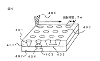
このことを利用して、電気抵抗の測定が可能であることを見出した例について述べる。用いた試料は、例えば、図4に示すようなシリコン(Si)407上にSiO2膜402が形成されたウエハに、タングステン(W)プラグ400(直径0.25μm)を埋め込んだコンタクトホール401の加工を行ったものである。各プラグは、プラグ底部のSiO2(402)の残膜403に起因する電気抵抗を持っており、残膜があるものは高抵抗なオープン欠陥404、プラグ同士がつながっているものは低抵抗なショート欠陥405である。
【0017】
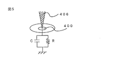
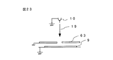
電子輸送経路の電気抵抗R、電気容量Cを含む等価回路としては、図5を仮定した。電気容量Cはプラグの形状から10-17F(ファラッド)とした。電子ビーム照射条件は、図23を仮定した。電子銃10から放出される電子ビーム19の初期エネルギーは10keV、リターディング電極63は接地、被検査基板9(ウエハ)にはリターディング電圧−9.5kVを印加してある。そのため、電子ビームのウエハへの入射エネルギーEPEは、500eVである。
【0018】
その結果、検出される二次電子および後方散乱電子の数の和NSEの経時変化まは、図6のようになることを見出した。縦軸は検出されるNSEを電子ビーム電流IP[A]で割ったもの、横軸は電子ビームの照射時間Te(もしくは走査電子ビームがプラグを横切る時間)とIPの積である。さらに、十分な時間が経過し、NSEが安定した状態での、NSEと電気抵抗Rの関係は図7のようになることを見出した。ただし、縦軸はNSEをIPでわったもの、横軸はRとIPの積である。電気抵抗Rを算出するためにはNSEに変化が必要である。
【0019】
このようなことから、このグラフから電気抵抗Rを算出するための電子ビームの電流IPの条件は、0<log(R・IP)<3であることを、本発明者らは、初めて見出した。図7のグラフは物質および試料形状に依存するが、変化分は僅かである。また、リターディング電圧VRを変化させたり、検査前に電子ビーム照射を行いプラグ周りを帯電(電圧VB)させることにより、図8、9のようにNSEを変えることができるため、高感度な検査条件が存在する。この計算予測を確認するために、各プラグに電子ビームを照射させ、各プラグからの二次電子および後方散乱電子の数の検出を行った。
【0020】
その後、各プラグの電気抵抗をナノプローバにより測定した。プラグ最上層はWであるため、ナノプローバのW針との接触抵抗は十分小さく出来る。その結果得られたNSEの電気抵抗R依存性を、図10に示した。ただし、縦軸はNSEとビーム電流IPの商であり、横軸は電気抵抗Rとビーム電流IPの積である。図中、実線は計算値、○印は実験値である。このように計算値と実験値は一致することを見出すことができた。すなわち、このような二次電子および後方散乱電子の数の和の抵抗依存性計算によって、電位コントラスト像から抵抗値を算出可能であることを見出した。
【0021】
次に、この半導体ウエハを用いて、電気容量の測定も可能であることを見出したので、そのことについて述べる。ビーム電流IPは100nAとした。用いたプラグは、上記ナノプローブ法により確認した抵抗1012Ω(オーム)以上のものを用いた。電子ビームの照射時間(もしくは電子ビームがプラグを横切る時間)を変化させることによって得られる検出されるNSEの変化を計算結果と比較し、比較結果により電気容量を見積もる。実験結果と計算結果を図11に示すように、計算結果において電気容量を10-17F(ファラッド)としたときのNSEの経時変化は、実験結果をほぼ忠実に再現した。この値はプラグ形状等から算出した値とほぼ等しい。
【0022】
さらに、ウエハの構造に対応した適切な検査条件を提供する方法について述べる。図8では、リターディング電圧VRを変化させることによってNSEが変わることを示した。このとき、電子ビーム照射域の帯電電圧US(V)も、図24のように変化する。ここでは、検査のためにこの現象を利用した。例えば、pn接合を持つ回路パターンに電子ビームを照射してpn接合が逆バイアス状態に帯電した場合、接合は高抵抗になるため、導通不良欠陥と判別が困難である。しかし、リターディング電圧を変化させることにより帯電電圧を増加させ、pn接合にブレークダウンを生じさせることによって抵抗値を減少可能である。この状態で検査を行えば、正常部と欠陥部の抵抗値の差が大きくなり、それら電位コントラスト像に差が生じ、検査が可能になる。
【0023】
また、pn接合自身に欠陥があり逆バイアス方向の抵抗値が小さい場合、リターディング電圧を調整し、欠陥部のpn接合のみをブレークダウンさせる帯電状態にする。このとき、正常部と欠陥部の抵抗値の差は増大され、それら電位コントラスト像に差が生じ、検査可能となる。ここでは、リターディング電圧を利用したが、それ以外の電子照射条件、例えば電子ビーム電流、電子ビームの試料への入射エネルギー、照射時間や照射回数を変化させることによっても、同様な効果を得ることができる。
【0024】
以上のような手法により、電気抵抗および電気容量を見積もることが可能であることや、ウエハの構造に対応した適切な欠陥検出条件が提供できることを見出した。ここでは、電子ビームを用いたが、それ以外でもプラスイオン、マイナスイオン等のような荷電粒子ビームであれば同様のことは可能である。
【0025】
実際の検査装置では、上記のような電気抵抗や電気容量の決定を次のように自動的に行う。
【0026】
まず、半導体ウエハに荷電粒子ビームを一回もしくは数回走査させる。このとき、各走査時間を変化させてもよい。そして、発生した二次電子や後方散乱電子を検出器に取りこみ、取りこんだ電子数に比例した信号を発生させ、その信号をもとに各走査に対応した検査画像を形成させる。
【0027】
次に、ワークステーションやパーソナルコンピュータ等の計算機で、荷電粒子ビームの電流値、照射エネルギー、走査時間、および走査回数、半導体ウエハ表面での電場、および二次電子や後方散乱電子の放出効率等を考慮し、電気抵抗や電気容量をパラメータとして画像を形成させる。そして、その画像が検査画像と一致するように電気抵抗や電気容量を決定する。
【0028】
なお、この検査で、荷電粒子ビームの電流値を変化させることによって、電気抵抗の測定範囲を変化させることが可能である。さらに、画像取得の前の処理として、荷電粒子ビームをあらかじめ一定量照射させておくことや、半導体ウエハにリターディング電圧を加えることによって、測定感度を変えることができる。 また、ウエハの構造に対応した適切な検査条件も提供することができる。
【0029】
【発明の実施の形態】
(実施例1)
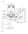
本発明の第1の実施例になる検査装置の構成を図1に示す。検査装置1は、室内が真空排気される検査室2と、検査室2内に被検査基板9を搬送するための予備室(本実施例では図示せず)を備えており、この予備室は検査室2とは独立して真空排気できるように構成されている。また、検査装置1は上記検査室2と予備室の他に制御部6、画像処理部5から構成されている。検査室2内は大別して、電子光学系3、検出部7、試料室8、光学顕微鏡部4から構成されている。
【0030】
電子光学系3は、電子銃10、電子ビーム引き出し電極11、コンデンサレンズ12、ブランキング用偏向器13、走査偏向器15、絞り14、対物レンズ16、反射板17、E×B偏向器18から構成されている。検出部7のうち、検出器20が検査室2内の対物レンズ16の上方に配置されている。検出器20の出力信号は、検査室2の外に設置されたプリアンプ21で増幅され、AD変換機22によりデジタルデータとなる。試料室8は、試料台30、Xステージ31、Yステージ32、回転ステージ33、位置モニタ用測長器34、被検査基板高さ測定器35から構成されている。
【0031】
光学顕微鏡部4は、検査室2の室内における電子光学系3の近傍であって、互いに影響を及ぼさない程度離れた位置に設備されており、電子光学系3と光学顕微鏡部4の間の距離は既知である。そして、Xステージ31またはYステージ32が電子光学系3と光学顕微鏡部4の間の既知の距離を往復移動するようになっている。光学顕微鏡部4は光源40、光学レンズ41、CCDカメラ42により構成されている。画像処理部5は、画像記憶部46、計算機48より構成されている。取り込まれた電子ビーム画像あるいは光学画像、測定した電気抵抗および電気容量はモニタ50に表示される。
【0032】
装置各部の動作命令および動作条件は、制御部6から入出力される。制御部6には、あらかじめ電子ビーム発生時の加速電圧、電子ビーム偏向幅、偏向速度、検出装置の信号取り込みタイミング、試料台移動速度等々の条件が、目的に応じて任意にあるいは選択して設定できるよう入力されている。制御部6は、補正制御回路43を用いて、位置モニタ用測長器34、被検査基板高さ測定器35の信号から位置や高さのずれをモニタし、その結果より補正信号を生成し、電子ビームが常に正しい位置に照射されるよう対物レンズ電源45や走査偏向器44に補正信号を送る。被検査基板9の画像を取得するためには、細く絞った電子ビーム19を該被検査基板9に照射し、二次電子および後方散乱電子51を発生させ、これらを電子ビーム19の走査およびステージ31、32の移動と同期して検出することで被検査基板9表面の画像を得る。
【0033】
電子銃10には拡散補給型の熱電界放出電子源が使用されている。この電子銃10を用いることにより、従来の、例えばタングステン(W)フィラメント電子源や、冷電界放出型電子源に比べて安定した電子ビーム電流を確保することができるため、明るさ変動の少ない電位コントラスト像が得られる。電子ビーム19は、電子銃10と引き出し電極11との間に電圧を印加することで電子銃10から引き出される。電子ビーム19の加速は、電子銃10に高電圧の負の電位を印加することでなされる。
【0034】
これにより、電子ビーム19は、その電位に相当するエネルギーで試料台30の方向に進み、コンデンサレンズ12で収束され、さらに対物レンズ16により細く絞られて試料台30上のX−Yステージ31、32の上に搭載された被検査基板9(半導体ウエハ、チップあるいは液晶、マスク等微細回路パターンを有する基板)に照射される。
なお、ブランキング用偏向器13には、走査信号およびブランキング信号を発生する信号発生器44が接続され、コンデンサレンズ12および対物レンズ16には、各々レンズ電源45が接続されている。被検査基板9には、高圧電源36により負の電圧(リターディング電圧)を印加できるようになっている。この高圧電源36の電圧を調節することにより一次電子ビームを減速し、電子銃10の電位を変えずに被検査基板9への電子ビーム照射エネルギーを最適な値に調節することができる。
【0035】
被検査基板9上に電子ビーム19を照射することによって発生した二次電子および後方散乱電子51は、基板9に印加された負の電圧により加速される。被検査基板9上方に、E×B偏向器18が配置され、これにより加速された二次電子および後方散乱電子51は所定の方向へ偏向される。E×B偏向器18にかける電圧と磁界の強度により、偏向量を調整することができる。また、この電磁界は、試料に印加した負の電圧に連動させて可変させることができる。E×B偏向器18により偏向された二次電子および後方散乱電子51は、所定の条件で反射板17に衝突する。この反射板17に加速された二次電子および後方散乱電子51が衝突すると、反射板17からは第二の二次電子および後方散乱電子52が発生する。
【0036】
検出部7は、真空排気された検査室2内の検出器20、検査室2の外のプリアンプ21、AD変換器22、光変換手段23、伝送手段24、電気変換手段25、高圧電源26、プリアンプ駆動電源27、AD変換器駆動電源28、逆バイアス電源29から構成されている。既に記述したように、検出部7のうち、検出器20が検査室2内の対物レンズ16の上方に配置されている。検出器20、プリアンプ21、AD変換器22、光変換器23、プリアンプ駆動電源27、AD変換器駆動電源28は、高圧電源26により正の電位にフローティングしている。
【0037】
上記反射板17に衝突して発生した第二の二次電子および後方散乱電子52は、この吸引電界により検出器20に導かれる。
検出器20は、電子ビーム19が被検査基板9に照射されている間に発生した二次電子および後方散乱電子51がその後加速されて反射板17に衝突して発生した第二の二次電子および後方散乱電子52を、電子ビーム19の走査のタイミングと連動して検出するように構成されている。検出器20の出力信号は、検査室2の外に設置されたプリアンプ21で増幅され、AD変換器22によりデジタルデータとなる。AD変換器22は、検出器20が検出したアナログ信号をプリアンプ21によって増幅された後に直ちにデジタル信号に変換して、画像処理部5に伝送するように構成されている。検出したアナログ信号を検出直後にデジタル化してから伝送するので、高速で且つSN比の高い信号を得ることができる。
【0038】
なお、ここでの検出器20として、例えば、半導体検出器を用いてもよい。
【0039】
X−Yステージ31、32上には被検査基板9が搭載されており、検査実行時にはX−Yステージ31、32を静止させて電子ビーム19を二次元に走査する方法と、検査実行時にX−Yステージ31、32をY方向に連続して一定速度で移動されるようにして電子ビーム19をX方向に直線に走査する方法のいずれかを選択できる。ある特定の比較的小さい領域を検査する場合には前者のステージを静止させて検査する方法、比較的広い領域を検査するときは、ステージを連続的に一定速度で移動して検査する方法が有効である。なお、電子ビーム19をブランキングする必要がある時には、ブランキング用偏向器13により電子ビーム19が偏向されて、電子ビームが絞り14を通過しないように制御できる。
【0040】
位置モニタ用測長器34として、本実施例ではレーザ干渉による測長計を用いた。Xステージ31およびYステージ32の位置が実時間でモニタでき、制御部6に転送されるようになっている。また、Xステージ31、Yステージ32、そして回転ステージ33のモータの回転数等のデータも同様に各々のドライバから制御部6に転送されるように構成されており、制御部6はこれらのデータに基いて電子ビーム19が照射されている領域や位置が正確に把握できるようになっており、必要に応じて実時間で電子ビーム19の照射位置の位置ずれを補正制御回路43より補正するようになっている。また、被検査基板毎に、電子ビームを照射した領域を記憶できるようになっている。
【0041】
光学式高さ測定器35は、電子ビーム以外の測定方式である光学式測定器、例えばレーザ干渉測定器や反射光の位置で変化を測定する反射光式測定器が使用されており、X−Yステージ上31、32に搭載された被検査基板9の高さを実時間で測定するように構成されている。本実施例では、スリットを通過した細長い白色光を透明な窓越しに被検査基板9に照射し、反射光の位置を位置検出モニタにて検出し、位置の変動から高さの変化量を算出する方式を用いた。この光学式高さ測定器35の測定データに基いて、電子ビーム19を細く絞るための対物レンズ16の焦点距離がダイナミックに補正され、常に非検査領域に焦点が合った電子ビーム19を照射できるようになっている。また、被検査基板9の反りや高さ歪みを電子ビーム照射前に予め測定しており、そのデータをもとに対物レンズ16の検査領域毎の補正条件を設定するように構成することも可能である。
【0042】
画像処理部5は、画像記憶部46、計算機48、モニタ50により構成されている。上記検出器20で検出された被検査基板9の画像信号は、プリアンプ21で増幅され、AD変換器22でデジタル化された後に光変換器23で光信号に変換され、光ファイバ24によって伝送され、電気変換器25にて再び電気信号に変換された後に画像記憶部46に記憶される。
【0043】
画像形成における電子ビームの照射条件および、検出系の各種検出条件は、あらかじめ検査条件設定時に設定され、ファイル化されてデータベースに登録されている。計算機48は、画像記憶部46に入力された画像信号を読み出し、電子ビーム照射条件をもとに、すでに述べた画像信号レベルと表面帯電電圧の対応を計算し、画像信号レベルに対応する電気抵抗および電気容量を計算する。モニタ50は、計算機48で求めた電気抵抗および電気容量、画像記憶部46に記憶された画像のいずれか、もしくはこれらを同時に表示する。
【0044】
次に、検査装置1により被検査基板9として、例えば、図12に示す部分的に完成した300mmφの半導体ウエハ(断面構造は図4と同様)について電気抵抗および電気容量を測定した場合の作用について説明する。まず、図1には記載されていないが、被検査基板の搬送手段により半導体ウエハは試料交換室へロードされる。そこでこの被検査半導体ウエハ9は試料ホルダに搭載され、保持固定された後に真空排気され、試料交換室がある程度の真空度に達したら検査のための検査室2に移載される。検査室2では、試料台30、X−Yステージ31、32、回転ステージ33の上に試料ホルダごと載せられ、保持固定される。
【0045】
セットされた被検査半導体ウエハ9は、予め登録された所定の検査条件に基きX−Yステージ31、32のXおよびY方向の移動により光学顕微鏡部4の下の所定の第一の座標に配置され、モニタ50により被検査半導体ウエハ9上に形成された回路パターンの光学顕微鏡画像が観察され、位置回転補正用に予め記憶された同じ位置の同等の回路パターン画像と比較され、第一の座標の位置補正値が算出される。次に第一の座標から一定距離離れ第一の座標と同等の回路パターンが存在する第二の座標に移動し、同様に光学顕微鏡画像が観察され、位置回転補正用に記憶された回路パターン画像と比較され、第二の座標の位置補正値および第一の座標に対する回転ずれ量が算出される。この算出された回転ずれ量分、回転ステージ33は回転し、その回転量を補正する。
なお、本実施例では回転ステージ33の回転により回転ずれ量を補正しているが、回転ステージ33なしで、算出された回転ずれの量に基き電子ビームの走査偏向位置を補正する方法でも補正できる。この光学顕微鏡画像観察においては、光学顕微鏡画像のみならず電子ビーム画像でも観察可能な回路パターンが選定される。また、今後の位置補正のために、第一の座標、光学顕微鏡画像観察による第一の回路パターンの位置ずれ量、第二の座標、光学顕微鏡画像観察による第二の回路パターンの位置ずれ量が記憶され、制御部6に転送される。
【0046】
さらに、光学顕微鏡による画像が用いられて、被検査半導体ウエハ9上に形成された回路パターンが観察され、被検査半導体ウエハ9上の回路パターンのチップの位置やチップ間の距離、あるいはメモリセルのような繰り返しパターンの繰り返しピッチ等が予め測定され、制御部6に測定値が入力される。また、被検査半導体ウエハ9上における被検査チップおよびチップ内の被検査領域が光学顕微鏡の画像から設定され、上記と同様に制御部6に入力される。光学顕微鏡の画像は、比較的低い倍率によって観察が可能であり、また、被検査半導体ウエハ9の表面が、例えばシリコン酸化膜等により覆われている場合には下地まで透過して観察可能であるので、チップの配列やチップ内の回路パターンのレイアウトを簡便に観察することができ、検査領域の設定を容易にできるためである。
【0047】
以上のようにして、光学顕微鏡部4による所定の補正作業や検査領域設定等の準備作業が完了すると、Xステージ31およびYステージ32の移動により、被検査半導体ウエハ9が電子光学系3の下に移動される。被検査半導体ウエハ9が電子光学系3の下に配置されると、上記光学顕微鏡部4により実施された補正作業や検査領域の設定と同様の作業を電子ビーム画像により実施する。この際の電子ビーム画像の取得は、次の方法でなされる。
上記光学顕微鏡画像による位置合せにおいて記憶され補正された座標値に基き、光学顕微鏡部4で観察されたものと同じ回路パターンに、電子ビーム19が走査偏向器44によりXY方向に二次元に走査されて照射される。この電子ビームの二次元走査により、被観察部位から発生する二次電子および後方散乱電子51が、上述した電子検出のための各部の構成および作用によって検出されることにより、電子ビーム画像が取得される。既に光学顕微鏡画像により簡便な検査位置確認や位置合せ、および位置調整が実施され、且つ回転補正も予め実施されているため、光学画像に比べ分解能が高く高倍率で高精度に位置合せや位置補正、回転補正を実施することができる。
【0048】
なお、電子ビーム19を被検査半導体ウエハ9に照射すると、その箇所が帯電する。この帯電は、時々像を歪ませるため、検査にとって悪い影響を与える。検査の際にその帯電の影響を避けるために、上記位置回転補正あるいは検査領域設定等の検査前準備作業において、電子ビーム19を照射する回路パターンは予め被検査領域外に存在する回路パターンを選択するか、あるいは被検査チップ以外のチップにおける同等の回路パターンを制御部6から自動的に選択できるようにしておく。これにより、検査時に上記検査前準備作業により電子ビーム19を照射した影響が検査画像に及ぶことはない。
【0049】
次に、被検査ウエハ上の任意の領域について電気抵抗および電気容量の測定が実施される。入射エネルギー500eV、電子ビーム電流100nAで画像を取得した。そのときに得られた画像で、図12に示すような直線に並ぶプラグ400のAB間の二次電子および後方散乱電子の信号を、図13に示す。この図の各グラフは、プラグにビームが照射された時間Teを0.15〜40μsまで変えたものである。電子照射時間Teと共に、いくつかのピークは減少しているが、十分大きなTe(今回の場合は2.5μs以上)においては安定した信号が得られた。
【0050】
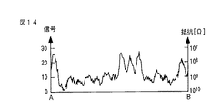
このような状態において、図7で述べたような計算結果により電気抵抗を求めたところ、図14のような107〜1010Ω(オーム)範囲の値(右縦軸)が得られた。次に、図13に示す矢印のピークの電子照射時間Te変化から電気容量を求めた。図15に示すピークの強度とTeの関係において、計算により電気容量を求めたところ10-17F(ファラッド)という値が得られた。以降、この動作が繰り返されることにより、すべての選択された検査領域について電気抵抗および電気容量の測定が実行されていく。
【0051】
また、この装置において、リターディング電圧を変化させたり、画像取得前にあらかじめ電子照射を行い絶縁部をある程度帯電させることにより、プラグ正面に形成されるポテンシャルを変化させ、図8、9のように抵抗測定の感度を変更することが可能である。図8によると、リターディング電圧を下げることによって測定範囲を狭めることができる。図9によると、絶縁部の帯電によって測定範囲を狭めることができる。なお、ここでは荷電粒子ビームとして電子ビームを用いたが、その代わりにイオンビームを用いても同様の測定が可能である。
【0052】
(実施例2)
第2の実施例では、第1の実施例で説明した検査装置を用いて電子ビーム画像を取得し、画像記憶部46に画像を記憶し、隣接する同等の回路パターンの画像同士を計算機48で比較して、差信号レベルが所定の値より大きかった場合に上記電子ビーム画像のある箇所を欠陥部として認識し、その欠陥位置を表示する。
【0053】
装置の詳細な説明は第1の実施例で述べているので省略する。この欠陥位置の画像信号レベルより計算機48で電気抵抗および容量を計算し、欠陥部が正常部に対してどの程度抵抗あるいは容量が異なるかを求める。該欠陥部の画像取得方法として、3つの方法がある。
【0054】
第一の方法は、上記画像を比較する検査を実施し、終了後に再度欠陥発生座標へ移動し、検査と同じ条件で画像を取得し、該画像より計算する方法である。
【0055】
第二の方法は、第一の方法と同様に検査実施後に再度欠陥発生座標に移動し、検査とは異なる電子ビーム条件で画像を取得し、この画像取得条件との画像信号レベルの対応により欠陥部の電気抵抗や容量を計算する方法である。この第二の方法においては、検査と異なる画像取得条件として、ビーム電流や走査速度、照射回数を変える等があり、そのなかの一つの方法で画像を取得し計算しても、複数の条件で画像を取得して計算してもよい。
【0056】
第三の方法は、検査実施中に欠陥と判定された画像は自動的に保存し、欠陥部の画像信号を読み出して計算する方法である。この場合には、検査終了後に再度欠陥部に移動して画像を再取得する必要はない。
【0057】
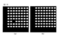
このようにして、第2の実施例で検査を実施し、欠陥部の電気的不良レベルを表示する検査方法の例を示す。図16は、例として、300mmφの半導体ウエハの検査実施後の欠陥部を含む回路パターン(a)と、それに隣接する同等の回路パターン(b)を示す。この中の任意の欠陥を画面から指定すると、図17に示すように、欠陥部の画像が表示され、同時に欠陥部の電気抵抗Rと電気容量C(図中、▲1▼1010Ω、10-16Fの欠陥部と、▲2▼108Ω、10-17Fの欠陥部)が表示される。リファレンスとして、正常部の電気抵抗(107Ω)と電気容量(10-17F)も同様に表示させることができる。
【0058】
また、各電気抵抗Rおよび電気容量Cの値から、各欠陥部の種類が判定できる。例えば、上記▲1▼のような欠陥部は、リファレンスと比較して、抵抗Rの増加、容量Cの増加からして残膜による完全非導通であり、上記▲2▼のような欠陥部は、容量Cは変化していないにもかかわらず、抵抗Rが増加していることから、コンタクトホールの埋め込んだプラグ材質の不良と判定できる。
【0059】
(実施例3)
第3の実施例は、第1の実施例で述べた検査装置を用いて、電子ビーム画像を取得しながら同時に回路パターン位置の画像明るさを所定の明るさと比較することにより、画像明るさより求まる所定の電気抵抗と異なる抵抗の回路パターンを欠陥として検出する検査方法および検査装置である。
【0060】
まず、図18において、被検査ウエハ181を検査装置内にロードし、あらかじめ登録してある電子ビーム照射条件および信号検出条件を設定する。試料台180には、所定の電子ビーム条件における抵抗があらかじめわかっている標準サンプル182が貼り付けてある。検査を開始する前にこの標準サンプルの画像を取得し、抵抗および容量と電子ビーム画像信号レベルの対応を校正する。
【0061】
また、被検査ウエハ181の抵抗あるいは容量の設計値に基づき、所定の許容レベル範囲を決めて設定しておく。その後に、被検査ウエハ181の任意の領域について電子ビーム画像を取得し、検査時に取得した画像明るさ信号と設定された許容レベルとを比較して、許容レベルからはずれた回路パターンを欠陥として認識し検出する。
【0062】
(実施例4)
第4の実施例は、第1の実施例で述べた装置を用い、第3の実施例で述べた方法により電子ビーム画像明るさと電気抵抗および容量の対応を校正し、設定された任意の領域について電子ビーム画像を取得し、抵抗のレベルに応じて色を変えたり、等高線で表示する検査方法および検査装置である。図19に検査結果の例を示す。抵抗レベルに応じて色を変えたり等高線表示をすることにより、抵抗絶対レベルの分布が求まる。
【0063】
(実施例5)
第5の実施例では、実施例1に記述した装置を用いて、電子ビームを走査させない条件で所定の回路パターン部に照射し、該回路パターンの信号レベルの変化により電気抵抗および電気容量を測定した例を示す。ここでは、被検査基板9の任意の場所から放出され、検出される二次電子および後方散乱電子の数の和NSEの経時変化を測定することにより、電気抵抗および電気容量を見積もる。そのために、まず前工程として細く絞った電子ビーム19を、その場所近傍に走査させることにより、その電位コントラスト像を得る。
【0064】
その後、その画像から、任意の場所に電子ビームを照射させるための偏向条件を設定し、その条件で電子ビームを照射させ検出される二次電子および後方散乱電子の数の和NSEの経時変化を得る。そのときに得られた結果が、図20である。この値は、電気抵抗108Ω、電気容量10-17Fとした計算結果とよく合う。このように、電気抵抗、および電気容量を決定することができた。
【0065】
(実施例6)
第6の実施例は、実施例1で述べた装置を用い、例えば、半導体ウエハ212のレジスト213の残膜214を検査する方法および装置である。第3の実施例で述べた方法により電子ビーム画像明るさと電気抵抗および容量の対応を校正し、設定された任意の領域について電子ビーム画像を取得し、残膜を抵抗のレベルに置き換えて表示する検査方法および検査装置である。図21に、検査結果の一例として、検査画像210、および検査画像210のA−B間の予測されるウエハ断面構造211を示す。残膜214の程度に応じて色を変えたり等高線表示をすることが可能である。
【0066】
(実施例7)
第7の実施例は、実施例1で述べた装置を用い、回路が形成された300mmφの半導体ウエハの反完成品を全面検査する方法、装置である。第2の実施例で述べた方法により数時間でウエハ全面検査した結果を、図22に示す。ここでは、108Ω以上の抵抗をもつ部位を欠陥220として表示してある。
【0067】
(実施例8)
回路パターンの欠陥検査で、このパターンの電気抵抗値が電圧依存性を持った場合(例えば、pn接合部や、ショットキー接合部を持つ場合)の、適切な検査条件を提供した例を示す。
【0068】
図25は、pn接合(501はp型Si、502はn型Si)をもつコンタクトホールの例を示す断面図である。このようなコンタクトホールの導通不良検査を、入射エネルギー500eVの電子ビーム(IP=100nA)を用いて行った。リターディング電圧0Vのときの電位コントラスト像を図26に示す。コンタクトホールの電位コントラストが小さく、欠陥部との判別が不能であった。
【0069】
この原因を解明するために、正常なコンタクトホール、残膜をもつコンタクトホールの抵抗値の電圧依存性を測定した。結果を図27の左のグラフに示す。正常なコンタクトホールが503、残膜をもつコンタクトホールが504である。正常な503のグラフは、順バイアス(電圧は負)では抵抗値は小さく、逆バイアス(電圧は正)では抵抗値が大きい。しかし、逆バイアスでも4V以上印加すればブレークダウンが起こり抵抗値は減少することがわかった。この場合、残膜をもつコンタクトホールの抵抗値504は高抵抗であるため、正常部と欠陥部の抵抗値の差は十分大きくなる。
【0070】
一方、実施例1では、リターディング電圧(もしくはウエハ近傍の電場)を変化させた場合、図8のようにNSEの抵抗依存性が変化し、抵抗値測定感度を変えることが可能であることを示した。このとき、同時に電子ビーム照射域の帯電電圧も変化している。リターディング電圧VRを変化させた場合について、帯電電圧の抵抗依存性を図24に示す。入射エネルギーは500eVであり、全電子放出効率は1より大きく、正に帯電する。リターディング電圧VRの減少(0→−28.5kV)によって、帯電電圧が増加していることがわかる。リターディング電圧が0Vの場合の帯電電圧は、最大でも3V程度である。すなわち、正常なコンタクトホールも抵抗値が十分大きいためリーク電流は発生せず、その程度の電圧まで帯電し、抵抗値が1018Ω程度になる。このとき、図27の右のグラフからわかるように放出電子数は少なく、電位コントラスト像は暗くなり、導通不良部と判別不可能となる。そのため、図26のような電位コントラスト像が得られたものであると考えられる。
【0071】
検査を可能にするためには、pn接合にブレークダウンを起こさせるほどの帯電電圧を生じさせることによって、pn接合の抵抗値を減少させる方法を用いた。リターディング電圧を−28.5kVにすることによって最大帯電電圧が5V以上になるため(図24)、pn接合にブレークダウンが起こり、抵抗値は十分減少(108Ω以下)することが予測される。そのため、正常部と導通不良部の抵抗値に差が生ずる。また、図27の右のグラフから、それらの電位コントラスト像(NSE)に十分な差がつき、検査が可能になると思われる。そこで、実際に、リターディング電圧を−28.5kVにして、電位コントラスト像を取得した(図28)。その結果、正常部と欠陥部の電位コントラスト像に十分な差が生じ、実施例2のように、欠陥部を「欠陥」と表示することができた。このように、帯電電圧の抵抗値依存性と回路の電気特性から電子ビーム照射条件を決定することができた。
【0072】
また、pn接合に欠陥を持つもの(残膜無し)の検査も可能である。例えば、pn接合のn拡散層の濃度が十分でないという欠陥を持つため、低い帯電電圧でブレークダウンが生じ、抵抗値が小さいというものについての検査例を示す。その抵抗値は、図27の505のように、正常なものと比較して小さい。この場合、リターディング電圧0Vで、検査可能である。正常なpn接合ではブレークダウンが起こらないため抵抗値が十分大きく、電位コントラスト像は暗い。一方、欠陥のあるpn接合はブレークダウンが起こるため抵抗が小さく、電位コントラスト像は明るくなる。電位コントラスト像は図29のようになった。このように検査条件を決定することができた。
【0073】
以上の場合、リターディング電圧を変化させたが、これはウエハ近傍の電場を変化させることが目的である。そのため、リターディング電圧を変化させる代わりに、対向する電極もしくは周辺の電極の位置を変位させ、ウエハ近傍の電場を変化させても同等の効果が得られる。
【0074】
また、ここでは、リターディング電圧をパラメータにした場合について考察したが、他の電子ビーム照射条件(ビーム電流、ビームエネルギー、照射時間等)に関しても同様に、試料の特性に依存した最適な条件が存在する。
【0075】
以上詳述した、これらの実施例では、荷電粒子ビームとして電子ビームを用いたが、その代わりにイオンビームを用いても同様の測定が可能である。
【0076】
【発明の効果】
以上のように、本発明の検査方法および装置によれば、回路パターンを有する半導体装置、等の部分的に完成した基板の検査において、従来の検査装置では不可能であった、微小部分の電気抵抗および電気容量の測定に実現することができた。本発明を基板製造プロセスへ適用することにより、電気抵抗や電気容量を見積もることができるため、基板製造プロセスにいち早く異常対策処理を講ずることができ、その結果、半導体装置その他の基板の不良率を低減し生産性を高めることができる。
【0077】
また、上記測定により、異常発生をいち早く検知することができるので、多量の不良発生を未然に防止することができ、さらにその結果、不良の発生そのものを低減させることができるので、半導体装置等の信頼性を高めることができ、新製品等の開発効率が向上し、且つ製造コストが削減できる。
【図面の簡単な説明】
【図1】本発明の検査装置の一実施例を示す構成図。
【図2】電子ビーム照射にともなうウエハ表面の帯電およびポテンシャル障壁の形成の様子を示す図。
【図3】二次電子および後方散乱電子の数の和NSEのエネルギーESE分布を示す図。
【図4】半導体ウエハの断面構造を含んだ模式図
【図5】図4のコンタクトホールの等価回路の例を示す図。
【図6】検出される二次電子および後方散乱電子の数の和NSEの経時変化を示す図。
【図7】検出される二次電子および後方散乱電子の数の和NSEの電気抵抗R依存性を示す図。
【図8】リターディング電圧VRを変えた場合の検出される二次電子および後方散乱電子の数の和NSEの電気抵抗R依存性を示す図。
【図9】プラグ周りの帯電電圧VBを変えた場合の検出される二次電子および後方散乱電子の数の和NSEの電気抵抗R依存性を示す図。
【図10】 検出される二次電子および後方散乱電子の数の和NSEの電気抵抗R依存性の計算値と実験結果の比較を示す図。
【図11】検出される二次電子および後方散乱電子の数の和NSEの経時変化の計算値と実験値の比較を示す図。
【図12】実施例1で用いた半導体ウエハと電位コントラスト像を示す図。
【図13】図12の半導体ウエハの電位コントラスト信号を示す図。
【図14】図13の電位コントラスト信号と計算により算出された電気抵抗値を示す図。
【図15】図13における矢印のピーク強度のビーム照射時間変化を示す図。
【図16】実施例2で得られた検査画像1を示す図。
【図17】実施例2で得られた検査画像2を示す図。
【図18】実施例3で用いる抵抗標準サンプル付き試料台を示す図。
【図19】実施例4で得られた検査画像を示す図。
【図20】実施例5で得られた検出される二次電子および後方散乱電子の数の和の経時変化の検査結果および計算値を示す図。
【図21】実施例6で得られた検査画像を示す図。
【図22】実施例7で得られた検査画像を示す図。
【図23】電子ビーム照射条件の一例を示す図。
【図24】リターディング電圧を変化させた場合の帯電電圧の抵抗依存性を示す図。
【図25】pn接合をもつコンタクトホールを加工したウエハ断面を示す図。
【図26】リターディング電圧が適切でない場合の電位コントラスト像を示す図。
【図27】pn接合の抵抗値電圧依存性を示す図。
【図28】リターディング電圧が適切である場合の電位コントラスト像を示す図。
【図29】リターディング電圧が適切である場合の電位コントラスト像を示す図。
【符号の説明】
1:回路パターン検査装置、2:検査室、3:電子光学系、4:光学顕微鏡部、5:画像処理部、6:制御部、7:検出部、8:試料室、9:被検査基板、10:電子銃、11:引き出し電極、12:コンデンサレンズ、13:ブランキング偏向器、14:絞り、15:走査偏向器、16:対物レンズ、17:反射板、18:E×B偏向器、19:電子ビーム、20:検出器、21:プリアンプ、22:AD変換器、23:光変換器、24:光ファイバ、25:電気変換器、26:高圧電源、27:プリアンプ駆動電源、28:AD変換器駆動電源、29:逆バイアス電源、30:試料台、31:Xステージ、32:Yステージ、33:回転ステージ、34:位置モニタ測長器、35:被検査基板高さ測定器、36:リターディング電源、40:白色光源、41:光学レンズ、42:CCDカメラ、43:補正制御回路、44:走査信号発生器、45:対物レンズ電源、46:画像記憶部、48:計算機、50:モニタ、51:二次電子および後方散乱電子、52:第二の二次電子および後方散乱電子、60:絶縁体、61:電子ビーム照射領域、62:試料交換室、63:リターデイング電極、180:試料台、181:ウエハ、182:抵抗標準サンプル、210:検査画像、211:断面予想図、212:Si、213:レジスト、214:レジストの残部、220:欠陥、400:プラグ、401:コンタクトホール、402:SiO2、403:残膜、404:オープン欠陥、405:ショート欠陥、406:電子ビーム、407:Si、501:p型Si、502:n型Si、503:正常なpn接合の抵抗値の電圧依存性、504:導通不良コンタクトホールの抵抗値の電圧依存性、505:欠陥をもつpn接合の抵抗値の電圧依存性。[0001]
BACKGROUND OF THE INVENTION
The present invention relates to a method and an apparatus for manufacturing a substrate having a fine circuit pattern such as a semiconductor device or a liquid crystal, and more particularly to a pattern inspection technique for a semiconductor device or a photomask, on an unfinished semiconductor wafer in the process of manufacturing a semiconductor device. The present invention relates to an inspection method and apparatus using a charged particle beam for inspecting a defect in an arbitrary portion.
[0002]
[Prior art]
For example, a semiconductor device is manufactured by repeating a process of transferring a pattern formed with a photomask on a semiconductor wafer by lithography and etching. In the manufacturing process of a semiconductor device, lithography processing, etching processing, and other good and bad influences greatly on the yield of the semiconductor device, so it is important to detect abnormalities and defects early or in advance. In particular, it is important for improving the yield to measure the electrical resistance and capacitance of the contact holes and wirings of the partially completed semiconductor wafer in the initial stage of the manufacturing process. Conventional techniques for inspecting these electrical defects include the following.
[0003]
One is a nanoprobe device (Japanese Patent Laid-Open No. 8-160109) in which a sharpened tungsten (W) needle (tip radius of curvature: about 0.1 μm) is brought into direct contact with the measurement unit. This is to measure the electrical resistance. However, with the recent miniaturization of patterns, the size of the portion to be measured has become the same as or smaller than the W needle, making measurement very difficult. As a means corresponding to this, it is conceivable to reduce the radius of curvature of the tip of the W needle. However, in that case, the tip becomes very soft, and the tip is deformed simultaneously with the contact with the measurement unit, which is not a practical method. Another problem is contact resistance. When the needle and the measuring part are different materials, particularly when at least one of them is a semiconductor, a Schottky junction is formed, and an electric resistance depending on the voltage is generated at that part, so an accurate measurement cannot be performed.
[0004]
The other one uses a SEM (Scanning Electron Microscope). This is disclosed in JP-A-5-258703, JP-A-11-121561, and JP-A-6-326165.
[0005]
Japanese Patent Application Laid-Open No. 5-258703 compares an inspection image with an image of an adjacent pattern, determines a portion having a different potential contrast (brightness) as a defect, and detects the defect. However, since there was no means for obtaining and displaying electric characteristics (electrical resistance and electric capacity), it was impossible to determine whether the part was fatal or non-fatal.
[0006]
In JP-A-11-121561, the degree of secondary electron emission is controlled by a control electrode above the wafer, the wafer surface is charged positively or negatively, and a normal portion, low resistance is detected from the potential contrast image at that time. A defective part and a high resistance defective part are discriminated.
[0007]
The control of the emission of secondary electrons by this control electrode is disclosed in Japanese Patent Laid-Open No. 59-155941. For example, when the control electrode is adjusted to be positively charged, the potential contrast image shows that the low-resistance defect part (for example, low resistance of several hundred Ω or less) is bright and the high-resistance defect part (electric resistance: ∞) The dark part and the normal part have a resistance larger than that of the low resistance defect part, so that the image is a bright part but slightly dark. Although it is possible to determine the magnitude of resistance from the brightness of this image, the absolute value cannot be calculated. Moreover, although the electrical resistance could be calculated by measuring the leakage current, it took time for the inspection and could not be measured at high speed.
[0008]
Japanese Patent Laid-Open No. 6-326165 discloses that an induced current is generated between a substrate and the ground by charging caused by irradiating an electron beam onto a wiring on the substrate, and the change with time is measured. Thereby, the magnitude of the electric capacity can be measured, but the absolute value cannot be measured.
[0009]
Further, in such an inspection using the SEM potential contrast image, there is a case where the difference in potential contrast between the normal part and the defective part is small depending on the structure of the wafer, and it may be difficult to detect the defect. For example, when the circuit has a pn junction, this portion has a high resistance if the charging due to the electron beam irradiation is a reverse bias with respect to the pn junction. Therefore, it was difficult to distinguish from a high-resistance conduction failure defect.
[0010]
[Problems to be solved by the invention]
As described above, in the nanoprobe device, there is a problem that the measurement target may be smaller than the tip of the needle, and there is a problem of contact resistance between the needle and the sample, and therefore, an accurate electric resistance cannot be estimated depending on the sample. . In addition, the entire wafer inspection requires a tremendous amount of time and is virtually impossible. In an apparatus using an SEM, it was possible to determine the magnitude of electric resistance and electric capacity from a potential contrast image, but it was impossible to estimate an absolute value. In addition, although the resistance value can be calculated by measuring the leakage current, it takes a long time for the inspection and the high-speed inspection is impossible, and the entire surface inspection of the wafer requires a very long time. Furthermore, depending on the structure of the wafer (for example, when a pn junction is processed), it is difficult to distinguish between a normal part and a defective part from a potential contrast image.
[0011]
The purpose of the present invention is to automatically calculate the values of electric resistance and electric capacity at high speed only by acquiring a potential contrast image, or to quickly determine the distribution and tendency of electric resistance and electric capacity on the surface of a substrate such as a wafer. An object is to provide a required inspection method and inspection apparatus. It is another object of the present invention to provide appropriate inspection conditions corresponding to the wafer structure.
[0012]
[Means for Solving the Problems]
In order to solve the above problem, the charged particle beam image (potential contrast image) obtained when the sample is irradiated with the charged particle beam depends on the electrical resistance between the irradiation region and the ground, the electric capacity, and the irradiation time. I found out that I should use.
[0013]
The mechanism will be described with reference to FIG. As an irradiation particle, electrons are taken as an example, and the incident energy EPE is set to about 500 eV where the emission efficiency σ of the sum of secondary electrons and backscattered electrons is larger than 1. Usually, those having an energy of 50 eV or less are secondary electrons, and those having an energy of 50 eV or more are often backscattered electrons. When the
[0014]
Therefore, as shown in the energy ESE distribution of the sum NSE of secondary electrons and backscattered electrons in FIG. 3, secondary electrons and backscattered electrons having lower energy are not emitted, and secondary electrons and backscattered electrons are not emitted. Even if released, it is pulled back to the insulator side. The number of electrons withdrawn is N 1 And the number of emitted electrons is N 2 Is the ratio of secondary electrons and backscattered electrons that are emitted without being pulled back to σ SE Then σ SE = N 2 / (N 1 + N 2 ) And the effective emission efficiency σ eff Is σ eff = Σ SE It is expressed by xσ. The charging voltage increases with irradiation of the electron beam, and the ratio of secondary electrons and backscattered electrons that are pulled back increases. Therefore, σ eff Gradually decreases, eventually σ eff Is stabilized at a charging voltage at which.
[0015]
On the other hand, if the electrical resistance is sufficiently small with a conductor, charging can be relaxed because electrons can be supplied, and σ and σ eff Are almost equal. Therefore, the insulator appears dark and the conductor appears bright.
[0016]
An example where it has been found that the electrical resistance can be measured using this fact will be described. The used sample is, for example,
[0017]
FIG. 5 is assumed as an equivalent circuit including the electric resistance R and electric capacity C of the electron transport path. The electric capacity C is 10 from the shape of the plug. -17 F (Farad). The electron beam irradiation conditions were assumed as shown in FIG. The initial energy of the
[0018]
As a result, the inventors have found that the change over time of the sum NSE of the number of secondary electrons and backscattered electrons detected is as shown in FIG. The vertical axis represents the detected NSE divided by the electron beam current IP [A], and the horizontal axis represents the product of the electron beam irradiation time Te (or the time that the scanning electron beam crosses the plug) and IP. Furthermore, it has been found that the relationship between NSE and electrical resistance R is as shown in FIG. 7 when sufficient time has elapsed and NSE is stable. However, the vertical axis represents NSE divided by IP, and the horizontal axis represents the product of R and IP. In order to calculate the electrical resistance R, NSE needs to be changed.
[0019]
For these reasons, the present inventors have found for the first time that the condition of the current IP of the electron beam for calculating the electrical resistance R from this graph is 0 <log (R · IP) <3. . The graph of FIG. 7 depends on the substance and the sample shape, but the change is slight. The retarding voltage V R Or charge around the plug by applying an electron beam before inspection (voltage V B ), The NSE can be changed as shown in FIGS. 8 and 9, and therefore there is a highly sensitive inspection condition. In order to confirm this calculation prediction, each plug was irradiated with an electron beam, and the number of secondary electrons and backscattered electrons from each plug was detected.
[0020]
Thereafter, the electrical resistance of each plug was measured with a nanoprober. Since the uppermost layer of the plug is W, the contact resistance with the W needle of the nanoprober can be made sufficiently small. The electric resistance R dependence of NSE obtained as a result is shown in FIG. However, the vertical axis is the quotient of NSE and beam current IP, and the horizontal axis is the product of electrical resistance R and beam current IP. In the figure, the solid line is the calculated value, and the circle is the experimental value. Thus, it was found that the calculated value and the experimental value agree. That is, it was found that the resistance value can be calculated from the potential contrast image by calculating the resistance dependence of the sum of the number of secondary electrons and backscattered electrons.
[0021]
Next, since it has been found that the capacitance can be measured using this semiconductor wafer, this will be described. The beam current IP was 100 nA. The plug used was a
[0022]
Further, a method for providing appropriate inspection conditions corresponding to the wafer structure will be described. FIG. 8 shows that NSE is changed by changing the retarding voltage VR. At this time, the charging voltage US (V) in the electron beam irradiation region also changes as shown in FIG. Here, this phenomenon was used for inspection. For example, when a circuit pattern having a pn junction is irradiated with an electron beam and the pn junction is charged in a reverse bias state, the junction has a high resistance, so that it is difficult to discriminate it from a defective conduction. However, the resistance value can be decreased by increasing the charging voltage by changing the retarding voltage and causing breakdown in the pn junction. If the inspection is performed in this state, the difference in resistance value between the normal portion and the defective portion is increased, and a difference is generated between these potential contrast images, thereby enabling the inspection.
[0023]
When the pn junction itself has a defect and the resistance value in the reverse bias direction is small, the retarding voltage is adjusted so that only the defective pn junction is broken down. At this time, the difference between the resistance values of the normal portion and the defective portion is increased, and a difference is generated between these potential contrast images, thereby enabling inspection. Although the retarding voltage is used here, the same effect can be obtained by changing other electron irradiation conditions such as electron beam current, electron beam incident energy, irradiation time and number of irradiations. Can do.
[0024]
It has been found that the electric resistance and electric capacity can be estimated by the above-described method, and that an appropriate defect detection condition corresponding to the wafer structure can be provided. Although an electron beam is used here, the same can be applied to other charged particle beams such as positive ions and negative ions.
[0025]
In an actual inspection apparatus, the determination of the electric resistance and the electric capacity as described above is automatically performed as follows.
[0026]
First, the semiconductor wafer is scanned with the charged particle beam once or several times. At this time, each scanning time may be changed. Then, the generated secondary electrons and backscattered electrons are taken into the detector, a signal proportional to the number of taken electrons is generated, and an inspection image corresponding to each scan is formed based on the signal.
[0027]
Next, with a computer such as a workstation or personal computer, the current value of the charged particle beam, the irradiation energy, the scanning time and the number of scans, the electric field on the surface of the semiconductor wafer, the emission efficiency of secondary electrons and backscattered electrons, etc. Considering this, an image is formed using the electric resistance and electric capacity as parameters. Then, the electrical resistance and capacitance are determined so that the image matches the inspection image.
[0028]
In this inspection, it is possible to change the measurement range of the electric resistance by changing the current value of the charged particle beam. Furthermore, as a process before image acquisition, the measurement sensitivity can be changed by irradiating a predetermined amount of charged particle beams in advance or applying a retarding voltage to the semiconductor wafer. In addition, appropriate inspection conditions corresponding to the wafer structure can be provided.
[0029]
DETAILED DESCRIPTION OF THE INVENTION
Example 1
The configuration of an inspection apparatus according to the first embodiment of the present invention is shown in FIG. The
[0030]
The electron
[0031]
The
[0032]
Operation commands and operation conditions of each part of the apparatus are input / output from the
[0033]
The
[0034]
Thereby, the
The blanking
[0035]
Secondary electrons and backscattered electrons 51 generated by irradiating the
[0036]
The
[0037]
Second secondary electrons and backscattered electrons 52 generated by colliding with the reflecting plate 17 are guided to the
The
[0038]
As the
[0039]
A
[0040]
As the position monitor
[0041]
The optical
[0042]
The
[0043]
The irradiation conditions of the electron beam in image formation and various detection conditions of the detection system are set in advance when setting the inspection conditions, and are filed and registered in the database. The
[0044]
Next, as an inspected
[0045]
The
In this embodiment, the amount of rotational deviation is corrected by the rotation of the
[0046]
Further, an image obtained by an optical microscope is used to observe a circuit pattern formed on the
[0047]
As described above, when the predetermined correction work and the preparation work such as the inspection area setting by the
Based on the coordinate values stored and corrected in the alignment by the optical microscope image, the
[0048]
When the
[0049]
Next, electric resistance and electric capacitance are measured for an arbitrary region on the wafer to be inspected. Images were acquired with an incident energy of 500 eV and an electron beam current of 100 nA. FIG. 13 shows signals of secondary electrons and backscattered electrons between AB of the
[0050]
In such a state, when the electrical resistance is obtained from the calculation result as described in FIG. 7 -10 Ten Values in the Ω (ohm) range (right vertical axis) were obtained. Next, the capacitance was determined from the change in electron irradiation time Te at the peak of the arrow shown in FIG. In the relationship between the peak intensity and Te shown in FIG. -17 A value of F (farad) was obtained. Thereafter, by repeating this operation, measurement of electric resistance and electric capacitance is performed for all selected inspection regions.
[0051]
Further, in this apparatus, the potential formed on the front face of the plug is changed by changing the retarding voltage or by preliminarily charging the insulating part to some extent by irradiating electrons before image acquisition, as shown in FIGS. It is possible to change the sensitivity of the resistance measurement. According to FIG. 8, the measurement range can be narrowed by lowering the retarding voltage. According to FIG. 9, the measurement range can be narrowed by charging the insulating portion. Although an electron beam is used here as the charged particle beam, the same measurement can be performed by using an ion beam instead.
[0052]
(Example 2)
In the second embodiment, an electron beam image is acquired using the inspection apparatus described in the first embodiment, the image is stored in the
[0053]
Since the detailed description of the apparatus is described in the first embodiment, it will be omitted. The electrical resistance and capacitance are calculated by the
[0054]
The first method is a method in which an inspection for comparing the images is performed, the image is moved again to the defect occurrence coordinate after completion, an image is acquired under the same conditions as the inspection, and the image is calculated from the image.
[0055]
The second method, like the first method, moves to the defect occurrence coordinates again after the inspection is performed, acquires an image under an electron beam condition different from the inspection, and the image signal level corresponds to this image acquisition condition. This is a method of calculating the electrical resistance and capacitance of the part. In this second method, the image acquisition conditions different from the inspection include changing the beam current, the scanning speed, the number of irradiations, etc. Even if the image is acquired and calculated by one of the methods, there are a plurality of conditions. Images may be acquired and calculated.
[0056]
The third method is a method in which an image determined to be defective during inspection is automatically stored, and an image signal of the defective portion is read and calculated. In this case, it is not necessary to re-acquire the image by moving to the defective portion again after the inspection is completed.
[0057]
In this way, an example of an inspection method for performing an inspection in the second embodiment and displaying an electrical failure level of a defective portion will be described. FIG. 16 shows, as an example, a circuit pattern (a) including a defective portion after inspection of a 300 mmφ semiconductor wafer and an equivalent circuit pattern (b) adjacent thereto. When an arbitrary defect is designated from the screen, an image of the defective portion is displayed as shown in FIG. 17, and at the same time, the electric resistance R and electric capacity C of the defective portion ((1) 10 in the drawing) Ten Ω, 10 -16 F defect and (2) 10 8 Ω, 10 -17 F defective portion) is displayed. As a reference, the electrical resistance of the normal part (10 7 Ω) and capacitance (10 -17 F) can be displayed in the same manner.
[0058]
Further, the type of each defective portion can be determined from the values of each electric resistance R and electric capacitance C. For example, the defective portion as in (1) is completely non-conductive due to the remaining film due to the increase in resistance R and the increase in capacitance C compared to the reference, and the defective portion as in (2) is Since the resistance R increases despite the capacitance C not changing, it can be determined that the plug material embedded in the contact hole is defective.
[0059]
(Example 3)
The third embodiment is obtained from the image brightness by using the inspection apparatus described in the first embodiment and simultaneously comparing the image brightness at a circuit pattern position with a predetermined brightness while acquiring an electron beam image. An inspection method and an inspection apparatus for detecting a circuit pattern having a resistance different from a predetermined electric resistance as a defect.
[0060]
First, in FIG. 18, a wafer 181 to be inspected is loaded into the inspection apparatus, and pre-registered electron beam irradiation conditions and signal detection conditions are set. A
[0061]
Further, a predetermined allowable level range is determined and set based on the design value of the resistance or capacitance of the wafer 181 to be inspected. Thereafter, an electron beam image is acquired for an arbitrary area of the wafer 181 to be inspected, and the image brightness signal acquired at the time of inspection is compared with a set allowable level, and a circuit pattern deviating from the allowable level is recognized as a defect. And detect.
[0062]
(Example 4)
The fourth embodiment uses the apparatus described in the first embodiment, calibrates the correspondence between the electron beam image brightness, the electric resistance, and the capacity by the method described in the third embodiment, and sets an arbitrary region. An inspection method and an inspection apparatus for acquiring an electron beam image and changing a color according to a resistance level or displaying the image with contour lines. FIG. 19 shows an example of the inspection result. By changing the color according to the resistance level or displaying contour lines, the distribution of the absolute resistance level can be obtained.
[0063]
(Example 5)
In the fifth embodiment, the apparatus described in the first embodiment is used to irradiate a predetermined circuit pattern portion under the condition that the electron beam is not scanned, and the electrical resistance and capacitance are measured by the change in the signal level of the circuit pattern. An example is shown. Here, the electrical resistance and the capacitance are estimated by measuring the change over time of the sum NSE of the number of secondary electrons and backscattered electrons emitted and detected from any location of the
[0064]
After that, a deflection condition for irradiating an electron beam to an arbitrary place is set from the image, and the time variation of the sum NSE of the number of secondary electrons and backscattered electrons detected by irradiating the electron beam under the condition is changed. obtain. The result obtained at that time is shown in FIG. This value is 10 8 Ω,
[0065]
(Example 6)
The sixth embodiment is a method and apparatus for inspecting the remaining
[0066]
(Example 7)
The seventh embodiment is a method and apparatus for inspecting the entire surface of a 300 mmφ semiconductor wafer on which a circuit is formed using the apparatus described in the first embodiment. FIG. 22 shows the result of the entire wafer inspection in several hours by the method described in the second embodiment. Here, 10 8 A portion having a resistance of Ω or more is indicated as a
[0067]
(Example 8)
An example in which an appropriate inspection condition is provided in the case of a defect inspection of a circuit pattern when the electrical resistance value of the pattern has voltage dependency (for example, in the case of having a pn junction or a Schottky junction) will be described.
[0068]
FIG. 25 is a cross-sectional view showing an example of a contact hole having a pn junction (501 is p-type Si, 502 is n-type Si). Such a contact hole conduction failure inspection was performed using an electron beam with an incident energy of 500 eV (IP = 100 nA). FIG. 26 shows a potential contrast image when the retarding voltage is 0V. The potential contrast of the contact hole was small and it was impossible to distinguish it from a defective part.
[0069]
In order to elucidate the cause, the voltage dependence of the resistance value of a normal contact hole and a contact hole having a residual film was measured. The results are shown in the left graph of FIG. The number of normal contact holes is 503, and the number of contact holes having a remaining film is 504. In the
[0070]
On the other hand, in Example 1, when the retarding voltage (or the electric field in the vicinity of the wafer) is changed, the resistance dependency of NSE changes as shown in FIG. 8, and the resistance measurement sensitivity can be changed. Indicated. At this time, the charging voltage in the electron beam irradiation area also changes. FIG. 24 shows the resistance dependence of the charging voltage when the retarding voltage VR is changed. The incident energy is 500 eV, the total electron emission efficiency is greater than 1, and it is positively charged. It can be seen that the charging voltage increases as the retarding voltage VR decreases (0 → −28.5 kV). When the retarding voltage is 0V, the charging voltage is about 3V at the maximum. That is, since a normal contact hole has a sufficiently large resistance value, a leak current does not occur, and it is charged to such a voltage. 18 It becomes about Ω. At this time, as can be seen from the graph on the right side of FIG. 27, the number of emitted electrons is small, and the potential contrast image becomes dark and cannot be distinguished from a poor conduction portion. For this reason, it is considered that a potential contrast image as shown in FIG. 26 was obtained.
[0071]
In order to enable inspection, a method of reducing the resistance value of the pn junction by generating a charging voltage that causes breakdown in the pn junction was used. By setting the retarding voltage to −28.5 kV, the maximum charging voltage becomes 5 V or more (FIG. 24), so that breakdown occurs in the pn junction and the resistance value is sufficiently reduced (10 8 Ω or less). Therefore, a difference occurs in the resistance value between the normal part and the poor conduction part. Further, from the graph on the right side of FIG. 27, it is considered that there is a sufficient difference between these potential contrast images (NSE) and the inspection can be performed. Therefore, actually, the retarding voltage was set to -28.5 kV, and a potential contrast image was acquired (FIG. 28). As a result, there was a sufficient difference in the potential contrast image between the normal part and the defective part, and the defective part could be displayed as “defect” as in Example 2. Thus, the electron beam irradiation conditions could be determined from the resistance dependency of the charging voltage and the electrical characteristics of the circuit.
[0072]
In addition, it is possible to inspect a pn junction having a defect (no remaining film). For example, an inspection example will be shown for a case where breakdown occurs at a low charging voltage and the resistance value is small because of the defect that the concentration of the n diffusion layer of the pn junction is not sufficient. The resistance value is smaller than that of a normal one as indicated by
[0073]
In the above case, the retarding voltage was changed, which is intended to change the electric field near the wafer. Therefore, instead of changing the retarding voltage, the same effect can be obtained by changing the electric field in the vicinity of the wafer by displacing the position of the opposing electrode or the peripheral electrode.
[0074]
Although the case where the retarding voltage is used as a parameter is considered here, the optimum conditions depending on the characteristics of the sample are similarly applied to other electron beam irradiation conditions (beam current, beam energy, irradiation time, etc.). Exists.
[0075]
In these embodiments described in detail above, the electron beam is used as the charged particle beam, but the same measurement can be performed by using an ion beam instead.
[0076]
【The invention's effect】
As described above, according to the inspection method and apparatus of the present invention, in the inspection of a partially completed substrate such as a semiconductor device having a circuit pattern, etc. It could be realized to measure resistance and capacitance. By applying the present invention to the substrate manufacturing process, it is possible to estimate the electrical resistance and capacitance, so that it is possible to take measures against the abnormality quickly in the substrate manufacturing process. As a result, the defect rate of semiconductor devices and other substrates can be reduced. It can be reduced and productivity can be increased.
[0077]
In addition, since the occurrence of abnormality can be quickly detected by the above measurement, it is possible to prevent the occurrence of a large number of defects, and as a result, the occurrence of defects itself can be reduced. Reliability can be increased, development efficiency of new products and the like can be improved, and manufacturing costs can be reduced.
[Brief description of the drawings]
FIG. 1 is a configuration diagram showing an embodiment of an inspection apparatus according to the present invention.
FIG. 2 is a view showing a state of charging of a wafer surface and formation of a potential barrier due to electron beam irradiation.
FIG. 3 is a diagram showing an energy ESE distribution of a sum NSE of the number of secondary electrons and backscattered electrons.
FIG. 4 is a schematic diagram including a cross-sectional structure of a semiconductor wafer.
5 is a diagram showing an example of an equivalent circuit of the contact hole in FIG. 4;
FIG. 6 is a diagram showing the change over time of the sum NSE of the number of detected secondary electrons and backscattered electrons.
FIG. 7 is a graph showing the electric resistance R dependence of the sum NSE of the number of detected secondary electrons and backscattered electrons.
FIG. 8 is a diagram showing the electric resistance R dependency of the sum NSE of the number of secondary electrons and backscattered electrons detected when the retarding voltage VR is changed.
FIG. 9 is a diagram showing the electric resistance R dependence of the sum NSE of the number of secondary electrons and backscattered electrons detected when the charging voltage VB around the plug is changed.
FIG. 10 is a diagram showing a comparison between the calculated value of the electric resistance R dependency of the sum NSE of the number of detected secondary electrons and backscattered electrons and the experimental result.
FIG. 11 is a diagram showing a comparison between calculated values and experimental values of the change over time of the sum NSE of the number of secondary electrons and backscattered electrons detected.
12 shows a semiconductor wafer used in Example 1 and a potential contrast image. FIG.
13 is a diagram showing a potential contrast signal of the semiconductor wafer of FIG.
14 is a diagram showing the electric potential contrast signal calculated by the potential contrast signal of FIG. 13 and the calculation.
15 is a diagram showing a beam irradiation time change of the peak intensity of the arrow in FIG.
16 is a view showing an
FIG. 17 is a view showing an
18 is a diagram showing a sample table with a resistance standard sample used in Example 3. FIG.
FIG. 19 is a diagram showing an inspection image obtained in Example 4;
FIG. 20 is a diagram showing inspection results and calculated values of changes over time in the sum of the numbers of secondary electrons and backscattered electrons detected obtained in Example 5.
FIG. 21 is a view showing an inspection image obtained in Example 6;
22 is a view showing an inspection image obtained in Example 7. FIG.
FIG. 23 is a diagram showing an example of electron beam irradiation conditions.
FIG. 24 is a diagram showing the resistance dependence of the charging voltage when the retarding voltage is changed.
FIG. 25 is a view showing a cross section of a wafer obtained by processing a contact hole having a pn junction.
FIG. 26 is a diagram showing a potential contrast image when the retarding voltage is not appropriate.
FIG. 27 is a graph showing resistance voltage dependency of a pn junction.
FIG. 28 is a diagram showing a potential contrast image when the retarding voltage is appropriate.
FIG. 29 is a diagram showing a potential contrast image when the retarding voltage is appropriate.
[Explanation of symbols]
1: circuit pattern inspection apparatus, 2: inspection room, 3: electron optical system, 4: optical microscope unit, 5: image processing unit, 6: control unit, 7: detection unit, 8: sample chamber, 9: substrate to be inspected 10: electron gun, 11: extraction electrode, 12: condenser lens, 13: blanking deflector, 14: stop, 15: scanning deflector, 16: objective lens, 17: reflector, 18: E × B deflector , 19: electron beam, 20: detector, 21: preamplifier, 22: AD converter, 23: optical converter, 24: optical fiber, 25: electrical converter, 26: high voltage power supply, 27: preamplifier drive power supply, 28 : AD converter drive power supply, 29: Reverse bias power supply, 30: Sample stage, 31: X stage, 32: Y stage, 33: Rotary stage, 34: Position monitor length measuring device, 35: Substrate height measuring device , 36: retarding power supply, 4 : White light source, 41: Optical lens, 42: CCD camera, 43: Correction control circuit, 44: Scanning signal generator, 45: Objective lens power supply, 46: Image storage unit, 48: Computer, 50: Monitor, 51: Two Secondary electrons and backscattered electrons, 52: Second secondary electrons and backscattered electrons, 60: Insulator, 61: Electron beam irradiation region, 62: Sample exchange chamber, 63: Retarding electrode, 180: Sample stage, 181 : Wafer, 182: Resistance standard sample, 210: Inspection image, 211: Predicted cross section, 212: Si, 213: Resist, 214: Resist remainder, 220: Defect, 400: Plug, 401: Contact hole, 402: SiO2 403: residual film, 404: open defect, 405: short defect, 406: electron beam, 407: Si, 501: p-type Si, 502: n-type Si, 503: voltage dependence of resistance value of normal pn junction, 504: voltage dependence of resistance value of poor contact hole, 505: voltage dependence of resistance value of defective pn junction.
Claims (5)
回路パターンが形成された被検査基板表面の所望の領域に対して所定のビーム照射条件で荷電粒子ビームを走査する工程と、
前記荷電粒子ビームの照射により前記領域から発生した二次電子または後方散乱電子を検出する工程と、
前記検出した二次電子または後方散乱電子の情報を基に電位コントラスト像を形成する工程と、
前記荷電粒子ビームの照射条件及び前記二次電子または後方散乱電子の検出条件に基づき、前記荷電粒子ビームの照射箇所の電気抵抗または電気容量をパラメータとして前記荷電粒子ビームの走査領域の画像を形成し、当該形成された画像が前記電位コントラスト像と一致するように当該電気抵抗または電気容量を決定する工程とを備えることを特徴とする検査方法。In an inspection method using a charged particle beam,
Scanning a charged particle beam under a predetermined beam irradiation condition with respect to a desired region on the surface of a substrate to be inspected on which a circuit pattern is formed;
Detecting secondary electrons or backscattered electrons generated from the region by irradiation of the charged particle beam;
Forming a potential contrast image based on the information of the detected secondary electrons or backscattered electrons;
Based on the irradiation condition of the charged particle beam and the detection condition of the secondary electrons or the backscattered electrons, an image of the scanning area of the charged particle beam is formed using the electric resistance or capacitance of the irradiation part of the charged particle beam as a parameter. And a step of determining the electric resistance or the electric capacitance so that the formed image coincides with the potential contrast image.
前記被検査基板は回路パターンを有するウエハであって、
前記回路パターンの電気抵抗値が電圧依存性を有することを特徴とする検査方法。The inspection method according to claim 1,
The substrate to be inspected is a wafer having a circuit pattern,
An inspection method, wherein the electric resistance value of the circuit pattern has voltage dependency.
前記電気抵抗値または電気容量の値より前記被検査基板の欠陥の種類を特定する工程を備えることを特徴とする検査方法。The inspection method according to claim 1,
An inspection method comprising a step of identifying a defect type of the substrate to be inspected from the electric resistance value or the capacitance value.
前記電気抵抗または電気容量の値が等高線表示、あるいは該値に応じて色を変えて表示されることを特徴とする検査方法。The inspection method according to claim 1,
The inspection method characterized in that the value of the electric resistance or electric capacity is displayed by contour lines or by changing the color according to the value.
前記荷電粒子ビームは電子ビームであることを特徴とする検査方法。In the inspection method according to any one of claims 1 to 4,
The inspection method, wherein the charged particle beam is an electron beam.
Priority Applications (5)
| Application Number | Priority Date | Filing Date | Title |
|---|---|---|---|
| JP2000276640A JP4015352B2 (en) | 2000-02-22 | 2000-09-07 | Inspection method using charged particle beam |
| US09/785,275 US6618850B2 (en) | 2000-02-22 | 2001-02-20 | Inspection method and inspection system using charged particle beam |
| US10/419,141 US6931620B2 (en) | 2000-02-22 | 2003-04-21 | Inspection method and inspection system using charged particle beam |
| US11/165,250 US7526747B2 (en) | 2000-02-22 | 2005-06-24 | Inspection method and inspection system using charged particle beam |
| US12/420,273 US20090189075A1 (en) | 2000-02-22 | 2009-04-08 | Inspection method and inspection system using charged particle beam |
Applications Claiming Priority (3)
| Application Number | Priority Date | Filing Date | Title |
|---|---|---|---|
| JP2000-50501 | 2000-02-22 | ||
| JP2000050501 | 2000-02-22 | ||
| JP2000276640A JP4015352B2 (en) | 2000-02-22 | 2000-09-07 | Inspection method using charged particle beam |
Related Child Applications (2)
| Application Number | Title | Priority Date | Filing Date |
|---|---|---|---|
| JP2005176062A Division JP4625375B2 (en) | 2000-02-22 | 2005-06-16 | Inspection device |
| JP2005176063A Division JP4625376B2 (en) | 2000-02-22 | 2005-06-16 | Inspection method by electron beam |
Publications (3)
| Publication Number | Publication Date |
|---|---|
| JP2001313322A JP2001313322A (en) | 2001-11-09 |
| JP2001313322A5 JP2001313322A5 (en) | 2005-10-20 |
| JP4015352B2 true JP4015352B2 (en) | 2007-11-28 |
Family
ID=26586172
Family Applications (1)
| Application Number | Title | Priority Date | Filing Date |
|---|---|---|---|
| JP2000276640A Expired - Fee Related JP4015352B2 (en) | 2000-02-22 | 2000-09-07 | Inspection method using charged particle beam |
Country Status (2)
| Country | Link |
|---|---|
| US (4) | US6618850B2 (en) |
| JP (1) | JP4015352B2 (en) |
Cited By (2)
| Publication number | Priority date | Publication date | Assignee | Title |
|---|---|---|---|---|
| CN101484688B (en) * | 2006-06-23 | 2011-06-08 | 丰田自动车株式会社 | Ring gear, internal combustion engine-starting torque-transmission mechanism, and method of manufacturing ring gear |
| KR20210012894A (en) * | 2019-07-25 | 2021-02-03 | 주식회사 히타치하이테크 | System for deriving electrical characteristics and non-transitory computer-readable medium |
Families Citing this family (36)
| Publication number | Priority date | Publication date | Assignee | Title |
|---|---|---|---|---|
| KR100327337B1 (en) * | 1999-08-17 | 2002-03-06 | 윤종용 | Method of noticing charge-up induced by plasma used in manufacturing semiconductor device and apparatus used therein |
| JP4015352B2 (en) * | 2000-02-22 | 2007-11-28 | 株式会社日立製作所 | Inspection method using charged particle beam |
| US7766921B2 (en) * | 2000-06-29 | 2010-08-03 | Concentric Medical, Inc. | Systems, methods and devices for removing obstructions from a blood vessel |
| JP4150493B2 (en) * | 2000-08-22 | 2008-09-17 | 株式会社東芝 | Temperature measuring method in pattern drawing apparatus |
| JP3661604B2 (en) * | 2001-04-05 | 2005-06-15 | 松下電器産業株式会社 | Microscopic observation apparatus and microscopic observation method |
| US6943569B1 (en) * | 2002-04-12 | 2005-09-13 | Advanced Micro Devices, Inc. | Method, system and apparatus to detect defects in semiconductor devices |
| US7081625B2 (en) * | 2002-11-06 | 2006-07-25 | Hitachi High-Technologies Corporation | Charged particle beam apparatus |
| DE10253717B4 (en) * | 2002-11-18 | 2011-05-19 | Applied Materials Gmbh | Device for contacting for the test of at least one test object, test system and method for testing test objects |
| JP3961427B2 (en) | 2003-01-14 | 2007-08-22 | 株式会社東芝 | Wiring pattern embedding inspection method, semiconductor device manufacturing method, and inspection apparatus |
| US7739064B1 (en) * | 2003-05-09 | 2010-06-15 | Kla-Tencor Corporation | Inline clustered defect reduction |
| JP4608272B2 (en) * | 2004-09-17 | 2011-01-12 | 株式会社リコー | Insulation resistance measuring method and apparatus, and latent image carrier evaluation method |
| KR100562701B1 (en) * | 2004-01-07 | 2006-03-23 | 삼성전자주식회사 | Electron source, apparatus and method for inspecting non-opening of a hole using the same |
| JP4528014B2 (en) | 2004-04-05 | 2010-08-18 | 株式会社日立ハイテクノロジーズ | Sample inspection method |
| US7256606B2 (en) * | 2004-08-03 | 2007-08-14 | Applied Materials, Inc. | Method for testing pixels for LCD TFT displays |
| JP4943733B2 (en) * | 2005-04-28 | 2012-05-30 | 株式会社日立ハイテクノロジーズ | Inspection method and inspection apparatus using charged particle beam |
| US8577171B1 (en) * | 2006-07-31 | 2013-11-05 | Gatan, Inc. | Method for normalizing multi-gain images |
| JP4920385B2 (en) * | 2006-11-29 | 2012-04-18 | 株式会社日立ハイテクノロジーズ | Charged particle beam apparatus, scanning electron microscope, and sample observation method |
| JP5171101B2 (en) * | 2007-05-01 | 2013-03-27 | 株式会社日立ハイテクノロジーズ | Electron beam irradiation condition determination support unit and electron beam sample inspection device |
| JP4769320B2 (en) * | 2009-09-25 | 2011-09-07 | ルネサスエレクトロニクス株式会社 | Semiconductor device failure analysis method and apparatus, and program thereof |
| JP5624774B2 (en) * | 2010-02-26 | 2014-11-12 | 株式会社日立ハイテクノロジーズ | Scanning electron microscope optical condition setting method and scanning electron microscope |
| US8951266B2 (en) | 2011-01-07 | 2015-02-10 | Restoration Robotics, Inc. | Methods and systems for modifying a parameter of an automated procedure |
| US20130088585A1 (en) * | 2011-10-07 | 2013-04-11 | Texas Instruments Incorporated | Surface imaging with materials identified by color |
| US8723115B2 (en) * | 2012-03-27 | 2014-05-13 | Kla-Tencor Corporation | Method and apparatus for detecting buried defects |
| US20150028204A1 (en) * | 2013-07-25 | 2015-01-29 | Kabushiki Kaisha Toshiba | Inspection apparatus and inspection method |
| CN103489809B (en) * | 2013-09-22 | 2016-08-10 | 上海华力微电子有限公司 | The defect detecting system of crystal column surface particulate matter and method of work thereof |
| US9449788B2 (en) | 2013-09-28 | 2016-09-20 | Kla-Tencor Corporation | Enhanced defect detection in electron beam inspection and review |
| JP6379018B2 (en) * | 2014-11-20 | 2018-08-22 | 株式会社日立ハイテクノロジーズ | Charged particle beam apparatus and inspection method |
| US9958501B1 (en) | 2015-03-04 | 2018-05-01 | Applieed Materials Israel Ltd. | System for electrical measurements of objects in a vacuumed environment |
| KR102592921B1 (en) * | 2015-12-31 | 2023-10-23 | 삼성전자주식회사 | Method of inspecting pattern defect |
| JP6937254B2 (en) * | 2018-02-08 | 2021-09-22 | 株式会社日立ハイテク | Inspection system, image processing equipment, and inspection method |
| US10866197B2 (en) * | 2018-09-20 | 2020-12-15 | KLA Corp. | Dispositioning defects detected on extreme ultraviolet photomasks |
| KR102708128B1 (en) | 2019-02-26 | 2024-09-19 | 에이에스엠엘 네델란즈 비.브이. | Inspection apparatus, lithography apparatus, measuring method |
| JP7173937B2 (en) * | 2019-08-08 | 2022-11-16 | 株式会社日立ハイテク | Charged particle beam device |
| FR3112393B1 (en) * | 2020-07-10 | 2022-07-08 | Centre Nat Rech Scient | Device for determining the electrical resistance of a system and associated method |
| US20230273254A1 (en) * | 2020-09-30 | 2023-08-31 | Hitachi High-Tech Corporation | Inspection Method |
| JP7458292B2 (en) | 2020-10-20 | 2024-03-29 | 東京エレクトロン株式会社 | Plasma Processing Equipment |
Family Cites Families (19)
| Publication number | Priority date | Publication date | Assignee | Title |
|---|---|---|---|---|
| DE3036734A1 (en) * | 1980-09-29 | 1982-05-06 | Siemens AG, 1000 Berlin und 8000 München | METHOD FOR MEASURING RESISTORS AND CAPACITIES OF ELECTRONIC COMPONENTS |
| US4629898A (en) * | 1981-10-02 | 1986-12-16 | Oregon Graduate Center | Electron and ion beam apparatus and passivation milling |
| JPS59155941A (en) | 1983-02-25 | 1984-09-05 | Hitachi Ltd | Electron-beam inspection device |
| JP3148353B2 (en) | 1991-05-30 | 2001-03-19 | ケーエルエー・インストルメンツ・コーポレーション | Electron beam inspection method and system |
| US5404110A (en) | 1993-03-25 | 1995-04-04 | International Business Machines Corporation | System using induced current for contactless testing of wiring networks |
| SG48773A1 (en) * | 1993-09-21 | 1998-05-18 | Advantest Corp | Ic analysis system having charged particle beam apparatus |
| US5430305A (en) * | 1994-04-08 | 1995-07-04 | The United States Of America As Represented By The United States Department Of Energy | Light-induced voltage alteration for integrated circuit analysis |
| US5523694A (en) * | 1994-04-08 | 1996-06-04 | Cole, Jr.; Edward I. | Integrated circuit failure analysis by low-energy charge-induced voltage alteration |
| JPH07288096A (en) | 1994-04-20 | 1995-10-31 | Jeol Ltd | Electrification detecting method for sample and scanning electron microscope |
| JPH08160109A (en) | 1994-12-02 | 1996-06-21 | Hitachi Ltd | Electronic element evaluating device |
| US5781017A (en) * | 1996-04-26 | 1998-07-14 | Sandia Corporation | Capacitive charge generation apparatus and method for testing circuits |
| JP3045111B2 (en) * | 1997-07-14 | 2000-05-29 | 日本電気株式会社 | LSI failure automatic analysis apparatus, analysis method therefor, and storage medium storing program for causing a computer to execute the method |
| US6504393B1 (en) * | 1997-07-15 | 2003-01-07 | Applied Materials, Inc. | Methods and apparatus for testing semiconductor and integrated circuit structures |
| JP3356056B2 (en) * | 1998-05-15 | 2002-12-09 | 日本電気株式会社 | Wiring fault detecting circuit, wiring fault detecting semiconductor wafer, and wiring fault detecting method using the same |
| US6566885B1 (en) * | 1999-12-14 | 2003-05-20 | Kla-Tencor | Multiple directional scans of test structures on semiconductor integrated circuits |
| US6445199B1 (en) * | 1999-12-14 | 2002-09-03 | Kla-Tencor Corporation | Methods and apparatus for generating spatially resolved voltage contrast maps of semiconductor test structures |
| JP4015352B2 (en) * | 2000-02-22 | 2007-11-28 | 株式会社日立製作所 | Inspection method using charged particle beam |
| US6515282B1 (en) * | 2000-03-28 | 2003-02-04 | Applied Materials, Inc. | Testing of interconnection circuitry using two modulated charged particle beams |
| US6627884B2 (en) * | 2001-03-19 | 2003-09-30 | Kla-Tencor Technologies Corporation | Simultaneous flooding and inspection for charge control in an electron beam inspection machine |
-
2000
- 2000-09-07 JP JP2000276640A patent/JP4015352B2/en not_active Expired - Fee Related
-
2001
- 2001-02-20 US US09/785,275 patent/US6618850B2/en not_active Expired - Fee Related
-
2003
- 2003-04-21 US US10/419,141 patent/US6931620B2/en not_active Expired - Fee Related
-
2005
- 2005-06-24 US US11/165,250 patent/US7526747B2/en not_active Expired - Fee Related
-
2009
- 2009-04-08 US US12/420,273 patent/US20090189075A1/en not_active Abandoned
Cited By (4)
| Publication number | Priority date | Publication date | Assignee | Title |
|---|---|---|---|---|
| CN101484688B (en) * | 2006-06-23 | 2011-06-08 | 丰田自动车株式会社 | Ring gear, internal combustion engine-starting torque-transmission mechanism, and method of manufacturing ring gear |
| KR20210012894A (en) * | 2019-07-25 | 2021-02-03 | 주식회사 히타치하이테크 | System for deriving electrical characteristics and non-transitory computer-readable medium |
| KR102426904B1 (en) | 2019-07-25 | 2022-07-28 | 주식회사 히타치하이테크 | System for deriving electrical characteristics and non-transitory computer-readable medium |
| US11776103B2 (en) | 2019-07-25 | 2023-10-03 | Hitachi High-Tech Corporation | System for deriving electrical characteristics and non-transitory computer-readable medium |
Also Published As
| Publication number | Publication date |
|---|---|
| JP2001313322A (en) | 2001-11-09 |
| US7526747B2 (en) | 2009-04-28 |
| US6618850B2 (en) | 2003-09-09 |
| US20030204826A1 (en) | 2003-10-30 |
| US20050236648A1 (en) | 2005-10-27 |
| US20010016938A1 (en) | 2001-08-23 |
| US20090189075A1 (en) | 2009-07-30 |
| US6931620B2 (en) | 2005-08-16 |
Similar Documents
| Publication | Publication Date | Title |
|---|---|---|
| JP4015352B2 (en) | Inspection method using charged particle beam | |
| JP3955450B2 (en) | Sample inspection method | |
| US7521679B2 (en) | Inspection method and inspection system using charged particle beam | |
| JP4248382B2 (en) | Inspection method and inspection apparatus using charged particle beam | |
| JP3973372B2 (en) | Substrate inspection apparatus and substrate inspection method using charged particle beam | |
| US6703850B2 (en) | Method of inspecting circuit pattern and inspecting instrument | |
| JP3791095B2 (en) | Circuit pattern inspection method and inspection apparatus | |
| JP3823073B2 (en) | Inspection method and inspection apparatus using electron beam | |
| JP4006119B2 (en) | Circuit pattern inspection apparatus and circuit pattern inspection method | |
| JP4625375B2 (en) | Inspection device | |
| JP2002118158A (en) | Method and apparatus for inspecting circuit pattern | |
| JP4041630B2 (en) | Circuit pattern inspection apparatus and inspection method | |
| JP4625376B2 (en) | Inspection method by electron beam | |
| JP5016799B2 (en) | Inspection equipment using charged particle beam | |
| JP4274247B2 (en) | Circuit pattern inspection method and inspection apparatus | |
| JP2004048002A (en) | Circuit-pattern inspecting apparatus and method | |
| JP2004157135A (en) | Method of and apparatus for inspecting circuit pattern | |
| JP2004347483A (en) | Pattern inspection device using electron beam and pattern inspection method using electron beam | |
| JP4090173B2 (en) | Circuit pattern inspection device | |
| JP2005223355A (en) | Circuit pattern inspection device | |
| JP2003100248A (en) | Pattern inspecting device using charged particle beam | |
| JP2008166635A (en) | Inspection instrument and inspection method of circuit pattern | |
| JP2000164657A (en) | Inspection device and method of circuit pattern |
Legal Events
| Date | Code | Title | Description |
|---|---|---|---|
| A521 | Request for written amendment filed |
Free format text: JAPANESE INTERMEDIATE CODE: A523 Effective date: 20050616 |
|
| A621 | Written request for application examination |
Free format text: JAPANESE INTERMEDIATE CODE: A621 Effective date: 20050616 |
|
| RD02 | Notification of acceptance of power of attorney |
Free format text: JAPANESE INTERMEDIATE CODE: A7422 Effective date: 20050616 |
|
| A977 | Report on retrieval |
Free format text: JAPANESE INTERMEDIATE CODE: A971007 Effective date: 20051031 |
|
| A131 | Notification of reasons for refusal |
Free format text: JAPANESE INTERMEDIATE CODE: A131 Effective date: 20061003 |
|
| A521 | Request for written amendment filed |
Free format text: JAPANESE INTERMEDIATE CODE: A523 Effective date: 20061130 |
|
| A131 | Notification of reasons for refusal |
Free format text: JAPANESE INTERMEDIATE CODE: A131 Effective date: 20070320 |
|
| A521 | Request for written amendment filed |
Free format text: JAPANESE INTERMEDIATE CODE: A523 Effective date: 20070515 |
|
| A131 | Notification of reasons for refusal |
Free format text: JAPANESE INTERMEDIATE CODE: A131 Effective date: 20070605 |
|
| A521 | Request for written amendment filed |
Free format text: JAPANESE INTERMEDIATE CODE: A523 Effective date: 20070611 |
|
| TRDD | Decision of grant or rejection written | ||
| A01 | Written decision to grant a patent or to grant a registration (utility model) |
Free format text: JAPANESE INTERMEDIATE CODE: A01 Effective date: 20070821 |
|
| A61 | First payment of annual fees (during grant procedure) |
Free format text: JAPANESE INTERMEDIATE CODE: A61 Effective date: 20070913 |
|
| FPAY | Renewal fee payment (event date is renewal date of database) |
Free format text: PAYMENT UNTIL: 20100921 Year of fee payment: 3 |
|
| R150 | Certificate of patent or registration of utility model |
Free format text: JAPANESE INTERMEDIATE CODE: R150 |
|
| FPAY | Renewal fee payment (event date is renewal date of database) |
Free format text: PAYMENT UNTIL: 20100921 Year of fee payment: 3 |
|
| FPAY | Renewal fee payment (event date is renewal date of database) |
Free format text: PAYMENT UNTIL: 20110921 Year of fee payment: 4 |
|
| FPAY | Renewal fee payment (event date is renewal date of database) |
Free format text: PAYMENT UNTIL: 20120921 Year of fee payment: 5 |
|
| FPAY | Renewal fee payment (event date is renewal date of database) |
Free format text: PAYMENT UNTIL: 20120921 Year of fee payment: 5 |
|
| FPAY | Renewal fee payment (event date is renewal date of database) |
Free format text: PAYMENT UNTIL: 20130921 Year of fee payment: 6 |
|
| LAPS | Cancellation because of no payment of annual fees |
