JP5016799B2 - Inspection equipment using charged particle beam - Google Patents
Inspection equipment using charged particle beam Download PDFInfo
- Publication number
- JP5016799B2 JP5016799B2 JP2005227657A JP2005227657A JP5016799B2 JP 5016799 B2 JP5016799 B2 JP 5016799B2 JP 2005227657 A JP2005227657 A JP 2005227657A JP 2005227657 A JP2005227657 A JP 2005227657A JP 5016799 B2 JP5016799 B2 JP 5016799B2
- Authority
- JP
- Japan
- Prior art keywords
- charged particle
- particle beam
- wafer
- inspection apparatus
- sample
- Prior art date
- Legal status (The legal status is an assumption and is not a legal conclusion. Google has not performed a legal analysis and makes no representation as to the accuracy of the status listed.)
- Expired - Fee Related
Links
Images
Landscapes
- Testing Or Measuring Of Semiconductors Or The Like (AREA)
Description
本発明は、ウエハに加工された微細な素子の評価や欠陥検査に用いられる装置に係わり、特に半導体デバイス製造過程途中の未完成なウエハ上の任意の部分、もしくはウエハ全面における評価や欠陥検査のための、荷電粒子ビームを用いた検査装置に関する。 The present invention relates to an apparatus used for evaluation of fine elements processed on a wafer and defect inspection, and in particular, evaluation or defect inspection of an arbitrary part on an unfinished wafer in the process of manufacturing a semiconductor device or the entire wafer surface. The present invention relates to an inspection apparatus using a charged particle beam.
半導体デバイスは、ウエハ上にホトマスクで形成されたパターンをリソグラフィー処理およびエッチング処理により転写する工程を繰り返すことにより製造される。また、各工程の間にイオン打ち込みを行い、pn接合等を形成させることも行う。これら半導体デバイスの製造過程において、リソグラフィー処理、エッチング処理やイオン打ち込み処理、その他の良否は、半導体デバイスの歩留まりに大きく影響を及ぼす。そのため、欠陥発生、およびその種類を早期にあるいは事前に検知し、製造条件へフィードバックさせ、歩留まりを向上させることが重要になる。 A semiconductor device is manufactured by repeating a process of transferring a pattern formed by a photomask on a wafer by lithography and etching. Further, ion implantation is performed between the respective steps to form a pn junction or the like. In the manufacturing process of these semiconductor devices, the lithography process, the etching process, the ion implantation process, and the other quality greatly affect the yield of the semiconductor devices. Therefore, it is important to detect the occurrence of defects and the type thereof at an early stage or in advance and feed back to manufacturing conditions to improve the yield.
上記欠陥検出の内、特に重要なものは、製造工程の初期の段階における、ビアホールの非導通欠陥、ショート欠陥、およびビアホール底に加工されたpn接合の不良である。これら欠陥の種類の判別には、所望の電圧における抵抗値の測定が不可欠であった。また、欠陥の原因を解明するためには、ある程度広範囲な電圧に対する抵抗値を測定する事が重要であった。それらを測定する従来の技術は以下の2つがあげられる。 Of the above-mentioned defect detections, particularly important are the via hole non-conducting defect, the short defect, and the failure of the pn junction processed at the bottom of the via hole in the initial stage of the manufacturing process. Measurement of the resistance value at a desired voltage is indispensable for discriminating types of these defects. Also, in order to elucidate the cause of the defect, it was important to measure the resistance value over a wide range of voltages. Conventional techniques for measuring them include the following two techniques.
一つは、ナノプロ−バ(特開平8−160109号公報:特許文献1)を用いた方法である。これは、先鋭化させたW針(先端の曲率半径約0.1μm)を直接測定部に接触させ、測定部に電圧を与え、流れる電流を測定することにより、電流−電圧特性を測定するものである。しかし、近年のパターン微細化に伴い、測定対象となる部分の大きさがW針と同程度もしくは小さくなり、測定は非常に困難なものとなっている。これに対応する手段として、W針の先端曲率半径を小さくすることが考えられる。ところが、その場合、先端が非常に柔らかくなるため、測定部への接触と同時に先端が変形してしまい、現実的な方法とはいえない。また、それ以外にも問題点として次の3点がある。(1)針と測定部が異種の物質、特に少なくとも一方が半導体の場合、ショットキー接合が生じ、その部分で電圧に依存した電気抵抗が生じるため正確な測定が出来ない。(2)測定速度が遅いため、ウエハの全面検査には不適当である。(3)針と試料の接触のため、ウエハが汚染され、インライン検査には不適当である。 One is a method using a nano probe (JP-A-8-160109: Patent Document 1). This is to measure the current-voltage characteristics by bringing a sharpened W needle (tip radius of curvature approximately 0.1 μm) into direct contact with the measuring unit, applying a voltage to the measuring unit, and measuring the flowing current. It is. However, with the recent miniaturization of the pattern, the size of the part to be measured has become the same as or smaller than that of the W needle, making measurement very difficult. As a means corresponding to this, it is conceivable to reduce the radius of curvature of the tip of the W needle. However, in that case, the tip becomes very soft, and the tip is deformed simultaneously with the contact with the measurement unit, which is not a practical method. In addition, there are the following three points as problems. (1) When the needle and the measuring part are different materials, particularly when at least one is a semiconductor, a Schottky junction is formed, and an electric resistance depending on the voltage is generated at that part, so that accurate measurement cannot be performed. (2) Since the measurement speed is slow, it is not suitable for the entire wafer inspection. (3) The wafer is contaminated due to contact between the needle and the sample, and is not suitable for in-line inspection.
もう一つは、SEM(走査型電子顕微鏡:Scanning Electron Microscope)を用いたものである。これは、特開平5−258703号公報(特許文献2)、特開平11−121561号公報(特許文献3)、第61回応用物理学会学術講演会講演予稿集No.2、 p671、 3p-K-4(非特許文献1) およびProceedings of SPIE Vol. 4344 (2001) P12(非特許文献2)に開示されている。 The other one uses a SEM (Scanning Electron Microscope). JP-A-5-258703 (Patent Document 2), JP-A-11-121561 (Patent Document 3), Proceedings of the 61st JSAP Academic Lecture Meeting, p671, 3p-K -4 (Non-Patent Document 1) and Proceedings of SPIE Vol. 4344 (2001) P12 (Non-Patent Document 2).
上記特開平5−258703号公報は、SEMを用いて取得したウエハに加工したパターンの電位コントラスト像と隣接するパターンの電位コントラスト像を比較して、電位コントラスト(明るさ)の異なる箇所を欠陥と判断し、それを検出するものである。検査は高速でありウエハ全面検査に適するが、電位コントラスト像の明るさの違いは電気抵抗の違いを示しているだけであり、この結果からは、定量性のあるデータを得ることができなかった。また、pn接合の方向によっては、帯電に伴い接合部が高抵抗となるため、導通不良欠陥の判別が困難であった。 The above-mentioned Japanese Patent Application Laid-Open No. 5-258703 compares a potential contrast image of a pattern processed on a wafer acquired using an SEM with a potential contrast image of an adjacent pattern, and determines a portion having a different potential contrast (brightness) as a defect. Judging and detecting it. The inspection is fast and suitable for wafer entire surface inspection, but the difference in the brightness of the potential contrast image only shows the difference in electrical resistance. From this result, quantitative data could not be obtained. . In addition, depending on the direction of the pn junction, the junction becomes high resistance due to charging, so that it is difficult to determine a defective conduction.
上記特開平11−121561号公報は、ウエハ前面にある制御電極によって、二次電子の放出を制御し、ウエハ表面を正もしくは負に帯電させ、そのときの電位コントラスト像から欠陥部を判別するものである。ただし、この制御電極による二次電子放出の制御は、特開昭59−155941号公報(特許文献4)に開示されている。 JP-A-11-121561 discloses control of emission of secondary electrons by a control electrode on the front surface of a wafer, charging the wafer surface positively or negatively, and discriminating a defective portion from a potential contrast image at that time. It is. However, the control of secondary electron emission by this control electrode is disclosed in Japanese Patent Application Laid-Open No. 59-155941 (Patent Document 4).
上記特開平11−121561号公報では、例えば、正に帯電するように制御電極を調整した場合の電位コントラスト像は、低抵抗部は明部、高抵抗部は暗部となる。負に帯電する場合は、コントラストがそれとは逆になる。検査は高速でありウエハ全面検査に適するが、この電位コントラスト像の明暗は抵抗の大小を示すだけで、像の明暗と抵抗値の対応が定量的に示されておらず、抵抗値の算出はできない。そのため、例えばpn接合の逆バイアス抵抗値が正常であるか否かを確かめることが不可能であった。また、正および負の帯電状態を利用して、pn接合の接合方向を特定するとの記述があるが、電子ビーム照射により高帯電状態になった場合、接合にブレークダウンが発生し、抵抗値が大きく減少するため、検査が不可能になる。 In the above Japanese Patent Laid-Open No. 11-121561, for example, in a potential contrast image when the control electrode is adjusted to be positively charged, the low resistance portion is a bright portion and the high resistance portion is a dark portion. If it is negatively charged, the contrast is reversed. Inspection is high-speed and suitable for wafer entire surface inspection, but the contrast of this potential contrast image only shows the magnitude of the resistance, the correspondence between the brightness of the image and the resistance value is not quantitatively shown, and the calculation of the resistance value is Can not. Therefore, for example, it is impossible to confirm whether or not the reverse bias resistance value of the pn junction is normal. In addition, there is a description that the junction direction of the pn junction is specified by using the positive and negative charged states. However, when the electron beam irradiation becomes a highly charged state, breakdown occurs in the junction and the resistance value is reduced. Because it is greatly reduced, inspection becomes impossible.
さらに、上記特開平11−121561号公報では、帯電電圧の測定ができないため、このことを回避することができない。さらに、リーク電流を測定することによって、電気抵抗の算出を行うことができたが、検査に時間を要し、高速に測定できなかった。 Further, in Japanese Patent Laid-Open No. 11-121561, since the charging voltage cannot be measured, this cannot be avoided. Furthermore, the electrical resistance could be calculated by measuring the leakage current, but the inspection took time and could not be measured at high speed.
上記第61回応用物理学会学術講演会講演予稿集No.2、 p671、 3p-K-4、およびProceedings of SPIE Vol. 4344 (2001) P12は、電位コントラスト像の信号値から電気抵抗を算出するものである。これは、上記特開平5−258703号公報、及び特開平11−121561号公報では不可能であったものである。しかし、この方法は、正の帯電状態での研究であり、負の帯電に関する記述は無い。また、正の帯電においても、特定の電圧下での抵抗値が算出できるだけであり、その電圧も不明であった。そのため、素子の抵抗―電圧特性を求めることができず、欠陥の種類を判別することが困難であった。さらに、帯電電圧の制御、測定が出来ないため、起きてはならないpn接合のブレークダウン発生を回避できないという問題もあった。
ナノプローバでは、針の先端よりも測定対象のほうが小さい場合もあるという問題や、針と試料の接触抵抗の問題があるため、試料によっては正確な電気抵抗を見積もれなかった。また、検査時間が長大となることから、ウエハ全面検査は不可能であった。 In the case of a nanoprober, the measurement target may be smaller than the tip of the needle, and there is a problem of contact resistance between the needle and the sample. Therefore, accurate electrical resistance cannot be estimated depending on the sample. Further, since the inspection time is long, the entire wafer inspection is impossible.
SEMを用いた装置では、電位コントラスト像の信号値から、抵抗値の相対的な評価をするものがほとんどであった。電位コントラスト像の信号値から、電気抵抗を決定する方法もあったが、両者とも特定の帯電電圧下での検査なので、高帯電のためにブレークダウンが発生し、欠陥検出感度が低下する恐れもあった。また、素子の電気特性(抵抗―電圧)の算出や、欠陥の種類の判別は不可能であった。さらに、リーク電流測定によって抵抗値の算出を行うものもあったが、これも特定の帯電電圧下での検査であるし、検査に時間を要するため高速検査が不可能であり、ウエハの全面検査は実質的に不可能であった。 Most apparatuses using SEM perform relative evaluation of the resistance value from the signal value of the potential contrast image. There was also a method of determining the electrical resistance from the signal value of the potential contrast image, but since both are inspections under a specific charging voltage, breakdown may occur due to high charging, and the defect detection sensitivity may decrease. there were. Also, it was impossible to calculate the electrical characteristics (resistance-voltage) of the element and to determine the type of defect. In addition, some devices calculate the resistance value by measuring leakage current, but this is also an inspection under a specific charging voltage, and it takes time to inspect, making high-speed inspection impossible. Was virtually impossible.
本発明の目的は、制御された帯電電圧下において欠陥検出を行うことを可能にし、また素子の抵抗―電圧特性の算出を可能にし、ウエハ全面の電気特性の分布や欠陥の種類別の分布を短時間に求められる試料検査技術を提供することにある。 The object of the present invention is to enable defect detection under a controlled charging voltage and to calculate the resistance-voltage characteristics of the device, and to distribute the distribution of electrical characteristics over the entire wafer surface and the distribution by defect type. The object is to provide a sample inspection technique required in a short time.
上記課題を解決するために、本発明者らは、帯電電圧測定機能を持ったSEMを用いることとした。ここでは、電子ビームを例に説明するが、荷電粒子ビームであれば、必ずしも電子ビームで無くても良い。必要な技術は、抵抗値、帯電電圧の測定、および帯電電圧の制御である。 In order to solve the above problems, the present inventors decided to use an SEM having a charging voltage measuring function. Here, an electron beam will be described as an example, but an electron beam is not necessarily required as long as it is a charged particle beam. Necessary techniques are resistance value, measurement of charging voltage, and control of charging voltage.
まず、電子ビームを、素子を加工したウエハ(試料)に照射させ、そこから発生する二次電子や後方散乱電子を検出することにより電位コントラスト像の信号を取得する。その信号は抵抗値に依存するため、素子の抵抗値算出が可能である。このときの帯電電圧を測定すれば、それが素子に印加されている電圧となる。帯電電圧制御には、測定した帯電電圧を電子ビーム照射条件にフィードバックさせる事により可能になる。 First, an electron beam is irradiated onto a wafer (sample) whose element is processed, and a secondary electron and a backscattered electron generated therefrom are detected to obtain a signal of a potential contrast image. Since the signal depends on the resistance value, the resistance value of the element can be calculated. If the charging voltage at this time is measured, it becomes the voltage applied to the element. The charging voltage can be controlled by feeding back the measured charging voltage to the electron beam irradiation conditions.
この電子ビーム条件とは、ビームの照射面積、ビームエネルギー、ウエハとウエハ周りの電極間の電位分布であり、これらを変化させる事により、帯電電圧を変えることができる。これら手法を組み合わせることによって、帯電電圧を制御した状態での抵抗値の算出や、抵抗−電圧の関係を算出することが可能となることを、本発明者らは初めて見出した。 The electron beam conditions are the irradiation area of the beam, the beam energy, and the potential distribution between the wafer and the electrodes around the wafer, and the charging voltage can be changed by changing these conditions. The present inventors have found for the first time that by combining these methods, it is possible to calculate a resistance value in a state in which the charging voltage is controlled and to calculate a resistance-voltage relationship.
以上を纏めると、本発明の荷電粒子ビームを用いた検査装置は、試料を載置する試料台と、一次荷電粒子ビームを前記試料台上の試料の所定の領域に照射するための対物レンズと、一次荷電粒子ビームを照射した領域の帯電電圧を測定する測定部と、該帯電電圧を決定するための一次荷電粒子ビームの照射条件を決定する制御部と、試料から発生する二次電子や後方散乱電子を検出する検出器と、前記検出器からの信号を用いて、検査されるべき領域の電位コントラスト像の信号を記憶するための記憶部と、前記記憶部からの信号で電位コントラスト像を形成する画像処理部と、前記電位コントラスト像信号から該試料の部位の電気抵抗対電圧の関係を算出する演算部と、を具備してなるものである。 In summary, the inspection apparatus using a charged particle beam according to the present invention includes a sample stage on which a sample is placed, an objective lens for irradiating a predetermined region of the sample on the sample stage with a primary charged particle beam, and A measurement unit for measuring the charging voltage of the region irradiated with the primary charged particle beam, a control unit for determining the irradiation condition of the primary charged particle beam for determining the charging voltage, and secondary electrons generated from the sample and the rear A detector for detecting scattered electrons, a storage unit for storing a signal of a potential contrast image of a region to be inspected using a signal from the detector, and a potential contrast image by a signal from the storage unit An image processing unit to be formed, and an arithmetic unit that calculates the relationship between the electrical resistance and the voltage of the part of the sample from the potential contrast image signal.
以下、本発明の検査技術の原理を説明する。
(1) 電位コントラスト像の信号から抵抗値を算出する方法
電子ビームがウエハに入射すると、二次電子および後方散乱電子が放出する。これら放出電子の量によって、電子ビーム照射領域は、正もしくは負に帯電することとなる。詳細は、参考文献;L. Reimer、 ”Scanning Electron Microscopy、” Springer-Verlag Berlin Heidelberg、 1998を参照されたい。その帯電の程度によって、放出電子数は変化し、電位コントラスト像が得られる。電位コントラスト像には、正帯電のもの(PVC、 positive voltage contrast)、負帯電のもの(NVC、 negative voltage contrast)があり、以下、これらを利用した方法について述べる。
・PVC
正帯電の電位コントラスト像から抵抗値を算出する方法の詳細は、Proceedings of SPIE Vol. 4344 (2001) P12を参照されたい。以下、簡単にこの説明を行う。入射電子数に対する放出電子数(二次電子数および後方散乱電子数の和)の割合である電子放出効率の入射電子ビームエネルギーEPE依存性を図2に示す。
Hereinafter, the principle of the inspection technique of the present invention will be described.
(1) Method of calculating resistance value from potential contrast image signal When an electron beam is incident on a wafer, secondary electrons and backscattered electrons are emitted. Depending on the amount of these emitted electrons, the electron beam irradiation region is charged positively or negatively. For details, see the reference; L. Reimer, “Scanning Electron Microscopy,” Springer-Verlag Berlin Heidelberg, 1998. The number of emitted electrons changes depending on the degree of charging, and a potential contrast image is obtained. There are positively charged potential images (PVC, positive voltage contrast) and negatively charged images (NVC, negative voltage contrast). A method using these will be described below.
・ PVC
Refer to Proceedings of SPIE Vol. 4344 (2001) P12 for details of the method for calculating the resistance value from the positively charged potential contrast image. This will be briefly described below. FIG. 2 shows the incident electron beam energy EPE dependence of the electron emission efficiency, which is the ratio of the number of emitted electrons (the sum of the number of secondary electrons and the number of backscattered electrons) to the number of incident electrons.
電子放出効率が1のときのEPEをE1およびE2とした。電子放出効率が1より大きい場合、電子ビーム照射領域は正に、電子放出効率が1より小さい場合、電子ビーム照射領域は負に帯電する。EPEがE1〜E2の間にある場合、電子放出効率が1より大きく、電子ビーム照射領域は正に帯電する。この正の帯電が、一度放出した二次電子を引き戻す働きをする。後方散乱電子は、エネルギーが十分高く、引き戻されることは無いので、以下の議論では後方散乱電子を無視する。 EPE when the electron emission efficiency was 1 was defined as E1 and E2. When the electron emission efficiency is larger than 1, the electron beam irradiation region is positively charged. When the electron emission efficiency is lower than 1, the electron beam irradiation region is negatively charged. When EPE is between E1 and E2, the electron emission efficiency is greater than 1, and the electron beam irradiation region is positively charged. This positive charge serves to pull back secondary electrons once emitted. Since backscattered electrons are sufficiently high in energy and are not pulled back, backscattered electrons are ignored in the following discussion.
この様子を図3で説明する。ここでは、左図に示すように、Si基板302にSiO2膜303を形成させ、Siプラグを埋め込んだビアホール301、および305を加工したウエハを用いて説明する。ビアホール305とSi基板302の間には、SiO2残膜304がある。ウエハには、高いエネルギーを持った入射電子ビームを減速させるために、リターディング電圧19として‐Vr(<0)[V]をウエハに印加している。ここで、電子ビーム34を、ビアホール301に照射させる場合を考える。電子放出効率が1より大きいため、ビアホール301には正の電荷が蓄積される。
This will be described with reference to FIG. Here, as shown in the left figure, a description will be given using a wafer in which a
しかし、ビアホール301はSi基板302とつながっているため、十分抵抗値が小さく、アースからの電子の供給があるため、すぐに正電荷は中和され、帯電はしない。そのため、発生した二次電子は307のように全て放出される。残膜304がある場合は、高抵抗となるため、アースからの電子の供給は少なく、ビアホールは正に帯電する。正の帯電は、負電荷を持つ二次電子にとって障壁となる。そのため、空間の電位分布は306のようになり、低い運動エネルギーを持つ二次電子は引き戻され、引き戻されずに放出する二次電子308は減少する。その結果、電位コントラスト像の信号は、低抵抗部が大きく、高抵抗部が小さくなる。
However, since the via
この議論を、エネルギー図、図3右図を用いて行う。縦軸はウエハからの距離、横軸はエネルギーである。電子ビーム照射前、ビアホールの真空準位は309、その前方に形成されるポテンシャル分布は311のようになる。十分低抵抗なビアホール301に電子ビームを照射した場合、発生する二次電子のエネルギー分布314は、真空準位309から高エネルギー側に連続して分布する。このときビアホールは、十分低抵抗なので、帯電はせず、真空準位309、ポテンシャル311は変化しない。そのため、二次電子に対するポテンシャル障壁は無いので、放出される二次電子のエネルギー分布316は二次電子のエネルギー分布314に等しい。一方。高抵抗なビアホール305に電子ビームを照射した場合、正に帯電するので、310のように真空準位が低下する。発生する二次電子315の位置も帯電電圧の分、下がる事になる。すると、ポテンシャル分布は312のようになり、ポテンシャル障壁313が形成される。この障壁により、一旦発生した二次電子の内、低エネルギーのものは引き戻され、残りの二次電子317のみが放出されることになる。その結果、電位コントラスト像の信号は、低抵抗部が大きく、高抵抗部が小さくなる。
・NVC
EPEがある程度大きい場合、例えば図2でEPEがE3の場合、電子放出効率をσとすればσ<1であり、電子ビーム照射領域は負に帯電する。この負の帯電によりEPEは低減し、EPEはE2に、電子放出効率は1に近づく。抵抗が十分大きい場合、電子放出効率が1となる状態、すなわち帯電電圧がE2−E3[V]で安定する。抵抗が小さい場合、リーク電流が発生するため、電圧はE2−E3[V]になる前に安定し、電子放出効率は1より小さくσより大きくなる。このように、高抵抗で放出電子数が多く、低抵抗で放出電子数が少なくなる。すなわち、電位コントラスト像の信号で、高抵抗部が大きく、低抵抗部が小さくなる。
This discussion is performed using the energy diagram and the right diagram of FIG. The vertical axis is the distance from the wafer, and the horizontal axis is the energy. Before the electron beam irradiation, the vacuum level of the via hole is 309, and the potential distribution formed in front thereof is 311. When the electron beam is irradiated to the sufficiently low resistance via
・ NVC
When the EPE is large to some extent, for example, when the EPE is E3 in FIG. 2, if the electron emission efficiency is σ, then σ <1, and the electron beam irradiation region is negatively charged. This negative charging reduces EPE, EPE approaches E2, and the electron emission efficiency approaches 1. When the resistance is sufficiently large, the electron emission efficiency is stabilized at 1, that is, the charging voltage is stabilized at E2-E3 [V]. When the resistance is small, a leakage current is generated, so that the voltage is stabilized before becoming E2-E3 [V], and the electron emission efficiency is smaller than 1 and larger than σ. As described above, the number of emitted electrons is high at a high resistance, and the number of emitted electrons is reduced at a low resistance. That is, in the signal of the potential contrast image, the high resistance portion is large and the low resistance portion is small.
電子放出効率が1より大きい場合でも、負の帯電状態にすることは可能である。その方法は、図15に示すように、ウエハ前面に帯電制御電極38を設置し、帯電制御電圧39を印加することで、ウエハに対して負の電位にするものである。このようにすると、二次電子の低エネルギー成分は帯電制御電極38を通り抜けることができず、ウエハに引き戻されるため、電子放出効率を減少させることが可能となる。その結果、低抵抗部からの放出電子数は少なくなる。一方、高抵抗部は、引き戻された電子によって負に帯電するため、電位が帯電制御電圧39程度にまで減少する。このとき、二次電子のエネルギーは増加するため、帯電制御電極38を通過する電子数は増加する。すなわち、電位コントラスト像の信号で、高抵抗部が大きく、低抵抗部が小さくなる。ただし、引き戻される電子は広がりを持っているため、高抵抗部が負に帯電するまでには時間がかかる。その時間を短縮させるためには、ウエハに垂直に磁場を印加し、引き戻される電子を狭める方法が有効である。
Even when the electron emission efficiency is larger than 1, it is possible to make a negative charge state. In this method, as shown in FIG. 15, a
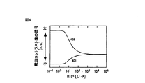
これらの現象を定量的に扱うと、抵抗と検出される放出電子数に図4のような関係が成り立つ。横軸は、抵抗Rと試料に照射するビーム電流IPの積、縦軸は、電位コントラスト像の信号である。PVCの場合が401の曲線、NVCの場合が402の曲線になる。NVCの場合については、本発明で我々が初めて明らかにした結果である。明るさの変化する領域は、両者とも0<log(R・IP)<3である。このように、電位コントラスト像の信号から、抵抗値を算出することは可能である。
(2)帯電電圧を測定する方法
帯電電圧の測定は、例えば、エネルギーフィルター(エネルギー分析器と言う場合もある)を用い、二次電子もしくは後方散乱電子のエネルギーを測定することによって実現される。ウエハが正もしくは負に帯電されると、二次電子および後方散乱電子のエネルギーも帯電電圧の分だけ変化するためである。エネルギーフィルターの詳細は、参考文献;L. Reimer、 ”Scanning Electron Microscopy、” Springer-Verlag Berlin Heidelberg、 1998、 P197 に記載してある。その一例は、二次電子および後方散乱電子を検出する検出器の前面にエネルギーフィルターの役目を果す、金属メッシュの板を設置するものである。エネルギーフィルターである金属メッシュに電圧を与え、その電圧を変化させると、二次電子および後方散乱電子の金属メッシュを通過する確率が変化する。その変化を検出器を用いて測定することによって、ウエハの帯電電圧が測定可能である。
When these phenomena are handled quantitatively, the relationship shown in FIG. 4 is established between the resistance and the number of detected electrons. The horizontal axis represents the product of the resistance R and the beam current IP applied to the sample, and the vertical axis represents the signal of the potential contrast image. In the case of PVC, the curve is 401, and in the case of NVC, the curve is 402. In the case of NVC, this is the result that we have clarified for the first time in the present invention. The areas where the brightness changes are both 0 <log (R · IP) <3. In this way, it is possible to calculate the resistance value from the signal of the potential contrast image.
(2) Method of Measuring Charging Voltage The charging voltage is measured by measuring the energy of secondary electrons or backscattered electrons using, for example, an energy filter (sometimes referred to as an energy analyzer). This is because when the wafer is charged positively or negatively, the energy of secondary electrons and backscattered electrons also changes by the charging voltage. Details of energy filters are described in the reference; L. Reimer, “Scanning Electron Microscopy,” Springer-Verlag Berlin Heidelberg, 1998, P197. One example is a metal mesh plate that serves as an energy filter in front of a detector that detects secondary and backscattered electrons. When a voltage is applied to a metal mesh that is an energy filter and the voltage is changed, the probability that secondary electrons and backscattered electrons pass through the metal mesh changes. By measuring the change using a detector, the charging voltage of the wafer can be measured.
測定原理の詳細を図17に示す。ここでは、二次電子のエネルギーを測定することによって帯電電圧を求める方法について述べるが、後方散乱電子の場合も同様である。この図は、二次電子放出の様子をエネルギー図で表したものである。横軸がウエハからの距離、縦軸がエネルギーである。ウエハの位置は距離0である。帯電前、真空準位は309であり、ウエハ前面にできるポテンシャル分布は311のようになる。 Details of the measurement principle are shown in FIG. Here, a method for obtaining the charging voltage by measuring the energy of secondary electrons will be described, but the same applies to the case of backscattered electrons. This figure is an energy diagram showing the state of secondary electron emission. The horizontal axis is the distance from the wafer, and the vertical axis is the energy. The position of the wafer is 0 distance. Before charging, the vacuum level is 309, and the potential distribution formed on the front surface of the wafer is 311.
ウエハには、通常リターディング電圧という、負の電圧を加えているため、真空準位309は0eVよりも高い。すると、電子ビーム照射により二次電子314が発生し、真空中に二次電子316が放出される。ただし、図中で、二次電子は全てエネルギー分布を表示する。電子放出効率が1より小さく、負に帯電した場合、ウエハの真空準位が309から320へと上昇し、ポテンシャル分布は321のようになる。正に帯電した場合は、それと逆である。
Since a negative voltage called a retarding voltage is normally applied to the wafer, the
負に帯電した状態で発生する二次電子のエネルギー分布は323のようになり、真空中に放出される二次電子は324となる。エネルギーフィルターは、これら真空中に放出された二次電子316および324のエネルギーを測定する事により帯電電圧を求める。エネルギーフィルターに電圧を印加すると、ポテンシャル障壁322が形成される。その電圧値が低ければ二次電子に対して障壁となるため、検出される電子325、326は減少し始める。図18に、エネルギーフィルター電圧とフィルターを通過し、検出器で検出される電子数を示す。信号が減少し始める電圧値が帯電電圧に等しく、この場合、帯電電圧は−2、−10、−15Vである。ここでは、負帯電の例について説明したが、正帯電の場合も同様である。このようにして、帯電電圧を測定する事が可能になった。
(3)帯電電圧を変化および制御させる方法
電子ビーム照射条件(ビームの照射面積、ビームエネルギー、ウエハ近傍の電位分布)を変えることにより、帯電電圧を変化させることが可能になる。例えば、入射エネルギー500eVの電子ビームを用いた場合、ビームの照射面積(FOV、 field of view)、およびウエハ近傍の電場を変えることによって、図5および6に示すように、帯電電圧を変化させることが可能になることがわかった。帯電電圧の値に幅があるのは、電子ビーム照射範囲で帯電電圧にばらつきがあることを示している。
The energy distribution of secondary electrons generated in a negatively charged state is 323, and the secondary electrons emitted into the vacuum are 324. The energy filter obtains the charging voltage by measuring the energy of the
(3) Method of changing and controlling charging voltage By changing the electron beam irradiation conditions (beam irradiation area, beam energy, potential distribution in the vicinity of the wafer), the charging voltage can be changed. For example, when an electron beam with an incident energy of 500 eV is used, the charging voltage is changed as shown in FIGS. 5 and 6 by changing the irradiation area (FOV) of the beam and the electric field in the vicinity of the wafer. It turns out that is possible. The range of the charging voltage value indicates that the charging voltage varies in the electron beam irradiation range.
他の例として、図15に示すように、ウエハ前面に帯電制御電圧39を印加可能な帯電制御電極38を設置し、ウエハ近傍の電位を変化させることにより帯電電圧を変化させることが可能であることもわかった。帯電制御電極38にウエハに対して正の電圧を加えれば、二次電子は放出しやすくなるため、帯電電圧は増加する。帯電制御電極38にウエハに対して負の電圧を加えれば、二次電子は引き戻されるので、帯電電圧は減少する。図16に実際の測定例を示すが、ここに示すように、帯電制御電圧39、Vcc、を変化させれば、帯電電圧が制御できる事がわかった。
As another example, as shown in FIG. 15, a charging
このように、電子ビーム照射条件を変化させた場合の、帯電増減の傾向の把握と、帯電電圧の測定によって、帯電電圧の制御が可能になった。例えば、ある領域を10Vにしたい場合、電子ビーム照射によって20Vに帯電したとしても、ウエハ近傍の電場を弱めることにより帯電電圧の低減が可能になる。このような指令を電子ビーム照射制御部に送ることにより、帯電制御を可能にした。 As described above, the charging voltage can be controlled by grasping the tendency of increase / decrease in charging and changing the charging voltage when the electron beam irradiation conditions are changed. For example, when it is desired to set a certain region to 10V, even if it is charged to 20V by electron beam irradiation, the charging voltage can be reduced by weakening the electric field in the vicinity of the wafer. By sending such a command to the electron beam irradiation control unit, charging control can be performed.
以上、(1)、(2)、および(3)の手段を組み合わせる事によって、抵抗−電圧特性の算出や、帯電電圧を所望の値に制御した状態での抵抗値測定が可能になった。 As described above, by combining the means (1), (2), and (3), it is possible to calculate the resistance-voltage characteristic and measure the resistance value in a state where the charging voltage is controlled to a desired value.
本発明によって得られる代表的な効果を以下に簡単に説明する。本発明の検査方法および装置を用いて回路パターンを有する半導体デバイス等の部分的に完成した基板を検査することにより、従来の検査装置では不可能であった、非接触で微小部分の所望の帯電電圧における電気抵抗や、電気抵抗の電圧依存性を測定可能になった。その結果から、欠陥の種類を判別することができるようになった。 A typical effect obtained by the present invention will be briefly described below. By inspecting a partially completed substrate such as a semiconductor device having a circuit pattern by using the inspection method and apparatus of the present invention, it is possible to achieve desired charging of a minute portion in a non-contact manner, which is impossible with a conventional inspection apparatus. It became possible to measure the electrical resistance in voltage and the voltage dependence of electrical resistance. As a result, the type of defect can be determined.
本検査を基板製造プロセスへ適用することにより、基板製造プロセスにいち早く異常対策処理を講ずることができ、その結果半導体デバイスその他の基板の不良率を低減し生産性を高めることができる。その結果、不良の発生そのものを低減させることができるので、半導体デバイス等の信頼性を高めることができ、新製品等の開発効率が向上し、且つ製造コストが削減できる。 By applying this inspection to the substrate manufacturing process, it is possible to quickly take an abnormality countermeasure process in the substrate manufacturing process, and as a result, the defect rate of semiconductor devices and other substrates can be reduced and productivity can be increased. As a result, since the occurrence of defects itself can be reduced, the reliability of semiconductor devices and the like can be increased, the development efficiency of new products and the like can be improved, and the manufacturing cost can be reduced.
(実施例1)
本実施例では、試料の抵抗―電圧特性を非接触で高速に測定し、欠陥の種類を判別した方法について述べる。ここで用いたウエハの断面構造を図7に示す。これは、Si基板701、SiO2膜702、poly−Siプラグを埋め込んだビアホール703、ビアホールの直下のn拡散層704、この拡散層704により形成されるpn接合706から成る。また、部分的に導通不良欠陥705も存在する。
Example 1
In this example, a method for measuring the resistance-voltage characteristics of a sample at high speed without contact and determining the type of defect will be described. A cross-sectional structure of the wafer used here is shown in FIG. This includes a
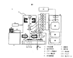
本実施例における半導体デバイスの検査装置の構成を図1に示す。半導体デバイスの検査装置1は、大きく分けると、電子光学系2、ステージ機構系3、ウエハ搬送系4、真空排気系5、光学顕微鏡6、制御系7、操作部8より構成されている。
The configuration of the semiconductor device inspection apparatus in this embodiment is shown in FIG. The semiconductor
電子光学系2は、電子ビーム34を放出するための電子銃9、電子ビーム34をウエハ18上に収束させるためのコンデンサレンズ10および対物レンズ11、電子ビーム34をブランキングするため(ウエハに照射させないため)のブランキング制御電極13、電子ビーム34をウエハ18上に走査させるための偏向器14、ウエハの高さを検出するウエハ高さセンサー15、ウエハ18で発生した放出電子(二次電子および後方散乱電子)35を検出する検出器12、検出器の前面に設置したエネルギーフィルター36、より構成されている。ウエハ18の電位コントラスト像を取得するためには、細く絞った電子ビーム34をウエハ18に照射し、放出電子35を発生させ、これらを電子ビーム34の走査と同期して検出する。
The electron
ステージ機構系(試料台)3は、XYステージ16およびウエハを載置するためのホルダ17(ビーム校正用パターン付)、ウエハ18に負の電圧を印加するためのリターディング電源19より構成されている。XYステージ16には、レーザ測長による位置検出器が取り付けられている。ウエハ搬送系4はウエハカセット20とウエハローダ21より構成されており、ウエハホルダ17はウエハ18を載置した状態でウエハローダ21とXYステージ16を行き来するようになっている。また、ウエハローダ21は検査室2とは独立して真空排気できるように構成されており、既に真空になっている検査室2を大気圧にすることなく、ウエハを搬送できるようになっている。
The stage mechanism system (sample stage) 3 includes an
真空排気系5は、図1には示していないが、一般にロータリーポンプ、ターボ分子ポンプ、およびイオンポンプから構成される。
光学顕微鏡6は、検査室2の室内に設置してある。XYステージ16を用いることによって電子光学系2からウエハを移動させ、光学顕微鏡6による観察が可能になっている。
Although not shown in FIG. 1, the
The
制御系7は、検出器12の信号を検出する信号検出系制御部22、ブランキング制御部23、ビーム偏向制御部24、電子光学系制御部25、ウエハ高さセンサー検出系26、ステージ制御部27、エネルギーフィルター制御部40より構成されている。ここでは、電子ビームの加速電圧、偏向幅、偏向速度、検出器12の信号取り込みタイミング、XYステージ16の移動速度、エネルギーフィルター36に印加する電圧等の条件が、目的に応じて任意にあるいは選択して設定できるよう入力されている。また、XYステージ16につけた位置検出器、およびウエハ高さセンサー15の信号から位置や高さのずれをモニタし、その結果より補正信号を生成し、電子ビームが常に正しい位置に照射されるよう各制御部に補正信号を送る役割も果たす。
The control system 7 includes a signal detection
操作部8は、装置の操作画面、取り込まれた電位コントラスト像、光学画像、および測定した電気特性を表示する操作画面および検査結果表示部28、画像処理部や制御系7へ命令を出す計算部29、電位コントラスト像・検査データ保存部30、外部サーバ31からのデータ授受を行うデータ入力部32、データ変換部33より構成されている。
The
次に、図1で示した検査装置を用いて、検査を行ったときの方法を説明する。まず、ウエハが任意の棚に設置されたウエハカセットを、図1のウエハ搬送系4におけるウエハカセット20に置く。操作画面28より、検査すべきウエハを指定するために、該ウエハがセットされたカセット内棚番号を指定する。
Next, a method when an inspection is performed using the inspection apparatus shown in FIG. 1 will be described. First, a wafer cassette in which wafers are placed on an arbitrary shelf is placed on the
そして、操作画面28より、各種検査条件を入力する。検査条件入力内容としては、電子ビーム電流、電子ビーム照射エネルギー、一画面の視野サイズ(FOV、Field of view)、リターディング電圧等である。個々のパラメータを入力することも可能であるが、通常は検出したい抵抗範囲および電圧範囲に応じて、上記各種検査パラメータの組合せが検査条件ファイルとしてデータベース化されており、それら範囲に応じた検査条件ファイルを選択して入力するだけでよい。データベースの作成は、次のように行われる。
Then, various inspection conditions are input from the
抵抗範囲の設定によって、0<log(R・IP)<3の式(課題を解決するための手段で述べたように、測定したい抵抗値をRとすると、この式を満たすようなビーム電流IPを用いる必要がある。)を用いてビーム電流IP範囲が決定される。電圧範囲の設定によって、図5、6、および16に示すような結果を用いて、ウエハ近傍の電場、電位や、FOVの範囲が決定される。これらの条件入力が完了したら、検査をスタートする。 By setting the resistance range, an equation of 0 <log (R · IP) <3 (as described in the means for solving the problem, if the resistance value to be measured is R, the beam current IP satisfying this equation is satisfied. Is used to determine the beam current IP range. Depending on the setting of the voltage range, the electric field, potential, and FOV range in the vicinity of the wafer are determined using the results shown in FIGS. When these condition inputs are completed, the inspection is started.
自動検査をスタートすると、まず、設定されたウエハ18を検査装置1内に搬送する。ウエハ搬送系4においては、被検査ウエハ18の直径が異なる場合にも、ウエハ形状がオリエンテーションフラット型あるいはノッチ型のように異なる場合にも、ウエハ18を載置するためのホルダ17を、ウエハの大きさや形状にあわせて交換することにより対応できるようになっている。該被検査ウエハ18は、ウエハカセット20からアーム、予備真空室等を含むウエハローダ21によりホルダ17上に載置され、保持固定されてホルダとともにウエハローダ21内で真空排気され、既に真空排気系5で真空になっている検査室に搬送される。
When the automatic inspection is started, first, the
ウエハがロードされたら、上記入力された検査条件に基づき、電子光学系制御部25より各部に電子ビーム照射条件が設定される。そして、ウエハホルダ17上の第一のビーム校正用パターンが電子光学系下にくるようにステージ16が移動し、該ビーム校正用パターンの電位コントラスト像を画像処理部で取得し、該電位コントラスト像より焦点・非点を合わせる。
When the wafer is loaded, the electron beam irradiation condition is set to each part by the electron optical
そして、被検査ウエハ18上の所定の箇所に移動し、ウエハ18の電位コントラスト像を取得し、コントラスト等を調整する。ここで、電子ビーム照射条件等を変更する必要が生じた場合には、再度ビーム校正を実施することが可能である。同時にウエハ18の高さを高さセンサー15より求め、ウエハ高さ検出系26により高さ情報と電子ビームの合焦点条件の相関を求め、この後の電位コントラスト像取得時には毎回焦点合わせを実行することなく、ウエハ高さ検出の結果より合焦点条件に自動的に調整する。これにより、高速連続電位コントラスト像取得が可能になった。
Then, it moves to a predetermined location on the
アライメントのために、あらかじめアラインメント用に形成されたパターン、アライメント用の光学顕微鏡像や電位コントラスト像、そしてパターンの位置情報を登録しておく。そして、検査条件の入力の際に、このデータを読み出せるようにしておく。 For alignment, a pattern previously formed for alignment, an optical microscope image for alignment, a potential contrast image, and pattern position information are registered. This data can be read when inputting the inspection conditions.
セットされたウエハ18は、光学顕微鏡部6でアラインメント用の第一の座標を観察するために、XYステージ16により移動される。モニタ28によりウエハ18上に形成されたアライメントパターンの光学顕微鏡画像が観察され、予め記憶された同じパターン画像と比較し、第一の座標の位置補正値が算出される。次に第一の座標から一定距離離れ第一の座標と同等の回路パターンが存在する第二の座標に移動し、同様に光学顕微鏡画像が観察され、アライメント用に記憶された回路パターン画像と比較され、第二の座標の位置補正値および第一の座標に対する回転ずれ量が算出される。
The
以上のようにして光学顕微鏡部6による所定の補正作業や検査領域設定等の準備作業が完了すると、XYステージ16の移動により、ウエハ18が電子光学系2の下に移動される。ウエハ18が電子光学系2の下に配置されると、上記光学顕微鏡部6により実施されたアライメント作業と同様の作業を電位コントラスト像により実施する。この際の電位コントラスト像の取得は、次の方法でなされる。
When the preparatory work such as predetermined correction work and inspection area setting by the
上記光学顕微鏡画像による位置合せにおいて記憶され補正された座標値に基き、光学顕微鏡部6で観察されたものと同じ回路パターンに、電子ビーム34が走査偏向器14によりXY方向に二次元に走査されて照射される。この電子ビームの二次元走査により、被観察部位から発生する放出電子35が上記の放出電子検出のための各部の構成および作用によって検出されることにより、電位コントラスト像が取得される。既に光学顕微鏡画像により簡便な検査位置確認や位置合せ、および位置調整が実施され、且つ回転補正も予め実施されているため、光学画像に比べ分解能が高く高倍率で高精度に位置合せや位置補正、回転補正を実施することができる。
Based on the coordinate values stored and corrected in the alignment by the optical microscope image, the electron beam 34 is scanned two-dimensionally in the XY directions by the scanning
なお、電子ビーム34をウエハ18に照射すると、その箇所が帯電する。検査の際にその帯電の影響を避けるために、上記位置回転補正あるいは検査領域設定等の検査前準備作業において電子ビーム34を照射する回路パターンは予め被検査領域外に存在する回路パターンを選択するか、あるいは被検査チップ以外のチップにおける同等の回路パターンを制御系7から自動的に選択できるようにしておく。
When the electron beam 34 is irradiated on the
また、帯電を緩和させるために、紫外線を照射させる手段を用いても良い。これにより、検査時に上記検査前準備作業により電子ビーム34を照射した影響が検査画像に及ぶことは無い。 In addition, means for irradiating with ultraviolet rays may be used to relieve charging. Thereby, the influence which irradiated with the electron beam 34 by the said pre-inspection preparatory work at the time of an inspection does not reach an inspection image.
このようにして行ったアライメント結果は、各制御部に転送される。検査の際は、各制御部によって回転や位置座標が補正される。 The alignment result thus performed is transferred to each control unit. At the time of inspection, the rotation and position coordinates are corrected by each control unit.
アライメントが完了したら、試料ホルダ17上に載置された第二の校正用パターンに移動する。第二の校正用パターンは、図4のグラフの信号スケールを検査で得られる電位コントラスト像の信号に一致させるものである。そのパターンは、十分低抵抗なビアホールと十分高抵抗なビアホールが加工されたパターンである。
When the alignment is completed, the second calibration pattern placed on the
該パターンの電位コントラスト像を用いて十分な低抵抗部および高抵抗部の信号値を校正する。十分な高抵抗部はパターンの無い絶縁部を用いても良い。この結果をふまえて、ウエハ18上に移動し、ウエハ上のパターン箇所の電位コントラスト像を取得し、明るさ調整すなわちキャリブレーションを実施する。
The signal values of the sufficiently low resistance portion and high resistance portion are calibrated using the potential contrast image of the pattern. An insulating part having no pattern may be used as the sufficiently high resistance part. Based on this result, the wafer moves onto the
キャリブレーションが完了したら、既に制御系7に入力されている電子照射条件をもとに、検査を実施する。そのときに得られた、電位コントラスト像の信号から図4を用いて換算したビアホールの抵抗分布、およびエネルギーフィルターにより測定したビアホールの帯電電圧分布を図8に示す。帯電電圧の測定をより具体的に説明すると、帯電電圧測定機能ここではエネルギーフィルタを用いている。 When the calibration is completed, an inspection is performed based on the electron irradiation conditions already input to the control system 7. FIG. 8 shows the via hole resistance distribution converted from the potential contrast image signal obtained at that time using FIG. 4 and the via hole charging voltage distribution measured by the energy filter. The measurement of the charging voltage will be described more specifically. The charging voltage measuring function here uses an energy filter.
一次電子ビームを、素子を加工したウエハ(試料)に照射させ、そこから発生する二次電子や後方散乱電子のエネルギーを測定する。その値は、試料上の一次電子の照射領域有る素子に印加されている電圧の分だけ変化するため、電圧の算出が可能になる。この像をビーム電流、ビームエネルギー、FOV、ウエハとウエハ周囲の電極間の電位分布を変化させる度に取得することにより、各ビアホールについて、抵抗の電圧依存性が算出できた。 A primary electron beam is irradiated onto a wafer (sample) on which the device is processed, and the energy of secondary electrons and backscattered electrons generated therefrom is measured. Since the value changes by the amount of voltage applied to the element having the irradiation region of the primary electrons on the sample, the voltage can be calculated. By acquiring this image every time the beam current, beam energy, FOV, and potential distribution between the wafer and the electrodes around the wafer are changed, the voltage dependency of the resistance can be calculated for each via hole.
その結果、図9に示すように、ビアホールの抵抗値は大きく3つ(結果901、結果902、結果903)に分類できた。
As a result, as shown in FIG. 9, the resistance value of the via hole was roughly classified into three (
結果901は、抵抗−電圧特性が、不純物ドーピング条件から予測されるものと等しく、正常なpn接合であることがわかった。結果902は順バイアス、逆バイアスとも高抵抗であり、導通不良欠陥が発生していることがわかった。結果903は、抵抗が非常に低く、リーク欠陥であることがわかった。また、結果904は、逆バイアスの抵抗値が大きいので、一見、正常に見えるが、ブレークダウン電圧が低く、将来、リーク欠陥となる可能性のある事もわかった。
The
さらに、この抵抗−電圧特性から、不純物をドーピングする工程における不具合箇所を推測する事も可能になった。このような結果は、ある程度広い範囲の電圧で抵抗値を求めなければわからなかったことである。 In addition, it is possible to estimate a defective portion in the impurity doping process from the resistance-voltage characteristics. Such a result cannot be understood unless the resistance value is obtained with a voltage within a relatively wide range.
次の検査領域に移動する際は、XYステージ16を静止させて電子ビーム34の走査域を移動させる方法と、XYステージ16を移動させる方法のいずれかを選択できる。ある特定の比較的小さい領域を検査する場合には前者のステージを静止させて検査する方法、比較的広い領域を検査するときは、ステージを移動して検査する方法が有効である。なお、検査領域を移動する場合、電子ビーム34をブランキングする必要がある時には、ブランキング用偏向器13により電子ビーム34を偏向し、電子ビームがウエハに照射されないように制御できる。
When moving to the next inspection region, either a method of moving the scanning region of the electron beam 34 by stopping the
このように指定された領域の検査を実施しながら、リアルタイムで画像処理を実施し、抵抗値の電圧依存性を測定していく。そして、検査結果を操作部28に表示し、且つデータをデータ変換部33を介して外部に出力する。このようにして、ウエハ全域もしくは、部分的に検査を完了したら、図10のように、ウエハ内の導通不良欠陥分布やリーク欠陥分布を得ることができた。その後、ウエハをアンロードして検査を終了する。
The image processing is performed in real time and the voltage dependence of the resistance value is measured while performing the inspection of the designated area. Then, the inspection result is displayed on the
ここでは、帯電電圧を変化させるために、ビームの照射面積、ビームエネルギー、ウエハとウエハ周りの電極間の電位分布を変化させた。それ以外でも、検査前に広範囲に電子ビームを照射しておくことによって、帯電電圧を変化させることが可能である。また、ウエハ表面を帯電させるために、本実施例では電子ビームを用いたが、その代わりにイオンビームを用いても同様の測定が可能である。 Here, in order to change the charging voltage, the irradiation area of the beam, the beam energy, and the potential distribution between the wafer and the electrode around the wafer were changed. Other than that, it is possible to change the charging voltage by irradiating the electron beam over a wide range before the inspection. Further, in this embodiment, an electron beam is used to charge the wafer surface, but the same measurement can be performed using an ion beam instead.
(実施例2)
第二の実施例では、第一の実施例で説明した検査装置を用いて、所望の帯電電圧における各素子の抵抗値を測定し、その抵抗値が所定の値と異なった場合に該箇所を欠陥として認識し、該欠陥位置を表示する。帯電電圧の制御は、エネルギーフィルター36を用いて測定した帯電電圧の情報を計算部29へ送り、計算部29で所望の帯電電圧にするための電子ビーム照射条件を決定し、その情報を制御部7を送ることにより行う。電子ビーム照射条件の設定が上手くいかず、帯電電圧が所望の値にならなかった場合は、再度、この方法を繰り返す。装置の詳細な説明は第一の実施例で述べているので省略する。
(Example 2)
In the second embodiment, the resistance value of each element at a desired charging voltage is measured using the inspection apparatus described in the first embodiment, and when the resistance value is different from a predetermined value, Recognize as a defect and display the position of the defect. For controlling the charging voltage, information on the charging voltage measured using the
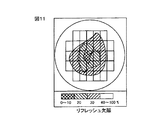
この方法を用いて、例えば、DRAMに加工したpn接合のリフレッシュ不良欠陥検査が可能である。このpn接合は、それに3V印加した時の抵抗値が10の14乗Ω以下では欠陥と判断するとした。ビアホールの底にpn接合が加工されたウエハを上記の方法で検査し、帯電電圧3Vでの抵抗値を算出し、10の14乗Ω以下のビアホールをリフレッシュ欠陥とし、ウエハ中の欠陥密度分布を図11のように示すことができた。 Using this method, for example, a refresh defect inspection of a pn junction processed into a DRAM can be performed. The pn junction was determined to be defective when the resistance value when 3 V was applied thereto was 10 14 Ω or less. The wafer having a pn junction processed at the bottom of the via hole is inspected by the above method, the resistance value at a charging voltage of 3V is calculated, a via hole having a power of 10 14 or less is made a refresh defect, and the defect density distribution in the wafer is calculated. As shown in FIG.
(実施例3)
第三の実施例では、第一の実施例で説明した検査装置を用いて、ウエハに加工されたCMOSのpn接合検査を行った。抵抗−電圧測定を行うことによって、pn接合の方向、導通不良欠陥、リーク欠陥の判別が可能になった。検査結果の例を図12に示す。ウエハ表面がn拡散層であるpn接合が1201、ウエハ表面がp拡散層であるpn接合が1202、導通不良欠陥が1203、リーク欠陥が1204である。また、帯電電圧を制御し、ブレークダウンが起こらない条件で検査を行えば、単に、NVC、およびPVCの電位コントラスト像の信号変化を見るだけで、その判断を行うことが可能になった。図13にその結果を示す。
(Example 3)
In the third embodiment, a pn junction inspection of the CMOS processed on the wafer was performed using the inspection apparatus described in the first embodiment. By performing resistance-voltage measurement, it is possible to determine the direction of the pn junction, the continuity defect, and the leak defect. An example of the inspection result is shown in FIG. A pn junction whose wafer surface is an n diffusion layer is 1201, a pn junction whose wafer surface is a p diffusion layer is 1202, a conduction defect is 1203, and a leak defect is 1204. Further, if the inspection is performed under the condition that the charging voltage is controlled and breakdown does not occur, it is possible to make the determination simply by looking at the signal change in the potential contrast images of NVC and PVC. FIG. 13 shows the result.
(実施例4)
第四の実施例は、第一の実施例で述べた装置を用い、検査用の電位コントラスト像の信号を取得しながら、もしくはその前に、抵抗値が既知の素子の電位コントラスト像の信号を取得し、電位コントラスト像の信号と抵抗値の関係を校正する検査方法および装置である。
Example 4
In the fourth embodiment, the apparatus described in the first embodiment is used, and a potential contrast image signal of an element having a known resistance value is obtained before or before acquiring a potential contrast image signal for inspection. An inspection method and apparatus for acquiring and calibrating a relationship between a signal of a potential contrast image and a resistance value.
まず、検査ウエハ18を検査装置内にロードし、あらかじめ登録してある電子ビーム照射条件を設定する。試料台17には、図14に示すように抵抗値があらかじめわかっている標準サンプル37が貼り付けてある。検査を開始する前にこの標準サンプルの電位コントラスト像を取得し、電気抵抗と電位コントラスト像の信号の関係を求める。その後、検査を開始し、その関係を用いて、得られた電位コントラスト像から抵抗値を算出した。この方法によって、より正確な抵抗測定が可能になった。
First, the
(実施例5)
第五の実施例は、第一の実施例で述べた装置を用い、帯電電圧を変化させるために、ウエハ18前面に設置した帯電制御電極38を用いるものである。図15に示すように、この帯電制御電極には電圧を印加可能である。ウエハに対して電圧Vccを印加すると帯電電圧値が図16のようになった。この方法によって、帯電電圧を正から負へ、もしくは負から正へと容易に連続変化させることが可能になった。また、帯電電圧の変化を高速に行うためには、ウエハに垂直に磁場を印加する方法が有効である。
(Example 5)
The fifth embodiment uses the apparatus described in the first embodiment and uses a
(実施例6)
第六の実施例は、第一の実施例で説明した検査装置を用いて、比較検査を行うことにより、欠陥を検出するものである。まず、第一の領域において、所望の帯電電圧における各素子の抵抗値を測定、もしくは所望の帯電電圧範囲における各素子の抵抗値を測定した。つぎに、第二の領域においても同様に抵抗値の測定を行った。第一の領域、および第二の領域の抵抗値を比較することで、異なる部分を判定し、その部分を欠陥と認識することができた。
(Example 6)
In the sixth embodiment, a defect is detected by performing a comparative inspection using the inspection apparatus described in the first embodiment. First, in the first region, the resistance value of each element in a desired charging voltage was measured, or the resistance value of each element in a desired charging voltage range was measured. Next, the resistance value was similarly measured in the second region. By comparing the resistance values of the first region and the second region, it was possible to determine a different portion and recognize that portion as a defect.
以上実施例を纏めると本発明は、以下の如くとなる。 The present invention is as follows when the embodiments are summarized.
第1の発明は、素子が形成されたウエハ表面の領域を荷電粒子ビームで走査する工程と、上記領域の帯電電圧を一つ、もしくは複数の所望の値にするために、荷電粒子ビームの照射条件を変化させる工程と、帯電電圧が上記所望の値に等しいか否かを確認するために帯電電圧を測定する工程と、上記所望の帯電電圧にさせた後、上記領域から発生する二次電子や後方散乱電子を検出し、その信号から該領域の電位コントラスト像を形成する信号を記憶する工程と、該電位コントラスト像の信号および荷電粒子ビーム電流値から電気抵抗値を算出する工程よりなる。 According to a first aspect of the present invention, there is provided a step of scanning a region of the wafer surface on which an element is formed with a charged particle beam, and irradiation with a charged particle beam in order to set the charging voltage in the region to one or a plurality of desired values. A step of changing conditions, a step of measuring a charging voltage to confirm whether or not the charging voltage is equal to the desired value, and secondary electrons generated from the region after the charging voltage is set to the desired value And backscattered electrons are detected, a signal for forming a potential contrast image of the region is stored from the signal, and an electric resistance value is calculated from the signal of the potential contrast image and the charged particle beam current value.
更に、素子が形成されたウエハ表面の領域を荷電粒子ビームで走査する工程と、上記荷電粒子ビームにより上記領域から発生する二次電子や後方散乱電子を検出し、その信号から該領域の電位コントラスト像を形成する信号を記憶する工程と、該電位コントラスト像の信号および荷電粒子ビーム電流値から電気抵抗値を算出する工程と、上記領域の帯電電圧を測定する工程と、これらすべての工程を、荷電粒子ビームの照射条件を変化させる度に行い、素子の抵抗と電圧の関係を求めることにより達成する。 Further, a step of scanning the region of the wafer surface on which the element is formed with a charged particle beam, and detecting secondary electrons and backscattered electrons generated from the region by the charged particle beam, and detecting the potential contrast of the region from the signal A step of storing a signal for forming an image, a step of calculating an electric resistance value from the signal of the potential contrast image and a charged particle beam current value, a step of measuring a charging voltage of the region, and all these steps. This is achieved by changing the irradiation condition of the charged particle beam and determining the relationship between the resistance and voltage of the element.
また、上記、荷電粒子ビームの照射条件は、ビーム電流、ビームエネルギー、照射面積、ウエハとウエハ周りの電極間の電位分布であり、照射条件を変化させる場合には、これらの内、一つ以上変化させることでも達成できる。更にまた、帯電状態が安定するまで該走査を行うことにより精度良く電位コントラスト信号を検出することを可能とする。 In addition, the charged particle beam irradiation conditions are beam current, beam energy, irradiation area, and potential distribution between the wafer and the electrodes around the wafer. When changing the irradiation conditions, one or more of them are used. It can also be achieved by changing. Furthermore, the potential contrast signal can be detected with high accuracy by performing the scanning until the charged state is stabilized.
更にまた、前記電位コントラスト像を形成する信号を記憶する工程の前処理として、該電位コントラスト像の信号取得領域よりも広い領域に荷電粒子ビームを走査し、帯電させることによっても達成される。 Furthermore, as a pre-process of the step of storing the signal for forming the potential contrast image, it is achieved by scanning and charging a charged particle beam in a region wider than the signal acquisition region of the potential contrast image.
抵抗値を決定する際に、これらの関係が既知の素子の電位コントラスト像を用いて校正することにより達成され、前記抵抗値の測定結果を用いて、前記基板における欠陥の種類を判別するよう構成したことにある。本発明の検査方法は、正および負に帯電させた状態で電位コントラスト像の取得を行うことにより、pn接合が形成された素子の接合の方向、リーク欠陥、導通不良欠陥を判別する検査方法を提供する。本発明の検査方法は、前記荷電粒子ビームは電子ビームであることを特徴とする検査方法である。本発明の検査方法は、測定したい電気抵抗をRとした場合、用いる電子ビームの電流値IPは0<log(R・IP)<3の範囲にあることを特徴とする検査方法である。 Structure when determining the resistance value is accomplished by these relationships are calibrated using voltage contrast image of a known device, using the measurement results of the resistance values, to determine the type of defect in the substrate It is to have done. The inspection method of the present invention is an inspection method for determining the junction direction, leakage defect, and continuity defect of an element in which a pn junction is formed by acquiring a potential contrast image in a positively and negatively charged state. To provide . Test method of the present invention, the charged particle beam is a test method which is characterized in that an electron beam. Test method of the present invention, when the electrical resistance to be measured is R, the electron beam current value IP of the used is inspection method characterized by the range of 0 <log (R · IP) <3.
第2の発明は、試料を載置する試料台と、一次荷電粒子ビームを前記試料台上の試料に照射するための対物レンズと、荷電粒子ビームを照射した領域の帯電電圧を測定する帯電電圧測定系と、該帯電電圧を所望の値にするために、荷電粒子ビームの照射条件の変更を行う制御部と、試料から発生する二次電子や後方散乱電子を検出する検出器と、前記検出器からの信号を用いて、検査されるべき領域の電位コントラスト像の信号を記憶するための電位コントラスト像記憶部と、前記電位コントラスト像信号から該試料の部位の電気抵抗を算出する演算部と、を具備してなることを特徴とする荷電粒子ビームによる検査装置にある。 According to a second aspect of the present invention, there is provided a sample table on which a sample is placed, an objective lens for irradiating a sample on the sample table with a primary charged particle beam, and a charging voltage for measuring a charging voltage in a region irradiated with the charged particle beam. A measurement system, a control unit that changes the irradiation condition of the charged particle beam in order to set the charging voltage to a desired value, a detector that detects secondary electrons and backscattered electrons generated from the sample, and the detection A potential contrast image storage unit for storing a signal of a potential contrast image of a region to be inspected using a signal from the instrument, and an arithmetic unit for calculating an electrical resistance of a part of the sample from the potential contrast image signal; And an inspection apparatus using a charged particle beam.
第3の発明は、試料を載置する試料台と、一次荷電粒子ビームを前記試料台上の試料に照射するための対物レンズと、試料から発生する二次電子や後方散乱電子を検出する検出器と、荷電粒子ビームを照射した領域の帯電電圧を測定する帯電電圧測定系と、前記検出器からの信号を用いて、検査されるべき領域の電位コントラスト像の信号を記憶するための電位コントラスト像記憶部と、該荷電粒子ビームの照射条件を変化させる度に帯電電圧を求め、同時に前記電位コントラスト像信号から該試料の部位の電気抵抗を算出し、各部における抵抗−電圧の関係を決定する演算部と、を具備してなることを特徴とする荷電粒子ビームによる検査装置にある。また、帯電電圧を変化させるために、ウエハの前面に電圧を印加することのできる帯電制御電極を具備してなる。また、帯電電圧を高速に変化させるために、ウエハに垂直方向に磁場を印加することにより達成できる。 According to a third aspect of the present invention, there is provided a sample table on which the sample is placed, an objective lens for irradiating the sample on the sample table with a primary charged particle beam, and detection for detecting secondary electrons and backscattered electrons generated from the sample. A voltage contrast system for measuring a charging voltage of a region irradiated with a charged particle beam, and a potential contrast for storing a signal of a potential contrast image of a region to be inspected using a signal from the detector Each time the irradiation condition of the image storage unit and the charged particle beam is changed, the charging voltage is obtained, and at the same time, the electric resistance of the part of the sample is calculated from the potential contrast image signal, and the resistance-voltage relationship in each part is determined. An inspection apparatus using a charged particle beam, comprising: an arithmetic unit. In order to change the charging voltage, a charging control electrode capable of applying a voltage to the front surface of the wafer is provided. Further, in order to change the charging voltage at a high speed, this can be achieved by applying a magnetic field in the vertical direction to the wafer.
1: 検査装置、2:電子光学系、3:ステージ機構系、4:ウエハ搬送系、5:真空排気系、6:光学顕微鏡、7:制御系、8:操作部、9:電子銃、10:コンデンサレンズ、11:対物レンズ、12:検出器、13:ブランキング制御電極、14:偏向器、15:高さセンサー、16:XYステージ、17:ウエハホルダ、18:ウエハ、19:リターディング電源、20:ウエハカセット、21:ウエハローダ、22:信号検出系制御部、23:ブランキング制御部、24:ビーム偏向制御部、25:電子光学系制御部、26:高さセンサー検出系、27:ステージ制御部、28:操作画面・検査結果表示部、29:計算部、30:データ保存部、31:外部サーバ、32:データ入力部、33:データ変換部、34:電子ビーム、35:放出電子(二次電子および後方散乱電子)、36:エネルギーフィルター、37抵抗標準サンプル、38:帯電制御電極、39:帯電制御電圧、40:エネルギーフィルター制御部、301:ビアホール(低抵抗)、302:Si基板、303:SiO2膜、304:SiO2残膜、305:ビアホール(高抵抗)、306:電位分布、307:二次電子、308:二次電子、309:帯電前の真空準位、310:正帯電した場合の真空準位、311:帯電前のポテンシャル分布、312:正帯電した場合のポテンシャル分布、313:ポテンシャル障壁、314:二次電子のエネルギー分布(帯電前)、315:二次電子のエネルギー分布(正帯電)、316:ポテンシャル障壁を越えて真空中に放出される二次電子のエネルギー分布(帯電前)、317:ポテンシャル障壁を越えて真空中に放出される二次電子のエネルギー分布(正帯電)、320:負帯電した場合の真空準位、321:負帯電した場合のポテンシャル分布、322:エネルギーフィルターによるポテンシャル障壁、323:二次電子のエネルギー分布、324:ポテンシャル障壁を越えて真空中に放出される二次電子のエネルギー分布、325:エネルギーフィルターを通り抜けた二次電子のエネルギー分布(帯電前)、326:エネルギーフィルターを通り抜けた二次電子のエネルギー分布(負帯電)、401:負帯電の場合、402:正帯電の場合、701:p−Si基板、702:SiO2層、703:poly−Siプラグを埋め込んだビアホール、704:n拡散層、705:SiO2残膜、706:pn接合、901:正常なpn接合、902:導通不良欠陥、903:pn接合の欠陥、904:リーク欠陥、1201:正常なpn接合(表面はn拡散層)、1202:正常なpn接合(表面はp拡散層)、1203:導通不良欠陥、1204:リーク欠陥 。 1: inspection apparatus, 2: electron optical system, 3: stage mechanism system, 4: wafer transfer system, 5: vacuum exhaust system, 6: optical microscope, 7: control system, 8: operation unit, 9: electron gun, 10 : Condenser lens, 11: objective lens, 12: detector, 13: blanking control electrode, 14: deflector, 15: height sensor, 16: XY stage, 17: wafer holder, 18: wafer, 19: retarding power supply 20: Wafer cassette, 21: Wafer loader, 22: Signal detection system control unit, 23: Blanking control unit, 24: Beam deflection control unit, 25: Electro-optical system control unit, 26: Height sensor detection system, 27: Stage control unit, 28: operation screen / inspection result display unit, 29: calculation unit, 30: data storage unit, 31: external server, 32: data input unit, 33: data conversion unit, 34: electron beam, 3 5: emitted electrons (secondary electrons and backscattered electrons), 36: energy filter, 37 resistance standard sample, 38: charge control electrode, 39: charge control voltage, 40: energy filter control unit, 301: via hole (low resistance) , 302: Si substrate, 303: SiO2 film, 304: SiO2 residual film, 305: Via hole (high resistance), 306: Potential distribution, 307: Secondary electron, 308: Secondary electron, 309: Vacuum level before charging 310: Vacuum level when positively charged, 311: Potential distribution before charging, 312: Potential distribution when positively charged, 313: Potential barrier, 314: Energy distribution of secondary electrons (before charging), 315: Energy distribution of secondary electrons (positive charge), 316: Energy distribution of secondary electrons released into the vacuum over the potential barrier Before charging) 317: Energy distribution of secondary electrons released into the vacuum over the potential barrier (positive charging), 320: Vacuum level when negatively charged, 321: Potential distribution when negatively charged, 322 : Potential barrier by energy filter, 323: energy distribution of secondary electrons, 324: energy distribution of secondary electrons emitted into the vacuum over the potential barrier, 325: energy distribution of secondary electrons that passed through the energy filter ( 326: energy distribution of secondary electrons that passed through the energy filter (negative charging), 401: negative charging, 402: positive charging, 701: p-Si substrate, 702: SiO 2 layer, 703: via hole embedded with a poly-Si plug, 704: n diffusion layer, 705: SiO 2 residual film, 06: pn junction, 901: normal pn junction, 902: conduction failure defect, 903: pn junction defect, 904: leak defect, 1201: normal pn junction (surface is n diffusion layer), 1202: normal pn junction (The surface is a p-diffusion layer) 1203: Conduction defect, 1204: Leak defect.
Claims (9)
一次荷電粒子ビームを前記試料台上の試料の所定の領域に照射するための対物レンズと、
前記一次荷電粒子ビームで前記試料の所定領域を走査する走査部と、
前記試料から発生する二次電子や後方散乱電子を検出する検出器と、
前記一次荷電粒子ビームの電流量が所定値に調整された荷電粒子ビームを用いて、検出された二次電子信号強度ないし後方散乱電子信号強度から、前記所定領域の帯電電圧を測定する測定部と、
前記試料の所定の領域に所望の帯電電位が形成されるように前記一次荷電粒子ビームの電流量を調整するための制御部と、
前記電流量が所定値に調整された前記一次荷電粒子ビームを用いて、前記検出器からの信号で電位コントラスト像を形成する画像処理部と、
前記帯電電圧と前記電位コントラスト像信号から前記荷電粒子ビームの照射位置の電気抵抗を求める演算部と、
を具備してなることを特徴とする荷電粒子ビームを用いた検査装置。 A sample stage on which the sample is placed;
An objective lens for irradiating a predetermined region of the sample on the sample stage with a primary charged particle beam;
A scanning unit that scans a predetermined region of the sample with the primary charged particle beam;
A detector for detecting secondary electrons and backscattered electrons generated from the sample;
A measuring unit that measures a charging voltage in the predetermined region from a detected secondary electron signal intensity or backscattered electron signal intensity using a charged particle beam in which a current amount of the primary charged particle beam is adjusted to a predetermined value; ,
A control unit for adjusting a current amount of the primary charged particle beam so that a desired charged potential is formed in a predetermined region of the sample;
An image processing unit that forms a potential contrast image with a signal from the detector using the primary charged particle beam in which the amount of current is adjusted to a predetermined value;
A calculation unit for obtaining an electric resistance at an irradiation position of the charged particle beam from the charging voltage and the potential contrast image signal;
An inspection apparatus using a charged particle beam.
前記測定部として試料面に電圧を印加することのできる電極を具備してなることを特徴とする荷電粒子ビームを用いた検査装置。 In the inspection apparatus using the charged particle beam according to claim 1,
An inspection apparatus using a charged particle beam, comprising an electrode capable of applying a voltage to a sample surface as the measurement unit.
前記測定部として前記試料に垂直方向に磁場を印加することを特徴とする荷電粒子ビームを用いた検査装置。 In the inspection apparatus using the charged particle beam according to claim 1,
An inspection apparatus using a charged particle beam, wherein a magnetic field is applied to the sample in the vertical direction as the measurement unit.
前記電流量は前記検出器で検出した信号強度が前記試料の正帯電および負帯電の帯電電圧に対して変化する領域内に調整する機能を持つことを特徴とする荷電粒子ビームを用いた検査装置。 In the inspection apparatus using the charged particle beam according to claim 1 ,
An inspection apparatus using a charged particle beam, wherein the current amount has a function of adjusting a signal intensity detected by the detector within a region where the signal intensity changes with respect to a positive charging voltage and a negative charging voltage of the sample. .
前記電流量に代えて、前記荷電粒子ビームの照射面積を調整することにより、前記荷電粒子ビームの照射条件を調整する機能を持つことを特徴とする荷電粒子ビームを用いた検査装置。 In the inspection apparatus using the charged particle beam according to claim 1 ,
An inspection apparatus using a charged particle beam having a function of adjusting an irradiation condition of the charged particle beam by adjusting an irradiation area of the charged particle beam instead of the amount of current .
前記走査する工程で、二次電子信号ないし後方散乱電子信号の取得領域よりも広い領域に前記荷電粒子ビームを走査し、前記試料を帯電させる機能を持つことを特徴とする荷電粒子ビームを用いた検査装置。 In the inspection apparatus using the charged particle beam according to claim 1 ,
In the scanning step, the charged particle beam having a function of scanning the charged particle beam over a region wider than the acquisition region of the secondary electron signal or the backscattered electron signal and charging the sample is used. Inspection device.
前記電流量の所定値を、前記二次電子信号ないし後方散乱電子信号強度を電位コントラスト像として表示した場合に、当該電位コントラスト像の明るさの変化する領域内に定める機能を持つことを特徴とする荷電粒子ビームを用いた検査装置。 In the inspection apparatus using the charged particle beam according to claim 1 ,
When the secondary electron signal or the backscattered electron signal intensity is displayed as a potential contrast image, the predetermined value of the current amount has a function to be determined in a region where the brightness of the potential contrast image changes. Inspection device using charged particle beam.
半導体素子が形成された試料に対して前記荷電粒子ビームを照射し、前記所定領域の抵抗値を算出し、当該算出された抵抗値と前記帯電電圧との関係を測定することにより、前記半導体素子のリーク欠陥、または導通不良欠陥の検査を行うことを特徴とする荷電粒子ビームを用いた検査装置。 In the inspection apparatus using the charged particle beam according to claim 1 ,
The semiconductor element is formed by irradiating the sample on which the semiconductor element is formed with the charged particle beam, calculating a resistance value of the predetermined region, and measuring a relationship between the calculated resistance value and the charging voltage. Inspection apparatus using a charged particle beam, characterized by inspecting a leak defect or a conduction defect.
前記荷電粒子ビームの電流値を当該荷電粒子ビームの照射位置の抵抗値をR、該電流値をIPとした場合に、0<log(R・IP)<3の範囲にある条件で定める機能を持つことを特徴とする荷電粒子ビームを用いた検査装置。 In the inspection apparatus using the charged particle beam according to claim 1 ,
A function of determining the current value of the charged particle beam under the condition of 0 <log (R · IP) <3, where R is the resistance value at the irradiation position of the charged particle beam and IP is the current value. Inspection apparatus using charged particle beam characterized by having.
Priority Applications (1)
| Application Number | Priority Date | Filing Date | Title |
|---|---|---|---|
| JP2005227657A JP5016799B2 (en) | 2005-08-05 | 2005-08-05 | Inspection equipment using charged particle beam |
Applications Claiming Priority (1)
| Application Number | Priority Date | Filing Date | Title |
|---|---|---|---|
| JP2005227657A JP5016799B2 (en) | 2005-08-05 | 2005-08-05 | Inspection equipment using charged particle beam |
Related Parent Applications (1)
| Application Number | Title | Priority Date | Filing Date |
|---|---|---|---|
| JP2001295399A Division JP3955450B2 (en) | 2001-09-27 | 2001-09-27 | Sample inspection method |
Publications (3)
| Publication Number | Publication Date |
|---|---|
| JP2005333161A JP2005333161A (en) | 2005-12-02 |
| JP2005333161A5 JP2005333161A5 (en) | 2008-11-06 |
| JP5016799B2 true JP5016799B2 (en) | 2012-09-05 |
Family
ID=35487548
Family Applications (1)
| Application Number | Title | Priority Date | Filing Date |
|---|---|---|---|
| JP2005227657A Expired - Fee Related JP5016799B2 (en) | 2005-08-05 | 2005-08-05 | Inspection equipment using charged particle beam |
Country Status (1)
| Country | Link |
|---|---|
| JP (1) | JP5016799B2 (en) |
Families Citing this family (5)
| Publication number | Priority date | Publication date | Assignee | Title |
|---|---|---|---|---|
| JP4891036B2 (en) * | 2006-11-16 | 2012-03-07 | ルネサスエレクトロニクス株式会社 | Semiconductor device manufacturing method and semiconductor inspection apparatus |
| JP5044813B2 (en) | 2007-02-19 | 2012-10-10 | エスアイアイ・ナノテクノロジー株式会社 | Focused ion beam apparatus and charged particle optical system adjustment method |
| JP2009099540A (en) * | 2007-09-27 | 2009-05-07 | Hitachi High-Technologies Corp | Method of inspecting and measuring sample and scanning electron microscope |
| JP5406308B2 (en) * | 2009-11-13 | 2014-02-05 | 株式会社日立ハイテクノロジーズ | Sample observation method using electron beam and electron microscope |
| US12169208B2 (en) | 2021-11-30 | 2024-12-17 | Innovatum Instruments Inc. | Probe tip X-Y location identification using a charged particle beam |
Family Cites Families (5)
| Publication number | Priority date | Publication date | Assignee | Title |
|---|---|---|---|---|
| US4843330A (en) * | 1986-10-30 | 1989-06-27 | International Business Machines Corporation | Electron beam contactless testing system with grid bias switching |
| JPH0786348A (en) * | 1993-09-13 | 1995-03-31 | Fujitsu Ltd | Electron beam equipment |
| JP2959529B2 (en) * | 1997-06-18 | 1999-10-06 | 日本電気株式会社 | Semiconductor wafer inspection apparatus and inspection method using charged particle beam |
| US6504393B1 (en) * | 1997-07-15 | 2003-01-07 | Applied Materials, Inc. | Methods and apparatus for testing semiconductor and integrated circuit structures |
| JP3724949B2 (en) * | 1998-05-15 | 2005-12-07 | 株式会社東芝 | Substrate inspection apparatus, substrate inspection system including the same, and substrate inspection method |
-
2005
- 2005-08-05 JP JP2005227657A patent/JP5016799B2/en not_active Expired - Fee Related
Also Published As
| Publication number | Publication date |
|---|---|
| JP2005333161A (en) | 2005-12-02 |
Similar Documents
| Publication | Publication Date | Title |
|---|---|---|
| JP3955450B2 (en) | Sample inspection method | |
| JP3973372B2 (en) | Substrate inspection apparatus and substrate inspection method using charged particle beam | |
| JP5164317B2 (en) | Inspection / measurement method and inspection / measurement device using electron beam | |
| US6931620B2 (en) | Inspection method and inspection system using charged particle beam | |
| US7019294B2 (en) | Inspection method and apparatus using charged particle beam | |
| US7521679B2 (en) | Inspection method and inspection system using charged particle beam | |
| US6586952B2 (en) | Method of inspecting pattern and inspecting instrument | |
| US8421008B2 (en) | Pattern check device and pattern check method | |
| JP2006332296A (en) | Focus correction method in electronic beam applied circuit pattern inspection | |
| JP4728361B2 (en) | Substrate inspection apparatus and substrate inspection method using charged particle beam | |
| JP5016799B2 (en) | Inspection equipment using charged particle beam | |
| TWI516760B (en) | Semiconductor inspection device and inspection method using charged particle line | |
| JP4625375B2 (en) | Inspection device | |
| JP4147233B2 (en) | Electron beam equipment | |
| JP4625376B2 (en) | Inspection method by electron beam | |
| JP2004157135A (en) | Method of and apparatus for inspecting circuit pattern | |
| JP2004347483A (en) | Pattern inspection device using electron beam and pattern inspection method using electron beam | |
| JP2006258445A (en) | Defective inspection method |
Legal Events
| Date | Code | Title | Description |
|---|---|---|---|
| A521 | Request for written amendment filed |
Free format text: JAPANESE INTERMEDIATE CODE: A523 Effective date: 20080918 |
|
| A621 | Written request for application examination |
Free format text: JAPANESE INTERMEDIATE CODE: A621 Effective date: 20080918 |
|
| A711 | Notification of change in applicant |
Free format text: JAPANESE INTERMEDIATE CODE: A711 Effective date: 20081030 |
|
| A521 | Request for written amendment filed |
Free format text: JAPANESE INTERMEDIATE CODE: A821 Effective date: 20081031 |
|
| RD02 | Notification of acceptance of power of attorney |
Free format text: JAPANESE INTERMEDIATE CODE: A7422 Effective date: 20090129 |
|
| A131 | Notification of reasons for refusal |
Free format text: JAPANESE INTERMEDIATE CODE: A131 Effective date: 20110412 |
|
| A521 | Request for written amendment filed |
Free format text: JAPANESE INTERMEDIATE CODE: A523 Effective date: 20110609 |
|
| A131 | Notification of reasons for refusal |
Free format text: JAPANESE INTERMEDIATE CODE: A131 Effective date: 20111101 |
|
| A521 | Request for written amendment filed |
Free format text: JAPANESE INTERMEDIATE CODE: A523 Effective date: 20111226 |
|
| TRDD | Decision of grant or rejection written | ||
| A01 | Written decision to grant a patent or to grant a registration (utility model) |
Free format text: JAPANESE INTERMEDIATE CODE: A01 Effective date: 20120515 |
|
| A01 | Written decision to grant a patent or to grant a registration (utility model) |
Free format text: JAPANESE INTERMEDIATE CODE: A01 |
|
| A61 | First payment of annual fees (during grant procedure) |
Free format text: JAPANESE INTERMEDIATE CODE: A61 Effective date: 20120611 |
|
| FPAY | Renewal fee payment (event date is renewal date of database) |
Free format text: PAYMENT UNTIL: 20150615 Year of fee payment: 3 |
|
| R150 | Certificate of patent or registration of utility model |
Free format text: JAPANESE INTERMEDIATE CODE: R150 |
|
| LAPS | Cancellation because of no payment of annual fees |


