JP3993550B2 - Gastrointestinal inspection device - Google Patents
Gastrointestinal inspection device Download PDFInfo
- Publication number
- JP3993550B2 JP3993550B2 JP2003342417A JP2003342417A JP3993550B2 JP 3993550 B2 JP3993550 B2 JP 3993550B2 JP 2003342417 A JP2003342417 A JP 2003342417A JP 2003342417 A JP2003342417 A JP 2003342417A JP 3993550 B2 JP3993550 B2 JP 3993550B2
- Authority
- JP
- Japan
- Prior art keywords
- tube body
- capsule
- string
- inspection apparatus
- tract inspection
- Prior art date
- Legal status (The legal status is an assumption and is not a legal conclusion. Google has not performed a legal analysis and makes no representation as to the accuracy of the status listed.)
- Expired - Fee Related
Links
- 238000007689 inspection Methods 0.000 title claims description 101
- 230000002496 gastric effect Effects 0.000 title claims description 8
- 239000002775 capsule Substances 0.000 claims description 174
- 210000001035 gastrointestinal tract Anatomy 0.000 claims description 72
- 239000012530 fluid Substances 0.000 claims description 8
- 238000002347 injection Methods 0.000 claims description 3
- 239000007924 injection Substances 0.000 claims description 3
- 238000005520 cutting process Methods 0.000 claims description 2
- 210000003238 esophagus Anatomy 0.000 description 29
- 210000002784 stomach Anatomy 0.000 description 24
- 238000000034 method Methods 0.000 description 23
- 238000003384 imaging method Methods 0.000 description 16
- ZCYVEMRRCGMTRW-UHFFFAOYSA-N 7553-56-2 Chemical compound [I] ZCYVEMRRCGMTRW-UHFFFAOYSA-N 0.000 description 11
- 229910052740 iodine Inorganic materials 0.000 description 11
- 239000011630 iodine Substances 0.000 description 11
- 230000003287 optical effect Effects 0.000 description 11
- 230000036407 pain Effects 0.000 description 11
- 239000000049 pigment Substances 0.000 description 11
- 238000003780 insertion Methods 0.000 description 10
- 230000037431 insertion Effects 0.000 description 10
- 238000012986 modification Methods 0.000 description 10
- 230000004048 modification Effects 0.000 description 10
- 230000000694 effects Effects 0.000 description 9
- 229920001971 elastomer Polymers 0.000 description 9
- 229920000742 Cotton Polymers 0.000 description 8
- 239000000243 solution Substances 0.000 description 8
- 238000012360 testing method Methods 0.000 description 8
- 210000003800 pharynx Anatomy 0.000 description 7
- 230000002040 relaxant effect Effects 0.000 description 7
- 210000002318 cardia Anatomy 0.000 description 6
- 238000001839 endoscopy Methods 0.000 description 6
- 239000007788 liquid Substances 0.000 description 6
- 239000004973 liquid crystal related substance Substances 0.000 description 5
- 229920006362 Teflon® Polymers 0.000 description 4
- 238000005286 illumination Methods 0.000 description 4
- 230000001151 other effect Effects 0.000 description 4
- 230000002572 peristaltic effect Effects 0.000 description 4
- 238000005507 spraying Methods 0.000 description 4
- 230000009747 swallowing Effects 0.000 description 4
- 230000000007 visual effect Effects 0.000 description 4
- 244000208734 Pisonia aculeata Species 0.000 description 3
- 229920001343 polytetrafluoroethylene Polymers 0.000 description 3
- 239000004810 polytetrafluoroethylene Substances 0.000 description 3
- 238000012545 processing Methods 0.000 description 3
- 239000011347 resin Substances 0.000 description 3
- 229920005989 resin Polymers 0.000 description 3
- 238000003860 storage Methods 0.000 description 3
- 210000002438 upper gastrointestinal tract Anatomy 0.000 description 3
- 239000004809 Teflon Substances 0.000 description 2
- 230000005540 biological transmission Effects 0.000 description 2
- 239000011248 coating agent Substances 0.000 description 2
- 238000000576 coating method Methods 0.000 description 2
- 238000010586 diagram Methods 0.000 description 2
- 210000001198 duodenum Anatomy 0.000 description 2
- 238000004043 dyeing Methods 0.000 description 2
- 238000010186 staining Methods 0.000 description 2
- 210000002268 wool Anatomy 0.000 description 2
- JOYRKODLDBILNP-UHFFFAOYSA-N Ethyl urethane Chemical compound CCOC(N)=O JOYRKODLDBILNP-UHFFFAOYSA-N 0.000 description 1
- YCKRFDGAMUMZLT-UHFFFAOYSA-N Fluorine atom Chemical compound [F] YCKRFDGAMUMZLT-UHFFFAOYSA-N 0.000 description 1
- 241000167880 Hirundinidae Species 0.000 description 1
- 229920001774 Perfluoroether Polymers 0.000 description 1
- 241000270506 Tupinambis Species 0.000 description 1
- 239000000853 adhesive Substances 0.000 description 1
- 230000001070 adhesive effect Effects 0.000 description 1
- 238000006243 chemical reaction Methods 0.000 description 1
- 239000003795 chemical substances by application Substances 0.000 description 1
- 238000004140 cleaning Methods 0.000 description 1
- 230000006835 compression Effects 0.000 description 1
- 238000007906 compression Methods 0.000 description 1
- 238000007599 discharging Methods 0.000 description 1
- 239000006185 dispersion Substances 0.000 description 1
- 230000035622 drinking Effects 0.000 description 1
- 239000003814 drug Substances 0.000 description 1
- 229910052731 fluorine Inorganic materials 0.000 description 1
- 239000011737 fluorine Substances 0.000 description 1
- 210000002429 large intestine Anatomy 0.000 description 1
- 238000002690 local anesthesia Methods 0.000 description 1
- 239000000463 material Substances 0.000 description 1
- 210000003097 mucus Anatomy 0.000 description 1
- 238000012634 optical imaging Methods 0.000 description 1
- 230000002093 peripheral effect Effects 0.000 description 1
- -1 polytetrafluoroethylene Polymers 0.000 description 1
- 238000009958 sewing Methods 0.000 description 1
- 229920002379 silicone rubber Polymers 0.000 description 1
- 210000000813 small intestine Anatomy 0.000 description 1
- 238000007447 staining method Methods 0.000 description 1
- 229940126585 therapeutic drug Drugs 0.000 description 1
- XLYOFNOQVPJJNP-UHFFFAOYSA-N water Substances O XLYOFNOQVPJJNP-UHFFFAOYSA-N 0.000 description 1
- 238000003466 welding Methods 0.000 description 1
Images


Classifications
-
- A—HUMAN NECESSITIES
- A61—MEDICAL OR VETERINARY SCIENCE; HYGIENE
- A61B—DIAGNOSIS; SURGERY; IDENTIFICATION
- A61B1/00—Instruments for performing medical examinations of the interior of cavities or tubes of the body by visual or photographical inspection, e.g. endoscopes; Illuminating arrangements therefor
- A61B1/04—Instruments for performing medical examinations of the interior of cavities or tubes of the body by visual or photographical inspection, e.g. endoscopes; Illuminating arrangements therefor combined with photographic or television appliances
- A61B1/041—Capsule endoscopes for imaging
-
- A—HUMAN NECESSITIES
- A61—MEDICAL OR VETERINARY SCIENCE; HYGIENE
- A61B—DIAGNOSIS; SURGERY; IDENTIFICATION
- A61B1/00—Instruments for performing medical examinations of the interior of cavities or tubes of the body by visual or photographical inspection, e.g. endoscopes; Illuminating arrangements therefor
- A61B1/273—Instruments for performing medical examinations of the interior of cavities or tubes of the body by visual or photographical inspection, e.g. endoscopes; Illuminating arrangements therefor for the upper alimentary canal, e.g. oesophagoscopes, gastroscopes
Landscapes
- Health & Medical Sciences (AREA)
- Life Sciences & Earth Sciences (AREA)
- Surgery (AREA)
- Biomedical Technology (AREA)
- Medical Informatics (AREA)
- Optics & Photonics (AREA)
- Pathology (AREA)
- Radiology & Medical Imaging (AREA)
- Biophysics (AREA)
- Engineering & Computer Science (AREA)
- Physics & Mathematics (AREA)
- Heart & Thoracic Surgery (AREA)
- Nuclear Medicine, Radiotherapy & Molecular Imaging (AREA)
- Molecular Biology (AREA)
- Animal Behavior & Ethology (AREA)
- General Health & Medical Sciences (AREA)
- Public Health (AREA)
- Veterinary Medicine (AREA)
- Gastroenterology & Hepatology (AREA)
- Endoscopes (AREA)
- Measurement Of The Respiration, Hearing Ability, Form, And Blood Characteristics Of Living Organisms (AREA)
- Medical Preparation Storing Or Oral Administration Devices (AREA)
Description
本発明は、消化管内検査を行うのに適した消化管内検査装置に関する。 The present invention relates to an intestinal tract inspection apparatus suitable for performing an intestinal tract inspection.
体腔内の検査等のために、各種のカプセル型医療装置が提案されている。例えば、第1の従来例として、特表2001−526072号公報には、カプセルをクランプ手段により保持した状態で可撓管付内視鏡と一緒に挿入し、目的部位でカプセルの保持を解除する装置が開示されている。
クランプ手段は、カプセルの外形を咥える形のもので、保持状態の外径は、カプセル単体外径よりも大きい。
Various capsule-type medical devices have been proposed for inspection of body cavities and the like. For example, as a first conventional example, Japanese Patent Laid-Open No. 2001-526072 discloses that a capsule is inserted together with an endoscope with a flexible tube while being held by a clamping means, and the capsule is released from being held at a target site. An apparatus is disclosed.
The clamping means is of a shape that can hold the outer shape of the capsule, and the outer diameter of the holding state is larger than the outer diameter of the capsule alone.
また、第2の従来例として、PCT国際公開番号WO01/89596A2号公報には、近端と遠端のある液体で充満されたチューブの遠端にカプセルを吸引して保持し、目的部位で吸引を解除してカプセルを開放する装置が開示されている。
さらに、第3の従来例として、特開2003−135388号公報には、バルーン付きカプセルの後端のゴム栓に二重管構造の針付きチューブ体を装着し、カプセルとの連結を解除する時に、二重管の外筒内に針状細径部を収納する装置が開示されている。
Furthermore, as a third conventional example, Japanese Patent Application Laid-Open No. 2003-135388 discloses that when a tube body with a needle having a double tube structure is attached to a rubber stopper at the rear end of a balloon-equipped capsule and the connection with the capsule is released. An apparatus for storing a needle-like small diameter portion in an outer cylinder of a double tube is disclosed.
しかしながら、上記第1ないし第3の従来例は以下のような欠点がある。
第1の従来例では、クランプ手段で保持した状態のカプセルを飲み込むことになるので、カプセル単体を飲むよりも被検者の苦痛が大きい。
また、体内にカプセルを挿入する時に、同時に可撓管付内視鏡も挿入するので、挿入補助用内視鏡の準備と使用後の内視鏡洗浄などの手間が掛かる。また、内視鏡挿入に必要な局部麻酔などの前処置を被検者に行う必要がある等の欠点がある。
However, the first to third conventional examples have the following drawbacks.
In the first conventional example, since the capsule held by the clamp means is swallowed, the pain of the subject is greater than that of the capsule alone.
Moreover, since the endoscope with a flexible tube is also inserted at the same time when the capsule is inserted into the body, it takes time and effort to prepare an insertion assisting endoscope and to clean the endoscope after use. In addition, there is a drawback that it is necessary to perform pretreatment such as local anesthesia necessary for endoscope insertion on the subject.
また、第2の従来例では、カプセルを吸引して保持するための保持部がカプセル外形よりも太いので、カプセル飲み込み時の苦痛がカプセル単体のみを飲むより大きい。
吸引を解除するとカプセルが外れるので、食道内の数カ所で色素観察を行うのが難しい等の欠点がある。
さらに第3の従来例では、連結解除後の二重管の抜去時に、二重管内に針が完全に収納されないで露出する状態で抜去しないよう留意することが必要になる。また、内針を抜くとカプセルが外れるので、さらに色素観察を行うのが難しい。
Further, in the second conventional example, the holding part for sucking and holding the capsule is thicker than the outer shape of the capsule, so that the pain when swallowing the capsule is larger than drinking only the capsule alone.
When the suction is released, the capsule comes off, so that there are disadvantages such as it is difficult to observe the pigment at several places in the esophagus.
Furthermore, in the third conventional example, it is necessary to take care not to remove the double tube after the connection is released so that the needle is not completely accommodated in the double tube and exposed. Further, when the inner needle is pulled out, the capsule comes off, and it is difficult to observe the dye further.
(発明の目的)
本発明は、上述した点に鑑みてなされたもので、被検者への苦痛が少なく、容易な方法で、上部側消化管での詳細な検査と、さらに深部側での無索検査との両方の実施が可能な消化管内検査装置を提供することを目的とする。より具体的には、食道内での引き戻し検査と、胃からより深部側の無索検査の両方を実施が可能になるカプセル型の消化管内検査装置を提供することを目的とする。
また、本発明は、被検者への苦痛を少なくでき、容易な方法で、噴門部付近から広い範囲の胃内検査を実施可能とするカプセル型の消化管内検査装置を提供することも目的とする。
更に、本発明は、被検者の苦痛が少なく、容易に、食道内の所望の位置での色素検査(染色検査)を可能とするカプセル型の消化管内検査装置を提供することも目的とする。
(Object of invention)
The present invention has been made in view of the above-described points, and has less pain to the subject, and is an easy method, with a detailed examination in the upper gastrointestinal tract and a further depth-free examination. An object of the present invention is to provide an intestinal tract inspection apparatus capable of performing both. More specifically, an object of the present invention is to provide a capsule-type intestinal tract inspection apparatus that can perform both a pull-back test in the esophagus and an undisturbed test deeper from the stomach.
Another object of the present invention is to provide a capsule-type gastrointestinal tract inspection apparatus that can reduce pain to the subject and can perform a wide range of gastric examinations from the vicinity of the cardia by an easy method. To do.
It is another object of the present invention to provide a capsule-type intestinal tract inspection apparatus that can easily perform a dye test (staining test) at a desired position in the esophagus with less pain for the subject. .
本発明の消化管内検査装置は、消化管内の検査が可能なカプセル型内視鏡と、
可撓性のチューブ体と、
前記カプセル型内視鏡に設けられ、前記チューブ体に挿通される可撓性の紐状部材を、着脱可能に連結する連結部と、
を具備したことを特徴とする。
The digestive tract inspection apparatus of the present invention includes a capsule endoscope capable of inspecting the digestive tract,
A flexible tube body;
A connecting portion provided in the capsule endoscope and removably connecting a flexible string-like member inserted through the tube body;
It is characterized by comprising.
上記構成により、チューブ体に挿通された紐状部材をカプセル型内視鏡に連結した状態で、上部側消化管内を検査に適した速度に設定して詳細に検査したり、必要に応じて引き戻して検査したりができ、さらに深部側に対しては連結部での連結を解除することにより、チューブ体と離脱された無索状態のカプセル型内視鏡による検査等が被検者へ与える苦痛を少なく、しかも容易に行えるようにしている。 With the above configuration, with the string-like member inserted through the tube body connected to the capsule endoscope, the upper gastrointestinal tract is set at a speed suitable for the inspection and inspected in detail, or pulled back as necessary The pain that the inspection by the unsecured capsule endoscope detached from the tube body gives to the subject by releasing the connection at the connection part for the deep side. It is possible to make it easy and easy.
本発明によれば、消化管内の検査が可能なカプセル型内視鏡と、
可撓性のチューブ体と、
前記カプセル型内視鏡に設けられ、前記チューブ体に挿通される可撓性の紐状部材を、着脱可能に連結する連結部と、
を具備しているので、チューブ体に挿通された紐状部材をカプセル型内視鏡に連結した状態で、上部側消化管内を検査に適した速度に設定して詳細に検査したり、必要に応じて引き戻しての検査ができ、さらに深部側に対しては連結部での連結を解除することにより、チューブ体と離脱された無索状態のカプセル型内視鏡による検査等が被検者へ与える苦痛を少なく、しかも容易に行える。
According to the present invention, a capsule endoscope capable of examining the digestive tract,
A flexible tube body;
A connecting portion provided in the capsule endoscope and removably connecting a flexible string-like member inserted through the tube body;
In the state where the string-like member inserted through the tube body is connected to the capsule endoscope, the inside of the upper gastrointestinal tract is set to a speed suitable for inspection, and detailed inspection is required. The test can be pulled back accordingly, and the connection at the connecting part is released for the deep part, so that the inspection with the undocked capsule endoscope detached from the tube body can be performed on the subject. Less pain and easy to do.
以下、図面を参照して本発明の実施例を説明する。 Embodiments of the present invention will be described below with reference to the drawings.
図1ないし図7は本発明の実施例1に係り、図1は本発明の実施例1を備えたカプセル型内視鏡システムの全体構成を示し、図2はカプセル型の消化管内検査装置の基本的な全体構成を示し、図3はカプセル型の消化管内検査装置の詳細な構成を示し、図4は消化管内検査装置の先端部の構成を示し、図5はチューブ体の構成例等を示し、図6は使用例及び検査方法を示し、図7は代表的な検査方法の手順を示す。
本実施例は、被検者への苦痛を少なくでき、容易な方法で、食道内での引き戻し検査と、胃内からより深部側での無索検査の両方を実施が可能になり、さらには、食道内の所望の位置での色素検査を可能とするカプセル型の消化管内検査装置及び検査方法を提供することを目的とする。
1 to 7 relate to a first embodiment of the present invention, FIG. 1 shows an overall configuration of a capsule endoscope system including the first embodiment of the present invention, and FIG. 2 shows a capsule-type digestive tract inspection apparatus. 3 shows a basic overall configuration, FIG. 3 shows a detailed configuration of a capsule-type intestinal tract inspection apparatus, FIG. 4 shows a configuration of a distal end portion of the GI inspection apparatus, and FIG. FIG. 6 shows a use example and an inspection method, and FIG. 7 shows a procedure of a typical inspection method.
This example can reduce pain to the subject, and it is possible to carry out both a pull-back test in the esophagus and an unrestrained test deeper from the stomach in an easy method. An object of the present invention is to provide a capsule-type intestinal tract inspection apparatus and inspection method that enable a pigment inspection at a desired position in the esophagus.
図1(A)に示すように本発明の実施例1を備えたカプセル型内視鏡システム1は、被検者としての患者2の口から飲み込まれることにより体腔内管路、具体的には消化管内を通過する際に、消化管内壁面を光学的に撮像した画像信号を無線で送信するカプセル型内視鏡(以下、カプセルと略記)3と、このカプセル3に紐状部材6を介して着脱可能に連結するチューブ体4とから構成される消化管内検査装置5を有する。
As shown in FIG. 1 (A), a
また、このカプセル型内視鏡システム1は、カプセル3で送信された信号を患者2の体外に設けたアンテナユニット7を介して受信し、画像を保存する機能を有する(患者2の体外に配置される)体外ユニット8も備えている。
また、図1(B)に示すように体外ユニット8は、パーソナルコンピュータ(以下、パソコンと略記)11と、例えばUSBケーブル12により着脱自在に接続される。このパソコン11は、CPUや画像を記録するハードディスク等を内蔵したパソコン本体13と、このパソコン本体13に接続され、画像の表示を行う表示手段としてのディスプレイ14と、データ入力等を行うキーボード15が接続されている。そして、体外ユニット8に蓄積した、画像をパソコン本体13内のハードディスクに保存したり、保存した画像をディスプレイ14で表示できるようにしている。
In addition, the
As shown in FIG. 1B, the
図1(A)に示すようにカプセル3を飲み込んで医療行為として、例えば内視鏡検査を行う場合、患者2が着るシャツ16には複数のアンテナ17が取り付けられたアンテナユニット7が装着される。カプセル3により撮像され、それに内蔵されたアンテナから送信された信号は、このアンテナユニット7で受信され、このアンテナユニット7に接続された体外ユニット8により撮像された画像を保存するようにしている。
また、この体外ユニット8には、液晶モニタ18が設けてあり、カプセル3から送信された画像を表示できるようにしている。この体外ユニット8は、例えば患者2のベルトに着脱自在のフックにより取り付けられる。
図2は体外ユニット8と共に、消化管内検査装置5の基本的な構成を示している。
図2に示すようにカプセル3は、円筒形状で一方の端部を半球形状にして閉塞した外装部材本体21と、この外装部材本体21における他方の開口端に嵌合固定した半球形状の透明カバー22とで、その内部が水密構造の外装容器が形成されている。
As shown in FIG. 1A, when a
The
FIG. 2 shows a basic configuration of the intestinal
As shown in FIG. 2, the
この外装容器内における透明カバー22の内側には観察対象物の光学像を結像する(対物)光学系23が配置され、その結像位置には例えばCMOSイメージャ等の撮像を行う撮像素子24が配置されている。
また、透明カバー22の内側には、光学系23に隣接して、例えば白色LED等の照明部25が配置され、光学系23により撮像素子24に結像される撮像範囲(観察範囲)を照明するようにしている。
撮像素子24の背面側には、照明部25を駆動すると共に、撮像素子24の駆動及び信号処理や制御を行う制御回路26,体外ユニット8に撮像素子24で撮像した画像信号を送信する無線送信部27、各回路等に動作用の電源を供給する電池等による電源28等が配置されている。なお、無線送信部27は、図示しないアンテナと接続されている。
An (objective)
In addition, an
On the back side of the
また、外装部材本体21における透明カバー22と反対側となる後端には、チューブ体4と連結する連結部の機能を持つ貫通孔29が設けてあり、この貫通孔29に紐状部材6を通すことにより、この紐状部材6を介してカプセル3はチューブ体4に着脱自在(分離ないしは離脱可能)に連結される。
つまり、紐状部材6の折り返し部を貫通孔29を通す状態にして折り返した2本の紐状部材6をチューブ体4の中空部内に挿通することにより、体内、具体的には消化管内にチューブ体4と連結されたカプセル3を挿入できるようにしている。
Further, a through
That is, by inserting the two string-shaped
そして、カプセル3に設けた照明部25で照明し、照明された内壁面等を光学系23と撮像素子24で撮像して内視鏡検査を行い、その撮像した画像情報を体外に無線で送信し、体外に配置された体外ユニット8により、カプセル3で得た内視鏡検査の画像情報をアンテナユニット7を介して受信し、蓄積できるようにしている。
この体外ユニット8は、アンテナユニット7(の各アンテナ17)と接続された無線受信部31と、この無線受信部31で受信して復調した信号に対して、A/D変換や圧縮処理等を行う制御回路32と、この制御回路32を経て圧縮処理された画像信号を記憶する記憶手段33と、制御回路32及び他の回路に電源を供給する電源34とを備えている。 また、制御回路32には、表示処理回路35が接続され、カプセル3から送信された画像を液晶モニタ18で表示する表示処理を行う。そして、ユーザは、液晶モニタ18に表示される画像により、カプセル3の撮像素子24で撮像された画像をモニタすることができるようにしている。
図3は消化管内検査装置5のより詳細な全体構成を示す。
And it illuminates with the
The
FIG. 3 shows a more detailed overall configuration of the intestinal
図3に示すように可撓性のチューブ体4は、その先端開口付近にカプセル3の貫通孔29を通した折り返し部が形成され、この折り返し部により折り返された紐状部材6をチューブ体4の中空部内を挿通することにより、カプセル3とチューブ体4とは連結される。 このチューブ体4の後端は、略V字形状に分岐したチューブ体手元部36と溶着又は接着等により接続されている。
このチューブ体手元部36は、チューブ体4の中空部と直線的に連結されるように延出した延出中空部37と、途中で斜めに分岐する中空部となる分岐中空部38とが形成されている。
この延出中空部37は、紐状部材6の挿通等に使用され、分岐中空部38はシリンジ39,40の先端を着脱自在に挿入するシリンジ挿入口の機能を持つ。
As shown in FIG. 3, the
The tube body
The extended
また、延出中空部37の後端付近には、その開口を閉じるようにすることにより挿通された紐状部材6の挿通状態を保持する紐状部材保持手段となる栓状部材41が例えばチューブ体手元部36と一体的に設けてある。
また、分岐中空部38にも、その後端付近には、この開口を閉じるようにする栓状部材42が例えばチューブ体手元部36と一体的に設けてあり、シリンジ39,40を使用しない場合にはこの栓状部材42で閉塞できるようにしている。
一方のシリンジ39内には染色して観察(検査)するための色素、例えばヨード染色用の1.5%程度のヨード液43が収納されており、このシリンジ39を用いることにより染色して観察ができるようにしている。
Further, near the rear end of the extended
Further, a plug-
One
また、他方のシリンジ40には、清浄水等の透明な液体44が入れてあり、このシリンジ40を用いることにより、染色した色素を洗い流したり、粘液や泡も洗い流して清浄にできるようにしている。
図3では、チューブ体4に挿通した紐状部材6によりカプセル3と連結した状態で示しているが、図4はカプセル3と連結する操作の手順を示している。
図4(A)に示すように、紐状部材6の先端にワイヤなどの挿入治具45をテープ等で予め固定しておき、この挿入治具45をチューブ体4の中空部を挿通することにより、紐状部材6を容易にチューブ体4の中空部内に挿通できる。このチューブ体4の中空部を挿通した挿入治具45を、さらにカプセル3の貫通孔29内に通す。
この場合の貫通孔29は、図4(A)のP矢視となる図4(B)に示すように、中央部の紐状部材6を係止する係止部46の両側が、例えば略長円形に切り欠かれた形状となっている。
The
In FIG. 3, the string-
As shown in FIG. 4 (A), an
In this case, as shown in FIG. 4 (B), which is a view of the arrow P in FIG. 4 (A), the through-
図4(A)或いは図4(B)のように貫通孔29を通した操作後に、この挿入治具45を再びチューブ体4の先端からその中空部内に通すことにより、図4(C)に示すようにチューブ体4とカプセル3とを(チューブ体4内に挿通された)紐状部材6で連結した状態の組み付けが完了する。
この場合、図4(C)に示すように、チューブ体4の先端開口を形成する部分の内面及び外面はR面取りされた面取り部4aが形成されている。また、カプセル3側の貫通孔29の間の係止部46もその両側の貫通孔29に沿ってR形状部46aが形成されており、紐状部材6を挿通した状態において、紐状部材6が係止部46の壁面に接触した状態においても円滑に紐状部材6を移動し易くしている。
図5はチューブ体4の構成例を示す。本実施例におけるチューブ体4は、可撓性を有するが、非圧縮性であり、挿通される糸等による紐状部材6に牽引力を作用させた場合にも屈曲しにくい特性を有するものが良い。また、チューブ体4を押し引きした場合にも、簡単には屈曲してしまわない特性を有するものが良い。
After the operation through the through
In this case, as shown in FIG. 4C, a chamfered portion 4a having a rounded chamfer is formed on the inner surface and the outer surface of the portion forming the tip opening of the
FIG. 5 shows a configuration example of the
またチューブ体4は、その外径が2〜3mm程度であることが、体腔内に挿入し易くかつ挿入の際等に、患者2にあまり負担をかけないで済むために望ましい。また、このチューブ体4は、カプセル3の外径、例えば10mm程度よりは遙かに小さい。このように、チューブ体4の外径は、カプセル3の外径に対して十分に細く、且つチューブ体4は、その中空部内に紐状部材6を挿通した状態で牽引等した時の力でも容易に撓まないレベルの可撓性を有するように設定されている。
さらにこの図5を参照してチューブ体4の具体的な構成例を説明する。
図5(A)に示す例ではチューブ体4を、トルクチューブ47で形成したものを示す。このトルクチューブ47は、ブレード48a等を可撓性のチューブ48b内に埋め込んだものである。
Further, it is desirable that the outer diameter of the
Further, a specific configuration example of the
In the example shown in FIG. 5A, the
また、図5(B)ではチューブ体4は、フレキシブルシャフト49aをチューブ48b内に埋め込んだもので形成されている。また、この場合、内側及び外側の表面に滑り性を向上するコート膜49bを形成しても良い。このコート膜49bとしては、テフロン(R)等のフッ素系樹脂を採用すると、良好な滑り性を持つ。
図5(C)は、PTFE(ポリレトラフルオロエチレン)、PFA(パーフロロアルコキシ樹脂)等、可撓性と共に、良好な滑り性を有するフッ素系等の樹脂のチューブ50でチューブ体4を形成したものを示す。この場合には、比較的薄肉にすることができる。 また、図5(D)は、肉厚のポアロンチューブ51等でチューブ体4を構成したものを示す。
なお、上述の説明では、チューブ体4の中空部は、1つのルーメンの場合を想定して説明したが、図5(E)に示すように2つのルーメン52a、52bを形成して、両ルーメン52a、52b内に折り返した紐状部材6を1本づつ挿通するようにしても良い。
In FIG. 5B, the
In FIG. 5C, the
In the above description, the hollow portion of the
一方、紐状部材6としては、滑り性が良好で細径でも強度を有するものが望ましく、例えば滑り性が良好なテフロン(R)の糸や、テグス、手術用の糸等を用いることができる。
なお、チューブ体4の長さは、カプセル3が胃57に達した場合にも、チューブ体4の後端が少なくとも口53より外部に存在するように、60cm程度以上に設定すると良い。また、紐状部材6はこのチューブ体4の長さの2倍以上に設定すると良い。
このような構成の消化管内検査装置5を用いて、消化管内を検査する動作を図6を参照して説明する。
図6(A)に示すように、消化管検査装置5のチューブ体手元部36に、ヨード液43を入れたシリンジ39を装着する。また、紐状部材6を挿通したチューブ体手元部36の後端は、栓状部材41で栓をして紐状部材6が無用に動いてしまうようなことを防止する。
On the other hand, as the string-
Note that the length of the
An operation for inspecting the inside of the digestive tract using the in-
As shown in FIG. 6A, a
そして、患者2は、口53からカプセル3を飲み込む。口53から飲み込まれたカプセル3は、咽喉54を経て食道55側に移動する。この食道55は、咽喉54側となる上部55a、中部55b、及び噴門部56付近の下部55cとに分けられる。
カプセル3単体の場合には、咽喉54を通過したカプセル3は、カプセル3の重みや食道55の蠕動運動により短時間に食道55を通過して胃57に達してしまう場合があるが、本実施例によればチューブ体4に連結されているので、チューブ体4の後端側を把持して押し引きしたりする等して、チューブ体4の先端のカプセル3の移動速度を所望とする速度に設定することができると共に、食道55の途中の任意の位置で止める等して、より詳しく検査できるようにすることもできる。
Then, the
In the case of the
例えば食道55の上部55aにおいて、その内壁を詳しく調べたいような場合には、図6(A)に示すようにカプセル3が上部55a内に位置する状態で、ヨード液を入れたシリンジ39を操作して、ヨード液をチューブ体4の中空部を経て上部55a付近の体内に注入する。注入されたヨード液43は、カプセル3の周囲に散布され、上部55aの内壁面に散布されることになる。
このようにヨード液43等の色素の散布により、カプセル3により撮像される画像は、染色されてその構造等がより明確になり、診断し易くなる。つまり、ヨード染色法による検査を簡単に行うことができる。
色素の散布により色素を散布した部位のカプセル3による撮像が終了してその前方(深部)側にカプセル3が移動した場合には、必要に応じてさらに色素の散布を行って撮像するようにしても良い。
For example, when it is desired to examine the inner wall of the upper portion 55a of the
Thus, by spraying the pigment such as the
When the
また、色素の散布を行わない状態で食道55の上部55aから下部55cまで、カプセル3を移動させて撮像を行った後、チューブ体4を引っ張りカプセル3を上部55aまで引き上げ(引き戻し)、その状態で色素の散布を行ってカプセル3で撮像を行うようにしても良い。
つまり、色素による染色を行わない状態での検査と、色素による染色を行った状態での検査との異なる条件のもとで繰り返し検査を行うことも容易にできる。このように引き戻しが可能であるので、チューブ体4を押したり、引いたり等して移動速度を変えて繰り返しの検査を行うこともできる。
このように本実施例ではカプセル3をチューブ体4で連結しているので、体外にあるチューブ体4の手元側を操作することにより、カプセル3を任意の位置に設定して、再度撮像を行うようにすることもできる。
In addition, after the
That is, it is also possible to easily perform repeated inspections under different conditions between the inspection in a state where the dye is not dyed and the inspection in which the dye is dyed. Since it can be pulled back in this way, repeated inspection can be performed by changing the moving speed by pushing or pulling the
As described above, since the
このようにして、食道55での撮像検査が終了すると、カプセル3は噴門部56を経て胃57内に達する。
胃57内をカプセル3で検査する場合、図6(B)に示すような方法で検査しても良い。図6(B)に示すように、チューブ体手元部36から出ている2本の紐状部材6を押し引き(牽引、弛緩)する操作と、チューブ体4を回動する操作とを行う。
この場合の押し引き(牽引、弛緩)する操作により、チューブ体4の先端から突出し、貫通孔29を通して折り返されている2本の紐状部材6を押し引き(牽引、弛緩)して、図6(B)における実線で示す状態から2点鎖線で示す状態へとカプセル3を矢印Aで示すように回転させることにより、視野方向を変更して胃57内を広範囲に検査することができる。
Thus, when the imaging examination in the
When the inside of the
By pushing and pulling (pulling and relaxing) in this case, the two string-
また、チューブ体4を捻るように回動する操作を行うことにより、チューブ体4の先端も図6(B)の矢印Bで示すように回動させることができ、この場合も視野方向を変更して胃57内を広範囲に検査することができる。
このようにして胃57内部を検査した後、2本の紐状部材6の一方のみを引いて、他方をチューブ体4の先端側に移動させることにより、図6(C)に示すように折り返しによる係止状態を解除して紐状部材6からカプセル3を離脱させ、カプセル3を胃57内に落下させて、チューブ体4から離脱させることができる。
胃57内に落下したカプセル3は、胃57の蠕動運動により、十二指腸58側に移動していくことになる。カプセル3は例えば一定周期で撮像を行い、撮像した画像データを無線で送信し、送信した画像データは体外ユニット8の記憶手段33に蓄積される。また、液晶モニタ18で撮像された画像を確認することもできる。
Further, by performing an operation of rotating the
After examining the inside of the
The
以上説明した検査方法の代表例を図7に示す。最初のステップS1ではカプセル3とチューブ体4を連結した状態で、カプセル3を飲み込む。次のステップS2で、カプセル3が咽頭通過後に、チューブ体4を牽引・弛緩の操作しながら食道55内の所望の部位でカプセル3で内視鏡検査(撮像検査)を行う。
次のステップS3で体外からチューブ体4内にヨード液43等の染色用の流体を送り、チューブ体4の先端からカプセル3の後端付近に流体を放出(食道55側に注入)してその周囲を染色する。
この流体の放出後のステップS4で、染色された食道55内を撮像検査を行う。
A representative example of the inspection method described above is shown in FIG. In the first step S1, the
In the next step S3, a dyeing fluid such as
In step S4 after the discharge of the fluid, an imaging inspection is performed in the stained
さらに次のステップS5において、カプセル3が噴門部56通過後の胃57内に達したら、複数本の紐状部材6を操作して、カプセル3の視野方向を変更して撮像検査を行う。 次のステップ6でチューブ体4とカプセル3とを切り離し、チューブ体4のみ体外に抜き取り、カプセル3のみで撮像検査を行う。
このようにすることにより、食道55から胃57内までを詳細に内視鏡検査でき、胃57を検査後にはチューブ体4と切り離してカプセル3単体で内視鏡検査ができる。
このように本実施例によれば、被検者の苦痛が少なく、容易な方法で、食道55内での引き戻し観察と、胃57内から深部の無索観察の両方を実施が可能になる。
また、被検者の苦痛が少なく、容易な方法で、噴門部56付近から広い範囲の胃57内の観察が実施可能となる。さらに、被検者の苦痛が少なく、容易な方法で、食道55内の所望の位置での色素観察が可能となる等の効果がある。
Further, in the next step S5, when the
By doing in this way, the endoscopy from the
As described above, according to the present embodiment, it is possible to perform both pullback observation in the
Further, it is possible to observe the
次に図8を参照して本発明の実施例2の消化管内検査装置を説明する。実施例1では、チューブ体4の中空部内に、折り返した2本の紐状部材6を挿通していたが、本実施例の消化管内検査装置5Bは、図8(A)に示すように、カプセル3の貫通孔29を通して折り返した紐状部材6を、チューブ体4の中空部の途中まで挿通するが、例えば先端付近で、チューブ体4を側面を貫通するように縫い付ける等して仮止めする仮止め部61を形成するようにしている。
この場合、図4(A)に示したように紐状部材4の先端にワイヤ等の挿入治具45や針状部材を取り付けてチューブ体4の先端付近でチューブ体4を貫通させるようにしても 良いし、予めチューブ体4の先端付近に多数の貫通孔を設けておいて、それらにおける挿通し易い貫通孔に通す等して仮止めする仮止め部61を形成しても良い。
Next, a digestive tract inspection apparatus according to
In this case, as shown in FIG. 4A, an
この場合の仮止め部61は、紐状部材6をループ状にした係止状態であるので、チューブ体4の後端側から延出している紐状部材6を強く引くことにより、図8(B)に示すように仮止め状態を解消でき、さらに後方側に紐状部材6を引く操作を行うことにより、チューブ体4側をカプセル3から離れた状態に設定することができる。
また、本実施例ではチューブ体4の後端には、図3等に示したチューブ体手元部36を設けない構造にしている。実施例1のように、色素散布して観察する場合には、チューブ体手元部36を設けるようにすれば良い。また、圧入等により、チューブ体手元部36をチューブ体4の後端に着脱自在に取り付けられるようにしても良い。本実施例におけるその他の構成は実施例1と同様である。
Since the
In the present embodiment, the tube
本実施例による検査方法は実施例1とほぼ同様に行うことができる。簡単に説明すると、図6に示したように食道55内を検査する場合にはカプセル3にチューブ体4を連結した状態、つまり、図8(A)に示す状態で検査する。
そして、食道55内を検査して、カプセル3が胃57内に達したらチューブ体4の後端から出ている紐状部材6を強く引く操作を行うことにより、紐状部材6におけるその先端側における仮止めした部分の紐状部材6を外すことができる。そして、チューブ体4とカプセル3とを離脱させることができる。
The inspection method according to the present embodiment can be performed in substantially the same manner as in the first embodiment. Briefly, when the inside of the
Then, when the inside of the
その後、チューブ体4を引く操作を行うことにより、体外にチューブ体4を引き抜くことができ、このチューブ体4を捨てる。一方、カプセル3は胃57内に落ち、その後十二指腸58側に蠕動運動により移動し、さらに小腸、大腸まで蠕動運動で移動し、その際にカプセル3は撮像を行う。つまり内視鏡検査を行う。カプセル3で撮像された画像データは、無線で体外ユニット8に送信され、送信された画像データが体外ユニット8の記憶手段33に記憶される。また、液晶モニタ18により、撮像された画像を確認することもできる。
本実施例によれば、少ない引き抜きの操作量でチューブ体4をカプセル3から離脱させることができる。その他は、実施例1とほぼ同様の効果を有する。
Thereafter, the
According to this embodiment, the
次に図9を参照して本発明の実施例3の消化管内検査装置を説明する。本実施例の消化管内検査装置5Cは、図9(A)及び図9(B)に示すように、カプセル3Cにおける貫通孔29付近の構造が特徴となっている。また、本実施例では紐状部材6の具体例として、切断し易い木綿糸6Cを採用している。なお、木綿糸6Cの他に、毛糸などの柔らかくて切れやすい糸でも良い。
本実施例におけるカプセル3Cは、実施例1のカプセル3における図4(C)に示す係止部46において、木綿糸6Cを略U字状に通す部分に、例えば楔形状にした鋭利部となる突起部46bを設けている。
つまり、図9(A)におけるC−C断面は、図9(B)のように鋭利な突起部46bが形成されている。
Next, an intestinal tract inspection apparatus according to
The
That is, a
従って、図9(A)の状態において、一方の木綿糸6Cを強く引っ張る操作を行うことにより、図9(C)に示すように突起部46bで木綿糸6Cを切断して、チューブ体4側の木綿糸6Cとカプセル3Cとを離脱させることができる。離脱されたカプセル3は下方に落下する。その他の構成は実施例1或いは実施例2と同様である。
本実施例の効果は、実施例2等とほぼ同様である。つまり、ユーザは、木綿糸6Cを強く引っ張る操作を行うことにより、簡単にチューブ体4側とカプセル3Cとを離脱させることができる。
なお、本実施例では、図9(A)のC−C断面を図9(B)に示す構成にしているが、これらの断面形状を入れ替えたような断面形状にしても良い。例えば、図9(A)の断面形状を図9(D)に示すように係止部46に鋭利な突起部46bを形成し、この場合におけるC−C断面形状では鋭利な突起部46bが現れないような構造にしても良い。
Accordingly, in the state of FIG. 9A, by performing an operation of pulling one
The effect of the present embodiment is almost the same as that of the second embodiment. That is, the user can easily separate the
In the present embodiment, the CC cross section in FIG. 9A is configured as shown in FIG. 9B, but the cross sectional shape may be changed. For example, a
次に図10を参照して本発明の実施例4の消化管内検査装置を説明する。本実施例の消化管内検査装置5Dは、紐状部材6が挿通されたチューブ体4と、このチューブ体4が紐状部材6を介して連結されるカプセル3Dとからなる。
このカプセル3Dは、例えば実施例1における外装部材本体21の後端に貫通孔29を設ける代わりに、凹部63が形成され、この凹部63には貫通孔64を設けた弾性部材65が収納され、接着剤66(或いは圧入)等で固定されている。
この弾性部材65は、例えばウレタンやシリコンゴム等で形成され、その貫通孔64は外面から近い距離に設けられており、その部分の肉厚は薄くなった薄肉部65aが形成されている。そして、その薄肉部65aに強い力が加わると、その薄肉部65aは、破断するように設定されている。その他の構成は例えば実施例2と同様である。
Next, a digestive tract inspection apparatus according to
In this
The
本実施例による検査方法は、実施例2の場合と同様に行うことができる。そして、本実施例は、チューブ体4側とカプセル3Dとを分離する場合の作用が異なるため、その場合の作用を説明する。
本実施例では、図10(A)に示すように、弾性部材65の貫通孔64に紐状部材6を貫通させた状態で、チューブ体4に挿通された2本の紐状部材6を後方側に強く引っ張る操作を行うことにより、図10(B)に示すように弾性部材65の薄肉部65aを破断させることができる。この破断により、チューブ体4側とカプセル3Dとは分離し、カプセル3Dは落下することになる。
本実施例の効果は、実施例2の場合とほぼ同様である。
The inspection method according to the present embodiment can be performed in the same manner as in the second embodiment. And since a present Example differs in the effect | action when isolate | separating the
In this embodiment, as shown in FIG. 10A, the two string-
The effect of the present embodiment is almost the same as that of the second embodiment.
図11(A)は、第1変形例の消化管内検査装置5Eを示す。この変形例におけるカプセル3Eは、図10のカプセル3Dと同様に凹部67が形成されている。但し、この凹部67は、開口端側が広くなるテーパ状に形成されている。
そして、この凹部67には、ループ状に折り返した紐状部材6が挿入され、この紐状部材6の挿入の後に、この紐状部材6の折り返し部の上からゴム栓68等の弾性部材を圧入してチューブ体4側とカプセル3Eとは連結される。
そして、本変形例では、紐状部材6を後方側に強く引っ張る操作を行うことにより、図11(B)に示すように凹部67に圧入されたゴム栓68を、この凹部67から離脱させることができるようにしている。その他は実施例3と同様の作用及び効果を有する。
FIG. 11A shows a gastrointestinal
Then, the string-
And in this modification, the
本変形例では、凹部67に収納された紐状部材6の上からゴム栓68を圧入している。この状態では、図11(A)に示すように凹部67とゴム栓68とで貫通孔が形成されている。
これに対して、図12に示す第2変形例の消化管内検査装置5Fのように、カプセル3Fの凹部69にゴム栓70を圧入した場合にも貫通孔が形成されない状態で連結できるようにしても良い。
本変形例では、図12(A)に示すように、カプセル3Fには、カプセル3Eのように凹部69が形成されている。この凹部69は、この凹部69の一方の内面は平坦面であるが、この平坦面に対向する内面には凹凸部69aが形成されている。図12(A)のD−D断面を図12(B)に示す。
In this modification, a
On the other hand, as in the digestive
In the present modification, as shown in FIG. 12A, the
この凹部69内にチューブ体4に挿通してループ状に折り返した紐状部材6とゴム栓70とが圧入することにより、チューブ体4側とカプセル3Fとを連結できるようにしている。
この場合、ゴム栓70は、凹部69の底部では圧縮された状態であり、その上部側は折り返された紐状部材6側に突出する突出部70aが形成されるようにしている。
このため、紐状部材6を後方側に引っ張る操作を行うことにより、紐状部材6と共にゴム栓70は、凹部69の底面からその上方側に移動するようになる。
この場合、凹部69には凹凸部69aがその凹部69の深さ方向に形成されているので、引っ張った際に凹凸部69aのピッチの移動をクリッック感で感じ取ることができる。図12の場合には、クリック感が2回あった後に、さらに引っ張ると離脱させることができることを把握できる。その他の効果は実施例3とほぼ同様である。
The
In this case, the
For this reason, by performing an operation of pulling the string-
In this case, since the concave and convex portion 69a is formed in the
次に図13を参照して本発明の実施例5の消化管内検査装置を説明する。本実施例の消化管内検査装置5Gは、2つの貫通孔29a、29bを設けたカプセル3Gと、先端に先端部材71を設けたチューブ体4と、チューブ体4の中空部内に挿通される第1及び第2の紐状部材6a、6bとを有する。
カプセル3Gは、例えば実施例1におけるカプセル3において、外装部材本体21の後端に1つの貫通孔29の代わりに、2つの貫通孔29a、29bが観察中心軸Oの両側に対称的に形成されたものである。なお、観察中心軸Oは、例えばカプセル3G内部の光学系23の光軸と一致している。また、この光学系23の観察範囲を符号θで示している。 チューブ体4の先端に取り付けた先端部材71は、チューブ体4の外径よりも大きな外径で例えば回転対称な形状にされ、その先端部には、略半球形状の突起部73が設けてある。
Next, a digestive tract inspection apparatus according to
In the
そして、先端部材71における略半球形状の突起部73のふもと付近には、前記2つの貫通孔29a、29bと対向する位置に、チューブ体4の中空部に連通するように2つの開口71a、71bが設けてある。
そして、チューブ体4の中空部内を挿通され、先端部材71に設けられた開口71a、71bから突出された紐状部材6a、6bは、対向するカプセル3Gの貫通孔29a、29bを通して折り返されて、再び開口71a、71b内を経てチューブ体4の中空部内を挿通されるようにしてチューブ体4側とカプセル3Gとは連結される。
本実施例では、第1及び第2の紐状部材6a、6bによるカプセル3G側に及ぼす牽引力を等しく設定した状態では、図13(A)に示すように先端部材71の突起部73の半球形状の突起部73の先端が観察中心軸O上となる外装部材本体21の後端と接触して、チューブ体4とカプセル3Gとは略直線状に設定できる。
Then, in the vicinity of the base of the substantially
And the string-
In this embodiment, when the traction force exerted on the
これに対して、第1及び第2の紐状部材6a、6bによるカプセル3G側に及ぼす牽引力を相対的に変更することにより、図13(B)に示すようにチューブ体4の長手方向からカプセル3Gの観察中心軸Oの方向を傾けることができるようにしている。
ユーザは、例えば図13(B)に示すように第1の紐状部材6aを緩め、第2の紐状部材6bを牽引することにより、カプセル3Gを牽引した第2の紐状部材6b側に屈曲ないしは傾くように設定することができる。このように第1の紐状部材6a及び第2の紐状部材6bにおける一方を牽引、他方を弛緩させることにより、カプセル3Gを傾かせて、その観察中心軸O方向を可変設定できる。
On the other hand, by relatively changing the traction force exerted on the
For example, as shown in FIG. 13B, the user loosens the first string-
そして、広範囲の方向を撮像検査できるようにしている。このため、例えば図6(B)で説明したように胃57の内部において、胃57の内壁面を広範囲に検査することができる。また、食道55内においても、図13(B)に示すような操作を行うことにより、観察方向を変えてより詳細に検査することもできる。その他は、実施例1や実施例2等とほぼ同様の効果を有する。
A wide range of directions can be imaged and inspected. Therefore, for example, as described with reference to FIG. 6B, the inner wall surface of the
なお、図14に示す第1変形例の消化管内検査装置5Hのようにしても良い。この変形例では、チューブ体4を例えば図5(E)に示す2つのルーメン52a,52bを有するダブルルーメンチューブで形成し、各ルーメン52a,52bから第1及び第2の紐状部材6a、6bを突出させて、カプセル3Gの貫通孔29a、29bを通して連結することができる。
また、このチューブ体4の先端面における中心に、半球状の突起部75が設けてある。 そして、この突起部75をカプセル3Gの後端表面に当接させ、第1の紐状部材6a及び第2の紐状部材6bにおける一方を牽引、他方を弛緩させることにより、カプセル3Gの観察方向を可変設定ができるようにしている。
この第1変形例の場合は、本実施例の場合とほぼ同様の作用及び効果を有する。
なお、この変形例として、2つのルーメン52a,52bの代わりに、4つのルーメンを設けたものとしても良い。
In addition, you may make it like the intestinal
A
In the case of the first modified example, there are substantially the same operations and effects as in the case of the present embodiment.
As a modification, four lumens may be provided instead of the two
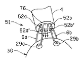
次に図15を参照して本発明の実施例6の消化管内検査装置を説明する。本実施例の消化管内検査装置5Iは、例えば図14において、2つのルーメン52a,52bの代わりに、4つのルーメン52a、52a′、52b、52b′とすると共に、突起部75を設けないで液体注入用ルーメン76を設けたチューブ体4にしている。
なお、このチューブ体4の後端側には、実施例1で説明したチューブ体手元部36に類似したものが接続され、液体注入用ルーメン76を介してヨード液や治療用の薬液を注入することができるようにしたものである。
本実施例は実施例1とほぼ同様の効果を有する。
Next, a digestive tract inspection apparatus according to
The
This embodiment has substantially the same effect as the first embodiment.
次に図16を参照して本発明の実施例7の消化管内検査装置を説明する。本実施例の消化管内検査装置5Jは、透明カバー22の基端付近に貫通孔77を設けたカプセル3Jと、紐状部材6が挿痛されるチューブ体4とにより構成される。この貫通孔77は、カプセル3Jの略直径に近い部分をを貫通するように形成される。
チューブ体4を挿通した紐状部材6を、チューブ体4の先端開口からカプセル3Jに設けた貫通孔77を通すことにより、チューブ体4とカプセル3Jとは紐状部材6を介して連結される。
この場合、貫通孔77は、カプセル3Jの長手方向の中央よりも透明カバー22が設けられた先端側に近い位置に形成されているので、チューブ体4とカプセル3Jとを紐状部材6で連結した状態では、透明カバー22がチューブ体4側に対向する状態となる。また、貫通孔77は観察視野θの外側に形成されている。
本実施例は、実施例1等とは観察方向(撮像方向)が逆となる。観察方向が異なることを除けば、実施例1や実施例2等とほぼ同様の効果を有する。
Next, a digestive tract inspection apparatus according to Embodiment 7 of the present invention will be described with reference to FIG. The gastrointestinal
By passing the string-
In this case, since the through
In this embodiment, the observation direction (imaging direction) is opposite to that of the first embodiment. Except for the fact that the observation directions are different, the present embodiment has substantially the same effects as those of the first embodiment and the second embodiment.
次に図17を参照して本発明の実施例8の消化管内検査装置を説明する。本実施例の消化管内検査装置5Kは、両端に透明カバー22a、22bを設けたカプセル3Kと、第1及び第2の紐状部材6a、6bが挿通されるチューブ体4とから構成される。
透明カバー22aの内側と、透明カバー22bの内側にはそれぞれ図2に示した光学系23及び撮像素子24と照明部25とが配置されている。なお、各光学系による観察範囲をθで示している。
また、透明カバー22a、22bの基端に近い位置の外装部材本体21には、それぞれ貫通孔77a、77bが設けてあり、それぞれ第1及び第2の紐状部材6a、6bが挿通される。
Next, an intestinal tract inspection apparatus according to
The
The exterior member
本実施例による検査方法を図17(A)及び図17(B)を参照して説明する。図17(A)に示すように食道55内のようにカプセル3Kの外径に近い管腔部分内を検査する場合には、カプセル3Kの長手方向が管腔の長手方向に沿うように第1及び第2の紐状部材6a、6bを操作する。
例えば、第1の紐状部材6aを第2の紐状部材6bよりも強く牽引することにより、図17(A)に示すような状態に設定して、食道55内を検査することができる。
これに対して、カプセル3Kのサイズに比べてはるかに大きい胃57内を検査する場合には、第1の紐状部材6aと第2の紐状部材6bとによる牽引力を等しくすることにより、図17(B)に示すように横長の状態に設定できる。
The inspection method according to this embodiment will be described with reference to FIGS. 17 (A) and 17 (B). As shown in FIG. 17A, when inspecting the inside of the lumen portion close to the outer diameter of the
For example, the inside of the
On the other hand, when examining the
この状態で観察した後、例えば第1の紐状部材6aを第2の紐状部材6bよりも強く牽引することにより、2点鎖線で示すようにカプセル3Kを傾けることができる。このように第1の紐状部材6aと第2の紐状部材6bとの牽引の操作を行うことにより、胃57内で観察方向を広く変えて検査することができる。
その他の効果は、実施例1等と同様である。
なお、上述した各実施例等を部分的に組み合わせて構成される実施例等も本発明に属する。また、上述した各実施例では光学的に撮像検査(内視鏡検査)を行う場合で説明したが、薬剤を散布して治療のための医療処置を行うような場合にも適用できる。
After observing in this state, the
Other effects are the same as in the first embodiment.
It should be noted that embodiments configured by partially combining the above-described embodiments also belong to the present invention. Further, in each of the above-described embodiments, the case where optical imaging inspection (endoscopic inspection) is performed has been described. However, the present invention can also be applied to cases where medical treatment for medical treatment is performed by spraying a medicine.
[付記]
0.消化管内の検査が可能なカプセル型内視鏡と、
可撓性のチューブ体と、
前記カプセル型内視鏡に設けられ、前記チューブ体に挿通される可撓性の紐状部材を、着脱可能に連結する(ことにより前記チューブ体と前記カプセル型内視鏡とを分離可能に連結する)連結部と、
を具備したことを特徴とする消化管内検査装置。
a.付記0において、前記連結部は、前記カプセル型内視鏡に設けられた貫通孔である。b.付記0において、前記連結部は、前記カプセル型内視鏡に設けられた凹部である。 1.消化管内の検査が可能なカプセル型内視鏡とカプセル型内視鏡と着脱可能に連結する可撓性のチューブ体とからなる消化管内検査装置において、
カプセル状本体の少なくとも一端に可撓性の紐状部材を挿通できる貫通孔を設け、前記貫通孔に挿通した紐状部材によりカプセル型内視鏡とチューブ体の着脱可能な連結を行うことを特徴とする消化管内検査装置。
[Appendix]
0. A capsule endoscope capable of examining the digestive tract;
A flexible tube body;
A flexible string-like member provided in the capsule endoscope and inserted through the tube body is detachably connected (thereby, the tube body and the capsule endoscope are detachably connected). Connecting part),
A gastrointestinal inspection apparatus characterized by comprising:
a. In
A through-hole through which a flexible string-like member can be inserted is provided in at least one end of the capsule-shaped main body, and the capsule endoscope and the tube body are detachably connected by the string-like member inserted through the through-hole. Gastrointestinal inspection device.
2.付記1において、前記チューブ体の外径は、カプセル型内視鏡の外径に対して十分に細く、且つ、前記紐状部材を内部に挿通した状態で牽引した時の力でも容易に撓まないレベルの可撓性を有することを特徴とする消化管内検査装置。
3.付記1において、前記紐状部材の両端を前記チューブ体の先端から手元端まで挿通し、前記手元端付近に着脱可能に保持する保持手段を具備したことを特徴とする消化管内検査装置。
4.付記1において、前記紐状部材の一端をチューブ体の途中に設けた固定部で引き抜き可能に固定し、前記紐状部材の他端を前記チューブ体の先端から手元端まで挿通したことを特徴とする消化管内検査装置。
2. In
3. 6. The intestinal tract inspection apparatus according to
4). In
5.付記1において、前記貫通孔の周辺部に前記紐状部材を強く引っ張ることで紐状部材を切断可能な鋭利部を設けたことを特徴とする消化管内検査装置。
6.付記1において、前記貫通孔周辺部に前記紐状部材を強く引っ張ることで切れるか、または外装本体から脱落する薄肉部を設けたことを特徴とする消化管内検査装置。
7.付記1〜6において、前記チューブ体の先端部外面に面取り部(R形状の面取り部)を設けたことを特徴とする消化管内検査装置。
8.付記1〜6において、前記チューブ体の先端部に略半球状部を有する(チューブ体とは)別体の先端部材を設けたことを特徴とする消化管内検査装置。
9.付記1〜8において、前記チューブ体の手元端付近に流体(液体・気体)注入口を設けたことを特徴とする消化管内検査装置。
5). 6. The intestinal tract inspection apparatus according to
6). 6. The intestinal tract inspection apparatus according to
7). In appendices 1-6, a gastrointestinal tract inspection apparatus, wherein a chamfered portion (R-shaped chamfered portion) is provided on the outer surface of the distal end portion of the tube body.
8). In
9. In
10.付記1〜9において、前記貫通孔を離れた位置に複数設け、それぞれの貫通孔に挿通した複数本の紐状部材をチューブ体に挿通して手元端から少なくとも各紐状部材の一端を引き出したことを特徴とする消化管内検査装置。
11.付記10において、前記複数本の紐状部材を別々に牽引・弛緩操作を行うことで、カプセル型内視鏡を揺動操作できるように構成したことを特徴とする消化管内検査装置。12.付記1〜11において、前記チューブ体は使用する紐状部材の本数の2倍以上のルーメンを有するマルチルーメンチューブであることを特徴とする消化管内検査装置。
10. In Additional Notes 1-9, a plurality of the through holes are provided at positions apart from each other, and a plurality of string members inserted through the respective through holes are inserted into the tube body, and at least one end of each string member is pulled out from the proximal end. Gastrointestinal tract inspection device characterized by that.
11. The appendage inspection apparatus according to appendix 10, wherein the capsule endoscope is configured to be swingable by separately pulling and relaxing the plurality of string-like members. 12 In
13.付記1〜12において、前記チューブ体は、ブレードなどを編みこんだトルクチューブ、滑り性と硬度を兼ね備えたテフロン(PTFEなど)チューブか、肉厚ポアロンチューブであることを特徴とする消化管内検査装置。
14.付記1〜13において、前記紐状部材は、テフロン糸、手術用糸、テグスなど滑り性が良くて細径でも強度を有する糸であることを特徴とする消化管内検査装置。
15.付記5において、前記紐状部材は、木綿糸や毛糸などの柔らかくて切れやすい糸であることを特徴とする消化管内検査装置。
16.カプセル型内視鏡と可撓性チューブ体を連結した状態で、カプセル型内視鏡を飲み込むステップと、
カプセル型内視鏡が咽頭通過後に、チューブ体を牽引・弛緩操作しながら食道内の所望の部位の内視鏡検査を行うステップと、
所望の位置でチューブ体とカプセル型内視鏡を切り離すステップと、
を有する検査方法。
13. In
14 In Additional remarks 1-13, the said string-like member is a thread | yarn which has good slidability, such as a Teflon thread | yarn, a surgical thread | yarn, and a tegus, and has the intensity | strength even if it is small diameter, The intestinal tract inspection apparatus characterized by the above-mentioned.
15. The appendix inspection apparatus according to
16. Swallowing the capsule endoscope in a state where the capsule endoscope and the flexible tube body are connected;
After the capsule endoscope passes through the pharynx, performing a endoscopic examination of a desired site in the esophagus while pulling and relaxing the tube body;
Separating the tube body and the capsule endoscope at a desired position;
Inspection method having
17.カプセル型内視鏡と可撓性チューブ体を連結した状態で、カプセル型内視鏡を飲み込むステップと、
カプセル型内視鏡が咽頭部を通過した後に、チューブ体を押し引き操作しながら食道内の内視鏡検査を行うステップと、
体外からチューブ内に流体を送り、チューブ体の先端からカプセル型内視鏡の後端付近に流体を放出するステップと、
流体放出後に食道内の内視鏡検査を行うステップと、
からなる検査方法。
18.カプセル型内視鏡と可撓性チューブ体を連結した状態で、カプセル型内視鏡を飲み込むステップと、
カプセル型内視鏡が噴門部通過後に、複数本の紐状部材を操作して、カプセル型内視鏡の視野方向を変更するステップと、
チューブ体とカプセル型内視鏡を切り離し、チューブ体のみ体外に抜き取るステップと、
を有する検査方法。
17. Swallowing the capsule endoscope in a state where the capsule endoscope and the flexible tube body are connected;
After the capsule endoscope has passed through the pharynx, performing endoscopy in the esophagus while pushing and pulling the tube body,
Sending a fluid from outside the body into the tube and discharging the fluid from the tip of the tube body to the vicinity of the rear end of the capsule endoscope;
Performing endoscopy in the esophagus after fluid discharge;
Inspection method consisting of.
18. Swallowing the capsule endoscope in a state where the capsule endoscope and the flexible tube body are connected;
After the capsule endoscope passes through the cardia part, operating a plurality of string members to change the visual field direction of the capsule endoscope; and
Separating the tube body and the capsule endoscope and extracting only the tube body outside the body;
Inspection method having
カプセル型内視鏡の貫通孔等の連結部に、チューブ体を挿通した紐状部材を通して連結した状態で体腔内の食道等の消化管を十分に検査し、その後は連結を解除してカプセル型内視鏡単独で深部側の検査も行える。 Fully inspect the digestive tract such as the esophagus in the body cavity in a state where it is connected to the connecting part such as the through hole of the capsule endoscope through the string member inserted through the tube body, and then the connection is released and the capsule type is released. Inspection of the deep side can be performed with an endoscope alone.
1…カプセル型医療システム
2…患者
3…カプセル(型内視鏡)
4…チューブ体
5…消化管検査装置
6…紐状部材
8…体外ユニット
21…外装部材本体
23…光学系
24…撮像素子
25…照明部
29…貫通孔
36…チューブ体手元部
39、40…シリンジ
43…ヨード液
53…口
54…咽喉
55…食道
57…胃
代理人 弁理士 伊藤 進
DESCRIPTION OF
DESCRIPTION OF
Claims (9)
可撓性のチューブ体と、
前記カプセル型内視鏡に設けられ、前記チューブ体に挿通される可撓性の紐状部材を、着脱可能に連結する連結部と、
を具備したことを特徴とする消化管内検査装置。 A capsule endoscope capable of examining the digestive tract;
A flexible tube body;
A connecting portion provided in the capsule endoscope and removably connecting a flexible string-like member inserted through the tube body;
A gastrointestinal inspection apparatus characterized by comprising:
Priority Applications (5)
| Application Number | Priority Date | Filing Date | Title |
|---|---|---|---|
| JP2003342417A JP3993550B2 (en) | 2003-09-30 | 2003-09-30 | Gastrointestinal inspection device |
| PCT/JP2004/013028 WO2005032352A1 (en) | 2003-09-30 | 2004-09-08 | Digestive tract interior examination instrument |
| CNB2004800285551A CN100446713C (en) | 2003-09-30 | 2004-09-08 | Gastrointestinal tract examining apparatus |
| EP20040787711 EP1671575B1 (en) | 2003-09-30 | 2004-09-08 | Digestive tract interior examination instrument |
| US10/951,100 US7448993B2 (en) | 2003-09-30 | 2004-09-27 | Gastrointestinal tract examining apparatus |
Applications Claiming Priority (1)
| Application Number | Priority Date | Filing Date | Title |
|---|---|---|---|
| JP2003342417A JP3993550B2 (en) | 2003-09-30 | 2003-09-30 | Gastrointestinal inspection device |
Publications (3)
| Publication Number | Publication Date |
|---|---|
| JP2005103092A JP2005103092A (en) | 2005-04-21 |
| JP2005103092A5 JP2005103092A5 (en) | 2006-09-14 |
| JP3993550B2 true JP3993550B2 (en) | 2007-10-17 |
Family
ID=34419261
Family Applications (1)
| Application Number | Title | Priority Date | Filing Date |
|---|---|---|---|
| JP2003342417A Expired - Fee Related JP3993550B2 (en) | 2003-09-30 | 2003-09-30 | Gastrointestinal inspection device |
Country Status (5)
| Country | Link |
|---|---|
| US (1) | US7448993B2 (en) |
| EP (1) | EP1671575B1 (en) |
| JP (1) | JP3993550B2 (en) |
| CN (1) | CN100446713C (en) |
| WO (1) | WO2005032352A1 (en) |
Families Citing this family (82)
| Publication number | Priority date | Publication date | Assignee | Title |
|---|---|---|---|---|
| US7429259B2 (en) | 2003-12-02 | 2008-09-30 | Cadeddu Jeffrey A | Surgical anchor and system |
| JP4445812B2 (en) | 2004-07-08 | 2010-04-07 | オリンパス株式会社 | Intra-subject introduction apparatus and intra-subject introduction system |
| CN100579497C (en) * | 2004-12-28 | 2010-01-13 | 奥林巴斯株式会社 | Introduction aiding apparatus for encapsulated medical device |
| US7530948B2 (en) * | 2005-02-28 | 2009-05-12 | University Of Washington | Tethered capsule endoscope for Barrett's Esophagus screening |
| JP4875315B2 (en) * | 2005-04-05 | 2012-02-15 | オリンパスメディカルシステムズ株式会社 | Intra-subject introduction device |
| WO2007007648A1 (en) * | 2005-07-08 | 2007-01-18 | Olympus Medical Systems Corp. | Apparatus for placing capsule type medical device, apparatus for placing capsule endoscope in the body and capsule type medical device for placement |
| WO2007007724A1 (en) * | 2005-07-08 | 2007-01-18 | Olympus Corporation | Device, system and method for acquiring information in living body |
| WO2007023671A1 (en) * | 2005-08-24 | 2007-03-01 | Konica Minolta Medical & Graphic, Inc. | Capsule-type medical apparatus and diagnosis system |
| US20080015413A1 (en) * | 2006-02-22 | 2008-01-17 | Olympus Medical Systems Corporation | Capsule endoscope system and medical procedure |
| JP4827570B2 (en) * | 2006-03-27 | 2011-11-30 | 佳彦 平尾 | Capsule insertion device |
| US7691103B2 (en) | 2006-04-29 | 2010-04-06 | Board Of Regents, The University Of Texas System | Devices for use in transluminal and endoluminal surgery |
| JP5132564B2 (en) * | 2006-09-12 | 2013-01-30 | オリンパスメディカルシステムズ株式会社 | Capsule endoscope system |
| US9675285B2 (en) * | 2006-10-16 | 2017-06-13 | Given Imaging Ltd. | Delivery device for implantable monitor |
| KR100876647B1 (en) * | 2006-11-22 | 2009-01-08 | 주식회사 코렌 | Capsule type image photographing apparatus and endoscopy using the same |
| WO2008088898A1 (en) | 2007-01-19 | 2008-07-24 | Sierra Scientific Instruments, Inc. | Micro-remote gastrointestinal physiological measurement device |
| US7824270B2 (en) | 2007-01-23 | 2010-11-02 | C-Flex Bearing Co., Inc. | Flexible coupling |
| US20080177141A1 (en) * | 2007-01-24 | 2008-07-24 | Hsien-Ming Wu | Memory-type two-section endoscopic system |
| DE102007000230A1 (en) | 2007-04-16 | 2008-10-30 | Hothan, Thorsten, Dr. | Endoscope for examining intestine of patient i.e. human being, has sphere/retainer, whose weight and/or shape are dimensioned such that sphere/retainer is movable against natural intestinal peristalsis due to net weight of sphere/retainer |
| JP2008307226A (en) * | 2007-06-14 | 2008-12-25 | Olympus Medical Systems Corp | Endoscope system |
| US9339174B2 (en) * | 2007-07-18 | 2016-05-17 | Given Imaging Ltd | Device and method for viewing a body lumen |
| JP5259141B2 (en) * | 2007-08-31 | 2013-08-07 | オリンパスメディカルシステムズ株式会社 | In-subject image acquisition system, in-subject image processing method, and in-subject introduction device |
| US20110282144A1 (en) * | 2008-11-17 | 2011-11-17 | Mayo Foundation For Medical Education And Research | Diagnostic capsules, delivery/retrieval systems, kits and methods |
| WO2010137024A1 (en) * | 2009-05-28 | 2010-12-02 | Given Imaging Ltd. | Apparatus for delivery of autonomous in-vivo capsules |
| WO2011024515A1 (en) * | 2009-08-24 | 2011-03-03 | オリンパスメディカルシステムズ株式会社 | Medical device |
| US20110087224A1 (en) * | 2009-10-09 | 2011-04-14 | Cadeddu Jeffrey A | Magnetic surgical sled with variable arm |
| US9186203B2 (en) | 2009-10-09 | 2015-11-17 | Ethicon Endo-Surgery, Inc. | Method for exchanging end effectors In Vivo |
| US10172669B2 (en) | 2009-10-09 | 2019-01-08 | Ethicon Llc | Surgical instrument comprising an energy trigger lockout |
| US9295485B2 (en) | 2009-10-09 | 2016-03-29 | Ethicon Endo-Surgery, Inc. | Loader for exchanging end effectors in vivo |
| US8623011B2 (en) * | 2009-10-09 | 2014-01-07 | Ethicon Endo-Surgery, Inc. | Magnetic surgical sled with locking arm |
| US8219171B2 (en) * | 2010-03-16 | 2012-07-10 | Given Imaging Ltd. | Delivery device for implantable monitor |
| US8764632B2 (en) | 2010-04-08 | 2014-07-01 | Eric James Kezirian | Endoscopic device and system |
| GB2480498A (en) | 2010-05-21 | 2011-11-23 | Ethicon Endo Surgery Inc | Medical device comprising RF circuitry |
| WO2012008188A1 (en) * | 2010-07-13 | 2012-01-19 | オリンパスメディカルシステムズ株式会社 | Medical device |
| JP4838398B1 (en) * | 2010-07-13 | 2011-12-14 | オリンパスメディカルシステムズ株式会社 | Medical equipment |
| CN203468565U (en) * | 2011-03-10 | 2014-03-12 | 松下电器产业株式会社 | Endoscopic camera and endoscopic device |
| WO2013062978A2 (en) | 2011-10-24 | 2013-05-02 | Ethicon Endo-Surgery, Inc. | Medical instrument |
| EP2596756B1 (en) * | 2011-11-22 | 2014-02-26 | Ovesco Endoscopy AG | Implanting apparatus |
| WO2013145855A1 (en) * | 2012-03-27 | 2013-10-03 | ソニー株式会社 | Capsule-type medical device and medical system |
| US11490797B2 (en) * | 2012-05-21 | 2022-11-08 | The General Hospital Corporation | Apparatus, device and method for capsule microscopy |
| CN102743148B (en) * | 2012-06-21 | 2015-02-25 | 中国人民解放军第一七五医院 | Spray liquid propelling type intestinal tract examination system |
| CN103156568A (en) * | 2012-08-22 | 2013-06-19 | 武汉安康通光电技术有限公司 | Filament tube capsule oesophagoscope capable of being released and magnetically controlled |
| US9125681B2 (en) | 2012-09-26 | 2015-09-08 | Ethicon Endo-Surgery, Inc. | Detachable end effector and loader |
| US9498207B2 (en) | 2012-12-13 | 2016-11-22 | Ethicon Endo-Surgery, Llc | Cartridge interface for surgical suturing device |
| US9451937B2 (en) | 2013-02-27 | 2016-09-27 | Ethicon Endo-Surgery, Llc | Percutaneous instrument with collet locking mechanisms |
| CN103222844B (en) * | 2013-04-25 | 2016-01-27 | 中国人民解放军成都军区总医院 | Controllable capsule endoscopy |
| US9981112B2 (en) * | 2013-11-29 | 2018-05-29 | Sharp Kabushiki Kaisha | Camera system for monitoring inside of body, accessory for support tube of camera system for monitoring inside of body, fixing tool for camera system for monitoring inside of body, and method for installing camera system for monitoring inside of body |
| WO2015116701A1 (en) * | 2014-01-28 | 2015-08-06 | The General Hospital Corporation | Apparatus, systems and methods which controls and facilitates information gathering using a tethered capsule catheter |
| CN103961048A (en) * | 2014-04-16 | 2014-08-06 | 姜泊 | Traction type capsule endoscopy |
| CN103961104A (en) * | 2014-04-16 | 2014-08-06 | 姜泊 | Traction-type recyclable capsule endoscopy |
| CN103961047A (en) * | 2014-04-16 | 2014-08-06 | 姜泊 | Flexible wire traction-type capsule endoscopy and manufacturing method thereof |
| US10159524B2 (en) | 2014-12-22 | 2018-12-25 | Ethicon Llc | High power battery powered RF amplifier topology |
| US10314638B2 (en) | 2015-04-07 | 2019-06-11 | Ethicon Llc | Articulating radio frequency (RF) tissue seal with articulating state sensing |
| CN104905757B (en) * | 2015-06-30 | 2016-08-24 | 中国人民解放军成都军区总医院 | There is the capsule endoscope of ultrasonic sensing |
| CN105011892A (en) * | 2015-06-30 | 2015-11-04 | 中国人民解放军成都军区总医院 | Multiple-pipe capsule endoscope |
| US10314565B2 (en) | 2015-08-26 | 2019-06-11 | Ethicon Llc | Surgical device having actuator biasing and locking features |
| US10335196B2 (en) | 2015-08-31 | 2019-07-02 | Ethicon Llc | Surgical instrument having a stop guard |
| US10251636B2 (en) | 2015-09-24 | 2019-04-09 | Ethicon Llc | Devices and methods for cleaning a surgical device |
| US10702257B2 (en) | 2015-09-29 | 2020-07-07 | Ethicon Llc | Positioning device for use with surgical instruments |
| US10959771B2 (en) | 2015-10-16 | 2021-03-30 | Ethicon Llc | Suction and irrigation sealing grasper |
| US10675009B2 (en) | 2015-11-03 | 2020-06-09 | Ethicon Llc | Multi-head repository for use with a surgical device |
| US10912543B2 (en) | 2015-11-03 | 2021-02-09 | Ethicon Llc | Surgical end effector loading device and trocar integration |
| US10265130B2 (en) | 2015-12-11 | 2019-04-23 | Ethicon Llc | Systems, devices, and methods for coupling end effectors to surgical devices and loading devices |
| US10959806B2 (en) | 2015-12-30 | 2021-03-30 | Ethicon Llc | Energized medical device with reusable handle |
| US10987156B2 (en) | 2016-04-29 | 2021-04-27 | Ethicon Llc | Electrosurgical instrument with electrically conductive gap setting member and electrically insulative tissue engaging members |
| US10856934B2 (en) | 2016-04-29 | 2020-12-08 | Ethicon Llc | Electrosurgical instrument with electrically conductive gap setting and tissue engaging members |
| CN107569204A (en) * | 2016-07-05 | 2018-01-12 | 曾锦顺 | The big colonoscopy of egg shape and traveling control method |
| US10751117B2 (en) | 2016-09-23 | 2020-08-25 | Ethicon Llc | Electrosurgical instrument with fluid diverter |
| CN106580238A (en) * | 2016-12-07 | 2017-04-26 | 广州中医药大学第附属医院 | Device capable of intelligently positioning and identifying throat anatomical structure and control method thereof |
| US11033325B2 (en) | 2017-02-16 | 2021-06-15 | Cilag Gmbh International | Electrosurgical instrument with telescoping suction port and debris cleaner |
| US10799284B2 (en) | 2017-03-15 | 2020-10-13 | Ethicon Llc | Electrosurgical instrument with textured jaws |
| US11497546B2 (en) | 2017-03-31 | 2022-11-15 | Cilag Gmbh International | Area ratios of patterned coatings on RF electrodes to reduce sticking |
| US10603117B2 (en) | 2017-06-28 | 2020-03-31 | Ethicon Llc | Articulation state detection mechanisms |
| US11033323B2 (en) | 2017-09-29 | 2021-06-15 | Cilag Gmbh International | Systems and methods for managing fluid and suction in electrosurgical systems |
| US11484358B2 (en) | 2017-09-29 | 2022-11-01 | Cilag Gmbh International | Flexible electrosurgical instrument |
| US11490951B2 (en) | 2017-09-29 | 2022-11-08 | Cilag Gmbh International | Saline contact with electrodes |
| CN109924937B (en) * | 2018-08-03 | 2024-04-12 | 上海安翰医疗技术有限公司 | Endoscope device and endoscopic detection method |
| JP2022526195A (en) * | 2019-04-09 | 2022-05-23 | アンクス ロボティカ コーポレーション | Systems and methods for delivering liquid biopsy substances and drugs |
| CN111808916A (en) | 2020-07-24 | 2020-10-23 | 上海安翰医疗技术有限公司 | Trypsin detection film, preparation method and application thereof and trypsin detection kit |
| CN113440093B (en) * | 2021-07-19 | 2022-11-25 | 山东第一医科大学附属省立医院(山东省立医院) | Digestive tract secretion microscopic detection system |
| US11957342B2 (en) | 2021-11-01 | 2024-04-16 | Cilag Gmbh International | Devices, systems, and methods for detecting tissue and foreign objects during a surgical operation |
| US20230190084A1 (en) * | 2021-12-16 | 2023-06-22 | Karl Storz Imaging, Inc. | Implantable Internal Observation Device and System |
| CN115399713B (en) * | 2022-05-24 | 2023-03-07 | 北京大学第一医院 | Magnetic control capsule endoscope traction cap and kit with spraying function |
Family Cites Families (25)
| Publication number | Priority date | Publication date | Assignee | Title |
|---|---|---|---|---|
| JPS5394515A (en) * | 1977-01-31 | 1978-08-18 | Kubota Ltd | Method of producing glass fiber reinforced cement plate |
| US5741429A (en) * | 1991-09-05 | 1998-04-21 | Cardia Catheter Company | Flexible tubular device for use in medical applications |
| JP3285235B2 (en) * | 1992-11-05 | 2002-05-27 | オリンパス光学工業株式会社 | Capsule device for in vivo observation |
| US5653677A (en) * | 1994-04-12 | 1997-08-05 | Fuji Photo Optical Co. Ltd | Electronic endoscope apparatus with imaging unit separable therefrom |
| US6071274A (en) * | 1996-12-19 | 2000-06-06 | Ep Technologies, Inc. | Loop structures for supporting multiple electrode elements |
| US5738110A (en) * | 1996-05-29 | 1998-04-14 | Beal; Charles B. | Device for the diagnosis of certain gastrointestinal pathogens |
| IL122716A0 (en) | 1997-12-22 | 1998-08-16 | Tally Eitan Zeev Pearl And Co | System and method for in vivo delivery of autonomous capsule |
| US6632171B2 (en) * | 1997-12-22 | 2003-10-14 | Given Imaging Ltd. | Method for in vivo delivery of autonomous capsule |
| US5984860A (en) | 1998-03-25 | 1999-11-16 | Shan; Yansong | Pass-through duodenal enteroscopic device |
| US6285897B1 (en) * | 1999-04-07 | 2001-09-04 | Endonetics, Inc. | Remote physiological monitoring system |
| JP4716594B2 (en) * | 2000-04-17 | 2011-07-06 | オリンパス株式会社 | Endoscope |
| US6475145B1 (en) * | 2000-05-17 | 2002-11-05 | Baymar, Inc. | Method and apparatus for detection of acid reflux |
| WO2001089596A2 (en) * | 2000-05-23 | 2001-11-29 | Given Imaging Ltd. | Device for positioning object in a body lumen |
| JP2002000556A (en) * | 2000-06-26 | 2002-01-08 | Nonomura Tomosuke | Endoscope |
| US6659981B2 (en) * | 2000-12-08 | 2003-12-09 | Medtronic, Inc. | Medical device delivery catheter with distal locator |
| US6951536B2 (en) * | 2001-07-30 | 2005-10-04 | Olympus Corporation | Capsule-type medical device and medical system |
| US6986738B2 (en) * | 2001-08-06 | 2006-01-17 | Given Imaging Ltd | System and method for maneuvering a device in vivo |
| JP4643089B2 (en) | 2001-09-27 | 2011-03-02 | オリンパス株式会社 | Capsule medical device |
| JP4081259B2 (en) * | 2001-10-30 | 2008-04-23 | オリンパス株式会社 | Endoscope apparatus and endoscope detachment method |
| US20030135266A1 (en) * | 2001-12-03 | 2003-07-17 | Xtent, Inc. | Apparatus and methods for delivery of multiple distributed stents |
| JP2003210393A (en) * | 2002-01-22 | 2003-07-29 | Olympus Optical Co Ltd | Capsule-type medical apparatus |
| US7001329B2 (en) * | 2002-07-23 | 2006-02-21 | Pentax Corporation | Capsule endoscope guidance system, capsule endoscope holder, and capsule endoscope |
| JP4133074B2 (en) | 2002-07-23 | 2008-08-13 | Hoya株式会社 | Capsule endoscope holding mechanism |
| JP4166525B2 (en) | 2002-07-23 | 2008-10-15 | Hoya株式会社 | Capsule endoscope having external terminal and capsule endoscope holder |
| JP4320329B2 (en) | 2006-02-28 | 2009-08-26 | サミー株式会社 | Slot machine |
-
2003
- 2003-09-30 JP JP2003342417A patent/JP3993550B2/en not_active Expired - Fee Related
-
2004
- 2004-09-08 CN CNB2004800285551A patent/CN100446713C/en not_active Expired - Fee Related
- 2004-09-08 WO PCT/JP2004/013028 patent/WO2005032352A1/en active Application Filing
- 2004-09-08 EP EP20040787711 patent/EP1671575B1/en not_active Expired - Lifetime
- 2004-09-27 US US10/951,100 patent/US7448993B2/en active Active
Also Published As
| Publication number | Publication date |
|---|---|
| WO2005032352A1 (en) | 2005-04-14 |
| EP1671575B1 (en) | 2015-05-13 |
| EP1671575A1 (en) | 2006-06-21 |
| EP1671575A4 (en) | 2009-06-17 |
| CN1859866A (en) | 2006-11-08 |
| US7448993B2 (en) | 2008-11-11 |
| JP2005103092A (en) | 2005-04-21 |
| CN100446713C (en) | 2008-12-31 |
| US20050085697A1 (en) | 2005-04-21 |
Similar Documents
| Publication | Publication Date | Title |
|---|---|---|
| JP3993550B2 (en) | Gastrointestinal inspection device | |
| JP4578740B2 (en) | Capsule medical device | |
| JP4081259B2 (en) | Endoscope apparatus and endoscope detachment method | |
| JP4643089B2 (en) | Capsule medical device | |
| EP1992271B1 (en) | Capsule endoscope system | |
| US10064544B2 (en) | Endoscopic capsule and endoscopic system | |
| JP4149838B2 (en) | Capsule medical device | |
| JP4744026B2 (en) | Capsule endoscope and capsule endoscope system | |
| JP4870670B2 (en) | In vivo information acquisition apparatus and in vivo information acquisition system | |
| US20060189844A1 (en) | Endoscopic devide | |
| US8317814B2 (en) | Medical device and process of installing medical device in patient | |
| US8314836B2 (en) | Medical capsule including shake absorption section and indwelled and fixed in vivo | |
| JP2005124708A (en) | Body inside observing device | |
| CN109922705A (en) | The wire rod method for dismounting of endoscope, the wire rod installation method of endoscope and endoscope | |
| JP2003135387A (en) | Capsule type medical apparatus | |
| JP4642424B2 (en) | In-body medical device | |
| JP2007289342A (en) | Endoscope and method of introducing capsule type endoscope | |
| CN110974126B (en) | Capsule endoscope system | |
| JP2006239439A (en) | Capsule type endoscope | |
| JP4937874B2 (en) | In-subject information acquisition system | |
| Swain et al. | New measurement methods and a randomized comparison of force transmission using flexible endoscopes and instruments before and after the application of Shapelock™ technology | |
| Swain et al. | The Cath-Cam. A new concept in colonoscopy | |
| KR20160010019A (en) | Method for input of probiotics using the endoscope | |
| Eloubeidi et al. | Frequency of complications after endoscopic ultrasound-guided fine needle aspiration of solid pancreatic masses: a prospective evaluation | |
| JP2006141809A (en) | Capsule and capsule device for ultrasonic diagnostician |
Legal Events
| Date | Code | Title | Description |
|---|---|---|---|
| A521 | Request for written amendment filed |
Free format text: JAPANESE INTERMEDIATE CODE: A523 Effective date: 20060728 |
|
| A621 | Written request for application examination |
Free format text: JAPANESE INTERMEDIATE CODE: A621 Effective date: 20060728 |
|
| TRDD | Decision of grant or rejection written | ||
| A01 | Written decision to grant a patent or to grant a registration (utility model) |
Free format text: JAPANESE INTERMEDIATE CODE: A01 Effective date: 20070724 |
|
| A61 | First payment of annual fees (during grant procedure) |
Free format text: JAPANESE INTERMEDIATE CODE: A61 Effective date: 20070726 |
|
| FPAY | Renewal fee payment (event date is renewal date of database) |
Free format text: PAYMENT UNTIL: 20100803 Year of fee payment: 3 |
|
| FPAY | Renewal fee payment (event date is renewal date of database) |
Free format text: PAYMENT UNTIL: 20100803 Year of fee payment: 3 |
|
| FPAY | Renewal fee payment (event date is renewal date of database) |
Free format text: PAYMENT UNTIL: 20110803 Year of fee payment: 4 |
|
| FPAY | Renewal fee payment (event date is renewal date of database) |
Free format text: PAYMENT UNTIL: 20120803 Year of fee payment: 5 |
|
| FPAY | Renewal fee payment (event date is renewal date of database) |
Free format text: PAYMENT UNTIL: 20130803 Year of fee payment: 6 |
|
| S531 | Written request for registration of change of domicile |
Free format text: JAPANESE INTERMEDIATE CODE: R313531 |
|
| R350 | Written notification of registration of transfer |
Free format text: JAPANESE INTERMEDIATE CODE: R350 |
|
| LAPS | Cancellation because of no payment of annual fees |